EP2034009A2 - Compositions and methods for restoring immune responsiveness in patients with immunological defects basing on cd3/cd28 costimulation - Google Patents
Compositions and methods for restoring immune responsiveness in patients with immunological defects basing on cd3/cd28 costimulation Download PDFInfo
- Publication number
- EP2034009A2 EP2034009A2 EP08172864A EP08172864A EP2034009A2 EP 2034009 A2 EP2034009 A2 EP 2034009A2 EP 08172864 A EP08172864 A EP 08172864A EP 08172864 A EP08172864 A EP 08172864A EP 2034009 A2 EP2034009 A2 EP 2034009A2
- Authority
- EP
- European Patent Office
- Prior art keywords
- cells
- cell
- population
- disease
- cancer
- Prior art date
- Legal status (The legal status is an assumption and is not a legal conclusion. Google has not performed a legal analysis and makes no representation as to the accuracy of the status listed.)
- Granted
Links
- 238000000034 method Methods 0.000 title claims abstract description 185
- 230000004043 responsiveness Effects 0.000 title claims description 35
- 239000000203 mixture Substances 0.000 title abstract description 54
- 230000001900 immune effect Effects 0.000 title description 4
- 230000007547 defect Effects 0.000 title description 3
- 230000004940 costimulation Effects 0.000 title description 2
- 210000001744 T-lymphocyte Anatomy 0.000 claims abstract description 390
- 210000004027 cell Anatomy 0.000 claims abstract description 291
- 206010028980 Neoplasm Diseases 0.000 claims abstract description 51
- 201000011510 cancer Diseases 0.000 claims abstract description 27
- 238000011282 treatment Methods 0.000 claims abstract description 24
- 239000003795 chemical substances by application Substances 0.000 claims description 100
- 206010061598 Immunodeficiency Diseases 0.000 claims description 66
- 108090000623 proteins and genes Proteins 0.000 claims description 55
- 239000012634 fragment Substances 0.000 claims description 35
- 230000014509 gene expression Effects 0.000 claims description 34
- 108091007433 antigens Proteins 0.000 claims description 29
- 102000036639 antigens Human genes 0.000 claims description 29
- 239000000427 antigen Substances 0.000 claims description 28
- 208000015181 infectious disease Diseases 0.000 claims description 22
- 241000700605 Viruses Species 0.000 claims description 20
- 230000027455 binding Effects 0.000 claims description 18
- 241000725303 Human immunodeficiency virus Species 0.000 claims description 16
- 241000701044 Human gammaherpesvirus 4 Species 0.000 claims description 14
- 206010035226 Plasma cell myeloma Diseases 0.000 claims description 14
- 241000701022 Cytomegalovirus Species 0.000 claims description 12
- 241000700584 Simplexvirus Species 0.000 claims description 12
- 238000001727 in vivo Methods 0.000 claims description 12
- 208000023275 Autoimmune disease Diseases 0.000 claims description 11
- 108020004414 DNA Proteins 0.000 claims description 11
- 241000701806 Human papillomavirus Species 0.000 claims description 11
- 208000029462 Immunodeficiency disease Diseases 0.000 claims description 11
- 208000031261 Acute myeloid leukaemia Diseases 0.000 claims description 10
- 206010067584 Type 1 diabetes mellitus Diseases 0.000 claims description 10
- 230000007813 immunodeficiency Effects 0.000 claims description 10
- 208000035473 Communicable disease Diseases 0.000 claims description 9
- 239000000872 buffer Substances 0.000 claims description 9
- 238000004519 manufacturing process Methods 0.000 claims description 7
- 239000008194 pharmaceutical composition Substances 0.000 claims description 7
- 208000024893 Acute lymphoblastic leukemia Diseases 0.000 claims description 6
- 241000894006 Bacteria Species 0.000 claims description 6
- 206010009944 Colon cancer Diseases 0.000 claims description 6
- 102000053602 DNA Human genes 0.000 claims description 6
- 208000005176 Hepatitis C Diseases 0.000 claims description 6
- 208000034578 Multiple myelomas Diseases 0.000 claims description 6
- 206010033661 Pancytopenia Diseases 0.000 claims description 6
- 108020004682 Single-Stranded DNA Proteins 0.000 claims description 6
- 208000024389 cytopenia Diseases 0.000 claims description 6
- 241001493065 dsRNA viruses Species 0.000 claims description 6
- 208000005252 hepatitis A Diseases 0.000 claims description 6
- 208000002672 hepatitis B Diseases 0.000 claims description 6
- 208000032839 leukemia Diseases 0.000 claims description 6
- 244000045947 parasite Species 0.000 claims description 6
- 208000026872 Addison Disease Diseases 0.000 claims description 5
- 208000032467 Aplastic anaemia Diseases 0.000 claims description 5
- 208000003174 Brain Neoplasms Diseases 0.000 claims description 5
- 206010006187 Breast cancer Diseases 0.000 claims description 5
- 208000026310 Breast neoplasm Diseases 0.000 claims description 5
- 201000009030 Carcinoma Diseases 0.000 claims description 5
- 206010008342 Cervix carcinoma Diseases 0.000 claims description 5
- 208000017667 Chronic Disease Diseases 0.000 claims description 5
- 206010008874 Chronic Fatigue Syndrome Diseases 0.000 claims description 5
- 208000015943 Coeliac disease Diseases 0.000 claims description 5
- 206010009900 Colitis ulcerative Diseases 0.000 claims description 5
- 208000001333 Colorectal Neoplasms Diseases 0.000 claims description 5
- 206010010356 Congenital anomaly Diseases 0.000 claims description 5
- 206010053138 Congenital aplastic anaemia Diseases 0.000 claims description 5
- 208000011231 Crohn disease Diseases 0.000 claims description 5
- 201000004939 Fanconi anemia Diseases 0.000 claims description 5
- 208000001640 Fibromyalgia Diseases 0.000 claims description 5
- 208000032612 Glial tumor Diseases 0.000 claims description 5
- 206010018338 Glioma Diseases 0.000 claims description 5
- 208000003807 Graves Disease Diseases 0.000 claims description 5
- 208000015023 Graves' disease Diseases 0.000 claims description 5
- 208000001204 Hashimoto Disease Diseases 0.000 claims description 5
- 208000030836 Hashimoto thyroiditis Diseases 0.000 claims description 5
- 208000035186 Hemolytic Autoimmune Anemia Diseases 0.000 claims description 5
- 208000017604 Hodgkin disease Diseases 0.000 claims description 5
- 208000010747 Hodgkins lymphoma Diseases 0.000 claims description 5
- 206010020850 Hyperthyroidism Diseases 0.000 claims description 5
- 206010021245 Idiopathic thrombocytopenic purpura Diseases 0.000 claims description 5
- 208000026350 Inborn Genetic disease Diseases 0.000 claims description 5
- 208000022559 Inflammatory bowel disease Diseases 0.000 claims description 5
- 208000008839 Kidney Neoplasms Diseases 0.000 claims description 5
- 206010058467 Lung neoplasm malignant Diseases 0.000 claims description 5
- 208000002454 Nasopharyngeal Carcinoma Diseases 0.000 claims description 5
- 206010061306 Nasopharyngeal cancer Diseases 0.000 claims description 5
- 206010030155 Oesophageal carcinoma Diseases 0.000 claims description 5
- 206010033128 Ovarian cancer Diseases 0.000 claims description 5
- 206010061535 Ovarian neoplasm Diseases 0.000 claims description 5
- 206010061902 Pancreatic neoplasm Diseases 0.000 claims description 5
- 208000007452 Plasmacytoma Diseases 0.000 claims description 5
- 206010060862 Prostate cancer Diseases 0.000 claims description 5
- 208000000236 Prostatic Neoplasms Diseases 0.000 claims description 5
- 201000004681 Psoriasis Diseases 0.000 claims description 5
- 206010038389 Renal cancer Diseases 0.000 claims description 5
- 208000006265 Renal cell carcinoma Diseases 0.000 claims description 5
- 206010039491 Sarcoma Diseases 0.000 claims description 5
- 208000021386 Sjogren Syndrome Diseases 0.000 claims description 5
- 208000031981 Thrombocytopenic Idiopathic Purpura Diseases 0.000 claims description 5
- 201000006704 Ulcerative Colitis Diseases 0.000 claims description 5
- 208000006105 Uterine Cervical Neoplasms Diseases 0.000 claims description 5
- 201000003710 autoimmune thrombocytopenic purpura Diseases 0.000 claims description 5
- 201000010881 cervical cancer Diseases 0.000 claims description 5
- 208000025302 chronic primary adrenal insufficiency Diseases 0.000 claims description 5
- 208000016361 genetic disease Diseases 0.000 claims description 5
- 208000003532 hypothyroidism Diseases 0.000 claims description 5
- 230000002989 hypothyroidism Effects 0.000 claims description 5
- 210000003734 kidney Anatomy 0.000 claims description 5
- 201000010982 kidney cancer Diseases 0.000 claims description 5
- 210000004185 liver Anatomy 0.000 claims description 5
- 201000005202 lung cancer Diseases 0.000 claims description 5
- 208000020816 lung neoplasm Diseases 0.000 claims description 5
- 208000015486 malignant pancreatic neoplasm Diseases 0.000 claims description 5
- 201000001441 melanoma Diseases 0.000 claims description 5
- 201000006417 multiple sclerosis Diseases 0.000 claims description 5
- 208000029766 myalgic encephalomeyelitis/chronic fatigue syndrome Diseases 0.000 claims description 5
- 206010028417 myasthenia gravis Diseases 0.000 claims description 5
- 201000011216 nasopharynx carcinoma Diseases 0.000 claims description 5
- 210000000496 pancreas Anatomy 0.000 claims description 5
- 201000002528 pancreatic cancer Diseases 0.000 claims description 5
- 208000008443 pancreatic carcinoma Diseases 0.000 claims description 5
- 206010039073 rheumatoid arthritis Diseases 0.000 claims description 5
- 201000000596 systemic lupus erythematosus Diseases 0.000 claims description 5
- 208000008732 thymoma Diseases 0.000 claims description 5
- 241000450599 DNA viruses Species 0.000 claims description 4
- 241000233866 Fungi Species 0.000 claims description 4
- 230000001717 pathogenic effect Effects 0.000 claims description 4
- 201000003793 Myelodysplastic syndrome Diseases 0.000 claims description 3
- 230000032683 aging Effects 0.000 claims description 3
- 201000000448 autoimmune hemolytic anemia Diseases 0.000 claims description 3
- 239000003085 diluting agent Substances 0.000 claims description 3
- 208000028018 Lymphocytic leukaemia Diseases 0.000 claims description 2
- 208000003747 lymphoid leukemia Diseases 0.000 claims description 2
- 238000010606 normalization Methods 0.000 claims description 2
- 208000014697 Acute lymphocytic leukaemia Diseases 0.000 claims 2
- 208000010833 Chronic myeloid leukaemia Diseases 0.000 claims 2
- 208000023661 Haematological disease Diseases 0.000 claims 2
- 239000003937 drug carrier Substances 0.000 claims 1
- 230000002265 prevention Effects 0.000 claims 1
- 108091008874 T cell receptors Proteins 0.000 abstract description 65
- 230000000638 stimulation Effects 0.000 abstract description 51
- 230000001965 increasing effect Effects 0.000 abstract description 36
- 230000008569 process Effects 0.000 abstract description 33
- 208000037265 diseases, disorders, signs and symptoms Diseases 0.000 abstract description 23
- 230000004936 stimulating effect Effects 0.000 abstract description 21
- 230000035755 proliferation Effects 0.000 abstract description 16
- 102000005962 receptors Human genes 0.000 abstract description 10
- 108020003175 receptors Proteins 0.000 abstract description 10
- 230000001939 inductive effect Effects 0.000 abstract description 5
- 230000003213 activating effect Effects 0.000 abstract description 4
- 230000019491 signal transduction Effects 0.000 abstract description 4
- 230000002776 aggregation Effects 0.000 abstract description 3
- 238000004220 aggregation Methods 0.000 abstract description 3
- 230000011664 signaling Effects 0.000 abstract description 3
- 208000036142 Viral infection Diseases 0.000 abstract description 2
- 230000009385 viral infection Effects 0.000 abstract description 2
- 230000009258 tissue cross reactivity Effects 0.000 abstract 1
- 239000011324 bead Substances 0.000 description 99
- 239000003446 ligand Substances 0.000 description 66
- 102000016266 T-Cell Antigen Receptors Human genes 0.000 description 63
- 101000914514 Homo sapiens T-cell-specific surface glycoprotein CD28 Proteins 0.000 description 50
- 102100027213 T-cell-specific surface glycoprotein CD28 Human genes 0.000 description 50
- 102000017420 CD3 protein, epsilon/gamma/delta subunit Human genes 0.000 description 49
- 239000002245 particle Substances 0.000 description 41
- 102100036011 T-cell surface glycoprotein CD4 Human genes 0.000 description 34
- 108090000765 processed proteins & peptides Proteins 0.000 description 30
- 238000001994 activation Methods 0.000 description 28
- 230000004913 activation Effects 0.000 description 25
- 102000004196 processed proteins & peptides Human genes 0.000 description 24
- 241001465754 Metazoa Species 0.000 description 23
- 208000032852 chronic lymphocytic leukemia Diseases 0.000 description 22
- 102000004169 proteins and genes Human genes 0.000 description 22
- 230000028993 immune response Effects 0.000 description 20
- 238000004458 analytical method Methods 0.000 description 18
- 201000010099 disease Diseases 0.000 description 18
- 102000004127 Cytokines Human genes 0.000 description 17
- 108090000695 Cytokines Proteins 0.000 description 17
- 210000001616 monocyte Anatomy 0.000 description 17
- 108010002350 Interleukin-2 Proteins 0.000 description 16
- 102000000588 Interleukin-2 Human genes 0.000 description 16
- 229920001184 polypeptide Polymers 0.000 description 16
- -1 PCR based assays Proteins 0.000 description 15
- 238000003752 polymerase chain reaction Methods 0.000 description 15
- LOKCTEFSRHRXRJ-UHFFFAOYSA-I dipotassium trisodium dihydrogen phosphate hydrogen phosphate dichloride Chemical compound P(=O)(O)(O)[O-].[K+].P(=O)(O)([O-])[O-].[Na+].[Na+].[Cl-].[K+].[Cl-].[Na+] LOKCTEFSRHRXRJ-UHFFFAOYSA-I 0.000 description 14
- 239000002953 phosphate buffered saline Substances 0.000 description 14
- 108010074328 Interferon-gamma Proteins 0.000 description 13
- 108060008682 Tumor Necrosis Factor Proteins 0.000 description 13
- 102000000852 Tumor Necrosis Factor-alpha Human genes 0.000 description 13
- 239000000047 product Substances 0.000 description 13
- 239000000243 solution Substances 0.000 description 13
- 108091000054 Prion Proteins 0.000 description 12
- MZOFCQQQCNRIBI-VMXHOPILSA-N (3s)-4-[[(2s)-1-[[(2s)-1-[[(1s)-1-carboxy-2-hydroxyethyl]amino]-4-methyl-1-oxopentan-2-yl]amino]-5-(diaminomethylideneamino)-1-oxopentan-2-yl]amino]-3-[[2-[[(2s)-2,6-diaminohexanoyl]amino]acetyl]amino]-4-oxobutanoic acid Chemical compound OC[C@@H](C(O)=O)NC(=O)[C@H](CC(C)C)NC(=O)[C@H](CCCN=C(N)N)NC(=O)[C@H](CC(O)=O)NC(=O)CNC(=O)[C@@H](N)CCCCN MZOFCQQQCNRIBI-VMXHOPILSA-N 0.000 description 11
- 208000010839 B-cell chronic lymphocytic leukemia Diseases 0.000 description 11
- 102100037850 Interferon gamma Human genes 0.000 description 11
- 102000004388 Interleukin-4 Human genes 0.000 description 11
- 108090000978 Interleukin-4 Proteins 0.000 description 11
- 210000004369 blood Anatomy 0.000 description 11
- 239000008280 blood Substances 0.000 description 11
- 238000011534 incubation Methods 0.000 description 11
- 230000005298 paramagnetic effect Effects 0.000 description 11
- 230000002829 reductive effect Effects 0.000 description 11
- 241000894007 species Species 0.000 description 11
- 230000006044 T cell activation Effects 0.000 description 10
- 210000003719 b-lymphocyte Anatomy 0.000 description 10
- 210000003819 peripheral blood mononuclear cell Anatomy 0.000 description 10
- 239000000126 substance Substances 0.000 description 10
- 102100032937 CD40 ligand Human genes 0.000 description 9
- IAZDPXIOMUYVGZ-UHFFFAOYSA-N Dimethylsulphoxide Chemical compound CS(C)=O IAZDPXIOMUYVGZ-UHFFFAOYSA-N 0.000 description 9
- 238000002617 apheresis Methods 0.000 description 9
- 238000006243 chemical reaction Methods 0.000 description 9
- 210000004408 hybridoma Anatomy 0.000 description 9
- 230000002458 infectious effect Effects 0.000 description 9
- 239000002773 nucleotide Substances 0.000 description 9
- 125000003729 nucleotide group Chemical group 0.000 description 9
- 229920000642 polymer Polymers 0.000 description 9
- 208000006404 Large Granular Lymphocytic Leukemia Diseases 0.000 description 8
- 208000033776 Myeloid Acute Leukemia Diseases 0.000 description 8
- 102000029797 Prion Human genes 0.000 description 8
- 210000000612 antigen-presenting cell Anatomy 0.000 description 8
- 238000002512 chemotherapy Methods 0.000 description 8
- 230000006870 function Effects 0.000 description 8
- JVTAAEKCZFNVCJ-UHFFFAOYSA-N lactic acid Chemical compound CC(O)C(O)=O JVTAAEKCZFNVCJ-UHFFFAOYSA-N 0.000 description 8
- 208000037393 large granular lymphocyte leukemia Diseases 0.000 description 8
- 230000005291 magnetic effect Effects 0.000 description 8
- 201000000050 myeloid neoplasm Diseases 0.000 description 8
- 239000000523 sample Substances 0.000 description 8
- 210000001519 tissue Anatomy 0.000 description 8
- 239000004593 Epoxy Substances 0.000 description 7
- 241000282412 Homo Species 0.000 description 7
- 208000015914 Non-Hodgkin lymphomas Diseases 0.000 description 7
- 230000007423 decrease Effects 0.000 description 7
- 239000012636 effector Substances 0.000 description 7
- 238000000684 flow cytometry Methods 0.000 description 7
- 230000008014 freezing Effects 0.000 description 7
- 238000007710 freezing Methods 0.000 description 7
- 210000000987 immune system Anatomy 0.000 description 7
- 238000001802 infusion Methods 0.000 description 7
- 230000007774 longterm Effects 0.000 description 7
- 210000004698 lymphocyte Anatomy 0.000 description 7
- 229920003023 plastic Polymers 0.000 description 7
- 239000004033 plastic Substances 0.000 description 7
- 230000028327 secretion Effects 0.000 description 7
- 239000006228 supernatant Substances 0.000 description 7
- 238000002560 therapeutic procedure Methods 0.000 description 7
- IJGRMHOSHXDMSA-UHFFFAOYSA-N Atomic nitrogen Chemical compound N#N IJGRMHOSHXDMSA-UHFFFAOYSA-N 0.000 description 6
- 210000001266 CD8-positive T-lymphocyte Anatomy 0.000 description 6
- 201000003874 Common Variable Immunodeficiency Diseases 0.000 description 6
- AEMRFAOFKBGASW-UHFFFAOYSA-N Glycolic acid Chemical compound OCC(O)=O AEMRFAOFKBGASW-UHFFFAOYSA-N 0.000 description 6
- 101000868215 Homo sapiens CD40 ligand Proteins 0.000 description 6
- 208000031422 Lymphocytic Chronic B-Cell Leukemia Diseases 0.000 description 6
- 241001529936 Murinae Species 0.000 description 6
- 230000015572 biosynthetic process Effects 0.000 description 6
- 238000009169 immunotherapy Methods 0.000 description 6
- 230000006698 induction Effects 0.000 description 6
- 238000000926 separation method Methods 0.000 description 6
- 210000002966 serum Anatomy 0.000 description 6
- 208000011580 syndromic disease Diseases 0.000 description 6
- 108091032973 (ribonucleotides)n+m Proteins 0.000 description 5
- 108010017213 Granulocyte-Macrophage Colony-Stimulating Factor Proteins 0.000 description 5
- 102100039620 Granulocyte-macrophage colony-stimulating factor Human genes 0.000 description 5
- 101001057504 Homo sapiens Interferon-stimulated gene 20 kDa protein Proteins 0.000 description 5
- 101001055144 Homo sapiens Interleukin-2 receptor subunit alpha Proteins 0.000 description 5
- 108010021625 Immunoglobulin Fragments Proteins 0.000 description 5
- 102000008394 Immunoglobulin Fragments Human genes 0.000 description 5
- 102100027268 Interferon-stimulated gene 20 kDa protein Human genes 0.000 description 5
- 108010047620 Phytohemagglutinins Proteins 0.000 description 5
- 241000700159 Rattus Species 0.000 description 5
- 230000006052 T cell proliferation Effects 0.000 description 5
- 230000000890 antigenic effect Effects 0.000 description 5
- 238000003556 assay Methods 0.000 description 5
- 230000010261 cell growth Effects 0.000 description 5
- 229940127089 cytotoxic agent Drugs 0.000 description 5
- 239000002254 cytotoxic agent Substances 0.000 description 5
- 231100000599 cytotoxic agent Toxicity 0.000 description 5
- 230000002354 daily effect Effects 0.000 description 5
- 230000000694 effects Effects 0.000 description 5
- 230000012010 growth Effects 0.000 description 5
- 210000002443 helper t lymphocyte Anatomy 0.000 description 5
- 208000014951 hematologic disease Diseases 0.000 description 5
- 239000003018 immunosuppressive agent Substances 0.000 description 5
- 229940125721 immunosuppressive agent Drugs 0.000 description 5
- 230000002401 inhibitory effect Effects 0.000 description 5
- 238000002955 isolation Methods 0.000 description 5
- 239000011159 matrix material Substances 0.000 description 5
- 230000001404 mediated effect Effects 0.000 description 5
- 239000003226 mitogen Substances 0.000 description 5
- 210000001539 phagocyte Anatomy 0.000 description 5
- 230000001885 phytohemagglutinin Effects 0.000 description 5
- 230000008707 rearrangement Effects 0.000 description 5
- 238000011084 recovery Methods 0.000 description 5
- 238000002054 transplantation Methods 0.000 description 5
- 238000005406 washing Methods 0.000 description 5
- 102100034452 Alternative prion protein Human genes 0.000 description 4
- 241000228212 Aspergillus Species 0.000 description 4
- 208000032791 BCR-ABL1 positive chronic myelogenous leukemia Diseases 0.000 description 4
- 241000283690 Bos taurus Species 0.000 description 4
- 241000222120 Candida <Saccharomycetales> Species 0.000 description 4
- 241000283707 Capra Species 0.000 description 4
- 108010047041 Complementarity Determining Regions Proteins 0.000 description 4
- 208000020406 Creutzfeldt Jacob disease Diseases 0.000 description 4
- 208000003407 Creutzfeldt-Jakob Syndrome Diseases 0.000 description 4
- 208000010859 Creutzfeldt-Jakob disease Diseases 0.000 description 4
- PMATZTZNYRCHOR-CGLBZJNRSA-N Cyclosporin A Chemical compound CC[C@@H]1NC(=O)[C@H]([C@H](O)[C@H](C)C\C=C\C)N(C)C(=O)[C@H](C(C)C)N(C)C(=O)[C@H](CC(C)C)N(C)C(=O)[C@H](CC(C)C)N(C)C(=O)[C@@H](C)NC(=O)[C@H](C)NC(=O)[C@H](CC(C)C)N(C)C(=O)[C@H](C(C)C)NC(=O)[C@H](CC(C)C)N(C)C(=O)CN(C)C1=O PMATZTZNYRCHOR-CGLBZJNRSA-N 0.000 description 4
- 108010036949 Cyclosporine Proteins 0.000 description 4
- 208000000461 Esophageal Neoplasms Diseases 0.000 description 4
- 208000003736 Gerstmann-Straussler-Scheinker Disease Diseases 0.000 description 4
- 206010072075 Gerstmann-Straussler-Scheinker syndrome Diseases 0.000 description 4
- 208000031886 HIV Infections Diseases 0.000 description 4
- 208000037357 HIV infectious disease Diseases 0.000 description 4
- 101000851370 Homo sapiens Tumor necrosis factor receptor superfamily member 9 Proteins 0.000 description 4
- 108060003951 Immunoglobulin Proteins 0.000 description 4
- TWRXJAOTZQYOKJ-UHFFFAOYSA-L Magnesium chloride Chemical compound [Mg+2].[Cl-].[Cl-] TWRXJAOTZQYOKJ-UHFFFAOYSA-L 0.000 description 4
- 241001494479 Pecora Species 0.000 description 4
- 241000233872 Pneumocystis carinii Species 0.000 description 4
- 102100036856 Tumor necrosis factor receptor superfamily member 9 Human genes 0.000 description 4
- 208000018756 Variant Creutzfeldt-Jakob disease Diseases 0.000 description 4
- 230000000735 allogeneic effect Effects 0.000 description 4
- 150000001413 amino acids Chemical class 0.000 description 4
- 239000010836 blood and blood product Substances 0.000 description 4
- 210000001772 blood platelet Anatomy 0.000 description 4
- 208000005881 bovine spongiform encephalopathy Diseases 0.000 description 4
- 229960001265 ciclosporin Drugs 0.000 description 4
- 238000013270 controlled release Methods 0.000 description 4
- 229930182912 cyclosporin Natural products 0.000 description 4
- 230000001419 dependent effect Effects 0.000 description 4
- 230000000779 depleting effect Effects 0.000 description 4
- 208000035475 disorder Diseases 0.000 description 4
- 239000003814 drug Substances 0.000 description 4
- 239000002158 endotoxin Substances 0.000 description 4
- 201000004101 esophageal cancer Diseases 0.000 description 4
- 201000006061 fatal familial insomnia Diseases 0.000 description 4
- 239000000835 fiber Substances 0.000 description 4
- 238000001914 filtration Methods 0.000 description 4
- 239000012530 fluid Substances 0.000 description 4
- 108020001507 fusion proteins Proteins 0.000 description 4
- 102000037865 fusion proteins Human genes 0.000 description 4
- 238000001415 gene therapy Methods 0.000 description 4
- 210000003714 granulocyte Anatomy 0.000 description 4
- 239000003102 growth factor Substances 0.000 description 4
- 208000033519 human immunodeficiency virus infectious disease Diseases 0.000 description 4
- 238000002649 immunization Methods 0.000 description 4
- 230000003053 immunization Effects 0.000 description 4
- 230000002163 immunogen Effects 0.000 description 4
- 102000018358 immunoglobulin Human genes 0.000 description 4
- 239000004310 lactic acid Substances 0.000 description 4
- 235000014655 lactic acid Nutrition 0.000 description 4
- 239000002105 nanoparticle Substances 0.000 description 4
- 239000000546 pharmaceutical excipient Substances 0.000 description 4
- 238000001959 radiotherapy Methods 0.000 description 4
- 230000009257 reactivity Effects 0.000 description 4
- 230000009467 reduction Effects 0.000 description 4
- 230000004044 response Effects 0.000 description 4
- 210000004989 spleen cell Anatomy 0.000 description 4
- 238000010561 standard procedure Methods 0.000 description 4
- 230000001225 therapeutic effect Effects 0.000 description 4
- 201000008827 tuberculosis Diseases 0.000 description 4
- 102100034540 Adenomatous polyposis coli protein Human genes 0.000 description 3
- 108010029697 CD40 Ligand Proteins 0.000 description 3
- OYPRJOBELJOOCE-UHFFFAOYSA-N Calcium Chemical compound [Ca] OYPRJOBELJOOCE-UHFFFAOYSA-N 0.000 description 3
- 206010057248 Cell death Diseases 0.000 description 3
- 102000019034 Chemokines Human genes 0.000 description 3
- 108010012236 Chemokines Proteins 0.000 description 3
- 102000008186 Collagen Human genes 0.000 description 3
- 108010035532 Collagen Proteins 0.000 description 3
- 206010010099 Combined immunodeficiency Diseases 0.000 description 3
- 241000699800 Cricetinae Species 0.000 description 3
- 102000010834 Extracellular Matrix Proteins Human genes 0.000 description 3
- 108010037362 Extracellular Matrix Proteins Proteins 0.000 description 3
- 241000282326 Felis catus Species 0.000 description 3
- 101000599852 Homo sapiens Intercellular adhesion molecule 1 Proteins 0.000 description 3
- 101000946889 Homo sapiens Monocyte differentiation antigen CD14 Proteins 0.000 description 3
- 101000914484 Homo sapiens T-lymphocyte activation antigen CD80 Proteins 0.000 description 3
- 108091006905 Human Serum Albumin Proteins 0.000 description 3
- 102000008100 Human Serum Albumin Human genes 0.000 description 3
- 102100022339 Integrin alpha-L Human genes 0.000 description 3
- 102100037877 Intercellular adhesion molecule 1 Human genes 0.000 description 3
- 101150008942 J gene Proteins 0.000 description 3
- 102100033467 L-selectin Human genes 0.000 description 3
- 108010064548 Lymphocyte Function-Associated Antigen-1 Proteins 0.000 description 3
- 241000124008 Mammalia Species 0.000 description 3
- 102000018697 Membrane Proteins Human genes 0.000 description 3
- 108010052285 Membrane Proteins Proteins 0.000 description 3
- 102100035877 Monocyte differentiation antigen CD14 Human genes 0.000 description 3
- 241000699666 Mus <mouse, genus> Species 0.000 description 3
- 108091007491 NSP3 Papain-like protease domains Proteins 0.000 description 3
- 102100027222 T-lymphocyte activation antigen CD80 Human genes 0.000 description 3
- QJJXYPPXXYFBGM-LFZNUXCKSA-N Tacrolimus Chemical compound C1C[C@@H](O)[C@H](OC)C[C@@H]1\C=C(/C)[C@@H]1[C@H](C)[C@@H](O)CC(=O)[C@H](CC=C)/C=C(C)/C[C@H](C)C[C@H](OC)[C@H]([C@H](C[C@H]2C)OC)O[C@@]2(O)C(=O)C(=O)N2CCCC[C@H]2C(=O)O1 QJJXYPPXXYFBGM-LFZNUXCKSA-N 0.000 description 3
- 108010006785 Taq Polymerase Proteins 0.000 description 3
- 101150117115 V gene Proteins 0.000 description 3
- 230000009286 beneficial effect Effects 0.000 description 3
- 230000008901 benefit Effects 0.000 description 3
- 229940125691 blood product Drugs 0.000 description 3
- 238000010322 bone marrow transplantation Methods 0.000 description 3
- 238000010804 cDNA synthesis Methods 0.000 description 3
- 229910052791 calcium Inorganic materials 0.000 description 3
- 239000011575 calcium Substances 0.000 description 3
- 229920001436 collagen Polymers 0.000 description 3
- 239000002299 complementary DNA Substances 0.000 description 3
- 238000007796 conventional method Methods 0.000 description 3
- 229920001577 copolymer Polymers 0.000 description 3
- 239000012228 culture supernatant Substances 0.000 description 3
- 230000016396 cytokine production Effects 0.000 description 3
- 230000009089 cytolysis Effects 0.000 description 3
- 206010012601 diabetes mellitus Diseases 0.000 description 3
- 230000029087 digestion Effects 0.000 description 3
- 238000009826 distribution Methods 0.000 description 3
- 230000003828 downregulation Effects 0.000 description 3
- 229940079593 drug Drugs 0.000 description 3
- 238000003114 enzyme-linked immunosorbent spot assay Methods 0.000 description 3
- 210000003743 erythrocyte Anatomy 0.000 description 3
- 210000002744 extracellular matrix Anatomy 0.000 description 3
- 230000004927 fusion Effects 0.000 description 3
- 239000000499 gel Substances 0.000 description 3
- 238000010353 genetic engineering Methods 0.000 description 3
- 239000011521 glass Substances 0.000 description 3
- 239000001963 growth medium Substances 0.000 description 3
- 229940088597 hormone Drugs 0.000 description 3
- 239000005556 hormone Substances 0.000 description 3
- 230000001976 improved effect Effects 0.000 description 3
- 238000000338 in vitro Methods 0.000 description 3
- 238000002347 injection Methods 0.000 description 3
- 239000007924 injection Substances 0.000 description 3
- 238000001990 intravenous administration Methods 0.000 description 3
- 238000005304 joining Methods 0.000 description 3
- 229920006008 lipopolysaccharide Polymers 0.000 description 3
- 239000002502 liposome Substances 0.000 description 3
- 239000007788 liquid Substances 0.000 description 3
- 239000003550 marker Substances 0.000 description 3
- 239000004005 microsphere Substances 0.000 description 3
- 229910052757 nitrogen Inorganic materials 0.000 description 3
- 231100000252 nontoxic Toxicity 0.000 description 3
- 230000003000 nontoxic effect Effects 0.000 description 3
- 244000052769 pathogen Species 0.000 description 3
- 108091033319 polynucleotide Proteins 0.000 description 3
- 102000040430 polynucleotide Human genes 0.000 description 3
- 239000002157 polynucleotide Substances 0.000 description 3
- 230000002797 proteolythic effect Effects 0.000 description 3
- 230000001105 regulatory effect Effects 0.000 description 3
- QFJCIRLUMZQUOT-HPLJOQBZSA-N sirolimus Chemical compound C1C[C@@H](O)[C@H](OC)C[C@@H]1C[C@@H](C)[C@H]1OC(=O)[C@@H]2CCCCN2C(=O)C(=O)[C@](O)(O2)[C@H](C)CC[C@H]2C[C@H](OC)/C(C)=C/C=C/C=C/[C@@H](C)C[C@@H](C)C(=O)[C@H](OC)[C@H](O)/C(C)=C/[C@@H](C)C(=O)C1 QFJCIRLUMZQUOT-HPLJOQBZSA-N 0.000 description 3
- 210000000130 stem cell Anatomy 0.000 description 3
- 238000003860 storage Methods 0.000 description 3
- 231100000617 superantigen Toxicity 0.000 description 3
- 238000003786 synthesis reaction Methods 0.000 description 3
- QJJXYPPXXYFBGM-SHYZHZOCSA-N tacrolimus Natural products CO[C@H]1C[C@H](CC[C@@H]1O)C=C(C)[C@H]2OC(=O)[C@H]3CCCCN3C(=O)C(=O)[C@@]4(O)O[C@@H]([C@H](C[C@H]4C)OC)[C@@H](C[C@H](C)CC(=C[C@@H](CC=C)C(=O)C[C@H](O)[C@H]2C)C)OC QJJXYPPXXYFBGM-SHYZHZOCSA-N 0.000 description 3
- 238000010361 transduction Methods 0.000 description 3
- 230000026683 transduction Effects 0.000 description 3
- 210000003171 tumor-infiltrating lymphocyte Anatomy 0.000 description 3
- 239000013598 vector Substances 0.000 description 3
- KIUKXJAPPMFGSW-DNGZLQJQSA-N (2S,3S,4S,5R,6R)-6-[(2S,3R,4R,5S,6R)-3-Acetamido-2-[(2S,3S,4R,5R,6R)-6-[(2R,3R,4R,5S,6R)-3-acetamido-2,5-dihydroxy-6-(hydroxymethyl)oxan-4-yl]oxy-2-carboxy-4,5-dihydroxyoxan-3-yl]oxy-5-hydroxy-6-(hydroxymethyl)oxan-4-yl]oxy-3,4,5-trihydroxyoxane-2-carboxylic acid Chemical compound CC(=O)N[C@H]1[C@H](O)O[C@H](CO)[C@@H](O)[C@@H]1O[C@H]1[C@H](O)[C@@H](O)[C@H](O[C@H]2[C@@H]([C@@H](O[C@H]3[C@@H]([C@@H](O)[C@H](O)[C@H](O3)C(O)=O)O)[C@H](O)[C@@H](CO)O2)NC(C)=O)[C@@H](C(O)=O)O1 KIUKXJAPPMFGSW-DNGZLQJQSA-N 0.000 description 2
- FHVDTGUDJYJELY-UHFFFAOYSA-N 6-{[2-carboxy-4,5-dihydroxy-6-(phosphanyloxy)oxan-3-yl]oxy}-4,5-dihydroxy-3-phosphanyloxane-2-carboxylic acid Chemical compound O1C(C(O)=O)C(P)C(O)C(O)C1OC1C(C(O)=O)OC(OP)C(O)C1O FHVDTGUDJYJELY-UHFFFAOYSA-N 0.000 description 2
- HGINCPLSRVDWNT-UHFFFAOYSA-N Acrolein Chemical compound C=CC=O HGINCPLSRVDWNT-UHFFFAOYSA-N 0.000 description 2
- HRPVXLWXLXDGHG-UHFFFAOYSA-N Acrylamide Chemical compound NC(=O)C=C HRPVXLWXLXDGHG-UHFFFAOYSA-N 0.000 description 2
- 229920000936 Agarose Polymers 0.000 description 2
- 102100022005 B-lymphocyte antigen CD20 Human genes 0.000 description 2
- 102000004506 Blood Proteins Human genes 0.000 description 2
- 108010017384 Blood Proteins Proteins 0.000 description 2
- 208000024172 Cardiovascular disease Diseases 0.000 description 2
- 108020004635 Complementary DNA Proteins 0.000 description 2
- 229930105110 Cyclosporin A Natural products 0.000 description 2
- 101150097493 D gene Proteins 0.000 description 2
- 229920002307 Dextran Polymers 0.000 description 2
- 201000009273 Endometriosis Diseases 0.000 description 2
- 241000588724 Escherichia coli Species 0.000 description 2
- 208000004332 Evans syndrome Diseases 0.000 description 2
- 229940117942 Factor IX inhibitor Drugs 0.000 description 2
- 229940124135 Factor VIII inhibitor Drugs 0.000 description 2
- 229920001917 Ficoll Polymers 0.000 description 2
- ZHNUHDYFZUAESO-UHFFFAOYSA-N Formamide Chemical compound NC=O ZHNUHDYFZUAESO-UHFFFAOYSA-N 0.000 description 2
- 206010018364 Glomerulonephritis Diseases 0.000 description 2
- 208000024869 Goodpasture syndrome Diseases 0.000 description 2
- 101000897405 Homo sapiens B-lymphocyte antigen CD20 Proteins 0.000 description 2
- 108010054477 Immunoglobulin Fab Fragments Proteins 0.000 description 2
- 102000001706 Immunoglobulin Fab Fragments Human genes 0.000 description 2
- 108700005091 Immunoglobulin Genes Proteins 0.000 description 2
- 108010067060 Immunoglobulin Variable Region Proteins 0.000 description 2
- 102000008070 Interferon-gamma Human genes 0.000 description 2
- 108010092694 L-Selectin Proteins 0.000 description 2
- 108090001090 Lectins Proteins 0.000 description 2
- 102000004856 Lectins Human genes 0.000 description 2
- 241000222722 Leishmania <genus> Species 0.000 description 2
- FYYHWMGAXLPEAU-UHFFFAOYSA-N Magnesium Chemical compound [Mg] FYYHWMGAXLPEAU-UHFFFAOYSA-N 0.000 description 2
- 241000699660 Mus musculus Species 0.000 description 2
- 241000699670 Mus sp. Species 0.000 description 2
- 208000012902 Nervous system disease Diseases 0.000 description 2
- 108091034117 Oligonucleotide Proteins 0.000 description 2
- 238000012408 PCR amplification Methods 0.000 description 2
- 208000000733 Paroxysmal Hemoglobinuria Diseases 0.000 description 2
- 208000031845 Pernicious anaemia Diseases 0.000 description 2
- 102100036050 Phosphatidylinositol N-acetylglucosaminyltransferase subunit A Human genes 0.000 description 2
- 239000004743 Polypropylene Substances 0.000 description 2
- 208000024777 Prion disease Diseases 0.000 description 2
- 239000012980 RPMI-1640 medium Substances 0.000 description 2
- 108020004511 Recombinant DNA Proteins 0.000 description 2
- 241000607142 Salmonella Species 0.000 description 2
- 241000242678 Schistosoma Species 0.000 description 2
- 206010039710 Scleroderma Diseases 0.000 description 2
- 229920002684 Sepharose Polymers 0.000 description 2
- VYPSYNLAJGMNEJ-UHFFFAOYSA-N Silicium dioxide Chemical compound O=[Si]=O VYPSYNLAJGMNEJ-UHFFFAOYSA-N 0.000 description 2
- 241000295644 Staphylococcaceae Species 0.000 description 2
- 241000191940 Staphylococcus Species 0.000 description 2
- 208000004732 Systemic Vasculitis Diseases 0.000 description 2
- IQFYYKKMVGJFEH-XLPZGREQSA-N Thymidine Chemical compound O=C1NC(=O)C(C)=CN1[C@@H]1O[C@H](CO)[C@@H](O)C1 IQFYYKKMVGJFEH-XLPZGREQSA-N 0.000 description 2
- 101710101607 Toxic shock syndrome toxin-1 Proteins 0.000 description 2
- 241000223104 Trypanosoma Species 0.000 description 2
- 206010047115 Vasculitis Diseases 0.000 description 2
- JLCPHMBAVCMARE-UHFFFAOYSA-N [3-[[3-[[3-[[3-[[3-[[3-[[3-[[3-[[3-[[3-[[3-[[5-(2-amino-6-oxo-1H-purin-9-yl)-3-[[3-[[3-[[3-[[3-[[3-[[5-(2-amino-6-oxo-1H-purin-9-yl)-3-[[5-(2-amino-6-oxo-1H-purin-9-yl)-3-hydroxyoxolan-2-yl]methoxy-hydroxyphosphoryl]oxyoxolan-2-yl]methoxy-hydroxyphosphoryl]oxy-5-(5-methyl-2,4-dioxopyrimidin-1-yl)oxolan-2-yl]methoxy-hydroxyphosphoryl]oxy-5-(6-aminopurin-9-yl)oxolan-2-yl]methoxy-hydroxyphosphoryl]oxy-5-(6-aminopurin-9-yl)oxolan-2-yl]methoxy-hydroxyphosphoryl]oxy-5-(6-aminopurin-9-yl)oxolan-2-yl]methoxy-hydroxyphosphoryl]oxy-5-(6-aminopurin-9-yl)oxolan-2-yl]methoxy-hydroxyphosphoryl]oxyoxolan-2-yl]methoxy-hydroxyphosphoryl]oxy-5-(5-methyl-2,4-dioxopyrimidin-1-yl)oxolan-2-yl]methoxy-hydroxyphosphoryl]oxy-5-(4-amino-2-oxopyrimidin-1-yl)oxolan-2-yl]methoxy-hydroxyphosphoryl]oxy-5-(5-methyl-2,4-dioxopyrimidin-1-yl)oxolan-2-yl]methoxy-hydroxyphosphoryl]oxy-5-(5-methyl-2,4-dioxopyrimidin-1-yl)oxolan-2-yl]methoxy-hydroxyphosphoryl]oxy-5-(6-aminopurin-9-yl)oxolan-2-yl]methoxy-hydroxyphosphoryl]oxy-5-(6-aminopurin-9-yl)oxolan-2-yl]methoxy-hydroxyphosphoryl]oxy-5-(4-amino-2-oxopyrimidin-1-yl)oxolan-2-yl]methoxy-hydroxyphosphoryl]oxy-5-(4-amino-2-oxopyrimidin-1-yl)oxolan-2-yl]methoxy-hydroxyphosphoryl]oxy-5-(4-amino-2-oxopyrimidin-1-yl)oxolan-2-yl]methoxy-hydroxyphosphoryl]oxy-5-(6-aminopurin-9-yl)oxolan-2-yl]methoxy-hydroxyphosphoryl]oxy-5-(4-amino-2-oxopyrimidin-1-yl)oxolan-2-yl]methyl [5-(6-aminopurin-9-yl)-2-(hydroxymethyl)oxolan-3-yl] hydrogen phosphate Polymers Cc1cn(C2CC(OP(O)(=O)OCC3OC(CC3OP(O)(=O)OCC3OC(CC3O)n3cnc4c3nc(N)[nH]c4=O)n3cnc4c3nc(N)[nH]c4=O)C(COP(O)(=O)OC3CC(OC3COP(O)(=O)OC3CC(OC3COP(O)(=O)OC3CC(OC3COP(O)(=O)OC3CC(OC3COP(O)(=O)OC3CC(OC3COP(O)(=O)OC3CC(OC3COP(O)(=O)OC3CC(OC3COP(O)(=O)OC3CC(OC3COP(O)(=O)OC3CC(OC3COP(O)(=O)OC3CC(OC3COP(O)(=O)OC3CC(OC3COP(O)(=O)OC3CC(OC3COP(O)(=O)OC3CC(OC3COP(O)(=O)OC3CC(OC3COP(O)(=O)OC3CC(OC3COP(O)(=O)OC3CC(OC3COP(O)(=O)OC3CC(OC3CO)n3cnc4c(N)ncnc34)n3ccc(N)nc3=O)n3cnc4c(N)ncnc34)n3ccc(N)nc3=O)n3ccc(N)nc3=O)n3ccc(N)nc3=O)n3cnc4c(N)ncnc34)n3cnc4c(N)ncnc34)n3cc(C)c(=O)[nH]c3=O)n3cc(C)c(=O)[nH]c3=O)n3ccc(N)nc3=O)n3cc(C)c(=O)[nH]c3=O)n3cnc4c3nc(N)[nH]c4=O)n3cnc4c(N)ncnc34)n3cnc4c(N)ncnc34)n3cnc4c(N)ncnc34)n3cnc4c(N)ncnc34)O2)c(=O)[nH]c1=O JLCPHMBAVCMARE-UHFFFAOYSA-N 0.000 description 2
- 238000010317 ablation therapy Methods 0.000 description 2
- 239000002671 adjuvant Substances 0.000 description 2
- 238000001042 affinity chromatography Methods 0.000 description 2
- 229940072056 alginate Drugs 0.000 description 2
- 229920000615 alginic acid Polymers 0.000 description 2
- 235000010443 alginic acid Nutrition 0.000 description 2
- 230000029936 alkylation Effects 0.000 description 2
- 238000005804 alkylation reaction Methods 0.000 description 2
- 230000003321 amplification Effects 0.000 description 2
- 238000000137 annealing Methods 0.000 description 2
- 230000000259 anti-tumor effect Effects 0.000 description 2
- 238000013459 approach Methods 0.000 description 2
- 210000001106 artificial yeast chromosome Anatomy 0.000 description 2
- 239000011230 binding agent Substances 0.000 description 2
- 238000004820 blood count Methods 0.000 description 2
- 229940112129 campath Drugs 0.000 description 2
- 150000001768 cations Chemical class 0.000 description 2
- 230000020411 cell activation Effects 0.000 description 2
- 238000004113 cell culture Methods 0.000 description 2
- 239000006143 cell culture medium Substances 0.000 description 2
- 230000011712 cell development Effects 0.000 description 2
- 239000006285 cell suspension Substances 0.000 description 2
- 238000001311 chemical methods and process Methods 0.000 description 2
- 239000003246 corticosteroid Substances 0.000 description 2
- 238000004132 cross linking Methods 0.000 description 2
- OPTASPLRGRRNAP-UHFFFAOYSA-N cytosine Chemical compound NC=1C=CNC(=O)N=1 OPTASPLRGRRNAP-UHFFFAOYSA-N 0.000 description 2
- 210000001151 cytotoxic T lymphocyte Anatomy 0.000 description 2
- 230000003247 decreasing effect Effects 0.000 description 2
- 230000003111 delayed effect Effects 0.000 description 2
- 238000004925 denaturation Methods 0.000 description 2
- 230000036425 denaturation Effects 0.000 description 2
- 201000001981 dermatomyositis Diseases 0.000 description 2
- 238000013461 design Methods 0.000 description 2
- 238000011161 development Methods 0.000 description 2
- 230000018109 developmental process Effects 0.000 description 2
- 238000001962 electrophoresis Methods 0.000 description 2
- 230000002255 enzymatic effect Effects 0.000 description 2
- 238000002474 experimental method Methods 0.000 description 2
- 208000037957 feline spongiform encephalopathy Diseases 0.000 description 2
- 210000004700 fetal blood Anatomy 0.000 description 2
- 239000012847 fine chemical Substances 0.000 description 2
- 230000037406 food intake Effects 0.000 description 2
- 150000004676 glycans Chemical class 0.000 description 2
- UYTPUPDQBNUYGX-UHFFFAOYSA-N guanine Chemical compound O=C1NC(N)=NC2=C1N=CN2 UYTPUPDQBNUYGX-UHFFFAOYSA-N 0.000 description 2
- 229920002674 hyaluronan Polymers 0.000 description 2
- 229960003160 hyaluronic acid Drugs 0.000 description 2
- 229910052588 hydroxylapatite Inorganic materials 0.000 description 2
- FDGQSTZJBFJUBT-UHFFFAOYSA-N hypoxanthine Chemical compound O=C1NC=NC2=C1NC=N2 FDGQSTZJBFJUBT-UHFFFAOYSA-N 0.000 description 2
- 239000007943 implant Substances 0.000 description 2
- 239000003112 inhibitor Substances 0.000 description 2
- 238000003780 insertion Methods 0.000 description 2
- 230000037431 insertion Effects 0.000 description 2
- NOESYZHRGYRDHS-UHFFFAOYSA-N insulin Chemical compound N1C(=O)C(NC(=O)C(CCC(N)=O)NC(=O)C(CCC(O)=O)NC(=O)C(C(C)C)NC(=O)C(NC(=O)CN)C(C)CC)CSSCC(C(NC(CO)C(=O)NC(CC(C)C)C(=O)NC(CC=2C=CC(O)=CC=2)C(=O)NC(CCC(N)=O)C(=O)NC(CC(C)C)C(=O)NC(CCC(O)=O)C(=O)NC(CC(N)=O)C(=O)NC(CC=2C=CC(O)=CC=2)C(=O)NC(CSSCC(NC(=O)C(C(C)C)NC(=O)C(CC(C)C)NC(=O)C(CC=2C=CC(O)=CC=2)NC(=O)C(CC(C)C)NC(=O)C(C)NC(=O)C(CCC(O)=O)NC(=O)C(C(C)C)NC(=O)C(CC(C)C)NC(=O)C(CC=2NC=NC=2)NC(=O)C(CO)NC(=O)CNC2=O)C(=O)NCC(=O)NC(CCC(O)=O)C(=O)NC(CCCNC(N)=N)C(=O)NCC(=O)NC(CC=3C=CC=CC=3)C(=O)NC(CC=3C=CC=CC=3)C(=O)NC(CC=3C=CC(O)=CC=3)C(=O)NC(C(C)O)C(=O)N3C(CCC3)C(=O)NC(CCCCN)C(=O)NC(C)C(O)=O)C(=O)NC(CC(N)=O)C(O)=O)=O)NC(=O)C(C(C)CC)NC(=O)C(CO)NC(=O)C(C(C)O)NC(=O)C1CSSCC2NC(=O)C(CC(C)C)NC(=O)C(NC(=O)C(CCC(N)=O)NC(=O)C(CC(N)=O)NC(=O)C(NC(=O)C(N)CC=1C=CC=CC=1)C(C)C)CC1=CN=CN1 NOESYZHRGYRDHS-UHFFFAOYSA-N 0.000 description 2
- 229960003130 interferon gamma Drugs 0.000 description 2
- 206010023497 kuru Diseases 0.000 description 2
- 239000002523 lectin Substances 0.000 description 2
- 210000000265 leukocyte Anatomy 0.000 description 2
- 230000000670 limiting effect Effects 0.000 description 2
- 150000002632 lipids Chemical class 0.000 description 2
- 210000001165 lymph node Anatomy 0.000 description 2
- 210000003563 lymphoid tissue Anatomy 0.000 description 2
- 229910052749 magnesium Inorganic materials 0.000 description 2
- 239000011777 magnesium Substances 0.000 description 2
- 229910001629 magnesium chloride Inorganic materials 0.000 description 2
- 239000006249 magnetic particle Substances 0.000 description 2
- 238000007885 magnetic separation Methods 0.000 description 2
- 239000006148 magnetic separator Substances 0.000 description 2
- 230000003211 malignant effect Effects 0.000 description 2
- 238000005259 measurement Methods 0.000 description 2
- 239000012528 membrane Substances 0.000 description 2
- 210000004379 membrane Anatomy 0.000 description 2
- 229910052751 metal Inorganic materials 0.000 description 2
- 239000002184 metal Substances 0.000 description 2
- HPNSFSBZBAHARI-UHFFFAOYSA-N micophenolic acid Natural products OC1=C(CC=C(C)CCC(O)=O)C(OC)=C(C)C2=C1C(=O)OC2 HPNSFSBZBAHARI-UHFFFAOYSA-N 0.000 description 2
- 238000002156 mixing Methods 0.000 description 2
- 238000003032 molecular docking Methods 0.000 description 2
- HPNSFSBZBAHARI-RUDMXATFSA-N mycophenolic acid Chemical compound OC1=C(C\C=C(/C)CCC(O)=O)C(OC)=C(C)C2=C1C(=O)OC2 HPNSFSBZBAHARI-RUDMXATFSA-N 0.000 description 2
- 210000000653 nervous system Anatomy 0.000 description 2
- 208000015122 neurodegenerative disease Diseases 0.000 description 2
- 238000003199 nucleic acid amplification method Methods 0.000 description 2
- 150000007523 nucleic acids Chemical class 0.000 description 2
- 201000003045 paroxysmal nocturnal hemoglobinuria Diseases 0.000 description 2
- XYJRXVWERLGGKC-UHFFFAOYSA-D pentacalcium;hydroxide;triphosphate Chemical compound [OH-].[Ca+2].[Ca+2].[Ca+2].[Ca+2].[Ca+2].[O-]P([O-])([O-])=O.[O-]P([O-])([O-])=O.[O-]P([O-])([O-])=O XYJRXVWERLGGKC-UHFFFAOYSA-D 0.000 description 2
- 239000011886 peripheral blood Substances 0.000 description 2
- 210000005259 peripheral blood Anatomy 0.000 description 2
- 230000002399 phagocytotic effect Effects 0.000 description 2
- 239000012071 phase Substances 0.000 description 2
- PHEDXBVPIONUQT-RGYGYFBISA-N phorbol 13-acetate 12-myristate Chemical compound C([C@]1(O)C(=O)C(C)=C[C@H]1[C@@]1(O)[C@H](C)[C@H]2OC(=O)CCCCCCCCCCCCC)C(CO)=C[C@H]1[C@H]1[C@]2(OC(C)=O)C1(C)C PHEDXBVPIONUQT-RGYGYFBISA-N 0.000 description 2
- 239000013612 plasmid Substances 0.000 description 2
- 229920002401 polyacrylamide Polymers 0.000 description 2
- 238000006116 polymerization reaction Methods 0.000 description 2
- 208000005987 polymyositis Diseases 0.000 description 2
- 229920001155 polypropylene Polymers 0.000 description 2
- 229920001282 polysaccharide Polymers 0.000 description 2
- 239000005017 polysaccharide Substances 0.000 description 2
- 229920001343 polytetrafluoroethylene Polymers 0.000 description 2
- 230000003389 potentiating effect Effects 0.000 description 2
- 238000012257 pre-denaturation Methods 0.000 description 2
- 238000002360 preparation method Methods 0.000 description 2
- 238000012545 processing Methods 0.000 description 2
- ZAHRKKWIAAJSAO-UHFFFAOYSA-N rapamycin Natural products COCC(O)C(=C/C(C)C(=O)CC(OC(=O)C1CCCCN1C(=O)C(=O)C2(O)OC(CC(OC)C(=CC=CC=CC(C)CC(C)C(=O)C)C)CCC2C)C(C)CC3CCC(O)C(C3)OC)C ZAHRKKWIAAJSAO-UHFFFAOYSA-N 0.000 description 2
- 230000006798 recombination Effects 0.000 description 2
- 238000005215 recombination Methods 0.000 description 2
- 230000010076 replication Effects 0.000 description 2
- 230000000717 retained effect Effects 0.000 description 2
- 201000003068 rheumatic fever Diseases 0.000 description 2
- 208000008864 scrapie Diseases 0.000 description 2
- 229960002930 sirolimus Drugs 0.000 description 2
- 150000003384 small molecules Chemical class 0.000 description 2
- 239000007787 solid Substances 0.000 description 2
- 125000006850 spacer group Chemical group 0.000 description 2
- 238000011476 stem cell transplantation Methods 0.000 description 2
- 150000003431 steroids Chemical class 0.000 description 2
- UCSJYZPVAKXKNQ-HZYVHMACSA-N streptomycin Chemical compound CN[C@H]1[C@H](O)[C@@H](O)[C@H](CO)O[C@H]1O[C@@H]1[C@](C=O)(O)[C@H](C)O[C@H]1O[C@@H]1[C@@H](NC(N)=N)[C@H](O)[C@@H](NC(N)=N)[C@H](O)[C@H]1O UCSJYZPVAKXKNQ-HZYVHMACSA-N 0.000 description 2
- RWQNBRDOKXIBIV-UHFFFAOYSA-N thymine Chemical compound CC1=CNC(=O)NC1=O RWQNBRDOKXIBIV-UHFFFAOYSA-N 0.000 description 2
- 210000001541 thymus gland Anatomy 0.000 description 2
- 238000011830 transgenic mouse model Methods 0.000 description 2
- 230000003827 upregulation Effects 0.000 description 2
- 229960005486 vaccine Drugs 0.000 description 2
- 230000003612 virological effect Effects 0.000 description 2
- JKMHFZQWWAIEOD-UHFFFAOYSA-N 2-[4-(2-hydroxyethyl)piperazin-1-yl]ethanesulfonic acid Chemical compound OCC[NH+]1CCN(CCS([O-])(=O)=O)CC1 JKMHFZQWWAIEOD-UHFFFAOYSA-N 0.000 description 1
- TVZGACDUOSZQKY-LBPRGKRZSA-N 4-aminofolic acid Chemical compound C1=NC2=NC(N)=NC(N)=C2N=C1CNC1=CC=C(C(=O)N[C@@H](CCC(O)=O)C(O)=O)C=C1 TVZGACDUOSZQKY-LBPRGKRZSA-N 0.000 description 1
- 108010013238 70-kDa Ribosomal Protein S6 Kinases Proteins 0.000 description 1
- 229930024421 Adenine Natural products 0.000 description 1
- GFFGJBXGBJISGV-UHFFFAOYSA-N Adenine Chemical compound NC1=NC=NC2=C1N=CN2 GFFGJBXGBJISGV-UHFFFAOYSA-N 0.000 description 1
- 102100021569 Apoptosis regulator Bcl-2 Human genes 0.000 description 1
- 206010003445 Ascites Diseases 0.000 description 1
- 108090001008 Avidin Proteins 0.000 description 1
- 108091008875 B cell receptors Proteins 0.000 description 1
- 208000004736 B-Cell Leukemia Diseases 0.000 description 1
- DWRXFEITVBNRMK-UHFFFAOYSA-N Beta-D-1-Arabinofuranosylthymine Natural products O=C1NC(=O)C(C)=CN1C1C(O)C(O)C(CO)O1 DWRXFEITVBNRMK-UHFFFAOYSA-N 0.000 description 1
- 108091003079 Bovine Serum Albumin Proteins 0.000 description 1
- 108700012434 CCL3 Proteins 0.000 description 1
- 101150013553 CD40 gene Proteins 0.000 description 1
- 108010084313 CD58 Antigens Proteins 0.000 description 1
- 102000004631 Calcineurin Human genes 0.000 description 1
- 108010042955 Calcineurin Proteins 0.000 description 1
- 241000282472 Canis lupus familiaris Species 0.000 description 1
- 102000000844 Cell Surface Receptors Human genes 0.000 description 1
- 108010001857 Cell Surface Receptors Proteins 0.000 description 1
- 102000000013 Chemokine CCL3 Human genes 0.000 description 1
- 102000001327 Chemokine CCL5 Human genes 0.000 description 1
- 108010055166 Chemokine CCL5 Proteins 0.000 description 1
- 229920002101 Chitin Polymers 0.000 description 1
- 229920001661 Chitosan Polymers 0.000 description 1
- 108020004705 Codon Proteins 0.000 description 1
- 108010062580 Concanavalin A Proteins 0.000 description 1
- CMSMOCZEIVJLDB-UHFFFAOYSA-N Cyclophosphamide Chemical compound ClCCN(CCCl)P1(=O)NCCCO1 CMSMOCZEIVJLDB-UHFFFAOYSA-N 0.000 description 1
- FBPFZTCFMRRESA-KVTDHHQDSA-N D-Mannitol Chemical compound OC[C@@H](O)[C@@H](O)[C@H](O)[C@H](O)CO FBPFZTCFMRRESA-KVTDHHQDSA-N 0.000 description 1
- WQZGKKKJIJFFOK-QTVWNMPRSA-N D-mannopyranose Chemical compound OC[C@H]1OC(O)[C@@H](O)[C@@H](O)[C@@H]1O WQZGKKKJIJFFOK-QTVWNMPRSA-N 0.000 description 1
- 108010008286 DNA nucleotidylexotransferase Proteins 0.000 description 1
- 102100029764 DNA-directed DNA/RNA polymerase mu Human genes 0.000 description 1
- 239000006144 Dulbecco’s modified Eagle's medium Substances 0.000 description 1
- KCXVZYZYPLLWCC-UHFFFAOYSA-N EDTA Chemical compound OC(=O)CN(CC(O)=O)CCN(CC(O)=O)CC(O)=O KCXVZYZYPLLWCC-UHFFFAOYSA-N 0.000 description 1
- 101710146739 Enterotoxin Proteins 0.000 description 1
- 102000004190 Enzymes Human genes 0.000 description 1
- 108090000790 Enzymes Proteins 0.000 description 1
- 101000867232 Escherichia coli Heat-stable enterotoxin II Proteins 0.000 description 1
- 108060002716 Exonuclease Proteins 0.000 description 1
- 108010087819 Fc receptors Proteins 0.000 description 1
- 102000009109 Fc receptors Human genes 0.000 description 1
- WQZGKKKJIJFFOK-GASJEMHNSA-N Glucose Natural products OC[C@H]1OC(O)[C@H](O)[C@@H](O)[C@@H]1O WQZGKKKJIJFFOK-GASJEMHNSA-N 0.000 description 1
- DHMQDGOQFOQNFH-UHFFFAOYSA-N Glycine Chemical compound NCC(O)=O DHMQDGOQFOQNFH-UHFFFAOYSA-N 0.000 description 1
- 102000002068 Glycopeptides Human genes 0.000 description 1
- 108010015899 Glycopeptides Proteins 0.000 description 1
- 102000003886 Glycoproteins Human genes 0.000 description 1
- 108090000288 Glycoproteins Proteins 0.000 description 1
- 229920002683 Glycosaminoglycan Polymers 0.000 description 1
- 108010051696 Growth Hormone Proteins 0.000 description 1
- 102000006354 HLA-DR Antigens Human genes 0.000 description 1
- 108010058597 HLA-DR Antigens Proteins 0.000 description 1
- 102100031573 Hematopoietic progenitor cell antigen CD34 Human genes 0.000 description 1
- 208000032843 Hemorrhage Diseases 0.000 description 1
- 101000971171 Homo sapiens Apoptosis regulator Bcl-2 Proteins 0.000 description 1
- 101000777663 Homo sapiens Hematopoietic progenitor cell antigen CD34 Proteins 0.000 description 1
- 101001046686 Homo sapiens Integrin alpha-M Proteins 0.000 description 1
- 101001018097 Homo sapiens L-selectin Proteins 0.000 description 1
- 101000917858 Homo sapiens Low affinity immunoglobulin gamma Fc region receptor III-A Proteins 0.000 description 1
- 101000917839 Homo sapiens Low affinity immunoglobulin gamma Fc region receptor III-B Proteins 0.000 description 1
- 101000946843 Homo sapiens T-cell surface glycoprotein CD8 alpha chain Proteins 0.000 description 1
- 101000679851 Homo sapiens Tumor necrosis factor receptor superfamily member 4 Proteins 0.000 description 1
- UGQMRVRMYYASKQ-UHFFFAOYSA-N Hypoxanthine nucleoside Natural products OC1C(O)C(CO)OC1N1C(NC=NC2=O)=C2N=C1 UGQMRVRMYYASKQ-UHFFFAOYSA-N 0.000 description 1
- 108010058683 Immobilized Proteins Proteins 0.000 description 1
- 102000009786 Immunoglobulin Constant Regions Human genes 0.000 description 1
- 108010009817 Immunoglobulin Constant Regions Proteins 0.000 description 1
- 102000018071 Immunoglobulin Fc Fragments Human genes 0.000 description 1
- 108010091135 Immunoglobulin Fc Fragments Proteins 0.000 description 1
- 102000017727 Immunoglobulin Variable Region Human genes 0.000 description 1
- 206010062016 Immunosuppression Diseases 0.000 description 1
- 102000004877 Insulin Human genes 0.000 description 1
- 108090001061 Insulin Proteins 0.000 description 1
- 102100034343 Integrase Human genes 0.000 description 1
- 102100025323 Integrin alpha-1 Human genes 0.000 description 1
- 102100022338 Integrin alpha-M Human genes 0.000 description 1
- 108010041341 Integrin alpha1 Proteins 0.000 description 1
- 108010055795 Integrin alpha1beta1 Proteins 0.000 description 1
- 108010017642 Integrin alpha2beta1 Proteins 0.000 description 1
- 108010008212 Integrin alpha4beta1 Proteins 0.000 description 1
- 102000003814 Interleukin-10 Human genes 0.000 description 1
- 108090000174 Interleukin-10 Proteins 0.000 description 1
- 102000003816 Interleukin-13 Human genes 0.000 description 1
- 108090000176 Interleukin-13 Proteins 0.000 description 1
- ZDXPYRJPNDTMRX-VKHMYHEASA-N L-glutamine Chemical compound OC(=O)[C@@H](N)CCC(N)=O ZDXPYRJPNDTMRX-VKHMYHEASA-N 0.000 description 1
- 229930182816 L-glutamine Natural products 0.000 description 1
- FBOZXECLQNJBKD-ZDUSSCGKSA-N L-methotrexate Chemical compound C=1N=C2N=C(N)N=C(N)C2=NC=1CN(C)C1=CC=C(C(=O)N[C@@H](CCC(O)=O)C(O)=O)C=C1 FBOZXECLQNJBKD-ZDUSSCGKSA-N 0.000 description 1
- XUIIKFGFIJCVMT-LBPRGKRZSA-N L-thyroxine Chemical compound IC1=CC(C[C@H]([NH3+])C([O-])=O)=CC(I)=C1OC1=CC(I)=C(O)C(I)=C1 XUIIKFGFIJCVMT-LBPRGKRZSA-N 0.000 description 1
- 235000014647 Lens culinaris subsp culinaris Nutrition 0.000 description 1
- 244000043158 Lens esculenta Species 0.000 description 1
- 102100029185 Low affinity immunoglobulin gamma Fc region receptor III-B Human genes 0.000 description 1
- 102000008072 Lymphokines Human genes 0.000 description 1
- 108010074338 Lymphokines Proteins 0.000 description 1
- 229930195725 Mannitol Natural products 0.000 description 1
- 101710085938 Matrix protein Proteins 0.000 description 1
- 101710127721 Membrane protein Proteins 0.000 description 1
- 206010027476 Metastases Diseases 0.000 description 1
- 102100032965 Myomesin-2 Human genes 0.000 description 1
- 208000025966 Neurological disease Diseases 0.000 description 1
- 108091028043 Nucleic acid sequence Proteins 0.000 description 1
- 239000004677 Nylon Substances 0.000 description 1
- 208000001388 Opportunistic Infections Diseases 0.000 description 1
- 241000283973 Oryctolagus cuniculus Species 0.000 description 1
- 108090000526 Papain Proteins 0.000 description 1
- 229930182555 Penicillin Natural products 0.000 description 1
- JGSARLDLIJGVTE-MBNYWOFBSA-N Penicillin G Chemical compound N([C@H]1[C@H]2SC([C@@H](N2C1=O)C(O)=O)(C)C)C(=O)CC1=CC=CC=C1 JGSARLDLIJGVTE-MBNYWOFBSA-N 0.000 description 1
- 102000057297 Pepsin A Human genes 0.000 description 1
- 108090000284 Pepsin A Proteins 0.000 description 1
- KHGNFPUMBJSZSM-UHFFFAOYSA-N Perforine Natural products COC1=C2CCC(O)C(CCC(C)(C)O)(OC)C2=NC2=C1C=CO2 KHGNFPUMBJSZSM-UHFFFAOYSA-N 0.000 description 1
- 241000276498 Pollachius virens Species 0.000 description 1
- 229920002732 Polyanhydride Polymers 0.000 description 1
- 239000002202 Polyethylene glycol Substances 0.000 description 1
- 229920001710 Polyorthoester Polymers 0.000 description 1
- 239000004793 Polystyrene Substances 0.000 description 1
- 239000004365 Protease Substances 0.000 description 1
- 102000003923 Protein Kinase C Human genes 0.000 description 1
- 108090000315 Protein Kinase C Proteins 0.000 description 1
- 108010092799 RNA-directed DNA polymerase Proteins 0.000 description 1
- 108010008281 Recombinant Fusion Proteins Proteins 0.000 description 1
- 238000012300 Sequence Analysis Methods 0.000 description 1
- XUIMIQQOPSSXEZ-UHFFFAOYSA-N Silicon Chemical compound [Si] XUIMIQQOPSSXEZ-UHFFFAOYSA-N 0.000 description 1
- 102100038803 Somatotropin Human genes 0.000 description 1
- 102000007451 Steroid Receptors Human genes 0.000 description 1
- 108010085012 Steroid Receptors Proteins 0.000 description 1
- 108010090804 Streptavidin Proteins 0.000 description 1
- 229930006000 Sucrose Natural products 0.000 description 1
- CZMRCDWAGMRECN-UGDNZRGBSA-N Sucrose Chemical compound O[C@H]1[C@H](O)[C@@H](CO)O[C@@]1(CO)O[C@@H]1[C@H](O)[C@@H](O)[C@H](O)[C@@H](CO)O1 CZMRCDWAGMRECN-UGDNZRGBSA-N 0.000 description 1
- 241000282887 Suidae Species 0.000 description 1
- 230000024932 T cell mediated immunity Effects 0.000 description 1
- 230000005867 T cell response Effects 0.000 description 1
- 108010092262 T-Cell Antigen Receptors Proteins 0.000 description 1
- 102100034922 T-cell surface glycoprotein CD8 alpha chain Human genes 0.000 description 1
- 210000000662 T-lymphocyte subset Anatomy 0.000 description 1
- 101710136739 Teichoic acid poly(glycerol phosphate) polymerase Proteins 0.000 description 1
- 101710120037 Toxin CcdB Proteins 0.000 description 1
- 102000004887 Transforming Growth Factor beta Human genes 0.000 description 1
- 108090001012 Transforming Growth Factor beta Proteins 0.000 description 1
- 102000009618 Transforming Growth Factors Human genes 0.000 description 1
- 108010009583 Transforming Growth Factors Proteins 0.000 description 1
- 102100022153 Tumor necrosis factor receptor superfamily member 4 Human genes 0.000 description 1
- 102100040245 Tumor necrosis factor receptor superfamily member 5 Human genes 0.000 description 1
- 229920006222 acrylic ester polymer Polymers 0.000 description 1
- 230000009471 action Effects 0.000 description 1
- 239000012190 activator Substances 0.000 description 1
- 239000013543 active substance Substances 0.000 description 1
- 239000000654 additive Substances 0.000 description 1
- 229960000643 adenine Drugs 0.000 description 1
- 239000000443 aerosol Substances 0.000 description 1
- 238000013019 agitation Methods 0.000 description 1
- 238000011129 allogeneic cell therapy Methods 0.000 description 1
- VREFGVBLTWBCJP-UHFFFAOYSA-N alprazolam Chemical compound C12=CC(Cl)=CC=C2N2C(C)=NN=C2CN=C1C1=CC=CC=C1 VREFGVBLTWBCJP-UHFFFAOYSA-N 0.000 description 1
- 230000004075 alteration Effects 0.000 description 1
- 150000004645 aluminates Chemical class 0.000 description 1
- WNROFYMDJYEPJX-UHFFFAOYSA-K aluminium hydroxide Chemical compound [OH-].[OH-].[OH-].[Al+3] WNROFYMDJYEPJX-UHFFFAOYSA-K 0.000 description 1
- 229960003896 aminopterin Drugs 0.000 description 1
- 239000003242 anti bacterial agent Substances 0.000 description 1
- 230000001093 anti-cancer Effects 0.000 description 1
- 230000003171 anti-complementary effect Effects 0.000 description 1
- 230000003302 anti-idiotype Effects 0.000 description 1
- 230000000692 anti-sense effect Effects 0.000 description 1
- 229940088710 antibiotic agent Drugs 0.000 description 1
- 230000005875 antibody response Effects 0.000 description 1
- 239000003963 antioxidant agent Substances 0.000 description 1
- 229940124522 antiretrovirals Drugs 0.000 description 1
- 239000003903 antiretrovirus agent Substances 0.000 description 1
- 230000006907 apoptotic process Effects 0.000 description 1
- 239000012298 atmosphere Substances 0.000 description 1
- QVGXLLKOCUKJST-UHFFFAOYSA-N atomic oxygen Chemical compound [O] QVGXLLKOCUKJST-UHFFFAOYSA-N 0.000 description 1
- 238000011130 autologous cell therapy Methods 0.000 description 1
- LMEKQMALGUDUQG-UHFFFAOYSA-N azathioprine Chemical compound CN1C=NC([N+]([O-])=O)=C1SC1=NC=NC2=C1NC=N2 LMEKQMALGUDUQG-UHFFFAOYSA-N 0.000 description 1
- 229960002170 azathioprine Drugs 0.000 description 1
- 230000001580 bacterial effect Effects 0.000 description 1
- WQZGKKKJIJFFOK-VFUOTHLCSA-N beta-D-glucose Chemical compound OC[C@H]1O[C@@H](O)[C@H](O)[C@@H](O)[C@@H]1O WQZGKKKJIJFFOK-VFUOTHLCSA-N 0.000 description 1
- IQFYYKKMVGJFEH-UHFFFAOYSA-N beta-L-thymidine Natural products O=C1NC(=O)C(C)=CN1C1OC(CO)C(O)C1 IQFYYKKMVGJFEH-UHFFFAOYSA-N 0.000 description 1
- 239000003462 bioceramic Substances 0.000 description 1
- 239000005312 bioglass Substances 0.000 description 1
- 238000001574 biopsy Methods 0.000 description 1
- 230000000740 bleeding effect Effects 0.000 description 1
- 229920001400 block copolymer Polymers 0.000 description 1
- 210000000601 blood cell Anatomy 0.000 description 1
- 210000001185 bone marrow Anatomy 0.000 description 1
- 229940098773 bovine serum albumin Drugs 0.000 description 1
- 229960005520 bryostatin Drugs 0.000 description 1
- MJQUEDHRCUIRLF-TVIXENOKSA-N bryostatin 1 Chemical compound C([C@@H]1CC(/[C@@H]([C@@](C(C)(C)/C=C/2)(O)O1)OC(=O)/C=C/C=C/CCC)=C\C(=O)OC)[C@H]([C@@H](C)O)OC(=O)C[C@H](O)C[C@@H](O1)C[C@H](OC(C)=O)C(C)(C)[C@]1(O)C[C@@H]1C\C(=C\C(=O)OC)C[C@H]\2O1 MJQUEDHRCUIRLF-TVIXENOKSA-N 0.000 description 1
- MUIWQCKLQMOUAT-AKUNNTHJSA-N bryostatin 20 Natural products COC(=O)C=C1C[C@@]2(C)C[C@]3(O)O[C@](C)(C[C@@H](O)CC(=O)O[C@](C)(C[C@@]4(C)O[C@](O)(CC5=CC(=O)O[C@]45C)C(C)(C)C=C[C@@](C)(C1)O2)[C@@H](C)O)C[C@H](OC(=O)C(C)(C)C)C3(C)C MUIWQCKLQMOUAT-AKUNNTHJSA-N 0.000 description 1
- 239000007975 buffered saline Substances 0.000 description 1
- 239000003710 calcium ionophore Substances 0.000 description 1
- 230000005907 cancer growth Effects 0.000 description 1
- 150000001720 carbohydrates Chemical class 0.000 description 1
- 235000014633 carbohydrates Nutrition 0.000 description 1
- 239000000969 carrier Substances 0.000 description 1
- 230000007910 cell fusion Effects 0.000 description 1
- 210000000170 cell membrane Anatomy 0.000 description 1
- 230000004663 cell proliferation Effects 0.000 description 1
- 239000002458 cell surface marker Substances 0.000 description 1
- 230000001413 cellular effect Effects 0.000 description 1
- 230000036755 cellular response Effects 0.000 description 1
- 239000001913 cellulose Substances 0.000 description 1
- 229920002678 cellulose Polymers 0.000 description 1
- 238000005119 centrifugation Methods 0.000 description 1
- 230000008859 change Effects 0.000 description 1
- 239000002738 chelating agent Substances 0.000 description 1
- 239000012829 chemotherapy agent Substances 0.000 description 1
- 229940045110 chitosan Drugs 0.000 description 1
- 238000004587 chromatography analysis Methods 0.000 description 1
- 230000001684 chronic effect Effects 0.000 description 1
- 238000010367 cloning Methods 0.000 description 1
- 239000011248 coating agent Substances 0.000 description 1
- 238000000576 coating method Methods 0.000 description 1
- 239000000084 colloidal system Substances 0.000 description 1
- 230000002860 competitive effect Effects 0.000 description 1
- 230000024203 complement activation Effects 0.000 description 1
- 230000000295 complement effect Effects 0.000 description 1
- 239000000306 component Substances 0.000 description 1
- 239000000356 contaminant Substances 0.000 description 1
- 238000011109 contamination Methods 0.000 description 1
- 239000000599 controlled substance Substances 0.000 description 1
- 229960001334 corticosteroids Drugs 0.000 description 1
- 230000008878 coupling Effects 0.000 description 1
- 238000010168 coupling process Methods 0.000 description 1
- 238000005859 coupling reaction Methods 0.000 description 1
- 239000003431 cross linking reagent Substances 0.000 description 1
- 229960004397 cyclophosphamide Drugs 0.000 description 1
- 108010057085 cytokine receptors Proteins 0.000 description 1
- 102000003675 cytokine receptors Human genes 0.000 description 1
- 230000001461 cytolytic effect Effects 0.000 description 1
- 229940104302 cytosine Drugs 0.000 description 1
- 230000003436 cytoskeletal effect Effects 0.000 description 1
- 231100000433 cytotoxic Toxicity 0.000 description 1
- 230000001472 cytotoxic effect Effects 0.000 description 1
- 230000006735 deficit Effects 0.000 description 1
- 238000012217 deletion Methods 0.000 description 1
- 230000037430 deletion Effects 0.000 description 1
- 239000003398 denaturant Substances 0.000 description 1
- 239000003599 detergent Substances 0.000 description 1
- 229960002086 dextran Drugs 0.000 description 1
- 230000004069 differentiation Effects 0.000 description 1
- 238000007865 diluting Methods 0.000 description 1
- 238000010790 dilution Methods 0.000 description 1
- 239000012895 dilution Substances 0.000 description 1
- 229940126534 drug product Drugs 0.000 description 1
- 230000004064 dysfunction Effects 0.000 description 1
- 210000003162 effector t lymphocyte Anatomy 0.000 description 1
- 239000008151 electrolyte solution Substances 0.000 description 1
- 238000005516 engineering process Methods 0.000 description 1
- 239000000147 enterotoxin Substances 0.000 description 1
- 231100000655 enterotoxin Toxicity 0.000 description 1
- 229940088598 enzyme Drugs 0.000 description 1
- CJAONIOAQZUHPN-KKLWWLSJSA-N ethyl 12-[[2-[(2r,3r)-3-[2-[(12-ethoxy-12-oxododecyl)-methylamino]-2-oxoethoxy]butan-2-yl]oxyacetyl]-methylamino]dodecanoate Chemical compound CCOC(=O)CCCCCCCCCCCN(C)C(=O)CO[C@H](C)[C@@H](C)OCC(=O)N(C)CCCCCCCCCCCC(=O)OCC CJAONIOAQZUHPN-KKLWWLSJSA-N 0.000 description 1
- 239000005038 ethylene vinyl acetate Substances 0.000 description 1
- 210000003527 eukaryotic cell Anatomy 0.000 description 1
- 230000003203 everyday effect Effects 0.000 description 1
- 102000013165 exonuclease Human genes 0.000 description 1
- 238000002710 external beam radiation therapy Methods 0.000 description 1
- 238000000605 extraction Methods 0.000 description 1
- 239000011554 ferrofluid Substances 0.000 description 1
- 230000001605 fetal effect Effects 0.000 description 1
- 229960000390 fludarabine Drugs 0.000 description 1
- GIUYCYHIANZCFB-FJFJXFQQSA-N fludarabine phosphate Chemical compound C1=NC=2C(N)=NC(F)=NC=2N1[C@@H]1O[C@H](COP(O)(O)=O)[C@@H](O)[C@@H]1O GIUYCYHIANZCFB-FJFJXFQQSA-N 0.000 description 1
- 239000007850 fluorescent dye Substances 0.000 description 1
- 238000009472 formulation Methods 0.000 description 1
- 239000012595 freezing medium Substances 0.000 description 1
- 238000002523 gelfiltration Methods 0.000 description 1
- 238000012239 gene modification Methods 0.000 description 1
- 239000008103 glucose Substances 0.000 description 1
- RWSXRVCMGQZWBV-WDSKDSINSA-N glutathione Chemical compound OC(=O)[C@@H](N)CCC(=O)N[C@@H](CS)C(=O)NCC(O)=O RWSXRVCMGQZWBV-WDSKDSINSA-N 0.000 description 1
- 230000005484 gravity Effects 0.000 description 1
- 239000000122 growth hormone Substances 0.000 description 1
- 210000004837 gut-associated lymphoid tissue Anatomy 0.000 description 1
- 238000003306 harvesting Methods 0.000 description 1
- 238000011134 hematopoietic stem cell transplantation Methods 0.000 description 1
- 208000018706 hematopoietic system disease Diseases 0.000 description 1
- 239000000833 heterodimer Substances 0.000 description 1
- 239000012510 hollow fiber Substances 0.000 description 1
- 230000006801 homologous recombination Effects 0.000 description 1
- 238000002744 homologous recombination Methods 0.000 description 1
- 230000028996 humoral immune response Effects 0.000 description 1
- 238000009396 hybridization Methods 0.000 description 1
- 239000000017 hydrogel Substances 0.000 description 1
- 230000002209 hydrophobic effect Effects 0.000 description 1
- 230000036737 immune function Effects 0.000 description 1
- 230000037451 immune surveillance Effects 0.000 description 1
- 208000026278 immune system disease Diseases 0.000 description 1
- 238000003018 immunoassay Methods 0.000 description 1
- 230000016784 immunoglobulin production Effects 0.000 description 1
- 229940072221 immunoglobulins Drugs 0.000 description 1
- 210000000428 immunological synapse Anatomy 0.000 description 1
- 230000001506 immunosuppresive effect Effects 0.000 description 1
- 238000002513 implantation Methods 0.000 description 1
- 230000006872 improvement Effects 0.000 description 1
- 238000007850 in situ PCR Methods 0.000 description 1
- 238000007901 in situ hybridization Methods 0.000 description 1
- 238000010348 incorporation Methods 0.000 description 1
- 239000012678 infectious agent Substances 0.000 description 1
- 230000036512 infertility Effects 0.000 description 1
- 230000000977 initiatory effect Effects 0.000 description 1
- 229940125396 insulin Drugs 0.000 description 1
- 230000003993 interaction Effects 0.000 description 1
- 238000010253 intravenous injection Methods 0.000 description 1
- PGHMRUGBZOYCAA-UHFFFAOYSA-N ionomycin Natural products O1C(CC(O)C(C)C(O)C(C)C=CCC(C)CC(C)C(O)=CC(=O)C(C)CC(C)CC(CCC(O)=O)C)CCC1(C)C1OC(C)(C(C)O)CC1 PGHMRUGBZOYCAA-UHFFFAOYSA-N 0.000 description 1
- PGHMRUGBZOYCAA-ADZNBVRBSA-N ionomycin Chemical compound O1[C@H](C[C@H](O)[C@H](C)[C@H](O)[C@H](C)/C=C/C[C@@H](C)C[C@@H](C)C(/O)=C/C(=O)[C@@H](C)C[C@@H](C)C[C@@H](CCC(O)=O)C)CC[C@@]1(C)[C@@H]1O[C@](C)([C@@H](C)O)CC1 PGHMRUGBZOYCAA-ADZNBVRBSA-N 0.000 description 1
- 230000001788 irregular Effects 0.000 description 1
- 108010045069 keyhole-limpet hemocyanin Proteins 0.000 description 1
- 230000002147 killing effect Effects 0.000 description 1
- 229950008325 levothyroxine Drugs 0.000 description 1
- 210000005210 lymphoid organ Anatomy 0.000 description 1
- 230000002934 lysing effect Effects 0.000 description 1
- 229920002521 macromolecule Polymers 0.000 description 1
- FVVLHONNBARESJ-NTOWJWGLSA-H magnesium;potassium;trisodium;(2r,3s,4r,5r)-2,3,4,5,6-pentahydroxyhexanoate;acetate;tetrachloride;nonahydrate Chemical compound O.O.O.O.O.O.O.O.O.[Na+].[Na+].[Na+].[Mg+2].[Cl-].[Cl-].[Cl-].[Cl-].[K+].CC([O-])=O.OC[C@@H](O)[C@@H](O)[C@H](O)[C@@H](O)C([O-])=O FVVLHONNBARESJ-NTOWJWGLSA-H 0.000 description 1
- 230000031852 maintenance of location in cell Effects 0.000 description 1
- 230000036210 malignancy Effects 0.000 description 1
- 239000000594 mannitol Substances 0.000 description 1
- 235000010355 mannitol Nutrition 0.000 description 1
- 239000000463 material Substances 0.000 description 1
- 230000007246 mechanism Effects 0.000 description 1
- 150000002739 metals Chemical class 0.000 description 1
- 230000009401 metastasis Effects 0.000 description 1
- MYWUZJCMWCOHBA-VIFPVBQESA-N methamphetamine Chemical compound CN[C@@H](C)CC1=CC=CC=C1 MYWUZJCMWCOHBA-VIFPVBQESA-N 0.000 description 1
- 229960000485 methotrexate Drugs 0.000 description 1
- 239000011859 microparticle Substances 0.000 description 1
- 230000003278 mimic effect Effects 0.000 description 1
- 230000004048 modification Effects 0.000 description 1
- 238000012986 modification Methods 0.000 description 1
- 102000035118 modified proteins Human genes 0.000 description 1
- 108091005573 modified proteins Proteins 0.000 description 1
- 230000000877 morphologic effect Effects 0.000 description 1
- 210000004877 mucosa Anatomy 0.000 description 1
- 229940014456 mycophenolate Drugs 0.000 description 1
- 229960000951 mycophenolic acid Drugs 0.000 description 1
- 239000013642 negative control Substances 0.000 description 1
- 230000024717 negative regulation of secretion Effects 0.000 description 1
- 230000007935 neutral effect Effects 0.000 description 1
- 102000039446 nucleic acids Human genes 0.000 description 1
- 108020004707 nucleic acids Proteins 0.000 description 1
- 230000005257 nucleotidylation Effects 0.000 description 1
- 229920001778 nylon Polymers 0.000 description 1
- 239000001301 oxygen Substances 0.000 description 1
- 229910052760 oxygen Inorganic materials 0.000 description 1
- 229940094443 oxytocics prostaglandins Drugs 0.000 description 1
- 229940055729 papain Drugs 0.000 description 1
- 235000019834 papain Nutrition 0.000 description 1
- 239000008188 pellet Substances 0.000 description 1
- 229940049954 penicillin Drugs 0.000 description 1
- 229940111202 pepsin Drugs 0.000 description 1
- 229930192851 perforin Natural products 0.000 description 1
- 210000004976 peripheral blood cell Anatomy 0.000 description 1
- 210000005105 peripheral blood lymphocyte Anatomy 0.000 description 1
- 230000002572 peristaltic effect Effects 0.000 description 1
- 210000003200 peritoneal cavity Anatomy 0.000 description 1
- 238000002823 phage display Methods 0.000 description 1
- 239000000825 pharmaceutical preparation Substances 0.000 description 1
- 239000008363 phosphate buffer Substances 0.000 description 1
- 230000010287 polarization Effects 0.000 description 1
- 229920000435 poly(dimethylsiloxane) Polymers 0.000 description 1
- 229920001200 poly(ethylene-vinyl acetate) Polymers 0.000 description 1
- 229920001606 poly(lactic acid-co-glycolic acid) Polymers 0.000 description 1
- 229920002627 poly(phosphazenes) Polymers 0.000 description 1
- 229920000728 polyester Polymers 0.000 description 1
- 229920000570 polyether Polymers 0.000 description 1
- 229920001223 polyethylene glycol Polymers 0.000 description 1
- 229920002223 polystyrene Polymers 0.000 description 1
- 229920002635 polyurethane Polymers 0.000 description 1
- 239000004814 polyurethane Substances 0.000 description 1
- 229920002689 polyvinyl acetate Polymers 0.000 description 1
- 230000024715 positive regulation of secretion Effects 0.000 description 1
- 239000000843 powder Substances 0.000 description 1
- 238000011533 pre-incubation Methods 0.000 description 1
- 238000001556 precipitation Methods 0.000 description 1
- 239000003755 preservative agent Substances 0.000 description 1
- 230000002035 prolonged effect Effects 0.000 description 1
- 230000000644 propagated effect Effects 0.000 description 1
- 230000000069 prophylactic effect Effects 0.000 description 1
- 150000003180 prostaglandins Chemical class 0.000 description 1
- 230000006337 proteolytic cleavage Effects 0.000 description 1
- 238000011002 quantification Methods 0.000 description 1
- 238000004445 quantitative analysis Methods 0.000 description 1
- 230000005855 radiation Effects 0.000 description 1
- 238000010188 recombinant method Methods 0.000 description 1
- 210000003289 regulatory T cell Anatomy 0.000 description 1
- 238000009877 rendering Methods 0.000 description 1
- 230000008521 reorganization Effects 0.000 description 1
- 238000011160 research Methods 0.000 description 1
- 230000000284 resting effect Effects 0.000 description 1
- 230000001177 retroviral effect Effects 0.000 description 1
- 230000002441 reversible effect Effects 0.000 description 1
- 230000000630 rising effect Effects 0.000 description 1
- 229960004641 rituximab Drugs 0.000 description 1
- 229960003452 romidepsin Drugs 0.000 description 1
- OHRURASPPZQGQM-GCCNXGTGSA-N romidepsin Chemical compound O1C(=O)[C@H](C(C)C)NC(=O)C(=C/C)/NC(=O)[C@H]2CSSCC\C=C\[C@@H]1CC(=O)N[C@H](C(C)C)C(=O)N2 OHRURASPPZQGQM-GCCNXGTGSA-N 0.000 description 1
- 150000003839 salts Chemical class 0.000 description 1
- 239000006152 selective media Substances 0.000 description 1
- 230000035945 sensitivity Effects 0.000 description 1
- 238000012163 sequencing technique Methods 0.000 description 1
- 239000010703 silicon Substances 0.000 description 1
- 229910052710 silicon Inorganic materials 0.000 description 1
- 239000000377 silicon dioxide Substances 0.000 description 1
- 239000007790 solid phase Substances 0.000 description 1
- 230000000392 somatic effect Effects 0.000 description 1
- 238000001179 sorption measurement Methods 0.000 description 1
- 230000009870 specific binding Effects 0.000 description 1
- 238000001228 spectrum Methods 0.000 description 1
- 210000000952 spleen Anatomy 0.000 description 1
- 230000010473 stable expression Effects 0.000 description 1
- 229960005322 streptomycin Drugs 0.000 description 1
- 239000005720 sucrose Substances 0.000 description 1
- 238000002198 surface plasmon resonance spectroscopy Methods 0.000 description 1
- 238000001356 surgical procedure Methods 0.000 description 1
- 230000004083 survival effect Effects 0.000 description 1
- 239000000725 suspension Substances 0.000 description 1
- 230000002459 sustained effect Effects 0.000 description 1
- 238000013268 sustained release Methods 0.000 description 1
- 239000012730 sustained-release form Substances 0.000 description 1
- 229920002994 synthetic fiber Polymers 0.000 description 1
- 229920005613 synthetic organic polymer Polymers 0.000 description 1
- 230000009885 systemic effect Effects 0.000 description 1
- 230000008685 targeting Effects 0.000 description 1
- ZRKFYGHZFMAOKI-QMGMOQQFSA-N tgfbeta Chemical compound C([C@H](NC(=O)[C@H](C(C)C)NC(=O)CNC(=O)[C@H](CCC(O)=O)NC(=O)[C@H](CCCNC(N)=N)NC(=O)[C@H](CC(N)=O)NC(=O)[C@H](CC(C)C)NC(=O)[C@H]([C@@H](C)O)NC(=O)[C@H](CCC(O)=O)NC(=O)[C@H]([C@@H](C)O)NC(=O)[C@H](CC(C)C)NC(=O)CNC(=O)[C@H](C)NC(=O)[C@H](CO)NC(=O)[C@H](CCC(N)=O)NC(=O)[C@@H](NC(=O)[C@H](C)NC(=O)[C@H](C)NC(=O)[C@@H](NC(=O)[C@H](CC(C)C)NC(=O)[C@@H](N)CCSC)C(C)C)[C@@H](C)CC)C(=O)N[C@@H]([C@@H](C)O)C(=O)N[C@@H](C(C)C)C(=O)N[C@@H](CC=1C=CC=CC=1)C(=O)N[C@@H](C)C(=O)N1[C@@H](CCC1)C(=O)N[C@@H]([C@@H](C)O)C(=O)N[C@@H](CC(N)=O)C(=O)N[C@@H](CCC(O)=O)C(=O)N[C@@H](C)C(=O)N[C@@H](CC=1C=CC=CC=1)C(=O)N[C@@H](CCCNC(N)=N)C(=O)N[C@@H](C)C(=O)N[C@@H](CC(C)C)C(=O)N1[C@@H](CCC1)C(=O)N1[C@@H](CCC1)C(=O)N[C@@H](CCCNC(N)=N)C(=O)N[C@@H](CCC(O)=O)C(=O)N[C@@H](CCCNC(N)=N)C(=O)N[C@@H](CO)C(=O)N[C@@H](CCCNC(N)=N)C(=O)N[C@@H](CC(C)C)C(=O)N[C@@H](CC(C)C)C(O)=O)C1=CC=C(O)C=C1 ZRKFYGHZFMAOKI-QMGMOQQFSA-N 0.000 description 1
- 229940104230 thymidine Drugs 0.000 description 1
- 229940113082 thymine Drugs 0.000 description 1
- XUIIKFGFIJCVMT-UHFFFAOYSA-N thyroxine-binding globulin Natural products IC1=CC(CC([NH3+])C([O-])=O)=CC(I)=C1OC1=CC(I)=C(O)C(I)=C1 XUIIKFGFIJCVMT-UHFFFAOYSA-N 0.000 description 1
- 238000004448 titration Methods 0.000 description 1
- 125000002088 tosyl group Chemical group [H]C1=C([H])C(=C([H])C([H])=C1C([H])([H])[H])S(*)(=O)=O 0.000 description 1
- 239000003053 toxin Substances 0.000 description 1
- 231100000765 toxin Toxicity 0.000 description 1
- 108700012359 toxins Proteins 0.000 description 1
- 238000012546 transfer Methods 0.000 description 1
- 230000009466 transformation Effects 0.000 description 1
- 230000009261 transgenic effect Effects 0.000 description 1
- 210000004881 tumor cell Anatomy 0.000 description 1
- 230000004614 tumor growth Effects 0.000 description 1
- 239000012808 vapor phase Substances 0.000 description 1
- 239000003981 vehicle Substances 0.000 description 1
- 230000035899 viability Effects 0.000 description 1
- 239000011782 vitamin Substances 0.000 description 1
- 229940088594 vitamin Drugs 0.000 description 1
- 229930003231 vitamin Natural products 0.000 description 1
- 235000013343 vitamin Nutrition 0.000 description 1
Images
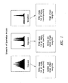


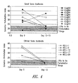


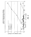
Classifications
-
- C—CHEMISTRY; METALLURGY
- C07—ORGANIC CHEMISTRY
- C07K—PEPTIDES
- C07K16/00—Immunoglobulins [IGs], e.g. monoclonal or polyclonal antibodies
- C07K16/18—Immunoglobulins [IGs], e.g. monoclonal or polyclonal antibodies against material from animals or humans
- C07K16/28—Immunoglobulins [IGs], e.g. monoclonal or polyclonal antibodies against material from animals or humans against receptors, cell surface antigens or cell surface determinants
- C07K16/2803—Immunoglobulins [IGs], e.g. monoclonal or polyclonal antibodies against material from animals or humans against receptors, cell surface antigens or cell surface determinants against the immunoglobulin superfamily
- C07K16/2818—Immunoglobulins [IGs], e.g. monoclonal or polyclonal antibodies against material from animals or humans against receptors, cell surface antigens or cell surface determinants against the immunoglobulin superfamily against CD28 or CD152
-
- A—HUMAN NECESSITIES
- A61—MEDICAL OR VETERINARY SCIENCE; HYGIENE
- A61K—PREPARATIONS FOR MEDICAL, DENTAL OR TOILETRY PURPOSES
- A61K39/00—Medicinal preparations containing antigens or antibodies
- A61K39/46—Cellular immunotherapy
- A61K39/461—Cellular immunotherapy characterised by the cell type used
- A61K39/4611—T-cells, e.g. tumor infiltrating lymphocytes [TIL], lymphokine-activated killer cells [LAK] or regulatory T cells [Treg]
-
- A—HUMAN NECESSITIES
- A61—MEDICAL OR VETERINARY SCIENCE; HYGIENE
- A61K—PREPARATIONS FOR MEDICAL, DENTAL OR TOILETRY PURPOSES
- A61K39/00—Medicinal preparations containing antigens or antibodies
- A61K39/46—Cellular immunotherapy
- A61K39/463—Cellular immunotherapy characterised by recombinant expression
- A61K39/4632—T-cell receptors [TCR]; antibody T-cell receptor constructs
-
- A—HUMAN NECESSITIES
- A61—MEDICAL OR VETERINARY SCIENCE; HYGIENE
- A61K—PREPARATIONS FOR MEDICAL, DENTAL OR TOILETRY PURPOSES
- A61K39/00—Medicinal preparations containing antigens or antibodies
- A61K39/46—Cellular immunotherapy
- A61K39/464—Cellular immunotherapy characterised by the antigen targeted or presented
- A61K39/4643—Vertebrate antigens
- A61K39/4644—Cancer antigens
-
- A—HUMAN NECESSITIES
- A61—MEDICAL OR VETERINARY SCIENCE; HYGIENE
- A61P—SPECIFIC THERAPEUTIC ACTIVITY OF CHEMICAL COMPOUNDS OR MEDICINAL PREPARATIONS
- A61P1/00—Drugs for disorders of the alimentary tract or the digestive system
- A61P1/04—Drugs for disorders of the alimentary tract or the digestive system for ulcers, gastritis or reflux esophagitis, e.g. antacids, inhibitors of acid secretion, mucosal protectants
-
- A—HUMAN NECESSITIES
- A61—MEDICAL OR VETERINARY SCIENCE; HYGIENE
- A61P—SPECIFIC THERAPEUTIC ACTIVITY OF CHEMICAL COMPOUNDS OR MEDICINAL PREPARATIONS
- A61P1/00—Drugs for disorders of the alimentary tract or the digestive system
- A61P1/16—Drugs for disorders of the alimentary tract or the digestive system for liver or gallbladder disorders, e.g. hepatoprotective agents, cholagogues, litholytics
-
- A—HUMAN NECESSITIES
- A61—MEDICAL OR VETERINARY SCIENCE; HYGIENE
- A61P—SPECIFIC THERAPEUTIC ACTIVITY OF CHEMICAL COMPOUNDS OR MEDICINAL PREPARATIONS
- A61P1/00—Drugs for disorders of the alimentary tract or the digestive system
- A61P1/18—Drugs for disorders of the alimentary tract or the digestive system for pancreatic disorders, e.g. pancreatic enzymes
-
- A—HUMAN NECESSITIES
- A61—MEDICAL OR VETERINARY SCIENCE; HYGIENE
- A61P—SPECIFIC THERAPEUTIC ACTIVITY OF CHEMICAL COMPOUNDS OR MEDICINAL PREPARATIONS
- A61P13/00—Drugs for disorders of the urinary system
- A61P13/12—Drugs for disorders of the urinary system of the kidneys
-
- A—HUMAN NECESSITIES
- A61—MEDICAL OR VETERINARY SCIENCE; HYGIENE
- A61P—SPECIFIC THERAPEUTIC ACTIVITY OF CHEMICAL COMPOUNDS OR MEDICINAL PREPARATIONS
- A61P17/00—Drugs for dermatological disorders
- A61P17/06—Antipsoriatics
-
- A—HUMAN NECESSITIES
- A61—MEDICAL OR VETERINARY SCIENCE; HYGIENE
- A61P—SPECIFIC THERAPEUTIC ACTIVITY OF CHEMICAL COMPOUNDS OR MEDICINAL PREPARATIONS
- A61P19/00—Drugs for skeletal disorders
- A61P19/02—Drugs for skeletal disorders for joint disorders, e.g. arthritis, arthrosis
-
- A—HUMAN NECESSITIES
- A61—MEDICAL OR VETERINARY SCIENCE; HYGIENE
- A61P—SPECIFIC THERAPEUTIC ACTIVITY OF CHEMICAL COMPOUNDS OR MEDICINAL PREPARATIONS
- A61P21/00—Drugs for disorders of the muscular or neuromuscular system
- A61P21/04—Drugs for disorders of the muscular or neuromuscular system for myasthenia gravis
-
- A—HUMAN NECESSITIES
- A61—MEDICAL OR VETERINARY SCIENCE; HYGIENE
- A61P—SPECIFIC THERAPEUTIC ACTIVITY OF CHEMICAL COMPOUNDS OR MEDICINAL PREPARATIONS
- A61P25/00—Drugs for disorders of the nervous system
-
- A—HUMAN NECESSITIES
- A61—MEDICAL OR VETERINARY SCIENCE; HYGIENE
- A61P—SPECIFIC THERAPEUTIC ACTIVITY OF CHEMICAL COMPOUNDS OR MEDICINAL PREPARATIONS
- A61P29/00—Non-central analgesic, antipyretic or antiinflammatory agents, e.g. antirheumatic agents; Non-steroidal antiinflammatory drugs [NSAID]
-
- A—HUMAN NECESSITIES
- A61—MEDICAL OR VETERINARY SCIENCE; HYGIENE
- A61P—SPECIFIC THERAPEUTIC ACTIVITY OF CHEMICAL COMPOUNDS OR MEDICINAL PREPARATIONS
- A61P3/00—Drugs for disorders of the metabolism
- A61P3/08—Drugs for disorders of the metabolism for glucose homeostasis
- A61P3/10—Drugs for disorders of the metabolism for glucose homeostasis for hyperglycaemia, e.g. antidiabetics
-
- A—HUMAN NECESSITIES
- A61—MEDICAL OR VETERINARY SCIENCE; HYGIENE
- A61P—SPECIFIC THERAPEUTIC ACTIVITY OF CHEMICAL COMPOUNDS OR MEDICINAL PREPARATIONS
- A61P31/00—Antiinfectives, i.e. antibiotics, antiseptics, chemotherapeutics
- A61P31/12—Antivirals
-
- A—HUMAN NECESSITIES
- A61—MEDICAL OR VETERINARY SCIENCE; HYGIENE
- A61P—SPECIFIC THERAPEUTIC ACTIVITY OF CHEMICAL COMPOUNDS OR MEDICINAL PREPARATIONS
- A61P31/00—Antiinfectives, i.e. antibiotics, antiseptics, chemotherapeutics
- A61P31/12—Antivirals
- A61P31/14—Antivirals for RNA viruses
-
- A—HUMAN NECESSITIES
- A61—MEDICAL OR VETERINARY SCIENCE; HYGIENE
- A61P—SPECIFIC THERAPEUTIC ACTIVITY OF CHEMICAL COMPOUNDS OR MEDICINAL PREPARATIONS
- A61P31/00—Antiinfectives, i.e. antibiotics, antiseptics, chemotherapeutics
- A61P31/12—Antivirals
- A61P31/20—Antivirals for DNA viruses
-
- A—HUMAN NECESSITIES
- A61—MEDICAL OR VETERINARY SCIENCE; HYGIENE
- A61P—SPECIFIC THERAPEUTIC ACTIVITY OF CHEMICAL COMPOUNDS OR MEDICINAL PREPARATIONS
- A61P35/00—Antineoplastic agents
-
- A—HUMAN NECESSITIES
- A61—MEDICAL OR VETERINARY SCIENCE; HYGIENE
- A61P—SPECIFIC THERAPEUTIC ACTIVITY OF CHEMICAL COMPOUNDS OR MEDICINAL PREPARATIONS
- A61P35/00—Antineoplastic agents
- A61P35/02—Antineoplastic agents specific for leukemia
-
- A—HUMAN NECESSITIES
- A61—MEDICAL OR VETERINARY SCIENCE; HYGIENE
- A61P—SPECIFIC THERAPEUTIC ACTIVITY OF CHEMICAL COMPOUNDS OR MEDICINAL PREPARATIONS
- A61P37/00—Drugs for immunological or allergic disorders
- A61P37/02—Immunomodulators
-
- A—HUMAN NECESSITIES
- A61—MEDICAL OR VETERINARY SCIENCE; HYGIENE
- A61P—SPECIFIC THERAPEUTIC ACTIVITY OF CHEMICAL COMPOUNDS OR MEDICINAL PREPARATIONS
- A61P37/00—Drugs for immunological or allergic disorders
- A61P37/02—Immunomodulators
- A61P37/04—Immunostimulants
-
- A—HUMAN NECESSITIES
- A61—MEDICAL OR VETERINARY SCIENCE; HYGIENE
- A61P—SPECIFIC THERAPEUTIC ACTIVITY OF CHEMICAL COMPOUNDS OR MEDICINAL PREPARATIONS
- A61P37/00—Drugs for immunological or allergic disorders
- A61P37/02—Immunomodulators
- A61P37/06—Immunosuppressants, e.g. drugs for graft rejection
-
- A—HUMAN NECESSITIES
- A61—MEDICAL OR VETERINARY SCIENCE; HYGIENE
- A61P—SPECIFIC THERAPEUTIC ACTIVITY OF CHEMICAL COMPOUNDS OR MEDICINAL PREPARATIONS
- A61P5/00—Drugs for disorders of the endocrine system
- A61P5/14—Drugs for disorders of the endocrine system of the thyroid hormones, e.g. T3, T4
-
- A—HUMAN NECESSITIES
- A61—MEDICAL OR VETERINARY SCIENCE; HYGIENE
- A61P—SPECIFIC THERAPEUTIC ACTIVITY OF CHEMICAL COMPOUNDS OR MEDICINAL PREPARATIONS
- A61P7/00—Drugs for disorders of the blood or the extracellular fluid
-
- A—HUMAN NECESSITIES
- A61—MEDICAL OR VETERINARY SCIENCE; HYGIENE
- A61P—SPECIFIC THERAPEUTIC ACTIVITY OF CHEMICAL COMPOUNDS OR MEDICINAL PREPARATIONS
- A61P7/00—Drugs for disorders of the blood or the extracellular fluid
- A61P7/06—Antianaemics
-
- C—CHEMISTRY; METALLURGY
- C07—ORGANIC CHEMISTRY
- C07K—PEPTIDES
- C07K16/00—Immunoglobulins [IGs], e.g. monoclonal or polyclonal antibodies
- C07K16/18—Immunoglobulins [IGs], e.g. monoclonal or polyclonal antibodies against material from animals or humans
- C07K16/28—Immunoglobulins [IGs], e.g. monoclonal or polyclonal antibodies against material from animals or humans against receptors, cell surface antigens or cell surface determinants
- C07K16/2803—Immunoglobulins [IGs], e.g. monoclonal or polyclonal antibodies against material from animals or humans against receptors, cell surface antigens or cell surface determinants against the immunoglobulin superfamily
- C07K16/2809—Immunoglobulins [IGs], e.g. monoclonal or polyclonal antibodies against material from animals or humans against receptors, cell surface antigens or cell surface determinants against the immunoglobulin superfamily against the T-cell receptor (TcR)-CD3 complex
-
- C—CHEMISTRY; METALLURGY
- C12—BIOCHEMISTRY; BEER; SPIRITS; WINE; VINEGAR; MICROBIOLOGY; ENZYMOLOGY; MUTATION OR GENETIC ENGINEERING
- C12N—MICROORGANISMS OR ENZYMES; COMPOSITIONS THEREOF; PROPAGATING, PRESERVING, OR MAINTAINING MICROORGANISMS; MUTATION OR GENETIC ENGINEERING; CULTURE MEDIA
- C12N5/00—Undifferentiated human, animal or plant cells, e.g. cell lines; Tissues; Cultivation or maintenance thereof; Culture media therefor
- C12N5/06—Animal cells or tissues; Human cells or tissues
- C12N5/0602—Vertebrate cells
- C12N5/0634—Cells from the blood or the immune system
- C12N5/0636—T lymphocytes
-
- A—HUMAN NECESSITIES
- A61—MEDICAL OR VETERINARY SCIENCE; HYGIENE
- A61K—PREPARATIONS FOR MEDICAL, DENTAL OR TOILETRY PURPOSES
- A61K39/00—Medicinal preparations containing antigens or antibodies
- A61K2039/51—Medicinal preparations containing antigens or antibodies comprising whole cells, viruses or DNA/RNA
- A61K2039/515—Animal cells
- A61K2039/5158—Antigen-pulsed cells, e.g. T-cells
-
- A—HUMAN NECESSITIES
- A61—MEDICAL OR VETERINARY SCIENCE; HYGIENE
- A61K—PREPARATIONS FOR MEDICAL, DENTAL OR TOILETRY PURPOSES
- A61K39/00—Medicinal preparations containing antigens or antibodies
- A61K2039/57—Medicinal preparations containing antigens or antibodies characterised by the type of response, e.g. Th1, Th2
-
- A—HUMAN NECESSITIES
- A61—MEDICAL OR VETERINARY SCIENCE; HYGIENE
- A61K—PREPARATIONS FOR MEDICAL, DENTAL OR TOILETRY PURPOSES
- A61K2239/00—Indexing codes associated with cellular immunotherapy of group A61K39/46
- A61K2239/38—Indexing codes associated with cellular immunotherapy of group A61K39/46 characterised by the dose, timing or administration schedule
-
- A—HUMAN NECESSITIES
- A61—MEDICAL OR VETERINARY SCIENCE; HYGIENE
- A61K—PREPARATIONS FOR MEDICAL, DENTAL OR TOILETRY PURPOSES
- A61K2239/00—Indexing codes associated with cellular immunotherapy of group A61K39/46
- A61K2239/46—Indexing codes associated with cellular immunotherapy of group A61K39/46 characterised by the cancer treated
- A61K2239/48—Blood cells, e.g. leukemia or lymphoma
-
- C—CHEMISTRY; METALLURGY
- C12—BIOCHEMISTRY; BEER; SPIRITS; WINE; VINEGAR; MICROBIOLOGY; ENZYMOLOGY; MUTATION OR GENETIC ENGINEERING
- C12N—MICROORGANISMS OR ENZYMES; COMPOSITIONS THEREOF; PROPAGATING, PRESERVING, OR MAINTAINING MICROORGANISMS; MUTATION OR GENETIC ENGINEERING; CULTURE MEDIA
- C12N2501/00—Active agents used in cell culture processes, e.g. differentation
- C12N2501/50—Cell markers; Cell surface determinants
- C12N2501/51—B7 molecules, e.g. CD80, CD86, CD28 (ligand), CD152 (ligand)
-
- C—CHEMISTRY; METALLURGY
- C12—BIOCHEMISTRY; BEER; SPIRITS; WINE; VINEGAR; MICROBIOLOGY; ENZYMOLOGY; MUTATION OR GENETIC ENGINEERING
- C12N—MICROORGANISMS OR ENZYMES; COMPOSITIONS THEREOF; PROPAGATING, PRESERVING, OR MAINTAINING MICROORGANISMS; MUTATION OR GENETIC ENGINEERING; CULTURE MEDIA
- C12N2501/00—Active agents used in cell culture processes, e.g. differentation
- C12N2501/50—Cell markers; Cell surface determinants
- C12N2501/515—CD3, T-cell receptor complex
Definitions
- the present invention relates generally to methods for stimulating T cells, and more particularly, to methods to increase polyclonality of the expressed T cell receptors (TCRs) in populations of T cells, thereby restoring the immune potential of said T cells.
- TCRs T cell receptors
- the present invention also relates to compositions of cells, including stimulated T cells having increased polyclonality and uses thereof.
- TCR T cell antigen receptor
- ⁇ (alpha) chain an ⁇ (alpha) chain and a ⁇ (beta) chain or a ⁇ (gamma) and a ⁇ (delta) chain.
- the proteins which make up these chains are encoded by DNA which employs a unique mechanism for generating the tremendous diversity of the TCR.
- the TCR ⁇ and ⁇ or ⁇ and ⁇ chains are linked by a disulfide bond ( Janeway, Travers, Walport. Immunobiology. Fourth Ed., 148-159. Elsevier Science Ltd/Garland Publishing. 1999 ).
- the enormous diversity of TCR specificities is generated much like immunoglobulin diversity, through somatic gene rearrangement.
- the ⁇ chain genes contain over 50 variable (V), 2 diversity (D), over 10 joining (J) segments, and 2 constant region segments (C).
- the ⁇ chain genes contain about 70 V segments, and over 60 J segments but no D segments, as well as one C segment.
- V, D, and J gene segments to combine together randomly introduces a large element of combinatorial diversity into the TCR repertoire.
- the precise point at which V, D, and J segments join can vary, giving rise to local amino acid diversity at the junction.
- the exact nucleotide position of joining can differ by as much as 10 residues resulting in deletion of nucleotides from the ends of the V, D, and J gene segments, thereby producing codon changes at the junctions of these segments.
- Diversity is further increased during the rearrangement process when additional nucleotides not encoded by either gene segment are added at the junction between the joined gene segments. (The variability created by this process is called "N-region diversity.") ( Janeway, Travers, Walport. Immunobiology. Fourth Ed., 98 and 150. Elsevier Science Ltd/Garland Publishing. 1999 ).
- the level of diversity for the T cell repertoire can be measured, in part, by evaluating which TCR V ⁇ chains are being employed by individual T cells within a pool of circulating T cells, and by the number of random nucleotides inserted next to the V ⁇ gene.
- the circulating T cell pool contains T cells expressing the full range of TCR V ⁇ chains and when those individual V ⁇ chains are derived from gene recombination events which utilize the broadest array of inserted nucleotides, the T cell arm of the immune system will have its greatest potential for recognizing the universe of potential antigens.
- TCR V ⁇ chains expressed by the circulating pool of T cells When the range of TCR V ⁇ chains expressed by the circulating pool of T cells is limited or reduced, and when expressed TCRs utilize chains encoded by recombined genes with limited nucleotide insertions, the breadth of the immune response potential is correspondingly reduced. The consequences of this are a reduced ability to respond to the wide variety of antigens leading to increased risks of infection and cancer.
- Spectratype analysis is a recently developed method for measuring TCR V ⁇ , V ⁇ , V ⁇ , or V ⁇ gene usage by a pool of T cells and levels of nucleotide insertion during the recombination process in T cell development (as described in U.S. Patent No. 5,837,447 ). Spectratype analysis can be used to measure the breadth or narrowness of the T cell immune response potential.
- Binding of ⁇ TCR to the antigenic peptide bound in the context of an MHC molecule on the antigen presenting cell (APC) is the central event in T-cell activation, which occurs at an immunological synapse at the point of contact between the T-cell and the APC.
- T lymphocytes typically require a second co-stimulatory signal.
- Co-stimulation is typically necessary for a T helper cell to produce sufficient cytokine levels that induce clonal expansion. Bretscher, Immunol. Today 13:74, 1992 ; June et al., Immunol. Today 15:321, 1994 .
- the major co-stimulatory signal occurs when a member of the B7 family ligands (CD80 (B7.1) or CD86 (B7.2)) on an activated antigen-presenting cell (APC) binds to CD28 on a T-cell.
- T-cell compositions useful in immunotherapy have the potential to generate a variety of T-cell compositions useful in immunotherapy.
- Successful immunotherapy can be aided by increasing the polyclonality, reactivity, and quantity of T-cells by efficient stimulation.
- IL-2 interleukin-2
- IL-2 has been used together with an anti-CD3 antibody to stimulate T-cell proliferation, predominantly expanding the CD8 + subpopulation of T-cells.
- Both APC signals are thought to be required for optimal T-cell activation, expansion, and long-term survival of the T-cells upon re-infusion.
- MHC-matched APCs as accessory cells presents a significant problem for long-term culture systems because APCs are relatively short-lived. Therefore, in a long-term culture system, APCs must be continually obtained from a source and replenished.
- the necessity for a renewable supply of accessory cells is problematic for treatment of immunodeficiencies in which accessory cells are affected.
- the cells may contaminate the entire T-cell population during long-term culture.
- One aspect of the present invention provides a method for restoring the polyclonality of TCR expression of a population of T cells from an immunocompromised patient, for use in restoring immune responsiveness in the patient comprising, providing a population of cells wherein at least a portion thereof comprises T cells, exposing the population of cells to a surface, wherein the surface has attached thereto one or more agents that ligate a cell surface moiety of at least a portion of the T cells and stimulates at least a portion of the T cells, growing said cells for a time sufficient to increase polyclonality of at least one TCR V ⁇ , V ⁇ , V ⁇ , and/or V ⁇ family, in terms of TCR expression and thereby restoring the polyclonality of the population of T cells.
- the restoration comprises a shift from monoclonality to oligoclonality, a shift from monoclonality to polyclonality, or a shift from oligoclonality to polyclonality, of the T cell population as measured by a V ⁇ , V ⁇ , V ⁇ , and/or V ⁇ spectratype profile of at least one V ⁇ , V ⁇ , V ⁇ , and/or V ⁇ family gene.
- the shift comprises an increase in polyclonal T cells expressing at least one V ⁇ , V ⁇ , V ⁇ , and/or V ⁇ family gene to sufficient numbers for use in therapy.
- Another aspect of the present invention provides a method for restoring immune responsiveness in an immunocompromised individual wherein the T cells of the individual have reduced polyclonality of TCR expression as compared to a nonimmunocompromised individual, comprising, obtaining a population of cells from the individual wherein at least a portion thereof comprises T cells; exposing the population of cells to a surface, wherein the surface has attached thereto one or more agents that ligate a cell surface moiety of at least a portion of the T cells and stimulates at least a portion of T cells; growing said cells for a time sufficient to increase polyclonality of at least one TCR V ⁇ , V ⁇ , V ⁇ , and/or V ⁇ family; and infusing the stimulated portion of T cells into the immunocompromised individual; and thereby restoring immune responsiveness in the immunocompromised individual.
- the polyclonality of the infused T cells is maintained in vivo for at least 3 to 6 months to a year following infusion.
- the immunocompromised individual has a cancer.
- the cancer may be any one of melanoma, non Hodgkin's lymphoma, Hodgkin's disease, leukemia, plasmocytoma, sarcoma, glioma, thymoma, breast cancer, prostate cancer, colo-rectal cancer, kidney cancer, renal cell carcinoma, pancreatic cancer, nasopharyngeal carcinoma, esophageal cancer, brain cancer, lung cancer, ovarian cancer, cervical cancer, multiple myeloma, heptocellular carcinoma, acute lymphoblastic leukemia (ALL), acute myelogenous leukemia (AML), chronic myelogenous leukemia (CML), large granular lymphocyte leukemia (LGL), and chronic lymphocytic leukemia (CLL).
- ALL acute lymphoblastic leukemia
- AML acute myelogenous leukemia
- CML chronic myelogenous leukemia
- the immunocompromised individual is infected with an infectious organism.
- the infectious organism may comprise a virus, such as a single stranded RNA virus or a single stranded DNA virus, human immunodeficiency virus (HIV), hepatitis A, B, or C virus, herpes simplex virus (HSV), human papilloma virus (HPV), cytomegalovirus (CMV), Epstein-Barr virus (EBV), a parasite, a bacterium, M. tuberculosis, Pneumocystis carinii, Candida, or Aspergillus or a combination thereof.
- a virus such as a single stranded RNA virus or a single stranded DNA virus, human immunodeficiency virus (HIV), hepatitis A, B, or C virus, herpes simplex virus (HSV), human papilloma virus (HPV), cytomegalovirus (CMV), Epstein-Barr virus (EB
- the immunocompromised individual has a congenital genetic disorder such as severe combined immunodeficiency (SCID) or common variable immunodeficiency (CVID).
- SCID severe combined immunodeficiency
- CVID common variable immunodeficiency
- the individual is immunocompromised as a result of treatment associated with cancer.
- the individual is immunocompromised as a result of treatment associated with hematopoeitic stem cell transplantation, bone marrow transplantation, cord blood, allogeneic, autologous, or xenogeneic cell transplantation, chemotherapy, radiation therapy, treatment with cytotoxic agents, treatment with an immunosuppressive agent (e,g., cyclosporine, corticosteroid, and the like).
- an immunosuppressive agent e,g., cyclosporine, corticosteroid, and the like.
- the immunocompromised individual has an immunodeficiency or an autoimmune disease.
- the immunocompromised individual has a chronic disease affecting the kidney, liver, or the pancreas.
- the individual has diabetes.
- the immunocompromised individual is affected by old age.
- the immunocompromised individual has undergone gene therapy, or other procedure involving gene transduction that has resulted in a skewing of the T cell repertoire.
- the immunocompromised individual is afflicted with a disorder associated with altered or skewed T cell repertoire, including but not limited to, diseases such as, rheumatoid arthritis, multiple sclerosis, insulin dependent diabetes, Addison's disease, celiac disease, chronic fatigue syndrome, inflammatory bowel disease, ulcerativecolitis, Crohn's disease, Fibromyalgia, systemic lupus erythematosus, psoriasis, Sjogren's syndrome, hyperthyroidism/Graves disease, hypothyroidism/Hashimoto's disease, Insulin-dependent diabetes (type 1), Myasthenia Gravis, endometriosis, scleroderma, pernicious anemia, Goodpasture syndrome, Wegener's disease, glomerulonephritis, aplastic anemia, any of a variety of cytopenias, paroxysmal nocturnal hemoglobinuria, myelodysplastic syndrome, id
- the immunocompromised individual is afflicted with a neurological disorder associated with T cell repertoire skewing or cardiovascular disease.
- the cell compositions of the present invention are administered to a patient with an autoimmune disease.
- One embodiment of the present invention provides a method for restoring immune responsiveness in an immunocompromised individual wherein the immunocompromised individual is afflicted with an autoimmune disease.
- autoimmune disease includes but is not limited to rheumatoid arthritis, multiple sclerosis, insulin dependent diabetes, Addison's disease, celiac disease, chronic fatigue syndrome, inflammatory bowel disease, ulcerativecolitis, Crohn's disease, Fibromyalgia, systemic lupus erythematosus, psoriasis, Sjogren's syndrome, hyperthyroidism/Graves disease, hypothyroidism/Hashimoto's disease, Insulin-dependent diabetes (type 1), and Myasthenia Gravis.
- the immunocompromised individual has been treated with chemotherapy.
- the immunocompromised individual has been treated with a cytotoxic agent.
- the immunocompromised individual has been treated with an immunosuppressive agent.
- the immunocompromised individual is afflicted with a hematological disorder associated with cytopenia, including but not limited to, aplastic anemia, myclodisplastic syndrome, Fanconi anemia, idiopathic thrombocytopenic purpura and autoimmune hemolytic anemia.
- compositions comprising a population of T cells wherein the polyclonality has been restored according to the methods described herein and a pharmaceutically acceptable excipient, for use in restoring immune responsiveness in an immunocompromised individual wherein the T cells of the individual have reduced polyclonality as compared to a nonimmunocompromised individual.
- compositions of the present invention are administered to a patient following T-cell ablative therapy using either chemotherapy agents such as, fludarabine, external-beam radiation therapy (XRT), cyclophosphamide, or antibodies such as OKT3 or CAWATH.
- chemotherapy agents such as, fludarabine, external-beam radiation therapy (XRT), cyclophosphamide, or antibodies such as OKT3 or CAWATH.
- the cell compositions of the present invention are administered to a patient following B-cell ablative therapy such as agents that react with CD20, e.g., Rituxan.
- the dosage of the above treatments to be administered to a patient will vary with the precise nature of the condition being treated and the recipient of the treatment. The scaling of dosages for human administration can be performed according to art-accepted practices.
- the dose for CAMPATH for example, will generally be in the range 1 to about 100 mg for an adult patient, usually administered daily for a period between 1 and 30 days.
- the preferred daily dose is 1 to 10 mg per day although in some instances larger doses of up to 40 mg per day may be used (described in U.S. Patent No. 6,120,766 ).
- the present invention provides compositions comprising a population of T cells wherein the polyclonality of TCR expression has been restored by the methods of the present invention and a pharmaceutically acceptable excipient, for use in restoring immune responsiveness in an immunocompromised individual wherein the T cells of the individual have reduced polyclonality as compared to a nonimmunocompromised individual.
- the present invention provides for methods wherein said surface has attached thereto a first agent that ligates a first cell surface moiety of a T-cell; and the same or a second surface has attached thereto a second agent that ligates a second moiety of said T-cell, wherein said ligation by the first and second agent induces proliferation of said T-cell.
- the first agent comprises an anti-CD3 antibody and said second agent comprises a ligand which binds an accessory molecule on the surface of said T cells.
- said accessory molecule is CD28.
- the first agent comprises an anti-CD3 antibody and said second agent comprises an anti-CD28 antibody.
- said first and second agents are attached to said surface or said second surface by covalent attachment.
- said first and second agents are attached to said surface or said second surface by direct attachment or indirect attachment.
- the present invention provides for methods wherein said surface has attached thereto one or more agents that ligate a cell surface moiety of at least a portion of the T cells and stimulates at least a portion of the T cells, wherein said ligation by the one or more agents induces activation of said T-cell.
- one or more surfaces are used in the present invention.
- 3 or more agents are attached to said surfaces, either in cis or in trans.
- biocompatible refers to the property of being predominantly non-toxic to living cells.
- stimulation refers to a primary response induced by ligation of a cell surface moiety.
- such stimulation entails the ligation of a receptor and a subsequent signal transduction event.
- stimulation refers to the ligation of a T cell surface moiety that in one embodiment subsequently induces a signal transduction event, such as binding the TCR/CD3 complex.
- the stimulation event may activate a cell and up or downregulate expression of cell surface molecules such as receptors or adhesion molecules, or up or downregulate secretion of a molecule, such as downregulation of Tumor Growth Factor beta (TGF- ⁇ ).
- TGF- ⁇ Tumor Growth Factor beta
- libation of cell surface moieties may result in the reorganization of cytoskeletal structures, or in the coalescing of cell surface moieties, each of which could serve to enhance, modify, or alter subsequent cell responses.
- activation refers to the state of a cell following sufficient cell surface moiety ligation to induce a measurable morphological, phenotypic, and/or functional change.
- T cells such activation may be the state of a T cell that has been sufficiently stimulated to induce cellular proliferation.
- Activation of a T cell may also induce cytokine production and/or secretion, and up or downregulation of expression of cell surface molecules such as receptors or adhesion molecules, or up or downregulation of secretion of certain molecules, and performance of regulatory or cytolytic effector functions.
- this term infers either up or down regulation of a particular physico-chemical process.
- target cell refers to any cell that is intended to be stimulated by cell surface moiety ligation.
- an "antibody”, as used herein, includes both polyclonal and monoclonal antibodies (mAb); primatized ( e.g ., humanized); murine; mouse-human; mouse-primate; and chimeric; and may be an intact molecule, a fragment thereof (such as scFv, Fv, Fd, Fab, Fab' and F(ab)' 2 fragments), or multimers or aggregates of intact molecules and/or fragments; and may occur in nature or be produced, e.g ., by immunization, synthesis or genetic engineering; an "antibody fragment,” as used herein, refers to fragments, derived from or related to an antibody, which bind antigen and which in some embodiments may be derivatized to exhibit structural features that facilitate clearance and uptake, e.g., by the incorporation of galactose residues. This includes, e.g., F(ab), F(ab)' 2 , scFv, light chain variable region (V L
- protein includes proteins, glycoproteins and other cell-derived modified proteins, polypeptides and peptides; and may be an intact molecule, a fragment thereof, or multimers or aggregates of intact molecules and/or fragments; and may occur in nature or be produced, e.g., by synthesis (including chemical and/or enzymatic) or genetic engineering.
- agent refers to a molecule that binds to a defined population of cells.
- the agent may bind any cell surface moiety, such as a receptor, an antigenic determinant, or other binding site present on the target cell population.
- the agent may be a protein, peptide, antibody and antibody fragments thereof, fusion proteins, synthetic molecule, an organic molecule (e.g., a small molecule), or the like.
- antibodies are used as a prototypical example of such an agent.
- cell surface moiety may refer to a cell surface receptor, an antigenic determinant, or any other binding site present on a target cell population.
- agent that binds a cell surface moiety and "cell surface moiety”, as used herein, should be viewed as a complementary/anti-complementary set of molecules that demonstrate specific binding, generally of relatively high affinity.
- a “co-stimulatory signal”, as used herein, refers to a signal, which in combination with a primary signal, such as TCR/CD3 ligation, leads to T cell proliferation and/or activation.
- Separatation includes any means of substantially purifying one component from another (e.g ., by filtration, affinity, buoyant density, or magnetic attraction).
- a "surface”, as used herein, refers to any surface capable of having an agent attached thereto and includes, without limitation, metals, glass, plastics, copolymers, colloids, lipids, cell surfaces, and the like. Essentially any surface that is capable of retaining an agent bound or attached thereto.
- a population of T cells is considered monoclonal (or mono-specific) when the V ⁇ , V ⁇ , V ⁇ , and/or V ⁇ spectratype profile for a given TCR V ⁇ , V ⁇ , V ⁇ , and/or V ⁇ family has a single predominant peak (see Figure 1 ).
- Spectratype analysis distinguishes rearranged variable genes of a particular size, not sequence.
- a single peak could represent a population of T cells expressing any one of a limited number of rearranged TCR variable genes (V ⁇ , V ⁇ , V ⁇ , or V ⁇ ) comprising any one of the 4 potential nucleotides (adenine (a), guanine (g), cytosine (c), or thymine (t)) or a combination of the 4 nucleotides at the junctional region.
- V ⁇ , V ⁇ , V ⁇ , or V ⁇ comprising any one of the 4 potential nucleotides (adenine (a), guanine (g), cytosine (c), or thymine (t)) or a combination of the 4 nucleotides at the junctional region.
- Oleclonality in the context of a population of T cells, refers to a population of T cells that has multiple, but narrow antigen specificity as defined by spectra type analysis (a measure of the TCR ⁇ chain hypervariable region repertoire).
- a population of T cells is considered oligoclonal when the V ⁇ spectratype profile for a given TCR V ⁇ family has between about 2 and about 4 predominant peaks (see Figure 1 ).
- Polyclonality in the context of a population of T cells, refers to a population of T cells that has multiple and broad antigen specificity as defined by spectratype analysis (a measure of the TCR ⁇ chain hypervariable region repertoire).
- a population ofT cells is considered polyclonal when the V ⁇ spectratype profile for a given TCR V ⁇ family has multiple peaks, typically 5 or more predominant peaks and in most cases with Gaussian distribution (see for example Figures 1 , 2 , and 3 ).
- Restoring or increasing the polyelonality refers to a shift from a monoclonal profile to an oligoclonal profile or to a polyclonal profile (in other words, a shift from monoclonality to oligoclonality or to polyclonality), or from an oligoclonal profile to a polyclonal profile (in other words, a shift from oligoclonality to polyclonality), in expressed TCR V ⁇ , V ⁇ , V ⁇ , and/or V ⁇ genes in a population of T cells, as measured by spectratype analysis or by similar analysis such as flow cytometry or sequence analysis.
- the shift from a monoclonal V ⁇ , V ⁇ , V ⁇ , and/or V ⁇ expression profile in a population of T cells to an oligoclonal profile or to a polyclonal profile is generally seen in at least one TCR V ⁇ , V ⁇ , V ⁇ , and/or V ⁇ family. In one embodiment of the present invention, this shift is observed in 2, 3, 4, or 5 V ⁇ families. In certain embodiments of the present invention, a shift is observed in 6, 7, 8, 9, or 10 V ⁇ families. In a further embodiment of the present invention, a shift is observed in from 11, 12, 13, or 14 V ⁇ families. In a further embodiment of the present invention, a shift is observed in from 15 to 20 V ⁇ families.
- a shift is observed in 20 to 24 V ⁇ families. In another embodiment, a shift is seen in all V ⁇ families.
- the functional significance of restoring or increasing the polyclonality of a population of T cells is that the immune potential, or the ability to respond to a full breadth of antigens, of the population of T cells is restored or increased.
- some T cells within a population may not have their TCRs engaged by the methods set forth herein (e.g ., T cells with downregulated TCR expression).
- T cells activated by the methods described herein may in turn upregulate their TCR expression thereby resulting in a further increase in the polyclonality of the population of T cells.
- animal or "mammal” as used herein, encompasses all mammals, including humans.
- the animal of the present invention is a human subject.
- exposing refers to bringing into the state or condition of immediate proximity or direct contact.
- proliferation means to grow or multiply by producing new cells.
- Immuno response or responsiveness refers to activation of cells of the immune system, including but not limited to, T cells, such that a particular effector function(s) of a particular cell is induced. Effector functions may include, but are not limited to, proliferation, secretion of cytokines, secretion of antibodies, expression of regulatory and/or adhesion molecules, and the ability to induce cytolysis.
- Stimulating an immune response refers to any stimulation such that activation and induction of effector functions of cells of the immune system are achieved.
- Immuno response dysfunction refers to the inappropriate activation and/or proliferation, or lack thereof, of cells of the immune system, and/or the inappropriate secretion, or lack thereof, of cytokines, and/or the inappropriate or inadequate induction of other effector functions of cells of the immune system, such as expression of regulatory, adhesion, and/or homing receptors, and the induction of cytolysis.
- preventing or “inhibiting the development of a cancer or cancer cells” as used herein, means the occurrence of the cancer is prevented or the onset of the cancer is delayed.
- treating or reducing the presence of a cancer or cancer cells or “treating or reducing the presence of a tumor or tumor cells” as used herein, mean that the cancer or tumor growth is inhibited, which is reflected by, e.g ., tumor volume or numbers of malignant cells.
- the reduction of cancer can be determined using any number of techniques in the art including measurements of M-protein, PCR based assays, RNA and DNA hybridization assays, or in situ PCR or hybridization, etc.
- Tumor volume may be determined by various known procedures, e.g., obtaining two dimensional measurements with a dial caliper.
- Preventing or inhibiting the development of an infectious disease means the occurrence of the infectious disease is prevented or the onset of the infectious disease is delayed, or the ,spread of an existing infection is reversed or stabilized.
- “Ameliorate” as used herein, is defined as: to make better; improve ( The American Heritage College Dictionary, 3rd Edition, Houghton Mifflin Company, 2000 ).
- Particles may include a colloidal particle, a microsphere, nanoparticle, a bead, or the like.
- a surface may be any surface capable of having a ligand bound thereto or integrated into, including cell surfaces (for example K562 cells), and that is biocompatible, that is, substantially non-toxic to the target cells to be stimulated.
- commercially available surfaces such as beads or other particles
- beads or other particles are useful (e.g ., Miltenyi Particles, Miltenyi Biotec, Germany; Sepharose beads, Pharmacia Fine Chemicals, Sweden; DYNABEADS TM , Dynal Inc., New York; PURABEADS TM , Prometic Biosciences, magnetic beads from Immunicon, Huntingdon Valley, PA, microspheres from Bangs Laboratories, Inc., Fishers, IN).
- Paramagnetic particles refer to particles, as defined above, that localize in response to a magnetic field.
- infectious disease refers to any disease that is caused by an infectious organism.
- Infectious organisms may comprise viruses, (e.g ., single stranded RNA viruses, single stranded DNA viruses, human immunodeficiency virus (HIV), hepatitis A, B, and C virus, herpes simplex virus (HSV), cytomegalovirus (CMV) Epstein-Barr virus (EBV), human papilloma virus (HPV)), parasites ( e.g ., protozoan and metazoan pathogens such as Plasmodia species, Leishmania species, Schistosoma species, Trypanosoma species), bacteria (e.g., Mycobacteria , in particular, M.
- viruses e.g ., single stranded RNA viruses, single stranded DNA viruses, human immunodeficiency virus (HIV), hepatitis A, B, and C virus, herpes simplex virus (HSV), cytomegalovirus (CM
- fungi e.g., Candida species, Aspergillus species
- Pneumocystis carinii fungi
- prions known prions infect animals to cause scrapie, a transmissible, degenerative disease of the nervous system of sheep and goats, as well as bovine spongiform encephalopathy (BSE), or "mad cow disease", and feline spongiform encephalopathy of cats.
- BSE bovine spongiform encephalopathy
- prion diseases known to affect humans are (1) kuru, (2) Creutzfeldt-Jakob Disease (CJD), (3) Gerstmann-Straussler-Scheinker Disease (GSS), and (4) fatal familial insomnia (FFI)).
- CJD Creutzfeldt-Jakob Disease
- GSS Gerstmann-Straussler-Scheinker Disease
- FFI fatal familial insomnia
- the stimulated and activated T cells with increased polyclonality of the present invention are generated by cell surface moiety ligation that induces activation.
- the stimulated and activated T cells with increased polyclonality are generated by activating a population of T cells and stimulating an accessory molecule on the surface of the T cells with a ligand which binds the accessory molecule, as described for example, in US patent application numbers 08/253,694 , 08/435,816 , 08/592,711 , 09/183,055 , 09/350,202 , and 09/252,150 , and patent numbers 6,352,694 , 5,858,358 and 5,883,223 .
- T cell activation and restoration of polyclonality may be accomplished by cell surface moiety ligation, such as stimulating the T cell receptor (TCR)/CD3 complex or the CD2 surface protein.
- TCR T cell receptor
- a number of anti-human CD3 monoclonal antibodies are commercially available, exemplary are, clone BC3 (XR-CD3; Fred Hutchinson Cancer Research Center, Seattle, WA), OKT3, prepared from hybridoma cells obtained from the American Type Culture Collection, and monoclonal antibody G19-4.
- stimulatory forms of anti-CD2 antibodies are known and available. Stimulation through CD2 with anti-CD2 antibodies is typically accomplished using a combination of at least two different anti-CD2 antibodies.
- Stimulatory combinations of anti-CD2 antibodies that have been described include the following: the T11.3 antibody in combination with the T11.1 or T11.2 antibody ( Meuer et al., Cell 36:897-906, 1984 ), and the 9.6 antibody (which recognizes the same epitope as T11.1) in combination with the 9-1 antibody ( Yang et al., J. Immunol. 137:1097-1100, 1986 ).
- Other antibodies that bind to the same epitopes as any of the above described antibodies can also be used.
- Additional antibodies, or combinations of antibodies can be prepared and identified by standard techniques.
- Stimulation may also be achieved through contact with superantigens (e.g., Staphylococcus enterotoxin A (SEA), Staphylococcus enterotoxin B (SEB), Toxic Shock Syndrome Toxin 1 (TSST-1)), endotoxin, or through a variety of mitogens, including but not limited to, phytohemagglutinin (PHA), phorbol myristate acetate (PMA) and ionomycin, lipopolysaccharide (LPS), T cell mitogen, and IL-2.
- superantigens e.g., Staphylococcus enterotoxin A (SEA), Staphylococcus enterotoxin B (SEB), Toxic Shock Syndrome Toxin 1 (TSST-1)
- mitogens including but not limited to, phytohemagglutinin (PHA), phorbol myristate acetate (PMA) and ionomycin, lipopolysaccharide (LP
- a co-stimulatory or accessory molecule on the surface of the T cells such as CD28
- a ligand that binds the accessory molecule can be used to stimulate T cells.
- any agent including an anti-CD28 antibody or fragment thereof capable of cross-linking the CD28 molecule, or a natural ligand for CD28 can be used to stimulate T cells.
- Exemplary anti-CD28 antibodies or fragments thereof useful in the context of the present invention include monoclonal antibody 9.3 (IgG2 a ) (Bristol-Myers Squibb, Princeton, NJ), monoclonal antibody KOLT-2 (IgG1), 15E8 (IgG1), 248.23.2 (IgM), clone B-T3 (XR-CD28; Diaclone, Besancon, France) and EX5.3D10 (IgG2 a ) (ATCC HB11373).
- Exemplary natural ligands include the B7 family of proteins, such as B7-1 (CD80) and B7-2 (CD86) ( Freeman et al., J. Immunol.
- accessory molecules on the surface of the T cells that can be stimulated with a ligand that binds the accessory molecule in the present invention include, but are not limited to, CD54, Ox-40, LFA-1, ICOS, 41-BB, and CD40.
- binding homologues of a natural ligand can also be used in accordance with the present invention.
- Other agents may include natural and synthetic ligands. Agents may include, but are not limited to, other antibodies or fragments thereof, growth factor, cytokine, chemokine, soluble receptor, steroid, hormone, mitogen, such as PHA, or other superantigens.
- ex vivo T-cell expansion can be performed by stimulation of a population of cells wherein at least a portion thereof comprises T cells.
- the T-cells may be stimulated by a single agent.
- T-cells are stimulated with two or more agents, one that induces a primary signal and additional agents that induce one or more co-stimulatory signals.
- Ligands useful for stimulating a single signal or stimulating a primary signal and an accessory molecule that stimulates a second signal may be used in soluble form, attached to the surface of a cell, or immobilized on a surface as described herein.
- a ligand or agent that is attached to a surface serves as a "surrogate" antigen presenting cell (APC).
- APC surrogate antigen presenting cell
- both primary and secondary agents are co-immobilized on a surface.
- the molecule providing the primary activation signal such as a CD3 ligand
- the co-stimulatory molecule such as a CD28 ligand
- the same surface for example, a particle.
- one, two, or more stimulatory molecules may be used on the same or differing surfaces.
- T-cells Prior to expansion, a source of T-cells is obtained from a subject.
- subject is intended to include living organisms in which an immune response can be elicited (e.g ., mammals). Examples of subjects include humans, dogs, cats, mice, rats, and transgenic species thereof.
- T cells can be obtained from a number of sources, including peripheral blood mononuclear cells, bone marrow, thymus, tissue biopsy, tumor, lymph node tissue, gut associated lymphoid tissue, mucosa associated lymphoid tissue, spleen tissue, or any other lymphoid tissue, and tumors.
- T cells can be obtained from T cell lines and from autologous or allogeneic sources.
- T cells may also be obtained from a xenogeneic source, for example, from mouse, rat, non-human primate, and pig.
- T cells can be obtained from a unit of blood collected from a subject using any number of techniques known to the skilled artisan, such as ficoll separation.
- cells from the circulating blood of an individual are obtained by apheresis or leukapheresis.
- the apheresis product typically contains lymphocytes, including T-cells, monocytes, granulocytes, B cells, other nucleated white blood cells, red blood cells, and platelets.
- the cells collected by apheresis may be washed to remove the plasma fraction and to place the cells in an appropriate buffer or media for subsequent processing steps.
- the cells are washed with phosphate buffered saline (PBS).
- PBS phosphate buffered saline
- the wash solution lacks calcium and may lack magnesium or may lack many if not all divalent cations.
- a washing step may be accomplished by methods known to those in the art, such as by using a semi-automated "flow-through" centrifuge (for example, the Cobe 2991 cell processor, Baxter) according to the manufacturer's instructions.
- the cells may be resuspended in a variety of biocompatible buffers, such as, for example, Ca-free, Mg-free PBS.
- a variety of biocompatible buffers such as, for example, Ca-free, Mg-free PBS.
- the undesirable components of the apheresis sample may be removed and the cells directly resuspended in culture media.
- T-cells are isolated from peripheral blood lymphocytes by lysing or removing the red blood cells and depleting the monocytes, for example, by centrifugation through a PERCOLL TM gradient.
- a specific subpopulation of T-cells such as CD28 + , CD4 + , CD8 + , CD45RA + , and CD45RO + T-cells, can be further isolated by positive or negative selection techniques.
- T-cells are isolated by incubation with anti-CD3/anti-CD2 (i.e ., 3x28)-conjugated beads, such as DYNABEADS® M-450 CD3/CD28 T, for a time period sufficient for positive selection of the desired T cells.
- the time period is about 30 minutes. In a further embodiment, the time period ranges from 30 minutes to 36 hours or longer and all integer values there between. In a further embodiment, the time period is at least 1, 2, 3, 4, 5, or 6 hours. In yet another preferred embodiment, the time period is 10 to 24 hours. In one preferred embodiment, the incubation time period is 24 hours. For isolation of T cells from patients with leukemia, use of longer incubation times, such as 24 hours, can increase cell yield. Longer incubation times may be used to isolate T cells in any situation where there are few T cells as compared to other cell types, such in isolating tumor infiltrating lymphocytes (TIL) from tumor tissue or from immunocompromised individuals.
- TIL tumor infiltrating lymphocytes
- CD3 + , CD28 + T cells can be positively selected using CD3/CD28 conjugated magnetic beads (e.g ., DYNABEADS ® M-450 CD3/CD28 T Cell Expander).
- CD3/CD28 conjugated magnetic beads e.g ., DYNABEADS ® M-450 CD3/CD28 T Cell Expander.
- enrichment of a T-cell population by negative selection can be accomplished with a combination of antibodies directed to surface markers unique to the negatively selected cells.
- a preferred method is cell sorting and/or selection via negative magnetic immunoadherence or flow cytometry that uses a cocktail of monoclonal antibodies directed to cell surface markers present on the cells negatively selected.
- a monoclonal antibody cocktail typically includes antibodies to CD14, CD20, CD11b, CD16, HLA-DR, and CD8.
- An additional aspect of the present invention provides a T-cell population or composition that has been depleted or enriched for populations of cells expressing a variety of markers, such as CD62L, CD45RA or CD45RO, cytokines (e.g ., IL-2, IFN- ⁇ , IL-4, IL-10), cytokine receptors ( e.g ., CD25), perforin, adhesion molecules (e.g ., VLA-1, VLA-2, VLA-4, LPAM-1, LFA-1), and/or homing molecules (e.g ., L-Selectin), prior to expansion.
- markers such as CD62L, CD45RA or CD45RO, cytokines (e.g ., IL-2, IFN- ⁇ , IL-4, IL-10), cytokine receptors ( e.g ., CD25), perforin, adhesion molecules (e.g ., VLA-1, VLA-2, VLA-4, LPAM-1, LFA-1
- cells expressing any of these markers are depleted or positively selected by antibodies or other ligands/binding agents directed to the marker.
- antibodies or other ligands/binding agents directed to the marker.
- One of ordinary skill in the art would readily be able to identify a variety of particular methodologies for depleting or positively selecting for a sample of cells expressing a desired marker.
- monocyte populations i.e ., CD14 + cells
- monocyte populations may be depleted from blood preparations prior to ex vivo expansion by a variety of methodologies, including anti-CD14 coated beads or columns, or utilization of the phagocytotic activity of these cells to facilitate removal or through adherence to plastic.
- the invention uses paramagnetic particles of a size sufficient to be engulfed by phagocytotic monocytes.
- the paramagnetic particles are commercially available beads, for example, those produced by Dynal AS under the trade name Dynabeads TM .
- Exemplary Dynabeads TM in this regard are M-280, M-450, and M-500.
- non-specific cells are removed by coating the paramagnetic particles with "irrelevant" proteins (e.g ., serum proteins or antibodies).
- Irrelevant proteins and antibodies include those proteins and antibodies or fragments thereof that do not specifically target the T-cells to be expanded.
- the irrelevant beads include beads coated with sheep anti-mouse antibodies, goat anti-mouse antibodies, and human serum albumin.
- Such depletion of monocytes is performed by preincubating PBMC that have been isolated from whole blood using Ficoll, or apheresed peripheral blood with one or more varieties of irrelevant or non-antibody coupled paramagnetic particles at any amount that allows for removal of monocytes (approximately a 20:1 bead:cell ratio)for about 30 minutes to 2 hours at 22 to 37 degrees C, followed by magnetic removal of cells which have attached to or engulfed the paramagnetic particles. Preincubation can also be done at temperatures as low as 3-4 degrees C. Such separation can be performed using standard methods available in the art.
- any magnetic separation methodology may be used including a variety of which are commercially available, (e.g ., DYNAL ® Magnetic Particle Concentrator (DYNAL MPC ® )).
- DYNAL MPC ® Magnetic Particle Concentrator
- Assurance of requisite depletion can be monitored by a variety of methodologies known to those of ordinary skill in the art, including flow cytometric analysis of CD14 positive cells, before and after said depletion.
- T-cells for stimulation may also be frozen after the washing step, which does not require the monocyte-removal step.
- the freeze and subsequent thaw step provides a more uniform product by removing granulocytes and to some extent monocytes in the cell population.
- the cells may be suspended in a freezing solution. While many freezing solutions and parameters are known in the art and will be useful in this context, one method involves using PBS containing 20% DMSO and 8% human serum albumin, or other suitable cell freezing media, the cells then are frozen to -80°C at a rate of 1° per minute and stored in the vapor phase of a liquid nitrogen storage tank.
- the cell population may be stimulated as described herein, such as by contact with an anti-CD3 antibody or an anti-CD2 antibody immobilized on a surface, or by contact with a protein kinase C activator (e.g ., bryostatin) in conjunction with a calcium ionophore.
- a protein kinase C activator e.g ., bryostatin
- a ligand that binds the accessory molecule is used.
- a population of CD4 + cells can be contacted with an anti-CD3 antibody and an anti-CD28 antibody, under conditions appropriate for stimulating proliferation of the T-cells.
- an anti-CD3 antibody and the anti-CD28 antibody B-T3, XR-CD28 (Diaclone, Besançon, France) can be used as can other methods commonly known in the art ( Berg et al., Transplant Proc. 30(8):3975-3977, 1998 ; Haanen et al., J. Exp. Med. 190(9):1319-1328, 1999 ; Garland et al., J. Immunol Meth. 227(1-2):53-63,1999 ).
- the primary stimulatory signal and the co-stimulatory signal for the T-cell may be provided by different protocols.
- the agents providing each signal may be in solution or coupled to a surface. When coupled to a surface, the agents may be coupled to the same surface ( i.e ., in "cis” formation) or to separate surfaces ( i.e ., in "trans” formation).
- one agent may be coupled to a surface and the other agent in solution.
- the agent providing the co-stimulatory signal is bound to a cell surface and the agent providing the primary activation signal is in solution or coupled to a surface. In certain embodiments, both agents can be in solution.
- the agents may be in soluble form, and then cross-linked to a surface, such as a cell expressing FC receptors or an antibody or other binding agent which will bind to the agents.
- the two agents are immobilized on a spherical or semi-spherical surface, the prototypic examples being beads or cells, either on the same bead, i.e ., "cis," or to separate beads, i.e ., "trans.”
- the agent providing the primary activation signal is an anti-CD3 antibody and the agent providing the co-stimulatory signal is an anti-CD28 antibody; and both agents are co-immobilized to the same bead in equivalent molecular amounts.
- a 1:1 ratio of each antibody bound to the beads for T-cell expansion and T-cell growth is used.
- a ratio of anti CD3:CD28 antibodies bound to the beads is used such that an increase in T cell expansion is observed as compared to the expansion observed using a ratio of 1:1. In one particular embodiment an increase of from about .5 to about 3 fold is observed as compared to the expansion observed using a ratio of 1:1.
- the ratio of CD3:CD28 antibody bound to the beads ranges from 100:1 to 1:100 and all integer values there between.
- more anti-CD28 antibody is bound to the particles than anti-CD3 antibody, i.e ., the ratio of CD3:CD28 is less than one.
- the ratio of anti CD28 antibody to anti CD3 antibody bound to the beads is greater than 2:1. In one particular embodiment, a 1:100 CD3:CD28 ratio of antibody bound to beads is used. In another embodiment, a 1:75 CD3:CD28 ratio of antibody bound to beads is used. In a further embodiment, a 1:50 CD3:CD28 ratio of antibody bound to beads is used. In another embodiment, a 1:30 CD3:CD28 ratio of antibody bound to beads is used. In one preferred embodiment, a 1:10 CD3:CD28 ratio of antibody bound to beads is used. In another embodiment, a 1:3 CD3:CD28 ratio of antibody bound to the beads is used. In yet another embodiment, a 3:1 CD3:CD28 ratio of antibody bound to the beads is used.
- Ratios of particles to cells from 1:500 to 500:1 and any integer values in between may be used to stimulate T-cells or other target cells.
- the ratio of particle to cells may depend on particle size relative to the target cell. For example, small sized beads could only bind a few cells, while larger beads could bind many.
- the ratio of cells to particles ranges from 1:100 to 100:1 and any integer values in-between and in further embodiments the ratio comprises 1:9 to 9:1 and any integer values in between, can also be used to stimulate T-cells.
- the ratio of anti-CD3- and anti-CD28-coupled particles to T-cells that result in T-cell stimulation can vary as noted above, however certain preferred values include at least 1:5, 1:4, 1:3, 1:2, 1:1, 2:1, 3:1, 4:1 to 6:1, with one preferred ratio being at least 1:1 particles per T-cell.
- a ratio of particles to cells of 1:1 or less is used.
- the ratio of particles to cells can be varied depending on the day of stimulation. For example, in one embodiment, the ratio of particles to cells is from 1:1 to 10:1 on the first day and additional particles are added to the cells every day or every other day thereafter for up to 10 days, at final ratios of from 1:1 to 1:10 (based on cell counts on the day of addition).
- the ratio of particles to cells is 1:1 on the first day of stimulation and adjusted to 1:5 on the third and fifth days of stimulation. In another embodiment, particles are added on a daily or every other day basis to a final ratio of 1:1 on the first day, and 1:5 on the third and fifth days of stimulation. In another embodiment, the ratio of particles to cells is 2:1 on the first day of stimulation and adjusted to 1:10 on the third and fifth days of stimulation. In another embodiment, particles are added on a daily or every other day basis to a final ratio of 1:1 on the first day, and 1:10 on the third and fifth days of stimulation.
- ratios will vary depending on particle size and on cell size and type.
- T-cell proliferation is monitored periodically (e.g ., daily) by, for example, examining the size or measuring the volume of the T-cells, such as with a Coulter Counter.
- a resting T-cell has a mean diameter of about 6.8 microns, and upon initial activation and stimulation, in the presence of the stimulating ligand, the T-cell mean diameter will increase to over 12 microns by day 4 and begin to decrease by about day 6.
- the T-cells When the mean T-cell diameter decreases to approximately 8 microns, the T-cells may be reactivated and re-stimulated to induce further proliferation of the T-cells.
- the rate of T-cell proliferation and time for T-cell re-stimulation can be monitored by assaying for the presence of cell surface molecules, such as, CD154, CD54, CD25, CD137, CD134, which are induced on activated T-cells.
- T-cells For inducing long-term stimulation of a population of CD4 + and/or CD8 + T-cells, it may be necessary to reactivate and re-stimulate the T-cells with a stimulatory agent such as an anti-CD3 antibody and an anti-CD28 antibody (B-T3, XR-CD28 (Diaclone, Besançon, France)) or monoclonal antibody ES5.2D8 several times to produce a population of CD4 + or CD8 + cells increased in number from about 10 to about 1,000-fold the original T-cell population.
- a stimulatory agent such as an anti-CD3 antibody and an anti-CD28 antibody (B-T3, XR-CD28 (Diaclone, Besanange, France)) or monoclonal antibody ES5.2D8 several times to produce a population of CD4 + or CD8 + cells increased in number from about 10 to about 1,000-fold the original T-cell population.
- a stimulatory agent such as an anti-CD3 antibody and an anti-
- T-cells expanded by the method of the present invention secrete substantial levels of cytokines (e.g., IL-2, IFN- ⁇ , IL-4, GM-CSF and TNF- ⁇ ) into the culture supernatants.
- cytokines e.g., IL-2, IFN- ⁇ , IL-4, GM-CSF and TNF- ⁇
- CD4 + T-cells expanded by use of anti-CD3 and anti-CD28 co-stimulation secrete high levels of GM-CSF and TNF- ⁇ into the culture medium.
- These cytokines can be purified from the culture supernatants or the supernatants can be used directly for maintaining cells in culture.
- the T-cells expanded by the method of the present invention together with the culture supernatant and cytokines can be administered to support the growth of cells in vivo .
- T-cell stimulation is performed, for example with anti-CD3 and anti-CD28 antibodies co-immobilized on beads (3x28 beads), for a period of time sufficient for the cells to return to a quiescent state (low or no proliferation) (approximately 8-14 days after initial stimulation).
- the stimulation signal is then removed from the cells and the cells are washed and infused back into the patient.
- the cells at the end of the stimulation phase are rendered "super-inducible" by the methods of the present invention, as demonstrated by their ability to respond to antigens and the ability of these cells to demonstrate a memory-like phenotype, as is evidence by the examples.
- the activated T-cells demonstrate a robust response characterized by unique phenotypic properties, such as sustained CD154 expression, increased cytokine production, etc.
- the cells such as T-cells are combined with agent-coated or conjugated beads, the beads and the cells are subsequently separated, and then the cells are cultured.
- the agent-coated or conjugated beads and cells prior to culture, are not separated but are cultured together.
- the beads and cells are first concentrated by application of a force, resulting in cell surface moiety ligation, thereby inducing cell stimulation and/or polarization of the activation signal.
- the cell surface moieties may be ligated by allowing paramagnetic beads to which anti-CD3 and anti-CD28 are attached (3x28 beads) to contact the T-cells prepared.
- the cells for example, 10 4 to 10 9 T-cells
- beads for example, DYNABEADS® M-450 CD3/CD28 T paramagnetic beads at a ratio of 1:1
- a buffer preferably PBS (without divalent cations such as, calcium and magnesium).
- the target cell may be very rare in the sample and comprise only 0.01% of the sample or the entire sample ( i.e ., 100%) may comprise the target cell of interest. Accordingly, any cell number is within the context of the present invention.
- a concentration of about 2 billion cells/ml is used.
- greater than 100 million cells/ml is used.
- a concentration of cells of 10,15,20,25,30, 35,40,45, or 50 million cells/ml is used.
- a concentration of cells from 75, 80, 85, 90, 95, or 100 million cells/ml is used. In further embodiments, concentrations of 125 or 150 million cells/ml can be used.
- concentrations can result in increased cell yield, cell activation, and cell expansion. Further, use of high cell concentrations allows more efficient capture of cells that may weakly express target antigens of interest, such as CD28-negative T cells. Such populations of cells may have therapeutic value and would be desirable to obtain. For example, using high concentration of cells allows more efficient selection of CD8+ T cells that normally have weaker CD28 expression.
- the concentration of cells used is about 5 X 10 6 /ml. In other embodiments, the concentration used can be from about 1 X 10 5 /ml to about 1 X 10 6 /ml, and any integer value in between.
- the buffer that the cells are suspended in may be any that is appropriate for the particular cell type.
- the buffer may contain other components, e.g ., 1-5% serum, necessary to maintain cell integrity during the process.
- the cells and beads may be combined in cell culture media.
- the cells and beads may be mixed, for example, by rotation, agitation or any means for mixing, for a period of time ranging from one minute to several hours.
- the container of beads and cells is then concentrated by a force, such as placing in a magnetic field. Media and unbound cells are removed and the cells attached to the beads or other surface are washed, for example, by plumping via a peristaltic pump, and then resuspended in media appropriate for cell culture.
- the mixture may be cultured for 30 minutes to several hours (about 3 hours) to about 14 days or any hourly or minute integer value in between. In another embodiment, the mixture may be cultured for 21 days. In one embodiment of the invention the beads and the T-cells are cultured together for about eight days. In another embodiment, the beads and T-cells are cultured together for 2-3 days. As described above, several cycles of stimulation may also be desired such that culture time of T cells can be 60 days or more.
- Conditions appropriate for T-cell culture include an appropriate media (e.g ., Minimal Essential Media or RPMI Media 1640 or, X-vivo 15, (BioWhittaker)) that may contain factors necessary for proliferation and viability, including serum (e.g ., fetal bovine or human serum) or interleukin-2 (IL-2). insulin, or any other additives for the growth of cells known to the skilled artisan.
- an appropriate media e.g ., Minimal Essential Media or RPMI Media 1640 or, X-vivo 15, (BioWhittaker)
- serum e.g fetal bovine or human serum
- IL-2 interleukin-2
- Media can include RPMI 1640, AIM-V, DMEM, MEM, ⁇ -MEM, F-12, X-Vivo 15, and X-Vivo 20, with added amino acids and vitamins, either serum-free or supplemented with an appropriate amount of serum (or plasma) or a defined set of hormones, and/or an amount of cytokine(s) sufficient for the growth and expansion of T-cells.
- Antibiotics e.g ., penicillin and streptomycin, are included only in experimental cultures, not in cultures of cells that are to be infused into a subject.
- the target cells are maintained under conditions necessary to support growth, for example, an appropriate temperature (e.g ., 37° C) and atmosphere ( e.g ., air plus 5% C0 2 ).
- the time of exposure to stimulatory agents such as anti-CD3/anti-CD28 (i.e ., 3x28)-coated beads may be modified or tailored in such a way to obtain a desired T-cell phenotype.
- a desired population of T-cells can be selected using any number of selection techniques, prior to stimulation.
- CD4 + T-cells While many specific immune responses are mediated by CD8 + antigen-specific T-cells, which can directly lyse or kill target cells, most immune responses require the help of CD4 + T-cells, which express important immune-regulatory molecules, such as GM-CSF, CD40L, and IL-2, for example. Where CD4-mediated help if preferred, a method, such as that described herein, which preserves or enhances the CD4:CD8 ratio could be of significant benefit. Increased numbers of CD4 + T-cells can increase the amount of cell-expressed CD40L introduced into patients, potentially improving target cell visibility (improved APC function).
- T-cells are isolated by incubation with 3x28 beads, such as Dynabeads M-450, for a time period sufficient for positive selection of the desired T cells.
- the time period is about 30 minutes.
- the time period is at least 1, 2, 3, 4, 5, or 6 hours.
- the time period is 10 to 24 hours or more.
- the incubation time period is 24 hours. For isolation of T cells from cancer patients, use of longer incubation times, such as 24 hours, can increase cell yield.
- stimulation and/or expansion times may be 10 weeks or less, 8 weeks or less, four weeks or less, 2 weeks or less, 10 days or less, or 8 days or less (four weeks or less includes all time ranges from 4 weeks down to 1 day (24 hours) or any value between these numbers).
- stimulation and expansion may be carried out for 6 days or less, 4 days or less, 2 days or less, and in other embodiments for as little as 24 or less hours, and preferably 4-6 hours or less (these ranges include any integer values in between).
- T-cell helper T-cells that recognize antigens presented by B lymphocytes deliver two types of stimuli, physical contact and cytokine production, that result in the proliferation and differentiation of B cells.
- one of ordinary skill in the art understands removal of the stimulation signal from the cells is dependent upon the type of surface used. For example, if paramagnetic beads are used, then magnetic separation is the feasible option. Separation techniques are described in detail by paramagnetic bead manufacturers' instructions (for example, DYNAL Inc., Oslo, Norway). Furthermore, filtration may be used if the surface is a bead large enough to be separated from the cells. In addition, a variety of transfusion filters are commercially available, including 20 micron and 80 micron transfusion filters (Baxter). Accordingly, so long as the beads are larger than the mesh size of the filter, such filtration is highly efficient. In a related embodiment, the beads may pass through the filter, but cells may remain, thus allowing separation. In one particular embodiment, the biocompatible surface used degrades ( i.e ., is biodegradable) in culture during the exposure period.
- antibodies used in the methods described herein can be readily obtained from public sources, such as the ATCC, antibodies to T-cell accessory molecules and the CD3 complex can be produced by standard techniques. Methodologies for generating antibodies for use in the methods of the invention are well-known in the art and are discussed in further detail herein.
- the methods of the present invention preferably use ligands bound to a surface.
- the surface may be any surface capable of having a ligand bound thereto or integrated into and that is biocompatible, that is, substantially non-toxic to the target cells to be stimulated.
- the biocompatible surface may be biodegradable or non-biodegradable.
- the surface may be natural or synthetic, and a synthetic surface may be a polymer.
- the surface may comprise collagen, purified proteins, purified peptides, polysaccharides, glycosaminoglycans, extracellular matrix compositions, liposomes, or cells.
- a polysaccharide may include for example, cellulose, agarose, dextran, chitosan, hyaluronic acid, or alginate.
- polymers may include polyesters, polyethers, polyanhydrides, polyalkylcyanoacryllates, polyacrylamides, polyorthoesters, polyphosphazenes, polyvinylacetates, block copolymers, polypropylene, polytetrafluorethylene (PTFE), or polyurethanes.
- the polymer may be lactic acid or a copolymer.
- a copolymer may comprise lactic acid and glycolic acid (PLGA).
- Non-biodegradable surfaces may include polymers, such as poly(dimethylsiloxane) and poly(ethylene-vinyl acetate).
- Biocompatible surfaces include for example, glass (e.g ., bioglass), collagen, chitin, metal, hydroxyapatite, aluminate, bioceramic materials, hyaluronic acid polymers, alginate, acrylic ester polymers, lactic acid polymer, glycolic acid polymer, lactic acid/glycolic acid polymer, purified proteins, purified peptides, or extracellular matrix compositions.
- Other polymers comprising a surface may include glass, silica, silicon, hydroxyapatite, hydrogels, collagen, acrolein, polyacrylamide, polypropylene, polystyrene, nylon, or any number of plastics or synthetic organic polymers, or the like.
- the surface may comprise a biological structure, such as a liposome or cell surface.
- the surface may be in the form of a lipid, a plate, bag, pellet, fiber, mesh, or particle.
- a particle may include, a colloidal particle, a microsphere, nanoparticle, a bead, or the like.
- commercially available surfaces such as beads or other particles, are useful (e.g ., Miltenyi Particles, Miltenyi Biotec, Germany; Sepharose beads, Pharmacia Fine Chemicals, Sweden; DYNABEADS TM , Dynal Inc., New York; PURABEADS TM , Prometic Biosciences).
- the bead may be of any size that effectuates target cell stimulation.
- beads are preferably from about 5 nanometers to about 500 ⁇ m in size. Accordingly, the choice of bead size depends on the particular use the bead will serve. For example, if the bead is used for monocyte depletion, a small size is chosen to facilitate monocyte ingestion (e.g ., 2.8 ⁇ m and 4.5 ⁇ m in diameter or any size that may be engulfed, such nanometer sizes); however, when separation of beads by filtration is desired, bead sizes of no less than 50 ⁇ m are typically used.
- the beads when using paramagnetic beads, the beads typically range in size from about 2.8 ⁇ m to about 500 ⁇ m and more preferably from about 2.8 ⁇ m to about 50 ⁇ m Lastly, one may choose to use super-paramagnetic nanoparticles which can be as small as about 10 nm. Accordingly, as is readily apparent from the discussion above, virtually any particle size may be utilized.
- An agent may be attached or coupled to, or integrated into a surface by a variety of methods known and available in the art.
- the agent may be a natural ligand, a protein ligand, or a synthetic ligand.
- the attachment may be covalent or noncovalent, electrostatic, or hydrophobic and may be accomplished by a variety of attachment means, including for example, chemical, mechanical, enzymatic, electrostatic, or other means whereby a ligand is capable of stimulating the cells.
- the attachment of the agent may be direct or indirect (e.g., tethered).
- the antibody to a ligand first may be attached to a surface (direct attachment), or avidin or streptavidin, or a second antibody that binds the first, may be attached to the surface for binding to a biotinylated ligand (indirect attachment).
- the antibody to the ligand may be attached to the surface via an anti-idiotype antibody.
- Another example includes using protein A or protein G, or other non-specific antibody binding molecules, attached to surfaces to bind an antibody.
- the ligand may be attached to the surface by chemical means, such as cross-linking to the surface, using commercially available cross-linking reagents (Pierce, Rockford, IL) or other means.
- the ligands are covalently bound to the surface.
- tosyl-activated DYNABEADS TM or DYNABEADS TM with epoxy-surface reactive groups are incubated with the polypeptide ligand of interest according to the manufacturer's instructions. Briefly, such conditions typically involve incubation in a phosphate buffer from pH 4 to pH 9.5 at temperatures ranging from 4 to 37 degrees C.
- the agent such as certain ligands may be of singular origin or multiple origins and may be antibodies or fragments thereof while in another aspect, when utilizing T-cells, the co-stimulatory ligand is a B7 molecule (e.g ., B7-1, B7-2). These ligands are coupled to the surface by any of the different attachment means discussed above.
- the B7 molecule to be coupled to the surface may be isolated from a cell expressing the co-stimulatory molecule, or obtained using standard recombinant DNA technology and expression systems that allow for production and isolation of the co-stimulatory molecule(s) as described herein.
- Fragments, mutants, or variants of a B7 molecule that retain the capability to trigger a co-stimulatory signal in T-cells when coupled to the surface of a cell can also be used.
- any ligand useful in the activation and induction of proliferation of a subset of T-cells may also be immobilized on beads or culture vessel surfaces or any surface.
- covalent binding of the ligand to the surface is one preferred methodology, adsorption or capture by a secondary monoclonal antibody may also be used.
- the amount of a particular ligand attached to a surface may be readily determined by flow cytometric analysis if the surface is that of beads or determined by enzyme-linked immunosorbant assay (ELISA) if the surface is a tissue culture dish, mesh, fibers, bags, for example.
- ELISA enzyme-linked immunosorbant assay
- the stimulatory form of a B7 molecule or an anti-CD28 antibody or fragment thereof is attached to the same solid phase surface as the agent that stimulates the TCR/CD3 complex, such as an anti-CD3 antibody.
- the agent that stimulates the TCR/CD3 complex such as an anti-CD3 antibody.
- anti-CD3 antibodies other antibodies that bind to receptors that mimic antigen signals may be used.
- the beads or other surfaces may be coated with combinations of anti-CD2 antibodies and a B7 molecule and in particular anti-CD3 antibodies and anti-CD28 antibodies.
- the agents When coupled to a surface, the agents may be coupled to the same surface (i.e ., in “cis” formation) or to separate surfaces ( i.e ., in "trans” formation). Alternatively, one agent may be coupled to a surface and the other agent in solution. In one embodiment, the agent providing the co-stimulatory signal is bound to a cell surface and the agent providing the primary activation signal is in solution or coupled to a surface.
- the two agents are immobilized on beads, either on the same bead, i.e ., "cis," or to separate beads, i.e ., "trans.”
- the agent providing the primary activation signal is an anti-CD3 antibody and the agent providing the co-stimulatory signal is an anti-CD28 antibody; and both agents are co-immobilized to the same bead in equivalent molecular amounts.
- a 1:1 ratio of each antibody bound to the beads for CD4 + T-cell expansion and T-cell growth is used.
- a ratio of anti CD3:CD28 antibodies bound to the beads is used such that an increase in T cell expansion is observed as compared to the expansion observed using a ratio of 1:1.
- an increase of from about .5 to about 3 fold is observed as compared to the expansion observed using a ratio of 1:1.
- the ratio of CD3:CD28 antibody bound to the beads ranges from 100:1 to 1:100 and all integer values there between.
- more anti-CD28 antibody is bound to the particles than anti-CD3 antibody, i.e ., the ratio of CD3:CD28 is less than one.
- the ratio of anti CD28 antibody to anti CD3 antibody bound to the beads is greater than 2:1.
- a 1:100 CD3:CD28 ratio of antibody bound to beads is used.
- a 1:75 CD3:CD28 ratio of antibody bound to beads is used.
- a 1:50 CD3:CD28 ratio of antibody bound to beads is used. In another embodiment, a 1:30 CD3:CD28 ratio of antibody bound to beads is used. In one preferred embodiment, a 1:10 CD3:CD28 ratio of antibody bound to beads is used. In another embodiment, a 1:3 CD3:CD28 ratio of antibody bound to the beads is used. In yet another embodiment, a 3:1 CD3:CD28 ratio of antibody bound to the beads is used.
- Agents contemplated by the present invention include protein ligands, natural ligands, and synthetic ligands.
- Agents that can bind to cell surface moieties, and under certain conditions, cause ligation and aggregation that leads to signaling include, but are not limited to, lectins (for example, phyotohaemagluttinin (PHA), lentil lectins, concanavalin A), antibodies, antibody fragments, peptides, polypeptides, glycopeptides, receptors, B cell receptor and T-cell receptor ligands, extracellular matrix components, steroids, hormones (for example, growth hormone, corticosteroids, prostaglandins, tetra-iodo thyronine), bacterial moieties (such as lipopolysaccharides), mitogens, superantigens and their derivatives, growth factors, cytokines, adhesion molecules (such as, L-selectin, LFA-3, CD54, LFA-1), chem
- useful agents when it is desirous to stimulate T-cells, useful agents include ligands that are capable of binding the CD3/TCR complex, CD2, and/or CD28 and initiating activation or proliferation, respectively.
- ligand includes those proteins that are the "natural" ligand for the cell surface protein, such as a B7 molecule for CD28, as well as artificial ligands such as antibodies directed to the cell surface protein.
- Such antibodies and fragments thereof may be produced in accordance with conventional techniques, such as hybridoma methods and recombinant DNA and protein expression techniques.
- Useful antibodies and fragments may be derived from any species, including humans, or may be formed as chimeric proteins, which employ sequences from more than one species.
- Antibodies may be produced as genetically engineered immunoglobulins (Ig) or Ig fragments designed to have desirable properties.
- Ig immunoglobulins
- antibodies may include a recombinant IgG that is a chimeric fusion protein having at least one variable (V) region domain from a first mammalian species and at least one constant region domain from a second distinct mammalian species. Most commonly, a chimeric antibody has murine variable region sequences and human constant region sequences.
- Such a murine/human chimeric immunoglobulin may be "humanized” by grafting the complementarity determining regions (CDRs), which confer binding specificity for an antigen, derived from a murine antibody into human-derived V region framework regions and human-derived constant regions.
- CDRs complementarity determining regions
- Antibodies containing CDRs of different specificities can also be combined to generate multi-specific (bi or tri-specific, etc.) antibodies. Fragments of these molecules may be generated by proteolytic digestion, or optionally, by proteolytic digestion followed by mild reduction of disulfide bonds and alkylation, or by recombinant genetic engineering techniques.
- Antibodies are defined to be "immunospecific" if they specifically bind the antigen with an affinity constant, K a , of greater than or equal to about 10 4 M -1 , preferably of greater than or equal to about 10 5 M -1 , more preferably of greater than or equal to about 10 6 M -1 , and still more preferably of greater than or equal to about 10 7 M -1 .
- affinity constant K a
- Affinities of binding partners or antibodies can be readily determined using conventional techniques, for example, those described by Scatchard et al. (Ann. N. Y. Acad. Sci. USA 51:660, 1949 ) or by surface plasmon resonance (BIAcore, Biosensor, Piscataway, NJ) See, e.g ., Wolff et al., Cancer Res., 53:2560-2565, 1993 ).
- Antibodies may generally be prepared by any of a variety of techniques known to those having ordinary skill in the art ( See, e.g., Harlow et al., Antibodies: A Laboratory Manual, 1988, Cold Spring Harbor Laboratory ). In one such technique, an animal is immunized with the ligand as antigen to generate polyclonal antisera. Suitable animals include rabbits, sheep, goats, pigs, cattle, and may include smaller mammalian species, such as, mice, rats, and hamsters. Antibodies of the present invention may also be generated as described in U.S. patent Nos: 6,150,584 , 6,130,364 , 6,114,598 , 5,833,985 , 6,071,517 , 5,756,096 , 5,736,137 , and 5,837,243 .
- An immunogen may be comprised of cells expressing the ligand, purified or partially purified ligand polypeptides or variants or fragments thereof, or ligand peptides.
- Ligand peptides may be generated by proteolytic cleavage or may be chemically synthesized.
- Peptides for immunization may be selected by analyzing the primary, secondary, or tertiary structure of the ligand according to methods know to those skilled in the art in order to determine amino acid sequences more likely to generate an antigenic response in a host animal ( See, e.g ., Novotny, Mol. Immunol. 28:201-207, 1991 ; Berzoksky, Science 229:932-40, 1985 ).
- Preparation of the immunogen may include covalent coupling of the ligand polypeptide or variant or fragment thereof, or peptide to another immunogenic protein, such as, keyhole limpet hemocyanin or bovine serum albumin.
- the peptide, polypeptide, or cells may be emulsified in an adjuvant ( See Harlow et al., Antibodies: A Laboratory Manual, 1988 Cold Spring Harbor Laboratory ).
- animals receive one or more booster immunizations according to a preferable schedule for the animal species.
- the immune response may be monitored by periodically bleeding the animal, separating the sera, and analyzing the sera in an immunoassay, such as an Ouchterlony assay, to assess the specific antibody titer.
- the animals may be bled periodically to accumulate the polyclonal antisera.
- Polyclonal antibodies that bind specifically to the ligand polypeptide or peptide may then be purified from such antisera, for example, by affinity chromatography using protein A or using the ligand polypeptide or peptide coupled to a suitable solid support.
- Monoclonal antibodies that specifically bind ligand polypeptides or fragments or variants thereof may be prepared, for example, using the technique of Kohler and Milstein ( Nature, 256:495-497, 1975 ; Eur. J. Immunol. 6:511-519, 1976 ) and improvements thereto.
- Hybridomas which are immortal eucaryotic cell lines, may be generated that produce antibodies having the desired specificity to a ligand polypeptide or variant or fragment thereof.
- Lymphoid cells most commonly, spleen cells, obtained from an immunized animal may be immortalized by fusion with a drug-sensitized myeloma cell fusion partner, preferably one that is syngeneic with the immunized animal.
- the spleen cells and myeloma cells may be combined for a few minutes with a membrane fusion-promoting agent, such as polyethylene glycol or a nonionic detergent, and then plated at low density on a selective medium that supports the growth of hybridoma cells, but not myeloma cells.
- a preferred selection media is HAT (hypoxanthine, aminopterin, thymidine).
- Hybridomas producing antibody with high affinity and specificity for the ligand antigen are preferred.
- Hybridomas that produce monoclonal antibodies that specifically bind to a ligand polypeptide or variant or fragment thereof are contemplated by the present invention.
- Monoclonal antibodies may be isolated from the supernatants of hybridoma cultures.
- An alternative method for production of a murine monoclonal antibody is to inject the hybridoma cells into the peritoneal cavity of a syngeneic mouse. The mouse produces ascites fluid containing the monoclonal antibody. Contaminants may be removed from the antibody by conventional techniques, such as chromatography, gel filtration, precipitation, or extraction.
- Human monoclonal antibodies may be generated by any number of techniques. Methods include but are not limited to, Epstein Barr Virus (EBV) transformation of human peripheral blood cells ( see, U. S. Patent No. 4,464,456 ), in vitro immunization of human B cells ( see, e.g ., Boerner et al., J. Immunol. 147:86-95, 1991 ), fusion of spleen cells from immunized transgenic mice carrying human immunoglobulin genes and fusion of spleen cells from immunized transgenic mice carrying immunoglobulin genes inserted by yeast artificial chromosome (YAC) ( see, e.g ., U. S. Patent No.
- EBV Epstein Barr Virus
- YAC yeast artificial chromosome
- Chimeric antibodies and humanized antibodies for use in the present invention may be generated.
- a chimeric antibody has at least one constant region domain derived from a first mammalian species and at least one variable region domain derived from a second distinct mammalian species ( See, e.g., Morrison et al., Proc. Natl. Acad. Sci. USA, 81:6851-55, 1984 ).
- a chimeric antibody may be constructed by cloning the polynucleotide sequences that encode at least one variable region domain derived from a non-human monoclonal antibody, such as the variable region derived from a murine, rat, or hamster monoclonal antibody, into a vector containing sequences that encode at least one human constant region.
- a non-human monoclonal antibody such as the variable region derived from a murine, rat, or hamster monoclonal antibody
- the human constant region chosen may depend upon the effector functions desired for the particular antibody.
- Another method known in the art for generating chimeric antibodies is homologous recombination ( U.S. Patent No. 5,482,856 ).
- the vectors will be transfected into eukaryotic cells for stable expression of the chimeric antibody.
- a non-human/human chimeric antibody may be further genetically engineered to create a "humanized" antibody.
- Such an antibody has a plurality of CDRs derived from an immunoglobulin of a non-human mammalian species, at least one human variable framework region, and at least one human immunoglobulin constant region. Humanization may yield an antibody that has decreased binding affinity when compared with the non-human monoclonal antibody or the chimeric antibody.
- Those having skill in the art therefore, use one or more strategies to design humanized antibodies.
- antigen-binding fragments of antibodies may be preferred.
- Such fragments include Fab fragments or F(ab') 2 fragments, which may be prepared by proteolytic digestion with papain or pepsin, respectively.
- the antigen binding fragments may be separated from the Fc fragments by affinity chromatography, for example, using immobilized protein A or immobilized ligand polypeptide or a variant or a fragment thereof.
- An alternative method to generate Fab fragments includes mild reduction of F(ab') 2 fragments followed by alkylation ( See, e.g., Weir, Handbook of Experimental Immunology, 1986, Blackwell Scientific, Bost on).
- Non-human, human, or humanized heavy chain and light chain variable regions of any of the above described Ig molecules may be constructed as single chain Fv (sFv) fragments (single chain antibodies). See, e.g ., Bird et al., Science 242:423-426, 1988 ; Huston et al., Proc. Natl. Acad. Sci. USA 85:5879-5883, 1988 .
- Multi-functional fusion proteins may be generated by linking polynucleotide sequences encoding an sFv in-frame with polynucleotide sequences encoding various effector proteins. These methods are known in the art, and are disclosed, for example, in EP-B1-0318554 , U.S. Patent No. 5,132,405 , U.S. Patent No. 5,091,513 , and U.S. Patent No. 5,476,786 .
- phage display See, e.g., Winter et al., Annul. Rev. Immunol. 12:433-55, 1994 ; Burton et al., Adv. Immunol. 57:191-280, 1994 ).
- Human or marine immunoglobulin variable region gene combinatorial libraries may be created in phage vectors that can be screened to select Ig fragments (Fab, Fv, sFv, or multimers thereof) that bind specifically to a ligand polypeptide or variant or fragment thereof ( See , e . g ., U.S. Patent No.
- T cells stimulated and/or activated by the methods herein described may be utilized in a variety of contexts.
- the T cells with increased polyclonality of the present invention can be infused into any individual with a condition where a skewed T cell repertoire is suspected or observed.
- the T cells with increased polyclonality of the present invention can be infused into donors to provide broad and potent immune protection.
- the compositions and methods described herein can be used to treat an immunocompromised individual, e.g., an individual with an immunological defect, or a skewed T cell repertoire as described herein (either naturally occurring or artificially induced by a drug or therapy).
- the immunocompromised individual is immunocompromised naturally, i . e ., due to naturally occurring causes, such as by any of the diseases or disorders described herein.
- an individual reaches an immunocompromised state by induction, for example as a result of any number of treatments for any of the diseases described herein.
- an individual can be immunocompromised, or in other words, may have an immunological defect, or a skewed T cell repertoire, as a result of chemotherapy, treatments typically administered in the context of transplantation, cytotoxic agents, immunosuppressive agents or any other treatments that lead to an altered or skewed T cell repertoire as described herein.
- individuals are immunocompromised as a result of chemotherapy, radiation, or treatment with agents such as cyclosporin, azathioprine, methotrexate, mycophenolate, and FK506, antibodies, or other immunoablative agents such as CAMPATH, anti-CD3 antibodies, cytoxin, fludaribine, cyclosporin, FK506, rapamycin, mycophenolic acid, steroids, FR901228, and irradiation.
- agents such as cyclosporin, azathioprine, methotrexate, mycophenolate, and FK506, antibodies, or other immunoablative agents such as CAMPATH, anti-CD3 antibodies, cytoxin, fludaribine, cyclosporin, FK506, rapamycin, mycophenolic acid, steroids, FR901228, and irradiation.
- agents such as cyclosporin, azathioprine, methotrexate, mycophenolate, and FK506, antibodies, or other
- the methodologies described herein can be used to selectively expand a population of CD28 + , CD4 + , CD8 + , CD45RA + , and/or CD45RO + T-cells with increased polyclonality in terms of TCR expression for use in the treatment of infectious diseases, autoimmune diseases, any number of cancers, hematological disease (e.g ., cytopenias), concurrent with transplantation (e . g ., hematopoietic stem cell transplantation) or any of a variety of states or conditions of immunodeficiency, and for use in immunotherapy.
- infectious diseases e.g ., cytopenias
- transplantation e.g ., hematopoietic stem cell transplantation
- any of a variety of states or conditions of immunodeficiency e.g ., hematopoietic stem cell transplantation
- T-cells which express TCRs that are polyclonal with respect to antigen reactivity, but essentially homogeneous with respect to either CD4 + or CD8 + can be produced.
- the method allows for the expansion of a population of T-cells in numbers sufficient to reconstitute an individual's total CD4 + or CD8 + T-cell population (the population of lymphocytes in an individual is approximately 5 X 10 11 ).
- the resulting T-cell population can also be genetically transduced and used for immunotherapy or can be used in methods of in vitro analyses of infectious agents.
- a population of tumor-infiltrating lymphocytes can be obtained from an individual afflicted with cancer and the T-cells stimulated to proliferate to sufficient numbers.
- the resulting T-cell population can be genetically transduced to express tumor necrosis factor (TNF) or other proteins (for example, any number of cytokines, inhibitors of apoptosis ( e . g ., Bcl-2), genes that protect cells from HIV infection such as RevM10 or intrakines, and the like, targeting molecules, adhesion and/or homing molecules and any variety of antibodies or fragments thereof ( e . g ., Scfv)) and given to the individual.
- TNF tumor necrosis factor
- other proteins for example, any number of cytokines, inhibitors of apoptosis (e . g ., Bcl-2), genes that protect cells from HIV infection such as RevM10 or intrakines, and the like, targeting molecules, adhesion and/or homing molecules and any
- CD4 + T-cells populations of the invention are used for the treatment of HIV infection in an individual. Prolonged infection with HIV eventually results in a marked decline in the number of CD4 + T lymphocytes. This decline, in turn, causes a profound state of immunodeficiency, rendering the patient susceptible to an array of life threatening opportunistic infections. Replenishing the number of CD4 + T-cells to normal levels may be expected to restore immune function to a significant degree.
- the method described herein provides a means for increasing the polyclonality of and expanding CD4 + T-cells to sufficient numbers to reconstitute this population in an HIV infected patient.
- T-cells may be rendered either resistant to EIV infection or incapable of producing virus prior to restoring the T-cells to the infected individual.
- one or more anti-retroviral agents can be cultured with CD4 + T-cells prior to expansion to inhibit HIV replication or viral production (e . g ., drugs that target reverse transcriptase and/or other components of the viral machinery, see e.g ., Chow et al. Nature 361:650-653, 1993 ).
- T-cells can be genetically transduced to produce transdominant inhibitors, "molecular decoys", antisense molecules, or toxins.
- transdominant inhibitors "molecular decoys"
- antisense molecules or toxins.
- Such methodologies are described in further detail in U.S. Patent Application Nos. 08/253,751 , 08/253,964 , and PCT Publication No. WO 95/33823 ..
- malignancies such as non-Hodgkins lymphoma (NHL) and B-cell chronic, lymphocytic leukemia (B-CLL) can be treated. While initial studies using expanded T-cells have been tested in NHL (see Liebowitz et al., Curr. Opin. Onc. 10:533-541, 1998 ), the T-cell populations of the present invention offer increased polyclonal characteristics that can dramatically enhance the success of immunotherapy and reactivity. As shown in Figure 3 , patients with B-CLL have a monoclonal or oligoclonal expression of TCRs within the T cell population for several V ⁇ families.
- T-cell populations of the present invention can provide dramatically improved efficacy in treating this disease and especially when combined with stem cell (CD34 + ) transplantation therapy. Accordingly, increasing T-cell function and anti-CLL T-cell activity with anti-CD3 x anti-CD28 co-immobilized beads would be beneficial.
- the present invention also provides compositions and methods for preventing, inhibiting, or reducing the presence of a cancer or malignant cells in an animal, which comprise administering to an animal an anti-cancer effective amount of the subject activated polyclonal T cells.
- the cancers contemplated by the present invention against which the immune response is induced, or which is to be prevented, inhibited, or reduced in presence, may include but are not limited to melanoma, non-Hodgkin's lymphoma, Hodgkin's disease, leukemia, plasmocytoma, sarcoma, glioma, thymoma, breast cancer, prostate cancer, colo-rectal cancer, kidney cancer, renal cell carcinoma, pancreatic cancer, nasopharyngeal carcinoma, esophageal cancer, brain cancer, lung cancer, ovarian cancer, cervical cancer, multiple myeloma, heptocellular carcinoma, acute lymphoblastic leukemia (ALL), acute myelogenous leukemia (AML), chronic myelogenous leukemia (CML), large granular lymphocyte leukemia (LGL), and chronic lymphocytic leukemia (CLL).
- the cancer is B-cell chronic lymphocytic leukemia
- compositions and methods of the present invention can also be used to restore immune responsiveness in individuals who have been treated with chemotherapy, cytotoxic agents, or any immunosuppressive agent as described herein and known to those of still in the art.
- the compositions and methods of the present invention can be used to treat ( i . e ., restore immune responsiveness in) individuals who have undergone hematopoeitic stem cell transplantation.
- individuals to be treated with the compositions of the present invention have received cord blood, allogeneic, autologous, or xenogeneic cell transplants.
- the methods and compositions of the present invention can be used to restore immune responsiveness in individuals who have undergone gene therapy, or any procedure involving gene transduction which can lead to skewing of the T cell repertoire. More specifically, retroviral-mediated gene transfer in primary T lymphocytes can induce an activation and transduction/selection-dependent TCR V ⁇ skewing in gene modified cells. However, activation and stimulation of cells following gene modification using the methods described herein ( e . g ., CD3/CD28 costimulation as described herein), prevents the alterations (skewing) of TCR V ⁇ repertoire in both CD4 and CD8 T cell subsets.
- compositions and methods of the present invention can be used to restore or otherwise improve immune responsiveness in individuals afflicted with any number of disorders associated with immune dysfunction, including altered T cell repertoire, including but not limited to, diseases such as, rheumatoid arthritis, multiple sclerosis, insulin dependent diabetes, Addison's disease, celiac disease, chronic fatigue syndrome, inflammatory bowel disease, ulcerativecolitis, Crohn's disease, Fibromyalgia, systemic lupus erythematosus, psoriasis, Sjogren's syndrome, hyperthyroidism/Graves disease, hypothyroidism/Hashimoto's disease, Insulin-dependent diabetes (type 1), Myasthenia Gravis, endometriosis, scleroderma, pernicious anemia, Goodpasture syndrome, Wegener's disease, glomerulonephritis, aplastic anemia, any of a variety of cytopenias, paroxysmal nocturnal hemo
- compositions and methods of the present invention can be used in the treatment of neurological disorders associated with T cell repertoire skewing.
- the compositions described herein are used to treat cardiovascular disease.
- T-cells can be stimulated and expanded as described herein to induce or enhance responsiveness to pathogenic agents, such as viruses (e . g ., human immunodeficiency virus), bacteria, parasites and fungi.
- Pathogenic agents include any disease that is caused by an infectious organism. Infectious organisms may comprise viruses, ( e . g ., single stranded RNA viruses, single stranded DNA viruses, human immunodeficiency virus (HIV), hepatitis A, B, and C virus, herpes simplex virus (HSV), cytomegalovirus (CMV) Epstein-Barr virus (EBV), human papilloma virus (BPV)), parasites ( e .
- protozoan and metazoan pathogens such as Plasmodia species, Leishmania species, Schistosoma species, Trypanosoma species), bacteria (e . g ., Mycobacteria , in particular, M . tuberculosis , Salmonella , Streptococci , E . coli , Staphylococci ), fungi ( e . g .
- prions known prions infect animals to cause scrapie, a transmissible, degenerative disease of the nervous system of sheep and goats, as well as bovine spongiform encephalopathy (BSE), or "mad cow disease", and feline spongiform encephalopathy of cats.
- BSE bovine spongiform encephalopathy
- prion diseases known to affect humans are kuru, Creutzfeldt-Jakob Disease (CJD), Gerstmann-Straussler-Scheinker Disease (GSS), and fatal familial insomnia (FFI)).
- prion includes all forms of prions causing all or any of these diseases or others in any animals used--and in particular in humans and domesticated farm animals.
- T-cells can be stimulated and expanded as described herein to induce or enhance responsiveness in an immunocompromised individual, for example, an individual who has a congenital genetic disorder such as severe combined immunodeficiency (SCID) or common variable immunodeficiency (CVID).
- SCID severe combined immunodeficiency
- CVID common variable immunodeficiency
- T-cells can be stimulated and expanded as described herein to induce or enhance responsiveness in an individual who is immunocompromised as a result of treatment associated with bone marrow transplantation, chemotherapy, radiation therapy or other cancer treatment.
- T-cells can be stimulated and expanded as described herein to induce or enhance responsiveness in an immunocompromised individual who has an immunodeficiency or an autoimmune disease.
- T-cells can be stimulated and expanded to induce or enhance responsiveness in an immunocompromised individual who has a chronic disease affecting the kidney, liver, or the pancreas.
- the T cells of the present invention are used to induce or enhance responsiveness in an individual who has diabetes.
- the T cells of the present invention are used to induce or enhance responsiveness in an individual who is affected by old age.
- the invention further provides methods to selectively expand a specific subpopulation of T-cells from a mixed population of T-cells.
- the invention provides specifically enriched populations of T-cells that have much higher ratio of CD4 + and CD8 + double positive T-cells.
- Another embodiment of the invention provides a method for selectively expanding a population of T H1 cells from a population of CD4 + T-cells.
- CD4 + T-cells are co-stimulated with an anti-CD28 antibody, such as the monoclonal antibody 9.3, inducing secretion of T H1 -specific cytokines, including IFN- ⁇ , resulting in enrichment of T H1 cells over T H2 cells.
- an anti-CD28 antibody such as the monoclonal antibody 9.3
- the present invention further provides a method for selectively expanding a population of T H2 cells from a population of CD4 + T-cells.
- CD4 + T-cells are co-stimulated with an anti-CD28 antibody, such as the monoclonal antibody B-T3, XR-CD28, inducing secretion of TH2-specific cytokines, resulting in enrichment of T H2 cells over T H1 cells (see for example, Fowler, et al. Blood 1994 Nov 15;84(10):354-9 ; Cohen, et al., Ciba Found Symp 1994; 187:179-93 ).
- an anti-CD28 antibody such as the monoclonal antibody B-T3, XR-CD28
- the present invention further provides a method for selectively expanding a population of T cells expressing a specific V ⁇ , V ⁇ , V ⁇ , or V ⁇ gene.
- T cells expressing a particular V ⁇ , V ⁇ , V ⁇ , or V ⁇ gene are positively or negatively selected and then further expanded/stimulated according to the methods of the present invention.
- stimulated and expanded T cells expressing a particular V ⁇ , V ⁇ , V ⁇ , or V ⁇ gene of interest can be positively or negatively selected and further stimulated and expanded.
- blood is drawn into a stand-alone disposable device directly from the patient that contains two or more immobilized antibodies (e . g ., anti-CD3 and anti-CD28) or other components to stimulate receptors required for T-cell activation prior to the cells being administered to the subject ( e . g ., immobilized on plastic surfaces or upon separable microparticles).
- the disposable device may comprise a container ( e . g ., a plastic bag, or flask) with appropriate tubing connections suitable for combing/docking with syringes and sterile docking devices. This device will contain a solid surface for immobilization of T-cell activation components ( e .
- anti-CD3 and anti-CD28 antibodies may be the surfaces of the container itself or an insert and will typically be a flat surface, an etched flat surface, an irregular surface, a porous pad, fiber, clinically acceptable/safe ferro-fluid, beads, etc.).
- the subject can remain connected to the device, or the device can be separable from the patient Further, the device may be utilized at room temperature or incubated at physiologic temperature using a portable incubator.
- devices and methods for collecting and processing blood and blood products are well known, one of skill in the art would readily recognize that given the teachings provided herein, that a variety of devices that fulfill the needs set forth above may be readily designed or existing devices modified. Accordingly, as such devices and methods are not limited by the specific embodiments set forth herein, but would include any device or methodology capable of maintaining sterility and which maintains blood in a fluid form in which complement activation is reduced and wherein components necessary for T-cell activation (e . g ., anti-CD3 and anti-CD28 antibodies or ligands thereto) may be immobilized or separated from the blood or blood product prior to administration to the subject.
- components necessary for T-cell activation e . g ., anti-CD3 and anti-CD28 antibodies or ligands thereto
- the methods and devices could be used to provide rapid activation of T-cells from cryopreserved whole blood, peripheral blood mononuclear cells, other cyropreserved blood-derived cells, or cryopreserved T-cell lines upon thaw and prior to subject administration,
- the methods and devices can be used to boost the activity of a previously ex vivo expanded T-cell product or T-cell line prior to administration to the subject, thus providing a highly activated T-cell product.
- the methods and devices above may be utilized for autologous or allogeneic cell therapy simultaneously with the subject and donor.
- the methods of the present invention may also be utilized with vaccines to enhance reactivity of the antigen and enhance in vivo effect.
- T-cells expanded by the present invention have a relatively long half-life in the body, these cells could act as perfect vehicles for gene therapy, by carrying a desired nucleic acid sequence of interest and potentially homing to sites of cancer, disease, or infection.
- the cells expanded by the present invention may be delivered to a patient in combination with a vaccine, one or more cytokines, one or more therapeutic antibodies, etc. Virtually any therapy that would benefit by a more robust T-cell population is within the context of the methods of use described herein.
- the present invention provides methods and compositions of T cells with increased polyclonality in TCR expression for use in preventing, inhibiting, or reducing the presence of such cancers as, but not limited to, melanoma, non-Hodgkin's lymphoma, Hodgkin's disease, nasopharyngeal carcinoma, leukemia, plasmocytoma, sarcoma, glioma, thymoma, breast cancer, prostate cancer, colo-rectal cancer, kidney cancer, renal cell carcinoma, pancreatic cancer, esophageal cancer, brain cancer, lung cancer, ovarian cancer, cervical cancer, multiple myeloma, heptocellular carcinoma, acute lymphoblastic leukemia (ALL), acute myelogenous leukemia (AML), chronic myelogenous leukemia (CML), chronic lymphocytic leukemia (CLL), large granular lymphocyte leukemia (LGL), and other neoplasms known in the art.
- the polyclonal T cell compositions as described herein can be used to induce or enhance responsiveness to an infectious organism.
- Infectious organisms may comprise a virus, such as a single stranded RNA virus or a single stranded DNA virus, human immunodeficiency virus (HIV), hepatitis A, B, or C virus, herpes simplex virus (HSV), human papilloma virus (HPV), cytomegalovirus (CMV), Epstein-Barr virus (EBV), a parasite, a bacterium, M. tuberculosis, Pneumocystis carinii , Candida , or Aspergillus or a combination thereof.
- a virus such as a single stranded RNA virus or a single stranded DNA virus, human immunodeficiency virus (HIV), hepatitis A, B, or C virus, herpes simplex virus (HSV), human papilloma virus (HPV), cyto
- the polyclonal T cell compositions as described herein can be used to induce or enhance responsiveness to correct a congenital genetic disorder or an immunodeficiency disorder such as severe combined immunodeficiency (SCID) or common variable immunodeficiency (CVID).
- SCID severe combined immunodeficiency
- CVID common variable immunodeficiency
- the polyclonal T cell compositions as described herein can be used to induce or enhance responsiveness to correct an immunodeficiency that is the result of treatment associated with bone marrow transplantation, chemotherapy, radiation therapy or other cancer treatment.
- the polyclonal T cell compositions as described herein can be used to induce or enhance responsiveness to correct an immunodeficiency or an autoimmune disease.
- the polyclonal T cell compositions as described herein can be used to induce or enhance responsiveness to correct a chronic disease affecting the kidney, liver, or the pancreas.
- the polyclonal T cell compositions as described herein can be used to induce or enhance responsiveness to treat diabetes.
- the polyclonal T cell compositions as described herein can be used to induce or enhance responsiveness to correct for immunodeficiencies associated with aging.
- the T cell compositions showing increased polyclonality of TCR expression of the present invention can be used in conjunction with other therapies traditionally utilized for the treatment of such infectious diseases and cancers.
- T-cell populations of the present invention may be administered either alone, or as a pharmaceutical composition in combination with diluents and/or with other components such as IL-2 or other cytokines or cell populations.
- pharmaceutical compositions of the present invention may comprise a target cell population as described herein, in combination with one or more pharmaceutically or physiologically acceptable carriers, diluents or excipients.
- compositions may comprise buffers such as neutral buffered saline, phosphate buffered saline and the like; carbohydrates such as glucose, mannose, sucrose or dextrans, mannitol; proteins; polypeptides or amino acids such as glycine; antioxidants; chelating agents such as EDTA or glutathione; adjuvants ( e . g ., aluminum hydroxide); and preservatives.
- Compositions of the present invention are preferably formulated for intravenous administration.
- compositions of the present invention may be administered in a manner appropriate to the disease to be treated (or prevented).
- the quantity and frequency of administration will be determined by such factors as the condition of the patient, and the type and severity of the patient's disease, although appropriate dosages may be determined by clinical trials.
- the immune response induced in the animal by administering the subject compositions of the present invention may include cellular immune responses mediated by cytotoxic T cells, capable of killing tumor and infected cells, and helper T cell responses.
- Humoral immune responses mediated primarily by helper T cells capable of activating B cells thus leading to antibody production, may also be induced.
- a variety of techniques may be used for analyzing the type of immune responses restored or induced by the compositions of the present invention, which are well described in the art; e . g ., Coligan et al. Current Protocols in Immunology, John Wiley & Sons Inc. (1994 ).
- an immunologically effective amount When “an immunologically effective amount”, “an anti-tumor effective amount”, “an tumor-inhibiting effective amount”, or “therapeutic amount” is indicated, the precise amount of the compositions of the present invention to be administered can be determined by a physician with consideration of individual differences in age, weight, tumor size, extent of infection or metastasis, and condition of the patient. Typically, in adoptive immunotherapy studies, activated antigen-specific T cells are administered approximately at 2 X 10 7 to 2 X 10 11 cells to the patient. (See, e . g ., U.S. Pat. No. 5,057,423 ).
- T cells are administered approximately at 1 X 10 8 cells to the patient.
- T cell compositions may be administered multiple times at dosages within these ranges.
- the activated T cells may be autologous or heterologous to the patient undergoing therapy.
- the treatment may also include administration of mitogens (e . g ., PHA) or lymphokines, cytokines, and/or chemokines ( e . g ., GM-CSF, IL-4, IL-13, Flt3-L, RANTES, MIP1 ⁇ , etc.) as described herein to enhance induction of the immune response.
- the administered T cells maintain their polyclonality in vivo following administration for at least between 2 weeks and 1 year. In further embodiments, the administered T cells maintain polyclonality for 3, 4, 5, 6, 7, 8, 9, 10, 11, 12, 13, 14, 15, 16, 17, 18, 19, or 20 weeks following administration. In yet further embodiments, the administered T cells maintain polyclonality for at least 5, 5.5, 6, 6.5, 7, 7.5, 8, 8.5, 9, 9.5, 10,10.5, 11, 11.5, 12 months, or longer, following administration.
- compositions of the present invention may be administered to a patient subcutaneously, intradermally, intramuscularly, by intravenous (i.v.) injection, intratumorally, or intraperitoneally.
- the T cell compositions of the present invention are administered by i.v. injection.
- the compositions of activated T cells may be injected directly into a tumor or lymph node.
- the pharmaceutical composition can be delivered in a controlled release system.
- a pump may be used (see Langer, 1990, Science 249:1527-1533 ; Sefton 1987, CRC Crit. Ref. Biomed. Eng. 14:201 ; Buchwald et al., 1980; Surgery 88:507 ; Saudek et al., 1989, N. Engl. J. Med. 321:574 ).
- polymeric materials can be used (see Medical Applications of Controlled Release, 1974, Langer and Wise (eds.), CRC Pres., Boca Raton, F la.; Controlled Drug Bioavailability, Drug Product Design and Performance, 1984, Smolen and Ball (eds.), Wiley, New York ; Ranger and Peppas, 1983; J. Macromol. Sci. Rev. Macramol. Chem. 23:61 ; see also Levy et al., 1985, Science 228:190 ; During et al., 1989, Ann. Neurol. 25:351 ; Howard et al., 1989, J. Neurosurg. 71:105 ).
- a controlled release system can be placed in proximity of the therapeutic target, thus requiring only a fraction of the systemic dose (see, e.g., Medical Applications of Controlled Release, 1984, Langer and Wise (eds.), CRC Pres., Boca Raton, Fla., vol. 2, pp. 115-138 ).
- the T cell compositions of the present invention may also be administered using any number of matrices.
- Matrices have been utilized for a number of years within the context of tissue engineering (see, e . g ., Principles of Tissue Engineering (Lanza, Langer, and Chick (eds.)), 1997 .
- the present invention utilizes such matrices within the novel context of acting as an artificial lymphoid organ to support, maintain, or modulate the immune system, typically through modulation of T cells. Accordingly, the present invention can utilize those matrix compositions and formulations which have demonstrated utility in tissue engineering. Accordingly, the type of matrix that may be used in the compositions, devices and methods of the invention is virtually limitless and may include both biological and synthetic matrices.
- compositions and devices set forth by U.S. Patent Nos: 5,980,889 ; 5,913,998 ; 5,902,745 ; 5,843,069 ; 5,787,900 ; or 5,626,561 are utilized.
- Matrices comprise features commonly associated with being biocompatible when administered to a mammalian host. Matrices may be formed from both natural or synthetic materials. The matrices may be non-biodegradable in instances where it is desirable to leave permanent structures or removable structures in the body of an animal, such as an implant; or biodegradable.
- the matrices may take the form of sponges, implants, tubes, telfa pads, fibers, hollow fibers, lyophilized components, gels, powders, porous compositions, liposomes, cells, or nanoparticles.
- matrices can be designed to allow for sustained release of seeded cells or produced cytokine or other active agent.
- the matrix of the present invention is flexible and elastic, and may be described as a semisolid scaffold that is permeable to substances such as inorganic salts, aqueous fluids and dissolved gaseous agents including oxygen.
- a matrix is used herein as an example of a biocompatible substance.
- the current invention is not limited to matrices and thus, wherever the term matrix or matrices appears these terms should be read to include devices and other substances which allow for cellular retention or cellular traversal, are biocompatible, and are capable of allowing traversal of macromolecules either directly through the substance such that the substance itself is a semi-permeable membrane or used in conjunction with a particular semi-permeable substance.
- the process referred to as XCELLERATE ITM was utilized.
- the XCELLERATED T cells are manufactured from a peripheral blood mononuclear cell (PBMC) apheresis product.
- PBMC peripheral blood mononuclear cell
- the PBMC apheresis are washed and then incubated with "uncoated" DYNABEADS® M-450 Epoxy.
- phagocytic cells such as monocytes ingest the beads.
- the cells and beads are processed over a MaxSep Magnetic Separator in order to remove the beads and any monocytic/phagocytic cells that are attached to the beads.
- a volume containing a total of 5 x 10 8 CD3 + T cells is taken and set-up with 1.5 x 10 9 DYNABEADS® M-450 CD3/CD28 T Cell Expander to initiate the XCELLERATE TM process (approx. 3:1 beads to T cells).
- the mixture of cells and DYNABEADS ® M-450 CD3/CD28 T Cell Expander are then incubated at 37°C, 5% CO 2 for approximately 8 days to generate XCELLERATED T Cells for a first infusion.
- the remaining monocyte-depleted PBMC are cryopreserved until a second or further cell product expansion (approximately 21 days later) at which time they are thawed, washed and then a volume containing a total of 5 x 10 8 CD3 + T cells is taken and set-up with 1.5 x 10 9 DYNABEADS ® M-450 CD3/CD28 T Cell Expander to initiate the XCELLERATE Process for a second infusion. During the incubation period of ⁇ 8 days at 37°C, 5% CO 2 , the CD3 + T cells activate and expand.
- the anti-CD3 mAb (clone BC3; XR-CD3) is obtained from the Fred Hutchinson Cancer Research Center, Seattle, WA and the anti-CD28 mAb (clone B-T3; XR-CD28) is obtained from Diaclone (Besançon, France).
- T cells were obtained from the circulating blood of a donor or patient by apheresis.
- Components of an apheresis product typically include lymphocytes, monocytes, granulocytes, B cells, other nucleated cells (white blood cells), red blood cells, and platelets.
- a typical apheresis product contains 1 - 2 x10 10 nucleated cells.
- the cells are washed with calcium-free, magnesium-free phosphate buffered saline to remove plasma proteins and platelets.
- the washing step was performed by centrifuging the cells and removing the supernatant fluid, which is then replaced by PBS.
- the process was accomplished using a semi-automated "flow through” centrifuge (COBE 2991 System, Gambro BCT, Lakewood, CO).
- COBE 2991 System Gambro BCT, Lakewood, CO
- the cells are maintained in a closed system as they are processed.
- the cells may be further processed by depleting the non-binding cells, including monocytes, (enriched for activated cells) and then continuing with the stimulation.
- the washed cells can be frozen, stored, and processed later, which is demonstrated herein to increase robustness of proliferation as well as depleting granulocytes.
- a 35 m1 suspension of cells is placed in a 250 m1 Cryocyte TM freezing bag (Baxter) along with 35 ml of the freezing solution.
- the 35 ml cell suspension typically contains 3.5x10 9 to 5.0x10 9 cells in PBS.
- An equal volume of freezing solution (20% DMSO and 8% human serum albumin in PBS) is added.
- the cells are at a final concentration of 50x10 6 cells/ml.
- the Cryocyte bag may contain volumes in the range of 30 - 70 ml, and the cell concentration can range from 10 to 200x10 6 cells/ml.
- the cells are removed from the liquid nitrogen storage system and are thawed at 37°C. To remove DMSO, the thawed cells are then washed with calcium-free, magnesium-free PBS on the COBE 2991 System. The washed cells are then passed through an 80 micron mesh filter.
- the thawed cells approximately 0.5x10 9 CD3 + cells, are placed in a plastic 1L Lifecell bag that contains 100 ml of calcium-free, magnesium-free PBS.
- the PBS contains 1% - 5% human serum.
- 1.5x10 9 3x28 beads (Dynabeads M-450 CD3/CD28 T Cell Expander) are also placed in the bag with the cells (3:1 DYNABEADS M-450 CD3/CD28 T Cell Expander:CD3 + T cells).
- the beads and cells are mixed at room temperature at 1 RPM (end-over-end rotation) for about 30 minutes.
- the bag containing the beads and cells is placed on the MaxSep Magnetic Separator (Nexell Therapeutics, Irvine, CA).
- a plastic spacer (approximately 6 mm thick) is placed. (To increase the magnetic strength the spacer can be removed.) The beads and any cells attached to beads are retained on the magnet while the PBS and unbound cells are pumped away.
- the 3x28 beads and concentrated cells bound to the beads are rinsed with cell culture media (1 liter containing X-Vivo 15, BioWhittaker; with 50 ml heat inactivated pooled human serum, 20 ml 1M Hepes, 10 ml 200 mM L-glutamine with or without about 100,000 I.U. IL-2) into a 3L Lifecell culture bag. After transferring the 3x28 beads and positively selected cells into the Lifecell bag, culture media is added until the bag contains 1000 ml. The bag containing the cells is placed in an incubator (37°C and 5% CO 2 ) and cells are allowed to expand, passaging the cells as necessary.
- cell culture media (1 liter containing X-Vivo 15, BioWhittaker; with 50 ml heat inactivated pooled human serum, 20 ml 1M Hepes, 10 ml 200 mM L-glutamine with or without about 100,000 I.U. IL-2) into a 3L Lifecell culture bag.
- culture media is added until
- T cell activation and proliferation were measured by harvesting cells after 3 days and 8 days in culture. Activation of T cells was assessed by measuring cell size, the level of cell surface marker expression, particularly the expression of CD25 and CD154 on day 3 of culture. On day 8 cells were allowed to flow under gravity (approx. 150 ml/min) over the MaxSep magnet to remove the magnetic particles and the cells were washed and concentrated using the COBE device noted above and resuspended in a balanced electrolyte solution suitable for intravenous administration, such as Plasma-Lyte A® (Baxter-Healthcare). Cells may also be frozen in appropriate freezing solution at this point.
- Plasma-Lyte A® Plasma-Lyte A®
- the XCELLERATE ITM refers to conditions similar to that above, except that stimulation and concentration were not performed and monocyte depletion was performed prior to stimulation.
- Monocyte-depleted PBMC from 4 donors were stimulated with 3x28 coupled beads (Dynabeads M-450 CD3/CD28 T Cell Expander).
- the concentration of IL-2, IL-4, TNF- ⁇ , and IFN- ⁇ in the supernatant was determined by ELISA. Concentrations of IL-4, TNT- ⁇ , and IFN- ⁇ , were also measured following reseeding of the cells with new Dynabeads M-450 CD3/CD28 T Cell Expander on day 12 (re-stimulation).
- the concentration of IL-4 in the supernatant was determined on days 2 & 4 by ELISA. On Day 12, cells were re-seeded with new anti-CD3 & anti-CD28 coupled Dynabeads M-450 Epoxy (re-stimulation) and the concentration of IL-4 determined 2 days later.
- CDw137 41BB
- CD154 CD40L
- CD25 Xcellerated T cells
- flow cytometry flow cytometry, and the mean fluorescence plotted.
- Expression levels of CDw137 (41BB) increase and peak at day 4 and then decrease gradually.
- expression of CDw137 increased rapidly.
- Expression of CD154 increases gradually until about day 7 and then decreases.
- levels of CD154 increase rapidly and to much higher levels than during the initial stimulation.
- Levels of CD25 increased until about day 3 and then decreased gradually until day 8 (the last time point analyzed).
- This example describes the use of spectratype analysis to determine the clonality of the expressed TCRs in T cell populations before and after stimulation using the XCELLERATE TM method. Described herein is the analysis of rearranged V ⁇ genes. The skilled artisan will readily recognize that the V ⁇ , V ⁇ , and V ⁇ TCR genes may be analyzed in a similar manner.
- Each TCR BV segment was amplified with 1 of the 24 TCR BV subfamily-specific primers and a C ⁇ primer recognizing the two constant regions C ⁇ 1 and C ⁇ 2 of the ⁇ chain of the TCR, as previously described ( Maisieux, et al., 1994, J. Immunol. 153, 2807-2818 ; Pannetier et al., 1995, Immunol. Today 16:176-181 ).
- the C ⁇ primer was coupled with a 6-Fam fluorescent dye (Gibco-BRL).
- cDNAs were coamplified with an internal standard (PTZ- ⁇ CD3 plasmid).
- PCR amplification for TCRBV transcript size pattern Aliquots of the cDNA synthesis reaction (corresponding to 85 ng of total RNA) were amplified in a thermocycler (PTC-200; MJ Research, Watertown, MA) in a 25- ⁇ l reaction with 1 of the 24 TCRBV oligonucleotides and the nonlabeled C ⁇ primer. Each reaction contained 1 x Taq polymerase buffer (Promega, Charbonniere, France), 1.5 m M MgCl 2 , a 0.2 ⁇ M concentration of each dNTP, a 0.5 ⁇ M concentration of each primer, and 0.5 U of Tag polymerase (Promega).
- a PCR was performed at saturation, using the following program: predenaturation for 3 min at 94°C; 40 cycles of denaturation (25 sec at 94°C), annealing (45 sec at 60°C), and polymerization (45 sec at 72°C); followed by a final extension for 5 min at 72°C. Electrophoresis in 2% agarose was performed for some TCRBV/C ⁇ PCR products and the negative control (absence of cDNA) included in the assay, in order to check amplification and possible contamination.
- ⁇ CD3 PCR was performed in a 25 ⁇ l reaction using 1X Taq polymerase buffer (Promega), 1.5 m M MgCl 2 , a 0.2 ⁇ M concentration of each dNTP, a 0.5 ⁇ M concentration of each primer, and 0.5 U of Taq polymerase (Promega).
- the PCR was performed at saturation, using the following program: predenaturation for 3 min at 94°C; 40 cycles of denaturation (1 min at 94°C) annealing (1 min at 60°C), and polymerization (45 sec at 72°C); followed by a final extension for 5 min at 72°C.
- TCRBV / C ⁇ PCR Quantitative TCRBV / C ⁇ PCR.
- the 24 TCRBV/C ⁇ reactions (15 ⁇ l) were performed during the linear phase of the PCR (26-28 cycles) from cDNA (corresponding to 5 X 10 7 copies of ⁇ CD3 RNA) under conditions similar to those described for the 40 amplification cycles, except for the use of a C ⁇ fluorescent primer at a concentration of 0.1 ⁇ M for each TCRBV subfamily primer.
- the relative percentage of each TCRBV subfamily was calculated by dividing the sum of all peaks of a TCRBV subfamily by the sum of all TCRBV subfamilies. Because the initial number of ⁇ CD3 copies was equivalent in all TCRBV PCRs, all samples were comparable to each other.
- PCR product lengths using this technique reflect the CDR3 lengths of the input TCR RNA, being dependent upon joining (J) and diversity (D) gene segment usage along with the balance of exonuclease activity and N nucleotide addition by terminal transferase at the junctional regions. Peaks corresponding to in-frame transcripts are detected.. The appearance of a dominant peak suggests the presence of an oligoclonal or clonal T-cell population, while the absence of peaks or entire subfamily spectratypes suggests the absence of T cells of the given CDR3 length or V ⁇ subfamily, respectively, or the absence of TCR transcripts in T cells with productive TCR gene rearrangements.
- T cell repertoire was maintained using the XCELLERATE TM process as compared to the skewing of the repertoire seen when T cells are activated using OKT3/IL-2.
- T cells from a B-CLL patient were analyzed before and after the XCELLERATE TM activation process.
- FIG 3 panels in the 4 th row, V ⁇ 4, 9, 15, and 22
- patients with B-CLL show a skewed T cell repertoire, i . e ., show a reduced polyclonality for T cells expressing numerous V ⁇ family genes, including V ⁇ 4, 9, 11, 13, 14, 15, and 22.
- Spectratype analysis of T cells on day 12 of the XCELLERATE TM process shows a restoration of the polyclonality of these T cells.
- Figure 4 uses the Gorochov analysis ( G. Gorochov, et al. Nat.Med, 4: 215-221, 1998 .) to ascribe a value summing the total level of repertoire "perturbation" for each donor prior to and post-XCELLERATETM expansion.
- Figure 4a reflects values obtained from 8 different B-CLL donors prior to and post small-scale XCELLERATE TM expansions
- Figure 4b reflects values obtained for 5 different donors prior to and post-clinical scale XCELLERATE TM expansions.
- Eight of the 13 samples analyzed went from high levels of repertoire perturbation back to normal levels.
- One of the 13 samples exhibited a reduction in perturbation, but not to levels considered normal, and 3 donor samples that were not skewed to start with retained normal distribution throughout the expansion process.
- TCR V ⁇ usage was also examined by analyzing surface expression of various TCR V ⁇ families by flow cytometry using standard techniques employing antibodies with specificities for members of distinct V ⁇ families.
- FIG 5a and 5b the percentage of CD4 T cells and CD8 T cells that expressed representative TCR V ⁇ families proteins on their surface (V ⁇ 's 1, 2, 5, 8, 14, 17, and 21.3) was determined.
- T cells isolated from 2 normal donors and T cells isolated from 2 CLL donors were analyzed.
- the plots reflect before and after XCELLERATE TM patterns. From these data, it is apparent that each of the B-CLL samples demonstrates both over- and under-representation of particular V ⁇ families, particularly among the CD8 populations.
- CLL donor 1 prior to activation and expansion, amongst the CD8 T cell population, CLL donor 1 has very high percentages of V ⁇ 2 and V ⁇ 21.3 expressing T cells, while CLL donor 2 has a high percentage of V ⁇ 14 expressing T cells. In contrast, these same two donors show extremely low percentages of V ⁇ 5, 8, 14 expressers for donor 1 and V ⁇ 1, 2, 8 expressers for donor 2. Similar to observations in the spectratype studies, after expansion via the XCELLERATE TM process, these percentages trend towards more normal levels, with the percentage of over-expressers coming down, and the percentage of underrepresented V ⁇ 's rising.
- interferon-gamma (IFN ⁇ ) ELISPOT analysis was performed by mixing XCELLERATED TM T Cells with autologous leukemic B cell targets. As shown in Table 4, tumor-specific T cells were detectable in the range of 1:167 to 1:2,500. Frequencies pre-XCELLERATE TM were ⁇ 1:10,000 (limit of sensitivity), suggesting that tumor-specific T cells had been selectively amplified in number, or, more likely, tumor-specific T cells were anergic prior to activation and expansion, and the XCELLERATE TM process restored responsiveness.
- IFN ⁇ interferon-gamma
- the XCELLERATE TM process can be used to maintain or restore polyclonality in T cells.
- Xcellerated T cells with increased polyclonality of the present invention can be used as a prophylactic measure or for treatment of existing ailments, such as B-CLL.
- Activated and expanded T cells generated using this process can thus be used to restore immune responsiveness in immunocompromised individuals.
- This example describes data from a preliminary clinical trial in a patient with multiple myeloma indicating that XCELLERATED T cells improve the recovery in transplanted myeloma patients.
- the XCELLERATE II process was carried out essentially as described in Example 1 on leukapheresed cells collected from the patient following registration in the clinical trial and prior to stem cell collection. XCELLERATED T cells were infused on day +3 following stem cell infusion. As shown in Figure 6 , the XCELLERATE TM process improves lymphocyte recovery in a transplanted myeloma patient, Additionally, both CD4 and CD8 T cells increased after XCELLERATED T cell infusion.
Abstract
Description
- The present invention relates generally to methods for stimulating T cells, and more particularly, to methods to increase polyclonality of the expressed T cell receptors (TCRs) in populations of T cells, thereby restoring the immune potential of said T cells. The present invention also relates to compositions of cells, including stimulated T cells having increased polyclonality and uses thereof.
- The ability of T cells to recognize the universe of antigens associated with various cancers or infectious organisms is conferred by its T cell antigen receptor (TCR), which is made up of both an α (alpha) chain and a β (beta) chain or a γ (gamma) and a δ (delta) chain. The proteins which make up these chains are encoded by DNA which employs a unique mechanism for generating the tremendous diversity of the TCR.The TCR α and β or γ and δ chains are linked by a disulfide bond (Janeway, Travers, Walport. Immunobiology. Fourth Ed., 148-159. Elsevier Science Ltd/Garland Publishing. 1999). The α/β or γ/δ heterodimer complexes with the invariant CD3 chains at the cell membrane and this complex recognizes specific antigenic peptides bound to MHC molecules, or in the case of γδ T cells, may recognize moieties independent of MHC restriction. The enormous diversity of TCR specificities is generated much like immunoglobulin diversity, through somatic gene rearrangement. The β chain genes contain over 50 variable (V), 2 diversity (D), over 10 joining (J) segments, and 2 constant region segments (C). The α chain genes contain about 70 V segments, and over 60 J segments but no D segments, as well as one C segment. During T cell development in the thymus, the D to J gene rearrangement of the β chain occurs, followed by the V gene segment rearrangement to the DJ. This functional VDJβ exon is transcribed and spliced to join to a Cβ. For the α chain, a Vα gene segment rearranges to a Jα gene segment to create the functional exon that is then transcribed and spliced to the Cα.
- The ability of V, D, and J gene segments to combine together randomly introduces a large element of combinatorial diversity into the TCR repertoire. The precise point at which V, D, and J segments join can vary, giving rise to local amino acid diversity at the junction. The exact nucleotide position of joining can differ by as much as 10 residues resulting in deletion of nucleotides from the ends of the V, D, and J gene segments, thereby producing codon changes at the junctions of these segments. Diversity is further increased during the rearrangement process when additional nucleotides not encoded by either gene segment are added at the junction between the joined gene segments. (The variability created by this process is called "N-region diversity.") (Janeway, Travers, Walport. Immunobiology. Fourth Ed., 98 and 150. Elsevier Science Ltd/Garland Publishing. 1999).
- The level of diversity for the T cell repertoire can be measured, in part, by evaluating which TCR Vβ chains are being employed by individual T cells within a pool of circulating T cells, and by the number of random nucleotides inserted next to the Vβ gene. In general, when the circulating T cell pool contains T cells expressing the full range of TCR Vβ chains and when those individual Vβ chains are derived from gene recombination events which utilize the broadest array of inserted nucleotides, the T cell arm of the immune system will have its greatest potential for recognizing the universe of potential antigens. When the range of TCR Vβ chains expressed by the circulating pool of T cells is limited or reduced, and when expressed TCRs utilize chains encoded by recombined genes with limited nucleotide insertions, the breadth of the immune response potential is correspondingly reduced. The consequences of this are a reduced ability to respond to the wide variety of antigens leading to increased risks of infection and cancer.
- Spectratype analysis is a recently developed method for measuring TCR Vβ, Vα, Vγ, or Vδ gene usage by a pool of T cells and levels of nucleotide insertion during the recombination process in T cell development (as described in
U.S. Patent No. 5,837,447 ). Spectratype analysis can be used to measure the breadth or narrowness of the T cell immune response potential. - Binding of αβ TCR to the antigenic peptide bound in the context of an MHC molecule on the antigen presenting cell (APC) is the central event in T-cell activation, which occurs at an immunological synapse at the point of contact between the T-cell and the APC. To sustain T-cell activation, T lymphocytes typically require a second co-stimulatory signal. Co-stimulation is typically necessary for a T helper cell to produce sufficient cytokine levels that induce clonal expansion. Bretscher, Immunol. Today 13:74, 1992; June et al., Immunol. Today 15:321, 1994. The major co-stimulatory signal occurs when a member of the B7 family ligands (CD80 (B7.1) or CD86 (B7.2)) on an activated antigen-presenting cell (APC) binds to CD28 on a T-cell.
- Methods of stimulating the expansion of certain subsets of T-cells have the potential to generate a variety of T-cell compositions useful in immunotherapy. Successful immunotherapy can be aided by increasing the polyclonality, reactivity, and quantity of T-cells by efficient stimulation.
- The various techniques available for expanding human T-cells have relied primarily on the use of accessory cells and/or exogenous growth factors, such as interleukin-2 (IL-2). IL-2 has been used together with an anti-CD3 antibody to stimulate T-cell proliferation, predominantly expanding the CD8+ subpopulation of T-cells. Both APC signals are thought to be required for optimal T-cell activation, expansion, and long-term survival of the T-cells upon re-infusion. The requirement for MHC-matched APCs as accessory cells presents a significant problem for long-term culture systems because APCs are relatively short-lived. Therefore, in a long-term culture system, APCs must be continually obtained from a source and replenished. The necessity for a renewable supply of accessory cells is problematic for treatment of immunodeficiencies in which accessory cells are affected. In addition, when treating viral infection, if accessory cells carry the virus, the cells may contaminate the entire T-cell population during long-term culture.
- Methods previously available in the art have made use of anti-CD3 and anti CD28 for the expansion of T-cells. However, none of these methods has described using such or similar methods to increase the polyclonality of a T cell population nor the beneficial results thereof. Furthermore, the applicability of expanded T-cells has been limited to only a few disease states. Moreover, the methods previously available tend to further skew the clonality of the T cell population rather than increase and/or maintain the polyclonality of a T cell population. For maximum in vivo effectiveness, theoretically, an ex vivo- or in vivo-generated, activated T-cell population should be in a state that can maximally orchestrate an immune response to cancer, infectious disease, or other disease states. The present invention provides methods to generate an increased number of more highly activated and more pure T-cells that have increased polyclonality in TCR expression.
- One aspect of the present invention provides a method for restoring the polyclonality of TCR expression of a population of T cells from an immunocompromised patient, for use in restoring immune responsiveness in the patient comprising, providing a population of cells wherein at least a portion thereof comprises T cells, exposing the population of cells to a surface, wherein the surface has attached thereto one or more agents that ligate a cell surface moiety of at least a portion of the T cells and stimulates at least a portion of the T cells, growing said cells for a time sufficient to increase polyclonality of at least one TCR Vβ, Vα, Vγ, and/or Vδ family, in terms of TCR expression and thereby restoring the polyclonality of the population of T cells.
- In one embodiment of the present invention, the restoration comprises a shift from monoclonality to oligoclonality, a shift from monoclonality to polyclonality, or a shift from oligoclonality to polyclonality, of the T cell population as measured by a Vβ, Vα, Vγ, and/or Vδ spectratype profile of at least one Vβ, Vα, Vγ, and/or Vδ family gene. In another embodiment of the methods provided herein, the shift comprises an increase in polyclonal T cells expressing at least one Vβ, Vα, Vγ, and/or Vδ family gene to sufficient numbers for use in therapy.
- Another aspect of the present invention provides a method for restoring immune responsiveness in an immunocompromised individual wherein the T cells of the individual have reduced polyclonality of TCR expression as compared to a nonimmunocompromised individual, comprising, obtaining a population of cells from the individual wherein at least a portion thereof comprises T cells; exposing the population of cells to a surface, wherein the surface has attached thereto one or more agents that ligate a cell surface moiety of at least a portion of the T cells and stimulates at least a portion of T cells; growing said cells for a time sufficient to increase polyclonality of at least one TCR Vβ, Vα, Vγ, and/or Vδ family; and infusing the stimulated portion of T cells into the immunocompromised individual; and thereby restoring immune responsiveness in the immunocompromised individual. In certain embodiments, the polyclonality of the infused T cells is maintained in vivo for at least 3 to 6 months to a year following infusion.
- In one embodiment, the immunocompromised individual has a cancer. The cancer may be any one of melanoma, non Hodgkin's lymphoma, Hodgkin's disease, leukemia, plasmocytoma, sarcoma, glioma, thymoma, breast cancer, prostate cancer, colo-rectal cancer, kidney cancer, renal cell carcinoma, pancreatic cancer, nasopharyngeal carcinoma, esophageal cancer, brain cancer, lung cancer, ovarian cancer, cervical cancer, multiple myeloma, heptocellular carcinoma, acute lymphoblastic leukemia (ALL), acute myelogenous leukemia (AML), chronic myelogenous leukemia (CML), large granular lymphocyte leukemia (LGL), and chronic lymphocytic leukemia (CLL). In one preferred embodiment, the cancer is B-cell lymphocytic leukemia.
- In another embodiment, the immunocompromised individual is infected with an infectious organism. The infectious organism may comprise a virus, such as a single stranded RNA virus or a single stranded DNA virus, human immunodeficiency virus (HIV), hepatitis A, B, or C virus, herpes simplex virus (HSV), human papilloma virus (HPV), cytomegalovirus (CMV), Epstein-Barr virus (EBV), a parasite, a bacterium, M. tuberculosis, Pneumocystis carinii, Candida, or Aspergillus or a combination thereof.
- In another embodiment, the immunocompromised individual has a congenital genetic disorder such as severe combined immunodeficiency (SCID) or common variable immunodeficiency (CVID). In certain embodiments, the individual is immunocompromised as a result of treatment associated with cancer. In certain embodiments, the individual is immunocompromised as a result of treatment associated with hematopoeitic stem cell transplantation, bone marrow transplantation, cord blood, allogeneic, autologous, or xenogeneic cell transplantation, chemotherapy, radiation therapy, treatment with cytotoxic agents, treatment with an immunosuppressive agent (e,g., cyclosporine, corticosteroid, and the like). In a further embodiment, the immunocompromised individual has an immunodeficiency or an autoimmune disease. In yet a further embodiment, the immunocompromised individual has a chronic disease affecting the kidney, liver, or the pancreas. In one particular embodiment, the individual has diabetes. In one embodiment, the immunocompromised individual is affected by old age. In a further embodiment, the immunocompromised individual has undergone gene therapy, or other procedure involving gene transduction that has resulted in a skewing of the T cell repertoire.
- In another embodiment, the immunocompromised individual is afflicted with a disorder associated with altered or skewed T cell repertoire, including but not limited to, diseases such as, rheumatoid arthritis, multiple sclerosis, insulin dependent diabetes, Addison's disease, celiac disease, chronic fatigue syndrome, inflammatory bowel disease, ulcerativecolitis, Crohn's disease, Fibromyalgia, systemic lupus erythematosus, psoriasis, Sjogren's syndrome, hyperthyroidism/Graves disease, hypothyroidism/Hashimoto's disease, Insulin-dependent diabetes (type 1), Myasthenia Gravis, endometriosis, scleroderma, pernicious anemia, Goodpasture syndrome, Wegener's disease, glomerulonephritis, aplastic anemia, any of a variety of cytopenias, paroxysmal nocturnal hemoglobinuria, myelodysplastic syndrome, idiopathic thrombocytopenic purpura, autoimmune hemolytic anemia, Fanconi anemia, Evan's syndrome, Factor VIII inhibitor syndrome, Factor IX inhibitor syndrome, systemic vasculitis, dermatomyositis, polymyositis and rheumatic fever. The methods and compositions described herein can be used to treat hematological disorders characterized by low blood counts.
- In another embodiment, the immunocompromised individual is afflicted with a neurological disorder associated with T cell repertoire skewing or cardiovascular disease.
- In a further embodiment, the cell compositions of the present invention are administered to a patient with an autoimmune disease. One embodiment of the present invention provides a method for restoring immune responsiveness in an immunocompromised individual wherein the immunocompromised individual is afflicted with an autoimmune disease. In certain embodiments, autoimmune disease includes but is not limited to rheumatoid arthritis, multiple sclerosis, insulin dependent diabetes, Addison's disease, celiac disease, chronic fatigue syndrome, inflammatory bowel disease, ulcerativecolitis, Crohn's disease, Fibromyalgia, systemic lupus erythematosus, psoriasis, Sjogren's syndrome, hyperthyroidism/Graves disease, hypothyroidism/Hashimoto's disease, Insulin-dependent diabetes (type 1), and Myasthenia Gravis. In a further embodiment, the immunocompromised individual has been treated with chemotherapy. In yet a further embodiment, the immunocompromised individual has been treated with a cytotoxic agent. In another embodiment, the immunocompromised individual has been treated with an immunosuppressive agent. In one embodiment, the immunocompromised individual is afflicted with a hematological disorder associated with cytopenia, including but not limited to, aplastic anemia, myclodisplastic syndrome, Fanconi anemia, idiopathic thrombocytopenic purpura and autoimmune hemolytic anemia.
- One aspect of the present invention provides compositions comprising a population of T cells wherein the polyclonality has been restored according to the methods described herein and a pharmaceutically acceptable excipient, for use in restoring immune responsiveness in an immunocompromised individual wherein the T cells of the individual have reduced polyclonality as compared to a nonimmunocompromised individual.
- In certain embodiments, the compositions of the present invention are administered to a patient following T-cell ablative therapy using either chemotherapy agents such as, fludarabine, external-beam radiation therapy (XRT), cyclophosphamide, or antibodies such as OKT3 or CAWATH. In another embodiment, the cell compositions of the present invention are administered to a patient following B-cell ablative therapy such as agents that react with CD20, e.g., Rituxan. The dosage of the above treatments to be administered to a patient will vary with the precise nature of the condition being treated and the recipient of the treatment. The scaling of dosages for human administration can be performed according to art-accepted practices. The dose for CAMPATH, for example, will generally be in the
range 1 to about 100 mg for an adult patient, usually administered daily for a period between 1 and 30 days. The preferred daily dose is 1 to 10 mg per day although in some instances larger doses of up to 40 mg per day may be used (described inU.S. Patent No. 6,120,766 ). - In one aspect, the present invention provides compositions comprising a population of T cells wherein the polyclonality of TCR expression has been restored by the methods of the present invention and a pharmaceutically acceptable excipient, for use in restoring immune responsiveness in an immunocompromised individual wherein the T cells of the individual have reduced polyclonality as compared to a nonimmunocompromised individual.
- In another aspect, the present invention provides for methods wherein said surface has attached thereto a first agent that ligates a first cell surface moiety of a T-cell; and the same or a second surface has attached thereto a second agent that ligates a second moiety of said T-cell, wherein said ligation by the first and second agent induces proliferation of said T-cell. In one embodiment, the first agent comprises an anti-CD3 antibody and said second agent comprises a ligand which binds an accessory molecule on the surface of said T cells. In a further embodiment, said accessory molecule is CD28. In another embodiment, the first agent comprises an anti-CD3 antibody and said second agent comprises an anti-CD28 antibody. In yet a further embodiment, said first and second agents are attached to said surface or said second surface by covalent attachment. In other embodiments, said first and second agents are attached to said surface or said second surface by direct attachment or indirect attachment.
- In another aspect, the present invention provides for methods wherein said surface has attached thereto one or more agents that ligate a cell surface moiety of at least a portion of the T cells and stimulates at least a portion of the T cells, wherein said ligation by the one or more agents induces activation of said T-cell. In one embodiment, one or more surfaces are used in the present invention. In a further embodiment, 3 or more agents are attached to said surfaces, either in cis or in trans.
-
-
Figure 1 is a schematic representation of a TCR Vβ chain spectratype analysis, illustrating typical spectratype profiles of polyclonal, oligoclonal, and monoclonal T cell populations. -
Figure 2 shows a comparison of T cell repertoire as measured by spectratype analysis for unactivated T cells, T cells activated with OKT3 and IL-2 and T cells activated using the XCELLERATE™ process. -
Figure 3 is a spectratype analysis of T cells from a B-CLL patient before and after XCELLERATE™ activation and shows that the XCELLERATE™ process corrects the T cell deficit observed in the patient. -
Figure 4 is a graph illustrating the total level of T cell repertoire "perturbation" for 8 donors using the Goroshov Perturbation Index (Gorochov, G., Neumann, A. U., Kereveur, A., Parizot, C., Li, T., Katlama, C., Karmochkine, M., Raguin, G., Autran, B., and Debre, P. Nat.Med, 4: 215-221, 1998.). -
Figure 5a and5b are bar graphs showing flow cytometric analysis of TCR Vβ cell surface expression for several Vβ families on unmanipulated CD4+ and CD8+ T cells from 2 normal donors, compared to unmanipulated and XCELLERATED™ CD4+ and CD8+ T cells from 2 B-CLL donors. The graphs show the percent of CD8 (5a) and CD4 (5b) cells expressing representative TCR Vβ family proteins on their surface. -
Figure 6 is a line graph showing that the XCELLERATE™ process improves lymphocyte recovery in transplanted myeloma patients. - Prior to setting forth the invention, it may be helpful to an understanding thereof to set forth definitions of certain terms that will be used hereinafter.
- The term "biocompatible", as used herein, refers to the property of being predominantly non-toxic to living cells.
- The term "stimulation", as used herein, refers to a primary response induced by ligation of a cell surface moiety. For example, in the context of receptors, such stimulation entails the ligation of a receptor and a subsequent signal transduction event. With respect to stimulation of a T cell, such stimulation refers to the ligation of a T cell surface moiety that in one embodiment subsequently induces a signal transduction event, such as binding the TCR/CD3 complex. Further, the stimulation event may activate a cell and up or downregulate expression of cell surface molecules such as receptors or adhesion molecules, or up or downregulate secretion of a molecule, such as downregulation of Tumor Growth Factor beta (TGF-β). Thus, libation of cell surface moieties, even in the absence of a direct signal transduction event, may result in the reorganization of cytoskeletal structures, or in the coalescing of cell surface moieties, each of which could serve to enhance, modify, or alter subsequent cell responses.
- The term "activation", as used herein, refers to the state of a cell following sufficient cell surface moiety ligation to induce a measurable morphological, phenotypic, and/or functional change. Within the context of T cells, such activation may be the state of a T cell that has been sufficiently stimulated to induce cellular proliferation. Activation of a T cell may also induce cytokine production and/or secretion, and up or downregulation of expression of cell surface molecules such as receptors or adhesion molecules, or up or downregulation of secretion of certain molecules, and performance of regulatory or cytolytic effector functions. Within the context of other cells, this term infers either up or down regulation of a particular physico-chemical process.
- The term "target cell", as used herein, refers to any cell that is intended to be stimulated by cell surface moiety ligation.
- An "antibody", as used herein, includes both polyclonal and monoclonal antibodies (mAb); primatized (e.g., humanized); murine; mouse-human; mouse-primate; and chimeric; and may be an intact molecule, a fragment thereof (such as scFv, Fv, Fd, Fab, Fab' and F(ab)'2 fragments), or multimers or aggregates of intact molecules and/or fragments; and may occur in nature or be produced, e.g., by immunization, synthesis or genetic engineering; an "antibody fragment," as used herein, refers to fragments, derived from or related to an antibody, which bind antigen and which in some embodiments may be derivatized to exhibit structural features that facilitate clearance and uptake, e.g., by the incorporation of galactose residues. This includes, e.g., F(ab), F(ab)'2, scFv, light chain variable region (VL), heavy chain variable region (VH), and combinations thereof.
- The term "protein", as used herein, includes proteins, glycoproteins and other cell-derived modified proteins, polypeptides and peptides; and may be an intact molecule, a fragment thereof, or multimers or aggregates of intact molecules and/or fragments; and may occur in nature or be produced, e.g., by synthesis (including chemical and/or enzymatic) or genetic engineering.
- The term "agent", "ligand", or "agent that binds a cell surface moiety", as used herein, refers to a molecule that binds to a defined population of cells. The agent may bind any cell surface moiety, such as a receptor, an antigenic determinant, or other binding site present on the target cell population. The agent may be a protein, peptide, antibody and antibody fragments thereof, fusion proteins, synthetic molecule, an organic molecule (e.g., a small molecule), or the like. Within the specification and in the context of T cell stimulation, antibodies are used as a prototypical example of such an agent.
- The term "cell surface moiety" as used herein may refer to a cell surface receptor, an antigenic determinant, or any other binding site present on a target cell population.
- The terms "agent that binds a cell surface moiety" and "cell surface moiety", as used herein, should be viewed as a complementary/anti-complementary set of molecules that demonstrate specific binding, generally of relatively high affinity.
- A "co-stimulatory signal", as used herein, refers to a signal, which in combination with a primary signal, such as TCR/CD3 ligation, leads to T cell proliferation and/or activation.
- "Separation", as used herein, includes any means of substantially purifying one component from another (e.g., by filtration, affinity, buoyant density, or magnetic attraction).
- A "surface", as used herein, refers to any surface capable of having an agent attached thereto and includes, without limitation, metals, glass, plastics, copolymers, colloids, lipids, cell surfaces, and the like. Essentially any surface that is capable of retaining an agent bound or attached thereto.
- "Monoclornality", as used herein, in the context of a population of T cells, refers to a population of T cells that has a single specificity as defined by spectratype analysis (a measure of the TCR Vβ, Vα, Vγ, or Vδ chain hypervariable region repertoire). A population of T cells is considered monoclonal (or mono-specific) when the Vβ, Vα, Vγ, and/or Vδ spectratype profile for a given TCR Vβ, Vα, Vγ, and/or Vδ family has a single predominant peak (see
Figure 1 ). Spectratype analysis distinguishes rearranged variable genes of a particular size, not sequence. Thus, it is understood that a single peak could represent a population of T cells expressing any one of a limited number of rearranged TCR variable genes (Vβ, Vα, Vγ, or Vδ) comprising any one of the 4 potential nucleotides (adenine (a), guanine (g), cytosine (c), or thymine (t)) or a combination of the 4 nucleotides at the junctional region. In certain embodiments of the present invention, it may be desirable to clone and sequence a particular band to determine the sequence(s) of the rearranged variable gene(s) present in the band representing a particular length.. - "Oligoclonality", as used herein, in the context of a population of T cells, refers to a population of T cells that has multiple, but narrow antigen specificity as defined by spectra type analysis (a measure of the TCR β chain hypervariable region repertoire). A population of T cells is considered oligoclonal when the Vβ spectratype profile for a given TCR Vβ family has between about 2 and about 4 predominant peaks (see
Figure 1 ). - "Polyclonality", as used herein, in the context of a population of T cells, refers to a population of T cells that has multiple and broad antigen specificity as defined by spectratype analysis (a measure of the TCR β chain hypervariable region repertoire). A population ofT cells is considered polyclonal when the Vβ spectratype profile for a given TCR Vβ family has multiple peaks, typically 5 or more predominant peaks and in most cases with Gaussian distribution (see for example
Figures 1 ,2 , and3 ). - "Restoring or increasing the polyelonality", as used herein refers to a shift from a monoclonal profile to an oligoclonal profile or to a polyclonal profile (in other words, a shift from monoclonality to oligoclonality or to polyclonality), or from an oligoclonal profile to a polyclonal profile (in other words, a shift from oligoclonality to polyclonality), in expressed TCR Vβ, Vα, Vγ, and/or Vδ genes in a population of T cells, as measured by spectratype analysis or by similar analysis such as flow cytometry or sequence analysis. The shift from a monoclonal Vβ, Vα, Vγ, and/or Vδ expression profile in a population of T cells to an oligoclonal profile or to a polyclonal profile is generally seen in at least one TCR Vβ, Vα, Vγ, and/or Vδ family. In one embodiment of the present invention, this shift is observed in 2, 3, 4, or 5 Vβ families. In certain embodiments of the present invention, a shift is observed in 6, 7, 8, 9, or 10 Vβ families. In a further embodiment of the present invention, a shift is observed in from 11, 12, 13, or 14 Vβ families. In a further embodiment of the present invention, a shift is observed in from 15 to 20 Vβ families. In a further embodiment of the present invention, a shift is observed in 20 to 24 Vβ families. In another embodiment, a shift is seen in all Vβ families. The functional significance of restoring or increasing the polyclonality of a population of T cells is that the immune potential, or the ability to respond to a full breadth of antigens, of the population of T cells is restored or increased. In certain aspects of the present invention, some T cells within a population may not have their TCRs engaged by the methods set forth herein (e.g., T cells with downregulated TCR expression). However, by being in close proximity to T cells activated by the methods described herein, and the factors secreted by them, these T cells may in turn upregulate their TCR expression thereby resulting in a further increase in the polyclonality of the population of T cells.
- The term "animal" or "mammal" as used herein, encompasses all mammals, including humans. Preferably, the animal of the present invention is a human subject.
- The term "exposing" as used herein, refers to bringing into the state or condition of immediate proximity or direct contact.
- The term "proliferation" as used herein, means to grow or multiply by producing new cells.
- "Immune response or responsiveness" as used herein, refers to activation of cells of the immune system, including but not limited to, T cells, such that a particular effector function(s) of a particular cell is induced. Effector functions may include, but are not limited to, proliferation, secretion of cytokines, secretion of antibodies, expression of regulatory and/or adhesion molecules, and the ability to induce cytolysis.
- "Stimulating an immune response" as used herein, refers to any stimulation such that activation and induction of effector functions of cells of the immune system are achieved.
- "Immune response dysfunction" as used herein, refers to the inappropriate activation and/or proliferation, or lack thereof, of cells of the immune system, and/or the inappropriate secretion, or lack thereof, of cytokines, and/or the inappropriate or inadequate induction of other effector functions of cells of the immune system, such as expression of regulatory, adhesion, and/or homing receptors, and the induction of cytolysis.
- The terms "preventing" or "inhibiting the development of a cancer or cancer cells" as used herein, means the occurrence of the cancer is prevented or the onset of the cancer is delayed.
- The terms "treating or reducing the presence of a cancer or cancer cells" or "treating or reducing the presence of a tumor or tumor cells" as used herein, mean that the cancer or tumor growth is inhibited, which is reflected by, e.g., tumor volume or numbers of malignant cells. The reduction of cancer can be determined using any number of techniques in the art including measurements of M-protein, PCR based assays, RNA and DNA hybridization assays, or in situ PCR or hybridization, etc. Tumor volume may be determined by various known procedures, e.g., obtaining two dimensional measurements with a dial caliper.
- "Preventing or inhibiting the development of an infectious disease" as used herein, means the occurrence of the infectious disease is prevented or the onset of the infectious disease is delayed, or the ,spread of an existing infection is reversed or stabilized.
- "Ameliorate" as used herein, is defined as: to make better; improve (The American Heritage College Dictionary, 3rd Edition, Houghton Mifflin Company, 2000).
- "Particles" or "surface" as used herein, may include a colloidal particle, a microsphere, nanoparticle, a bead, or the like. A surface may be any surface capable of having a ligand bound thereto or integrated into, including cell surfaces (for example K562 cells), and that is biocompatible, that is, substantially non-toxic to the target cells to be stimulated. In the various embodiments, commercially available surfaces, such as beads or other particles, are useful (e.g., Miltenyi Particles, Miltenyi Biotec, Germany; Sepharose beads, Pharmacia Fine Chemicals, Sweden; DYNABEADS™, Dynal Inc., New York; PURABEADS™, Prometic Biosciences, magnetic beads from Immunicon, Huntingdon Valley, PA, microspheres from Bangs Laboratories, Inc., Fishers, IN).
- "Paramagnetic particles" as used herein, refer to particles, as defined above, that localize in response to a magnetic field.
- The term "infectious disease" as used herein, refers to any disease that is caused by an infectious organism. Infectious organisms may comprise viruses, (e.g., single stranded RNA viruses, single stranded DNA viruses, human immunodeficiency virus (HIV), hepatitis A, B, and C virus, herpes simplex virus (HSV), cytomegalovirus (CMV) Epstein-Barr virus (EBV), human papilloma virus (HPV)), parasites (e.g., protozoan and metazoan pathogens such as Plasmodia species, Leishmania species, Schistosoma species, Trypanosoma species), bacteria (e.g., Mycobacteria, in particular, M. tuberculosis, Salmonella, Streptococci, E. coli, Staphylococci), fungi (e.g., Candida species, Aspergillus species), Pneumocystis carinii, and prions (known prions infect animals to cause scrapie, a transmissible, degenerative disease of the nervous system of sheep and goats, as well as bovine spongiform encephalopathy (BSE), or "mad cow disease", and feline spongiform encephalopathy of cats. Four prion diseases known to affect humans are (1) kuru, (2) Creutzfeldt-Jakob Disease (CJD), (3) Gerstmann-Straussler-Scheinker Disease (GSS), and (4) fatal familial insomnia (FFI)). As used herein "prion" includes all forms of prions causing all or any of these diseases or others in any animals used--and in particular in humans and domesticated farm animals.
- The stimulated and activated T cells with increased polyclonality of the present invention are generated by cell surface moiety ligation that induces activation. The stimulated and activated T cells with increased polyclonality are generated by activating a population of T cells and stimulating an accessory molecule on the surface of the T cells with a ligand which binds the accessory molecule, as described for example, in
US patent application numbers 08/253,694 ,08/435,816 ,08/592,711 ,09/183,055 09/350,202 09/252,150 6,352,694 ,5,858,358 and5,883,223 . - Generally, T cell activation and restoration of polyclonality may be accomplished by cell surface moiety ligation, such as stimulating the T cell receptor (TCR)/CD3 complex or the CD2 surface protein. A number of anti-human CD3 monoclonal antibodies are commercially available, exemplary are, clone BC3 (XR-CD3; Fred Hutchinson Cancer Research Center, Seattle, WA), OKT3, prepared from hybridoma cells obtained from the American Type Culture Collection, and monoclonal antibody G19-4. Similarly, stimulatory forms of anti-CD2 antibodies are known and available. Stimulation through CD2 with anti-CD2 antibodies is typically accomplished using a combination of at least two different anti-CD2 antibodies. Stimulatory combinations of anti-CD2 antibodies that have been described include the following: the T11.3 antibody in combination with the T11.1 or T11.2 antibody (Meuer et al., Cell 36:897-906, 1984), and the 9.6 antibody (which recognizes the same epitope as T11.1) in combination with the 9-1 antibody (Yang et al., J. Immunol. 137:1097-1100, 1986). Other antibodies that bind to the same epitopes as any of the above described antibodies can also be used. Additional antibodies, or combinations of antibodies, can be prepared and identified by standard techniques. Stimulation may also be achieved through contact with superantigens (e.g., Staphylococcus enterotoxin A (SEA), Staphylococcus enterotoxin B (SEB), Toxic Shock Syndrome Toxin 1 (TSST-1)), endotoxin, or through a variety of mitogens, including but not limited to, phytohemagglutinin (PHA), phorbol myristate acetate (PMA) and ionomycin, lipopolysaccharide (LPS), T cell mitogen, and IL-2.
- To further activate and increase polyclonality of a population of T cells, a co-stimulatory or accessory molecule on the surface of the T cells, such as CD28, is stimulated with a ligand that binds the accessory molecule. Accordingly, one of ordinary skill in the art will recognize that any agent, including an anti-CD28 antibody or fragment thereof capable of cross-linking the CD28 molecule, or a natural ligand for CD28 can be used to stimulate T cells. Exemplary anti-CD28 antibodies or fragments thereof useful in the context of the present invention include monoclonal antibody 9.3 (IgG2a) (Bristol-Myers Squibb, Princeton, NJ), monoclonal antibody KOLT-2 (IgG1), 15E8 (IgG1), 248.23.2 (IgM), clone B-T3 (XR-CD28; Diaclone, Besancon, France) and EX5.3D10 (IgG2a) (ATCC HB11373). Exemplary natural ligands include the B7 family of proteins, such as B7-1 (CD80) and B7-2 (CD86) (Freeman et al., J. Immunol. 137:3260-3267, 1987; Freeman et al., J. Immunol. 143:2714-2722, 1989; Freeman et al., J. Exp. Med. 174:625-631, 1991; Freeman et al., Science 262:909-911, 1993; Azuma et al., Nature 366:76-79, 1993; Freeman et al., J. Exp. Med. 178:2185-2192, 1993).
- Other illustrative accessory molecules on the surface of the T cells that can be stimulated with a ligand that binds the accessory molecule in the present invention include, but are not limited to, CD54, Ox-40, LFA-1, ICOS, 41-BB, and CD40.
- In addition, binding homologues of a natural ligand, whether native or synthesized by chemical or recombinant techniques, can also be used in accordance with the present invention. Other agents may include natural and synthetic ligands. Agents may include, but are not limited to, other antibodies or fragments thereof, growth factor, cytokine, chemokine, soluble receptor, steroid, hormone, mitogen, such as PHA, or other superantigens.
- In one aspect of the present invention, ex vivo T-cell expansion can be performed by stimulation of a population of cells wherein at least a portion thereof comprises T cells. In one embodiment of the invention, the T-cells may be stimulated by a single agent. In another embodiment, T-cells are stimulated with two or more agents, one that induces a primary signal and additional agents that induce one or more co-stimulatory signals. Ligands useful for stimulating a single signal or stimulating a primary signal and an accessory molecule that stimulates a second signal may be used in soluble form, attached to the surface of a cell, or immobilized on a surface as described herein. A ligand or agent that is attached to a surface serves as a "surrogate" antigen presenting cell (APC). In a preferred embodiment both primary and secondary agents are co-immobilized on a surface. In one embodiment, the molecule providing the primary activation signal, such as a CD3 ligand, and the co-stimulatory molecule, such as a CD28 ligand, are coupled to the same surface, for example, a particle. Further, as noted earlier, one, two, or more stimulatory molecules may be used on the same or differing surfaces.
- Prior to expansion, a source of T-cells is obtained from a subject. The term "subject" is intended to include living organisms in which an immune response can be elicited (e.g., mammals). Examples of subjects include humans, dogs, cats, mice, rats, and transgenic species thereof. T cells can be obtained from a number of sources, including peripheral blood mononuclear cells, bone marrow, thymus, tissue biopsy, tumor, lymph node tissue, gut associated lymphoid tissue, mucosa associated lymphoid tissue, spleen tissue, or any other lymphoid tissue, and tumors. T cells can be obtained from T cell lines and from autologous or allogeneic sources. T cells may also be obtained from a xenogeneic source, for example, from mouse, rat, non-human primate, and pig. In certain embodiments of the present invention, T cells can be obtained from a unit of blood collected from a subject using any number of techniques known to the skilled artisan, such as ficoll separation. In one preferred embodiment, cells from the circulating blood of an individual are obtained by apheresis or leukapheresis. The apheresis product typically contains lymphocytes, including T-cells, monocytes, granulocytes, B cells, other nucleated white blood cells, red blood cells, and platelets. In one embodiment, the cells collected by apheresis may be washed to remove the plasma fraction and to place the cells in an appropriate buffer or media for subsequent processing steps. In one embodiment of the invention, the cells are washed with phosphate buffered saline (PBS). In an alternative embodiment, the wash solution lacks calcium and may lack magnesium or may lack many if not all divalent cations. As those of ordinary skill in the art would readily appreciate a washing step may be accomplished by methods known to those in the art, such as by using a semi-automated "flow-through" centrifuge (for example, the Cobe 2991 cell processor, Baxter) according to the manufacturer's instructions. After washing, the cells may be resuspended in a variety of biocompatible buffers, such as, for example, Ca-free, Mg-free PBS. Alternatively, the undesirable components of the apheresis sample may be removed and the cells directly resuspended in culture media.
- In another embodiment, T-cells are isolated from peripheral blood lymphocytes by lysing or removing the red blood cells and depleting the monocytes, for example, by centrifugation through a PERCOLL™ gradient. A specific subpopulation of T-cells, such as CD28+, CD4+, CD8+, CD45RA+, and CD45RO+T-cells, can be further isolated by positive or negative selection techniques. For example, in one preferred embodiment, T-cells are isolated by incubation with anti-CD3/anti-CD2 (i.e., 3x28)-conjugated beads, such as DYNABEADS® M-450 CD3/CD28 T, for a time period sufficient for positive selection of the desired T cells. In one embodiment, the time period is about 30 minutes. In a further embodiment, the time period ranges from 30 minutes to 36 hours or longer and all integer values there between. In a further embodiment, the time period is at least 1, 2, 3, 4, 5, or 6 hours. In yet another preferred embodiment, the time period is 10 to 24 hours. In one preferred embodiment, the incubation time period is 24 hours. For isolation of T cells from patients with leukemia, use of longer incubation times, such as 24 hours, can increase cell yield. Longer incubation times may be used to isolate T cells in any situation where there are few T cells as compared to other cell types, such in isolating tumor infiltrating lymphocytes (TIL) from tumor tissue or from immunocompromised individuals. Further, use of longer incubation times can increase the efficiency of capture of CD8+ T cells. For example, CD3+, CD28+ T cells can be positively selected using CD3/CD28 conjugated magnetic beads (e.g., DYNABEADS® M-450 CD3/CD28 T Cell Expander). In one aspect of the present invention, enrichment of a T-cell population by negative selection can be accomplished with a combination of antibodies directed to surface markers unique to the negatively selected cells. A preferred method is cell sorting and/or selection via negative magnetic immunoadherence or flow cytometry that uses a cocktail of monoclonal antibodies directed to cell surface markers present on the cells negatively selected. For example, to enrich for CD4+ cells by negative selection, a monoclonal antibody cocktail typically includes antibodies to CD14, CD20, CD11b, CD16, HLA-DR, and CD8.
- An additional aspect of the present invention provides a T-cell population or composition that has been depleted or enriched for populations of cells expressing a variety of markers, such as CD62L, CD45RA or CD45RO, cytokines (e.g., IL-2, IFN-γ, IL-4, IL-10), cytokine receptors (e.g., CD25), perforin, adhesion molecules (e.g., VLA-1, VLA-2, VLA-4, LPAM-1, LFA-1), and/or homing molecules (e.g., L-Selectin), prior to expansion. In one embodiment, cells expressing any of these markers are depleted or positively selected by antibodies or other ligands/binding agents directed to the marker. One of ordinary skill in the art would readily be able to identify a variety of particular methodologies for depleting or positively selecting for a sample of cells expressing a desired marker.
- With respect to monocyte depletion noted above, monocyte populations (i.e., CD14+ cells) may be depleted from blood preparations prior to ex vivo expansion by a variety of methodologies, including anti-CD14 coated beads or columns, or utilization of the phagocytotic activity of these cells to facilitate removal or through adherence to plastic. Accordingly, in one embodiment, the invention uses paramagnetic particles of a size sufficient to be engulfed by phagocytotic monocytes. In certain embodiments, the paramagnetic particles are commercially available beads, for example, those produced by Dynal AS under the trade name Dynabeads™. Exemplary Dynabeads™ in this regard are M-280, M-450, and M-500. In one aspect, other non-specific cells are removed by coating the paramagnetic particles with "irrelevant" proteins (e.g., serum proteins or antibodies). Irrelevant proteins and antibodies include those proteins and antibodies or fragments thereof that do not specifically target the T-cells to be expanded. In certain embodiments the irrelevant beads include beads coated with sheep anti-mouse antibodies, goat anti-mouse antibodies, and human serum albumin.
- In brief such depletion of monocytes is performed by preincubating PBMC that have been isolated from whole blood using Ficoll, or apheresed peripheral blood with one or more varieties of irrelevant or non-antibody coupled paramagnetic particles at any amount that allows for removal of monocytes (approximately a 20:1 bead:cell ratio)for about 30 minutes to 2 hours at 22 to 37 degrees C, followed by magnetic removal of cells which have attached to or engulfed the paramagnetic particles. Preincubation can also be done at temperatures as low as 3-4 degrees C. Such separation can be performed using standard methods available in the art. For example, any magnetic separation methodology may be used including a variety of which are commercially available, (e.g., DYNAL® Magnetic Particle Concentrator (DYNAL MPC®)). Assurance of requisite depletion can be monitored by a variety of methodologies known to those of ordinary skill in the art, including flow cytometric analysis of CD14 positive cells, before and after said depletion.
- T-cells for stimulation may also be frozen after the washing step, which does not require the monocyte-removal step. Wishing not to be bound by theory, the freeze and subsequent thaw step provides a more uniform product by removing granulocytes and to some extent monocytes in the cell population. After the washing step that removes plasma and platelets, the cells may be suspended in a freezing solution. While many freezing solutions and parameters are known in the art and will be useful in this context, one method involves using PBS containing 20% DMSO and 8% human serum albumin, or other suitable cell freezing media, the cells then are frozen to -80°C at a rate of 1° per minute and stored in the vapor phase of a liquid nitrogen storage tank.
- The cell population may be stimulated as described herein, such as by contact with an anti-CD3 antibody or an anti-CD2 antibody immobilized on a surface, or by contact with a protein kinase C activator (e.g., bryostatin) in conjunction with a calcium ionophore. For co-stimulation of an accessory molecule on the surface of the T-cells, a ligand that binds the accessory molecule is used. For example, a population of CD4+ cells can be contacted with an anti-CD3 antibody and an anti-CD28 antibody, under conditions appropriate for stimulating proliferation of the T-cells. Similarly, to stimulate proliferation of CD8+ T-cells, an anti-CD3 antibody and the anti-CD28 antibody B-T3, XR-CD28 (Diaclone, Besançon, France) can be used as can other methods commonly known in the art (Berg et al., Transplant Proc. 30(8):3975-3977, 1998; Haanen et al., J. Exp. Med. 190(9):1319-1328, 1999; Garland et al., J. Immunol Meth. 227(1-2):53-63,1999).
- The primary stimulatory signal and the co-stimulatory signal for the T-cell may be provided by different protocols. For example, the agents providing each signal may be in solution or coupled to a surface. When coupled to a surface, the agents may be coupled to the same surface (i.e., in "cis" formation) or to separate surfaces (i.e., in "trans" formation). Alternatively, one agent may be coupled to a surface and the other agent in solution. In one embodiment, the agent providing the co-stimulatory signal is bound to a cell surface and the agent providing the primary activation signal is in solution or coupled to a surface. In certain embodiments, both agents can be in solution. In another embodiment, the agents may be in soluble form, and then cross-linked to a surface, such as a cell expressing FC receptors or an antibody or other binding agent which will bind to the agents. In a preferred embodiment, the two agents are immobilized on a spherical or semi-spherical surface, the prototypic examples being beads or cells, either on the same bead, i.e., "cis," or to separate beads, i.e., "trans." By way of example, the agent providing the primary activation signal is an anti-CD3 antibody and the agent providing the co-stimulatory signal is an anti-CD28 antibody; and both agents are co-immobilized to the same bead in equivalent molecular amounts. In one embodiment, a 1:1 ratio of each antibody bound to the beads for T-cell expansion and T-cell growth is used. In certain aspects of the present invention, a ratio of anti CD3:CD28 antibodies bound to the beads is used such that an increase in T cell expansion is observed as compared to the expansion observed using a ratio of 1:1. In one particular embodiment an increase of from about .5 to about 3 fold is observed as compared to the expansion observed using a ratio of 1:1. In one embodiment, the ratio of CD3:CD28 antibody bound to the beads ranges from 100:1 to 1:100 and all integer values there between. In one aspect of the present invention, more anti-CD28 antibody is bound to the particles than anti-CD3 antibody, i.e., the ratio of CD3:CD28 is less than one. In certain embodiments of the invention, the ratio of anti CD28 antibody to anti CD3 antibody bound to the beads is greater than 2:1. In one particular embodiment, a 1:100 CD3:CD28 ratio of antibody bound to beads is used. In another embodiment, a 1:75 CD3:CD28 ratio of antibody bound to beads is used. In a further embodiment, a 1:50 CD3:CD28 ratio of antibody bound to beads is used. In another embodiment, a 1:30 CD3:CD28 ratio of antibody bound to beads is used. In one preferred embodiment, a 1:10 CD3:CD28 ratio of antibody bound to beads is used. In another embodiment, a 1:3 CD3:CD28 ratio of antibody bound to the beads is used. In yet another embodiment, a 3:1 CD3:CD28 ratio of antibody bound to the beads is used.
- Ratios of particles to cells from 1:500 to 500:1 and any integer values in between may be used to stimulate T-cells or other target cells. As those of ordinary skill in the art can readily appreciate, the ratio of particle to cells may depend on particle size relative to the target cell. For example, small sized beads could only bind a few cells, while larger beads could bind many. In certain embodiments the ratio of cells to particles ranges from 1:100 to 100:1 and any integer values in-between and in further embodiments the ratio comprises 1:9 to 9:1 and any integer values in between, can also be used to stimulate T-cells. The ratio of anti-CD3- and anti-CD28-coupled particles to T-cells that result in T-cell stimulation can vary as noted above, however certain preferred values include at least 1:5, 1:4, 1:3, 1:2, 1:1, 2:1, 3:1, 4:1 to 6:1, with one preferred ratio being at least 1:1 particles per T-cell. In one embodiment, a ratio of particles to cells of 1:1 or less is used. In further embodiments, the ratio of particles to cells can be varied depending on the day of stimulation. For example, in one embodiment, the ratio of particles to cells is from 1:1 to 10:1 on the first day and additional particles are added to the cells every day or every other day thereafter for up to 10 days, at final ratios of from 1:1 to 1:10 (based on cell counts on the day of addition). In one particular embodiment, the ratio of particles to cells is 1:1 on the first day of stimulation and adjusted to 1:5 on the third and fifth days of stimulation. In another embodiment, particles are added on a daily or every other day basis to a final ratio of 1:1 on the first day, and 1:5 on the third and fifth days of stimulation. In another embodiment, the ratio of particles to cells is 2:1 on the first day of stimulation and adjusted to 1:10 on the third and fifth days of stimulation. In another embodiment, particles are added on a daily or every other day basis to a final ratio of 1:1 on the first day, and 1:10 on the third and fifth days of stimulation. One of skill in the art will appreciate that a variety of other ratios may be suitable for use in the present invention. In particular, ratios will vary depending on particle size and on cell size and type.
- Using certain methodologies it may be advantageous to maintain long-term stimulation of a population of T-cells following the initial activation and stimulation, by separating the T-cells from the stimulus after a period of about 12 to about 14 days. The rate of T-cell proliferation is monitored periodically (e.g., daily) by, for example, examining the size or measuring the volume of the T-cells, such as with a Coulter Counter. In this regard, a resting T-cell has a mean diameter of about 6.8 microns, and upon initial activation and stimulation, in the presence of the stimulating ligand, the T-cell mean diameter will increase to over 12 microns by
day 4 and begin to decrease by aboutday 6. When the mean T-cell diameter decreases to approximately 8 microns, the T-cells may be reactivated and re-stimulated to induce further proliferation of the T-cells. Alternatively, the rate of T-cell proliferation and time for T-cell re-stimulation can be monitored by assaying for the presence of cell surface molecules, such as, CD154, CD54, CD25, CD137, CD134, which are induced on activated T-cells. - For inducing long-term stimulation of a population of CD4+ and/or CD8+ T-cells, it may be necessary to reactivate and re-stimulate the T-cells with a stimulatory agent such as an anti-CD3 antibody and an anti-CD28 antibody (B-T3, XR-CD28 (Diaclone, Besançon, France)) or monoclonal antibody ES5.2D8 several times to produce a population of CD4+ or CD8+ cells increased in number from about 10 to about 1,000-fold the original T-cell population. For example, in one embodiment of the present invention, T-cells are stimulated as described in Example 1 for 2-3 times. In further embodiments, T-cells are stimulated as described in Example 1 for 4 or 5 times. Using the present methodology, it is possible to achieve T-cell numbers from about 100 to about 100,000-fold that have increased polyclonality as compared to prior to stimulation. Moreover, T-cells expanded by the method of the present invention secrete substantial levels of cytokines (e.g., IL-2, IFN-γ, IL-4, GM-CSF and TNF-α) into the culture supernatants. For example, as compared to stimulation with IL-2, CD4+ T-cells expanded by use of anti-CD3 and anti-CD28 co-stimulation secrete high levels of GM-CSF and TNF-α into the culture medium. These cytokines can be purified from the culture supernatants or the supernatants can be used directly for maintaining cells in culture. Similarly, the T-cells expanded by the method of the present invention together with the culture supernatant and cytokines can be administered to support the growth of cells in vivo.
- In one embodiment, T-cell stimulation is performed, for example with anti-CD3 and anti-CD28 antibodies co-immobilized on beads (3x28 beads), for a period of time sufficient for the cells to return to a quiescent state (low or no proliferation) (approximately 8-14 days after initial stimulation). The stimulation signal is then removed from the cells and the cells are washed and infused back into the patient. The cells at the end of the stimulation phase are rendered "super-inducible" by the methods of the present invention, as demonstrated by their ability to respond to antigens and the ability of these cells to demonstrate a memory-like phenotype, as is evidence by the examples. Accordingly, upon re-stimulation either exogenously or by an antigen in vivo after infusion, the activated T-cells demonstrate a robust response characterized by unique phenotypic properties, such as sustained CD154 expression, increased cytokine production, etc.
- In further embodiments of the present invention, the cells, such as T-cells are combined with agent-coated or conjugated beads, the beads and the cells are subsequently separated, and then the cells are cultured. In an alternative embodiment, prior to culture, the agent-coated or conjugated beads and cells are not separated but are cultured together. In a further embodiment, the beads and cells are first concentrated by application of a force, resulting in cell surface moiety ligation, thereby inducing cell stimulation and/or polarization of the activation signal.
- By way of example, when T-cells are the target cell population, the cell surface moieties may be ligated by allowing paramagnetic beads to which anti-CD3 and anti-CD28 are attached (3x28 beads) to contact the T-cells prepared. In one embodiment the cells (for example, 104 to 109 T-cells) and beads (for example, DYNABEADS® M-450 CD3/CD28 T paramagnetic beads at a ratio of 1:1) are combined in a buffer, preferably PBS (without divalent cations such as, calcium and magnesium). Again, those of ordinary skill in the art can readily appreciate any cell concentration may be used. For example, the target cell may be very rare in the sample and comprise only 0.01% of the sample or the entire sample (i.e., 100%) may comprise the target cell of interest. Accordingly, any cell number is within the context of the present invention. In certain embodiments, it may be desirable to significantly decrease the volume in which particles and cells are mixed together (i.e., increase the concentration of cells), to ensure maximum contact of cells and particles. For example, in one embodiment, a concentration of about 2 billion cells/ml is used. In another embodiment, greater than 100 million cells/ml is used. In a further embodiment, a concentration of cells of 10,15,20,25,30, 35,40,45, or 50 million cells/ml is used. In yet another embodiment, a concentration of cells from 75, 80, 85, 90, 95, or 100 million cells/ml is used. In further embodiments, concentrations of 125 or 150 million cells/ml can be used. Using high concentrations can result in increased cell yield, cell activation, and cell expansion. Further, use of high cell concentrations allows more efficient capture of cells that may weakly express target antigens of interest, such as CD28-negative T cells. Such populations of cells may have therapeutic value and would be desirable to obtain. For example, using high concentration of cells allows more efficient selection of CD8+ T cells that normally have weaker CD28 expression.
- In a related embodiment, it may be desirable to use lower concentrations of cells. By significantly diluting the mixture of T cells and particles, interactions between particles and cells is minimized. This selects for cells that express high amounts of desired antigens to be bound to the particles. For example, CD4+ T cells express higher levels of CD28 and are more efficiently captured and stimulated than CD8+ T cells in dilute concentrations. In one embodiment, the concentration of cells used is about 5 X 10⁶/ml. In other embodiments, the concentration used can be from about 1 X 10⁵/ml to about 1 X 10⁶/ml, and any integer value in between.
- The buffer that the cells are suspended in may be any that is appropriate for the particular cell type. When utilizing certain cell types the buffer may contain other components, e.g., 1-5% serum, necessary to maintain cell integrity during the process. In another embodiment, the cells and beads may be combined in cell culture media. The cells and beads may be mixed, for example, by rotation, agitation or any means for mixing, for a period of time ranging from one minute to several hours. The container of beads and cells is then concentrated by a force, such as placing in a magnetic field. Media and unbound cells are removed and the cells attached to the beads or other surface are washed, for example, by plumping via a peristaltic pump, and then resuspended in media appropriate for cell culture.
- In one embodiment of the present invention, the mixture may be cultured for 30 minutes to several hours (about 3 hours) to about 14 days or any hourly or minute integer value in between. In another embodiment, the mixture may be cultured for 21 days. In one embodiment of the invention the beads and the T-cells are cultured together for about eight days. In another embodiment, the beads and T-cells are cultured together for 2-3 days. As described above, several cycles of stimulation may also be desired such that culture time of T cells can be 60 days or more. Conditions appropriate for T-cell culture include an appropriate media (e.g., Minimal Essential Media or RPMI Media 1640 or,
X-vivo 15, (BioWhittaker)) that may contain factors necessary for proliferation and viability, including serum (e.g., fetal bovine or human serum) or interleukin-2 (IL-2). insulin, or any other additives for the growth of cells known to the skilled artisan. Media can include RPMI 1640, AIM-V, DMEM, MEM, α-MEM, F-12, X-Vivo 15, and X-Vivo 20, with added amino acids and vitamins, either serum-free or supplemented with an appropriate amount of serum (or plasma) or a defined set of hormones, and/or an amount of cytokine(s) sufficient for the growth and expansion of T-cells. Antibiotics, e.g., penicillin and streptomycin, are included only in experimental cultures, not in cultures of cells that are to be infused into a subject. The target cells are maintained under conditions necessary to support growth, for example, an appropriate temperature (e.g., 37° C) and atmosphere (e.g., air plus 5% C02). - In another embodiment, the time of exposure to stimulatory agents such as anti-CD3/anti-CD28 (i.e., 3x28)-coated beads may be modified or tailored in such a way to obtain a desired T-cell phenotype. Alternatively, a desired population of T-cells can be selected using any number of selection techniques, prior to stimulation. One may desire a greater population of helper T-cells (TH), typically CD4+ as opposed to CD8+ cytotoxic or regulatory T-cells, because an expansion of TH cells could improve or restore overall immune responsiveness. While many specific immune responses are mediated by CD8+ antigen-specific T-cells, which can directly lyse or kill target cells, most immune responses require the help of CD4+ T-cells, which express important immune-regulatory molecules, such as GM-CSF, CD40L, and IL-2, for example. Where CD4-mediated help if preferred, a method, such as that described herein, which preserves or enhances the CD4:CD8 ratio could be of significant benefit. Increased numbers of CD4+ T-cells can increase the amount of cell-expressed CD40L introduced into patients, potentially improving target cell visibility (improved APC function). Similar effects can be seen by increasing the number of infused cells expressing GM-CSF, or IL-2, all of which are expressed predominantly by CD4+ T-cells. Alternatively, in situations where CD4-help is needed less and increased numbers of CD8+ T-cells are desirous, the XCELLERATE™ approaches described herein (see Example 1) can also be utilized, by for example, pre-selecting for CD8+ cells prior to stimulation and/or culture. Such situations may exist where increased levels of IFN-γ or increased cytolysis of a target cell is preferred. One may also modify time and type of exposure to stimulatory agents to expand T cells with a desired TCR repertoire, e.g., expressing desired Vβ family genes.
- To effectuate isolation of different T-cell populations, exposure times to the to the particles may be varied. For example, in one preferred embodiment, T-cells are isolated by incubation with 3x28 beads, such as Dynabeads M-450, for a time period sufficient for positive selection of the desired T cells. In one embodiment, the time period is about 30 minutes. In a further embodiment, the time period is at least 1, 2, 3, 4, 5, or 6 hours. In yet another preferred embodiment, the time period is 10 to 24 hours or more. In one preferred embodiment, the incubation time period is 24 hours. For isolation of T cells from cancer patients, use of longer incubation times, such as 24 hours, can increase cell yield.
- In certain embodiments, stimulation and/or expansion times may be 10 weeks or less, 8 weeks or less, four weeks or less, 2 weeks or less, 10 days or less, or 8 days or less (four weeks or less includes all time ranges from 4 weeks down to 1 day (24 hours) or any value between these numbers). In some embodiments in may be desirable to clone T cells using, for example, limiting dilution or cell sorting, wherein longer stimulation time may be necessary. In some embodiments, stimulation and expansion may be carried out for 6 days or less, 4 days or less, 2 days or less, and in other embodiments for as little as 24 or less hours, and preferably 4-6 hours or less (these ranges include any integer values in between). When stimulation of T-cells is carried out for shorter periods of time, the population of T-cells may not increase in number as dramatically, but the population will provide more robust and healthy activated T-cells that can continue to proliferate in vivo and more closely resemble the natural effector T-cell pool. As the availability of T-cell help is often the limiting factor in antibody responses to protein antigens, the ability to selectively expand or selectively infuse a CD4+ rich population of T-cells into a subject is extremely beneficial. Further benefits of such enriched populations are readily apparent in that activated helper T-cells that recognize antigens presented by B lymphocytes deliver two types of stimuli, physical contact and cytokine production, that result in the proliferation and differentiation of B cells.
- In the various embodiments, one of ordinary skill in the art understands removal of the stimulation signal from the cells is dependent upon the type of surface used. For example, if paramagnetic beads are used, then magnetic separation is the feasible option. Separation techniques are described in detail by paramagnetic bead manufacturers' instructions (for example, DYNAL Inc., Oslo, Norway). Furthermore, filtration may be used if the surface is a bead large enough to be separated from the cells. In addition, a variety of transfusion filters are commercially available, including 20 micron and 80 micron transfusion filters (Baxter). Accordingly, so long as the beads are larger than the mesh size of the filter, such filtration is highly efficient. In a related embodiment, the beads may pass through the filter, but cells may remain, thus allowing separation. In one particular embodiment, the biocompatible surface used degrades (i.e., is biodegradable) in culture during the exposure period.
- Although the antibodies used in the methods described herein can be readily obtained from public sources, such as the ATCC, antibodies to T-cell accessory molecules and the CD3 complex can be produced by standard techniques. Methodologies for generating antibodies for use in the methods of the invention are well-known in the art and are discussed in further detail herein.
- As indicated above, the methods of the present invention preferably use ligands bound to a surface. The surface may be any surface capable of having a ligand bound thereto or integrated into and that is biocompatible, that is, substantially non-toxic to the target cells to be stimulated. The biocompatible surface may be biodegradable or non-biodegradable. The surface may be natural or synthetic, and a synthetic surface may be a polymer. The surface may comprise collagen, purified proteins, purified peptides, polysaccharides, glycosaminoglycans, extracellular matrix compositions, liposomes, or cells. A polysaccharide may include for example, cellulose, agarose, dextran, chitosan, hyaluronic acid, or alginate. Other polymers may include polyesters, polyethers, polyanhydrides, polyalkylcyanoacryllates, polyacrylamides, polyorthoesters, polyphosphazenes, polyvinylacetates, block copolymers, polypropylene, polytetrafluorethylene (PTFE), or polyurethanes. The polymer may be lactic acid or a copolymer. A copolymer may comprise lactic acid and glycolic acid (PLGA). Non-biodegradable surfaces may include polymers, such as poly(dimethylsiloxane) and poly(ethylene-vinyl acetate). Biocompatible surfaces include for example, glass (e.g., bioglass), collagen, chitin, metal, hydroxyapatite, aluminate, bioceramic materials, hyaluronic acid polymers, alginate, acrylic ester polymers, lactic acid polymer, glycolic acid polymer, lactic acid/glycolic acid polymer, purified proteins, purified peptides, or extracellular matrix compositions. Other polymers comprising a surface may include glass, silica, silicon, hydroxyapatite, hydrogels, collagen, acrolein, polyacrylamide, polypropylene, polystyrene, nylon, or any number of plastics or synthetic organic polymers, or the like. The surface may comprise a biological structure, such as a liposome or cell surface. The surface may be in the form of a lipid, a plate, bag, pellet, fiber, mesh, or particle. A particle may include, a colloidal particle, a microsphere, nanoparticle, a bead, or the like. In the various embodiments, commercially available surfaces, such as beads or other particles, are useful (e.g., Miltenyi Particles, Miltenyi Biotec, Germany; Sepharose beads, Pharmacia Fine Chemicals, Sweden; DYNABEADS™, Dynal Inc., New York; PURABEADS™, Prometic Biosciences).
- When beads are used, the bead may be of any size that effectuates target cell stimulation. In one embodiment, beads are preferably from about 5 nanometers to about 500 µm in size. Accordingly, the choice of bead size depends on the particular use the bead will serve. For example, if the bead is used for monocyte depletion, a small size is chosen to facilitate monocyte ingestion (e.g., 2.8 µm and 4.5 µm in diameter or any size that may be engulfed, such nanometer sizes); however, when separation of beads by filtration is desired, bead sizes of no less than 50 µm are typically used. Further, when using paramagnetic beads, the beads typically range in size from about 2.8 µm to about 500 µm and more preferably from about 2.8 µm to about 50 µm Lastly, one may choose to use super-paramagnetic nanoparticles which can be as small as about 10 nm. Accordingly, as is readily apparent from the discussion above, virtually any particle size may be utilized.
- An agent may be attached or coupled to, or integrated into a surface by a variety of methods known and available in the art. The agent may be a natural ligand, a protein ligand, or a synthetic ligand. The attachment may be covalent or noncovalent, electrostatic, or hydrophobic and may be accomplished by a variety of attachment means, including for example, chemical, mechanical, enzymatic, electrostatic, or other means whereby a ligand is capable of stimulating the cells. The attachment of the agent may be direct or indirect (e.g., tethered). For example, the antibody to a ligand first may be attached to a surface (direct attachment), or avidin or streptavidin, or a second antibody that binds the first, may be attached to the surface for binding to a biotinylated ligand (indirect attachment). The antibody to the ligand may be attached to the surface via an anti-idiotype antibody. Another example includes using protein A or protein G, or other non-specific antibody binding molecules, attached to surfaces to bind an antibody. Alternatively, the ligand may be attached to the surface by chemical means, such as cross-linking to the surface, using commercially available cross-linking reagents (Pierce, Rockford, IL) or other means. In certain embodiments, the ligands are covalently bound to the surface. Further, in one embodiment, commercially available tosyl-activated DYNABEADS™ or DYNABEADS™ with epoxy-surface reactive groups are incubated with the polypeptide ligand of interest according to the manufacturer's instructions. Briefly, such conditions typically involve incubation in a phosphate buffer from
pH 4 to pH 9.5 at temperatures ranging from 4 to 37 degrees C. - In one aspect, the agent, such as certain ligands may be of singular origin or multiple origins and may be antibodies or fragments thereof while in another aspect, when utilizing T-cells, the co-stimulatory ligand is a B7 molecule (e.g., B7-1, B7-2). These ligands are coupled to the surface by any of the different attachment means discussed above. The B7 molecule to be coupled to the surface may be isolated from a cell expressing the co-stimulatory molecule, or obtained using standard recombinant DNA technology and expression systems that allow for production and isolation of the co-stimulatory molecule(s) as described herein. Fragments, mutants, or variants of a B7 molecule that retain the capability to trigger a co-stimulatory signal in T-cells when coupled to the surface of a cell can also be used. Furthermore, one of ordinary skill in the art will recognize that any ligand useful in the activation and induction of proliferation of a subset of T-cells may also be immobilized on beads or culture vessel surfaces or any surface. In addition, while covalent binding of the ligand to the surface is one preferred methodology, adsorption or capture by a secondary monoclonal antibody may also be used. The amount of a particular ligand attached to a surface may be readily determined by flow cytometric analysis if the surface is that of beads or determined by enzyme-linked immunosorbant assay (ELISA) if the surface is a tissue culture dish, mesh, fibers, bags, for example.
- In a particular embodiment, the stimulatory form of a B7 molecule or an anti-CD28 antibody or fragment thereof is attached to the same solid phase surface as the agent that stimulates the TCR/CD3 complex, such as an anti-CD3 antibody. In addition to anti-CD3 antibodies, other antibodies that bind to receptors that mimic antigen signals may be used. For example, the beads or other surfaces may be coated with combinations of anti-CD2 antibodies and a B7 molecule and in particular anti-CD3 antibodies and anti-CD28 antibodies.
- When coupled to a surface, the agents may be coupled to the same surface (i.e., in "cis" formation) or to separate surfaces (i.e., in "trans" formation). Alternatively, one agent may be coupled to a surface and the other agent in solution. In one embodiment, the agent providing the co-stimulatory signal is bound to a cell surface and the agent providing the primary activation signal is in solution or coupled to a surface. In a preferred embodiment, the two agents are immobilized on beads, either on the same bead, i.e., "cis," or to separate beads, i.e., "trans." By way of example, the agent providing the primary activation signal is an anti-CD3 antibody and the agent providing the co-stimulatory signal is an anti-CD28 antibody; and both agents are co-immobilized to the same bead in equivalent molecular amounts. In one embodiment, a 1:1 ratio of each antibody bound to the beads for CD4+ T-cell expansion and T-cell growth is used. In certain aspects of the present invention, a ratio of anti CD3:CD28 antibodies bound to the beads is used such that an increase in T cell expansion is observed as compared to the expansion observed using a ratio of 1:1. In one particular embodiment an increase of from about .5 to about 3 fold is observed as compared to the expansion observed using a ratio of 1:1. In one embodiment, the ratio of CD3:CD28 antibody bound to the beads ranges from 100:1 to 1:100 and all integer values there between. In one aspect of the present invention, more anti-CD28 antibody is bound to the particles than anti-CD3 antibody, i.e., the ratio of CD3:CD28 is less than one. In certain embodiments of the invention, the ratio of anti CD28 antibody to anti CD3 antibody bound to the beads is greater than 2:1. In one particular embodiment, a 1:100 CD3:CD28 ratio of antibody bound to beads is used. In another embodiment, a 1:75 CD3:CD28 ratio of antibody bound to beads is used. In a further embodiment, a 1:50 CD3:CD28 ratio of antibody bound to beads is used. In another embodiment, a 1:30 CD3:CD28 ratio of antibody bound to beads is used. In one preferred embodiment, a 1:10 CD3:CD28 ratio of antibody bound to beads is used. In another embodiment, a 1:3 CD3:CD28 ratio of antibody bound to the beads is used. In yet another embodiment, a 3:1 CD3:CD28 ratio of antibody bound to the beads is used.
- Agents contemplated by the present invention include protein ligands, natural ligands, and synthetic ligands. Agents that can bind to cell surface moieties, and under certain conditions, cause ligation and aggregation that leads to signaling include, but are not limited to, lectins (for example, phyotohaemagluttinin (PHA), lentil lectins, concanavalin A), antibodies, antibody fragments, peptides, polypeptides, glycopeptides, receptors, B cell receptor and T-cell receptor ligands, extracellular matrix components, steroids, hormones (for example, growth hormone, corticosteroids, prostaglandins, tetra-iodo thyronine), bacterial moieties (such as lipopolysaccharides), mitogens, superantigens and their derivatives, growth factors, cytokines, adhesion molecules (such as, L-selectin, LFA-3, CD54, LFA-1), chemokines, and small molecules. The agents may be isolated from natural sources such as cells, blood products, and tissues, or isolated from cells propagated in vitro, prepared recombinantly, by chemical synthesis, or by other methods known to those with skill in the art.
- In one aspect of the present invention, when it is desirous to stimulate T-cells, useful agents include ligands that are capable of binding the CD3/TCR complex, CD2, and/or CD28 and initiating activation or proliferation, respectively. Accordingly, the term ligand includes those proteins that are the "natural" ligand for the cell surface protein, such as a B7 molecule for CD28, as well as artificial ligands such as antibodies directed to the cell surface protein. Such antibodies and fragments thereof may be produced in accordance with conventional techniques, such as hybridoma methods and recombinant DNA and protein expression techniques. Useful antibodies and fragments may be derived from any species, including humans, or may be formed as chimeric proteins, which employ sequences from more than one species.
- Methods well known in the art may be used to generate antibodies, polyclonal antisera, or monoclonal antibodies that are specific for a ligand. Antibodies also may be produced as genetically engineered immunoglobulins (Ig) or Ig fragments designed to have desirable properties. For example, by way of illustration and not limitation, antibodies may include a recombinant IgG that is a chimeric fusion protein having at least one variable (V) region domain from a first mammalian species and at least one constant region domain from a second distinct mammalian species. Most commonly, a chimeric antibody has murine variable region sequences and human constant region sequences. Such a murine/human chimeric immunoglobulin may be "humanized" by grafting the complementarity determining regions (CDRs), which confer binding specificity for an antigen, derived from a murine antibody into human-derived V region framework regions and human-derived constant regions. Antibodies containing CDRs of different specificities can also be combined to generate multi-specific (bi or tri-specific, etc.) antibodies. Fragments of these molecules may be generated by proteolytic digestion, or optionally, by proteolytic digestion followed by mild reduction of disulfide bonds and alkylation, or by recombinant genetic engineering techniques.
- Antibodies are defined to be "immunospecific" if they specifically bind the antigen with an affinity constant, Ka, of greater than or equal to about 104 M-1, preferably of greater than or equal to about 105 M-1, more preferably of greater than or equal to about 106 M-1, and still more preferably of greater than or equal to about 107 M-1. Affinities of binding partners or antibodies can be readily determined using conventional techniques, for example, those described by Scatchard et al. (Ann. N. Y. Acad. Sci. USA 51:660, 1949) or by surface plasmon resonance (BIAcore, Biosensor, Piscataway, NJ) See, e.g., Wolff et al., Cancer Res., 53:2560-2565, 1993).
- Antibodies may generally be prepared by any of a variety of techniques known to those having ordinary skill in the art (See, e.g., Harlow et al., Antibodies: A Laboratory Manual, 1988, Cold Spring Harbor Laboratory). In one such technique, an animal is immunized with the ligand as antigen to generate polyclonal antisera. Suitable animals include rabbits, sheep, goats, pigs, cattle, and may include smaller mammalian species, such as, mice, rats, and hamsters. Antibodies of the present invention may also be generated as described in
U.S. patent Nos: 6,150,584 ,6,130,364 ,6,114,598 ,5,833,985 ,6,071,517 ,5,756,096 ,5,736,137 , and5,837,243 . - An immunogen may be comprised of cells expressing the ligand, purified or partially purified ligand polypeptides or variants or fragments thereof, or ligand peptides. Ligand peptides may be generated by proteolytic cleavage or may be chemically synthesized. Peptides for immunization may be selected by analyzing the primary, secondary, or tertiary structure of the ligand according to methods know to those skilled in the art in order to determine amino acid sequences more likely to generate an antigenic response in a host animal (See, e.g., Novotny, Mol. Immunol. 28:201-207, 1991; Berzoksky, Science 229:932-40, 1985).
- Preparation of the immunogen may include covalent coupling of the ligand polypeptide or variant or fragment thereof, or peptide to another immunogenic protein, such as, keyhole limpet hemocyanin or bovine serum albumin. In addition, the peptide, polypeptide, or cells may be emulsified in an adjuvant (See Harlow et al., Antibodies: A Laboratory Manual, 1988 Cold Spring Harbor Laboratory). In general, after the first injection, animals receive one or more booster immunizations according to a preferable schedule for the animal species. The immune response may be monitored by periodically bleeding the animal, separating the sera, and analyzing the sera in an immunoassay, such as an Ouchterlony assay, to assess the specific antibody titer. Once an antibody titer is established, the animals may be bled periodically to accumulate the polyclonal antisera. Polyclonal antibodies that bind specifically to the ligand polypeptide or peptide may then be purified from such antisera, for example, by affinity chromatography using protein A or using the ligand polypeptide or peptide coupled to a suitable solid support.
- Monoclonal antibodies that specifically bind ligand polypeptides or fragments or variants thereof may be prepared, for example, using the technique of Kohler and Milstein (Nature, 256:495-497, 1975; Eur. J. Immunol. 6:511-519, 1976) and improvements thereto. Hybridomas, which are immortal eucaryotic cell lines, may be generated that produce antibodies having the desired specificity to a ligand polypeptide or variant or fragment thereof. An animal-for example, a rat, hamster, or preferably mouse-is immunized with the ligand immunogen prepared as described above. Lymphoid cells, most commonly, spleen cells, obtained from an immunized animal may be immortalized by fusion with a drug-sensitized myeloma cell fusion partner, preferably one that is syngeneic with the immunized animal. The spleen cells and myeloma cells may be combined for a few minutes with a membrane fusion-promoting agent, such as polyethylene glycol or a nonionic detergent, and then plated at low density on a selective medium that supports the growth of hybridoma cells, but not myeloma cells. A preferred selection media is HAT (hypoxanthine, aminopterin, thymidine). After a sufficient time, usually about 1 to 2 weeks, colonies of cells are observed. Single colonies are isolated, and antibodies produced by the cells may be tested for binding activity to the ligand polypeptide or variant or fragment thereof. Hybridomas producing antibody with high affinity and specificity for the ligand antigen are preferred. Hybridomas that produce monoclonal antibodies that specifically bind to a ligand polypeptide or variant or fragment thereof are contemplated by the present invention.
- Monoclonal antibodies may be isolated from the supernatants of hybridoma cultures. An alternative method for production of a murine monoclonal antibody is to inject the hybridoma cells into the peritoneal cavity of a syngeneic mouse. The mouse produces ascites fluid containing the monoclonal antibody. Contaminants may be removed from the antibody by conventional techniques, such as chromatography, gel filtration, precipitation, or extraction.
- Human monoclonal antibodies may be generated by any number of techniques. Methods include but are not limited to, Epstein Barr Virus (EBV) transformation of human peripheral blood cells (see,
U. S. Patent No. 4,464,456 ), in vitro immunization of human B cells (see, e.g., Boerner et al., J. Immunol. 147:86-95, 1991), fusion of spleen cells from immunized transgenic mice carrying human immunoglobulin genes and fusion of spleen cells from immunized transgenic mice carrying immunoglobulin genes inserted by yeast artificial chromosome (YAC) (see, e.g.,U. S. Patent No. 5,877,397 ; Bruggemann et al., Curr. Opin. Biotechnol. 8:455-58, 1997; Jakobovits et al., Ann. N. Y. Acad. Sci. 764:525-35, 1995), or isolation from human immunoglobulin V region phage libraries. - Chimeric antibodies and humanized antibodies for use in the present invention may be generated. A chimeric antibody has at least one constant region domain derived from a first mammalian species and at least one variable region domain derived from a second distinct mammalian species (See, e.g., Morrison et al., Proc. Natl. Acad. Sci. USA, 81:6851-55, 1984). Most commonly, a chimeric antibody may be constructed by cloning the polynucleotide sequences that encode at least one variable region domain derived from a non-human monoclonal antibody, such as the variable region derived from a murine, rat, or hamster monoclonal antibody, into a vector containing sequences that encode at least one human constant region. (See, e.g., Shin et al., Methods Enzymol. 178:459-76, 1989; Walls et al., Nucleic Acids Res. 21:2921-29, 1993). The human constant region chosen may depend upon the effector functions desired for the particular antibody. Another method known in the art for generating chimeric antibodies is homologous recombination (
U.S. Patent No. 5,482,856 ). Preferably, the vectors will be transfected into eukaryotic cells for stable expression of the chimeric antibody. - A non-human/human chimeric antibody may be further genetically engineered to create a "humanized" antibody. Such an antibody has a plurality of CDRs derived from an immunoglobulin of a non-human mammalian species, at least one human variable framework region, and at least one human immunoglobulin constant region. Humanization may yield an antibody that has decreased binding affinity when compared with the non-human monoclonal antibody or the chimeric antibody. Those having skill in the art, therefore, use one or more strategies to design humanized antibodies.
- Within certain embodiments, the use of antigen-binding fragments of antibodies may be preferred. Such fragments include Fab fragments or F(ab')2 fragments, which may be prepared by proteolytic digestion with papain or pepsin, respectively. The antigen binding fragments may be separated from the Fc fragments by affinity chromatography, for example, using immobilized protein A or immobilized ligand polypeptide or a variant or a fragment thereof. An alternative method to generate Fab fragments includes mild reduction of F(ab')2 fragments followed by alkylation (See, e.g., Weir, Handbook of Experimental Immunology, 1986, Blackwell Scientific, Boston).
- Non-human, human, or humanized heavy chain and light chain variable regions of any of the above described Ig molecules may be constructed as single chain Fv (sFv) fragments (single chain antibodies). See, e.g., Bird et al., Science 242:423-426, 1988; Huston et al., Proc. Natl. Acad. Sci. USA 85:5879-5883, 1988. Multi-functional fusion proteins may be generated by linking polynucleotide sequences encoding an sFv in-frame with polynucleotide sequences encoding various effector proteins. These methods are known in the art, and are disclosed, for example, in
EP-B1-0318554 ,U.S. Patent No. 5,132,405 ,U.S. Patent No. 5,091,513 , andU.S. Patent No. 5,476,786 . - An additional method for selecting antibodies that specifically bind to a ligand polypeptide or variant or fragment thereof is by phage display (See, e.g., Winter et al., Annul. Rev. Immunol. 12:433-55, 1994; Burton et al., Adv. Immunol. 57:191-280, 1994). Human or marine immunoglobulin variable region gene combinatorial libraries may be created in phage vectors that can be screened to select Ig fragments (Fab, Fv, sFv, or multimers thereof) that bind specifically to a ligand polypeptide or variant or fragment thereof (See, e.g.,
U.S. Patent No. 5,223,409 ; Huse et al., Science 246:1275-81, 1989; Kang et al., Proc. Natl. Acad. Sci. USA 88:4363-66, 1991; Hoogenboom et al., J. Molec. Biol. 227:381-388, 1992; Schlebusch et al., Hybridoma 16:47-52, 1997 and references cited therein). - In addition to the methods described above, cells stimulated and/or activated by the methods herein described may be utilized in a variety of contexts. The T cells with increased polyclonality of the present invention can be infused into any individual with a condition where a skewed T cell repertoire is suspected or observed. The T cells with increased polyclonality of the present invention can be infused into donors to provide broad and potent immune protection. Within the context of the invention, the compositions and methods described herein can be used to treat an immunocompromised individual, e.g., an individual with an immunological defect, or a skewed T cell repertoire as described herein (either naturally occurring or artificially induced by a drug or therapy).
- In certain embodiments, the immunocompromised individual is immunocompromised naturally, i.e., due to naturally occurring causes, such as by any of the diseases or disorders described herein. In other embodiments, an individual reaches an immunocompromised state by induction, for example as a result of any number of treatments for any of the diseases described herein. Within this context, an individual can be immunocompromised, or in other words, may have an immunological defect, or a skewed T cell repertoire, as a result of chemotherapy, treatments typically administered in the context of transplantation, cytotoxic agents, immunosuppressive agents or any other treatments that lead to an altered or skewed T cell repertoire as described herein. In certain embodiments, individuals are immunocompromised as a result of chemotherapy, radiation, or treatment with agents such as cyclosporin, azathioprine, methotrexate, mycophenolate, and FK506, antibodies, or other immunoablative agents such as CAMPATH, anti-CD3 antibodies, cytoxin, fludaribine, cyclosporin, FK506, rapamycin, mycophenolic acid, steroids, FR901228, and irradiation. These drugs inhibit either the calcium dependent phosphatase calcineurin (cyclosporine and FK506) or inhibit the p70S6 kinase that is important for growth factor induced signaling (rapamycin). (Liu et al., Cell 66:807-815, 1991; Henderson et al., Immun. 73:316-321, 1991; Bierer et al., Curr. Opin. Immun. 5:763-773, 1993; Isoniemi (supra)).
- Naturally occurring immune responses single out a few immunodominant epitopes for any given antigen and the T cells with specificity for these epitopes are activated, expand, and mediate immune responses. Unfortunately, many other potential epitopes fail to compete in the in vivo T cell activation process and remain non-activated/non-participatory in the immune response, thereby increasing the likelihood that immune surveillance can be overpowered by the pathogen/tumor. By activating and increasing the polyclonality of a donor's T cells, these less dominant T cells with TCR's capable of responding to target antigens, can be driven to a state of improved responsiveness making them potential players in an immune response. This broadens the immune system's armamentarium of T cells with different specificities to challenge any immunological insults. This approach thus serves to help protect against escape variants that occur when narrow immune responses are the mode of action.
- The methodologies described herein can be used to selectively expand a population of CD28+, CD4+, CD8+, CD45RA+, and/or CD45RO+ T-cells with increased polyclonality in terms of TCR expression for use in the treatment of infectious diseases, autoimmune diseases, any number of cancers, hematological disease (e.g., cytopenias), concurrent with transplantation (e.g., hematopoietic stem cell transplantation) or any of a variety of states or conditions of immunodeficiency, and for use in immunotherapy. As a result, a population of T-cells, which express TCRs that are polyclonal with respect to antigen reactivity, but essentially homogeneous with respect to either CD4+ or CD8+ can be produced. In addition, the method allows for the expansion of a population of T-cells in numbers sufficient to reconstitute an individual's total CD4+ or CD8+ T-cell population (the population of lymphocytes in an individual is approximately 5 X 10¹¹). The resulting T-cell population can also be genetically transduced and used for immunotherapy or can be used in methods of in vitro analyses of infectious agents. For example, a population of tumor-infiltrating lymphocytes can be obtained from an individual afflicted with cancer and the T-cells stimulated to proliferate to sufficient numbers. The resulting T-cell population can be genetically transduced to express tumor necrosis factor (TNF) or other proteins (for example, any number of cytokines, inhibitors of apoptosis (e.g., Bcl-2), genes that protect cells from HIV infection such as RevM10 or intrakines, and the like, targeting molecules, adhesion and/or homing molecules and any variety of antibodies or fragments thereof (e.g., Scfv)) and given to the individual.
- One particular use for the CD4+ T-cells populations of the invention is the treatment of HIV infection in an individual. Prolonged infection with HIV eventually results in a marked decline in the number of CD4+ T lymphocytes. This decline, in turn, causes a profound state of immunodeficiency, rendering the patient susceptible to an array of life threatening opportunistic infections. Replenishing the number of CD4+ T-cells to normal levels may be expected to restore immune function to a significant degree. Thus, the method described herein provides a means for increasing the polyclonality of and expanding CD4+ T-cells to sufficient numbers to reconstitute this population in an HIV infected patient. It may also be necessary to avoid infecting the T-cells during long-term stimulation or it may desirable to render the T-cells permanently resistant to HIV Infection. There are a number of techniques by which T-cells may be rendered either resistant to EIV infection or incapable of producing virus prior to restoring the T-cells to the infected individual. For example, one or more anti-retroviral agents can be cultured with CD4+ T-cells prior to expansion to inhibit HIV replication or viral production (e.g., drugs that target reverse transcriptase and/or other components of the viral machinery, see e.g., Chow et al. Nature 361:650-653, 1993).
- Several methods can be used to genetically transduce T-cells to produce molecules which inhibit HIV infection or replication. For example, in various embodiments, T-cells can be genetically transduced to produce transdominant inhibitors, "molecular decoys", antisense molecules, or toxins. Such methodologies are described in further detail in
U.S. Patent Application Nos. 08/253,751 ,08/253,964 , andPCT Publication No. WO 95/33823 - In one embodiment, malignancies such as non-Hodgkins lymphoma (NHL) and B-cell chronic, lymphocytic leukemia (B-CLL) can be treated. While initial studies using expanded T-cells have been tested in NHL (see Liebowitz et al., Curr. Opin. Onc. 10:533-541, 1998), the T-cell populations of the present invention offer increased polyclonal characteristics that can dramatically enhance the success of immunotherapy and reactivity. As shown in
Figure 3 , patients with B-CLL have a monoclonal or oligoclonal expression of TCRs within the T cell population for several Vβ families. Following a 12 day XCELLERATE™ process, polyclonality of TCR expression is restored to these T cell populations. Additionally, patients with B-CLL present special difficulties, including low relative T-cell numbers with high leukemic cell burden in the peripheral blood, accompanied by a general T-cell immunosuppression. The T-cell populations of the present invention can provide dramatically improved efficacy in treating this disease and especially when combined with stem cell (CD34+) transplantation therapy. Accordingly, increasing T-cell function and anti-CLL T-cell activity with anti-CD3 x anti-CD28 co-immobilized beads would be beneficial. - The present invention also provides compositions and methods for preventing, inhibiting, or reducing the presence of a cancer or malignant cells in an animal, which comprise administering to an animal an anti-cancer effective amount of the subject activated polyclonal T cells.
- The cancers contemplated by the present invention, against which the immune response is induced, or which is to be prevented, inhibited, or reduced in presence, may include but are not limited to melanoma, non-Hodgkin's lymphoma, Hodgkin's disease, leukemia, plasmocytoma, sarcoma, glioma, thymoma, breast cancer, prostate cancer, colo-rectal cancer, kidney cancer, renal cell carcinoma, pancreatic cancer, nasopharyngeal carcinoma, esophageal cancer, brain cancer, lung cancer, ovarian cancer, cervical cancer, multiple myeloma, heptocellular carcinoma, acute lymphoblastic leukemia (ALL), acute myelogenous leukemia (AML), chronic myelogenous leukemia (CML), large granular lymphocyte leukemia (LGL), and chronic lymphocytic leukemia (CLL). In one embodiment, the cancer is B-cell chronic lymphocytic leukemia.
- The compositions and methods of the present invention can also be used to restore immune responsiveness in individuals who have been treated with chemotherapy, cytotoxic agents, or any immunosuppressive agent as described herein and known to those of still in the art. In a further embodiment, the compositions and methods of the present invention can be used to treat (i.e., restore immune responsiveness in) individuals who have undergone hematopoeitic stem cell transplantation. In certain embodiments, individuals to be treated with the compositions of the present invention have received cord blood, allogeneic, autologous, or xenogeneic cell transplants.
- In a further embodiment, the methods and compositions of the present invention can be used to restore immune responsiveness in individuals who have undergone gene therapy, or any procedure involving gene transduction which can lead to skewing of the T cell repertoire. More specifically, retroviral-mediated gene transfer in primary T lymphocytes can induce an activation and transduction/selection-dependent TCR Vβ skewing in gene modified cells. However, activation and stimulation of cells following gene modification using the methods described herein (e.g., CD3/CD28 costimulation as described herein), prevents the alterations (skewing) of TCR Vβ repertoire in both CD4 and CD8 T cell subsets.
- In certain embodiments, the compositions and methods of the present invention can be used to restore or otherwise improve immune responsiveness in individuals afflicted with any number of disorders associated with immune dysfunction, including altered T cell repertoire, including but not limited to, diseases such as, rheumatoid arthritis, multiple sclerosis, insulin dependent diabetes, Addison's disease, celiac disease, chronic fatigue syndrome, inflammatory bowel disease, ulcerativecolitis, Crohn's disease, Fibromyalgia, systemic lupus erythematosus, psoriasis, Sjogren's syndrome, hyperthyroidism/Graves disease, hypothyroidism/Hashimoto's disease, Insulin-dependent diabetes (type 1), Myasthenia Gravis, endometriosis, scleroderma, pernicious anemia, Goodpasture syndrome, Wegener's disease, glomerulonephritis, aplastic anemia, any of a variety of cytopenias, paroxysmal nocturnal hemoglobinuria, myelodysplastic syndrome, idiopathic thrombocytopenic purpura, autoimmune hemolytic anemia, Fanconi anemia, Evan's syndrome, Factor VIII inhibitor syndrome, Factor IX inhibitor syndrome, systemic vasculitis, dermatomyositis, polymyositis and rheumatic fever. The methods and compositions described herein can be used to treat hematological disorders characterized by low blood counts.
- In certain embodiments, the compositions and methods of the present invention can be used in the treatment of neurological disorders associated with T cell repertoire skewing. In an additional embodiment, the compositions described herein are used to treat cardiovascular disease.
- T-cells can be stimulated and expanded as described herein to induce or enhance responsiveness to pathogenic agents, such as viruses (e.g., human immunodeficiency virus), bacteria, parasites and fungi. Pathogenic agents include any disease that is caused by an infectious organism. Infectious organisms may comprise viruses, (e.g., single stranded RNA viruses, single stranded DNA viruses, human immunodeficiency virus (HIV), hepatitis A, B, and C virus, herpes simplex virus (HSV), cytomegalovirus (CMV) Epstein-Barr virus (EBV), human papilloma virus (BPV)), parasites (e.g., protozoan and metazoan pathogens such as Plasmodia species, Leishmania species, Schistosoma species, Trypanosoma species), bacteria (e.g., Mycobacteria, in particular, M. tuberculosis, Salmonella, Streptococci, E. coli, Staphylococci), fungi (e.g., Candida species, Aspergillus species), Pneumocystis carinii, and prions (known prions infect animals to cause scrapie, a transmissible, degenerative disease of the nervous system of sheep and goats, as well as bovine spongiform encephalopathy (BSE), or "mad cow disease", and feline spongiform encephalopathy of cats. Four prion diseases known to affect humans are kuru, Creutzfeldt-Jakob Disease (CJD), Gerstmann-Straussler-Scheinker Disease (GSS), and fatal familial insomnia (FFI)). As used herein "prion" includes all forms of prions causing all or any of these diseases or others in any animals used--and in particular in humans and domesticated farm animals.
- T-cells can be stimulated and expanded as described herein to induce or enhance responsiveness in an immunocompromised individual, for example, an individual who has a congenital genetic disorder such as severe combined immunodeficiency (SCID) or common variable immunodeficiency (CVID). In one embodiment, T-cells can be stimulated and expanded as described herein to induce or enhance responsiveness in an individual who is immunocompromised as a result of treatment associated with bone marrow transplantation, chemotherapy, radiation therapy or other cancer treatment. In one embodiment, T-cells can be stimulated and expanded as described herein to induce or enhance responsiveness in an immunocompromised individual who has an immunodeficiency or an autoimmune disease. In yet a further embodiment, T-cells can be stimulated and expanded to induce or enhance responsiveness in an immunocompromised individual who has a chronic disease affecting the kidney, liver, or the pancreas. In one particular embodiment, the T cells of the present invention are used to induce or enhance responsiveness in an individual who has diabetes. In another embodiment, the T cells of the present invention are used to induce or enhance responsiveness in an individual who is affected by old age.
- The invention further provides methods to selectively expand a specific subpopulation of T-cells from a mixed population of T-cells. In particular, the invention provides specifically enriched populations of T-cells that have much higher ratio of CD4+ and CD8+ double positive T-cells.
- Another embodiment of the invention, provides a method for selectively expanding a population of TH1 cells from a population of CD4+ T-cells. In this method, CD4+ T-cells are co-stimulated with an anti-CD28 antibody, such as the monoclonal antibody 9.3, inducing secretion of TH1-specific cytokines, including IFN-γ, resulting in enrichment of TH1 cells over TH2 cells.
- The present invention further provides a method for selectively expanding a population of TH2 cells from a population of CD4+ T-cells. In this method, CD4+ T-cells are co-stimulated with an anti-CD28 antibody, such as the monoclonal antibody B-T3, XR-CD28, inducing secretion of TH2-specific cytokines, resulting in enrichment of TH2 cells over TH1 cells (see for example, Fowler, et al. Blood 1994 ; Cohen, et al., Ciba Found Symp 1994; 187:179-93).
- The present invention further provides a method for selectively expanding a population of T cells expressing a specific Vβ, Vα, Vγ, or Vδ gene. For example, in this method, T cells expressing a particular Vβ, Vα, Vγ, or Vδ gene are positively or negatively selected and then further expanded/stimulated according to the methods of the present invention. Alternatively, stimulated and expanded T cells expressing a particular Vβ, Vα, Vγ, or Vδ gene of interest can be positively or negatively selected and further stimulated and expanded.
- In another example, blood is drawn into a stand-alone disposable device directly from the patient that contains two or more immobilized antibodies (e.g., anti-CD3 and anti-CD28) or other components to stimulate receptors required for T-cell activation prior to the cells being administered to the subject (e.g., immobilized on plastic surfaces or upon separable microparticles). In one embodiment, the disposable device may comprise a container (e.g., a plastic bag, or flask) with appropriate tubing connections suitable for combing/docking with syringes and sterile docking devices. This device will contain a solid surface for immobilization of T-cell activation components (e.g., anti-CD3 and anti-CD28 antibodies); these may be the surfaces of the container itself or an insert and will typically be a flat surface, an etched flat surface, an irregular surface, a porous pad, fiber, clinically acceptable/safe ferro-fluid, beads, etc.). Additionally when using the stand-alone device, the subject can remain connected to the device, or the device can be separable from the patient Further, the device may be utilized at room temperature or incubated at physiologic temperature using a portable incubator.
- As devices and methods for collecting and processing blood and blood products are well known, one of skill in the art would readily recognize that given the teachings provided herein, that a variety of devices that fulfill the needs set forth above may be readily designed or existing devices modified. Accordingly, as such devices and methods are not limited by the specific embodiments set forth herein, but would include any device or methodology capable of maintaining sterility and which maintains blood in a fluid form in which complement activation is reduced and wherein components necessary for T-cell activation (e.g., anti-CD3 and anti-CD28 antibodies or ligands thereto) may be immobilized or separated from the blood or blood product prior to administration to the subject. Further, as those of ordinary skill in the art can readily appreciate a variety of blood products can be utilized in conjunction with the devices and methods described herein. For example, the methods and devices could be used to provide rapid activation of T-cells from cryopreserved whole blood, peripheral blood mononuclear cells, other cyropreserved blood-derived cells, or cryopreserved T-cell lines upon thaw and prior to subject administration, In another example, the methods and devices can be used to boost the activity of a previously ex vivo expanded T-cell product or T-cell line prior to administration to the subject, thus providing a highly activated T-cell product. Lastly, as will be readily appreciated the methods and devices above may be utilized for autologous or allogeneic cell therapy simultaneously with the subject and donor.
- The methods of the present invention may also be utilized with vaccines to enhance reactivity of the antigen and enhance in vivo effect. Further, given that T-cells expanded by the present invention have a relatively long half-life in the body, these cells could act as perfect vehicles for gene therapy, by carrying a desired nucleic acid sequence of interest and potentially homing to sites of cancer, disease, or infection. Accordingly, the cells expanded by the present invention may be delivered to a patient in combination with a vaccine, one or more cytokines, one or more therapeutic antibodies, etc. Virtually any therapy that would benefit by a more robust T-cell population is within the context of the methods of use described herein.
- The present invention provides methods and compositions of T cells with increased polyclonality in TCR expression for use in preventing, inhibiting, or reducing the presence of such cancers as, but not limited to, melanoma, non-Hodgkin's lymphoma, Hodgkin's disease, nasopharyngeal carcinoma, leukemia, plasmocytoma, sarcoma, glioma, thymoma, breast cancer, prostate cancer, colo-rectal cancer, kidney cancer, renal cell carcinoma, pancreatic cancer, esophageal cancer, brain cancer, lung cancer, ovarian cancer, cervical cancer, multiple myeloma, heptocellular carcinoma, acute lymphoblastic leukemia (ALL), acute myelogenous leukemia (AML), chronic myelogenous leukemia (CML), chronic lymphocytic leukemia (CLL), large granular lymphocyte leukemia (LGL), and other neoplasms known in the art.
- Alternatively, the polyclonal T cell compositions as described herein can be used to induce or enhance responsiveness to an infectious organism. Infectious organisms may comprise a virus, such as a single stranded RNA virus or a single stranded DNA virus, human immunodeficiency virus (HIV), hepatitis A, B, or C virus, herpes simplex virus (HSV), human papilloma virus (HPV), cytomegalovirus (CMV), Epstein-Barr virus (EBV), a parasite, a bacterium, M. tuberculosis, Pneumocystis carinii, Candida, or Aspergillus or a combination thereof.
- In another embodiment of the present invention, the polyclonal T cell compositions as described herein can be used to induce or enhance responsiveness to correct a congenital genetic disorder or an immunodeficiency disorder such as severe combined immunodeficiency (SCID) or common variable immunodeficiency (CVID). In certain embodiments, the polyclonal T cell compositions as described herein can be used to induce or enhance responsiveness to correct an immunodeficiency that is the result of treatment associated with bone marrow transplantation, chemotherapy, radiation therapy or other cancer treatment. In a further embodiment, the polyclonal T cell compositions as described herein can be used to induce or enhance responsiveness to correct an immunodeficiency or an autoimmune disease. In yet a further embodiment, the polyclonal T cell compositions as described herein can be used to induce or enhance responsiveness to correct a chronic disease affecting the kidney, liver, or the pancreas. In yet another embodiment, the polyclonal T cell compositions as described herein can be used to induce or enhance responsiveness to treat diabetes. In one certain embodiment, the polyclonal T cell compositions as described herein can be used to induce or enhance responsiveness to correct for immunodeficiencies associated with aging.
- In a further embodiment, the T cell compositions showing increased polyclonality of TCR expression of the present invention can be used in conjunction with other therapies traditionally utilized for the treatment of such infectious diseases and cancers.
- T-cell populations of the present invention may be administered either alone, or as a pharmaceutical composition in combination with diluents and/or with other components such as IL-2 or other cytokines or cell populations. Briefly, pharmaceutical compositions of the present invention may comprise a target cell population as described herein, in combination with one or more pharmaceutically or physiologically acceptable carriers, diluents or excipients. Such compositions may comprise buffers such as neutral buffered saline, phosphate buffered saline and the like; carbohydrates such as glucose, mannose, sucrose or dextrans, mannitol; proteins; polypeptides or amino acids such as glycine; antioxidants; chelating agents such as EDTA or glutathione; adjuvants (e.g., aluminum hydroxide); and preservatives. Compositions of the present invention are preferably formulated for intravenous administration.
- Pharmaceutical compositions of the present invention may be administered in a manner appropriate to the disease to be treated (or prevented). The quantity and frequency of administration will be determined by such factors as the condition of the patient, and the type and severity of the patient's disease, although appropriate dosages may be determined by clinical trials.
- The immune response induced in the animal by administering the subject compositions of the present invention may include cellular immune responses mediated by cytotoxic T cells, capable of killing tumor and infected cells, and helper T cell responses. Humoral immune responses, mediated primarily by helper T cells capable of activating B cells thus leading to antibody production, may also be induced. A variety of techniques may be used for analyzing the type of immune responses restored or induced by the compositions of the present invention, which are well described in the art; e.g., Coligan et al. Current Protocols in Immunology, John Wiley & Sons Inc. (1994).
- When "an immunologically effective amount", "an anti-tumor effective amount", "an tumor-inhibiting effective amount", or "therapeutic amount" is indicated, the precise amount of the compositions of the present invention to be administered can be determined by a physician with consideration of individual differences in age, weight, tumor size, extent of infection or metastasis, and condition of the patient. Typically, in adoptive immunotherapy studies, activated antigen-specific T cells are administered approximately at 2 X 10⁷ to 2
X 1011 cells to the patient. (See, e.g.,U.S. Pat. No. 5,057,423 ). In some aspects of the present invention, particularly in the use of allogeneic or xenogeneic cells, lower numbers of cells, in the range of 106/kilogram (106-1011 per patient) may be administered. In one embodiment of the present invention, T cells are administered approximately at 1X 10⁸ cells to the patient. T cell compositions may be administered multiple times at dosages within these ranges. The activated T cells may be autologous or heterologous to the patient undergoing therapy. If desired, the treatment may also include administration of mitogens (e.g., PHA) or lymphokines, cytokines, and/or chemokines (e.g., GM-CSF, IL-4, IL-13, Flt3-L, RANTES, MIP1α, etc.) as described herein to enhance induction of the immune response. - In certain aspects of the present invention, the administered T cells maintain their polyclonality in vivo following administration for at least between 2 weeks and 1 year. In further embodiments, the administered T cells maintain polyclonality for 3, 4, 5, 6, 7, 8, 9, 10, 11, 12, 13, 14, 15, 16, 17, 18, 19, or 20 weeks following administration. In yet further embodiments, the administered T cells maintain polyclonality for at least 5, 5.5, 6, 6.5, 7, 7.5, 8, 8.5, 9, 9.5, 10,10.5, 11, 11.5, 12 months, or longer, following administration.
- The administration of the subject pharmaceutical compositions may be carried out in any convenient manner, including by aerosol inhalation, injection, ingestion, transfusion, implantation or transplantation. The compositions of the present invention may be administered to a patient subcutaneously, intradermally, intramuscularly, by intravenous (i.v.) injection, intratumorally, or intraperitoneally. Preferably, the T cell compositions of the present invention are administered by i.v. injection. The compositions of activated T cells may be injected directly into a tumor or lymph node.
- In yet another embodiment, the pharmaceutical composition can be delivered in a controlled release system. In one embodiment, a pump may be used (see Langer, 1990, Science 249:1527-1533; Sefton 1987, CRC Crit. Ref. Biomed. Eng. 14:201; Buchwald et al., 1980; Surgery 88:507; Saudek et al., 1989, N. Engl. J. Med. 321:574). In another embodiment, polymeric materials can be used (see Medical Applications of Controlled Release, 1974, Langer and Wise (eds.), CRC Pres., Boca Raton, Fla.; Controlled Drug Bioavailability, Drug Product Design and Performance, 1984, Smolen and Ball (eds.), Wiley, New York; Ranger and Peppas, 1983; J. Macromol. Sci. Rev. Macramol. Chem. 23:61; see also Levy et al., 1985, Science 228:190; During et al., 1989, Ann. Neurol. 25:351; Howard et al., 1989, J. Neurosurg. 71:105). In yet another embodiment, a controlled release system can be placed in proximity of the therapeutic target, thus requiring only a fraction of the systemic dose (see, e.g., Medical Applications of Controlled Release, 1984, Langer and Wise (eds.), CRC Pres., Boca Raton, Fla., vol. 2, pp. 115-138).
- The T cell compositions of the present invention may also be administered using any number of matrices. Matrices have been utilized for a number of years within the context of tissue engineering (see, e.g., Principles of Tissue Engineering (Lanza, Langer, and Chick (eds.)), 1997. The present invention utilizes such matrices within the novel context of acting as an artificial lymphoid organ to support, maintain, or modulate the immune system, typically through modulation of T cells. Accordingly, the present invention can utilize those matrix compositions and formulations which have demonstrated utility in tissue engineering. Accordingly, the type of matrix that may be used in the compositions, devices and methods of the invention is virtually limitless and may include both biological and synthetic matrices. In one particular example, the compositions and devices set forth by
U.S. Patent Nos: 5,980,889 ;5,913,998 ;5,902,745 ;5,843,069 ;5,787,900 ; or5,626,561 are utilized.. Matrices comprise features commonly associated with being biocompatible when administered to a mammalian host. Matrices may be formed from both natural or synthetic materials. The matrices may be non-biodegradable in instances where it is desirable to leave permanent structures or removable structures in the body of an animal, such as an implant; or biodegradable. The matrices may take the form of sponges, implants, tubes, telfa pads, fibers, hollow fibers, lyophilized components, gels, powders, porous compositions, liposomes, cells, or nanoparticles. In addition, matrices can be designed to allow for sustained release of seeded cells or produced cytokine or other active agent. In certain embodiments, the matrix of the present invention is flexible and elastic, and may be described as a semisolid scaffold that is permeable to substances such as inorganic salts, aqueous fluids and dissolved gaseous agents including oxygen. - A matrix is used herein as an example of a biocompatible substance. However, the current invention is not limited to matrices and thus, wherever the term matrix or matrices appears these terms should be read to include devices and other substances which allow for cellular retention or cellular traversal, are biocompatible, and are capable of allowing traversal of macromolecules either directly through the substance such that the substance itself is a semi-permeable membrane or used in conjunction with a particular semi-permeable substance.
- All references referred to herein are hereby incorporated by reference in their entirety. Moreover, all numerical ranges utilized herein explicitly include all integer values within the range and selection of specific numerical values within the range is contemplated depending on the particular use. Further, the following examples are offered by way of illustration, and not by way of limitation.
- In certain experiments described herein, the process referred to as XCELLERATE I™ was utilized. In brief, in this process, the XCELLERATED T cells are manufactured from a peripheral blood mononuclear cell (PBMC) apheresis product. After collection from the patient at the clinical site, the PBMC apheresis are washed and then incubated with "uncoated" DYNABEADS® M-450 Epoxy. During this time phagocytic cells such as monocytes ingest the beads. After the incubation, the cells and beads are processed over a MaxSep Magnetic Separator in order to remove the beads and any monocytic/phagocytic cells that are attached to the beads. Following this monocyte-depletion step, a volume containing a total of 5 x 10⁸ CD3+ T cells is taken and set-up with 1.5 x 10⁹ DYNABEADS® M-450 CD3/CD28 T Cell Expander to initiate the XCELLERATE™ process (approx. 3:1 beads to T cells). The mixture of cells and DYNABEADS® M-450 CD3/CD28 T Cell Expander are then incubated at 37°C, 5% CO2 for approximately 8 days to generate XCELLERATED T Cells for a first infusion. The remaining monocyte-depleted PBMC are cryopreserved until a second or further cell product expansion (approximately 21 days later) at which time they are thawed, washed and then a volume containing a total of 5 x 10⁸ CD3+ T cells is taken and set-up with 1.5 x 10⁹ DYNABEADS® M-450 CD3/CD28 T Cell Expander to initiate the XCELLERATE Process for a second infusion. During the incubation period of ≈8 days at 37°C, 5% CO2, the CD3+ T cells activate and expand. The anti-CD3 mAb (clone BC3; XR-CD3) is obtained from the Fred Hutchinson Cancer Research Center, Seattle, WA and the anti-CD28 mAb (clone B-T3; XR-CD28) is obtained from Diaclone (Besançon, France).
- With a modified process referred to as XCELLERATE II™ the process described above was utilized with some modifications in which no separate monocyte depletion step was utilized and in certain processes the cells were frozen prior to initial contact with beads and further concentration and stimulation were performed. In one version of this process T cells were obtained from the circulating blood of a donor or patient by apheresis. Components of an apheresis product typically include lymphocytes, monocytes, granulocytes, B cells, other nucleated cells (white blood cells), red blood cells, and platelets. A typical apheresis product contains 1 - 2 x10¹⁰ nucleated cells. The cells are washed with calcium-free, magnesium-free phosphate buffered saline to remove plasma proteins and platelets. The washing step was performed by centrifuging the cells and removing the supernatant fluid, which is then replaced by PBS. The process was accomplished using a semi-automated "flow through" centrifuge (COBE 2991 System, Gambro BCT, Lakewood, CO). The cells are maintained in a closed system as they are processed.
- The cells may be further processed by depleting the non-binding cells, including monocytes, (enriched for activated cells) and then continuing with the stimulation. Alternatively, the washed cells can be frozen, stored, and processed later, which is demonstrated herein to increase robustness of proliferation as well as depleting granulocytes. In one example, to freeze the cells, a 35 m1 suspension of cells is placed in a 250 m1 Cryocyte™ freezing bag (Baxter) along with 35 ml of the freezing solution. The 35 ml cell suspension typically contains 3.5x10⁹ to 5.0x10⁹ cells in PBS. An equal volume of freezing solution (20% DMSO and 8% human serum albumin in PBS) is added. The cells are at a final concentration of 50x106 cells/ml. The Cryocyte bag may contain volumes in the range of 30 - 70 ml, and the cell concentration can range from 10 to 200x106 cells/ml. Once the Cryocyte bag is filled with cells and freezing solution, the bag is placed in a controlled rate freezer and the cells are frozen at 1 °C/minute down to-80°C. The frozen cells are then placed in a liquid nitrogen storage system until needed.
- The cells are removed from the liquid nitrogen storage system and are thawed at 37°C. To remove DMSO, the thawed cells are then washed with calcium-free, magnesium-free PBS on the COBE 2991 System. The washed cells are then passed through an 80 micron mesh filter.
- The thawed cells, approximately 0.5x10⁹ CD3+ cells, are placed in a plastic 1L Lifecell bag that contains 100 ml of calcium-free, magnesium-free PBS. The PBS contains 1% - 5% human serum. 1.5x10⁹ 3x28 beads (Dynabeads M-450 CD3/CD28 T Cell Expander) are also placed in the bag with the cells (3:1 DYNABEADS M-450 CD3/CD28 T Cell Expander:CD3+ T cells). The beads and cells are mixed at room temperature at 1 RPM (end-over-end rotation) for about 30 minutes. The bag containing the beads and cells is placed on the MaxSep Magnetic Separator (Nexell Therapeutics, Irvine, CA). Between the bag and the MaxSep, a plastic spacer (approximately 6 mm thick) is placed. (To increase the magnetic strength the spacer can be removed.) The beads and any cells attached to beads are retained on the magnet while the PBS and unbound cells are pumped away.
- The 3x28 beads and concentrated cells bound to the beads are rinsed with cell culture media (1 liter containing X-Vivo 15, BioWhittaker; with 50 ml heat inactivated pooled human serum, 20 ml 1M Hepes, 10 ml 200 mM L-glutamine with or without about 100,000 I.U. IL-2) into a 3L Lifecell culture bag. After transferring the 3x28 beads and positively selected cells into the Lifecell bag, culture media is added until the bag contains 1000 ml. The bag containing the cells is placed in an incubator (37°C and 5% CO2) and cells are allowed to expand, passaging the cells as necessary.
- T cell activation and proliferation, were measured by harvesting cells after 3 days and 8 days in culture. Activation of T cells was assessed by measuring cell size, the level of cell surface marker expression, particularly the expression of CD25 and CD154 on
day 3 of culture. Onday 8 cells were allowed to flow under gravity (approx. 150 ml/min) over the MaxSep magnet to remove the magnetic particles and the cells were washed and concentrated using the COBE device noted above and resuspended in a balanced electrolyte solution suitable for intravenous administration, such as Plasma-Lyte A® (Baxter-Healthcare). Cells may also be frozen in appropriate freezing solution at this point. - As described, the XCELLERATE I™ refers to conditions similar to that above, except that stimulation and concentration were not performed and monocyte depletion was performed prior to stimulation.
- Monocyte-depleted PBMC from 4 donors were stimulated with 3x28 coupled beads (Dynabeads M-450 CD3/CD28 T Cell Expander). The concentration of IL-2, IL-4, TNF-α, and IFN-γ in the supernatant was determined by ELISA. Concentrations of IL-4, TNT-α, and IFN-γ, were also measured following reseeding of the cells with new Dynabeads M-450 CD3/CD28 T Cell Expander on day 12 (re-stimulation).
- As shown in Table 1, Table 2, and Table 3, concentrations of IFN-γ, IL-4, and TNF-α, were measured by ELISA on various days during XCELLERATE™ and Re-stimulation.
TABLE 1: PRODUCTION OF INTERFERON-γ BY T CELLS ON DAY 3 OF THE XCELLERATE™ PROCESS AND ONDAY 3 OF RE-STIMULATION OF XCELLERATE™ ACTIVATED T CELLSXCELLERATE™ Process Day 2 [IFN-γ] ng/mL Re-stimulation Day 2 [IFN-γ] ng/mL Average 13.61 31.59 Range 7.99-27.11 10.8 - 95.5 Standard Dev. 5.64 22.98 Median 11.95 26.4 N 24 24 Day 2 by ELISA. OnDay 12, cells were re-seeded with new anti-CD3 & anti-CD28 coupled Dynabeads M-450 Epoxy (re-stimulation) and the concentration of IFN-γ determined 2 days later.TABLE 2: PRODUCTION OF IL-4 BY T CELLS ON DAY 2 OF THE XCELLERATE™ PROCESS AND ONDAY 2 OF RE-STIMULATION OF XCELLERATE™ ACTIVATED T CELLSXCELLERATE™ Process Day 2 [IL-4] Pg/ml Re-stimulation Day 2 [IL-4] Pg/ml Average 310 274 Range 170-460 50-500 Standard Dev. 143 224 Median 297 268 N 3 3 days 2 & 4 by ELISA. OnDay 12, cells were re-seeded with new anti-CD3 & anti-CD28 coupled Dynabeads M-450 Epoxy (re-stimulation) and the concentration of IL-4 determined 2 days later.TABLE 3: PRODUCTION OF TNF-α BY T CELLS ON DAY 2 &DAY 4 OF THE XCELLERATE™ PROCESS AND ONDAY 2 &DAY 4 OF RE-STIMULATION OF XCELLERATE™ ACTIVATEDT CELLS Day 2 Day 4XCELLER ATE™ [TNF-α] ng/mL Restimulation [TNF-α] ng/mL XCELLER ATE™ [TNF-α] ng/mL Restimulation [TNF-α] ng/mL Average 1.710 0.594 1.635 0.252 Range 1.11 - 2.81 0.299-0.782 1.09-2.5 0.21-0.288 Standard Dev. 0.762 0.211 0.534 0.036 Median 1.460 0.647 1.55 0.255 N 4 4 4 4 days 2 & 4 by ELISA. OnDay 12, cells were re-seeded with new anti-CD3 & anti-CD28 coupled Dynabeads M-450 Epoxy (re-stimulation) and the concentration of TNF-α determined 2 & 4 days later. - Expression levels of CDw137 (41BB), CD154 (CD40L), and CD25 on Xcellerated T cells were analyzed by flow cytometry, and the mean fluorescence plotted. Expression levels of CDw137 (41BB) increase and peak at
day 4 and then decrease gradually. Following re-stimulations, expression of CDw137 increased rapidly. Expression of CD154 increases gradually until aboutday 7 and then decreases. Following re-stimulation, however, levels of CD154 increase rapidly and to much higher levels than during the initial stimulation. Levels of CD25 increased until aboutday 3 and then decreased gradually until day 8 (the last time point analyzed). - This example describes the use of spectratype analysis to determine the clonality of the expressed TCRs in T cell populations before and after stimulation using the XCELLERATE™ method. Described herein is the analysis of rearranged Vβ genes. The skilled artisan will readily recognize that the Vα, Vγ, and Vδ TCR genes may be analyzed in a similar manner.
- Spectratype analysis was carried out essentially as described in
U.S. Patent No. 5,837,447 , and C. Ferrand, et al (C. Ferrand, E. Robinet, Emmanuel Contassot, J-M Certoux, Annick Lim, P. Herve, and P. Tiberghien. Human Gene Therapy 11:1151-1164, 2000). Briefly, starting cell suspensions were from PBMCs, cell lines, PBMC depleted of CD8+ cells, and/or XCELLERATED T cells. Total RNA was isolated using Trizol (Gibco-BRL) and 2 ug were reverse transcribed with random hexamers (Pharmacia Biotech) in a standard cDNA synthesis reaction. - Each TCR BV segment was amplified with 1 of the 24 TCR BV subfamily-specific primers and a Cβ primer recognizing the two constant regions Cβ1 and Cβ2 of the β chain of the TCR, as previously described (Puisieux, et al., 1994, J. Immunol. 153, 2807-2818; Pannetier et al., 1995, Immunol. Today 16:176-181). The Cβ primer was coupled with a 6-Fam fluorescent dye (Gibco-BRL). For the quantitative analysis, cDNAs were coamplified with an internal standard (PTZ-δCD3 plasmid).
- Aliquots of the cDNA synthesis reaction were amplified in a thermocycler in a 25-µl reaction with 1 of the 24 TCRBV oligonucleotides and the nonlabled Cβ primer.
- PCR amplification for TCRBV transcript size pattern. Aliquots of the cDNA synthesis reaction (corresponding to 85 ng of total RNA) were amplified in a thermocycler (PTC-200; MJ Research, Watertown, MA) in a 25-µl reaction with 1 of the 24 TCRBV oligonucleotides and the nonlabeled Cβ primer. Each reaction contained 1 x Taq polymerase buffer (Promega, Charbonniere, France), 1.5 mM MgCl2, a 0.2 µM concentration of each dNTP, a 0.5 µM concentration of each primer, and 0.5 U of Tag polymerase (Promega). A PCR was performed at saturation, using the following program: predenaturation for 3 min at 94°C; 40 cycles of denaturation (25 sec at 94°C), annealing (45 sec at 60°C), and polymerization (45 sec at 72°C); followed by a final extension for 5 min at 72°C. Electrophoresis in 2% agarose was performed for some TCRBV/Cβ PCR products and the negative control (absence of cDNA) included in the assay, in order to check amplification and possible contamination. Two microliters of each of the 24 TCRBV/Cβ-40 cycle PCR products was subjected to two cycles of elongation (runoff) under the same conditions, except that the Cβ fluorescent primer was at a final concentration of 0.1 µM in 10 µl.
- Competitive δCD3 PCR. For each sample, the synthesized cDNAs were amplified by adding serially diluted defined amounts of DNA plasmid (4 bp-deleted δCD3 chain) ranging from 1011 to 107 copies of competitor (Garderet et al., 1998). The optimal titration point was defined as the concentration of standard at which PCR products yielded signals of comparable intensity for standard and native cDNA. Briefly, δCD3 PCR was performed in a 25 µl reaction using 1X Taq polymerase buffer (Promega), 1.5 mM MgCl2, a 0.2 µM concentration of each dNTP, a 0.5 µM concentration of each primer, and 0.5 U of Taq polymerase (Promega). The PCR was performed at saturation, using the following program: predenaturation for 3 min at 94°C; 40 cycles of denaturation (1 min at 94°C) annealing (1 min at 60°C), and polymerization (45 sec at 72°C); followed by a final extension for 5 min at 72°C. Two microliters of the first PCR was stained during two cycles of elongation under the same conditions, except that the 3' δCD3 fluorescent primer was at a final concentration of 0.1 µM in a volume of 10 µl. Fluorescent PCR products were separated on a
denaturant 6% acrylamide gel and analyzed on an automated DNA sequencer with Genescan version 1.2.1 (Applied Biosystems, Foster City, CA) analysis software. - Quantitative TCRBV/C β PCR. To quantify the TCRBV subfamily representation in a full repertoire, the 24 TCRBV/Cβ reactions (15 µl) were performed during the linear phase of the PCR (26-28 cycles) from cDNA (corresponding to 5 X 10⁷ copies of δCD3 RNA) under conditions similar to those described for the 40 amplification cycles, except for the use of a Cβ fluorescent primer at a concentration of 0.1 µM for each TCRBV subfamily primer. For the TCRBV representation in the full repertoire, the relative percentage of each TCRBV subfamily was calculated by dividing the sum of all peaks of a TCRBV subfamily by the sum of all TCRBV subfamilies. Because the initial number of δCD3 copies was equivalent in all TCRBV PCRs, all samples were comparable to each other.
- Electrophoresis and CDR3 fragment size analysis. The reactions of both 40-cycle and 26- to 28-cycle PCR amplifications were mixed with an equal volume (10 or 15 µl, respectively) of 20 mM EDTA-deionized formamide, Rox-1000 size standard was used as molecular weight marker (Applied Biosystems). A 2.5-µl volume of the mix was loaded on a 24-
cm 6% acrylamide sequencing gel and analyzed on an automated 373A DNA sequencer (Applied Biosystems) for size and fluorescence intensity determination with Immunoscope software. - The polymerase chain reaction (PCR) product lengths using this technique reflect the CDR3 lengths of the input TCR RNA, being dependent upon joining (J) and diversity (D) gene segment usage along with the balance of exonuclease activity and N nucleotide addition by terminal transferase at the junctional regions. Peaks corresponding to in-frame transcripts are detected.. The appearance of a dominant peak suggests the presence of an oligoclonal or clonal T-cell population, while the absence of peaks or entire subfamily spectratypes suggests the absence of T cells of the given CDR3 length or Vβ subfamily, respectively, or the absence of TCR transcripts in T cells with productive TCR gene rearrangements.
- As shown in
Figure 2 , T cell repertoire was maintained using the XCELLERATE™ process as compared to the skewing of the repertoire seen when T cells are activated using OKT3/IL-2. T cells from a B-CLL patient were analyzed before and after the XCELLERATE™ activation process. As shown inFigure 3 (in particular, panels in the 4th row,Vβ Vβ day 12 of the XCELLERATE™ process shows a restoration of the polyclonality of these T cells. -
Figure 4 uses the Gorochov analysis (G. Gorochov, et al. Nat.Med, 4: 215-221, 1998.) to ascribe a value summing the total level of repertoire "perturbation" for each donor prior to and post-XCELLERATE™ expansion.Figure 4a reflects values obtained from 8 different B-CLL donors prior to and post small-scale XCELLERATE™ expansions, whileFigure 4b reflects values obtained for 5 different donors prior to and post-clinical scale XCELLERATE™ expansions. With the exception of one donor, where an already skewed repertoire became more skewed, all other donors that had initial skewing gravitated towards normalization (Gaussian distribution). Eight of the 13 samples analyzed went from high levels of repertoire perturbation back to normal levels. One of the 13 samples exhibited a reduction in perturbation, but not to levels considered normal, and 3 donor samples that were not skewed to start with retained normal distribution throughout the expansion process. - TCR Vβ usage was also examined by analyzing surface expression of various TCR Vβ families by flow cytometry using standard techniques employing antibodies with specificities for members of distinct Vβ families.. As shown in
Figure 5a and5b , the percentage of CD4 T cells and CD8 T cells that expressed representative TCR Vβ families proteins on their surface (Vβ's 1, 2, 5, 8, 14, 17, and 21.3) was determined. T cells isolated from 2 normal donors and T cells isolated from 2 CLL donors were analyzed. In the case of the B-CLL sample, the plots reflect before and after XCELLERATE™ patterns. From these data, it is apparent that each of the B-CLL samples demonstrates both over- and under-representation of particular Vβ families, particularly among the CD8 populations. For example, prior to activation and expansion, amongst the CD8 T cell population,CLL donor 1 has very high percentages of Vβ2 and Vβ21.3 expressing T cells, whileCLL donor 2 has a high percentage of Vβ14 expressing T cells. In contrast, these same two donors show extremely low percentages of Vβ5, 8, 14 expressers fordonor 1 and Vβ1, 2, 8 expressers fordonor 2. Similar to observations in the spectratype studies, after expansion via the XCELLERATE™ process, these percentages trend towards more normal levels, with the percentage of over-expressers coming down, and the percentage of underrepresented Vβ's rising. - In order to evaluate the frequency of T cells with specificity towards the leukemic B cells, interferon-gamma (IFNγ) ELISPOT analysis was performed by mixing XCELLERATED™ T Cells with autologous leukemic B cell targets. As shown in Table 4, tumor-specific T cells were detectable in the range of 1:167 to 1:2,500. Frequencies pre-XCELLERATE™ were < 1:10,000 (limit of sensitivity), suggesting that tumor-specific T cells had been selectively amplified in number, or, more likely, tumor-specific T cells were anergic prior to activation and expansion, and the XCELLERATE™ process restored responsiveness. The frequency of tumor reactive T cells reported reflects the subtraction of background frequency of IFNγ positive cells in the absence of CLL stimulatory cells.
TABLE 4: FREQUENCY OF TUMOR-REACTIVE T CELLS FOLLOWING XCELLERATE™ EXPANSION Experiment Scale Donor Frequency of Tumor-Reactive T Cells Measured by ELISPOT (IFNγ) CLL-3 Small-scale OHSU-10 1:256 CLL-4 Small-scale OHSU-11 1:556 CLL-5 Small-scale OHSU-12 1:333 CLL-6 Small-scale OHSU-17 1:681 CLL-7 Small-scale OH-CL-101B 1:284 CLL-8 Small-scale OH-CL-103B 1:850 CLL-18 Small-scale RCLL-1 1:1667 CLL-20 Small-scale OH-CL-105L 1:200 CLL-23 Small-scale OHSU-16 1:2500 CLL-30 Small-scale RCLL-7 1:1111 CLL-31 Small-scale CLL-14 1:1000 CLL-32 Small-scale RCLL-14 1:909 CLL-33 Small-scale RCLL-8 1:1111 CLL-34 Small-scale RCLL-7 1:555 CPDCLL-12 Wave RCLL-8 1:167 CPDCLL-13 Wave RCLL-7 1:833 MEAN (N=16) 1:813 - Thus, as shown herein, not only can reduced TCR expression and thus responsiveness to antigen be restored, but the breadth of the immune response can be widened by ex-vivo Xcelleration of an individual's T cells. The XCELLERATE™ process can be used to maintain or restore polyclonality in T cells. Xcellerated T cells with increased polyclonality of the present invention can be used as a prophylactic measure or for treatment of existing ailments, such as B-CLL. Activated and expanded T cells generated using this process can thus be used to restore immune responsiveness in immunocompromised individuals.
- This example describes data from a preliminary clinical trial in a patient with multiple myeloma indicating that XCELLERATED T cells improve the recovery in transplanted myeloma patients.
- The XCELLERATE II process was carried out essentially as described in Example 1 on leukapheresed cells collected from the patient following registration in the clinical trial and prior to stem cell collection. XCELLERATED T cells were infused on day +3 following stem cell infusion. As shown in
Figure 6 , the XCELLERATE™ process improves lymphocyte recovery in a transplanted myeloma patient, Additionally, both CD4 and CD8 T cells increased after XCELLERATED T cell infusion. - Thus, this clinical data shows that XCELLERATED T cells improve the recovery in transplanted myeloma patients and support the notion that the T cell compositions described herein can be infused into donors to provide broad and potent immune protection.
- Also included in the present disclosure is the subject matter of the following paragraphs:
- 1. A method for restoring the polyclonality of a population of T cells comprising,
- (a) providing a population of cells wherein at least a portion thereof comprises T cells;
- (b) exposing the population of cells to one or more agents that ligate a cell surface moiety of at least a portion of the T cells and stimulates at least a portion of the T cells,
thereby restoring the polyclonality of the population of T cells. - 2. The method of
paragraph 1 wherein the restoration comprises a shift selected from the group consisting of,- (a) a shift from monoclonality to oligoclonality ;
- (b) a shift from monoclonality to polyclonality; and
- (c) a shift from oligoclonality to polyclonality;
- 3. The method of
paragraph 2, wherein the shift comprises an increase in polyclonal T cells expressing the at least one Vβ, Vα, Vγ, or Vδ family gene to sufficient numbers for use in therapy. - 4. The method according to
paragraph 1 wherein said one or more agents are attached to a surface. - 5. The method according to
paragraph 4 wherein said surface has attached thereto a first agent that ligates a first cell surface moiety of a T-cell; and the same or a second surface has attached thereto a second agent that ligates a second moiety of said T-cell, wherein said ligation by the first and second agent induces proliferation of said T-cell. - 6. The method of
paragraph 5 wherein the first agent comprises an anti-CD3 antibody and said second agent comprises a ligand which binds an accessory molecule on the surface of said T cells. - 7. The method of
paragraph 6 wherein said accessory molecule is CD28. - 8. The method of
paragraph 5 wherein the first agent comprises an anti-CD3 antibody and said second agent comprises an anti-CD28 antibody. - 9. The method of
paragraph 5 wherein said first and second agents are attached to said surface or said second surface by covalent attachment. - 10. The method of
paragraph 5 wherein said first and second agents are attached to said surface or said second surface by direct attachment. - 11. The method of
paragraph 5 wherein said first and second agents are attached to said surface or said second surface by indirect attachment. - 12. A method for restoring immune responsiveness in an immunocompromised individual, comprising,
- (a) obtaining a population of cells from the individual wherein at least a portion thereof comprises T cells;
- (b) exposing the population of cells to one or more agents that ligate a cell surface moiety of at least a portion of the T cells and stimulates at least a portion of the T cells,
wherein the exposure of said cells to said one or more agents is for a time sufficient to increase polyclonality; - (c) administering the stimulated portion of T cells into the immunocompromised individual; thereby restoring immune responsiveness in the immunocompromised individual.
- 13. The method of
paragraph 12 wherein said one or more agents are attached to a surface. - 14. The method according to
paragraph 13 wherein said surface has attached thereto a first agent that ligates a first cell surface moiety of a T-cell; and the same or a second surface has attached thereto a second agent that ligates a second moiety of said T-cell, wherein said ligation by the first and second agent induces proliferation of said T-cell. - 15. The method of
paragraph 12 wherein the polyclonality of the administered T cells is maintained in vivo for at least 3 months following administration. - 16. The method of
paragraph 12 wherein the polyclonality of the administered T cells is maintained in vivo for at least 6 months following administration. - 17. The method of
paragraph 12 wherein the polyclonality of the administered T cells is maintained in vivo for at least 1 year following administration. - 18. The method of
paragraph 12 wherein the immunocomprnmised individual has a cancer. - 19. The method of
paragraph 18 wherein the cancer is selected from the group consisting of melanoma, non-Hodgkin's lymphoma, Hodgkin's disease, nasopharyngeal carcinoma, leukemia, plasmocytoma, sarcoma, glioma, thymoma, breast cancer, prostate cancer, colo-rectal cancer, kidney cancer, renal cell carcinoma, pancreatic cancer, esophageal cancer, brain cancer, lung cancer, ovarian cancer, cervical cancer, multiple myeloma, heptocellular carcinoma, acute lymphoblastic leukemia (ALL), acute myelogenous leukemia (AML), chronic myelogenous leukemia (CML), large granular lymphocyte leukemia (LGL), and chronic lymphocytic leukemia (CLL). - 20. The method of
paragraph 18 wherein the cancer is B-cell chronic lymphocytic leukemia. - 21. The method of
paragraph 12 wherein the immunocompromised individual is infected with a virus. - 22. The method of
paragraph 21 wherein the virus is selected from the group consisting of single stranded RNA viruses, single stranded DNA viruses, human immunodeficiency virus (HIV), hepatitis A, B, or C virus, herpes simplex virus (HSV), human papilloma virus (HPV), cytomegalovirus (CMV), and Epstein-Barr virus (EBV). - 23. The method of
paragraph 12 wherein the immunocompromised individual has a congenital genetic disorder. - 24. The method of
paragraph 12 wherein the immunocompromised individual has a chronic disease affecting the kidney, liver, or the pancreas. - 25. The method of
paragraph 12 wherein the immunocompromised individual has an immunodeficiency associated with aging. - 26. The method of
paragraph 12 wherein the immunocompromised individual is afflicted with an autoimmune disease. - 27. The method of paragraph 26 wherein said autoimmune disease is selected from the group consisting of, rheumatoid arthritis, multiple sclerosis, insulin dependent diabetes, Addison's disease, celiac disease, chronic fatigue syndrome, inflammatory bowel disease, ulcerativecolitis, Crohn's disease, Fibromyalgia, systemic lupus erythematosus, psoriasis, Sjogren's syndrome, hyperthyroidism/Graves disease, hypothyroidism/Hashimoto's disease, Insulin-dependent diabetes (type 1), and Myasthenia Gravis.
- 28. The method of
paragraph 12 wherein the immunocompromised individual has been treated with chemotherapy. - 29. The method of
paragraph 12 wherein the immunocompromised individual has been treated with a cytotoxic agent. - 30. The method of
paragraph 12 wherein the immunocompromised individual has been treated with an immunosuppressive agent. - 31. The method of
paragraph 12 wherein the immunocompromised individual is afflicted with a hematological disorder associated with cytopenia. - 32. The method of paragraph 31 wherein said disorder is selected from the group consisting of aplastic anemia, myelodisplastic syndrome, Fanconi anemia, idiopathic thrombocytopenic purpura and autoimmune hemolytic anemia.
- 33. A composition comprising a population of T cells wherein the polyclonality has been restored according to
paragraph 1, and a pharmaceutically acceptable excipient, for use in restoring immune responsiveness in an immunocompromised individual wherein the T cells of the individual have reduced polyclonality as compared to a nonimmunocompromised individual.
Claims (15)
- An ex vivo method for normalising a population of T cells obtained from an individual with respect to the repertoire of at least one Vβ family gene expressed,
wherein the at least one Vβ family gene is selected from the group consisting of Vβ4, 9, 11, 13, 14, 15 and 22, said method comprising:(a) providing a population of cells from an individual wherein at least a portion thereof comprises T cells;(b) exposing the population of cells to a first and a second agent, wherein the first agent comprises an anti-CD3 antibody, or antigen-binding fragment thereof, and the second agent comprises an anti-CD28 antibody, or antigen-binding fragment thereof;wherein the exposure of said T cells to said first or second agent is for a time sufficient to normalise with respect to expression of at least one Vβ family gene. - The method according to claim 1 wherein said first and second agents are attached to a surface.
- The method of claim 2 wherein said first and second agents are attached to said surface by covalent attachment, by direct attachment or by indirect attachment.
- The method of any of claims 1 to 3, wherein the resulting normalised T cell population is formulated for the treatment of an immunocompromised individual suffering from a T cell population with a restricted repertoire of Vβ family gene expression.
- A pharmaceutical composition comprising a normalised population of T cells obtainable by the method of any of claims 1 to 3 together with one or more pharmaceutically acceptable carrier, diluent or buffer.
- Use of the population of T cells obtainable according to any of claims 1 to 3 for the manufacture of a pharmaceutical for restoring immune responsiveness in an immunocompromised individual suffering from a T cell population with a restricted repertoire of Vβ family gene expression.
- Use of the population of T cells obtainable according to any of claims 1 to 3 for the prevention or treatment of an infectious disease in an immunocompromised individual, wherein the infectious disease is caused by a pathogenic agent selected from the group consisting of viruses, bacteria, parasites and fungi.
- The use of claim 6 wherein the normalisation of the repertoire of Vβ family gene expression is maintained in vivo for at least 3 months following administration.
- The method of claim 4 or the use of claim 6, wherein the immunocompromised individual has a cancer selected from the group consisting of melanoma, Hodgkin's disease, nasopharyngeal carcinoma, plasmocytoma, sarcoma, glioma, thymoma, breast cancer, prostate cancer, colo-rectal cancer, kidney cancer, renal cell carcinoma, pancreatic cancer, oesophageal cancer, brain cancer, lung cancer, ovarian cancer, cervical cancer, multiple myeloma, heptocellular carcinoma, acute lymphoblastic leukaemia (ALL), acute myelogenous leukaemia (AML), chronic myelogenous leukaemia (CML), large granular lumphocyte leukaemia (LGL) and chonric lymphocytic leukaemia (CLL).
- The method or the use of any of claims 4 to 9 wherein the immunocompromised individual is infected with a virus.
- The method or use of claim 10 wherein the virus is selected from the group consisting of single stranded RNA viruses, single stranded DNA viruses, human immunodeficiency virus (HIV), hepatitis A, B or C virus, herpes simplex virus (HSV), human papilloma virus (HPV), cytomegalovirus (CMV) and Epstein-Barr virus (EBV).
- The method of claim 4 or the use of claim 6, wherein the immunocompromised individual has a congenital genetic disorder, a chronic disease affecting the kidney, liver or the pancreas, or has an immunodeficiency associated with ageing.
- The use of claim 4 or the use of claim 6, wherein the immunocompromised individual is afflicted with autoimmune disease, or with a haematological disorder associated with cytopenia.
- The method or use of claim 13 wherein said autoimmune disease is selected from the group consisting of rheumatoid arthritis, multiple sclerosis, insulin dependent diabetes, Addison's disease, coeliac disease, chronic fatigue syndrome, inflammatory bowel disease, ulcerative colitis, Crohn's disease, fibromyalgia, systemic lupus erythematosis, psoriasis, Sjogren's syndrome, hyperthyroidism/Graves disease, hypothyroidism/Hashimoto's disease, insulin-dependent diabetes (type 1) and myasthenia gravis.
- The method or use of claim 13 wherein the immunocompromised individual is afflicted with a haematological disorder selected from the group consisting of aplastic anaemia, myelodysplastic syndrome, Fanconi anemia, idiopathic thrombocytopenic purpura and autoimmune haemolytic anaemia.
Applications Claiming Priority (3)
| Application Number | Priority Date | Filing Date | Title |
|---|---|---|---|
| US35539102P | 2002-02-08 | 2002-02-08 | |
| US37573302P | 2002-04-26 | 2002-04-26 | |
| EP03737733A EP1472341A4 (en) | 2002-02-08 | 2003-02-07 | Compositions and methods for restoring immune responsiveness in patients with immunological defects |
Related Parent Applications (1)
| Application Number | Title | Priority Date | Filing Date |
|---|---|---|---|
| EP03737733A Division EP1472341A4 (en) | 2002-02-08 | 2003-02-07 | Compositions and methods for restoring immune responsiveness in patients with immunological defects |
Publications (3)
| Publication Number | Publication Date |
|---|---|
| EP2034009A2 true EP2034009A2 (en) | 2009-03-11 |
| EP2034009A3 EP2034009A3 (en) | 2009-03-18 |
| EP2034009B1 EP2034009B1 (en) | 2014-01-15 |
Family
ID=27737495
Family Applications (2)
| Application Number | Title | Priority Date | Filing Date |
|---|---|---|---|
| EP03737733A Withdrawn EP1472341A4 (en) | 2002-02-08 | 2003-02-07 | Compositions and methods for restoring immune responsiveness in patients with immunological defects |
| EP08172864.4A Expired - Lifetime EP2034009B1 (en) | 2002-02-08 | 2003-02-07 | Compositions and methods for restoring immune responsiveness in patients with immunological defects basing on cd3/cd28 costimulation |
Family Applications Before (1)
| Application Number | Title | Priority Date | Filing Date |
|---|---|---|---|
| EP03737733A Withdrawn EP1472341A4 (en) | 2002-02-08 | 2003-02-07 | Compositions and methods for restoring immune responsiveness in patients with immunological defects |
Country Status (7)
| Country | Link |
|---|---|
| US (3) | US20040005298A1 (en) |
| EP (2) | EP1472341A4 (en) |
| JP (2) | JP2005517025A (en) |
| AU (2) | AU2003216436A1 (en) |
| CA (1) | CA2474186A1 (en) |
| TW (1) | TW200307129A (en) |
| WO (1) | WO2003067221A2 (en) |
Cited By (2)
| Publication number | Priority date | Publication date | Assignee | Title |
|---|---|---|---|---|
| US9790467B2 (en) | 2015-09-22 | 2017-10-17 | Qt Holdings Corp | Methods and compositions for activation or expansion of T lymphocytes |
| US10739338B2 (en) | 2014-03-24 | 2020-08-11 | Qt Holdings Corp | Shaped articles including hydrogels and methods of manufacture and use thereof |
Families Citing this family (20)
| Publication number | Priority date | Publication date | Assignee | Title |
|---|---|---|---|---|
| US20050084967A1 (en) | 2002-06-28 | 2005-04-21 | Xcyte Therapies, Inc. | Compositions and methods for eliminating undesired subpopulations of T cells in patients with immunological defects related to autoimmunity and organ or hematopoietic stem cell transplantation |
| WO2005030251A1 (en) * | 2003-09-22 | 2005-04-07 | Xcyte Therapies, Inc. | Compositions and methods to accelerate hematologic recovery |
| DK1687066T3 (en) | 2003-11-14 | 2012-11-26 | Brigham & Womens Hospital | Methods of Immunomodulation |
| WO2006021955A2 (en) * | 2004-08-23 | 2006-03-02 | Mor Research Applications Ltd. | Use of bat monoclonal antibody for immunotherapy |
| WO2006110582A1 (en) * | 2005-04-08 | 2006-10-19 | Xcyte Therapies, Inc. | Compositions and methods for the treatment of burns and sepsis |
| ES2618543T3 (en) * | 2005-11-23 | 2017-06-21 | Genentech, Inc. | Methods and compositions related to B lymphocyte assays |
| EP2079483A4 (en) * | 2006-07-29 | 2010-10-20 | Robert Lamar Bjork Jr | Bi-specific monoclonal antibody (specific for both cd3 and cd11b) therapeutic drug |
| US8415150B2 (en) * | 2009-02-24 | 2013-04-09 | The Trustees Of The University Of Pennsylvania | Methods for treating progressive multifocal leukoencephalopathy (PML) |
| US9133436B2 (en) * | 2010-02-04 | 2015-09-15 | The Trustees Of The University Of Pennsylvania | ICOS critically regulates the expansion and function of inflammatory human Th17 cells |
| EP2518504A1 (en) * | 2011-04-29 | 2012-10-31 | Miltenyi Biotec GmbH | A method for the quantitative and qualitative characterization of antigen-specific T cells recognizing a specific antigen |
| BR102013020194A2 (en) * | 2012-08-31 | 2014-10-29 | Gm Global Tech Operations Inc | POSITIONING AND REINFORCEMENT STRUCTURE FOR A VEHICLE, FRONT END ASSEMBLY FOR A VEHICLE, AND VEHICLE |
| EP2711418B1 (en) * | 2012-09-25 | 2017-08-23 | Miltenyi Biotec GmbH | Method for polyclonal stimulation of T cells by flexible nanomatrices |
| CA2916865A1 (en) * | 2013-06-24 | 2014-12-31 | Anthrogenesis Corporation | Methods of expanding t cells |
| US9580504B1 (en) | 2013-11-07 | 2017-02-28 | Curetech Ltd. | Pidilizumab monoclonal antibody therapy following stem cell transplantation |
| US11390921B2 (en) | 2014-04-01 | 2022-07-19 | Adaptive Biotechnologies Corporation | Determining WT-1 specific T cells and WT-1 specific T cell receptors (TCRs) |
| EP3091032A1 (en) * | 2015-05-08 | 2016-11-09 | Miltenyi Biotec GmbH | Humanized antibody or fragment thereof specific for cd3 |
| JP7409773B2 (en) | 2015-07-31 | 2024-01-09 | リージェンツ オブ ザ ユニバーシティ オブ ミネソタ | Modified cells and methods of treatment |
| US10294454B2 (en) | 2016-08-24 | 2019-05-21 | General Electric Company | Methods and kits for cell activation |
| GB2605540B (en) | 2016-10-18 | 2022-12-21 | Univ Minnesota | Tumor infiltrating lymphocytes and methods of therapy |
| JP2020530307A (en) | 2017-06-30 | 2020-10-22 | インティマ・バイオサイエンス,インコーポレーテッド | Adeno-associated virus vector for gene therapy |
Citations (25)
| Publication number | Priority date | Publication date | Assignee | Title |
|---|---|---|---|---|
| US4464456A (en) | 1982-01-06 | 1984-08-07 | Toray Industries, Inc. | Photosensitive polymer compositions comprising polyether-ester amides |
| US5057423A (en) | 1987-12-18 | 1991-10-15 | University Of Pittsburgh | Method for the preparation of pure LAK-active lymphocytes |
| US5091513A (en) | 1987-05-21 | 1992-02-25 | Creative Biomolecules, Inc. | Biosynthetic antibody binding sites |
| US5132405A (en) | 1987-05-21 | 1992-07-21 | Creative Biomolecules, Inc. | Biosynthetic antibody binding sites |
| US5223409A (en) | 1988-09-02 | 1993-06-29 | Protein Engineering Corp. | Directed evolution of novel binding proteins |
| EP0318554B1 (en) | 1987-05-21 | 1995-04-05 | Creative Biomolecules, Inc. | Targeted multifunctional proteins |
| WO1995033823A1 (en) | 1994-06-03 | 1995-12-14 | The United States Of America, Represented By The Secretary Of The Navy | Methods for selectively stimulating proliferation of t cells |
| US5482856A (en) | 1987-10-27 | 1996-01-09 | Oncogen Inc. | Production of chimeric antibodies by homologous recombination |
| US5626561A (en) | 1995-06-07 | 1997-05-06 | Gore Hybrid Technologies, Inc. | Implantable containment apparatus for a therapeutical device and method for loading and reloading the device therein |
| US5736137A (en) | 1992-11-13 | 1998-04-07 | Idec Pharmaceuticals Corporation | Therapeutic application of chimeric and radiolabeled antibodies to human B lymphocyte restricted differentiation antigen for treatment of B cell lymphoma |
| US5756096A (en) | 1991-07-25 | 1998-05-26 | Idec Pharmaceuticals Corporation | Recombinant antibodies for human therapy |
| US5833985A (en) | 1994-03-07 | 1998-11-10 | Medarex, Inc. | Bispecific molecules for use in inducing antibody dependent effector cell-mediated cytotoxicity |
| US5837447A (en) | 1992-04-15 | 1998-11-17 | Blood Center Research Foundation, Inc., The | Monitoring an immune response by analysis of amplified immunoglobulin or T-cell-receptor nucleic acid |
| US5837243A (en) | 1995-06-07 | 1998-11-17 | Medarex, Inc. | Therapeutic compounds comprised of anti-Fc receptor antibodies |
| US5843069A (en) | 1995-06-07 | 1998-12-01 | Gore Hybrid Technologies, Inc. | Implantable containment apparatus for a therapeutical device and method for loading and reloading the device therein |
| US5858358A (en) | 1992-04-07 | 1999-01-12 | The United States Of America As Represented By The Secretary Of The Navy | Methods for selectively stimulating proliferation of T cells |
| US5877397A (en) | 1990-08-29 | 1999-03-02 | Genpharm International Inc. | Transgenic non-human animals capable of producing heterologous antibodies of various isotypes |
| US5902745A (en) | 1995-09-22 | 1999-05-11 | Gore Hybrid Technologies, Inc. | Cell encapsulation device |
| US5980889A (en) | 1993-08-10 | 1999-11-09 | Gore Hybrid Technologies, Inc. | Cell encapsulating device containing a cell displacing core for maintaining cell viability |
| US6071517A (en) | 1986-07-07 | 2000-06-06 | Medarex, Inc. | Bispecific heteroantibodies with dual effector functions |
| US6114598A (en) | 1990-01-12 | 2000-09-05 | Abgenix, Inc. | Generation of xenogeneic antibodies |
| US6120766A (en) | 1991-12-04 | 2000-09-19 | Hale; Geoffrey | CDW52-specific antibody for treatment of multiple sclerosis |
| US6130364A (en) | 1995-03-29 | 2000-10-10 | Abgenix, Inc. | Production of antibodies using Cre-mediated site-specific recombination |
| US6150584A (en) | 1990-01-12 | 2000-11-21 | Abgenix, Inc. | Human antibodies derived from immunized xenomice |
| US6352694B1 (en) | 1994-06-03 | 2002-03-05 | Genetics Institute, Inc. | Methods for inducing a population of T cells to proliferate using agents which recognize TCR/CD3 and ligands which stimulate an accessory molecule on the surface of the T cells |
Family Cites Families (12)
| Publication number | Priority date | Publication date | Assignee | Title |
|---|---|---|---|---|
| US5081029A (en) * | 1985-09-25 | 1992-01-14 | Oncogen | Methods of adoptive immunotherapy for treatment of aids |
| US6010902A (en) * | 1988-04-04 | 2000-01-04 | Bristol-Meyers Squibb Company | Antibody heteroconjugates and bispecific antibodies for use in regulation of lymphocyte activity |
| US6129916A (en) * | 1991-04-19 | 2000-10-10 | Tanox, Inc. | Method of Increasing activation on proliferation of T cells using antibody-microbead conjugates |
| US5827642A (en) * | 1994-08-31 | 1998-10-27 | Fred Hutchinson Cancer Research Center | Rapid expansion method ("REM") for in vitro propagation of T lymphocytes |
| DE69739951D1 (en) * | 1996-03-04 | 2010-09-16 | Calyx Bio Ventures Inc | MODIFIED QUICK-REINFORCEMENT METHOD ('MODIFIED-REM') FOR THE IN VITRO REPRODUCTION OF T-LYMPHOCYTES |
| WO1998033891A1 (en) * | 1997-01-31 | 1998-08-06 | Hemosol Inc. | Method for the production of selected lymphocytes |
| US6867041B2 (en) * | 2000-02-24 | 2005-03-15 | Xcyte Therapies, Inc. | Simultaneous stimulation and concentration of cells |
| US20030119185A1 (en) * | 2000-02-24 | 2003-06-26 | Xcyte Therapies, Inc. | Activation and expansion of cells |
| KR20030032922A (en) * | 2000-02-24 | 2003-04-26 | 싸이트 테라피스 인코포레이티드 | Simultaneous stimulation and concentration of cells |
| WO2001089539A2 (en) * | 2000-05-25 | 2001-11-29 | Xcyte Therapies, Inc. | Methods for restoring or enhancing t-cell immune surveillance following naturally or artifically induced immunosuppression |
| US20050226857A1 (en) * | 2001-06-01 | 2005-10-13 | Xcyte Therapies, Inc. | T cell therapy for the treatment of cachexia and chronic diseases |
| US20050084967A1 (en) * | 2002-06-28 | 2005-04-21 | Xcyte Therapies, Inc. | Compositions and methods for eliminating undesired subpopulations of T cells in patients with immunological defects related to autoimmunity and organ or hematopoietic stem cell transplantation |
-
2003
- 2003-02-07 US US10/360,507 patent/US20040005298A1/en not_active Abandoned
- 2003-02-07 CA CA002474186A patent/CA2474186A1/en not_active Abandoned
- 2003-02-07 EP EP03737733A patent/EP1472341A4/en not_active Withdrawn
- 2003-02-07 AU AU2003216436A patent/AU2003216436A1/en not_active Abandoned
- 2003-02-07 JP JP2003566523A patent/JP2005517025A/en not_active Ceased
- 2003-02-07 WO PCT/US2003/005850 patent/WO2003067221A2/en active Application Filing
- 2003-02-07 TW TW092102522A patent/TW200307129A/en unknown
- 2003-02-07 EP EP08172864.4A patent/EP2034009B1/en not_active Expired - Lifetime
-
2007
- 2007-01-25 US US11/627,279 patent/US20070274974A1/en not_active Abandoned
-
2008
- 2008-11-07 AU AU2008243157A patent/AU2008243157A1/en not_active Ceased
-
2009
- 2009-09-09 US US12/556,282 patent/US20100092436A1/en not_active Abandoned
-
2010
- 2010-10-06 JP JP2010226115A patent/JP2011036264A/en active Pending
Patent Citations (29)
| Publication number | Priority date | Publication date | Assignee | Title |
|---|---|---|---|---|
| US4464456A (en) | 1982-01-06 | 1984-08-07 | Toray Industries, Inc. | Photosensitive polymer compositions comprising polyether-ester amides |
| US6071517A (en) | 1986-07-07 | 2000-06-06 | Medarex, Inc. | Bispecific heteroantibodies with dual effector functions |
| US5476786A (en) | 1987-05-21 | 1995-12-19 | Creative Biomolecules, Inc. | Biosynthetic antibody binding sites |
| US5091513A (en) | 1987-05-21 | 1992-02-25 | Creative Biomolecules, Inc. | Biosynthetic antibody binding sites |
| US5132405A (en) | 1987-05-21 | 1992-07-21 | Creative Biomolecules, Inc. | Biosynthetic antibody binding sites |
| EP0318554B1 (en) | 1987-05-21 | 1995-04-05 | Creative Biomolecules, Inc. | Targeted multifunctional proteins |
| US5482856A (en) | 1987-10-27 | 1996-01-09 | Oncogen Inc. | Production of chimeric antibodies by homologous recombination |
| US5057423A (en) | 1987-12-18 | 1991-10-15 | University Of Pittsburgh | Method for the preparation of pure LAK-active lymphocytes |
| US5223409A (en) | 1988-09-02 | 1993-06-29 | Protein Engineering Corp. | Directed evolution of novel binding proteins |
| US5883223A (en) | 1988-11-23 | 1999-03-16 | Gray; Gary S. | CD9 antigen peptides and antibodies thereto |
| US6150584A (en) | 1990-01-12 | 2000-11-21 | Abgenix, Inc. | Human antibodies derived from immunized xenomice |
| US6114598A (en) | 1990-01-12 | 2000-09-05 | Abgenix, Inc. | Generation of xenogeneic antibodies |
| US5877397A (en) | 1990-08-29 | 1999-03-02 | Genpharm International Inc. | Transgenic non-human animals capable of producing heterologous antibodies of various isotypes |
| US5756096A (en) | 1991-07-25 | 1998-05-26 | Idec Pharmaceuticals Corporation | Recombinant antibodies for human therapy |
| US6120766A (en) | 1991-12-04 | 2000-09-19 | Hale; Geoffrey | CDW52-specific antibody for treatment of multiple sclerosis |
| US5858358A (en) | 1992-04-07 | 1999-01-12 | The United States Of America As Represented By The Secretary Of The Navy | Methods for selectively stimulating proliferation of T cells |
| US5837447A (en) | 1992-04-15 | 1998-11-17 | Blood Center Research Foundation, Inc., The | Monitoring an immune response by analysis of amplified immunoglobulin or T-cell-receptor nucleic acid |
| US5736137A (en) | 1992-11-13 | 1998-04-07 | Idec Pharmaceuticals Corporation | Therapeutic application of chimeric and radiolabeled antibodies to human B lymphocyte restricted differentiation antigen for treatment of B cell lymphoma |
| US5980889A (en) | 1993-08-10 | 1999-11-09 | Gore Hybrid Technologies, Inc. | Cell encapsulating device containing a cell displacing core for maintaining cell viability |
| US5833985A (en) | 1994-03-07 | 1998-11-10 | Medarex, Inc. | Bispecific molecules for use in inducing antibody dependent effector cell-mediated cytotoxicity |
| WO1995033823A1 (en) | 1994-06-03 | 1995-12-14 | The United States Of America, Represented By The Secretary Of The Navy | Methods for selectively stimulating proliferation of t cells |
| US6352694B1 (en) | 1994-06-03 | 2002-03-05 | Genetics Institute, Inc. | Methods for inducing a population of T cells to proliferate using agents which recognize TCR/CD3 and ligands which stimulate an accessory molecule on the surface of the T cells |
| US6130364A (en) | 1995-03-29 | 2000-10-10 | Abgenix, Inc. | Production of antibodies using Cre-mediated site-specific recombination |
| US5843069A (en) | 1995-06-07 | 1998-12-01 | Gore Hybrid Technologies, Inc. | Implantable containment apparatus for a therapeutical device and method for loading and reloading the device therein |
| US5837243A (en) | 1995-06-07 | 1998-11-17 | Medarex, Inc. | Therapeutic compounds comprised of anti-Fc receptor antibodies |
| US5913998A (en) | 1995-06-07 | 1999-06-22 | Gore Hybrid Technologies, Inc. | Method of making an implantable containment apparatus for a therapeutical device |
| US5787900A (en) | 1995-06-07 | 1998-08-04 | Gore Hybrid Technologies, Inc. | Method for loading and reloading a therapeutical device in a vascularized implantable containment apparatus |
| US5626561A (en) | 1995-06-07 | 1997-05-06 | Gore Hybrid Technologies, Inc. | Implantable containment apparatus for a therapeutical device and method for loading and reloading the device therein |
| US5902745A (en) | 1995-09-22 | 1999-05-11 | Gore Hybrid Technologies, Inc. | Cell encapsulation device |
Non-Patent Citations (8)
| Title |
|---|
| BERG ET AL., TRANSPLANT PROC., vol. 30, no. 8, 1998, pages 3975 - 3977 |
| GARLAND ET AL., J. IMMUNOL METH., vol. 227, no. 1-2, 1999, pages 53 - 63 |
| GOROCHOV, G.; NEUMANN, A. U.; KEREVEUR, A.; PARIZOT, C.; LI, T.; KATLAMA, C.; KARMOCHKINE, M; RAGUIN, G.; AUTRAN, B.; DEBRE, P., NAT.MED, vol. 4, 1998, pages 215 - 221 |
| HAANEN ET AL., J. EXP. MED, vol. 190, no. 9, 1999, pages 1319 - 1328 |
| HOOGENBOOM ET AL., J. MOLEC. BIOL, vol. 227, 1992, pages 381 - 388 |
| HUSE ET AL., SCIENCE, vol. 246, 1989, pages 1275 - 81 |
| KANG ET AL., PROC, NATL. ACAD SCI. USA, vol. 88, 1991, pages 4363 - 66 |
| SCHLEBUSCH ET AL., HYBRIDOMA, vol. 16, 1997, pages 47 - 52 |
Cited By (2)
| Publication number | Priority date | Publication date | Assignee | Title |
|---|---|---|---|---|
| US10739338B2 (en) | 2014-03-24 | 2020-08-11 | Qt Holdings Corp | Shaped articles including hydrogels and methods of manufacture and use thereof |
| US9790467B2 (en) | 2015-09-22 | 2017-10-17 | Qt Holdings Corp | Methods and compositions for activation or expansion of T lymphocytes |
Also Published As
| Publication number | Publication date |
|---|---|
| EP2034009B1 (en) | 2014-01-15 |
| AU2003216436A1 (en) | 2003-09-02 |
| AU2008243157A1 (en) | 2008-12-04 |
| EP1472341A4 (en) | 2005-06-15 |
| CA2474186A1 (en) | 2003-08-14 |
| EP2034009A3 (en) | 2009-03-18 |
| EP1472341A2 (en) | 2004-11-03 |
| TW200307129A (en) | 2003-12-01 |
| US20070274974A1 (en) | 2007-11-29 |
| JP2005517025A (en) | 2005-06-09 |
| US20100092436A1 (en) | 2010-04-15 |
| JP2011036264A (en) | 2011-02-24 |
| WO2003067221A3 (en) | 2004-04-01 |
| WO2003067221A2 (en) | 2003-08-14 |
| US20040005298A1 (en) | 2004-01-08 |
Similar Documents
| Publication | Publication Date | Title |
|---|---|---|
| US20100092436A1 (en) | Compositions and methods for restoring immune responsiveness in patients with immunological defects | |
| US20200071663A1 (en) | Methods for eliminating at least a substantial portion of a clonal antigen-specific memory t cell subpopulation | |
| US20230302052A1 (en) | Methods for treating chronic lymphocytic leukemia (cll) | |
| US7572631B2 (en) | Activation and expansion of T cells | |
| EP1594958B1 (en) | Activation and expansion of t cells | |
| EP1539929B1 (en) | Methods for restoring immune repertoire in patients with immunological defects related to autoimmunity and organ or hematopoietic stem cell transplantation | |
| US7541184B2 (en) | Activation and expansion of cells | |
| CA2459587A1 (en) | Activation and expansion of cells | |
| US20040175373A1 (en) | Compositions and methods for eliminating undesired subpopulations of T cells in patients with immunological defects related to autoimmunity and organ or hematopoietic stem cell transplantation | |
| US20050118173A1 (en) | Compositions and methods to accelerate hematologic recovery | |
| KR20050023347A (en) | Compositions and Methods for Restoring Immune Repertoire in Patients with Immunological Defects Related to Autoimmunity and Organ or Hematopoietic Stem Cell Transplantation |
Legal Events
| Date | Code | Title | Description |
|---|---|---|---|
| PUAI | Public reference made under article 153(3) epc to a published international application that has entered the european phase |
Free format text: ORIGINAL CODE: 0009012 |
|
| PUAL | Search report despatched |
Free format text: ORIGINAL CODE: 0009013 |
|
| AC | Divisional application: reference to earlier application |
Ref document number: 1472341 Country of ref document: EP Kind code of ref document: P |
|
| AK | Designated contracting states |
Kind code of ref document: A2 Designated state(s): AT BE BG CH CY CZ DE DK EE ES FI FR GB GR HU IE IT LI LU MC NL PT SE SI SK TR |
|
| AK | Designated contracting states |
Kind code of ref document: A3 Designated state(s): AT BE BG CH CY CZ DE DK EE ES FI FR GB GR HU IE IT LI LU MC NL PT SE SI SK TR |
|
| RAP1 | Party data changed (applicant data changed or rights of an application transferred) |
Owner name: LIFE TECHNOLOGIES CORPORATION |
|
| 17P | Request for examination filed |
Effective date: 20090916 |
|
| AKX | Designation fees paid |
Designated state(s): AT BE BG CH CY CZ DE DK EE ES FI FR GB GR HU IE IT LI LU MC NL PT SE SI SK TR |
|
| 17Q | First examination report despatched |
Effective date: 20091103 |
|
| REG | Reference to a national code |
Ref country code: DE Ref legal event code: R079 Ref document number: 60345635 Country of ref document: DE Free format text: PREVIOUS MAIN CLASS: C12N0005000000 Ipc: C12N0005078300 |
|
| RIC1 | Information provided on ipc code assigned before grant |
Ipc: A61K 35/26 20060101ALI20130627BHEP Ipc: A01N 63/00 20060101ALI20130627BHEP Ipc: A61K 35/28 20060101ALI20130627BHEP Ipc: C12N 5/00 20060101ALI20130627BHEP Ipc: C12N 5/02 20060101ALI20130627BHEP Ipc: C07K 16/28 20060101ALI20130627BHEP Ipc: A61L 27/38 20060101ALI20130627BHEP Ipc: C12N 5/0783 20100101AFI20130627BHEP |
|
| GRAP | Despatch of communication of intention to grant a patent |
Free format text: ORIGINAL CODE: EPIDOSNIGR1 |
|
| INTG | Intention to grant announced |
Effective date: 20130809 |
|
| RIN1 | Information on inventor provided before grant (corrected) |
Inventor name: BERENSON, RONALD J. Inventor name: BONYHADI, MARK |
|
| GRAS | Grant fee paid |
Free format text: ORIGINAL CODE: EPIDOSNIGR3 |
|
| GRAA | (expected) grant |
Free format text: ORIGINAL CODE: 0009210 |
|
| AC | Divisional application: reference to earlier application |
Ref document number: 1472341 Country of ref document: EP Kind code of ref document: P |
|
| AK | Designated contracting states |
Kind code of ref document: B1 Designated state(s): AT BE BG CH CY CZ DE DK EE ES FI FR GB GR HU IE IT LI LU MC NL PT SE SI SK TR |
|
| REG | Reference to a national code |
Ref country code: CH Ref legal event code: EP Ref country code: GB Ref legal event code: FG4D |
|
| REG | Reference to a national code |
Ref country code: AT Ref legal event code: REF Ref document number: 649873 Country of ref document: AT Kind code of ref document: T Effective date: 20140215 |
|
| REG | Reference to a national code |
Ref country code: IE Ref legal event code: FG4D |
|
| REG | Reference to a national code |
Ref country code: DE Ref legal event code: R096 Ref document number: 60345635 Country of ref document: DE Effective date: 20140227 |
|
| REG | Reference to a national code |
Ref country code: CH Ref legal event code: NV Representative=s name: REUTELER AND CIE S.A., CH |
|
| REG | Reference to a national code |
Ref country code: NL Ref legal event code: VDEP Effective date: 20140115 |
|
| REG | Reference to a national code |
Ref country code: AT Ref legal event code: MK05 Ref document number: 649873 Country of ref document: AT Kind code of ref document: T Effective date: 20140115 |
|
| PG25 | Lapsed in a contracting state [announced via postgrant information from national office to epo] |
Ref country code: NL Free format text: LAPSE BECAUSE OF FAILURE TO SUBMIT A TRANSLATION OF THE DESCRIPTION OR TO PAY THE FEE WITHIN THE PRESCRIBED TIME-LIMIT Effective date: 20140115 Ref country code: FI Free format text: LAPSE BECAUSE OF FAILURE TO SUBMIT A TRANSLATION OF THE DESCRIPTION OR TO PAY THE FEE WITHIN THE PRESCRIBED TIME-LIMIT Effective date: 20140115 Ref country code: CY Free format text: LAPSE BECAUSE OF FAILURE TO SUBMIT A TRANSLATION OF THE DESCRIPTION OR TO PAY THE FEE WITHIN THE PRESCRIBED TIME-LIMIT Effective date: 20140115 Ref country code: AT Free format text: LAPSE BECAUSE OF FAILURE TO SUBMIT A TRANSLATION OF THE DESCRIPTION OR TO PAY THE FEE WITHIN THE PRESCRIBED TIME-LIMIT Effective date: 20140115 Ref country code: PT Free format text: LAPSE BECAUSE OF FAILURE TO SUBMIT A TRANSLATION OF THE DESCRIPTION OR TO PAY THE FEE WITHIN THE PRESCRIBED TIME-LIMIT Effective date: 20140515 Ref country code: ES Free format text: LAPSE BECAUSE OF FAILURE TO SUBMIT A TRANSLATION OF THE DESCRIPTION OR TO PAY THE FEE WITHIN THE PRESCRIBED TIME-LIMIT Effective date: 20140115 Ref country code: SE Free format text: LAPSE BECAUSE OF FAILURE TO SUBMIT A TRANSLATION OF THE DESCRIPTION OR TO PAY THE FEE WITHIN THE PRESCRIBED TIME-LIMIT Effective date: 20140115 |
|
| PG25 | Lapsed in a contracting state [announced via postgrant information from national office to epo] |
Ref country code: BE Free format text: LAPSE BECAUSE OF FAILURE TO SUBMIT A TRANSLATION OF THE DESCRIPTION OR TO PAY THE FEE WITHIN THE PRESCRIBED TIME-LIMIT Effective date: 20140115 |
|
| REG | Reference to a national code |
Ref country code: DE Ref legal event code: R097 Ref document number: 60345635 Country of ref document: DE |
|
| PG25 | Lapsed in a contracting state [announced via postgrant information from national office to epo] |
Ref country code: CZ Free format text: LAPSE BECAUSE OF FAILURE TO SUBMIT A TRANSLATION OF THE DESCRIPTION OR TO PAY THE FEE WITHIN THE PRESCRIBED TIME-LIMIT Effective date: 20140115 Ref country code: EE Free format text: LAPSE BECAUSE OF FAILURE TO SUBMIT A TRANSLATION OF THE DESCRIPTION OR TO PAY THE FEE WITHIN THE PRESCRIBED TIME-LIMIT Effective date: 20140115 Ref country code: MC Free format text: LAPSE BECAUSE OF FAILURE TO SUBMIT A TRANSLATION OF THE DESCRIPTION OR TO PAY THE FEE WITHIN THE PRESCRIBED TIME-LIMIT Effective date: 20140115 Ref country code: DK Free format text: LAPSE BECAUSE OF FAILURE TO SUBMIT A TRANSLATION OF THE DESCRIPTION OR TO PAY THE FEE WITHIN THE PRESCRIBED TIME-LIMIT Effective date: 20140115 |
|
| PLBE | No opposition filed within time limit |
Free format text: ORIGINAL CODE: 0009261 |
|
| STAA | Information on the status of an ep patent application or granted ep patent |
Free format text: STATUS: NO OPPOSITION FILED WITHIN TIME LIMIT |
|
| PG25 | Lapsed in a contracting state [announced via postgrant information from national office to epo] |
Ref country code: SK Free format text: LAPSE BECAUSE OF FAILURE TO SUBMIT A TRANSLATION OF THE DESCRIPTION OR TO PAY THE FEE WITHIN THE PRESCRIBED TIME-LIMIT Effective date: 20140115 |
|
| REG | Reference to a national code |
Ref country code: IE Ref legal event code: MM4A |
|
| 26N | No opposition filed |
Effective date: 20141016 |
|
| PG25 | Lapsed in a contracting state [announced via postgrant information from national office to epo] |
Ref country code: IE Free format text: LAPSE BECAUSE OF NON-PAYMENT OF DUE FEES Effective date: 20140207 |
|
| REG | Reference to a national code |
Ref country code: DE Ref legal event code: R097 Ref document number: 60345635 Country of ref document: DE Effective date: 20141016 |
|
| PG25 | Lapsed in a contracting state [announced via postgrant information from national office to epo] |
Ref country code: SI Free format text: LAPSE BECAUSE OF FAILURE TO SUBMIT A TRANSLATION OF THE DESCRIPTION OR TO PAY THE FEE WITHIN THE PRESCRIBED TIME-LIMIT Effective date: 20140115 |
|
| REG | Reference to a national code |
Ref country code: FR Ref legal event code: PLFP Year of fee payment: 14 |
|
| PG25 | Lapsed in a contracting state [announced via postgrant information from national office to epo] |
Ref country code: BG Free format text: LAPSE BECAUSE OF FAILURE TO SUBMIT A TRANSLATION OF THE DESCRIPTION OR TO PAY THE FEE WITHIN THE PRESCRIBED TIME-LIMIT Effective date: 20140115 |
|
| PG25 | Lapsed in a contracting state [announced via postgrant information from national office to epo] |
Ref country code: IT Free format text: LAPSE BECAUSE OF FAILURE TO SUBMIT A TRANSLATION OF THE DESCRIPTION OR TO PAY THE FEE WITHIN THE PRESCRIBED TIME-LIMIT Effective date: 20140115 Ref country code: GR Free format text: LAPSE BECAUSE OF FAILURE TO SUBMIT A TRANSLATION OF THE DESCRIPTION OR TO PAY THE FEE WITHIN THE PRESCRIBED TIME-LIMIT Effective date: 20140416 |
|
| PG25 | Lapsed in a contracting state [announced via postgrant information from national office to epo] |
Ref country code: LU Free format text: LAPSE BECAUSE OF NON-PAYMENT OF DUE FEES Effective date: 20140207 Ref country code: HU Free format text: LAPSE BECAUSE OF FAILURE TO SUBMIT A TRANSLATION OF THE DESCRIPTION OR TO PAY THE FEE WITHIN THE PRESCRIBED TIME-LIMIT; INVALID AB INITIO Effective date: 20030207 Ref country code: TR Free format text: LAPSE BECAUSE OF FAILURE TO SUBMIT A TRANSLATION OF THE DESCRIPTION OR TO PAY THE FEE WITHIN THE PRESCRIBED TIME-LIMIT Effective date: 20140115 |
|
| REG | Reference to a national code |
Ref country code: FR Ref legal event code: PLFP Year of fee payment: 15 |
|
| REG | Reference to a national code |
Ref country code: FR Ref legal event code: PLFP Year of fee payment: 16 |
|
| PGFP | Annual fee paid to national office [announced via postgrant information from national office to epo] |
Ref country code: GB Payment date: 20211230 Year of fee payment: 20 |
|
| PGFP | Annual fee paid to national office [announced via postgrant information from national office to epo] |
Ref country code: DE Payment date: 20211230 Year of fee payment: 20 Ref country code: CH Payment date: 20220114 Year of fee payment: 20 |
|
| PGFP | Annual fee paid to national office [announced via postgrant information from national office to epo] |
Ref country code: FR Payment date: 20220118 Year of fee payment: 20 |
|
| REG | Reference to a national code |
Ref country code: DE Ref legal event code: R071 Ref document number: 60345635 Country of ref document: DE |
|
| REG | Reference to a national code |
Ref country code: CH Ref legal event code: PL |
|
| REG | Reference to a national code |
Ref country code: GB Ref legal event code: PE20 Expiry date: 20230206 |
|
| PG25 | Lapsed in a contracting state [announced via postgrant information from national office to epo] |
Ref country code: GB Free format text: LAPSE BECAUSE OF EXPIRATION OF PROTECTION Effective date: 20230206 |