EP1917957A2 - Biodegradable ocular implant - Google Patents
Biodegradable ocular implant Download PDFInfo
- Publication number
- EP1917957A2 EP1917957A2 EP08002892A EP08002892A EP1917957A2 EP 1917957 A2 EP1917957 A2 EP 1917957A2 EP 08002892 A EP08002892 A EP 08002892A EP 08002892 A EP08002892 A EP 08002892A EP 1917957 A2 EP1917957 A2 EP 1917957A2
- Authority
- EP
- European Patent Office
- Prior art keywords
- implant
- bioerodible implant
- active agent
- dexamethasone
- plga
- Prior art date
- Legal status (The legal status is an assumption and is not a legal conclusion. Google has not performed a legal analysis and makes no representation as to the accuracy of the status listed.)
- Granted
Links
- 239000007943 implant Substances 0.000 title claims abstract description 303
- 238000002513 implantation Methods 0.000 claims abstract description 84
- 230000002209 hydrophobic effect Effects 0.000 claims abstract description 36
- 239000000203 mixture Substances 0.000 claims abstract description 27
- 238000000034 method Methods 0.000 claims abstract description 23
- 229920001606 poly(lactic acid-co-glycolic acid) Polymers 0.000 claims abstract 7
- UREBDLICKHMUKA-CXSFZGCWSA-N dexamethasone Chemical compound C1CC2=CC(=O)C=C[C@]2(C)[C@]2(F)[C@@H]1[C@@H]1C[C@@H](C)[C@@](C(=O)CO)(O)[C@@]1(C)C[C@@H]2O UREBDLICKHMUKA-CXSFZGCWSA-N 0.000 claims description 94
- 229960003957 dexamethasone Drugs 0.000 claims description 92
- 238000001125 extrusion Methods 0.000 claims description 14
- 206010046851 Uveitis Diseases 0.000 claims description 11
- 208000002158 Proliferative Vitreoretinopathy Diseases 0.000 claims description 10
- 206010038934 Retinopathy proliferative Diseases 0.000 claims description 10
- 208000021971 neovascular inflammatory vitreoretinopathy Diseases 0.000 claims description 10
- 230000006785 proliferative vitreoretinopathy Effects 0.000 claims description 10
- 125000002887 hydroxy group Chemical group [H]O* 0.000 claims description 8
- 125000003178 carboxy group Chemical group [H]OC(*)=O 0.000 claims description 6
- 208000001344 Macular Edema Diseases 0.000 claims description 5
- 206010025415 Macular oedema Diseases 0.000 claims description 5
- 239000002202 Polyethylene glycol Substances 0.000 claims description 5
- 206010038848 Retinal detachment Diseases 0.000 claims description 5
- 125000005907 alkyl ester group Chemical group 0.000 claims description 5
- 238000009792 diffusion process Methods 0.000 claims description 5
- 201000010230 macular retinal edema Diseases 0.000 claims description 5
- 229920001223 polyethylene glycol Polymers 0.000 claims description 5
- 230000004264 retinal detachment Effects 0.000 claims description 5
- 206010012689 Diabetic retinopathy Diseases 0.000 claims description 4
- 206010017533 Fungal infection Diseases 0.000 claims description 4
- 201000002563 Histoplasmosis Diseases 0.000 claims description 4
- 208000010164 Multifocal Choroiditis Diseases 0.000 claims description 4
- 208000031888 Mycoses Diseases 0.000 claims description 4
- 206010053648 Vascular occlusion Diseases 0.000 claims description 4
- 208000036142 Viral infection Diseases 0.000 claims description 4
- 208000024519 eye neoplasm Diseases 0.000 claims description 4
- 208000002780 macular degeneration Diseases 0.000 claims description 4
- 201000008106 ocular cancer Diseases 0.000 claims description 4
- 230000008569 process Effects 0.000 claims description 4
- 208000011580 syndromic disease Diseases 0.000 claims description 4
- 208000021331 vascular occlusion disease Diseases 0.000 claims description 4
- 230000009385 viral infection Effects 0.000 claims description 4
- HBGGXOJOCNVPFY-UHFFFAOYSA-N diisononyl phthalate Chemical compound CC(C)CCCCCCOC(=O)C1=CC=CC=C1C(=O)OCCCCCCC(C)C HBGGXOJOCNVPFY-UHFFFAOYSA-N 0.000 claims description 3
- 206010042742 Sympathetic ophthalmia Diseases 0.000 claims 1
- 239000013543 active substance Substances 0.000 abstract description 133
- 210000001508 eye Anatomy 0.000 description 74
- 230000001186 cumulative effect Effects 0.000 description 65
- 238000001727 in vivo Methods 0.000 description 53
- 241000283973 Oryctolagus cuniculus Species 0.000 description 38
- 239000011159 matrix material Substances 0.000 description 38
- 229920002988 biodegradable polymer Polymers 0.000 description 37
- 239000004621 biodegradable polymer Substances 0.000 description 37
- 238000000338 in vitro Methods 0.000 description 31
- 229920000642 polymer Polymers 0.000 description 28
- 239000003795 chemical substances by application Substances 0.000 description 27
- 239000003814 drug Substances 0.000 description 22
- 229940079593 drug Drugs 0.000 description 18
- 210000001742 aqueous humor Anatomy 0.000 description 16
- 229940121363 anti-inflammatory agent Drugs 0.000 description 14
- 239000002260 anti-inflammatory agent Substances 0.000 description 14
- JYGXADMDTFJGBT-VWUMJDOOSA-N hydrocortisone Chemical compound O=C1CC[C@]2(C)[C@H]3[C@@H](O)C[C@](C)([C@@](CC4)(O)C(=O)CO)[C@@H]4[C@@H]3CCC2=C1 JYGXADMDTFJGBT-VWUMJDOOSA-N 0.000 description 14
- 239000007891 compressed tablet Substances 0.000 description 13
- 229920001577 copolymer Polymers 0.000 description 13
- 239000002294 steroidal antiinflammatory agent Substances 0.000 description 13
- 210000004127 vitreous body Anatomy 0.000 description 10
- AEMRFAOFKBGASW-UHFFFAOYSA-N Glycolic acid Polymers OCC(O)=O AEMRFAOFKBGASW-UHFFFAOYSA-N 0.000 description 9
- 210000003786 sclera Anatomy 0.000 description 9
- FAPWRFPIFSIZLT-UHFFFAOYSA-M Sodium chloride Chemical compound [Na+].[Cl-] FAPWRFPIFSIZLT-UHFFFAOYSA-M 0.000 description 8
- 230000000694 effects Effects 0.000 description 8
- 229960005205 prednisolone Drugs 0.000 description 8
- OIGNJSKKLXVSLS-VWUMJDOOSA-N prednisolone Chemical compound O=C1C=C[C@]2(C)[C@H]3[C@@H](O)C[C@](C)([C@@](CC4)(O)C(=O)CO)[C@@H]4[C@@H]3CCC2=C1 OIGNJSKKLXVSLS-VWUMJDOOSA-N 0.000 description 8
- FUFLCEKSBBHCMO-UHFFFAOYSA-N 11-dehydrocorticosterone Natural products O=C1CCC2(C)C3C(=O)CC(C)(C(CC4)C(=O)CO)C4C3CCC2=C1 FUFLCEKSBBHCMO-UHFFFAOYSA-N 0.000 description 7
- 239000005541 ACE inhibitor Substances 0.000 description 7
- 208000024304 Choroidal Effusions Diseases 0.000 description 7
- MFYSYFVPBJMHGN-ZPOLXVRWSA-N Cortisone Chemical compound O=C1CC[C@]2(C)[C@H]3C(=O)C[C@](C)([C@@](CC4)(O)C(=O)CO)[C@@H]4[C@@H]3CCC2=C1 MFYSYFVPBJMHGN-ZPOLXVRWSA-N 0.000 description 7
- MFYSYFVPBJMHGN-UHFFFAOYSA-N Cortisone Natural products O=C1CCC2(C)C3C(=O)CC(C)(C(CC4)(O)C(=O)CO)C4C3CCC2=C1 MFYSYFVPBJMHGN-UHFFFAOYSA-N 0.000 description 7
- 102000004127 Cytokines Human genes 0.000 description 7
- 108090000695 Cytokines Proteins 0.000 description 7
- FQISKWAFAHGMGT-SGJOWKDISA-M Methylprednisolone sodium succinate Chemical compound [Na+].C([C@@]12C)=CC(=O)C=C1[C@@H](C)C[C@@H]1[C@@H]2[C@@H](O)C[C@]2(C)[C@@](O)(C(=O)COC(=O)CCC([O-])=O)CC[C@H]21 FQISKWAFAHGMGT-SGJOWKDISA-M 0.000 description 7
- 239000000048 adrenergic agonist Substances 0.000 description 7
- 239000000674 adrenergic antagonist Substances 0.000 description 7
- 239000003288 aldose reductase inhibitor Substances 0.000 description 7
- 229940090865 aldose reductase inhibitors used in diabetes Drugs 0.000 description 7
- 229940035676 analgesics Drugs 0.000 description 7
- 229940035674 anesthetics Drugs 0.000 description 7
- 239000004037 angiogenesis inhibitor Substances 0.000 description 7
- 229940044094 angiotensin-converting-enzyme inhibitor Drugs 0.000 description 7
- 239000000730 antalgic agent Substances 0.000 description 7
- 230000003266 anti-allergic effect Effects 0.000 description 7
- 230000000844 anti-bacterial effect Effects 0.000 description 7
- 230000000340 anti-metabolite Effects 0.000 description 7
- 239000000043 antiallergic agent Substances 0.000 description 7
- 229940088710 antibiotic agent Drugs 0.000 description 7
- 229940121375 antifungal agent Drugs 0.000 description 7
- 239000002220 antihypertensive agent Substances 0.000 description 7
- 229940030600 antihypertensive agent Drugs 0.000 description 7
- 229960005475 antiinfective agent Drugs 0.000 description 7
- 239000002256 antimetabolite Substances 0.000 description 7
- 229940100197 antimetabolite Drugs 0.000 description 7
- 239000002246 antineoplastic agent Substances 0.000 description 7
- 239000003443 antiviral agent Substances 0.000 description 7
- 210000002469 basement membrane Anatomy 0.000 description 7
- 239000000064 cholinergic agonist Substances 0.000 description 7
- 210000000795 conjunctiva Anatomy 0.000 description 7
- 229960004544 cortisone Drugs 0.000 description 7
- 210000002889 endothelial cell Anatomy 0.000 description 7
- FEBLZLNTKCEFIT-VSXGLTOVSA-N fluocinolone acetonide Chemical compound C1([C@@H](F)C2)=CC(=O)C=C[C@]1(C)[C@]1(F)[C@@H]2[C@@H]2C[C@H]3OC(C)(C)O[C@@]3(C(=O)CO)[C@@]2(C)C[C@@H]1O FEBLZLNTKCEFIT-VSXGLTOVSA-N 0.000 description 7
- 239000003193 general anesthetic agent Substances 0.000 description 7
- 229960000890 hydrocortisone Drugs 0.000 description 7
- 229960004584 methylprednisolone Drugs 0.000 description 7
- 229960004618 prednisone Drugs 0.000 description 7
- XOFYZVNMUHMLCC-ZPOLXVRWSA-N prednisone Chemical compound O=C1C=C[C@]2(C)[C@H]3C(=O)C[C@](C)([C@@](CC4)(O)C(=O)CO)[C@@H]4[C@@H]3CCC2=C1 XOFYZVNMUHMLCC-ZPOLXVRWSA-N 0.000 description 7
- 210000001525 retina Anatomy 0.000 description 7
- 230000001225 therapeutic effect Effects 0.000 description 7
- 229960005294 triamcinolone Drugs 0.000 description 7
- GFNANZIMVAIWHM-OBYCQNJPSA-N triamcinolone Chemical compound O=C1C=C[C@]2(C)[C@@]3(F)[C@@H](O)C[C@](C)([C@@]([C@H](O)C4)(O)C(=O)CO)[C@@H]4[C@@H]3CCC2=C1 GFNANZIMVAIWHM-OBYCQNJPSA-N 0.000 description 7
- 206010022557 Intermediate uveitis Diseases 0.000 description 6
- 206010025421 Macule Diseases 0.000 description 6
- 210000002159 anterior chamber Anatomy 0.000 description 6
- 230000000842 anti-protozoal effect Effects 0.000 description 6
- 239000004599 antimicrobial Substances 0.000 description 6
- 229940036589 antiprotozoals Drugs 0.000 description 6
- 229940121357 antivirals Drugs 0.000 description 6
- 210000003161 choroid Anatomy 0.000 description 6
- 230000007423 decrease Effects 0.000 description 6
- 229940043075 fluocinolone Drugs 0.000 description 6
- 230000003628 erosive effect Effects 0.000 description 5
- 239000003862 glucocorticoid Substances 0.000 description 5
- JVTAAEKCZFNVCJ-UHFFFAOYSA-N lactic acid Chemical compound CC(O)C(O)=O JVTAAEKCZFNVCJ-UHFFFAOYSA-N 0.000 description 5
- 238000004519 manufacturing process Methods 0.000 description 5
- 239000000178 monomer Substances 0.000 description 5
- 238000011587 new zealand white rabbit Methods 0.000 description 5
- 239000002245 particle Substances 0.000 description 5
- XLYOFNOQVPJJNP-UHFFFAOYSA-N water Substances O XLYOFNOQVPJJNP-UHFFFAOYSA-N 0.000 description 5
- IJGRMHOSHXDMSA-UHFFFAOYSA-N Atomic nitrogen Chemical compound N#N IJGRMHOSHXDMSA-UHFFFAOYSA-N 0.000 description 4
- 210000005252 bulbus oculi Anatomy 0.000 description 4
- 230000002596 correlated effect Effects 0.000 description 4
- 238000004090 dissolution Methods 0.000 description 4
- 238000012377 drug delivery Methods 0.000 description 4
- 230000007062 hydrolysis Effects 0.000 description 4
- 238000006460 hydrolysis reaction Methods 0.000 description 4
- 238000002347 injection Methods 0.000 description 4
- 239000007924 injection Substances 0.000 description 4
- 238000001294 liquid chromatography-tandem mass spectrometry Methods 0.000 description 4
- 229940021182 non-steroidal anti-inflammatory drug Drugs 0.000 description 4
- 206010061218 Inflammation Diseases 0.000 description 3
- 239000002253 acid Substances 0.000 description 3
- QVGXLLKOCUKJST-UHFFFAOYSA-N atomic oxygen Chemical compound [O] QVGXLLKOCUKJST-UHFFFAOYSA-N 0.000 description 3
- 230000015556 catabolic process Effects 0.000 description 3
- 238000006731 degradation reaction Methods 0.000 description 3
- 239000012530 fluid Substances 0.000 description 3
- 238000009472 formulation Methods 0.000 description 3
- 230000004054 inflammatory process Effects 0.000 description 3
- 239000003999 initiator Substances 0.000 description 3
- 239000003094 microcapsule Substances 0.000 description 3
- 239000000041 non-steroidal anti-inflammatory agent Substances 0.000 description 3
- 239000001301 oxygen Substances 0.000 description 3
- 229910052760 oxygen Inorganic materials 0.000 description 3
- 229920000728 polyester Polymers 0.000 description 3
- 239000000843 powder Substances 0.000 description 3
- 239000000243 solution Substances 0.000 description 3
- 229910001220 stainless steel Inorganic materials 0.000 description 3
- 239000010935 stainless steel Substances 0.000 description 3
- 230000002889 sympathetic effect Effects 0.000 description 3
- 230000009885 systemic effect Effects 0.000 description 3
- 229940124597 therapeutic agent Drugs 0.000 description 3
- 210000001519 tissue Anatomy 0.000 description 3
- WRMNZCZEMHIOCP-UHFFFAOYSA-N 2-phenylethanol Chemical compound OCCC1=CC=CC=C1 WRMNZCZEMHIOCP-UHFFFAOYSA-N 0.000 description 2
- 208000002177 Cataract Diseases 0.000 description 2
- RTZKZFJDLAIYFH-UHFFFAOYSA-N Diethyl ether Chemical compound CCOCC RTZKZFJDLAIYFH-UHFFFAOYSA-N 0.000 description 2
- JVTAAEKCZFNVCJ-REOHCLBHSA-N L-lactic acid Chemical compound C[C@H](O)C(O)=O JVTAAEKCZFNVCJ-REOHCLBHSA-N 0.000 description 2
- 241001465754 Metazoa Species 0.000 description 2
- WCUXLLCKKVVCTQ-UHFFFAOYSA-M Potassium chloride Chemical compound [Cl-].[K+] WCUXLLCKKVVCTQ-UHFFFAOYSA-M 0.000 description 2
- CDBYLPFSWZWCQE-UHFFFAOYSA-L Sodium Carbonate Chemical compound [Na+].[Na+].[O-]C([O-])=O CDBYLPFSWZWCQE-UHFFFAOYSA-L 0.000 description 2
- 150000001408 amides Chemical class 0.000 description 2
- -1 antibacterials Substances 0.000 description 2
- 125000003118 aryl group Chemical group 0.000 description 2
- 239000012620 biological material Substances 0.000 description 2
- 239000008280 blood Substances 0.000 description 2
- 210000004369 blood Anatomy 0.000 description 2
- 239000006172 buffering agent Substances 0.000 description 2
- OSASVXMJTNOKOY-UHFFFAOYSA-N chlorobutanol Chemical compound CC(C)(O)C(Cl)(Cl)Cl OSASVXMJTNOKOY-UHFFFAOYSA-N 0.000 description 2
- 238000009833 condensation Methods 0.000 description 2
- 230000005494 condensation Effects 0.000 description 2
- 239000003246 corticosteroid Substances 0.000 description 2
- 229960001334 corticosteroids Drugs 0.000 description 2
- 238000000315 cryotherapy Methods 0.000 description 2
- 230000003247 decreasing effect Effects 0.000 description 2
- 239000006185 dispersion Substances 0.000 description 2
- 150000002148 esters Chemical class 0.000 description 2
- 230000002538 fungal effect Effects 0.000 description 2
- 230000004410 intraocular pressure Effects 0.000 description 2
- 239000004310 lactic acid Substances 0.000 description 2
- 235000014655 lactic acid Nutrition 0.000 description 2
- 230000007246 mechanism Effects 0.000 description 2
- LXCFILQKKLGQFO-UHFFFAOYSA-N methylparaben Chemical compound COC(=O)C1=CC=C(O)C=C1 LXCFILQKKLGQFO-UHFFFAOYSA-N 0.000 description 2
- 239000004005 microsphere Substances 0.000 description 2
- 238000002156 mixing Methods 0.000 description 2
- XBGNERSKEKDZDS-UHFFFAOYSA-N n-[2-(dimethylamino)ethyl]acridine-4-carboxamide Chemical compound C1=CC=C2N=C3C(C(=O)NCCN(C)C)=CC=CC3=CC2=C1 XBGNERSKEKDZDS-UHFFFAOYSA-N 0.000 description 2
- 229910052757 nitrogen Inorganic materials 0.000 description 2
- 230000035699 permeability Effects 0.000 description 2
- 230000036470 plasma concentration Effects 0.000 description 2
- 239000003755 preservative agent Substances 0.000 description 2
- 238000011002 quantification Methods 0.000 description 2
- 230000009467 reduction Effects 0.000 description 2
- 238000000638 solvent extraction Methods 0.000 description 2
- 150000003431 steroids Chemical class 0.000 description 2
- 230000002459 sustained effect Effects 0.000 description 2
- 230000008961 swelling Effects 0.000 description 2
- 229940037128 systemic glucocorticoids Drugs 0.000 description 2
- 239000003826 tablet Substances 0.000 description 2
- 230000000699 topical effect Effects 0.000 description 2
- MDKGKXOCJGEUJW-VIFPVBQESA-N (2s)-2-[4-(thiophene-2-carbonyl)phenyl]propanoic acid Chemical compound C1=CC([C@@H](C(O)=O)C)=CC=C1C(=O)C1=CC=CS1 MDKGKXOCJGEUJW-VIFPVBQESA-N 0.000 description 1
- JCIIKRHCWVHVFF-UHFFFAOYSA-N 1,2,4-thiadiazol-5-amine;hydrochloride Chemical compound Cl.NC1=NC=NS1 JCIIKRHCWVHVFF-UHFFFAOYSA-N 0.000 description 1
- MPDGHEJMBKOTSU-YKLVYJNSSA-N 18beta-glycyrrhetic acid Chemical compound C([C@H]1C2=CC(=O)[C@H]34)[C@@](C)(C(O)=O)CC[C@]1(C)CC[C@@]2(C)[C@]4(C)CC[C@@H]1[C@]3(C)CC[C@H](O)C1(C)C MPDGHEJMBKOTSU-YKLVYJNSSA-N 0.000 description 1
- LCSKNASZPVZHEG-UHFFFAOYSA-N 3,6-dimethyl-1,4-dioxane-2,5-dione;1,4-dioxane-2,5-dione Chemical group O=C1COC(=O)CO1.CC1OC(=O)C(C)OC1=O LCSKNASZPVZHEG-UHFFFAOYSA-N 0.000 description 1
- MYYIMZRZXIQBGI-HVIRSNARSA-N 6alpha-Fluoroprednisolone Chemical compound O=C1C=C[C@]2(C)[C@H]3[C@@H](O)C[C@](C)([C@@](CC4)(O)C(=O)CO)[C@@H]4[C@@H]3C[C@H](F)C2=C1 MYYIMZRZXIQBGI-HVIRSNARSA-N 0.000 description 1
- BSYNRYMUTXBXSQ-UHFFFAOYSA-N Aspirin Chemical compound CC(=O)OC1=CC=CC=C1C(O)=O BSYNRYMUTXBXSQ-UHFFFAOYSA-N 0.000 description 1
- KUVIULQEHSCUHY-XYWKZLDCSA-N Beclometasone Chemical compound C1CC2=CC(=O)C=C[C@]2(C)[C@]2(Cl)[C@@H]1[C@@H]1C[C@H](C)[C@@](C(=O)COC(=O)CC)(OC(=O)CC)[C@@]1(C)C[C@@H]2O KUVIULQEHSCUHY-XYWKZLDCSA-N 0.000 description 1
- VOVIALXJUBGFJZ-KWVAZRHASA-N Budesonide Chemical compound C1CC2=CC(=O)C=C[C@]2(C)[C@@H]2[C@@H]1[C@@H]1C[C@H]3OC(CCC)O[C@@]3(C(=O)CO)[C@@]1(C)C[C@@H]2O VOVIALXJUBGFJZ-KWVAZRHASA-N 0.000 description 1
- 241000282472 Canis lupus familiaris Species 0.000 description 1
- OKTJSMMVPCPJKN-UHFFFAOYSA-N Carbon Chemical compound [C] OKTJSMMVPCPJKN-UHFFFAOYSA-N 0.000 description 1
- FBRAWBYQGRLCEK-AVVSTMBFSA-N Clobetasone butyrate Chemical compound C1CC2=CC(=O)C=C[C@]2(C)[C@]2(F)[C@@H]1[C@@H]1C[C@H](C)[C@@](C(=O)CCl)(OC(=O)CCC)[C@@]1(C)CC2=O FBRAWBYQGRLCEK-AVVSTMBFSA-N 0.000 description 1
- 241000777300 Congiopodidae Species 0.000 description 1
- OMFXVFTZEKFJBZ-UHFFFAOYSA-N Corticosterone Natural products O=C1CCC2(C)C3C(O)CC(C)(C(CC4)C(=O)CO)C4C3CCC2=C1 OMFXVFTZEKFJBZ-UHFFFAOYSA-N 0.000 description 1
- 229930182843 D-Lactic acid Natural products 0.000 description 1
- JVTAAEKCZFNVCJ-UWTATZPHSA-N D-lactic acid Chemical compound C[C@@H](O)C(O)=O JVTAAEKCZFNVCJ-UWTATZPHSA-N 0.000 description 1
- WYQPLTPSGFELIB-JTQPXKBDSA-N Difluprednate Chemical compound C1([C@@H](F)C2)=CC(=O)C=C[C@]1(C)[C@]1(F)[C@@H]2[C@@H]2CC[C@@](C(=O)COC(C)=O)(OC(=O)CCC)[C@@]2(C)C[C@@H]1O WYQPLTPSGFELIB-JTQPXKBDSA-N 0.000 description 1
- LFQSCWFLJHTTHZ-UHFFFAOYSA-N Ethanol Chemical compound CCO LFQSCWFLJHTTHZ-UHFFFAOYSA-N 0.000 description 1
- 241000282326 Felis catus Species 0.000 description 1
- WJOHZNCJWYWUJD-IUGZLZTKSA-N Fluocinonide Chemical compound C1([C@@H](F)C2)=CC(=O)C=C[C@]1(C)[C@]1(F)[C@@H]2[C@@H]2C[C@H]3OC(C)(C)O[C@@]3(C(=O)COC(=O)C)[C@@]2(C)C[C@@H]1O WJOHZNCJWYWUJD-IUGZLZTKSA-N 0.000 description 1
- POPFMWWJOGLOIF-XWCQMRHXSA-N Flurandrenolide Chemical compound C1([C@@H](F)C2)=CC(=O)CC[C@]1(C)[C@@H]1[C@@H]2[C@@H]2C[C@H]3OC(C)(C)O[C@@]3(C(=O)CO)[C@@]2(C)C[C@@H]1O POPFMWWJOGLOIF-XWCQMRHXSA-N 0.000 description 1
- MPDGHEJMBKOTSU-UHFFFAOYSA-N Glycyrrhetinsaeure Natural products C12C(=O)C=C3C4CC(C)(C(O)=O)CCC4(C)CCC3(C)C1(C)CCC1C2(C)CCC(O)C1(C)C MPDGHEJMBKOTSU-UHFFFAOYSA-N 0.000 description 1
- MUQNGPZZQDCDFT-JNQJZLCISA-N Halcinonide Chemical compound C1CC2=CC(=O)CC[C@]2(C)[C@]2(F)[C@@H]1[C@@H]1C[C@H]3OC(C)(C)O[C@@]3(C(=O)CCl)[C@@]1(C)C[C@@H]2O MUQNGPZZQDCDFT-JNQJZLCISA-N 0.000 description 1
- YCISZOVUHXIOFY-HKXOFBAYSA-N Halopredone acetate Chemical compound C1([C@H](F)C2)=CC(=O)C(Br)=C[C@]1(C)[C@]1(F)[C@@H]2[C@@H]2CC[C@](OC(C)=O)(C(=O)COC(=O)C)[C@@]2(C)C[C@@H]1O YCISZOVUHXIOFY-HKXOFBAYSA-N 0.000 description 1
- 241000282412 Homo Species 0.000 description 1
- 206010020772 Hypertension Diseases 0.000 description 1
- HEFNNWSXXWATRW-UHFFFAOYSA-N Ibuprofen Chemical compound CC(C)CC1=CC=C(C(C)C(O)=O)C=C1 HEFNNWSXXWATRW-UHFFFAOYSA-N 0.000 description 1
- FBOZXECLQNJBKD-ZDUSSCGKSA-N L-methotrexate Chemical compound C=1N=C2N=C(N)N=C(N)C2=NC=1CN(C)C1=CC=C(C(=O)N[C@@H](CCC(O)=O)C(O)=O)C=C1 FBOZXECLQNJBKD-ZDUSSCGKSA-N 0.000 description 1
- 241000124008 Mammalia Species 0.000 description 1
- GZENKSODFLBBHQ-ILSZZQPISA-N Medrysone Chemical compound C([C@@]12C)CC(=O)C=C1[C@@H](C)C[C@@H]1[C@@H]2[C@@H](O)C[C@]2(C)[C@@H](C(C)=O)CC[C@H]21 GZENKSODFLBBHQ-ILSZZQPISA-N 0.000 description 1
- 241000699670 Mus sp. Species 0.000 description 1
- CMWTZPSULFXXJA-UHFFFAOYSA-N Naproxen Natural products C1=C(C(C)C(O)=O)C=CC2=CC(OC)=CC=C21 CMWTZPSULFXXJA-UHFFFAOYSA-N 0.000 description 1
- MKPDWECBUAZOHP-AFYJWTTESA-N Paramethasone Chemical compound C1([C@@H](F)C2)=CC(=O)C=C[C@]1(C)[C@@H]1[C@@H]2[C@@H]2C[C@@H](C)[C@@](C(=O)CO)(O)[C@@]2(C)C[C@@H]1O MKPDWECBUAZOHP-AFYJWTTESA-N 0.000 description 1
- 208000008469 Peptic Ulcer Diseases 0.000 description 1
- 229920000954 Polyglycolide Polymers 0.000 description 1
- 239000004372 Polyvinyl alcohol Substances 0.000 description 1
- 241000288906 Primates Species 0.000 description 1
- 208000028017 Psychotic disease Diseases 0.000 description 1
- 241000700159 Rattus Species 0.000 description 1
- 206010048955 Retinal toxicity Diseases 0.000 description 1
- 206010038923 Retinopathy Diseases 0.000 description 1
- VMHLLURERBWHNL-UHFFFAOYSA-M Sodium acetate Chemical compound [Na+].CC([O-])=O VMHLLURERBWHNL-UHFFFAOYSA-M 0.000 description 1
- UIIMBOGNXHQVGW-DEQYMQKBSA-M Sodium bicarbonate-14C Chemical compound [Na+].O[14C]([O-])=O UIIMBOGNXHQVGW-DEQYMQKBSA-M 0.000 description 1
- DWAQJAXMDSEUJJ-UHFFFAOYSA-M Sodium bisulfite Chemical compound [Na+].OS([O-])=O DWAQJAXMDSEUJJ-UHFFFAOYSA-M 0.000 description 1
- TZIZWYVVGLXXFV-FLRHRWPCSA-N Triamcinolone hexacetonide Chemical compound C1CC2=CC(=O)C=C[C@]2(C)[C@]2(F)[C@@H]1[C@@H]1C[C@H]3OC(C)(C)O[C@@]3(C(=O)COC(=O)CC(C)(C)C)[C@@]1(C)C[C@@H]2O TZIZWYVVGLXXFV-FLRHRWPCSA-N 0.000 description 1
- 229960001138 acetylsalicylic acid Drugs 0.000 description 1
- 239000000654 additive Substances 0.000 description 1
- 229960000552 alclometasone Drugs 0.000 description 1
- DJHCCTTVDRAMEH-DUUJBDRPSA-N alclometasone dipropionate Chemical compound C([C@H]1Cl)C2=CC(=O)C=C[C@]2(C)[C@@H]2[C@@H]1[C@@H]1C[C@@H](C)[C@@](C(=O)COC(=O)CC)(OC(=O)CC)[C@@]1(C)C[C@@H]2O DJHCCTTVDRAMEH-DUUJBDRPSA-N 0.000 description 1
- 229960001900 algestone Drugs 0.000 description 1
- CXDWHYOBSJTRJU-SRWWVFQWSA-N algestone Chemical compound C1CC2=CC(=O)CC[C@]2(C)[C@@H]2[C@@H]1[C@@H]1C[C@@H](O)[C@@](C(=O)C)(O)[C@@]1(C)CC2 CXDWHYOBSJTRJU-SRWWVFQWSA-N 0.000 description 1
- 229960003099 amcinonide Drugs 0.000 description 1
- ILKJAFIWWBXGDU-MOGDOJJUSA-N amcinonide Chemical compound O([C@@]1([C@H](O2)C[C@@H]3[C@@]1(C[C@H](O)[C@]1(F)[C@@]4(C)C=CC(=O)C=C4CC[C@H]13)C)C(=O)COC(=O)C)C12CCCC1 ILKJAFIWWBXGDU-MOGDOJJUSA-N 0.000 description 1
- 150000008064 anhydrides Chemical class 0.000 description 1
- 230000002924 anti-infective effect Effects 0.000 description 1
- 239000003429 antifungal agent Substances 0.000 description 1
- 239000003904 antiprotozoal agent Substances 0.000 description 1
- MDJRZSNPHZEMJH-MTMZYOSNSA-N artisone acetate Chemical compound C1C=C2C[C@@H](O)CC[C@]2(C)[C@@H]2[C@@H]1[C@@H]1CC[C@H](C(=O)COC(=O)C)[C@@]1(C)CC2 MDJRZSNPHZEMJH-MTMZYOSNSA-N 0.000 description 1
- 229940092705 beclomethasone Drugs 0.000 description 1
- 230000008901 benefit Effects 0.000 description 1
- 229960000686 benzalkonium chloride Drugs 0.000 description 1
- CADWTSSKOVRVJC-UHFFFAOYSA-N benzyl(dimethyl)azanium;chloride Chemical compound [Cl-].C[NH+](C)CC1=CC=CC=C1 CADWTSSKOVRVJC-UHFFFAOYSA-N 0.000 description 1
- 229960002537 betamethasone Drugs 0.000 description 1
- UREBDLICKHMUKA-DVTGEIKXSA-N betamethasone Chemical compound C1CC2=CC(=O)C=C[C@]2(C)[C@]2(F)[C@@H]1[C@@H]1C[C@H](C)[C@@](C(=O)CO)(O)[C@@]1(C)C[C@@H]2O UREBDLICKHMUKA-DVTGEIKXSA-N 0.000 description 1
- 238000006065 biodegradation reaction Methods 0.000 description 1
- 230000015572 biosynthetic process Effects 0.000 description 1
- 229910021538 borax Inorganic materials 0.000 description 1
- 229960004436 budesonide Drugs 0.000 description 1
- 229910052799 carbon Inorganic materials 0.000 description 1
- 125000002915 carbonyl group Chemical group [*:2]C([*:1])=O 0.000 description 1
- 150000001732 carboxylic acid derivatives Chemical class 0.000 description 1
- 150000001733 carboxylic acid esters Chemical class 0.000 description 1
- 150000001735 carboxylic acids Chemical class 0.000 description 1
- 230000001413 cellular effect Effects 0.000 description 1
- 230000008859 change Effects 0.000 description 1
- 229960004926 chlorobutanol Drugs 0.000 description 1
- 229950006229 chloroprednisone Drugs 0.000 description 1
- NPSLCOWKFFNQKK-ZPSUVKRCSA-N chloroprednisone Chemical compound O=C1C=C[C@]2(C)[C@H]3C(=O)C[C@](C)([C@@](CC4)(O)C(=O)CO)[C@@H]4[C@@H]3C[C@H](Cl)C2=C1 NPSLCOWKFFNQKK-ZPSUVKRCSA-N 0.000 description 1
- 229960002842 clobetasol Drugs 0.000 description 1
- CBGUOGMQLZIXBE-XGQKBEPLSA-N clobetasol propionate Chemical compound C1CC2=CC(=O)C=C[C@]2(C)[C@]2(F)[C@@H]1[C@@H]1C[C@H](C)[C@@](C(=O)CCl)(OC(=O)CC)[C@@]1(C)C[C@@H]2O CBGUOGMQLZIXBE-XGQKBEPLSA-N 0.000 description 1
- 229960001146 clobetasone Drugs 0.000 description 1
- 229960004299 clocortolone Drugs 0.000 description 1
- YMTMADLUXIRMGX-RFPWEZLHSA-N clocortolone Chemical compound C1([C@@H](F)C2)=CC(=O)C=C[C@]1(C)[C@]1(Cl)[C@@H]2[C@@H]2C[C@@H](C)[C@H](C(=O)CO)[C@@]2(C)C[C@@H]1O YMTMADLUXIRMGX-RFPWEZLHSA-N 0.000 description 1
- 229960002219 cloprednol Drugs 0.000 description 1
- YTJIBEDMAQUYSZ-FDNPDPBUSA-N cloprednol Chemical compound O=C1C=C[C@]2(C)[C@H]3[C@@H](O)C[C@](C)([C@@](CC4)(O)C(=O)CO)[C@@H]4[C@@H]3C=C(Cl)C2=C1 YTJIBEDMAQUYSZ-FDNPDPBUSA-N 0.000 description 1
- 150000001875 compounds Chemical class 0.000 description 1
- 238000013270 controlled release Methods 0.000 description 1
- 239000000599 controlled substance Substances 0.000 description 1
- OMFXVFTZEKFJBZ-HJTSIMOOSA-N corticosterone Chemical compound O=C1CC[C@]2(C)[C@H]3[C@@H](O)C[C@](C)([C@H](CC4)C(=O)CO)[C@@H]4[C@@H]3CCC2=C1 OMFXVFTZEKFJBZ-HJTSIMOOSA-N 0.000 description 1
- 229960003840 cortivazol Drugs 0.000 description 1
- RKHQGWMMUURILY-UHRZLXHJSA-N cortivazol Chemical compound C([C@H]1[C@@H]2C[C@H]([C@]([C@@]2(C)C[C@H](O)[C@@H]1[C@@]1(C)C2)(O)C(=O)COC(C)=O)C)=C(C)C1=CC1=C2C=NN1C1=CC=CC=C1 RKHQGWMMUURILY-UHRZLXHJSA-N 0.000 description 1
- 125000004093 cyano group Chemical group *C#N 0.000 description 1
- 229940022769 d- lactic acid Drugs 0.000 description 1
- 229960001145 deflazacort Drugs 0.000 description 1
- FBHSPRKOSMHSIF-GRMWVWQJSA-N deflazacort Chemical compound C1CC2=CC(=O)C=C[C@]2(C)[C@@H]2[C@@H]1[C@@H]1C[C@H]3OC(C)=N[C@@]3(C(=O)COC(=O)C)[C@@]1(C)C[C@@H]2O FBHSPRKOSMHSIF-GRMWVWQJSA-N 0.000 description 1
- 239000007857 degradation product Substances 0.000 description 1
- 230000001419 dependent effect Effects 0.000 description 1
- 230000000994 depressogenic effect Effects 0.000 description 1
- 229960003662 desonide Drugs 0.000 description 1
- WBGKWQHBNHJJPZ-LECWWXJVSA-N desonide Chemical compound C1CC2=CC(=O)C=C[C@]2(C)[C@@H]2[C@@H]1[C@@H]1C[C@H]3OC(C)(C)O[C@@]3(C(=O)CO)[C@@]1(C)C[C@@H]2O WBGKWQHBNHJJPZ-LECWWXJVSA-N 0.000 description 1
- 229960002593 desoximetasone Drugs 0.000 description 1
- VWVSBHGCDBMOOT-IIEHVVJPSA-N desoximetasone Chemical compound C1CC2=CC(=O)C=C[C@]2(C)[C@]2(F)[C@@H]1[C@@H]1C[C@@H](C)[C@H](C(=O)CO)[C@@]1(C)C[C@@H]2O VWVSBHGCDBMOOT-IIEHVVJPSA-N 0.000 description 1
- 229960001259 diclofenac Drugs 0.000 description 1
- DCOPUUMXTXDBNB-UHFFFAOYSA-N diclofenac Chemical compound OC(=O)CC1=CC=CC=C1NC1=C(Cl)C=CC=C1Cl DCOPUUMXTXDBNB-UHFFFAOYSA-N 0.000 description 1
- 229960004154 diflorasone Drugs 0.000 description 1
- WXURHACBFYSXBI-XHIJKXOTSA-N diflorasone Chemical compound C1([C@@H](F)C2)=CC(=O)C=C[C@]1(C)[C@]1(F)[C@@H]2[C@@H]2C[C@H](C)[C@@](C(=O)CO)(O)[C@@]2(C)C[C@@H]1O WXURHACBFYSXBI-XHIJKXOTSA-N 0.000 description 1
- 229960004091 diflucortolone Drugs 0.000 description 1
- OGPWIDANBSLJPC-RFPWEZLHSA-N diflucortolone Chemical compound C1([C@@H](F)C2)=CC(=O)C=C[C@]1(C)[C@]1(F)[C@@H]2[C@@H]2C[C@@H](C)[C@H](C(=O)CO)[C@@]2(C)C[C@@H]1O OGPWIDANBSLJPC-RFPWEZLHSA-N 0.000 description 1
- 229960004875 difluprednate Drugs 0.000 description 1
- 201000010099 disease Diseases 0.000 description 1
- 208000037265 diseases, disorders, signs and symptoms Diseases 0.000 description 1
- 239000002552 dosage form Substances 0.000 description 1
- 231100000673 dose–response relationship Toxicity 0.000 description 1
- 239000003937 drug carrier Substances 0.000 description 1
- 239000003792 electrolyte Substances 0.000 description 1
- 206010014801 endophthalmitis Diseases 0.000 description 1
- 229960003720 enoxolone Drugs 0.000 description 1
- 150000002170 ethers Chemical class 0.000 description 1
- 238000011156 evaluation Methods 0.000 description 1
- 230000001747 exhibiting effect Effects 0.000 description 1
- 239000000835 fiber Substances 0.000 description 1
- 229950002335 fluazacort Drugs 0.000 description 1
- BYZCJOHDXLROEC-RBWIMXSLSA-N fluazacort Chemical compound C1CC2=CC(=O)C=C[C@]2(C)[C@]2(F)[C@@H]1[C@@H]1C[C@H]3OC(C)=N[C@@]3(C(=O)COC(=O)C)[C@@]1(C)C[C@@H]2O BYZCJOHDXLROEC-RBWIMXSLSA-N 0.000 description 1
- NJNWEGFJCGYWQT-VSXGLTOVSA-N fluclorolone acetonide Chemical compound C1([C@@H](F)C2)=CC(=O)C=C[C@]1(C)[C@]1(Cl)[C@@H]2[C@@H]2C[C@H]3OC(C)(C)O[C@@]3(C(=O)CO)[C@@]2(C)C[C@@H]1Cl NJNWEGFJCGYWQT-VSXGLTOVSA-N 0.000 description 1
- 229940094766 flucloronide Drugs 0.000 description 1
- 229960004511 fludroxycortide Drugs 0.000 description 1
- 229960003469 flumetasone Drugs 0.000 description 1
- WXURHACBFYSXBI-GQKYHHCASA-N flumethasone Chemical compound C1([C@@H](F)C2)=CC(=O)C=C[C@]1(C)[C@]1(F)[C@@H]2[C@@H]2C[C@@H](C)[C@@](C(=O)CO)(O)[C@@]2(C)C[C@@H]1O WXURHACBFYSXBI-GQKYHHCASA-N 0.000 description 1
- 229960000676 flunisolide Drugs 0.000 description 1
- 229960001347 fluocinolone acetonide Drugs 0.000 description 1
- 229960000785 fluocinonide Drugs 0.000 description 1
- XWTIDFOGTCVGQB-FHIVUSPVSA-N fluocortin butyl Chemical group C1([C@@H](F)C2)=CC(=O)C=C[C@]1(C)[C@@H]1[C@@H]2[C@@H]2C[C@@H](C)[C@H](C(=O)C(=O)OCCCC)[C@@]2(C)C[C@@H]1O XWTIDFOGTCVGQB-FHIVUSPVSA-N 0.000 description 1
- 229950008509 fluocortin butyl Drugs 0.000 description 1
- 229960003973 fluocortolone Drugs 0.000 description 1
- GAKMQHDJQHZUTJ-ULHLPKEOSA-N fluocortolone Chemical compound C1([C@@H](F)C2)=CC(=O)C=C[C@]1(C)[C@@H]1[C@@H]2[C@@H]2C[C@@H](C)[C@H](C(=O)CO)[C@@]2(C)C[C@@H]1O GAKMQHDJQHZUTJ-ULHLPKEOSA-N 0.000 description 1
- 229960001048 fluorometholone Drugs 0.000 description 1
- FAOZLTXFLGPHNG-KNAQIMQKSA-N fluorometholone Chemical compound C([C@@]12C)=CC(=O)C=C1[C@@H](C)C[C@@H]1[C@]2(F)[C@@H](O)C[C@]2(C)[C@@](O)(C(C)=O)CC[C@H]21 FAOZLTXFLGPHNG-KNAQIMQKSA-N 0.000 description 1
- 229960003590 fluperolone Drugs 0.000 description 1
- HHPZZKDXAFJLOH-QZIXMDIESA-N fluperolone Chemical compound C1CC2=CC(=O)C=C[C@]2(C)[C@]2(F)[C@@H]1[C@@H]1CC[C@@](C(=O)[C@@H](OC(C)=O)C)(O)[C@@]1(C)C[C@@H]2O HHPZZKDXAFJLOH-QZIXMDIESA-N 0.000 description 1
- 229960002650 fluprednidene acetate Drugs 0.000 description 1
- DEFOZIFYUBUHHU-IYQKUMFPSA-N fluprednidene acetate Chemical compound C1CC2=CC(=O)C=C[C@]2(C)[C@]2(F)[C@@H]1[C@@H]1CC(=C)[C@@](C(=O)COC(=O)C)(O)[C@@]1(C)C[C@@H]2O DEFOZIFYUBUHHU-IYQKUMFPSA-N 0.000 description 1
- 229960000618 fluprednisolone Drugs 0.000 description 1
- 229960002390 flurbiprofen Drugs 0.000 description 1
- SYTBZMRGLBWNTM-UHFFFAOYSA-N flurbiprofen Chemical compound FC1=CC(C(C(O)=O)C)=CC=C1C1=CC=CC=C1 SYTBZMRGLBWNTM-UHFFFAOYSA-N 0.000 description 1
- 229960000289 fluticasone propionate Drugs 0.000 description 1
- WMWTYOKRWGGJOA-CENSZEJFSA-N fluticasone propionate Chemical compound C1([C@@H](F)C2)=CC(=O)C=C[C@]1(C)[C@]1(F)[C@@H]2[C@@H]2C[C@@H](C)[C@@](C(=O)SCF)(OC(=O)CC)[C@@]2(C)C[C@@H]1O WMWTYOKRWGGJOA-CENSZEJFSA-N 0.000 description 1
- 229960000671 formocortal Drugs 0.000 description 1
- QNXUUBBKHBYRFW-QWAPGEGQSA-N formocortal Chemical compound C1C(C=O)=C2C=C(OCCCl)CC[C@]2(C)[C@]2(F)[C@@H]1[C@@H]1C[C@H]3OC(C)(C)O[C@@]3(C(=O)COC(=O)C)[C@@]1(C)C[C@@H]2O QNXUUBBKHBYRFW-QWAPGEGQSA-N 0.000 description 1
- 150000004676 glycans Chemical class 0.000 description 1
- 229960002383 halcinonide Drugs 0.000 description 1
- 229960002475 halometasone Drugs 0.000 description 1
- GGXMRPUKBWXVHE-MIHLVHIWSA-N halometasone Chemical compound C1([C@@H](F)C2)=CC(=O)C(Cl)=C[C@]1(C)[C@]1(F)[C@@H]2[C@@H]2C[C@@H](C)[C@@](C(=O)CO)(O)[C@@]2(C)C[C@@H]1O GGXMRPUKBWXVHE-MIHLVHIWSA-N 0.000 description 1
- 229950004611 halopredone acetate Drugs 0.000 description 1
- 238000004128 high performance liquid chromatography Methods 0.000 description 1
- 239000007970 homogeneous dispersion Substances 0.000 description 1
- FWFVLWGEFDIZMJ-FOMYWIRZSA-N hydrocortamate Chemical compound C1CC2=CC(=O)CC[C@]2(C)[C@@H]2[C@@H]1[C@@H]1CC[C@@](C(=O)COC(=O)CN(CC)CC)(O)[C@@]1(C)C[C@@H]2O FWFVLWGEFDIZMJ-FOMYWIRZSA-N 0.000 description 1
- 229950000208 hydrocortamate Drugs 0.000 description 1
- 239000000017 hydrogel Substances 0.000 description 1
- 239000001257 hydrogen Substances 0.000 description 1
- 229910052739 hydrogen Inorganic materials 0.000 description 1
- 125000004435 hydrogen atom Chemical class [H]* 0.000 description 1
- 201000001421 hyperglycemia Diseases 0.000 description 1
- 229960001680 ibuprofen Drugs 0.000 description 1
- 229940125721 immunosuppressive agent Drugs 0.000 description 1
- 239000003018 immunosuppressive agent Substances 0.000 description 1
- 208000015181 infectious disease Diseases 0.000 description 1
- 239000007972 injectable composition Substances 0.000 description 1
- 238000003780 insertion Methods 0.000 description 1
- 230000037431 insertion Effects 0.000 description 1
- 229960004752 ketorolac Drugs 0.000 description 1
- OZWKMVRBQXNZKK-UHFFFAOYSA-N ketorolac Chemical compound OC(=O)C1CCN2C1=CC=C2C(=O)C1=CC=CC=C1 OZWKMVRBQXNZKK-UHFFFAOYSA-N 0.000 description 1
- 238000011031 large-scale manufacturing process Methods 0.000 description 1
- 239000007788 liquid Substances 0.000 description 1
- 238000011068 loading method Methods 0.000 description 1
- DMKSVUSAATWOCU-HROMYWEYSA-N loteprednol etabonate Chemical compound C1CC2=CC(=O)C=C[C@]2(C)[C@@H]2[C@@H]1[C@@H]1CC[C@@](C(=O)OCCl)(OC(=O)OCC)[C@@]1(C)C[C@@H]2O DMKSVUSAATWOCU-HROMYWEYSA-N 0.000 description 1
- 229960003744 loteprednol etabonate Drugs 0.000 description 1
- 238000004949 mass spectrometry Methods 0.000 description 1
- CZBOZZDZNVIXFC-VRRJBYJJSA-N mazipredone Chemical compound C1CN(C)CCN1CC(=O)[C@]1(O)[C@@]2(C)C[C@H](O)[C@@H]3[C@@]4(C)C=CC(=O)C=C4CC[C@H]3[C@@H]2CC1 CZBOZZDZNVIXFC-VRRJBYJJSA-N 0.000 description 1
- 229950002555 mazipredone Drugs 0.000 description 1
- 229960001011 medrysone Drugs 0.000 description 1
- 229960001810 meprednisone Drugs 0.000 description 1
- PIDANAQULIKBQS-RNUIGHNZSA-N meprednisone Chemical compound C1CC2=CC(=O)C=C[C@]2(C)[C@@H]2[C@@H]1[C@@H]1C[C@H](C)[C@@](C(=O)CO)(O)[C@@]1(C)CC2=O PIDANAQULIKBQS-RNUIGHNZSA-N 0.000 description 1
- 229960000485 methotrexate Drugs 0.000 description 1
- 235000010270 methyl p-hydroxybenzoate Nutrition 0.000 description 1
- 239000004292 methyl p-hydroxybenzoate Substances 0.000 description 1
- 229960002216 methylparaben Drugs 0.000 description 1
- 238000012986 modification Methods 0.000 description 1
- 230000004048 modification Effects 0.000 description 1
- 229960002744 mometasone furoate Drugs 0.000 description 1
- WOFMFGQZHJDGCX-ZULDAHANSA-N mometasone furoate Chemical compound O([C@]1([C@@]2(C)C[C@H](O)[C@]3(Cl)[C@@]4(C)C=CC(=O)C=C4CC[C@H]3[C@@H]2C[C@H]1C)C(=O)CCl)C(=O)C1=CC=CO1 WOFMFGQZHJDGCX-ZULDAHANSA-N 0.000 description 1
- 229960002009 naproxen Drugs 0.000 description 1
- CMWTZPSULFXXJA-VIFPVBQESA-N naproxen Chemical compound C1=C([C@H](C)C(O)=O)C=CC2=CC(OC)=CC=C21 CMWTZPSULFXXJA-VIFPVBQESA-N 0.000 description 1
- 150000002895 organic esters Chemical class 0.000 description 1
- 150000002905 orthoesters Chemical class 0.000 description 1
- 229960002858 paramethasone Drugs 0.000 description 1
- 230000001575 pathological effect Effects 0.000 description 1
- 230000037361 pathway Effects 0.000 description 1
- 230000035515 penetration Effects 0.000 description 1
- 229940067107 phenylethyl alcohol Drugs 0.000 description 1
- 229940096826 phenylmercuric acetate Drugs 0.000 description 1
- PDTFCHSETJBPTR-UHFFFAOYSA-N phenylmercuric nitrate Chemical compound [O-][N+](=O)O[Hg]C1=CC=CC=C1 PDTFCHSETJBPTR-UHFFFAOYSA-N 0.000 description 1
- 229920000747 poly(lactic acid) Polymers 0.000 description 1
- 238000012667 polymer degradation Methods 0.000 description 1
- 238000006116 polymerization reaction Methods 0.000 description 1
- 229920001282 polysaccharide Polymers 0.000 description 1
- 239000005017 polysaccharide Substances 0.000 description 1
- 229920002451 polyvinyl alcohol Polymers 0.000 description 1
- 229940068984 polyvinyl alcohol Drugs 0.000 description 1
- 239000001103 potassium chloride Substances 0.000 description 1
- 235000011164 potassium chloride Nutrition 0.000 description 1
- 229960002794 prednicarbate Drugs 0.000 description 1
- FNPXMHRZILFCKX-KAJVQRHHSA-N prednicarbate Chemical compound C1CC2=CC(=O)C=C[C@]2(C)[C@@H]2[C@@H]1[C@@H]1CC[C@@](C(=O)COC(=O)CC)(OC(=O)OCC)[C@@]1(C)C[C@@H]2O FNPXMHRZILFCKX-KAJVQRHHSA-N 0.000 description 1
- JDOZJEUDSLGTLU-VWUMJDOOSA-N prednisolone phosphate Chemical compound O=C1C=C[C@]2(C)[C@H]3[C@@H](O)C[C@](C)([C@@](CC4)(O)C(=O)COP(O)(O)=O)[C@@H]4[C@@H]3CCC2=C1 JDOZJEUDSLGTLU-VWUMJDOOSA-N 0.000 description 1
- 229960002943 prednisolone sodium phosphate Drugs 0.000 description 1
- 229950000696 prednival Drugs 0.000 description 1
- BOFKYYWJAOZDPB-FZNHGJLXSA-N prednival Chemical compound C1CC2=CC(=O)C=C[C@]2(C)[C@@H]2[C@@H]1[C@@H]1CC[C@@](C(=O)CO)(OC(=O)CCCC)[C@@]1(C)C[C@@H]2O BOFKYYWJAOZDPB-FZNHGJLXSA-N 0.000 description 1
- 229960001917 prednylidene Drugs 0.000 description 1
- WSVOMANDJDYYEY-CWNVBEKCSA-N prednylidene Chemical group O=C1C=C[C@]2(C)[C@H]3[C@@H](O)C[C@](C)([C@@](C(=C)C4)(O)C(=O)CO)[C@@H]4[C@@H]3CCC2=C1 WSVOMANDJDYYEY-CWNVBEKCSA-N 0.000 description 1
- 230000002265 prevention Effects 0.000 description 1
- 125000002924 primary amino group Chemical group [H]N([H])* 0.000 description 1
- 238000012545 processing Methods 0.000 description 1
- 230000002035 prolonged effect Effects 0.000 description 1
- MIXMJCQRHVAJIO-TZHJZOAOSA-N qk4dys664x Chemical compound O.C1([C@@H](F)C2)=CC(=O)C=C[C@]1(C)[C@@H]1[C@@H]2[C@@H]2C[C@H]3OC(C)(C)O[C@@]3(C(=O)CO)[C@@]2(C)C[C@@H]1O.C1([C@@H](F)C2)=CC(=O)C=C[C@]1(C)[C@@H]1[C@@H]2[C@@H]2C[C@H]3OC(C)(C)O[C@@]3(C(=O)CO)[C@@]2(C)C[C@@H]1O MIXMJCQRHVAJIO-TZHJZOAOSA-N 0.000 description 1
- 229920005604 random copolymer Polymers 0.000 description 1
- 230000003252 repetitive effect Effects 0.000 description 1
- 230000004044 response Effects 0.000 description 1
- 231100000385 retinal toxicity Toxicity 0.000 description 1
- 229930002330 retinoic acid Natural products 0.000 description 1
- SHGAZHPCJJPHSC-YCNIQYBTSA-N retinoic acid group Chemical group C\C(=C/C(=O)O)\C=C\C=C(\C=C\C1=C(CCCC1(C)C)C)/C SHGAZHPCJJPHSC-YCNIQYBTSA-N 0.000 description 1
- 238000012552 review Methods 0.000 description 1
- 229960001487 rimexolone Drugs 0.000 description 1
- QTTRZHGPGKRAFB-OOKHYKNYSA-N rimexolone Chemical compound C1CC2=CC(=O)C=C[C@]2(C)[C@@H]2[C@@H]1[C@@H]1C[C@@H](C)[C@@](C(=O)CC)(C)[C@@]1(C)C[C@@H]2O QTTRZHGPGKRAFB-OOKHYKNYSA-N 0.000 description 1
- 239000001632 sodium acetate Substances 0.000 description 1
- 235000017281 sodium acetate Nutrition 0.000 description 1
- WBHQBSYUUJJSRZ-UHFFFAOYSA-M sodium bisulfate Chemical compound [Na+].OS([O-])(=O)=O WBHQBSYUUJJSRZ-UHFFFAOYSA-M 0.000 description 1
- 229910000342 sodium bisulfate Inorganic materials 0.000 description 1
- 229940100996 sodium bisulfate Drugs 0.000 description 1
- 229940001607 sodium bisulfite Drugs 0.000 description 1
- 229910000029 sodium carbonate Inorganic materials 0.000 description 1
- 235000017550 sodium carbonate Nutrition 0.000 description 1
- 239000011780 sodium chloride Substances 0.000 description 1
- 235000010267 sodium hydrogen sulphite Nutrition 0.000 description 1
- 239000001488 sodium phosphate Substances 0.000 description 1
- 229910000162 sodium phosphate Inorganic materials 0.000 description 1
- 235000011008 sodium phosphates Nutrition 0.000 description 1
- 235000010339 sodium tetraborate Nutrition 0.000 description 1
- AKHNMLFCWUSKQB-UHFFFAOYSA-L sodium thiosulfate Chemical compound [Na+].[Na+].[O-]S([O-])(=O)=S AKHNMLFCWUSKQB-UHFFFAOYSA-L 0.000 description 1
- 229940001474 sodium thiosulfate Drugs 0.000 description 1
- 235000019345 sodium thiosulphate Nutrition 0.000 description 1
- 239000007787 solid Substances 0.000 description 1
- 238000000935 solvent evaporation Methods 0.000 description 1
- WNIFXKPDILJURQ-UHFFFAOYSA-N stearyl glycyrrhizinate Natural products C1CC(O)C(C)(C)C2CCC3(C)C4(C)CCC5(C)CCC(C(=O)OCCCCCCCCCCCCCCCCCC)(C)CC5C4=CC(=O)C3C21C WNIFXKPDILJURQ-UHFFFAOYSA-N 0.000 description 1
- 239000000126 substance Substances 0.000 description 1
- 229960004492 suprofen Drugs 0.000 description 1
- 238000001356 surgical procedure Methods 0.000 description 1
- 239000000725 suspension Substances 0.000 description 1
- 238000012360 testing method Methods 0.000 description 1
- 229940126585 therapeutic drug Drugs 0.000 description 1
- 238000011285 therapeutic regimen Methods 0.000 description 1
- RTKIYNMVFMVABJ-UHFFFAOYSA-L thimerosal Chemical compound [Na+].CC[Hg]SC1=CC=CC=C1C([O-])=O RTKIYNMVFMVABJ-UHFFFAOYSA-L 0.000 description 1
- 229940033663 thimerosal Drugs 0.000 description 1
- 229960004631 tixocortol Drugs 0.000 description 1
- BISFDZNIUZIKJD-XDANTLIUSA-N tixocortol pivalate Chemical compound C1CC2=CC(=O)CC[C@]2(C)[C@@H]2[C@@H]1[C@@H]1CC[C@@](C(=O)CSC(=O)C(C)(C)C)(O)[C@@]1(C)C[C@@H]2O BISFDZNIUZIKJD-XDANTLIUSA-N 0.000 description 1
- 229940126702 topical medication Drugs 0.000 description 1
- 229960001727 tretinoin Drugs 0.000 description 1
- 229960002117 triamcinolone acetonide Drugs 0.000 description 1
- YNDXUCZADRHECN-JNQJZLCISA-N triamcinolone acetonide Chemical compound C1CC2=CC(=O)C=C[C@]2(C)[C@]2(F)[C@@H]1[C@@H]1C[C@H]3OC(C)(C)O[C@@]3(C(=O)CO)[C@@]1(C)C[C@@H]2O YNDXUCZADRHECN-JNQJZLCISA-N 0.000 description 1
- 229950006782 triamcinolone benetonide Drugs 0.000 description 1
- GUYPYYARYIIWJZ-CYEPYHPTSA-N triamcinolone benetonide Chemical compound O=C([C@]12[C@H](OC(C)(C)O1)C[C@@H]1[C@@]2(C[C@H](O)[C@]2(F)[C@@]3(C)C=CC(=O)C=C3CC[C@H]21)C)COC(=O)C(C)CNC(=O)C1=CC=CC=C1 GUYPYYARYIIWJZ-CYEPYHPTSA-N 0.000 description 1
- 229960004221 triamcinolone hexacetonide Drugs 0.000 description 1
- BSVBQGMMJUBVOD-UHFFFAOYSA-N trisodium borate Chemical compound [Na+].[Na+].[Na+].[O-]B([O-])[O-] BSVBQGMMJUBVOD-UHFFFAOYSA-N 0.000 description 1
- RYFMWSXOAZQYPI-UHFFFAOYSA-K trisodium phosphate Chemical compound [Na+].[Na+].[Na+].[O-]P([O-])([O-])=O RYFMWSXOAZQYPI-UHFFFAOYSA-K 0.000 description 1
- 229950008396 ulobetasol propionate Drugs 0.000 description 1
- BDSYKGHYMJNPAB-LICBFIPMSA-N ulobetasol propionate Chemical compound C1([C@@H](F)C2)=CC(=O)C=C[C@]1(C)[C@]1(F)[C@@H]2[C@@H]2C[C@H](C)[C@@](C(=O)CCl)(OC(=O)CC)[C@@]2(C)C[C@@H]1O BDSYKGHYMJNPAB-LICBFIPMSA-N 0.000 description 1
- 230000002792 vascular Effects 0.000 description 1
- PAPBSGBWRJIAAV-UHFFFAOYSA-N ε-Caprolactone Chemical compound O=C1CCCCCO1 PAPBSGBWRJIAAV-UHFFFAOYSA-N 0.000 description 1
Images


Classifications
-
- A—HUMAN NECESSITIES
- A61—MEDICAL OR VETERINARY SCIENCE; HYGIENE
- A61K—PREPARATIONS FOR MEDICAL, DENTAL OR TOILETRY PURPOSES
- A61K9/00—Medicinal preparations characterised by special physical form
-
- A—HUMAN NECESSITIES
- A61—MEDICAL OR VETERINARY SCIENCE; HYGIENE
- A61K—PREPARATIONS FOR MEDICAL, DENTAL OR TOILETRY PURPOSES
- A61K9/00—Medicinal preparations characterised by special physical form
- A61K9/0012—Galenical forms characterised by the site of application
- A61K9/0048—Eye, e.g. artificial tears
- A61K9/0051—Ocular inserts, ocular implants
-
- A—HUMAN NECESSITIES
- A61—MEDICAL OR VETERINARY SCIENCE; HYGIENE
- A61F—FILTERS IMPLANTABLE INTO BLOOD VESSELS; PROSTHESES; DEVICES PROVIDING PATENCY TO, OR PREVENTING COLLAPSING OF, TUBULAR STRUCTURES OF THE BODY, e.g. STENTS; ORTHOPAEDIC, NURSING OR CONTRACEPTIVE DEVICES; FOMENTATION; TREATMENT OR PROTECTION OF EYES OR EARS; BANDAGES, DRESSINGS OR ABSORBENT PADS; FIRST-AID KITS
- A61F2/00—Filters implantable into blood vessels; Prostheses, i.e. artificial substitutes or replacements for parts of the body; Appliances for connecting them with the body; Devices providing patency to, or preventing collapsing of, tubular structures of the body, e.g. stents
-
- A—HUMAN NECESSITIES
- A61—MEDICAL OR VETERINARY SCIENCE; HYGIENE
- A61K—PREPARATIONS FOR MEDICAL, DENTAL OR TOILETRY PURPOSES
- A61K9/00—Medicinal preparations characterised by special physical form
- A61K9/20—Pills, tablets, discs, rods
-
- A—HUMAN NECESSITIES
- A61—MEDICAL OR VETERINARY SCIENCE; HYGIENE
- A61P—SPECIFIC THERAPEUTIC ACTIVITY OF CHEMICAL COMPOUNDS OR MEDICINAL PREPARATIONS
- A61P27/00—Drugs for disorders of the senses
- A61P27/02—Ophthalmic agents
-
- A—HUMAN NECESSITIES
- A61—MEDICAL OR VETERINARY SCIENCE; HYGIENE
- A61P—SPECIFIC THERAPEUTIC ACTIVITY OF CHEMICAL COMPOUNDS OR MEDICINAL PREPARATIONS
- A61P29/00—Non-central analgesic, antipyretic or antiinflammatory agents, e.g. antirheumatic agents; Non-steroidal antiinflammatory drugs [NSAID]
-
- A—HUMAN NECESSITIES
- A61—MEDICAL OR VETERINARY SCIENCE; HYGIENE
- A61K—PREPARATIONS FOR MEDICAL, DENTAL OR TOILETRY PURPOSES
- A61K9/00—Medicinal preparations characterised by special physical form
- A61K9/20—Pills, tablets, discs, rods
- A61K9/2004—Excipients; Inactive ingredients
- A61K9/2022—Organic macromolecular compounds
- A61K9/2031—Organic macromolecular compounds obtained otherwise than by reactions only involving carbon-to-carbon unsaturated bonds, e.g. polyethylene glycol, polyethylene oxide, poloxamers
- A61K9/204—Polyesters, e.g. poly(lactide-co-glycolide)
Definitions
- the present invention relates to the field of ophthalmology.
- biodegradable implants and methods for treating medical conditions of the eye are provided.
- Immunosuppressive agents are routinely used for the treatment of uveitis of various etiologies.
- topical or oral glucocorticoids are often included in the therapeutic regimen; however, a major problem with these routes of administration is the inability to achieve an adequate intraocular drug concentration of the glucocorticoid.
- the difficulties of treating uveitis due to poor intraocular penetration of topical medications into the posterior segment is well known ( Bloch-Michel E. (1992). "Opening address: intermediate uveitis," In Intermediate Uveitis, Dev. Ophthalmol. W.R.F. Böke et al. eds., Basel: Karger, 23:1-2 ; Pinar, V. Intermediate uveitis.
- Systemic glucocorticoid administration may be used alone or in addition to topical glucocorticoids for the treatment of uveitis. Prolonged exposure to high plasma concentrations (administration of 1 mg/kg/day for 2-3 weeks) of steroid is often necessary so that therapeutic levels can be achieved in the eye (Pinar, V. "Intermediate uveitis," Massachusetts Eye & Ear Infirmary Immunology Service at ⁇ http://www.immunology.meei.harvard.edu/imed.htm> (visited in 1998)).
- intravitreal injection has shown promising results, but due to the short intraocular half-life of glucocorticoids (approximately 3 hours), intravitreal injections must be repeated to maintain drug levels. In turn, this repetitive process increases the potential for side effects such as retinal detachment, endophthalmitis, and cataracts ( Maurice, D.M. (1983). "Micropharmaceutics of the eye,” Ocular Inflammation Ther. 1:97-102 ; Olsen, T.W. et al. (1995). "Human scleral permeability: effects of age, cryotherapy, transscleral diode laser, and surgical thinning," Invest. Ophthalmol. Vis. Sci.
- One of the alternatives to intravitreal injection to administer, drugs is the placement of biodegradable implants under the sclera or into the subconjunctival or suprachoroidal space, as described in U.S. 4,863,457 to Lee ; WO 95/13765 to Wong et al. ; WO 00/37056 to Wong et al. ; EP 430,539 to Wong ; in Gould et al., Can. J. Ophthalmol. 29(4):168-171 (1994 ); and in Apel et al., Curr. Eye Res. 14:659-667 (1995 ).
- the composition delivers non-steroidal anti-inflammatory drugs from PLGA microspheres made by a solvent extraction process or PLGA microcapsules prepared by a solvent evaporation process over a duration of 24 hours to 2 months.
- the composition delivers various pharmaceuticals from PLGA microcapsules over a duration of 1-100 days.
- the PLGA microspheres or microcapsules are administered orally or as an aqueous injectable formulation. As mentioned above, there is poor partitioning of drug into the eye with oral administration.
- an aqueous injectable drug composition for injecting into the eye
- an injectable may increase intraocular volume to a point where intraocular pressures would then become pathologic.
- a biodegradable implant for delivering a therapeutic agent to an ocular region may provide significant medical benefit for patients afflicted with a medical condition of the eye.
- biodegradable implants and methods of this invention are typically used to treat medical conditions of the eye. Consequently, the implants are sized such that they are appropriate for implantation in the intended ocular region.
- the bioerodible implant for treating medical conditions of the eye includes an active agent dispersed within a biodegradable polymer matrix, wherein the bioerodible implant has an in vivo in rabbit eye cumulative release profile in which less than about 15 percent of the active agent is released about one day after implantation of the bioerodible implant and greater than about 80 percent of the active agent is released about 28 days after implantation of the bioerodible implant, and wherein the biodegradable polymer matrix comprises a mixture of hydrophilic end group PLGA and hydrophobic end group PLGA.
- the bioerodible implant for treating medical conditions of the eye includes an active agent dispersed within a biodegradable polymer matrix, wherein the bioerodible implant is formed by an extrusion method, and wherein the bioerodible implant has an in vivo in rabbit eye cumulative release profile in which greater than about 80 percent of the active agent is released about 28 days after implantation of the bioerodible implant.
- the bioerodible implant for treating medical conditions of the eye includes an active agent dispersed within a biodegradable polymer matrix, wherein the bioerodible implant exhibits a cumulative release profile in which greater than about 80 percent of the active agent is released about 28 days after implantation of the bioerodible implant, and wherein the cumulative release profile is approximately sigmoidal in shape over about 28 days after implantation.
- the bioerodible implant for treating medical conditions.of the eye includes an active agent dispersed within a biodegradable polymer matrix, wherein the biodegradable polymer matrix comprises a mixture of PLGA having hydrophilic end groups and PLGA having hydrophobic end groups.
- hydrophilic end groups include, but are not limited to, carboxyl, hydroxyl, and polyethylene glycol.
- hydrophobic end groups include, but are not limited to, alkyl esters and aromatic esters.
- the bioerodible implant for treating medical conditions of the eye includes an active agent dispersed within a biodegradable polymer matrix, wherein the bioerodible implant has an in vivo in rabbit eye cumulative release profile in which less than about 15 percent of the active agent is released about one day after implantation of the bioerodible implant and greater than about 80 percent of the active agent is released about 28 days after implantation of the bioerodible implant.
- active agents may be incorporated into the bioerodible implants.
- anti-inflammatory agents including, but not limited to nonsteroidal anti-inflammatory agents and steroidal anti-inflammatory agents may be used.
- active agents that may be used in the bioerodible implants are ace-inhibitors, endogenous cytokines, agents that influence basement membrane, agents that influence the growth of endothelial cells, adrenergic agonists or blockers, cholinergic agonists or blockers, aldose reductase inhibitors, analgesics, anesthetics, antiallergics, antibacterials, antihypertensives, pressors, antiprotozoal agents, antiviral agents, antifungal agents, anti-infective agents, antitumor agents, antimetabolites, and antiangiogenic agents.
- the implants may be used to treat medical conditions of the eye in mammalian subjects, e.g., human subjects.
- medical conditions include, but are not limited to, uveitis, macular edema, macular degeneration, retinal detachment, ocular tumors, fungal or viral infections, multifocal choroiditis, diabetic retinopathy, proliferative vitreoretinopathy (PVR), sympathetic opthalmia, Vogt Koyanagi-Harada (VKH) syndrome, histoplasmosis, uveal diffusion, vascular occlusion, and the like.
- PVR proliferative vitreoretinopathy
- VKH Vogt Koyanagi-Harada
- the bioerodible implants deliver the active agent such that the resulting concentration of active agent in vivo in rabbit aqueous humor is approximately 10-fold less than in rabbit vitreous humor.
- the active agent is delivered so that a therapeutic amount of active agent is provided in the ocular region of interest.
- the therapeutic amount of active agent in an ocular region may be modified by varying the size of the bioerodible implant.
- Figure 1 shows the in vivo concentration of dexamethasone in the vitreous of rabbit eyes over a 42 day period after implantation of compressed and extruded biodegradable implants containing 350 ⁇ g dexamethasone into the posterior segment of rabbit eyes.
- Figure 2 shows the in vivo cumulative percentage release of dexamethasone in the vitreous of rabbit eyes over a 42 day period after implantation of compressed and extruded biodegradable implants containing 350 ⁇ g dexamethasone and 700 ⁇ g dexamethasone into the posterior segment of rabbit eyes.
- Figure 3 shows the in vivo concentration of dexamethasone in the aqueous humor of rabbit eyes over a 42 day period after implantation of compressed and extruded biodegradable implants containing 350 ⁇ g dexamethasone into the posterior segment of rabbit eyes.
- Figure 4 shows the in vivo concentration of dexamethasone in the plasma (from a rabbit blood sample) over a 42 day period after implantation of compressed and extruded biodegradable implants containing 350 ⁇ g dexamethasone into the posterior segment of rabbit eyes.
- Figure 5 shows the in vivo concentration of dexamethasone in the vitreous of rabbit eyes over a 42 day period after implantation of compressed and extruded biodegradable implants containing 700 ⁇ g dexamethasone into the posterior segment of rabbit eyes.
- Figure 6 shows the in vivo concentration of dexamethasone in the aqueous humor of rabbit eyes over a 42 day period after implantation of compressed and extruded biodegradable implants containing 700 ⁇ g dexamethasone into the posterior segment of rabbit eyes.
- Figure 7 shows the in vivo concentration of dexamethasone in the plasma (from a rabbit blood sample) over a 42 day period after implantation of compressed and extruded biodegradable implants containing 700 ⁇ g dexamethasone into the posterior segment of rabbit eyes.
- Figure 8 shows the in vivo concentration of dexamethasone in the vitreous of rabbit eyes over a 42 day period after implantation of compressed and extruded biodegradable implants containing 350 ⁇ g dexamethasone and 700 ⁇ g dexamethasone into the posterior segment of rabbit eyes.
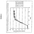
- Figure 9 shows the in vitro total cumulative percentage release of dexamethasone into a saline solution at 37°C from 60/40 w/w dexamethasone/PLGA implants having a weight ratio of 40:0 hydrophobic end to hydrophilic end PLGA (312-140-2), weight ratio of 30:10 hydrophobic end to hydrophilic end PLGA (312-140-4), weight ratio of 20:20 hydrophobic end to hydrophilic end PLGA (312-140-3), and weight ratio of 0:40 hydrophobic end to hydrophilic end PLGA (312-140-1).
- Figure 10 compares the in vitro cumulative percentage release of dexamethasone into a saline solution at 37°C for six lots of extruded implants having 60% by weight dexamethasone, 30% by weight hydrophilic end PLGA, and 10% by weight hydrophobic end PLGA.
- the present invention provides biodegradable ocular implants and methods for treating medical conditions of the eye.
- the implants are formed to be monolithic, i.e., the particles of active agent are distributed throughout the biodegradable polymer matrix.
- the implants are formed to release an active agent into an ocular region of the eye over various time periods.
- the active agent may be release over a time period including, but is not limited to, approximately six months, approximately three months, approximately one month, or less than one month.
- the term "ocular region" refers generally to any area of the eyeball, including the anterior and posterior segment of the eye, and which generally includes, but is not limited to, any functional (e.g., for vision) or structural tissues found in the eyeball, or tissues or cellular layers that partly or completely line the interior or exterior of the eyeball.
- areas of the eyeball in an ocular region include the anterior chamber, the posterior chamber, the vitreous cavity, the choroid, the suprachoroidal space, the conjunctiva, the subconjunctival space, the episcleral space, the intracorneal space, the epicorneal space, the sclera, the pars plana, surgically-induced avascular regions, the macula, and the retina.
- subject it is meant mammalian subjects, preferably humans.
- Mammals include, but are not limited to, primates, farm animals, sport animals, e.g., horses (including race horses), cats, dogs, rabbits, mice, and rats.
- treat or “treating” or “treatment” refers to the resolution, reduction, or prevention of a medical condition of the eye or the sequelae of a medical condition of the eye.
- active agent and “drug” are used interchangeably and refer to any substance used to treat a medical condition of the eye.
- medical condition refers to conditions that are generally treated non-invasively, e.g., with drugs, as well as conditions that are generally treated using a surgical procedure.
- therapeutic amount it is meant a concentration of active agent that has been locally delivered to an ocular region that is appropriate to safely treat a medical condition of the eye.
- the term "cumulative release profile” refers to the cumulative total percent of agent released from the implant either into the posterior segment in vivo in rabbit eyes over time or into the specific release medium in vitro over time.
- the implants of the invention include an active agent dispersed within a biodegradable polymer.
- the implant compositions typically vary according to the preferred drug release profile, the particular active agent used, the condition being treated, and the medical history of the patient.
- Active agents that may be used include, but are not limited to, ace-inhibitors, endogenous cytokines, agents that influence basement membrane, agents that influence the growth of endothelial cells, adrenergic agonists or blockers, cholinergic agonists or blockers, aldose reductase inhibitors, analgesics, anesthetics, antiallergics, anti-inflammatory agents, antihypertensives, pressors, antibacterials, antivirals, antifungals, antiprotozoals, antiinfectives, antitumor agents, antimetabolites, and antiangiogenic agents.
- the active agent is methotrexate. In another variation, the active agent is retinoic acid.
- the anti-inflammatory agent is a nonsteroidal anti-inflammatory agent.
- Nonsteroidal anti-inflammatory agents that may be used include, but are not limited to, aspirin, diclofenac, flurbiprofen, ibuprofen, ketorolac, naproxen, and suprofen. In a more preferred variation, the anti-inflammatory agent is a steroidal anti-inflammatory agent.
- the steroidal anti-inflammatory agents that may be used in the ocular implants include, but are not limited to, 21-acetoxypregnenolone, alclometasone, algestone, amcinonide, beclomethasone, betamethasone, budesonide, chloroprednisone, clobetasol, clobetasone, clocortolone, cloprednol, corticosterone, cortisone, cortivazol, deflazacort, desonide, desoximetasone, dexamethasone, diflorasone, diflucortolone, difluprednate, enoxolone, fluazacort, flucloronide, flumethasone, flunisolide, fluocinolone acetonide, fluocinonide, fluocortin butyl, fluocortolone, fluorometholone, fluperolone acetate, flupredniden
- cortisone, dexamethasone, fluocinolone, hydrocortisone, methylprednisolone, prednisolone, prednisone, and triamcinolone, and their derivatives are preferred steroidal anti-inflammatory agents.
- the steroidal anti-inflammatory agent is dexamethasone.
- the biodegradable implant includes a combination of two or more steroidal anti-inflammatory agents.
- the steroidal anti-inflammatory agent may constitute from about 10% to about 90% by weight of the implant. In one variation, the agent is from about 40% to about 80% by weight of the implant. In a preferred variation, the agent comprises about 60% by weight of the implant.
- the active agent may be homogeneously dispersed in the biodegradable polymer matrix of the implants.
- the selection of the biodegradable polymer matrix to be employed will vary with the desired release kinetics, patient tolerance, the nature of the disease to be treated, and the like. Polymer characteristics that are considered include, but are not limited to, the biocompatibility and biodegradability at the site of implantation, compatibility with the active agent of interest, and processing temperatures.
- the biodegradable polymer matrix usually comprises at least about 10, at least about 20, at least about 30, at least about 40, at least about 50, at least about 60, at least about 70, at least about 80, or at least about 90 weight percent of the implant. In one variation, the biodegradable polymer matrix comprises about 40% by weight of the implant.
- Biodegradable polymer matrices which may be employed include, but are not limited to, polymers made of monomers such as organic esters or ethers, which when degraded result in physiologically acceptable degradation products. Anhydrides, amides, orthoesters, or the like, by themselves or in combination with other monomers, may also be used.
- the polymers are generally condensation polymers.
- the polymers may be crosslinked or non-crosslinked. If crosslinked, they are usually not more than lightly crosslinked, and are less than 5% crosslinked, usually less than 1% crosslinked.
- the polymers will include oxygen and nitrogen, particularly oxygen.
- the oxygen may be present as oxy, e.g., hydroxy or ether; carbonyl, e.g., non-oxo-carbonyl, such as carboxylic acid ester, and the like.
- the nitrogen may be present as amide, cyano, and amino.
- polymers of hydroxyaliphatic carboxylic acids include polymers of hydroxyaliphatic carboxylic acids, either homo- or copolymers, and polysaccharides. Included among the polyesters of interest are homo- or copolymers of D-lactic acid, L-lactic acid, racemic lactic acid, glycolic acid, caprolactone, and combinations thereof. Copolymers of glycolic and lactic acid are of particular interest, where the rate of biodegradation is controlled by the ratio of glycolic to lactic acid.
- the percent of each monomer in poly(lactic-co-glycolic)acid (PLGA) copolymer may be 0-100%, about 15-85%, about 25-75%, or about 35-65%. In a preferred variation, a 50/50 PLGA copolymer is used . More preferably, a random copolymer of 50/50 PLGA is used.
- Biodegradable polymer matrices that include mixtures of hydrophilic and hydrophobic ended PLGA may also be employed, and are useful in modulating polymer matrix degradation rates.
- Hydrophobic ended (also referred to as capped or end-capped) PLGA has an ester linkage hydrophobic in nature at the polymer terminus. Typical hydrophobic end groups include, but are not limited to alkyl esters and aromatic esters.
- Hydrophilic ended (also referred to as uncapped) PLGA has an end group hydrophilic in nature at the polymer terminus.
- PLGA with a hydrophilic end groups at the polymer terminus degrades faster than hydrophobic ended PLGA because it takes up water and undergoes hydrolysis at a faster rate ( Tracy et al., Biomaterials 20:1057-1062 (1099 )).
- suitable hydrophilic end groups that may be incorporated to enhance hydrolysis include, but are not limited to, carboxyl, hydroxyl, and polyethylene glycol.
- the specific end group will typically result from the initiator employed in the polymerization process. For example, if the initiator is water or carboxylic acid, the resulting end groups will be carboxyl and hydroxyl. Similarly, if the initiator is a monofunctional alcohol, the resulting end groups will be ester or hydroxyl.
- the implants may be formed from all hydrophilic end PLGA or all hydrophobic end PLGA.
- the ratio of hydrophilic end to hydrophobic end PLGA in the biodegradable polymer matrices of this invention range from about 10:1 to about 1:10 by weight.
- the ratio may be 3:1, 2:1, or 1:1 by weight.
- an implant with a ratio of hydrophilic end to hydrophobic end PLGA of 3:1 w/w is used.
- buffering agents and preservatives may be employed.
- Preservatives which may be used include, but are not limited to, sodium bisulfite, sodium bisulfate, sodium thiosulfate, benzalkonium chloride, chlorobutanol, thimerosal, phenylmercuric acetate, phenylmercuric nitrate, methylparaben, polyvinyl alcohol and phenylethyl alcohol.
- buffering agents that may be employed include, but are not limited to, sodium carbonate, sodium borate, sodium phosphate, sodium acetate, sodium bicarbonate, and the like, as approved by the FDA for the desired route of administration.
- Electrolytes such as sodium chloride and potassium chloride may also be included in the formulation.
- the biodegradable ocular implants may also include additional hydrophilic or hydrophobic compounds that accelerate or retard release of the active agent.
- hydrophilic end PLGA has a higher degradation rate than hydrophobic end PLGA due to its ability to take up water more readily, increasing the amount of hydrophilic end PLGA in the implant polymer matrix will result in faster dissolution rates.
- Figure 9 shows that the time from implantation to significant release of active agent (lag time) increases with decreasing amounts of hydrophilic end PLGA in the ocular implant.
- the lag time for implants having 0% hydrophilic end PLGA (40% w/w hydrophobic end) was shown to be about 21 days. In comparison, a significant reduction in lag time was seen with implants having 10% w/w and 20% w/w hydrophilic end PLGA.
- the implants of the invention are formulated with particles of an active agent dispersed within a biodegradable polymer matrix. Without being bound by theory, the inventors believe that release of the active agent is achieved by erosion of the biodegradable polymer matrix and by diffusion of the particulate agent into an ocular fluid, e.g., the vitreous, with subsequent dissolution of the polymer matrix and release of the active agent.
- the factors that influence the release kinetics include such characteristics as the size of the active agent particles, the solubility of the active agent, the ratio of active agent to polymer(s), the method of manufacture, the surface area exposed, and the erosion rate of the polymer(s).
- the release kinetics achieved by this form of active agent release are different than that achieved through formulations which release active agents through polymer swelling, such as with crosslinked hydrogels. In that case, the active agent is not released through polymer erosion, but through polymer swelling, which releases agent as liquid diffuses through the pathways exposed.
- the release rate of the active agent depends at least in part on the rate of degradation of the polymer backbone component or components making up the biodegradable polymer matrix.
- condensation polymers may be degraded by hydrolysis (among other mechanisms) and therefore any change in the composition of the implant that enhances water uptake by the implant will likely increase the rate of hydrolysis, thereby increasing the rate of polymer degradation and erosion, and thus increasing the rate of active agent release.
- the release kinetics of the implants of the invention are dependent in part on the surface area of the implants.
- a larger surface area exposes more polymer and active agent to ocular fluid, causing faster erosion of the polymer matrix and dissolution of the active agent particles in the fluid.
- the size and shape of the implant may also be used to control the rate of release, period of treatment, and active agent concentration at the site of implantation. At equal active agent loads, larger implants will deliver a proportionately larger dose, but depending on the surface to mass ratio, may possess a slower release rate.
- the total weight of the implant preferably ranges, e.g., from about 100-5000 ⁇ g, usually from about 500-1500 ⁇ g. In one variation, the total weight of the implant is about 600 ⁇ g. In another variation, the total weight of the implant is about 1200 ⁇ g.
- the bioerodible implants are typically solid, and may be formed as particles, sheets, patches, plaques, films, discs, fibers, rods, and the like, or may be of any size or shape compatible with the selected site of implantation, as long as the implants have the desired release kinetics and deliver an amount of active agent that is therapeutic for the intended medical condition of the eye.
- the upper limit for the implant size will be determined by factors such as the desired release kinetics, toleration for the implant at the site of implantation, size limitations on insertion, and ease of handling.
- the vitreous chamber is able to accommodate relatively large rod-shaped implants, generally having diameters of about 0.05 mm to 3 mm and a length of about 0.5 to about 10 mm.
- the rods have diameters of about 0.1 mm to about 1 mm. In another variation, the rods have diameters of about 0.3 mm to about 0.75 mm. In yet a further variation, other implants having variable geometries but approximately similar volumes may also be used.
- the release of an active agent from a biodegradable polymer matrix may also be modulated by varying the ratio of hydrophilic end PLGA to hydrophobic end PLGA in the matrix. Release rates may be further manipulated by the method used to manufacture the implant. For instance, as illustrated in Examples 4-7, extruded 60/40 w/w dexamethasone/PLGA implants having a ratio of hydrophilic end and hydrophobic end PLGA of 3:1, compared to compressed tablet implants, demonstrate a different drug release profile and concentration of agent in the vitreous over about a one month period. Overall, a lower burst of agent release and a more consistent level of agent in the vitreous is demonstrated with the extruded implants.
- a higher initial burst of active agent release occurs on day one after implantation with the 350 ⁇ g dexamethasone compressed tablet implant (350T) in comparison to the 350 ⁇ g dexamethasone extruded implant (350E).
- a higher initial burst of active agent release also occurs with the 700 ⁇ g dexamethasone compressed implant (700T) in comparison to the 700 ⁇ g dexamethasone extruded implant (700E) on day 1, as shown in Figure 2 and Examples 6 and 7.
- the proportions of active agent, biodegradable polymer matrix, and any other additives may be empirically determined by formulating several implants with varying proportions and determining the release profile in vitro or in vivo.
- a USP approved method for dissolution or release test can be used to measure the rate of release in vitro (USP 24; NF 19 (2000) pp. 1941-1951). For example, a weighed sample of the implant is added to a measured volume of a solution containing 0.9% NaCl in water, where the solution volume will be such that the active agent concentration after release is less than 20% of saturation. The mixture is maintained at 37°C and stirred or shaken slowly to maintain the implants in suspension.
- the release of the dissolved active agent as a function of time may then be followed by various methods known in the art, such as spectrophotometrically, HPLC, mass spectroscopy, and the like, until the solution concentration becomes constant or until greater than 90% of the active agent has been released.
- the extruded implants described herewith may have in vivo cumulative percentage release profiles with the following described characteristics, as shown in Figure 2 , where the release profiles are for release of the active agent in vivo after implantation of the implants into the vitreous of rabbit eyes.
- the volume of rabbit eyes is approximately 60-70% of human eyes.
- the percentage in vivo cumulative release may be between about 0% and about 15%, and more usually between about 0% and about 10%. At day one after implantation, the percentage in vivo cumulative release may be less than about 15%, and more usually less than about 10%.
- the percentage in vivo cumulative release may be between about 0% and about 20%, and more usually between about 5% and about 15%. At day three after implantation, the percentage in vivo cumulative release may be less than about 20%, and more usually less than about 15%.
- the percentage in vivo cumulative release may be between about 0% and about 35%, more usually between about 5% and about 30%, and more usually still between about 10% and about 25%.
- the percentage in vivo cumulative release may be greater than about 2%, more usually greater than about 5%, and more usually still greater than about 10%.
- the percentage in vivo cumulative release may be between about 20% and about 60%, more usually between about 25% and about 55%, and more usually still between about 30% and about 50%. At day fourteen after implantation, the percentage in vivo cumulative release may be greater than about 20%, more usually greater than about 25%, and more usually still greater than about 30%.
- the percentage in vivo cumulative release may be between about 55% and about 95%, more usually between about 60% and about 90%, and more usually still between about 65% and about 85%. At day twenty-one after implantation, the percentage in vivo cumulative release may be greater than about 55% , more usually greater'than about 60% , and more usually still greater than about 65%.
- the percentage in vivo cumulative release may be between about 80% and about 100%, more usually between about. 85% and about 100%, and more usually still between about 90% and about 100%. At day twenty-eight after implantation, the percentage in vivo cumulative release may be greater'than about 80%, more usually greater than about 85%, and more usually still greater than about 90%.
- the percentage in vivo cumulative release may be between about 95% and about 100%, and more usually between about 97% and about 100%. At day thirty-five after implantation, the percentage in vivo cumulative release may be greater than about 95%, and more usually greater than about 97%.
- the percentage in vivo cumulative release has the following characteristics: one day after implantation it is less than about 15%; three days after implantation it is less than about 20%; seven days after implantation it is greater than about 5%; fourteen days after implantation it is greater than about 25%; twenty-one days after implantation it is greater than about 60%; and twenty-eight days after implantation it is greater than about 80%.
- the percentage in vivo cumulative release has the following characteristics: one day after implantation it is less than about 10%; three days after implantation it is less than about 15%; seven days after implantation it is greater than about 10%; fourteen days after implantation it is greater than about 30%; twenty-one days after implantation it is greater than about 65%; twenty-eight days after implantation it is greater than about 85%.
- the extruded implants described in this patent may have in vitro cumulative percentage release profiles in saline solution at 37°C with the following characteristics, as further described below, and as shown in Figure 10 .
- the percentage in vitro cumulative release at day one may be between about. 0% and about 5%, and more usually between about 0% and about 3%.
- the percentage in vitro cumulative release at day one may be less than about 5%, and more usually less than about 3%.
- the percentage in vitro cumulative release at day four may be between about 0% and about 7%, and more usually between about 0% and about 5%.
- the percentage in vitro cumulative release at day four may be less than about 7%, and more usually less than about 5%.
- the percentage in vitro cumulative release at day seven may be between about 1% and about 10%, and more usually between about 2% and about 8%.
- the percentage in vitro cumulative release at day seven may be greater than about 1%, and more usually greater than about 2%.
- the percentage in vitro cumulative release at day 14 may be between about 25% and about 65%, more usually between about 30% and about 60%, and more usually still between about 35% and about 55%.
- the percentage in vitro cumulative release at day 14 may be greater than about 25%, more usually greater than about 30%, and more usually still greater than about 35%.
- the percentage in vitro cumulative release at day 21 may be between about 60% and about 100%, more usually between about 65% and about 95%, and more usually still between about 70% and about 90%.
- the percentage in vitro cumulative release at day 21 may be greater than about 60%, more usually greater than about 65%, and more usually still greater than about 70%.
- the percentage in vitro cumulative release at day 28 may be between about 75% and about 100%, more usually between about 80% and about 100%, and more usually still between about 85% and about 95%.
- the percentage in vitro cumulative release at day 28 may be greater than about 75%, more usually greater than about 80%, and more usually still greater than about 85%.
- the percentage in vitro cumulative release at day 35 may be between about 85% and about 100%, more usually between about 90% and about 100%, and more usually still between about 95% and about 100%.
- the percentage in vitro cumulative release at day 35 may be greater than about 85%, more usually greater than about 90%, and more usually still greater than about 95%.
- the percentage in vitro cumulative release has the following characteristics: after one day it is less than about 1%, after four days it is less than about 7%; after seven days it is greater than about 2%; after 14 days it is greater than about 30%; after 21 days it is greater than about 65%; after 28 days it is greater than about 80%; and after 35 days it is greater than about 90%.
- the percentage in vitro cumulative release has the following characteristics: after one day it is less than about 3%; after four days it is less than about 5%; after seven days it is greater than about 2%; after 14 days it is greater than about 35%; after 21 days it is greater than about 70%; after 28 days it is greater than about 85%; and after 35 days it is greater than about 90%.
- Figures 2 and 10 show that after 28 days in vivo in rabbit eyes, or in vitro in a saline solution at 37°C, respectively, almost all of the active agent has been released from the implants. Furthermore, Figures 2 and 10 show that the active agent release profiles for the extruded implants in vivo (from the time of implantation) and in vitro (from the time of placement into a saline solution at 37°C) are substantially similar and follow approximately a sigmoidal curve, releasing substantially all of the active agent over 28 days.
- the curves show approximately an upward curvature (i.e., the derivative of the curve increases as time increases), and from approximately day 17 onwards the curves show approximately a downward curvature (i.e., the derivative of the curve decreases as time increases).
- the plots shown in Figure 2 for the 350 ⁇ g and 700 ⁇ g dexamethasone compressed tablet implants exhibit a higher initial burst of agent release generally followed by a gradual increase in release.
- implantation of a compressed implant results in different concentrations of active agent in the vitreous at various time points from implants that have been extruded. For example, as shown in Figures 1 and 5 , with extruded implants there is a gradual increase, plateau, and gradual decrease in intravitreal agent concentrations.
- intravitreal concentration curve for extruded implants results in more sustained levels of active agent in the ocular region.
- implants may also be formulated to release a therapeutic agent for any desirable duration of time, for example, for about one week, for about two weeks, for about three weeks, for about four weeks, for about five weeks, for about six weeks, for about seven weeks, for about eight weeks, for about nine weeks, for about ten weeks, for about eleven weeks, for about twelve weeks, or for more than 12 weeks.
- extruded implants Another important feature of the extruded implants is that different concentration levels of active agent may be established in the vitreous using different doses of the active agent. As illustrated in Figure 8 , the concentration of agent in the vitreous is significantly larger with the 700 ⁇ g dexamethasone extruded implant than with the 350 ⁇ g dexamethasone extruded implant. Different active agent concentrations are not demonstrated with the compressed tablet implant. Thus, by using an extruded implant, it is possible to more easily control the concentration of active agent in the vitreous. In particular, specific dose-response relationships may be established since the implants can be sized to deliver a predetermined amount of active agent.
- Examples of medical conditions of the eye which may be treated by the implants and methods of the invention include, but are not limited to, uveitis, macular edema, macular degeneration, retinal detachment, ocular tumors, fungal or viral infections, multifocal choroiditis, diabetic retinopathy, proliferative vitreoretinopathy (PVR), sympathetic opthalmia, Vogt Koyanagi-Harada (VKH) syndrome, histoplasmosis, uveal diffusion, and vascular occlusion.
- the implants are particularly useful in treating such medical conditions as uveitis, macular edema, vascular occlusive conditions, proliferative vitreoretinopathy (PVR), and various other retinopathies.
- the biodegradable implants may be inserted into the eye by a variety of methods, including placement by forceps, by trocar, or by other types of applicators, after making an incision in the sclera. In some instances, a trocar or applicator may be used without creating an incision. In a preferred variation, a hand held applicator is used to insert one or more biodegradable implants into the eye.
- the hand held applicator typically comprises an 18-30 GA stainless steel needle, a lever, an actuator, and a plunger.
- the method of implantation generally first involves accessing the target area within the ocular region with the needle. Once within the target area, e.g., the vitreous cavity, the lever on the hand held device is depressed to cause the actuator to drive the plunger forward. As the plunger moves forward, it pushes the implant into the target area.
- the target area e.g., the vitreous cavity
- extrusion methods allows for large-scale manufacture of implants and results in implants with a homogeneous dispersion of the drug within the polymer matrix.
- the polymers and active agents that are chosen are stable at temperatures required for manufacturing, usually at least about 50°C.
- Extrusion methods use temperatures of about 25°C to about 150°C, more preferably about 60°C to about 130°C.
- Different extrusion methods may yield implants with different characteristics, including but not limited to the homogeneity of the dispersion of the active agent within the polymer matrix.
- a piston extruder a single screw extruder, and a twin screw extruder will generally produce implants with progressively more homogeneous, dispersion of the active.
- extrusion parameters such as temperature, extrusion speed, die geometry, and die surface finish will have an effect on the release profile of the implants produced.
- the drug and polymer are first mixed at room temperature and then heated to a temperature range of about 60°C to about 150°C, more usually to about 130°C for a time period of about 0 to about 1 hour, more usually from about 0 to about 30 minutes, more usually still from about 5 minutes to about 15 minutes, and most usually for about 10 minutes.
- the implants are then extruded at a temperature of about 60°C to about 130°C, preferably at a temperature of about 75°C.
- the powder blend of active agent and PLGA is added to a single or twin screw extruder preset at a temperature of about 80°C to about 130°C, and directly extruded as a filament or rod with minimal residence time in the extruder.
- the extruded filament or rod is then cut into small implants having the loading dose of active agent appropriate to treat the medical condition of its intended use.
- Micronized dexamethasone (Pharmacia, Peapack, NJ) and micronized hydrophobic end 50/50 PLGA (Birmingham Polymers, Inc., Birmingham, AL) were accurately weighed and placed in a stainless steel mixing vessel. The vessel was sealed, placed on a Turbula mixer and mixed at a prescribed intensity, e.g., 96 rpm, and time, e.g., 15 minutes. The resulting powder blend was loaded one unit dose at a time into a single-cavity tablet press. The press was activated at a pre-set pressure, e.g., 25 psi, and duration, e.g., 6 seconds, and the tablet was formed and ejected from the press at room temperature. The ratio of dexamethasone to PLGA was 70/30 w/w for all compressed tablet implants.
- Micronized dexamethasone (Pharmacia, Peapack, NJ) and unmicronized PLGA were accurately weighed and placed in a stainless steel mixing vessel. The vessel was sealed, placed on a Turbula mixer and mixed at a prescribed intensity, e.g., 96 rpm, and time, e.g., 10-15 minutes.
- the unmicronized PLGA composition comprised a 30/10 w/w mixture of hydrophilic end PLGA (Boehringer Ingelheim, Wallingford, CT) and hydrophobic end PLGA (Boehringer Ingelheim, Wallingford, CT).
- the resulting powder blend was fed into a DACA Microcompounder-Extruder (DACA, Goleta, CA) and subjected to a pre-set temperature, e.g., 115°C, and screw speed, e.g., 12 rpm.
- the filament was extruded into a guide mechanism and cut into exact lengths that corresponded to the designated implant weight.
- the ratio of dexamethasone to total PLGA (hydrophilic and hydrophobic end) was 60/40 w/w for all extruded implants.
- Implants were placed into the posterior segment of the right eye of New Zealand White Rabbits by incising the conjunctiva and sclera between the 10 and 12 o'clock positions with a 20-gauge microvitreoretinal (MVR) blade. Fifty to 100 ⁇ L of vitreous humor was removed with a 1-cc syringe fitted with a 27-gauge needle. A sterile trocar, preloaded with the appropriate implant (drug delivery system, DDS), was inserted 5 mm through the sclerotomy, and then retracted with the push wire in place, leaving the implant in the posterior segment. Sclerae and conjunctivae were than closed using a 7-0 Vicryl suture.
- DDS drug delivery system
- Example 4 demonstrates the high initial release but generally lower intravitreal concentration of dexamethasone from compressed tablet implants as compared to extruded implants.
- the 350 ⁇ g compressed tablet implant (350T) was placed in the right eye of New Zealand White Rabbits as described in Example 3.
- Vitreous samples were taken periodically and assayed by LC/MS/MS to determine in vivo dexamethasone delivery performance.
- dexamethasone reached detectable mean intravitreal concentrations from day 1 (142.20 ng/ml) through day 35 (2.72 ng/ml), and the intravitreal concentration of dexamethasone gradually decreased over time.
- aqueous humor and plasma samples were also taken.
- the 350T showed a gradual decrease in aqueous humor dexamethasone concentrations over time, exhibiting a detectable mean dexamethasone aqueous humor concentration at day 1 (14.88 ng/ml) through day 21 (3.07 ng/ml), as demonstrated in Figure 3 .
- the levels of dexamethasone in the aqueous humor strongly correlated with the levels of dexamethasone in the vitreous humor, but at a much lower level (approximately 10-fold lower).
- Figure 4 shows that only trace amounts of dexamethasone was found in the plasma.
- Example 5 demonstrates the lower initial release and generally more sustained intravitreal concentration of dexamethasone from extruded implants.
- the 350 ⁇ g extruded implant (350E) was placed in the right eye of New Zealand White Rabbits as described in Example 3. Vitreous samples were taken periodically and assayed by LC/MS/MS to determine in vivo dexamethasone delivery performance.
- 350E showed detectable mean vitreous humor concentrations on day 1 (10.66 ng/ml) through day 28 (6.99 ng/ml).
- aqueous humor and plasma samples were also taken.
- the 350E showed detectable mean dexamethasone aqueous humor concentrations at day 1 (6.67 ng/ml) through day 42 (2.58 ng/ml) with the exception of day 35 in which the values were below the quantification limit.
- the levels of dexamethasone in the aqueous strongly correlated with the levels of dexamethasone in the vitreous humor, but at a much lower level (approximately 10-fold lower).
- Figure 4 demonstrates that only a trace amount of dexamethasone was found in the plasma.
- Example 6 also shows the high initial release and generally lower intravitreal concentration of dexamethasone from compressed tablet implants.
- the 700 ⁇ g compressed tablet dosage form (700T) was placed in the right eye of New Zealand White Rabbits as described in Example 3. Vitreous samples were taken periodically and assayed by LC/MS/MS to determine in vivo dexamethasone delivery performance. As seen in Figure 5 , the 700T reached detectable mean dexamethasone vitreous humor concentrations at day 1 (198.56 ng/ml) through day 42 (2.89 ng/ml), and a gradual decrease in the intravitreal dexamethasone concentration over time.
- aqueous humor and plasma samples were also obtained.
- the 700T exhibited a gradual decrease in aqueous humor dexamethasone concentrations over time, and reached detectable mean dexamethasone aqueous humor concentrations at day 1 (25.90 ng/ml) through day 42 (2.64 ng/ml) with the exception of day 35 in which the values were below the quantification limit.
- the levels of dexamethasone in the aqueous humor strongly correlated with the levels of dexamethasone in the vitreous humor, but at a much lower level (approximately 10-fold lower).
- Figure 7 demonstrates that only a trace amount of dexamethasone was found in the plasma.
- Example 7 also illustrates the lower initial release and generally higher intravitreal concentration of dexamethasone from extruded implants.
- the 700 ⁇ g extruded implant (700E) was placed in the right eye of New Zealand White Rabbits as described in Example 3. Vitreous samples were taken periodically and assayed by LC/MS/MS to determine in vivo dexamethasone delivery performance. As seen in Figure 5 , the 700E had a mean detectable vitreous humor concentration of dexamethasone from day 1 (52.63 ng/ml) through day 28 (119.70 ng/ml).
- aqueous humor and plasma samples were also taken. As seen in Figure 6 , the 700E reached a detectable mean aqueous humor concentration on day 1 (5.04 ng/ml) through day 28 (5.93 ng/ml). The levels of dexamethasone in the aqueous strongly correlated with the levels of dexamethasone in the vitreous humor, but at a much lower level (approximately 10-fold lower). Figure 7 demonstrates that only a trace amount of dexamethasone was found in the plasma.
Abstract
Description
- The present invention relates to the field of ophthalmology. In particular, biodegradable implants and methods for treating medical conditions of the eye are provided.
- Immunosuppressive agents are routinely used for the treatment of uveitis of various etiologies. For example, topical or oral glucocorticoids are often included in the therapeutic regimen; however, a major problem with these routes of administration is the inability to achieve an adequate intraocular drug concentration of the glucocorticoid. In fact, the difficulties of treating uveitis due to poor intraocular penetration of topical medications into the posterior segment is well known (Bloch-Michel E. (1992). "Opening address: intermediate uveitis," In Intermediate Uveitis, Dev. Ophthalmol. W.R.F. Böke et al. eds., Basel: Karger, 23:1-2; Pinar, V. Intermediate uveitis. Massachusetts Eye & Ear Infirmary Immunology Service at <http://www.immunology.meei.harvard.edu/imed.htm> (visited in 1998); Rao, N.A. et al. (1997). "Intraocular inflammation and uveitis," In Basic and Clinical Science Course. Section 9 (1997-1998) San Francisco: American Academy of Ophthalmology, pp. 57-80, 102-103, 152-156; Böke, W. (1992). "Clinical picture of intermediate uveitis," In Intermediate Uveitis, Dev. Ophthalmol. W.R.F. Böke et al. eds., Basel: Karger, 23:20-7; and Cheng C-K et al. (1995). "Intravitreal sustained-release dexamethasone device in the treatment of experimental uveitis," Invest. Ophthalmol. Vis. Sci. 36:442-53).
- Systemic glucocorticoid administration may be used alone or in addition to topical glucocorticoids for the treatment of uveitis. Prolonged exposure to high plasma concentrations (administration of 1 mg/kg/day for 2-3 weeks) of steroid is often necessary so that therapeutic levels can be achieved in the eye (Pinar, V. "Intermediate uveitis," Massachusetts Eye & Ear Infirmary Immunology Service at <http://www.immunology.meei.harvard.edu/imed.htm> (visited in 1998)).
- However, these high drug plasma levels commonly lead to systemic side effects such as hypertension, hyperglycemia, increased susceptibility to infection; peptic ulcers, psychosis, and other complications (Cheng C-K et al. (1995). "Intravitreal sustained-release dexamethasone device in the treatment of experimental uveitis," Invest. Ophthalmol. Vis. Sci. 36:442-53; Schwartz, B. (1966). "The response of ocular pressure to corticosteroids," Ophthalmol. Clin. North Am. 6:929-89; Skalka, H.W. et al. (1980). "Effect of corticosteroids on cataract formation," Arch Ophthalmol 98:1773-7; and Renfro, L. et al. (1992). "Ocular effects of topical and systemic steroids," Dermatologic Clinics 10:505-12).
- In addition, overall drug delivery to the eye may be poor for drugs with short plasma half-lives since their exposure to intraocular tissues is limited. Therefore, the most efficient way of delivering a drug to the posterior segment is to place it directly into the vitreous (Maurice, D.M. (1983). "Micropharmaceutics of the eye," Ocular Inflammation Ther. 1:97-102; Lee, V.H.L. et al. (1989). "Drug delivery to the posterior segment" ; and Olsen, T.W. et al. (1995). "Human scleral permeability: effects of age, cryotherapy, transscleral diode laser, and surgical thinning," Invest. Ophthalmol. Vis. Sci. 36:1893-1903).
- Techniques such as intravitreal injection have shown promising results, but due to the short intraocular half-life of glucocorticoids (approximately 3 hours), intravitreal injections must be repeated to maintain drug levels. In turn, this repetitive process increases the potential for side effects such as retinal detachment, endophthalmitis, and cataracts (Maurice, D.M. (1983). "Micropharmaceutics of the eye," Ocular Inflammation Ther. 1:97-102; Olsen, T.W. et al. (1995). "Human scleral permeability: effects of age, cryotherapy, transscleral diode laser, and surgical thinning," Invest. Ophthalmol. Vis. Sci. 36:1.893-1903; and Kwak, H.W. and D'Amico, D. J. (1992). "Evaluation of the retinal toxicity and pharmacokinetics of dexamethasone after intravitreal injection," Arch. Ophthalmol. 110:259-66).
- One of the alternatives to intravitreal injection to administer, drugs is the placement of biodegradable implants under the sclera or into the subconjunctival or suprachoroidal space, as described in
U.S. 4,863,457 to Lee ;WO 95/13765 to Wong et al. WO 00/37056 to Wong et al. EP 430,539 to Wong - Furthermore, the controlled release of drugs from polylactide/polyglycolide (PLGA) copolymers into the vitreous has been disclosed, e.g., in
U.S. 5,501,856 to Ohtori et al. andEP 654,256 to Ogura - Recent experimental work has demonstrated that uncapped PLGA degrades faster than capped (end-capped) PLGA (Park et al., J. Control. Rel. 55:181-191 (1998); Tracy et al., Biomaterials 20:1057-1062 (1999); and Jong et al., Polymer 42:2795-2802 (2001). Accordingly, implants containing mixtures of uncapped and capped PLGA have been formed to modulate drug release. For example;
U.S. 6,217,911 to Vaughn et al. ('911.) andU.S. 6,309,669 to Setterstrom et al. ('669) disclose the delivery of drugs from a blend of uncapped and capped PLGA copolymer to curtail initial burst release of the drugs. In the '911 patent, the composition delivers non-steroidal anti-inflammatory drugs from PLGA microspheres made by a solvent extraction process or PLGA microcapsules prepared by a solvent evaporation process over a duration of 24 hours to 2 months. In the '669 patent, the composition delivers various pharmaceuticals from PLGA microcapsules over a duration of 1-100 days. The PLGA microspheres or microcapsules are administered orally or as an aqueous injectable formulation. As mentioned above, there is poor partitioning of drug into the eye with oral administration. Furthermore, use of an aqueous injectable drug composition (for injecting into the eye) should be avoided since the eye is a closed space (limited volume) with intraocular pressure ranges that are strictly maintained. Administration of an injectable may increase intraocular volume to a point where intraocular pressures would then become pathologic. - Consequently, a biodegradable implant for delivering a therapeutic agent to an ocular region may provide significant medical benefit for patients afflicted with a medical condition of the eye.
- The biodegradable implants and methods of this invention are typically used to treat medical conditions of the eye. Consequently, the implants are sized such that they are appropriate for implantation in the intended ocular region.
- In one variation, the bioerodible implant for treating medical conditions of the eye includes an active agent dispersed within a biodegradable polymer matrix, wherein the bioerodible implant has an in vivo in rabbit eye cumulative release profile in which less than about 15 percent of the active agent is released about one day after implantation of the bioerodible implant and greater than about 80 percent of the active agent is released about 28 days after implantation of the bioerodible implant, and wherein the biodegradable polymer matrix comprises a mixture of hydrophilic end group PLGA and hydrophobic end group PLGA.
- In another variation, the bioerodible implant for treating medical conditions of the eye includes an active agent dispersed within a biodegradable polymer matrix, wherein the bioerodible implant is formed by an extrusion method, and wherein the bioerodible implant has an in vivo in rabbit eye cumulative release profile in which greater than about 80 percent of the active agent is released about 28 days after implantation of the bioerodible implant.
- In a further variation, the bioerodible implant for treating medical conditions of the eye includes an active agent dispersed within a biodegradable polymer matrix, wherein the bioerodible implant exhibits a cumulative release profile in which greater than about 80 percent of the active agent is released about 28 days after implantation of the bioerodible implant, and wherein the cumulative release profile is approximately sigmoidal in shape over about 28 days after implantation.
- In yet a further variation, the bioerodible implant for treating medical conditions.of the eye includes an active agent dispersed within a biodegradable polymer matrix, wherein the biodegradable polymer matrix comprises a mixture of PLGA having hydrophilic end groups and PLGA having hydrophobic end groups. Examples of hydrophilic end groups include, but are not limited to, carboxyl, hydroxyl, and polyethylene glycol. Examples of hydrophobic end groups include, but are not limited to, alkyl esters and aromatic esters.
- In yet another variation, the bioerodible implant for treating medical conditions of the eye includes an active agent dispersed within a biodegradable polymer matrix, wherein the bioerodible implant has an in vivo in rabbit eye cumulative release profile in which less than about 15 percent of the active agent is released about one day after implantation of the bioerodible implant and greater than about 80 percent of the active agent is released about 28 days after implantation of the bioerodible implant.
- Various active agents may be incorporated into the bioerodible implants. In one variation, anti-inflammatory agents, including, but not limited to nonsteroidal anti-inflammatory agents and steroidal anti-inflammatory agents may be used. In another variation, active agents that may be used in the bioerodible implants are ace-inhibitors, endogenous cytokines, agents that influence basement membrane, agents that influence the growth of endothelial cells, adrenergic agonists or blockers, cholinergic agonists or blockers, aldose reductase inhibitors, analgesics, anesthetics, antiallergics, antibacterials, antihypertensives, pressors, antiprotozoal agents, antiviral agents, antifungal agents, anti-infective agents, antitumor agents, antimetabolites, and antiangiogenic agents.
- The implants may be used to treat medical conditions of the eye in mammalian subjects, e.g., human subjects. Examples of such medical conditions include, but are not limited to, uveitis, macular edema, macular degeneration, retinal detachment, ocular tumors, fungal or viral infections, multifocal choroiditis, diabetic retinopathy, proliferative vitreoretinopathy (PVR), sympathetic opthalmia, Vogt Koyanagi-Harada (VKH) syndrome, histoplasmosis, uveal diffusion, vascular occlusion, and the like.
- Furthermore, upon implantation in an ocular region of the subject, the bioerodible implants deliver the active agent such that the resulting concentration of active agent in vivo in rabbit aqueous humor is approximately 10-fold less than in rabbit vitreous humor. The active agent is delivered so that a therapeutic amount of active agent is provided in the ocular region of interest. In general, the therapeutic amount of active agent in an ocular region may be modified by varying the size of the bioerodible implant.
-
Figure 1 shows the in vivo concentration of dexamethasone in the vitreous of rabbit eyes over a 42 day period after implantation of compressed and extruded biodegradable implants containing 350 µg dexamethasone into the posterior segment of rabbit eyes. -
Figure 2 shows the in vivo cumulative percentage release of dexamethasone in the vitreous of rabbit eyes over a 42 day period after implantation of compressed and extruded biodegradable implants containing 350 µg dexamethasone and 700 µg dexamethasone into the posterior segment of rabbit eyes. -
Figure 3 shows the in vivo concentration of dexamethasone in the aqueous humor of rabbit eyes over a 42 day period after implantation of compressed and extruded biodegradable implants containing 350 µg dexamethasone into the posterior segment of rabbit eyes. -
Figure 4 shows the in vivo concentration of dexamethasone in the plasma (from a rabbit blood sample) over a 42 day period after implantation of compressed and extruded biodegradable implants containing 350 µg dexamethasone into the posterior segment of rabbit eyes. -
Figure 5 shows the in vivo concentration of dexamethasone in the vitreous of rabbit eyes over a 42 day period after implantation of compressed and extruded biodegradable implants containing 700 µg dexamethasone into the posterior segment of rabbit eyes. -
Figure 6 shows the in vivo concentration of dexamethasone in the aqueous humor of rabbit eyes over a 42 day period after implantation of compressed and extruded biodegradable implants containing 700 µg dexamethasone into the posterior segment of rabbit eyes. -
Figure 7 shows the in vivo concentration of dexamethasone in the plasma (from a rabbit blood sample) over a 42 day period after implantation of compressed and extruded biodegradable implants containing 700 µg dexamethasone into the posterior segment of rabbit eyes. -
Figure 8 shows the in vivo concentration of dexamethasone in the vitreous of rabbit eyes over a 42 day period after implantation of compressed and extruded biodegradable implants containing 350 µg dexamethasone and 700 µg dexamethasone into the posterior segment of rabbit eyes. -
Figure 9 shows the in vitro total cumulative percentage release of dexamethasone into a saline solution at 37°C from 60/40 w/w dexamethasone/PLGA implants having a weight ratio of 40:0 hydrophobic end to hydrophilic end PLGA (312-140-2), weight ratio of 30:10 hydrophobic end to hydrophilic end PLGA (312-140-4), weight ratio of 20:20 hydrophobic end to hydrophilic end PLGA (312-140-3), and weight ratio of 0:40 hydrophobic end to hydrophilic end PLGA (312-140-1). -
Figure 10 compares the in vitro cumulative percentage release of dexamethasone into a saline solution at 37°C for six lots of extruded implants having 60% by weight dexamethasone, 30% by weight hydrophilic end PLGA, and 10% by weight hydrophobic end PLGA. - The present invention provides biodegradable ocular implants and methods for treating medical conditions of the eye. Usually, the implants are formed to be monolithic, i.e., the particles of active agent are distributed throughout the biodegradable polymer matrix. Furthermore, the implants are formed to release an active agent into an ocular region of the eye over various time periods. The active agent may be release over a time period including, but is not limited to, approximately six months, approximately three months, approximately one month, or less than one month.
- For the purposes of this description, we use the following terms as defined in this section, unless the context of the word indicates a different meaning.
- As used herein, the term "ocular region" refers generally to any area of the eyeball, including the anterior and posterior segment of the eye, and which generally includes, but is not limited to, any functional (e.g., for vision) or structural tissues found in the eyeball, or tissues or cellular layers that partly or completely line the interior or exterior of the eyeball. Specific examples of areas of the eyeball in an ocular region include the anterior chamber, the posterior chamber, the vitreous cavity, the choroid, the suprachoroidal space, the conjunctiva, the subconjunctival space, the episcleral space, the intracorneal space, the epicorneal space, the sclera, the pars plana, surgically-induced avascular regions, the macula, and the retina.
- By "subject" it is meant mammalian subjects, preferably humans. Mammals include, but are not limited to, primates, farm animals, sport animals, e.g., horses (including race horses), cats, dogs, rabbits, mice, and rats.
- As used herein, the term "treat" or "treating" or "treatment" refers to the resolution, reduction, or prevention of a medical condition of the eye or the sequelae of a medical condition of the eye.
- As used herein, the terms "active agent" and "drug" are used interchangeably and refer to any substance used to treat a medical condition of the eye.
- As used herein, the term "medical condition" refers to conditions that are generally treated non-invasively, e.g., with drugs, as well as conditions that are generally treated using a surgical procedure.
- By "therapeutic amount" it is meant a concentration of active agent that has been locally delivered to an ocular region that is appropriate to safely treat a medical condition of the eye.
- As used herein, the term "cumulative release profile" refers to the cumulative total percent of agent released from the implant either into the posterior segment in vivo in rabbit eyes over time or into the specific release medium in vitro over time.
- The implants of the invention include an active agent dispersed within a biodegradable polymer. The implant compositions typically vary according to the preferred drug release profile, the particular active agent used, the condition being treated, and the medical history of the patient. Active agents that may be used include, but are not limited to, ace-inhibitors, endogenous cytokines, agents that influence basement membrane, agents that influence the growth of endothelial cells, adrenergic agonists or blockers, cholinergic agonists or blockers, aldose reductase inhibitors, analgesics, anesthetics, antiallergics, anti-inflammatory agents, antihypertensives, pressors, antibacterials, antivirals, antifungals, antiprotozoals, antiinfectives, antitumor agents, antimetabolites, and antiangiogenic agents.
- In one variation the active agent is methotrexate. In another variation, the active agent is retinoic acid. In a preferred variation, the anti-inflammatory agent is a nonsteroidal anti-inflammatory agent. Nonsteroidal anti-inflammatory agents that may be used include, but are not limited to, aspirin, diclofenac, flurbiprofen, ibuprofen, ketorolac, naproxen, and suprofen. In a more preferred variation, the anti-inflammatory agent is a steroidal anti-inflammatory agent.
- The steroidal anti-inflammatory agents that may be used in the ocular implants include, but are not limited to, 21-acetoxypregnenolone, alclometasone, algestone, amcinonide, beclomethasone, betamethasone, budesonide, chloroprednisone, clobetasol, clobetasone, clocortolone, cloprednol, corticosterone, cortisone, cortivazol, deflazacort, desonide, desoximetasone, dexamethasone, diflorasone, diflucortolone, difluprednate, enoxolone, fluazacort, flucloronide, flumethasone, flunisolide, fluocinolone acetonide, fluocinonide, fluocortin butyl, fluocortolone, fluorometholone, fluperolone acetate, fluprednidene acetate, fluprednisolone, flurandrenolide, fluticasone propionate, formocortal, halcinonide, halobetasol propionate, halometasone, halopredone acetate, hydrocortamate, hydrocortisone, loteprednol etabonate, mazipredone, medrysone, meprednisone, methylprednisolone, mometasone furoate, paramethasone, prednicarbate, prednisolone, prednisolone 25-diethylamino-acetate, prednisolone sodium phosphate, prednisone, prednival, prednylidene, rimexolone, tixocortol, triamcinolone, triamcinolone acetonide, triamcinolone benetonide, triamcinolone hexacetonide, and any of their derivatives.
- In one variation, cortisone, dexamethasone, fluocinolone, hydrocortisone, methylprednisolone, prednisolone, prednisone, and triamcinolone, and their derivatives, are preferred steroidal anti-inflammatory agents. In another preferred variation, the steroidal anti-inflammatory agent is dexamethasone. In another variation, the biodegradable implant includes a combination of two or more steroidal anti-inflammatory agents.
- The steroidal anti-inflammatory agent may constitute from about 10% to about 90% by weight of the implant. In one variation, the agent is from about 40% to about 80% by weight of the implant. In a preferred variation, the agent comprises about 60% by weight of the implant.
- In one variation, the active agent may be homogeneously dispersed in the biodegradable polymer matrix of the implants. The selection of the biodegradable polymer matrix to be employed will vary with the desired release kinetics, patient tolerance, the nature of the disease to be treated, and the like. Polymer characteristics that are considered include, but are not limited to, the biocompatibility and biodegradability at the site of implantation, compatibility with the active agent of interest, and processing temperatures. The biodegradable polymer matrix usually comprises at least about 10, at least about 20, at least about 30, at least about 40, at least about 50, at least about 60, at least about 70, at least about 80, or at least about 90 weight percent of the implant. In one variation, the biodegradable polymer matrix comprises about 40% by weight of the implant.
- Biodegradable polymer matrices which may be employed include, but are not limited to, polymers made of monomers such as organic esters or ethers, which when degraded result in physiologically acceptable degradation products. Anhydrides, amides, orthoesters, or the like, by themselves or in combination with other monomers, may also be used. The polymers are generally condensation polymers. The polymers may be crosslinked or non-crosslinked. If crosslinked, they are usually not more than lightly crosslinked, and are less than 5% crosslinked, usually less than 1% crosslinked.
- For the most part, besides carbon and hydrogen, the polymers will include oxygen and nitrogen, particularly oxygen. The oxygen may be present as oxy, e.g., hydroxy or ether; carbonyl, e.g., non-oxo-carbonyl, such as carboxylic acid ester, and the like. The nitrogen may be present as amide, cyano, and amino. An exemplary list of biodegradable polymers that may be used are described in Heller, Biodegradable Polymers in Controlled Drug Delivery, In: "CRC Critical Reviews in Therapeutic Drug Carrier Systems", Vol. 1. CRC Press, Boca Raton, FL (1987).
- Of particular interest are polymers of hydroxyaliphatic carboxylic acids, either homo- or copolymers, and polysaccharides. Included among the polyesters of interest are homo- or copolymers of D-lactic acid, L-lactic acid, racemic lactic acid, glycolic acid, caprolactone, and combinations thereof. Copolymers of glycolic and lactic acid are of particular interest, where the rate of biodegradation is controlled by the ratio of glycolic to lactic acid. The percent of each monomer in poly(lactic-co-glycolic)acid (PLGA) copolymer may be 0-100%, about 15-85%, about 25-75%, or about 35-65%. In a preferred variation, a 50/50 PLGA copolymer is used . More preferably, a random copolymer of 50/50 PLGA is used.
- Biodegradable polymer matrices that include mixtures of hydrophilic and hydrophobic ended PLGA may also be employed, and are useful in modulating polymer matrix degradation rates. Hydrophobic ended (also referred to as capped or end-capped) PLGA has an ester linkage hydrophobic in nature at the polymer terminus. Typical hydrophobic end groups include, but are not limited to alkyl esters and aromatic esters. Hydrophilic ended (also referred to as uncapped) PLGA has an end group hydrophilic in nature at the polymer terminus. PLGA with a hydrophilic end groups at the polymer terminus degrades faster than hydrophobic ended PLGA because it takes up water and undergoes hydrolysis at a faster rate (Tracy et al., Biomaterials 20:1057-1062 (1099)). Examples of suitable hydrophilic end groups that may be incorporated to enhance hydrolysis include, but are not limited to, carboxyl, hydroxyl, and polyethylene glycol. The specific end group will typically result from the initiator employed in the polymerization process. For example, if the initiator is water or carboxylic acid, the resulting end groups will be carboxyl and hydroxyl. Similarly, if the initiator is a monofunctional alcohol, the resulting end groups will be ester or hydroxyl.
- The implants may be formed from all hydrophilic end PLGA or all hydrophobic end PLGA. In general, however, the ratio of hydrophilic end to hydrophobic end PLGA in the biodegradable polymer matrices of this invention range from about 10:1 to about 1:10 by weight. For example, the ratio may be 3:1, 2:1, or 1:1 by weight. In a preferred variation, an implant with a ratio of hydrophilic end to hydrophobic end PLGA of 3:1 w/w is used.
- Other agents may be employed in the formulation for a variety of purposes. For example, buffering agents and preservatives may be employed. Preservatives which may be used include, but are not limited to, sodium bisulfite, sodium bisulfate, sodium thiosulfate, benzalkonium chloride, chlorobutanol, thimerosal, phenylmercuric acetate, phenylmercuric nitrate, methylparaben, polyvinyl alcohol and phenylethyl alcohol. Examples of buffering agents that may be employed include, but are not limited to, sodium carbonate, sodium borate, sodium phosphate, sodium acetate, sodium bicarbonate, and the like, as approved by the FDA for the desired route of administration. Electrolytes such as sodium chloride and potassium chloride may also be included in the formulation.
- The biodegradable ocular implants may also include additional hydrophilic or hydrophobic compounds that accelerate or retard release of the active agent. Furthermore, the inventors believe that because hydrophilic end PLGA has a higher degradation rate than hydrophobic end PLGA due to its ability to take up water more readily, increasing the amount of hydrophilic end PLGA in the implant polymer matrix will result in faster dissolution rates.
Figure 9 shows that the time from implantation to significant release of active agent (lag time) increases with decreasing amounts of hydrophilic end PLGA in the ocular implant. InFigure 9 , the lag time for implants having 0% hydrophilic end PLGA (40% w/w hydrophobic end) was shown to be about 21 days. In comparison, a significant reduction in lag time was seen with implants having 10% w/w and 20% w/w hydrophilic end PLGA. - The inventors believe the implants of the invention are formulated with particles of an active agent dispersed within a biodegradable polymer matrix. Without being bound by theory, the inventors believe that release of the active agent is achieved by erosion of the biodegradable polymer matrix and by diffusion of the particulate agent into an ocular fluid, e.g., the vitreous, with subsequent dissolution of the polymer matrix and release of the active agent. The inventors believe that the factors that influence the release kinetics include such characteristics as the size of the active agent particles, the solubility of the active agent, the ratio of active agent to polymer(s), the method of manufacture, the surface area exposed, and the erosion rate of the polymer(s). The release kinetics achieved by this form of active agent release are different than that achieved through formulations which release active agents through polymer swelling, such as with crosslinked hydrogels. In that case, the active agent is not released through polymer erosion, but through polymer swelling, which releases agent as liquid diffuses through the pathways exposed.
- The inventors believe that the release rate of the active agent depends at least in part on the rate of degradation of the polymer backbone component or components making up the biodegradable polymer matrix. For example, condensation polymers may be degraded by hydrolysis (among other mechanisms) and therefore any change in the composition of the implant that enhances water uptake by the implant will likely increase the rate of hydrolysis, thereby increasing the rate of polymer degradation and erosion, and thus increasing the rate of active agent release.
- The release kinetics of the implants of the invention are dependent in part on the surface area of the implants. A larger surface area exposes more polymer and active agent to ocular fluid, causing faster erosion of the polymer matrix and dissolution of the active agent particles in the fluid. The size and shape of the implant may also be used to control the rate of release, period of treatment, and active agent concentration at the site of implantation. At equal active agent loads, larger implants will deliver a proportionately larger dose, but depending on the surface to mass ratio, may possess a slower release rate. For implantation in an ocular region, the total weight of the implant preferably ranges, e.g., from about 100-5000 µg, usually from about 500-1500 µg. In one variation, the total weight of the implant is about 600 µg. In another variation, the total weight of the implant is about 1200 µg.
- The bioerodible implants are typically solid, and may be formed as particles, sheets, patches, plaques, films, discs, fibers, rods, and the like, or may be of any size or shape compatible with the selected site of implantation, as long as the implants have the desired release kinetics and deliver an amount of active agent that is therapeutic for the intended medical condition of the eye. The upper limit for the implant size will be determined by factors such as the desired release kinetics, toleration for the implant at the site of implantation, size limitations on insertion, and ease of handling. For example, the vitreous chamber is able to accommodate relatively large rod-shaped implants, generally having diameters of about 0.05 mm to 3 mm and a length of about 0.5 to about 10 mm. In one variation, the rods have diameters of about 0.1 mm to about 1 mm. In another variation, the rods have diameters of about 0.3 mm to about 0.75 mm. In yet a further variation, other implants having variable geometries but approximately similar volumes may also be used.
- As previously discussed, the release of an active agent from a biodegradable polymer matrix may also be modulated by varying the ratio of hydrophilic end PLGA to hydrophobic end PLGA in the matrix. Release rates may be further manipulated by the method used to manufacture the implant. For instance, as illustrated in Examples 4-7, extruded 60/40 w/w dexamethasone/PLGA implants having a ratio of hydrophilic end and hydrophobic end PLGA of 3:1, compared to compressed tablet implants, demonstrate a different drug release profile and concentration of agent in the vitreous over about a one month period. Overall, a lower burst of agent release and a more consistent level of agent in the vitreous is demonstrated with the extruded implants.
- As shown in
Figure 2 and Examples 4 and 5, a higher initial burst of active agent release occurs on day one after implantation with the 350 µg dexamethasone compressed tablet implant (350T) in comparison to the 350 µg dexamethasone extruded implant (350E). A higher initial burst of active agent release also occurs with the 700 µg dexamethasone compressed implant (700T) in comparison to the 700 µg dexamethasone extruded implant (700E) onday 1, as shown inFigure 2 and Examples 6 and 7. - The proportions of active agent, biodegradable polymer matrix, and any other additives may be empirically determined by formulating several implants with varying proportions and determining the release profile in vitro or in vivo. A USP approved method for dissolution or release test can be used to measure the rate of release in vitro (USP 24; NF 19 (2000) pp. 1941-1951). For example, a weighed sample of the implant is added to a measured volume of a solution containing 0.9% NaCl in water, where the solution volume will be such that the active agent concentration after release is less than 20% of saturation. The mixture is maintained at 37°C and stirred or shaken slowly to maintain the implants in suspension. The release of the dissolved active agent as a function of time may then be followed by various methods known in the art, such as spectrophotometrically, HPLC, mass spectroscopy, and the like, until the solution concentration becomes constant or until greater than 90% of the active agent has been released.
- In one variation, the extruded implants described herewith (ratio of hydrophilic end PLGA to hydrophobic end PLGA of 3:1) may have in vivo cumulative percentage release profiles with the following described characteristics, as shown in
Figure 2 , where the release profiles are for release of the active agent in vivo after implantation of the implants into the vitreous of rabbit eyes. The volume of rabbit eyes is approximately 60-70% of human eyes. - At day one after implantation, the percentage in vivo cumulative release may be between about 0% and about 15%, and more usually between about 0% and about 10%. At day one after implantation, the percentage in vivo cumulative release may be less than about 15%, and more usually less than about 10%.
- At day three after implantation, the percentage in vivo cumulative release may be between about 0% and about 20%, and more usually between about 5% and about 15%. At day three after implantation, the percentage in vivo cumulative release may be less than about 20%, and more usually less than about 15%.
- At day seven after implantation, the percentage in vivo cumulative release may be between about 0% and about 35%, more usually between about 5% and about 30%, and more usually still between about 10% and about 25%. At day seven after implantation, the percentage in vivo cumulative release may be greater than about 2%, more usually greater than about 5%, and more usually still greater than about 10%.
- At day fourteen after implantation, the percentage in vivo cumulative release may be between about 20% and about 60%, more usually between about 25% and about 55%, and more usually still between about 30% and about 50%. At day fourteen after implantation, the percentage in vivo cumulative release may be greater than about 20%, more usually greater than about 25%, and more usually still greater than about 30%.
- At day twenty-one after implantation, the percentage in vivo cumulative release may be between about 55% and about 95%, more usually between about 60% and about 90%, and more usually still between about 65% and about 85%. At day twenty-one after implantation, the percentage in vivo cumulative release may be greater than about 55% , more usually greater'than about 60% , and more usually still greater than about 65%.
- At day twenty-eight after implantation, the percentage in vivo cumulative release may be between about 80% and about 100%, more usually between about. 85% and about 100%, and more usually still between about 90% and about 100%. At day twenty-eight after implantation, the percentage in vivo cumulative release may be greater'than about 80%, more usually greater than about 85%, and more usually still greater than about 90%.
- At day thirty-five after implantation, the percentage in vivo cumulative release may be between about 95% and about 100%, and more usually between about 97% and about 100%. At day thirty-five after implantation, the percentage in vivo cumulative release may be greater than about 95%, and more usually greater than about 97%.
- In one variation, the percentage in vivo cumulative release has the following characteristics: one day after implantation it is less than about 15%; three days after implantation it is less than about 20%; seven days after implantation it is greater than about 5%; fourteen days after implantation it is greater than about 25%; twenty-one days after implantation it is greater than about 60%; and twenty-eight days after implantation it is greater than about 80%. In another variation, the percentage in vivo cumulative release has the following characteristics: one day after implantation it is less than about 10%; three days after implantation it is less than about 15%; seven days after implantation it is greater than about 10%; fourteen days after implantation it is greater than about 30%; twenty-one days after implantation it is greater than about 65%; twenty-eight days after implantation it is greater than about 85%.
- In yet another variation, the extruded implants described in this patent may have in vitro cumulative percentage release profiles in saline solution at 37°C with the following characteristics, as further described below, and as shown in
Figure 10 . - The percentage in vitro cumulative release at day one may be between about. 0% and about 5%, and more usually between about 0% and about 3%. The percentage in vitro cumulative release at day one may be less than about 5%, and more usually less than about 3%.
- The percentage in vitro cumulative release at day four may be between about 0% and about 7%, and more usually between about 0% and about 5%. The percentage in vitro cumulative release at day four may be less than about 7%, and more usually less than about 5%.
- The percentage in vitro cumulative release at day seven may be between about 1% and about 10%, and more usually between about 2% and about 8%. The percentage in vitro cumulative release at day seven may be greater than about 1%, and more usually greater than about 2%.
- The percentage in vitro cumulative release at day 14 may be between about 25% and about 65%, more usually between about 30% and about 60%, and more usually still between about 35% and about 55%. The percentage in vitro cumulative release at day 14 may be greater than about 25%, more usually greater than about 30%, and more usually still greater than about 35%.
- The percentage in vitro cumulative release at day 21 may be between about 60% and about 100%, more usually between about 65% and about 95%, and more usually still between about 70% and about 90%. The percentage in vitro cumulative release at day 21 may be greater than about 60%, more usually greater than about 65%, and more usually still greater than about 70%.
- The percentage in vitro cumulative release at day 28 may be between about 75% and about 100%, more usually between about 80% and about 100%, and more usually still between about 85% and about 95%. The percentage in vitro cumulative release at day 28 may be greater than about 75%, more usually greater than about 80%, and more usually still greater than about 85%.
- The percentage in vitro cumulative release at
day 35 may be between about 85% and about 100%, more usually between about 90% and about 100%, and more usually still between about 95% and about 100%. The percentage in vitro cumulative release atday 35 may be greater than about 85%, more usually greater than about 90%, and more usually still greater than about 95%. - In one variation, the percentage in vitro cumulative release has the following characteristics: after one day it is less than about 1%, after four days it is less than about 7%; after seven days it is greater than about 2%; after 14 days it is greater than about 30%; after 21 days it is greater than about 65%; after 28 days it is greater than about 80%; and after 35 days it is greater than about 90%. In another variation, the percentage in vitro cumulative release has the following characteristics: after one day it is less than about 3%; after four days it is less than about 5%; after seven days it is greater than about 2%; after 14 days it is greater than about 35%; after 21 days it is greater than about 70%; after 28 days it is greater than about 85%; and after 35 days it is greater than about 90%.
- Besides showing a lower burst effect for the extruded implants,
Figures 2 and10 also demonstrate that after 28 days in vivo in rabbit eyes, or in vitro in a saline solution at 37°C, respectively, almost all of the active agent has been released from the implants. Furthermore,Figures 2 and10 show that the active agent release profiles for the extruded implants in vivo (from the time of implantation) and in vitro (from the time of placement into a saline solution at 37°C) are substantially similar and follow approximately a sigmoidal curve, releasing substantially all of the active agent over 28 days. From day one to approximately day 17, the curves show approximately an upward curvature (i.e., the derivative of the curve increases as time increases), and from approximately day 17 onwards the curves show approximately a downward curvature (i.e., the derivative of the curve decreases as time increases). - In contrast, the plots shown in
Figure 2 for the 350 µg and 700 µg dexamethasone compressed tablet implants exhibit a higher initial burst of agent release generally followed by a gradual increase in release. Furthermore, as shown inFigures 1 and5 , implantation of a compressed implant results in different concentrations of active agent in the vitreous at various time points from implants that have been extruded. For example, as shown inFigures 1 and5 , with extruded implants there is a gradual increase, plateau, and gradual decrease in intravitreal agent concentrations. In contrast, for compressed tablet implants, there is a higher initial active agent release followed by an approximately constant decrease over time. Consequently, the intravitreal concentration curve for extruded implants results in more sustained levels of active agent in the ocular region. - In addition to the previously described implants releasing substantially all of the therapeutic agent within 35 days, by varying implant components including, but not limited to, the composition of the biodegradable polymer matrix, implants may also be formulated to release a therapeutic agent for any desirable duration of time, for example, for about one week, for about two weeks, for about three weeks, for about four weeks, for about five weeks, for about six weeks, for about seven weeks, for about eight weeks, for about nine weeks, for about ten weeks, for about eleven weeks, for about twelve weeks, or for more than 12 weeks.
- Another important feature of the extruded implants is that different concentration levels of active agent may be established in the vitreous using different doses of the active agent. As illustrated in
Figure 8 , the concentration of agent in the vitreous is significantly larger with the 700 µg dexamethasone extruded implant than with the 350 µg dexamethasone extruded implant. Different active agent concentrations are not demonstrated with the compressed tablet implant. Thus, by using an extruded implant, it is possible to more easily control the concentration of active agent in the vitreous. In particular, specific dose-response relationships may be established since the implants can be sized to deliver a predetermined amount of active agent. - Examples of medical conditions of the eye which may be treated by the implants and methods of the invention include, but are not limited to, uveitis, macular edema, macular degeneration, retinal detachment, ocular tumors, fungal or viral infections, multifocal choroiditis, diabetic retinopathy, proliferative vitreoretinopathy (PVR), sympathetic opthalmia, Vogt Koyanagi-Harada (VKH) syndrome, histoplasmosis, uveal diffusion, and vascular occlusion. In one variation, the implants are particularly useful in treating such medical conditions as uveitis, macular edema, vascular occlusive conditions, proliferative vitreoretinopathy (PVR), and various other retinopathies.
- The biodegradable implants may be inserted into the eye by a variety of methods, including placement by forceps, by trocar, or by other types of applicators, after making an incision in the sclera. In some instances, a trocar or applicator may be used without creating an incision. In a preferred variation, a hand held applicator is used to insert one or more biodegradable implants into the eye. The hand held applicator typically comprises an 18-30 GA stainless steel needle, a lever, an actuator, and a plunger.
- The method of implantation generally first involves accessing the target area within the ocular region with the needle. Once within the target area, e.g., the vitreous cavity, the lever on the hand held device is depressed to cause the actuator to drive the plunger forward. As the plunger moves forward, it pushes the implant into the target area.
- The use of extrusion methods allows for large-scale manufacture of implants and results in implants with a homogeneous dispersion of the drug within the polymer matrix. When using extrusion methods, the polymers and active agents that are chosen are stable at temperatures required for manufacturing, usually at least about 50°C. Extrusion methods use temperatures of about 25°C to about 150°C, more preferably about 60°C to about 130°C.
- Different extrusion methods may yield implants with different characteristics, including but not limited to the homogeneity of the dispersion of the active agent within the polymer matrix. For example, using a piston extruder, a single screw extruder, and a twin screw extruder will generally produce implants with progressively more homogeneous, dispersion of the active. When using one extrusion method, extrusion parameters such as temperature, extrusion speed, die geometry, and die surface finish will have an effect on the release profile of the implants produced.
- In one variation of producing implants by extrusion methods, the drug and polymer are first mixed at room temperature and then heated to a temperature range of about 60°C to about 150°C, more usually to about 130°C for a time period of about 0 to about 1 hour, more usually from about 0 to about 30 minutes, more usually still from about 5 minutes to about 15 minutes, and most usually for about 10 minutes. The implants are then extruded at a temperature of about 60°C to about 130°C, preferably at a temperature of about 75°C.
- In a preferred extrusion method, the powder blend of active agent and PLGA is added to a single or twin screw extruder preset at a temperature of about 80°C to about 130°C, and directly extruded as a filament or rod with minimal residence time in the extruder. The extruded filament or rod is then cut into small implants having the loading dose of active agent appropriate to treat the medical condition of its intended use.
- The following examples serve to more fully describe the manner of using the above-described invention. It is understood that these examples in no way serve to limit the scope of this invention, but rather are presented for illustrative purposes.
- Micronized dexamethasone (Pharmacia, Peapack, NJ) and micronized
hydrophobic end 50/50 PLGA (Birmingham Polymers, Inc., Birmingham, AL) were accurately weighed and placed in a stainless steel mixing vessel. The vessel was sealed, placed on a Turbula mixer and mixed at a prescribed intensity, e.g., 96 rpm, and time, e.g., 15 minutes. The resulting powder blend was loaded one unit dose at a time into a single-cavity tablet press. The press was activated at a pre-set pressure, e.g., 25 psi, and duration, e.g., 6 seconds, and the tablet was formed and ejected from the press at room temperature. The ratio of dexamethasone to PLGA was 70/30 w/w for all compressed tablet implants. - Micronized dexamethasone (Pharmacia, Peapack, NJ) and unmicronized PLGA were accurately weighed and placed in a stainless steel mixing vessel. The vessel was sealed, placed on a Turbula mixer and mixed at a prescribed intensity, e.g., 96 rpm, and time, e.g., 10-15 minutes. The unmicronized PLGA composition comprised a 30/10 w/w mixture of hydrophilic end PLGA (Boehringer Ingelheim, Wallingford, CT) and hydrophobic end PLGA (Boehringer Ingelheim, Wallingford, CT). The resulting powder blend was fed into a DACA Microcompounder-Extruder (DACA, Goleta, CA) and subjected to a pre-set temperature, e.g., 115°C, and screw speed, e.g., 12 rpm. The filament was extruded into a guide mechanism and cut into exact lengths that corresponded to the designated implant weight. The ratio of dexamethasone to total PLGA (hydrophilic and hydrophobic end) was 60/40 w/w for all extruded implants.
- Implants were placed into the posterior segment of the right eye of New Zealand White Rabbits by incising the conjunctiva and sclera between the 10 and 12 o'clock positions with a 20-gauge microvitreoretinal (MVR) blade. Fifty to 100 µL of vitreous humor was removed with a 1-cc syringe fitted with a 27-gauge needle. A sterile trocar, preloaded with the appropriate implant (drug delivery system, DDS), was inserted 5 mm through the sclerotomy, and then retracted with the push wire in place, leaving the implant in the posterior segment. Sclerae and conjunctivae were than closed using a 7-0 Vicryl suture.
- Example 4 demonstrates the high initial release but generally lower intravitreal concentration of dexamethasone from compressed tablet implants as compared to extruded implants. The 350µg compressed tablet implant (350T) was placed in the right eye of New Zealand White Rabbits as described in Example 3. Vitreous samples were taken periodically and assayed by LC/MS/MS to determine in vivo dexamethasone delivery performance. As seen in
Figure 1 , dexamethasone reached detectable mean intravitreal concentrations from day 1 (142.20 ng/ml) through day 35 (2.72 ng/ml), and the intravitreal concentration of dexamethasone gradually decreased over time. - In addition to the vitreous samples, aqueous humor and plasma samples were also taken. The 350T showed a gradual decrease in aqueous humor dexamethasone concentrations over time, exhibiting a detectable mean dexamethasone aqueous humor concentration at day 1 (14.88 ng/ml) through day 21 (3.07 ng/ml), as demonstrated in
Figure 3 . The levels of dexamethasone in the aqueous humor strongly correlated with the levels of dexamethasone in the vitreous humor, but at a much lower level (approximately 10-fold lower).Figure 4 shows that only trace amounts of dexamethasone was found in the plasma. - Example 5 demonstrates the lower initial release and generally more sustained intravitreal concentration of dexamethasone from extruded implants. The 350µg extruded implant (350E) was placed in the right eye of New Zealand White Rabbits as described in Example 3. Vitreous samples were taken periodically and assayed by LC/MS/MS to determine in vivo dexamethasone delivery performance. Referring to
Figure 1 , 350E showed detectable mean vitreous humor concentrations on day 1 (10.66 ng/ml) through day 28 (6.99 ng/ml). The 350T implant had statistically significant higher dexamethasone concentrations on day 1 (p=0.037) while the 350E had a statistically significant higher dexamethasone level on day 21 (p=0.041). - In addition to the vitreous samples, aqueous humor and plasma samples were also taken. In
Figure 3 , the 350E showed detectable mean dexamethasone aqueous humor concentrations at day 1 (6.67 ng/ml) through day 42 (2.58 ng/ml) with the exception ofday 35 in which the values were below the quantification limit. On the whole, the levels of dexamethasone in the aqueous strongly correlated with the levels of dexamethasone in the vitreous humor, but at a much lower level (approximately 10-fold lower).Figure 4 demonstrates that only a trace amount of dexamethasone was found in the plasma. - Example 6 also shows the high initial release and generally lower intravitreal concentration of dexamethasone from compressed tablet implants. The 700µg compressed tablet dosage form (700T) was placed in the right eye of New Zealand White Rabbits as described in Example 3. Vitreous samples were taken periodically and assayed by LC/MS/MS to determine in vivo dexamethasone delivery performance. As seen in
Figure 5 , the 700T reached detectable mean dexamethasone vitreous humor concentrations at day 1 (198.56 ng/ml) through day 42 (2.89 ng/ml), and a gradual decrease in the intravitreal dexamethasone concentration over time. - In addition to the vitreous samples, aqueous humor and plasma samples were also obtained. As seen in
Figure 6 , the 700T exhibited a gradual decrease in aqueous humor dexamethasone concentrations over time, and reached detectable mean dexamethasone aqueous humor concentrations at day 1 (25.90 ng/ml) through day 42 (2.64 ng/ml) with the exception ofday 35 in which the values were below the quantification limit. The levels of dexamethasone in the aqueous humor strongly correlated with the levels of dexamethasone in the vitreous humor, but at a much lower level (approximately 10-fold lower).Figure 7 demonstrates that only a trace amount of dexamethasone was found in the plasma. - Example 7 also illustrates the lower initial release and generally higher intravitreal concentration of dexamethasone from extruded implants. The 700µg extruded implant (700E) was placed in the right eye of New Zealand White Rabbits as described in Example 3. Vitreous samples were taken periodically and assayed by LC/MS/MS to determine in vivo dexamethasone delivery performance. As seen in
Figure 5 , the 700E had a mean detectable vitreous humor concentration of dexamethasone from day 1 (52.63 ng/ml) through day 28 (119.70 ng/ml). - In addition to the vitreous samples, aqueous humor and plasma samples were also taken. As seen in
Figure 6 , the 700E reached a detectable mean aqueous humor concentration on day 1 (5.04 ng/ml) through day 28 (5.93 ng/ml). The levels of dexamethasone in the aqueous strongly correlated with the levels of dexamethasone in the vitreous humor, but at a much lower level (approximately 10-fold lower).Figure 7 demonstrates that only a trace amount of dexamethasone was found in the plasma. - Although the foregoing invention has been described in some detail by way of illustration and example for purposes of clarity of understanding, it will be readily apparent to those of ordinary skill in the art in light of the teachings of this invention that certain changes and modifications may be made thereto The following items represent preferred embodiments of this invention.
- 1. A bioerodible implant for treating a medical condition of the eye comprising an active agent dispersed within a biodegradable polymer matrix, wherein the bioerodible implant has an in vivo in rabbit eye cumulative release profile in which less than about 15 percent of the active agent is released about one day after implantation of the bioerodible implant and greater than about 80 percent of the active agent is released about 28 days after implantation of the bioerodible implant, and wherein the biodegradable polymer matrix comprises a mixture of hydrophilic end group PLGA and hydrophobic end group PLGA.
- 2. The bioerodible implant of
item 1 wherein the active agent is selected from the group consisting of ace-inhibitors, endogenous cytokines, agents that influence basement membrane, agents that influence the growth of endothelial cells, adrenergic agonists or blockers, cholinergic agonists or blockers, aldose reductase inhibitors, analgesics, anesthetics, antiallergics, anti-inflammatory agents, antihypertensives, pressors, antibacterials, antivirals, antifungals, antiprotozoals, anti-infective agents, antitumor agents, antimetabolites, and antiangiogenic agents. - 3. The bioerodible implant of
item 1 wherein the active agent comprises an anti-inflammatory agent or any derivative thereof. - 4. The bioerodible implant of
item 1 wherein the active agent comprises a steroidal anti-inflammatory agent or any derivative thereof. - 5. The bioerodible implant of item 4 wherein the active agent is selected from the group consisting of cortisone, dexamethasone, fluocinolone, hydrocortisone, methylprednisolone, prednisolone, prednisone, triamcinolone, and any derivative thereof.
- 6. The bioerodible implant of item 4 wherein the active agent comprises dexamethasone.
- 7. The bioerodible implant of
item 1 wherein the implant is sized for implantation in an ocular region. - 8. The bioerodible implant of item 7 wherein the ocular region is selected from the group consisting of the anterior chamber, the posterior chamber, the vitreous cavity, the choroid, the suprachoroidal space, the conjunctiva, the subconjunctival space, the episcleral space, the intracorneal space, the epicorneal space, the sclera, the pars plana, surgically-induced avascular regions, the macula, and the retina.
- 9. The bioerodible implant of item 7 wherein the ocular region is the vitreous cavity.
- 10. A bioerodible implant for treating a medical condition of the eye comprising an active agent dispersed within a biodegradable polymer matrix, wherein the bioerodible implant is formed by an extrusion method, and wherein the bioerodible implant has an in vivo in rabbit eye cumulative release profile in which greater than about 80 percent of the active agent is released about 28 days after implantation of the bioerodible implant.
- 11. The bioerodible implant of
item 10 wherein the active agent is selected from the group consisting of ace-inhibitors, endogenous cytokines, agents that influence basement membrane, agents that influence the growth of endothelial cells, adrenergic agonists or blockers, cholinergic agonists or blockers, aldose reductase inhibitors, analgesics, anesthetics, antiallergics, anti-inflammatory agents, antihypertensives, pressors, antibacterials, antivirals, antifungals, antiprotozoals, anti-infective agents, antitumor agents, antimetabolites, and antiangiogenic agents. - 12. The bioerodible implant of
item 10 wherein the active agent comprises an anti-inflammatory agent or any derivative thereof. - 13. The bioerodible implant of
item 10 wherein the active agent comprises a steroidal anti-inflammatory agent or any derivative thereof. - 14. The bioerodible implant of item 13 wherein the active agent is selected from the group consisting of cortisone, dexamethasone, fluocinolone, hydrocortisone, methylprednisolone, prednisolone, prednisone, triamcinolone, and any derivative thereof.
- 15. The bioerodible implant of item 13 wherein the active agent comprises dexamethasone.
- 16. The bioerodible implant of
item 10 wherein the active agent is about 10 to about 90 percent by weight of the bioerodible implant. - 17. The bioerodible implant of item 16 wherein the active agent is about 60 percent by weight of the bioerodible implant.
- 18. The bioerodible implant of
item 10 wherein the biodegradable polymer matrix comprises a polyester. - 19. The bioerodible implant of item 18 wherein the biodegradable polymer matrix comprises poly(lactic-co-glycolic)acid (PLGA) copolymer.
- 20. The bioerodible implant of item 19 wherein the ratio of lactic to glycolic acid monomers is about 50/50 weight percentage.
- 21. The bioerodible implant of item 19 wherein the PLGA copolymer is about 20 to about 90 weight percent of the bioerodible implant.
- 22. The bioerodible implant of item 21 wherein the PLGA copolymer is about 40 percent by weight of the bioerodible implant.
- 23. The bioerodible implant of
item 10 wherein the implant is sized for implantation in an ocular region. - 24. The bioerodible implant of item 23 wherein the ocular region is selected from the group consisting of the anterior chamber, the posterior chamber, the vitreous cavity, the choroid, the suprachoroidal space, the conjunctiva, the subconjunctival space, the episcleral space, the intracorneal space, the epicorneal space, the sclera, the pars plana, surgically-induced avascular regions, the macula, and the retina.
- 25. The bioerodible implant of item 23 wherein the ocular region is the vitreous cavity.
- 26. A bioerodible implant for treating a medical condition of the eye comprising an active agent dispersed within a biodegradable polymer matrix, wherein the bioerodible implant exhibits a cumulative release profile in which greater than about 80 percent of the active agent is released about 28 days after implantation of the bioerodible implant, and wherein the cumulative release profile is approximately sigmoidal in shape over about 28 days after implantation.
- 27. The bioerodible implant of item 26 wherein the cumulative release profile is an in vivo in rabbit eye cumulative release profile.
- 28. The bioerodible implant of item 26 wherein the cumulative release profile is an in vitro cumulative release profile.
- 29. The bioerodible implant of item 26 wherein the active agent is selected from the group consisting of ace-inhibitors, endogenous cytokines, agents that influence basement membrane, agents that influence the growth of endothelial cells, adrenergic agonists or blockers, cholinergic agonists or blockers, aldose reductase inhibitors, analgesics, anesthetics, antiallergics, anti-inflammatory agents, antihypertensives, pressors, antibacterials, antivirals, antifungals, antiprotozoals, anti-infective agents, antitumor agents, antimetabolites, and antiangiogenic agents.
- 30. The bioerodible implant of item 26 wherein the active agent comprises an anti-inflammatory agent or any derivative thereof.
- 31. The bioerodible implant of item 26 wherein the active agent comprises a steroidal anti-inflammatory agent or any derivative thereof.
- 32. The bioerodible implant of item 31 wherein the active agent is selected from the group consisting of cortisone, dexamethasone, fluocinolone, hydrocortisone, methylprednisolone, prednisolone, prednisone, triamcinolone, and any derivative thereof.
- 33. The bioerodible implant of item 31 wherein the active agent comprises dexamethasone.
- 34. The bioerodible implant of item 26 wherein the active agent is about 10 to about 90 percent by weight of the bioerodible implant.
- 35. The bioerodible implant of item 34 wherein the active agent is about 60 percent by weight of the bioerodible implant.
- 36. The bioerodible implant of item 26 wherein the biodegradable polymer matrix comprises a polyester.
- 37. The bioerodible implant of item 36 wherein the biodegradable polymer matrix comprises poly(lactic-co-glycolic)acid (PLGA) copolymer.
- 38. The bioerodible implant of item 37 wherein the ratio of lactic to glycolic acid monomers is about 50/50 weight percentage.
- 39. The bioerodible implant of item 37 wherein the PLGA copolymer is about 20 to about 90 weight percent of the bioerodible implant.
- 40. The bioerodible implant of item 39 wherein the PLGA copolymer is about 40 percent by weight of the bioerodible implant.
- 41. The bioerodible implant of item 26 wherein the implant is sized for implantation in an ocular region.
- 42. The bioerodible implant of item 41 wherein the ocular region is selected from the group consisting of the anterior chamber, the posterior chamber, the vitreous cavity, the choroid, the suprachoroidal space, the conjunctiva, the subconjunctival space, the episcleral space, the intracorneal space, the epicorneal space, the sclera, the pars plana, surgically-induced avascular regions, the macula, and the retina.
- 43. The bioerodible implant of item 41 wherein the ocular region is the vitreous cavity.
- 44. A bioerodible implant for treating a medical condition of the eye comprising an active agent dispersed within a biodegradable polymer matrix, wherein the biodegradable polymer matrix comprises a mixture of PLGA having hydrophilic end groups and PLGA having hydrophobic end groups, and wherein the bioerodible implant is sized for implantation in an ocular region.
- 45. The bioerodible implant of item 44 wherein the active agent is selected from the group consisting of ace-inhibitors, endogenous cytokines, agents that influence basement membrane, agents that influence the growth of endothelial cells, adrenergic agonists or blockers, cholinergic agonists or blockers, aldose reductase inhibitors, analgesics, anesthetics, antiallergics, anti-inflammatory agents, antihypertensives, pressors, antibacterials, antivirals, antifungals, antiprotozoals, anti-infective agents, antitumor agents, antimetabolites, and antiangiogenic agents.
- 46. The bioerodible implant of item 44 wherein the active agent comprises an anti-inflammatory agent or any derivative thereof.
- 47. The bioerodible implant of item 44 wherein the active agent comprises a steroidal anti-inflammatory agent or any derivative thereof.
- 48. The bioerodible implant of item 47 wherein the active agent is selected from the group consisting of cortisone, dexamethasone, fluocinolone, hydrocortisone, methylprednisolone, prednisolone, prednisone, triamcinolone, and any derivative thereof.
- 49. The bioerodible implant of item 47 wherein the active agent comprises dexamethasone.
- 50. The bioerodible implant of item 44 wherein the active agent is about 10 to about 90 percent by weight of the bioerodible implant.
- 51. The bioerodible implant of
item 50 wherein the active agent is about 60 percent by weight of the bioerodible implant. - 52. The bioerodible implant of item 44 wherein said hydrophilic end group is carboxyl, hydroxyl, polyethylene glycol, or a combination thereof.
- 53. The bioerodible implant of item 44 wherein said hydrophobic end group is an alkyl ester or aromatic ester.
- 54. The bioerodible implant of item 44 wherein the mixture has a weight ratio of hydrophilic end group PLGA to hydrophobic end group PLGA of about 3:1.
- 55. The bioerodible implant of item 44 wherein the bioerodible implant has a cumulative release profile in vivo in rabbit eye in which less than about 15 percent of the active agent is released about one day after implantation of the bioerodible implant.
- 56. The bioerodible implant of item 44 wherein the bioerodible implant has a cumulative release profile in vivo in rabbit eye in which less than about 20 percent of the active agent is released about three days after implantation of the bioerodible implant.
- 57. The bioerodible implant of item 44 wherein the bioerodible implant has a cumulative release profile in vivo in rabbit eye in which greater than about 65 percent of the active agent is released about 21 days after implantation of the bioerodible implant.
- 58. The bioerodible implant of item 44 wherein the bioerodible implant has a cumulative release profile in vivo in rabbit eye in which greater than about 80 percent of the active agent is released about 28 days after implantation of the bioerodible implant.
- 59. The bioerodible implant of item 44 wherein the bioerodible implant has a cumulative release profile in vivo in rabbit eye in which greater than about 95 percent of the active agent is released about 35 days after implantation of the bioerodible implant.
- 60. The bioerodible implant of item 44 wherein the bioerodible implant has a cumulative release profile in vivo in rabbit eye in which less than about 15 percent of the active agent is released about one day after implantation of the bioerodible implant and greater than about 80 percent of the active agent is released about 28 days after implantation of the bioerodible implant.
- 61. The bioerodible implant of item 44 wherein the bioerodible implant has a cumulative release profile in vitro in which less than about 5 percent of the active agent is released about one day after implantation of the bioerodible implant.
- 62. The bioerodible implant of item 44 wherein the bioerodible implant has a cumulative release profile in vitro in which less than about 7 percent of the active agent is released about four days after implantation of the bioerodible implant.
- 63. The bioerodible implant of item 44 wherein the bioerodible implant has a cumulative release profile in vitro in which greater than about 70 percent of the active agent is released about 21 days after implantation of the bioerodible implant.
- 64. The bioerodible implant of item 44 wherein the bioerodible implant has a cumulative release profile in vitro in which greater than about 85 percent of the active agent is released about 28 days after implantation of the bioerodible implant.
- 65. The bioerodible implant of item 44 wherein the bioerodible implant has a cumulative release profile in vitro in which greater than about 95 percent of the active agent is released about 35 days after implantation of the bioerodible implant
- 66. The bioerodible implant of item 44 wherein the bioerodible implant has a cumulative release profile in vitro in which less than about 5 percent of the active agent is released about one day after implantation of the bioerodible implant and in which greater than about 85 percent of the active agent is released about 28 days after implantation of the bioerodible implant.
- 67. The bioerodible implant of item 44 wherein the bioerodible implant is formed by an extrusion method.
- 68. The bioerodible implant of item 44 wherein the ocular region is selected from the group consisting of the anterior chamber, the posterior chamber, the vitreous cavity, the choroid, the suprachoroidal space, the conjunctiva, the subconjunctival space, the episcleral space, the intracorneal space, the epicorneal space, the sclera, the pars plana, surgically-induced avascular regions, the macula, and the retina.
- 69. The bioerodible implant of item 44 wherein the ocular region is the vitreous cavity.
- 70. A method for treating a medical condition of the eye in a subject comprising implanting into an ocular region of the subject a bioerodible implant of any one of items 1-69 and delivering a therapeutic amount of an active agent to the ocular region.
- 71. The method of
item 70, wherein the subject is human. - 72. The method of
item 70, wherein the medical condition of the eye is selected from the group, consisting of uveitis, macular edema, macular degeneration, retinal detachment, ocular tumors, fungal infections, viral infections, multifocal choroiditis, diabetic retinopathy, proliferative vitreoretinopathy (PVR), sympathetic opthalmia, Vogt Koyanagi-Harada (VKH) syndrome, histoplasmosis, uveal diffusion, and vascular occlusion. - 73. The method of
item 70 wherein the step of implantation of the bioerodible implant results in an approximately 10-fold less concentration of the active agent in vivo in rabbit aqueous humor than in rabbit vitreous humor. - 74. The method of
item 70 further comprising the step of varying the size of the bioerodible implant to modify the therapeutic amount of active agent in the ocular region. - 75. A bioerodible implant for treating a medical condition of the eye comprising an active agent dispersed within a biodegradable polymer matrix, wherein the bioerodible implant has an in vivo in rabbit eye cumulative release profile in which less than about 15 percent of the active agent is released about one day after implantation of the bioerodible implant and greater than about 80 percent of the active agent is released about 28 days after implantation of the bioerodible implant.
- 76. The bioerodible implant of item 75 wherein the active agent is selected from the group consisting of ace-inhibitors, endogenous cytokines, agents that influence basement membrane, agents that influence the growth of endothelial cells, adrenergic agonists or blockers, cholinergic agonists or blockers, aldose reductase inhibitors, analgesics, anesthetics, antiallergics, anti-inflammatory agents, antihypertensives, pressors, antibacterials, antivirals, antifungals, antiprotozoals, anti-infective agents, antitumor agents, antimetabolites, and antiangiogenic agents.
- 77. The bioerodible implant of item 75 wherein the active agent comprises an anti-inflammatory agent or any derivative thereof.
- 78. The bioerodible implant of item 75 wherein the active agent comprises a steroidal anti-inflammatory agent or any derivative thereof.
- 79. The bioerodible implant of item 78 wherein the active agent is selected from the group consisting of cortisone, dexamethasone, fluocinolone, hydrocortisone, methylprednisolone, prednisolone, prednisone, triamcinolone, and any derivative thereof
- 80. The bioerodible implant of item 78 wherein the active agent comprises dexamethasone.
- 81. The bioerodible implant of item 75 wherein the active agent is about 10 to about 90 percent by weight of the bioerodible implant.
- 82. The bioerodible implant of item 81 wherein the active agent is about 60 percent by weight of the bioerodible implant.
- 83. The bioerodible implant of item 75 wherein biodegradable polymer matrix comprises a mixture of hydrophilic end group PLGA and hydrophobic end group PLGA.
- 84. The bioerodible implant of item 83 wherein said hydrophilic end group is carboxyl, hydroxyl, polyethylene glycol, or a combination thereof.
- 85. The bioerodible implant of item 83 wherein said hydrophobic end group is an alkyl ester or aromatic ester.
- 86. The bioerodible implant of item 83 wherein the mixture has a weight ratio of hydrophilic end group PLGA to hydrophobic end group PLGA of about 3:1.
- 87. The bioerodible implant of item 75 wherein the implant is sized for implantation in an ocular region.
- 88. The bioerodible implant of item 87 wherein the ocular region is selected from the group consisting of the anterior chamber, the posterior chamber, the vitreous cavity, the choroid, the suprachoroidal space, the conjunctiva, the subconjunctival space, the episcleral space, the intracorneal space, the epicorneal space, the sclera, the pars plana, surgically-induced avascular regions, the macula, and the retina.
- 89. The bioerodible implant of item 87 wherein the ocular region is the vitreous cavity.
Claims (10)
- A bioerodible implant for treating a medical condition of the human eye, the implant comprising about 60 wt% dexamethasone and about 40 wt% PLGA, and the implant being made by an extrusion process.
- The implant of claim 1, wherein the implant comprises about 350 to about 700 µg of the dexamethasone.
- The implant of claim 2, wherein the implant comprises about 350 µg of the dexamethasone.
- The implant of claim 2, wherein the implant comprises about 700 µg of the dexamethasone.
- The bioerodible implant of any one of claims 1 to 4, wherein the bioerodible implant is sized for implantation in the vitreous cavity.
- The bioerodible implant of any one of claims 1 to 5, wherein the PLGA is a mixture of PLGA having hydrophilic end groups and PLGA having hydrophobic end groups.
- The bioerodible implant of claim 6, wherein the weight ratio of PLGA having hydrophilic end groups and PLGA having hydrophobic end groups is 3:1.
- The bioerodible implant of claim 6 or 7, wherein said hydrophilic end group is carboxyl, hydroxyl, polyethylene glycol, or a combination thereof.
- The bioerodible implant of claim 6, 7 or 8, wherein said hydrophobic end group is an alkyl ester or aromatic ester.
- The bioerodible implant of any one of claims 1 to 9, wherein the medical condition of the eye is selected from the group consisting of uveitis, macular edema, macular degeneration, retinal detachment, ocular tumors, fungal infections, viral infections, multifocal choroiditis, diabetic retinopathy, proliferative vitreoretinopathy (PVR), sympathetic ophthalmia, Vogt Koyanagi-Harada (VKH) syndrome, histoplasmosis, uveal diffusion, and vascular occlusion.
Priority Applications (9)
| Application Number | Priority Date | Filing Date | Title |
|---|---|---|---|
| DK14165593.6T DK2799064T3 (en) | 2003-01-09 | 2004-01-07 | Bioerodible eye implant |
| DK16198417.4T DK3187177T3 (en) | 2003-01-09 | 2004-01-07 | BIODEGRADABLE EYE IMPLANT |
| EP14165593.6A EP2799064B1 (en) | 2003-01-09 | 2004-01-07 | Biodegradable ocular implant |
| EP19174341.8A EP3563839A1 (en) | 2003-01-09 | 2004-01-07 | Biodegradable ocular implant |
| EP16198417.4A EP3187177B1 (en) | 2003-01-09 | 2004-01-07 | Biodegradable ocular implant |
| EP17188114.7A EP3305284B1 (en) | 2003-01-09 | 2004-01-07 | Biodegradable ocular implant |
| CY20181100250T CY1120147T1 (en) | 2003-01-09 | 2018-02-28 | Biodegradable Ocular Implant |
| CY20191100615T CY1121731T1 (en) | 2003-01-09 | 2019-06-13 | BIODEGRADABLE EYE IMPLANT |
| CY20191100865T CY1121912T1 (en) | 2003-01-09 | 2019-08-13 | BIODEGRADABLE EYE IMPLANT |
Applications Claiming Priority (2)
| Application Number | Priority Date | Filing Date | Title |
|---|---|---|---|
| US10/340,237 US20040137059A1 (en) | 2003-01-09 | 2003-01-09 | Biodegradable ocular implant |
| EP04700611A EP1581193B1 (en) | 2003-01-09 | 2004-01-07 | Biodegradable ocular implant |
Related Parent Applications (2)
| Application Number | Title | Priority Date | Filing Date |
|---|---|---|---|
| EP04700611.9 Division | 2004-01-07 | ||
| EP04700611A Division EP1581193B1 (en) | 2003-01-09 | 2004-01-07 | Biodegradable ocular implant |
Related Child Applications (5)
| Application Number | Title | Priority Date | Filing Date |
|---|---|---|---|
| EP14165593.6A Division EP2799064B1 (en) | 2003-01-09 | 2004-01-07 | Biodegradable ocular implant |
| EP14165593.6A Division-Into EP2799064B1 (en) | 2003-01-09 | 2004-01-07 | Biodegradable ocular implant |
| EP16198417.4A Division EP3187177B1 (en) | 2003-01-09 | 2004-01-07 | Biodegradable ocular implant |
| EP17188114.7A Division EP3305284B1 (en) | 2003-01-09 | 2004-01-07 | Biodegradable ocular implant |
| EP19174341.8A Division EP3563839A1 (en) | 2003-01-09 | 2004-01-07 | Biodegradable ocular implant |
Publications (3)
| Publication Number | Publication Date |
|---|---|
| EP1917957A2 true EP1917957A2 (en) | 2008-05-07 |
| EP1917957A3 EP1917957A3 (en) | 2011-06-15 |
| EP1917957B1 EP1917957B1 (en) | 2016-11-16 |
Family
ID=32711277
Family Applications (6)
| Application Number | Title | Priority Date | Filing Date |
|---|---|---|---|
| EP04700611A Expired - Lifetime EP1581193B1 (en) | 2003-01-09 | 2004-01-07 | Biodegradable ocular implant |
| EP16198417.4A Expired - Lifetime EP3187177B1 (en) | 2003-01-09 | 2004-01-07 | Biodegradable ocular implant |
| EP08002892.1A Expired - Lifetime EP1917957B1 (en) | 2003-01-09 | 2004-01-07 | Biodegradable ocular implant |
| EP17188114.7A Expired - Lifetime EP3305284B1 (en) | 2003-01-09 | 2004-01-07 | Biodegradable ocular implant |
| EP14165593.6A Expired - Lifetime EP2799064B1 (en) | 2003-01-09 | 2004-01-07 | Biodegradable ocular implant |
| EP19174341.8A Withdrawn EP3563839A1 (en) | 2003-01-09 | 2004-01-07 | Biodegradable ocular implant |
Family Applications Before (2)
| Application Number | Title | Priority Date | Filing Date |
|---|---|---|---|
| EP04700611A Expired - Lifetime EP1581193B1 (en) | 2003-01-09 | 2004-01-07 | Biodegradable ocular implant |
| EP16198417.4A Expired - Lifetime EP3187177B1 (en) | 2003-01-09 | 2004-01-07 | Biodegradable ocular implant |
Family Applications After (3)
| Application Number | Title | Priority Date | Filing Date |
|---|---|---|---|
| EP17188114.7A Expired - Lifetime EP3305284B1 (en) | 2003-01-09 | 2004-01-07 | Biodegradable ocular implant |
| EP14165593.6A Expired - Lifetime EP2799064B1 (en) | 2003-01-09 | 2004-01-07 | Biodegradable ocular implant |
| EP19174341.8A Withdrawn EP3563839A1 (en) | 2003-01-09 | 2004-01-07 | Biodegradable ocular implant |
Country Status (21)
| Country | Link |
|---|---|
| US (1) | US20040137059A1 (en) |
| EP (6) | EP1581193B1 (en) |
| JP (1) | JP4223040B2 (en) |
| KR (1) | KR20050099008A (en) |
| CN (2) | CN100364514C (en) |
| AT (1) | ATE555779T1 (en) |
| AU (1) | AU2004204744B2 (en) |
| BR (2) | BR122015016901B8 (en) |
| CA (1) | CA2512315C (en) |
| CY (4) | CY1118431T1 (en) |
| DK (5) | DK1581193T3 (en) |
| ES (5) | ES2613028T3 (en) |
| FR (1) | FR12C0067I1 (en) |
| HK (1) | HK1084021A1 (en) |
| HU (4) | HUE030319T2 (en) |
| MX (1) | MXPA05007342A (en) |
| NZ (1) | NZ540451A (en) |
| PT (4) | PT2799064T (en) |
| SI (3) | SI3305284T1 (en) |
| TR (2) | TR201908857T4 (en) |
| WO (1) | WO2004062649A2 (en) |
Families Citing this family (94)
| Publication number | Priority date | Publication date | Assignee | Title |
|---|---|---|---|---|
| US5869079A (en) * | 1995-06-02 | 1999-02-09 | Oculex Pharmaceuticals, Inc. | Formulation for controlled release of drugs by combining hydrophilic and hydrophobic agents |
| US20060280774A1 (en) * | 1995-06-02 | 2006-12-14 | Allergan, Inc. | Compositions and methods for treating glaucoma |
| US6726918B1 (en) | 2000-07-05 | 2004-04-27 | Oculex Pharmaceuticals, Inc. | Methods for treating inflammation-mediated conditions of the eye |
| CA2429998C (en) | 2000-11-29 | 2012-01-17 | Oculex Pharmaceuticals, Inc. | Intraocular implants for preventing transplant rejection in the eye |
| US7431710B2 (en) | 2002-04-08 | 2008-10-07 | Glaukos Corporation | Ocular implants with anchors and methods thereof |
| US20130041025A1 (en) | 2002-02-04 | 2013-02-14 | Allergan, Inc. | Compositions and methods for treating hair loss, hair thinning, and hair color loss |
| US20050048099A1 (en) * | 2003-01-09 | 2005-03-03 | Allergan, Inc. | Ocular implant made by a double extrusion process |
| ATE476960T1 (en) | 2003-05-02 | 2010-08-15 | Surmodics Inc | SYSTEM FOR THE CONTROLLED RELEASE OF A BIOACTIVE INGREDIENT IN THE BACK OF THE EYE |
| US8246974B2 (en) | 2003-05-02 | 2012-08-21 | Surmodics, Inc. | Medical devices and methods for producing the same |
| US7060723B2 (en) * | 2003-08-29 | 2006-06-13 | Allergan, Inc. | Treating neurological disorders using selective antagonists of persistent sodium current |
| US7125908B2 (en) * | 2003-08-29 | 2006-10-24 | Allergan, Inc. | Treating pain using selective antagonists of persistent sodium current |
| US20070224278A1 (en) | 2003-11-12 | 2007-09-27 | Lyons Robert T | Low immunogenicity corticosteroid compositions |
| US20050101582A1 (en) * | 2003-11-12 | 2005-05-12 | Allergan, Inc. | Compositions and methods for treating a posterior segment of an eye |
| BRPI0506983A (en) | 2004-01-20 | 2007-07-03 | Allergan Inc | localized eye therapy compositions, preferably comprising triamcinolone acetonide and hyaluronic acid |
| US8119154B2 (en) | 2004-04-30 | 2012-02-21 | Allergan, Inc. | Sustained release intraocular implants and related methods |
| US20050244458A1 (en) * | 2004-04-30 | 2005-11-03 | Allergan, Inc. | Sustained release intraocular implants and methods for treating ocular neuropathies |
| US8147865B2 (en) | 2004-04-30 | 2012-04-03 | Allergan, Inc. | Steroid-containing sustained release intraocular implants and related methods |
| US20050244466A1 (en) * | 2004-04-30 | 2005-11-03 | Allergan, Inc. | Photodynamic therapy in conjunction with intraocular implants |
| US7799336B2 (en) | 2004-04-30 | 2010-09-21 | Allergan, Inc. | Hypotensive lipid-containing biodegradable intraocular implants and related methods |
| US8685435B2 (en) | 2004-04-30 | 2014-04-01 | Allergan, Inc. | Extended release biodegradable ocular implants |
| US8673341B2 (en) | 2004-04-30 | 2014-03-18 | Allergan, Inc. | Intraocular pressure reduction with intracameral bimatoprost implants |
| US9498457B2 (en) | 2004-04-30 | 2016-11-22 | Allergan, Inc. | Hypotensive prostamide-containing biodegradable intraocular implants and related implants |
| US20050244469A1 (en) * | 2004-04-30 | 2005-11-03 | Allergan, Inc. | Extended therapeutic effect ocular implant treatments |
| US8425929B2 (en) * | 2004-04-30 | 2013-04-23 | Allergan, Inc. | Sustained release intraocular implants and methods for preventing retinal dysfunction |
| US20050244461A1 (en) * | 2004-04-30 | 2005-11-03 | Allergan, Inc. | Controlled release drug delivery systems and methods for treatment of an eye |
| US8722097B2 (en) | 2004-04-30 | 2014-05-13 | Allergan, Inc. | Oil-in-water method for making polymeric implants containing a hypotensive lipid |
| WO2005110374A1 (en) * | 2004-04-30 | 2005-11-24 | Allergan, Inc. | Intraocular drug delivery systems containing a therapeutic component, a cyclodextrin, and a polymeric component |
| US7993634B2 (en) | 2004-04-30 | 2011-08-09 | Allergan, Inc. | Oil-in-oil emulsified polymeric implants containing a hypotensive lipid and related methods |
| EP2295036A2 (en) * | 2004-12-10 | 2011-03-16 | Talima Therapeutics, Inc. | Liquid compositions and methods for treating conditions of the nail unit |
| US20060275230A1 (en) * | 2004-12-10 | 2006-12-07 | Frank Kochinke | Compositions and methods for treating conditions of the nail unit |
| AU2011236024B2 (en) * | 2005-03-01 | 2013-05-23 | Allergan, Inc. | Microimplants for ocular administration |
| US20060204548A1 (en) * | 2005-03-01 | 2006-09-14 | Allergan, Inc. | Microimplants for ocular administration |
| US20090082321A1 (en) * | 2007-09-21 | 2009-03-26 | Allergan, Inc. | Steroid containing drug delivery systems |
| US20070178138A1 (en) * | 2006-02-01 | 2007-08-02 | Allergan, Inc. | Biodegradable non-opthalmic implants and related methods |
| US20070260203A1 (en) * | 2006-05-04 | 2007-11-08 | Allergan, Inc. | Vasoactive agent intraocular implant |
| US8802128B2 (en) | 2006-06-23 | 2014-08-12 | Allergan, Inc. | Steroid-containing sustained release intraocular implants and related methods |
| EP1917971A1 (en) * | 2006-10-27 | 2008-05-07 | Société de Conseils de Recherches et d'Applications Scientifiques ( S.C.R.A.S.) | Substained release formulations comprising very low molecular weight polymers |
| US8969415B2 (en) * | 2006-12-01 | 2015-03-03 | Allergan, Inc. | Intraocular drug delivery systems |
| US8536190B2 (en) * | 2007-01-30 | 2013-09-17 | Allergan, Inc. | Treating unwanted ocular conditions using an ascomycin macrolactam |
| EP2134350A1 (en) * | 2007-03-14 | 2009-12-23 | The University of Washington | Device and method for intraocular drug delivery |
| US20080317805A1 (en) * | 2007-06-19 | 2008-12-25 | Mckay William F | Locally administrated low doses of corticosteroids |
| WO2009035562A2 (en) * | 2007-09-07 | 2009-03-19 | Qlt Plug Delivery, Inc | Drug cores for sustained release of therapeutic agents |
| US8353862B2 (en) | 2007-11-02 | 2013-01-15 | Allergan, Inc. | Drug delivery systems and methods |
| US8524267B2 (en) * | 2008-04-18 | 2013-09-03 | Warsaw Orthopedic, Inc. | Dexamethasone formulations in a biodegradable material |
| US9125917B2 (en) * | 2008-04-18 | 2015-09-08 | Warsaw Orthopedic, Inc. | Fluocinolone formulations in a biodegradable polymer carrier |
| US9877973B2 (en) | 2008-05-12 | 2018-01-30 | University Of Utah Research Foundation | Intraocular drug delivery device and associated methods |
| US10064819B2 (en) | 2008-05-12 | 2018-09-04 | University Of Utah Research Foundation | Intraocular drug delivery device and associated methods |
| US10588855B2 (en) | 2008-05-12 | 2020-03-17 | University Of Utah Research Foundation | Intraocular drug delivery device and associated methods |
| WO2009140246A2 (en) | 2008-05-12 | 2009-11-19 | University Of Utah Research Foundation | Intraocular drug delivery device and associated methods |
| US9095404B2 (en) | 2008-05-12 | 2015-08-04 | University Of Utah Research Foundation | Intraocular drug delivery device and associated methods |
| EP2296621A1 (en) * | 2008-05-20 | 2011-03-23 | Yale University | Biodegradable sustained-release polymeric microparticulates comprising a hydrophobic drug and determined for ophthalmic use |
| US20110206773A1 (en) * | 2008-05-20 | 2011-08-25 | Yale University | Sustained delivery of drugs from biodegradable polymeric microparticles |
| CN101612111B (en) * | 2008-06-24 | 2011-11-23 | 上海医药工业研究院 | Sustained-release microsphere of nomegestrol acetate of analogs thereof and preparation method and application thereof |
| US10842669B2 (en) | 2008-11-13 | 2020-11-24 | Gholam A. Peyman | Ophthalmic drug delivery method |
| US20110125090A1 (en) * | 2008-11-13 | 2011-05-26 | Peyman Gholam A | Ophthalmic drug delivery system and method |
| US10272035B2 (en) | 2008-11-13 | 2019-04-30 | Gholam A. Peyman | Ophthalmic drug delivery method |
| US20100204325A1 (en) * | 2009-02-11 | 2010-08-12 | Allergan, Inc. | Valproic acid drug delivery systems and intraocular therapeutic uses thereof |
| US10206813B2 (en) | 2009-05-18 | 2019-02-19 | Dose Medical Corporation | Implants with controlled drug delivery features and methods of using same |
| US20100303794A1 (en) | 2009-05-29 | 2010-12-02 | Allergan, Inc. | Methods of Treating Urogenital-Neurological Disorders Using Glucagon Like Hormone Retargeted Endopepidases |
| US20100303757A1 (en) | 2009-05-29 | 2010-12-02 | Allergan, Inc. | Methods of Treating Chronic Neurogenic Inflammation Using Interleukin Retargeted Endopepidases |
| US20100303788A1 (en) * | 2009-05-29 | 2010-12-02 | Allergan, Inc. | Methods of Treating Chronic Neurogenic Inflammation Using Galanin Retargeted Endopepidases |
| US20100303783A1 (en) * | 2009-05-29 | 2010-12-02 | Allergan, Inc. | Methods of Treating Urogenital-Neurological Disorders Using Tachykinin Retargeted Endopepidases |
| CN102573876A (en) | 2009-08-14 | 2012-07-11 | 阿勒根公司 | Methods of treating cancer using galanin retargeted endpeptidases |
| KR20120061879A (en) | 2009-08-14 | 2012-06-13 | 알러간, 인코포레이티드 | Methods of treating cancer using neurotrophin retargeted endopeptidases |
| WO2011020119A2 (en) | 2009-08-14 | 2011-02-17 | Allergan, Inc. | Methods of treating cancer using glucagon-like hormone retargeted endopeptidases |
| WO2011020052A1 (en) | 2009-08-14 | 2011-02-17 | Allergan, Inc. | Methods of treating cancer using opioid retargeted endpeptidases |
| KR20140015129A (en) | 2009-08-14 | 2014-02-06 | 알러간, 인코포레이티드 | Methods of treating cancer using growth factor retargeted endopeptidases |
| CA2771164A1 (en) | 2009-08-14 | 2011-02-17 | Allergan, Inc. | Methods of treating cancer using tachykinin retargeted endopeptidases |
| JP2013518907A (en) | 2010-02-08 | 2013-05-23 | アラーガン インコーポレイテッド | Pyridazine derivatives useful as cannabinoid-2 agonists |
| US8349005B2 (en) | 2011-01-03 | 2013-01-08 | Masatoshi Murata | Method for burying implant to choroid |
| WO2012112426A1 (en) | 2011-02-14 | 2012-08-23 | Allergan, Inc. | Arginine vasopressin retargeted clostridial endopeptidases for use in treating benign prostatic hyperplasia |
| US20120207742A1 (en) | 2011-02-14 | 2012-08-16 | Allergan, Inc. | Treatments Using PSMA Ligand Endopeptidases |
| US20120207743A1 (en) | 2011-02-14 | 2012-08-16 | Allergan, Inc. | Inhibiting Aberrant Blood Vessel Formation Using Retargeted Endopeptidases |
| WO2012112432A1 (en) | 2011-02-14 | 2012-08-23 | Allergan, Inc. | Methods of inhibiting aberrant blood vessel formation using opioid retargeted endopeptidases |
| US20120207704A1 (en) | 2011-02-14 | 2012-08-16 | Allergan, Inc. | Inhibiting Aberrant Blood Vessel Formation Using Growth Factor Retargeted Endopeptidases |
| US10245178B1 (en) | 2011-06-07 | 2019-04-02 | Glaukos Corporation | Anterior chamber drug-eluting ocular implant |
| JP6549482B2 (en) | 2012-06-01 | 2019-07-24 | サーモディクス,インコーポレイテッド | Device and method for coating a balloon catheter |
| US9827401B2 (en) | 2012-06-01 | 2017-11-28 | Surmodics, Inc. | Apparatus and methods for coating medical devices |
| US20150290170A1 (en) | 2012-10-26 | 2015-10-15 | Allergan, Inc. | Ketorolac-containing sustained release drug delivery systems |
| JP6486279B2 (en) * | 2013-01-15 | 2019-03-20 | ザ リージェンツ オブ ザ ユニバーシティ オブ コロラド,ア ボディー コーポレイトTHE REGENTS OF THE UNIVERSITY OF COLORADO,a body corporate | Lacrimal drug delivery device |
| CN105377355A (en) | 2013-03-14 | 2016-03-02 | 拇趾公司 | Method of treating infections, diseases or disorders of nail unit |
| BR112016009575B1 (en) * | 2013-10-31 | 2022-12-13 | Allergan, Inc | BIODEGRADABLE INTRAOCULAR IMPLANT, USE THEREOF, APPLIANCE FOR DELIVERING A BIODEGRADABLE INTRAOCULAR IMPLANT AND METHOD FOR PREPARING A BIODEGRADABLE INTRAOCULAR IMPLANT |
| RU2728195C2 (en) * | 2014-02-26 | 2020-07-28 | Аллерган, Инк. | Device for delivery of intraocular implant and methods of its use |
| WO2015184173A1 (en) | 2014-05-29 | 2015-12-03 | Dose Medical Corporation | Implants with controlled drug delivery features and methods of using same |
| RU2687176C2 (en) | 2014-09-11 | 2019-05-07 | Айпоинт Фармасьютикалз Юэс, Инк. | Injection device |
| FR3028410A1 (en) | 2014-11-18 | 2016-05-20 | Pierre Coulon | MULTIFUNCTIONAL CAPSULAR IMPLANT |
| WO2017040853A1 (en) | 2015-09-02 | 2017-03-09 | Glaukos Corporation | Drug delivery implants with bi-directional delivery capacity |
| US11564833B2 (en) | 2015-09-25 | 2023-01-31 | Glaukos Corporation | Punctal implants with controlled drug delivery features and methods of using same |
| USD851755S1 (en) | 2015-10-22 | 2019-06-18 | Eyepoint Pharmaceuticals Us, Inc. | Ocular inserter |
| EP3380040B1 (en) | 2015-11-23 | 2023-04-19 | The Regents of The University of Colorado, A Body Corporate | Lacrimal system for drug delivery |
| WO2017184881A1 (en) | 2016-04-20 | 2017-10-26 | Harold Alexander Heitzmann | Bioresorbable ocular drug delivery device |
| WO2017201255A1 (en) | 2016-05-20 | 2017-11-23 | The Regents Of The University Of Colorado, A Body Corporate | Lacrimal drug delivery device |
| US11628466B2 (en) | 2018-11-29 | 2023-04-18 | Surmodics, Inc. | Apparatus and methods for coating medical devices |
| US11819590B2 (en) | 2019-05-13 | 2023-11-21 | Surmodics, Inc. | Apparatus and methods for coating medical devices |
Citations (8)
| Publication number | Priority date | Publication date | Assignee | Title |
|---|---|---|---|---|
| US4863457A (en) | 1986-11-24 | 1989-09-05 | Lee David A | Drug delivery device |
| EP0430539A2 (en) | 1989-11-22 | 1991-06-05 | Visionex, Inc. | Ocular implants |
| EP0654256A1 (en) | 1993-02-26 | 1995-05-24 | Santen Pharmaceutical Co., Ltd. | Biodegradable sclera plug |
| WO1995013765A1 (en) | 1993-11-15 | 1995-05-26 | Oculex Pharmaceuticals, Inc. | Biocompatible ocular implants |
| US5501856A (en) | 1990-11-30 | 1996-03-26 | Senju Pharmaceutical Co., Ltd. | Controlled-release pharmaceutical preparation for intra-ocular implant |
| WO2000037056A2 (en) | 1998-12-23 | 2000-06-29 | Oculex Pharmaceuticals, Inc. | Composition and method for treating glaucoma |
| US6217911B1 (en) | 1995-05-22 | 2001-04-17 | The United States Of America As Represented By The Secretary Of The Army | sustained release non-steroidal, anti-inflammatory and lidocaine PLGA microspheres |
| US6309669B1 (en) | 1984-03-16 | 2001-10-30 | The United States Of America As Represented By The Secretary Of The Army | Therapeutic treatment and prevention of infections with a bioactive materials encapsulated within a biodegradable-biocompatible polymeric matrix |
Family Cites Families (15)
| Publication number | Priority date | Publication date | Assignee | Title |
|---|---|---|---|---|
| US4322323A (en) * | 1980-12-01 | 1982-03-30 | Alza Corporation | Erodible device comprising surfactant for modifying the rate of erosion of the device |
| DE3168032D1 (en) * | 1980-11-10 | 1985-02-14 | Alza Corp | Erodible polymer containing erosion rate modifier |
| US4883457A (en) * | 1983-08-18 | 1989-11-28 | Drug Delivery Systems Inc. | Disposable and/or replenishable transdermal drug applicators and methods of manufacturing same |
| JPS63291419A (en) * | 1987-05-24 | 1988-11-29 | Tatsumo Kk | Heat treatment device |
| US4997652A (en) * | 1987-12-22 | 1991-03-05 | Visionex | Biodegradable ocular implants |
| US4853224A (en) * | 1987-12-22 | 1989-08-01 | Visionex | Biodegradable ocular implants |
| US5378475A (en) * | 1991-02-21 | 1995-01-03 | University Of Kentucky Research Foundation | Sustained release drug delivery devices |
| US5707643A (en) * | 1993-02-26 | 1998-01-13 | Santen Pharmaceutical Co., Ltd. | Biodegradable scleral plug |
| US5869079A (en) * | 1995-06-02 | 1999-02-09 | Oculex Pharmaceuticals, Inc. | Formulation for controlled release of drugs by combining hydrophilic and hydrophobic agents |
| NZ325561A (en) * | 1996-01-24 | 1999-06-29 | Us Army | Pharmaceutical composition containing an active ingredient and poly(lactide/glycolide) microcapsules which is a burst free controlled release formulation |
| KR20010014384A (en) * | 1997-07-02 | 2001-02-26 | 모리타 다카카즈 | Polylactic acid scleral plugs |
| US6306426B1 (en) * | 1997-08-11 | 2001-10-23 | Allergan Sales, Inc. | Implant device with a retinoid for improved biocompatibility |
| US6217895B1 (en) * | 1999-03-22 | 2001-04-17 | Control Delivery Systems | Method for treating and/or preventing retinal diseases with sustained release corticosteroids |
| US6726918B1 (en) * | 2000-07-05 | 2004-04-27 | Oculex Pharmaceuticals, Inc. | Methods for treating inflammation-mediated conditions of the eye |
| CA2429998C (en) * | 2000-11-29 | 2012-01-17 | Oculex Pharmaceuticals, Inc. | Intraocular implants for preventing transplant rejection in the eye |
-
2003
- 2003-01-09 US US10/340,237 patent/US20040137059A1/en not_active Abandoned
-
2004
- 2004-01-07 PT PT141655936T patent/PT2799064T/en unknown
- 2004-01-07 DK DK04700611.9T patent/DK1581193T3/en active
- 2004-01-07 CA CA002512315A patent/CA2512315C/en not_active Expired - Lifetime
- 2004-01-07 CN CNB2004800020739A patent/CN100364514C/en not_active Expired - Lifetime
- 2004-01-07 JP JP2005507695A patent/JP4223040B2/en not_active Expired - Lifetime
- 2004-01-07 SI SI200432476T patent/SI3305284T1/en unknown
- 2004-01-07 EP EP04700611A patent/EP1581193B1/en not_active Expired - Lifetime
- 2004-01-07 SI SI200432470T patent/SI3187177T1/en unknown
- 2004-01-07 ES ES08002892.1T patent/ES2613028T3/en not_active Expired - Lifetime
- 2004-01-07 AU AU2004204744A patent/AU2004204744B2/en not_active Expired
- 2004-01-07 TR TR2019/08857T patent/TR201908857T4/en unknown
- 2004-01-07 ES ES16198417T patent/ES2735132T3/en not_active Expired - Lifetime
- 2004-01-07 HU HUE08002892A patent/HUE030319T2/en unknown
- 2004-01-07 ES ES04700611T patent/ES2384875T3/en not_active Expired - Lifetime
- 2004-01-07 HU HUE14165593A patent/HUE036209T2/en unknown
- 2004-01-07 KR KR1020057012697A patent/KR20050099008A/en not_active Application Discontinuation
- 2004-01-07 BR BR122015016901A patent/BR122015016901B8/en not_active IP Right Cessation
- 2004-01-07 PT PT17188114T patent/PT3305284T/en unknown
- 2004-01-07 NZ NZ540451A patent/NZ540451A/en not_active IP Right Cessation
- 2004-01-07 EP EP16198417.4A patent/EP3187177B1/en not_active Expired - Lifetime
- 2004-01-07 MX MXPA05007342A patent/MXPA05007342A/en active IP Right Grant
- 2004-01-07 EP EP08002892.1A patent/EP1917957B1/en not_active Expired - Lifetime
- 2004-01-07 HU HUE17188114 patent/HUE044694T2/en unknown
- 2004-01-07 ES ES17188114T patent/ES2742152T3/en not_active Expired - Lifetime
- 2004-01-07 DK DK16198417.4T patent/DK3187177T3/en active
- 2004-01-07 DK DK14165593.6T patent/DK2799064T3/en active
- 2004-01-07 WO PCT/US2004/000351 patent/WO2004062649A2/en active Application Filing
- 2004-01-07 EP EP17188114.7A patent/EP3305284B1/en not_active Expired - Lifetime
- 2004-01-07 EP EP14165593.6A patent/EP2799064B1/en not_active Expired - Lifetime
- 2004-01-07 BR BR0406692-8A patent/BRPI0406692A/en not_active Application Discontinuation
- 2004-01-07 TR TR2018/02793T patent/TR201802793T4/en unknown
- 2004-01-07 SI SI200432427T patent/SI2799064T1/en unknown
- 2004-01-07 DK DK08002892.1T patent/DK1917957T3/en active
- 2004-01-07 HU HUE16198417 patent/HUE044965T2/en unknown
- 2004-01-07 CN CNA2007101990278A patent/CN101259280A/en active Pending
- 2004-01-07 AT AT04700611T patent/ATE555779T1/en active
- 2004-01-07 PT PT16198417T patent/PT3187177T/en unknown
- 2004-01-07 ES ES14165593.6T patent/ES2661383T3/en not_active Expired - Lifetime
- 2004-01-07 EP EP19174341.8A patent/EP3563839A1/en not_active Withdrawn
- 2004-01-07 PT PT80028921T patent/PT1917957T/en unknown
- 2004-01-07 DK DK17188114.7T patent/DK3305284T3/en active
-
2006
- 2006-04-04 HK HK06104158.4A patent/HK1084021A1/en not_active IP Right Cessation
-
2012
- 2012-11-02 FR FR12C0067C patent/FR12C0067I1/en active Active
-
2017
- 2017-01-17 CY CY20171100057T patent/CY1118431T1/en unknown
-
2018
- 2018-02-28 CY CY20181100250T patent/CY1120147T1/en unknown
-
2019
- 2019-06-13 CY CY20191100615T patent/CY1121731T1/en unknown
- 2019-08-13 CY CY20191100865T patent/CY1121912T1/en unknown
Patent Citations (8)
| Publication number | Priority date | Publication date | Assignee | Title |
|---|---|---|---|---|
| US6309669B1 (en) | 1984-03-16 | 2001-10-30 | The United States Of America As Represented By The Secretary Of The Army | Therapeutic treatment and prevention of infections with a bioactive materials encapsulated within a biodegradable-biocompatible polymeric matrix |
| US4863457A (en) | 1986-11-24 | 1989-09-05 | Lee David A | Drug delivery device |
| EP0430539A2 (en) | 1989-11-22 | 1991-06-05 | Visionex, Inc. | Ocular implants |
| US5501856A (en) | 1990-11-30 | 1996-03-26 | Senju Pharmaceutical Co., Ltd. | Controlled-release pharmaceutical preparation for intra-ocular implant |
| EP0654256A1 (en) | 1993-02-26 | 1995-05-24 | Santen Pharmaceutical Co., Ltd. | Biodegradable sclera plug |
| WO1995013765A1 (en) | 1993-11-15 | 1995-05-26 | Oculex Pharmaceuticals, Inc. | Biocompatible ocular implants |
| US6217911B1 (en) | 1995-05-22 | 2001-04-17 | The United States Of America As Represented By The Secretary Of The Army | sustained release non-steroidal, anti-inflammatory and lidocaine PLGA microspheres |
| WO2000037056A2 (en) | 1998-12-23 | 2000-06-29 | Oculex Pharmaceuticals, Inc. | Composition and method for treating glaucoma |
Non-Patent Citations (20)
| Title |
|---|
| APEI ET AL., CURR. EYE RES., vol. 14, 1995, pages 659 - 667 |
| BLOCH-MICHEL E. ET AL.: "Intermediate Uveitis, Dev. Ophthalmol.", vol. 23, 1992, article "Opening address: intermediate uveitis", pages: 1 - 2 |
| BÖKE, W. ET AL.: "Intermediate Uveitis, Dev. Ophthalmol.", vol. 23, 1992, article "Clinical picture of intermediate uveitis", pages: 20 - 7 |
| CHENG C-K ET AL.: "Intravitreal sustained-release dexamethasone device in the treatment of experimental uveitis", INVEST. OPHTHALMOL. VIS. SCI., vol. 36, 1995, pages 442 - 53 |
| CHENG C-K ET AL.: "Intravitreal sustained-release dexamethasone device, in the treatment of-experimental uveitis", INVEST. OPHTHALMOL. VIS. SCI., vol. 36, 1995, pages 442 - 53 |
| GOULD ET AL., CAN. J. OPHTHALMOL., vol. 29, no. 4, 1994, pages 168 - 171 |
| HELLER: "Biodegradable Polymers in Controlled Drug Delivery", vol. 1, 1987, CRC PRESS, article "CRC Critical Reviews in Therapeutic Drug Carrier Systems" |
| JONG ET AL., POLYMER, vol. 42, 2001, pages 2795 - 2802 |
| KWAK, H.W.; D'AMICO, D. J.: "Evaluation of the retinal toxicity and pharmacoldnetics of dexamethasone after intravitreal injection", ARCH. OPHTHALMOL., vol. 110, 1992, pages 259 - 66 |
| LEE, V.H.L ET AL.: "Drug delivery to the posterior segment", vol. 1, 1989, pages: 483 - 98 |
| MAURICE, D.M.: "Micropharmaceutics of the eye", OCULAR INFLAMMATION THER., vol. 1, 1983, pages 97 - 102 |
| MAURICE, D.M.: "Micropharmaceutics of the-eye", OCULAR INFLAMMATION THER., vol. 1, 1983, pages 97 - 102 |
| OLSEN, T.W. ET AL.: "Human scleral permeability: effects of age, cryotherapy, transscleral diode laser, and surgical thinning", INVEST. OPHTHALMOL. VIS. SCI., vol. 36, 1995, pages 1893 - 1903 |
| PARK ET AL., J. CONTROL. REL., vol. 55, 1998, pages 181 - 191 |
| PINAR, V., INTERMEDIATE UVEITIS. MASSACHUSETTS EYE & EAR INFIRMARY IMMUNOLOGY SERVICE, 1998, Retrieved from the Internet <URL:<http://www.immunology.meei.harvard.edu/imed.htm>> |
| RAO, N.A. ET AL.: "Basic and Clinical Science Course", 1997, AMERICAN -ACADEMY OF OPHTHALMOLOGY, article "Intraocular inflammation and uveitis", pages: 57 - 80 |
| RENFRO, L. ET AL.: "Ocular effects of topical and systemic steroids", DERMATOLOGIC CLINICS, vol. 10, 1992, pages 505 - 12 |
| SCHWARTZ, B.: "The response of ocular pressure to corticosteroids", OPHTHALMOL. CLIN. NORTH AM., vol. 6, 1966, pages 929 - 89 |
| SKALKA, H.W. ET AL.: "Effect of corticosteroids on cataract formation", ARCH OPHTHALMOL, vol. 98, 1980, pages 1773 - 7 |
| TRACY ET AL., BIOMATERIALS, vol. 20, 1999, pages 1057 - 1062 |
Also Published As
Similar Documents
| Publication | Publication Date | Title |
|---|---|---|
| AU2004204744B2 (en) | Biodegradable ocular implant | |
| US20210113592A1 (en) | Ocular implant made by a double extrusion proces | |
| AU2014201844B2 (en) | Ocular implant made by a double extrusion process | |
| AU2018236685A1 (en) | Ocular implant made by a double extrusion process |
Legal Events
| Date | Code | Title | Description |
|---|---|---|---|
| PUAI | Public reference made under article 153(3) epc to a published international application that has entered the european phase |
Free format text: ORIGINAL CODE: 0009012 |
|
| 17P | Request for examination filed |
Effective date: 20080215 |
|
| AC | Divisional application: reference to earlier application |
Ref document number: 1581193 Country of ref document: EP Kind code of ref document: P |
|
| AK | Designated contracting states |
Kind code of ref document: A2 Designated state(s): AT BE BG CH CY CZ DE DK EE ES FI FR GB GR HU IE IT LI LU MC NL PT RO SE SI SK TR |
|
| PUAL | Search report despatched |
Free format text: ORIGINAL CODE: 0009013 |
|
| AK | Designated contracting states |
Kind code of ref document: A3 Designated state(s): AT BE BG CH CY CZ DE DK EE ES FI FR GB GR HU IE IT LI LU MC NL PT RO SE SI SK TR |
|
| AKX | Designation fees paid |
Designated state(s): AT BE BG CH CY CZ DE DK EE ES FI FR GB GR HU IE IT LI LU MC NL PT RO SE SI SK TR |
|
| 17Q | First examination report despatched |
Effective date: 20141008 |
|
| GRAP | Despatch of communication of intention to grant a patent |
Free format text: ORIGINAL CODE: EPIDOSNIGR1 |
|
| INTG | Intention to grant announced |
Effective date: 20160603 |
|
| GRAS | Grant fee paid |
Free format text: ORIGINAL CODE: EPIDOSNIGR3 |
|
| GRAA | (expected) grant |
Free format text: ORIGINAL CODE: 0009210 |
|
| AC | Divisional application: reference to earlier application |
Ref document number: 1581193 Country of ref document: EP Kind code of ref document: P |
|
| AK | Designated contracting states |
Kind code of ref document: B1 Designated state(s): AT BE BG CH CY CZ DE DK EE ES FI FR GB GR HU IE IT LI LU MC NL PT RO SE SI SK TR |
|
| REG | Reference to a national code |
Ref country code: GB Ref legal event code: FG4D |
|
| REG | Reference to a national code |
Ref country code: CH Ref legal event code: EP |
|
| REG | Reference to a national code |
Ref country code: IE Ref legal event code: FG4D |
|
| REG | Reference to a national code |
Ref country code: AT Ref legal event code: REF Ref document number: 845225 Country of ref document: AT Kind code of ref document: T Effective date: 20161215 |
|
| REG | Reference to a national code |
Ref country code: DE Ref legal event code: R096 Ref document number: 602004050330 Country of ref document: DE |
|
| REG | Reference to a national code |
Ref country code: RO Ref legal event code: EPE |
|
| REG | Reference to a national code |
Ref country code: PT Ref legal event code: SC4A Ref document number: 1917957 Country of ref document: PT Date of ref document: 20170123 Kind code of ref document: T Free format text: AVAILABILITY OF NATIONAL TRANSLATION Effective date: 20170112 |
|
| REG | Reference to a national code |
Ref country code: FR Ref legal event code: PLFP Year of fee payment: 14 |
|
| REG | Reference to a national code |
Ref country code: NL Ref legal event code: FP |
|
| REG | Reference to a national code |
Ref country code: DK Ref legal event code: T3 Effective date: 20170206 |
|
| REG | Reference to a national code |
Ref country code: SE Ref legal event code: TRGR |
|
| REG | Reference to a national code |
Ref country code: EE Ref legal event code: FG4A Ref document number: E013090 Country of ref document: EE Effective date: 20170120 |
|
| REG | Reference to a national code |
Ref country code: ES Ref legal event code: FG2A Ref document number: 2613028 Country of ref document: ES Kind code of ref document: T3 Effective date: 20170522 |
|
| REG | Reference to a national code |
Ref country code: HU Ref legal event code: AG4A Ref document number: E030319 Country of ref document: HU |
|
| REG | Reference to a national code |
Ref country code: SK Ref legal event code: T3 Ref document number: E 23136 Country of ref document: SK |
|
| REG | Reference to a national code |
Ref country code: DE Ref legal event code: R097 Ref document number: 602004050330 Country of ref document: DE |
|
| PLBE | No opposition filed within time limit |
Free format text: ORIGINAL CODE: 0009261 |
|
| STAA | Information on the status of an ep patent application or granted ep patent |
Free format text: STATUS: NO OPPOSITION FILED WITHIN TIME LIMIT |
|
| 26N | No opposition filed |
Effective date: 20170817 |
|
| REG | Reference to a national code |
Ref country code: FR Ref legal event code: PLFP Year of fee payment: 15 |
|
| REG | Reference to a national code |
Ref country code: AT Ref legal event code: UEP Ref document number: 845225 Country of ref document: AT Kind code of ref document: T Effective date: 20161116 |
|
| PGFP | Annual fee paid to national office [announced via postgrant information from national office to epo] |
Ref country code: SK Payment date: 20221221 Year of fee payment: 20 Ref country code: RO Payment date: 20221227 Year of fee payment: 20 Ref country code: NL Payment date: 20221220 Year of fee payment: 20 Ref country code: MC Payment date: 20221228 Year of fee payment: 20 Ref country code: LU Payment date: 20221220 Year of fee payment: 20 Ref country code: IE Payment date: 20221227 Year of fee payment: 20 Ref country code: GB Payment date: 20221214 Year of fee payment: 20 Ref country code: FR Payment date: 20221216 Year of fee payment: 20 Ref country code: FI Payment date: 20221227 Year of fee payment: 20 Ref country code: EE Payment date: 20221219 Year of fee payment: 20 Ref country code: CZ Payment date: 20221223 Year of fee payment: 20 Ref country code: BG Payment date: 20221229 Year of fee payment: 20 |
|
| PGFP | Annual fee paid to national office [announced via postgrant information from national office to epo] |
Ref country code: PT Payment date: 20221227 Year of fee payment: 20 Ref country code: GR Payment date: 20221227 Year of fee payment: 20 Ref country code: BE Payment date: 20221220 Year of fee payment: 20 |
|
| PGFP | Annual fee paid to national office [announced via postgrant information from national office to epo] |
Ref country code: ES Payment date: 20230208 Year of fee payment: 20 Ref country code: DK Payment date: 20230103 Year of fee payment: 20 Ref country code: CH Payment date: 20230201 Year of fee payment: 20 Ref country code: AT Payment date: 20221227 Year of fee payment: 20 |
|
| PGFP | Annual fee paid to national office [announced via postgrant information from national office to epo] |
Ref country code: TR Payment date: 20230105 Year of fee payment: 20 Ref country code: SI Payment date: 20221221 Year of fee payment: 20 Ref country code: SE Payment date: 20230110 Year of fee payment: 20 Ref country code: IT Payment date: 20230116 Year of fee payment: 20 Ref country code: HU Payment date: 20221219 Year of fee payment: 20 Ref country code: DE Payment date: 20221215 Year of fee payment: 20 Ref country code: CY Payment date: 20221216 Year of fee payment: 20 |
|
| P01 | Opt-out of the competence of the unified patent court (upc) registered |
Effective date: 20230331 |
|
| REG | Reference to a national code |
Ref country code: DE Ref legal event code: R071 Ref document number: 602004050330 Country of ref document: DE |
|
| REG | Reference to a national code |
Ref country code: DK Ref legal event code: EUP Expiry date: 20240107 |
|
| REG | Reference to a national code |
Ref country code: NL Ref legal event code: MK Effective date: 20240106 |
|
| REG | Reference to a national code |
Ref country code: CH Ref legal event code: PL |
|
| REG | Reference to a national code |
Ref country code: SK Ref legal event code: MK4A Ref document number: E 23136 Country of ref document: SK Expiry date: 20240107 |
|
| REG | Reference to a national code |
Ref country code: ES Ref legal event code: FD2A Effective date: 20240129 |
|
| REG | Reference to a national code |
Ref country code: BE Ref legal event code: MK Effective date: 20240107 |
|
| PG25 | Lapsed in a contracting state [announced via postgrant information from national office to epo] |
Ref country code: SI Free format text: LAPSE BECAUSE OF EXPIRATION OF PROTECTION Effective date: 20240108 Ref country code: CZ Free format text: LAPSE BECAUSE OF EXPIRATION OF PROTECTION Effective date: 20240107 |
|
| REG | Reference to a national code |
Ref country code: GB Ref legal event code: PE20 Expiry date: 20240106 |
|
| REG | Reference to a national code |
Ref country code: AT Ref legal event code: MK07 Ref document number: 845225 Country of ref document: AT Kind code of ref document: T Effective date: 20240107 |
|
| REG | Reference to a national code |
Ref country code: IE Ref legal event code: MK9A |
|
| REG | Reference to a national code |
Ref country code: SE Ref legal event code: EUG |