BACKGROUND
1. Technical Field
-
Engineered polypeptides and chimeric polypeptides having incorporated amino acids which enhance or otherwise modify properties of such polypeptides.
2. Description of Related Art
-
Genetic engineering allows polypeptide production to be transferred from one organism to another. In doing so, a portion of the production apparatus indigenous to an original host is transplanted into a recipient. Frequently, the original host has evolved certain unique processing pathways in association with polypeptide production which are not contained in or transferred to the recipient. For example, it is well known that mammalian cells incorporate a complex set of post-translational enzyme systems which impart unique characteristics to protein products of the systems. When a gene encoding a protein normally produced by mammalian cells is transferred into a bacterial or yeast cell, the protein may not be subjected to such post translational modification and the protein may not function as originally intended.
-
Normally, the process of polypeptide or protein synthesis in living cells involves transcription of DNA into RNA and translation of RNA into protein. Three forms of RNA are involved in protein synthesis: messenger RNA (mRNA) carries genetic information to ribosomes made of ribosomal RNA (rRNA) while transfer RNA (tRNA) links to free amino acids in the cell pool. Amino acid/tRNA complexes line up next to codons of mRNA, with actual recognition and binding being mediated by tRNA. Cells can contain up to twenty amino acids which are combined and incorporated in sequences of varying permutations into proteins. Each amino acid is distinguished from the other nineteen amino acids and charged to tRNA by enzymes known as aminoacyl-tRNA synthetases. As a general rule, amino acid/tRNA complexes are quite specific and normally only a molecule with an exact stereochemical configuration is acted upon by a particular aminoacyl-tRNA synthetase.
-
In many living cells some amino acids are taken up from the surrounding environment and some are synthesized within the cell from precursors, which in turn have been assimilated from outside the cell. In certain instances, a cell is auxotrophic, i.e., it requires a specific growth substance beyond the minimum required for normal metabolism and reproduction which it must obtain from the surrounding environment. Some auxotrophs depend upon the external environment to supply certain amino acids. This feature allows certain amino acid analogs to be incorporated into proteins produced by auxotrophs by taking advantage of relatively rare exceptions to the above rule regarding stereochemical specificity of aminoacyl-tRNA synthetases. For example, proline is such an exception, i.e., the amino acid activating enzymes responsible for the synthesis of prolyl-tRNA complex are not as specific as others. As a consequence certain proline analogs have been incorporated into bacterial, plant, and animal cell systems. See Tan et al., Proline Analogues Inhibit Human Skin Fibroblast Growth and Collagen Production in Culture, Journal of Investigative Dermatology, 80:261-267(1983).
-
A method of incorporating unnatural amino acids into proteins is described, e.g., in Noren et al., A General Method For Site-Specific Incorporation of Unnatural Amino Acids Into Proteins, Science, Vol. 244, pp. 182-188 (1989) wherein chemically acylated suppressor tRNA is used to insert an amino acid in response to a stop codon substituted for the codon encoding residue of interest. See also, Dougherty et al., Synthesis of a Genetically Engineered Repetitive Polypeptide Containing Periodic Selenomethionine Residues, Macromolecules, Vol. 26, No. 7, pp. 1779-1781 (1993), which describes subjecting an E. coli methionine auxotroph to selenomethionine containing medium and postulates on the basis of experimental data that selenomethionine may completely replace methionine in all proteins produced by the cell.
-
cis-Hydroxy-L-proline has been used to study its effects on collagen by incorporation into eukaryotic cells such as cultured normal skin fibroblasts (see Tan et al., supra) and tendon cells from chick embryos (see e.g., Uitto et al., Procollagen Polypeptides Containing cis-4-Hydroxy-L-proline are Overglycosylated and Secreted as Nonhelical Pro-γ-Chains, Archives of Biochemistry and Biophysics, 185:1:214-221(1978)). However, investigators found that trans-4-hydroxyproline would not link with proline specific tRNA of prokaryotic E. coli. See Papas et al., Analysis of the Amino Acid Binding to the Proline Transfer Ribonucleic Acid Synthetase of Escherichia coli, Journal of Biological Chemistry, 245:7:1588-1595(1970). Another unsuccessful attempt to incorporate trans-4-hydroxyproline into prokaryotes is described in Deming et al., In Vitro Incorporation of Proline Analogs into Artificial Proteins, Poly. Mater. Sci. Engin. Proceed., Vol. 71, p. 673-674 (1994). Deming et al. report surveying the potential for incorporation of certain proline analogs, i.e., L-azetidine-2-carboxylic acid, L-γ-thiaproline, 3,4-dehydroproline and L-trans-4-hydroxyproline into artificial proteins expressed in E. coli cells. Only L-azetidine-2-carboxylic acid, L-γ-thiaproline and 3,4 dehydroproline are reported as being incorporated into proteins in E. coli cells in vivo.
-
Extracellular matrix proteins ("EMPs") are found in spaces around or near cells of multicellular organisms and are typically fibrous proteins of two functional types: mainly structural, e.g., collagen and elastin, and mainly adhesive, e.g., fibronectin and laminin. Collagens are a family of fibrous proteins typically secreted by connective tissue cells. Twenty distinct collagen chains have been identified which assemble to form a total of about ten different collagen molecules. A general discussion of collagen is provided by Alberts, et al., The Cell, Garland Publishing, pp. 802-823 (1989), incorporated herein by reference. Other fibrous or filamentous proteins include Type I IF proteins, e.g., keratins; Type II IF proteins, e.g., vimentin, desmin and glial fibrillary acidic protein; Type III IF proteins, e.g., neurofilament proteins; and Type IV IF proteins, e.g., nuclear laminins.
-
Type I collagen is the most abundant form of the fibrillar, interstitial collagens and is the main component of the extracellular matrix. Collagen monomers consist of about 1000 amino acid residues in a repeating array of Gly-X-Y triplets. Approximately 35% of the X and Y positions are occupied by proline and trans 4-hydroxyproline. Collagen monomers associate into triple helices which consist of one α2 and two α1 chains. The triple helices associate into fibrils which are oriented into tight bundles. The bundles of collagen fibrils are further organized to form the scaffold for extracellular matrix.
-
In mammalian cells, post-translational modification of collagen contributes to its ultimate chemical and physical properties and includes proteolytic digestion of pro-regions, hydroxylation of lysine and proline, and glycosylation of hydroxylated lysine. The proteolytic digestion of collagen involves the cleavage of pro regions from the N and C termini. It is known that hydroxylation of proline is essential for the mechanical properties of collagen. Collagen with low levels of 4-hydroxyproline has poor mechanical properties, as highlighted by the sequelae associated with scurvy. 4-hydroxyproline adds stability to the triple helix through hydrogen bonding and through restricting rotation about C-N bonds in the polypeptide backbone. In the absence of a stable structure, naturally occurring cellular enzymes contribute to degrading the collagen polypeptide.
-
The structural attributes of Type I collagen along with its generally perceived biocompatability make it a desirable surgical implant material. Collagen is purified from bovine skin or tendon and used to fashion a variety of medical devices including hemostats, implantable gels, drug delivery vehicles and bone substitutes. However, when implanted into humans bovine collagen can cause acute and delayed immune responses.
-
As a consequence, researchers have attempted to produce human recombinant collagen with all of its structural attributes in commercial quantities through genetic engineering. Unfortunately, production of collagen by commercial mass producers of protein such as E. coli has not been successful. A major problem is the extensive post-translational modification of collagen by enzymes not present in E. coli. Failure of E. coli cells to provide proline hydroxylation of unhydroxylated collagen proline prevents manufacture of structurally sound collagen in commercial quantities.
-
Another problem in attempting to use E. coli to produce human collagen is that E. coli prefer particular codons in the production of polypeptides. Although the genetic code is identical in both prokaryotic and eukaryotic organisms, the particular codon (of the several possible for most amino acids) that is most commonly utilized can vary widely between prokaryotes and eukaryotes. See, Wada, K.-N., Y. Wada, F. Ishibashi, T. Gojobori and T. Ikemura. Nucleic Acids Res. 20, Supplement: 2111-2118, 1992. Efficient expression of heterologous (e.g. mammalian) genes in prokaryotes such as E. coli can be adversely affected by the presence in the gene of codons infrequently used in E. coli and expression levels of the heterologous protein often rise when rare codons are replaced by more common ones. See, e.g., Williams, D.P., D. Regier, D. Akiyoshi, F. Genbauffe and J.R. Murphy. Nucleic Acids Res. 16: 10453-10467, 1988 and Höög, J.-O., H. v. Bahr-Lindström, H. Jörnvall and A. Holmgren. Gene. 43: 13-21, 1986. This phenomenon is thought to be related, at least in part, to the observation that a low frequency of occurrence of a particular codon correlates with a low cellular level of the transfer RNA for that codon. See, Ikemura, T.J. Mol. Biol. 158: 573-597, 1982 and Ikemura, T.J. Mol. Biol. 146: 1-21, 1981. Thus, the cellular tRNA level may limit the rate of translation of the codon and therefore influence the overall translation rate of the full-length protein. See, Ikemura, T.J. Mol. Biol. 146: 1-21, 1981; Bonekamp, F. and F.K. Jensen. Nucleic Acids Res. 16: 3013-3024, 1988; Misra, R. and P. Reeves, Eur. J. Biochem. 152: 151-155, 1985; and Post, L.E., G.D. Strycharz, M. Nomura, H. Lewis and P.P. Lewis. Proc. Natl. Acad. Sci. U.S.A. 76: 1697-1701, 1979. In support of this hypothesis is the observation that the genes for abundant E. coli proteins generally exhibit bias towards commonly used codons that represent highly abundant tRNAs. See, Ikemura, T.J. Mol. Biol. 146: 1-21, 1981; Bonekamp, F. and F.K. Jensen. Nucleic Acids Res. 16: 3013-3024, 1988; Misra, R. and P. Reeves, Eur. J. Biochem. 152: 151-155, 1985; and Post, L.E., G.D. Strycharz, M. Nomura, H. Lewis and P.P. Lewis. Proc. Natl. Acad. Sci. U.S.A. 76: 1697-1701, 1979. In addition to codon frequency, the codon context (i.e. the surrounding nucleotides) can also affect expression.
-
Although it would appear that substituting preferred codons for rare codons could be expected to increase expression of heterologous proteins in host organisms, such is not the case. Indeed, "it has not been possible to formulate general and unambiguous rules to predict whether the content of low-usage codons in a specific gene might adversely affect the efficiency of its expression in E. coli." See page 524 of S.C. Makrides (1996), Strategies for Achieving High-Level Expression of Genes in Escherichia coli. . For example, in one case, various gene fusions between yeast α factor and somatomedin C were made that differed only in coding sequence. In these experiments, no correlation was found between codon bias and expression levels in E. coli. Ernst, J.F. and Kawashima, E. (1988), J. Biotechnology, 7, 1-10. In another instance, it was shown that despite the higher frequency of optimal codons in a synthetic β-globin gene compared to the native sequence, no difference was found in the protein expression from these two constructs when they were placed behind the T7 promoter. Hernan et al. (1992), Biochemistry, 31, 8619-8628. Conversely, there are many examples of proteins with a relatively high percentage of rare codons that are well expressed in E. coli. A table listing some of these examples and a general discussion can be found in Makoff, A.J. et al. (1989), Nucleic Acids Research, 17, 10191-10202. In one case, introduction of non-optimal, rare arginine codons at the 3' end of a gene actually increased the yield of expressed protein. Gursky, Y.G. and Beabealashvilli, R.Sh. (1994), Gene 148, 15-21.
-
Failure to provide post-translational modifications such as hydroxylation of proline and the presence in human collagen of rare codons for E. coli may be contributing to the difficulties encountered in the expression of human collagen genes in E. coli.
SUMMARY
-
A method of incorporating an ammo acid analog into a polypeptide produced by a cell is provided which includes providing a cell selected from the group consisting of prokaryotic cell and eukaryotic cell, providing growth media containing at least one amino acid analog selected from the group consisting of trans-4-hydroxyproline, 3-hydroxyproline, cis-4-fluoro-L-proline and combinations thereof and contacting the cell with the growth media wherein the at least one amino acid analog is assimilated into the cell and incorporated into at least one polypeptide.
-
Also provided is a method of substituting an amino acid analog of an amino acid in a polypeptide produced by a cell selected from the group consisting of prokaryotic cell and eukaryotic cell, which includes providing a cell selected from the group consisting of prokaryotic cell and eukaryotic cell, providing growth media containing at least one amino acid analog selected from the group consisting of trans-4-hydroxyproline, 3-hydroxyproline, cis-4-fluoro-L-proline and combinations thereof and contacting the cell with the growth media wherein the at least one amino acid analog is assimilated into the cell and incorporated as a substitution for at least one naturally occurring amino acid in at least one polypeptide.
-
A method of controlling the amount of an amino acid analog incorporated into a polypeptide is also provided which includes providing at least a first cell selected from the group consisting of prokaryotic cell and eukaryotic cell, providing a first growth media containing a first predetermined amount of at least one amino acid analog selected from the group consisting of trans-4-hydroxyproline, 3-hydroxyproline, cis-4-fluoro-L-proline and combinations thereof and contacting the first cell with the first growth media wherein a first amount of amino acid analog is assimilated into the first cell and incorporated into at least one polypeptide. At least a second cell selected from the group consisting of prokaryotic cell and eukaryotic cell, is also provided along with a second growth media containing a second predetermined amount of an amino acid analog selected from the group consisting of trans-4-hydroxyproline, 3-hydroxyproline, cis-4-fluoro-L-proline and combinations thereof and the at least second cell is contacted with the second growth media wherein a second amount of amino acid analog is assimilated into the second cell and incorporated into at least one polypeptide.
-
Also provided is a method of increasing stability of a recombinant polypeptide produced by a cell which includes providing a cell selected from the group consisting of prokaryotic cell and eukaryotic cell, and providing growth media containing an amino acid analog selected from the group consisting of trans-4-hydroxyproline, 3-hydroxyproline, cis-4-fluoro-L-proline and combinations thereof and contacting the cell with the growth media wherein the amino acid analog is assimilated into the cell and incorporated into a recombinant polypeptide, thereby stabilizing the polypeptide.
-
A method of increasing uptake of an amino acid analog into a cell and causing formation of an amino acid analog/tRNA complex is also provided which includes providing a cell selected from the group consisting of prokaryotic cell and eukaryotic cell, providing hypertonic growth media containing amino acid analog selected from the group consisting of trans-4-hydroxyproline, 3-hydroxyproline, cis-4-fluoro-L-proline and combinations thereof and contacting the cell with the hypertonic growth media wherein the amino acid analog is assimilated into the cell and incorporated into an amino acid analog/tRNA complex. In any of the other above methods, a hypertonic growth media can optionally be incorporated to increase uptake of an aminoacid analog into a cell.
-
A composition is provided which includes a cell selected from the group consisting of prokaryotic cell and eukaryotic cell, and hypertonic media including an amino acid analog selected from the group consisting of trans-4-hydroxyproline, 3-hydroxyproline, cis-4-fluoro-L-proline and combinations thereof.
-
Also provided is a method of producing an Extracellular Matrix Protein (EMP) or a fragment thereof capable of providing a self-aggregate in a cell which does not ordinarily hydroxylate proline which includes providing a nucleic acid sequence encoding the EMP or fragment thereof which has been optimized for expression in the cell by substitution of codons preferred by the cell for naturally occurring codons not preferred by the cell, incorporating the nucleic acid sequence into the cell, providing hypertonic growth media containing at least one amino acid selected from the group consisting of trans-4-hydroxyproline and 3-hydroxyproline, and contacting the cell with the growth media wherein the at least one amino acid is assimilated into the cell and incorporated into the EMP or fragment thereof.
-
Nucleic acid encoding a chimeric protein is provided which includes a domain from a physiologically active peptide and a domain from an extracellular matrix protein (EMP) which is capable of providing a self-aggregate. The nucleic acid may be inserted into a cloning vector which can then be incorporated into a cell.
-
Also provided is a chimeric protein including a domain from a physiologically active peptide and a domain from an extracellular matrix protein (EMP) which is capable of providing a self aggregate.
-
Also provided is human collagen produced by a prokaryotic cell, the human collagen being capable of providing a self aggregate.
-
Also provided is nucleic acid encoding a human Extracellular Matrix Protein (EMP) wherein the codon usage in the nucleic acid sequence reflects preferred codon usage in a prokaryotic cell.
BRIEF DESCRIPTION OF THE DRAWINGS
-
- Figure 1 is a plasmid map illustrating pMAL-c2.
- Figure 2 is a graphical representation of the concentration of intracellular hydroxyproline based upon concentration of trans-4-hydroxyproline in growth culture over time.
- Figure 2A is a graphical representation of the concentration of intracellular hydroxyproline as a function of sodium chloride concentration.
- Figures 3A and 3B depict a DNA sequence encoding human Type 1 (α1) collagen (SEQ. ID. NO. 1).
- Figure 4 is a plasmid map illustrating pHuCol.
- Figure 5 depicts a DNA sequence encoding a fragment of human Type 1 (α1) collagen (SEQ. ID. NO.2).
- Figure 6 is a plasmid map illustrating pHuCol-Fl.
- Figure 7 depicts a DNA sequence encoding a collagen-like peptide wherein the region coding for gene collagen-like peptide is underlined (SEQ. ID. NO. 3).
- Figure 8 depicts an amino acid sequence of a collagen-like peptide (SEQ. ID. NO. 4).
- Figure 9 is a plasmid map illustrating pCLP.
- Figure 10 depicts a DNA sequence encoding mature bone morphogenic protein (SEQ. ID. NO. 5).
- Figure 11 is a plasmid map illustrating pCBC.
- Figure 12 is a graphical representation of the percent incorporation of proline and trans-4-hydroxyproline into maltose binding protein under various conditions.
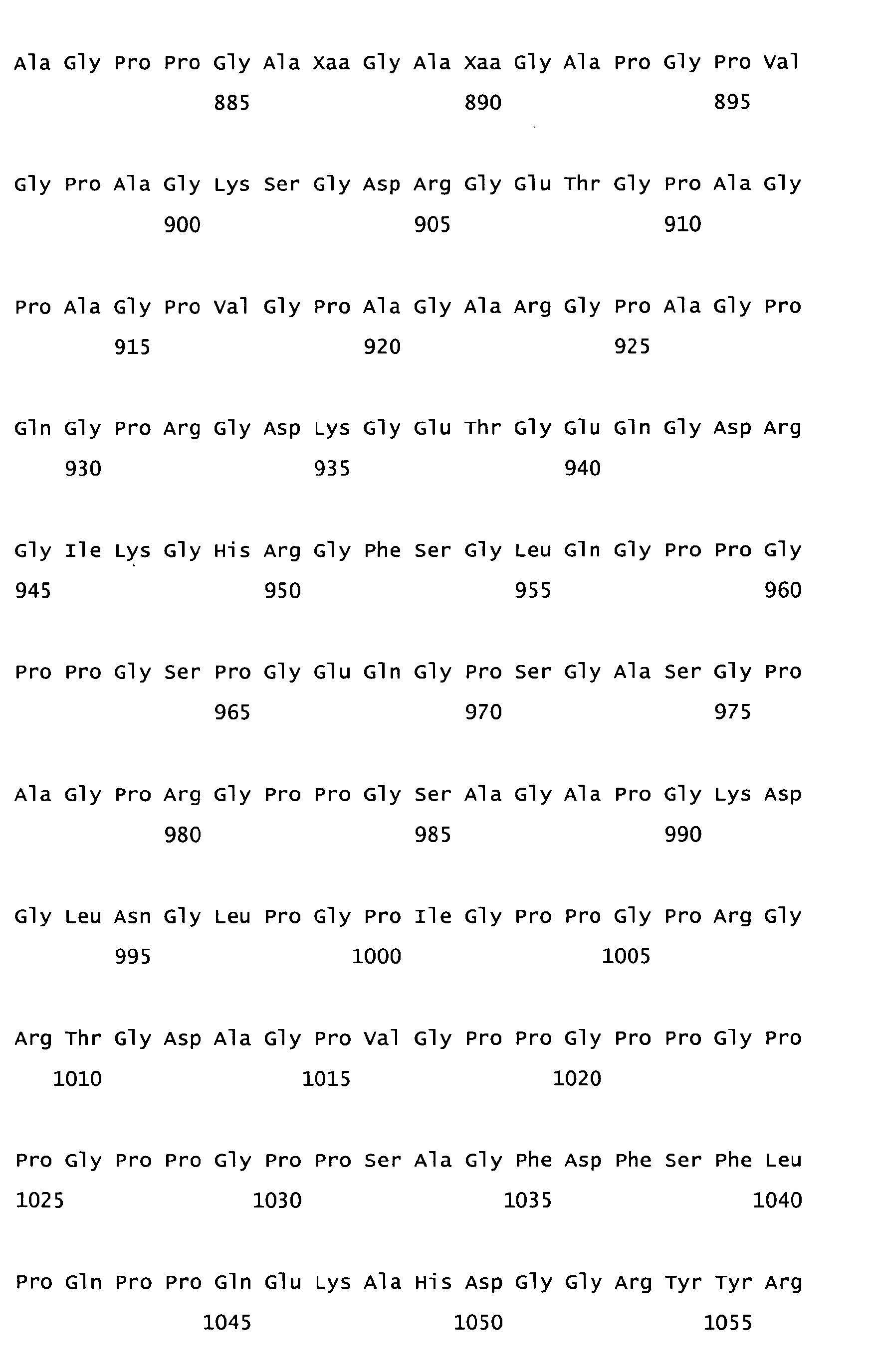
- Figure 13 depicts a collagen I (α1)BMP-2B chimeric amino acid sequence (SEQ. ID. NO. 6).
- Figure 14A-14C depicts a collagen I (α1)BMP-2B chimeric nucleotide sequence (SEQ. ID. NO. 7).
- Figure 15 depicts a collagen I (α1)/TGF-β1amino acid sequence (SEQ. ID. NO. 8).
- Figure 16A-16C depict a collagen I (α1)/TGF-β1 nucleotide sequence (SEQ. ID. NO. 9). Lower case lettering indicates non-coding sequence.
- Figures 17A-17B depict a collagen I (α1)/decorin amino acid sequence (SEQ. ID. NO. 10).
- Figure 18 depicts a collagen I (α1)/decorin peptide amino acid sequence (SEQ. ID. NO. 11).
- Figures 19A-19D depict a collagen I (α1)/decorin nucleotide sequence (SEQ. ID. NO. 12).
- Figures 20A-20C depict a collagen/decorin peptide nucleotide sequence (SEQ. ID. NO. 13). Lower case lettering indicates non-coding sequence.
- Figure 21 depicts a pMal cloning vector and polylinker cloning site.
- Figure 22 depicts a polylinker cloning site contained in the pMal cloning vector of Fig. 21 (SEQ. ID. NO. 14).
- Figure 23 depicts a pMal cloning vector containing a BMP/collagen nucleotide chimeric construct.
- Figure 24 depicts a pMal cloning vector containing a TGF-β1/collagen nucleotide chimeric construct.
- Figure 25 depicts a pMal cloning vector containing a decorin/collagen nucleotide chimeric construct.
- Figure 26 depicts a pMal cloning vector containing a decorin peptide/collagen nucleotide chimeric construct.
- Figure 27A-27E depicts a human collagen Type I (α1) nucleotide sequence (SEQ. ID. NO. 15) and corresponding amino acid sequence (SEQ. ID. NO. 16).
- Figure 28 is a schematic diagram of the construction of the human collagen gene from synthetic oligonucleotides.
- Figure 29 is a schematic depiction of the amino acid sequence of chimeric proteins GST-ColECol (SEQ. ID. NO. 17) and GST-D4 (SEQ. ID. NO. 18).
- Figure 30 is a Table depicting occurrence of four proline and four glycine codons in the human Collagen Type I (α1) gene with optimized codon usage (ColECol).
- Figure 31 depicts a gel reflecting expression and dependence of expression of GST-D4 on hydroxyproline.
- Figure 32 depicts a gel showing expression of GST-D4 in hypertonic media.
- Figure 33 is a graph showing circular dichroism spectra of native and denatured D4 in neutral phosphate buffer.
- Figure 34 depicts a gel representing digestion of D4 with bovine pepsin.
- Figure 35 depicts a gel representing expression of GST-H Col and GST-ColECol under specified conditions.
- Figure 36 depicts a gel representing expression of GST-CM4 in media with or without NaCl and either proline or hydroxyproline.
- Figure 37 depicts a gel of six hour post induction samples of GST-CM4 expressed in E. coli with varying concentrations of NaCl.
- Figure 38 depicts a gel of 4 hour post induction samples of GST-CM4 expressed in E. coli with constant amounts of hydroxyproline and varying amounts of proline.
- Figures 39A-39E depict the nucleotide (SEQ. ID. NO. 19) and amino acid (SEQ. ID. NO. 20) sequence of HuCol Ec , the helical region of human Type I (α1) collagen plus 17 amino terminal extra-helical amino acids and 26 carboxy terminal extra-helical amino acids with codon usage optimized for E. coli.
- Figure 40 depicts sequence and restriction maps of synthetic oligos used to reconstruct the first 243 base pairs of the human Type I (α1) collagen gene with optimized E. coli codon usage. The synthetic oligos are labelled N1-1 (SEQ. ID. NO. 21), N1-2 (SEQ. ID. NO. 22), N1-3 (SEQ. ID. NO. 23) and N1-4 (SEQ. ID. NO. 24).
- Figure 41 depicts a plasmid map of pBSNI-1 containing a 114 base pair fragment of human collagen Type I (α1) with optimized E. coli codon usage.
- Figure 42 depicts the nucleotide (SEQ. ID. NO. 25) and amino acid (SEQ. ID. NO. 26) sequence of a fragment of human collagen Type I (α1) gene with optimized E. coli codon usage encoded by plasmid pBSN1-1.
- Figure 43 depicts a plasmid map of pBSN1-2 containing a 243 base pair fragment of human collagen Type I (α1) with optimized E. coli codon usage.
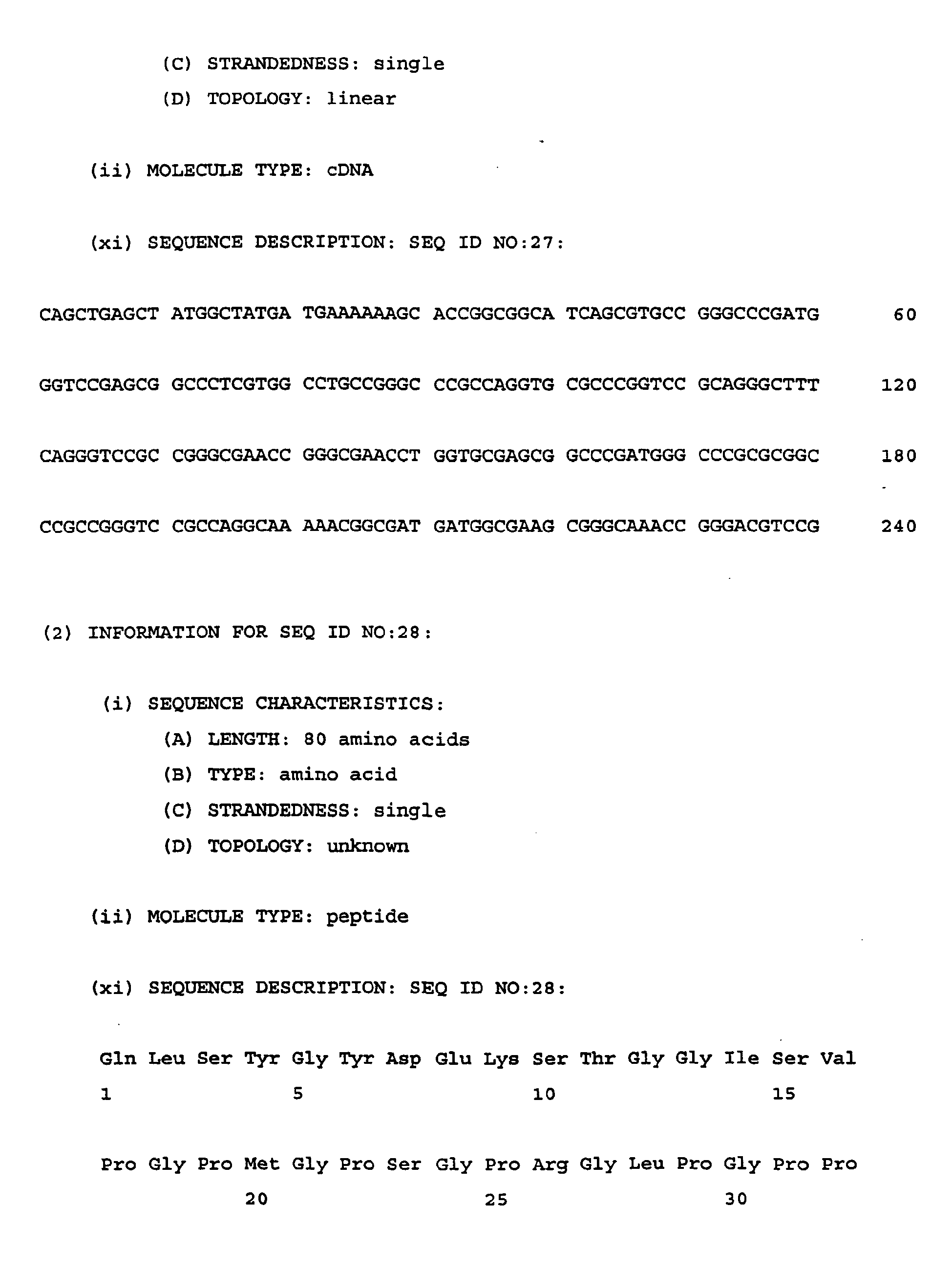
- Figure 44 depicts the nucleotide (SEQ. ID. NO. 27) and amino acid (SEQ. ID. NO. 28) sequence of a fragment of human collagen Type I (α1) gene with optimized E. coli codon usage encoded by plasmid pBSN1-2.
- Figure 45 depicts a plasmid map of pHuCol Ec containing human collagen Type I (α1) with optimized E. coli codon usage.
- Figure 46 depicts a plasmid map of pTrc N1-2 containing a 234 nucleotide human collagen Type I (α1) fragment with optimized E. coli codon usage.
- Figure 47 depicts a plasmid map of pN1-3 containing a 360 nucleotide human collagen Type I (α1) fragment with optimized E. coli codon usage.
- Figure 48 depicts a plasmid map of pD4 containing a 657 nucleotide human collagen Type I (α1) 3' fragment with optimized E. coli codon usage.
- Figures 49A-49E depict the nucleotide (SEQ. ID. NO. 29) and amino acid (SEQ. ID. NO. 30) sequence of a helical region of human Type I (α2) collagen plus 11 amino terminal extra-helical amino acids and 12 carboxy terminal extrahelical amino acids.
- Figures 50A-50E depict the nucleotide (SEQ. ID. NO. 31) and amino acid (SEQ. ID. NO. 32) sequence of HuCol(α2) Ec , the helical region of human Type I (α2) collagen plus 11 amino terminal extra-helical amino acids and 12 carboxy terminal extra-helical amino acids with codon usage optimized for E. coli.
- Figure 51 depicts sequence and restriction maps of synthetic oligos used to reconstruct the first 240 base pairs of human Type I (α2) collagen gene with optimized E. coli codon usage. The synthetic oligos are labelled N1-1 (α2) (SEQ. ID. NO. 33), N1-2 (α2) (SEQ. ID. NO. 34), N1-3 (α2) (SEQ. ID. NO. 35) and N1-4 (α2) (SEQ. ID. NO. 36).
- Figure 52 depicts a plasmid map of pBSN1-1 (α2) containing a 117 base pair fragment of human collagen Type I (α2) with optimized E. coli codon usage.
- Figure 53 depicts a plasmid map of pBSN1-2 (α2) containing a 240 base pair fragment of human collagen Type I (α2) with optimized E. coli codon usage.
- Figure 54 depicts the nucleotide (SEQ. ID. NO. 37) and amino acid (SEQ. ID. NO. 38) sequence of a fragment of human collagen Type I (α2) gene with optimized E. coli usage encoded by plasmid pBSN1-2(α2).
- Figure 55 depicts a plasmid map of pHuCol(α2) Ec containing the entire human collagen Type I (α2) gene with optimized E. coli codon usage.
- Figure 56 depicts a plasmid map of pN1-2 (α2) containing a 240 base pair fragment of human collagen Type I (α2) with optimized E. coli codon usage.
- Figure 57 depicts a gel reflecting expression of GST and TGF-β1 under specified conditions.
- Figure 58 depicts a gel reflecting expression of MBP, FN-BMP-2A, FN-TGF-β1 and FN under specified conditions.
- Figure 59 depicts a gel showing expression of GST-Coll under specified conditions.
- Figure 60 depicts a plasmid map of pGST-CM4 containing the gene for glutathione S- transferase fused to the gene for collagen mimetic 4.
- Figure 61 depicts the nucleotide (SEQ. ID. NO. 39) and amino acid (SEQ. ID. NO. 40) sequence of collagen mimetic 4.
- Figure 62A depicts a chromatogram of the elution of hydroxyproline containing collagen mimetic 4 from a Poros RP2 column. The arrow indicates the peak containing hydroxyproline containing collagen mimetic 4.
- Figure 62B depicts a chromatogram of the elution of proline-containing collagen mimetic 4 from a Poros RP2 column. The arrow indicates the peak containing proline containing collagen mimetic 4.
- Figure 63A depicts a chromatogram of a proline amino acid standard (250 pmol).
- Figure 63B depicts a chromatogram of a hydroxyproline amino acid standard (250 pmol).
- Figure 63C depicts an amino acid analysis chromatogram of the hydrolysis of proline containing collagen mimetic 4.
- Figure 63D depicts an amino acid analysis chromatogram of the hydrolysis of hydroxyproline containing collagen mimetic 4.
- Figure 64 is a graph of OD600 versus time for cultures of E. coli JM109 (F-) grown to plateau and then supplemented with various amino acids.
- Figure 65 depicts a plasmid map of pcEc-α1 containing the gene for HuCol(α1) Ec .
- Figure 66 depicts a plasmid map of pcEc-α2 containing the gene for HuCol(α2) Ec
- Figure 67 depicts a plasmid map of pD4-α1 containing the gene for a 219 amino acid C-terminal fragment of Type I (α1) human collagen with optimized E. coli codon usage fused to the gene for glutathione S-transferase.
- Figure 68 depicts a plasmid map of pD4-α2 containing the gene for a 207 amino acid C-terminal fragment of Type I (α2) human collagen with optimized E. coli codon usage fused to the gene for glutathione S-transferase.
- Figure 69 depicts the predicted amino acid sequence from the DNA sequence of the first 13 amino acid acids of protein D4-α1 (SEQ. ID. NO. 41) and the amino acid sequence as experimentally determined (SEQ. ID NO. 42).
- Figure 70 depicts the mass spectrum of hydroxyproline containing D4-α1.
- Figure 71 depicts the nucleotide sequence of a 657 nucleotide human collagen Type I (α1)3' fragment with optimized E. coli codon usage designated D4 (SEQ. ID. NO. 43).
- Figure 72 depicts the amino acid sequence of a 219 amino acid C-terminal fragment of human collagen Type I (α1) designed D4 (SEQ. ID. NO. 44).
- Figure 73 is a plasmid map illustrating pGEX-4T.1 containing the gene for glutatione S-transferase.
- Figure 74 is a plasmid map illustrating pTrc-TGF containing the gene for the mature human TGF-β1 polypeptide.
- Figure 75 is a plasmid map illustrating pTrc-Fn containing the gene for a 70 kDa fragment of human fibronectin.
- Figure 76 is a plasmid map illustrating pTrc-Fn-TGF containing the gene for a fusion protein of a 70 kDA fragment of human fibronectin and the mature human TGF-β1 polypeptide.
- Figure 77 is a plasmid map illustrating pTrc-Fn-BMP containing the gene for a fusion protein of a 70 kDa fragment of human fibronectin and human bone morphogenic protein 2A.
- Figure 78 is a plasmid map illustrating pGEX-HuCollEc containing the gene for a fusion between glutathione S-transferase and Type I (α1) human collagen with optimized E. coli codon usage.
- Figure 79 depicts the nucleotide sequence of a 627 nucleotide human collagen Type I (α2) 3' fragment with optimized E. coli codon usage (SEQ. ID. NO.45).
- Figure 80 depicts the amino acid sequence of a 209 amino acid C-terminal fragment of human collagen Type I (α2) (SEQ. ID. NO. 46).
- Figure 81 depicts the sequence of synthetic oligos used to reconstruct the first 282 base pairs of the gene for the carboxy terminal 219 amino acids of human Type I (α1) collagen with optimized E. coli codon usage designated N4-1 (SEQ. ID. NO. 47), N4-2 (SEQ. ID. NO. 48), N4-3 (SEQ. ID. NO. 49) and N4-4 (SEQ. ID. NO. 50).
DETAILED DESCRIPTION OF PREFERRED EMBODIMENTS
-
Prokaryotic cells and eukaryotic cells can unexpectedly be made to assimilate and incorporate trans-4-hydroxyproline into proteins contrary to both Papas et al. and Deming et al., supra. Such assimilation and incorporation is especially useful when the structure and function of a polypeptide depends on post translational hydroxylation of proline not provided by the native protein production system of a recombinant host. Thus, prokaryotic bacteria such as E. coli and eukaryotic cells such as Saccharomyces cerevisiae, Saccharomyces carlsbergensis and Schizosaccharomyces pombe that ordinarily do not hydroxylate proline and additional eukaryotes such as insect cells including lepidopteran cell lines including Spodoptera frugiperda, Trichoplasia ni, Heliothis virescens, Bombyx mori infected with a baculovirus; CHO cells, COS cells and NIH 3T3 cells which fail to adequately produce certain polypeptides whose structure and function depend on such hydroxylation can be made to produce polypeptides having hydroxylated prolines. Incorporation includes adding trans-4-hydroxyproline to a polypeptide, for example, by first changing an amino acid to proline, creating a new proline position that can in turn be substituted with trans-4-hydroxyproline or substituting a naturally occurring proline in a polypeptide with trans-4-hydroxyproline as well.
-
The process of producing recombinant polypeptides in mass producing organisms is well known. Replicable expression vectors such as plasmids, viruses, cosmids and artificial chromosomes are commonly used to transport genes encoding desired proteins from one host to another. It is contemplated that any known method of cloning a gene, ligating the gene into an expression vector and transforming a host cell with such expression vector can be used in furtherance of the present disclosure.
-
Not only is incorporation of trans-4-hydroxyproline into polypeptides which depend upon trans-4-hydroxyproline for chemical and physical properties useful in production systems which do not have the appropriate systems for converting proline to trans-4-hydroxyproline, but useful as well in studying the structure and function of polypeptides which do not normally contain trans-4-hydroxyproline. It is contemplated that the following amino acid analogs may also be incorporated in accordance with the present disclosure: trans-4 hydroxyproline, 3-hydroxyproline, cis-4-fluoro-L-proline and combinations thereof (hereinafter referred to as the "amino acid analogs"). Use of prokaryotes and eukaryotes is desirable since they allow relatively inexpensive mass production of such polypeptides. It is contemplated that the amino acid analogs can be incorporated into any desired polypeptide. In a preferred embodiment the prokaryotic cells and eukaryotic cells are starved for proline by decreasing or eliminating the amount of proline in growth media prior to addition of an amino acid analog herein.
-
Expression vectors containing the gene for maltose binding protein (MBP), e.g., see Figure 1 illustrating plasmid pMAL-c2, commercially available from New England Bio-Labs, are transformed into prokaryotes such as E. coli proline auxotrophs or eukaryotes such as S. cerevisiae auxotrophs which depend upon externally supplied proline for protein synthesis and anabolism. Other preferred expression vectors for use in prokaryotes are commercially available plasmids which include pKK-223 (Pharmacia), pTRC (Invitrogen), pGEX (Pharmacia), pET (Novagen) and pQE (Quiagen). It should be understood that any suitable expression vector may be utilized by those with skill in the art.
-
Substitution of the amino acid analogs for proline in protein synthesis occurs since prolyl tRNA synthetase is sufficiently promiscuous to allow misacylation of proline tRNA with any one of the amino acid analogs. A sufficient quantity, i.e., typically ranging from about .001M to about 1.0 M, but more preferably from about .005M to about 0.5M of the amino acid analog(s) is added to the growth medium for the transformed cells to compete with proline in cellular uptake. After sufficient time, generally from about 30 minutes to about 24 hours or more, the amino acid analog(s) is assimilated by the cell and incorporated into protein synthetic pathways. As can be seen from Figures 2 and 2A, intracellular concentration of trans-4-hydroxyproline increases by increasing the concentration of sodium chloride in the growth media. In a preferred embodiment the prokaryotic cells and/or eukaryotic cells are starved for proline by decreasing or eliminating the amount of proline in growth media prior to addition of an amino acid analog herein.
-
Expression vectors containing the gene for human Type I (α1) collagen (DNA sequence illustrated in Figures 3 and 3A; plasmid map illustrated in Figure 4) are transformed into prokaryotic or eukaryotic proline auxotrophs which depend upon externally supplied proline for protein synthesis and anabolism. As above, substitution of the amino acid analog(s) occurs since prolyl tRNA synthetase is sufficiently promiscuous to allow misacylation of proline tRNA with the amino acid analog(s). The quantity of amino acid analog(s) in media given above is again applicable.
-
Expression vectors containing DNA encoding fragments of human Type 1 (α1) collagen (e.g., DNA sequence illustrated in Figure 5 and plasmid map illustrated in Figure 6) are transformed into prokaryotic or eukaryotic auxotrophs as above. Likewise, expression vectors containing DNA encoding collagen-like polypeptide (e.g., DNA sequence illustrated in Figure 7, amino acid sequence illustration in Figure 8 and plasmid map illustrated in Figure 9) can be used to transform prokaryotic or eukaryotic auxotrophs as above. Collagen-like peptides are those which contain at least partial homology with collagen and exhibit similar chemical and physical characteristics to collagen. Thus, collagen-like peptides consist, e.g., of repeating arrays of Gly-X-Y triplets in which about 35% of the X and Y positions are occupied by proline and 4-hydroxyproline. Collagen-like peptides are interchangeably referred to herein as collagen-like proteins, collagen-like polypeptides, collagen mimetic polypeptides and collagen mimetic. Certain preferred collagen fragments and collagen-like peptides in accordance herewith are capable of assembling into an extracellular matrix. In both collagen fragments and collagen-like peptides as described above, substitution with amino acid analog(s) occurs since prolyl tRNA synthetase is sufficiently promiscuous to allow misacylation of proline tRNA with one or more of the amino acid analog(s). The quantity of amino acid analog(s) given above is again applicable.
-
It is contemplated that any polypeptide having an extracellular matrix protein domain such as a collagen, collagen fragment or collagen-like peptide domain can be made to incorporate amino acid analog(s) in accordance with the disclosure herein. Such polypeptides include collagen, a collagen fragment or collagen-like peptide domain and a domain having a region incorporating one or more physiologically active agents such as glycoproteins, proteins, peptides and proteoglycans. As used herein, physiologically active agents exert control over or modify existing physiologic functions in living things. Physiologically active agents include hormones, growth factors, enzymes, ligands and receptors. Many active domains of physiologically active agents have been defined and isolated. It is contemplated that polypeptides having a collagen, collagen fragment or collagen-like peptide domain can also have a domain incorporating one or more physiologically active domains which are active fragments of such physiologically active agents. As used herein, physiologically active agent is meant to include entire peptides, polypeptides, proteins, glycoproteins, proteoglycans and active fragments of any of them. Thus, chimeric proteins are made to incorporate amino acid analog(s) by transforming a prokaryotic proline auxotroph or a eukaryotic proline auxotroph with an appropriate expression vector and contacting the transformed auxotroph with growth media containing at least one of the amino acid analogs. For example, a chimeric collagen/bone morphogenic protein (BMP) construct or various chimeric collagen/growth factor constructs are useful in accordance herein. Such growth factors are well-known and include insulin-like growth factor, transforming growth factor, platelet derived growth factor and the like. Figure 10 illustrates DNA of BMP which can be fused to the 3' terminus of DNA encoding collagen, DNA encoding a collagen fragment or DNA encoding a collagen-like peptide. Figure 11 illustrates a map of plasmid pCBC containing a collagen/BMP construct. In a preferred embodiment, proteins having a collagen, collagen fragment or collagen-like peptide domain assemble or aggregate to form an extracellular matrix which can be used as a surgical implant. The property of self-aggregation as used herein includes the ability to form an aggregate with the same or similar molecules or to form an aggregate with different molecules that share the property of aggregation to form, e.g., a double or triple helix. An example of such aggregation is the structure of assembled collagen matrices.
-
Indeed, chimeric polypeptides which may also be referred to herein as chimeric proteins provide an integrated combination of a therapeutically active domain from a physiologically active agent and one or more EMP moieties. The EMP domain provides an integral vehicle for delivery of the therapeutically active moiety to a target site. The two domains are linked covalently by one or more peptide bonds contained in a linker region. As used herein, integrated or integral means characteristics which result from the covalent association of one or more domains of the chimeric proteins. The therapeutically active moieties disclosed herein are typically made of amino acids linked to form peptides, polypeptides, proteins, glycoproteins or proteoglycans. As used herein, peptide encompasses polypeptides and proteins.
-
The inherent characteristics of EMPs are ideal for use as a vehicle for the therapeutic moiety. One such characteristic is the ability of the EMPs to form the self-aggregate. Examples of suitable EMPs are collagen, elastin, fibronectin, fibrinogen and fibrin. Fibrillar collagens (Type I, II and III) assemble into ordered polymers and often aggregate into larger bundles. Type IV collagen assembles into sheetlike meshworks. Elastin molecules form filaments and sheets in which the elastin molecules are highly cross-linked to one another to provide good elasticity and high tensile strength. The cross-linked, random-coiled structure of the fiber network allows it to stretch and recoil like a rubber band. Fibronectin is a large fibril forming glycoprotein, which, in one of its forms, consists of highly insoluble fibrils cross-linked to each other by disulfide bonds. Fibrin is an insoluble protein formed from fibrinogen by the proteolytic activity of thrombin during the normal clotting of blood.
-
The molecular and macromolecular morphology of the above EMPs defines networks or matrices to provide substratum or scaffolding in integral covalent association with the therapeutically active moiety. The networks or matrices formed by the EMP domain provide an environment particularly well suited for ingrowth of autologous cells involved in growth, repair and replacement of existing tissue. The integral therapeutically active moieties covalently bound within the networks or matrices provide maximum exposure of the active agents to their targets to elicit a desired response.
-
Implants formed of or from the present chimeric proteins provide sustained release activity in or at a desired locus or target site. Since it is linked to an EMP domain, the therapeutically active domain of the present chimeric protein is not free to separately diffuse or otherwise be transported away from the vehicle which carries it, absent cleavage of peptide bonds. Consequently, chimeric proteins herein provide an effective anchor for therapeutic activity which allows the activity to be confined to a target location for a prolonged duration. Because the supply of therapeutically active agent does not have to be replenished as often when compared to non-sustained release dosage forms, smaller amounts of therapeutically active agent may be used over the course of therapy. Consequently, certain advantages provided by the present chimeric proteins are a decrease or elimination of local and systemic side effects, less potentiation or reduction in therapeutic activity with chronic use, and minimization of drug accumulation in body tissue with chronic dosing.
-
Use of recombinant technology allows manufacturing of non-immunogenic chimeric proteins. The DNA encoding both the therapeutically active moiety and the EMP moiety should preferably be derived from the same species as the patient being treated to avoid an immunogenic reaction. For example, if the patient is human, the therapeutically active moiety as well as the EMP moiety is preferably derived from human DNA.
-
Osteogenic/EMP chimeric proteins provide biodegradable and biocompatible agents for inducing bone formation at a desired site. As stated above, in one embodiment, a BMP moiety is covalently linked with an EMP to form chimeric protein. The BMP moiety induces osteogenesis and the extracellular matrix protein moiety provides an integral substratum or scaffolding for the BMP moiety and cells which are involved in reconstruction and growth. Compositions containing the BMP/EMP chimeric protein provide effective sustained release delivery of the BMP moiety to desired target sites. The method of manufacturing such an osteogenic agent is efficient because the need for extra time consuming steps as purifying EMP and then admixing it with the purified BMP are eliminated. An added advantage of the BMP/EMP chimeric protein results from the stability created by the covalent bond between BMP and the EMP, i.e., the BMP portion is not free to separately diffuse away from the EMP, thus providing a more stable therapeutic agent.
-
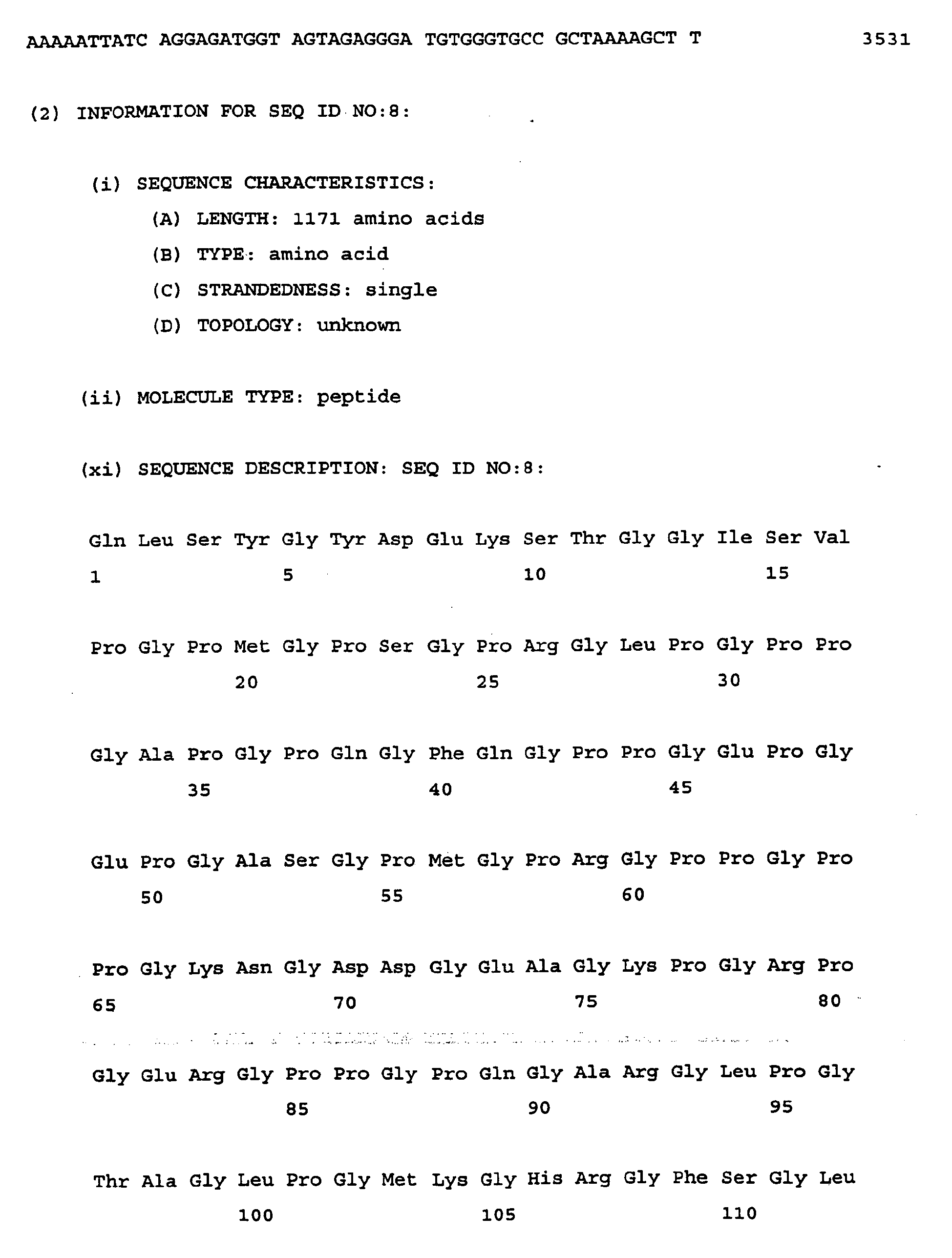
Bone morphogenic proteins are class identified as BMP-1 through BMP-9. A preferred osteogenic protein for use in human patients is human BMP-2B. A BMP-2B/collagen IA chimeric protein is illustrated in Fig. 13 (SEQ. ID. NO. 6). The protein sequence illustrated in Fig. 15 (SEQ. ID. NO. 8) includes a collagen helical domain depicted at amino acids 1-1057 and a mature form of BMP-2B at amino acids 1060-1169. The physical properties of the chimeric protein are dominated in part by the EMP component. In the case of a collagen moiety, a concentrated solution of chimeric protein will have a gelatinous consistency that allows easy handling by the medical practitioner. The EMP moiety acts as a sequestering agent to prevent rapid desorption of the BMP moiety from the desired site and to provide sustained release of BMP activity. As a result, the BMP moiety remains at the desired site and provides sustained release of BMP activity at the desired site for a period of time necessary to effectively induce bone formation. The EMP moiety also provides a matrix which allows a patient's autologous cells, e.g., chondrocytes and the like, which are normally involved in osteogenesis to collect therein and form an autologous network for new tissue growth. The gelatinous consistency of the chimeric protein also provides a useful and convenient therapeutic manner for immobilizing active BMP on a suitable vehicle or implant for delivering the BMP moiety to a site where bone growth is desired.
-
The BMP moiety and the EMP moiety are optionally linked together by linker sequences of amino acids. Examples of linker sequences used are illustrated within the sequence depicted in Figs. 14A-14C (SEQ. ID. NO. 7), 16A-16C (SEQ. ID. NO. 9), 19A-19C (SEQ. ID. NO. 12) and 20A-20C (SEQ. ID. NO. 13), and are described in more detail below. Linker sequences may be chosen based on particular properties which they impart to the chimeric protein. For example, amino acid sequences such as Ile-Glu-Gly-Arg and Leu-Val-Pro-Arg are cleaved by factor XA and thrombin enzymes, respectively. Incorporating sequences which are cleaved by proteolytic enzymes into chimeric proteins herein provides cleavage at the linker site upon exposure to the appropriate enzyme and separation of the two domains into separate entities. It is contemplated that numerous linker sequences can be incorporated into any of the chimeric proteins.
-
In another embodiment, a chimeric DNA construct includes a gene encoding an osteogenic protein or a fragment thereof linked to gene encoding an EMP or a fragment thereof. The gene sequence for various BMPs are known, see, e.g.,
U.S. Patent Nos. 4,294,753 ,
4,761,471 ,
5,106,748 ,
5,187,076 ,
5,141,905 ,
5,108,922 ,
5,116,738 and
5,168,050 , each incorporated herein by reference. A BMP-2B gene for use herein is synthesized by ligating oligonucleotides encoding a BMP protein. The oligonucleotides encoding BMP-2B are synthesized using an automated DNA synthesizer (Beckmen Oligo-1000). In preferred embodiment, the nucleotide sequence encoding the BMP is maximized for expression in
E.coli. This is accomplished by using
E.coli utilization tables to translate the sequence of amino acids of the BMP into codons that are utilized most often by
E.coli. Alternatively, native DNA encoding BMP isolated from mammals including humans may be purified and used.
-
The BMP gene and the DNA sequence encoding an extracellular matrix protein are cloned by standard genetic engineering methods as described in Sambrook et al., Molecular Cloning: A Laboratory Manual, , hereby incorporated by reference.
-
The DNA sequence corresponding to the helical and telepeptide region of collagen I(α1) is cloned from a human fibroblast cell line. Two sets of polymerase chain reactions are carried out using cDNA prepared by standard methods from AG02261 A cells. The first pair of PCR primers include a 5' primer bearing an XmnI linker sequence and a 3' primer bearing the BsmI site at nucleotide number 1722. The resulting PCR product consists of sequence from position 1 to 1722. The second pair of primers includes the BsmI site at 1722 and a linker sequence at the 3' end bearing a BgIII site. The resulting PCR product consists of sequence from position 1722 to 3196. The complete sequence is assembled by standard cloning techniques. The two PCR products are ligated together at the BsmI site, and the combined clone is inserted into any vector with XmnI-BglII sites such as pMAL-c2 vector.
-
To clone the BMP-2B gene, total cellular RNA is isolated from human osteosarcoma cells (U-20S) by the method described by Robert E. Farrel Jr. (Academic Press, CA, 1993 pp. 68-69) (herein incorporated by reference). The integrity of the RNA is verified by spectrophotometric analysis and electrophoresis through agarose gels. Typical yields of total RNA are 50 µg from a 100mm confluent tissue culture dish. The RNA is used to generate cDNA by reverse transcription using the Superscript pre-amplification system by Gibco BRL. The cDNA is used as template for PCR amplification using upstream and downstream primers specific for BMP-2B (GenBank HUMBMP2B accession #M22490). The resulting PCR product consists of BMP-2B sequence from position 1289-1619. The PCR product is resolved by electrophoresis through agarose gels, purified with gene clean (BIO 101) and ligated into pMal-c2 vector (New England Biolabs). The domain of human collagen I(α1) chain is cloned in a similar manner. However, the total cellular RNA is isolated from a human fibroblast cell line (AG02261A human skin fibroblasts).
-
A chimeric BMP/EMP DNA construct is obtained by ligating a synthetic BMP gene to a DNA sequence encoding an EMP such as collagen, fibrinogen, fibrin, fibronectin, elastin or laminin. However, chimeric polypeptides herein are not limited to these particular proteins. Figs. 14A-14C (SEQ. ID. NO. 7) illustrate a DNA construct which encodes a BMP-2B/collagen I(α1) chimeric protein. The coding sequence for an EMP may be ligated upstream and/or downstream and in-frame with a coding sequence for the BMP. The DNA encoding an EMP may be a portion of the gene or an entire EMP gene. Furthermore, two different EMPs may be ligated upstream and downstream from the BMP.
-
The BMP-2B/collagen I(α1) chimeric protein illustrated in Figs. 14A-14C includes an XmnI linker sequence at base pairs (bp) 1-19, a collagen domain (bp 20-3190), a BglII/BamHI linker sequence (bp 3191-3196), a mature form of BMP2b (bp 3197-3529) and a HindIII linker sequence (bp 3530-3535).
-
Any combination of growth factor and matrix protein sequences are contemplated including repeating units, or multiple arrays of each segment in any order.
-
Incorporation of fragments of both matrix and growth factor proteins is also contemplated. For example, in the case of collagen, only the helical domain may be included. Other matrix proteins have defined domains, such as laminin, which has EGF-like domains. In these cases, specific functionalities can be chosen to achieve desired effects. Moreover, it may be useful to combine domains from disparate matrix proteins, such as the helical region of collagen and the cell attachment regions of fibronectin. In the case of growth factors, specific segments have been shown to be removed from the mature protein by post translational processing. Chimeric proteins can be designed to include only the mature biologically active region. For example, in the case of BMP-2B only the final 110 amino acids are found in the active protein.
-
In another embodiment, a transforming growth factor (TGF) moiety is covalently linked with an EMP to form a chimeric protein. The TGF moiety increases efficacy of the body's normal soft tissue repair response and also induces osteogenesis. Consequently; TGF/EMP-chimeric-proteins may be used for either or both functions. One of the fundamental properties of the TGF-βs is their ability to turn on various activities that result in the synthesis of new connective tissue. See, Piez and Sporn eds., Transforming Growth Factor-βs Chemistry, Biology and Therapeutics, Annals of the New York Academy of Sciences, Vol. 593, (1990). TGF-β is known to exist in at least five different isoforms. The DNA,sequence for Human TGF-β1 is known and has been cloned. See Derynck et al., Human Transforming Growth Factor-Beta cDNA Sequence and Expression in Tumour Cell Lines, Nature, Vol. 316, pp. 701-705 (1985), herein incorporated by reference. TGF-β2 has been isolated from bovine bone, human glioblastoma cells and porcine platelets. TGF-B3 has also been cloned. See ten Dijke, et al., Identification of a New Member of the Transforming Growth Factor-β Gene Family, Proc. Natl. Acad. Sci. (USA), Vol. 85, pp. 4715-4719 (1988) herein incorporated by reference.
-
A TGF-β/EMP chimeric protein incorporates the known activities of TGF-βs and provides integral scaffolding or substratum of the EMP as described above to yield a composition which further provides sustained release focal delivery at target sites.
-
The TGF-β moiety and the EMP moiety are optionally linked together by linker sequences of amino acids. Linker sequences may be chosen based upon particular properties which they impart to the chimeric protein. For example, amino acid sequences such as Ile-Glu-Glyn-Arg and Leu-Val-Pro-Arg are cleaved by Factor XA and Thrombin enzymes, respectively. Incorporating sequences which are cleaved by proteolytic enzymes into the chimeric protein provides cleavage at the linker site upon exposure to the appropriate enzyme and separation of the domains into separate entities. Fig. 15 depicts an amino acid sequence for a TGF-β1/collagen IA chimeric protein (SEQ. ID. NO. 8). The illustrated amino acid sequence includes the collagen domain (1-1057) and a mature form of TGF-β1 (1060-1171).
-
A chimeric DNA construct includes a gene encoding TGF-β1 or a fragment thereof, or a gene encoding TGF-β2 or a fragment thereof, or a gene encoding TGF-β3 or a fragment thereof, ligated to a DNA sequence encoding an EMP protein such as collagen (I-IV), fibrin, fibrinogen, fibronectin, elastin or laminin. A preferred chimeric DNA construct combines DNA encoding TGF-β1, a DNA linker sequence, and DNA encoding collagen IA. A chimeric DNA construct containing TGF-β1 gene and a collagen I(α1) gene is shown in Figs. 16A-16C (SEQ. ID: NO. 9). The illustrated construct includes an XmnI linker sequence (bp 1-19), DNA encoding a collagen domain (bp 20-3190), a BglII linker sequence (bp 3191-3196), DNA encoding a mature form of TGF-β1 (3197-3535), and an XbaI linker sequence (bp 3536-3541).
-
The coding sequence for EMP may be ligated upstream and/or downstream and in-frame with a coding sequence for the TGFβ. The DNA encoding the extracellular matrix protein may encode a portion of a fragment of the EMP or may encode the entire EMP. Likewise, the DNA encoding the TGF-β may be one or more fragments thereof or the entire gene. Furthermore, two or more different TGF-βs or two or more different EMPs may be ligated upstream or downstream of alternate moieties.
-
In yet another embodiment, a dermatan sulfate proteoglycan moiety, also known as decorin or proteoglycan II, is covalently linked with an EMP to form a chimeric protein. Decorin is known to bind to type I collagen and thus affect fibril formation, and to inhibit the cell attachment-promoting activity of collagen and fibrinogen by binding to such molecules near their cell binding sites. Chimeric proteins which contain a decorin moiety act to reduce scarring of healing tissue. The primary structure of the core protein of decorin has been deduced from cloned cDNA. See Krusius et al., Primary Structure of an Extracellular Matrix Proteoglycan Core Protein-Deduced from Cloned cDNA, Proc. Natl. Acad. Sci. (USA), Vol. 83, pp. 7683-7687 (1986) incorporated herein by reference.
-
A decorin/EMP chimeric protein incorporates the known activities of decorin and provides integral scaffolding or substratum of the EMP as described above to yield a composition which allows sustained release focal delivery to target sites. Figs. 17A-17B illustrate a decorin/collagen IA chimeric protein (SEQ. ID. NO. 10) in which the collagen domain includes amino acids 1-1057 and the decorin mature protein incudes amino acids 1060-1388. Fig. 18 illustrates a decorin peptide/collagen IA chimeric protein (SEQ. ID. NO. 11) in which the collagen helical domain includes amino acids 1-1057 and the decorin peptide fragment includes amino acids 1060-1107. The decorin peptide fragment is composed of P46 to G93 of the mature form of decorin.
-
Further provided is a chimeric DNA construct which includes a gene encoding decorin or one or more fragments thereof, optionally ligated via a DNA linker sequence to a DNA sequence encoding an EMP such as collagen (I-IV), fibrin, fibrinogen, fibronectin, elastin or laminin. A preferred chimeric DNA construct combines DNA encoding decorin, a DNA linker sequence, and DNA encoding collagen I(α1). A chimeric DNA construct containing a decorin gene and a collagen I(α1) gene is shown in Figs. 19A-19D (SEQ. ID. NO. 12). The illustrated construct includes an XmnI linker sequence (bp 1-19), DNA encoding a collagen domain (bp 20-3190), a BglII linker sequence (bp 3191-3196), DNA encoding a mature form of decorin (bp 3197-4186) and a PstI linker sequence. A chimeric DNA construct containing a decorin peptide gene and a collagen I(α1) gene is shown in Figs. 20A-20C (SEQ. ID. NO. 13). The illustrated construct includes an XmnI linker sequence (bp 1-19), DNA encoding a collagen domain (bp 20-3190), a BgIII linker sequence (bp 3191-3196), DNA encoding a peptide fragment of decorin (bp 3197-3343), and a PstI linker sequence (bp 3344-3349).
-
The coding sequence for an EMP may be ligated upstream and/or downstream and in-frame with a coding sequence for decorin. The DNA encoding the EMP may encode a portion or fragment of the EMP or may encode the entire EMP. Likewise, the DNA encoding decorin may be a fragment thereof or the entire gene. Furthermore, two or more different EMPs may be ligated upstream and/or downstream from the DNA encoding decorin moiety.
-
Any of the above described chimeric DNA constructs may be incorporated into a suitable cloning vector. Fig. 21 depicts a pMal cloning vector containing a polylinker cloning site. Examples of cloning vectors are the plasmids pMal-p2 and pMal-c2 (commercially available from New England Biolabs). The desired chimeric DNA construct is incorporated into a polylinker sequence of the plasmid which contains certain useful restriction endonuclease sites which are depicted in Fig. 22 (SEQ. ID. NO. 14). The pMal-p2 polylinker sequence has XmnI, EcoRI, BamHI, HindIII, XbaI, SalI and PstI restriction endonuclease sites which are depicted in Fig 22. The polylinker sequence is digested with an appropriate restriction endonuclease and the chimeric construct is incorporated into the cloning vector by ligating it to the DNA sequences of the plasmid. The chimeric DNA construct may be joined to the plasmid by digesting the ends of the DNA construct and the plasmid with the same restriction endonuclease to generate "sticky ends" having 5' phosphate and 3' hydroxyl groups which allow the DNA construct to anneal to the cloning vector. Gaps between the inserted DNA construct and the plasmid are then sealed with DNA ligase. Other techniques for incorporating the DNA construct into plasmid DNA include blunt end ligation, poly(dA.dT) tailing techniques, and the use of chemically synthesized linkers. An alternative method for introducing the chimeric DNA construct into a cloning vector is to incorporate the DNA encoding the extracellular matrix protein into a cloning vector already containing a gene encoding a therapeutically active moiety.
-
The cloning sites in the above-identified polylinker site allow the cDNA for the collagen I(α1)BMP-2B chimeric protein illustrated in Figs. 14A-14C (SEQ. ID. NO. 7) to be inserted between the XmnI and the HindIII sites. The cDNA encoding the collagen I(α1)/TGF-β1 protein illustrated in Figs. 16A-16C (SEQ. ID. NO. 9) is inserted between the XmnI and the XbaI sites. The cDNA encoding the collagen I(α1)/decorin protein illustrated in Figs. 19A-19D (SEQ. ID. NO. 12) inserted between the XmnI and the PstI sites. The cDNA encoding the collagen I(α1)/decorin peptide illustrated in Figs. 20A-20C (SEQ. ID. NO. 13) is inserted between the XmnI and PstI sites.
-
Plasmids containing the chimeric DNA construct are identified by standard techniques such as gel electrophoresis. Procedures and materials for preparation of recombinant vectors, transformation of host cells with the vectors, and host cell expression of polypeptides are described in Sambrook et al., Molecular Cloning: A Laboratory Manual, supra. Generally, prokaryotic or eukaryotic host cells may be transformed with the recombinant DNA plasmids. Transformed host cells may be located through phenotypic selection genes of the cloning vector which provide resistance to a particular antibiotic when the host cells are grown in a culture medium containing that antibiotic.
-
Transformed host cells are isolated and cultured to promote expression of the chimeric protein. The chimeric protein may then be isolated from the culture medium and purified by various methods such as dialysis, density gradient centrifugation, liquid column chromatography, isoelectric precipitation, solvent fractionation, and electrophoresis. However, purification of the chimeric protein by affinity chromatography is preferred whereby the chimeric protein is purified by ligating it to a binding protein and contacting it with a ligand or substrate to which the binding protein has a specific affinity.
-
In order to obtain more effective expression of mammalian or human eukaryotic genes in bacteria (prokaryotes), the mammalian or human gene may be placed under the control of a bacterial promoter. A protein fusion and purification system is employed to obtain the chimeric protein. Preferably, any of the above-described chimeric DNA constructs is cloned into a pMal vector at a site in the vector's polylinker sequence. As a result, the chimeric DNA construct is operably fused with the malE gene of the pMal vector. The malE gene encodes maltose binding protein (MBP). Fig. 23 depicts a pMal cloning vector containing a BMP/collagen DNA construct. A spacer sequence coding for 10 asparagine residues is located between the maIE sequence and the polylinker sequence. This spacer sequence insulates MBP from the protein of interest. Figs. 24, 25 and 26 depict pMal cloning vectors containing DNA encoding collagen chimeras with TGF-β1, decorin and a decorin peptide, respectively. The pMal vector containing any of the chimeric DNA constructs fused to the malE gene is transformed into E. coli.
-
The E. coli is cultured in a medium which induces the bacteria to produce the maltose-binding protein fused to the chimeric protein. This technique utilizes the Ptac promoter of the pMal vector. The MBP contains a 26 amino acid N-terminal signal sequence which directs the MBP-chimeric protein through the E. coli cytoplasmic membrane. The protein can then be purified from the periplasm. Alternatively, the pMal-c2 cloning vector can be used with this protein fusion and purification system. The pMal-c2 vector contains an exact deletion of the malE signal sequence which results in cytoplasmic expression of the fusion protein. A crude cell extract containing the fusion protein is prepared and poured over a column of amylose resin. Since MBP has an affinity for the amylose it binds to the resin. Alternatively, the column can include any substrate for which MBP has a specific affinity. Unwanted proteins present in the crude extract are washed through the column. The MBP fused to the chimeric protein is eluted from the column with a neutral buffer containing maltose or other dilute solution of a desorbing agent for displacing the hybrid polypeptide. The purified MBP-chimeric protein is cleaved with a protease such as factor Xa protease to cleave the MBP from the chimeric protein. The pMal-p2 plasmid has a sequence encoding the recognition site for protease factor Xa which cleaves after the amino acid sequence Isoleucine-Glutamic acid-Glycine-Arginine of the polylinker sequence.
-
The chimeric protein is then separated from the cleaved MBP by passing the mixture over an amylose column. An alternative method for separating the MBP from the chimeric protein is by ion exchange chromatography. This system yields up to 100mg of MBP-chimeric protein per liter of culture. See
Riggs, P., in Ausebel, F.M., Kingston, R.E., Moore, D.D., Seidman, J.G., Smith, J.A., Struhl, K. (eds.) Current Protocols in Molecular Biology, Supplement 19 (16.6.1-16.6.10) (1990) Green Associates/Wiley Interscience, New York, New England Biolabs (cat # 800-65S 9pMALc2) pMal protein fusion and purification system hereby incorporated herein by reference. (See also
European Patent No. 286 239 herein incorporated by reference which discloses a similar method for production and purification of a protein such as collagen.)
-
Other protein fusion and purification systems may be employed to produce chimeric proteins. Prokaryotes such as E. coli are the preferred host cells for expression of the chimeric protein. However, systems which utilize eukaryote host cell lines are also acceptable such as yeast, human, mouse, rat, hamster, monkey, amphibian, insect, algae, and plant cell lines. For example, HeLa (human epithelial), 3T3 (mouse fibroblast), CHO (Chinese hamster ovary), and SP 2 (mouse plasma cell) are acceptable cell lines. The particular host cells that are chosen should be compatible with the particular cloning vector that is chosen.
-
Another acceptable protein expression system is the Baculovirus Expression System manufactured by Invitrogen of San Diego, California. Baculoviruses form prominent crystal occlusions within the nuclei of cells they infect. Each crystal occlusion consists of numerous virus particles enveloped in a protein called polyhedrin. In the baculovirus expression system, the native gene encoding polyhedrin is substituted with a DNA construct encoding a protein or peptide having a desired activity. The virus then produces large amounts of protein encoded by the foreign DNA construct. The preferred cloning vector for use with this system is pBlueBac III (obtained from Invitrogen of San Diego, California). The baculovirus system utilizes the Autograph californica multiple nuclear polyhidrosis virus (ACMNPV) regulated polyhedrin promoter to drive expression of foreign genes. The chimeric gene, i.e., the DNA construct encoding the chimeric protein, is inserted into the pBlueBac III vector immediately downstream from the baculovirus polyhedrin promoter.
-
The pBlueBac III transfer vector contains a B-galactosidase reporter gene which allows for identification of recombinant virus. The B-galactosidase gene is driven by the baculovirus ETL promoter (PETL) which is positioned in opposite orientation to the polyhedrin promoter (PPH) and the multiple cloning site of the vector. Therefore, recombinant virus coexpresses B-galactosidase and the chimeric gene.
-
Spodoptera frugiperda (Sf9) insect cells are then cotransfected with wild type viral DNA and the pBlueBac III vector containing the chimeric gene. Recombination sequences in the pBlueBac III vector direct the vector's integration into the genome of the wild type baculovirus. Homologous recombination occurs resulting in replacement of the native polyhedrin gene of the baculovirus with the DNA construct encoding the chimeric protein. Wild type baculovirus which do not contain foreign DNA express the polyhedrin protein in the nuclei of the infected insect cells. However, the recombinants do not produce polyhedrin protein and do not produce viral occlusions. Instead, the recombinants produce the chimeric protein.
-
Alternative insect host cells for use with this expression system are Sf21 cell line derived from Spodoptera frugiperda and High Five cell lines derived from Trichoplusia ni.
-
Other acceptable cloning vectors include phages, cosmids or artificial chromosomes. For example, bacteriophage lambda is a useful cloning vector. This phage can accept pieces of foreign DNA up to about 20,000 base pairs in length. The lambda phage genome is a linear double stranded DNA molecule with single stranded complementary (cohesive) ends which can hybridize with each other when inside an infected host cell. The lambda DNA is cut with a restriction endonuclease and the foreign DNA, e.g. the DNA to be cloned, is ligated to the phage DNA fragments. The resulting recombinant molecule is then packaged into infective phage particles. Host cells are infected with the phage particles containing the recombinant DNA. The phage DNA replicates in the host cell to produce many copies of the desired DNA sequence.
-
Cosmids are hybrid plasmid/bacteriophage vectors which can be used to clone DNA fragments of about 40,000 base pairs. Cosmids are plasmids which have one or more DNA sequences called "cos" sites derived from bacteriophage lambda for packaging lambda DNA into infective phage particles. Two cosmids are ligated to the DNA to be cloned. The resulting molecule is packaged into infective lambda phage particles and transfected into bacteria host cells. When the cosmids are inside the host cell they behave like plasmids and multiply under the control of a plasmid origin of replication. The origin of replication is a sequence of DNA which allows a plasmid to multiply within a host cell.
-
Yeast artificial chromosome vectors are similar to plasmids but allow for the incorporation of much larger DNA sequences of about 400,000 base pairs. The yeast artificial chromosomes contain sequences for replication in yeast. The yeast artificial chromosome containing the DNA to be cloned is transformed into yeast cells where it replicates thereby producing many copies of the desired DNA sequence. Where phage, cosmids, or yeast artificial chromosomes are employed as cloning vectors, expression of the chimeric protein may be obtained by culturing host cells that have been transfected or transformed with the cloning vector in a suitable culture medium.
-
Chimeric proteins disclosed herein are intended for use in treating mammals or other animals. The therapeutically active moieties described above, e.g., osteogenic agents such as BMPs, TGFs, decorin, and/or fragments of each of them, are all to be considered as being or having been derived from physiologically active agents for purposes of this description. The chimeric proteins and DNA constructs which incorporate a domain derived from one or more cellular physiologically active agents can be used for in vivo therapeutic treatment, in vitro research or for diagnostic purposes in general.
-
When used in vivo, formulations containing the present chimeric proteins may be placed in direct contact with viable tissue, including bone, to induce or enhance growth, repair and/or replacement of such tissue. This may be accomplished by applying a chimeric protein directly to a target site during surgery. It is contemplated that minimally invasive techniques such as endoscopy are to be used to apply a chimeric protein to a desired location. Formulations containing the chimeric proteins disclosed herein may consist solely of one or more chimeric proteins or may also incorporate one or more pharmaceutically acceptable adjuvants.
-
In an alternate embodiment, any of the above-described chimeric proteins may be contacted with, adhered to, or otherwise incorporated into an implant such as a drug delivery device or a prosthetic device. Chimeric proteins may be microencapsulated or macroencapsulated by liposomes or other membrane forming materials such as alginic acid derivatives prior to implantation and then implanted in the form of a pouchlike implant. The chimeric protein may be microencapsulated in structures in the form of spheres, aggregates of core material embedded in a continuum of wall material or capillary designs. Microencapsulation techniques are well known in the art and are described in the Encyclopedia of Polymer Science and Engineering, Vol. 9, pp. 724 et seq. (1980) hereby incorporated herein by reference.
-
Chimeric proteins may also be coated on or incorporated into medically useful materials such as meshes, pads, felts, dressings or prosthetic devices such as rods, pins, bone plates;-artificial joints, artificial limbs or bone augmentation implants. The implants may, in part, be made of biocompatible materials such as glass, metal, ceramic, calcium phosphate or calcium carbonate based materials. Implants having biocompatible biomaterials are well known in the art and are all suitable for use herein. Implant biomaterials derived from natural sources such as protein fibers, polysaccharides, and treated naturally derived tissues are described in the Encyclopedia of Polymer Science and Engineering, Vol. 2, pp. 267 et seq. (1989) hereby incorporated herein by reference. Synthetic biocompatible polymers are well known in the art and are also suitable implant materials. Examples of suitable synthetic polymers include urethanes, olefins, terephthalates, acrylates, polyesters and the like. Other acceptable implant materials are biodegradable hydrogels or aggregations of closely packed particles such as polymethylmethacrylate beads with a polymerized hydroxyethyl methacrylate coating. See the Encyclopedia of Polymer Science and Engineering, Vol. 2, pp. 267 et seq. (1989) hereby incorporated herein by reference.
-
The chimeric protein herein provides a useful way for immobilizing or coating a physiologically active agent on a pharmaceutically acceptable vehicle to deliver the physiologically active agent to desired sites in viable tissue. Suitable vehicles include those made of bioabsorbable polymers, biocompatible nonabsorbable polymers, lactoner putty and plaster of Paris. Examples of suitable bioabsorbable and biocompatible polymers include homopolymers, copolymers and blends of hydroxyacids such as lactide and glycolide, other absorbable polymers which may be used alone or in combination with hydroxyacids including dioxanones, carbonates such as trimethylene carbonate, lactones such as caprolactone, polyoxyalkylenes, and oxylates. See the Encyclopedia of Polymer Science and Engineering, Vol. 2, pp. 230 et seq. (1989) hereby incorporated herein by reference.
-
These vehicles may be in the form of beads, particles, putty, coatings or film vehicles. Diffusional systems in which a core of chimeric protein is surrounded by a porous membrane layer are other acceptable vehicles.
-
In another aspect, the amount of amino acid analog(s) transport into a target cell can be regulated by controlling the tonicity of the growth media. A hypertonic growth media increases uptake of trans-4-hydroxyproline into E. coli as illustrated in Figure 2A. All known methods of increasing osmolality of growth media are appropriate for use herein including addition of salts such as sodium chloride, KCl, MgCl2 and the like, and sugars such as sucrose, glucose, maltose, etc. and polymers such as polyethylene glycol (PEG), dextran, cellulose, etc. and amino acids such as glycine. Increasing the osmolality of growth media results in greater intracellular concentration of amino acid analog(s) and a higher degree of complexation of amino acid analog(s) to tRNA. As a consequence, proteins produced by the cell achieve a higher degree of incorporation of amino acid analogs. Figure 12 illustrates percentage of incorporation of proline and hydroxyproline into MBP under isotonic and hypertonic media conditions in comparison to proline in native MBP. Thus, manipulating osmolality, in addition to adjusting concentration of amino acid analog(s) in growth media allows a dual-faceted approach to regulating their uptake into prokaryotic cells and eukaryotic cells as described above and consequent incorporation into target polypeptides.
-
Any growth media can be used herein including commercially available growth media such as M9 minimal medium (available from Gibco Life Technologies, Inc.), LB medium, NZCYM medium, terrific broth, SOB medium and others that are well known in the art.
-
Collagen from different tissues can contain different amounts of trans-4-hydroxyproline. For example, tissues that require greater strength such as bone contain a higher number of trans-4-hydroxyproline residues than collagen in tissues requiring less strength, e.g., skin. The present system provides a method of adjusting the amount of trans-4-hydroxyproline in collagen, collagen fragments, collagen-like peptides, and chimeric peptides having a collagen domain, collagen fragment domain or collagen-like peptide domain fused to a physiologically active domain, since by increasing or decreasing the concentration of trans-4-hydroxyproline in growth media, the amount of trans-4-hydroxyproline incorporated into such polypeptides is increased or decreased accordingly. The collagen, collagen fragments, collagen-like peptides and above-chimeric peptides can be expressed with predetermined levels of trans-4-hydroxyproline. In this manner physical characteristics of an extracellular matrix can be adjusted based upon requirements of end use. Without wishing to be bound by any particular theory, it is believed that incorporation of trans-4-hydroxyproline into the EMP moieties herein provides a basis for self aggregation as described herein.
-
In another aspect, the combination of incorporation of trans-4-hydroxyproline into collagen and fragments thereof using hyperosmotic media and genes which have been altered such that codon usage more closely reflects that found in E. coli, but retaining the amino acid sequence found in native human collagen, surprisingly resulted in production by E. coli of human collagen and fragments thereof which were capable of self aggregation.
-
The human collagen Type I (α
1) gene sequence (
Figure 27A-27E) (SEQ. ID. NO. 15) contains a large number of glycine and proline codons (347 glycine and 240 proline codons) arranged in a highly repetitive manner. Table I below is a codon frequency tabulation for the human Type I (α
1) collagen gene. Of particular note is that the GGA glycine codon occurs 64 times and the CCC codon for proline occurs 93 times. Both of these codons are considered to be rare codons in
E. coli. See,
Sharp, P.M. and W.-H. Li. Nucleic Acids Res. 14: 7737-7749, 1986. These, and similar considerations for other human collagen genes are shown herein to account for the difficulty in expressing human collagen genes in
E. coli.
TABLE 1 | Codon | Count | %age | Codon | Count | %age | Codon | Count | %age | Codon | Count | %age |
| TTT-Phe | 1 | 0.09 | TCT-Ser | 18 | 1.70 | TAT-Tyr | 2 | 0.18 | TGT-Cys | 0 | 0.00 |
| TTC-Phe | 14 | 1.32 | TCC-Ser | 4 | 0.37 | TAC-Tyr | 2 | 0.18 | TGC-Cys | 0 | 0.00 |
| TTA-Leu | 0 | 0.00 | TCA-Ser | 2 | 0.18 | TAA-*** | 0 | 0.00 | TGA-*** | 0 | 0.00 |
| TTG-Leu | 3 | 0.28 | TCG-Ser | 0 | 0.00 | TAG-*** | 0 | 0.00 | TGG-Trp | 0 | 0.00 |
| CTT-Leu | 4 | 0.37 | CCT-Pro | 141 | 13.33 | CAT-His | 0 | 0.00 | CGT-Arg | 26 | 2.45 |
| CTC-Leu | 7 | 0.66 | CCC-Pro | 93 | 8.79 | CAC-His | 3 | 0.28 | CGC-Arg | 6 | 0.56 |
| CTA-Leu | 0 | 0.00 | CCA-Pro | 6 | 0.56 | CAA-Gln | 13 | 1.22 | CGA-Arg | 11 | 1.04 |
| CTG-Leu | 7 | 0.66 | CCG-Pro | 0 | 0.00 | CAG-Gln | 17 | 1.60 | CGG-Arg | 1 | 0.09 |
| ATT-Ile | 6 | 0.56 | ACT-Thr | 11 | 1.04 | AAT-Asn | 6 | 0.56 | AGT-Ser | 4 | 0.37 |
| ATC-Ile | 0 | 0.00 | ACC-Thr | 4 | 0.37 | AAC-Asn | 5 | 0.47 | AGC-Ser | 11 | 1.04 |
| ATA-Ile | 1 | 0.09 | ACA-Thr | 2 | 0.18 | AAA-Lys | 19 | 1.79 | AGA-Arg | 9 | 0.85 |
| ATG-Met | 7 | 0.66 | ACG-Thr | 0 | 0.00 | AAG-Lys | 19 | 1.79 | AGG-Arg | 0 | 0.00 |
| GTT-Val | 10 | 0.94 | GCT-Ala | 93 | 8.79 | GAT-Asp | 23 | 2.17 | GGT-Gly | 174 | 16.46 |
| GTC-Val | 5 | 0.47 | GCC-Ala | 24 | 2.27 | GAC-Asp | 11 | 1.04 | GGC-Gly | 97 | 9.17 |
| GTA-val | 0 | 0.00 | GCA-Ala | 6 | 0.56 | GAA-Glu | 24 | 2.27 | GGA-Gly | 64 | 6.05 |
| GTG-Val | 5 | 0.47 | GCG-Ala | 0 | 0.00 | GAG-Glu | 25 | 2.36 | GGG-Glv | 11 | 1.04 |
-
In a first step, the sequence of the heterologous collagen gene is changed to reflect the codon bias in E. coli as given in codon usage tables (e.g. Ausubel et al., (1995) Current Protocols in Molecular Biology, John Wiley & Sons, New York, New York; Wada et al., 1992, supra). Rare E. coli codons (See, Sharp, P.M. and W.-H. Li. Nucleic Acids Res. 14: 7737-7749, 1986) are avoided. Second, unique restriction enzyme sites are chosen that are located approximately every 120-150 base pairs in the sequence. In certain cases this entails altering the nucleotide sequence but does not change the amino acid sequence. Third, oligos of approximately 80 nucleotides are synthesized such that when two such oligos are annealed together and extended with a DNA polymerase they reconstruct a approximately 120-150 base pair section of the gene (Figure 28). The section of the gene encoding the very amino terminal portion of the protein has an initiating methionine (ATG) codon at the 5' end and a unique restriction site followed by a stop (TAAT) signal at the 3' end. The remaining sections have unique restriction sites at the 5' end and unique restriction sites followed by a TAAT stop signal the 3' end. The gene is assembled by sequential addition of each section to the preceding 5' section. In this manner, each successively larger section can be independently constructed and expressed. Figure 28 is a schematic representation of the construction of the human collagen gene starting from synthetic oligos.
-
A fragment of the human Type I α1 collagen chain fused to the C-terminus of glutathione S-transferase (GST-D4, Fig. 29) (SEQ. ID. NO. 18) was prepared and tested for expression in E. coli strain JM109 (F-) under conditions of hyperosmotic shock. The collagen fragment included the C-terminal 193 amino acids of the triple helical region and the 26 amino acid C-terminal telopeptide. Fig. 29 is a schematic of the amino acid sequence of the GST-ColECol (SEQ. ID. NO. 17) and GST-D4 (SEQ. ID. NO. 18) fusion proteins. ColECol comprises the 17 amino acid N-terminal telopeptide, 338 Gly-X-Y repeating tripeptides, and the 26 amino acid C-terminal telopeptide. There is a unique methionine at the junction of GST and D4, followed by 64 Gly-X-Y repeats, and the 26 amino acid telopeptide. The residue (Phe199) in the C-terminal telopeptide of D4 where pepsin cleaves is indicated. The gene was synthesized for the collagen fragment from synthetic oligonucleotides designed to reflect optimal E. coli usage. Fig. 30 is a table depicting occurrence of the four proline and four glycine codons in the human Type I α1 gene (HCol) and the Type I α1 gene with optimized E. coli codon usage (ColECol). Usage of the remaining codons in ColECol was also optimized for E. coli expression according to Wada et al., supra. Protein GST-D4 was efficiently expressed in JM109 (F-) in minimal media lacking proline but supplemented with Hyp and NaC 1 (See Figs. 31 and 32). Expression was dependent on induction with isopropyl-1-thio-β-galactopyranoside (IPTG), trans-4-hydroxyproline and NaCl. At a fixed NaCl concentration of 500 mM, expression was minimal at trans-4-hydroxyproline concentrations below ~20 mM while the expression level plateaued at trans-4-hydroxyproline concentrations above 40 mM. See Fig. 31 which depicts a gel showing expression and dependence of expression of GST-D4 on hydroxyproline. The concentration of hydroxyproline is indicated above each lane. Osmolyte (NaCl) was added at 500 mM in each culture and each was induced with 1.5 mM IPTG. The arrow marks the position of GST-D4. Likewise, at a fixed trans-4-hydroxyproline concentration of 40 mM, NaCl concentrations below 300 mM resulted in little protein accumulation and expression decreased above 700-800 mM NaCl. See Fig. 32 which depicts a gel showing expression of GST-D4 in hyperosmotic media. Lanes 2 and 3 are uninduced and induced samples, respectively, each without added osmolyte. The identity and quantity of osmolyte is indicated above each of the other lanes. Trans-4-Hydroxyproline was added at 40mM in each culture and all cultures except that in lane 1 were induced with 1.5 mM IPTG. The arrow marks the position of GST-D4.
-
Either sucrose or KCl can be substituted for NaCl as the osmolyte (See Fig. 32). Thus, the osmotic shock-mediated intracellular accumulation of trans-4-hydroxyproline was a critical determinant of expression rather than the precise chemical identity of the osmolyte. Despite the large number of prolines (66) in GST-D4, its size (46 kDA), and non-optimal growth conditions, it was expressed at ~ 10% of the total cellular protein. Expressed proteins of less than full-length indicative of aborted transcription, translation, or mRNA instability were not detected.
-
The gene for protein D4 contains 52 proline codons. In the expression experiments reflected in Figs. 31 and 32, it was expected that trans-4-hydroxyproline would be inserted at each of these codons resulting in a protein where trans-4-hydroxyproline had been substituted for all prolines. To confirm this, GST-D4 was cleaved with BrCN in 0.1 N HCl at methionines within GST and at the unique methionine at the N-terminal end of D4, and D4 purified by reverse phase HPLC. Crude GST-D4 was dissolved in 0.1 M HCl in a round bottom flask with stirring. Following addition of a 2-10 fold molar excess of clear, crystalline BrCN, the flask was evacuated and filled with nitrogen. Cleavage was allowed to proceed for 24 hours, at which time the solvent was removed in vacuo. The residue was dissolved in 0.1% trifluoroacetic acid (TFA) and purified by reverse-phase HPLC using a Vydac C4 RP-HPLC column (10 x-250 mm, 5 µ, 300 Å) on a BioCad Sprint system (Perceptive Biosystems, Framingham, MA). D4 was eluted with a gradient of 15 to 40% acetonitrile/0.1% TFA over a 45 min. period. D4 eluted as a single peak at 26% acetonitrile/0.1% TFA. Standard BrCN cleavage conditions (70% formic acid) resulted in extensive formylation of D4, presumably at the hydroxyl groups of the trans-4-hydroxyproline residues. Formylation of BrCN/formic acid-cleaved proteins had been noted before (Beavis et al., Anal. Chem., 62, 1836 (1990)). Amino acid analysis was carried out on a Beckman ion exchange instrument with post-column derivatization. N-terminal sequencing was performed on an Applied Biosystems sequencer equipped with an on-line HLPC system. Electrospray mass spectra were obtained with a VG Biotech BIO-Q quadropole analyzer by M-Scan, Inc. (West Chester, PA). For CD thermal melts, the temperature was raised in 0.5°C increments from 4°C to 85°C with a four minute equilibration between steps. Data were recorded at 221.5 nm. The thermal transition was calculated using the program ThermoDyne (MORE). The electrospray mass spectroscopy of this protein gave a single molecular ion corresponding to a mass of 20,807 Da. This mass is within 0.05% of that expected for D4 if it contains 100% trans-4-hydroxyproline in lieu of proline. Proline was not detected in amino acid analysis of purified D4, again consistent with complete substitution of trans-4-hydroxyproline for proline. To confirm further that trans-4-hydroxyproline substitution had only occurred at proline codons, the N-terminal 13 amino acids of D4 was sequenced as above. The first 13 codons of D4 specify the protein sequence H2N-Gly-Pro-Pro-Gly-Leu-Ala-Gly-Pro-Pro-Gly-Glu-Ser-Gly (SEQ. ID. NO. 41). The sequence found was H2N-Gly-Hyp-Hyp-Gly-Leu-Ala-Gly-Hyp-Hyp-Gly-Glu-Ser-Gly (SEQ. ID. NO. 42), see Fig. 69. Taken together, these results indicate that trans-4-hydroxyproline (Hyp) was inserted only at proline codons and that the fidelity of the E. coli translational machinery was not otherwise altered by either the high intracellular concentration or trans-4-hydroxyproline or hyperosmotic culture conditions.
-
To determine whether D4, containing trans-4-hydroxyproline in both the X and Y positions, forms homotrimeric helices and to compare stability to native collagen, the following was noted: In neutral pH phosphate buffer, D4 exhibits a circular dichroism (CD) spectrum characteristic of a triple helix (See Fig. 33 and Bhatnagar et al., Circular Dichroism and the Conformational Analysis of Biomolecules, G.D. Fasman, Ed. Plenum Press, New York, (1996 p. 183). Fig. 33 illustrates circular dichroism spectra of native and heat-denatured D4 in neutral phosphate buffer. HPLC-purified D4 was dissolved in 0.1M sodium phosphate, pH 7.0, to a final concentration of 1 mg/mL (E280=3628 M-1·cm-1). The solution was incubated at 4°C for two days to allow triple helices to form prior to analysis. Spectra were obtained on an Aviv model 62DS spectropolarimeter (Yale University, Molecular Biophysics and Biochemistry Department). A 1 mm path length quartz suprasil fluorimeter cell was used. Following a 10 min. incubation period at 4°C, standard wavelength spectra were recorded from 260 to 190 nm using 10 sec acquisition times and 0.5 nm scan steps. This spectrum is characterized by a negative ellipticity at 198 nm and a positive ellipticity at 221 nm. The magnitudes of both of these absorbances was greater in neutral pH buffer compared to acidic conditions. Comparable dependence of stability on pH has been noted for collagen-like triple helices. See, e.g., Venugopal et al., Biochemistry, 33, 7948 (1994). Heating at 85°C for five minutes prior to obtaining the CD spectrum decreased the magnitude of the absorbance at 198 nm and abolished the absorbance at 221 nm (Fig. 33). This behavior is also typical of the triple helical structure of collagen. See, R.S. Bhatnagar et al., Circular Dichroism and the Conformational Analysis of Biomolecules G.D. Fasman, Ed., supra. A thermal melt profile of D4 conducted as above in phosphate buffer gave a melting temperature of about 29°C. A fragment of the C-terminal region of the bovine Type I α1 collagen chain comparable in length to D4 forms homotrimeric helices with a melting temperature of 26°C. (See, A. Rossi, et al., Biochemistry 35, 6048 (1996)).
-
Resistance to pepsin digestion is a second commonly used indication of triple helical structure. At 4°C, the majority of D4 is digested rapidly by pepsin to a protein of slightly lower molecular weight. Fig. 34 is a gel illustrating the result of digestion of D4 with bovine pepsin. Purified D4 was dissolved in 0.1 M sodium phosphate, pH 7.0, to 1.6 µg/µl and incubated at 4°C for 7 days. Aliquots (10 µl) were placed into 1.5 ml centrifuge tubes and adjusted with water and 1 M acetic acid solutions to 25 µl final volume and 200 mM final acetic acid concentration. Each tube was then incubated for 20 min. at the indicated temperature and pepsin (0.5 µl of a 0.25 µg/µl solution) was added to each tube and digestion allowed to proceed for 45 minutes. Following digestion, samples were quenched with loading buffer and analyzed by SDS-PAGE. However, the initial pepsin cleavage product is resistant to further digestion up to ~30°C. Amino terminal sequencing as above of the initial pepsin cleavage product showed that the N-terminus was identical to that of full-length D4. Mass spectral analysis as above of the digestion product gave a parent ion with a molecular weight consistent with cleavage in the C-terminal telopeptide on the N-terminal side of Phe119 (See Fig. 29) suggesting that this portion of the protein is either globular or of ill-defined structure and rapidly cleaved by pepsin while the triple helical region is resistant to digestion. Thus, despite global trans-4-hydroxyproline for proline substitution in both the X and Y positions, D4 formed triple helices of stability similar to comparably sized fragments of bovine collagen containing Hyp at the normal percentage and only in the Y position.
-
The full-length human Type I α1 collagen chain, although more than four times the size of D4, also expressed as a N-terminal fusion with GST (GST-ColECol, Fig. 29) in JM109(F-) in Hyp/NaCl media. Fig. 35 is a gel depicting expression of GST-HCol and GST-ColECol. Trans-4-hydroxyproline was added at 40 mM and NaCl at 500 mM. Expression was induced with 1.5 mM IPTG. The arrow marks the position of GST-ColECol. In the procedures resulting in the gels shown in Figs. 31, 32 and 35, five ml cultures of JM109 (F-) harboring the expression plasmid in LB media containing 100 µg/ml ampicillin were grown overnight. Cultures were centrifuged and the cell pellets washed twice with five ml of M9/Amp media (See, J. Sambrook, E.F. Fritsch, T. Maniatis, Molecular Cloning. A Laboratory Manual. (Cold Spring Harbor Laboratory, Cold Spring Harbor, NY, 1989)) supplemented with 0.5% glucose and 100 µg/ml of all amino acids except glycine and alanine which were at 200 µg/ml and containing no proline. The cells were finally resuspended in five ml of the above media. Following incubation at 37°C for 30 min., hydroxyproline, osmolyte, or IPTG were added as indicated. After four hours, aliquots of the cultures were analyzed by SDS-PAGE.
-
Like D4, the gene for protein ColECol was constructed from synthetic oligonucleotides designed to mimic codon usage in highly-expressed E. coli genes. In contrast to GST-ColECol, expression from a GST-human Type I α1 gene fusion (pHCol) identical to GST-ColECol in coded amino acid sequence but containing the human codon distribution could not be detected in Coomassie blue-stained SDS-PAGE gels of total cell lysates of induced JM109 (F-)/pHCol cultures (Fig. 35). The gene for the Type I α1 collagen polypeptide was cloned by polymerase chain reaction of the gene from mRNA isolated from human foreskin cells (HS27, ATCC 1634) with primers designed from the published gene sequence (GenBank Z74615). The 5' primer added a flanking EcoR I recognition site and the 3' primer a flanking Hind III recognition site. The gene was cloned into the EcoR I/Hind III site of plasmid pBSKS+ (Stratagene, La Jolla, CA), four mutations corrected using the ExSite mutagenesis kit (Stratagene, La Jolla, CA), the sequence confirmed by dideoxy sequencing, and finally the EcoR I/Xho I fragment subcloned into plasmid pGEX-4T.1 (Pharmacia, Piscataway, NJ). The GST-HCol gene is expression-competent because a protein of the same molecular weight as GST-ColECol is detected when immunoblots of total cell lysates are probed with an anti-Type I collagen antibody. Thus, sequence or structural differences between the genes for ColECol and HCol are critical determinants of expression efficiency in E. coli. This is likely due to the codon distribution in these genes and ultimately to differences in tRNA isoacceptor levels in E. coli compared to humans. GST-ColECol, GST-D4, and GST-HCol do not accumulate in hyperosmotic shock media when proline is substituted for hydroxyproline or in rich media. A possible explanation is that the trans-4-hydroxyproline-containing proteins may be resistant to degradation because they fold into a protease-resistant triple helix while the proline-containing proteins do not adopt this structure. The large number of codons non-optimal for E. coli found in the human gene and the instability of proline-containing collagen in E. coli may, in part, explain why expression of human collagen in E. coli has not been previously reported.
-
As discussed above, collagen mimetic polypeptides, i.e., engineered polypeptides having certain compositional and structural traits in common with collagen are also provided herein. Such collagen mimetic polypeptides may also be made to incorporate amino acid analogs as described above. GST-CM4 consists of glutathione S-transferase fused to 30 repeats of a Gly-X-Y sequence. The Gly-X-Y repeating section mimics the Gly-X-Y repeating unit of human collagen and is referred to as collagen mimetic 4 or CM4 herein. Thus, the hydroxyproline-incorporating technology was also demonstrated to work with a protein and DNA sequence analogous to that found in human collagen. Amino acid analysis of purified CM4 protein express in E. coli strain JM 109 (F-) under hydroxyproline-incorporating conditions compared to analysis of the same protein expressed under proline-incorporating conditions, demonstrates that the techniques herein result in essentially complete substitution of hydroxyproline for proline. The amino acid analysis was performed on CM4 protein that had been cleaved from and purified away from GST. This removes any possible ambiguities associated with the fusion protein.
-
Expression in media containing at least about 200 mM NaCl is preferable to accumulate significant amount of protein containing hydroxyproline. A concentration of about 400-500 mM NaCl appears to be optimal. Either KCl, sucrose or combinations thereof may be used in substitution of or with NaCl. However, expression in media without an added osmolyte (i.e. under conditions that more closely mimic those of Deming et al., In Vivo Incorporation of Proline Analogs into Artificial Protein, Poly. Mater. Sci. Engin. Proceed., supra.) did not result in significant expression of hydroxyproline-containing proteins in JM109 (F-). This is illustrated in Figure 36 which is a scan of a SDS-PAGE gel showing the expression of GST-CM4 in media with or without 500 mM NaCl and containing either proline or hydroxyproline. The SDS-PAGE gel reflects 5 hour post-induction samples of GST-CM4 expressed in JM109 (F-). Equivalent amounts, based on OD600nm, of each culture were loaded in each lane. Gels were stained with Coomasie Blue, destained, and scanned on a PDI 420oe scanner. Lane 1: 2.5mM proline/0mM NaCl. Lane 2: 2.5mM proline/500mM NaCl. Lane 3: 80mM hydroxyproline/0mM NaCl. Lane 4: 80mM hydroxyproline/500mM NaCl. Lane 5: Molecular weight markers. The lower arrow indicates the migration position of proline-containing GST-CM4 in lanes 1 and 2. The upper arrow indicates the migration position of hydroxyproline-containing GST-CM4 in lanes 3 and 4. Note that GST-CM4 expressed in the presence of hydroxyproline runs at a higher apparent molecular weight (compare lanes 1 and 4). This is expected since hydroxyproline is of greater molecular weight than proline. If all the prolines in GST-CM4 are substituted with hydroxyproline, the increase in molecular weight is 671 Da (+2%). Note also that protein expressed in the presence of proline accumulates in cultures irrespective of the NaCl concentration (compare lanes 1 and 2). In contrast, significant expression in the presence of hydroxyproline only occurs in the culture containing 500 mM NaCl (compare lanes 3 and 4). Figure 37 further illustrates the dependence of expression on NaCl concentration by showing that significant-expression of GST-CM4 occurs only at NaCl concentration greater than 200 mM. The SDS-PAGE gel reflects 6 hour post-induction samples of GST-CM4 expressed in JM109 (F-) with varying concentrations of NaCl. All cultures contained 80 mM hydroxyproline. Lane 1: 500 mM NaCl, not induced. Lanes 2-6: 500 mM, 400 mM, 300 mM, 200 mM, and 100 mM NaCl, respectively. All induced with 1.5 mM IPTG. Lane 7: Molecular weight markers. The arrow indicates the migration position of hydroxyproline-containing GST-CM4. Figure 38 is a scan of an SDS-PAGE gel of expression of GST-CM4 in either 400 mM NaCl or 800 mM sucrose. The SDS-PAGE gel reflects 4 hour post-induction samples of GST-CM4 expressed in JM109 (F-). All cultures contained 80 mM hydroxyproline and all, except that electrophoresed in lane 2, contained 400 mM Nad. Lane 2 demonstrates expression in sucrose in lieu of NaCI. Lane 1: Molecular weight markers. Lane 2: 800 mM sucrose (no NaCI). Lanes 3-9: 0 mM, 0.025 mM, 0.1 mM, 0.4 mM, 0.8 mM, 1.25 mM, 2.5 mM proline, respectively. The upper arrow indicates the migration position of hydroxyproline-containing GST-CM4 and the lower arrow indicates the migration position of proline-containing GST-CM4. Expression is apparent in both cases (compare lanes 2 and 3).
-
If expression of GST-CM4, as described in Example 17 below, is performed in varying ratios of hydroxyproline and proline the expressed protein appears to contain varying amounts of hydroxyproline. Thus, if only hydroxyproline is present during expression, a single expressed protein of the expected molecular weight is evident on a SDS-PAGE gel (Figure 38, lane 3). If greater than approximately 1 mM proline is present, again a single expressed protein is evident, but at a lower apparent molecular weight, as expected for the protein containing only proline (Figure 38, lanes 7-9). If lesser amount of proline are used during expression, species of apparent molecular weight intermediate between these extremes are evident. This phenomenon, evident as a "smear" or "ladder" of proteins running between the two molecular weight extremes on an SDS-PAGE gel, is illustrated in lanes 3-9 of Figure 38. Lanes 3-9 on this gel are proteins from expression in a fixed concentration of 80 mM hydroxyproline and 400 mM NaCl. However, in moving from lane 3 to 9 the proline concentration increases from none (lane 3) to 2.5 mM (lane 9) and expression shifts from a protein of higher molecular weight (hydroxyproline-containing GST-CM4) to lower molecular weight (proline-containing GST-CM4). At proline concentrations of 0.025 mM and 0.1 mM, species of intermediate molecular weight are apparent (lanes 4 and 5). This clearly demonstrates that the percent incorporation of hydroxyproline in an expressed protein can be controlled by expression in varying ratios of analogue to amino acid.
-
Proline starvation prior to hydroxyproline incorporation is an important technique used herein. It insures that no residual proline is present during expression to compete with hydroxyproline. This enables essentially 100% substitution with the analogue. As shown in Figure 38, starvation conditions allow expression under precisely controlled ratios of proline and hydroxyproline. The amount of hydroxyproline vs. proline incorporated into the recombinant protein can therefore be controlled. Thus, particular properties of the recombinant protein that depend upon the relative amount of analogue incorporated can be tailored by the present methodology to produce polypeptides with unique and beneficial properties.
-
Human collagen, collagen fragments, collagen-like peptides (collagen mimetics) and the above chimeric polypeptides produced by recombinant processes have distinct advantages over collagen and its derivatives obtained from non-human animals. Since the human gene is used, the collagen will not act as a xenograft in the context of a medical implant. Moreover, unlike naturally occurring collagen, the extent of proline hydroxylation can be predetermined. This unprecedented degree of control permits detailed investigation of the contribution of trans-4-hydroxyproline to triple helix stabilization, fibril formation and biological activity. In addition, design of medical implants based upon the desired strength of collagen fibrils is enabled.
-
The following examples are included for purposes of illustration and are not to be construed as limitations herein.
EXAMPLE 1
Trans-membrane Transport
-
A 5 mL culture of E. coli strain DH5α (supE44 ΔlacU169 (φ80lacZ ΔM15) hsdR17 recA1 endA1 gyrA96 thi-1 relA1) containing a plasmid conferring resistance to ampicillin (pMAL-c2, Fig. 1) was grown in Luria Broth to confluency (~16 hours from inoculation). These cells were used to inoculate a 1 L shaker flask containing 500 mL of M9 minimal medium (M9 salts, 2% glucose, 0.01 mg/mL thiamine, 100 µg/mL ampicillin supplemented with all amino acids at 20 µg/mL) which was grown to an AU600 of 1.0 (18-20 hours). The culture was divided in half and the cells harvested by centrifugation. The cells from one culture, were resuspended in 250 mL M9 media and those from the other in 250 mL of M9 media containing 0.5M NaCl. The cultures were equilibrated in an air shaker for 20 minutes at 37 °C (225 rpm) and divided into ten 25 mL aliquots. The cultures were returned to the shaker and 125 µl of 1M hydroxyproline in distilled H2O was added to each tube. At 2, 4, 8, 12, and 20 minutes, 4 culture tubes (2 isotonic, 2 hypertonic) were vacuum filtered onto 1 µm polycarbonate filters that were immediately placed into 2 mL microfuge tubes containing 1.2 mL of 0.2M NaOH/2% SDS in distilled H 20. After overnight lysis, the filters were carefully removed from the tubes, and the supernatant buffer was assayed for hydroxyproline according to the method of Grant, Journal of Clinical Pathology, 17:685 (1964). The intracellular concentration of trans-4-hydroxyproline versus time is illustrated graphically in Figure 2.
EXAMPLE 2
Effects of Salt Concentration on Transmembrane Transport
-
To determine the effects of salt concentration on transmembrane transport, an approach similar to Example 1 was taken. A 5 mL culture of E. coli strain DH5α (supE44 ΔlacU169 (φ80lacZ ΔM15) hsdR17 recA1 ental gyrA96 thi-1 relA1) containing a plasmid conferring resistance to ampicillin (pMAL-c2, Fig. 1) was grown in Luria Broth to confluency (~16 hours from inoculation). These cells were used to inoculate a 1 L shaker flask containing 500 mL of M9 minimal medium (M9 salts, 2% glucose, 0.01 mg/mL thiamine, 100 µg/mL ampicillin supplemented with all amino acids at 20 µg/mL) that was then grown to an AU600 of 0.6. The culture was divided into three equal parts, the cells in each collected by centrifugation and resuspended in 150 mL M9 media, 150 mL M9 media containing 0.5M NaCl, and 150 mL M9 media containing 1.0M NaCl, respectively. The cultures were equilibrated for 20 minutes on a shaker at 37o C (225rpm) and then divided into six 25 mL aliquots. The cultures were returned to the shaker and 125 µL of 1 M hydroxyproline in distilled H 20 was added to each tube. At 5 and 15 minutes, 9 culture tubes (3 isotonic, 3 x 0.5M NaCl, and 3 x 1.0M NaCl) were vacuum filtered onto 1 µm polycarbonate filters that were immediately placed into 2 mL microfuge tubes containing 1.2 mL of 0.2M NaOH/2% SDS in distilled H 20. After overnight lysis, the filters were removed from the tubes and the supernatant buffer assayed for hydroxyproline according to the method of Grant, supra.
EXAMPLE 2A
Effects of Salt Concentration on Transmembrane Transport
-
To determine the effects of salt concentration on transmembrane transport, an approach similar to Example 1 was taken. A saturated culture of JM109 (F-) harboring plasmid pD4 (Fig. 48) growing in Luria Broth (LB) containing 100µg/ml ampicillin (Amp) was used to inoculate 20 ml cultures of LB/Amp to an OD at 600 nm of 0.1 AU. The cultures were grown with shaking at 37°C to an OD 600 nm between 0.7 and 1.0 AU. Cells were collected by centrifugation and washed with 10 ml of M9 media. Each cell pellet was resuspended in 20 ml of M9/Amp media supplemented with 0.5% glucose and 100µg/ml of all of the amino acids except proline. Cultures were grown at 37°C for 30 min. to deplete endogenous proline. After out-growth, NaCl was added to the indicated concentration, Hyp was added to 40mM, and IPTG to 1.5mM. After 3 hours at 37°C, cells from three 5 ml aliquots of each culture were collected separately on polycarbonate filters and washed twice with five ml of M9 media containing 0.5% glucose and the appropriate concentration of NaCl. Cells were lysed in 1 ml of 70% ethanol by vortexing for 30 min. at room temperature. Cell lysis supernatants were taken to dryness, resuspended in 100µl of 2.5 N NaOH, and assayed for Hyp by the method of Neuman and Logan, R.E. Neuman and M.A. Logan, Journal of Biological Chemistry, 184:299 (1950). Total protein was determined with the BCA kit (Pierce, Rockford II) after cell lysis by three sonication/freeze-thaw cycles. The data are the mean ± standard error of three separate experiments. The intracellular concentration of trans-4-hydroxyproline versus NaCl concentration is illustrated graphically in Figure 2A.
EXAMPLE 3
Determination Of Proline Starvation Conditions in E. Coli
-
Proline auxotrophic E. coli strain NM519 (pro -) including plasmid pMAL-c2 which confers ampicillin resistance was grown in M9 minimal medium (M9 salts, 2% glucose, 0.01 mg/mL thiamine, 100µg mL ampicillin supplemented with all amino acids at 20 µg/mL except proline which was supplemented at 12.5 mg/L) to a constant AU600 of 0.53 AU (17 hours post-inoculation). Hydroxyproline was added to 0.08M and hydroxyproline-dependent growth was demonstrated by the increase in the OD600 to 0.61 AU over a one hour period.
EXAMPLE 4
Hydroxyproline Incorporation Into Protein in E. coli Under Proline Starvation Conditions
-
Plasmid pMAL-c2 (commercially available from New England Biolabs) containing DNA encoding for maltose-binding protein (MBP) was used to transform proline auxotrophic E. coli strain NM519 (pro- ). Two 1 L cultures of transformed NM519 (pro- ) in M9 minimal medium (M9 salts, 2% glucose, 0.01 mg/mL thiamine, 100 µg/mL ampicillin supplemented with all amino acids at 20 µg/mL except proline which was supplemented at 12.5 mg/L) were grown to an AU600 Of 0.53 (~17 hours post-inoculation). The cells were harvested by centrifugation, the media in one culture was replaced with an equal volume of M9 media containing 0.08M hydroxyproline and the media in the second culture was replaced with an equal volume of M9 media containing 0.08M hydroxyproline and 0.5M NaCl. After a one hour equilibration, the cultures were induced with 1mM isopropyl-β-D-thiogalactopyranoside. After growing for an additional 3.25 hours, cells were harvested by centrifugation, resuspended in 10 mL of 10mM Tris-HCl (pH 8), 1mM EDTA, 100mM NaCl (TEN buffer), and lysed by freezing and sonication. MBP was purified by passing the lysates over 4 mL amylose resin spin columns, washing the columns with 10 mL of TEN buffer, followed by elution of bound MBP with 2 mL of TEN buffer containing 10mM maltose. Eluted samples were sealed in ampules under nitrogen with an equal volume of concentrated HCI (11.7M) and hydrolysed for 12 hours at 120 oC. After clarification with activated charcoal, hydroxyproline content in the samples was determined by HPLC and the method of Grant, supra. The percent incorporation of trans-4-hydroxyproline compared to proline into MBP is shown graphically in Figure 12.
EXAMPLE 5
Hydroxyproline Incorporation Into Protein in S. cerevisiae via Integrating Vectors Under Proline Starvation Conditions
-
The procedure described in Example 4 above is performed in yeast using an integrating vector which disrupts the proline biosynthetic pathway. A gene encoding human Type 1 (α1) collagen is inserted into a unique shuttle vector behind the inducible GAL10 promoter. This promoter/gene cassette is flanked by a 5' and 3' terminal sequence derived from a S. cerevisiae proline synthetase gene. The plasmid is linearized by restriction digestion in both the 5' and 3' terminal regions and used to transform a proline-prototrophic S. cerevisiae strain. The transformation mixture is plated onto selectable media and transformants are selected. By homologous recombination and gene disruption, the construct simultaneously forms a stable integration and converts the S. cerevisiae strain into a proline auxotroph. A single transformant is selected and grown at 30 °C in YPD media to an OD600 of 2 AU. The culture is centrifuged and the cells resuspended in yeast dropout media supplemented with all amino acids except proline and grown to a constant OD600 indicating proline starvation conditions. 0.08M L-hydroxyproline and 2% (w/v) galactose is then added. Cultures are grown for an additional 6-48 hours. Cells are harvested by centrifugation (5000 rpm, 10 minutes) and lysed by mechanical disruption. Hydroxyproline-containing human Type 1 (α1) collagen is purified by ammonium sulfate fractionation and column chromatography.
EXAMPLE 6
Hydroxyproline Incorporation Into Protein in S. cerevisiae via Non-Integrating Vectors Under Proline Starvation Conditions
-
The procedure described above in Example 4 is performed in a yeast proline auxotroph using a non-integrating vector. A gene encoding human Type 1 (α1) collagen is inserted behind the inducible GAL10 promoter in the YEp24 shuttle vector that contains the selectable Ura+ marker. The resulting plasmid is transformed into proline auxotrophic S. cerevisiae by spheroplast transformation. The transformation mixture is plated on selectable media and transformants are selected. A single transformant is grown at 30 °C in YPD media to an OD600 of 2 AU. The culture is centrifuged and the cells resuspended in yeast dropout media supplemented with all amino acids except proline and grown to a constant OD600 indicating proline starvation conditions. 0.08M L-hydroxyproline and 2% (w/v) galactose is then added. Cultures are grown for an additional 6-48 hours. Cells are harvested by centrifugation (5000 rpm, 10 minutes) and lysed by mechanical disruption. Hydroxyproline-containing human Type 1 (α1) collagen is purified by ammonium sulfate fractionation and column chromatography.
EXAMPLE 7
Hydroxyproline Incorporation Into Protein in a Baculovirus Expression System
-
A gene encoding human Type 1 (α1) collagen is inserted into the pBacPAK8 baculovirus expression vector behind the AcMNPV polyhedron promoter. This construct is co-transfected into SF9 cells along with linearized AcMNPV DNA by standard calcium phosphate co-precipitation. Transfectants are cultured for 4 days at 27 oC in TNM-FH media supplemented with 10 % FBS. The media is harvested and recombinant virus particles are isolated by a plaque assay. Recombinant virus is used to infect 1 liter of SF9 cells growing in Grace's media minus proline supplemented with 10% FBS and 0.08 M hydroxyproline. After growth at 27 oC for 2-10 days, cells are harvested by centrifugation and lysed by mechanical disruption.
Hydroxyproline-containing human Type 1 (α1) collagen is purified by ammonium sulfate fractionation and column chromatography.
EXAMPLE 8
Hydroxyproline Incorporation Into Human Collagen Protein in Escherichia coli Under Proline Starvation Conditions
-
A plasmid (pHuCol, Fig. 4) encoding the gene sequence of human Type I (α1) collagen (Figures 3A and 3B) (SEQ. ID. NO. 1) placed behind the isopropyl-β-D-thiogalactopyranoside (IPTG)-inducible tac promotor and also encoding β-lactamase is transformed into Escherichia coli proline auxotrophic strain NM519 (pro -) by standard heat shock transformation. Transformation cultures are plated on Luria Broth (LB) containing 100 µg/ml ampicillin and after overnight growth a single ampicillin-resistant colony is used to inoculate 5 ml of LB containing 100 µg/ml ampicillin. After growth for 10-16 hours with shaking (225 rpm) at 37 oC, this culture is used to inoculate 1 L of M9 minimal medium (M9 salts, 2% glucose, 0.01 mg/mL thiamine, 100 µg/mL ampicillin, supplemented with all amino acids at 20 µg/mL except proline which is supplemented at 12.5 mg/L) in a 1.5 L shaker flask. After growth at 37 oC, 225 rpm, for 15-20 hours post-inoculation, the optical density at 600 nm is constant at approximately 0.5 OD/mL. The cells are harvested by centrifugation (5000 rpm, 5 minutes), the media decanted, and the cells resuspended in 1 L of M9 minimal media containing 100 µg/mL ampicillin, 0.08M L-hydroxyproline, and 0.5M NaCI. Following growth for 1 hour at 37 oC, 225 rpm, IPTG is added to 1 mM and the cultures allowed to grow for an additional 5-15 hours. Cells are harvested by centrifugation (5000 rpm, 10-minutes) and lysed by mechanical disruption. Hydroxyproline-containing,collagen is purified by ammonium sulfate fractionation and column chromatography.
EXAMPLE 9
Hydroxyproline Incorporation Into Fragments of Human Collagen Protein in Escherichia coli Under Proline Starvation Conditions
-
A plasmid (pHuCol-Fl, Figure 6) encoding the gene sequence of the first 80 amino acids of human Type 1 (α1) collagen (Figure 5) (SEQ. ID. NO. 2) placed behind the isopropyl-β-D-thiogalactopyranoside (IPTG)-inducible tac promotor and also encoding β-lactamase is transformed into Escherichia coli proline auxotrophic strain NM519 (pro-) by standard heat shock transformation. Transformation cultures are plated on Luria Broth (LB) containing 100 µg/mL ampicillin and after overnight growth a single ampicillin-resistant colony is used to inoculate 5 mL of LB containing 100 µg/mL ampicillin. After growth for 10-16 hours with shaking (225 rpm) at 37 oC, this culture is used to inoculate 1 L of M9 minimal medium (M9 salts, 2% glucose, 0.01 mg/mL thiamine, 100 µg/mL ampicillin, supplemented with all amino acids at 20 µg/mL except proline which is supplemented at 12,5 mg/L) in a 1.5 L shaker flask. After growth at 37 oC, 225 rpm, for 15-20 hours post-inoculation, the optical density at 600 nm is constant at approximately 0.5 OD/mL. The cells are harvested by centrifugation (5000 rpm, 5 minutes), the media decanted, and the cells resuspended in 1 L of M9 minimal media containing 100 µg/mL ampicillin, 0.08M L-hydroxyproline, and 0.5M NaCl. Following growth for 1 hour at 37 oC, 225 rpm, IPTG is added to 1mM and the cultures allowed to grow for an additional 5-15 hours. Cells are harvested by centrifugation (5000 rpm, 10 minutes) and lysed by mechanical disruption. The hydroxyproline-containing collagen fragment is purified by ammonium sulfate fractionation and column chromatography.
EXAMPLE 10
Construction and Expression in E. coli of the Human Collagen Type 1 (α1) Gene with Optimized E. coli Codon Usage
A. Construction of the gene:
-
The nucleotide sequence of the helical region of human collagen Type I (α
1) gene flanked by 17 amino acids of the amino terminal extra-helical and 26 amino acids of the C-terminal extra-helical region is shown in
Figure 27 (SEQ. ID. NO. 15). A tabulation of the codon frequency of this gene is given in Table I. The gene sequence shown in
Figure 27 was first changed to reflect
E. coli codon bias. An initiating methionine was inserted at the 5' end of the gene and a TAAT stop sequence at the 3' end. Unique restriction sites were identified or created approximately every 150 base pairs. The resulting gene (HuCol
EC ,
Figure 39A-39E) (SEQ. ID. NO. 20) has the codon usage given in Table II as shown below. Other sequences that approximate
E. coli codon bias are also acceptable.
TABLE II | Codon | Count | %age | Codon | Count | %age | Codon | Count | %age | Codon | Count | %age |
| TTT-Phe | 6 | 0.56 | TCT-Ser | 3 | 0.28 | TAT-Tyr | 2 | 0.18 | TGT-Cys | 0 | 0.03 |
| TTC-Phe | 9 | 0.85 | TCC-Ser | 3 | 0.28 | TAC-Tyr | 2 | 0.18 | TGC-Cys | 0 | 0.00 |
| TTA-Leu | 0 | 0.00 | TCA-Ser | 0 | 0.00 | TAA-*** | 0 | 0.00 | TGA-*** | 0 | 0.00 |
| TTG-Leu | 0 | 0.00 | TCG-Ser | 0 | 0.00 | TAG-*** | 0 | 0.00 | TGG-Trp | 0 | 0.00 |
| CTT-Leu | 0 | 0.00 | CCT-Pro | 13 | 1.22 | CAT-His | 0 | 0.00 | CGT-Arg | 26 | 2.45 |
| CTC-Leu | 1 | 0.09 | CCC-Pro | 12 | 1.13 | CAC-His | 3 | 0.28 | CGC-Arg | 26 | 2.45 |
| CTA-Leu | 1 | 0.09 | CCA-Pro | 29 | 2.74 | CAA-Gln | 5 | 0.47 | CGA-Axg | 0 | 0.00 |
| CTG-Leu | 19 | 1.79 | CCG-Pro | 186 | 17.58 | CAG-Gln | 25 | 2.36 | CGG-Arg | 1 | 0.09 |
| ATT-Ile | 3 | 0.26 | ACT-Thr | 2 | 0.18 | AAT-Asn | 0 | 0.00 | AGT-Ser | 1 | 0.09 |
| ATC-Ile | 4 | 0.37 | ACC-Thr | 11 | 1.00 | AAC-Asn | 11 | 1.03 | AGC-Ser | 32 | 3.02 |
| ATA-Ile | 0 | 0.00 | ACA-Thr | 0 | 0. 00 | AAA-Lys | 38 | 3.59 | AGA-Arg | 0 | 0.00 |
| ATG-Met | 8 | 0.75 | ACG-Thr | 4 | 0.37 | AAG-Lys | 0 | 0.00 | AGG-Arg | 0 | 0.00 |
| GTT-Val | 3 | 0.28 | GCT-Ala | 10 | 0.91 | GAT-Asp | 20 | 1.89 | GGT-Gly | 148 | 13.98 |
| GTC-Val | 5 | 0.47 | GCC-Ala | 24 | 2.26 | GAC-Asp | 14 | 1.32 | GGC-Gly | 178 | 16.82 |
| GTA-Val | 0 | 0.00 | GCA-Ala | 8 | 0.75 | GAA-Glu | 40 | 3.78 | GGA-Gly | 9 | 0.85 |
| GTG-Val | 12 | 1.13 | GCG-Ala | 80 | 7.56 | GAG-Glu | 9 | 0.85 | GGG-Gly | 12 | 1.13 |
-
Oligos of approximately 80 nucleotides were synthesized on a Beckman Oligo 1000 DNA synthesizer, cleaved and deprotected with aqueous NH4OH, and purified by electrophoresis in 7M urea/12% polyacrylamide gels. Each set of oligos was designed to have an EcoR I restriction enzyme site at the 5' end, a unique restriction site near the 3' end, followed by the TAAT stop sequence and a Hind III restriction enzyme site at the very 3' end. The first four oligos, comprising the first 81 amino acids of the human collagen Type I (α1) gene, are given in Figure 40 which shows the sequence and restriction maps of synthetic oligos used to construct the first 243 base pairs of the human Type I (α1) collagen gene with optimized E. coli codon usage. Oligos N1-1 (SEQ. ID. NO. 21) and N1-2 (SEQ. ID. NO. 22) were designed to insert an initiating methionine (ATG) codon at the 5' end of the gene.
-
In one instance, oligos N1-1 and N1-2 (1µg each) were annealed in 20 µL of T7 DNA polymerase buffer (40mM Tris·HCl (pH 8.0), 5mM MgCl2, 5mM dithiothreitol, 50mM NaCl, 0.05 mg/mL bovine serum albumin) by heating at 90°C for 5 minutes followed by slow cooling to room temperature. After brief centrifugation at 14,000 rpm, 10 units of T7 DNA polymerase and 2 µL of a solution of all four dNTPs (dATP, dGTP, dCTP, dTTP, 2.5mM each) were added to the annealed oligos. Extension reactions were incubated at 37°C for 30 minutes and then heated at 70°C for 10 minutes. After cooling to room temperature, Hind III buffer (5 µL of 10x concentration), 20 µL of H2O, and 10 units of Hind III restriction enzyme were added and the tubes incubated at 37°C for 10 hours. Hind III buffer (2µL of 10x concentration), 13.5µL of 0.5M Tris·HCl (pH 7.5), 1.8 µL of 1% Triton X100, 5.6 µL of H2O, and 20 U of EcoR I were added to each tube and incubation continued for 2 hours at 37°C. Digests were extracted once with an equal volume of phenol, once with phenol/chloroform/isoamyl alcohol, and once with chloroform/isoamyl alcohol. After ethanol precipitation, the pellet was resuspended in 10 µL of TE buffer (10mM Tris·HCl (pH 8.0), 1mM EDTA). Resuspended pellet (4 µL) was ligated overnight at 16°C with agarose gel-purified EcoRI/Hind III digested pBSKS+ vector (1µg) using T4 DNA ligase (100 units). One half of the transformation mixture was transformed by heat shock into DH5α cells and 100 µL of the 1.0 mL transformation mixture was plated on Luria Broth (LB) agar plates containing 70 µg/mL ampicillin. Plates were incubated overnight at 37°C. Ampicillin resistant colonies (6-12) were picked and grown overnight in LB media containing 70 mg/mL ampicillin. Plasmid DNA was isolated from each culture by Wizard Minipreps (Promega Corporation, Madison WI) and screened for the presence of the approximately 120 base pair insert by digestion with EcoR I and Hind III and running the digestion products on agarose electrophoresis gels. Clones with inserts were confirmed by standard dideoxy termination DNA sequencing. The correct clone was named pBSN1-1 (Figure 41) and the collagen fragment has the nucleic acid sequence given in Figure 42 (SEQ. ID. NO. 25).
-
Oligos N1-3 (SEQ. ID. NO. 23) and N1-4 (SEQ. ID. NO. 24) (Figure 40) were synthesized, purified, annealed, extended, and cloned into pBSKS+ following the same procedure given above for oligos N1-1 and N1-2. The resulting plasmid was named pBSN1-2A. To clone together the sections of the collagen gene from pBSN1-1 and pBSN1-2A, plasmid pBSN1-1 (1 µg) was digested for 2 hours at 37°C with Rsr II and Hind III. The digested vector was purified by agarose gel electrophoresis. Plasmid pBSN1-2A (3 µg) was digested for 2 hours at 37°C with Rsr II and Hind III and the insert purified by agarose gel electrophoresis. Rsr II/Hind III-digested pBSN1-1 was ligated with this insert overnight at 16°C with T4 DNA ligase. One half of the ligation mixture was transformed into DH5α cells and 1/10 of the transformation mixture was plated on LB agar plates containing 70 µg/mL ampicillin. After overnight incubation at 37°C, ampicillin-resistant clones were picked and screened for the presence of insert DNA as described above. Clones were confirmed by dideoxy termination sequencing. The correct clone was named pBSN1-2 (Figure 43) and the collagen fragment has the sequence given in Figure 44.
-
In similar manner, the remainder of the collagen gene is constructed such that the final DNA sequence is that given in Figure 39A-39E (SEQ. ID. NO. 19). B) Expression of the gene in E. coli:
- Following construction of the entire human collagen Type I (1) gene with codon usage optimized for E. coli, the cloned gene is expressed in E. coli. A plasmid (pHuCol Ec , Figure 45) encoding the entire synthetic collagen gene (Figure 39A-39E) placed behind the isopropyl-β-D-thiogalactopyranoside (IPTG)-inducible tac promotor and also encoding β-lactamase is transformed into Escherichia coli strain DH5α (supE44 ΔlacU169 (φ80lacZ ΔM15) hsdR17 recA1 endA1 gyrA96 thi-1 relA1) by standard heat shock transformation. Transformation cultures are plated on Luria Broth (LB) containing 100 µg/mL ampicillin and after overnight growth a single ampicillin-resistant colony is used to inoculate 10 mL of LB containing 100 µg/mL ampicillin. After growth for 10-16 hours with shaking (225 rpm) at 37°C, this culture is used to inoculate 1 L of LB containing 100 µg/mL ampicillin in a 1.5 L shaker flask. After growth at 37°C, 225 rpm, for 2 hours post-inoculation, the optical density at 600 nm is approximately 0.5 OD/mL. IPTG is added to 1mM and the culture allowed to grow for an additional 5-10 hours. Cells are harvested by centrifugation (5000 rpm, 10 minutes) and lysed by mechanical disruption. Recombinant human collagen is purified by ammonium sulfate fractionation and column chromatography. The yield is typically 15-25 mg/L of culture.
EXAMPLE 11
Expression in E. coli of an 81 Amino Acid Fragment of Human Collagen Type I(α1) with Optimized E. coli Codon Usage
-
A plasmid (pTrcN1-2, Figure 46) encoding the gene sequence of the first 81 amino acids of human Type I (α1) collagen with optimized E. coli codon usage cloned in fusion with a 6 histidine tag at the 5' end of the gene and placed behind the isopropyl-β-D-thiogalactopyranoside (IPTG)-inducible trc promotor and also encoding β-lactamase was constructed by subcloning the EcoR I/Hind III insert from pBSN1-2 into the EcoR I/Hind III site of plasmid pTrcB (Invitrogen, San Diego, CA). Plasmid pTrcN1-2 was transformed into Escherichia coli strain DH5α (supE44ΔlαcU169 (φ80lacIZ ΔM15) hsdR17 recA1 endA1 gyrA96 thi-1 relA1) by standard heat shock transformation. Transformation cultures were plated on Luria Broth (LB) containing 100 µg/mL ampicillin and after overnight growth a single ampicillin-resistant colony was used to inoculate 5mL of LB containing 100 µg/mL ampicillin. After growth for 10-16 hours with shaking (225 rpm) at 37°C, this culture was used to inoculate 50 mL of LB containing 100 µg/mL ampicillin in a 250 mL shaker flask. After growth at 37°C, 225 rpm, for 2 hours post-inoculation, the optical density at 600 nm was approximately 0.5 OD/mL. IPTG was added to 1mM and the culture allowed to grow for an additional 5-10 hours. Cells were harvested by centrifugation (5000 rpm, 10 minutes) and stored at - 20°C. The 6 histidine tag-collagen fragment fusion was purified on nickel resin columns. Cell pellets were resuspended in 10 mL of 6M guanidine hydrochloride/20mM sodium phosphate/500mM NaCl (pH 7.8) and bound in two 5 mL batches to the nickel resin. Columns were washed two times with 4 mL of binding buffer (8M urea/20mM sodium phosphate/500mM NaCl (pH 7.8)), two times with wash buffer 1 (8M urea/20mM sodium phosphate/500mM NaCl (pH 6.0)), and two times with wash buffer 2 (8m urea/20mM sodium phosphate/500mM NaCl (pH 5.3). The 6 histidine tag-collagen fragment fusion was eluted from the column with 5mL of elution buffer (8M urea/20mM sodium phosphate/500mM NaCl (pH 4.0) in 1 mL fractions. Fractions were assessed for protein by gel electrophoresis and fusion-containing fractions were concentrated and stored at -20°C. The yield was typically 15-25 mg/L of culture.
-
The collagen is cleaved from the 6 histidine tag with enterokinase. Fusion-containing fractions are dialyzed against cleavage buffer (50mM Tris·HCl, pH 8.0/5mM CaCl2). After addition of enterokinase at 1 µg enzyme for each 100 µg fusion, the solution is incubated at 37°C for 4-10 hours. Progress of the cleavage is monitored by gel electrophoresis. The cleaved 6 histidine tag may be separated from the collagen fragment by passage over a nickel resin column as outlined above.
EXAMPLE 12
Expression in E. coli of Fragments of Human Collagen Type I (α1) with Optimized E. coli Codon Usage
-
A plasmid (pN1-3, Figure 47) encoding the gene for the amino terminal 120 amino acids of human collagen Type I (α1) with optimized E. coli codon usage placed behind the isopropyl-β-D-thiogalactopyranoside (IPTG)-inducible tac promotor and also encoding β-lactamase is transformed into Escherichia coli strain DH5α (sup E44 ΔlacU169 (φ80lacZ ΔM15) hsdR17 recA1 endA1 gyrA96 thi-1 relA1) by standard heat shock transformation. Transformation cultures are plated on Luria Broth (LB) containing 100 µg/mL ampicillin and after overnight growth a single ampicillin-resistant colony is used to inoculate 10 mL of LB containing 100 µg/mL ampicillin. After growth for 10-16 hours with shaking (225 rpm) at 37°C, this culture is used to inoculate 1 L of LB containing 100 µg/mL ampicillin in a 1.5 L shaker flask. After growth at 37°C, 225 rpm, for 2 hours post-inoculation, the optical density at 600 nm is approximately 0.5 OD/mL. IPTG is added to 1mM and the culture allowed to grow for an additional 5-10 hours. Cells are harvested by centrifugation (5000 rpm, 10 minutes) and lysed by mechanical disruption. Recombinant human collagen is purified by ammonium sulfate fractionation and column chromatography. The yield is typically 15-25 mg/L of culture.
EXAMPLE 13
Expression in E. coli of a C-terminal Fragment of Human Collagen Type I (α1) with Optimized E. coli Codon Usage.
-
A plasmid (pD4, Figure 48) encoding the gene for the carboxy terminal 219 amino acids of human collagen Type I (α1) with optimized E. coli codon usage placed behind the isopropyl-β-D-thiogalactopyranoside (IPTG)-inducible tac promotor and also encoding β-lactamase is transformed into Escherichia coli strain DH5α (sup E44 ΔlacU169 (φ80lacZ ΔM15) hsdR17 recA1 endA1 gyrA96 thi-1 relA1) by standard heat shock transformation. Transformation cultures are plated on Luria Broth (LB) containing 100 µg/mL ampicillin and after overnight growth a single ampicillin-resistant colony is used to inoculate 10 mL of LB containing 100 µg/mL ampicillin. After growth for 10-16 hours with shaking (225 rpm) at 37°C, this culture is used to inoculate 1L of LB containing 100 µg/mL ampicillin in a 1.5 L shaker flask. After growth at 37°C, 225 rpm, for 2 hours post-inoculation, the optical density at 600 nm is approximately 0.5 OD/mL. IPTG is added to 1mM and the culture allowed to grow for an additional 5-10 hours. Cells are harvested by centrifugation (5000 rmp, 10 minutes) and lysed by mechanical disruption. Recombinant human collagen fragment is purified by ammonium sulfate fractionation and column chromatography. The yield is typically 15-25 mg/L of culture.
EXAMPLE 14
Construction and Expression in E. coli of the Human Collagen Type 1 (α2) Gene with Optimized E. coli Codon Usage
A) Construction of the gene:
-
The nucleotide sequence of the helical region of human collagen Type I (α
2) gene flanked by 11 amino acids of the amino terminal extra-helical and 12 amino acids of the C-terminal extra-helical region is shown in
Figures 49A-49E (SEQ. ID. NO. 29). A tabulation of the codon frequency of this gene is given in Table III below. The gene sequence shown in
Figures 49A-49E was first changed to reflect
E.
coli codon bias. An initiating methionine was inserted at the 5' end of the gene and a TAAT stop sequence at the 3' end. Unique restriction sites are identified or created approximately every 150 base pairs. The resulting gene (HuCol(α
2)
Ec ,
Figures 50A-50E) (SEQ. ID. NO. 31) has the codon usage given in Table IV below. Other sequences that approximate
E. coli. codon bias are also acceptable.
TABLE III | Codon | Count | %ace | Codon | Count | %age | Codon | Count | %age | Codon | Count | %age |
| TTT-Phe | 3 | 0.28 | TCT-Ser | 11 | 1.06 | TAT-Tyr | 2 | 0.19 | TGT-Cys | 0 | 0.00 |
| TTC-Phe | 10 | 0.96 | TCC-Ser | 4 | 0.38 | TAC-Tyr | 3 | 0.28 | TGC-Cys | 0 | 0.00 |
| TTA-Leu | 1 | 0.09 | TCA-Ser | 1 | 0.09 | TAA-*** | 0 | 0.00 | TGA-*** | 0 | 0.00 |
| TTG-Leu | 2 | 0.19 | TCG-Ser | 1 | 0.09 | TAG-*** | 0 | 0.00 | TGG-Trp | 0 | 0.00 |
| CTT-Leu | 16 | 1.54 | CCT-Pro | 125 | 12.06 | CAT-His | 7 | 0.67 | CGT-Arg | 17 | 1.64 |
| CTC-Leu | 9 | 0.86 | CCC-Pro | 42 | 4.05 | CAC-His | 6 | 0.57 | CGC-Arg | 6 | 0.57 |
| CTA-Leu | 2 | 0.19 | CCA-Pro | 30 | 2.89 | CAA-Gln | 13 | 1.25 | CGA-Arg | 6 | 0.57 |
| CTG-Leu | 5 | 0.48 | CCG-Pro | 3 | 0.28 | CAG-Gln | 9 | 0.85 | CGG-Arg | 4 | 0.38 |
| ATT-Ile | 14 | 1.35 | ACT-Thr | 14 | 1.35 | AAT-Asn | 10 | 0.96 | AGT-Ser | 11 | 1.06 |
| ATC-Ile | 3 | 0.28 | ACC-Thr | 0 | 0.00 | AAC-Asn | 14 | 1.35 | AGC-Ser | 4 | 0.33 |
| ATA-Ile | 1 | 0.09 | ACA-Thr | 3 | 0.28 | AAA-Lys | 15 | 1.44 | AGA-Arg | 16 | 1.54 |
| ATG-Met | 5 | 0.48 | ACG-Thr | 1 | 0.09 | AAG-Lys | 16 | 1.54 | AGG-Arg | 6 | 0.57 |
| GTT-Val | 20 | 1.93 | GCT-Ala | 82 | 7.91 | GAT-Asp | 20 | 1.93 | GGT-Gly | 179 | 17.27 |
| GTC-Val | 5 | 0.48 | GCC-Ala | 17 | 1.64 | GAC-Asp | 5 | 0.48 | GGC-Gly | 74 | 7.14 |
| GTA-Val | 3 | 0.28 | GCA-Ala | 9 | 0.86 | GAA-Glu | 29 | 2.79 | GGA-Gly | 80 | 7.72 |
| GTG-Val | 10 | 0.96 | GCG-Ala | 0 | 0.00 | GAG-Glu | 16 | 1.54 | GGG-Gly | 16 | 1.54 |
TABLE IV | Codon | Count | %age | Codon | Count | %age | Codon | Count | %age | Codon | Count | %age |
| TTT-Phe | 5 | 0.48 | TCT-Ser | 7 | 0.67 | TAT-Tyr | 3 | 0.28 | TGT-Cys | 0 | 0.00 |
| TTC-Phe | 7 | 0.67 | TCC-Ser | 12 | 1.15 | TAC-Tyr | 2 | 0.19 | TGC-Cys | 0 | 0.00 |
| TTA-Leu | 0 | 0.00 | TCA-Ser | 0 | 0.00 | TAA-*** | 0 | 0.00 | TGA-*** | 0 | 0.00 |
| TTG-Leu | 0 | 0.00 | TCG-Ser | 0 | 0.00 | TAG-*** | 0 | 0.00 | TGG-Trp | 0 | 0.00 |
| CTr-Leu | 1 | 0.09 | CCT-Pro | 10 | 0. 96 | CAT-His | 2 | 0.19 | CGT-Arg | 37 | 3.55 |
| CTC-Leu | 1 | 0.08 | CCC-Pro | 0 | 0.00 | CM-His | 11 | 1. 05 | COC-Arg | 18 | 1.71 |
| CTA-Leu | 0 | 0.00 | CCA-Pm | 15 | 1.44 | CAA-Gln | 7 | 0.67 | CGA-Arg | 0 | 0.00 |
| CTG-Leu | 32 | 3.07 | CCG- Pro | 177 | 17. 00 | CAG-GIn | 15 | 1.44 | CGG-Arg | 0 | 0.00 |
| ATT-Ile | 11 | 1.05 | ACT-Thr | 3 | 0.28 | AAT-Asn | 6 | 0.57 | AGT-Ser | 0 | 0.00 |
| ATC-Ile | 7 | 0.67 | ACC-Thr | 6 | 0.57 | AAC-Asn | 18 | 1.72 | AGC-Ser | 13 | 1.21 |
| ATA-Ile | 0 | 0.00 | ACA-Thr | 0 | 0.00 | AAA-Lys | 25 | 2.40 | AGA-Arg | 0 | 0.00 |
| ATG-Met | 6 | 0.57 | ACG-Thr | 10 | 0.96 | AAG-Lys | 6 | 0.57 | AGG-Arg | 0 | 0.00 |
| GTT-Val | 18 | 1.72 | GCT-Ala | 30 | 2.89 | GAT-Asp | 11 | 1.06 | GGT-Gly | 209 | 20.07 |
| GTC-Val | 7 | 0.67 | GCC-Ala | 21 | 2.01 | GAC-Asp | 13 | 1.24 | GGC-Gly | 141 | 13.54 |
| GTA-Val | 9 | 0.85 | GCA-Ala | 20 | 1.92 | GAA-Glu | 33 | 3.17 | GGA-Gly | 0 | 0.00 |
| GTG-Val | 6 | 0.57 | GCG-Ala | 38 | 3.65 | GAG-Gly | 12 | 1.15 | GGG-Gly | 0 | 0.00 |
-
Oligos of approximately 80 nucleotides are synthesized on a Beckman Oligo 1000 DNA synthesizer, cleaved and deprotected with aqueous NH4OH, and purified by electrophoresis in 7M urea/12% polyacrylamide gels. Each set of oligos is designed to have an EcoR I restriction enzyme site at the 5' end, a unique restriction site near the 3' end, followed by the TAAT stop sequence and a Hind III restriction enzyme site at the very 3' end. Oligos N1-1(α2) and N1-2(α2) are designed to insert an initiating methionine (ATG) codon at the 5' end of the gene.
-
In one instance, oligos N1-1(α2) and N1-2(α2) (1 µg each) (Figure 51 depicts sequence and restriction maps of synthetic oligos used to construct the first 240 base pairs of human Type I (α2) collagen gene with optimized E. coli codon usage) are annealed in 20 µL of T7 DNA polymerase buffer (40mM Tris·HCl (pH 8.0), 5mM MgCl2, 5mM dithiothreitol, 50mM NaCl, 0.05 mg/mL bovine serum albumin) by heating at 90°C for 5 minutes followed by slow cooling to room temperature. After brief centrifugation at 14,000 rpm, 10 units of T7 DNA polymerase and 2 µL of a solution of all four dNTPs (dATP, dGTP, dCTP, dTTP, 2.5mM each) are added to the annealed oligos. Extension reactions are incubated at 37°C for 30 minutes and then heated at 70°C for 10 minutes. After cooling to room temperature, Hind III buffer (5 µL of 10x concentration), 20 µL of H2O, and 10 units of Hind III restriction enzyme are added and the tubes incubated at 37°C for 10-16 hours. Hind III buffer (2 µL of 10x concentration), 13.5 µL of 0.5 Tris·HCl (pH 7.5), 1.8 µL of 1% Triton X100, 5.6 µL of H2O, and 20 U of EcoR I are added to each tube and incubation continued for 2 hours at 37°C. Digests are extracted once with an equal volume of phenol, once with phenol/chloroform/isoamyl alcohol, and once with chloroform/isoamyl alcohol. After ethanol precipitation, the pellet is resuspended in 10 µL of TE buffer (10mM Tris·HCl (pH 8.0), 1mM EDTA). Resuspended pellet (4 µL) is ligated overnight at 16°C with agarose gel-purified EcoRI/Hind III digested pBSKS+ vector (1 µg) using T4 DNA ligase (100 units). One half of the transformation mixture is transformed by heat shock into DH5α cells and 100 µL of the 1.0 mL transformation mixture is plated on Luria Broth (LB) agar plates containing 70 µg/mL ampicillin. Plates are incubated overnight at 37°C. Ampicillin resistant colonies (6-12) are picked and grown overnight in LB media containing 70 µg/mL ampicillin. Plasmid DNA is isolated from each culture by Wizard Minipreps (Promega Corporation, Madison, WI) and screened for the presence of the approximately 120 base pair insert by digestion with EcoR I and Hind III and running the digestion products on agarose electrophoresis gels. Clones with inserts are confirmed by standard dideoxy termination DNA sequencing. The correct clone is named pBSN1-1(α2) Figure 52).
-
Oligos N1-3(α2) and N1-4(α2) are synthesized, purified, annealed, extended, and cloned into pBSKS+ following the same procedure given above for oligos N1-1(α2) and N1-2(α2). The resulting plasmid is named pBSN1-2A. To clone together the sections of the collagen gene from pBSN1-1(α2) (1 µg) is digested for 2 hours at 37°C with BsrF I and Hind III. The digested vector is purified by agarose gel electrophoresis. Plasmid pBSn1-2(α2) (3 µg) is digested for 2 hours at 37°C with BsrF I and Hind III and the insert purified by agarose gel electrophoresis. BsrF I/Hind III-digested pBSN1-1 is ligated with this insert overnight at 16°C with T4 DNA ligase. One half of the ligation mixture is transformed into DH5α cells and 1/10 of the transformation mixture is plated on LB agar plates containing 70 µg/mL ampicillin. After overnight incubation at 37°C, ampicillin-resistant clones are picked and screened for the presence of insert DNA as described above. Clones are confirmed by dideoxy termination sequencing. The correct clone is name pBSN1-2(α2) (Figure 53) and the collagen fragment has the sequence given in Figure 54 (SEQ. ID. NO. 37).
-
In a similar manner, the remainder of the collagen gene is constructed such that the final DNA sequence is that given in Figures 50A-50E (SEQ. ID. NO. 31).
B) Expression of the gene in E. coli:
-
Following construction of the entire human collagen Type I (α2) gene with codon usage optimized for E. coli, the cloned gene is expressed in E. coli. A plasmid (pHuCol(α2) Ec , Figure 55) encoding the entire synthetic collagen gene (Figures 50A-50E) placed behind the isopropyl-β-D-thiogalactopyranoside (IPTG)-inducible tac promotor and also encoding β-lactamase is transformed into Escherichia coli strain DH5α (supE44 ΔlacU169 (φ80lacZ ΔM15) hsdR17 recA1 endA1 gyrA96 thi-1 relA1) by standard heat shock transformation. Transformation cultures are plated on Luria Broth (LB) containing 100 µg/mL ampicillin and after overnight growth a single ampicillin-resistant colony is used to inoculate 10 mL of LB containing 100 µg/mL ampicillin and after overnight growth a single ampicillin-resistant colony is used to inoculate 10 mL of LB containing 100 µg/mL ampicillin. After growth for 10-16 hours with shaking (225 rpm) at 37°C, this culture is used to inoculate 1L of LB containing 100 µg/mL ampicillin in a 1.5 L shaker flask. After growth at 37°C, 225 rpm, for 2 hours post-inoculation, the optical density at 600 nm is approximately 0.5 OD/mL. IPTG is added to 1mM and the culture allowed to grow for an additional 5-10 hours. Cells are harvested by centrifugation (5000 rpm, 10 minutes) and lysed by mechanical disruption. Recombinant human collagen is purified by ammonium sulfate fractionation and column chromatography. The yield is typically 15-25 mg/L of culture.
EXAMPLE 14A
Alternative Construction and Expression in E. Coli of the Human Collagen Type 1 (α2) Gene with Optimized E. coli Codon Usage
A) Construction of the gene:
-
The nucleotide sequence of the helical region of human collagen Type 1 (α2) gene flanked by 11 amino acids of the amino terminal extra-helical and 12 amino acids of the C-terminal extra-helical region is shown in Figures 49A-49E (SEQ. ID. NO. 29). A tabulation of the codon frequency of this gene is given in Table III. The gene sequence shown in Figures 49A-49E was first changed to reflect E. coli codon bias. An initiating methionine was inserted at the 5' end of the gene and a TAAT stop sequence at the 3' end. Unique restriction sites were identified or created at appropriate locations in the gene (approximately every 150 base pairs). The resulting gene (HuCol(α2) Ec , Figures 50A-50E) (SEQ. ID. NO. 31) has the codon usage given in Table IV. Other sequences that approximate E. coli codon bias are also acceptable.
-
Oligonucleotides were synthesized on a Beckman Oligo 1000 DNA synthesizer, cleaved and deprotected with aqueous NH4OH, and purified by electrophoresis in 7M urea/12% polyacrylamide gels. Purified oligos (32.5 pmol) were dissolved in 20pL of ligation buffer (Boehringer Mannheim, Cat. No. 1635 379) and annealed by heating to 95°C followed by slow cooling to 20°C over 45 minutes. The annealed oligonucleotides were ligated for 5 minutes at room temperature with digested vector (1µg) using T4 DNA ligase (5 units). One half of the transformation mixture was transformed by heat shock into DH5α cells and 100µL of the 1.0mL transformation mixture plated on Luria Broth (LB) agar plates containing 70µg/mL ampicillin. Plates were incubated overnight at 37°C. Ampicillin resistant colonies (6-12) were picked and grown overnight in LB media containing 70µg/mL ampicillin. Plasmid DNA was isolated from each culture by QIAprep Miniprep (Qiagen, Valencia, CA) and screened for the presence of insert by digestion with flanking restriction enzymes and running the digestion products on agarose electrophoresis gels. Clones with inserts were confirmed by standard dideoxy termination DNA sequencing. To clone together the sections of the collagen gene, and insert covering a flanking portion of the gene was ligated into vector containing the neighboring gene portion. Inserts were isolated from plasmids and vectors were cut by double digestion for 2 hours at 37°C with the appropriate restriction enzymes. The digested vector and insert were purified by agarose gel electrophoresis. Insert and vector were ligated for 5 minutes at room temperature following the procedure in the Rapid DNA Ligation Kit (Boehringer Mannheim). One half of the ligation mixture is transformed into DH5α cells and 1/10 of the transformation mixture was plated on LB agar plates containing 70µg/mL ampicillin. After overnight incubation at 37°C, ampicillin-resistant clones were picked and screened for the presence of insert DNA as described above. Clones were confirmed by dideoxy termination sequencing.
-
In a similar manner, the remainder of the collagen gene was constructed such that the final DNA sequence is that given in Figures 50A-50E (SEQ. ID. NO. 31).
B) Expression of the gene in E. coli:
-
Following construction of the entire human collagen Type 1(α2) gene with codon usage optimized for E. coli, the cloned gene is expressed in E. coli. A plasmid (pHuCol)(α2)Ec, Figure 55) encoding the entire collagen gene (Figures 50A-50E) placed behind the isopropyl-β-D-thiogalactopyranoside (IPTG)-inducible tac promoter and also encoding β-lactamase is transformed into Escherichia coli strain DH5α (supE44 ΔlacU169 (φ80lacZ ΔM15) hsdR17 recA1 endA1 gyrA96 thi-1 relA1) by standard heat shock transformation. Transformation cultures are plated on Luria Broth (LB) containing 100/µg/mL ampicillin and after overnight growth a single ampicillin-resistant colony is used to inoculate 10 mL of LB containing 100µg/mL ampicillin. After growth for 10-16 hours with shaking (225 rpm) at 37°C, this culture is used to inoculate 1 L of LB containing 100µg/mL ampicillin in a 1.5 L shaker flask. After growth at 37°C, 225 rpm, for 2 hours post-inoculation, the optical density at 600 nm is approximately 0.5 OD/mL. IPTG is added to 1mM and the culture allowed to grow for an additional 5-10 hours. Cells are harvested by centrifugation (5000 rpm, 10 minutes) and lysed by mechanical disruption. Recombinant human collagen is purified by ammonium sulfate fractionation and column chromatograph. The yield is typically 15-25 mg/L of culture.
EXAMPLE 15
Expression in E. coli of Fragments of Human Collagen Type I (α2) with Optimized E. coli Codon Usage
-
A plasmid (pN1-2, Figure 56) encoding the gene for the amino terminal 80 amino acids of human collagen Type I (α2) (SEQ. ID. NO.31, Fig. 54) with optimized E. coli. codon usage placed behind the isopropyl-β-D-thiogalactopyranoside (IPTG)-inducible tac promotor and also encoding β-lactamase is transformed into Escherichia coli strain DH5α (supE44 ΔlacU169 ((φ80lacZ ΔM15) hsdR17 recA1 endA1 gyrA96 thi-1 relA1) by standard heat shock transformation. Transformation cultures are plated on Luria Broth (LB) containing 100 µg/mL ampicillin and after overnight growth a single ampicillin-resistant colony is used to inoculate 10 mL of LB containing 100 µg/mL ampicillin. After growth for 10-16 hours with shaking (225 rpm) at 37°C, this culture is used to inoculate 1 L of LB containing 100 µg/mL ampicillin in a 1.5 L shaker flask. After growth at 37°C, 225 rpm, for 2 hours post-inoculation, the optical density at 600 nm is approximately 0.5 OD/mL. IPTG is added to 1mM and the culture allowed to grow for an additional 5-10 hours. Cells are harvested by centrifugation (5000 rpm, 10 minutes) and lysed by mechanical disruption. Recombinant human collagen is purified by ammonium sulfate fractionation and column chromatography. The yield is typically 15-25 mg/L of culture.
EXAMPLE 16
Hydroxyproline Incorporation Into Proteins In E. coli Under Proline Starvation Conditions
-
Seven plasmids, pGEX-4T.1 (Fig. 73), pTrc-TGF (Fig. 74), pMal-C2 (Fig. 1), pTrc-FN (Fig. 75), pTrc-FN-TGF (Fig. 76), pTrc-FN-Bmp (Fig. 77) and pGEX-HuColl Ec , each separately containing genes encoding the following proteins: glutathione S-transferase (GST), the mature human TGF-β1 polypeptide (TGF-β1), mannose-binding protein (MBP), a 70 kDA fragment of human fibronectin (FN), a fusion of FN and TGF-β1 (FN-TGF-β1), a fusion of FN and human bone morphogenic protein 2A (FN-BMP-2A), and a fusion of GST and collagen (GST-Coll), were used individually to transform proline auxotrophic E. coli strain JM109 (F-). Transformation cultures were plated on LB agar containing 100 µg/ml ampicillin. After overnight incubation at 37°C, a single colony from a fresh transformation plate was used to inoculate 5 ml of LB media containing 400 mg ampicillin. After overnight growth at 37°C, this culture was centrifuged, the supernatant discarded, and the cell pellet washed twice with 5 ml of M9 medium (1X M9 salts, 0.5% glucose, 1 mM MgCl2, 0.01% thiamine, 200 µg/ml glycine, 200 µg/ml alanine, 100 µg/ml of the other amino acids except proline, and 400 µg/ml ampicillin). The cells were finally resuspended in 5 ml of M9 medium. After incubation with shaking at 37°C for 30 minutes, trans-4-hydroxyproline was added to 40mM, NaCl to 0.5 M, and isopropyl-B-D-thiogalactopyyranoside to 1.5 mM. In certain cultures one of these additions was not made, as indicated in the labels for the lanes of the gels. After addition, incubation with shaking at 37°C was continued. After 4 hours, the cultures were centrifuged, the supernatants discarded, and the cell pellets resuspended in SDS-PAGE sample buffer (300 mM Tris (pH6.8)/0.5% SDS/10% glycerol/0.4M β-mercapthoethanol/0.2% bromophenol blue) to 15 OD600nm AU/ml, placed in boiling water bath for five minutes, and electrophoresed in denaturing polyacrylaminde gels. Proteins in the gels were visualized by staining with Coomassie Blue R250. The results of the gels are depicted in scans shown in Figs. 57-59. The scans relating to GST, TGF-β1, MBP, FN, FN-TGF-β1, and FN-BMP-2A (Figs. 57 and 58) show three lanes relating to each peptide, i.e., one lane indicating +NaCl/+Hyp wherein NaCl (hyperosmotic) and trans-4-hydroxyproline are present; one lane indicating -NaCl wherein trans-4-hydroxyproline is present but NaCl is not; and one lane indicating -Hyp which is +NaCl but absent trans-4-hydroxyproline. Asterisks on the scans mark protein bands which correspond to the expressed target protein. The instances in which target protein was expressed all involve +NaCl in connection with +Hyp thus demonstrating +NaCl and +Hyp dependence.
-
The scan shown in Fig. 59 relating to GST-collagen shows four lanes relating to GST-Coll, i.e., one lane indicating +Hyp/+NaCl/-IPTG wherein trans-4-hydroxyproline and NaCl are present but IPTG (the protein expression inducer) is not and since there is no inducer, there is no target protein band; one lane indicating +NaCl/+IPTG/-Hyp wherein NaCl and IPTG are present but trans-4-hydroxyproline is not and, since trans-4-hydroxyproline is not present no target protein band is evident; one lane indicating +NaCl/+Pro/+IPTG wherein NaCl, proline and IPTG are present, but since the target protein is not stable when it contains proline, there is no target protein band; and one lane designated +IPTG/+NaCl/+Hyp wherein IPTG, NaCl and trans-4-hydroxyproline are present and since the protein is stabilized by the presence of trans-4-hydroxyproline an asterisk marked protein band is evident.
EXAMPLE 17
Hydroxyproline incorporation into a collagen-like peptide in E. coli.
-
A plasmid (pGST-CM4, Figure 60) containing the gene for collagen mimetic 4 (CM4, Figure 61) (SEQ. ID. NO. 39) genetically linked to the 3' end of the gene for S. japonicum glutathione S-transferase was used to transform by electroporation proline auxotrophic E. coli strain JM109 (F-). Transformation cultures were plated on LB agar containing 100 µg/ml ampicillin. After overnight incubation at 37° C, a single colony from a fresh transformation plate was used to inoculate 5 ml of LB media containing 100 µg/ml ampicillin. After overnight growth at 37° C, 500 µl of this culture was centrifuged, the supernatent discarded, and the cell pellet washed once with 500 µl of M9 medium (1X M9 salts, 0.5 % glucose, 1 mM MgCl2, 0.01 % thiamine, 200 µg/ml glycine, 200 µg/ml alanine, 100 µg/ml of the other amino acids except proline, and 400 µg/ml ampicillin). The cells were finally suspended in 5 ml of M9 medium containing 10 µg/ml proline and 2 ml of this was used to inoculate 30 ml of M9 medium containing 10 µg/ml proline. After incubation with shaking at 37° C for 8 hours, the culture was centrifuged and the cell pellet washed once with M9 medium containing 5 µg/ml proline. The pellet was resuspended in 15 ml of M9 medium containing 5 µg/ml of proline and this culture was used to inoculate 1 L of M9 medium containing 5 µg/ml of proline. This culture was grown for 18 hours at 37° C to proline starvation. At this time, the culture was centrifuged, the cells washed once with M9 medium (with no proline), and the cells resuspended in 1 L of M9 medium containing 80 mM hydroxyproline, 0.5 M NaCl, and 1.5 mM isopropyl-β-D-thiogalactopyranoside. Incubation was continued at 37° C with shaking for 22 hours. The cultures were centrifuged and the cell pellets stored at -20°C until processed further.
EXAMPLE 18
Proline incorporation into a collagen-like peptide in E. coli.
-
A plasmid (pGST-CM4, Figure 60) containing the gene for collagen mimetic 4 (CM4, Figure 61) (SEQ. ID. NO. 39) genetically linked to the 3' end of the gene for S. japonicum glutathione S-transferase was used to transform by electroporation proline auxotrophic E. coli strain JM109 (F-). Transformation cultures were plated on LB agar containing 100 µg/ml ampicillin. After overnight incubation at 37° C, a single colony from a fresh transformation plate was used to inoculate 5 ml of LB media containing 100 µg/ml ampicillin. After overnight growth at 37° C, 500 µl of this culture was centrifuged, the supematent discarded, and the cell pellet washed once with 500 µl of M9 medium (1X M9 salts, 0.5 % glucose, 1 mM MgCl2, 0.01 % thiamine, 200 µg/ml glycine, 200 µg/ml alanine, 100 µg/ml of the other amino acids except proline, and 400 µg/mL ampicillin). The cells were finally resuspended in 5 ml of M9 medium containing 10 µg/ml proline and 2 ml of this was used to inoculate 30 ml of M9 medium containing 10 µg/ml proline. This culture was incubated with shaking at 37° C for 8 hours. The culture was centrifuged and the cell pellet washed once with M9 medium containing 5 µg/ml proline. The pellet was resuspended in 15 ml of M9 medium containing 5 µg/ml of proline and this culture was used to inoculate 1 L of M9 medium containing 5 µg/ml of proline. This culture was grown for 18 hours at 37°C to proline starvation. At this time, the culture was centrifuged, the cells washed once with M9 medium (with no proline), and finally the cells were resuspended in 1 L of M9 medium containing 2.5 mM proline, O.5 M NaCl, and 1.5 mM isopropyl-p-β-thiogalactopyranoside. Incubation was continued at 37°C with shaking for 22 hours. The cultures were then centrifuged and the cell pellets stored at -20°C until processed further.
EXAMPLE 19
Purification of hydroxyproline-containing collagen-like peptide from E. coli
-
The cell pellet from a 1 L fermentation culture prepared as described in Example 17 above, was resuspended in 20 ml of Dulbecco's phosphate buffered saline (pH 7.1) (PBS) containing 1 mM EDTA, 100 µM PMSF, 0.5 µg/ml E64, and 0.7 µg/ml pepstatin (resuspension buffer). The cells were lysed by twice passing through a French press. Following lysis, the suspension was centrifuged for 30 minutes at 30,000 xg. The supernatent was discarded and the pellet washed once with 5 ml of resuspension buffer containing 1 M urea and 0.5% Triton X100 followed by one wash with 7 ml of resuspension buffer without urea or Triton X100. The pellet was finally resuspended in 5 ml of 6M guanidine hydrochloride in Dulbecco's phosphate buffered saline (pH7.1) containing 1 mM EDTA and 2 mM β-mercaptoethanol and sonicated on ice for 3 x 60 seconds (microtip, power = 3.5, Heat Systems XL-2020 model sonicator). The sonicated suspension was incubated at 4° C for 18 hours and then centrifuged at 14,000 rpm in a microcentrifuge. The supernatent (6 ml) was dialyzed (10,000 MWCO) against 4 x 4 L of distilled water at 4°C. The contents of the dialysis tubing were transferred to a 150 ml round bottom flask and lyophilized to dryness. The residue (-30 mg) was dissolved in 3 ml of 70% formic acid and 40 mg of cyanogen bromide was added. The flask was flushed once with nitrogen, evacuated, and allowed to stir for 18 hours at room temperature. The contents of the flask were taken to dryness in vacuo at room temperature, the residue resuspended in 5 ml of distilled water and evaporated to dryness again. This was repeated 2 times. The residue was finally dissolved in 2 ml of 0.2% trifluoroacetic acid (TFA). The trifluoroacetic acid-soluble material was applied in 100 µl aliquots to a Poros R2 column (4.6 mm x 100 mm) running at 5 ml/min. with a starting buffer of 98% 0.1% trifluoroacetic acid in water/2% 0.1% TFA in acetonitrile. The hydroxyproline-containing protein was eluted with of gradient of 2% 0.1% TFA/acetonitrile to 40% 0.1% TFA/acetonitrile over 25 column volumes (Fig. 62A). The collagen-mimetic eluted between 18 and 23% 0.1% TFA/acetonitrile. Figure 62A is a chromatogram of the elution of hydroxyproline containing CM4 from a Poros RP2 column (available from Perseptive Biosystems, Framingham, MA). The arrow indicates the peak containing hydroxyproline containing CM4. Fractions were assayed by SDS-PAGE and collagen mimetic-containing fractions were pooled and lyophilized. Lyophilized material was stored at -20° C.
EXAMPLE 20
Purification of proline-containing collagen-like peptide from E. coli
-
The cell pellet from a 500 ml fermentation culture prepared as described in Example 18 above, was resuspended in 20 ml of Dulbecco's phosphate buffered saline (pH 7.1) (PBS) containing 10 mM EDTA, 100 µM PMSF, 0.5 µg/ml E64, and 0.06 µg/ml aprotinin. Lysozyme (2 mg) was added and the suspension incubated at 4° C for 60 minutes. The suspension was sonicated for 5 x 60 seconds (microtip, power = 3.5, Heat Systems XL-2020 model sonicator). The sonicated suspension was centrifuged at 20,000 xg for 15 minutes. The supernatent was adjusted to 1% Triton X100 and incubated for 30 minutes at room temperature with 7 ml of glutathione sepharose 4B pre-equilibrated in PBS. The suspension was centrifuged at 500 rpm for 3 minutes. The supernatent decanted, and the resin washed 3 times with 8 ml of PBS. Bound proteins were eluted with 3 aliquots (2 ml each, 10 minutes gentle rocking at room temperature) of 10 mM glutathione in 50 mM Tris (pH 8.0). Eluants were combined and dialyzed (10,000 MWCO) against 3 x 4 L of distilled water at 4° C. The contents of the dialysis tubing were transferred to a 150 ml round bottom flask and lyophilized to dryness. The residue was dissolved in 3 ml of 70% formic acid and 4 mg of cyanogen bromide was added. The flask was flushed once with nitrogen. evacuated, and allowed to stir for 18 hours at room temperature. The contents of the flask were taken to dryness in vacuo at room temperature, the residue resuspended in 5 ml of distilled water, and evaporated to dryness again. This was repeated 2 times. The residue was finally dissolved in 2 ml of 0.2% trifluoroacctic acid (TFA). The trifluoroacetic acid-soluble material was applied in 100 µl aliquots to a Poros R2 column (4.6 mm x 100 mm) running at 5 ml/min. with a starting buffer of 98% 0.1% trifluoroacetic acid in water/2% 0.1% TFA in acetonitrile. Bound protein was eluted with of gradient of 2% 0.1% TFA/acetonitrile to 40% 0.1% TFA/acetonitrile over 25 column volumes (Figure 62B). The collagen-mimetic eluted between 24 and 27% 0.1% TFA/acetonitrile. Figure 62B is a chromatogram of the elution of proline containing CM4 from a Poros RP2 column. The arrow indicates the peak containing proline containing CM4. Fractions were assayed by SDS-PAGE and collagen mimetic-containing fractions were pooled and lyophilized. Lyophilized material was stored at -20° C.
EXAMPLE 21
Amino acid analysis of hydroxyproline-containing collagen mimetic and proline-containing collagen mimetic.
-
Approximately 30 µg of purified hydroxyproline-containing collagen mimetic and proline-containing collagen mimetic prepared as described in Examples 19 and 20, respectively, were dissolved in 250 µl of 6N hydrochloric acid in glass ampules. The ampules were flushed two times with nitrogen, sealed under vacuum, and incubated at 110°C for 23 hours. Following hydrolysis, samples were removed from the ampules and taken to dryness in vacuo. The samples were dissolved in 15 µl of 0.1N hydrochloric acid and subjected to amino acid analysis on a Hewlett Packard AminoQuant 1090 amino acid analyzer utilizing standard OPA and FMOC derivitization chemistry. Examples of the results of the amino acid analysis that illustrate the region of the chromatograms where the secondary amino acids (proline and hydroxyproline) elute are shown in Figures 63A through 63D. These Figures also show chromatograms of proline and hydroxyproline amino acid standards. More particularly, Figure 63A, depicts a chromatogram of a proline amino acid standard (250 pmol). *indicates a contaminating peak; Figure 63B depicts a chromatogram of a hydroxyproline amino acid standard (250 pool). *indicates a contaminating peak. Figure 63C depicts an amino analysis chromatogram of the hydrolysis of proline-containing CM4. Only the region of the chromatogram where proline and hydroxyproline elute is shown. *indicates a contaminating peak. Figure 63D depicts an amino acid analysis chromatogram of the hydrolysis of hydroxyproline-containing CM4. Only the region of the chromatogram where proline and hydroxyproline elute is shown. *indicates a contaminating peak.
EXAMPLE 22
Determination of proline starvation conditions for E. coli (strain JM109 (F-))
-
A plasmid (pGST-CM4, Figure 60) containing the gene for collagen mimetic 4 (CM4, Figure 61) genetically linked to the 3' end of the gene for S. japonicum glutathione S-transferase was used to transform by electroporation proline auxotrophic E. coli strain JM109 (F-). Transformation cultures were plated on LB agar containing 100 µg/ml ampicillin. After overnight incubation at 37° C, a single colony from a fresh transformation plate was used to inoculate 2 ml of M9 media (1X M9 salts, 0.5 % glucose, 1 mM MgCl2, 0.01 % thiamine, 200 µg/ml glycine, 200 µg/ml alanine, 100 µg/ml of the other amino acids except proline, and 200 µg/ml carbenicillin) and containing 20 µg/ml proline. After growth at 37° C with shaking for 8 hours, 1.5 ml was used to inoculate 27 ml of M9 media containing 45 µg/ml proline. After incubation at 37° C with shaking for 7 hours, the culture was centrifuged, the cell pellet washed with 7 ml of M9 media with no proline, and finally resuspended in 17 ml of M9 media with no proline. This culture was used to inoculate four 35 ml cultures of M9 media containing 4 µg/ml proline at an OD600 of 0.028. Cultures were incubated with shaking at 37° C and the OD600 monitored. After 13.5 hours growth, the OD600 had plateaued. At this time, one culture was supplemented with proline at 15 µg/ml, one with hydroxyproline at 15 µg/ml, one with all of the amino acids at 15 µg/ml except proline and hydroxyproline, and one culture with nothing. Incubation was continued and the OD600 monitored for a total of 24 hours. Figure 64 is a graph of OD600 vs. time for cultures of JM109 (F-) grown to plateau and then supplemented with various amino acids. The point at which the cultures were supplemented is indicated with an arrow. Proline starvation is evident since only the culture supplemented with proline continued to grow past plateau.
EXAMPLE 23
Hydroxyproline Incorporation Into Type I (α1) Collagen in E. coli
-
A plasmid (pHuCol(α1) Ec , Figure 65) containing the gene for Type I (α1) collagen with optimized E. coli codon usage (Figure 39A-39E) (SEQ. ID. NO. 19) under control of the tac promoter and containing the gene for chloramphenicol resistance was used to transform by electroporation proline auxotrophic E. coli strain JM109 (F-). Transformation cultures were plated on LB agar containing 20 µg/ml chloramphenicol. After overnight incubation at 37° C, a single colony from a fresh transformation plate was used to inoculate 100 ml of LB media containing 20 µg/ml chloramphenicol. This culture was grown to an OD600nm of 0.5 and 100 µl aliquots transferred to 1.5 ml tubes. The tubes were stored at -80° C. For expression, a tube was thawed on ice and used to inoculate 25 ml of LB media containing 20 µg/ml chloramphenicol. After overnight growth at 37° C, a four ml aliquot was withdrawn, centrifuged, the cell pellet washed once with 1 ml of 2x YT media containing 20 µg/ml chloramphenicol, and the washed cells used to inoculate 1 L of 2x YT medium containing 20 µg/ml chloramphenicol. This culture was grown at 37° C to an OD600nm of 0.8. The culture was centrifuged and the cell pellet washed once with 100 ml of M9 medium (1X M9 salts, 0.5 % glucose, 1 mM MgCl2, 0.01 % thiamine, 200 µg/ml glycine, 200 µg/ml alanine, 100 µg/ml of the other amino acids except proline, and 20 µg/ml chloramphenicol). The cells were resuspended in 910 ml of M9 medium (1X M9 salts, 0.5 % glucose, 1 mM MgCl2, 0.01 % thiamine, 200 µg/ml glycine, 200 µg/ml alanine, 100 µg/ml of the other amino acids except proline, and 20 µg/ml chloramphenicol) and allowed to grow at 37° C for 30 minutes. NaCl (80 ml of 5 M), hydroxyproline (7.5 ml of 2M), and IPTG (500 µl of 1 M) were added and growth continued for 3 hours. Cells were harvested by centrifugation and stored at - 20° C.
EXAMPLE 24
Hydroxyproline Incorporation Into Type I (α2) in E. coli
-
A plasmid (pHuCol(α2) Ec , Figure 66) containing the gene for Type I (α2) collagen with optimized E. coli codon usage (Figure 50A-50E) (SEQ. ID. NO. 31) under control of the tac promoter and containing the gene for chloramphenicol resistance was used to transform by electroporation proline auxotrophic E. coli strain JM109 (F-). Transformation cultures were plated on LB agar containing 20 µg/ml chloramphenicol. After overnight incubation at 37° C, a single colony from a fresh transformation plate was used to inoculate 100 ml of LB media containing 20 µg/ml chloramphenicol. This culture was grown to an OD600nm of 0.5 and 100 µl aliquots transferred to 1.5 ml tubes. The tubes were stored at -80 ° C. For expression, a tube was thawed on ice and used to inoculate 25 ml of LB media containing 20 µg/ml chloramphenicol. After overnight growth at 37° C, a four ml aliquot was withdrawn, centrifuged, the cell pellet washed once with 1 ml of 2x YT media containing 20 µg/ml chloramphenicol, and the washed cells used to inoculate 1 L of 2x YT medium containing 20 µg/ml chloramphenicol. This culture was grown at 37° C to an OD600nm of 0.8. The culture was centrifuged and the cell pellet washed once with 100 ml of M9 medium (1XM9 salts, 0.5 % glucose, 1 mM MgCl2, 0.01 % thiamine, 200 µg/ml glycine, 200 µg/ml alanine, 100 µg/ml of the other amino acids except proline, and 20 µg/ml chloramphenicol). The cells were resuspended in 910 ml of M9 medium (1X M9 salts, 0.5 % glucose, 1 mM MgCl2, 0.01 % thiamine, 200 µg/ml glycine, 200 µg/ml alanine, 100 µg/ml of the other amino acids except proline, and 20 µg/ml chloramphenicol) and allowed to grow at 37° C for 30 minutes. NaCl (80 ml of 5 M), hydroxyproline (7.5 ml of 2M), and IPTG (500 µl of 1 M) were added and growth continued for 3 hours. Cells were harvested by centrifugation and stored at -20° C.
EXAMPLE 25
Hydroxyproline Incorporation Into a C-terminal Fragment of Type I (α1) Collagen in E. coli
-
A plasmid (pD4-α1, Figure 67) encoding the gene for the carboxy terminal 219 amino acids of human Type I (α1) collagen with optimized E. coli codon usage fused to the 3'-end of the gene for glutathione S-transferase and under control of the tac promoter and containing the gene for ampicillin resistance was used to transform by electroporation proline auxotrophic E. coli strain JM109 (F-). Transformation cultures were plated on LB agar containing 100 µg/ml ampicillin. After overnight incubation at 37° C, a single colony from a fresh transformation plate was used to inoculate 100 ml of LB media containing 100 µg/ml ampicillin. This culture was grown to an OD600nm of 0.5 and 100 µl aliquots transferred to 1.5 ml tubes. The tubes were stored at -80 °C. For expression, a tube was thawed on ice and used to inoculate 25 ml of LB media containing 400 µg/ml ampicillin. After overnight growth at 37° C, a four ml aliquot was withdrawn, centrifuged, the cell pellet washed once with 1 ml of 2x YT media containing 400 µg/ml ampicillin, and the washed cells used to inoculate 1 L of 2x YT medium containing 400 µg/ml ampicillin. This culture was grown at 37° C to an OD600nm of 0.8. The culture was centrifuged and the cell pellet washed once with 100 ml of M9 medium (1X M9 salts, 0.5 % glucose, 1 mM MgCl2, 0.01 % thiamine, 200 µg/ml glycine, 200 µg/ml alanine, 100 µg/ml of the other amino acids except proline, and 400 µg/ml ampicillin). The cells were resuspended in 910 ml of M9 medium (1X M9 salts, 0.5 % glucose, 1 mM MgCl2, 0.01 % thiamine, 200 µg/ml glycine, 200 µg/ml alanine, 100 µg/ml of the other amino acids except proline, and 400 µg/ml ampicillin) and allowed to grow at 37° C for 30 minutes. NaCl (80 ml of 5 M), hydroxyproline (7.5 ml of 2M), and IPTG (500 µl of 1 M) were added and growth continued for 3 hours. Cells were harvested by centrifugation and stored at -20 °C.
EXAMPLE 26
Hydroxyproline Incorporation Into a C-terminal Fragment of Type I (α2) Collagen in E. coli
-
A plasmid (pD4-α2, Figure 68) encoding the gene for the carboxy terminal 219 amino acids of human Type I (α2) collagen with optimized E. coli codon usage as constructed in accordance with Example 14A fused to the 3'-end of the gene for glutathione S-transferase and under control of the tac promoter and containing the gene for ampicillin resistance was used to transform by electroporation proline auxotrophic E. coli strain JM109 (F-). Transformation cultures were plated on LB agar containing 100 µg/ml ampicillin. After overnight incubation at 37° C, a single colony from a fresh transformation plate was used to inoculate 100 ml of LB media containing 100 µg/ml ampicillin. This culture was grown to an OD600nm of 0.5 and 100 µl aliquots transferred to 1.5 ml tubes. The tubes were stored at -80 °C. For expression, a tube was thawed on ice and used to inoculate 25 ml of LB media containing 400 µg/ml ampicillin. After overnight growth at 37° C, a four ml aliquot was withdrawn, centrifuged, the cell pellet washed once with 1 ml of 2x YT media containing 400 µg/ml ampicillin, and the washed cells used to inoculate 1 L of 2x YT medium containing 400 µg/ml ampicillin. This culture was grown at 37°C to an OD600nm of 0.8. The culture was centrifuged and the cell pellet washed once with 100 ml of M9 medium (1X M9 salts, 0.5 % glucose, 1 mM MgCl2, 0.01 % thiamine, 200 µg/ml glycine, 200 µg/ml alanine, 100 µg/ml of the other amino acids except proline, and 400 µg/ml ampicillin). The cells were resuspended in 910 ml of M9 medium (1X M9 salts, 0.5 % glucose, 1 mM MgCl2, 0.01 % thiamine, 200 µg/ml glycine, 200 µg/ml alanine, 100 µg/ml of the other amino acids except proline, and 400 µg/ml ampicillin) and allowed to grow at 37° C for 30 minutes. NaCl (80 ml of 5 M), hydroxyproline (7.5 ml of 2M), and IPTG (500 µl of 1 M) were added and growth continued for 3 hours. Cells were harvested by centrifugation and stored at - 20° C.
EXAMPLE 27
Purification of Hydroxyproline-containing C-terminal Fragment of Type I(α1) Collagen
-
Cell paste harvested from a 1 L culture grown as in Example 25 was resuspended in 30 ml of lysis buffer (2M urea, 137mM NaCl, 2.7mM KCl, 4.3mM Na2HPO4, 1.4mM KH2PO4, 10mM EDTA, 10mM βME, 0.1% Triton X-100, pH 7.4) at 4°C. Lysozyme (chicken egg white) was added to 100 µg/ml and the solution incubated at 4°C for 30 minutes. The solution was passed twice through a cell disruption press (SLM Instruments, Rochester, NY) and then centrifuged at 30,000 x g for 30 minutes. The pellet was resuspended in 30 ml of 50 mM Tris-HCl, pH 7.6, centrifuged at 30,000 x g for 30 minutes, and the pellet solubilized in 25 ml of solubilization buffer (8M urea, 137mM NaCl, 2.7mM KCl, 4.3mM Na2HPO4, 1.4mM KH2PO4, 5mM EDTA, 5mM βME). The solution was centrifuged at 30,000xg for 30 minutes and supematent dialyzed against two changes of 4 L of distilled water at 4°C. Following dialysis, the entire mixture was lyophilized. The lyophilized solid was dissolved in 0.1M HCl in a flask with stirring. After addition of a 5-fold excess of crystalline BrCN, the flask was evacuated and filled with nitrogen. Cleavage was allowed to proceed for 24 hrs, at which time the solvent was removed in vacuo. The residue was dissolved in 0.1% trifluoroacetic acid (TFA) and purified by reverse-phase HPLC using a Vydac C4 RP-HPLC column (10x250mm, 5µ, 300 Å) on a BioCad Sprint system (Perceptive Biosystems, Framingham, MA). Hydroxyproline-containing D4 protein was eluted with a gradient of 15-40% acetonitrile/0.1% TFA over a 45 minute period. Protein D4-α1 eluted at 26% acetonitrile/0.1% TFA.
EXAMPLE 28
Purification of Hydroxyproline-containing C-terminal Fragment of Type I (α2) Collagen
-
Cell paste harvested from a 1 L culture grown as in Example 26 was resuspended in 30 ml of lysis buffer (2M urea, 137mM NaCl, 2.7mM KCl, 4.3mM Na2HPO4, 1.4mM KH2PO4, 10mM EDTA, 10mM βME, 0.1% Triton X-100, pH 7.4) at 4°C. Lysozyme (chicken egg white) was added to 100 µg/ml and the solution incubated at 4°C for 30 minutes. The solution was passed twice through a cell disruption press (SLM Instruments, Rochester, NY) and then centrifuged at 30,000 x g for 30 minutes. The pellet was resuspended in 30 ml of 50 mM Tris-HCl, pH 7.6, centrifuged at 30,000 x g for 30 minutes, and the pellet solubilized in 25 ml of solubilization buffer (8M urea, 137mM NaCl, 2.7mM KCl, 4.3mM Na2HPO4, 1.4mM KH2PO4, 5mM EDTA, 5mM βME). The solution was centrifuged at 30,000xg for 30 minutes and supernatent dialyzed against two changes of 4 L of distilled water at 4°C. Following dialysis, the entire mixture was lyophilized. The lyophilized solid was dissolved in 0.1M HCl in a flask with stirring. After addition of a 5-fold excess of crystalline BrCN, the flask was evacuated and filled with nitrogen. Cleavage was allowed to proceed for 24 hrs, at which time the solvent was removed in vacuo. The residue was dissolved in 0.1% trifluoroacetic acid (TFA) and purified by reverse-phase HPLC using a Vydac C4 RP-HPLC column (10x250mm, 5µ, 300 Å) on a BioCad Sprint system (Perceptive Biosystems, Framingham, MA). Hydroxyproline-containing D4 protein was eluted with a gradient of 15-40% acetonitrile/0.1% TFA over a 45 minute period. Protein D4-α2 eluted at 25% acetonitrile/0.1% TFA.
EXAMPLE 29
Amino Acid Composition Analysis of Hydroxyproline-containing C-terminal Fragment of Type I (α1) Collagen
-
Protein D4-α1 (10µg) purified as in Example 27 was taken to dryness in vacuo in a 1.5 ml microcentrifuge tube. A sample was subjected to amino acid analysis at the W.M. Keck Foundation Biotechnology Resource Laboratory (New Haven, CT) on an Applied Biosystems sequencer equipped with an on-line HPLC system. The experimentally determined sequence of the first 13 amino acids (SEQ. ID. NO. 41) and the sequence predicted from the DNA sequence (SEQ. ID. NO. 42) are shown in Figure 69. A sample of protein D4-α1 was subjected to mass spectral analysis on a VG Biotech BIO-Q quadrople analyzer at M-Scan, Inc. (West Chester, PA). The mass spectrum and the predicted molecular weight of protein D4-α1 if it contained 100% hydroxyproline in lieu of proline are given in Figure 70. The predicted molecular weight of protein D4-α1 containing 100% hydroxyproline in lieu of proline is 20807.8 Da. The experimentally determined molecular weight was 20807.5 Da.
EXAMPLE 30
Construction of Carboxy Terminal 219 Amino Acids of Human Collagen Type I (α1) Fragment Gene with Optimized E. Coli Codon Usage.
-
The nucleotide sequence of the 657 nucleotide gene for the carboxy terminal 219 amino acids of human Type I (α1) collagen with optimized E. Coli codon usage is shown in Figure 71. For synthesis of this gene, unique restriction sites were identified or created approximately every 150 base pairs. Oligos of approximately 80 nucleotides were synthesized on a Beckman Oligo 1000 DNA synthesizer, cleaved and deprotected with aqueous NH4OH and purified by electrophoresis in 7M urea/12% polyacrylamide gels. Each set of oligos was designed to have an EcoR I restriction enzyme site at the 5' end, a unique restriction site near the 3' end, followed by the TAAT stop sequence and a Hind III restriction enzyme site at the very 3' end. The first four oligos, comprising the first 84 amino acids of the carboxy terminal 219 amino acids of human Type I (α1) collagen with optimized E. coli codon usage, are given in Figure 81 (SEQ. ID. NOS. 47-50).
-
Oligos N4-1 (SEQ. ID. NO.47) and N4-2 (SEQ. ID. NO. 48) (1 µg each) were annealed in 20 µL of T7 DNA polymerase buffer (40mM Tris-HCl (pH 8.0), 5mM MgCl2, 5mM dithiothreitol, 50mM NaCl, 0.05 mg/mL bovine serum albumin) by heating at 90°C for 5 minutes followed by slow cooling to room temperature. After brief centrifugation at 14,000 rpm, 10 units of T7 DNA polymase and 2 µL of a solution of all four dNTPs (dATP, dGTP, dCTP, dTTP, 2.5mM each) were added to the annealed oligos. Extension reactions were incubated at 37°C for 30 minutes and then heated at 70°C for 10 minutes. After cooling to room temperature, Hind III buffer (5 µL of 10 x concentration), 20 µL of H2O, and 10 units of Hind III restriction enzyme were added and the tubes incubated at 37°C for 10 hours. Hind III buffer (2 µL of 10x concentration), 13.5 µL of 0.5M Tris HC1 (pH 7.5), 1.8 µL of 1% Triton X100, 5.6 µL of H2O, and 20 U of EcoR I were added to each tube and incubation continued for 2 hours at 37°C. Digests were extracted once with an equal volume of phenol, once with phenol/chloroform/isoamyl alcohol, and once with chloroform/isoamyl alcohol. After ethanol precipitation, the pellet was resuspended in 10 µL of TE buffer (10mM Tris HCl (pH 8.0), 1mM EDTA). Resuspended pellet 4 µL of was ligated overnight at 16°C with agarose gel-purified EcoRI/Hind III digested pBSKS+ vector (1 µg) using T4 DNA ligase (100 units). One half of the transformation mixture was transformed by heat shock into DH5α cells and 100 µL of the 1.0 mL transformation mixture was plated on Luria Broth (LB) agar plates containing 70 µg/mL ampicillin. Plates were incubated overnight at 37°C. Ampicillin resistant colonies (6-12) were picked and grown overnight in LB media containing 70µg/mL ampicillin. Plasmid DNA was isolated from each culture by Wizard Minipreps (Promega Corporation, Madison WI) and screened for the presence of the approximately 120 base pair insert by digestion with EcoRI and Hind III and running the digestion products on agarose electrophoresis gels. Clones with inserts were confirmed by standard dideoxy termination DNA sequencing. The correct clone was named pBSN4-1.
-
Oligos N4-3 (SEQ. ID. NO. 49) and N4-4 (SEQ. ID. NO. 50) (Figure 81) were synthesized, purified, annealed, extended, and cloned into pBSKS+ following exactly the same procedure given above for oligos N4-1 and N4-2. The resulting plasmid was named pBSN4-2A. To clone together the sections of the collagen gene from pBSN4-1 and pBSN4-2A, plasmid pBSN4-1 (1µg) was digested for 2 hours at 37°C with Apa L1 and Hind III. The digested vector was purified by agarose gel electrophoresis. Plasmid pBSN4-2A (3 µg) was digested for 2 hours at 37°C with Apa L1 and Hind III and the insert purified by agarose gel electrophoresis. Apa L1/Hind III-digested pBSN4-1 was ligated with this insert overnight at 16°C with T4 DNA ligase. One half of the ligation mixture was transformed into DH5α cells and 1/10 of the transformation mixture was plated on LB agar plates containing 70 µg/mL ampicillin. After overnight incubation at 37°C, ampicillin-resistant clones were picked and screened for the presence of insert DNA as described above. Clones were confirmed by dideoxy termination sequencing. The correct clone was named pBSN4-2.
-
In a similar manner, the remainder of the gene for the carboxy terminal 219 amino acids of human Type I (α1) collagen with optimized E. coli codon usage was constructed such that the final DNA sequence is that given in Figure 71 (SEQ. ID.NO.43).
-
It will be understood that various modifications may be made to the embodiments disclosed herein. For example, it is contemplated that any protein produced by prokaryotes and eukaryotes can be made to incorporate one or more amino acid analogs in accordance with the present disclosure. Therefore, the above description should not be construed as limiting, but merely as exemplifications of preferred embodiments. Those skilled in art will envision other modifications within the scope and spirit of the claims appended hereto.
Preferred embodiment
-
- 1. A method of producing an Extracellular Matrix Protein (EMP) or fragment thereof capable of providing a self aggregate in a cell which does not ordinarily hydroxylate proline comprising
providing a nucleic acid sequence encoding the EMP or fragment thereof which has been optimized for expression in the cell by substitution of codons preferred by the cell for naturally occurring codons not preferred by the cell;
incorporating the nucleic acid sequence into the cell; providing hypertonic growth media containing at least one amino acid selected from the group consisting of trans-4-hydroxyproline and 3-hydroxyproline; and
contacting the cell with the growth media wherein the at least one amino acid is assimilated into the cell and incorporated into the EMP or fragment thereof.
- 2. A method of producing an Extracellular Matrix Protein (EMP) or fragment thereof according to item 1 wherein the EMP is selected from the group consisting of human collagen, fibrinogen, fibronectin and collagen-like peptide.
- 3. A method of producing an Extracellular Matrix Protein (EMP) or fragment thereof according to item 1 or 2, wherein the cell is a prokaryote.
- 4. A method of producing an Extracellular Matrix Protein (EMP) or fragment thereof according to item 3, wherein the prokaryote is E. coli.
- 5. A method of producing an Extracellular Matrix Protein (EMP) or fragment thereof according to any of items 2 - 4, wherein the human collagen is Type I (α1).
- 6. A method of producing an Extracellular Matrix Protein (EMP) or fragment thereof according to item 5, wherein the nucleic acid encoding human collagen Type I (α1) includes the sequence shown in SEQ. ID. NO.19.
- 7. A method of producing an Extracellular Matrix Protein (EMP) or fragment thereof according to any of item 2 to 4, wherein the human collagen is Type I (α2).
- 8. A method of producing an Extracellular Matrix Protein (EMP) or fragment thereof according to item 7, wherein the nucleic acid encoding human collagen Type I (α2)= includes the sequence shown in SEQ.ID.NO.31.
- 9. A method of producing an Extracellular Matrix Protein (EMP) or fragment thereof according to any of items 1 to 8, wherein the nucleic acid encoding the EMP includes the sequence shown in SEQ.ID.NO. 43.
- 10. A method of producing an Extracellular Matrix Protein (EMP) or fragment thereof according to any of items 1 to 8, wherein the nucleic acid encoding the EMP includes the sequence shown in SEQ.ID.NO. 46.
- 11. A method of producing an Extracellular Matrix Protein (EMP) or fragment thereof according to any of items 1 to 10, wherein the nucleic acid sequence includes nucleic acid encoding a physiologically active peptide.
- 12. A method of producing an Extracellular Matrix Protein (EMP) or fragment thereof according to item 11, wherein the physiologically active peptide is selected from the group consisting of bone morphogenic protein, transforming growth factor-β and decorin.
- 13. A method of producing an Extracellular Matrix Protein (EMP) or fragment thereof according to any of items 1 to 4, wherein the EMP or fragment thereof is a collagen-like peptide.
- 14. A method of producing an Extracellular Matrix Protein (EMP) or fragment thereof according to item 13, wherein the EMP or fragment thereof includes the amino acid sequence depicted in SEQ.ID.NO. 4.
- 15. A method of producing an Extracellular Matrix Protein (EMP) or fragment thereof according to item 13, wherein the EMP includes the amino acid sequence depicted in SEQ.ID.NO.40.
- 16. A method of producing an Extracellular Matrix Protein (EMP) or fragment thereof according to item 1, wherein the EMP includes the amino acid sequence depicted in SEQ.ID.NO. 44.
- 17. A method of producing an Extracellular Matrix Protein (EMP) or fragment thereof according to item 1, wherein the EMP is a collagen fragment including the amino acid sequence depicted in SEQ.ID.NO. 26.
- 18. A method of producing an Extracellular Matrix Protein (EMP) or fragment thereof according to item 1, wherein the EMP is a collagen fragment including the amino acid sequence depicted in SEQ.ID.NO. 46.
- 19. Nucleic acid encoding a chimeric protein comprising a domain from a physiologically active peptide and a domain from an Extracellular Matrix Protein (EMP) which is capable of providing a self-aggregate.
- 20. Nucleic acid encoding a chimeric protein according to item 19, wherein said EMP is selected from the group consisting of human collagen, fibrinogen, fibronectin and collagen-like peptide.
- 21. Nucleic acid encoding a chimeric protein according to item 19 or 20 wherein said domain from a physiologically active peptide is selected from the group consisting of bone morphogenic protein, transforming growth factor - β and decorin.
- 22. Nucleic acid encoding a chimeric protein according to any of items 19 - 21, wherein said chimeric protein includes the sequence shown in SEQ.ID.NO.6.
- 23. Nucleic acid encoding a chimeric protein according to any of items 19 - 21, wherein said chimeric protein includes the sequence shown in SEQ.ID.NO.8.
- 24. Nucleic acid encoding a chimeric protein according to any of items 19 - 21, wherein said chimeric protein includes the sequence shown in SEQ.ID.NO.11.
- 25. Nucleic acid encoding a chimeric protein according to any of items 19 - 21, wherein said chimeric protein includes the sequence shown in SEQ.ID.NO.10.
- 26. A cloning vector comprising nucleic acid according to any of items 19 - 21.
- 27. A cloning vector according to item 26 wherein said cloning vector is selected from the group consisting of plasmid, phage, cosmid and artificial chromosome.
- 28. A cell transformed by a vector according to item 26 or 27.
- 29. A chimeric protein comprising a domain from a physiologically active peptide and a domain from an Extracellular Matrix Protein (EMP) which is capable of providing a self-aggregate.
- 30. A chimeric protein according to item 29 wherein said EMP is selected from the group consisting of human collagen, fibrinogen, fibronectin and collagen-like peptide.
- 31. A chimeric protein according to item 29 or 30 wherein said domain from a physiologically active peptide is selected from the group consisting of bone morphogenic protein, transforming growth factor - β and decorin.
- 32. A chimeric protein according to any of items 29 - 31, wherein said chimeric protein includes the sequence shown in SEQ.ID.NO.6.
- 33. A chimeric protein according to any of items 29 - 31, wherein said chimeric protein includes the sequence shown in SEQ.ID.NO.8.
- 34. A chimeric protein according to any of items 29 - 31, wherein said chimeric protein includes the sequence shown in SEQ. ID. NO.10.
- 35. A chimeric protein according to any of items 29 - 31, wherein said chimeric protein includes the sequence shown in SEQ.ID.NO.11.
- 36. Human collagen or fragment thereof produced by a prokaryotic cell, the human collagen or fragment thereof being capable of providing a self-aggregate.
- 37. Human collagen or fragment thereof produced by a prokaryotic cell according to item 36 wherein the human collagen or fragment thereof is encoded for by nucleic acid having the sequence shown in SEQ.ID.NO.19.
- 38. Human collagen or fragment thereof produced by a prokaryotic cell according to item 36 wherein the human collagen or fragment thereof is encoded for by nucleic acid having the sequence shown in SEQ.ID.NO.39.
- 39. Human collagen or fragment thereof produced by a prokaryotic cell according to item 36 wherein the human collagen or fragment thereof is encoded for by nucleic acid having the sequence shown in SEQ.ID.NO.43.
- 40. Human collagen or fragment thereof produced by a prokaryotic cell according to item 36 wherein the human collagen or fragment thereof is encoded for by nucleic acid having the sequence shown in SEQ.ID.NO.45.
- 41. Human collagen or fragment thereof produced by a prokaryotic cell according to item 36 wherein the collagen or fragment thereof is encoded for by nucleic acid having the sequence shown in SEQ.ID.NO.31.
- 42. Nucleic acid comprising the sequence shown in SEQ.ID.NO. 19.
- 43. Nucleic acid comprising the sequence shown in SEQ.ID.NO. 31.
- 44. Nucleic acid comprising the sequence shown in SEQ.ID.NO. 43.
- 45. Nucleic acid comprising the sequence shown in SEQ.ID.NO. 45.
- 46. Nucleic acid encoding a human Extracellular Matrix Protein (EMP) or fragment thereof wherein the codon usage in the nucleic acid sequence reflects preferred codon usage in a prokaryotic cell.
- 47. Nucleic acid according to item 46 wherein the prokaryotic cell is E. coli.
- 48. Nucleic acid according to item 43 wherein the EMP is selected from the group consisting of collagen, fibrinogen, fibronectin and collagen-like peptide.