EP1909643B1 - Portable automatic brain function assessment apparatus - Google Patents
Portable automatic brain function assessment apparatus Download PDFInfo
- Publication number
- EP1909643B1 EP1909643B1 EP06800351.6A EP06800351A EP1909643B1 EP 1909643 B1 EP1909643 B1 EP 1909643B1 EP 06800351 A EP06800351 A EP 06800351A EP 1909643 B1 EP1909643 B1 EP 1909643B1
- Authority
- EP
- European Patent Office
- Prior art keywords
- processor
- subject
- instructions
- signals
- output interface
- Prior art date
- Legal status (The legal status is an assumption and is not a legal conclusion. Google has not performed a legal analysis and makes no representation as to the accuracy of the status listed.)
- Active
Links
- 230000003925 brain function Effects 0.000 title description 13
- 230000000763 evoking effect Effects 0.000 claims description 21
- 238000004422 calculation algorithm Methods 0.000 claims description 18
- 230000000926 neurological effect Effects 0.000 claims description 18
- 238000012360 testing method Methods 0.000 claims description 11
- 230000002159 abnormal effect Effects 0.000 claims description 10
- 210000001061 forehead Anatomy 0.000 claims description 10
- 238000004458 analytical method Methods 0.000 claims description 7
- 238000012545 processing Methods 0.000 claims description 7
- 230000006996 mental state Effects 0.000 claims description 3
- 230000000638 stimulation Effects 0.000 claims description 3
- 230000007175 bidirectional communication Effects 0.000 claims description 2
- 238000003745 diagnosis Methods 0.000 claims description 2
- 238000001914 filtration Methods 0.000 claims description 2
- 238000011022 operating instruction Methods 0.000 claims 5
- 238000010998 test method Methods 0.000 claims 2
- 210000005069 ears Anatomy 0.000 claims 1
- 210000004556 brain Anatomy 0.000 description 29
- 238000000034 method Methods 0.000 description 23
- 230000004044 response Effects 0.000 description 20
- 230000005856 abnormality Effects 0.000 description 14
- 230000008569 process Effects 0.000 description 14
- 230000006870 function Effects 0.000 description 11
- 230000000694 effects Effects 0.000 description 10
- 230000007177 brain activity Effects 0.000 description 9
- 230000002269 spontaneous effect Effects 0.000 description 8
- 230000001154 acute effect Effects 0.000 description 7
- 206010010904 Convulsion Diseases 0.000 description 6
- 230000007170 pathology Effects 0.000 description 6
- 238000002591 computed tomography Methods 0.000 description 5
- 208000032382 Ischaemic stroke Diseases 0.000 description 4
- 210000003169 central nervous system Anatomy 0.000 description 4
- 238000013170 computed tomography imaging Methods 0.000 description 4
- 210000003128 head Anatomy 0.000 description 4
- 238000003384 imaging method Methods 0.000 description 4
- 238000013519 translation Methods 0.000 description 4
- 230000014616 translation Effects 0.000 description 4
- 230000000007 visual effect Effects 0.000 description 4
- 230000009514 concussion Effects 0.000 description 3
- 238000000354 decomposition reaction Methods 0.000 description 3
- 238000002595 magnetic resonance imaging Methods 0.000 description 3
- 210000001595 mastoid Anatomy 0.000 description 3
- 230000003340 mental effect Effects 0.000 description 3
- 230000001629 suppression Effects 0.000 description 3
- 238000012935 Averaging Methods 0.000 description 2
- 206010010254 Concussion Diseases 0.000 description 2
- 206010019196 Head injury Diseases 0.000 description 2
- 208000016988 Hemorrhagic Stroke Diseases 0.000 description 2
- 208000012759 altered mental status Diseases 0.000 description 2
- QVGXLLKOCUKJST-UHFFFAOYSA-N atomic oxygen Chemical compound [O] QVGXLLKOCUKJST-UHFFFAOYSA-N 0.000 description 2
- 230000004888 barrier function Effects 0.000 description 2
- 210000000133 brain stem Anatomy 0.000 description 2
- 230000001427 coherent effect Effects 0.000 description 2
- 238000010586 diagram Methods 0.000 description 2
- 230000010339 dilation Effects 0.000 description 2
- 239000003814 drug Substances 0.000 description 2
- 230000005684 electric field Effects 0.000 description 2
- 238000005516 engineering process Methods 0.000 description 2
- 230000001747 exhibiting effect Effects 0.000 description 2
- 238000000605 extraction Methods 0.000 description 2
- 239000000203 mixture Substances 0.000 description 2
- 229910052760 oxygen Inorganic materials 0.000 description 2
- 239000001301 oxygen Substances 0.000 description 2
- 238000011160 research Methods 0.000 description 2
- 238000010183 spectrum analysis Methods 0.000 description 2
- 230000004936 stimulating effect Effects 0.000 description 2
- 208000024827 Alzheimer disease Diseases 0.000 description 1
- 206010063292 Brain stem syndrome Diseases 0.000 description 1
- 206010012289 Dementia Diseases 0.000 description 1
- 208000003870 Drug Overdose Diseases 0.000 description 1
- LFQSCWFLJHTTHZ-UHFFFAOYSA-N Ethanol Chemical compound CCO LFQSCWFLJHTTHZ-UHFFFAOYSA-N 0.000 description 1
- 241001465754 Metazoa Species 0.000 description 1
- 206010033296 Overdoses Diseases 0.000 description 1
- 208000006011 Stroke Diseases 0.000 description 1
- 208000002667 Subdural Hematoma Diseases 0.000 description 1
- 208000003443 Unconsciousness Diseases 0.000 description 1
- 230000003044 adaptive effect Effects 0.000 description 1
- 230000002238 attenuated effect Effects 0.000 description 1
- 230000006399 behavior Effects 0.000 description 1
- 239000008280 blood Substances 0.000 description 1
- 210000004369 blood Anatomy 0.000 description 1
- 230000005821 brain abnormality Effects 0.000 description 1
- 208000029028 brain injury Diseases 0.000 description 1
- 230000001684 chronic effect Effects 0.000 description 1
- 238000007635 classification algorithm Methods 0.000 description 1
- 230000006378 damage Effects 0.000 description 1
- 230000001419 dependent effect Effects 0.000 description 1
- 238000001514 detection method Methods 0.000 description 1
- 238000009792 diffusion process Methods 0.000 description 1
- 230000000916 dilatatory effect Effects 0.000 description 1
- 201000010099 disease Diseases 0.000 description 1
- 208000037265 diseases, disorders, signs and symptoms Diseases 0.000 description 1
- 229940079593 drug Drugs 0.000 description 1
- 231100000725 drug overdose Toxicity 0.000 description 1
- 230000009977 dual effect Effects 0.000 description 1
- 238000003487 electrochemical reaction Methods 0.000 description 1
- 206010014599 encephalitis Diseases 0.000 description 1
- 238000011156 evaluation Methods 0.000 description 1
- 230000004424 eye movement Effects 0.000 description 1
- 210000000887 face Anatomy 0.000 description 1
- 238000009472 formulation Methods 0.000 description 1
- 238000002599 functional magnetic resonance imaging Methods 0.000 description 1
- 238000011990 functional testing Methods 0.000 description 1
- 230000036541 health Effects 0.000 description 1
- 208000020658 intracerebral hemorrhage Diseases 0.000 description 1
- 201000009941 intracranial hypertension Diseases 0.000 description 1
- 230000001788 irregular Effects 0.000 description 1
- 230000000302 ischemic effect Effects 0.000 description 1
- 238000002955 isolation Methods 0.000 description 1
- 238000009593 lumbar puncture Methods 0.000 description 1
- 238000005259 measurement Methods 0.000 description 1
- 230000001404 mediated effect Effects 0.000 description 1
- 230000002503 metabolic effect Effects 0.000 description 1
- 230000003387 muscular Effects 0.000 description 1
- 230000036403 neuro physiology Effects 0.000 description 1
- 238000002610 neuroimaging Methods 0.000 description 1
- 230000009251 neurologic dysfunction Effects 0.000 description 1
- 208000015015 neurological dysfunction Diseases 0.000 description 1
- 231100000878 neurological injury Toxicity 0.000 description 1
- 210000002569 neuron Anatomy 0.000 description 1
- 201000001119 neuropathy Diseases 0.000 description 1
- 230000007823 neuropathy Effects 0.000 description 1
- 239000002858 neurotransmitter agent Substances 0.000 description 1
- 238000010606 normalization Methods 0.000 description 1
- 230000003287 optical effect Effects 0.000 description 1
- 210000000056 organ Anatomy 0.000 description 1
- 208000033808 peripheral neuropathy Diseases 0.000 description 1
- 231100000614 poison Toxicity 0.000 description 1
- 239000002574 poison Substances 0.000 description 1
- 238000002600 positron emission tomography Methods 0.000 description 1
- 230000002265 prevention Effects 0.000 description 1
- 208000020016 psychiatric disease Diseases 0.000 description 1
- 230000008707 rearrangement Effects 0.000 description 1
- 230000006798 recombination Effects 0.000 description 1
- 238000005215 recombination Methods 0.000 description 1
- 230000011514 reflex Effects 0.000 description 1
- 230000008439 repair process Effects 0.000 description 1
- 230000033764 rhythmic process Effects 0.000 description 1
- 239000000523 sample Substances 0.000 description 1
- 230000035945 sensitivity Effects 0.000 description 1
- 230000001953 sensory effect Effects 0.000 description 1
- 238000001228 spectrum Methods 0.000 description 1
- 230000008925 spontaneous activity Effects 0.000 description 1
- 238000007619 statistical method Methods 0.000 description 1
- 238000012109 statistical procedure Methods 0.000 description 1
- 239000000126 substance Substances 0.000 description 1
- 238000003325 tomography Methods 0.000 description 1
- 231100000331 toxic Toxicity 0.000 description 1
- 230000002588 toxic effect Effects 0.000 description 1
- 230000001988 toxicity Effects 0.000 description 1
- 231100000419 toxicity Toxicity 0.000 description 1
- 231100000765 toxin Toxicity 0.000 description 1
- 239000003053 toxin Substances 0.000 description 1
- 108700012359 toxins Proteins 0.000 description 1
- 230000009466 transformation Effects 0.000 description 1
Images


Classifications
-
- A—HUMAN NECESSITIES
- A61—MEDICAL OR VETERINARY SCIENCE; HYGIENE
- A61B—DIAGNOSIS; SURGERY; IDENTIFICATION
- A61B5/00—Measuring for diagnostic purposes; Identification of persons
- A61B5/72—Signal processing specially adapted for physiological signals or for diagnostic purposes
- A61B5/7235—Details of waveform analysis
- A61B5/7264—Classification of physiological signals or data, e.g. using neural networks, statistical classifiers, expert systems or fuzzy systems
-
- A—HUMAN NECESSITIES
- A61—MEDICAL OR VETERINARY SCIENCE; HYGIENE
- A61B—DIAGNOSIS; SURGERY; IDENTIFICATION
- A61B5/00—Measuring for diagnostic purposes; Identification of persons
- A61B5/24—Detecting, measuring or recording bioelectric or biomagnetic signals of the body or parts thereof
- A61B5/316—Modalities, i.e. specific diagnostic methods
- A61B5/369—Electroencephalography [EEG]
- A61B5/372—Analysis of electroencephalograms
- A61B5/374—Detecting the frequency distribution of signals, e.g. detecting delta, theta, alpha, beta or gamma waves
-
- A—HUMAN NECESSITIES
- A61—MEDICAL OR VETERINARY SCIENCE; HYGIENE
- A61B—DIAGNOSIS; SURGERY; IDENTIFICATION
- A61B5/00—Measuring for diagnostic purposes; Identification of persons
- A61B5/24—Detecting, measuring or recording bioelectric or biomagnetic signals of the body or parts thereof
- A61B5/25—Bioelectric electrodes therefor
- A61B5/279—Bioelectric electrodes therefor specially adapted for particular uses
- A61B5/291—Bioelectric electrodes therefor specially adapted for particular uses for electroencephalography [EEG]
-
- A—HUMAN NECESSITIES
- A61—MEDICAL OR VETERINARY SCIENCE; HYGIENE
- A61B—DIAGNOSIS; SURGERY; IDENTIFICATION
- A61B5/00—Measuring for diagnostic purposes; Identification of persons
- A61B5/24—Detecting, measuring or recording bioelectric or biomagnetic signals of the body or parts thereof
- A61B5/316—Modalities, i.e. specific diagnostic methods
- A61B5/369—Electroencephalography [EEG]
- A61B5/377—Electroencephalography [EEG] using evoked responses
- A61B5/378—Visual stimuli
-
- A—HUMAN NECESSITIES
- A61—MEDICAL OR VETERINARY SCIENCE; HYGIENE
- A61B—DIAGNOSIS; SURGERY; IDENTIFICATION
- A61B5/00—Measuring for diagnostic purposes; Identification of persons
- A61B5/24—Detecting, measuring or recording bioelectric or biomagnetic signals of the body or parts thereof
- A61B5/316—Modalities, i.e. specific diagnostic methods
- A61B5/369—Electroencephalography [EEG]
- A61B5/377—Electroencephalography [EEG] using evoked responses
- A61B5/38—Acoustic or auditory stimuli
-
- A—HUMAN NECESSITIES
- A61—MEDICAL OR VETERINARY SCIENCE; HYGIENE
- A61B—DIAGNOSIS; SURGERY; IDENTIFICATION
- A61B5/00—Measuring for diagnostic purposes; Identification of persons
- A61B5/24—Detecting, measuring or recording bioelectric or biomagnetic signals of the body or parts thereof
- A61B5/316—Modalities, i.e. specific diagnostic methods
- A61B5/369—Electroencephalography [EEG]
- A61B5/377—Electroencephalography [EEG] using evoked responses
- A61B5/383—Somatosensory stimuli, e.g. electric stimulation
-
- A—HUMAN NECESSITIES
- A61—MEDICAL OR VETERINARY SCIENCE; HYGIENE
- A61B—DIAGNOSIS; SURGERY; IDENTIFICATION
- A61B5/00—Measuring for diagnostic purposes; Identification of persons
- A61B5/40—Detecting, measuring or recording for evaluating the nervous system
- A61B5/4058—Detecting, measuring or recording for evaluating the nervous system for evaluating the central nervous system
- A61B5/4064—Evaluating the brain
-
- A—HUMAN NECESSITIES
- A61—MEDICAL OR VETERINARY SCIENCE; HYGIENE
- A61B—DIAGNOSIS; SURGERY; IDENTIFICATION
- A61B5/00—Measuring for diagnostic purposes; Identification of persons
- A61B5/40—Detecting, measuring or recording for evaluating the nervous system
- A61B5/4076—Diagnosing or monitoring particular conditions of the nervous system
-
- A—HUMAN NECESSITIES
- A61—MEDICAL OR VETERINARY SCIENCE; HYGIENE
- A61B—DIAGNOSIS; SURGERY; IDENTIFICATION
- A61B5/00—Measuring for diagnostic purposes; Identification of persons
- A61B5/41—Detecting, measuring or recording for evaluating the immune or lymphatic systems
- A61B5/411—Detecting or monitoring allergy or intolerance reactions to an allergenic agent or substance
-
- A—HUMAN NECESSITIES
- A61—MEDICAL OR VETERINARY SCIENCE; HYGIENE
- A61B—DIAGNOSIS; SURGERY; IDENTIFICATION
- A61B5/00—Measuring for diagnostic purposes; Identification of persons
- A61B5/72—Signal processing specially adapted for physiological signals or for diagnostic purposes
- A61B5/7203—Signal processing specially adapted for physiological signals or for diagnostic purposes for noise prevention, reduction or removal
-
- A—HUMAN NECESSITIES
- A61—MEDICAL OR VETERINARY SCIENCE; HYGIENE
- A61B—DIAGNOSIS; SURGERY; IDENTIFICATION
- A61B5/00—Measuring for diagnostic purposes; Identification of persons
- A61B5/72—Signal processing specially adapted for physiological signals or for diagnostic purposes
- A61B5/7235—Details of waveform analysis
- A61B5/7253—Details of waveform analysis characterised by using transforms
- A61B5/726—Details of waveform analysis characterised by using transforms using Wavelet transforms
-
- A—HUMAN NECESSITIES
- A61—MEDICAL OR VETERINARY SCIENCE; HYGIENE
- A61B—DIAGNOSIS; SURGERY; IDENTIFICATION
- A61B5/00—Measuring for diagnostic purposes; Identification of persons
- A61B5/74—Details of notification to user or communication with user or patient ; user input means
- A61B5/742—Details of notification to user or communication with user or patient ; user input means using visual displays
- A61B5/7445—Display arrangements, e.g. multiple display units
-
- A—HUMAN NECESSITIES
- A61—MEDICAL OR VETERINARY SCIENCE; HYGIENE
- A61B—DIAGNOSIS; SURGERY; IDENTIFICATION
- A61B5/00—Measuring for diagnostic purposes; Identification of persons
- A61B5/74—Details of notification to user or communication with user or patient ; user input means
- A61B5/7475—User input or interface means, e.g. keyboard, pointing device, joystick
-
- A—HUMAN NECESSITIES
- A61—MEDICAL OR VETERINARY SCIENCE; HYGIENE
- A61B—DIAGNOSIS; SURGERY; IDENTIFICATION
- A61B2560/00—Constructional details of operational features of apparatus; Accessories for medical measuring apparatus
- A61B2560/04—Constructional details of apparatus
- A61B2560/0431—Portable apparatus, e.g. comprising a handle or case
-
- A—HUMAN NECESSITIES
- A61—MEDICAL OR VETERINARY SCIENCE; HYGIENE
- A61B—DIAGNOSIS; SURGERY; IDENTIFICATION
- A61B5/00—Measuring for diagnostic purposes; Identification of persons
- A61B5/24—Detecting, measuring or recording bioelectric or biomagnetic signals of the body or parts thereof
- A61B5/316—Modalities, i.e. specific diagnostic methods
- A61B5/369—Electroencephalography [EEG]
- A61B5/377—Electroencephalography [EEG] using evoked responses
-
- A—HUMAN NECESSITIES
- A61—MEDICAL OR VETERINARY SCIENCE; HYGIENE
- A61B—DIAGNOSIS; SURGERY; IDENTIFICATION
- A61B5/00—Measuring for diagnostic purposes; Identification of persons
- A61B5/72—Signal processing specially adapted for physiological signals or for diagnostic purposes
- A61B5/7235—Details of waveform analysis
- A61B5/7253—Details of waveform analysis characterised by using transforms
- A61B5/7257—Details of waveform analysis characterised by using transforms using Fourier transforms
-
- G—PHYSICS
- G16—INFORMATION AND COMMUNICATION TECHNOLOGY [ICT] SPECIALLY ADAPTED FOR SPECIFIC APPLICATION FIELDS
- G16H—HEALTHCARE INFORMATICS, i.e. INFORMATION AND COMMUNICATION TECHNOLOGY [ICT] SPECIALLY ADAPTED FOR THE HANDLING OR PROCESSING OF MEDICAL OR HEALTHCARE DATA
- G16H50/00—ICT specially adapted for medical diagnosis, medical simulation or medical data mining; ICT specially adapted for detecting, monitoring or modelling epidemics or pandemics
- G16H50/20—ICT specially adapted for medical diagnosis, medical simulation or medical data mining; ICT specially adapted for detecting, monitoring or modelling epidemics or pandemics for computer-aided diagnosis, e.g. based on medical expert systems
Definitions
- the invention relates to the field of emergency triage, and specifically, an apparatus for performing emergency neurological triage.
- CNS central nervous system
- contemporary health care lacks sophisticated tools to objectively assess their function.
- a patient's mental and neurological status is typically assessed clinically by an interview and a subjective physical exam.
- the clinical laboratory currently has no capacity to assess brain function or pathology, contributing little more than identification of poisons, toxins, or drugs that may have externally impacted the CNS.
- Brain imaging studies such as computed tomography imaging (CT), magnetic resonance imaging (MRI), though widely used and useful, are structural/anatomical tests revealing little or nothing about brain function. In the immediate time of acute brain injury, stroke, or seizure, imaging studies typically reveal no abnormality, even when there is clear and dramatically abnormal brain function.
- CT computed tomography imaging
- MRI magnetic resonance imaging
- CT and MRI only detect the condition after the morphology or structure of the brain has changed. In some cases it can take from hours to days after the patient is present in an emergency room (ER) before overt changes are evident on the CT or MRI, and before severe neurological pathology is visible. Electrical activity of the brain, however, is affected immediately. New imaging modalities such as functional MRI (fMRI) measure the changes in oxygen saturation in different parts of the brain. Radioisotope imaging such as positron emission tomography (PET) and single photon emission computerized tomography (SPECT) assess chemical changes within the brain as a measurement of function with limited sensitivity and specificity.
- PET positron emission tomography
- SPECT single photon emission computerized tomography
- emergency room patients with altered mental status, acute neuropathy, or head trauma must undergo costly and time consuming tests to determine an appropriate course of treatment.
- the task of the ER physician is to basically establish whether the brain is functioning normally, whether the abnormality is psychiatric or organic in nature, whether an organic abnormality is global or lateralized, and to develop an initial assessment of the diagnostic possibilities.
- the problem that faces ER physicians is that their resources are quite literally limited to a flashlight and a rubber reflex hammer.
- all of the physician's decisions concerning the administration of emergency treatment or intervention, including CT scan, spinal tap, additional consultation or discharge are based on the results of this simplistic exam.
- EEG electroencephalogram
- Evoked potentials are particular waves that have characteristic shapes, amplitudes and duration of peaks within those wave shapes, and many other features, all of which have well established normative data, generated over decades of research. Normative data for all of the EEG and evoked response waves are remarkably constant across different genders, ages, and ethnicities. Moreover, any variability that does exist is well described and explained.
- EEG electrocardiogram
- An irregular brain wave pattern is a strong indication of a particular brain pathology.
- a wide array of pathologies have been well characterized: acute and chronic, structural, toxic, metabolic, and even specific diagnoses such as: ischemic stroke, epileptic seizures, concussion, alcohol, and drug overdose, psychiatric conditions, and dementias including Alzheimer's disease.
- EEG-based neurometric technology is accepted today and a tremendous body of data exists, application in the clinical environment is notably limited.
- Some of the barriers limiting its adoption include: the cost of EEG equipment, its lack of portability, the need for a technician to administer the test, the time it takes to conduct the test, and the need for expert interpretation of the raw data. More importantly, the technology is neither available nor practical in the acute care setting, especially at the point of care.
- a complete diagnostic EEG instrument typically costs $80,000, fully equipped. Despite the high costs, the instrument produces essentially raw waveforms which must be carefully interpreted by an expert. Moreover, use of the standard EEG equipment remains extremely cumbersome. It can take 30 minutes or more to apply the required 19 electrodes.
- the recording itself can take from 1 to 4 hours.
- Data is collected and analyzed by an EEG technician, and are then presented to a neurologist for interpretation and clinical assessment.
- EEG technicians There are some self-standing dedicated neurodiagnostic laboratories which focus strictly on detailed analysis of electrical brain data. Neither the specialized centers, nor the typically large hospital EEG machines are practical for the ER, operating room (OR), intensive care unit (ICU), or any other acute care medicine setting where patients are in the greatest need. Immediate, functional brain state assessment is needed to treat patients with acute neurological injury and disease for the prevention of further damage and disability.
- US Patent Application US 2002/0091335 A1 discloses a portable EEG (electroencephalograph) instrument, especially for use in emergencies and brain assessments in physicians' offices, which detects and amplifies brain waves and converts them into digital data for analysis by comparison with data from normal groups.
- EEG electroencephalograph
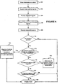
- Figure 1 shows a flowchart illustrating a method for assessing the brain state of a patient. This method may be implemented by an apparatus or device which is manufactured to perform the method given herein.
- An electrode set is placed on a subject (step 100).
- Typical electrode sets for acquiring EEG data use at least 19 electrodes.
- An electrode set consistent with an embodiment of the present invention may comprise a reduced electrode set, with less than 19 electrodes.
- the electrodes measure the electrical fields that are produced as a result of the subject's brain activity (step 102).
- the activity may be spontaneous, evoked or a combination thereof.
- the spontaneous brain activity is measured and an evoked response is measured.
- the spontaneous activity may comprise the subject's EEG signals.
- the evoked response may be obtained by stimulating the subject using visual, physical, auditory, or other stimulation.
- an auditory stimulus is given to the subject to obtain an Auditory Evoked Potential (AEP).
- AEP Auditory Evoked Potential
- the Auditory Evoked Potentials may comprise any of auditory brainstem response (ABR) potentials, auditory mid-latency response (AMLR) potentials, or auditory late response (ALR) potentials, including P100 responses, and P300 responses.
- ABR auditory brainstem response
- AMLR auditory mid-latency response
- ALR auditory late response
- the spontaneous and evoked signals are acquired by the electrode set and are subsequently subjected to a signal processor, wherein artifacts are removed from the signals (step 104).
- Artifacts that may be removed are a result of such factors as a disconnected electrode, electromyogram (EMG) artifacts resulting from muscular movement, eye movement and other significant artifacts.
- EMG electromyogram
- the artifacts may be removed by removing discrete artifact sections from the signals.
- the artifacts may be removed by subtracting out any artifacts present in the acquired signals.
- the artifact-free signals are subjected to further processing to extract statistical signal features (step 106).
- a quantitative EEG algorithm may be used to extract features.
- a wavelet packet algorithm may be used for feature extraction.
- spectral analysis and statistical procedures may be performed to extract features.
- diffusion geometric analysis may be performed to extract features.
- microstate analysis may be performed to extract features.
- wavelet-packet local discriminant basis algorithms may be applied to extract features.
- the extracted features are classified according to one or more diagnostic categories, wherein a probability that features extracted from a subject can be classified in one or more diagnostic categories is determined (step 108).
- classifying may be performed by applying discriminant analysis to the extracted features, or by applying wavelet-packets to the extracted features. Regardless of the classifying method used, the classification algorithm first determined if the results are normal (step 110). If the features extracted from the subject's brain waves are normal, then the device will display that the subject's brain activity is normal (step 122). If there is a higher probability that the subject's extracted features are not normal, the device will attempt to classify the extracted features as an emergency or "Alert" condition (step 112).
- the device will attempt to classify the extracted features as either brainstem dysfunction, active seizure, or burst suppression (step 114). If the device determines that the extracted features have a high probability of being one of the emergency states, the device will display this result so the subject can receive immediate treatment (step 122). If the extracted features do not have a high probability of being an emergency, the device will determine if the abnormality of the extracted features appears to be organic in nature (step 116).
- the device will then attempt to determine if the extracted feature abnormality is lateral or global in nature (step 118), and will display the result (122).
- the extracted feature abnormalities will be tested to determine if they are psychiatric or "functional" in nature (step 120), and this result will be shown (step 122).
- FIG. 2 shows an apparatus consistent with an embodiment of the present invention.
- An electrode set 200 is placed on the head of a subject 201.
- the subject is a human, but the subject can be an animal as well.
- An electrode set 200 consistent with an embodiment of the present invention may comprise a reduced electrode set, with less than 19 electrodes.
- Electrode set 200 may comprise a plurality of electrodes which may be affixed to the head of a subject 201.
- electrode set 200 comprises nine electrodes that may be affixed to the forehead, shoulder and ear of the subject. This reduced electrode set 200 allows for placement on the forehead, and eliminates the need to place any electrodes over any hair that a subject may have on their head. This further eliminates any conduction problems that arise due to the hair, and also eliminates the need for any hair removal.
- the electrodes may be placed on the right mastoid 302, far right of the forehead 304, near right of the forehead 306, center top of the forehead 308, near left of the forehead 310, far left of the forehead 312, left mastoid 314, and an ECG electrode on the left shoulder 316. Additionally, in an illustrative embodiment, there is an electrode placed on the center of the forehead 318 that is grounded.
- the electrodes on the right and left mastoids 302, 314 and the center of the forehead 318 may be used in an embodiment wherein an AEP signal is acquired.
- An illustrative embodiment consistent with the present invention is able to use an electrode set 200 with a reduced number of electrodes because the signal processing algorithms eliminate the need for additional electrodes.
- the electrodes measure the electrical fields that are produced as a result of subject's 201 brain activity.
- the activity may be spontaneous, evoked or a combination thereof.
- the spontaneous brain activity is measured, for example the EEG of subject 210, and an evoked response is measured.
- the evoked response may be obtained by stimulating subject 201 using visual, physical, aural or other stimulation.
- an auditory stimulus is given to subject 201 to obtain an Auditory Evoked Response (AEP).
- a pulse oximeter 203 is connected to subject 201 to monitor subject's 201 pulse and blood oxygen levels 209.
- Electrode headset 202 and pulse oximeter 203 can be connected to a handheld device 205. Electrode headset 202 can be connected to handheld device 205 through a low-voltage preamplifier 222. Low-voltage preamplifier 222 has a high noise tolerance and is designed to amplify the signals that are transmitted to and from electrode headset 202.
- Handheld device 205 is designed to be able to fit in one's hand. In one embodiment handheld device 205 may have a size of about 115mm x 190mm x 60mm, and a weight of less than about 600 g. Handheld device 205 has a display 219, which can be an LCD screen, and can further have a touch screen interface and a traditional user interface 220 such as a keyboard.
- handheld device 205, electrodes 200 and electrode headset 202 may come in a kit, designed for performing neurological triage of a patient suffering from an altered mental state, wherein the kit includes instructions for using handheld device 205, and comes in a portable carrying case.
- Handheld device 205 contains analog and digital hardware on the front end 221, and is controlled through processor 210.
- processor 210 is a Texas Instruments OMAP microcontroller/digital signal processor.
- Front end 221 is separated from processor 210 by isolation barrier 208.
- Front end 221 acts as a multi-channel input/output interface for the device, further facilitating the bi-directional communication of transmitted and received signals to processor 210.
- the multi-channel input/output interface is a wireless multi-channel input/output interface.
- a command from a user, entered through user interface 220, will begin a test routine.
- Analog brain waves are acquired through electrode headset 202 and are transmitted through cables to analog front end 204 of handheld device 205.
- Analog brain waves are then converted to digital signals through an ADC contained in analog front end 204 and transmitted to digital front end 206.
- Digital front end 206 transmits the digital signals to processor 210 where digital signals are processed in accordance with instructions contained in internal memory 211 of processor 210.
- the signals are processed to remove noise, processed to extract features, and processed to classify the extracted features.
- the instructions contained in internal memory 211 of processor 210 comprise instructions for performing the method illustrated in Figure 1 .
- Processor 210 may then output results, which may be in real-time, concerning the assessment of subject's 201 brain in accordance with the classification. Outputs may be displayed on LCD screen 219 of handheld device 205, or may be saved to external memory 216, or may be displayed on PC 215 connected to handheld device 205 by serial or universal serial bus connection. In one embodiment, display may display a representation of subject's 201 brain based on the assessment. In another embodiment consistent with the present invention, processor 210 transmits the raw, unprocessed brainwaves to an external memory 216. External memory 216 may be a hard disk drive, an optical disk drive, a floppy disk drive, or a removable, non-volatile memory device.
- results are transmitted through serial bus to infrared transmitter 217 which is configured to transmit data wirelessly to printer 218 to wirelessly print results.
- Handheld device 205 contains an internal rechargeable battery 212 that is able to be charged during use or in between uses through charger 213 connected to a typical AC outlet 214.
- a test routine may require a stimulus to be given to subject 200 to evoke a response.
- the command to produce a stimulus is transmitted from the processor 210 to digital front end 206, where it is converted to an analog signal by a DAC contained therein.
- the analog signal is output from the analog front end 204 through the cables and to a stimulus emitter 224 which stimulates subject 201.
- the stimulus can be auditory, sensory, or visual, or other.
- the stimulus is an auditory stimulus given through transmitters that are placed in subject's ear.
- the stimulus emitter 224 may be an Etymotic Research ER 10D probe with dual speakers and a single microphone in each ear.
- the evoked signal is acquired by electrode headset 202, and is transmitted along with spontaneous signals to analog front end 204 of handheld device 205, where it is converted to a digital signal and transmitted to digital front end 206.
- Digital front end 206 transmits the digital acquired signals to processor 210, where evoked response signals are filtered out from spontaneous signals.
- the evoked response signals are filtered out using an adaptive wavelet based filter.
- internal memory 211 can contain instructions that are executed by the processor 210 which uses a Dual-Tree Complex Wavelet Transform as an invertible transform to adaptively filter evoked signal response signals from spontaneous response signals.
- the instructions further can contain an implementation of an algorithm carried out by processor 210, wherein a complex wavelet transform is computed for each sub-average, and then the phase variance of each normalized wavelet coefficient w i,j is computed. The magnitude of each wavelet coefficient is selectively scaled according to the phase variance of the coefficients at this location across the sub-averages.
- the filtered evoked signal is averaged and an automatic peak detection algorithm is implemented by processor 210 to determine the following peak locations and latencies : Peak 1, Peak 2, and Interpeak 1-5 latency. These values are then compared to normative data contained in internal memory 211 of processor 210.
- processing the signals comprises removing noise from the acquired signals, or "de-noising.”
- Internal memory 211 of processor 210 contains instructions for instructing processor 210 to perform an algorithm on acquired signals.
- the algorithm utilizes wavelet based signal processing using wavelet transforms.
- the wavelet transform a member of the family of Fourier transforms, is a process of decomposing a given signal into a set of orthonormal basis functions called wavelets.
- DFT discrete Fourier transform
- DWT discrete wavelet transform
- DWT uses a family of specifically designed wavelets, or little waves, as basis functions.
- a family of wavelets is created by dilating the original wavelet function, termed the "mother wavelet.”
- a wavelet transform decomposes the signal in both time and frequency using different dilations of the mother wavelet.
- the one dimensional finite signal x[n] is represented in two-dimensional "wavelet coordinates.”
- Individual levels of signal decomposition are created, called scales.
- a set of coefficients is created by computing the inner product of the original signal x[n] with a scaled version of the mother wavelet.
- the mother wavelet function is designated by ⁇
- its dilations are designated by ⁇ (j).
- the position index of a wavelet at scale j is called a translation.
- the value of the wavelet is completely described by the two dimensional sequence ⁇ (j,k), where j is the scale index of the wavelet, and k is the translation index.
- Coefficients C(j,k) are the wavelet coefficients at different scales j and translations k of the inner product of the wavelet Y(j,k) with the original signal x[n].
- wavelet coordinates information about both the frequency and the location (time) of the signal energy is preserved.
- the wavelet coefficient thresholding algorithm reduces sets of wavelet coefficients in the wavelet domain. This process is based on the assumption that the underlying signal is smooth and coherent, while the noise that is mixed with the signal is rough and incoherent.
- Smoothness of a signal is a property related to its bandwidth, and is defined in relation to how many times a signal can be differentiated. The degree of smoothness is equal to the number of continuous derivatives that can be calculated.
- a signal is coherent if its energy is concentrated in both time and frequency domains.
- An incoherent noise is "spread out," and not concentrated.
- One measure of coherence is how many wavelet coefficients are required to represent 99% of the signal energy.
- a time-frequency signal space is completely spanned by wavelet coefficients at all scales and translations.
- a well-concentrated signal decomposition in an appropriately selected wavelet basis will require very few coefficients to represent 99% of signal energy.
- a completely incoherent noise will require 99% of the coefficients that span the entire space to represent 99% of its energy.
- This conventional wavelet de-noising process is a three step process:
- the noise components of the signal are attenuated by selectively setting the wavelet coefficients to zero.
- De-noising is thus a non-linear operation, because different coefficients are affected differently by the thresholding function.
- level of wavelet decomposition threshold selection, using different thresholds at different wavelet coefficients that are kept by a fixed amount.
- the de-noising process involves dividing the acquired signals into discrete intervals, or "frames," and then averaging the frames, and de-noising the averaged frames.
- frames are combined by using two adjacent frames and calculating their linear average. This method is chosen for its simplicity, computational stability, and well-understood behavior. This dyadic linear average is then de-noised, and a new frame is created. The overall idea is to generate as many permutations of the original arrangement of frames as possible, and keep averaging and de-noising those new combinations of frames.
- This recombination process is a tree-like process, and may comprise the dual-tree process described above, in which new levels of recombined frames are created.
- the average and de-noise operation creates frames at level k, which are no longer a linear combination of frames from level k-1.
- Processor 210 is further configured to execute instructions contained in internal memory 211 to perform an algorithm for extracting signals from processed signals.
- processor 210 executes instructions which performs a quantitative EEG (QEEG) feature extraction algorithm on the processed signals.
- QEEG quantitative EEG
- the algorithm utilizes Fast Fourier Transform (FFT) Analysis is applied to characterize the frequency composition of the processed signals, typically dividing the signals into the traditional frequency bands: delta (1.5-3.5 Hz), theta (3.5-7.5 Hz), alpha (7.5-12.5 Hz), beta (12.5-25 Hz), and gamma (25-50 Hz). Higher EEG frequencies, up to and beyond 1000 Hz may also be used.
- FFT Fast Fourier Transform
- These features can include characteristics of the processed signals such as absolute and relative power, symmetry, and coherence.
- absolute power is the average amount of power in each frequency band and in the total frequency spectrum of the processed signals, and is a measure of the strength of the brain's electrical activity.
- Relative power is the percentage of the total power contributed for a respective electrode and a respective frequency band and is a measure of how brain activity is distributed.
- Symmetry is the ratio of levels of activity between corresponding regions of the two brain hemispheres in each frequency band and is a measure of the balance of the observed activity.
- Coherence is the degree of synchronization of electrical events in corresponding regions of the two hemispheres and is a measure of the coordination of the brain activity.
- the significance of the Z-transform is that it allows measures with different metrics to be combined using the common metric of probability.
- the distribution of these response signals is determined for each electrode.
- each extracted feature or factor score is converted to a Z-transform score, or factor Z-score which characterizes the probability that the extracted feature value or factor score observed in the subject will conform to a normal value.
- Processor 210 is further configured to perform an algorithm wherein the extracted features, or the Z-scores are classified. In one embodiment, these sets of univariate data is subjected to Gaussian normalization in order to improve the accuracy of any subsequent statistical analysis.
- the Z-scores are given a selected discriminant score. Each discriminant score is a respective weighted combination of a selected subset of Z-scores for monopolar and/or bipolar univariate and multivariate features derived from the processed signals of a subject.
- the processor 210 executes an algorithm wherein a respective discriminant score is evaluated for each of two or more diagnostic categories multiplying each of several selected Z-scores by a respective coefficient and adding the resulting products.
- the coefficients typically differ as between diagnostic categories and as between Z-scores.
- the probability is evaluated that the subject belongs to one of the two or more diagnostic categories through a probability evaluating expression which is a function of the relevant discriminant scores, matching results against limits provided by internal memory 211 for selected brain states.
- the diagnostic categories may be indicative of whether a subject is exhibiting normal or abnormal brain function.
- abnormal brain function may be further broken down into diagnostic categories which are indicative of psychiatric or “functional” in nature, organic in nature, either lateral or global, or an emergency or "Alert" condition, which may include seizure, abnormal brainstem response, or burst suppression.
- Psychiatric or “functional” brain function may further be broken down into specific diagnostic categories indicative of specific types of psychiatric disorders.
- organic lateral and global brain functions may further be broken down into specific diagnostic categories indicative of specific types of lateral and global abnormalities. The ability of the apparatus to determine a probability that subject 201 is experiencing a particular type of abnormal brain function allows a medical professional to act accordingly.
- the apparatus will further determine whether the brain function has a higher probability of being indicative of a lateral or global abnormality, allowing a medical professional to distinguish between global abnormalities such as concussion, toxicity, encephalitis and the like, and lateral abnormalities such as ischemic and hemorrhagic strokes.
- This probability that subject 201 belongs to a particular diagnostic category can be displayed on LCD display 219.
- LCD display 219 can further display that subject's brain function is 80% indicative of a hemorrhagic stroke, 15% indicative of an ischemic stroke, and 5% of a subdural hematoma. Furthermore, if a subject 201 is diagnosed as having a high probability of suffering from an emergency or "Alert" condition, such as active seizure, a medical professional may be able to provide immediate emergency care to subject 201.
- an emergency or "Alert" condition such as active seizure
- the novel apparatus allows the rapid triage assessment of the neurological state of a subject, allowing for immediate diagnosis and care of victims of head injury and neurological maladies.
- the apparatus may further be packaged in a portable kit with instructions on using the apparatus for performing rapid triage assessment.
Description
- This application claims priority to
U.S. Patent Application No. 11/195,001, filed August 2, 2005 - The invention relates to the field of emergency triage, and specifically, an apparatus for performing emergency neurological triage.
- The central nervous system (CNS) and the brain in particular, perform the most complex and essential processes in the human body. Surprisingly, contemporary health care lacks sophisticated tools to objectively assess their function. A patient's mental and neurological status is typically assessed clinically by an interview and a subjective physical exam. The clinical laboratory currently has no capacity to assess brain function or pathology, contributing little more than identification of poisons, toxins, or drugs that may have externally impacted the CNS. Brain imaging studies, such as computed tomography imaging (CT), magnetic resonance imaging (MRI), though widely used and useful, are structural/anatomical tests revealing little or nothing about brain function. In the immediate time of acute brain injury, stroke, or seizure, imaging studies typically reveal no abnormality, even when there is clear and dramatically abnormal brain function. CT and MRI only detect the condition after the morphology or structure of the brain has changed. In some cases it can take from hours to days after the patient is present in an emergency room (ER) before overt changes are evident on the CT or MRI, and before severe neurological pathology is visible. Electrical activity of the brain, however, is affected immediately. New imaging modalities such as functional MRI (fMRI) measure the changes in oxygen saturation in different parts of the brain. Radioisotope imaging such as positron emission tomography (PET) and single photon emission computerized tomography (SPECT) assess chemical changes within the brain as a measurement of function with limited sensitivity and specificity. All of these assessment tools play an important role in selected cases, but they are costly, not universally available, and they do not provide critical information at the early stages of acute care situations. None of the current techniques provides the immediate, actionable information critical to timely intervention, appropriate triage, or the formulation of an appropriate plan of care.
- The CNS and brain, of all organs in the human body, are also the most time sensitive and have the least capacity for repair. Currently, emergency room patients with altered mental status, acute neuropathy, or head trauma must undergo costly and time consuming tests to determine an appropriate course of treatment. Unfortunately, in many cases, the clinical condition of patients continue to deteriorate as they wait for equipment to become available or for specialists to interpret tests. The task of the ER physician is to basically establish whether the brain is functioning normally, whether the abnormality is psychiatric or organic in nature, whether an organic abnormality is global or lateralized, and to develop an initial assessment of the diagnostic possibilities. The problem that faces ER physicians is that their resources are quite literally limited to a flashlight and a rubber reflex hammer. Amazingly, all of the physician's decisions concerning the administration of emergency treatment or intervention, including CT scan, spinal tap, additional consultation or discharge are based on the results of this simplistic exam.
- Often, ER patients are sent for imaging studies, yet many functional brain abnormalities, such as seizure, are not visible on a CT scan. Some abnormalities which will eventually have anatomical and structural consequences often take time to become visible. This is true for many important conditions such as ischemic stroke, concussion, raised intracranial pressure, and others. Thus, while the location, expense, and limited availability of the CT scan can be problematic, so indeed can the fact that it is a structural as opposed to functional test.
- One-third of over 200 physicians surveyed at the American College of Emergency Physicians feel that the combination of a good clinical laboratory, a neurological exam, and a CT scan of the head, is not adequate for the assessment of every patient with altered mental status or neurological dysfunction. Consensus estimates from the CDC NHS database and practicing ER physicians, is that patients requiring a mental status exam represent 15% of the more than 100 million ER visits annually in the U.S., and in some institutions, considerably more.
- There are more than 100 million ER visits per year in the US alone (CDC/NCHS) database. In year 2000, more than 13 million of these patients required a formal mental status exam and nearly 5 million had CT scans. This data indicates the need for real-time functional brain state assessment which can be performed in the hospital, in an ambulance, at a sporting event, or any other location where acute neurological evaluation may be necessary.
- All of the brain's activity, whether reflexive, automatic, unconscious, or conscious, is electrical in nature. Through a series of electro-chemical reactions, mediated by molecules called neurotransmitters, electrical potentials (voltages) are generated and transmitted throughout the brain, traveling continuously between and among the myriad of neurons. This activity establishes the basic electrical signatures of the electroencephalogram (EEG) and creates identifiable frequencies which have a basis in anatomic structure and function. Understanding these basic rhythms and their significance makes it possible to characterize the EEG as being within or beyond normal limits. At this basic level, the EEG serves as a signature for both normal and abnormal brain function.
- The electrical activity of the brain has been studied extensively since the first recordings over 75 years ago, and especially since the advent of computers. "Normal" electrical activity of the brain has been well characterized in hundreds of studies, with a narrow standard deviation. The frequencies of electrical activity of some parts of the brain are the normal response to various stimuli , such as acoustic, visual, or pain, known as "evoked potentials." Evoked potentials (EP) are particular waves that have characteristic shapes, amplitudes and duration of peaks within those wave shapes, and many other features, all of which have well established normative data, generated over decades of research. Normative data for all of the EEG and evoked response waves are remarkably constant across different genders, ages, and ethnicities. Moreover, any variability that does exist is well described and explained.
- Neuroscientists have also characterized the EEG signature of various different brain pathologies. Just as an abnormal electrocardiogram (ECG) pattern is a strong indication of a particular heart pathology, an irregular brain wave pattern is a strong indication of a particular brain pathology. A wide array of pathologies have been well characterized: acute and chronic, structural, toxic, metabolic, and even specific diagnoses such as: ischemic stroke, epileptic seizures, concussion, alcohol, and drug overdose, psychiatric conditions, and dementias including Alzheimer's disease. A large body of data, with continuing refinements and contributions, constitutes the field of clinical neurophysiology.
- Even though EEG-based neurometric technology is accepted today and a tremendous body of data exists, application in the clinical environment is notably limited. Some of the barriers limiting its adoption include: the cost of EEG equipment, its lack of portability, the need for a technician to administer the test, the time it takes to conduct the test, and the need for expert interpretation of the raw data. More importantly, the technology is neither available nor practical in the acute care setting, especially at the point of care. A complete diagnostic EEG instrument typically costs $80,000, fully equipped. Despite the high costs, the instrument produces essentially raw waveforms which must be carefully interpreted by an expert. Moreover, use of the standard EEG equipment remains extremely cumbersome. It can take 30 minutes or more to apply the required 19 electrodes. Once the patient is prepared for the test, the recording itself can take from 1 to 4 hours. Data is collected and analyzed by an EEG technician, and are then presented to a neurologist for interpretation and clinical assessment. There are some self-standing dedicated neurodiagnostic laboratories which focus strictly on detailed analysis of electrical brain data. Neither the specialized centers, nor the typically large hospital EEG machines are practical for the ER, operating room (OR), intensive care unit (ICU), or any other acute care medicine setting where patients are in the greatest need. Immediate, functional brain state assessment is needed to treat patients with acute neurological injury and disease for the prevention of further damage and disability.
- US Patent Application
US 2002/0091335 A1 discloses a portable EEG (electroencephalograph) instrument, especially for use in emergencies and brain assessments in physicians' offices, which detects and amplifies brain waves and converts them into digital data for analysis by comparison with data from normal groups. - An apparatus according to the invention is defined in independent claim 1. Preferred embodiments are defined by the dependent claims.
- It is to be understood that both the foregoing general description and the following detailed description are exemplary and explanatory only and are not restrictive of the invention, as claimed.
- The accompanying drawings, which are incorporated in and constitute a part of this specification, illustrate several embodiment of the invention and together with the description, serve to explain the principles of the invention.
-
-
Figure 1 is a flowchart illustrating the method of assessing the brain state of a subject carried out by an apparatus according to an embodiment consistent with the present invention. -
Figure 2 is a diagram illustrating an apparatus according to an embodiment consistent with the present invention. -
Figure 3 is a diagram illustrating an electrode set according to an embodiment consistent with the present invention. - Reference will now be made in detail to present embodiments of the invention, an example of which is illustrated in the accompanying drawings. Wherever possible, the same reference numbers will be used throughout the drawings to refer to the same or like parts.
- In accordance with embodiments consistent with the present invention,
Figure 1 shows a flowchart illustrating a method for assessing the brain state of a patient. This method may be implemented by an apparatus or device which is manufactured to perform the method given herein. An electrode set is placed on a subject (step 100). Typical electrode sets for acquiring EEG data use at least 19 electrodes. An electrode set consistent with an embodiment of the present invention may comprise a reduced electrode set, with less than 19 electrodes. - The electrodes measure the electrical fields that are produced as a result of the subject's brain activity (step 102). The activity may be spontaneous, evoked or a combination thereof. In an embodiment consistent with the present invention, the spontaneous brain activity is measured and an evoked response is measured. The spontaneous activity may comprise the subject's EEG signals. The evoked response may be obtained by stimulating the subject using visual, physical, auditory, or other stimulation. In an embodiment consistent with the present invention, an auditory stimulus is given to the subject to obtain an Auditory Evoked Potential (AEP). Moreover, the Auditory Evoked Potentials may comprise any of auditory brainstem response (ABR) potentials, auditory mid-latency response (AMLR) potentials, or auditory late response (ALR) potentials, including P100 responses, and P300 responses.
- The spontaneous and evoked signals are acquired by the electrode set and are subsequently subjected to a signal processor, wherein artifacts are removed from the signals (step 104). Artifacts that may be removed are a result of such factors as a disconnected electrode, electromyogram (EMG) artifacts resulting from muscular movement, eye movement and other significant artifacts. In one embodiment, the artifacts may be removed by removing discrete artifact sections from the signals. In another embodiment, the artifacts may be removed by subtracting out any artifacts present in the acquired signals.
- The artifact-free signals are subjected to further processing to extract statistical signal features (step 106). In one embodiment consistent with the present invention, a quantitative EEG algorithm may be used to extract features. In another embodiment, a wavelet packet algorithm may be used for feature extraction. In a further embodiment, spectral analysis and statistical procedures may be performed to extract features. In yet a further embodiment, diffusion geometric analysis may be performed to extract features. In yet another embodiment, microstate analysis may be performed to extract features. In a further embodiment, wavelet-packet local discriminant basis algorithms may be applied to extract features.
- Referring again to
Figure 1 , the extracted features are classified according to one or more diagnostic categories, wherein a probability that features extracted from a subject can be classified in one or more diagnostic categories is determined (step 108). According to embodiments consistent with the invention, classifying may be performed by applying discriminant analysis to the extracted features, or by applying wavelet-packets to the extracted features. Regardless of the classifying method used, the classification algorithm first determined if the results are normal (step 110). If the features extracted from the subject's brain waves are normal, then the device will display that the subject's brain activity is normal (step 122). If there is a higher probability that the subject's extracted features are not normal, the device will attempt to classify the extracted features as an emergency or "Alert" condition (step 112). If there is a high probability that the extracted features match features typical of someone in an emergency mental state or an "Alert" condition, the device will attempt to classify the extracted features as either brainstem dysfunction, active seizure, or burst suppression (step 114). If the device determines that the extracted features have a high probability of being one of the emergency states, the device will display this result so the subject can receive immediate treatment (step 122). If the extracted features do not have a high probability of being an emergency, the device will determine if the abnormality of the extracted features appears to be organic in nature (step 116). If the extracted features are determined to correlate with an extracted feature abnormality that is organic in nature, the device will then attempt to determine if the extracted feature abnormality is lateral or global in nature (step 118), and will display the result (122). The extracted feature abnormalities will be tested to determine if they are psychiatric or "functional" in nature (step 120), and this result will be shown (step 122). -
Figure 2 shows an apparatus consistent with an embodiment of the present invention. An electrode set 200 is placed on the head of a subject 201. In an illustrative embodiment, the subject is a human, but the subject can be an animal as well. An electrode set 200 consistent with an embodiment of the present invention may comprise a reduced electrode set, with less than 19 electrodes. -
Figure 3 shows anelectrode set 200 consistent with an embodiment of the present invention. Electrode set 200 may comprise a plurality of electrodes which may be affixed to the head of a subject 201. In an illustrative embodiment, electrode set 200 comprises nine electrodes that may be affixed to the forehead, shoulder and ear of the subject. This reducedelectrode set 200 allows for placement on the forehead, and eliminates the need to place any electrodes over any hair that a subject may have on their head. This further eliminates any conduction problems that arise due to the hair, and also eliminates the need for any hair removal. In an illustrative embodiment, the electrodes may be placed on theright mastoid 302, far right of theforehead 304, near right of theforehead 306, center top of theforehead 308, near left of theforehead 310, far left of theforehead 312, left mastoid 314, and an ECG electrode on theleft shoulder 316. Additionally, in an illustrative embodiment, there is an electrode placed on the center of theforehead 318 that is grounded. The electrodes on the right and leftmastoids forehead 318 may be used in an embodiment wherein an AEP signal is acquired. An illustrative embodiment consistent with the present invention is able to use anelectrode set 200 with a reduced number of electrodes because the signal processing algorithms eliminate the need for additional electrodes. - Referring back to
Figure 2 , the electrodes measure the electrical fields that are produced as a result of subject's 201 brain activity. The activity may be spontaneous, evoked or a combination thereof. In an embodiment consistent with the present invention, the spontaneous brain activity is measured, for example the EEG ofsubject 210, and an evoked response is measured. The evoked response may be obtained by stimulating subject 201 using visual, physical, aural or other stimulation. In an embodiment consistent with the present invention, an auditory stimulus is given to subject 201 to obtain an Auditory Evoked Response (AEP). In one embodiment of the present invention, apulse oximeter 203 is connected to subject 201 to monitor subject's 201 pulse andblood oxygen levels 209. -
Electrode headset 202 andpulse oximeter 203 can be connected to ahandheld device 205.Electrode headset 202 can be connected tohandheld device 205 through a low-voltage preamplifier 222. Low-voltage preamplifier 222 has a high noise tolerance and is designed to amplify the signals that are transmitted to and fromelectrode headset 202.Handheld device 205 is designed to be able to fit in one's hand. In oneembodiment handheld device 205 may have a size of about 115mm x 190mm x 60mm, and a weight of less than about 600 g.Handheld device 205 has adisplay 219, which can be an LCD screen, and can further have a touch screen interface and atraditional user interface 220 such as a keyboard. In one embodiment,handheld device 205,electrodes 200 andelectrode headset 202 may come in a kit, designed for performing neurological triage of a patient suffering from an altered mental state, wherein the kit includes instructions for usinghandheld device 205, and comes in a portable carrying case. -
Handheld device 205 contains analog and digital hardware on thefront end 221, and is controlled throughprocessor 210. In one embodiment,processor 210 is a Texas Instruments OMAP microcontroller/digital signal processor.Front end 221 is separated fromprocessor 210 byisolation barrier 208.Front end 221 acts as a multi-channel input/output interface for the device, further facilitating the bi-directional communication of transmitted and received signals toprocessor 210. In one embodiment consistent with the present invention, the multi-channel input/output interface is a wireless multi-channel input/output interface. - In an embodiment consistent with the present invention, a command from a user, entered through
user interface 220, will begin a test routine. Analog brain waves are acquired throughelectrode headset 202 and are transmitted through cables to analogfront end 204 ofhandheld device 205. Analog brain waves are then converted to digital signals through an ADC contained in analogfront end 204 and transmitted to digitalfront end 206. Digitalfront end 206 transmits the digital signals toprocessor 210 where digital signals are processed in accordance with instructions contained ininternal memory 211 ofprocessor 210. In an embodiment consistent with the present invention, the signals are processed to remove noise, processed to extract features, and processed to classify the extracted features. In another embodiment, the instructions contained ininternal memory 211 ofprocessor 210 comprise instructions for performing the method illustrated inFigure 1 .Processor 210 may then output results, which may be in real-time, concerning the assessment of subject's 201 brain in accordance with the classification. Outputs may be displayed onLCD screen 219 ofhandheld device 205, or may be saved toexternal memory 216, or may be displayed onPC 215 connected tohandheld device 205 by serial or universal serial bus connection. In one embodiment, display may display a representation of subject's 201 brain based on the assessment. In another embodiment consistent with the present invention,processor 210 transmits the raw, unprocessed brainwaves to anexternal memory 216.External memory 216 may be a hard disk drive, an optical disk drive, a floppy disk drive, or a removable, non-volatile memory device. In another embodiment, results are transmitted through serial bus toinfrared transmitter 217 which is configured to transmit data wirelessly toprinter 218 to wirelessly print results.Handheld device 205 contains an internalrechargeable battery 212 that is able to be charged during use or in between uses throughcharger 213 connected to a typical AC outlet 214. - In another embodiment, a test routine may require a stimulus to be given to subject 200 to evoke a response. The command to produce a stimulus is transmitted from the
processor 210 to digitalfront end 206, where it is converted to an analog signal by a DAC contained therein. The analog signal is output from the analogfront end 204 through the cables and to astimulus emitter 224 which stimulates subject 201. The stimulus can be auditory, sensory, or visual, or other. In a preferred embodiment, the stimulus is an auditory stimulus given through transmitters that are placed in subject's ear. Thestimulus emitter 224 may be an Etymotic Research ER 10D probe with dual speakers and a single microphone in each ear. The evoked signal is acquired byelectrode headset 202, and is transmitted along with spontaneous signals to analogfront end 204 ofhandheld device 205, where it is converted to a digital signal and transmitted to digitalfront end 206. Digitalfront end 206 transmits the digital acquired signals toprocessor 210, where evoked response signals are filtered out from spontaneous signals. In an embodiment consistent with the present invention, the evoked response signals are filtered out using an adaptive wavelet based filter. More specifically,internal memory 211 can contain instructions that are executed by theprocessor 210 which uses a Dual-Tree Complex Wavelet Transform as an invertible transform to adaptively filter evoked signal response signals from spontaneous response signals. The instructions further can contain an implementation of an algorithm carried out byprocessor 210, wherein a complex wavelet transform is computed for each sub-average, and then the phase variance of each normalized wavelet coefficient wi,j is computed. The magnitude of each wavelet coefficient is selectively scaled according to the phase variance of the coefficients at this location across the sub-averages. The scaling has the form:

processor 210 to determine the following peak locations and latencies : Peak 1, Peak 2, and Interpeak 1-5 latency. These values are then compared to normative data contained ininternal memory 211 ofprocessor 210. - In an embodiment consistent with the present invention, processing the signals comprises removing noise from the acquired signals, or "de-noising."
Internal memory 211 ofprocessor 210 contains instructions for instructingprocessor 210 to perform an algorithm on acquired signals. In one embodiment, the algorithm utilizes wavelet based signal processing using wavelet transforms. The wavelet transform, a member of the family of Fourier transforms, is a process of decomposing a given signal into a set of orthonormal basis functions called wavelets. In traditional discrete Fourier transform (DFT), a signal is decomposed using complex sinusoids as basis functions, producing a frequency domain representation of the signal. In contrast, a discrete wavelet transform (DWT) uses a family of specifically designed wavelets, or little waves, as basis functions. A family of wavelets is created by dilating the original wavelet function, termed the "mother wavelet." A wavelet transform decomposes the signal in both time and frequency using different dilations of the mother wavelet. With the application of DWT, the one dimensional finite signal x[n] is represented in two-dimensional "wavelet coordinates." Individual levels of signal decomposition are created, called scales. At each scale a set of coefficients is created by computing the inner product of the original signal x[n] with a scaled version of the mother wavelet. The mother wavelet function is designated by ψ, and its dilations are designated by ψ(j). The position index of a wavelet at scale j is called a translation. The value of the wavelet is completely described by the two dimensional sequence ψ(j,k), where j is the scale index of the wavelet, and k is the translation index. The DWT is the defined as:
- Coefficients C(j,k) are the wavelet coefficients at different scales j and translations k of the inner product of the wavelet Y(j,k) with the original signal x[n]. In wavelet coordinates, information about both the frequency and the location (time) of the signal energy is preserved. This is a process of noise suppression that utilizes assumptions about smoothness and coherence properties of both the underlying signal and the noise that contaminates it. Similar to filtering in the frequency domain, the wavelet coefficient thresholding algorithm reduces sets of wavelet coefficients in the wavelet domain. This process is based on the assumption that the underlying signal is smooth and coherent, while the noise that is mixed with the signal is rough and incoherent. Smoothness of a signal is a property related to its bandwidth, and is defined in relation to how many times a signal can be differentiated. The degree of smoothness is equal to the number of continuous derivatives that can be calculated. A signal is coherent if its energy is concentrated in both time and frequency domains. An incoherent noise is "spread out," and not concentrated. One measure of coherence is how many wavelet coefficients are required to represent 99% of the signal energy. A time-frequency signal space is completely spanned by wavelet coefficients at all scales and translations. A well-concentrated signal decomposition in an appropriately selected wavelet basis will require very few coefficients to represent 99% of signal energy. However, a completely incoherent noise will require 99% of the coefficients that span the entire space to represent 99% of its energy.
- This conventional wavelet de-noising process is a three step process:
- 1. Wavelet transform the signal to obtain wavelet coefficients at different scales
- 2. Threshold the coefficients and set to zero any smaller than a threshold δ
- 3. Perform the inverse wavelet transform to approximate the original signal
- In the de-noising process, the noise components of the signal are attenuated by selectively setting the wavelet coefficients to zero. De-noising is thus a non-linear operation, because different coefficients are affected differently by the thresholding function. There are many parameters to control in this algorithm: level of wavelet decomposition, threshold selection, using different thresholds at different wavelet coefficients that are kept by a fixed amount.
- In accordance with an embodiment of the present invention, the de-noising process involves dividing the acquired signals into discrete intervals, or "frames," and then averaging the frames, and de-noising the averaged frames. The greater amount of frames that are de-noised prior recomposing the signal, the better the results of the de-noising process. Preferably, the frames are combined by using two adjacent frames and calculating their linear average. This method is chosen for its simplicity, computational stability, and well-understood behavior. This dyadic linear average is then de-noised, and a new frame is created. The overall idea is to generate as many permutations of the original arrangement of frames as possible, and keep averaging and de-noising those new combinations of frames. This recombination process is a tree-like process, and may comprise the dual-tree process described above, in which new levels of recombined frames are created. The average and de-noise operation creates frames at level k, which are no longer a linear combination of frames from level k-1.
- The many possible algorithms to accomplish this task can be evaluated by different criteria: ease of implementation, computational efficiency, computational stability, etc. For the present invention, ease of implementation is used, because the key aspect of the invention is implementation of different wavelet de-noising techniques and not combinatorics of frame rearrangements. The goal of the preferred embodiment in frame rearranging is to produce enough new frames to obtain acceptable performance.
-
Processor 210 is further configured to execute instructions contained ininternal memory 211 to perform an algorithm for extracting signals from processed signals. In one embodiment,processor 210 executes instructions which performs a quantitative EEG (QEEG) feature extraction algorithm on the processed signals. The algorithm utilizes Fast Fourier Transform (FFT) Analysis is applied to characterize the frequency composition of the processed signals, typically dividing the signals into the traditional frequency bands: delta (1.5-3.5 Hz), theta (3.5-7.5 Hz), alpha (7.5-12.5 Hz), beta (12.5-25 Hz), and gamma (25-50 Hz). Higher EEG frequencies, up to and beyond 1000 Hz may also be used. These features can include characteristics of the processed signals such as absolute and relative power, symmetry, and coherence. In the context of analyzing process brainwaves, absolute power is the average amount of power in each frequency band and in the total frequency spectrum of the processed signals, and is a measure of the strength of the brain's electrical activity. Relative power is the percentage of the total power contributed for a respective electrode and a respective frequency band and is a measure of how brain activity is distributed. Symmetry is the ratio of levels of activity between corresponding regions of the two brain hemispheres in each frequency band and is a measure of the balance of the observed activity. Coherence is the degree of synchronization of electrical events in corresponding regions of the two hemispheres and is a measure of the coordination of the brain activity. These four basic categories of univariate features, resulting from the spectral analysis of the process signals, are believed to characterize independent aspects of brain activity and each is believed to be sensitive to a variety of different clinical conditions and changes of state. A full set of individual and pairwise features is calculated and transformed for Gaussianity using, for example, the log function. Once a Gaussian distribution has been demonstrated and age regression applied, statistical Z transformation is performed. The Z-transform is used to describe the deviations from age expected normal values: - Z= Probability that subject value lies within the normal range

- The significance of the Z-transform is that it allows measures with different metrics to be combined using the common metric of probability. Using a database of response signals from a large population of subjects believed to be normal, or to have other conditions, the distribution of these response signals is determined for each electrode. In particular, each extracted feature or factor score is converted to a Z-transform score, or factor Z-score which characterizes the probability that the extracted feature value or factor score observed in the subject will conform to a normal value.
-
Processor 210 is further configured to perform an algorithm wherein the extracted features, or the Z-scores are classified. In one embodiment, these sets of univariate data is subjected to Gaussian normalization in order to improve the accuracy of any subsequent statistical analysis. The Z-scores are given a selected discriminant score. Each discriminant score is a respective weighted combination of a selected subset of Z-scores for monopolar and/or bipolar univariate and multivariate features derived from the processed signals of a subject. Theprocessor 210 executes an algorithm wherein a respective discriminant score is evaluated for each of two or more diagnostic categories multiplying each of several selected Z-scores by a respective coefficient and adding the resulting products. The coefficients typically differ as between diagnostic categories and as between Z-scores. The probability is evaluated that the subject belongs to one of the two or more diagnostic categories through a probability evaluating expression which is a function of the relevant discriminant scores, matching results against limits provided byinternal memory 211 for selected brain states. - The diagnostic categories may be indicative of whether a subject is exhibiting normal or abnormal brain function. Moreover, abnormal brain function may be further broken down into diagnostic categories which are indicative of psychiatric or "functional" in nature, organic in nature, either lateral or global, or an emergency or "Alert" condition, which may include seizure, abnormal brainstem response, or burst suppression. Psychiatric or "functional" brain function may further be broken down into specific diagnostic categories indicative of specific types of psychiatric disorders. Similarly, organic lateral and global brain functions may further be broken down into specific diagnostic categories indicative of specific types of lateral and global abnormalities. The ability of the apparatus to determine a probability that subject 201 is experiencing a particular type of abnormal brain function allows a medical professional to act accordingly. For example, should a subject be diagnosed as having a high probability of having a brain function that is indicative of an organic abnormality, the apparatus will further determine whether the brain function has a higher probability of being indicative of a lateral or global abnormality, allowing a medical professional to distinguish between global abnormalities such as concussion, toxicity, encephalitis and the like, and lateral abnormalities such as ischemic and hemorrhagic strokes. This probability that subject 201 belongs to a particular diagnostic category can be displayed on
LCD display 219. For example, in the above scenario in which subject 201 is exhibiting an organic, lateral abnormality,LCD display 219 can further display that subject's brain function is 80% indicative of a hemorrhagic stroke, 15% indicative of an ischemic stroke, and 5% of a subdural hematoma. Furthermore, if a subject 201 is diagnosed as having a high probability of suffering from an emergency or "Alert" condition, such as active seizure, a medical professional may be able to provide immediate emergency care to subject 201. - The novel apparatus allows the rapid triage assessment of the neurological state of a subject, allowing for immediate diagnosis and care of victims of head injury and neurological maladies. The apparatus may further be packaged in a portable kit with instructions on using the apparatus for performing rapid triage assessment.
- Other embodiments of the invention will be apparent to those skilled in the art from consideration of the specification and practice of the invention disclosed herein. It is intended that the specification and examples be considered as exemplary only, with a true scope of the invention being indicated by the following claims.
Claims (21)
- An apparatus for assessing the neurological state of a subject, comprising:an electrode set (200);a processor (210);a memory (211) operatively coupled to the processor, wherein the memory stores one or more operating instructions and classification limits for categories comprising psychiatric, organic, and emergency condition;a multi-channel input-output interface (221) operatively coupled to the processor, whereinthe multi-channel input-output interface (221) is configured to receive external electrical signals through the set of electrodes (200) placed on the subject; andthe processor (210) is configured to utilize the one or more operating instructions to perform a series of operations on signals received from the multi-channel input-output interface (221)wherein the a series of operations comprise:extracting desired features from the signals,applying discriminant analysis to the extracted features to classify of the extracted features into one or more categories,determining of the neurological state of the subject based on the classificationwherein the step of determining the neurological state of the subject comprises:determining the probability of the neurological state being normal or abnormal, andcharacterized in that the step of determining the neurological state of the subject further comprises:if there is a higher probability of the neurological state being abnormal, further determining if the probability of the neurological state being within the classification limits stored in memory (211) for psychiatric, organic, or emergency condition categories.
- The apparatus of claim 1, further comprising:a display device (219), whereinthe display device is operatively connected to the processor and the processor is further configured to display results of the one or more operations on the display device.
- The apparatus of claim 1, further comprising:a bi-directional communications channel operatively connected to the multi-channel input-output interface and to the processor to communicate the received external signals.
- The apparatus of claim 1, further comprising:a low voltage multi-channel preamplifier operatively connected to the multi-channel input-output interface.
- The apparatus of claim 4, whereinthe low voltage multi-channel preamplifier has a high noise tolerance.
- The apparatus of claim 1, wherein the multi-channel input-output interface includes an electroencephalograph testing interface and an auditory evoked potentials testing interface; andwherein the processor is configured with the one or more stored operating instructions for auditory tests selected from the group comprising electroencephalograph test procedures, auditory evoked potentials test procedures, and combinations thereof.
- The apparatus of claim 1, whereinthe processor is configured to execute the one or more operating instructions stored in the memory for processing the received external signals.
- The apparatus of claim 1, wherein the operating instructions include:instructions for processing the external signals;instructions for extracting features from the external signals;instructions for classifying the subject based on extracted features; andinstructions for determining the neurological state of a subject based on the classification.
- The apparatus of claim 8, whereinthe instructions for processing the external signals include instructions to filter the received external signals.
- The apparatus of claim 9, wherein the filtering comprises:using a dual-tree complex wavelet transform of the received external signals.
- The apparatus of claim 1, wherein the set of electrodes comprises:a plurality of electrodes placed on the subject's forehead; andat least one electrode placed in at least one of the ears of the subject.
- The apparatus of claim 1, further comprising:a stimulation device (224) operatively connected to the multi-channel input-output interface for evoking an auditory potential in the subject.
- The apparatus of claim 1, whereinthe apparatus is housed in a hand-held enclosure.
- The apparatus of claim 1, wherein the multi-channel input-output interface further comprises:a wireless multi-channel input-output interface.
- A kit for performing an emergency neurological diagnosis of a patient suffering from an altered mental state, the kit including:the apparatus of claim 1;instructions for using the apparatus; anda portable carrying case for the apparatus.
- The apparatus of claim 1 further comprising: an user interface (220) operatively connected to the processor.
- The apparatus of claim 1, further comprising: an amplifier (222) operatively connected to the electrode set.
- The apparatus of claim 1, wherein the electrode set comprises less than 19 electrodes.
- The apparatus of claim 1, wherein the processor is configured to extract the features from the signals using wavelet packet algorithms.
- The apparatus of claim 1, wherein memory (211) further stores classification limits for organic subcategories comprising lateral or global conditions;wherein the one or more operations further comprise:determining the probability of an abnormal neurological state with a probability of being organic being global or being lateral in nature.
- The apparatus of claim 1, wherein the processor is configured to extract signal features applying wavelet-packet local discriminant basis algorithms.
Applications Claiming Priority (2)
| Application Number | Priority Date | Filing Date | Title |
|---|---|---|---|
| US11/195,001 US7904144B2 (en) | 2005-08-02 | 2005-08-02 | Method for assessing brain function and portable automatic brain function assessment apparatus |
| PCT/US2006/028985 WO2007016149A2 (en) | 2005-08-02 | 2006-07-26 | Automatic brain function assessment apparatus and method |
Publications (3)
| Publication Number | Publication Date |
|---|---|
| EP1909643A2 EP1909643A2 (en) | 2008-04-16 |
| EP1909643A4 EP1909643A4 (en) | 2010-03-24 |
| EP1909643B1 true EP1909643B1 (en) | 2016-03-30 |
Family
ID=37709137
Family Applications (1)
| Application Number | Title | Priority Date | Filing Date |
|---|---|---|---|
| EP06800351.6A Active EP1909643B1 (en) | 2005-08-02 | 2006-07-26 | Portable automatic brain function assessment apparatus |
Country Status (10)
| Country | Link |
|---|---|
| US (2) | US7904144B2 (en) |
| EP (1) | EP1909643B1 (en) |
| JP (2) | JP2009502424A (en) |
| KR (1) | KR20080068003A (en) |
| CN (2) | CN105520732B (en) |
| AU (1) | AU2006275864A1 (en) |
| CA (2) | CA2939790C (en) |
| HK (1) | HK1219217A1 (en) |
| IL (1) | IL189163A0 (en) |
| WO (1) | WO2007016149A2 (en) |
Families Citing this family (196)
| Publication number | Priority date | Publication date | Assignee | Title |
|---|---|---|---|---|
| US7672717B1 (en) * | 2003-10-22 | 2010-03-02 | Bionova Technologies Inc. | Method and system for the denoising of large-amplitude artifacts in electrograms using time-frequency transforms |
| US7904144B2 (en) | 2005-08-02 | 2011-03-08 | Brainscope Company, Inc. | Method for assessing brain function and portable automatic brain function assessment apparatus |
| US7720530B2 (en) | 2005-08-02 | 2010-05-18 | Brainscope Company, Inc. | Field-deployable concussion detector |
| US9615744B2 (en) * | 2007-01-31 | 2017-04-11 | Medtronic, Inc. | Chopper-stabilized instrumentation amplifier for impedance measurement |
| US7385443B1 (en) * | 2007-01-31 | 2008-06-10 | Medtronic, Inc. | Chopper-stabilized instrumentation amplifier |
| US8265769B2 (en) * | 2007-01-31 | 2012-09-11 | Medtronic, Inc. | Chopper-stabilized instrumentation amplifier for wireless telemetry |
| US7391257B1 (en) * | 2007-01-31 | 2008-06-24 | Medtronic, Inc. | Chopper-stabilized instrumentation amplifier for impedance measurement |
| US9554721B1 (en) | 2007-04-23 | 2017-01-31 | Neurowave Systems Inc. | Seizure detector, brain dysfunction monitor and method |
| US8781595B2 (en) | 2007-04-30 | 2014-07-15 | Medtronic, Inc. | Chopper mixer telemetry circuit |
| WO2008157544A1 (en) * | 2007-06-18 | 2008-12-24 | New York University | Electronic identity card |
| US20090012386A1 (en) * | 2007-07-06 | 2009-01-08 | The President And Fellows Of Harvard College | Systems and methods for pre-operatively identifying functional regions of a patient's brain to assist in the preparation of a contemplated surgery |
| EP2200692B1 (en) * | 2007-09-26 | 2016-11-09 | Medtronic, Inc. | Frequency selective monitoring of physiological signals |
| EP2207590A1 (en) * | 2007-09-26 | 2010-07-21 | Medtronic, INC. | Therapy program selection |
| US8380314B2 (en) * | 2007-09-26 | 2013-02-19 | Medtronic, Inc. | Patient directed therapy control |
| WO2009051638A1 (en) | 2007-10-16 | 2009-04-23 | Medtronic, Inc. | Therapy control based on a patient movement state |
| CN101925377A (en) * | 2008-01-25 | 2010-12-22 | 麦德托尼克公司 | The detection of Sleep stages |
| US9787841B2 (en) | 2008-01-28 | 2017-10-10 | Afiniti Europe Technologies Limited | Techniques for hybrid behavioral pairing in a contact center system |
| US9654641B1 (en) | 2008-01-28 | 2017-05-16 | Afiniti International Holdings, Ltd. | Systems and methods for routing callers to an agent in a contact center |
| US9300802B1 (en) | 2008-01-28 | 2016-03-29 | Satmap International Holdings Limited | Techniques for behavioral pairing in a contact center system |
| US8824658B2 (en) | 2008-11-06 | 2014-09-02 | Satmap International Holdings Limited | Selective mapping of callers in a call center routing system |
| US10708430B2 (en) | 2008-01-28 | 2020-07-07 | Afiniti Europe Technologies Limited | Techniques for benchmarking pairing strategies in a contact center system |
| US9781269B2 (en) | 2008-01-28 | 2017-10-03 | Afiniti Europe Technologies Limited | Techniques for hybrid behavioral pairing in a contact center system |
| US8903079B2 (en) * | 2008-01-28 | 2014-12-02 | Satmap International Holdings Limited | Routing callers from a set of callers based on caller data |
| US10708431B2 (en) | 2008-01-28 | 2020-07-07 | Afiniti Europe Technologies Limited | Techniques for hybrid behavioral pairing in a contact center system |
| US9692898B1 (en) | 2008-01-28 | 2017-06-27 | Afiniti Europe Technologies Limited | Techniques for benchmarking paring strategies in a contact center system |
| US9712679B2 (en) | 2008-01-28 | 2017-07-18 | Afiniti International Holdings, Ltd. | Systems and methods for routing callers to an agent in a contact center |
| US10567586B2 (en) * | 2008-11-06 | 2020-02-18 | Afiniti Europe Technologies Limited | Pooling callers for matching to agents based on pattern matching algorithms |
| US20090190745A1 (en) * | 2008-01-28 | 2009-07-30 | The Resource Group International Ltd | Pooling callers for a call center routing system |
| US9712676B1 (en) | 2008-01-28 | 2017-07-18 | Afiniti Europe Technologies Limited | Techniques for benchmarking pairing strategies in a contact center system |
| US20090232294A1 (en) * | 2008-01-28 | 2009-09-17 | Qiaobing Xie | Skipping a caller in queue for a call routing center |
| US10750023B2 (en) | 2008-01-28 | 2020-08-18 | Afiniti Europe Technologies Limited | Techniques for hybrid behavioral pairing in a contact center system |
| US8781100B2 (en) * | 2008-01-28 | 2014-07-15 | Satmap International Holdings Limited | Probability multiplier process for call center routing |
| US8879715B2 (en) | 2012-03-26 | 2014-11-04 | Satmap International Holdings Limited | Call mapping systems and methods using variance algorithm (VA) and/or distribution compensation |
| US8670548B2 (en) * | 2008-01-28 | 2014-03-11 | Satmap International Holdings Limited | Jumping callers held in queue for a call center routing system |
| US20090190750A1 (en) * | 2008-01-28 | 2009-07-30 | The Resource Group International Ltd | Routing callers out of queue order for a call center routing system |
| US8718271B2 (en) * | 2008-01-28 | 2014-05-06 | Satmap International Holdings Limited | Call routing methods and systems based on multiple variable standardized scoring |
| US9774740B2 (en) | 2008-01-28 | 2017-09-26 | Afiniti Europe Technologies Limited | Techniques for benchmarking pairing strategies in a contact center system |
| US20090247893A1 (en) * | 2008-03-27 | 2009-10-01 | The General Electric Company | Method and apparatus for measuring responsiveness of a subject |
| US20090247894A1 (en) * | 2008-03-31 | 2009-10-01 | Brainscope Company, Inc. | Systems and Methods For Neurological Evaluation and Treatment Guidance |
| US20090264785A1 (en) * | 2008-04-18 | 2009-10-22 | Brainscope Company, Inc. | Method and Apparatus For Assessing Brain Function Using Diffusion Geometric Analysis |
| US20090264786A1 (en) * | 2008-04-21 | 2009-10-22 | Brainscope Company, Inc. | System and Method For Signal Denoising Using Independent Component Analysis and Fractal Dimension Estimation |
| US8041136B2 (en) * | 2008-04-21 | 2011-10-18 | Brainscope Company, Inc. | System and method for signal processing using fractal dimension analysis |
| US20090312663A1 (en) * | 2008-06-13 | 2009-12-17 | New York University | System and Method for Neurometric Analysis |
| US20100016753A1 (en) * | 2008-07-18 | 2010-01-21 | Firlik Katrina S | Systems and Methods for Portable Neurofeedback |
| US20100020959A1 (en) * | 2008-07-28 | 2010-01-28 | The Resource Group International Ltd | Routing callers to agents based on personality data of agents |
| US8473024B2 (en) * | 2008-08-12 | 2013-06-25 | Brainscope Company, Inc. | Flexible headset for sensing brain electrical activity |
| US8644490B2 (en) * | 2008-08-29 | 2014-02-04 | Satmap International Holdings Limited | Shadow queue for callers in a performance/pattern matching based call routing system |
| US8781106B2 (en) * | 2008-08-29 | 2014-07-15 | Satmap International Holdings Limited | Agent satisfaction data for call routing based on pattern matching algorithm |
| KR20100032054A (en) * | 2008-09-17 | 2010-03-25 | 인제대학교 산학협력단 | Apparatus and method for alzheimer's disease diagnosis using eeg(electroencephalogram) analysis |
| US8478402B2 (en) * | 2008-10-31 | 2013-07-02 | Medtronic, Inc. | Determining intercardiac impedance |
| US8472611B2 (en) | 2008-11-06 | 2013-06-25 | The Resource Group International Ltd. | Balancing multiple computer models in a call center routing system |
| US20100111288A1 (en) * | 2008-11-06 | 2010-05-06 | Afzal Hassan | Time to answer selector and advisor for call routing center |
| USRE48412E1 (en) | 2008-11-06 | 2021-01-26 | Afiniti, Ltd. | Balancing multiple computer models in a call center routing system |
| US8634542B2 (en) | 2008-12-09 | 2014-01-21 | Satmap International Holdings Limited | Separate pattern matching algorithms and computer models based on available caller data |
| US8364254B2 (en) * | 2009-01-28 | 2013-01-29 | Brainscope Company, Inc. | Method and device for probabilistic objective assessment of brain function |
| US9451883B2 (en) | 2009-03-04 | 2016-09-27 | The Regents Of The University Of California | Apparatus and method for decoding sensory and cognitive information from brain activity |
| WO2010141764A2 (en) * | 2009-06-03 | 2010-12-09 | University Of Pittsburgh - Of The Commonwealth System Of Higher Education | Steady state measurement and analysis approach to profiling auditory evoked potentials from short-latency to long-latency |
| US10321840B2 (en) * | 2009-08-14 | 2019-06-18 | Brainscope Company, Inc. | Development of fully-automated classifier builders for neurodiagnostic applications |
| US20110087125A1 (en) * | 2009-10-09 | 2011-04-14 | Elvir Causevic | System and method for pain monitoring at the point-of-care |
| US20110112426A1 (en) | 2009-11-10 | 2011-05-12 | Brainscope Company, Inc. | Brain Activity as a Marker of Disease |
| US9770204B2 (en) | 2009-11-11 | 2017-09-26 | Medtronic, Inc. | Deep brain stimulation for sleep and movement disorders |
| US8579812B2 (en) | 2009-12-15 | 2013-11-12 | Brainscope Company, Inc. | System and methods for management of disease over time |
| US8577451B2 (en) | 2009-12-16 | 2013-11-05 | Brainscope Company, Inc. | System and methods for neurologic monitoring and improving classification and treatment of neurologic states |
| US20110144520A1 (en) * | 2009-12-16 | 2011-06-16 | Elvir Causevic | Method and device for point-of-care neuro-assessment and treatment guidance |
| US8700141B2 (en) * | 2010-03-10 | 2014-04-15 | Brainscope Company, Inc. | Method and apparatus for automatic evoked potentials assessment |
| US20110245707A1 (en) * | 2010-04-01 | 2011-10-06 | James Sherman Castle | Portable stroke monitoring apparatus |
| EP2584963B1 (en) * | 2010-06-22 | 2016-11-09 | National Research Council of Canada | Cognitive function assessment in a patient |
| JP5838557B2 (en) * | 2010-07-05 | 2016-01-06 | ソニー株式会社 | Biological information processing method and apparatus, and recording medium |
| US8478394B2 (en) | 2010-08-16 | 2013-07-02 | Brainscope Company, Inc. | Field deployable concussion assessment device |
| US9211411B2 (en) | 2010-08-26 | 2015-12-15 | Medtronic, Inc. | Therapy for rapid eye movement behavior disorder (RBD) |
| US8699694B2 (en) | 2010-08-26 | 2014-04-15 | Satmap International Holdings Limited | Precalculated caller-agent pairs for a call center routing system |
| US8724797B2 (en) | 2010-08-26 | 2014-05-13 | Satmap International Holdings Limited | Estimating agent performance in a call routing center system |
| US8750488B2 (en) | 2010-08-31 | 2014-06-10 | Satmap International Holdings Limited | Predicted call time as routing variable in a call routing center system |
| KR101249069B1 (en) * | 2011-01-14 | 2013-03-29 | 한국과학기술원 | Method and apparatus for quantifying risk of developing schizophrenia using brainwave synchronization level, and computer-readable media recording codes for the method |
| GB2502011B (en) * | 2011-01-21 | 2015-03-04 | Fondamenta Llc | Electrode for attention training techniques |
| MY152983A (en) * | 2011-02-28 | 2014-12-15 | Inst Of Technology Petronas Sdn Bhd | Methodology of extracting brain erp signals from background noise |
| US20150038869A1 (en) * | 2011-07-16 | 2015-02-05 | Cerora, Inc. | Systems and methods for the physiological assessment of brain health and the remote quality control of eeg systems |
| CN103917159B (en) * | 2011-07-20 | 2017-02-15 | 艾欧敏达有限公司 | Method and system for estimating brain concussion |
| EP2739587B1 (en) | 2011-08-01 | 2020-05-27 | Denovo Sciences | Cell capture system |
| US20130035579A1 (en) | 2011-08-02 | 2013-02-07 | Tan Le | Methods for modeling neurological development and diagnosing a neurological impairment of a patient |
| US9888861B2 (en) * | 2011-08-25 | 2018-02-13 | Medtronic, Inc. | Method and apparatus for detecting a biomarker in the presence of electrical stimulation |
| US20140012151A1 (en) * | 2012-07-06 | 2014-01-09 | Persyst Development Corporation | Method And System For Displaying EEG Data |
| US8666484B2 (en) * | 2011-11-25 | 2014-03-04 | Persyst Development Corporation | Method and system for displaying EEG data |
| US9269046B2 (en) | 2012-01-18 | 2016-02-23 | Brainscope Company, Inc. | Method and device for multimodal neurological evaluation |
| US8792974B2 (en) | 2012-01-18 | 2014-07-29 | Brainscope Company, Inc. | Method and device for multimodal neurological evaluation |
| US20130204153A1 (en) * | 2012-02-06 | 2013-08-08 | Emily Ruth Buzhardt | Generating an alarm based on brain wave patterns of a user |
| US20140051945A1 (en) * | 2012-03-12 | 2014-02-20 | Terra Cotta Star Llc | Expanded frequency electroencephalography/electrocardiography light apparatus and method of use |
| US10849563B2 (en) | 2012-03-19 | 2020-12-01 | University Of Florida Research Foundation, Inc. | Methods and systems for brain function analysis |
| US9025757B2 (en) | 2012-03-26 | 2015-05-05 | Satmap International Holdings Limited | Call mapping systems and methods using bayesian mean regression (BMR) |
| KR101325368B1 (en) * | 2012-04-24 | 2013-11-08 | 연세대학교 원주산학협력단 | Apparatus for closed-loop electrical brain stimulation using neural response signals |
| US9763592B2 (en) * | 2012-05-25 | 2017-09-19 | Emotiv, Inc. | System and method for instructing a behavior change in a user |
| WO2013177592A2 (en) | 2012-05-25 | 2013-11-28 | Emotiv Lifesciences, Inc. | System and method for providing and aggregating biosignals and action data |
| US9622660B2 (en) * | 2012-05-25 | 2017-04-18 | Emotiv Lifesciences Inc. | System and method for enabling collaborative analysis of a biosignal |
| US9814426B2 (en) | 2012-06-14 | 2017-11-14 | Medibotics Llc | Mobile wearable electromagnetic brain activity monitor |
| US11172859B2 (en) * | 2014-01-28 | 2021-11-16 | Medibotics | Wearable brain activity device with auditory interface |
| CN102727311B (en) * | 2012-06-27 | 2015-04-15 | 深圳市美德医疗电子技术有限公司 | Brain function stimulating device and equipment for awakening operation |
| US8792630B2 (en) | 2012-09-24 | 2014-07-29 | Satmap International Holdings Limited | Use of abstracted data in pattern matching system |
| BR112015007002B1 (en) | 2012-09-28 | 2022-10-18 | The Regents Of The University Of California | METHOD AND SYSTEM FOR PROVIDING COGNITIVE OR SENSORY ASSESSMENT OF AN INDIVIDUAL |
| JP6454944B2 (en) * | 2012-10-12 | 2019-01-23 | ザ リージェンツ オブ ザ ユニバーシティ オブ カリフォルニア | Configuration and spatial arrangement of front electrode sensor for detecting physiological signals |
| US20140114165A1 (en) | 2012-10-24 | 2014-04-24 | Dreamscape Medical Llc | Systems and methods for detecting brain-based bio-signals |
| WO2014066855A1 (en) * | 2012-10-26 | 2014-05-01 | The Regents Of The University Of California | Methods of decoding speech from brain activity data and devices for practicing the same |
| CN104902806B (en) | 2012-11-10 | 2019-06-04 | 加利福尼亚大学董事会 | The assessment system and method for europathology |
| US9265458B2 (en) | 2012-12-04 | 2016-02-23 | Sync-Think, Inc. | Application of smooth pursuit cognitive testing paradigms to clinical drug development |
| US20150351657A1 (en) * | 2013-01-17 | 2015-12-10 | Sensodetect Ab | Method And System For Monitoring Depth Of Anaesthesia And Sensory Functioning |
| USD728801S1 (en) | 2013-03-08 | 2015-05-05 | Brainscope Company, Inc. | Electrode headset |
| US8989836B2 (en) | 2013-03-08 | 2015-03-24 | Brainscope Company, Inc. | Electrode array and method of placement |
| US9380976B2 (en) | 2013-03-11 | 2016-07-05 | Sync-Think, Inc. | Optical neuroinformatics |
| WO2014158921A1 (en) * | 2013-03-14 | 2014-10-02 | Persyst Development Corporation | Method and system to calculate qeeg |
| US9439150B2 (en) | 2013-03-15 | 2016-09-06 | Medtronic, Inc. | Control of spectral agressors in a physiological signal montoring device |
| US9521979B2 (en) | 2013-03-15 | 2016-12-20 | Medtronic, Inc. | Control of spectral agressors in a physiological signal monitoring device |
| US10028703B2 (en) | 2013-07-30 | 2018-07-24 | Emotiv, Inc. | Wearable system for detecting and measuring biosignals |
| CN103654773B (en) * | 2013-12-20 | 2016-02-03 | 北京飞宇星电子科技有限公司 | Electroencephalo experimental teaching unit |
| US10929753B1 (en) | 2014-01-20 | 2021-02-23 | Persyst Development Corporation | System and method for generating a probability value for an event |
| WO2015187401A1 (en) | 2014-06-04 | 2015-12-10 | Neil Rothman | Method and device for multimodal neurological evaluation |
| US9924904B2 (en) | 2014-09-02 | 2018-03-27 | Medtronic, Inc. | Power-efficient chopper amplifier |
| US10028694B2 (en) | 2014-11-25 | 2018-07-24 | Scienceplusplease, Llc | Non-invasive systems and methods to detect cortical spreading depression for the detection and assessment of brain injury and concussion |
| CN104545905B (en) * | 2015-01-06 | 2017-09-15 | 朱志茹 | The experiment frame and experimental method of recording electrode are installed |
| AU2016221435B2 (en) | 2015-02-16 | 2019-10-03 | Nathan Intrator | Systems and methods for brain activity interpretation |
| USD765259S1 (en) | 2015-02-19 | 2016-08-30 | Brainscope Company, Inc. | Ear attachment for medical headset |
| US10108264B2 (en) | 2015-03-02 | 2018-10-23 | Emotiv, Inc. | System and method for embedded cognitive state metric system |
| JP6618702B2 (en) * | 2015-04-06 | 2019-12-11 | 国立研究開発法人情報通信研究機構 | Perceptual meaning content estimation device and perceptual meaning content estimation method by analyzing brain activity |
| CN107635472A (en) | 2015-06-19 | 2018-01-26 | 神经系统分析公司 | Transcranial doppler detector |
| CN105147281A (en) * | 2015-08-25 | 2015-12-16 | 上海医疗器械高等专科学校 | Portable stimulating, awaking and evaluating system for disturbance of consciousness |
| CN105147282A (en) * | 2015-08-25 | 2015-12-16 | 上海医疗器械高等专科学校 | Cognitive disorder electroencephalogram recognition system |
| CN105193410A (en) * | 2015-09-14 | 2015-12-30 | 武汉智普天创科技有限公司 | EEG (electroencephalogram) signal amplifying system |
| CN105342609B (en) * | 2015-11-19 | 2018-04-03 | 南方医科大学 | A kind of portable auditory evoked potential detecting system and its implementation |
| CN113095662B (en) | 2015-12-01 | 2024-03-19 | 阿菲尼帝有限公司 | Techniques for case distribution |
| WO2017096358A1 (en) * | 2015-12-04 | 2017-06-08 | University Of Iowa Research Foundation | Apparatus, systems and methods for predicting, screening and monitoring of encephalopathy / delirium |
| CN108601548A (en) | 2015-12-31 | 2018-09-28 | 布赖恩斯科普公司 | For Neural monitoring and the system and method for auxiliary diagnosis |
| US10617388B2 (en) | 2016-01-05 | 2020-04-14 | Neural Analytics, Inc. | Integrated probe structure |
| US11589836B2 (en) | 2016-01-05 | 2023-02-28 | Novasignal Corp. | Systems and methods for detecting neurological conditions |
| EP3399918A4 (en) | 2016-01-05 | 2019-08-21 | Neural Analytics, Inc. | Systems and methods for determining clinical indications |
| US10736557B2 (en) | 2016-03-30 | 2020-08-11 | Brain F.I.T. Imaging, LLC | Methods and magnetic imaging devices to inventory human brain cortical function |
| US10142473B1 (en) | 2016-06-08 | 2018-11-27 | Afiniti Europe Technologies Limited | Techniques for benchmarking performance in a contact center system |
| US11759147B2 (en) | 2016-06-08 | 2023-09-19 | The Trustees Of Columbia University In The City Of New York | Systems and methods for real-time concussion diagnosis by electroencephalogram activity monitoring |
| US9692899B1 (en) | 2016-08-30 | 2017-06-27 | Afiniti Europe Technologies Limited | Techniques for benchmarking pairing strategies in a contact center system |
| US9888121B1 (en) | 2016-12-13 | 2018-02-06 | Afiniti Europe Technologies Limited | Techniques for behavioral pairing model evaluation in a contact center system |
| US11577090B2 (en) | 2016-12-19 | 2023-02-14 | The Board Of Trustees Of The Leland Stanford Junior University | Machine learning based artifact rejection for transcranial magnetic stimulation electroencephalogram |
| EP3562384B1 (en) | 2016-12-29 | 2024-03-13 | Finkelstein, Elliot Steven | A portable system for monitoring brain trauma |
| US11831808B2 (en) | 2016-12-30 | 2023-11-28 | Afiniti, Ltd. | Contact center system |
| US10320984B2 (en) | 2016-12-30 | 2019-06-11 | Afiniti Europe Technologies Limited | Techniques for L3 pairing in a contact center system |
| US10326882B2 (en) | 2016-12-30 | 2019-06-18 | Afiniti Europe Technologies Limited | Techniques for workforce management in a contact center system |
| US9955013B1 (en) | 2016-12-30 | 2018-04-24 | Afiniti Europe Technologies Limited | Techniques for L3 pairing in a contact center system |
| US10257354B2 (en) | 2016-12-30 | 2019-04-09 | Afiniti Europe Technologies Limited | Techniques for L3 pairing in a contact center system |
| US10135986B1 (en) | 2017-02-21 | 2018-11-20 | Afiniti International Holdings, Ltd. | Techniques for behavioral pairing model evaluation in a contact center system |
| JP7187473B2 (en) * | 2017-03-22 | 2022-12-12 | セーフオプ サージカル インコーポレイテッド | Medical system and method for detecting changes in electrophysiological evoked potentials |
| US10970658B2 (en) | 2017-04-05 | 2021-04-06 | Afiniti, Ltd. | Techniques for behavioral pairing in a dispatch center system |
| US9930180B1 (en) | 2017-04-28 | 2018-03-27 | Afiniti, Ltd. | Techniques for behavioral pairing in a contact center system |
| US10122860B1 (en) | 2017-07-10 | 2018-11-06 | Afiniti Europe Technologies Limited | Techniques for estimating expected performance in a task assignment system |
| EP3435246A1 (en) * | 2017-07-24 | 2019-01-30 | Tata Consultancy Services Limited | System and method for signal analysis |
| US11051691B2 (en) | 2017-07-31 | 2021-07-06 | Vye, Llc | Ocular analysis |
| US11723579B2 (en) | 2017-09-19 | 2023-08-15 | Neuroenhancement Lab, LLC | Method and apparatus for neuroenhancement |
| WO2019070895A1 (en) | 2017-10-03 | 2019-04-11 | Braint F.I.T. Imaging, Llc | Methods and magnetic imaging devices to inventory human brian cortical function |
| US10509669B2 (en) | 2017-11-08 | 2019-12-17 | Afiniti Europe Technologies Limited | Techniques for benchmarking pairing strategies in a task assignment system |
| US10110746B1 (en) | 2017-11-08 | 2018-10-23 | Afiniti Europe Technologies Limited | Techniques for benchmarking pairing strategies in a task assignment system |
| KR20190059364A (en) * | 2017-11-23 | 2019-05-31 | 주식회사 아이메디신 | Brain age estimation method and system using brain wave analysis |
| KR20190059376A (en) * | 2017-11-23 | 2019-05-31 | 주식회사 아이메디신 | Brain cognitive function evaluation platform and method using drain wave analysis |
| KR20190059377A (en) * | 2017-11-23 | 2019-05-31 | 주식회사 아이메디신 | Traumatic brain injury evaluation system and method using brain wave analysis |
| US11399096B2 (en) | 2017-11-29 | 2022-07-26 | Afiniti, Ltd. | Techniques for data matching in a contact center system |
| US11717686B2 (en) | 2017-12-04 | 2023-08-08 | Neuroenhancement Lab, LLC | Method and apparatus for neuroenhancement to facilitate learning and performance |
| US10509671B2 (en) | 2017-12-11 | 2019-12-17 | Afiniti Europe Technologies Limited | Techniques for behavioral pairing in a task assignment system |
| US11478603B2 (en) | 2017-12-31 | 2022-10-25 | Neuroenhancement Lab, LLC | Method and apparatus for neuroenhancement to enhance emotional response |
| US10623565B2 (en) | 2018-02-09 | 2020-04-14 | Afiniti Europe Technologies Limited | Techniques for behavioral pairing in a contact center system |
| US11219416B2 (en) | 2018-03-12 | 2022-01-11 | Persyst Development Corporation | Graphically displaying evoked potentials |
| US11457855B2 (en) | 2018-03-12 | 2022-10-04 | Persyst Development Corporation | Method and system for utilizing empirical null hypothesis for a biological time series |
| AU2019251467A1 (en) | 2018-04-10 | 2020-11-26 | Cerenetex, Inc. | Systems and methods for the identification of medical conditions, and determination of appropriate therapies, by passively detecting acoustic signals generated from cerebral vasculature |
| US11364361B2 (en) | 2018-04-20 | 2022-06-21 | Neuroenhancement Lab, LLC | System and method for inducing sleep by transplanting mental states |
| US11250359B2 (en) | 2018-05-30 | 2022-02-15 | Afiniti, Ltd. | Techniques for workforce management in a task assignment system |
| US10638967B2 (en) | 2018-07-30 | 2020-05-05 | United States Of America As Represented By The Navy | Handheld radio device for assessing cognitive, auditory, visual, and speech function |
| EP3849410A4 (en) | 2018-09-14 | 2022-11-02 | Neuroenhancement Lab, LLC | System and method of improving sleep |
| US10496438B1 (en) | 2018-09-28 | 2019-12-03 | Afiniti, Ltd. | Techniques for adapting behavioral pairing to runtime conditions in a task assignment system |
| CN109431496A (en) * | 2018-10-16 | 2019-03-08 | 北京工业大学 | A kind of assessment system and method using EEG signals assessment brain ability |
| KR102163170B1 (en) * | 2018-10-25 | 2020-10-08 | 재단법인대구경북과학기술원 | Method and Device for stimulating the neual tissue and measuring the response signal |
| US10867263B2 (en) | 2018-12-04 | 2020-12-15 | Afiniti, Ltd. | Techniques for behavioral pairing in a multistage task assignment system |
| US11144344B2 (en) | 2019-01-17 | 2021-10-12 | Afiniti, Ltd. | Techniques for behavioral pairing in a task assignment system |
| CA3135689A1 (en) | 2019-04-03 | 2020-10-08 | Brain F.I.T. Imaging, LLC | Methods and magnetic imaging devices to inventory human brain cortical function |
| US11786694B2 (en) | 2019-05-24 | 2023-10-17 | NeuroLight, Inc. | Device, method, and app for facilitating sleep |
| US10757261B1 (en) | 2019-08-12 | 2020-08-25 | Afiniti, Ltd. | Techniques for pairing contacts and agents in a contact center system |
| US11445062B2 (en) | 2019-08-26 | 2022-09-13 | Afiniti, Ltd. | Techniques for behavioral pairing in a task assignment system |
| US10757262B1 (en) | 2019-09-19 | 2020-08-25 | Afiniti, Ltd. | Techniques for decisioning behavioral pairing in a task assignment system |
| CN110613446A (en) * | 2019-09-30 | 2019-12-27 | 京东方科技集团股份有限公司 | Signal processing method and device |
| WO2021158436A1 (en) | 2020-02-03 | 2021-08-12 | Afiniti, Ltd. | Techniques for behavioral pairing in a task assignment system |
| CN115244513A (en) | 2020-02-04 | 2022-10-25 | 阿菲尼帝有限公司 | Techniques for error handling in a task distribution system with an external pairing system |
| US11050886B1 (en) | 2020-02-05 | 2021-06-29 | Afiniti, Ltd. | Techniques for sharing control of assigning tasks between an external pairing system and a task assignment system with an internal pairing system |
| US20210307672A1 (en) * | 2020-04-05 | 2021-10-07 | Epitel, Inc. | Eeg recording and analysis |
| CN111477299B (en) * | 2020-04-08 | 2023-01-03 | 广州艾博润医疗科技有限公司 | Method and device for regulating and controlling sound-electricity stimulation nerves by combining electroencephalogram detection and analysis control |
| RU2753267C1 (en) * | 2020-06-05 | 2021-08-12 | ОБЩЕСТВО С ОГРАНИЧЕННОЙ ОТВЕТСТВЕННОСТЬЮ "СберМедИИ" | Method for detecting focal epileptiform discharges in long-term eeg recording |
| WO2022120913A1 (en) * | 2020-12-09 | 2022-06-16 | 中国人民解放军中部战区总医院 | Brain injury electroencephalogram neural oscillation analysis system and method |
| WO2022149789A1 (en) * | 2021-01-07 | 2022-07-14 | 가톨릭대학교 산학협력단 | Post-operative delirium prediction method using electroencephalogram analysis, and analysis apparatus |
| BR112023021898A2 (en) * | 2021-04-28 | 2023-12-19 | Cumulus Neuroscience Ltd | TECHNIQUE FOR OBTAINING AND PROCESSING A MEASUREMENT OF A BIOSIGNAL |
| KR102533262B1 (en) * | 2021-05-11 | 2023-05-15 | 재단법인대구경북과학기술원 | Method and apparatus for simulating body tissues and recording responses |
| CN113647963B (en) * | 2021-08-09 | 2023-08-25 | 北京宁矩科技有限公司 | Wearable brainstem response recording device |
| WO2023225291A2 (en) * | 2022-05-19 | 2023-11-23 | Asterion Ai Inc. | Radiography-concurrent dynamic electropotential neuroactivity monitor |
| WO2023238575A1 (en) * | 2022-06-06 | 2023-12-14 | 株式会社村田製作所 | Cognitive function evaluation device and cognitive function evaluation system |
| US11857330B1 (en) | 2022-10-19 | 2024-01-02 | Epitel, Inc. | Systems and methods for electroencephalogram monitoring |
| CN116098634A (en) * | 2023-01-31 | 2023-05-12 | 首都医科大学宣武医院 | Brain function detection and assessment method, device and system based on stimulation event |
Citations (2)
| Publication number | Priority date | Publication date | Assignee | Title |
|---|---|---|---|---|
| US5083571A (en) * | 1988-04-18 | 1992-01-28 | New York University | Use of brain electrophysiological quantitative data to classify and subtype an individual into diagnostic categories by discriminant and cluster analysis |
| US6658287B1 (en) * | 1998-08-24 | 2003-12-02 | Georgia Tech Research Corporation | Method and apparatus for predicting the onset of seizures based on features derived from signals indicative of brain activity |
Family Cites Families (37)
| Publication number | Priority date | Publication date | Assignee | Title |
|---|---|---|---|---|
| US3706308A (en) | 1970-10-19 | 1972-12-19 | Neuro Data Inc | Life detecting medical instrument |
| US4913160A (en) | 1987-09-30 | 1990-04-03 | New York University | Electroencephalographic system and method using factor structure of the evoked potentials |
| US5384725A (en) | 1990-05-18 | 1995-01-24 | Yale University | Method and apparatus for encoding and decoding using wavelet-packets |
| US5287859A (en) | 1992-09-25 | 1994-02-22 | New York University | Electroencephalograph instrument for mass screening |
| US5488474A (en) | 1994-05-06 | 1996-01-30 | D.O.M. Associates Int'l. | Hadamard phasing in a multisource array |
| US6406427B1 (en) * | 1997-06-10 | 2002-06-18 | Auckland Uniservices Limited | Brain rescue instrument and method |
| US6052619A (en) * | 1997-08-07 | 2000-04-18 | New York University | Brain function scan system |
| US6385486B1 (en) | 1997-08-07 | 2002-05-07 | New York University | Brain function scan system |
| US6016449A (en) | 1997-10-27 | 2000-01-18 | Neuropace, Inc. | System for treatment of neurological disorders |
| US6353689B1 (en) | 1997-11-11 | 2002-03-05 | Sony Corporation | Apparatus for and method of processing image and information recording medium |
| WO1999034865A1 (en) * | 1998-01-08 | 1999-07-15 | Levin Richard B | Eeg based consciousness-alert monitoring system |
| WO2000077675A1 (en) | 1999-06-10 | 2000-12-21 | Koninklijke Philips Electronics N.V. | Interference suppression for measuring signals with periodic wanted signal |
| US6287290B1 (en) * | 1999-07-02 | 2001-09-11 | Pulmonx | Methods, systems, and kits for lung volume reduction |
| JP2001129100A (en) * | 1999-10-29 | 2001-05-15 | Taketoshi Yamada | Pulse wave passing system |
| AU761251B2 (en) * | 2000-01-05 | 2003-05-29 | Rolf-Dieter Battmer | Device for measuring or collecting auditory evoked potentials |
| US6434419B1 (en) * | 2000-06-26 | 2002-08-13 | Sam Technology, Inc. | Neurocognitive ability EEG measurement method and system |
| AU2002242192A1 (en) * | 2001-02-13 | 2002-08-28 | Jordan Neuroscience, Inc. | Automated realtime interpretation of brain waves |
| EP1460933B1 (en) | 2002-01-04 | 2008-12-17 | Aspect Medical Systems, Inc. | System and method of assessment of mood disorders using eeg |
| AU2003219874A1 (en) | 2002-02-28 | 2003-09-16 | General Hospital Corporation | Electroencephalograph sensor for use with magnetic resonance imaging and methods using such arrangements |
| US7054453B2 (en) | 2002-03-29 | 2006-05-30 | Everest Biomedical Instruments Co. | Fast estimation of weak bio-signals using novel algorithms for generating multiple additional data frames |
| US7054454B2 (en) | 2002-03-29 | 2006-05-30 | Everest Biomedical Instruments Company | Fast wavelet estimation of weak bio-signals using novel algorithms for generating multiple additional data frames |
| US7299088B1 (en) | 2002-06-02 | 2007-11-20 | Nitish V Thakor | Apparatus and methods for brain rhythm analysis |
| US7373198B2 (en) | 2002-07-12 | 2008-05-13 | Bionova Technologies Inc. | Method and apparatus for the estimation of anesthetic depth using wavelet analysis of the electroencephalogram |
| US6866639B2 (en) | 2002-09-23 | 2005-03-15 | Everest Biomedical Instruments | Handheld low voltage testing device |
| JP2004135829A (en) * | 2002-10-17 | 2004-05-13 | Fuji Xerox Co Ltd | Brain wave diagnostic apparatus and method |
| WO2004071289A1 (en) | 2003-02-10 | 2004-08-26 | Everest Biomedical Instruments | Apparatus for evoking and recording bio-potentials |
| US7269455B2 (en) | 2003-02-26 | 2007-09-11 | Pineda Jaime A | Method and system for predicting and preventing seizures |
| CN2605022Y (en) * | 2003-02-27 | 2004-03-03 | 中国人民解放军南京军区南京总医院 | Nerve threshold stimulator |
| EP1622510A4 (en) | 2003-05-06 | 2009-06-03 | Everest Biomedical Instr Compa | Anesthesia and sedation monitoring system and method |
| US7715480B2 (en) | 2003-10-17 | 2010-05-11 | Mitsubishi Electric Research Laboratories, Inc. | Video encoding with motion-selective wavelet transform |
| CN1547090A (en) * | 2003-12-03 | 2004-11-17 | 中国科学院昆明动物研究所 | Artificial intelligent brain wave acquisition and analytic system |
| US20050165327A1 (en) | 2004-01-28 | 2005-07-28 | Thibault Lawrence E. | Apparatus and method for detecting the severity of brain function impairment |
| US20070173732A1 (en) | 2004-01-29 | 2007-07-26 | Elvir Causevic | Method and apparatus for wireless brain interface |
| WO2005072459A2 (en) | 2004-01-29 | 2005-08-11 | Everest Biomedical Instruments | Method and apparatus for signal encoding evoked responses |
| US20080262371A1 (en) | 2004-09-16 | 2008-10-23 | Elvir Causevic | Method for Adaptive Complex Wavelet Based Filtering of Eeg Signals |
| US7904144B2 (en) | 2005-08-02 | 2011-03-08 | Brainscope Company, Inc. | Method for assessing brain function and portable automatic brain function assessment apparatus |
| US7720530B2 (en) | 2005-08-02 | 2010-05-18 | Brainscope Company, Inc. | Field-deployable concussion detector |
-
2005
- 2005-08-02 US US11/195,001 patent/US7904144B2/en active Active
-
2006
- 2006-07-26 KR KR1020087005301A patent/KR20080068003A/en not_active Application Discontinuation
- 2006-07-26 CN CN201510991326.XA patent/CN105520732B/en active Active
- 2006-07-26 CA CA2939790A patent/CA2939790C/en not_active Expired - Fee Related
- 2006-07-26 WO PCT/US2006/028985 patent/WO2007016149A2/en active Application Filing
- 2006-07-26 CN CNA2006800325721A patent/CN101500471A/en active Pending
- 2006-07-26 CA CA2616974A patent/CA2616974C/en active Active
- 2006-07-26 AU AU2006275864A patent/AU2006275864A1/en not_active Abandoned
- 2006-07-26 EP EP06800351.6A patent/EP1909643B1/en active Active
- 2006-07-26 JP JP2008525027A patent/JP2009502424A/en not_active Withdrawn
-
2008
- 2008-01-31 IL IL189163A patent/IL189163A0/en unknown
-
2011
- 2011-02-01 US US13/018,533 patent/US8838227B2/en active Active
-
2014
- 2014-02-03 JP JP2014018221A patent/JP5829699B2/en active Active
-
2016
- 2016-06-23 HK HK16107287.9A patent/HK1219217A1/en unknown
Patent Citations (2)
| Publication number | Priority date | Publication date | Assignee | Title |
|---|---|---|---|---|
| US5083571A (en) * | 1988-04-18 | 1992-01-28 | New York University | Use of brain electrophysiological quantitative data to classify and subtype an individual into diagnostic categories by discriminant and cluster analysis |
| US6658287B1 (en) * | 1998-08-24 | 2003-12-02 | Georgia Tech Research Corporation | Method and apparatus for predicting the onset of seizures based on features derived from signals indicative of brain activity |
Also Published As
| Publication number | Publication date |
|---|---|
| US20110125048A1 (en) | 2011-05-26 |
| KR20080068003A (en) | 2008-07-22 |
| JP2009502424A (en) | 2009-01-29 |
| CA2939790A1 (en) | 2007-02-08 |
| CN105520732B (en) | 2019-06-25 |
| EP1909643A2 (en) | 2008-04-16 |
| US7904144B2 (en) | 2011-03-08 |
| WO2007016149A3 (en) | 2009-04-23 |
| US20070032737A1 (en) | 2007-02-08 |
| CA2616974A1 (en) | 2007-02-08 |
| WO2007016149A2 (en) | 2007-02-08 |
| HK1219217A1 (en) | 2017-03-31 |
| CA2616974C (en) | 2016-10-04 |
| US8838227B2 (en) | 2014-09-16 |
| CA2939790C (en) | 2017-01-17 |
| AU2006275864A1 (en) | 2007-02-08 |
| CN101500471A (en) | 2009-08-05 |
| JP2014128691A (en) | 2014-07-10 |
| JP5829699B2 (en) | 2015-12-09 |
| CN105520732A (en) | 2016-04-27 |
| IL189163A0 (en) | 2008-08-07 |
| EP1909643A4 (en) | 2010-03-24 |
Similar Documents
| Publication | Publication Date | Title |
|---|---|---|
| EP1909643B1 (en) | Portable automatic brain function assessment apparatus | |
| EP2262422B1 (en) | Field-deployable concussion detector | |
| CA2784267C (en) | Method and device for point-of-care neuro-assessment and treatment guidance | |
| US10898130B2 (en) | System and method for pain detection and computation of a pain quantification index | |
| EP2391265B1 (en) | Method and device for probabilistic objective assessment of brain function | |
| Rambhia et al. | Classification of Mental Arithmetic States Using Single Electrode |
Legal Events
| Date | Code | Title | Description |
|---|---|---|---|
| PUAI | Public reference made under article 153(3) epc to a published international application that has entered the european phase |
Free format text: ORIGINAL CODE: 0009012 |
|
| 17P | Request for examination filed |
Effective date: 20080222 |
|
| AK | Designated contracting states |
Kind code of ref document: A2 Designated state(s): AT BE BG CH CY CZ DE DK EE ES FI FR GB GR HU IE IS IT LI LT LU LV MC NL PL PT RO SE SI SK TR |
|
| AX | Request for extension of the european patent |
Extension state: AL BA HR MK RS |
|
| R17D | Deferred search report published (corrected) |
Effective date: 20090423 |
|
| RIC1 | Information provided on ipc code assigned before grant |
Ipc: A61B 5/00 20060101AFI20090619BHEP |
|
| A4 | Supplementary search report drawn up and despatched |
Effective date: 20100223 |
|
| 17Q | First examination report despatched |
Effective date: 20100507 |
|
| RAX | Requested extension states of the european patent have changed |
Extension state: BA Payment date: 20080222 |
|
| RAP1 | Party data changed (applicant data changed or rights of an application transferred) |
Owner name: BRAINSCOPE COMPANY, INC. |
|
| GRAP | Despatch of communication of intention to grant a patent |
Free format text: ORIGINAL CODE: EPIDOSNIGR1 |
|
| RIC1 | Information provided on ipc code assigned before grant |
Ipc: A61B 5/00 20060101ALN20150904BHEP Ipc: A61B 5/0484 20060101ALN20150904BHEP Ipc: A61B 5/048 20060101ALI20150904BHEP Ipc: A61B 5/0476 20060101AFI20150904BHEP |
|
| INTG | Intention to grant announced |
Effective date: 20151002 |
|
| REG | Reference to a national code |
Ref country code: DE Ref legal event code: R079 Ref document number: 602006048468 Country of ref document: DE Free format text: PREVIOUS MAIN CLASS: A61B0005040000 Ipc: A61B0005047600 |
|
| GRAS | Grant fee paid |
Free format text: ORIGINAL CODE: EPIDOSNIGR3 |
|
| GRAA | (expected) grant |
Free format text: ORIGINAL CODE: 0009210 |
|
| RIC1 | Information provided on ipc code assigned before grant |
Ipc: A61B 5/0484 20060101ALN20160204BHEP Ipc: A61B 5/00 20060101ALN20160204BHEP Ipc: A61B 5/0476 20060101AFI20160204BHEP Ipc: A61B 5/048 20060101ALI20160204BHEP |
|
| INTG | Intention to grant announced |
Effective date: 20160209 |
|
| AK | Designated contracting states |
Kind code of ref document: B1 Designated state(s): AT BE BG CH CY CZ DE DK EE ES FI FR GB GR HU IE IS IT LI LT LU LV MC NL PL PT RO SE SI SK TR |
|
| AX | Request for extension of the european patent |
Extension state: BA |
|
| REG | Reference to a national code |
Ref country code: GB Ref legal event code: FG4D |
|
| REG | Reference to a national code |
Ref country code: CH Ref legal event code: EP |
|
| REG | Reference to a national code |
Ref country code: AT Ref legal event code: REF Ref document number: 784399 Country of ref document: AT Kind code of ref document: T Effective date: 20160415 |
|
| REG | Reference to a national code |
Ref country code: IE Ref legal event code: FG4D |
|
| REG | Reference to a national code |
Ref country code: DE Ref legal event code: R096 Ref document number: 602006048468 Country of ref document: DE |
|
| REG | Reference to a national code |
Ref country code: NL Ref legal event code: FP |
|
| REG | Reference to a national code |
Ref country code: LT Ref legal event code: MG4D |
|
| PG25 | Lapsed in a contracting state [announced via postgrant information from national office to epo] |
Ref country code: FI Free format text: LAPSE BECAUSE OF FAILURE TO SUBMIT A TRANSLATION OF THE DESCRIPTION OR TO PAY THE FEE WITHIN THE PRESCRIBED TIME-LIMIT Effective date: 20160330 Ref country code: GR Free format text: LAPSE BECAUSE OF FAILURE TO SUBMIT A TRANSLATION OF THE DESCRIPTION OR TO PAY THE FEE WITHIN THE PRESCRIBED TIME-LIMIT Effective date: 20160701 |
|
| REG | Reference to a national code |
Ref country code: AT Ref legal event code: MK05 Ref document number: 784399 Country of ref document: AT Kind code of ref document: T Effective date: 20160330 |
|
| PG25 | Lapsed in a contracting state [announced via postgrant information from national office to epo] |
Ref country code: LV Free format text: LAPSE BECAUSE OF FAILURE TO SUBMIT A TRANSLATION OF THE DESCRIPTION OR TO PAY THE FEE WITHIN THE PRESCRIBED TIME-LIMIT Effective date: 20160330 Ref country code: LT Free format text: LAPSE BECAUSE OF FAILURE TO SUBMIT A TRANSLATION OF THE DESCRIPTION OR TO PAY THE FEE WITHIN THE PRESCRIBED TIME-LIMIT Effective date: 20160330 Ref country code: SE Free format text: LAPSE BECAUSE OF FAILURE TO SUBMIT A TRANSLATION OF THE DESCRIPTION OR TO PAY THE FEE WITHIN THE PRESCRIBED TIME-LIMIT Effective date: 20160330 |
|
| PG25 | Lapsed in a contracting state [announced via postgrant information from national office to epo] |
Ref country code: EE Free format text: LAPSE BECAUSE OF FAILURE TO SUBMIT A TRANSLATION OF THE DESCRIPTION OR TO PAY THE FEE WITHIN THE PRESCRIBED TIME-LIMIT Effective date: 20160330 Ref country code: PL Free format text: LAPSE BECAUSE OF FAILURE TO SUBMIT A TRANSLATION OF THE DESCRIPTION OR TO PAY THE FEE WITHIN THE PRESCRIBED TIME-LIMIT Effective date: 20160330 Ref country code: IS Free format text: LAPSE BECAUSE OF FAILURE TO SUBMIT A TRANSLATION OF THE DESCRIPTION OR TO PAY THE FEE WITHIN THE PRESCRIBED TIME-LIMIT Effective date: 20160730 |
|
| PG25 | Lapsed in a contracting state [announced via postgrant information from national office to epo] |
Ref country code: CZ Free format text: LAPSE BECAUSE OF FAILURE TO SUBMIT A TRANSLATION OF THE DESCRIPTION OR TO PAY THE FEE WITHIN THE PRESCRIBED TIME-LIMIT Effective date: 20160330 Ref country code: RO Free format text: LAPSE BECAUSE OF FAILURE TO SUBMIT A TRANSLATION OF THE DESCRIPTION OR TO PAY THE FEE WITHIN THE PRESCRIBED TIME-LIMIT Effective date: 20160330 Ref country code: PT Free format text: LAPSE BECAUSE OF FAILURE TO SUBMIT A TRANSLATION OF THE DESCRIPTION OR TO PAY THE FEE WITHIN THE PRESCRIBED TIME-LIMIT Effective date: 20160801 Ref country code: SK Free format text: LAPSE BECAUSE OF FAILURE TO SUBMIT A TRANSLATION OF THE DESCRIPTION OR TO PAY THE FEE WITHIN THE PRESCRIBED TIME-LIMIT Effective date: 20160330 Ref country code: ES Free format text: LAPSE BECAUSE OF FAILURE TO SUBMIT A TRANSLATION OF THE DESCRIPTION OR TO PAY THE FEE WITHIN THE PRESCRIBED TIME-LIMIT Effective date: 20160330 Ref country code: AT Free format text: LAPSE BECAUSE OF FAILURE TO SUBMIT A TRANSLATION OF THE DESCRIPTION OR TO PAY THE FEE WITHIN THE PRESCRIBED TIME-LIMIT Effective date: 20160330 |
|
| REG | Reference to a national code |
Ref country code: FR Ref legal event code: PLFP Year of fee payment: 11 |
|
| PG25 | Lapsed in a contracting state [announced via postgrant information from national office to epo] |
Ref country code: BE Free format text: LAPSE BECAUSE OF FAILURE TO SUBMIT A TRANSLATION OF THE DESCRIPTION OR TO PAY THE FEE WITHIN THE PRESCRIBED TIME-LIMIT Effective date: 20160330 Ref country code: IT Free format text: LAPSE BECAUSE OF FAILURE TO SUBMIT A TRANSLATION OF THE DESCRIPTION OR TO PAY THE FEE WITHIN THE PRESCRIBED TIME-LIMIT Effective date: 20160330 |
|
| REG | Reference to a national code |
Ref country code: DE Ref legal event code: R097 Ref document number: 602006048468 Country of ref document: DE |
|
| PG25 | Lapsed in a contracting state [announced via postgrant information from national office to epo] |
Ref country code: DK Free format text: LAPSE BECAUSE OF FAILURE TO SUBMIT A TRANSLATION OF THE DESCRIPTION OR TO PAY THE FEE WITHIN THE PRESCRIBED TIME-LIMIT Effective date: 20160330 |
|
| PLBE | No opposition filed within time limit |
Free format text: ORIGINAL CODE: 0009261 |
|
| STAA | Information on the status of an ep patent application or granted ep patent |
Free format text: STATUS: NO OPPOSITION FILED WITHIN TIME LIMIT |
|
| REG | Reference to a national code |
Ref country code: CH Ref legal event code: PL |
|
| 26N | No opposition filed |
Effective date: 20170103 |
|
| PG25 | Lapsed in a contracting state [announced via postgrant information from national office to epo] |
Ref country code: MC Free format text: LAPSE BECAUSE OF FAILURE TO SUBMIT A TRANSLATION OF THE DESCRIPTION OR TO PAY THE FEE WITHIN THE PRESCRIBED TIME-LIMIT Effective date: 20160330 |
|
| PG25 | Lapsed in a contracting state [announced via postgrant information from national office to epo] |
Ref country code: CH Free format text: LAPSE BECAUSE OF NON-PAYMENT OF DUE FEES Effective date: 20160731 Ref country code: LI Free format text: LAPSE BECAUSE OF NON-PAYMENT OF DUE FEES Effective date: 20160731 |
|
| REG | Reference to a national code |
Ref country code: IE Ref legal event code: MM4A |
|
| PG25 | Lapsed in a contracting state [announced via postgrant information from national office to epo] |
Ref country code: SI Free format text: LAPSE BECAUSE OF FAILURE TO SUBMIT A TRANSLATION OF THE DESCRIPTION OR TO PAY THE FEE WITHIN THE PRESCRIBED TIME-LIMIT Effective date: 20160330 |
|
| REG | Reference to a national code |
Ref country code: FR Ref legal event code: PLFP Year of fee payment: 12 |
|
| PG25 | Lapsed in a contracting state [announced via postgrant information from national office to epo] |
Ref country code: IE Free format text: LAPSE BECAUSE OF NON-PAYMENT OF DUE FEES Effective date: 20160726 |
|
| PG25 | Lapsed in a contracting state [announced via postgrant information from national office to epo] |
Ref country code: LU Free format text: LAPSE BECAUSE OF NON-PAYMENT OF DUE FEES Effective date: 20160726 |
|
| PG25 | Lapsed in a contracting state [announced via postgrant information from national office to epo] |
Ref country code: CY Free format text: LAPSE BECAUSE OF FAILURE TO SUBMIT A TRANSLATION OF THE DESCRIPTION OR TO PAY THE FEE WITHIN THE PRESCRIBED TIME-LIMIT Effective date: 20160330 Ref country code: HU Free format text: LAPSE BECAUSE OF FAILURE TO SUBMIT A TRANSLATION OF THE DESCRIPTION OR TO PAY THE FEE WITHIN THE PRESCRIBED TIME-LIMIT; INVALID AB INITIO Effective date: 20060726 |
|
| PG25 | Lapsed in a contracting state [announced via postgrant information from national office to epo] |
Ref country code: TR Free format text: LAPSE BECAUSE OF FAILURE TO SUBMIT A TRANSLATION OF THE DESCRIPTION OR TO PAY THE FEE WITHIN THE PRESCRIBED TIME-LIMIT Effective date: 20160330 |
|
| REG | Reference to a national code |
Ref country code: FR Ref legal event code: PLFP Year of fee payment: 13 |
|
| PG25 | Lapsed in a contracting state [announced via postgrant information from national office to epo] |
Ref country code: BG Free format text: LAPSE BECAUSE OF FAILURE TO SUBMIT A TRANSLATION OF THE DESCRIPTION OR TO PAY THE FEE WITHIN THE PRESCRIBED TIME-LIMIT Effective date: 20160330 |
|
| REG | Reference to a national code |
Ref country code: DE Ref legal event code: R079 Ref document number: 602006048468 Country of ref document: DE Free format text: PREVIOUS MAIN CLASS: A61B0005047600 Ipc: A61B0005369000 |
|
| P01 | Opt-out of the competence of the unified patent court (upc) registered |
Effective date: 20230610 |
|
| PGFP | Annual fee paid to national office [announced via postgrant information from national office to epo] |
Ref country code: NL Payment date: 20230726 Year of fee payment: 18 |
|
| PGFP | Annual fee paid to national office [announced via postgrant information from national office to epo] |
Ref country code: GB Payment date: 20230727 Year of fee payment: 18 |
|
| PGFP | Annual fee paid to national office [announced via postgrant information from national office to epo] |
Ref country code: FR Payment date: 20230725 Year of fee payment: 18 Ref country code: DE Payment date: 20230727 Year of fee payment: 18 |