EP1902664A1 - Device, system and method for acquiring information in living body - Google Patents
Device, system and method for acquiring information in living body Download PDFInfo
- Publication number
- EP1902664A1 EP1902664A1 EP06780925A EP06780925A EP1902664A1 EP 1902664 A1 EP1902664 A1 EP 1902664A1 EP 06780925 A EP06780925 A EP 06780925A EP 06780925 A EP06780925 A EP 06780925A EP 1902664 A1 EP1902664 A1 EP 1902664A1
- Authority
- EP
- European Patent Office
- Prior art keywords
- vivo information
- information acquiring
- acquiring apparatus
- string member
- apparatus main
- Prior art date
- Legal status (The legal status is an assumption and is not a legal conclusion. Google has not performed a legal analysis and makes no representation as to the accuracy of the status listed.)
- Withdrawn
Links
Images


Classifications
-
- A—HUMAN NECESSITIES
- A61—MEDICAL OR VETERINARY SCIENCE; HYGIENE
- A61B—DIAGNOSIS; SURGERY; IDENTIFICATION
- A61B1/00—Instruments for performing medical examinations of the interior of cavities or tubes of the body by visual or photographical inspection, e.g. endoscopes; Illuminating arrangements therefor
- A61B1/273—Instruments for performing medical examinations of the interior of cavities or tubes of the body by visual or photographical inspection, e.g. endoscopes; Illuminating arrangements therefor for the upper alimentary canal, e.g. oesophagoscopes, gastroscopes
- A61B1/2736—Gastroscopes
-
- A—HUMAN NECESSITIES
- A61—MEDICAL OR VETERINARY SCIENCE; HYGIENE
- A61B—DIAGNOSIS; SURGERY; IDENTIFICATION
- A61B1/00—Instruments for performing medical examinations of the interior of cavities or tubes of the body by visual or photographical inspection, e.g. endoscopes; Illuminating arrangements therefor
- A61B1/00147—Holding or positioning arrangements
-
- A—HUMAN NECESSITIES
- A61—MEDICAL OR VETERINARY SCIENCE; HYGIENE
- A61B—DIAGNOSIS; SURGERY; IDENTIFICATION
- A61B1/00—Instruments for performing medical examinations of the interior of cavities or tubes of the body by visual or photographical inspection, e.g. endoscopes; Illuminating arrangements therefor
- A61B1/04—Instruments for performing medical examinations of the interior of cavities or tubes of the body by visual or photographical inspection, e.g. endoscopes; Illuminating arrangements therefor combined with photographic or television appliances
- A61B1/041—Capsule endoscopes for imaging
-
- A—HUMAN NECESSITIES
- A61—MEDICAL OR VETERINARY SCIENCE; HYGIENE
- A61B—DIAGNOSIS; SURGERY; IDENTIFICATION
- A61B2562/00—Details of sensors; Constructional details of sensor housings or probes; Accessories for sensors
- A61B2562/16—Details of sensor housings or probes; Details of structural supports for sensors
- A61B2562/162—Capsule shaped sensor housings, e.g. for swallowing or implantation
-
- A—HUMAN NECESSITIES
- A61—MEDICAL OR VETERINARY SCIENCE; HYGIENE
- A61B—DIAGNOSIS; SURGERY; IDENTIFICATION
- A61B5/00—Measuring for diagnostic purposes; Identification of persons
- A61B5/68—Arrangements of detecting, measuring or recording means, e.g. sensors, in relation to patient
- A61B5/6846—Arrangements of detecting, measuring or recording means, e.g. sensors, in relation to patient specially adapted to be brought in contact with an internal body part, i.e. invasive
- A61B5/6879—Means for maintaining contact with the body
- A61B5/6882—Anchoring means
Abstract
Description
- The present invention relates to an in-vivo information acquiring apparatus, an in-vivo information acquiring system, and an in-vivo information acquiring method that are suitable for, for example, monitoring an existence of a bleeding site inside a stomach.
- Recently, an endoscopic surgical procedure such as an endoscopic mucosal resection (EMR) and an endoscopic submucosal dissection (ESD) for an inside of a stomach has become available with the improvement of an endoscopic technology. After the endoscopic surgical procedure of the inside of the stomach, bleeding in a surgical site is stopped; however, because there is a possibility that the surgical site bleeds again at night, it is required to monitor an existence of the bleeding. Conventionally, as a method of monitoring the existence of the bleeding inside the stomach, a tube with a continuous length from the inside of the stomach to the outside of a body of a patient is inserted from a nose or a mouth and placed inside the stomach to monitor whether blood comes out of the body through the tube.
- As a technology for detecting a status of the inside of a body cavity, for example, a technology for placing an indwelling capsule at around a pyloric by attaching a string to the pH-sensor indwelling capsule and fixing the end portion of the string to a region around teeth or a mouth is disclosed according to Patent Document 1. Patent Document 2 discloses a technology for sensing a physiological parameter of the inside of an esophageal region by placing a sensor capsule to a sensing target region inside the esophageal region of an examinee. Patent Document 3 discloses a technology for fixing a medical capsule to the inside of the body cavity by attaching a clip to the medical capsule and by clipping body tissue inside the body cavity by the clip.
- Patent Document 1:
Japanese Patent Application Laid-Open No. H6-63051
Patent Document 2:US Patent No. 6,285,977 - Patent Document 3:
Japanese Patent Application Laid-Open No. H5-23322 - However, with the conventional method of monitoring the existence of the bleeding, because the tube is being inserted from the stomach to the nose or the mouth, there increases patient discomfort and there is a defect that the bleeding cannot be detected unless severe bleeding occurs.
- The technologies disclosed in Patent Documents 1 to 3 are not desired for monitoring the existence of the bleeding inside the stomach, and therefore, not suitable for checking the existence of the bleeding inside the stomach without difficulty. For example, with the technology in Patent Document 1, a monitored region inside the stomach is limited to a surrounding region of the pyloric region, the sensor capsule is not fixed but can be moved in accordance with a movement of the oral cavity portion, and therefore, a monitoring condition becomes unstable. According to Patent Document 2, an observation region is limited to the inside of the esophageal region, and the existence of the bleeding inside the stomach cannot be monitored. According to Patent Document 3, there are problems that it is difficult to set a placement position of the medical capsule and if the medical capsule is to be fixed at a region where the inside of the stomach can easily be monitored, a scope for performing the clipping needs to be turned back for an operation, which is difficult. If the technology in Patent Document 2 is applied, the condition is the same as that in Patent Document 3.
- The present invention is made in view of the above problems and an object of the present invention is to provide an in-vivo information acquiring apparatus, an in-vivo information acquiring system, and an in-vivo information acquiring method that enable to stably and properly monitor the inside of a body by using an easily operable placement technique.
- To solve the above described problems and achieve the object, an in-vivo information acquiring apparatus according to the present invention includes an in-vivo information acquiring apparatus main-body that is swallowed from an oral cavity of a subject to acquire in-vivo information of the subject and to wirelessly output the in-vivo information for a transmission to an outside of a body; a string member that is connected to the in-vivo information acquiring apparatus main-body to locate the swallowed in-vivo information acquiring apparatus main-body at a gastric cardia of the subject; and a fixing portion that is provided on a portion of the sting member to fix the string member to an esophageal region by an endoscopic fixture, the string member having located the in-vivo information acquiring apparatus main-body at the gastric cardia.
- Further, an in-vivo information acquiring apparatus according to the present invention includes an in-vivo information acquiring apparatus main-body that acquires in-vivo information; and a string member extended from the in-vivo information acquiring apparatus main-body, wherein the string member includes an extracorporeal gripper that is provided to be located on an extracorporeal side from an oral cavity of a subject, in such an intragastric insertion state where the in-vivo information acquiring apparatus main-body is orally inserted into the subject and located inside a stomach, a marked portion that is located inside an esophageal region and indicates a fixing position of the sting member in the intragastric insertion state, a fixing portion that fixes the string member to an inner wall of the esophageal region, and a cutting portion that is provided at a position on the string member between the fixing portion or the marked portion and the extracorporeal gripper, and enables to cut the string member at the position.
- Moreover, an in-vivo information acquiring apparatus according to the present invention includes an in-vivo information acquiring apparatus main-body that is swallowable by a subject; and a string member that includes a string member main-body extended from the in-vivo information acquiring apparatus main-body, an extracorporeal gripper that is provided on an extended end portion of the string member main-body, a fixing portion that is provided on a side of an extended rear anchor from the extracorporeal gripper on the string member main-body to enable to fix the string member main-body, a marked portion that enables to visually distinguish the fixing portion from the string member main-body, and a cutting portion that is provided at a position on the side of an extended end portion from the fixing portion on the string member main-body and enables to cut the string member main-body at the position.
- In the in-vivo information acquiring apparatus according to the invention as set forth above, the fixing portion is located at a position where a length from the connected in-vivo information acquiring main-body corresponds to a length from the gastric cardia to the inside of the esophageal region of the subject.
- In the in-vivo information acquiring apparatus according to the present invention as set forth above, the in-vivo information acquiring apparatus main-body detects at least an existence of a bleeding inside the stomach of the subject as in-vivo information to be acquired.
- In the in-vivo information acquiring apparatus according to the present invention as set forth above, the in-vivo information acquiring apparatus main-body is a capsule endoscope.
- In the in-vivo information acquiring apparatus according to the present invention as set forth above, the in-vivo information acquiring apparatus main-body is a hemoglobin sensor that detects the existence of the bleeding inside the stomach based on an adhesion of blood.
- In the in-vivo information acquiring apparatus according to the present invention as set forth above, the in-vivo information acquiring apparatus main-body is a red detection sensor.
- In the in-vivo information acquiring apparatus according to the present invention as set forth above, the in-vivo information acquiring apparatus main-body is configured to be thicker than a thickness of the string member.
- In the in-vivo information acquiring apparatus according to the present invention as set forth above, the string member is connected to an end portion of the in-vivo information acquiring apparatus main-body, and the in-vivo information acquiring apparatus main-body has a reduced diameter shape with which a diameter of the in-vivo information acquiring apparatus main-body becomes smaller toward an end portion thereof to which the string member is connected.
- The in-vivo information acquiring apparatus according to the present invention further includes a retaining member that engages and retains the in-vivo information acquiring apparatus main-body, wherein the string member is connected to the in-vivo information acquiring apparatus main-body via the retaining member.
- In the in-vivo information acquiring apparatus according to the present invention as set forth above, the string member is made of a material to be digested by gastric fluid.
- In the in-vivo information acquiring apparatus according to the present invention as set forth above, the fixing portion is located at a position to be fixed to the esophageal region at an upper portion from a lower esophageal sphincter.
- In the in-vivo information acquiring apparatus according to the present invention as set forth above, the fixing portion is located at a position to be fixed to the esophageal region between a lower esophageal sphincter and an upper esophageal sphincter.
- In the in-vivo information acquiring apparatus according to the present invention as set forth above, the fixing portion is in a latch shape for latching the endoscopic fixture.
- In the in-vivo information acquiring apparatus according to the present invention as set forth above, the fixing portion is provided at a plurality of portions of the string member.
- In the in-vivo information acquiring apparatus according to the present invention as set forth above, the in-vivo information acquiring apparatus main-body includes a reel portion that reels out and reels off the string member.
- An in-vivo information acquiring system according to the present invention includes an in-vivo information acquiring apparatus according to any one of claims 1 to 3; a receiving device that receives in-vivo information wirelessly output and transmitted from an in-vivo information acquiring apparatus main-body; and a display device that displays the in-vivo information received by the receiving device.
- An in-vivo information acquiring method according to the present invention includes the steps of: swallowing an in-vivo information acquiring apparatus main-body to make a state where the in-vivo information acquiring apparatus main-body travels through an esophageal region and a gastric cardia and is hung down inside a stomach, the in-vivo information acquiring apparatus main-body being connected to a string member to acquire in-vivo information of a subject and to output the in-vivo information for a transmission to an outside; locating the in-vivo information acquiring apparatus main-body at the gastric cardia by pulling and loosening the string member to shift the in-vivo information acquiring apparatus main-body up and down, the in-vivo information acquiring apparatus main-body being hung down inside the stomach; fixing the string member to the esophageal region by an endoscopic fixture; transmitting in-vivo information acquired from the in-vivo information acquiring apparatus main-body to an outside of a body wirelessly; and cutting a lower stomach side of the string member fixed at the fixing step.
- The in-vivo information acquiring method according to the present invention further includes the step of: cutting an upper oral-cavity side of the string member fixed at the fixing step to remove the cut upper oral-cavity side of the string member to an outside of a body.
- The in-vivo information acquiring apparatus according to the present invention further includes the step of: retrieving the in-vivo information acquiring apparatus main-body that is cut at the cutting step and placed inside the stomach, to an outside of a body by a scooping.
- According to the in-vivo information acquiring apparatus, the in-vivo information acquiring system, and the in-vivo information acquiring method of the present invention, it becomes possible to stably and properly monitor the inside of a body by fixing a placement condition of an in-vivo information acquiring apparatus main-body that is to be inserted from an oral cavity for acquiring in-vivo information of a subject.
-
-
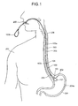
FIG. 1 is a schematic view for describing an example of applying an in-vivo information acquiring apparatus to an intragastric bleeding-existence detector and an initial state of an insertion thereof into a body cavity; -
FIG. 2 is a schematic view for describing a condition of a pulling/loosening operation and a clip fixing of a string member; -
FIG. 3 is a schematic view for describing a condition of a cutting by a scissor forceps; -
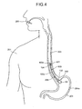
FIG. 4 is a schematic view for describing a final insertion condition of the string member and a capsule endoscope; -
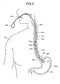
FIG. 5 is a schematic view for describing a condition of the cutting by the scissor forceps after a monitoring is finished; -
FIG. 6 is a schematic view for describing a condition of a retrieving by a retrieving net; -
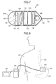
FIG. 7 is a sectional view for describing an internal configuration of the capsule endoscope; -
FIG. 8 is a schematic view for describing an example of a general configuration of a wireless intragastric bleeding-existence detecting system; -
FIG. 9 is a schematic view for describing a condition of providing a plurality of fixing portions; -
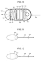
FIG. 10 is a sectional view for describing an internal configuration of a capsule endoscope in a type with a retaining member; -
FIG. 11 is a schematic sectional view for describing an example of using a hemoglobin sensor as a sensor; -
FIG. 12 is a schematic sectional view for describing an example of using a red detection sensor as the sensor; and -
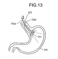
FIG. 13 is a schematic view for describing a condition of an adjustment when a plurality of the string members is used. -
- 100
- Intragastric bleeding-existence detector
- 101
- Capsule endoscope
- 102
- End portion
- 103
- String member
- 103a
- String-member main body
- 103b
- Extracorporeal gripper
- 103c
- Marked portion
- 103d
- Cutting portion
- 104
- Fixing portion
- 111
- Reel portion
- 200
- Oral cavity
- 201
- Subject
- 202
- Gastric cardia
- 203
- Esophageal region
- 204
- Stomach
- 205
- Lower esophageal sphincter
- 206
- Upper esophageal sphincter
- 301
- Receiving device
- 302
- Display device
- 501
- Retaining member
- 502
- Capsule endoscope
- 601
- Hemoglobin sensor
- 611
- Red detection sensor
- Exemplary embodiments of an in-vivo information acquiring apparatus, an in-vivo information acquiring system, and an in-vivo information acquiring method of the present invention will be described below with reference to the accompanying drawings. The present invention is not limited to the below embodiments. In a description of the drawings, same reference numerals are assigned to same components.
- An embodiment of the present invention is described.
FIG. 1 is a schematic view for describing an example of applying the in-vivo information acquiring apparatus to an intragastric bleeding-existence detector and an initial state of an insertion thereof into a body cavity. An intragastric bleeding-existence detector 100 includes
acapsule endoscope 101, as an in-vivo information acquiring apparatus main-body, to be swallowed from anoral cavity 200 and inserted into a body cavity of a subject 201 to capture a body cavity image as body cavity information, and to wirelessly output data transmission of the captured body cavity image for a transmission, astring member 103 that is connected to anend portion 102 of thecapsule endoscope 101 and locates thecapsule endoscope 101 swallowed inside the body cavity at agastric cardia 202 of the subject 201, and a fixingportion 104 that is provided on a portion of thestring member 103 for fixing thestring member 103 that has located thecapsule endoscope 202 at thegastric cardiac 202 to body tissue (esophageal inner wall) inside anesophageal region 203 by an endoscopic fixture such as a clip described later. - The
string member 103 connected to theend portion 102 of thecapsule endoscope 101 includes a string-membermain body 103a having an adequate length for extracting thereof from astomach 204 to an outside via theoral cavity 200. In other words, thestring member 103 includes anextracorporeal gripper 103b at an extended end portion of the string-membermain body 103a on an extracorporeal side from theoral cavity 200, in such an intragastric insertion state that thecapsule endoscope 101 is orally inserted by the subject 201 and located inside the stomach. It is preferable to make thestring member 103 as thin as possible as long as it can easily be cut by a scissor forceps and the like described later and is strong. - The fixing
portion 104 is formed in a pinhole shape as a simple latch shape to latch the clip described later in the middle of the string-membermain body 103a, and also used as amarked portion 103c that is located inside theesophageal region 203 for enabling to visually distinguish the fixingportion 104 from the string-membermain body 103a, in the intragastric insertion state in which thecapsule endoscope 101 is located inside the stomach. The fixingportion 104 is provided on a position on which a length from thecapsule endoscope 101 to which thestring member 103 is connected corresponds to a length from thegastric cardiac 202 to theesophageal region 203 of the subject 201. More specifically, the fixingportion 104 is provided on the position to be fixed to the body tissue inside theesophageal region 203 by the clip, between a loweresophageal sphincter 205 and an upperesophageal sphincter 206. - The
capsule endoscope 101 is configured to be thicker than the thickness of the string-membermain body 103a and swallowable from theoral cavity 200, and is formed in a tapered reduced diameter shape with which a diameter becomes smaller toward theend portion 102 on the side of theend portion 102 to be connected to thestring member 103. The reduced diameter shape is not limited to the tapered shape and a semispherical-dome shape similar to a front end portion can be acceptable. - The
capsule endoscope 101 is described with reference toFIG. 7. FIG. 7 is a sectional view for describing an internal configuration of thecapsule endoscope 101. Thecapsule endoscope 101 is configured to accommodate acapsule case 124, a plurality of illuminatingunits 121 such as a light emitting diode (LED) for illuminating a region inside the body cavity of the subject 201, animaging element 122 such as a charge coupled device (CCD) or a complementary metal-oxide-semiconductor (CMOS) that captures the body cavity image, and abutton type battery 123 that supplies electric power to the illuminatingunit 121 and theimaging element 122. Thebattery 123 can be a silver oxide battery, a rechargeable battery, a generator battery and the like. - The
capsule case 124 includes a transparent semispherical-dome shaped front-end cover case 124a that covers the illuminatingunit 121 and a cylindrical-shapedbody case 124b that is watertightly provided with the front-end cover case 124a and accommodates thebattery 123, and is formed in a size swallowable from theoral cavity 200 of the subject 201. Thebody case 124b is made of a color material that is optically impermeable. - The
imaging element 122 is implemented on animaging board 125 and anoptical system 126 such as an imaging lens is provided on a front surface of theimaging element 122. Theimaging board 125 implements acontroller 127 that processes or controls each of units, on a rear surface thereof. - The
capsule endoscope 101 further accommodates areed switch 128 that changes an ON/OFF state by an external magnetic filed, for controlling a drive of thecapsule endoscope 101. The configuration is such that, thecapsule endoscope 101 is installed in a package including a permanent magnet that supplies the external magnetic field in a storage condition, and the OFF sate is kept under a circumstance in which a magnetic field with a specific strength or above is provided, while it is to be the ON state when the strength of the external magnetic field decreases. Accordingly, when installed in the package, thecapsule endoscope 101 is not to be activated. - The
capsule endoscope 101 includes a transmittingdevice 130 with anantenna 129 attached on the rear side, which wirelessly outputs image information captured by the imaging element of thebattery 123 to an outside.
According to the present embodiment, an image inside thestomach 204 is optically captured as a color image by theimaging element 122 for serving to detect an existence of a bleeding site inside thestomach 204. - The intragastric bleeding-
existence detector 100 including the above describedcapsule endoscope 101 structures an intragastric bleeding-existence detecting system as a medical system when combined with a receiving device and the like.FIG. 8 is a schematic view for describing an example of a general configuration of a wireless intragastric bleeding-existence detecting system. As shown inFIG. 8 , the wireless intragastric bleeding-existence detecting system includes thecapsule endoscope 101 that is to be inserted inside the subject 201 to capture the color image of the inside of thestomach 204 at a position of thegastric cardia 202 and to perform a data transmission of a picture signal by radio to areceiving device 301, theportable receiving device 301 that receives color image data wirelessly transmitted from thecapsule endoscope 101, and adisplay device 302 such as a portable viewer that displays the color image based on the picture signal received by the receivingdevice 301. The receivingdevice 301 includes a receivingantenna 303 attached on the extracorporeal surface of the subject 201 around, for example, thegastric cardia 202. - A medical operation procedure including a placement of the
capsule endoscope 101 inside the body cavity is sequentially described with reference toFIG.1 to FIG. 6 . Thecapsule endoscope 101 is swallowed and placed inside the body cavity for monitoring the existence of the bleeding site inside thestomach 204, after an endoscopic surgical procedure for the inside of thestomach 204 of thetarget subject 201 is finished. InFIG. 1 and other drawings,reference numeral 204a describes a surgical site of the endoscopic surgical procedure. The receivingantenna 303 is attached to the extracorporeal surface of the subject 201 at a proper timing before or after thecapsule endoscope 101 is swallowed. - Firstly, as shown in
FIG. 1 , thecapsule endoscope 101 to which thestring member 103 is connected is swallowed from theoral cavity 200 and inserted until thecapsule endoscope 101 is hung down inside thestomach 204 via theesophageal region 203 and thegastric cardia 202. At this state, theextracorporeal gripper 103b on the front end side of thestring member 103 is positioned at the extracorporeal side from theoral cavity 200 for enabling a pulling/loosening operation outside theoral cavity 200. - Next, as shown in
FIG. 2 , by shifting thecapsule endoscope 101 swallowed inside thestomach 204 up and down if necessary through the pulling/loosening operation of theextracorporeal gripper 103b of thestring member 103 as an extraoral operation, thecapsule endoscope 101 is located at the position of thegastric cardia 202. At this state, theend portion 102 on the side of thestring member 103 of thecapsule endoscope 101 is in tapered diameter-reduced portion and fits to a shape of a cardia that joins thestomach 204 and theesophageal region 103. Therefore, it is easy to position thecapsule endoscope 101 to thegastric cardia 202 in such a state that the front-end cover case 124a becomes downward by pulling thestring member 103, and thecapsule endoscope 101 can easily be in a stable position. Thegastric cardia 202 is a site from which the entire inside of thestomach 204 is easily inspected, and by locating thecapsule endoscope 101 downwardly at thegastric cardia 202, it becomes possible to properly monitor the existence of the bleeding site inside thestomach 204 including thesurgical site 204a. Further, because thecapsule endoscope 101 is thicker than thestring member 103, unless excessive pulling force is exerted, thecapsule endoscope 101 is not extracted to the side of the esophageal region. - It is acceptable to perform an up/down movement of the
capsule endoscope 101 not by the pulling/loosening operation of theextracorporeal gripper 103b but by using areel portion 111 provided in thecapsule endoscope 101. Thereel portion 111 is for reeling out and reeling up theextracorporeal gripper 103b and enables to shift thecapsule endoscope 101 up and down without shifting thestring member 103. More specifically, thereel portion 111 includes a not shown rotary member that reels out and reels up thestring member 103 and a not shown magnet that rotates the rotary member by an external rotary magnetic field generated outside the subject 201, and reels out and reels up thestring member 103 in conjunction with the rotation of the magnet. Alternately, thereel portion 111 can be connected to the rotary member, include a not shown driving unit that rotates the rotary member, and perform a drive control of the driving unit by receiving an external control signal including the magnetic field. In both cases, because thecapsule endoscope 101 by itself reels out and reels up thestring member 103 and thestring member 103 does not shift, it becomes possible to locate thecapsule endoscope 101 at thegastric cardia 202 without injuring a pharyngeal portion. - Before or after performing an operation of locating the
capsule endoscope 101 to thegastric cardia 202, anendoscope 402 that loads aclip 401 as the endoscopic fixture on a forceps channel is inserted inside theesophageal region 203 of the subject 201. Thereafter, with thecapsule endoscope 101 located at thegastric cardia 202, the extraoral operation for the forceps channel of theendoscope 402 is performed for performing a clipping operation to find the pinhole shapedmarked portion 103c and the fixingportion 104 for latching theclip 401 to fix the fixingportion 104 to the body tissue inside theesophageal region 203. Prior to the clipping, it is acceptable to check whether thecapsule endoscope 101 is located at thegastric cardia 202 in a desired posture, by monitoring a captured image of thecapsule endoscope 101 through the receivingdevice 301 and thedisplay device 302. - With the
capsule endoscope 101 located at thegastric cardia 202 through the extraoral operation, thestring member 103 is in a strained state and the like, and by fixing the fixingportion 104 as a portion of thestring member 103 to the inside of theesophageal region 203 by theclip 401, thecapsule endoscope 101 is stably placed at thegastric cardia 203. In other words, when thecapsule endoscope 101 is to be placed at thegastric cardia 202 from which the entire inside of thestomach 204 can be inspected, if it is directly fixed to the body tissue inside thestomach 104 by the endoscopic fixture, there is such a difficulty in an operation that the scope needs to be turned back. However, according to the present embodiment, although thecapsule endoscope 101 itself is to be placed at thegastric cardia 202, the fixing is to be performed by using the fixingportion 104 of thestring member 103 inside theesophageal region 203, which is easy. Further, since the fixingportion 104 is located to be fixed inside theesophageal region 203 other than such a movable site as the loweresophageal sphincter 205 and the upperesophageal sphincter 206, the posture of thecapsule endoscope 101 after the fixing is less moved. Moreover, theendoscope 402 itself is basically required to be inserted into theesophageal region 203, inclusive of an operation described later, it is possible to reduce a burden of the subject 201 as much as possible. - Next, as shown in
FIG. 3 , ascissor forceps 403 is inserted into the forceps channel of theendoscope 402 to cut thestring member 103 at the upper portion of the fixingportion 104 fixed by theclip 301. In other words, thestring member 103 according to the present embodiment includes a cuttingportion 103d that is a portion on the side of theextracorporeal gripper 103b from the fixing portion 104 (themarked portion 103c) on the string-membermain body 103a and that enables to cut the string-membermain body 103a in the portion. The upper side of thestring member 103 cut by the cuttingportion 103d is removed to the outside of the body. Thereafter, as shown inFIG. 4 , theendoscope 402 is also removed to the outside of the oral cavity. Accordingly, the intragastric bleeding-existence detector 100 becomes in a final condition with which the monitoring becomes enable by thecapsule endoscope 101 placed at thegastric cardia 202 and thestring member 103 fixed inside theesophageal region 203 by the fixingportion 104. Therefore, during a monitoring period after a surgery, theswallowable capsule endoscope 101 and thestring member 103 are exclusively inserted in the body cavity of the subject 201, and there is less pain for the subject 201. If there is no harm in leaving thestring member 103 inside theesophageal region 203 and theoral cavity 200, the operation of cutting thestring member 103 can be omitted. - At this state, by enabling the intragastric bleeding-existence detecting system shown in
FIG. 8 , the existence of the bleeding site inside thestomach 204 after the surgery can be properly monitored. In other words, by capturing an image of the inside of thestomach 204 from the position of thegastric cardia 202 by theimaging element 122 at a proper cycle, by wirelessly outputting the captured color image for a transmission to the receivingantenna 303 via thecontroller 127, the transmittingdevice 130, and the transmittingantenna 129, and by outputting the color image received by the receivingantenna 303 for a display on thedisplay device 302 by the receivingdevice 301, the existence of the bleeding site inside thestomach 204 after the surgery can be properly monitored. - If the monitoring of the existence of the bleeding site inside the
stomach 204 after the surgery is finished, as shown inFIG. 5 , theendoscope 402 is again inserted into theesophageal region 203 of the subject 201 to cut thestring member 103 at a position below the fixingportion 104 by operating thescissor forceps 403 of the forceps channel, and thecut string member 103 is poured into thestomach 204 together with thecapsule endoscope 101. - Further, the forceps channel of the
endoscope 402 is replaced with a retrieving net 404 and, as shown inFIG. 6 , the retrieving net 404 is inserted into thestomach 204 to scoop thecapsule endoscope 101 and thestring member 103 inside thestomach 204 and removed together with theendoscope 402 to the outside of the body, which ends a series of the operations. The fixingportion 104 fixed by theclip 401 is put into thestomach 204 by being dropped down along with a necrosis of the body tissue of the portion of theclip 401, and excreted to the outside of the body. If thestring member 103 is made of a material to be digested by strongly acidic gastric fluid, thecut string member 103 is digested inside thestomach 204, and the remainedcapsule endoscope 101 is to be excreted to the outside of the body through a small intestine and a large intestine, the retrieving operation by the retrieving net 404 can be omitted. - As shown in
FIG. 9 , it is acceptable to provide fixingportions string member 103. Accordingly, when the fixing portion is fixed inside theesophageal region 203 by theclip 401, a portion at which the clipping is easily performed can be selected from among a plurality of the fixingportions - According to the present embodiment, the
capsule endoscope 101 to which thestring member 103 is directly connected is used. However, as shown inFIG. 10 , such acapsule endoscope 502 can be used that is engaged and retained through a press fitting by a retainingmember 501 to which thestring member 103 is connected. In this case, the all-purpose capsule endoscope 502 that is mass produced as a capsule endoscope for a purpose of observing the small intestine and the like can be used and general versatility can be improved. - Further, according to the present embodiment, the example in which the
capsule endoscope 101 is used as the in-vivo information acquiring apparatus that monitors the inside of the stomach is described. However, ahemoglobin sensor 601 as shown inFIG. 11 or ared detection sensor 611 as shown inFIG. 12 can be used instead of thecapsule endoscope 101. Thehemoglobin sensor 601 detects the existence of the bleeding site by wirelessly performing a transmission output to the outside of a body through awireless device 602, when blood in a specific amount or above is adhered to an outer surface. Thered detection sensor 611 detects the existence of the bleeding site by wirelessly performing the transmission output to the outside of the body through awireless device 612, when responding to red such as blood. - Moreover, according to the present embodiment, the one
string member 103 is provided for the capsule endoscope as a configuration example. However, it is acceptable to connect a plurality of the string members at different connection points to make an angle adjustable.FIG. 13 describes an example in which such a binoculartype capsule endoscope 701 is used that includes two imaging elements that can observe both a front direction and a rear direction, and twostring members capsule endoscope 701. Accordingly, it is acceptable to make such a condition that the surgical site can be clearly viewed by determining a placement position of thecapsule endoscope 701 by, for example, the onestring member 702a, and thereafter, adjusting the angle of thecapsule endoscope 701 by pulling or loosening theother string member 702b. The above can also be applicable to a monocular type shown inFIG. 7 andFIG. 10 . - The present invention is not limited to the above described embodiments, and various modifications can be acceptable without departing a scope and a spirit of the present invention.
- As described above, the in-vivo information acquiring apparatus, the in-vivo information acquiring system, and the in-vivo information acquiring method according to the present invention are suitable for properly performing a monitoring of the inside of a body by using an easily operable placement technique, and more specifically suitable for a type using a capsule endoscope.
Claims (21)
- An in-vivo information acquiring apparatus comprising:an in-vivo information acquiring apparatus main-body that is swallowed from an oral cavity of a subject to acquire in-vivo information of the subject and to wirelessly output the in-vivo information for a transmission to an outside of a body;a string member that is connected to the in-vivo information acquiring apparatus main-body to locate the swallowed in-vivo information acquiring apparatus main-body at a gastric cardia of the subject; anda fixing portion that is provided on a portion of the sting member to fix the string member to an esophageal region by an endoscopic fixture, the string member having located the in-vivo information acquiring apparatus main-body at the gastric cardia.
- An in-vivo information acquiring apparatus comprising:an in-vivo information acquiring apparatus main-body that acquires in-vivo information; anda string member extended from the in-vivo information acquiring apparatus main-body, wherein the string member includesan extracorporeal gripper that is provided to be located on an extracorporeal side from an oral cavity of a subject, in such an intragastric insertion state where the in-vivo information acquiring apparatus main-body is orally inserted into the subject and located inside a stomach,a marked portion that is located inside an esophageal region and indicates a fixing position of the sting member in the intragastric insertion state,a fixing portion that fixes the string member to an inner wall of the esophageal region, anda cutting portion that is provided at a position on the string member between the fixing portion or the marked portion and the extracorporeal gripper, and enables to cut the string member at the position.
- An in-vivo information acquiring apparatus comprising:an in-vivo information acquiring apparatus main-body that is swallowable by a subject; anda string member that includes a string member main-body extended from the in-vivo information acquiring apparatus main-body, an extracorporeal gripper that is provided on an extended end portion of the string member main-body, a fixing portion that is provided on a side of an extended rear anchor from the extracorporeal gripper on the string member main-body to enable to fix the string member main-body, a marked portion that enables to visually distinguish the fixing portion from the string member main-body, and a cutting portion that is provided at a position on the side of an extended end portion from the fixing portion on the string member main-body and enables to cut the string member main-body at the position.
- The in-vivo information acquiring apparatus according to any one of claims 1 to 3, wherein the fixing portion is located at a position where a length from the connected in-vivo information acquiring main-body corresponds to a length from the gastric cardia to the inside of the esophageal region of the subject.
- The in-vivo information acquiring apparatus according to any one of claims 1 to 3, wherein the in-vivo information acquiring apparatus main-body detects at least an existence of a bleeding inside the stomach of the subject as in-vivo information to be acquired.
- The in-vivo information acquiring apparatus according to any one of claims 1 to 3, wherein the in-vivo information acquiring apparatus main-body is a capsule endoscope.
- The in-vivo information acquiring apparatus according to claim 5, wherein the in-vivo information acquiring apparatus main-body is a hemoglobin sensor that detects the existence of the bleeding inside the stomach based on an adhesion of blood.
- The in-vivo information acquiring apparatus according to claim 5, wherein the in-vivo information acquiring apparatus main-body is a red detection sensor.
- The in-vivo information acquiring apparatus according to any one of claims 1 to 3, wherein the in-vivo information acquiring apparatus main-body is configured to be thicker than a thickness of the string member.
- The in-vivo information acquiring apparatus according to any one of claims 1 to 3, wherein
the string member is connected to an end portion of the in-vivo information acquiring apparatus main-body, and
the in-vivo information acquiring apparatus main-body has a reduced diameter shape with which a diameter of the in-vivo information acquiring apparatus main-body becomes smaller toward an end portion thereof to which the string member is connected. - The in-vivo information acquiring apparatus according to any one of claims 1 to 3, further comprising:a retaining member that engages and retains the in-vivo information acquiring apparatus main-body, whereinthe string member is connected to the in-vivo information acquiring apparatus main-body via the retaining member.
- The in-vivo information acquiring apparatus according to any one of claims 1 to 3, wherein the string member is made of a material to be digested by gastric fluid.
- The in-vivo information acquiring apparatus according to any one of claims 1 to 3, wherein the fixing portion is located at a position to be fixed to the esophageal region at an upper portion from a lower esophageal sphincter.
- The in-vivo information acquiring apparatus according to any one of claims 1 to 3, wherein the fixing portion is located at a position to be fixed to the esophageal region between a lower esophageal sphincter and an upper esophageal sphincter.
- The in-vivo information acquiring apparatus according to any one of claims 1 to 3, wherein the fixing portion is in a latch shape for latching the endoscopic fixture.
- The in-vivo information acquiring apparatus according to any one of claims 1 to 3, wherein the fixing portion is provided at a plurality of portions of the string member.
- The in-vivo information acquiring apparatus according to any one of claims 1 to 3, wherein the in-vivo information acquiring apparatus main-body includes a reel portion that reels out and reels off the string member.
- An in-vivo information acquiring system comprising:an in-vivo information acquiring apparatus according to any one of claims 1 to 3;a receiving device that receives in-vivo information wirelessly output and transmitted from an in-vivo information acquiring apparatus main-body; anda display device that displays the in-vivo information received by the receiving device.
- An in-vivo information acquiring method comprising the steps of:swallowing an in-vivo information acquiring apparatus main-body to make a state where the in-vivo information acquiring apparatus main-body travels through an esophageal region and a gastric cardia and is hung down inside a stomach, the in-vivo information acquiring apparatus main-body being connected to a string member to acquire in-vivo information of a subject and to output the in-vivo information for a transmission to an outside;locating the in-vivo information acquiring apparatus main-body at the gastric cardia by pulling and loosening the string member to shift the in-vivo information acquiring apparatus main-body up and down, the in-vivo information acquiring apparatus main-body being hung down inside the stomach;fixing the string member to the esophageal region by an endoscopic fixture;transmitting in-vivo information acquired from the in-vivo information acquiring apparatus main-body to an outside of a body wirelessly; andcutting a lower stomach side of the string member fixed at the fixing step.
- The in-vivo information acquiring method according to claim 19, further comprising the step of:cutting an upper oral-cavity side of the string member fixed at the fixing step to remove the cut upper oral-cavity side of the string member to an outside of a body.
- The in-vivo information acquiring method according to claim 19, further comprising the step of:retrieving the in-vivo information acquiring apparatus main-body that is cut at the cutting step and placed inside the stomach, to an outside of a body by a scooping.
Applications Claiming Priority (2)
| Application Number | Priority Date | Filing Date | Title |
|---|---|---|---|
| JP2005200885 | 2005-07-08 | ||
| PCT/JP2006/313703 WO2007007724A1 (en) | 2005-07-08 | 2006-07-10 | Device, system and method for acquiring information in living body |
Publications (2)
| Publication Number | Publication Date |
|---|---|
| EP1902664A1 true EP1902664A1 (en) | 2008-03-26 |
| EP1902664A4 EP1902664A4 (en) | 2012-06-20 |
Family
ID=37637113
Family Applications (1)
| Application Number | Title | Priority Date | Filing Date |
|---|---|---|---|
| EP06780925A Withdrawn EP1902664A4 (en) | 2005-07-08 | 2006-07-10 | Device, system and method for acquiring information in living body |
Country Status (5)
| Country | Link |
|---|---|
| US (1) | US8491464B2 (en) |
| EP (1) | EP1902664A4 (en) |
| JP (1) | JP4870670B2 (en) |
| CN (1) | CN101217909B (en) |
| WO (1) | WO2007007724A1 (en) |
Cited By (5)
| Publication number | Priority date | Publication date | Assignee | Title |
|---|---|---|---|---|
| EP2111148A4 (en) * | 2007-01-19 | 2011-12-21 | Sierra Scient Instr Inc | Micro-remote gastrointestinal physiological measurement device |
| EP2425761A1 (en) * | 2010-05-10 | 2012-03-07 | Olympus Medical Systems Corp. | Medical device |
| EP2571573A1 (en) * | 2010-03-17 | 2013-03-27 | Photopill Medical Ltd. | Capsule phototherapy |
| US10300296B2 (en) | 2010-03-17 | 2019-05-28 | Photopill Medical Ltd. | Capsule phototherapy |
| WO2020099997A1 (en) * | 2018-11-12 | 2020-05-22 | Zilinska Univerzita V Ziline | Swallow capsule endoscope |
Families Citing this family (25)
| Publication number | Priority date | Publication date | Assignee | Title |
|---|---|---|---|---|
| US7530948B2 (en) | 2005-02-28 | 2009-05-12 | University Of Washington | Tethered capsule endoscope for Barrett's Esophagus screening |
| DE102006000318A1 (en) * | 2006-07-03 | 2008-01-10 | Novineon Healthcare Technology Partners Gmbh | Device for bleeding detection |
| JP2008029527A (en) * | 2006-07-27 | 2008-02-14 | Olympus Medical Systems Corp | Endoscope system |
| US20080177141A1 (en) * | 2007-01-24 | 2008-07-24 | Hsien-Ming Wu | Memory-type two-section endoscopic system |
| WO2009032016A1 (en) * | 2007-09-07 | 2009-03-12 | University Of Washington | Monitoring disposition of tethered capsule endoscope in esophagus |
| JP5362636B2 (en) * | 2010-03-31 | 2013-12-11 | 富士フイルム株式会社 | Medical air supply system |
| EP3632308B1 (en) * | 2010-09-29 | 2023-12-06 | Dexcom, Inc. | Advanced continuous analyte monitoring system |
| US9155677B2 (en) * | 2011-08-09 | 2015-10-13 | Franklin R. Lacy | System for gastrointestinal and vascular atrophy engineering to restore normal youthful bodily functions |
| US9662018B2 (en) | 2012-03-30 | 2017-05-30 | Covidien Lp | Integrated self-fixating visualization devices, systems and methods |
| CN103222844B (en) * | 2013-04-25 | 2016-01-27 | 中国人民解放军成都军区总医院 | Controllable capsule endoscopy |
| US10264995B2 (en) * | 2013-12-04 | 2019-04-23 | Obalon Therapeutics, Inc. | Systems and methods for locating and/or characterizing intragastric devices |
| WO2015107848A1 (en) * | 2014-01-16 | 2015-07-23 | シャープ株式会社 | In-body monitoring camera system and auxiliary implement |
| CN103961048A (en) * | 2014-04-16 | 2014-08-06 | 姜泊 | Traction type capsule endoscopy |
| US9895248B2 (en) | 2014-10-09 | 2018-02-20 | Obalon Therapeutics, Inc. | Ultrasonic systems and methods for locating and/or characterizing intragastric devices |
| US10350100B2 (en) | 2016-04-12 | 2019-07-16 | Obalon Therapeutics, Inc. | System for detecting an intragastric balloon |
| TWI592128B (en) * | 2016-06-03 | 2017-07-21 | 群曜醫電股份有限公司 | Auxiliry device for endoscopy |
| TWI616180B (en) * | 2016-06-29 | 2018-03-01 | 國立成功大學 | Upper gastrointestinal bleeding monitoring system |
| WO2018237218A1 (en) | 2017-06-23 | 2018-12-27 | The Procter & Gamble Company | Composition and method for improving the appearance of skin |
| CN109833102A (en) * | 2017-11-29 | 2019-06-04 | 上海复拓知达医疗科技有限公司 | Position the marker and telltale mark system of the small space occupying lesion of intrapulmonary |
| US11622963B2 (en) | 2018-07-03 | 2023-04-11 | The Procter & Gamble Company | Method of treating a skin condition |
| CN110151110B (en) * | 2019-06-05 | 2024-04-23 | 上海长海医院 | Fixable capsule endoscope for monitoring gastrorrhagia and real-time monitoring system for gastrorrhagia |
| CN115843238A (en) | 2020-06-01 | 2023-03-24 | 宝洁公司 | Method for improving penetration of vitamin b3 compounds into the skin |
| US10959933B1 (en) | 2020-06-01 | 2021-03-30 | The Procter & Gamble Company | Low pH skin care composition and methods of using the same |
| CN112674719B (en) * | 2020-12-24 | 2022-06-07 | 珠海格力电器股份有限公司 | Sleep product |
| CN114699033A (en) * | 2022-05-13 | 2022-07-05 | 北京善行医疗科技有限公司 | Control device for controllable superfine endoscope |
Citations (5)
| Publication number | Priority date | Publication date | Assignee | Title |
|---|---|---|---|---|
| US3528429A (en) * | 1968-10-21 | 1970-09-15 | Charles B Beal | Method and device for orally admitting an elongated flexible element in the alimentary canal |
| US20030181788A1 (en) * | 2002-03-25 | 2003-09-25 | Olympus Optical Co., Ltd. | Capsule-type medical device |
| US20050043601A1 (en) * | 1999-04-07 | 2005-02-24 | Endonetics, Inc. | Implantable monitoring probe |
| WO2005032644A1 (en) * | 2003-10-01 | 2005-04-14 | Olympus Corporation | Capsule dosing system and dosing method |
| US20050085697A1 (en) * | 2003-09-30 | 2005-04-21 | Olympus Corporation | Gastrointestinal tract examining apparatus |
Family Cites Families (22)
| Publication number | Priority date | Publication date | Assignee | Title |
|---|---|---|---|---|
| US2773502A (en) * | 1953-12-21 | 1956-12-11 | Arthur L Kaslow | Device for treating alimentary tract |
| JPH04347138A (en) * | 1991-05-24 | 1992-12-02 | Olympus Optical Co Ltd | Medical capsule |
| JP3176653B2 (en) | 1991-07-19 | 2001-06-18 | オリンパス光学工業株式会社 | Medical capsule device |
| JPH0663051A (en) | 1992-08-20 | 1994-03-08 | Olympus Optical Co Ltd | Capsule device for medical treatment |
| JPH06114036A (en) | 1992-10-05 | 1994-04-26 | Olympus Optical Co Ltd | Capsule for medical treatment |
| JP3285235B2 (en) | 1992-11-05 | 2002-05-27 | オリンパス光学工業株式会社 | Capsule device for in vivo observation |
| JPH076142A (en) | 1993-04-20 | 1995-01-10 | Mitsubishi Electric Corp | Multiagent coordination system and its method |
| US5611787A (en) * | 1994-10-13 | 1997-03-18 | Methodist Hospital Of Indiana, Inc. | Method and device for gastric line insertion |
| US5738110A (en) * | 1996-05-29 | 1998-04-14 | Beal; Charles B. | Device for the diagnosis of certain gastrointestinal pathogens |
| US6285897B1 (en) | 1999-04-07 | 2001-09-04 | Endonetics, Inc. | Remote physiological monitoring system |
| GB2352636B (en) * | 1999-08-03 | 2003-05-14 | Univ College London Hospitals | Improved passage-travelling device |
| US6475145B1 (en) * | 2000-05-17 | 2002-11-05 | Baymar, Inc. | Method and apparatus for detection of acid reflux |
| US7160258B2 (en) * | 2001-06-26 | 2007-01-09 | Entrack, Inc. | Capsule and method for treating or diagnosing the intestinal tract |
| US7245954B2 (en) * | 2003-03-27 | 2007-07-17 | Given Imaging Ltd. | Measuring a gradient in-vivo |
| JP2004305593A (en) * | 2003-04-09 | 2004-11-04 | Olympus Corp | Insertion attachment for endoscope |
| EP1690490B1 (en) * | 2003-11-11 | 2012-04-18 | Olympus Corporation | Capsule type medical device system |
| WO2005053517A1 (en) * | 2003-12-01 | 2005-06-16 | Olympus Corporation | Endoscope system |
| CN2706123Y (en) * | 2004-02-28 | 2005-06-29 | 重庆金山科技(集团)有限公司 | Medical radio capsule type endoscope system |
| JP4347138B2 (en) | 2004-05-28 | 2009-10-21 | 三菱電機株式会社 | Access control device |
| TW200630066A (en) * | 2005-02-23 | 2006-09-01 | Chung Shan Inst Of Science | Disposable two-stage endoscope |
| US7530948B2 (en) * | 2005-02-28 | 2009-05-12 | University Of Washington | Tethered capsule endoscope for Barrett's Esophagus screening |
| US7580751B2 (en) * | 2005-04-29 | 2009-08-25 | Medtronic, Inc. | Intra-luminal device for gastrointestinal stimulation |
-
2006
- 2006-07-10 US US11/571,510 patent/US8491464B2/en active Active
- 2006-07-10 CN CN2006800249501A patent/CN101217909B/en not_active Expired - Fee Related
- 2006-07-10 JP JP2007524642A patent/JP4870670B2/en not_active Expired - Fee Related
- 2006-07-10 WO PCT/JP2006/313703 patent/WO2007007724A1/en active Application Filing
- 2006-07-10 EP EP06780925A patent/EP1902664A4/en not_active Withdrawn
Patent Citations (5)
| Publication number | Priority date | Publication date | Assignee | Title |
|---|---|---|---|---|
| US3528429A (en) * | 1968-10-21 | 1970-09-15 | Charles B Beal | Method and device for orally admitting an elongated flexible element in the alimentary canal |
| US20050043601A1 (en) * | 1999-04-07 | 2005-02-24 | Endonetics, Inc. | Implantable monitoring probe |
| US20030181788A1 (en) * | 2002-03-25 | 2003-09-25 | Olympus Optical Co., Ltd. | Capsule-type medical device |
| US20050085697A1 (en) * | 2003-09-30 | 2005-04-21 | Olympus Corporation | Gastrointestinal tract examining apparatus |
| WO2005032644A1 (en) * | 2003-10-01 | 2005-04-14 | Olympus Corporation | Capsule dosing system and dosing method |
Non-Patent Citations (1)
| Title |
|---|
| See also references of WO2007007724A1 * |
Cited By (8)
| Publication number | Priority date | Publication date | Assignee | Title |
|---|---|---|---|---|
| EP2111148A4 (en) * | 2007-01-19 | 2011-12-21 | Sierra Scient Instr Inc | Micro-remote gastrointestinal physiological measurement device |
| EP2571573A1 (en) * | 2010-03-17 | 2013-03-27 | Photopill Medical Ltd. | Capsule phototherapy |
| EP2571573A4 (en) * | 2010-03-17 | 2013-12-04 | Photopill Medical Ltd | Capsule phototherapy |
| US10300296B2 (en) | 2010-03-17 | 2019-05-28 | Photopill Medical Ltd. | Capsule phototherapy |
| EP2425761A1 (en) * | 2010-05-10 | 2012-03-07 | Olympus Medical Systems Corp. | Medical device |
| EP2425761A4 (en) * | 2010-05-10 | 2012-03-28 | Olympus Medical Systems Corp | Medical device |
| US8355043B2 (en) | 2010-05-10 | 2013-01-15 | Olympus Medical Systems Corp. | Medical apparatus |
| WO2020099997A1 (en) * | 2018-11-12 | 2020-05-22 | Zilinska Univerzita V Ziline | Swallow capsule endoscope |
Also Published As
| Publication number | Publication date |
|---|---|
| JP4870670B2 (en) | 2012-02-08 |
| EP1902664A4 (en) | 2012-06-20 |
| US20090076325A1 (en) | 2009-03-19 |
| CN101217909A (en) | 2008-07-09 |
| JPWO2007007724A1 (en) | 2009-01-29 |
| CN101217909B (en) | 2010-05-19 |
| WO2007007724A1 (en) | 2007-01-18 |
| US8491464B2 (en) | 2013-07-23 |
Similar Documents
| Publication | Publication Date | Title |
|---|---|---|
| US8491464B2 (en) | In-vivo information acquiring apparatus, in-vivo information acquiring system, and in-vivo information acquiring method | |
| US8269823B2 (en) | In vivo imaging device, display device, imaging and displaying system and intra-subject indwelling system using the same | |
| JP4971209B2 (en) | Medical equipment | |
| JP4578740B2 (en) | Capsule medical device | |
| JP3793368B2 (en) | Swallowing endoscope device | |
| EP1796529B1 (en) | Capsule type medical device | |
| JP2004523254A (en) | Immobilizable in-vivo detection device | |
| JP2010012222A (en) | Medical apparatus | |
| JP4734051B2 (en) | Capsule type medical device indwelling device and capsule endoscope in vivo indwelling device | |
| JP2010035825A (en) | Medical apparatus | |
| JP2007167214A (en) | Apparatus and system for photographing in vivo image | |
| US9241614B2 (en) | Tools for use in esophagus | |
| JP4800692B2 (en) | Capsule type medical device indwelling device | |
| JP5945882B2 (en) | Marking capsule and capsule endoscope system | |
| US20060173361A1 (en) | Endoscopy capsule with site marking capability and application of the same | |
| WO2012165299A1 (en) | Receiving device and capsule-type endoscope system | |
| US20120016198A1 (en) | Medical apparatus | |
| EP2446804A1 (en) | Medical device | |
| JP6261953B2 (en) | Endoscope device | |
| JP4934086B2 (en) | Medical equipment | |
| JP2006239439A (en) | Capsule type endoscope | |
| JP2005073887A (en) | Radio type intra-examinee-body information acquisition device | |
| CN213345599U (en) | PH capsule operating means and detecting instrument | |
| KR20090102308A (en) | Device, system and method for acquiring information in living body | |
| JP3884454B2 (en) | Capsule medical device |
Legal Events
| Date | Code | Title | Description |
|---|---|---|---|
| PUAI | Public reference made under article 153(3) epc to a published international application that has entered the european phase |
Free format text: ORIGINAL CODE: 0009012 |
|
| 17P | Request for examination filed |
Effective date: 20071228 |
|
| AK | Designated contracting states |
Kind code of ref document: A1 Designated state(s): DE FR GB |
|
| DAX | Request for extension of the european patent (deleted) | ||
| RBV | Designated contracting states (corrected) |
Designated state(s): DE FR GB |
|
| REG | Reference to a national code |
Ref country code: HK Ref legal event code: DE Ref document number: 1113994 Country of ref document: HK |
|
| A4 | Supplementary search report drawn up and despatched |
Effective date: 20120518 |
|
| RIC1 | Information provided on ipc code assigned before grant |
Ipc: A61B 1/00 20060101AFI20120511BHEP Ipc: A61B 5/07 20060101ALI20120511BHEP |
|
| DAX | Request for extension of the european patent (deleted) | ||
| 17Q | First examination report despatched |
Effective date: 20130507 |
|
| RAP1 | Party data changed (applicant data changed or rights of an application transferred) |
Owner name: OLYMPUS CORPORATION |
|
| REG | Reference to a national code |
Ref country code: HK Ref legal event code: WD Ref document number: 1113994 Country of ref document: HK |
|
| RAP1 | Party data changed (applicant data changed or rights of an application transferred) |
Owner name: OLYMPUS CORPORATION |
|
| RAP1 | Party data changed (applicant data changed or rights of an application transferred) |
Owner name: OLYMPUS CORPORATION |
|
| RAP1 | Party data changed (applicant data changed or rights of an application transferred) |
Owner name: OLYMPUS CORPORATION |
|
| RIN1 | Information on inventor provided before grant (corrected) |
Inventor name: FUJITA, MANABU Inventor name: UCHIYAMA, AKIO Inventor name: YOKOI, TAKESHI Inventor name: TAKIZAWA, HIRONOBU Inventor name: HIRAKAWA, KATSUMI Inventor name: TANAKA, SHINSUKE |
|
| GRAP | Despatch of communication of intention to grant a patent |
Free format text: ORIGINAL CODE: EPIDOSNIGR1 |
|
| INTG | Intention to grant announced |
Effective date: 20171221 |
|
| STAA | Information on the status of an ep patent application or granted ep patent |
Free format text: STATUS: THE APPLICATION IS DEEMED TO BE WITHDRAWN |
|
| 18D | Application deemed to be withdrawn |
Effective date: 20180501 |