EP1811017A1 - Cell cultivating device, image processing device and cell detecting system - Google Patents
Cell cultivating device, image processing device and cell detecting system Download PDFInfo
- Publication number
- EP1811017A1 EP1811017A1 EP05806165A EP05806165A EP1811017A1 EP 1811017 A1 EP1811017 A1 EP 1811017A1 EP 05806165 A EP05806165 A EP 05806165A EP 05806165 A EP05806165 A EP 05806165A EP 1811017 A1 EP1811017 A1 EP 1811017A1
- Authority
- EP
- European Patent Office
- Prior art keywords
- image
- focal point
- cells
- images
- incubator
- Prior art date
- Legal status (The legal status is an assumption and is not a legal conclusion. Google has not performed a legal analysis and makes no representation as to the accuracy of the status listed.)
- Granted
Links
Images


Classifications
-
- G—PHYSICS
- G02—OPTICS
- G02B—OPTICAL ELEMENTS, SYSTEMS OR APPARATUS
- G02B21/00—Microscopes
- G02B21/36—Microscopes arranged for photographic purposes or projection purposes or digital imaging or video purposes including associated control and data processing arrangements
- G02B21/365—Control or image processing arrangements for digital or video microscopes
- G02B21/367—Control or image processing arrangements for digital or video microscopes providing an output produced by processing a plurality of individual source images, e.g. image tiling, montage, composite images, depth sectioning, image comparison
-
- G—PHYSICS
- G06—COMPUTING; CALCULATING OR COUNTING
- G06T—IMAGE DATA PROCESSING OR GENERATION, IN GENERAL
- G06T5/00—Image enhancement or restoration
- G06T5/50—Image enhancement or restoration by the use of more than one image, e.g. averaging, subtraction
-
- G—PHYSICS
- G06—COMPUTING; CALCULATING OR COUNTING
- G06T—IMAGE DATA PROCESSING OR GENERATION, IN GENERAL
- G06T7/00—Image analysis
- G06T7/10—Segmentation; Edge detection
- G06T7/11—Region-based segmentation
-
- G—PHYSICS
- G06—COMPUTING; CALCULATING OR COUNTING
- G06T—IMAGE DATA PROCESSING OR GENERATION, IN GENERAL
- G06T7/00—Image analysis
- G06T7/10—Segmentation; Edge detection
- G06T7/174—Segmentation; Edge detection involving the use of two or more images
-
- G—PHYSICS
- G06—COMPUTING; CALCULATING OR COUNTING
- G06V—IMAGE OR VIDEO RECOGNITION OR UNDERSTANDING
- G06V20/00—Scenes; Scene-specific elements
- G06V20/60—Type of objects
- G06V20/69—Microscopic objects, e.g. biological cells or cellular parts
- G06V20/695—Preprocessing, e.g. image segmentation
-
- C—CHEMISTRY; METALLURGY
- C12—BIOCHEMISTRY; BEER; SPIRITS; WINE; VINEGAR; MICROBIOLOGY; ENZYMOLOGY; MUTATION OR GENETIC ENGINEERING
- C12M—APPARATUS FOR ENZYMOLOGY OR MICROBIOLOGY; APPARATUS FOR CULTURING MICROORGANISMS FOR PRODUCING BIOMASS, FOR GROWING CELLS OR FOR OBTAINING FERMENTATION OR METABOLIC PRODUCTS, i.e. BIOREACTORS OR FERMENTERS
- C12M41/00—Means for regulation, monitoring, measurement or control, e.g. flow regulation
- C12M41/30—Means for regulation, monitoring, measurement or control, e.g. flow regulation of concentration
- C12M41/36—Means for regulation, monitoring, measurement or control, e.g. flow regulation of concentration of biomass, e.g. colony counters or by turbidity measurements
-
- G—PHYSICS
- G06—COMPUTING; CALCULATING OR COUNTING
- G06T—IMAGE DATA PROCESSING OR GENERATION, IN GENERAL
- G06T2207/00—Indexing scheme for image analysis or image enhancement
- G06T2207/10—Image acquisition modality
- G06T2207/10056—Microscopic image
-
- G—PHYSICS
- G06—COMPUTING; CALCULATING OR COUNTING
- G06T—IMAGE DATA PROCESSING OR GENERATION, IN GENERAL
- G06T2207/00—Indexing scheme for image analysis or image enhancement
- G06T2207/10—Image acquisition modality
- G06T2207/10141—Special mode during image acquisition
- G06T2207/10148—Varying focus
-
- G—PHYSICS
- G06—COMPUTING; CALCULATING OR COUNTING
- G06T—IMAGE DATA PROCESSING OR GENERATION, IN GENERAL
- G06T2207/00—Indexing scheme for image analysis or image enhancement
- G06T2207/20—Special algorithmic details
- G06T2207/20212—Image combination
- G06T2207/20224—Image subtraction
-
- G—PHYSICS
- G06—COMPUTING; CALCULATING OR COUNTING
- G06T—IMAGE DATA PROCESSING OR GENERATION, IN GENERAL
- G06T2207/00—Indexing scheme for image analysis or image enhancement
- G06T2207/30—Subject of image; Context of image processing
- G06T2207/30004—Biomedical image processing
- G06T2207/30024—Cell structures in vitro; Tissue sections in vitro
-
- Y—GENERAL TAGGING OF NEW TECHNOLOGICAL DEVELOPMENTS; GENERAL TAGGING OF CROSS-SECTIONAL TECHNOLOGIES SPANNING OVER SEVERAL SECTIONS OF THE IPC; TECHNICAL SUBJECTS COVERED BY FORMER USPC CROSS-REFERENCE ART COLLECTIONS [XRACs] AND DIGESTS
- Y10—TECHNICAL SUBJECTS COVERED BY FORMER USPC
- Y10S—TECHNICAL SUBJECTS COVERED BY FORMER USPC CROSS-REFERENCE ART COLLECTIONS [XRACs] AND DIGESTS
- Y10S128/00—Surgery
- Y10S128/92—Computer assisted medical diagnostics
- Y10S128/922—Computer assisted medical diagnostics including image analysis
Definitions
- the present invention relates to a cell culture device, image processing device and cell detecting system capable of extracting a cell portion from a plurality of image data obtained during the cell culture process.
- Fig. 4 is a flow chart showing an example of a cell-extracting process implemented by an image-processing unit. Steps for cell-extracting process shown in Fig. 4 will be described below.
- step S44 with respect to the two images selected in steps S42 ⁇ S43, differential/positioning processing for coordinating the position of the cells in both images is carried out by setting a small image area of 256 pixels X 256 pixels in the central portion of the images, and by calculating the difference between the small area of one image and the small area of the other image through shifting the images by 1 pixel each in X and Y directions.
- the reason for including the step for positioning of images in the embodiment is to take into consideration the possibility of some amount of displacement in the respective image positioning on the screen, though it depends somewhat on the degree of assembly accuracy of the device. Therefore, in a device free of displacement of image positioning while moving CCD camera 13, the step for image positioning can be omitted.
Abstract
Description
- The present invention relates to a cell culture device, image processing device and cell detecting system capable of extracting a cell portion from a plurality of image data obtained during the cell culture process.
- In the process of cell culture, image observation of a culture state of microscopic cells has been carried out in the past mainly in laboratories of organization such as universities and institutes, by taking out the petri dish in which the culture is placed from a thermostatic chamber and observing the cells using a microscope.
Also, in order to extract the cells from image data obtained during cell culture, the threshold value has been calculated based on distribution of pixel values, and pixels equal to the threshold value and above or below have been extracted as the cell. However, the method using the above-mentioned threshold processing had a problem that the image turned out whitish, whereby detecting as if more cells exist than the actual amount. Consequently, in order to stably extract the cells without being influenced by color variation of the medium, light volume variation of the light source, difference in brightness between the central and peripheral areas of the image and the noise in the image, it has been fundamental to carry out a filtering process such as denoising, smoothing filter or edge enhancement. - In
Patent Document 1, a method is described which monitors brightness variation in image data, generate a trigger signal at a specific threshold value, take images repeatedly with a camera, and obtains the added and averaged value of the images. - Patent Document 1:
JP-A-2000-275539 - Also, a technique for enabling observation of a motion state of individual cells by arranging a light source and a CCD camera facing each other placing an incubator therebetween, and imaging the cells in the incubator at an early stage of culture using the CCD camera is described in Non-patent
Document 1. - Non-patent Document 1: Journal of Bioscience and Bioengineering Vo. 94, no. 4,351 - 356.2002" Characterization of Cellular Motion Through Direct Observation of Individual Cells at Early Stage in Anchorage-Dependent Culture".
- In a conventional method for observing the cells in culture process by taking them out of the thermostatic chamber and observing them using a microscope, since the cells are exposed to the atmosphere in the laboratory, there has been a problem of having difficulty continuing the culture again after observing the cells with the microscope.
A conceivable means to solve the problem would be to incorporate the microscope into the incubator, but it would create another problem i.e. the increase in size and cost.
Also, a conventional method of carrying out the filtering process has been influencing the accuracy of cell-extraction through the filtering process to be executed before the cell-extraction process. The problems such as the difference in brightness between the central and peripheral areas or noise included in the image can be eliminated to some extent by this filtering process, and the technique described inPatent Document 1 can also have the effect of eliminating noise included in the image to some extent. However, depending on image data, there have been cases that the above-mentioned effectiveness was limited or the outline of the cells was indistinct.
Also, the technique described in the above-mentionednon-patent document 1 is for simply imaging the cells in the incubator, and the respective cells are individually identifiable at early stage in culturing, but it becomes harder to individually identify them as the culturing process progresses. - The present invention paid attention to the above-mentioned problems, and the obj ective is to provide a cell culture device capable of observing the progress of cell culture without taking the cells out of the device.
Also, the present invention is for providing a cell culture device, image processing device and cell detecting system capable of extracting the cells in images without being influenced by color variation of the medium, difference in brightness between the central and the peripheral areas of the image, and the noise. - A cell culture device related to the present invention is characterized in having a container capable of forming a space sealed off from the outer atmosphere, having arranged in the container an incubator for culturing cells, a light source for irradiating light to the cells in culture process in the incubator, and image acquiring device placed in the back of the incubator with respect to the light source and is for imaging the cells in the culture process, as well as comprising means for creating image data for extracting the cells by processing the acquired image using the image acquiring means.
Also, the present invention is characterized in comprising: - incubator means for culturing the cells;
- image acquiring means for acquiring images of the cells from the incubator means;
- focal position adjusting means for moving either the image acquiring means or the incubator means, and adjusting a focal point of the image acquiring means to be set in front and back of the cells in culture process in the incubator; and
- extracting means for extracting only the cell portions, with respect to the plurality of focal points set by the focal point position adjusting means, by subj ecting the plural pieces of image data each having a different focal point acquired by the image acquiring process to differential processing, and performing a binarization processing on the images subjected with differential processing.
- Furthermore, the cell culture device related to the present invention comprises focal point position calculating means for calculating the focal point of the image acquiring means in order to acquire the most suitable image for extracting only the cell part using the extracting means.
This means calculates the focal point position where the outline of the cells becomes clear or the position of image acquiring means with respect to the acquired image using methods such as profiling, Fourier transformation and differentiation. The image obtained at the calculated focal point becomes a suitable image for the cell extracting process. And by carrying out the positioning of image acquiring means to the calculated focal point position upon the next measurement time, the focus can be taken consistently with the same accuracy. - The cell culture device related to the present invention comprises position-compensating means for compensating the position of a plurality of image data acquired by the image acquiring means.
By this means the deterioration of accuracy for extracting the cells due to displacement of the plurality of images can be prevented, by compensating the displacement in X/Y-directions and rotation direction of images caused upon changing the focus. - An image-processing device relating to the present invention comprises:
- image acquiring means for acquiring images of the objects being scattered on a plane surface and having a predetermined range of thickness;
- focal point position adjusting means for adjusting a focal point of the image acquiring means to be positioned in front and back of thickness direction of the object; and
- extracting means for extracting only an object, in the plurality of focal point positions set by the focal point position adjusting means, by subjecting the plurality of image data having different focal point positions acquired by the image acquiring means to differential processing.
- A cell extracting system relating to the present invention comprises:
- image acquiring means for acquiring images of the cells scattered on a plane surface;
- focal point position adjusting means for adjusting the focal point of the image acquiring means to the upper and lower portion in the orthogonal direction of the plane surface on which the cells are scattered; and
- extracting means for extracting only the cell portions, in a plurality of focal point positions set by the focal point position adjusting means, by subjecting the plurality of image data having different focal point positions acquired by the image acquiring means to differential processing.
- According to the present invention, observation of the cells in culture process can be carried out without taking them out of the device, thus fungi other than culture target would not be mixed into the incubator.
Also, according to the present invention, it is possible to extract only the cell portions without being influenced by color variation of the medium, variation of light volume, difference in brightness between the central and peripheral areas of the image or noise. -
- Fig. 1 is a block diagram showing the configuration of a cell culture device, image processing device and cell extracting system relating to the present embodiment.
- Fig. 2 is a diagram showing a schematic view of the arrangement of the respective configuration means in the cell culture device.
- Fig. 3 is a diagram showing details of an image-processing unit in Fig. 1.
- Fig. 4 is a flow chart showing an example of a cell extracting process by which the image-processing unit executes.
- Fig. 5 is a diagram showing the relationship between shifting distance of a camera and summation of difference between adjacent pixel values.
- Fig. 6 is a diagram showing an example of an image when the focal point position of an objective lens is positioned on the bottom surface of an incubator.
- Fig. 7 is a diagram showing an example of an image when the focal point position of an objective lens is positioned in front (camera side) of the bottom surface of an incubator.
- Fig. 8 is a diagram showing an example of an image when the focal point position of an objective lens is positioned in back (light source side) of an incubator.
- Fig. 9 is a diagram showing an image when differential / positioning processing and binarization processing is performed on images of Figs. 7 and 8.
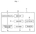
- 11... cell culture device, 12... incubator, 13... CCD camera, 14... image-processing unit, 15... converter, 16... camera/incubator drive unit, 17... motor controller, 18... light source, 21... shifting guide, 22... objective lens, 31... data bus, 32... CPU, 33... main memory, 34... external storage, 35... transmission port, 36... monitor, 37... keyboard
- Hereinafter, an embodiment of the present invention will be described based on the attached diagrams. Fig. 1 is a block diagram showing the configuration of a cell culture device, image-processing device and cell extracting system related to the present embodiment. As seen in the diagram,
cell culture device 11 has a box structure which seals off the inside thereof from outer space, and contains therein: -
incubator 12 for culturing cells;CCD camera 13 for imaging the cells inincubator 12; -
converter 15 for transferring image data obtained fromCCD camera 13 to image-processingunit 14; - camera/
incubator drive unit 16 for movingCCD camera 13 orincubator 12; -
motor controller 17 for shifting camera/incubator drive unit 16 to an arbitrary position; and -
light source 18 mounted on the upper part ofCCD camera 13.Incubator 12 has a transparent bottom surface, and CCD camera is configured to image transmitted light which is the light irradiated fromlight source 18 and transmitted through the bottom surface ofincubator 12. In the above configuration, it is desirable thatCCD camera 13 is provided with CCD device having about 400,000 pixels, and as forlight source 18 it is preferable to use an LED or miniature bulb that emits not parallel light but diffusion light. - Fig. 2 is a schematic view of the arrangement of the respective configuration means in
culture device 11, respectively illustrating the arrangement ofincubator 12, camera/incubator drive unit 16, shiftingguide 21 andobjective lens 22 for the case of movingCCD camera 13 inculture device 11. As shown in the diagram,light source 18 is mounted on the top surface inside ofculture device 11.Incubator 12 is placed underlight source 18, andCCD camera 13 comprisingobjective lens 22 is placed under and near the center ofincubator 12.CCD camera 13 carries out the imaging of the cells inincubator 12 by moving up and down according to the guidance of shiftingguide 21 based on drive control of camera/incubator drive unit 16 operated by the command outputted fromCPU 32, changing the focal point to upper and lower directions. In addition, it is desirable to configure an imaging device which is the combination ofCCD camera 13 andobjective lens 22 as capable of imaging minute areas, e.g. around 1.5mm X 2.0mm, in the vicinity of the bottom surface positioned near the center ofincubator 12. - Fig. 3 is a diagram showing details of image-processing
unit 14 in Fig. 1. Image-processingunit 14 comprises: -
CPU 32 for performing calculation process viadata bus 31; -
main memory 33 for whichCPU 32 temporarily uses as memory area; -
external storage 34 for storing image data or positional information; -
transmission port 35 for communicating withmotor controller 17; - monitor 36 for displaying images after extracting the cells; and
-
keyboard 37 for receiving the input by a user. - Fig. 4 is a flow chart showing an example of a cell-extracting process implemented by an image-processing unit. Steps for cell-extracting process shown in Fig. 4 will be described below.
- A sequence of steps described in Fig. 4 is repeated at a frequency of, e.g. one time/day at predetermined time intervals in the process of cell culture, and is carried out by the unit described in Figs. 1 ~ 3 being sequentially drive-controlled by the command outputted from the CPU. As for the timekeeper for repeatedly carrying out the sequence of steps described in Fig. 4 at predetermined time intervals, output of a clock generator provided in
CPU 32 can be used. - In step S41, images are acquired at predetermined pitch intervals, e.g. 27 µm intervals, while moving
CCD camera 13 in up and down directions according to the command outputted fromCPU 32 tomotor controller 17. The plurality of image data acquired by this image-acquiring operation is stored inexternal storage 34 viaconverter 15. - In this step S42, image-selecting process is executed after obtaining all of the images. In this image-selecting process, two or more images of which the periphery of the cells is clear are selected from the plurality of images stored in
external storage 34. In images having clear periphery of the cells, the changes in pixel values are greater compared to the images with unclear periphery of the cells. Given this factor, absolute values of the difference between adjacent pixel values is calculated, summation thereof is obtained and stored inmain memory 33. Fig. 5 is a diagram showing relationship between shifting distance of the camera and summation of the difference between adjacent pixel values. There are two peaks shown in Fig. 5, and images indicating these peak values turn out as images having clear periphery of the cells. Figs. 6 ~ 8 show the images at representative focal points of the camera in Fig. 5. Here, Fig. 6 is a diagram showing an example of an image in the case that the focal point position ofobjective lens 22 is positioned at the bottom surface ofincubator 12, Fig. 7 is a diagram showing an example of an image in the case that the focal point position ofobjective lens 22 is positioned at the front side (camera side) of the bottom surface ofincubator 12, and Fig. 8 is a diagram showing an example of an image in the case that the focal point position ofobjective lens 22 is positioned on the back side (light source side) of the bottom surface ofincubator 12. These images are obtained by setting positions of the focal point shifting by very slight distance, thus intensity difference of these images is about the same. As is clear by the diagrams, it became evident from experimentation by the inventor that images having clearer periphery of the cells can be obtained when the focal point position ofobjective lens 22 is shifted either to the front or back of the bottom surface ofincubator 12. - In step S43, j judgment whether more than two pieces of images comparable to the above-mentioned two peaks are selected or not, i.e. judgment of whether images having distinctive periphery of the cells shown in Figs. 7 and 8 are obtained or not is made, when the answer is yes the next step S44 is carried out, and when the answer is no the step returns to step S42. In this image-selecting step, contour definition of the cell image is executed by carrying out image processing using profiling process, Fourier transformation or differentiation with respect to the images stored in the
external storage 34. And the acquisition of the position ofCCD camera 13 when it obtained an image having clear periphery of the cells is included in this step. The position ofCCD camera 13 when it obtained the clear cell image is stored in themain memory 33 and is prepared for use upon the next cell-extracting process. - In step S44, with respect to the two images selected in steps S42 ~ S43, differential/positioning processing for coordinating the position of the cells in both images is carried out by setting a small image area of 256 pixels X 256 pixels in the central portion of the images, and by calculating the difference between the small area of one image and the small area of the other image through shifting the images by 1 pixel each in X and Y directions. The reason for including the step for positioning of images in the embodiment is to take into consideration the possibility of some amount of displacement in the respective image positioning on the screen, though it depends somewhat on the degree of assembly accuracy of the device. Therefore, in a device free of displacement of image positioning while moving
CCD camera 13, the step for image positioning can be omitted. - The next judgment is made based on the result of differential/positioning processing of the previous step S44. That is, when the image of the cells displayed on the small image region is tilted heavily in longitudinal direction, judgment is made on whether the number of cells is minimum or not. The reason for this is because when the cells are in heavily tilted condition, if positioning is not accurate the cells are displayed being reduplicated and are observed as if many cells exist. Also, when the image of the cells displayed on the small image region is tilted heavily in lateral direction, judgment is made on whether the total length of the cells is minimum or not. The reason for this is because when the cells are ranged sideways, if positioning is not accurate the cells are observed as wider than the actual arrangement. As a result of the judgment, when the answer is yes the differential/positioning processing is ended at the position thereof and the step proceeds to step S46, and when answer is no the step returns back to step S44. Differential/positioning processing will be continued until judgment of step S45 comes out as yes. Here, when positioning of the two images is carried out by completion of differential/positioning processing, the cells are extracted on the difference image at that point. Though the reason for this is uncertain, the inventor assumes that it results from the two images having different focal point positions by which the cells received light being irradiated from a point source such as LED lamp, having information with different scattering state of light on surface of the cells. Also, as described in step S42, while images devoted for difference process has 19% density difference as shown in Fig. 5, influence by color variation of the medium, variation of the light volume, difference in brightness between the central and peripheral areas of the image and noise can be practically eliminated by calculating the difference.
- In step S46, in order to facilitate analysis of information of the cells such as their length, number or shape, a binarization process of images is carried out. Fig. 9 is a diagram showing an image as a result of implementing a binarization process with respect to the difference images having the minimum number of the cells and the minimum summation of the length of the cells, which is a result of the differential/positioning processing with respect to the images in Figs. 7 and 8. In this way, by performing a binarization process on the difference images having the minimum number of the cells and the minimum summation of length of the cells thereof, individual cells are extracted clearly as white portions. The binarization process in step S46 is for facilitating data management thereafter by the computer, such as, e.g. counting the number of cells which will be described later, interval measurement between the cells and size measurement of the cells, and if these operations are not to be carried out step S46 can be omitted.
- Analysis of information such as length, number and shape of the cells can be easily implemented based on the image in Fig. 9. More specifically, since the white portions in the binarization image of Fig. 9 represent the cells, reproduction rate of the cells can be estimated by counting the pixel number, and reproduction rate of the cells can also be estimated by measuring width of the black portions. Furthermore, by measuring length of the cells, growth rate of the cells can be estimated.
- As described above, according to the present invention, since the cell culture device is configured comprising:
-
incubator 12 having a box structure which blocks off the inside thereof from outer space, and for culturing the cells therein; -
CCD camera 13 for imaging the cells inincubator 12; -
converter 15 for transferring image data obtained fromCCD camera 13 to image-processingunit 14; - camera/
incubator drive unit 16 for movingCCD camera 13 orincubator 12; -
motor controller 17 for shifting camera/incubator drive unit 16 to an arbitrary position; and -
light source 18 mounted on the upper part ofCCD camera 13, - reproduction ratio of the cells can be observed without taking the cells in culture process out of the device. Also, since image analysis of the cells is regularly and automatically executed by the software installed in advance, labor involved in monitoring the cell culture can be reduced.
- In the present invention, various changes may be made without departing from the scope of the invention. For example, in configuration of Fig. 1, images of the cells photographed by a camera or images on which the differential processing is performed can be displayed by connecting display for monitoring, e.g. liquid crystal display to image
unit 14. Furthermore, size (length or radius) of the cells or distribution density of the cells in an incubator can be measured, and the measurement results can be displayed on the display for monitoring.
Claims (12)
- A cell culture device provided with a storage container capable of forming a space sealed off from outer atmosphere, in which the following devices are disposed:an incubator for cell culturing;a light source for irradiating light to the cells in culture process in the incubator; andan image acquiring device for imaging the cells in culture process, placed in the back of the incubator with respect to the light source,as well as comprising means for processing images acquired by the image acquiring device and creating image data for extracting the cells.
- The cell culture device according to claim 1, wherein the image acquiring device is disposed as movable in light source direction, and acquires the cell images of each focal point positions.
- The cell culture device according to claim 2, wherein the image data creating means comprises:means for selecting at least two images having clear periphery of the cells from the images acquired by changing the focal point positions;means for adjusting positions of the selected images; andmeans for carrying out the differential processing on the aligned images.
- The cell culture device according to claim 3, wherein the image data creating means further comprises means for performing binarization processing on the image data implemented with differential processing.
- The cell culture device according to claim 3, characterized in that the positioning/differential processing is carried out on the region smaller than the acquired images.
- The cell culture device according to claim 1, comprising the means for automatically and repeatedly performing image acquisition, image analysis and creation of data for judging growth rate of the cells, at predetermined period of time intervals.
- The cell culture device according to claim 1, characterized in comprising a display unit for displaying image data based on the images acquired by the image acquiring means.
- A cell culture device comprising:incubator means for culturing cells;image acquiring means for acquiring images of the cells in the incubator means;focal point position adjusting means for moving either the image acquiring means or the incubator means, and adjusting the focal point of the image acquiring means to front and back of the cells in culture process in the incubator means; andextracting means for extracting only the cell portions, with respect to the plurality of focal point positions set by the focal point position adjusting means, by carrying out differential processing on the plurality of image data having different focal point positions being acquired by the image acquiring means.
- The cell culture device according to claim 8, characterized in comprising focal point position calculating means for calculating the focal point position of the image acquiring means for acquiring most suitable images for extracting only the cell portions by the extracting means.
- The cell culture device according to claim 8, characterized in comprising position compensating means for compensating position of the plurality of image data acquired by the image acquiring means.
- An image processing device comprising:image acquiring means for acquiring images of objects scattered on a plane surface and having a predetermined range of thickness;focal point position adjusting means for adjusting the focal point of the image acquiring means to be positioned in front and back in the thickness direction of the objects; andextracting means for extracting only the object, with respect to the plurality of focal point positions set by the focal point position adjusting means, by carrying out differential processing on the plurality of image data having different focal point positions being acquired by the image acquiring means.
- A cell detecting means comprising:image acquiring means for acquiring images of the cells distributed on a plane surface;focal point position adjusting means for adjusting the focal point of the image acquiring means to above and under the above-mentioned plane surface in longitudinal direction; andextracting means for extracting only the cell portions, with respect to the plurality of focal point positions set by the focal point position adjusting means, by carrying out differential processing on the plurality of image data having different focal point positions acquired by the image acquiring means.
Applications Claiming Priority (2)
| Application Number | Priority Date | Filing Date | Title |
|---|---|---|---|
| JP2004324456 | 2004-11-09 | ||
| PCT/JP2005/020529 WO2006051813A1 (en) | 2004-11-09 | 2005-11-09 | Cell cultivating device, image processing device and cell detecting system |
Publications (3)
| Publication Number | Publication Date |
|---|---|
| EP1811017A1 true EP1811017A1 (en) | 2007-07-25 |
| EP1811017A4 EP1811017A4 (en) | 2010-12-01 |
| EP1811017B1 EP1811017B1 (en) | 2013-01-09 |
Family
ID=36336490
Family Applications (1)
| Application Number | Title | Priority Date | Filing Date |
|---|---|---|---|
| EP05806165A Not-in-force EP1811017B1 (en) | 2004-11-09 | 2005-11-09 | Cell cultivating device, image processing device and cell detecting system |
Country Status (5)
| Country | Link |
|---|---|
| US (1) | US8064661B2 (en) |
| EP (1) | EP1811017B1 (en) |
| JP (1) | JP4921978B2 (en) |
| CN (1) | CN101048492B (en) |
| WO (1) | WO2006051813A1 (en) |
Cited By (3)
| Publication number | Priority date | Publication date | Assignee | Title |
|---|---|---|---|---|
| DE102012101086A1 (en) | 2011-02-10 | 2012-08-16 | Presens Precision Sensing Gmbh | Device for detecting image of sample or for detecting variable of sample, comprises container for sample, and sensor in container, where sensor indicates optical behavior corresponding to variable |
| EP3009500A1 (en) * | 2014-10-17 | 2016-04-20 | Olympus Corporation | Cultere oberservation apparatus and culture observation system |
| EP3203290A4 (en) * | 2014-09-30 | 2018-06-20 | SCREEN Holdings Co., Ltd. | Image processing method and image processing device |
Families Citing this family (34)
| Publication number | Priority date | Publication date | Assignee | Title |
|---|---|---|---|---|
| JP4649231B2 (en) * | 2005-02-28 | 2011-03-09 | 株式会社カネカ | Flow cytometer, cell analysis method, cell analysis program, sensitivity setting method of fluorescence detector, and reference gate setting method in positive rate determination method |
| FR2897783B1 (en) * | 2006-02-24 | 2008-05-30 | Millipore Corp | DEVICE FOR MICROBIOLOGICAL CONTROL, CONTROL AND INCUBATION ASSEMBLIES COMPRISING THE SAME, AND METHOD FOR CARRYING OUT THE SAME |
| FR2897941B1 (en) * | 2006-02-24 | 2009-01-16 | Millipore Corp | DEVICE AND METHOD FOR RAPID MICROBIOLOGICAL ANALYSIS. |
| JP5040191B2 (en) * | 2006-06-29 | 2012-10-03 | 富士通株式会社 | Microinjection apparatus and automatic focus adjustment method |
| JP5148854B2 (en) * | 2006-10-06 | 2013-02-20 | 株式会社カネカ | Cell culture equipment |
| JP5039355B2 (en) * | 2006-10-13 | 2012-10-03 | 株式会社カネカ | Automatic culture equipment |
| JP2008212017A (en) * | 2007-03-01 | 2008-09-18 | Nikon Corp | Apparatus for determining cell state, and method for determining cell state |
| FR2915487B1 (en) | 2007-04-26 | 2009-06-05 | Millipore Corp | ASSEMBLY AND METHOD FOR MICROBIOLOGICAL ANALYSIS |
| JP5438315B2 (en) * | 2008-12-24 | 2014-03-12 | 株式会社カネカ | Cell detection system for cell culture equipment |
| HUP0900431A2 (en) * | 2009-07-10 | 2011-01-28 | Cryo Innovation Kft | Sample imaging system and pocedure for transmitting imager of cells or tissues located in breeder space towards a data processing device |
| JP2013516999A (en) * | 2010-01-20 | 2013-05-16 | イー・エム・デイー・ミリポア・コーポレイシヨン | Cell image acquisition and remote monitoring system |
| JP2013094127A (en) * | 2011-11-01 | 2013-05-20 | Hioki Ee Corp | Method and apparatus for imaging light transmitting particle |
| US10416686B2 (en) | 2012-06-18 | 2019-09-17 | Greenonyx Ltd | Compact apparatus for continuous production of a product substance from a starter material grown in aquaculture conditions |
| JP6218394B2 (en) * | 2013-02-28 | 2017-10-25 | オリンパス株式会社 | Specimen observation method and specimen observation apparatus |
| JP6385643B2 (en) * | 2013-02-28 | 2018-09-05 | オリンパス株式会社 | Specimen observation method and specimen observation apparatus |
| WO2014132485A1 (en) | 2013-02-28 | 2014-09-04 | オリンパス株式会社 | Specimen observation method and specimen observation device |
| EP2986702B1 (en) * | 2013-07-05 | 2024-03-20 | Esco Medical Technologies, UAB | A device for monitoring the development of a biological material |
| JP6097952B2 (en) * | 2013-08-22 | 2017-03-22 | 富士フイルム株式会社 | Observation image determination apparatus and method, and program |
| US10039244B2 (en) | 2014-03-04 | 2018-08-07 | Greenonyx Ltd | Systems and methods for cultivating and distributing aquatic organisms |
| CN105785560B (en) * | 2016-05-16 | 2022-11-08 | 上海吉倍生物技术有限公司 | Dark field microscope for cell culture on-line observation |
| US10934513B2 (en) | 2015-12-23 | 2021-03-02 | Shanghai GenBase Biotechnology Co., Ltd. | Fully automated continuous cell culture system |
| CN105925481A (en) * | 2016-05-16 | 2016-09-07 | 上海吉凯基因科技有限公司 | Full-automatic cell continuous culture system |
| CN105586259A (en) * | 2016-01-20 | 2016-05-18 | 中国科学院广州生物医药与健康研究院 | Control system and control method for cell image acquisition device in cell culture |
| CN106554917A (en) * | 2016-03-30 | 2017-04-05 | 厦门医学高等专科学校 | Portable biometric precision detecting instrument and detection method |
| CN107629959B (en) * | 2016-06-25 | 2022-03-15 | 山西君诺康源生物科技有限公司 | Multifunctional intelligent cell culture dish |
| ES2907811T3 (en) * | 2016-11-10 | 2022-04-26 | Becton Dickinson Co | Timeline system to monitor a culture medium protocol |
| EP3578955B1 (en) * | 2017-03-10 | 2022-04-06 | Yamaha Hatsudoki Kabushiki Kaisha | Imaging system |
| JP6883648B2 (en) * | 2017-04-20 | 2021-06-09 | ヤマハ発動機株式会社 | Cell handling device |
| CN117686427A (en) * | 2017-06-26 | 2024-03-12 | 仪景通株式会社 | Cell observation system |
| EP3690016A4 (en) * | 2017-09-29 | 2020-11-18 | FUJIFILM Corporation | Observation device, observation method, and observation program |
| WO2020070777A1 (en) * | 2018-10-01 | 2020-04-09 | オリンパス株式会社 | Observation device and observation method |
| USD907244S1 (en) | 2019-06-14 | 2021-01-05 | Emd Millipore Corporation | Cell imager |
| CN110487766A (en) * | 2019-09-03 | 2019-11-22 | 河南中医药大学 | Fluorescence microscope observes living cells device in real time |
| EP4328567A1 (en) * | 2021-04-21 | 2024-02-28 | FUJIFILM Corporation | Method for acquiring evaluation value |
Citations (5)
| Publication number | Priority date | Publication date | Assignee | Title |
|---|---|---|---|---|
| EP0099229A2 (en) * | 1982-07-08 | 1984-01-25 | Mitsubishi Rayon Co., Ltd. | Image measuring system |
| WO1994017493A1 (en) * | 1993-01-29 | 1994-08-04 | Q-Dot Photonics, Inc. | Methods and apparatus for image processing |
| WO1999067739A1 (en) * | 1998-06-25 | 1999-12-29 | University Of Iowa Research Foundation | Three dimensional dynamic image analysis system |
| US6433325B1 (en) * | 1999-08-07 | 2002-08-13 | Institute Of Microelectronics | Apparatus and method for image enhancement |
| WO2002086498A1 (en) * | 2001-04-20 | 2002-10-31 | Yale University | Systems and methods for automated analysis of cells and tissues |
Family Cites Families (10)
| Publication number | Priority date | Publication date | Assignee | Title |
|---|---|---|---|---|
| JP3961729B2 (en) * | 1999-03-03 | 2007-08-22 | 株式会社デンソー | All-focus imaging device |
| JP4397993B2 (en) | 1999-03-24 | 2010-01-13 | オリンパス株式会社 | Photomicroscope |
| JP3431883B2 (en) * | 2000-04-27 | 2003-07-28 | 科学技術振興事業団 | Cell lineage extraction method |
| JP4402249B2 (en) | 2000-03-31 | 2010-01-20 | 正仁 田谷 | Cell culture method, cell culture apparatus and recording medium |
| JP2002312761A (en) * | 2001-04-12 | 2002-10-25 | Matsushita Electric Ind Co Ltd | Image processing method for cell image |
| JP4535645B2 (en) | 2001-07-06 | 2010-09-01 | 株式会社 ジャパン・ティッシュ・エンジニアリング | Adherent cell sorting apparatus, cell proliferating capacity evaluation apparatus, program thereof and method thereof |
| EP1461592B1 (en) * | 2001-12-05 | 2019-04-10 | The J. David Gladstone Institutes | Robotic microscopy systems |
| JP4095811B2 (en) * | 2002-02-20 | 2008-06-04 | 正仁 田谷 | Exfoliated cell sorting apparatus, exfoliated cell sorting method and program thereof |
| JP2004271337A (en) | 2003-03-07 | 2004-09-30 | Hiroo Iwata | Multi-specimen simultaneous analysis system for cell using surface plasmon resonance phenomenon |
| JP4565845B2 (en) * | 2004-01-07 | 2010-10-20 | 株式会社カネカ | Cell culture state detector |
-
2005
- 2005-11-09 EP EP05806165A patent/EP1811017B1/en not_active Not-in-force
- 2005-11-09 CN CN2005800372380A patent/CN101048492B/en not_active Expired - Fee Related
- 2005-11-09 US US11/718,807 patent/US8064661B2/en not_active Expired - Fee Related
- 2005-11-09 WO PCT/JP2005/020529 patent/WO2006051813A1/en active Application Filing
- 2005-11-09 JP JP2006544915A patent/JP4921978B2/en not_active Expired - Fee Related
Patent Citations (5)
| Publication number | Priority date | Publication date | Assignee | Title |
|---|---|---|---|---|
| EP0099229A2 (en) * | 1982-07-08 | 1984-01-25 | Mitsubishi Rayon Co., Ltd. | Image measuring system |
| WO1994017493A1 (en) * | 1993-01-29 | 1994-08-04 | Q-Dot Photonics, Inc. | Methods and apparatus for image processing |
| WO1999067739A1 (en) * | 1998-06-25 | 1999-12-29 | University Of Iowa Research Foundation | Three dimensional dynamic image analysis system |
| US6433325B1 (en) * | 1999-08-07 | 2002-08-13 | Institute Of Microelectronics | Apparatus and method for image enhancement |
| WO2002086498A1 (en) * | 2001-04-20 | 2002-10-31 | Yale University | Systems and methods for automated analysis of cells and tissues |
Non-Patent Citations (1)
| Title |
|---|
| See also references of WO2006051813A1 * |
Cited By (4)
| Publication number | Priority date | Publication date | Assignee | Title |
|---|---|---|---|---|
| DE102012101086A1 (en) | 2011-02-10 | 2012-08-16 | Presens Precision Sensing Gmbh | Device for detecting image of sample or for detecting variable of sample, comprises container for sample, and sensor in container, where sensor indicates optical behavior corresponding to variable |
| EP3203290A4 (en) * | 2014-09-30 | 2018-06-20 | SCREEN Holdings Co., Ltd. | Image processing method and image processing device |
| US10036882B2 (en) | 2014-09-30 | 2018-07-31 | SCREEN Holdings Co., Ltd. | Image processing method and image processing apparatus for cells carried in container |
| EP3009500A1 (en) * | 2014-10-17 | 2016-04-20 | Olympus Corporation | Cultere oberservation apparatus and culture observation system |
Also Published As
| Publication number | Publication date |
|---|---|
| EP1811017A4 (en) | 2010-12-01 |
| EP1811017B1 (en) | 2013-01-09 |
| WO2006051813A1 (en) | 2006-05-18 |
| CN101048492B (en) | 2013-01-09 |
| US20080273786A1 (en) | 2008-11-06 |
| JPWO2006051813A1 (en) | 2008-05-29 |
| CN101048492A (en) | 2007-10-03 |
| JP4921978B2 (en) | 2012-04-25 |
| US8064661B2 (en) | 2011-11-22 |
Similar Documents
| Publication | Publication Date | Title |
|---|---|---|
| US8064661B2 (en) | Cell culture device, image processing device and cell detecting system | |
| JP5481696B2 (en) | Fertilized egg quality evaluation support system, fertilized egg quality evaluation support device, and fertilized egg quality evaluation support method | |
| EP3065105B1 (en) | Technique for determining the state of a cell aggregation, image processing program and image processing device using the technique, and method for producing a cell aggregation | |
| JP6837493B2 (en) | Cell image evaluation device and cell image evaluation control program | |
| US8508588B2 (en) | Methods and systems for identifying well wall boundaries of microplates | |
| JP4968595B2 (en) | Cell state discrimination method and cell observation image processing apparatus | |
| JP6595156B2 (en) | Cell image acquisition apparatus and method, and program | |
| US10324022B2 (en) | Analysis accuracy improvement in automated testing apparatus | |
| EP3124953B1 (en) | Evaluation method of spheroid and spheroid evaluation apparatus | |
| US10538729B2 (en) | Cell imaging control device, method, and program | |
| EP2444479A1 (en) | State determination method for cell cluster, image processing program and imaging processing device using said method, and method for producing cell cluster | |
| JP2013526717A5 (en) | ||
| JP5438315B2 (en) | Cell detection system for cell culture equipment | |
| EP3121264A1 (en) | Cell region display control device, method and program | |
| WO2015133193A1 (en) | Device, method, and program for cell image evaluation | |
| US20230334683A1 (en) | Image Processing and Segmentation of Sets of Z-Stacked Images of Three-Dimensional Biological Samples | |
| EP3517886A1 (en) | Method of detecting positional displacement of sample container, image capturing method employing same, and sample container positional displacement detecting device | |
| US11257301B2 (en) | Image analysis apparatus, image analysis method, and image analysis program | |
| US9176132B2 (en) | Method of calibration | |
| US11061214B2 (en) | Cell observation apparatus and method | |
| JP6951853B2 (en) | Analysis result viewing device | |
| US20230351602A1 (en) | Cell segmentation image processing methods | |
| JP2009074937A (en) | Image processor and image processing program | |
| CN117099128A (en) | Cell counting method, method for constructing machine learning model for cell counting, computer program, and recording medium | |
| JP2018157830A (en) | Cell image acquisition apparatus, method, and program |
Legal Events
| Date | Code | Title | Description |
|---|---|---|---|
| PUAI | Public reference made under article 153(3) epc to a published international application that has entered the european phase |
Free format text: ORIGINAL CODE: 0009012 |
|
| 17P | Request for examination filed |
Effective date: 20070606 |
|
| AK | Designated contracting states |
Kind code of ref document: A1 Designated state(s): DE FR GB IT NL |
|
| RBV | Designated contracting states (corrected) |
Designated state(s): DE FR GB IT NL |
|
| DAX | Request for extension of the european patent (deleted) | ||
| RAP1 | Party data changed (applicant data changed or rights of an application transferred) |
Owner name: KANEKA CORPORATION |
|
| A4 | Supplementary search report drawn up and despatched |
Effective date: 20101103 |
|
| 17Q | First examination report despatched |
Effective date: 20110829 |
|
| REG | Reference to a national code |
Ref country code: DE Ref legal event code: R079 Ref document number: 602005037855 Country of ref document: DE Free format text: PREVIOUS MAIN CLASS: C12M0001340000 Ipc: G02B0021360000 |
|
| RIC1 | Information provided on ipc code assigned before grant |
Ipc: G06T 7/00 20060101ALI20120220BHEP Ipc: G06T 5/50 20060101ALI20120220BHEP Ipc: G02B 21/36 20060101AFI20120220BHEP Ipc: G06K 9/00 20060101ALI20120220BHEP |
|
| GRAP | Despatch of communication of intention to grant a patent |
Free format text: ORIGINAL CODE: EPIDOSNIGR1 |
|
| GRAS | Grant fee paid |
Free format text: ORIGINAL CODE: EPIDOSNIGR3 |
|
| GRAA | (expected) grant |
Free format text: ORIGINAL CODE: 0009210 |
|
| AK | Designated contracting states |
Kind code of ref document: B1 Designated state(s): DE FR GB IT NL |
|
| REG | Reference to a national code |
Ref country code: GB Ref legal event code: FG4D |
|
| RAP2 | Party data changed (patent owner data changed or rights of a patent transferred) |
Owner name: KANEKA CORPORATION |
|
| REG | Reference to a national code |
Ref country code: DE Ref legal event code: R096 Ref document number: 602005037855 Country of ref document: DE Effective date: 20130307 |
|
| REG | Reference to a national code |
Ref country code: NL Ref legal event code: VDEP Effective date: 20130109 |
|
| PG25 | Lapsed in a contracting state [announced via postgrant information from national office to epo] |
Ref country code: NL Free format text: LAPSE BECAUSE OF FAILURE TO SUBMIT A TRANSLATION OF THE DESCRIPTION OR TO PAY THE FEE WITHIN THE PRESCRIBED TIME-LIMIT Effective date: 20130109 |
|
| PLBE | No opposition filed within time limit |
Free format text: ORIGINAL CODE: 0009261 |
|
| STAA | Information on the status of an ep patent application or granted ep patent |
Free format text: STATUS: NO OPPOSITION FILED WITHIN TIME LIMIT |
|
| 26N | No opposition filed |
Effective date: 20131010 |
|
| PG25 | Lapsed in a contracting state [announced via postgrant information from national office to epo] |
Ref country code: IT Free format text: LAPSE BECAUSE OF FAILURE TO SUBMIT A TRANSLATION OF THE DESCRIPTION OR TO PAY THE FEE WITHIN THE PRESCRIBED TIME-LIMIT Effective date: 20130109 |
|
| REG | Reference to a national code |
Ref country code: DE Ref legal event code: R097 Ref document number: 602005037855 Country of ref document: DE Effective date: 20131010 |
|
| REG | Reference to a national code |
Ref country code: FR Ref legal event code: PLFP Year of fee payment: 11 |
|
| REG | Reference to a national code |
Ref country code: FR Ref legal event code: PLFP Year of fee payment: 12 |
|
| REG | Reference to a national code |
Ref country code: FR Ref legal event code: PLFP Year of fee payment: 13 |
|
| REG | Reference to a national code |
Ref country code: FR Ref legal event code: PLFP Year of fee payment: 14 |
|
| PGFP | Annual fee paid to national office [announced via postgrant information from national office to epo] |
Ref country code: DE Payment date: 20181030 Year of fee payment: 14 |
|
| PGFP | Annual fee paid to national office [announced via postgrant information from national office to epo] |
Ref country code: FR Payment date: 20181011 Year of fee payment: 14 Ref country code: GB Payment date: 20181107 Year of fee payment: 14 |
|
| REG | Reference to a national code |
Ref country code: DE Ref legal event code: R119 Ref document number: 602005037855 Country of ref document: DE |
|
| GBPC | Gb: european patent ceased through non-payment of renewal fee |
Effective date: 20191109 |
|
| PG25 | Lapsed in a contracting state [announced via postgrant information from national office to epo] |
Ref country code: FR Free format text: LAPSE BECAUSE OF NON-PAYMENT OF DUE FEES Effective date: 20191130 Ref country code: GB Free format text: LAPSE BECAUSE OF NON-PAYMENT OF DUE FEES Effective date: 20191109 Ref country code: DE Free format text: LAPSE BECAUSE OF NON-PAYMENT OF DUE FEES Effective date: 20200603 |