-
The present invention relates to a device having osteoinductive and osteoconductive
properties in vivo comprising a ceramic carrier, preferably containing calcium phosphate, and
an active agent, preferably an osteoinductive protein / peptide or a drug, and a polymer,
wherein the active agent is homogeneously coated on carrier and within the polymer, which
is preferably a biodegradable polymer.
-
Said polymer modulates the release kinetic of the active agent and protects same from
degradation to prolong the half-life in vivo. Moreover, the present invention relates to a
method for the production of a device having osteoinductive and osteoconductive properties
in vivo. The invention encompasses a pharmaceutical composition comprising the device of
the invention or a device which is obtainable by the method of the invention and relates to
the use of said device for the preparation of a pharmaceutical composition for tissue
regeneration, especially bone augmentation or treatment of bone defects, for treating
degenerative and traumatic disc disease and for treatment of bone dehiscence. Finally, the
invention relates to a kit comprising the device of the invention or a device, which is
obtainable by the method of the invention.
Background Technology
-
Various calcium phosphates such as beta-tricalcium phosphate (Ca3(PO4)2; beta-TCP),
alpha-tricalcium phosphate (alpha-TCP) and hydroxyapatite (HA) have been shown to be
effective as bone replacement materials. Beta-TCP, for example, is suitable both as
granulate and in pieces (blocks) for the treatment of bone defects. The bone replacements
materials containing calcium phosphate are usually used when the regeneration of the bone
is not possible any more or is possible with difficulties only. In addition, bone replacement
materials are used when the formation of additional bone is a prerequisite for a subsequent
setting of an implant. The calcium phosphates exhibit an osteoconductive effect, i.e. they
represent an inert structure facilitating the migration of cells from the neighbouring bone. The
presence of bones or different mesenchymal cells, however, is a precondition for the new
formation of bones.
-
The effect of calcium phosphates can be significantly increased by adding bone chips. The
bones are not only osteoconductive, but also osteoinductive, i.e. they cause the
transformation of undifferentiated mesenchymal stem cells into osteoblasts and
chondrocytes. For reasons of safety, autogenic bone chips are preferred to the allogenic or
xenogenic preparations. The production of autogenic bones, however, always involves a
second surgical procedure, which, in many cases, is not accepted by the patient.
-
Another important biomaterial class which plays a predominant role in the field of bone tissue
engineering are bioresorbable polymers (Vert, 1989). Especially the compound class of
poly(hydroxy acids) has interesting application prospects due to their intrinsic
biodegradability. These materials of which poly(glycolic acid) (PGA) and poly(lactic acid)
(PLA) are the most prominent undergo hydrolytic chain cleavage (degradation) in a moist
environment. Sustained degradation finally leads to the corresponding hydroxy acid units.
Most of these hydrolytic end products occur as metabolites of many bacteria and cell
phenotypes.
-
The degradation potential and their mechanical properties offers application for the use as
substrates for temporary implants in medical technology. Studies for various polymers in
different tissues document the excellent biocompatibility of these compounds in vivo forming
the bases for the development of commercial implants as medical devices (Middleton, 2000).
-
In clinical surgery, polyesters presumably play the most important role in connection with the
fixation, augmentation and replacement of bone. Devices like screws, plates, anchors or pins
serve for positioning and fixation of bone fragments after bone loss or damage. The major
feature of these absorbable polymers in application is the lacking necessity for a removal
operation. Another important point is in favor of polymeric fixation devices: the mechanical
integration of the implant in the bone tissue.
-
An important drawback in totally polyester based applications is the possible accumulation of
degradation products reaching cytotoxic levels and the accompanying acidification at the
implant site due to the pH lowering release of acid monomers.
-
To avoid such negative consequences caused by local pH decrease it has been suggested
to incorporate basic salts within PLA / PGA implants (Agrawal et al., 1997). Other
approaches employ basic ceramics like calcium phosphates as fillers for polyesters to
balance the local pH value and increase the mechanical stability (Schiller 2003). Also
composites of hydroxyapatite and PLA/PLGA (polylactide-co-glycolide) have been produced
(Durucan 2000). The processing of such composites requires thermal treatment and the use
of chloroform solutions of the polymeric component. As an alternative one can use polymers
with higher hydrolytic stability like poly(hydroxybutyric acid).
-
For the use of these polymers as bone substitutes the common strategy is to design an
implant which temporarily fulfills the function to allow a healing process and to retain strength
during the early stages at the implantation site after operation. Afterwards the loss of strength
and modulus of the implant should be in harmony with the increasing strength of the injured
tissue (Tormälä, 1995). Proceeding degradation creates space for restoring processes to fill
the gap with ingrown of vital host tissue. Presently, no filling material is available that fits this
requirements satisfactorily to form new homogeneous bone in large defects (Rueger 1996).
-
To achieve an osteoinductive effect an alternative to the use of autogenic bones is the use of
specific bone growth and differentiation factors such as GDF-5 or different bone
morphogenetic proteins (BMPs). The osteoinductive effect, however, can only be exerted by
these protein factors if they are used in an immobilized form. In the literature, calcium
phosphates, collagen, mineralised collagen (collagen-containing calcium phosphate) and
bioresorbable polymers are described as carriers (hydroxy apatite and beta-TCP (Hotz,
1994), hydroxylic apatite from algae extracts (Gao, 1996), bone extracts (Gombotz, 1996),
collagen (Friess, 1999) and poly(α-hydroxy acids) (Hollinger, 1996).
-
The analyses of the potency of the coated carriers, which are described in the literature, do
not present a uniform picture but exhibit significant variations which are a consequence of
either the carrier type selected or the coating method (Terheyden et al. (1997)). Various
methods are described.
-
In WO 98/21972 coating by rapid precipitation of GDF-5 onto beta-TCP is achieved by first
dissolving GDF-5 in an organic solvent and then precipitating it by adding water. Due to the
toxicity of many solvents, however, such a process is not preferred for the production of
pharmaceutical compositions. Lind et al. (1996) carry out the coating of various calcium
phosphate ceramics in the presence of gelatine (usually obtained from bovine or pig bones)
as protection protein. Due to the increased risk of infection and immunogenic reactions,
however, the use of animal substances should be avoided for the production of
pharmaceutical compositions and medicinal products. Friess (1999) and Gao et al. (1996)
describe the coating of collagens with BMP-2. Due to the low compressive strength of
collagens, such carriers, however, are not suitable for many indications. This particularly
applies to indications with which the newly-formed bone has to sustain a later pressure load.
Furthermore, pharmaceutical qualities of collagen are so far available from animal sources
only. Finally, according to the fast degradation rate and release of the growth factors in the
state of the art products (e.g. rhBMP-2 and collagen sponge) the drug substance content is
often dramaticaly above the physiological level in the bone tissue.
-
Advantageously, as disclosed in WO 03/043673, it has been found by the present inventors
that improved and reliable osteoinductive and osteoconductive properties in vivo after
implantation into a subject, preferably a human, is achieved in a device, wherein a
homogenous distribution of the composite carrier, such as β-TCP or other calcium
phosphates, with biologically active, non-aggregated osteoinductive protein can be realized.
Such aggregation causes micro-precipitation, which is the reason for an inhomogenous
distribution resulting in at least significantly decreased osteoinductive properties as described
for other devices in the prior art, e.g., in WO98/21972. Moreover, it has been found that
undesirable side effects, such as inflammation and toxic reactions of the subject after
implantation, can be avoided by the device of the present invention which is free of toxic
impurities or infectious contaminants. In particular, the use of protecting proteins (such as
e.g. gelatine) as solubility mediator is totally unnecessary for the device of WO 03/043673.
However, such devices are not suitable for applications requiring a retarded release of the
active agent.
-
In the field of bone augmentation retarded release systems are especially required in view of
short half-life of proteins or peptides in the human body with respect to bone induction, either
due to dispersion from the implant site or through degradation. In first attempts to achieve a
retarded release of bone morphogenic proteins, devices have been disclosed, wherein such
proteins have been combined with bioresorbable polymers. Hollinger et al (1996) published
the use of poly (α-hydroxy acids) as carriers for BMP-2. In combination with osteogenic
proteins or peptides these polymers are of special interests with regards to achieve a
controlled release of the active agent. Whang et al (2000) disclose an emulsion freeze-drying
process starting with a PLA solution in methylene chloride for the fabrication of a
biodegradable scaffold capable of incorporating and delivering bioactive macromelecules for
bone regeneration. Schmidmaier et al (2000) disclose the use of a chloroform solution of
PLA together with the osteoinductive factors IGF-I and TGF-1 in the coating of implants.
-
WO02/070029 discloses a porous β-TCP matrix which is optionally admixed with PLGA
microspheres encapsulated with OP-1 (osteogenic protein 1, a bone morphogenic protein) to
form a heterogeneous material. In contrast to WO 03/043673 the β-TCP matrix in
WO02/070029 exhibits single separate voids instead of interconnected pores. The pores of
this matrix are not capable to be equipped with a homogeneous coating of the polymer and/or
active agent component. The microspheres are produced by Alkmeres, Inc and exhibit a
20 to 500 µm diameter permitting microaggregation of the encapsulated active agent. For the
production of such microspheres methylene chloride solutions of the polymeric component
together with the protein are sprayed and frozen in a deeply cold ethanol. (Herbert, P. et al.
1998 and see e.g. US Patent Application No 6,726,860), both steps in combination with two
different organic solvents imparting chemical and mechanical stress to the protein.
-
Accordingly, an object underlying the present invention is the provision of a device suitable
for implantation into a subject in the need of bone augmentation, which device allows a
retarded release of an attached active agent and preferably further optimized local activity of
an active agent.
-
Another object underlying the present invention is the provision of a device suitable for
implantation into a subject in the need of bone augmentation allowing retarded release of an
attached active agent and avoiding the problems associated with a local pH decrease
induced by polymer degradation.
-
Another object underlying the present invention is the provision of a device suitable for
implantation into a subject in the need of bone augmentation allowing retarded release of an
attached active agent and avoiding toxic side effects and / or inflammatory responses.
-
Another object underlying the present invention is the provision of a device suitable for
implantation into a subject in the need of bone augmentation allowing retarded release of an
attached active agent and allowing lower doses of the active agent compared to conventional
devices.
-
Another object underlying the present invention is the provision of a device suitable for
implantation into a subject in the need of bone allowing retarded release of an attached
active agent and showing the same mechanical properties as a polymer free device,
preferably with the mechanical properties of a free flowing granule.
Summary of the Invention
-
Surprisingly, the present inventors were able to provide a device solving these objects and
corresponding methods for the production of said device.
-
Accordingly, the present invention provides a method for the production of a device
comprising the steps of:
- (a) providing a solution comprising an active agent, and a polymer, wherein said active
agent and said polymer are kept dissolved for a time sufficient to allow homogenous
coating of a carrier, preferably a ceramic carrier, when said carrier is contacted with
said solution;
- (b) contacting the solution of step (a) with a carrier, preferably a ceramic carrier, more
preferably a ceramic carrier containing calcium phosphate
- (c) allowing homogenous coating of the surface of said carrier with said dissolved active
agent and polymer;
- (d) drying the coated carrier obtained in step (c); and
- (e) thermally treating said coated carrier obtained in step (d).
-
-
This first method ("method A") is suitable for an active agent soluble in and compatible with
an organic polymer solutions.
-
Additionally, a method for the production of a device comprising the steps of:
- (a) providing an aqueous solution comprising an active agent and a buffer, which buffer
keeps said active agent dissolved for a time sufficient to allow homogenous coating of
a carrier, preferably a ceramic carrier when said carrier is contacted with said
solution;
- (b) contacting the solution of step (a) with a carrier, preferably a ceramic carrier,
preferably a ceramic carrier containing calcium phosphate;
- (c) allowing homogenous coating of the surface of said carrier with said dissolved active
agent;
- (d) drying the coated carrier obtained in step (c);
- (e) providing a further solution comprising dissolved polymer, which polymer stays
dissolved for a time sufficient to allow homogenous coating of the carrier obtained in
step (d) when said carrier is contacted with said solution, wherein the active agent
coated onto said carrier is not soluble in said solution;
- (f) drying the polymer coated carrier obtained in step (e); and
- (g) thermally treating said coated carrier obtained in step (f)
is provided.
-
-
This second method ("method B") is suitable also for an active agent insoluble in or
incompatible with an organic polymer solution.
-
The present invention further relates to a device produced by one of said methods, which
device has osteoinductive and osteoconductive properties in vivo, comprises a carrier,
wherein an active agent, preferably an osteoinductive protein, is coated onto the carrier
together with a polymer, preferably a biodegradable polymer.
Brief description of the Figures
-
- Figure 1: Scheme MD05 retard
The production process in a preferred embodiment of the present invention (i.e. for Calcium
Phosphate based porous granules) in comparison to the production process set forth in WO
03/043673 is schematically shown.
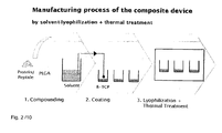
- Figure 2: Manufacturing process MD05 Retard
The process flow chart shows the three steps of the manufacturing process:
- Step A: dissolution of the polymer and eventually the active agent in a suitable organic
solvent.
- Step B: coating the ceramic device with polymer and/ore active agent by soaking and
subsequent drying.
- Step C: After removal of the solvent the thermal treatment leads to a defined polymeric shell
- Figure 3: Coating principle
- Step A: shows the protein loaded ceramic granule schematically in a cross sectional view
- Step B: shows the protein loaded ceramic granule, with freeze dried polymer coat
schematically in a cross sectional view
- Step C: shows the protein loaded ceramic granule, with the defined polymeric shell achieved
by thermal treatment schematically in a cross sectional view
- Figure 4: Stability of rhGDF-5 on β-TCP in contact with various organic solvents
Fig. 4 shows the results of solvent screening: The stability of rhGDF-5 on β-TCP in contact
with various organic solvents after drying at 25 °C he selected solvents are DMSO, glacial
acetic acid and anisole.The graph shows the content of modified species after contacting rhGDF-5 bound onto beta-TCP
with various organic solvents at room temperature for 30 minutes. The solvents tested
are acetone, chloroform, ethyl acetate, tetrahydrofurane, anisole, n-butylacetate, 1-pentanol,
dimethylsulfoxide, glacial acetic acid
- Figure 5: Stability of rhGDF-5 on β-TCP in contact with various organic solvents and
annealing.
The graph shows the content of modified species after contacting rhGDF-5 bound onto beta-TCP
with various organic solvents at room temperature for 30 minutes. After the subsequent
dying step the remaining protein coated granules were incubated at 60 °C to simulate the
conditions at thermal treatment process. The solvents tested are acetone, chloroform, ethyl
acetate, tetrahydrofurane, anisole, n-butylacetate, 1-pentanol, dimethylsulfoxide (DMSO),
glacial acetic acid
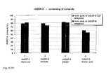
- Figure 6: Stability of pure rhGDF-5 in contact with various organic solvents and annealing
The graph shows the relative content of unmodified species after contacting pure rhGDF-5
with various organic solvents at room temperature for 30 minutes. After the subsequent dying
step the remaining pure protein was incubated at 60 °C to simulate the conditions at thermal
treatment process. The solvents tested are anisole, dimethylsulfoxide (DMSO), and glacial
acetic acid.
- Figure 7: Stability of pure parathormone in contact with various organic solvents and
annealing
The graph shows the relative content of unmodified species after contacting pure
parathormone PTH 1-34 with various organic solvents at room temperature for 30 minutes.
After the subsequent dying step the remaining pure parathormone PTH 1-34 was incubated
at 60 °C to simulate the conditions at thermal treatment process . The solvents tested are
anisole, dimethylsulfoxide (DMSO), glacial acetic acid.
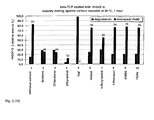
- Figure 8: Stability of rhGDF-5 on β-TCP in contact with various organic solvents and
annealing after optimized conditions.
The graph shows the relative content of unmodified species after contacting the rhGDF-5
bound onto beta-TCP with various organic solvents at room temperature for 30 minutes.
After the subsequent freeze drying step the remaining protein coated granules were
incubated at 60 °C at high vacuum (≤ 0.1 mbar) to simulate the conditions at thermal
treatment process. The solvents tested are anisole, dimethylsulfoxide (DMSO), and glacial
acetic acid.
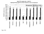
- Figure 9: Stability of PLGA coated rhGDF-5 on β-TCP in contact with various organic
solvents and annealing after optimized conditions.
The graph shows the relative content of unmodified species after contacting the rhGDF-5
bound onto beta-TCP with a solution of PLGA in various organic solvents at room
temperature for 30 minutes. After the subsequent freeze dying step the remaining protein
coated granules were incubated at 60 °C at high vacuum (≤ 0.1 mbar) for thermal treatment
process to achieve the defined polymeric shell . The solvents tested are anisole,
dimethylsulfoxide (DMSO), and glacial acetic acid.
- Figure 10: Timetable showing lyophilisation details
This table shows the details of the lyophilization program for the manufacturing of protein or
peptide PLGA loaded composite PLGA/β-TCP granules to achieve a minimum solvent
induced protein- or peptide degradation.
-
Detailed description of the preferred embodiments
-
The term "device" as used in accordance to the present invention refers to an entity, which
comprises at least three components as set forth below. In one embodiment the device is a
drug delivery system with porous scaffold. This device is preferably suitable for surgical
defect filling and tissue regeneration. In another embodiment the device is a drug delivery
system for the controlled release of active substances after implantation.
-
One of the components of said device is an inorganic matrix, the so-called "carrier".
Preferably, said inorganic matrix consists of ceramics. More preferably, said carrier is a
calcium phosphate. Most preferably said inorganic matrix is a calcium phosphate, which is
beta tricalcium phosphate, alpha tricalcium phosphate, apatite or a calcium phosphate
containing cement. Alternatively, said carrier is selected from the group consisting of calcium
carbonate, magnesium carbonate, magnesium oxide or magnesium hydroxide.
-
Said ceramics may have a particularly high surface due to the small particles size or the
presence of macro- and micropores. In a preferred embodiment, said macropores have a
diameter of 100 to 400 µm. In another preferred embodiment, said micropores have a
diameter of less than 10 µm. In still another preferred embodiment, the pores are
interconnected to allow the influx of coating substances in the device preparation as well as
in-growth of bone and tissue cells in the application in vivo.
-
The term "carrier" encompasses three-dimensional matrices, such as the above-mentioned
ceramics and ceramic/polymer composites. The carrier, preferably, has an enlarged surface
due to the small particles size or the formation of macro- and micropores during the
manufacturing process.
-
The carrier comprised by the device of the invention may be brought into a suitable form for
administration of the device in vivo, such as solid composite materials in form of blocks,
cubes, discs or granules. In addition, the composite carrier may be coated onto a metallic
surface.
-
In a preferred embodiment, the carrier containing calcium phosphate is in a granular form,
more preferably in the form of free flowing granules. Granular products as moldable systems
are well established for surgical defect filling especially in orthopedic indications (Draenert et
al.). Therefore it is important to meet this preferred application form.
-
The term "calcium phosphate" encompasses compositions comprising calcium ions (Ca2+),
phosphate ions (PO3 3-), optionally, further ions like hydroxyl ions (OH-), carbonate (CO3 2-) or
magnesium (Mg2+) or other ions which are suitable for the carrier of the present invention.
The calcium phosphates as used in accordance with the present invention are crystals
having a three dimensional structure suitable for the device of the present invention as set
forth above. Said calcium phosphates are particularly well suited as carriers for the device of
the present invention. Their in vivo properties have been described in Hotz, 1994, Gao, 1996,
and in WO98/21972. A list of preferred and well-known calcium phosphates is given above.
-
The second component of the device of the present invention is a polymer. Preferably said
polymer is a "biocompatible", a "biodegradable" or a "bioresorbable" polymer.
-
The term "biocompatible" means the ability of a material to perform with an appropriate host
response in a specific application (Wintermantel E. et al., 2002). "Biodegradable polymers"
specifies polymers, which break down due to macromolecular degradation with dispersion in
vivo but for which no proof exists for the elimination from the body. The term "bioresorbable
polymer" specifies polymeric materials, which underwent degradation and further resorption
in vivo; i.e. polymers, which are eliminated through natural pathways either because of
simple filtration of degradation by-products or after their metabolization. Bioresorption is thus
a concept which reflects total elimination of the initial foreign material.
-
In a preferred embodiment of the device or the method of the invention said bioresorbable
polymer is a polymer that undergoes a chain cleavage due to macromolecular degradation in
an aqueous environment.
-
Alternatively, said polymer is selected from the group consisting of polyethylene (PE),
polypropylene (PP), polyethylenerephthalate (PET), polyglactine, polyamide (PA),
polymethylmethacrylate (PMMA), polyhydroxymethylmethacrylate (PHEMA),
polyvinylchloride (PVC), polyvinylalcohole (PVA), polytetrafluorethylene (PTFE),
polyetheretherketone (PEEK), polysulfon (PSU), polyvinylpyrolidone, polyurethane or
polysiloxane. These polymers are at least biocompatible.
-
More preferably, said polymer is selected from the group consisting of poly(α-hydroxy
acids), poly (ortho esters), poly(anhydrides), poly(aminoacids), polyglycolid (PGA), polylactid
(PLLA), poly(D,L-lactide) (PDLLA), poly(D,L-lactide co-glycolide) PLGA), poly(3-hydroxybutyricacid)
(P(3-HB)), poly(3-hydroxy valeric acid) (P(3-HV)), poly(p-dioxanone)
(PDS), poly(ε-caprolactone) (PCL), polyanhydride (PA). These polymers are biocompatible
and bioresorbable.
-
The third component of the device of the present invention is an "active agent". The term
"active agent" comprises a polypeptide or a small molecule drug which is immobilized on
and/or in the carrier. Preferably, said polypeptide or drug is homogeneously distributed on
the calcium phosphate containing carrier and / or within the polymer.
-
The term "osteoconductive" refers to substrates that provide a favourable scaffolding for
vascular ingress, cellular infiltration and attachment, cartilage formation, and calcified tissue
deposition. Osteoconductive materials may support osseous generation via the scaffolding
effect (Kenley, R.A., 1993).
-
The term "osteoinductive" refers to the capability of the transformation of mesenchymal stem
cells into osteoblasts and chondrocytes. A prerequisite for osteoinduction is a signal which is
distributed by the device into the surrounding tissues where the aforementioned osteoblast
precursors become activated. Osteoinduction as used herein encompasses the
differentiation of mesenchymal cells into the bone precursor cells, the osteblasts. Moreover,
osteoinduction also comprises the differentiation of said osteoblasts into osteocytes, the
mature cells of the bone. Moreover, also encompassed by osteoinduction is the
differentiation of mesenchymal cells into chondrocytes. In particular in the long bones, the
chondroblasts and the chondrocytes residing in the perichondrium of the bone can also
differentiate into osteocytes. Thus, osteoinduction requires differentiation of undifferentiated
or less-differentiated cells into osteocytes which are capable of forming the bone. Thus, a
prerequisite for osteoinduction is a signal which is distributed by the device into the
surrounding tissues where the aforementioned osteocyte precursors usually reside. As has
been described above, the osteoinductive proteins or peptides used in accordance with the
present invention are sustained released from the device after implantation and are
distributed efficiently in the surrounding tissues. Moreover, the proteins and peptides
encompassed by the present invention have osteoinductive properties in vivo. For example,
it is well known in the art that the Transforming Growth Factor-β (TGF-β) superfamily
encompasses members which have osteoinductive properties. Individual members of said
TGF-β superfamily which have particular well osteoinductive properties are listed infra. In
conclusion, the osteoinductive proteins or peptides of the device of the present invention
after having been released from the carrier serving as a osteoinductive signal for the
osteocyte precursors of the tissue surrounding the side of implantation of the device.
-
The term "osteogenic" describes the synthesis of new bone by osteoblasts. In accordance
with the present invention, preexisting bone in the surrounding of the side of implantation of
the device grows into the device using the structure of the device as a matrix onto which the
osteocytes can adhere.
-
The term "osteoinductive polypeptide" refers to polypeptides, such as the members of the
Transforming Growth Factor-β (TGF-β) superfamily, which have osteoinductive properties.
-
In a further preferred embodiment of the device or the method of the invention said
osteoinductive protein is a member of the TGF-β family.
-
The TGF-β family of growth and differentiation factors has been shown to be involved in
numerous biological processes comprising bone formation. All members of said family are
secreted polypeptides comprising a characteristic domain structure. On the very N-terminus,
the TGF-β family members comprise a signal peptide or secretion leader. This sequence is
followed at the C-terminus by the prodomain and by the sequence of the mature polypeptide.
The sequence of the mature polypeptide comprises seven conserved cysteins, six of which
are required for the formation of intramolecular disulfide bonds whereas one is required for
dimerization of two polypeptides. The biologically active TGF-β family member is a dimer,
preferably composed of two mature polypeptides. The TGF-β family members are usually
secreted as proteins comprising in addition to the mature sequence the prodomain. The
prodomains are extracellularly cleaved off and are not part of the signalling molecule. It has
been reported, however, that the prodomain(s) may be required for extracellular stabilization
of the mature polypeptides.
-
In the context of the present invention, the term "TGF-β family member" or the proteins of
said family referred to below encompass all biologically active variants of the said proteins or
members and all variants as well as their inactive precursors. Thus, proteins comprising
merely the mature sequence as well as proteins comprising the mature protein and the
prodomain or the mature protein, the prodomain and the leader sequence are within the
scope of the invention as well as biologically active fragments thereof. Whether a fragment of
a TGF-β member has the biological activity can be easily determined by biological assays
described, e.g. in: Katagiri T, Yamaguchi A, Ikeda T, Yoshiki S, Wozney JM, Rosen V, Wang
EA, Tanka H, Omura S, Suda T, (1990): The non-osteogenic mouse pluripotent cell line,
C3H10T1/2, is induced to differentiate into osteoblastic cells by recombinant human bone
morphogenetic protein-2. Biochem. Biophys. Res. Commun. 172: 295-299 or Nishitoh H,
Ichijo H, Kimura M, Matsumoto T, Makishima F, Yamaguchi A, Yamashita H, Enomoto S,
Miyazono K (1996): Identification of type ( and type (( serine/ threonine kinase receptors for
growth/ differentiation factor-5. J. Biol. Chem. 271: 21345-21352.
-
Preferably, the biological activity according to the invention can be determined by in vivo
models as described in the accompanied Examples. Furthermore, encompassed by the
present invention are variants of the TGF-β members which have an amino acid sequences
being at least 75%, at least 80%, at least 90%, at least 95%, at least 96%, at least 97%, at
least 98% or at least 99% identical to the amino acid sequences of the members of the TGF-β
family.
-
An overview of the members of the TGF-β superfamily is given in: Wozney JM, Rosen V
(1998): Bone morphogenetic protein and bone morphogenetic protein gene family in bone
formation and repair. Clin Orthop 346: 26-37. The amino acid sequences of the members of
the TGF-β family can be obtained from the well known databases such as Swiss-Prot via the
internet (http://www.expasy.ch/sprot/sprot-top.html) Amino acid sequences for BMP2, BMP7
and GDF-5, members of the TGF-(family with a particularly high osteoinductive potential, are
also shown in SEQ ID No: 1 to 3, respectively. Amino acid sequences for BMP2, BMP7 and
GDF-5, members of the TGF-β family with a particularly high osteogenic potential, are also
shown in SEQ ID No:1 to 3, respectively.
-
More preferably, said member of the TGF-β family is a member of the BMP subfamily.
The members of the Bone Morphogenetic Protein (BMP) subfamily have been shown to be
involved, inter alia, in the induction and re-modeling of bone tissue. BMPs were originally
isolated from bone matrix. These proteins are characterized by their ability to induce new
bone formation at ectopic sites. Various in vivo studies demonstrated the promotion of
osteogenesis and chondrogenesis of precursor cells by BMPs and raise the possibility that
each BMP molecule has distinct role during the skeletal development. More details about the
molecular and biological properties of the BMPs are described in:
Wozney JM, Rosen V (1998): Bone morphogenetic protein and bone morphogenetic protein
gene family in bone formation and repair. Clin Orthop 346: 26-27, Schmitt J, Hwang K, Winn,
SR, Hollinger J (1999): Bone morphogenetic proteins: an update on basic biology and clinical
relevance. J Orthop Res 17: 269-278 and Lind M (1996): Growth factors: possible new
clinical tools. A review. Acta Orthop Scand 67: 407-17.
-
The osteoinductive polypeptide of the present invention is preferably selected from the group
consisting of BMP-1, BMP2, BMP-3, BMP-4, BMP-5, BMP-6, BMP7, BMP-8, BMP-9, BMP-10,
BMP-11, BMP-12, BMP-13, BMP-14, BMP-15 and BMP-16. Most preferably, said
member of the BMP family is BMP2 or BMP7.
-
The amino acid sequence for the preproform of BMP2 is deposited under Swiss-Prot
Accession number P12643 and is shown below. Amino acids 1 to 23 correspond to the
signal sequence, amino acids 24 to 282 correspond to the propeptide and amino acids 283
to 396 correspond to the mature protein. The amino acid sequence for the preproform of
BMP7 is deposited under Swiss-Prot Accession number P18075 or shown in SEQ ID No: 2.
Amino acids 1 to 29 correspond to the leader sequence, amino acids 30 to 292 correspond
to the proform and amino acids 293 to 431 correspond to the mature protein. Preferably,
BMP-2 or BMP7 refers to the preproform, to the proform or to the mature BMP-2 or BMP-7
peptide, respectively. Moreover also encompassed are fragments of said proteins having
essentially the same biological activity, preferably osteoinductive properties. More sequence
information for BMP2 and BMP7 is provided below.
-
Alternatively, the osteoinductive polypeptide of the present invention is selected from another
TGF-β family, i.e. the GDF family.
-
Growth and Differentiation Factor (GDF) have been also shown to be involved, inter alia, in
the induction and re-modeling of bone tissue. Growth Differentiation Factor 5 (GDF-5), also
known as cartilage-derived morphogenetic protein 1 (CDMP-1) is a member of subgroup of
the BMP family, which also includes other related proteins, preferably, GDF-6 and GDF-7.
The mature form of the protein is a 27 kDa homodimer. Various in vivo and in vitro studies
demonstrate the role of GDP-5 during the formation of different morphological features in the
mammalian skeleton. Mutations of GDF-5 are responsible for skeletal abnormalities including
decrease of the length of long bones of limbs, abnormal joint development in the limb and
sternum (Storm & Kingsley (1999), Development Biology, 209, 11-27). The amino acid
sequence between mouse and human is highly conserved.
-
Preferably, the osteoinductive polypeptide of the present invention is selected from the
group consisting of GDF-1, GDF-2, GDF-3, GDF-4, GDF-5, GDF-6, GDF-7, GDF-8, GDF-9,
GDF-10 and GDF-11. Most preferably, said member of the GDF subfamily is GDF-5.
The amino acid sequence for the preproform of GDF-5 is deposited under Swiss-Prot
Accession number P 43026 or shown in SEQ ID No: 3. Amino acids 1 to 27 correspond to
the leader sequence, amino acids 28 to 381 correspond to the proform and amino acids 382
to 501 correspond to the mature protein. Preferably, GDF-5 refers to the preproform, to the
proform or to the mature GDF-5 peptide. Moreover also encompassed are fragments of
GDF-5 having essentially the same biological activity, prefrably osteoinductive properties.
Most preferably, said fragment comprises amino acids 383 to 501 of the sequence shown in
SEQ ID No: 3.
-
The following tables show amino acid sequences for BMP-2, BMP-7 and GDF-5:
-
Also encompassed by the present invention are embodiments, wherein said active agent is
selected from hormones, cytokines, growth factors, antibiotics and other natural and/or
synthesized drug substances like steroids, prostaglandines etc.
-
Preferably, said active agent is parathyroid hormone (PTH) and/or PTH 1-34 peptide.
-
The device of the present invention may optionally, comprise additional excipients. These
excipients serve the stabilization of the protein or peptide, e.g., saccharides, amino acids,
polyols, detergents or maintenance of the pH, e.g., buffer substances.
-
In a preferred embodiment the device of the present invention is free of toxic substances.
Preferably such toxic substances are already avoided in the production process, as their
production requires additional expenditure due to required removal steps during the
production process and necessary expensive means for highly sensitive chemical analysis.
-
The term "toxic substances", in particular, encompasses those toxic organic solvents and
additives which are used by the methods described in the art, which are classified by the ICH
as class 2 solvents (ICH Topic Q 3 C Impurities: Residual Solvents) e.g. methylene chloride.
Said substances may cause systemic or local toxic effects, inflammation and / or other
reactions after implantation of devices containing said substances. Said prior art devices are
therapeutically less acceptable due to said undesirable side effects, which cannot be avoided
by the conventionally coating methods described in the art. Moreover, the international
guidance for the development of therapeutic proteins require that in the manufacturing
process harmful and toxic substances should be avoided (for details see: International
Conference on Harmonization (ICH), Topic Q3C; www.emea.eu.int/). However, the device of
the present invention or a device which is obtainable by the method of the present invention
is, advantageously, free of said class 1 classified toxic substances. Moreover the present
invention contains only solvents classified as class 3 by the ICH Topic Q 3C and, therefore,
therapeutically well acceptable and fulfills the requirements of the regulatory authorities.
-
Moreover, in a further preferred embodiment of the device or the method of the invention
said device is free of infectious material.
-
Besides toxic substances, infectious material comprised by the device may cause severe
infections in a subject into which the device has been transplanted. Potentially infectious
gelatin derived from bovine or procine bones is, however, used as a protecting protein in
many state of the art methods (Lind, 1996).
-
Solutions sufficient for the two methods A and B of the present invention (used in step (a) of
method A and step (e) of method B, respectively) to produce the device of the present
invention have a melting point > -40 °C and a boiling point < 200 °C.
They are inactive towards the active agent.
-
Preferable this solution contains a solvent selected from anisole, tetramethylurea, acetic
acid, dimethylsulfoxide and tert-butanol (2-methyl-2-propanole trimethylcarbinole-butyl
alcohol), acetone, 1-butanole, 2-butanole, butyl acetate, tert-butylmethyl ether, cumene,
ethanole, ethyl acetate, dieethylether, ethylformate, formic acid, isobutyl acetate,
isopropylacetate, methyl acetate, 3-methyl-1-butanol, methylethyl ketone,
methylisobutylketone, 2-methyl-1-propanol, pentane, 1-pentanol, 2-propanol, propylacetate,
and tetrahydrofurane. Most preferred are acetic acid, dimethylsulfoxide and anisole.
-
The terms "homogeneously distributed" and "homogeneously coateded" mean that identical
amounts of the active agent are present in each and every area of said composite carrier.
This area preferably includes the pores of a porous matrix. Homogenous distribution is a
prerequisite for efficient release and activity of the active agent into the tissue surrounding
the site of implantation. Moreover, it is to be understood that the active agent is not
aggregated and partially or entirely inactivated due to precipitation or micro-precipitation,
rather attachment of biologically active, non-aggregated proteins is to be achieved by
homogenous coating. Said homogenous distribution can be achieved by the two above
methods of the present invention.
-
The homogenous coating of the carrier with said active agent and the simultaneous and / or
additional homogeneous coating with the bioresorbable polymer do achieve an onion-like
layer structure which acts in two manners as a protective film and as diffusion barrier to slow
down the dissolution of the protein or peptide to achieve a sustained release. The described
methods A and B allow the homogenous distribution and immobilization of the osteoinductive
active agent into and / or on the carrier and the sustained release of the active agent due to
the polymeric component.
-
The efficacy of the coating process is, furthermore, supported by the carrier due to capillary
forces resulting from the presence of numerous, preferably interconnected macro- and micropores
which due to their size are capable of soaking the solutions into the pores.
-
Moreover, in contrast to other methods described in the art, e.g., in WO98/21972, the active
agent is - according to the methods A and B of the present invention - applied by attachment
to the carriers from the soluble state to achieve a homogeneous coating. The findings
underlying the present invention demonstrate that the aggregation of the proteins can be
avoided in a tri-component-system by the use of suitable solvents and / or additives as
described herein. An important precondition is the knowledge of the solubility of the
osteoinductive active agent dependent on the nature of the solvent, i.e. aqueous and / or
organic solvent, pH value, ionic strength and surfaces present.
-
The term "aqueous solution" specifies any solution comprising water. The slowing down of
the pH increase caused by the contact of the coating solution with the calcium phosphates in
the carrier reacting in an alkaline manner, in particular, plays an important role during the
coating, preferably in method B.
-
The methods A and B of the present invention, distribute the active agent homogeneously
across the inner surface of the carrier material and allow binding to the surface before a
precipitation of the said protein takes place.
-
In method B this precipitation is pH-induced. It could be demonstrated that in this case the
pH increase taking place during the coating of calcium phosphates is decelerated sufficiently
by the use of a weak acid, such as acetic acid. Furthermore, the addition of organic
compounds such as ethanol or sucrose proves to be additionally advantageous here.
Furthermore, a low ionic strength is an important precondition for successful coating of the
protein or peptide onto the calcium phosphate. Moreover, our tests show that the volume of
the coating solutions (solution containing active agent and / or polymer), too, has a
considerable effect on the quality of both coatings.
-
Finally, the methods A and B of the present invention allow the use of non toxic organic
solvents (see below), such as dimethyl sulfoxide, anisol or glacial acid. These solvents are
routinely used in the methods described in the art. Normally they damage the protein during
contacting and/or especially during drying but such damage is surprisingly avoided by using
the special drying technique of the present invention, because the active agent is adsorbed/or
attached onto the inorganic solid carrier.
-
In a preferred embodiment of the method B of the invention said active agent coating buffer
has a buffer concentration of preferably less than 100 mmol/l, more preferably less than 50
mmol/l and even more preferably less than 20 mmol/l to achieve a sufficient solubility of the
active agent during the adsorbtion process and to avoid any modification of a ceramic carrier.
-
In another preferred embodiment of the method of the invention said buffer has a buffer
concentration of 10 mmol/l to achieve a sufficient solubility of the active agent during the
adsorbtion process and to avoid any modification of the monophasic calcium phosphate
ceramic beta TCP. The pH of the solution shifts in a controlled manner during the coating
and drying process from pH 3 to pH 7, more preferably from 3 to 6 and most preferably from
4 to 5,5. This pH shift causes a defined reduction of the solubility of the bone growth factor to
result in a homogenous, defined attachment on the TCP.
-
In a preferred embodiment of the methods of the invention the solutions comprise non toxic
organic solvents. The first aspect for method A is to find a common suitable organic solvent
for both, the active agent and the polymer, without inducing modifications at the active agent.
A second aspect is the ability of the solvent(s) in step (a) method A and / or step (e) of
method B for the drying process, which is preferably a freeze-drying process. The preferred
solvents are anisole, dimethylsulfoxide (DMSO) and glacial acetic acid. In a preferred
embodiment these solvents in both method A and in method B are used in volumina to
achieve a compleate soaking of the polymer solution and to avoid any remaining solution.
-
It follows from the above that preferably, said buffer contains a weak acid. The term "weak
acid" refers to organic or inorganic compounds containing at least one ionogenically bound
hydrogen atom. Weak acids are well known in the art and are described in standard text
books, such as Römpp, "dictionary of chemistry". Preferably, said weak acids which have low
dissociation degrees and are described by pK values between 3 and 7, preferably between 4
and 6. Most preferably, said weak acid is acetic acid or succinic acid.
-
In another preferred embodiment of method B of the invention said buffer containing solution
further comprises at least one saccharides in an aqueous solution, more preferably in an
aqueous solution without any further solvent apart from water.
-
In a further preferred embodiment of the method of the invention said buffer containing
solution comprises an polyol and/or alcohol. Suitable alcohols or polyols are well known in
the art and are described in standard text books, such as Römpp, dictionary of chemistry.
More preferably, said alcohol is ethanol and said polyol is mannitol.
-
In a more preferred embodiment the concentration of the polyol and or alcohol is between 0
- and 10 % (w/v).
-
The term "saccharides" encompasses mono-, di- and polysaccharides. The structure and
composition of mono-, di-, and polysaccharides are well known in the art and are described
in standard text books, such as Römpp, "dictionary of chemistry".
-
More preferably, said saccharide is a disaccharide. Most preferably, said dissaccharide is
sucrose or trehalose.
-
Further means and methods for controlling homogeneous distribution, quantification and
characterization of the active agent are described in the accompanied examples.
-
Surprisingly, active agents, in particular surprisingly proteins, when adsorbed on the surface
of ceramic carriers are much more resistant against degradation caused by organic solvents
than proteins freely dissolved or suspended in organic solutions or integrated in biphasic
emulsion systems. Thus, this aspect of the invention opens a new possibility to produce
polymer based drug delivery systems for proteins without denaturation and / or modification
of polypeptides in singular or multiphase organic systems, preferably for active agents
incompatible with organic solvents.
-
Furthermore the type of ceramic carrier preferably used in present invention opens the
possibility to quantitatively remove organic solvents (see below).
-
Suitable for one of the two methods of the present invention as active agents are all proteins,
polypeptides and small molecule drugs. Especially such active agents with low or no affinity
for inorganic carrier matrices can be immobilized in the polymer - calcium phosphate
composite material. Preferably, the binding of said active agent to the carrier is reversible.
-
Thereby, dissolution of said active agent is allowed once the device has been brought into a
suitable in vivo surrounding, such as a bone cavity. More preferably, said dissolution of the
immobilized compounds is a sustained release allowing diffusion of the active agent into the
tissue which surrounds the device. Thus, the device is suitable to serve as an in vivo source
for e.g. osteoinductive proteins, peptides or small molecule drugs, which are slowly released
and which can be thereby efficiently distributed into the surrounding tissues or have an effect
in the immobilized form.
-
The term "drying" encompasses measures for removing liquids, such as excess buffer
solution, or organic solvents which are still present after coating of the carrier with the
osteoinductive protein or polymer solution. Preferably, drying is achieved by convection at
under inert gas atmosphere, by vacuum- or freeze-drying. It is important for the composite
ceramic of the present invention that after drying the ceramic matrix is substantially free of
organic solvent to allow for a softening of the polymeric component in a thermal treatment
step, such as steps (e) of method A and step (g) of method B. Substantially free of organic
solvent means a content preferably below ≤1 % residual solvent, more preferably ≤0,05 %,
even more preferably ≤ 0,025 % and most preferably ≤ 0,01 %.
-
The term "buffer" which assists in keeping the active agent dissolved in aqueous solutions for
a time sufficient to allow "homogenous coating" refers to a component allowing the active
agent to be effectively dissolved in the solution and / or homogeneously coated in a carrier
system tending to cause pH induced precipitation. This buffer is preferably capable of
avoiding and or balancing the increase of pH caused by contacting the solution with the
calcium phosphate carrier so that the protein does not immediately precipitate, e.g., due to a
pH increase. Said buffer can be determined by the person skilled in the art considering the
solubility of the osteoinductive protein (which depends on the pH and the ionic strength) and
the influence of the carrier on said parameters after contacting the carrier with said buffer
containing solution. In accordance with the present invention it has been found that a suitable
buffer is needed for the homogeneous distribution of the active agent onto the surface of the
carrier, e.g. calcium phosphate, said buffer comprising preferably a weak acid, an alcohol
and a saccharide. The solvent for the dissolution of the preferably bioresorbable polymer in
which the protein or peptide is not soluble described by the method B of the present
invention comprises a suitable organic solvent for the homogeneous distribution of the
polymer onto the surface of the protein or peptide coated carrier e.g. dimethyl sulfoxide,
anisol or glacial acid.
-
The term "thermal treatment" refers to a heating step which is applied after the solvent has
been removed by drying to condense the polymeric phase by a definite collapse of the freeze
dried structure and thus providing a dense and homogenous polymeric shell covering the
ceramic surface of the granules. The purpose of this procedure is to modulate the release
kinetics for the active substance and to achieve free flowing granules.
-
The temperature should be equal or higher than the glass transition temperature of the
corresponding polymer system. For thermal sensitive active agents the glass transition
temperature of the polymer can be decreased by the use of plasticizers, e.g. polyethylene
glycol. The thermal treatment applies a temperature between the final drying temperature of
≥ 20°C, preferably ≥ 25°C, and most preferably ≥ 30 °C and the maximum temperature,
limited by the active agent of ≤ 60 °C, preferably ≤ 55, and most preferably ≤ 50 °C. The time
range for the thermal treatment in a preferred embodiment is as follows: Heating from 20 °C
up to 60 °C in 30 minutes following by an isothermic period of 50 minutes at this temperature.
Afterwards the samples are cooled down to 20 °C for 1 hour. The integrity of the active agent
was determined after extraction the polymeric shell as demonstrated in example 4.
-
The invention encompasses a pharmaceutical composition comprising the device of the
invention or a device, which is obtainable by the method of the invention.
-
The product of the present invention can be formulated as a pharmaceutical composition or a
medical device. The composition of said product may comprise additional compounds like
stabilizers, buffer substances and other excipients. The amount of the product of the present
invention applied to the patient will be determined by the attending physician and other
clinical factors; preferably in accordance with any of the above described methods. As it is
well known in the medical arts, the amount applied to a patient depends upon many factors,
including the patient's size, body surface area, age, sex, time and route of administration,
general health conditions, and other drugs being administered concurrently. Progress can be
monitored by periodic assessment.
-
Thanks to the present invention, it is possible to treat various bone defects including large
cavities in a new manner. In particular, large cavities could not or only under use of
autogenous bone material be efficiently treated. However, due to the reliable and efficient
osteoinductive and the osteoconductive properties of the device of the present invention or
the device which can be obtained by the method of the invention, treatment of bone defects
which requires extensive bone augmentation or repair has now become possible without a
second surgery for gaining autologous bone material.
-
The invention also encompasses the use of the device of the invention or a device which is
obtainable by the method of the invention for the preparation of a pharmaceutical
composition for bone augmentation.
-
The term "bone augmentation" refers to the induced formation of bone, which is indicated in
order to treat bone defects, cavities in bones, or to treat diseases and disorders
accompanied with loss of bone tissue or to prepare the subsequent setting of an implant. The
diseases and disorders described in the following are well known in the art and are described
in detail in standard medical text books such as Pschyrembel or Stedman.
-
Preferably, said bone augmentation follows traumatic, malignant or artificial defects.
-
Another embodiment of the present invention relates to the use of the device of the invention
or the preparation of a pharmaceutical composition for treating bone defects.
-
More preferably, said bone defects are long bone defects or bone defects following
apicoectomy, extirpation of cysts or tumors, tooth extraction, or surgical removal of retained
teeth.
-
The invention also relates to the use of the device of the invention for filing of cavities and
support guided tissue regeneration in periodontology.
-
Another embodiment of the present invention relates to the use of the device of the invention
for the preparation of a pharmaceutical composition for sinus floor elevation, augmentation of
the atrophied maxillary and mandibulary ridge and stabilization of immediate implants.
-
Also within the scope of the present invention is a method for treating one or more of the
diseases referred to in accordance with the uses of the present invention, wherein said
method comprises at least the step of administering the device of the invention in a
pharmaceutically acceptable form to a subject. Preferably, said subject is a human.
-
Finally, the invention relates to a kit comprising the device of the invention.
-
The parts of the kit of the invention can be packaged individually in vials or other appropriate
means depending on the respective ingredient or in combination in suitable containers or
multicontainer units. Manufacture of the kit follows preferably standard procedures which are
known to the person skilled in the art.
Contribution to the field by the present invention
-
Polymer and carrier together form the ceramic/polymer composite carrier material of the
present invention, which binds an osteoinductive active agent to result in the device of the
present invention, in order to allow the sustained release of said active agent in vivo.
-
In accordance with the present invention, the composite carrier is based on a calcium
phosphate and a polymer, preferably a biodegradable polymer. In such a composite carrier
the calcium phosphate shows excellent local buffering capacity and the permeable
composite structure avoids even local pH decrease when the polymer is degraded in vivo.
Cytotoxic side effects due to degradation of the polymer are, hence, reduced or avoided.
This is especially valid, since the ceramic carrier is chief ingredient of the ceramic
carrier/polymer composite carrier material of the present invention, which preferably contains
less than 20% of the polymer, most preferably less than 20% of PLGA.
-
Furthermore, the ceramic/polymer composite carrier material of the present invention shows
improved mechanical stability compared to conventional systems using polymeric granules
for retarded release. In one preferred embodiment, the resulting free flowing granules have
mechanical properties, which are the same when compared with the untreated polymer free
granules. These untreated granules represent the well established system to withstand
tissue pressure in various therapies e.g. in orthopedic indications.
-
The ceramic/polymer composite carrier material of the present invention shows improved
mechanical stability compared to conventional systems using polymeric granules for retarded
release. In porous embodiments the composite matrix allows for improved osteoconductive
properties compared to prior art systems due to the porous system, in particular free of
interconnected pores.
-
The ceramic/polymer composite carrier material of the present invention is suitable to replace
conventional encapsualting polymeric granules important for retarded release. Due to the
homogeneous coating of the system (ceramic carrier plus polymer coating), the amount of
polymer can be significantly reduced compared with totally polymer based scaffolds, which
reduced amount of polymer leads to a reduced risk of cytotoxicity. A further aspect of the
invention is the increased mechanical stability of the composite material compared with
totally polymer based scaffolds.
-
The invention will now be described by reference to the following examples which are merely
illustrative and which shall not limit the scope of the present invention. Some of the measures
and results of the methods set forth in the examples can be obtained from the accompanying
figures.
Examples
Example 0: Solvent screening
-
Step 1: Coating of beta-TCP with rhGDF-5: 500 mg β-TCP (0.5 - 1.0 mm granule size) are
placed in a dry form in a 6R-glass. The stock solution of rhGDF-5 (4 mg/ml in 10 mM HCl) is
diluted to 1 g/ml (with the corresponding coating buffer). 425 µl of the rhGDF-5 solution
obtained in that manner are pipetted on the beta-TCP and adsorbed. The damp granulate is
incubated for 1 hour at 25°C and then lyophilised.
Step 2: In the subsequent step 425 µl of the following organic solvents were incubated with
the coated granules from step 1 for 30 min at room temperature. After drying a sample was
taken. The remaining samples were tempered at 60 °C for 1 h.
From each sample the rhGDF-5 was extracted according Example 4 to quantify the modified
species by RP-HPLC.
Example 1: Manufacturing of MD05 500 retard (with a protein).
-
Provide 0.5 g of β-TCP in a 6R glass vial, take 475 µl of the coating solution (0.525 mg/ ml
rhGDF-5 in 10.0 mM acetic acid, 2.5 mM HCl, 10.0 % saccharide).
Dry by freeze-drying. Second coating by adding 425 µl of PLGA (Resomer RG 502 H)
dissolved in DMSO, containing 150 mg PLGA/ ml DMSO.
Again drying by freeze-drying , including a terminal treatment step by 60 °C over 1 hour.
Example 2: Manufacturing of MD05 500 retard (with a peptide).
-
Provide 0.5 g of β-TCP in a 6R glass vial, take 475 µl of the coating solution (0.525 mg/ ml
PTH 1-34 and 134 mg/ml PLGA in DMSO).
Freeze-drying , including a terminal treatment step by 60 °C over 1 hour
Example 3: Release study of MD05 retard.
-
MD05 retard (containing a protein; rhGDF-5).
Weight 150 mg of MD05 retard into a 50 ml tube, ad 48 ml alpha-MEM-medium including 10
% of FCS and shake continuously at 4 °C and 37 °C for defined time, (final concentration of
the release-assay is ~1.6 µg rhGDF-5/ ml medium). The defined time in this example is ≤ 30
days can be adapted to the respective active agent.
At defined time, an aliquot of 100 µl was taken (the taken volume will not be replaced),
centrifuged for 5 minutes at 13 000 rpm, the supernatant is frozen at -70 °C.
The quantification of the selected aliquots will be done by Elisa-assay.
-
MD05 retard (containing a peptide, Parathormon, PTH 1-34)
0.2 g of product is taken in 1 ml PBS (final concentration of PTH is 0.015 µg/ml)incubated at
4°C and 37 °C under continuously shaking.
At defined time, the incubation medium was replaced with fresh PBS.
The supernatant was centrifuged at 13000 rpm for about 5 minutes, an aliquot from 250 µl
was analyzed by HPLC.
Example 4: Extraction of the immobilized protein
-
Coated Calcium phosphate with rhGDF-5 or rhBMP-2 (656 mg) was extracted in a 1 ml
polypropylene reaction cup after resuspending in 1 ml extraction buffer (80mM EDTA, 8mM
1,2 Diaminopropan pH 6.7 in 20 % Ethanol) and the addition of 480 mg urea under gentle
agitation for 1 h at 4 °C.
After centrifugation (13 200 rpm g, 2 min) the supernatant was analyzed by RP-HPLC.
Example 5: Quantification and determination of chemical modifications of the extracted
polypeptide:
Method A for bone growth factor
-
The amount of chemical modifications i.e. oxidation of bone growth factor in solutions
containing extracted protein was determined by RP-HPLC. The sample is applied to a Vydak
C8-18 column (2 x 250 mm) which has been equilibrated with 0.15 % TFA, 20 % acetonitrile.
After washing of the column, the elution is performed with a mixture of 0.1 % TFA, 20 %
acetonitrile, and a stepwise gradient of 20 % - 84 % acetonitrile (flow: 0.3 ml/min). The
elution is observed by measuring the absorption at 215 nm. The quantification is calculated
by the ratio of the peak area of modified species to the total peak area.
Method B for peptide
-
The amount of chemical modifications, i.e. oxidation of bone growth factor in solutions
containing extracted protein was determined by RP-HPLC. The sample is applied to a Vydak
C8-18 column (4.6 x 250 mm) which has been equilibrated with 0.15 % TFA, 13.5 %
acetonitrile. After washing of the column, the elution is performed with a mixture of 0.1 %
TFA, 13.5 % acetonitrile and a stepwise gradient of 13.5 % - 84 % acetonitrile (flow: 0.5
ml/min). The elution is observed by measuring the absorption at 215 nm. The quantification
is calculated by the ratio of the peak area of modified species to the total peak area.
Example 6: Quantification of soluble aggregates in solutions containing extracted
polypeptides (Proteins and peptides as well):
-
The amount of soluble aggregates in samples containing extracted protein was determined
by size exclusion HPLC. The column (TSK 3000) was equilibrated with 50 mmol/l phosphoric
acid, 50 mmol/l acetic acid/NaOH, pH 3.0.
The elution is observed by UV-detection at 220 nm. The quantification is calculated by the
ratio of the aggregate peak area to the total peak area.
Example 7: Stability testing of rhGDF-5 coated β-TCP granules in various organic solvents
-
The amount of solvent induced protein degradation was determined by incubating 500 mg of
rhGDF-5 coated granules with 425 µl of the solvent for 30 min. Afterwards the solvent were
removed by evaporation under vacuum and analyzed by RP-HPLC described in example 4
and 5.
Example 8: Stability testing of rhGDF-5 coated β-TCP granules after drying from various
organic solvents and annealing
-
The amount of solvent induced protein degradation during the annealing was determined by
incubating 550 mg of rhGDF-5 coated granules with 425 µl of the solvent for 30 min. The
solvent were removed by evaporation under vacuum and afterwards the vials were heated
up at a temperature of 60 °C for 1 hour in an oven and analyzed by RP-HPLC described in
example 4 and 5.
Example 9: Stability testing of pure rhGDF-5 after drying from various organic solvents and
annealing
-
To analyze the effect of various organic solvents on the pure GDF-5 100 µg GDF-5 were
dried under reduced pressure and 100µl of the solvent were added. The samples were
incubated for 1 hour and dried again. The GDF-5 were dissolved in extraction buffer and
analyzed by RP-HPLC described in example 5.
Example 10: Stability testing of rhGDF-5 coated β-TCP granules after drying from various
organic solvents and annealing with optimized lyophilization conditions
-
To measure the effect of the optimized manufacturing process on the solvent induced GDF-5
degradation 425 µl of the organic solvents were added to 550 mg MD05 incubated for 30
minutes and dried under the optimized lyophilization conditions described in Fig 10. The
GDF-5 degradation during manufacturing were quantified by RP-HPLC described in example
4 and 5.
Example 11: Stability testing of rhGDF-5 coated β-TCP granules with PLGA shell after drying
from various organic solvents and annealing with optimized conditions.
-
425 µl of a PLGA solution in DMSO (5 % w/v and 20 % w/v) were added to 550 mg of
rhGDF-5 coated granules and dried under the optimized lyophilization conditions described
in Fig 10. The GDF-5 degradation during this process was quantified by RP-HPLC described
in example 4 and 5.
References:
-
Agrawal, C. M. et al. (1997) "Technique to Control pH in Vicinity of Biodegrading PLA-PGA
Implants"; J. Biomed. Mater. Res. (Appl. Biomater.), 38: 105-114
-
Breitenbach Jörg, (2002) "Melt extrusion: from process to drug delivery technology", Eur. J.
Pharm. Biopharm. 54, 107-117
-
Celeste A.J., et al. (1990) "Identification of transforming growth factor ..."; Proc. Natl. Acad.
Sci. U.S.A. 87:9843-9847.
-
Chang, S. et al. (1994); "Cartilage-derived morphogenetic proteins..."; J. Biol. Chem. 269:
28227-28234.
-
Draenert, K. et al (2001), Trauma Berufskrankh. 3, 293 - 300.
-
Driessens, F.C.M. et al. (2002) "The Ca/P range of nanoapatitic calcium phosphate cements"
Biomaterials 23, 4011-4017
-
Dunn et al. US patent 5,702,716
-
Durucan, C. et al.; "Calcium-deficient hydroxyapatite-PLGA composites: mechanical and
microstructural investigation." J. Biomed. Mater. Res. 51:726-724.
-
EMEA, ICH Topic Q 3 C, Impurities: Residual Solvents
-
Friess, W. et al. (1998); Pharm. Dev. Technol. 4, 387 - 396.
-
Friess, W. (1999); "Collagen - biomaterial for drug delivery"; Eur J Pharm Biopharm 45: 113-36.
-
Gao, T et al. (1996); Int. Orthopaedics 20, 321 - 325.
-
Gombotz, W et al. (1996) in Formulation, characterization and stability of protein drugs,
Plenum Press, New York, USA, pp 219 - 245.
-
Griffith, D.L. et al. (1996); "Three-dimensional structure of recombinant human..."; Proc. Nati.
Acad. Sci. U.S.A. 93: 878-883.
-
Herbert, P. et al., 1998. A large scale process to produce microencapsulated Proteins.
Pharm Res., 15: 357-361
Hoetten, G. et al. (1994); "Coning and expression of recombinant human
growth/differentiation factor 5."; Biochem. Biophys. Res. Commun. 204: 646-652.
-
J.O. Hollinger et al. (1996); "Poly(α-hydroxy acids): carriers for bone morphogenetic proteins,
Biomaterials"; 17: 187-194
-
Hotz, G et al. (1994); Int. J. Oral Maxillofac. Surg. 23, 413 - 417.
-
Katagiri, T. et al. (1990); Biochem. Biophys. Res. Commun. 172: 295-299.
-
Kenley, R.A., et al. (1993) Pharm. Res. , 10 (10) 1393-1401.
-
Lind, M et al. (1996); J. Orthopaedic Res. 14, 343 - 350
-
Lind, M. (1996); Acta. Orthop. Scand. 67: 407-17.
-
Middleton, J.C. et al. (2000); "Synthetic biopolymers as orthopedic devices"; Biomaterials 21:
2335-2346
-
Nishitoh, H. et al (1996); J Biol. Chem. 271: 21345-21352.
-
Oezkayanak, E. et al. (1990); "OP-1 cDNA encodes an osteogenic protein in the TGF-beta
family"; EMBO J. 9: 2085-2093
Rueger, J.M. et al (1996); "Knochenersatzmittel"; Unfallchir. 99: 228-236
-
Scheufler, C. et al. (1990); "Crystal structure of human bone morphogenetic protein-2 at 2.7
A resolution".
-
Schiller, C. et al. (2003); "Carbonated calcium phosphates are suitable pH-stabilising fillers
for biodegradable polyesters"; Biomaterials 24:2037-2043.
-
Schmidmaier, G. et al. (2000); "Local liberation of IGF-I and TGF-beta 1 from a
biodegradable poly(D,L-lactide) coating of implants accelerates fracture healing" Chirurg 71:
1016-1022
-
Schmitt, J. et al. (1999); J. Orthop. Res. 17: 269-278.
-
Seeherman, H. (2003), "A review of preclinical program development for evaluating in
injectable carriers for osteogenic factors" J. Bone. Joint. Surg. Am. 85-A Suppl. 3, 96-108
-
Shively M.L. et al. (1995), "Physico-chemical characterization of a polymeric injectable
implant delivery system" J. Controlled Rel. 33, 237-243
-
Shore, E.M. et al. (1997); "Human bone morphogenetic protein-2 (BMP-2) genomic DNA
sequence".
-
Storm & Kingsley (1999); Development Biology, 209, 11-27.
-
Terheyden, H et al. (1997); Mund Kiefer Gesichtschir. 1, 272 - 275.
-
Tormälä, P et al. (1995) "Biodegradable polymers in orthopaedics: experimental and clinical"
Mat. Clin. Appl. 639-651
-
Tormälä, P et al. (1992) "Biodegradable self-reinforced composite materials; manufacturing
structure and mechanical properties" Clin. Mater. 10, 29-34
-
Vert, M. (1989); "Bioresorbable polymers for temporary therapeutic applications" Angew.
Makromol. Chem. 166/167: 155-168
-
Wang, E.A. et al. (1990); "Identification of transforming growth factor beta family members
present in bone-inductive protein purified from bovine bone."; Proc. Natl. Acad. Sci. U.S.A.
87: 9843-9847.
-
Wintermantel, E. et al., Medizintechnik mit biokompatiblen Werkstoffen und Verfahren, S. 7,
3. Auflage, Springer 2002, ISBN 3-540-41261-1
-
Wozney, J.M. et al. (1998); Clin. Orthop. 346: 26-37.
-
Wozney, J.M. et al. (1988); Science 242: 1528-1534.