EP1593395A2 - Gas filled microspheres as magnetic resonance imaging contrast agents - Google Patents
Gas filled microspheres as magnetic resonance imaging contrast agents Download PDFInfo
- Publication number
- EP1593395A2 EP1593395A2 EP05075094A EP05075094A EP1593395A2 EP 1593395 A2 EP1593395 A2 EP 1593395A2 EP 05075094 A EP05075094 A EP 05075094A EP 05075094 A EP05075094 A EP 05075094A EP 1593395 A2 EP1593395 A2 EP 1593395A2
- Authority
- EP
- European Patent Office
- Prior art keywords
- microspheres
- gas
- contrast medium
- lipid
- contrast
- Prior art date
- Legal status (The legal status is an assumption and is not a legal conclusion. Google has not performed a legal analysis and makes no representation as to the accuracy of the status listed.)
- Withdrawn
Links
- 239000004005 microsphere Substances 0.000 title claims abstract description 390
- 239000002405 nuclear magnetic resonance imaging agent Substances 0.000 title description 2
- 239000002872 contrast media Substances 0.000 claims abstract description 125
- 238000002595 magnetic resonance imaging Methods 0.000 claims abstract description 69
- 239000002243 precursor Substances 0.000 claims description 199
- 150000002632 lipids Chemical class 0.000 claims description 142
- 238000000034 method Methods 0.000 claims description 91
- 150000001875 compounds Chemical class 0.000 claims description 75
- 239000007788 liquid Substances 0.000 claims description 74
- 230000007704 transition Effects 0.000 claims description 52
- 230000005298 paramagnetic effect Effects 0.000 claims description 44
- PEDCQBHIVMGVHV-UHFFFAOYSA-N Glycerine Chemical compound OCC(O)CO PEDCQBHIVMGVHV-UHFFFAOYSA-N 0.000 claims description 42
- DNIAPMSPPWPWGF-UHFFFAOYSA-N Propylene glycol Chemical compound CC(O)CO DNIAPMSPPWPWGF-UHFFFAOYSA-N 0.000 claims description 39
- 239000003795 chemical substances by application Substances 0.000 claims description 39
- 229920000642 polymer Polymers 0.000 claims description 28
- UQSXHKLRYXJYBZ-UHFFFAOYSA-N Iron oxide Chemical compound [Fe]=O UQSXHKLRYXJYBZ-UHFFFAOYSA-N 0.000 claims description 26
- KILNVBDSWZSGLL-KXQOOQHDSA-N 1,2-dihexadecanoyl-sn-glycero-3-phosphocholine Chemical compound CCCCCCCCCCCCCCCC(=O)OC[C@H](COP([O-])(=O)OCC[N+](C)(C)C)OC(=O)CCCCCCCCCCCCCCC KILNVBDSWZSGLL-KXQOOQHDSA-N 0.000 claims description 25
- 239000000203 mixture Substances 0.000 claims description 24
- 229920001223 polyethylene glycol Polymers 0.000 claims description 23
- 239000002202 Polyethylene glycol Substances 0.000 claims description 21
- 239000002904 solvent Substances 0.000 claims description 17
- 230000033001 locomotion Effects 0.000 claims description 16
- 230000005291 magnetic effect Effects 0.000 claims description 16
- 239000007900 aqueous suspension Substances 0.000 claims description 13
- 238000003260 vortexing Methods 0.000 claims description 13
- SLKDGVPOSSLUAI-PGUFJCEWSA-N 1,2-dihexadecanoyl-sn-glycero-3-phosphoethanolamine zwitterion Chemical compound CCCCCCCCCCCCCCCC(=O)OC[C@H](COP(O)(=O)OCCN)OC(=O)CCCCCCCCCCCCCCC SLKDGVPOSSLUAI-PGUFJCEWSA-N 0.000 claims description 12
- XEEYBQQBJWHFJM-UHFFFAOYSA-N Iron Chemical compound [Fe] XEEYBQQBJWHFJM-UHFFFAOYSA-N 0.000 claims description 10
- 239000011572 manganese Substances 0.000 claims description 10
- PORPENFLTBBHSG-MGBGTMOVSA-N 1,2-dihexadecanoyl-sn-glycerol-3-phosphate Chemical compound CCCCCCCCCCCCCCCC(=O)OC[C@H](COP(O)(O)=O)OC(=O)CCCCCCCCCCCCCCC PORPENFLTBBHSG-MGBGTMOVSA-N 0.000 claims description 8
- 150000002500 ions Chemical class 0.000 claims description 8
- 238000004519 manufacturing process Methods 0.000 claims description 8
- 239000008139 complexing agent Substances 0.000 claims description 7
- 150000003839 salts Chemical class 0.000 claims description 7
- 229910000859 α-Fe Inorganic materials 0.000 claims description 7
- 229910052688 Gadolinium Inorganic materials 0.000 claims description 6
- WAEMQWOKJMHJLA-UHFFFAOYSA-N Manganese(2+) Chemical compound [Mn+2] WAEMQWOKJMHJLA-UHFFFAOYSA-N 0.000 claims description 5
- 229910052742 iron Inorganic materials 0.000 claims description 5
- 229920002521 macromolecule Polymers 0.000 claims description 5
- 229910044991 metal oxide Inorganic materials 0.000 claims description 5
- 150000004706 metal oxides Chemical class 0.000 claims description 5
- 238000002156 mixing Methods 0.000 claims description 5
- AVXURJPOCDRRFD-UHFFFAOYSA-N Hydroxylamine Chemical class ON AVXURJPOCDRRFD-UHFFFAOYSA-N 0.000 claims description 4
- PWHULOQIROXLJO-UHFFFAOYSA-N Manganese Chemical compound [Mn] PWHULOQIROXLJO-UHFFFAOYSA-N 0.000 claims description 4
- 229910052748 manganese Inorganic materials 0.000 claims description 4
- 239000000693 micelle Substances 0.000 claims description 4
- PXHVJJICTQNCMI-UHFFFAOYSA-N nickel Substances [Ni] PXHVJJICTQNCMI-UHFFFAOYSA-N 0.000 claims description 4
- JPVYNHNXODAKFH-UHFFFAOYSA-N Cu2+ Chemical compound [Cu+2] JPVYNHNXODAKFH-UHFFFAOYSA-N 0.000 claims description 3
- CWYNVVGOOAEACU-UHFFFAOYSA-N Fe2+ Chemical compound [Fe+2] CWYNVVGOOAEACU-UHFFFAOYSA-N 0.000 claims description 3
- VTLYFUHAOXGGBS-UHFFFAOYSA-N Fe3+ Chemical compound [Fe+3] VTLYFUHAOXGGBS-UHFFFAOYSA-N 0.000 claims description 3
- 229910052768 actinide Inorganic materials 0.000 claims description 3
- 150000001255 actinides Chemical class 0.000 claims description 3
- 239000010941 cobalt Substances 0.000 claims description 3
- 229910017052 cobalt Inorganic materials 0.000 claims description 3
- GUTLYIVDDKVIGB-UHFFFAOYSA-N cobalt atom Chemical compound [Co] GUTLYIVDDKVIGB-UHFFFAOYSA-N 0.000 claims description 3
- 239000003995 emulsifying agent Substances 0.000 claims description 3
- 230000001804 emulsifying effect Effects 0.000 claims description 3
- 229910052747 lanthanoid Inorganic materials 0.000 claims description 3
- 150000002602 lanthanoids Chemical class 0.000 claims description 3
- 239000004131 EU approved raising agent Substances 0.000 claims description 2
- 102000008857 Ferritin Human genes 0.000 claims description 2
- 108050000784 Ferritin Proteins 0.000 claims description 2
- 238000008416 Ferritin Methods 0.000 claims description 2
- 229960001231 choline Drugs 0.000 claims description 2
- BFGKITSFLPAWGI-UHFFFAOYSA-N chromium(3+) Chemical compound [Cr+3] BFGKITSFLPAWGI-UHFFFAOYSA-N 0.000 claims description 2
- XLJKHNWPARRRJB-UHFFFAOYSA-N cobalt(2+) Chemical compound [Co+2] XLJKHNWPARRRJB-UHFFFAOYSA-N 0.000 claims description 2
- 229910052976 metal sulfide Inorganic materials 0.000 claims description 2
- NQNBVCBUOCNRFZ-UHFFFAOYSA-N nickel ferrite Chemical compound [Ni]=O.O=[Fe]O[Fe]=O NQNBVCBUOCNRFZ-UHFFFAOYSA-N 0.000 claims description 2
- 239000003921 oil Substances 0.000 claims description 2
- 239000000375 suspending agent Substances 0.000 claims description 2
- 239000004034 viscosity adjusting agent Substances 0.000 claims description 2
- 229910006297 γ-Fe2O3 Inorganic materials 0.000 claims description 2
- 229910052751 metal Inorganic materials 0.000 claims 1
- 239000002184 metal Substances 0.000 claims 1
- 239000007789 gas Substances 0.000 description 324
- -1 under 20 nm diameter Chemical compound 0.000 description 70
- 230000000087 stabilizing effect Effects 0.000 description 51
- 210000001519 tissue Anatomy 0.000 description 50
- 239000012071 phase Substances 0.000 description 44
- 230000000694 effects Effects 0.000 description 36
- 238000003384 imaging method Methods 0.000 description 35
- 239000000243 solution Substances 0.000 description 29
- 239000003570 air Substances 0.000 description 28
- XLYOFNOQVPJJNP-UHFFFAOYSA-N water Substances O XLYOFNOQVPJJNP-UHFFFAOYSA-N 0.000 description 28
- 239000000725 suspension Substances 0.000 description 25
- IJGRMHOSHXDMSA-UHFFFAOYSA-N Atomic nitrogen Chemical compound N#N IJGRMHOSHXDMSA-UHFFFAOYSA-N 0.000 description 24
- FAPWRFPIFSIZLT-UHFFFAOYSA-M Sodium chloride Chemical compound [Na+].[Cl-] FAPWRFPIFSIZLT-UHFFFAOYSA-M 0.000 description 23
- 239000002245 particle Substances 0.000 description 23
- 239000002502 liposome Substances 0.000 description 20
- 230000007423 decrease Effects 0.000 description 19
- 230000002496 gastric effect Effects 0.000 description 19
- 239000007864 aqueous solution Substances 0.000 description 18
- 230000015572 biosynthetic process Effects 0.000 description 18
- 238000009835 boiling Methods 0.000 description 18
- QYSGYZVSCZSLHT-UHFFFAOYSA-N octafluoropropane Chemical compound FC(F)(F)C(F)(F)C(F)(F)F QYSGYZVSCZSLHT-UHFFFAOYSA-N 0.000 description 18
- 229960004065 perflutren Drugs 0.000 description 18
- 239000002616 MRI contrast agent Substances 0.000 description 17
- 239000006260 foam Substances 0.000 description 17
- KAVGMUDTWQVPDF-UHFFFAOYSA-N perflubutane Chemical compound FC(F)(F)C(F)(F)C(F)(F)C(F)(F)F KAVGMUDTWQVPDF-UHFFFAOYSA-N 0.000 description 17
- 229950003332 perflubutane Drugs 0.000 description 17
- 229910052757 nitrogen Inorganic materials 0.000 description 16
- 235000013980 iron oxide Nutrition 0.000 description 15
- 239000000463 material Substances 0.000 description 15
- TXEYQDLBPFQVAA-UHFFFAOYSA-N tetrafluoromethane Chemical compound FC(F)(F)F TXEYQDLBPFQVAA-UHFFFAOYSA-N 0.000 description 15
- 239000000523 sample Substances 0.000 description 14
- 239000000499 gel Substances 0.000 description 13
- NJCBUSHGCBERSK-UHFFFAOYSA-N perfluoropentane Chemical compound FC(F)(F)C(F)(F)C(F)(F)C(F)(F)C(F)(F)F NJCBUSHGCBERSK-UHFFFAOYSA-N 0.000 description 13
- 238000007710 freezing Methods 0.000 description 12
- 230000008014 freezing Effects 0.000 description 12
- 230000006870 function Effects 0.000 description 12
- 238000001208 nuclear magnetic resonance pulse sequence Methods 0.000 description 12
- 229960004692 perflenapent Drugs 0.000 description 12
- 230000008569 process Effects 0.000 description 12
- 229960004063 propylene glycol Drugs 0.000 description 12
- 235000013772 propylene glycol Nutrition 0.000 description 12
- NRJAVPSFFCBXDT-HUESYALOSA-N 1,2-distearoyl-sn-glycero-3-phosphocholine Chemical compound CCCCCCCCCCCCCCCCCC(=O)OC[C@H](COP([O-])(=O)OCC[N+](C)(C)C)OC(=O)CCCCCCCCCCCCCCCCC NRJAVPSFFCBXDT-HUESYALOSA-N 0.000 description 11
- HVYWMOMLDIMFJA-DPAQBDIFSA-N cholesterol Chemical compound C1C=C2C[C@@H](O)CC[C@]2(C)[C@@H]2[C@@H]1[C@@H]1CC[C@H]([C@H](C)CCCC(C)C)[C@@]1(C)CC2 HVYWMOMLDIMFJA-DPAQBDIFSA-N 0.000 description 11
- 239000000839 emulsion Substances 0.000 description 11
- 238000001914 filtration Methods 0.000 description 11
- 238000001727 in vivo Methods 0.000 description 11
- 229910018503 SF6 Inorganic materials 0.000 description 10
- 210000001035 gastrointestinal tract Anatomy 0.000 description 10
- 238000002360 preparation method Methods 0.000 description 10
- SFZCNBIFKDRMGX-UHFFFAOYSA-N sulfur hexafluoride Chemical compound FS(F)(F)(F)(F)F SFZCNBIFKDRMGX-UHFFFAOYSA-N 0.000 description 10
- 239000002253 acid Substances 0.000 description 9
- 230000004913 activation Effects 0.000 description 9
- 229940039231 contrast media Drugs 0.000 description 9
- 229920006395 saturated elastomer Polymers 0.000 description 9
- 230000001954 sterilising effect Effects 0.000 description 9
- 238000004659 sterilization and disinfection Methods 0.000 description 9
- XKRFYHLGVUSROY-UHFFFAOYSA-N Argon Chemical compound [Ar] XKRFYHLGVUSROY-UHFFFAOYSA-N 0.000 description 8
- 238000001125 extrusion Methods 0.000 description 8
- 210000003128 head Anatomy 0.000 description 8
- 230000005381 magnetic domain Effects 0.000 description 8
- 239000012528 membrane Substances 0.000 description 8
- 210000004379 membrane Anatomy 0.000 description 8
- 229910052754 neon Inorganic materials 0.000 description 8
- GKAOGPIIYCISHV-UHFFFAOYSA-N neon atom Chemical compound [Ne] GKAOGPIIYCISHV-UHFFFAOYSA-N 0.000 description 8
- 239000002356 single layer Substances 0.000 description 8
- 229960000909 sulfur hexafluoride Drugs 0.000 description 8
- 210000005166 vasculature Anatomy 0.000 description 8
- VEXZGXHMUGYJMC-UHFFFAOYSA-M Chloride anion Chemical compound [Cl-] VEXZGXHMUGYJMC-UHFFFAOYSA-M 0.000 description 7
- 239000004372 Polyvinyl alcohol Substances 0.000 description 7
- 235000014113 dietary fatty acids Nutrition 0.000 description 7
- 239000000194 fatty acid Substances 0.000 description 7
- 229930195729 fatty acid Natural products 0.000 description 7
- 238000010438 heat treatment Methods 0.000 description 7
- 229920001477 hydrophilic polymer Polymers 0.000 description 7
- 238000005259 measurement Methods 0.000 description 7
- 150000003904 phospholipids Chemical class 0.000 description 7
- 229920002451 polyvinyl alcohol Polymers 0.000 description 7
- 235000019422 polyvinyl alcohol Nutrition 0.000 description 7
- 230000006641 stabilisation Effects 0.000 description 7
- 238000011105 stabilization Methods 0.000 description 7
- 231100000331 toxic Toxicity 0.000 description 7
- 230000002588 toxic effect Effects 0.000 description 7
- 229920001285 xanthan gum Polymers 0.000 description 7
- 229920003171 Poly (ethylene oxide) Polymers 0.000 description 6
- 239000012298 atmosphere Substances 0.000 description 6
- QVGXLLKOCUKJST-UHFFFAOYSA-N atomic oxygen Chemical compound [O] QVGXLLKOCUKJST-UHFFFAOYSA-N 0.000 description 6
- 230000008901 benefit Effects 0.000 description 6
- 229910001873 dinitrogen Inorganic materials 0.000 description 6
- 238000009826 distribution Methods 0.000 description 6
- 229940050410 gluconate Drugs 0.000 description 6
- 229930182480 glucuronide Natural products 0.000 description 6
- VBMVTYDPPZVILR-UHFFFAOYSA-N iron(2+);oxygen(2-) Chemical class [O-2].[Fe+2] VBMVTYDPPZVILR-UHFFFAOYSA-N 0.000 description 6
- 239000001301 oxygen Substances 0.000 description 6
- 229910052760 oxygen Inorganic materials 0.000 description 6
- WTWWXOGTJWMJHI-UHFFFAOYSA-N perflubron Chemical compound FC(F)(F)C(F)(F)C(F)(F)C(F)(F)C(F)(F)C(F)(F)C(F)(F)C(F)(F)Br WTWWXOGTJWMJHI-UHFFFAOYSA-N 0.000 description 6
- 229960001217 perflubron Drugs 0.000 description 6
- 229920000036 polyvinylpyrrolidone Chemical group 0.000 description 6
- 235000013855 polyvinylpyrrolidone Nutrition 0.000 description 6
- 238000004513 sizing Methods 0.000 description 6
- 238000000264 spin echo pulse sequence Methods 0.000 description 6
- 235000010493 xanthan gum Nutrition 0.000 description 6
- 239000000230 xanthan gum Substances 0.000 description 6
- 229940082509 xanthan gum Drugs 0.000 description 6
- 229910052724 xenon Inorganic materials 0.000 description 6
- FHNFHKCVQCLJFQ-UHFFFAOYSA-N xenon atom Chemical compound [Xe] FHNFHKCVQCLJFQ-UHFFFAOYSA-N 0.000 description 6
- 102000009027 Albumins Human genes 0.000 description 5
- 108010088751 Albumins Proteins 0.000 description 5
- 239000000232 Lipid Bilayer Substances 0.000 description 5
- QPCDCPDFJACHGM-UHFFFAOYSA-N N,N-bis{2-[bis(carboxymethyl)amino]ethyl}glycine Chemical compound OC(=O)CN(CC(O)=O)CCN(CC(=O)O)CCN(CC(O)=O)CC(O)=O QPCDCPDFJACHGM-UHFFFAOYSA-N 0.000 description 5
- 230000009471 action Effects 0.000 description 5
- 125000000217 alkyl group Chemical group 0.000 description 5
- 230000036760 body temperature Effects 0.000 description 5
- 125000004432 carbon atom Chemical group C* 0.000 description 5
- 229940107161 cholesterol Drugs 0.000 description 5
- 235000012000 cholesterol Nutrition 0.000 description 5
- 230000001419 dependent effect Effects 0.000 description 5
- 239000003085 diluting agent Substances 0.000 description 5
- 239000003814 drug Substances 0.000 description 5
- 230000002526 effect on cardiovascular system Effects 0.000 description 5
- 150000002148 esters Chemical class 0.000 description 5
- UIWYJDYFSGRHKR-UHFFFAOYSA-N gadolinium atom Chemical compound [Gd] UIWYJDYFSGRHKR-UHFFFAOYSA-N 0.000 description 5
- 239000007792 gaseous phase Substances 0.000 description 5
- 239000010410 layer Substances 0.000 description 5
- 239000007791 liquid phase Substances 0.000 description 5
- 230000007246 mechanism Effects 0.000 description 5
- 239000002609 medium Substances 0.000 description 5
- 230000010355 oscillation Effects 0.000 description 5
- 229960003330 pentetic acid Drugs 0.000 description 5
- 239000001267 polyvinylpyrrolidone Chemical group 0.000 description 5
- 239000011148 porous material Substances 0.000 description 5
- 238000000527 sonication Methods 0.000 description 5
- 239000003381 stabilizer Substances 0.000 description 5
- 231100000419 toxicity Toxicity 0.000 description 5
- 230000001988 toxicity Effects 0.000 description 5
- RGHNJXZEOKUKBD-SQOUGZDYSA-N D-gluconic acid Chemical compound OC[C@@H](O)[C@@H](O)[C@H](O)[C@@H](O)C(O)=O RGHNJXZEOKUKBD-SQOUGZDYSA-N 0.000 description 4
- RTZKZFJDLAIYFH-UHFFFAOYSA-N Diethyl ether Chemical compound CCOCC RTZKZFJDLAIYFH-UHFFFAOYSA-N 0.000 description 4
- KCXVZYZYPLLWCC-UHFFFAOYSA-N EDTA Chemical compound OC(=O)CN(CC(O)=O)CCN(CC(O)=O)CC(O)=O KCXVZYZYPLLWCC-UHFFFAOYSA-N 0.000 description 4
- IAJILQKETJEXLJ-UHFFFAOYSA-N Galacturonsaeure Natural products O=CC(O)C(O)C(O)C(O)C(O)=O IAJILQKETJEXLJ-UHFFFAOYSA-N 0.000 description 4
- 229910021380 Manganese Chloride Inorganic materials 0.000 description 4
- GLFNIEUTAYBVOC-UHFFFAOYSA-L Manganese chloride Chemical compound Cl[Mn]Cl GLFNIEUTAYBVOC-UHFFFAOYSA-L 0.000 description 4
- 238000005481 NMR spectroscopy Methods 0.000 description 4
- WDLRUFUQRNWCPK-UHFFFAOYSA-N Tetraxetan Chemical compound OC(=O)CN1CCN(CC(O)=O)CCN(CC(O)=O)CCN(CC(O)=O)CC1 WDLRUFUQRNWCPK-UHFFFAOYSA-N 0.000 description 4
- 150000007513 acids Chemical class 0.000 description 4
- 125000002252 acyl group Chemical group 0.000 description 4
- 238000013019 agitation Methods 0.000 description 4
- 239000012736 aqueous medium Substances 0.000 description 4
- 229910052786 argon Inorganic materials 0.000 description 4
- 238000000576 coating method Methods 0.000 description 4
- 238000009792 diffusion process Methods 0.000 description 4
- 150000004665 fatty acids Chemical class 0.000 description 4
- WMIYKQLTONQJES-UHFFFAOYSA-N hexafluoroethane Chemical compound FC(F)(F)C(F)(F)F WMIYKQLTONQJES-UHFFFAOYSA-N 0.000 description 4
- 150000002430 hydrocarbons Chemical class 0.000 description 4
- 230000002209 hydrophobic effect Effects 0.000 description 4
- 238000002347 injection Methods 0.000 description 4
- 239000007924 injection Substances 0.000 description 4
- 239000012705 liquid precursor Substances 0.000 description 4
- 230000005415 magnetization Effects 0.000 description 4
- 229940099607 manganese chloride Drugs 0.000 description 4
- 235000002867 manganese chloride Nutrition 0.000 description 4
- 239000011565 manganese chloride Substances 0.000 description 4
- 230000004048 modification Effects 0.000 description 4
- 238000012986 modification Methods 0.000 description 4
- BCCOBQSFUDVTJQ-UHFFFAOYSA-N octafluorocyclobutane Chemical compound FC1(F)C(F)(F)C(F)(F)C1(F)F BCCOBQSFUDVTJQ-UHFFFAOYSA-N 0.000 description 4
- 235000019407 octafluorocyclobutane Nutrition 0.000 description 4
- QMMOXUPEWRXHJS-UHFFFAOYSA-N pentene-2 Natural products CCC=CC QMMOXUPEWRXHJS-UHFFFAOYSA-N 0.000 description 4
- 229960004624 perflexane Drugs 0.000 description 4
- ZJIJAJXFLBMLCK-UHFFFAOYSA-N perfluorohexane Chemical compound FC(F)(F)C(F)(F)C(F)(F)C(F)(F)C(F)(F)C(F)(F)F ZJIJAJXFLBMLCK-UHFFFAOYSA-N 0.000 description 4
- 230000010412 perfusion Effects 0.000 description 4
- 210000000664 rectum Anatomy 0.000 description 4
- 210000002784 stomach Anatomy 0.000 description 4
- 238000003860 storage Methods 0.000 description 4
- 230000001225 therapeutic effect Effects 0.000 description 4
- 210000003462 vein Anatomy 0.000 description 4
- BQPPJGMMIYJVBR-UHFFFAOYSA-N (10S)-3c-Acetoxy-4.4.10r.13c.14t-pentamethyl-17c-((R)-1.5-dimethyl-hexen-(4)-yl)-(5tH)-Delta8-tetradecahydro-1H-cyclopenta[a]phenanthren Natural products CC12CCC(OC(C)=O)C(C)(C)C1CCC1=C2CCC2(C)C(C(CCC=C(C)C)C)CCC21C BQPPJGMMIYJVBR-UHFFFAOYSA-N 0.000 description 3
- FCBJLBCGHCTPAQ-UHFFFAOYSA-N 1-fluorobutane Chemical compound CCCCF FCBJLBCGHCTPAQ-UHFFFAOYSA-N 0.000 description 3
- QVGLDPPIMKSVBG-UHFFFAOYSA-N 2-methylbutane Chemical compound CCC(C)C.CCC(C)C QVGLDPPIMKSVBG-UHFFFAOYSA-N 0.000 description 3
- USCSRAJGJYMJFZ-UHFFFAOYSA-N 3-methyl-1-butyne Chemical compound CC(C)C#C USCSRAJGJYMJFZ-UHFFFAOYSA-N 0.000 description 3
- FBPFZTCFMRRESA-FSIIMWSLSA-N D-Glucitol Natural products OC[C@H](O)[C@H](O)[C@@H](O)[C@H](O)CO FBPFZTCFMRRESA-FSIIMWSLSA-N 0.000 description 3
- RGHNJXZEOKUKBD-SQOUGZDYSA-M D-gluconate Chemical compound OC[C@@H](O)[C@@H](O)[C@H](O)[C@@H](O)C([O-])=O RGHNJXZEOKUKBD-SQOUGZDYSA-M 0.000 description 3
- 241000124008 Mammalia Species 0.000 description 3
- 241001465754 Metazoa Species 0.000 description 3
- 239000004341 Octafluorocyclobutane Substances 0.000 description 3
- 108010039918 Polylysine Proteins 0.000 description 3
- 229930182558 Sterol Natural products 0.000 description 3
- 238000010521 absorption reaction Methods 0.000 description 3
- 150000001412 amines Chemical class 0.000 description 3
- 210000001367 artery Anatomy 0.000 description 3
- 230000001174 ascending effect Effects 0.000 description 3
- 229920000249 biocompatible polymer Polymers 0.000 description 3
- 210000004369 blood Anatomy 0.000 description 3
- 239000008280 blood Substances 0.000 description 3
- 210000004204 blood vessel Anatomy 0.000 description 3
- 230000015556 catabolic process Effects 0.000 description 3
- 230000008859 change Effects 0.000 description 3
- 230000004087 circulation Effects 0.000 description 3
- 238000006731 degradation reaction Methods 0.000 description 3
- XNMQEEKYCVKGBD-UHFFFAOYSA-N dimethylacetylene Natural products CC#CC XNMQEEKYCVKGBD-UHFFFAOYSA-N 0.000 description 3
- 201000010099 disease Diseases 0.000 description 3
- 208000037265 diseases, disorders, signs and symptoms Diseases 0.000 description 3
- 229940079593 drug Drugs 0.000 description 3
- 210000003238 esophagus Anatomy 0.000 description 3
- 238000002474 experimental method Methods 0.000 description 3
- 239000012530 fluid Substances 0.000 description 3
- NBVXSUQYWXRMNV-UHFFFAOYSA-N fluoromethane Chemical compound FC NBVXSUQYWXRMNV-UHFFFAOYSA-N 0.000 description 3
- 230000037406 food intake Effects 0.000 description 3
- 238000009472 formulation Methods 0.000 description 3
- 238000000265 homogenisation Methods 0.000 description 3
- 229930195733 hydrocarbon Natural products 0.000 description 3
- 210000000936 intestine Anatomy 0.000 description 3
- 210000004072 lung Anatomy 0.000 description 3
- 229910021645 metal ion Inorganic materials 0.000 description 3
- 229920000609 methyl cellulose Polymers 0.000 description 3
- 235000010981 methylcellulose Nutrition 0.000 description 3
- 239000001923 methylcellulose Substances 0.000 description 3
- 238000012544 monitoring process Methods 0.000 description 3
- 229920005615 natural polymer Polymers 0.000 description 3
- 235000021313 oleic acid Nutrition 0.000 description 3
- 229920000724 poly(L-arginine) polymer Polymers 0.000 description 3
- 108010011110 polyarginine Proteins 0.000 description 3
- 229920000656 polylysine Polymers 0.000 description 3
- 229920001451 polypropylene glycol Polymers 0.000 description 3
- 229920001282 polysaccharide Polymers 0.000 description 3
- 239000005017 polysaccharide Substances 0.000 description 3
- 150000004804 polysaccharides Chemical class 0.000 description 3
- 150000003254 radicals Chemical class 0.000 description 3
- 230000004044 response Effects 0.000 description 3
- 239000011780 sodium chloride Substances 0.000 description 3
- 239000000600 sorbitol Substances 0.000 description 3
- 235000003702 sterols Nutrition 0.000 description 3
- 235000000346 sugar Nutrition 0.000 description 3
- 229920001059 synthetic polymer Polymers 0.000 description 3
- 238000002604 ultrasonography Methods 0.000 description 3
- 238000011144 upstream manufacturing Methods 0.000 description 3
- 238000012800 visualization Methods 0.000 description 3
- OILXMJHPFNGGTO-UHFFFAOYSA-N (22E)-(24xi)-24-methylcholesta-5,22-dien-3beta-ol Natural products C1C=C2CC(O)CCC2(C)C2C1C1CCC(C(C)C=CC(C)C(C)C)C1(C)CC2 OILXMJHPFNGGTO-UHFFFAOYSA-N 0.000 description 2
- RQOCXCFLRBRBCS-UHFFFAOYSA-N (22E)-cholesta-5,7,22-trien-3beta-ol Natural products C1C(O)CCC2(C)C(CCC3(C(C(C)C=CCC(C)C)CCC33)C)C3=CC=C21 RQOCXCFLRBRBCS-UHFFFAOYSA-N 0.000 description 2
- GMBQZIIUCVWOCD-UQHLGXRBSA-N (25R)-5beta-spirostan-3beta-ol Chemical compound O([C@@H]1[C@@H]([C@]2(CC[C@@H]3[C@@]4(C)CC[C@H](O)C[C@H]4CC[C@H]3[C@@H]2C1)C)[C@@H]1C)[C@]11CC[C@@H](C)CO1 GMBQZIIUCVWOCD-UQHLGXRBSA-N 0.000 description 2
- KIUKXJAPPMFGSW-DNGZLQJQSA-N (2S,3S,4S,5R,6R)-6-[(2S,3R,4R,5S,6R)-3-Acetamido-2-[(2S,3S,4R,5R,6R)-6-[(2R,3R,4R,5S,6R)-3-acetamido-2,5-dihydroxy-6-(hydroxymethyl)oxan-4-yl]oxy-2-carboxy-4,5-dihydroxyoxan-3-yl]oxy-5-hydroxy-6-(hydroxymethyl)oxan-4-yl]oxy-3,4,5-trihydroxyoxane-2-carboxylic acid Chemical group CC(=O)N[C@H]1[C@H](O)O[C@H](CO)[C@@H](O)[C@@H]1O[C@H]1[C@H](O)[C@@H](O)[C@H](O[C@H]2[C@@H]([C@@H](O[C@H]3[C@@H]([C@@H](O)[C@H](O)[C@H](O3)C(O)=O)O)[C@H](O)[C@@H](CO)O2)NC(C)=O)[C@@H](C(O)=O)O1 KIUKXJAPPMFGSW-DNGZLQJQSA-N 0.000 description 2
- CHGIKSSZNBCNDW-UHFFFAOYSA-N (3beta,5alpha)-4,4-Dimethylcholesta-8,24-dien-3-ol Natural products CC12CCC(O)C(C)(C)C1CCC1=C2CCC2(C)C(C(CCC=C(C)C)C)CCC21 CHGIKSSZNBCNDW-UHFFFAOYSA-N 0.000 description 2
- WRIDQFICGBMAFQ-UHFFFAOYSA-N (E)-8-Octadecenoic acid Chemical group CCCCCCCCCC=CCCCCCCC(O)=O WRIDQFICGBMAFQ-UHFFFAOYSA-N 0.000 description 2
- SCYULBFZEHDVBN-UHFFFAOYSA-N 1,1-Dichloroethane Chemical compound CC(Cl)Cl SCYULBFZEHDVBN-UHFFFAOYSA-N 0.000 description 2
- AHFMSNDOYCFEPH-UHFFFAOYSA-N 1,2-difluoroethane Chemical compound FCCF AHFMSNDOYCFEPH-UHFFFAOYSA-N 0.000 description 2
- VKJLDXGFBJBTRQ-UHFFFAOYSA-N 1,2-dimethylcyclopropane Chemical compound CC1CC1C VKJLDXGFBJBTRQ-UHFFFAOYSA-N 0.000 description 2
- TZCPCKNHXULUIY-RGULYWFUSA-N 1,2-distearoyl-sn-glycero-3-phosphoserine Chemical compound CCCCCCCCCCCCCCCCCC(=O)OC[C@H](COP(O)(=O)OC[C@H](N)C(O)=O)OC(=O)CCCCCCCCCCCCCCCCC TZCPCKNHXULUIY-RGULYWFUSA-N 0.000 description 2
- VXNZUUAINFGPBY-UHFFFAOYSA-N 1-Butene Chemical compound CCC=C VXNZUUAINFGPBY-UHFFFAOYSA-N 0.000 description 2
- RFCAUADVODFSLZ-UHFFFAOYSA-N 1-Chloro-1,1,2,2,2-pentafluoroethane Chemical compound FC(F)(F)C(F)(F)Cl RFCAUADVODFSLZ-UHFFFAOYSA-N 0.000 description 2
- VBICKXHEKHSIBG-UHFFFAOYSA-N 1-monostearoylglycerol Chemical compound CCCCCCCCCCCCCCCCCC(=O)OCC(O)CO VBICKXHEKHSIBG-UHFFFAOYSA-N 0.000 description 2
- XYTLYKGXLMKYMV-UHFFFAOYSA-N 14alpha-methylzymosterol Natural products CC12CCC(O)CC1CCC1=C2CCC2(C)C(C(CCC=C(C)C)C)CCC21C XYTLYKGXLMKYMV-UHFFFAOYSA-N 0.000 description 2
- VUZNLSBZRVZGIK-UHFFFAOYSA-N 2,2,6,6-Tetramethyl-1-piperidinol Chemical compound CC1(C)CCCC(C)(C)N1O VUZNLSBZRVZGIK-UHFFFAOYSA-N 0.000 description 2
- HHLZCENAOIROSL-UHFFFAOYSA-N 2-[4,7-bis(carboxymethyl)-1,4,7,10-tetrazacyclododec-1-yl]acetic acid Chemical compound OC(=O)CN1CCNCCN(CC(O)=O)CCN(CC(O)=O)CC1 HHLZCENAOIROSL-UHFFFAOYSA-N 0.000 description 2
- JHALWMSZGCVVEM-UHFFFAOYSA-N 2-[4,7-bis(carboxymethyl)-1,4,7-triazonan-1-yl]acetic acid Chemical compound OC(=O)CN1CCN(CC(O)=O)CCN(CC(O)=O)CC1 JHALWMSZGCVVEM-UHFFFAOYSA-N 0.000 description 2
- ATEBGNALLCMSGS-UHFFFAOYSA-N 2-chloro-1,1-difluoroethane Chemical compound FC(F)CCl ATEBGNALLCMSGS-UHFFFAOYSA-N 0.000 description 2
- MHNNAWXXUZQSNM-UHFFFAOYSA-N 2-methylbut-1-ene Chemical compound CCC(C)=C MHNNAWXXUZQSNM-UHFFFAOYSA-N 0.000 description 2
- BKOOMYPCSUNDGP-UHFFFAOYSA-N 2-methylbut-2-ene Chemical compound CC=C(C)C BKOOMYPCSUNDGP-UHFFFAOYSA-N 0.000 description 2
- LQJBNNIYVWPHFW-UHFFFAOYSA-N 20:1omega9c fatty acid Chemical group CCCCCCCCCCC=CCCCCCCCC(O)=O LQJBNNIYVWPHFW-UHFFFAOYSA-N 0.000 description 2
- FPTJELQXIUUCEY-UHFFFAOYSA-N 3beta-Hydroxy-lanostan Natural products C1CC2C(C)(C)C(O)CCC2(C)C2C1C1(C)CCC(C(C)CCCC(C)C)C1(C)CC2 FPTJELQXIUUCEY-UHFFFAOYSA-N 0.000 description 2
- CQTRUFMMCCOKTA-UHFFFAOYSA-N 4-amino-4-methylpentan-2-one Chemical compound CC(=O)CC(C)(C)N CQTRUFMMCCOKTA-UHFFFAOYSA-N 0.000 description 2
- OQMZNAMGEHIHNN-UHFFFAOYSA-N 7-Dehydrostigmasterol Natural products C1C(O)CCC2(C)C(CCC3(C(C(C)C=CC(CC)C(C)C)CCC33)C)C3=CC=C21 OQMZNAMGEHIHNN-UHFFFAOYSA-N 0.000 description 2
- QSBYPNXLFMSGKH-UHFFFAOYSA-N 9-Heptadecensaeure Chemical group CCCCCCCC=CCCCCCCCC(O)=O QSBYPNXLFMSGKH-UHFFFAOYSA-N 0.000 description 2
- KAKZBPTYRLMSJV-UHFFFAOYSA-N Butadiene Chemical compound C=CC=C KAKZBPTYRLMSJV-UHFFFAOYSA-N 0.000 description 2
- FERIUCNNQQJTOY-UHFFFAOYSA-M Butyrate Chemical compound CCCC([O-])=O FERIUCNNQQJTOY-UHFFFAOYSA-M 0.000 description 2
- 244000025254 Cannabis sativa Species 0.000 description 2
- 239000004215 Carbon black (E152) Substances 0.000 description 2
- CURLTUGMZLYLDI-UHFFFAOYSA-N Carbon dioxide Chemical compound O=C=O CURLTUGMZLYLDI-UHFFFAOYSA-N 0.000 description 2
- 229920002134 Carboxymethyl cellulose Polymers 0.000 description 2
- 229920002101 Chitin Chemical group 0.000 description 2
- BHYOQNUELFTYRT-UHFFFAOYSA-N Cholesterol sulfate Natural products C1C=C2CC(OS(O)(=O)=O)CCC2(C)C2C1C1CCC(C(C)CCCC(C)C)C1(C)CC2 BHYOQNUELFTYRT-UHFFFAOYSA-N 0.000 description 2
- 102000008186 Collagen Human genes 0.000 description 2
- 108010035532 Collagen Proteins 0.000 description 2
- FBPFZTCFMRRESA-KVTDHHQDSA-N D-Mannitol Chemical compound OC[C@@H](O)[C@@H](O)[C@H](O)[C@H](O)CO FBPFZTCFMRRESA-KVTDHHQDSA-N 0.000 description 2
- FBPFZTCFMRRESA-JGWLITMVSA-N D-glucitol Chemical compound OC[C@H](O)[C@@H](O)[C@H](O)[C@H](O)CO FBPFZTCFMRRESA-JGWLITMVSA-N 0.000 description 2
- RGHNJXZEOKUKBD-UHFFFAOYSA-N D-gluconic acid Natural products OCC(O)C(O)C(O)C(O)C(O)=O RGHNJXZEOKUKBD-UHFFFAOYSA-N 0.000 description 2
- SRBFZHDQGSBBOR-IOVATXLUSA-N D-xylopyranose Chemical compound O[C@@H]1COC(O)[C@H](O)[C@H]1O SRBFZHDQGSBBOR-IOVATXLUSA-N 0.000 description 2
- 229920002307 Dextran Polymers 0.000 description 2
- ROSDSFDQCJNGOL-UHFFFAOYSA-N Dimethylamine Chemical compound CNC ROSDSFDQCJNGOL-UHFFFAOYSA-N 0.000 description 2
- DNVPQKQSNYMLRS-NXVQYWJNSA-N Ergosterol Natural products CC(C)[C@@H](C)C=C[C@H](C)[C@H]1CC[C@H]2C3=CC=C4C[C@@H](O)CC[C@]4(C)[C@@H]3CC[C@]12C DNVPQKQSNYMLRS-NXVQYWJNSA-N 0.000 description 2
- PIICEJLVQHRZGT-UHFFFAOYSA-N Ethylenediamine Chemical compound NCCN PIICEJLVQHRZGT-UHFFFAOYSA-N 0.000 description 2
- XPDWGBQVDMORPB-UHFFFAOYSA-N Fluoroform Chemical compound FC(F)F XPDWGBQVDMORPB-UHFFFAOYSA-N 0.000 description 2
- 229930091371 Fructose Natural products 0.000 description 2
- 239000005715 Fructose Substances 0.000 description 2
- RFSUNEUAIZKAJO-ARQDHWQXSA-N Fructose Chemical compound OC[C@H]1O[C@](O)(CO)[C@@H](O)[C@@H]1O RFSUNEUAIZKAJO-ARQDHWQXSA-N 0.000 description 2
- BKLIAINBCQPSOV-UHFFFAOYSA-N Gluanol Natural products CC(C)CC=CC(C)C1CCC2(C)C3=C(CCC12C)C4(C)CCC(O)C(C)(C)C4CC3 BKLIAINBCQPSOV-UHFFFAOYSA-N 0.000 description 2
- ZWZWYGMENQVNFU-UHFFFAOYSA-N Glycerophosphorylserin Natural products OC(=O)C(N)COP(O)(=O)OCC(O)CO ZWZWYGMENQVNFU-UHFFFAOYSA-N 0.000 description 2
- DHMQDGOQFOQNFH-UHFFFAOYSA-N Glycine Chemical compound NCC(O)=O DHMQDGOQFOQNFH-UHFFFAOYSA-N 0.000 description 2
- 241000282412 Homo Species 0.000 description 2
- DGAQECJNVWCQMB-PUAWFVPOSA-M Ilexoside XXIX Chemical compound C[C@@H]1CC[C@@]2(CC[C@@]3(C(=CC[C@H]4[C@]3(CC[C@@H]5[C@@]4(CC[C@@H](C5(C)C)OS(=O)(=O)[O-])C)C)[C@@H]2[C@]1(C)O)C)C(=O)O[C@H]6[C@@H]([C@H]([C@@H]([C@H](O6)CO)O)O)O.[Na+] DGAQECJNVWCQMB-PUAWFVPOSA-M 0.000 description 2
- RRHGJUQNOFWUDK-UHFFFAOYSA-N Isoprene Chemical compound CC(=C)C=C RRHGJUQNOFWUDK-UHFFFAOYSA-N 0.000 description 2
- LOPKHWOTGJIQLC-UHFFFAOYSA-N Lanosterol Natural products CC(CCC=C(C)C)C1CCC2(C)C3=C(CCC12C)C4(C)CCC(C)(O)C(C)(C)C4CC3 LOPKHWOTGJIQLC-UHFFFAOYSA-N 0.000 description 2
- 235000010643 Leucaena leucocephala Nutrition 0.000 description 2
- 240000007472 Leucaena leucocephala Species 0.000 description 2
- 238000012307 MRI technique Methods 0.000 description 2
- 229930195725 Mannitol Natural products 0.000 description 2
- CAHGCLMLTWQZNJ-UHFFFAOYSA-N Nerifoliol Natural products CC12CCC(O)C(C)(C)C1CCC1=C2CCC2(C)C(C(CCC=C(C)C)C)CCC21C CAHGCLMLTWQZNJ-UHFFFAOYSA-N 0.000 description 2
- 101100514733 Neurospora crassa (strain ATCC 24698 / 74-OR23-1A / CBS 708.71 / DSM 1257 / FGSC 987) mri-1 gene Proteins 0.000 description 2
- GQPLMRYTRLFLPF-UHFFFAOYSA-N Nitrous Oxide Chemical compound [O-][N+]#N GQPLMRYTRLFLPF-UHFFFAOYSA-N 0.000 description 2
- 239000005642 Oleic acid Chemical group 0.000 description 2
- ZQPPMHVWECSIRJ-UHFFFAOYSA-N Oleic acid Chemical group CCCCCCCCC=CCCCCCCCC(O)=O ZQPPMHVWECSIRJ-UHFFFAOYSA-N 0.000 description 2
- 229910019142 PO4 Inorganic materials 0.000 description 2
- 235000019483 Peanut oil Nutrition 0.000 description 2
- OFBQJSOFQDEBGM-UHFFFAOYSA-N Pentane Chemical compound CCCCC OFBQJSOFQDEBGM-UHFFFAOYSA-N 0.000 description 2
- 239000004698 Polyethylene Substances 0.000 description 2
- ATUOYWHBWRKTHZ-UHFFFAOYSA-N Propane Chemical compound CCC ATUOYWHBWRKTHZ-UHFFFAOYSA-N 0.000 description 2
- QQONPFPTGQHPMA-UHFFFAOYSA-N Propene Chemical compound CC=C QQONPFPTGQHPMA-UHFFFAOYSA-N 0.000 description 2
- 241000700159 Rattus Species 0.000 description 2
- GMBQZIIUCVWOCD-WWASVFFGSA-N Sarsapogenine Chemical compound O([C@@H]1[C@@H]([C@]2(CC[C@@H]3[C@@]4(C)CC[C@H](O)C[C@H]4CC[C@H]3[C@@H]2C1)C)[C@@H]1C)[C@]11CC[C@H](C)CO1 GMBQZIIUCVWOCD-WWASVFFGSA-N 0.000 description 2
- VYPSYNLAJGMNEJ-UHFFFAOYSA-N Silicium dioxide Chemical compound O=[Si]=O VYPSYNLAJGMNEJ-UHFFFAOYSA-N 0.000 description 2
- PXIPVTKHYLBLMZ-UHFFFAOYSA-N Sodium azide Chemical compound [Na+].[N-]=[N+]=[N-] PXIPVTKHYLBLMZ-UHFFFAOYSA-N 0.000 description 2
- 235000021355 Stearic acid Nutrition 0.000 description 2
- RAHZWNYVWXNFOC-UHFFFAOYSA-N Sulphur dioxide Chemical compound O=S=O RAHZWNYVWXNFOC-UHFFFAOYSA-N 0.000 description 2
- RTMWIZOXNKJHRE-UHFFFAOYSA-N Tigogenin Natural products CC1COC2CC(C)(OC12)C3CCC4C5CCC6CC(O)CCC6(C)C5CCC34C RTMWIZOXNKJHRE-UHFFFAOYSA-N 0.000 description 2
- LBAFWCXLYPHUPY-UHFFFAOYSA-N acetic acid;n-(2-aminoethyl)-n-benzylhydroxylamine Chemical compound CC(O)=O.CC(O)=O.NCCN(O)CC1=CC=CC=C1 LBAFWCXLYPHUPY-UHFFFAOYSA-N 0.000 description 2
- 230000003213 activating effect Effects 0.000 description 2
- 150000001298 alcohols Chemical class 0.000 description 2
- IAJILQKETJEXLJ-QTBDOELSSA-N aldehydo-D-glucuronic acid Chemical compound O=C[C@H](O)[C@@H](O)[C@H](O)[C@H](O)C(O)=O IAJILQKETJEXLJ-QTBDOELSSA-N 0.000 description 2
- 235000010443 alginic acid Nutrition 0.000 description 2
- 229920000615 alginic acid Polymers 0.000 description 2
- 239000000783 alginic acid Substances 0.000 description 2
- 229960001126 alginic acid Drugs 0.000 description 2
- 150000004781 alginic acids Chemical class 0.000 description 2
- 230000002009 allergenic effect Effects 0.000 description 2
- PYMYPHUHKUWMLA-UHFFFAOYSA-N arabinose Natural products OCC(O)C(O)C(O)C=O PYMYPHUHKUWMLA-UHFFFAOYSA-N 0.000 description 2
- YZXBAPSDXZZRGB-DOFZRALJSA-N arachidonic acid Chemical group CCCCC\C=C/C\C=C/C\C=C/C\C=C/CCCC(O)=O YZXBAPSDXZZRGB-DOFZRALJSA-N 0.000 description 2
- SRBFZHDQGSBBOR-UHFFFAOYSA-N beta-D-Pyranose-Lyxose Natural products OC1COC(O)C(O)C1O SRBFZHDQGSBBOR-UHFFFAOYSA-N 0.000 description 2
- MSWZFWKMSRAUBD-UHFFFAOYSA-N beta-D-galactosamine Natural products NC1C(O)OC(CO)C(O)C1O MSWZFWKMSRAUBD-UHFFFAOYSA-N 0.000 description 2
- WTEOIRVLGSZEPR-UHFFFAOYSA-N boron trifluoride Chemical compound FB(F)F WTEOIRVLGSZEPR-UHFFFAOYSA-N 0.000 description 2
- YNKZSBSRKWVMEZ-UHFFFAOYSA-N bromo-chloro-fluoromethane Chemical compound FC(Cl)Br YNKZSBSRKWVMEZ-UHFFFAOYSA-N 0.000 description 2
- 239000000872 buffer Substances 0.000 description 2
- KDKYADYSIPSCCQ-UHFFFAOYSA-N but-1-yne Chemical compound CCC#C KDKYADYSIPSCCQ-UHFFFAOYSA-N 0.000 description 2
- IAQRGUVFOMOMEM-UHFFFAOYSA-N but-2-ene Chemical compound CC=CC IAQRGUVFOMOMEM-UHFFFAOYSA-N 0.000 description 2
- QNRMTGGDHLBXQZ-UHFFFAOYSA-N buta-1,2-diene Chemical compound CC=C=C QNRMTGGDHLBXQZ-UHFFFAOYSA-N 0.000 description 2
- LLCSWKVOHICRDD-UHFFFAOYSA-N buta-1,3-diyne Chemical compound C#CC#C LLCSWKVOHICRDD-UHFFFAOYSA-N 0.000 description 2
- 238000004364 calculation method Methods 0.000 description 2
- 244000309466 calf Species 0.000 description 2
- JJWKPURADFRFRB-UHFFFAOYSA-N carbonyl sulfide Chemical compound O=C=S JJWKPURADFRFRB-UHFFFAOYSA-N 0.000 description 2
- 235000010948 carboxy methyl cellulose Nutrition 0.000 description 2
- 239000001768 carboxy methyl cellulose Substances 0.000 description 2
- 239000008112 carboxymethyl-cellulose Substances 0.000 description 2
- 235000010418 carrageenan Nutrition 0.000 description 2
- 229920001525 carrageenan Polymers 0.000 description 2
- 239000000679 carrageenan Substances 0.000 description 2
- 229940113118 carrageenan Drugs 0.000 description 2
- 125000002091 cationic group Chemical group 0.000 description 2
- 229920002678 cellulose Polymers 0.000 description 2
- 239000001913 cellulose Substances 0.000 description 2
- OSASVXMJTNOKOY-UHFFFAOYSA-N chlorobutanol Chemical compound CC(C)(O)C(Cl)(Cl)Cl OSASVXMJTNOKOY-UHFFFAOYSA-N 0.000 description 2
- 235000019406 chloropentafluoroethane Nutrition 0.000 description 2
- BHYOQNUELFTYRT-DPAQBDIFSA-N cholesterol sulfate Chemical compound C1C=C2C[C@@H](OS(O)(=O)=O)CC[C@]2(C)[C@@H]2[C@@H]1[C@@H]1CC[C@H]([C@H](C)CCCC(C)C)[C@@]1(C)CC2 BHYOQNUELFTYRT-DPAQBDIFSA-N 0.000 description 2
- 239000011248 coating agent Substances 0.000 description 2
- 229920001436 collagen Polymers 0.000 description 2
- 238000007596 consolidation process Methods 0.000 description 2
- 238000001816 cooling Methods 0.000 description 2
- 125000004122 cyclic group Chemical group 0.000 description 2
- MWKFXSUHUHTGQN-UHFFFAOYSA-N decan-1-ol Chemical compound CCCCCCCCCCO MWKFXSUHUHTGQN-UHFFFAOYSA-N 0.000 description 2
- 230000003247 decreasing effect Effects 0.000 description 2
- 229960000958 deferoxamine Drugs 0.000 description 2
- 230000000994 depressogenic effect Effects 0.000 description 2
- 238000010586 diagram Methods 0.000 description 2
- 238000000502 dialysis Methods 0.000 description 2
- RWRIWBAIICGTTQ-UHFFFAOYSA-N difluoromethane Chemical compound FCF RWRIWBAIICGTTQ-UHFFFAOYSA-N 0.000 description 2
- QBSJHOGDIUQWTH-UHFFFAOYSA-N dihydrolanosterol Natural products CC(C)CCCC(C)C1CCC2(C)C3=C(CCC12C)C4(C)CCC(C)(O)C(C)(C)C4CC3 QBSJHOGDIUQWTH-UHFFFAOYSA-N 0.000 description 2
- MWRBNPKJOOWZPW-CLFAGFIQSA-N dioleoyl phosphatidylethanolamine Chemical compound CCCCCCCC\C=C/CCCCCCCC(=O)OCC(COP(O)(=O)OCCN)OC(=O)CCCCCCC\C=C/CCCCCCCC MWRBNPKJOOWZPW-CLFAGFIQSA-N 0.000 description 2
- 239000006185 dispersion Substances 0.000 description 2
- 238000004090 dissolution Methods 0.000 description 2
- 238000012377 drug delivery Methods 0.000 description 2
- 238000001035 drying Methods 0.000 description 2
- 210000000613 ear canal Anatomy 0.000 description 2
- 210000003027 ear inner Anatomy 0.000 description 2
- 238000005538 encapsulation Methods 0.000 description 2
- 238000005516 engineering process Methods 0.000 description 2
- FJKIXWOMBXYWOQ-UHFFFAOYSA-N ethenoxyethane Chemical compound CCOC=C FJKIXWOMBXYWOQ-UHFFFAOYSA-N 0.000 description 2
- 210000002388 eustachian tube Anatomy 0.000 description 2
- 238000011049 filling Methods 0.000 description 2
- 238000007667 floating Methods 0.000 description 2
- 238000002194 freeze distillation Methods 0.000 description 2
- 239000011521 glass Substances 0.000 description 2
- 239000000174 gluconic acid Substances 0.000 description 2
- 235000012208 gluconic acid Nutrition 0.000 description 2
- 229940097043 glucuronic acid Drugs 0.000 description 2
- BXWNKGSJHAJOGX-UHFFFAOYSA-N hexadecan-1-ol Chemical compound CCCCCCCCCCCCCCCCO BXWNKGSJHAJOGX-UHFFFAOYSA-N 0.000 description 2
- 229920002674 hyaluronan Chemical group 0.000 description 2
- 229960003160 hyaluronic acid Drugs 0.000 description 2
- 235000010979 hydroxypropyl methyl cellulose Nutrition 0.000 description 2
- 239000001866 hydroxypropyl methyl cellulose Substances 0.000 description 2
- 229920003088 hydroxypropyl methyl cellulose Polymers 0.000 description 2
- UFVKGYZPFZQRLF-UHFFFAOYSA-N hydroxypropyl methyl cellulose Chemical compound OC1C(O)C(OC)OC(CO)C1OC1C(O)C(O)C(OC2C(C(O)C(OC3C(C(O)C(O)C(CO)O3)O)C(CO)O2)O)C(CO)O1 UFVKGYZPFZQRLF-UHFFFAOYSA-N 0.000 description 2
- 238000011065 in-situ storage Methods 0.000 description 2
- QWTDNUCVQCZILF-UHFFFAOYSA-N iso-pentane Natural products CCC(C)C QWTDNUCVQCZILF-UHFFFAOYSA-N 0.000 description 2
- NNPPMTNAJDCUHE-UHFFFAOYSA-N isobutane Chemical compound CC(C)C NNPPMTNAJDCUHE-UHFFFAOYSA-N 0.000 description 2
- QXJSBBXBKPUZAA-UHFFFAOYSA-N isooleic acid Chemical group CCCCCCCC=CCCCCCCCCC(O)=O QXJSBBXBKPUZAA-UHFFFAOYSA-N 0.000 description 2
- ULYZAYCEDJDHCC-UHFFFAOYSA-N isopropyl chloride Chemical compound CC(C)Cl ULYZAYCEDJDHCC-UHFFFAOYSA-N 0.000 description 2
- 229940058690 lanosterol Drugs 0.000 description 2
- 239000006194 liquid suspension Substances 0.000 description 2
- 239000000594 mannitol Substances 0.000 description 2
- 235000010355 mannitol Nutrition 0.000 description 2
- 239000011159 matrix material Substances 0.000 description 2
- VNWKTOKETHGBQD-UHFFFAOYSA-N methane Chemical compound C VNWKTOKETHGBQD-UHFFFAOYSA-N 0.000 description 2
- 125000000250 methylamino group Chemical group [H]N(*)C([H])([H])[H] 0.000 description 2
- BDJAEZRIGNCQBZ-UHFFFAOYSA-N methylcyclobutane Chemical compound CC1CCC1 BDJAEZRIGNCQBZ-UHFFFAOYSA-N 0.000 description 2
- LXCFILQKKLGQFO-UHFFFAOYSA-N methylparaben Chemical compound COC(=O)C1=CC=C(O)C=C1 LXCFILQKKLGQFO-UHFFFAOYSA-N 0.000 description 2
- CRSOQBOWXPBRES-UHFFFAOYSA-N neopentane Chemical compound CC(C)(C)C CRSOQBOWXPBRES-UHFFFAOYSA-N 0.000 description 2
- 230000007935 neutral effect Effects 0.000 description 2
- GLDOVTGHNKAZLK-UHFFFAOYSA-N octadecan-1-ol Chemical compound CCCCCCCCCCCCCCCCCCO GLDOVTGHNKAZLK-UHFFFAOYSA-N 0.000 description 2
- QIQXTHQIDYTFRH-UHFFFAOYSA-N octadecanoic acid Chemical class CCCCCCCCCCCCCCCCCC(O)=O QIQXTHQIDYTFRH-UHFFFAOYSA-N 0.000 description 2
- ZQPPMHVWECSIRJ-KTKRTIGZSA-N oleic acid Chemical group CCCCCCCC\C=C/CCCCCCCC(O)=O ZQPPMHVWECSIRJ-KTKRTIGZSA-N 0.000 description 2
- 229960002969 oleic acid Drugs 0.000 description 2
- 230000003287 optical effect Effects 0.000 description 2
- 210000000056 organ Anatomy 0.000 description 2
- 239000000312 peanut oil Substances 0.000 description 2
- 229920001277 pectin Polymers 0.000 description 2
- 235000010987 pectin Nutrition 0.000 description 2
- 239000001814 pectin Substances 0.000 description 2
- YWAKXRMUMFPDSH-UHFFFAOYSA-N pentene Chemical compound CCCC=C YWAKXRMUMFPDSH-UHFFFAOYSA-N 0.000 description 2
- 210000004303 peritoneum Anatomy 0.000 description 2
- NBIIXXVUZAFLBC-UHFFFAOYSA-K phosphate Chemical compound [O-]P([O-])([O-])=O NBIIXXVUZAFLBC-UHFFFAOYSA-K 0.000 description 2
- 239000010452 phosphate Substances 0.000 description 2
- 229920000573 polyethylene Polymers 0.000 description 2
- 229920002704 polyhistidine Polymers 0.000 description 2
- 125000001844 prenyl group Chemical group [H]C([*])([H])C([H])=C(C([H])([H])[H])C([H])([H])[H] 0.000 description 2
- QELSKZZBTMNZEB-UHFFFAOYSA-N propylparaben Chemical compound CCCOC(=O)C1=CC=C(O)C=C1 QELSKZZBTMNZEB-UHFFFAOYSA-N 0.000 description 2
- 235000018102 proteins Nutrition 0.000 description 2
- 102000004169 proteins and genes Human genes 0.000 description 2
- 108090000623 proteins and genes Proteins 0.000 description 2
- 235000003441 saturated fatty acids Nutrition 0.000 description 2
- 150000004671 saturated fatty acids Chemical class 0.000 description 2
- 210000000813 small intestine Anatomy 0.000 description 2
- 239000011734 sodium Substances 0.000 description 2
- 235000015424 sodium Nutrition 0.000 description 2
- 229910052708 sodium Inorganic materials 0.000 description 2
- 239000007787 solid Substances 0.000 description 2
- 230000003068 static effect Effects 0.000 description 2
- 229960004274 stearic acid Drugs 0.000 description 2
- 238000011146 sterile filtration Methods 0.000 description 2
- 239000004094 surface-active agent Substances 0.000 description 2
- 238000012360 testing method Methods 0.000 description 2
- HLZKNKRTKFSKGZ-UHFFFAOYSA-N tetradecan-1-ol Chemical compound CCCCCCCCCCCCCCO HLZKNKRTKFSKGZ-UHFFFAOYSA-N 0.000 description 2
- 238000010257 thawing Methods 0.000 description 2
- 229940124597 therapeutic agent Drugs 0.000 description 2
- 150000007970 thio esters Chemical class 0.000 description 2
- 238000003325 tomography Methods 0.000 description 2
- VMPHSYLJUKZBJJ-UHFFFAOYSA-N trilaurin Chemical compound CCCCCCCCCCCC(=O)OCC(OC(=O)CCCCCCCCCCC)COC(=O)CCCCCCCCCCC VMPHSYLJUKZBJJ-UHFFFAOYSA-N 0.000 description 2
- DUXYWXYOBMKGIN-UHFFFAOYSA-N trimyristin Chemical compound CCCCCCCCCCCCCC(=O)OCC(OC(=O)CCCCCCCCCCCCC)COC(=O)CCCCCCCCCCCCC DUXYWXYOBMKGIN-UHFFFAOYSA-N 0.000 description 2
- PVNIQBQSYATKKL-UHFFFAOYSA-N tripalmitin Chemical compound CCCCCCCCCCCCCCCC(=O)OCC(OC(=O)CCCCCCCCCCCCCCC)COC(=O)CCCCCCCCCCCCCCC PVNIQBQSYATKKL-UHFFFAOYSA-N 0.000 description 2
- DCXXMTOCNZCJGO-UHFFFAOYSA-N tristearoylglycerol Chemical compound CCCCCCCCCCCCCCCCCC(=O)OCC(OC(=O)CCCCCCCCCCCCCCCCC)COC(=O)CCCCCCCCCCCCCCCCC DCXXMTOCNZCJGO-UHFFFAOYSA-N 0.000 description 2
- 231100000402 unacceptable toxicity Toxicity 0.000 description 2
- 235000021122 unsaturated fatty acids Nutrition 0.000 description 2
- 150000004670 unsaturated fatty acids Chemical class 0.000 description 2
- 210000003932 urinary bladder Anatomy 0.000 description 2
- 238000013022 venting Methods 0.000 description 2
- UHVMMEOXYDMDKI-JKYCWFKZSA-L zinc;1-(5-cyanopyridin-2-yl)-3-[(1s,2s)-2-(6-fluoro-2-hydroxy-3-propanoylphenyl)cyclopropyl]urea;diacetate Chemical compound [Zn+2].CC([O-])=O.CC([O-])=O.CCC(=O)C1=CC=C(F)C([C@H]2[C@H](C2)NC(=O)NC=2N=CC(=CC=2)C#N)=C1O UHVMMEOXYDMDKI-JKYCWFKZSA-L 0.000 description 2
- HDTRYLNUVZCQOY-UHFFFAOYSA-N α-D-glucopyranosyl-α-D-glucopyranoside Natural products OC1C(O)C(O)C(CO)OC1OC1C(O)C(O)C(O)C(CO)O1 HDTRYLNUVZCQOY-UHFFFAOYSA-N 0.000 description 1
- OQQOAWVKVDAJOI-UHFFFAOYSA-N (2-dodecanoyloxy-3-hydroxypropyl) dodecanoate Chemical compound CCCCCCCCCCCC(=O)OCC(CO)OC(=O)CCCCCCCCCCC OQQOAWVKVDAJOI-UHFFFAOYSA-N 0.000 description 1
- FROLUYNBHPUZQU-IIZJPUEISA-N (2R,3R,4S,5R)-2-(hydroxymethyl)-6-[3-[3-[(3R,4S,5R,6R)-3,4,5-trihydroxy-6-(hydroxymethyl)oxan-2-yl]oxypropoxy]propoxy]oxane-3,4,5-triol Chemical compound OC[C@H]1OC(OCCCOCCCOC2O[C@H](CO)[C@H](O)[C@H](O)[C@H]2O)[C@H](O)[C@@H](O)[C@H]1O FROLUYNBHPUZQU-IIZJPUEISA-N 0.000 description 1
- SQGBPXZJXZCRMK-HBGLRBBFSA-N (2R,3R,4S,5R,6S)-2-(aminomethyl)-6-[6-[[(3S,8S,9S,10R,13R,14S,17R)-10,13-dimethyl-17-[(2R)-6-methylheptan-2-yl]-2,3,4,7,8,9,11,12,14,15,16,17-dodecahydro-1H-cyclopenta[a]phenanthren-3-yl]oxy]hexylsulfanyl]oxane-3,4,5-triol Chemical compound O([C@@H]1CC2=CC[C@H]3[C@@H]4CC[C@@H]([C@]4(CC[C@@H]3[C@@]2(C)CC1)C)[C@H](C)CCCC(C)C)CCCCCCS[C@@H]1O[C@H](CN)[C@H](O)[C@H](O)[C@H]1O SQGBPXZJXZCRMK-HBGLRBBFSA-N 0.000 description 1
- MTCFGRXMJLQNBG-REOHCLBHSA-N (2S)-2-Amino-3-hydroxypropansäure Chemical compound OC[C@H](N)C(O)=O MTCFGRXMJLQNBG-REOHCLBHSA-N 0.000 description 1
- JNYAEWCLZODPBN-JGWLITMVSA-N (2r,3r,4s)-2-[(1r)-1,2-dihydroxyethyl]oxolane-3,4-diol Polymers OC[C@@H](O)[C@H]1OC[C@H](O)[C@H]1O JNYAEWCLZODPBN-JGWLITMVSA-N 0.000 description 1
- LNAZSHAWQACDHT-XIYTZBAFSA-N (2r,3r,4s,5r,6s)-4,5-dimethoxy-2-(methoxymethyl)-3-[(2s,3r,4s,5r,6r)-3,4,5-trimethoxy-6-(methoxymethyl)oxan-2-yl]oxy-6-[(2r,3r,4s,5r,6r)-4,5,6-trimethoxy-2-(methoxymethyl)oxan-3-yl]oxyoxane Chemical compound CO[C@@H]1[C@@H](OC)[C@H](OC)[C@@H](COC)O[C@H]1O[C@H]1[C@H](OC)[C@@H](OC)[C@H](O[C@H]2[C@@H]([C@@H](OC)[C@H](OC)O[C@@H]2COC)OC)O[C@@H]1COC LNAZSHAWQACDHT-XIYTZBAFSA-N 0.000 description 1
- OEANUJAFZLQYOD-CXAZCLJRSA-N (2r,3s,4r,5r,6r)-6-[(2r,3r,4r,5r,6r)-5-acetamido-3-hydroxy-2-(hydroxymethyl)-6-methoxyoxan-4-yl]oxy-4,5-dihydroxy-3-methoxyoxane-2-carboxylic acid Chemical compound CC(=O)N[C@H]1[C@H](OC)O[C@H](CO)[C@H](O)[C@@H]1O[C@H]1[C@H](O)[C@@H](O)[C@H](OC)[C@H](C(O)=O)O1 OEANUJAFZLQYOD-CXAZCLJRSA-N 0.000 description 1
- ZFTFOHBYVDOAMH-XNOIKFDKSA-N (2r,3s,4s,5r)-5-[[(2r,3s,4s,5r)-5-[[(2r,3s,4s,5r)-3,4-dihydroxy-2,5-bis(hydroxymethyl)oxolan-2-yl]oxymethyl]-3,4-dihydroxy-2-(hydroxymethyl)oxolan-2-yl]oxymethyl]-2-(hydroxymethyl)oxolane-2,3,4-triol Chemical class O[C@H]1[C@H](O)[C@@H](CO)O[C@@]1(CO)OC[C@@H]1[C@@H](O)[C@H](O)[C@](CO)(OC[C@@H]2[C@H]([C@H](O)[C@@](O)(CO)O2)O)O1 ZFTFOHBYVDOAMH-XNOIKFDKSA-N 0.000 description 1
- OFBLZCXWVROESG-PKPIPKONSA-N (2s)-1,2,3-trihydroxyheptan-4-one Chemical compound CCCC(=O)C(O)[C@@H](O)CO OFBLZCXWVROESG-PKPIPKONSA-N 0.000 description 1
- WPWJFABXGZAMQI-SFHVURJKSA-N (2s)-2-(hexadecanoylamino)-4-sulfanylbutanoic acid Chemical compound CCCCCCCCCCCCCCCC(=O)N[C@H](C(O)=O)CCS WPWJFABXGZAMQI-SFHVURJKSA-N 0.000 description 1
- YWUIUNGMQOICND-UHFFFAOYSA-N (2z)-2-diazo-1,1,1-trifluoroethane Chemical compound FC(F)(F)C=[N+]=[N-] YWUIUNGMQOICND-UHFFFAOYSA-N 0.000 description 1
- NTWLPZMPTFQYQI-UHFFFAOYSA-N (3alpha)-olean-12-ene-3,23-diol Natural products C1CC(O)C(C)(CO)C2CCC3(C)C4(C)CCC5(C)CCC(C)(C)CC5C4=CCC3C21C NTWLPZMPTFQYQI-UHFFFAOYSA-N 0.000 description 1
- ALSTYHKOOCGGFT-KTKRTIGZSA-N (9Z)-octadecen-1-ol Chemical compound CCCCCCCC\C=C/CCCCCCCCO ALSTYHKOOCGGFT-KTKRTIGZSA-N 0.000 description 1
- WSJULBMCKQTTIG-OWOJBTEDSA-N (e)-1,1,1,2,3,4,4,4-octafluorobut-2-ene Chemical compound FC(F)(F)C(/F)=C(\F)C(F)(F)F WSJULBMCKQTTIG-OWOJBTEDSA-N 0.000 description 1
- FFJCNSLCJOQHKM-CLFAGFIQSA-N (z)-1-[(z)-octadec-9-enoxy]octadec-9-ene Chemical compound CCCCCCCC\C=C/CCCCCCCCOCCCCCCCC\C=C/CCCCCCCC FFJCNSLCJOQHKM-CLFAGFIQSA-N 0.000 description 1
- DENAZRNHZJGMBE-UHFFFAOYSA-N 1,1,1,2,2,3,3,4,4,4-decafluorobutane Chemical compound FC(F)(F)C(F)(F)C(F)(F)C(F)(F)F.FC(F)(F)C(F)(F)C(F)(F)C(F)(F)F DENAZRNHZJGMBE-UHFFFAOYSA-N 0.000 description 1
- KWXGJTSJUKTDQU-UHFFFAOYSA-N 1,1,1,2,2,3,3,4,4,5,5,6,6,7,7,8,8-heptadecafluoro-8-iodooctane Chemical compound FC(F)(F)C(F)(F)C(F)(F)C(F)(F)C(F)(F)C(F)(F)C(F)(F)C(F)(F)I KWXGJTSJUKTDQU-UHFFFAOYSA-N 0.000 description 1
- AFTRQVQLPMYCGG-UHFFFAOYSA-N 1,1,1,2,2,3,3-heptafluoro-3-nitropropane Chemical compound [O-][N+](=O)C(F)(F)C(F)(F)C(F)(F)F AFTRQVQLPMYCGG-UHFFFAOYSA-N 0.000 description 1
- GZFDAWWYGPIJCP-UHFFFAOYSA-N 1,1,1,2,2,3,3-heptafluoro-3-nitrosopropane Chemical compound FC(F)(F)C(F)(F)C(F)(F)N=O GZFDAWWYGPIJCP-UHFFFAOYSA-N 0.000 description 1
- SUAMPXQALWYDBK-UHFFFAOYSA-N 1,1,1,2,2,3-hexafluoropropane Chemical compound FCC(F)(F)C(F)(F)F SUAMPXQALWYDBK-UHFFFAOYSA-N 0.000 description 1
- NYAZEAIBIYGMKL-UHFFFAOYSA-N 1,1,1,2,2-pentafluoro-2-nitroethane Chemical compound [O-][N+](=O)C(F)(F)C(F)(F)F NYAZEAIBIYGMKL-UHFFFAOYSA-N 0.000 description 1
- FGUGXWGCSKSQFY-UHFFFAOYSA-N 1,1,1,2,2-pentafluoro-2-nitrosoethane Chemical compound FC(F)(F)C(F)(F)N=O FGUGXWGCSKSQFY-UHFFFAOYSA-N 0.000 description 1
- NSGXIBWMJZWTPY-UHFFFAOYSA-N 1,1,1,3,3,3-hexafluoropropane Chemical compound FC(F)(F)CC(F)(F)F NSGXIBWMJZWTPY-UHFFFAOYSA-N 0.000 description 1
- UJPMYEOUBPIPHQ-UHFFFAOYSA-N 1,1,1-trifluoroethane Chemical compound CC(F)(F)F UJPMYEOUBPIPHQ-UHFFFAOYSA-N 0.000 description 1
- PWMJXZJISGDARB-UHFFFAOYSA-N 1,1,2,2,3,3,4,4,5,5-decafluorocyclopentane Chemical compound FC1(F)C(F)(F)C(F)(F)C(F)(F)C1(F)F PWMJXZJISGDARB-UHFFFAOYSA-N 0.000 description 1
- GQUXQQYWQKRCPL-UHFFFAOYSA-N 1,1,2,2,3,3-hexafluorocyclopropane Chemical compound FC1(F)C(F)(F)C1(F)F GQUXQQYWQKRCPL-UHFFFAOYSA-N 0.000 description 1
- WXGNWUVNYMJENI-UHFFFAOYSA-N 1,1,2,2-tetrafluoroethane Chemical compound FC(F)C(F)F WXGNWUVNYMJENI-UHFFFAOYSA-N 0.000 description 1
- ZVJOQYFQSQJDDX-UHFFFAOYSA-N 1,1,2,3,3,4,4,4-octafluorobut-1-ene Chemical compound FC(F)=C(F)C(F)(F)C(F)(F)F ZVJOQYFQSQJDDX-UHFFFAOYSA-N 0.000 description 1
- PBWHQPOHADDEFU-UHFFFAOYSA-N 1,1,2,3,3,4,4,5,5,5-decafluoropent-1-ene Chemical compound FC(F)=C(F)C(F)(F)C(F)(F)C(F)(F)F PBWHQPOHADDEFU-UHFFFAOYSA-N 0.000 description 1
- LGPPATCNSOSOQH-UHFFFAOYSA-N 1,1,2,3,4,4-hexafluorobuta-1,3-diene Chemical compound FC(F)=C(F)C(F)=C(F)F LGPPATCNSOSOQH-UHFFFAOYSA-N 0.000 description 1
- AJDIZQLSFPQPEY-UHFFFAOYSA-N 1,1,2-Trichlorotrifluoroethane Chemical compound FC(F)(Cl)C(F)(Cl)Cl AJDIZQLSFPQPEY-UHFFFAOYSA-N 0.000 description 1
- PWWKERINVYVSIE-UHFFFAOYSA-N 1,1,3,3-tetrafluoropropa-1,2-diene Chemical compound FC(F)=C=C(F)F PWWKERINVYVSIE-UHFFFAOYSA-N 0.000 description 1
- BAMUEXIPKSRTBS-UHFFFAOYSA-N 1,1-dichloro-1,2,2,2-tetrafluoroethane Chemical compound FC(F)(F)C(F)(Cl)Cl BAMUEXIPKSRTBS-UHFFFAOYSA-N 0.000 description 1
- OIQOUHIUUREZOM-UHFFFAOYSA-N 1,1-dichloro-1,2-difluoroethane Chemical compound FCC(F)(Cl)Cl OIQOUHIUUREZOM-UHFFFAOYSA-N 0.000 description 1
- FRCHKSNAZZFGCA-UHFFFAOYSA-N 1,1-dichloro-1-fluoroethane Chemical compound CC(F)(Cl)Cl FRCHKSNAZZFGCA-UHFFFAOYSA-N 0.000 description 1
- RGZHHTKDHXSAQQ-UHFFFAOYSA-N 1,1-dichloro-2-fluoroethane Chemical compound FCC(Cl)Cl RGZHHTKDHXSAQQ-UHFFFAOYSA-N 0.000 description 1
- ZORQXIQZAOLNGE-UHFFFAOYSA-N 1,1-difluorocyclohexane Chemical compound FC1(F)CCCCC1 ZORQXIQZAOLNGE-UHFFFAOYSA-N 0.000 description 1
- PBIJFSCPEFQXBB-UHFFFAOYSA-N 1,1-dimethylcyclopropane Chemical compound CC1(C)CC1 PBIJFSCPEFQXBB-UHFFFAOYSA-N 0.000 description 1
- JFBCSFJKETUREV-UHFFFAOYSA-N 1,2 ditetradecanoylglycerol Chemical compound CCCCCCCCCCCCCC(=O)OCC(CO)OC(=O)CCCCCCCCCCCCC JFBCSFJKETUREV-UHFFFAOYSA-N 0.000 description 1
- YBMDPYAEZDJWNY-UHFFFAOYSA-N 1,2,3,3,4,4,5,5-octafluorocyclopentene Chemical compound FC1=C(F)C(F)(F)C(F)(F)C1(F)F YBMDPYAEZDJWNY-UHFFFAOYSA-N 0.000 description 1
- QVHWOZCZUNPZPW-UHFFFAOYSA-N 1,2,3,3,4,4-hexafluorocyclobutene Chemical compound FC1=C(F)C(F)(F)C1(F)F QVHWOZCZUNPZPW-UHFFFAOYSA-N 0.000 description 1
- CITHEXJVPOWHKC-UUWRZZSWSA-N 1,2-di-O-myristoyl-sn-glycero-3-phosphocholine Chemical compound CCCCCCCCCCCCCC(=O)OC[C@H](COP([O-])(=O)OCC[N+](C)(C)C)OC(=O)CCCCCCCCCCCCC CITHEXJVPOWHKC-UUWRZZSWSA-N 0.000 description 1
- JSEUKVSKOHVLOV-UHFFFAOYSA-N 1,2-dichloro-1,1,2,3,3,3-hexafluoropropane Chemical compound FC(F)(F)C(F)(Cl)C(F)(F)Cl JSEUKVSKOHVLOV-UHFFFAOYSA-N 0.000 description 1
- IJFVSSZAOYLHEE-SSEXGKCCSA-N 1,2-dilauroyl-sn-glycero-3-phosphocholine Chemical compound CCCCCCCCCCCC(=O)OC[C@H](COP([O-])(=O)OCC[N+](C)(C)C)OC(=O)CCCCCCCCCCC IJFVSSZAOYLHEE-SSEXGKCCSA-N 0.000 description 1
- SNKAWJBJQDLSFF-NVKMUCNASA-N 1,2-dioleoyl-sn-glycero-3-phosphocholine Chemical compound CCCCCCCC\C=C/CCCCCCCC(=O)OC[C@H](COP([O-])(=O)OCC[N+](C)(C)C)OC(=O)CCCCCCC\C=C/CCCCCCCC SNKAWJBJQDLSFF-NVKMUCNASA-N 0.000 description 1
- AFSHUZFNMVJNKX-LLWMBOQKSA-N 1,2-dioleoyl-sn-glycerol Chemical compound CCCCCCCC\C=C/CCCCCCCC(=O)OC[C@H](CO)OC(=O)CCCCCCC\C=C/CCCCCCCC AFSHUZFNMVJNKX-LLWMBOQKSA-N 0.000 description 1
- GFAZGHREJPXDMH-UHFFFAOYSA-N 1,3-dipalmitoylglycerol Chemical compound CCCCCCCCCCCCCCCC(=O)OCC(O)COC(=O)CCCCCCCCCCCCCCC GFAZGHREJPXDMH-UHFFFAOYSA-N 0.000 description 1
- RUAUPNFNQOGIFF-UHFFFAOYSA-N 1-(4-tert-butyl-2,5-dimethoxyphenyl)propan-2-amine Chemical compound COC1=CC(C(C)(C)C)=C(OC)C=C1CC(C)N RUAUPNFNQOGIFF-UHFFFAOYSA-N 0.000 description 1
- JQZFYIGAYWLRCC-UHFFFAOYSA-N 1-chloro-1,1,2,2-tetrafluoroethane Chemical compound FC(F)C(F)(F)Cl JQZFYIGAYWLRCC-UHFFFAOYSA-N 0.000 description 1
- ZJSKEGAHBAHFON-UHFFFAOYSA-N 1-ethenyl-3-fluorobenzene Chemical compound FC1=CC=CC(C=C)=C1 ZJSKEGAHBAHFON-UHFFFAOYSA-N 0.000 description 1
- QAQSNXHKHKONNS-UHFFFAOYSA-N 1-ethyl-2-hydroxy-4-methyl-6-oxopyridine-3-carboxamide Chemical compound CCN1C(O)=C(C(N)=O)C(C)=CC1=O QAQSNXHKHKONNS-UHFFFAOYSA-N 0.000 description 1
- JRHNUZCXXOTJCA-UHFFFAOYSA-N 1-fluoropropane Chemical compound CCCF JRHNUZCXXOTJCA-UHFFFAOYSA-N 0.000 description 1
- HOHSBFCSOARUBF-UHFFFAOYSA-N 1-isothiocyanato-2,4-dimethylbenzene Chemical compound CC1=CC=C(N=C=S)C(C)=C1 HOHSBFCSOARUBF-UHFFFAOYSA-N 0.000 description 1
- IXPNQXFRVYWDDI-UHFFFAOYSA-N 1-methyl-2,4-dioxo-1,3-diazinane-5-carboximidamide Chemical compound CN1CC(C(N)=N)C(=O)NC1=O IXPNQXFRVYWDDI-UHFFFAOYSA-N 0.000 description 1
- IIZPXYDJLKNOIY-JXPKJXOSSA-N 1-palmitoyl-2-arachidonoyl-sn-glycero-3-phosphocholine Chemical compound CCCCCCCCCCCCCCCC(=O)OC[C@H](COP([O-])(=O)OCC[N+](C)(C)C)OC(=O)CCC\C=C/C\C=C/C\C=C/C\C=C/CCCCC IIZPXYDJLKNOIY-JXPKJXOSSA-N 0.000 description 1
- OWEGMIWEEQEYGQ-UHFFFAOYSA-N 100676-05-9 Natural products OC1C(O)C(O)C(CO)OC1OCC1C(O)C(O)C(O)C(OC2C(OC(O)C(O)C2O)CO)O1 OWEGMIWEEQEYGQ-UHFFFAOYSA-N 0.000 description 1
- XDOFQFKRPWOURC-UHFFFAOYSA-N 16-methylheptadecanoic acid Chemical class CC(C)CCCCCCCCCCCCCCC(O)=O XDOFQFKRPWOURC-UHFFFAOYSA-N 0.000 description 1
- SFFUEHODRAXXIA-UHFFFAOYSA-N 2,2,2-trifluoroacetonitrile Chemical compound FC(F)(F)C#N SFFUEHODRAXXIA-UHFFFAOYSA-N 0.000 description 1
- OHMHBGPWCHTMQE-UHFFFAOYSA-N 2,2-dichloro-1,1,1-trifluoroethane Chemical compound FC(F)(F)C(Cl)Cl OHMHBGPWCHTMQE-UHFFFAOYSA-N 0.000 description 1
- YZXSQDNPKVBDOG-UHFFFAOYSA-N 2,2-difluoropropane Chemical compound CC(C)(F)F YZXSQDNPKVBDOG-UHFFFAOYSA-N 0.000 description 1
- VPTSQBHQXVQANU-UHFFFAOYSA-N 2,3,6-trimethylpiperidine Chemical compound CC1CCC(C)C(C)N1 VPTSQBHQXVQANU-UHFFFAOYSA-N 0.000 description 1
- LDGWQMRUWMSZIU-LQDDAWAPSA-M 2,3-bis[(z)-octadec-9-enoxy]propyl-trimethylazanium;chloride Chemical compound [Cl-].CCCCCCCC\C=C/CCCCCCCCOCC(C[N+](C)(C)C)OCCCCCCCC\C=C/CCCCCCCC LDGWQMRUWMSZIU-LQDDAWAPSA-M 0.000 description 1
- KSXTUUUQYQYKCR-LQDDAWAPSA-M 2,3-bis[[(z)-octadec-9-enoyl]oxy]propyl-trimethylazanium;chloride Chemical compound [Cl-].CCCCCCCC\C=C/CCCCCCCC(=O)OCC(C[N+](C)(C)C)OC(=O)CCCCCCC\C=C/CCCCCCCC KSXTUUUQYQYKCR-LQDDAWAPSA-M 0.000 description 1
- CHHHXKFHOYLYRE-UHFFFAOYSA-M 2,4-Hexadienoic acid, potassium salt (1:1), (2E,4E)- Chemical compound [K+].CC=CC=CC([O-])=O CHHHXKFHOYLYRE-UHFFFAOYSA-M 0.000 description 1
- VOZKAJLKRJDJLL-UHFFFAOYSA-N 2,4-diaminotoluene Chemical compound CC1=CC=C(N)C=C1N VOZKAJLKRJDJLL-UHFFFAOYSA-N 0.000 description 1
- JNYAEWCLZODPBN-UHFFFAOYSA-N 2-(1,2-dihydroxyethyl)oxolane-3,4-diol Polymers OCC(O)C1OCC(O)C1O JNYAEWCLZODPBN-UHFFFAOYSA-N 0.000 description 1
- HZAXFHJVJLSVMW-UHFFFAOYSA-N 2-Aminoethan-1-ol Chemical compound NCCO HZAXFHJVJLSVMW-UHFFFAOYSA-N 0.000 description 1
- FKOKUHFZNIUSLW-UHFFFAOYSA-N 2-Hydroxypropyl stearate Chemical compound CCCCCCCCCCCCCCCCCC(=O)OCC(C)O FKOKUHFZNIUSLW-UHFFFAOYSA-N 0.000 description 1
- LJARBVLDSOWRJT-UHFFFAOYSA-O 2-[2,3-di(pentadecanoyloxy)propoxy-hydroxyphosphoryl]oxyethyl-trimethylazanium Chemical compound CCCCCCCCCCCCCCC(=O)OCC(COP(O)(=O)OCC[N+](C)(C)C)OC(=O)CCCCCCCCCCCCCC LJARBVLDSOWRJT-UHFFFAOYSA-O 0.000 description 1
- OIALAIQRYISUEV-UHFFFAOYSA-N 2-[2-[2-[2-[2-[2-[2-[2-[2-[2-[2-[2-[2-[2-[2-[2-[2-[2-[2-[2-[2-[2-[2-[2-[2-[2-[2-[2-[2-[2-[2-[2-[2-[2-[2-[2-[2-[2-[2-[2-[2-[2-[2-[2-[2-[2-[2-[2-[2-(2-hydroxyethoxy)ethoxy]ethoxy]ethoxy]ethoxy]ethoxy]ethoxy]ethoxy]ethoxy]ethoxy]ethoxy]ethoxy]ethoxy]ethoxy]e Polymers CCCCCCCCCCCCCCCCCC(=O)OCCOCCOCCOCCOCCOCCOCCOCCOCCOCCOCCOCCOCCOCCOCCOCCOCCOCCOCCOCCOCCOCCOCCOCCOCCOCCOCCOCCOCCOCCOCCOCCOCCOCCOCCOCCOCCOCCOCCOCCOCCOCCOCCOCCOCCOCCOCCOCCOCCOCCO OIALAIQRYISUEV-UHFFFAOYSA-N 0.000 description 1
- MSWZFWKMSRAUBD-GASJEMHNSA-N 2-amino-2-deoxy-D-galactopyranose Chemical compound N[C@H]1C(O)O[C@H](CO)[C@H](O)[C@@H]1O MSWZFWKMSRAUBD-GASJEMHNSA-N 0.000 description 1
- MSWZFWKMSRAUBD-IVMDWMLBSA-N 2-amino-2-deoxy-D-glucopyranose Chemical compound N[C@H]1C(O)O[C@H](CO)[C@@H](O)[C@@H]1O MSWZFWKMSRAUBD-IVMDWMLBSA-N 0.000 description 1
- HAIGSNHRJAADFJ-UHFFFAOYSA-N 2-bromobutanal Chemical compound CCC(Br)C=O HAIGSNHRJAADFJ-UHFFFAOYSA-N 0.000 description 1
- XLMXUUQMSMKFMH-UZRURVBFSA-N 2-hydroxyethyl (z,12r)-12-hydroxyoctadec-9-enoate Chemical compound CCCCCC[C@@H](O)C\C=C/CCCCCCCC(=O)OCCO XLMXUUQMSMKFMH-UZRURVBFSA-N 0.000 description 1
- RMGHERXMTMUMMV-UHFFFAOYSA-N 2-methoxypropane Chemical compound COC(C)C RMGHERXMTMUMMV-UHFFFAOYSA-N 0.000 description 1
- NINCFRRECKAMLC-UHFFFAOYSA-N 24alpha-methylzymosterol acetate Natural products CC12CCC(OC(C)=O)CC1CCC1=C2CCC2(C)C(C(CCC=C(C)C)C)CCC21C NINCFRRECKAMLC-UHFFFAOYSA-N 0.000 description 1
- PRDFNJUWGIQQBW-UHFFFAOYSA-N 3,3,3-trifluoroprop-1-yne Chemical compound FC(F)(F)C#C PRDFNJUWGIQQBW-UHFFFAOYSA-N 0.000 description 1
- WZHHYIOUKQNLQM-UHFFFAOYSA-N 3,4,5,6-tetrachlorophthalic acid Chemical compound OC(=O)C1=C(Cl)C(Cl)=C(Cl)C(Cl)=C1C(O)=O WZHHYIOUKQNLQM-UHFFFAOYSA-N 0.000 description 1
- MAUHKVNIHVSMSP-UHFFFAOYSA-N 3-bromopent-1-ene Chemical compound CCC(Br)C=C MAUHKVNIHVSMSP-UHFFFAOYSA-N 0.000 description 1
- LPSWJRSLXCPGBK-UHFFFAOYSA-N 3-chlorocyclopentene Chemical compound ClC1CCC=C1 LPSWJRSLXCPGBK-UHFFFAOYSA-N 0.000 description 1
- MLTKZVHOZAKJMV-UHFFFAOYSA-N 3-ethyl-3-methyldiaziridine Chemical compound CCC1(C)NN1 MLTKZVHOZAKJMV-UHFFFAOYSA-N 0.000 description 1
- YHQXBTXEYZIYOV-UHFFFAOYSA-N 3-methylbut-1-ene Chemical compound CC(C)C=C YHQXBTXEYZIYOV-UHFFFAOYSA-N 0.000 description 1
- MIJYXULNPSFWEK-GTOFXWBISA-N 3beta-hydroxyolean-12-en-28-oic acid Chemical compound C1C[C@H](O)C(C)(C)[C@@H]2CC[C@@]3(C)[C@]4(C)CC[C@@]5(C(O)=O)CCC(C)(C)C[C@H]5C4=CC[C@@H]3[C@]21C MIJYXULNPSFWEK-GTOFXWBISA-N 0.000 description 1
- NKOTXYPTXKUCDL-UHFFFAOYSA-N 4-(trifluoromethyl)pyrimidin-2-amine Chemical compound NC1=NC=CC(C(F)(F)F)=N1 NKOTXYPTXKUCDL-UHFFFAOYSA-N 0.000 description 1
- XIIQDZOQBSLDBF-UHFFFAOYSA-N 4-[1,3-di(hexadecanoyloxy)propan-2-yloxy]-4-oxobutanoic acid Chemical compound CCCCCCCCCCCCCCCC(=O)OCC(OC(=O)CCC(O)=O)COC(=O)CCCCCCCCCCCCCCC XIIQDZOQBSLDBF-UHFFFAOYSA-N 0.000 description 1
- XZIIFPSPUDAGJM-UHFFFAOYSA-N 6-chloro-2-n,2-n-diethylpyrimidine-2,4-diamine Chemical compound CCN(CC)C1=NC(N)=CC(Cl)=N1 XZIIFPSPUDAGJM-UHFFFAOYSA-N 0.000 description 1
- LPEKGGXMPWTOCB-UHFFFAOYSA-N 8beta-(2,3-epoxy-2-methylbutyryloxy)-14-acetoxytithifolin Natural products COC(=O)C(C)O LPEKGGXMPWTOCB-UHFFFAOYSA-N 0.000 description 1
- RZVAJINKPMORJF-UHFFFAOYSA-N Acetaminophen Chemical compound CC(=O)NC1=CC=C(O)C=C1 RZVAJINKPMORJF-UHFFFAOYSA-N 0.000 description 1
- NIXOWILDQLNWCW-UHFFFAOYSA-N Acrylic acid Chemical compound OC(=O)C=C NIXOWILDQLNWCW-UHFFFAOYSA-N 0.000 description 1
- 229920001817 Agar Polymers 0.000 description 1
- 229920000936 Agarose Polymers 0.000 description 1
- GUBGYTABKSRVRQ-XLOQQCSPSA-N Alpha-Lactose Chemical compound O[C@@H]1[C@@H](O)[C@@H](O)[C@@H](CO)O[C@H]1O[C@@H]1[C@@H](CO)O[C@H](O)[C@H](O)[C@H]1O GUBGYTABKSRVRQ-XLOQQCSPSA-N 0.000 description 1
- 229920000945 Amylopectin Polymers 0.000 description 1
- 229920000856 Amylose Polymers 0.000 description 1
- 239000004475 Arginine Substances 0.000 description 1
- DCXYFEDJOCDNAF-UHFFFAOYSA-N Asparagine Natural products OC(=O)C(N)CC(N)=O DCXYFEDJOCDNAF-UHFFFAOYSA-N 0.000 description 1
- 241000416162 Astragalus gummifer Species 0.000 description 1
- 229910015900 BF3 Inorganic materials 0.000 description 1
- 102000006734 Beta-Globulins Human genes 0.000 description 1
- 108010087504 Beta-Globulins Proteins 0.000 description 1
- CKDZWMVGDHGMFR-UHFFFAOYSA-N Buttersaeure-cholesterylester Natural products C12CCC3(C)C(C(C)CCCC(C)C)CCC3C2CC=C2C1(C)CCC(OC(=O)CCC)C2 CKDZWMVGDHGMFR-UHFFFAOYSA-N 0.000 description 1
- 239000004255 Butylated hydroxyanisole Substances 0.000 description 1
- 239000004322 Butylated hydroxytoluene Substances 0.000 description 1
- NLZUEZXRPGMBCV-UHFFFAOYSA-N Butylhydroxytoluene Chemical compound CC1=CC(C(C)(C)C)=C(O)C(C(C)(C)C)=C1 NLZUEZXRPGMBCV-UHFFFAOYSA-N 0.000 description 1
- OYPRJOBELJOOCE-UHFFFAOYSA-N Calcium Chemical compound [Ca] OYPRJOBELJOOCE-UHFFFAOYSA-N 0.000 description 1
- KXDHJXZQYSOELW-UHFFFAOYSA-M Carbamate Chemical compound NC([O-])=O KXDHJXZQYSOELW-UHFFFAOYSA-M 0.000 description 1
- CXRFDZFCGOPDTD-UHFFFAOYSA-M Cetrimide Chemical compound [Br-].CCCCCCCCCCCCCC[N+](C)(C)C CXRFDZFCGOPDTD-UHFFFAOYSA-M 0.000 description 1
- LZZYPRNAOMGNLH-UHFFFAOYSA-M Cetrimonium bromide Chemical compound [Br-].CCCCCCCCCCCCCCCC[N+](C)(C)C LZZYPRNAOMGNLH-UHFFFAOYSA-M 0.000 description 1
- VOPWNXZWBYDODV-UHFFFAOYSA-N Chlorodifluoromethane Chemical compound FC(F)Cl VOPWNXZWBYDODV-UHFFFAOYSA-N 0.000 description 1
- XWCDCDSDNJVCLO-UHFFFAOYSA-N Chlorofluoromethane Chemical compound FCCl XWCDCDSDNJVCLO-UHFFFAOYSA-N 0.000 description 1
- 239000004340 Chloropentafluoroethane Substances 0.000 description 1
- PMPVIKIVABFJJI-UHFFFAOYSA-N Cyclobutane Chemical compound C1CCC1 PMPVIKIVABFJJI-UHFFFAOYSA-N 0.000 description 1
- LVZWSLJZHVFIQJ-UHFFFAOYSA-N Cyclopropane Chemical compound C1CC1 LVZWSLJZHVFIQJ-UHFFFAOYSA-N 0.000 description 1
- GUBGYTABKSRVRQ-CUHNMECISA-N D-Cellobiose Chemical compound O[C@@H]1[C@@H](O)[C@H](O)[C@@H](CO)O[C@H]1O[C@@H]1[C@@H](CO)OC(O)[C@H](O)[C@H]1O GUBGYTABKSRVRQ-CUHNMECISA-N 0.000 description 1
- YTBSYETUWUMLBZ-UHFFFAOYSA-N D-Erythrose Natural products OCC(O)C(O)C=O YTBSYETUWUMLBZ-UHFFFAOYSA-N 0.000 description 1
- WQZGKKKJIJFFOK-CBPJZXOFSA-N D-Gulose Chemical compound OC[C@H]1OC(O)[C@H](O)[C@H](O)[C@H]1O WQZGKKKJIJFFOK-CBPJZXOFSA-N 0.000 description 1
- WQZGKKKJIJFFOK-WHZQZERISA-N D-aldose Chemical compound OC[C@H]1OC(O)[C@@H](O)[C@@H](O)[C@H]1O WQZGKKKJIJFFOK-WHZQZERISA-N 0.000 description 1
- WQZGKKKJIJFFOK-IVMDWMLBSA-N D-allopyranose Chemical compound OC[C@H]1OC(O)[C@H](O)[C@H](O)[C@@H]1O WQZGKKKJIJFFOK-IVMDWMLBSA-N 0.000 description 1
- LKDRXBCSQODPBY-JDJSBBGDSA-N D-allulose Chemical compound OCC1(O)OC[C@@H](O)[C@@H](O)[C@H]1O LKDRXBCSQODPBY-JDJSBBGDSA-N 0.000 description 1
- YTBSYETUWUMLBZ-IUYQGCFVSA-N D-erythrose Chemical compound OC[C@@H](O)[C@@H](O)C=O YTBSYETUWUMLBZ-IUYQGCFVSA-N 0.000 description 1
- DSLZVSRJTYRBFB-LLEIAEIESA-N D-glucaric acid Chemical compound OC(=O)[C@@H](O)[C@@H](O)[C@H](O)[C@@H](O)C(O)=O DSLZVSRJTYRBFB-LLEIAEIESA-N 0.000 description 1
- WQZGKKKJIJFFOK-QTVWNMPRSA-N D-mannopyranose Chemical compound OC[C@H]1OC(O)[C@@H](O)[C@@H](O)[C@@H]1O WQZGKKKJIJFFOK-QTVWNMPRSA-N 0.000 description 1
- AEMOLEFTQBMNLQ-VANFPWTGSA-N D-mannopyranuronic acid Chemical compound OC1O[C@H](C(O)=O)[C@@H](O)[C@H](O)[C@@H]1O AEMOLEFTQBMNLQ-VANFPWTGSA-N 0.000 description 1
- HMFHBZSHGGEWLO-SOOFDHNKSA-N D-ribofuranose Chemical compound OC[C@H]1OC(O)[C@H](O)[C@@H]1O HMFHBZSHGGEWLO-SOOFDHNKSA-N 0.000 description 1
- ZAQJHHRNXZUBTE-NQXXGFSBSA-N D-ribulose Chemical compound OC[C@@H](O)[C@@H](O)C(=O)CO ZAQJHHRNXZUBTE-NQXXGFSBSA-N 0.000 description 1
- ZAQJHHRNXZUBTE-UHFFFAOYSA-N D-threo-2-Pentulose Natural products OCC(O)C(O)C(=O)CO ZAQJHHRNXZUBTE-UHFFFAOYSA-N 0.000 description 1
- YTBSYETUWUMLBZ-QWWZWVQMSA-N D-threose Chemical compound OC[C@@H](O)[C@H](O)C=O YTBSYETUWUMLBZ-QWWZWVQMSA-N 0.000 description 1
- ZAQJHHRNXZUBTE-WUJLRWPWSA-N D-xylulose Chemical compound OC[C@@H](O)[C@H](O)C(=O)CO ZAQJHHRNXZUBTE-WUJLRWPWSA-N 0.000 description 1
- 239000004287 Dehydroacetic acid Substances 0.000 description 1
- UCTLRSWJYQTBFZ-UHFFFAOYSA-N Dehydrocholesterol Natural products C1C(O)CCC2(C)C(CCC3(C(C(C)CCCC(C)C)CCC33)C)C3=CC=C21 UCTLRSWJYQTBFZ-UHFFFAOYSA-N 0.000 description 1
- 229920000045 Dermatan sulfate Polymers 0.000 description 1
- 239000004338 Dichlorodifluoromethane Substances 0.000 description 1
- BWGNESOTFCXPMA-UHFFFAOYSA-N Dihydrogen disulfide Chemical compound SS BWGNESOTFCXPMA-UHFFFAOYSA-N 0.000 description 1
- LCGLNKUTAGEVQW-UHFFFAOYSA-N Dimethyl ether Chemical compound COC LCGLNKUTAGEVQW-UHFFFAOYSA-N 0.000 description 1
- QMMFVYPAHWMCMS-UHFFFAOYSA-N Dimethyl sulfide Chemical compound CSC QMMFVYPAHWMCMS-UHFFFAOYSA-N 0.000 description 1
- JKLISIRFYWXLQG-UHFFFAOYSA-N Epioleonolsaeure Natural products C1CC(O)C(C)(C)C2CCC3(C)C4(C)CCC5(C(O)=O)CCC(C)(C)CC5C4CCC3C21C JKLISIRFYWXLQG-UHFFFAOYSA-N 0.000 description 1
- 206010056474 Erythrosis Diseases 0.000 description 1
- KMTRUDSVKNLOMY-UHFFFAOYSA-N Ethylene carbonate Chemical compound O=C1OCCO1 KMTRUDSVKNLOMY-UHFFFAOYSA-N 0.000 description 1
- CTKXFMQHOOWWEB-UHFFFAOYSA-N Ethylene oxide/propylene oxide copolymer Chemical compound CCCOC(C)COCCO CTKXFMQHOOWWEB-UHFFFAOYSA-N 0.000 description 1
- MBMLMWLHJBBADN-UHFFFAOYSA-N Ferrous sulfide Chemical class [Fe]=S MBMLMWLHJBBADN-UHFFFAOYSA-N 0.000 description 1
- PXGOKWXKJXAPGV-UHFFFAOYSA-N Fluorine Chemical compound FF PXGOKWXKJXAPGV-UHFFFAOYSA-N 0.000 description 1
- 229920002670 Fructan Polymers 0.000 description 1
- 229920000855 Fucoidan Polymers 0.000 description 1
- 108010010803 Gelatin Proteins 0.000 description 1
- 229920001503 Glucan Polymers 0.000 description 1
- WQZGKKKJIJFFOK-GASJEMHNSA-N Glucose Natural products OC[C@H]1OC(O)[C@H](O)[C@@H](O)[C@@H]1O WQZGKKKJIJFFOK-GASJEMHNSA-N 0.000 description 1
- WHUUTDBJXJRKMK-UHFFFAOYSA-N Glutamic acid Natural products OC(=O)C(N)CCC(O)=O WHUUTDBJXJRKMK-UHFFFAOYSA-N 0.000 description 1
- 239000004471 Glycine Substances 0.000 description 1
- 244000068988 Glycine max Species 0.000 description 1
- 235000010469 Glycine max Nutrition 0.000 description 1
- 229920002527 Glycogen Polymers 0.000 description 1
- 229930186217 Glycolipid Natural products 0.000 description 1
- 229920002907 Guar gum Polymers 0.000 description 1
- GCGBHJLBFAPRDB-UHFFFAOYSA-N Hederagenin Natural products CC1(C)CCC2(CCC3(C)C4CCC5C(C)(CO)C(O)CCC5(C)C4CC=C3C2C1)C(=O)O GCGBHJLBFAPRDB-UHFFFAOYSA-N 0.000 description 1
- 102000001554 Hemoglobins Human genes 0.000 description 1
- 108010054147 Hemoglobins Proteins 0.000 description 1
- 101001047746 Homo sapiens Lamina-associated polypeptide 2, isoform alpha Proteins 0.000 description 1
- 101001047731 Homo sapiens Lamina-associated polypeptide 2, isoforms beta/gamma Proteins 0.000 description 1
- 102000008100 Human Serum Albumin Human genes 0.000 description 1
- 108091006905 Human Serum Albumin Proteins 0.000 description 1
- UFHFLCQGNIYNRP-UHFFFAOYSA-N Hydrogen Chemical compound [H][H] UFHFLCQGNIYNRP-UHFFFAOYSA-N 0.000 description 1
- 239000004354 Hydroxyethyl cellulose Substances 0.000 description 1
- 229920000663 Hydroxyethyl cellulose Polymers 0.000 description 1
- 229920001202 Inulin Polymers 0.000 description 1
- 229910021578 Iron(III) chloride Inorganic materials 0.000 description 1
- YQEZLKZALYSWHR-UHFFFAOYSA-N Ketamine Chemical compound C=1C=CC=C(Cl)C=1C1(NC)CCCCC1=O YQEZLKZALYSWHR-UHFFFAOYSA-N 0.000 description 1
- 208000007976 Ketosis Diseases 0.000 description 1
- LKDRXBCSQODPBY-AMVSKUEXSA-N L-(-)-Sorbose Chemical compound OCC1(O)OC[C@H](O)[C@@H](O)[C@@H]1O LKDRXBCSQODPBY-AMVSKUEXSA-N 0.000 description 1
- XUJNEKJLAYXESH-REOHCLBHSA-N L-Cysteine Chemical compound SC[C@H](N)C(O)=O XUJNEKJLAYXESH-REOHCLBHSA-N 0.000 description 1
- WQZGKKKJIJFFOK-VSOAQEOCSA-N L-altropyranose Chemical compound OC[C@@H]1OC(O)[C@H](O)[C@@H](O)[C@H]1O WQZGKKKJIJFFOK-VSOAQEOCSA-N 0.000 description 1
- ODKSFYDXXFIFQN-BYPYZUCNSA-P L-argininium(2+) Chemical compound NC(=[NH2+])NCCC[C@H]([NH3+])C(O)=O ODKSFYDXXFIFQN-BYPYZUCNSA-P 0.000 description 1
- QAQJMLQRFWZOBN-LAUBAEHRSA-N L-ascorbyl-6-palmitate Chemical compound CCCCCCCCCCCCCCCC(=O)OC[C@H](O)[C@H]1OC(=O)C(O)=C1O QAQJMLQRFWZOBN-LAUBAEHRSA-N 0.000 description 1
- 239000011786 L-ascorbyl-6-palmitate Substances 0.000 description 1
- DCXYFEDJOCDNAF-REOHCLBHSA-N L-asparagine Chemical compound OC(=O)[C@@H](N)CC(N)=O DCXYFEDJOCDNAF-REOHCLBHSA-N 0.000 description 1
- CKLJMWTZIZZHCS-REOHCLBHSA-N L-aspartic acid Chemical compound OC(=O)[C@@H](N)CC(O)=O CKLJMWTZIZZHCS-REOHCLBHSA-N 0.000 description 1
- WHUUTDBJXJRKMK-VKHMYHEASA-N L-glutamic acid Chemical compound OC(=O)[C@@H](N)CCC(O)=O WHUUTDBJXJRKMK-VKHMYHEASA-N 0.000 description 1
- ZDXPYRJPNDTMRX-VKHMYHEASA-N L-glutamine Chemical compound OC(=O)[C@@H](N)CCC(N)=O ZDXPYRJPNDTMRX-VKHMYHEASA-N 0.000 description 1
- HNDVDQJCIGZPNO-YFKPBYRVSA-N L-histidine Chemical compound OC(=O)[C@@H](N)CC1=CN=CN1 HNDVDQJCIGZPNO-YFKPBYRVSA-N 0.000 description 1
- KDXKERNSBIXSRK-YFKPBYRVSA-N L-lysine Chemical compound NCCCC[C@H](N)C(O)=O KDXKERNSBIXSRK-YFKPBYRVSA-N 0.000 description 1
- AYFVYJQAPQTCCC-GBXIJSLDSA-N L-threonine Chemical compound C[C@@H](O)[C@H](N)C(O)=O AYFVYJQAPQTCCC-GBXIJSLDSA-N 0.000 description 1
- OUYCCCASQSFEME-QMMMGPOBSA-N L-tyrosine Chemical compound OC(=O)[C@@H](N)CC1=CC=C(O)C=C1 OUYCCCASQSFEME-QMMMGPOBSA-N 0.000 description 1
- GUBGYTABKSRVRQ-QKKXKWKRSA-N Lactose Natural products OC[C@H]1O[C@@H](O[C@H]2[C@H](O)[C@@H](O)C(O)O[C@@H]2CO)[C@H](O)[C@@H](O)[C@H]1O GUBGYTABKSRVRQ-QKKXKWKRSA-N 0.000 description 1
- 102100023981 Lamina-associated polypeptide 2, isoform alpha Human genes 0.000 description 1
- 229920000161 Locust bean gum Polymers 0.000 description 1
- KDXKERNSBIXSRK-UHFFFAOYSA-N Lysine Natural products NCCCCC(N)C(O)=O KDXKERNSBIXSRK-UHFFFAOYSA-N 0.000 description 1
- 239000004472 Lysine Substances 0.000 description 1
- GUBGYTABKSRVRQ-PICCSMPSSA-N Maltose Natural products O[C@@H]1[C@@H](O)[C@H](O)[C@@H](CO)O[C@@H]1O[C@@H]1[C@@H](CO)OC(O)[C@H](O)[C@H]1O GUBGYTABKSRVRQ-PICCSMPSSA-N 0.000 description 1
- 229920000057 Mannan Polymers 0.000 description 1
- 235000021360 Myristic acid Nutrition 0.000 description 1
- WHNWPMSKXPGLAX-UHFFFAOYSA-N N-Vinyl-2-pyrrolidone Chemical compound C=CN1CCCC1=O WHNWPMSKXPGLAX-UHFFFAOYSA-N 0.000 description 1
- UBQYURCVBFRUQT-UHFFFAOYSA-N N-benzoyl-Ferrioxamine B Chemical compound CC(=O)N(O)CCCCCNC(=O)CCC(=O)N(O)CCCCCNC(=O)CCC(=O)N(O)CCCCCN UBQYURCVBFRUQT-UHFFFAOYSA-N 0.000 description 1
- 206010028813 Nausea Diseases 0.000 description 1
- 206010028980 Neoplasm Diseases 0.000 description 1
- 239000004677 Nylon Substances 0.000 description 1
- REYJJPSVUYRZGE-UHFFFAOYSA-N Octadecylamine Chemical compound CCCCCCCCCCCCCCCCCCN REYJJPSVUYRZGE-UHFFFAOYSA-N 0.000 description 1
- YBRJHZPWOMJYKQ-UHFFFAOYSA-N Oleanolic acid Natural products CC1(C)CC2C3=CCC4C5(C)CCC(O)C(C)(C)C5CCC4(C)C3(C)CCC2(C1)C(=O)O YBRJHZPWOMJYKQ-UHFFFAOYSA-N 0.000 description 1
- MIJYXULNPSFWEK-UHFFFAOYSA-N Oleanolinsaeure Natural products C1CC(O)C(C)(C)C2CCC3(C)C4(C)CCC5(C(O)=O)CCC(C)(C)CC5C4=CCC3C21C MIJYXULNPSFWEK-UHFFFAOYSA-N 0.000 description 1
- 235000021314 Palmitic acid Nutrition 0.000 description 1
- BBJQPKLGPMQWBU-UHFFFAOYSA-N Palmitinsaeurecholesterylester Natural products C12CCC3(C)C(C(C)CCCC(C)C)CCC3C2CC=C2C1(C)CCC(OC(=O)CCCCCCCCCCCCCCC)C2 BBJQPKLGPMQWBU-UHFFFAOYSA-N 0.000 description 1
- 229920002230 Pectic acid Polymers 0.000 description 1
- RVGRUAULSDPKGF-UHFFFAOYSA-N Poloxamer Chemical compound C1CO1.CC1CO1 RVGRUAULSDPKGF-UHFFFAOYSA-N 0.000 description 1
- 229920002508 Poloxamer 181 Polymers 0.000 description 1
- 239000004952 Polyamide Substances 0.000 description 1
- 229920002675 Polyoxyl Polymers 0.000 description 1
- 229920002669 Polyoxyl 20 Cetostearyl Ether Polymers 0.000 description 1
- 229920002685 Polyoxyl 35CastorOil Polymers 0.000 description 1
- 229920002701 Polyoxyl 40 Stearate Polymers 0.000 description 1
- 239000004743 Polypropylene Substances 0.000 description 1
- 229920001213 Polysorbate 20 Polymers 0.000 description 1
- 229920001219 Polysorbate 40 Polymers 0.000 description 1
- 229920001214 Polysorbate 60 Polymers 0.000 description 1
- 239000004793 Polystyrene Substances 0.000 description 1
- HDSBZMRLPLPFLQ-UHFFFAOYSA-N Propylene glycol alginate Chemical compound OC1C(O)C(OC)OC(C(O)=O)C1OC1C(O)C(O)C(C)C(C(=O)OCC(C)O)O1 HDSBZMRLPLPFLQ-UHFFFAOYSA-N 0.000 description 1
- GOOHAUXETOMSMM-UHFFFAOYSA-N Propylene oxide Chemical compound CC1CO1 GOOHAUXETOMSMM-UHFFFAOYSA-N 0.000 description 1
- 239000004373 Pullulan Substances 0.000 description 1
- 229920001218 Pullulan Polymers 0.000 description 1
- 241000700157 Rattus norvegicus Species 0.000 description 1
- PYMYPHUHKUWMLA-LMVFSUKVSA-N Ribose Natural products OC[C@@H](O)[C@@H](O)[C@@H](O)C=O PYMYPHUHKUWMLA-LMVFSUKVSA-N 0.000 description 1
- 235000019485 Safflower oil Nutrition 0.000 description 1
- GCGBHJLBFAPRDB-KCVAUKQGSA-N Scutellaric acid Natural products CC1(C)CC[C@@]2(CC[C@@]3(C)[C@@H]4CC[C@H]5[C@@](C)(CO)[C@H](O)CC[C@]5(C)[C@H]4CC=C3[C@@H]2C1)C(=O)O GCGBHJLBFAPRDB-KCVAUKQGSA-N 0.000 description 1
- MTCFGRXMJLQNBG-UHFFFAOYSA-N Serine Natural products OCC(N)C(O)=O MTCFGRXMJLQNBG-UHFFFAOYSA-N 0.000 description 1
- DWAQJAXMDSEUJJ-UHFFFAOYSA-M Sodium bisulfite Chemical compound [Na+].OS([O-])=O DWAQJAXMDSEUJJ-UHFFFAOYSA-M 0.000 description 1
- DBMJMQXJHONAFJ-UHFFFAOYSA-M Sodium laurylsulphate Chemical compound [Na+].CCCCCCCCCCCCOS([O-])(=O)=O DBMJMQXJHONAFJ-UHFFFAOYSA-M 0.000 description 1
- 229920002125 Sokalan® Polymers 0.000 description 1
- IYFATESGLOUGBX-YVNJGZBMSA-N Sorbitan monopalmitate Chemical compound CCCCCCCCCCCCCCCC(=O)OC[C@@H](O)[C@H]1OC[C@H](O)[C@H]1O IYFATESGLOUGBX-YVNJGZBMSA-N 0.000 description 1
- HVUMOYIDDBPOLL-XWVZOOPGSA-N Sorbitan monostearate Chemical compound CCCCCCCCCCCCCCCCCC(=O)OC[C@@H](O)[C@H]1OC[C@H](O)[C@H]1O HVUMOYIDDBPOLL-XWVZOOPGSA-N 0.000 description 1
- 229920002472 Starch Polymers 0.000 description 1
- 229930006000 Sucrose Natural products 0.000 description 1
- CZMRCDWAGMRECN-UGDNZRGBSA-N Sucrose Chemical compound O[C@H]1[C@H](O)[C@@H](CO)O[C@@]1(CO)O[C@@H]1[C@H](O)[C@@H](O)[C@H](O)[C@@H](CO)O1 CZMRCDWAGMRECN-UGDNZRGBSA-N 0.000 description 1
- NINIDFKCEFEMDL-UHFFFAOYSA-N Sulfur Chemical compound [S] NINIDFKCEFEMDL-UHFFFAOYSA-N 0.000 description 1
- AYFVYJQAPQTCCC-UHFFFAOYSA-N Threonine Natural products CC(O)C(N)C(O)=O AYFVYJQAPQTCCC-UHFFFAOYSA-N 0.000 description 1
- 239000004473 Threonine Substances 0.000 description 1
- 229920001615 Tragacanth Polymers 0.000 description 1
- HDTRYLNUVZCQOY-WSWWMNSNSA-N Trehalose Natural products O[C@@H]1[C@@H](O)[C@@H](O)[C@@H](CO)O[C@@H]1O[C@@H]1[C@H](O)[C@@H](O)[C@@H](O)[C@@H](CO)O1 HDTRYLNUVZCQOY-WSWWMNSNSA-N 0.000 description 1
- GSEJCLTVZPLZKY-UHFFFAOYSA-N Triethanolamine Chemical compound OCCN(CCO)CCO GSEJCLTVZPLZKY-UHFFFAOYSA-N 0.000 description 1
- OKJPEAGHQZHRQV-UHFFFAOYSA-N Triiodomethane Natural products IC(I)I OKJPEAGHQZHRQV-UHFFFAOYSA-N 0.000 description 1
- XZTUSOXSLKTKJQ-UHFFFAOYSA-N Uzarigenin Natural products CC12CCC(C3(CCC(O)CC3CC3)C)C3C1(O)CCC2C1=CC(=O)OC1 XZTUSOXSLKTKJQ-UHFFFAOYSA-N 0.000 description 1
- QYKIQEUNHZKYBP-UHFFFAOYSA-N Vinyl ether Chemical compound C=COC=C QYKIQEUNHZKYBP-UHFFFAOYSA-N 0.000 description 1
- GCSPRLPXTPMSTL-IBDNADADSA-N [(2s,3r,4s,5s,6r)-2-[(2s,3s,4s,5r)-3,4-dihydroxy-2,5-bis(hydroxymethyl)oxolan-2-yl]-3,4,5-trihydroxy-6-(hydroxymethyl)oxan-2-yl] dodecanoate Chemical compound CCCCCCCCCCCC(=O)O[C@@]1([C@]2(CO)[C@H]([C@H](O)[C@@H](CO)O2)O)O[C@H](CO)[C@@H](O)[C@H](O)[C@H]1O GCSPRLPXTPMSTL-IBDNADADSA-N 0.000 description 1
- ZPVGIKNDGJGLCO-VGAMQAOUSA-N [(2s,3r,4s,5s,6r)-2-[(2s,3s,4s,5r)-3,4-dihydroxy-2,5-bis(hydroxymethyl)oxolan-2-yl]-3,4,5-trihydroxy-6-(hydroxymethyl)oxan-2-yl] hexadecanoate Chemical compound CCCCCCCCCCCCCCCC(=O)O[C@@]1([C@]2(CO)[C@H]([C@H](O)[C@@H](CO)O2)O)O[C@H](CO)[C@@H](O)[C@H](O)[C@H]1O ZPVGIKNDGJGLCO-VGAMQAOUSA-N 0.000 description 1
- SZYSLWCAWVWFLT-UTGHZIEOSA-N [(2s,3s,4s,5r)-3,4-dihydroxy-5-(hydroxymethyl)-2-[(2r,3r,4s,5s,6r)-3,4,5-trihydroxy-6-(hydroxymethyl)oxan-2-yl]oxyoxolan-2-yl]methyl octadecanoate Chemical compound O([C@@H]1[C@@H]([C@@H](O)[C@H](O)[C@@H](CO)O1)O)[C@]1(COC(=O)CCCCCCCCCCCCCCCCC)O[C@H](CO)[C@@H](O)[C@@H]1O SZYSLWCAWVWFLT-UTGHZIEOSA-N 0.000 description 1
- BQPPJGMMIYJVBR-VBGFMNGASA-N [(3s,5r,10s,13r,14r,17r)-4,4,10,13,14-pentamethyl-17-[(2r)-6-methylhept-5-en-2-yl]-2,3,5,6,7,11,12,15,16,17-decahydro-1h-cyclopenta[a]phenanthren-3-yl] acetate Chemical compound C([C@@]12C)C[C@H](OC(C)=O)C(C)(C)[C@@H]1CCC1=C2CC[C@]2(C)[C@@H]([C@@H](CCC=C(C)C)C)CC[C@]21C BQPPJGMMIYJVBR-VBGFMNGASA-N 0.000 description 1
- PUUPGXQPWDWTPH-GTPODGLVSA-N [(3s,8s,9s,10r,13r,14s,17r)-10,13-dimethyl-17-[(2r)-6-methylheptan-2-yl]-2,3,4,7,8,9,11,12,14,15,16,17-dodecahydro-1h-cyclopenta[a]phenanthren-3-yl] 2-methylpropanoate Chemical compound C1C=C2C[C@@H](OC(=O)C(C)C)CC[C@]2(C)[C@@H]2[C@@H]1[C@@H]1CC[C@H]([C@H](C)CCCC(C)C)[C@@]1(C)CC2 PUUPGXQPWDWTPH-GTPODGLVSA-N 0.000 description 1
- CKDZWMVGDHGMFR-GTPODGLVSA-N [(3s,8s,9s,10r,13r,14s,17r)-10,13-dimethyl-17-[(2r)-6-methylheptan-2-yl]-2,3,4,7,8,9,11,12,14,15,16,17-dodecahydro-1h-cyclopenta[a]phenanthren-3-yl] butanoate Chemical compound C([C@@H]12)C[C@]3(C)[C@@H]([C@H](C)CCCC(C)C)CC[C@H]3[C@@H]1CC=C1[C@]2(C)CC[C@H](OC(=O)CCC)C1 CKDZWMVGDHGMFR-GTPODGLVSA-N 0.000 description 1
- ATBOMIWRCZXYSZ-XZBBILGWSA-N [1-[2,3-dihydroxypropoxy(hydroxy)phosphoryl]oxy-3-hexadecanoyloxypropan-2-yl] (9e,12e)-octadeca-9,12-dienoate Chemical compound CCCCCCCCCCCCCCCC(=O)OCC(COP(O)(=O)OCC(O)CO)OC(=O)CCCCCCC\C=C\C\C=C\CCCCC ATBOMIWRCZXYSZ-XZBBILGWSA-N 0.000 description 1
- XLLNINGEDIOQGQ-UHFFFAOYSA-N [acetyloxy(hydroxy)phosphoryl] acetate Chemical compound CC(=O)OP(O)(=O)OC(C)=O XLLNINGEDIOQGQ-UHFFFAOYSA-N 0.000 description 1
- NOSIYYJFMPDDSA-UHFFFAOYSA-N acepromazine Chemical compound C1=C(C(C)=O)C=C2N(CCCN(C)C)C3=CC=CC=C3SC2=C1 NOSIYYJFMPDDSA-UHFFFAOYSA-N 0.000 description 1
- 229960005054 acepromazine Drugs 0.000 description 1
- 125000003668 acetyloxy group Chemical group [H]C([H])([H])C(=O)O[*] 0.000 description 1
- 239000004480 active ingredient Substances 0.000 description 1
- 239000000654 additive Substances 0.000 description 1
- 230000002411 adverse Effects 0.000 description 1
- 239000008272 agar Substances 0.000 description 1
- 235000010419 agar Nutrition 0.000 description 1
- IAJILQKETJEXLJ-RSJOWCBRSA-N aldehydo-D-galacturonic acid Chemical compound O=C[C@H](O)[C@@H](O)[C@@H](O)[C@H](O)C(O)=O IAJILQKETJEXLJ-RSJOWCBRSA-N 0.000 description 1
- 150000001323 aldoses Chemical class 0.000 description 1
- 150000001335 aliphatic alkanes Chemical class 0.000 description 1
- 125000001931 aliphatic group Chemical group 0.000 description 1
- 125000005600 alkyl phosphonate group Chemical group 0.000 description 1
- 150000001361 allenes Chemical class 0.000 description 1
- HDTRYLNUVZCQOY-LIZSDCNHSA-N alpha,alpha-trehalose Chemical compound O[C@@H]1[C@@H](O)[C@H](O)[C@@H](CO)O[C@@H]1O[C@@H]1[C@H](O)[C@@H](O)[C@H](O)[C@@H](CO)O1 HDTRYLNUVZCQOY-LIZSDCNHSA-N 0.000 description 1
- HMFHBZSHGGEWLO-UHFFFAOYSA-N alpha-D-Furanose-Ribose Natural products OCC1OC(O)C(O)C1O HMFHBZSHGGEWLO-UHFFFAOYSA-N 0.000 description 1
- WQZGKKKJIJFFOK-PHYPRBDBSA-N alpha-D-galactose Chemical compound OC[C@H]1O[C@H](O)[C@H](O)[C@@H](O)[C@H]1O WQZGKKKJIJFFOK-PHYPRBDBSA-N 0.000 description 1
- SRBFZHDQGSBBOR-STGXQOJASA-N alpha-D-lyxopyranose Chemical compound O[C@@H]1CO[C@H](O)[C@@H](O)[C@H]1O SRBFZHDQGSBBOR-STGXQOJASA-N 0.000 description 1
- AWUCVROLDVIAJX-UHFFFAOYSA-N alpha-glycerophosphate Natural products OCC(O)COP(O)(O)=O AWUCVROLDVIAJX-UHFFFAOYSA-N 0.000 description 1
- WMGSQTMJHBYJMQ-UHFFFAOYSA-N aluminum;magnesium;silicate Chemical compound [Mg+2].[Al+3].[O-][Si]([O-])([O-])[O-] WMGSQTMJHBYJMQ-UHFFFAOYSA-N 0.000 description 1
- 239000012080 ambient air Substances 0.000 description 1
- 150000001408 amides Chemical class 0.000 description 1
- 230000000922 anti-bactericidal effect Effects 0.000 description 1
- 238000013459 approach Methods 0.000 description 1
- PYMYPHUHKUWMLA-WDCZJNDASA-N arabinose Chemical compound OC[C@@H](O)[C@@H](O)[C@H](O)C=O PYMYPHUHKUWMLA-WDCZJNDASA-N 0.000 description 1
- 229940114079 arachidonic acid Drugs 0.000 description 1
- 235000021342 arachidonic acid Nutrition 0.000 description 1
- ODKSFYDXXFIFQN-UHFFFAOYSA-N arginine Natural products OC(=O)C(N)CCCNC(N)=N ODKSFYDXXFIFQN-UHFFFAOYSA-N 0.000 description 1
- 210000001815 ascending colon Anatomy 0.000 description 1
- 235000010385 ascorbyl palmitate Nutrition 0.000 description 1
- 235000009582 asparagine Nutrition 0.000 description 1
- 229960001230 asparagine Drugs 0.000 description 1
- 235000003704 aspartic acid Nutrition 0.000 description 1
- 125000004429 atom Chemical group 0.000 description 1
- 239000012752 auxiliary agent Substances 0.000 description 1
- 230000001580 bacterial effect Effects 0.000 description 1
- 239000003899 bactericide agent Substances 0.000 description 1
- 229910052788 barium Inorganic materials 0.000 description 1
- DSAJWYNOEDNPEQ-UHFFFAOYSA-N barium atom Chemical compound [Ba] DSAJWYNOEDNPEQ-UHFFFAOYSA-N 0.000 description 1
- 230000004888 barrier function Effects 0.000 description 1
- 239000000440 bentonite Substances 0.000 description 1
- 229910000278 bentonite Inorganic materials 0.000 description 1
- SVPXDRXYRYOSEX-UHFFFAOYSA-N bentoquatam Chemical compound O.O=[Si]=O.O=[Al]O[Al]=O SVPXDRXYRYOSEX-UHFFFAOYSA-N 0.000 description 1
- DLNWMWYCSOQYSQ-UHFFFAOYSA-M benzyl-hexadecyl-dimethylazanium;bromide Chemical compound [Br-].CCCCCCCCCCCCCCCC[N+](C)(C)CC1=CC=CC=C1 DLNWMWYCSOQYSQ-UHFFFAOYSA-M 0.000 description 1
- WQZGKKKJIJFFOK-VFUOTHLCSA-N beta-D-glucose Chemical compound OC[C@H]1O[C@@H](O)[C@H](O)[C@@H](O)[C@@H]1O WQZGKKKJIJFFOK-VFUOTHLCSA-N 0.000 description 1
- OQFSQFPPLPISGP-UHFFFAOYSA-N beta-carboxyaspartic acid Natural products OC(=O)C(N)C(C(O)=O)C(O)=O OQFSQFPPLPISGP-UHFFFAOYSA-N 0.000 description 1
- GUBGYTABKSRVRQ-QUYVBRFLSA-N beta-maltose Chemical compound OC[C@H]1O[C@H](O[C@H]2[C@H](O)[C@@H](O)[C@H](O)O[C@@H]2CO)[C@H](O)[C@@H](O)[C@@H]1O GUBGYTABKSRVRQ-QUYVBRFLSA-N 0.000 description 1
- 230000002051 biphasic effect Effects 0.000 description 1
- 210000001124 body fluid Anatomy 0.000 description 1
- 239000010839 body fluid Substances 0.000 description 1
- 210000004556 brain Anatomy 0.000 description 1
- FDTGWJBCRVYHGY-UHFFFAOYSA-N bromo-difluoro-nitrosomethane Chemical compound FC(F)(Br)N=O FDTGWJBCRVYHGY-UHFFFAOYSA-N 0.000 description 1
- LHMHCLYDBQOYTO-UHFFFAOYSA-N bromofluoromethane Chemical compound FCBr LHMHCLYDBQOYTO-UHFFFAOYSA-N 0.000 description 1
- RJCQBQGAPKAMLL-UHFFFAOYSA-N bromotrifluoromethane Chemical compound FC(F)(F)Br RJCQBQGAPKAMLL-UHFFFAOYSA-N 0.000 description 1
- 210000000621 bronchi Anatomy 0.000 description 1
- 210000003123 bronchiole Anatomy 0.000 description 1
- 239000007975 buffered saline Substances 0.000 description 1
- WFYPICNXBKQZGB-UHFFFAOYSA-N butenyne Chemical group C=CC#C WFYPICNXBKQZGB-UHFFFAOYSA-N 0.000 description 1
- QQHZPQUHCAKSOL-UHFFFAOYSA-N butyl nitrate Chemical compound CCCCO[N+]([O-])=O QQHZPQUHCAKSOL-UHFFFAOYSA-N 0.000 description 1
- 235000019282 butylated hydroxyanisole Nutrition 0.000 description 1
- CZBZUDVBLSSABA-UHFFFAOYSA-N butylated hydroxyanisole Chemical compound COC1=CC=C(O)C(C(C)(C)C)=C1.COC1=CC=C(O)C=C1C(C)(C)C CZBZUDVBLSSABA-UHFFFAOYSA-N 0.000 description 1
- 229940043253 butylated hydroxyanisole Drugs 0.000 description 1
- 235000010354 butylated hydroxytoluene Nutrition 0.000 description 1
- 229940095259 butylated hydroxytoluene Drugs 0.000 description 1
- 239000011575 calcium Substances 0.000 description 1
- 229910052791 calcium Inorganic materials 0.000 description 1
- 235000001465 calcium Nutrition 0.000 description 1
- 239000000828 canola oil Substances 0.000 description 1
- 235000019519 canola oil Nutrition 0.000 description 1
- 150000001720 carbohydrates Chemical class 0.000 description 1
- 235000014633 carbohydrates Nutrition 0.000 description 1
- 229960001631 carbomer Drugs 0.000 description 1
- 229910052799 carbon Inorganic materials 0.000 description 1
- 239000001569 carbon dioxide Substances 0.000 description 1
- 229910002092 carbon dioxide Inorganic materials 0.000 description 1
- 239000000969 carrier Substances 0.000 description 1
- 239000004359 castor oil Substances 0.000 description 1
- 235000019438 castor oil Nutrition 0.000 description 1
- 229920006317 cationic polymer Polymers 0.000 description 1
- 210000004027 cell Anatomy 0.000 description 1
- 229940106189 ceramide Drugs 0.000 description 1
- 150000001783 ceramides Chemical class 0.000 description 1
- 229960000541 cetyl alcohol Drugs 0.000 description 1
- 229940115457 cetyldimethylethylammonium bromide Drugs 0.000 description 1
- 239000013522 chelant Substances 0.000 description 1
- 238000006243 chemical reaction Methods 0.000 description 1
- 239000007795 chemical reaction product Substances 0.000 description 1
- ZRZHWNKHBGRJKD-UHFFFAOYSA-N chloro(dinitro)methane Chemical compound [O-][N+](=O)C(Cl)[N+]([O-])=O ZRZHWNKHBGRJKD-UHFFFAOYSA-N 0.000 description 1
- OIXGHJWWNQEQIQ-UHFFFAOYSA-N chloro-difluoro-nitromethane Chemical compound [O-][N+](=O)C(F)(F)Cl OIXGHJWWNQEQIQ-UHFFFAOYSA-N 0.000 description 1
- 229960004926 chlorobutanol Drugs 0.000 description 1
- HRYZWHHZPQKTII-UHFFFAOYSA-N chloroethane Chemical compound CCCl HRYZWHHZPQKTII-UHFFFAOYSA-N 0.000 description 1
- AFYPFACVUDMOHA-UHFFFAOYSA-N chlorotrifluoromethane Chemical compound FC(F)(F)Cl AFYPFACVUDMOHA-UHFFFAOYSA-N 0.000 description 1
- XHRPOTDGOASDJS-UHFFFAOYSA-N cholesterol n-octadecanoate Natural products C12CCC3(C)C(C(C)CCCC(C)C)CCC3C2CC=C2C1(C)CCC(OC(=O)CCCCCCCCCCCCCCCCC)C2 XHRPOTDGOASDJS-UHFFFAOYSA-N 0.000 description 1
- WLNARFZDISHUGS-MIXBDBMTSA-N cholesteryl hemisuccinate Chemical compound C1C=C2C[C@@H](OC(=O)CCC(O)=O)CC[C@]2(C)[C@@H]2[C@@H]1[C@@H]1CC[C@H]([C@H](C)CCCC(C)C)[C@@]1(C)CC2 WLNARFZDISHUGS-MIXBDBMTSA-N 0.000 description 1
- BBJQPKLGPMQWBU-JADYGXMDSA-N cholesteryl palmitate Chemical compound C([C@@H]12)C[C@]3(C)[C@@H]([C@H](C)CCCC(C)C)CC[C@H]3[C@@H]1CC=C1[C@]2(C)CC[C@H](OC(=O)CCCCCCCCCCCCCCC)C1 BBJQPKLGPMQWBU-JADYGXMDSA-N 0.000 description 1
- XHRPOTDGOASDJS-XNTGVSEISA-N cholesteryl stearate Chemical compound C([C@@H]12)C[C@]3(C)[C@@H]([C@H](C)CCCC(C)C)CC[C@H]3[C@@H]1CC=C1[C@]2(C)CC[C@H](OC(=O)CCCCCCCCCCCCCCCCC)C1 XHRPOTDGOASDJS-XNTGVSEISA-N 0.000 description 1
- OEYIOHPDSNJKLS-UHFFFAOYSA-N choline Chemical compound C[N+](C)(C)CCO OEYIOHPDSNJKLS-UHFFFAOYSA-N 0.000 description 1
- 210000001072 colon Anatomy 0.000 description 1
- 238000004040 coloring Methods 0.000 description 1
- 230000024203 complement activation Effects 0.000 description 1
- 238000010276 construction Methods 0.000 description 1
- 239000000356 contaminant Substances 0.000 description 1
- 229920001577 copolymer Polymers 0.000 description 1
- 235000005687 corn oil Nutrition 0.000 description 1
- 239000002285 corn oil Substances 0.000 description 1
- 239000002537 cosmetic Substances 0.000 description 1
- 230000008878 coupling Effects 0.000 description 1
- 238000010168 coupling process Methods 0.000 description 1
- 238000005859 coupling reaction Methods 0.000 description 1
- NKKMVIVFRUYPLQ-NSCUHMNNSA-N crotononitrile Chemical compound C\C=C\C#N NKKMVIVFRUYPLQ-NSCUHMNNSA-N 0.000 description 1
- XUJNEKJLAYXESH-UHFFFAOYSA-N cysteine Natural products SCC(N)C(O)=O XUJNEKJLAYXESH-UHFFFAOYSA-N 0.000 description 1
- 235000018417 cysteine Nutrition 0.000 description 1
- GVJHHUAWPYXKBD-UHFFFAOYSA-N d-alpha-tocopherol Natural products OC1=C(C)C(C)=C2OC(CCCC(C)CCCC(C)CCCC(C)C)(C)CCC2=C1C GVJHHUAWPYXKBD-UHFFFAOYSA-N 0.000 description 1
- 235000019258 dehydroacetic acid Nutrition 0.000 description 1
- 229940061632 dehydroacetic acid Drugs 0.000 description 1
- JEQRBTDTEKWZBW-UHFFFAOYSA-N dehydroacetic acid Chemical compound CC(=O)C1=C(O)OC(C)=CC1=O JEQRBTDTEKWZBW-UHFFFAOYSA-N 0.000 description 1
- PGRHXDWITVMQBC-UHFFFAOYSA-N dehydroacetic acid Natural products CC(=O)C1C(=O)OC(C)=CC1=O PGRHXDWITVMQBC-UHFFFAOYSA-N 0.000 description 1
- 230000003111 delayed effect Effects 0.000 description 1
- 229940124447 delivery agent Drugs 0.000 description 1
- 210000001731 descending colon Anatomy 0.000 description 1
- 238000001514 detection method Methods 0.000 description 1
- 239000003599 detergent Substances 0.000 description 1
- AZSZCFSOHXEJQE-UHFFFAOYSA-N dibromodifluoromethane Chemical compound FC(F)(Br)Br AZSZCFSOHXEJQE-UHFFFAOYSA-N 0.000 description 1
- RNPXCFINMKSQPQ-UHFFFAOYSA-N dicetyl hydrogen phosphate Chemical compound CCCCCCCCCCCCCCCCOP(O)(=O)OCCCCCCCCCCCCCCCC RNPXCFINMKSQPQ-UHFFFAOYSA-N 0.000 description 1
- 229940093541 dicetylphosphate Drugs 0.000 description 1
- PXBRQCKWGAHEHS-UHFFFAOYSA-N dichlorodifluoromethane Chemical compound FC(F)(Cl)Cl PXBRQCKWGAHEHS-UHFFFAOYSA-N 0.000 description 1
- 235000019404 dichlorodifluoromethane Nutrition 0.000 description 1
- UMNKXPULIDJLSU-UHFFFAOYSA-N dichlorofluoromethane Chemical compound FC(Cl)Cl UMNKXPULIDJLSU-UHFFFAOYSA-N 0.000 description 1
- 229940099364 dichlorofluoromethane Drugs 0.000 description 1
- ZBCBWPMODOFKDW-UHFFFAOYSA-N diethanolamine Chemical compound OCCNCCO ZBCBWPMODOFKDW-UHFFFAOYSA-N 0.000 description 1
- 229940043237 diethanolamine Drugs 0.000 description 1
- 230000004069 differentiation Effects 0.000 description 1
- YSLFMGDEEXOKHF-UHFFFAOYSA-N difluoro(iodo)methane Chemical compound FC(F)I YSLFMGDEEXOKHF-UHFFFAOYSA-N 0.000 description 1
- XZTUSOXSLKTKJQ-CESUGQOBSA-N digitoxigenin Chemical compound C1([C@H]2CC[C@]3(O)[C@H]4[C@@H]([C@]5(CC[C@H](O)C[C@H]5CC4)C)CC[C@@]32C)=CC(=O)OC1 XZTUSOXSLKTKJQ-CESUGQOBSA-N 0.000 description 1
- QONQRTHLHBTMGP-UHFFFAOYSA-N digitoxigenin Natural products CC12CCC(C3(CCC(O)CC3CC3)C)C3C11OC1CC2C1=CC(=O)OC1 QONQRTHLHBTMGP-UHFFFAOYSA-N 0.000 description 1
- UGMCXQCYOVCMTB-UHFFFAOYSA-K dihydroxy(stearato)aluminium Chemical compound CCCCCCCCCCCCCCCCCC(=O)O[Al](O)O UGMCXQCYOVCMTB-UHFFFAOYSA-K 0.000 description 1
- UYAAVKFHBMJOJZ-UHFFFAOYSA-N diimidazo[1,3-b:1',3'-e]pyrazine-5,10-dione Chemical compound O=C1C2=CN=CN2C(=O)C2=CN=CN12 UYAAVKFHBMJOJZ-UHFFFAOYSA-N 0.000 description 1
- CSNHNGDROQRZKT-DSVPTQILSA-M dimethyl-[(2,3,4,5,6-pentadeuteriophenyl)methyl]-tetradecylazanium bromide Chemical compound [Br-].C(C1=C(C(=C(C(=C1[2H])[2H])[2H])[2H])[2H])[N+](CCCCCCCCCCCCCC)(C)C CSNHNGDROQRZKT-DSVPTQILSA-M 0.000 description 1
- 229960003724 dimyristoylphosphatidylcholine Drugs 0.000 description 1
- ZGSPNIOCEDOHGS-UHFFFAOYSA-L disodium [3-[2,3-di(octadeca-9,12-dienoyloxy)propoxy-oxidophosphoryl]oxy-2-hydroxypropyl] 2,3-di(octadeca-9,12-dienoyloxy)propyl phosphate Chemical compound [Na+].[Na+].CCCCCC=CCC=CCCCCCCCC(=O)OCC(OC(=O)CCCCCCCC=CCC=CCCCCC)COP([O-])(=O)OCC(O)COP([O-])(=O)OCC(OC(=O)CCCCCCCC=CCC=CCCCCC)COC(=O)CCCCCCCC=CCC=CCCCCC ZGSPNIOCEDOHGS-UHFFFAOYSA-L 0.000 description 1
- 229950010592 dodecafluoropentane Drugs 0.000 description 1
- LQZZUXJYWNFBMV-UHFFFAOYSA-N dodecan-1-ol Chemical compound CCCCCCCCCCCCO LQZZUXJYWNFBMV-UHFFFAOYSA-N 0.000 description 1
- POULHZVOKOAJMA-UHFFFAOYSA-M dodecanoate Chemical compound CCCCCCCCCCCC([O-])=O POULHZVOKOAJMA-UHFFFAOYSA-M 0.000 description 1
- XJWSAJYUBXQQDR-UHFFFAOYSA-M dodecyltrimethylammonium bromide Chemical compound [Br-].CCCCCCCCCCCC[N+](C)(C)C XJWSAJYUBXQQDR-UHFFFAOYSA-M 0.000 description 1
- 239000002552 dosage form Substances 0.000 description 1
- ODQWQRRAPPTVAG-GZTJUZNOSA-N doxepin Chemical compound C1OC2=CC=CC=C2C(=C/CCN(C)C)/C2=CC=CC=C21 ODQWQRRAPPTVAG-GZTJUZNOSA-N 0.000 description 1
- 230000009977 dual effect Effects 0.000 description 1
- 210000000883 ear external Anatomy 0.000 description 1
- 239000002961 echo contrast media Substances 0.000 description 1
- 229920001971 elastomer Polymers 0.000 description 1
- 239000008387 emulsifying waxe Substances 0.000 description 1
- 229940124642 endogenous agent Drugs 0.000 description 1
- 230000002255 enzymatic effect Effects 0.000 description 1
- DNVPQKQSNYMLRS-SOWFXMKYSA-N ergosterol Chemical compound C1[C@@H](O)CC[C@]2(C)[C@H](CC[C@]3([C@H]([C@H](C)/C=C/[C@@H](C)C(C)C)CC[C@H]33)C)C3=CC=C21 DNVPQKQSNYMLRS-SOWFXMKYSA-N 0.000 description 1
- OJDWINNMESMCGK-UHFFFAOYSA-N ergosterol palmitate Natural products C12CCC3(C)C(C(C)C=CC(C)C(C)C)CCC3C2=CC=C2C1(C)CCC(OC(=O)CCCCCCCCCCCCCCC)C2 OJDWINNMESMCGK-UHFFFAOYSA-N 0.000 description 1
- OJDWINNMESMCGK-NXCSPJMSSA-N ergosteryl palmitate Chemical compound C([C@@H]12)C[C@]3(C)[C@@H]([C@H](C)\C=C\[C@H](C)C(C)C)CC[C@H]3C1=CC=C1[C@]2(C)CC[C@H](OC(=O)CCCCCCCCCCCCCCC)C1 OJDWINNMESMCGK-NXCSPJMSSA-N 0.000 description 1
- UQPHVQVXLPRNCX-UHFFFAOYSA-N erythrulose Chemical compound OCC(O)C(=O)CO UQPHVQVXLPRNCX-UHFFFAOYSA-N 0.000 description 1
- BEFDCLMNVWHSGT-UHFFFAOYSA-N ethenylcyclopentane Chemical compound C=CC1CCCC1 BEFDCLMNVWHSGT-UHFFFAOYSA-N 0.000 description 1
- AEOCXXJPGCBFJA-UHFFFAOYSA-N ethionamide Chemical compound CCC1=CC(C(N)=S)=CC=N1 AEOCXXJPGCBFJA-UHFFFAOYSA-N 0.000 description 1
- 229960003750 ethyl chloride Drugs 0.000 description 1
- VUFOSBDICLTFMS-UHFFFAOYSA-M ethyl-hexadecyl-dimethylazanium;bromide Chemical compound [Br-].CCCCCCCCCCCCCCCC[N+](C)(C)CC VUFOSBDICLTFMS-UHFFFAOYSA-M 0.000 description 1
- FOTXAJDDGPYIFU-UHFFFAOYSA-N ethylcyclopropane Chemical compound CCC1CC1 FOTXAJDDGPYIFU-UHFFFAOYSA-N 0.000 description 1
- 229940012017 ethylenediamine Drugs 0.000 description 1
- 235000019197 fats Nutrition 0.000 description 1
- 150000002191 fatty alcohols Chemical class 0.000 description 1
- 230000002349 favourable effect Effects 0.000 description 1
- 230000005293 ferrimagnetic effect Effects 0.000 description 1
- 230000005669 field effect Effects 0.000 description 1
- 206010016766 flatulence Diseases 0.000 description 1
- 239000011737 fluorine Substances 0.000 description 1
- 229910052731 fluorine Inorganic materials 0.000 description 1
- UHCBBWUQDAVSMS-UHFFFAOYSA-N fluoroethane Chemical compound CCF UHCBBWUQDAVSMS-UHFFFAOYSA-N 0.000 description 1
- 235000010855 food raising agent Nutrition 0.000 description 1
- 239000012634 fragment Substances 0.000 description 1
- 238000004108 freeze drying Methods 0.000 description 1
- 230000004927 fusion Effects 0.000 description 1
- RJOJUSXNYCILHH-UHFFFAOYSA-N gadolinium(3+) Chemical compound [Gd+3] RJOJUSXNYCILHH-UHFFFAOYSA-N 0.000 description 1
- 229930182830 galactose Natural products 0.000 description 1
- 108010074605 gamma-Globulins Proteins 0.000 description 1
- QPJBWNIQKHGLAU-IQZHVAEDSA-N ganglioside GM1 Chemical compound O[C@@H]1[C@@H](O)[C@H](OC[C@H](NC(=O)CCCCCCCCCCCCCCCCC)[C@H](O)\C=C\CCCCCCCCCCCCC)O[C@H](CO)[C@H]1O[C@H]1[C@H](O)[C@@H](O[C@]2(O[C@H]([C@H](NC(C)=O)[C@@H](O)C2)[C@H](O)[C@H](O)CO)C(O)=O)[C@@H](O[C@H]2[C@@H]([C@@H](O[C@H]3[C@@H]([C@@H](O)[C@@H](O)[C@@H](CO)O3)O)[C@@H](O)[C@@H](CO)O2)NC(C)=O)[C@@H](CO)O1 QPJBWNIQKHGLAU-IQZHVAEDSA-N 0.000 description 1
- GIVLTTJNORAZON-HDBOBKCLSA-N ganglioside GM2 (18:0) Chemical compound O[C@@H]1[C@@H](O)[C@H](OC[C@H](NC(=O)CCCCCCCCCCCCCCCCC)[C@H](O)\C=C\CCCCCCCCCCCCC)O[C@H](CO)[C@H]1O[C@H]1[C@H](O)[C@@H](O[C@]2(O[C@H]([C@H](NC(C)=O)[C@@H](O)C2)[C@H](O)[C@H](O)CO)C(O)=O)[C@@H](O[C@H]2[C@@H]([C@@H](O)[C@@H](O)[C@@H](CO)O2)NC(C)=O)[C@@H](CO)O1 GIVLTTJNORAZON-HDBOBKCLSA-N 0.000 description 1
- 210000005095 gastrointestinal system Anatomy 0.000 description 1
- 229920000159 gelatin Polymers 0.000 description 1
- 239000008273 gelatin Substances 0.000 description 1
- 235000019322 gelatine Nutrition 0.000 description 1
- 235000011852 gelatine desserts Nutrition 0.000 description 1
- 229950006191 gluconic acid Drugs 0.000 description 1
- 229960003681 gluconolactone Drugs 0.000 description 1
- 229960002442 glucosamine Drugs 0.000 description 1
- 239000008103 glucose Substances 0.000 description 1
- 235000013922 glutamic acid Nutrition 0.000 description 1
- 239000004220 glutamic acid Substances 0.000 description 1
- ZDXPYRJPNDTMRX-UHFFFAOYSA-N glutamine Natural products OC(=O)C(N)CCC(N)=O ZDXPYRJPNDTMRX-UHFFFAOYSA-N 0.000 description 1
- UHUSDOQQWJGJQS-UHFFFAOYSA-N glycerol 1,2-dioctadecanoate Chemical compound CCCCCCCCCCCCCCCCCC(=O)OCC(CO)OC(=O)CCCCCCCCCCCCCCCCC UHUSDOQQWJGJQS-UHFFFAOYSA-N 0.000 description 1
- JEJLGIQLPYYGEE-UHFFFAOYSA-N glycerol dipalmitate Natural products CCCCCCCCCCCCCCCC(=O)OCC(CO)OC(=O)CCCCCCCCCCCCCCC JEJLGIQLPYYGEE-UHFFFAOYSA-N 0.000 description 1
- ZEMPKEQAKRGZGQ-XOQCFJPHSA-N glycerol triricinoleate Natural products CCCCCC[C@@H](O)CC=CCCCCCCCC(=O)OC[C@@H](COC(=O)CCCCCCCC=CC[C@@H](O)CCCCCC)OC(=O)CCCCCCCC=CC[C@H](O)CCCCCC ZEMPKEQAKRGZGQ-XOQCFJPHSA-N 0.000 description 1
- 150000002314 glycerols Chemical class 0.000 description 1
- 229940075507 glyceryl monostearate Drugs 0.000 description 1
- 229940096919 glycogen Drugs 0.000 description 1
- 150000002339 glycosphingolipids Chemical class 0.000 description 1
- 235000010417 guar gum Nutrition 0.000 description 1
- 239000000665 guar gum Substances 0.000 description 1
- 229960002154 guar gum Drugs 0.000 description 1
- PGOYMURMZNDHNS-MYPRUECHSA-N hederagenin Chemical compound C1C[C@H](O)[C@@](C)(CO)[C@@H]2CC[C@@]3(C)[C@]4(C)CC[C@@]5(C(O)=O)CCC(C)(C)C[C@H]5C4=CC[C@@H]3[C@]21C PGOYMURMZNDHNS-MYPRUECHSA-N 0.000 description 1
- 229910052734 helium Inorganic materials 0.000 description 1
- 239000001307 helium Substances 0.000 description 1
- SWQJXJOGLNCZEY-UHFFFAOYSA-N helium atom Chemical compound [He] SWQJXJOGLNCZEY-UHFFFAOYSA-N 0.000 description 1
- 229920000140 heteropolymer Polymers 0.000 description 1
- SELIRUAKCBWGGE-UHFFFAOYSA-N hexadecanoic acid;octadecanoic acid Chemical group CCCCCCCCCCCCCCCC(O)=O.CCCCCCCCCCCCCCCCCC(O)=O SELIRUAKCBWGGE-UHFFFAOYSA-N 0.000 description 1
- WBCLXFIDEDJGCC-UHFFFAOYSA-N hexafluoro-2-butyne Chemical compound FC(F)(F)C#CC(F)(F)F WBCLXFIDEDJGCC-UHFFFAOYSA-N 0.000 description 1
- VBZWSGALLODQNC-UHFFFAOYSA-N hexafluoroacetone Chemical compound FC(F)(F)C(=O)C(F)(F)F VBZWSGALLODQNC-UHFFFAOYSA-N 0.000 description 1
- HNDVDQJCIGZPNO-UHFFFAOYSA-N histidine Natural products OC(=O)C(N)CC1=CN=CN1 HNDVDQJCIGZPNO-UHFFFAOYSA-N 0.000 description 1
- 229920001519 homopolymer Polymers 0.000 description 1
- 230000036571 hydration Effects 0.000 description 1
- 238000006703 hydration reaction Methods 0.000 description 1
- 239000001257 hydrogen Substances 0.000 description 1
- 229910052739 hydrogen Inorganic materials 0.000 description 1
- 235000019447 hydroxyethyl cellulose Nutrition 0.000 description 1
- 229910052588 hydroxylapatite Inorganic materials 0.000 description 1
- 229920003063 hydroxymethyl cellulose Polymers 0.000 description 1
- 229940031574 hydroxymethyl cellulose Drugs 0.000 description 1
- 230000005364 hyperfine coupling Effects 0.000 description 1
- 150000002454 idoses Chemical class 0.000 description 1
- 210000003405 ileum Anatomy 0.000 description 1
- 239000012216 imaging agent Substances 0.000 description 1
- 210000000987 immune system Anatomy 0.000 description 1
- 230000006872 improvement Effects 0.000 description 1
- 238000010348 incorporation Methods 0.000 description 1
- 238000011534 incubation Methods 0.000 description 1
- 238000001802 infusion Methods 0.000 description 1
- 238000009434 installation Methods 0.000 description 1
- 230000003993 interaction Effects 0.000 description 1
- 230000000968 intestinal effect Effects 0.000 description 1
- 238000007912 intraperitoneal administration Methods 0.000 description 1
- 229940029339 inulin Drugs 0.000 description 1
- JYJIGFIDKWBXDU-MNNPPOADSA-N inulin Chemical compound O[C@H]1[C@H](O)[C@@H](CO)O[C@@]1(CO)OC[C@]1(OC[C@]2(OC[C@]3(OC[C@]4(OC[C@]5(OC[C@]6(OC[C@]7(OC[C@]8(OC[C@]9(OC[C@]%10(OC[C@]%11(OC[C@]%12(OC[C@]%13(OC[C@]%14(OC[C@]%15(OC[C@]%16(OC[C@]%17(OC[C@]%18(OC[C@]%19(OC[C@]%20(OC[C@]%21(OC[C@]%22(OC[C@]%23(OC[C@]%24(OC[C@]%25(OC[C@]%26(OC[C@]%27(OC[C@]%28(OC[C@]%29(OC[C@]%30(OC[C@]%31(OC[C@]%32(OC[C@]%33(OC[C@]%34(OC[C@]%35(OC[C@]%36(O[C@@H]%37[C@@H]([C@@H](O)[C@H](O)[C@@H](CO)O%37)O)[C@H]([C@H](O)[C@@H](CO)O%36)O)[C@H]([C@H](O)[C@@H](CO)O%35)O)[C@H]([C@H](O)[C@@H](CO)O%34)O)[C@H]([C@H](O)[C@@H](CO)O%33)O)[C@H]([C@H](O)[C@@H](CO)O%32)O)[C@H]([C@H](O)[C@@H](CO)O%31)O)[C@H]([C@H](O)[C@@H](CO)O%30)O)[C@H]([C@H](O)[C@@H](CO)O%29)O)[C@H]([C@H](O)[C@@H](CO)O%28)O)[C@H]([C@H](O)[C@@H](CO)O%27)O)[C@H]([C@H](O)[C@@H](CO)O%26)O)[C@H]([C@H](O)[C@@H](CO)O%25)O)[C@H]([C@H](O)[C@@H](CO)O%24)O)[C@H]([C@H](O)[C@@H](CO)O%23)O)[C@H]([C@H](O)[C@@H](CO)O%22)O)[C@H]([C@H](O)[C@@H](CO)O%21)O)[C@H]([C@H](O)[C@@H](CO)O%20)O)[C@H]([C@H](O)[C@@H](CO)O%19)O)[C@H]([C@H](O)[C@@H](CO)O%18)O)[C@H]([C@H](O)[C@@H](CO)O%17)O)[C@H]([C@H](O)[C@@H](CO)O%16)O)[C@H]([C@H](O)[C@@H](CO)O%15)O)[C@H]([C@H](O)[C@@H](CO)O%14)O)[C@H]([C@H](O)[C@@H](CO)O%13)O)[C@H]([C@H](O)[C@@H](CO)O%12)O)[C@H]([C@H](O)[C@@H](CO)O%11)O)[C@H]([C@H](O)[C@@H](CO)O%10)O)[C@H]([C@H](O)[C@@H](CO)O9)O)[C@H]([C@H](O)[C@@H](CO)O8)O)[C@H]([C@H](O)[C@@H](CO)O7)O)[C@H]([C@H](O)[C@@H](CO)O6)O)[C@H]([C@H](O)[C@@H](CO)O5)O)[C@H]([C@H](O)[C@@H](CO)O4)O)[C@H]([C@H](O)[C@@H](CO)O3)O)[C@H]([C@H](O)[C@@H](CO)O2)O)[C@@H](O)[C@H](O)[C@@H](CO)O1 JYJIGFIDKWBXDU-MNNPPOADSA-N 0.000 description 1
- 239000000193 iodinated contrast media Substances 0.000 description 1
- INQOMBQAUSQDDS-UHFFFAOYSA-N iodomethane Chemical compound IC INQOMBQAUSQDDS-UHFFFAOYSA-N 0.000 description 1
- 230000005865 ionizing radiation Effects 0.000 description 1
- 150000002505 iron Chemical class 0.000 description 1
- RBTARNINKXHZNM-UHFFFAOYSA-K iron trichloride Chemical compound Cl[Fe](Cl)Cl RBTARNINKXHZNM-UHFFFAOYSA-K 0.000 description 1
- SZVJSHCCFOBDDC-UHFFFAOYSA-N iron(II,III) oxide Inorganic materials O=[Fe]O[Fe]O[Fe]=O SZVJSHCCFOBDDC-UHFFFAOYSA-N 0.000 description 1
- 239000001282 iso-butane Substances 0.000 description 1
- JJWLVOIRVHMVIS-UHFFFAOYSA-N isopropylamine Chemical compound CC(C)N JJWLVOIRVHMVIS-UHFFFAOYSA-N 0.000 description 1
- 210000001630 jejunum Anatomy 0.000 description 1
- KXCLCNHUUKTANI-RBIYJLQWSA-N keratan Chemical compound CC(=O)N[C@@H]1[C@@H](O)C[C@@H](COS(O)(=O)=O)O[C@H]1O[C@@H]1[C@@H](O)[C@H](O[C@@H]2[C@H](O[C@@H](O[C@H]3[C@H]([C@@H](COS(O)(=O)=O)O[C@@H](O)[C@@H]3O)O)[C@H](NC(C)=O)[C@H]2O)COS(O)(=O)=O)O[C@H](COS(O)(=O)=O)[C@@H]1O KXCLCNHUUKTANI-RBIYJLQWSA-N 0.000 description 1
- 229960003299 ketamine Drugs 0.000 description 1
- BJHIKXHVCXFQLS-PQLUHFTBSA-N keto-D-tagatose Chemical compound OC[C@@H](O)[C@H](O)[C@H](O)C(=O)CO BJHIKXHVCXFQLS-PQLUHFTBSA-N 0.000 description 1
- BQINXKOTJQCISL-GRCPKETISA-N keto-neuraminic acid Chemical compound OC(=O)C(=O)C[C@H](O)[C@@H](N)[C@@H](O)[C@H](O)[C@H](O)CO BQINXKOTJQCISL-GRCPKETISA-N 0.000 description 1
- 150000002584 ketoses Chemical class 0.000 description 1
- 210000003734 kidney Anatomy 0.000 description 1
- 239000008101 lactose Substances 0.000 description 1
- 229940099367 lanolin alcohols Drugs 0.000 description 1
- CAHGCLMLTWQZNJ-RGEKOYMOSA-N lanosterol Chemical compound C([C@]12C)C[C@@H](O)C(C)(C)[C@H]1CCC1=C2CC[C@]2(C)[C@H]([C@H](CCC=C(C)C)C)CC[C@@]21C CAHGCLMLTWQZNJ-RGEKOYMOSA-N 0.000 description 1
- 210000002429 large intestine Anatomy 0.000 description 1
- 229940070765 laurate Drugs 0.000 description 1
- 235000010445 lecithin Nutrition 0.000 description 1
- 239000000787 lecithin Substances 0.000 description 1
- 229940067606 lecithin Drugs 0.000 description 1
- AIHDCSAXVMAMJH-GFBKWZILSA-N levan Chemical compound O[C@H]1[C@H](O)[C@@H](CO)O[C@@]1(CO)OC[C@@H]1[C@@H](O)[C@H](O)[C@](CO)(CO[C@@H]2[C@H]([C@H](O)[C@@](O)(CO)O2)O)O1 AIHDCSAXVMAMJH-GFBKWZILSA-N 0.000 description 1
- 239000003446 ligand Substances 0.000 description 1
- 230000000670 limiting effect Effects 0.000 description 1
- 239000013554 lipid monolayer Substances 0.000 description 1
- 239000006193 liquid solution Substances 0.000 description 1
- 235000010420 locust bean gum Nutrition 0.000 description 1
- 239000000711 locust bean gum Substances 0.000 description 1
- 150000004668 long chain fatty acids Chemical class 0.000 description 1
- 210000003750 lower gastrointestinal tract Anatomy 0.000 description 1
- 210000001165 lymph node Anatomy 0.000 description 1
- 150000002678 macrocyclic compounds Chemical class 0.000 description 1
- 210000002540 macrophage Anatomy 0.000 description 1
- 239000006249 magnetic particle Substances 0.000 description 1
- 238000012423 maintenance Methods 0.000 description 1
- 150000002696 manganese Chemical class 0.000 description 1
- 229940071125 manganese acetate Drugs 0.000 description 1
- 229940093474 manganese carbonate Drugs 0.000 description 1
- 235000006748 manganese carbonate Nutrition 0.000 description 1
- 239000011656 manganese carbonate Substances 0.000 description 1
- 235000014012 manganese gluconate Nutrition 0.000 description 1
- 239000011683 manganese gluconate Substances 0.000 description 1
- 229940072543 manganese gluconate Drugs 0.000 description 1
- AMWRITDGCCNYAT-UHFFFAOYSA-L manganese oxide Inorganic materials [Mn].O[Mn]=O.O[Mn]=O AMWRITDGCCNYAT-UHFFFAOYSA-L 0.000 description 1
- OXHQNTSSPHKCPB-IYEMJOQQSA-L manganese(2+);(2r,3s,4r,5r)-2,3,4,5,6-pentahydroxyhexanoate Chemical compound [Mn+2].OC[C@@H](O)[C@@H](O)[C@H](O)[C@@H](O)C([O-])=O.OC[C@@H](O)[C@@H](O)[C@H](O)[C@@H](O)C([O-])=O OXHQNTSSPHKCPB-IYEMJOQQSA-L 0.000 description 1
- UOGMEBQRZBEZQT-UHFFFAOYSA-L manganese(2+);diacetate Chemical compound [Mn+2].CC([O-])=O.CC([O-])=O UOGMEBQRZBEZQT-UHFFFAOYSA-L 0.000 description 1
- WPBNNNQJVZRUHP-UHFFFAOYSA-L manganese(2+);methyl n-[[2-(methoxycarbonylcarbamothioylamino)phenyl]carbamothioyl]carbamate;n-[2-(sulfidocarbothioylamino)ethyl]carbamodithioate Chemical compound [Mn+2].[S-]C(=S)NCCNC([S-])=S.COC(=O)NC(=S)NC1=CC=CC=C1NC(=S)NC(=O)OC WPBNNNQJVZRUHP-UHFFFAOYSA-L 0.000 description 1
- PPNAOCWZXJOHFK-UHFFFAOYSA-N manganese(2+);oxygen(2-) Chemical class [O-2].[Mn+2] PPNAOCWZXJOHFK-UHFFFAOYSA-N 0.000 description 1
- 229910000016 manganese(II) carbonate Inorganic materials 0.000 description 1
- XMWCXZJXESXBBY-UHFFFAOYSA-L manganese(ii) carbonate Chemical compound [Mn+2].[O-]C([O-])=O XMWCXZJXESXBBY-UHFFFAOYSA-L 0.000 description 1
- LUEWUZLMQUOBSB-GFVSVBBRSA-N mannan Chemical class O[C@H]1[C@@H](O)[C@H](O)[C@@H](CO)O[C@H]1O[C@@H]1[C@@H](CO)O[C@@H](O[C@@H]2[C@H](O[C@@H](O[C@H]3[C@H](O[C@@H](O)[C@@H](O)[C@H]3O)CO)[C@@H](O)[C@H]2O)CO)[C@H](O)[C@H]1O LUEWUZLMQUOBSB-GFVSVBBRSA-N 0.000 description 1
- 238000013507 mapping Methods 0.000 description 1
- 230000001404 mediated effect Effects 0.000 description 1
- 238000002844 melting Methods 0.000 description 1
- 230000008018 melting Effects 0.000 description 1
- 230000004060 metabolic process Effects 0.000 description 1
- DNMZJIGSDQVGSA-UHFFFAOYSA-N methoxymethane;hydrochloride Chemical compound Cl.COC DNMZJIGSDQVGSA-UHFFFAOYSA-N 0.000 description 1
- 125000002496 methyl group Chemical group [H]C([H])([H])* 0.000 description 1
- 229940057867 methyl lactate Drugs 0.000 description 1
- BLLFVUPNHCTMSV-UHFFFAOYSA-N methyl nitrite Chemical compound CON=O BLLFVUPNHCTMSV-UHFFFAOYSA-N 0.000 description 1
- 235000010270 methyl p-hydroxybenzoate Nutrition 0.000 description 1
- 239000004292 methyl p-hydroxybenzoate Substances 0.000 description 1
- XJRBAMWJDBPFIM-UHFFFAOYSA-N methyl vinyl ether Chemical compound COC=C XJRBAMWJDBPFIM-UHFFFAOYSA-N 0.000 description 1
- VNXBKJFUJUWOCW-UHFFFAOYSA-N methylcyclopropane Chemical compound CC1CC1 VNXBKJFUJUWOCW-UHFFFAOYSA-N 0.000 description 1
- 229960002216 methylparaben Drugs 0.000 description 1
- 239000011859 microparticle Substances 0.000 description 1
- 239000001788 mono and diglycerides of fatty acids Substances 0.000 description 1
- PJUIMOJAAPLTRJ-UHFFFAOYSA-N monothioglycerol Chemical compound OCC(O)CS PJUIMOJAAPLTRJ-UHFFFAOYSA-N 0.000 description 1
- 229940043348 myristyl alcohol Drugs 0.000 description 1
- JWDVRIOQNXFNES-UHFFFAOYSA-N n,1,1,1-tetrafluoro-n-(trifluoromethyl)methanamine Chemical compound FC(F)(F)N(F)C(F)(F)F JWDVRIOQNXFNES-UHFFFAOYSA-N 0.000 description 1
- DAZXVJBJRMWXJP-UHFFFAOYSA-N n,n-dimethylethylamine Chemical compound CCN(C)C DAZXVJBJRMWXJP-UHFFFAOYSA-N 0.000 description 1
- SNMVRZFUUCLYTO-UHFFFAOYSA-N n-propyl chloride Chemical compound CCCCl SNMVRZFUUCLYTO-UHFFFAOYSA-N 0.000 description 1
- 239000002105 nanoparticle Substances 0.000 description 1
- 229940031182 nanoparticles iron oxide Drugs 0.000 description 1
- 210000000492 nasalseptum Anatomy 0.000 description 1
- 230000008693 nausea Effects 0.000 description 1
- CERZMXAJYMMUDR-UHFFFAOYSA-N neuraminic acid Natural products NC1C(O)CC(O)(C(O)=O)OC1C(O)C(O)CO CERZMXAJYMMUDR-UHFFFAOYSA-N 0.000 description 1
- 229910052759 nickel Inorganic materials 0.000 description 1
- 239000002353 niosome Substances 0.000 description 1
- 239000001272 nitrous oxide Substances 0.000 description 1
- PQTWNYIRLGTLFQ-UHFFFAOYSA-N non-1-en-3-yne Chemical compound CCCCCC#CC=C PQTWNYIRLGTLFQ-UHFFFAOYSA-N 0.000 description 1
- RKSFWNXSXBDVII-UHFFFAOYSA-N nonadecane-1,2,3-tricarboxylic acid Chemical compound CCCCCCCCCCCCCCCCC(C(O)=O)C(C(O)=O)CC(O)=O RKSFWNXSXBDVII-UHFFFAOYSA-N 0.000 description 1
- 231100000252 nontoxic Toxicity 0.000 description 1
- 230000003000 nontoxic effect Effects 0.000 description 1
- 229920001778 nylon Polymers 0.000 description 1
- OQCDKBAXFALNLD-UHFFFAOYSA-N octadecanoic acid Natural products CCCCCCCC(C)CCCCCCCCC(O)=O OQCDKBAXFALNLD-UHFFFAOYSA-N 0.000 description 1
- 235000019198 oils Nutrition 0.000 description 1
- 229940100243 oleanolic acid Drugs 0.000 description 1
- 150000002889 oleic acids Chemical class 0.000 description 1
- 229940055577 oleyl alcohol Drugs 0.000 description 1
- XMLQWXUVTXCDDL-UHFFFAOYSA-N oleyl alcohol Natural products CCCCCCC=CCCCCCCCCCCO XMLQWXUVTXCDDL-UHFFFAOYSA-N 0.000 description 1
- 150000002482 oligosaccharides Polymers 0.000 description 1
- 239000004006 olive oil Substances 0.000 description 1
- 235000008390 olive oil Nutrition 0.000 description 1
- 239000011368 organic material Substances 0.000 description 1
- 125000002524 organometallic group Chemical group 0.000 description 1
- 239000003973 paint Substances 0.000 description 1
- 235000019629 palatability Nutrition 0.000 description 1
- IPCSVZSSVZVIGE-UHFFFAOYSA-N palmitic acid group Chemical group C(CCCCCCCCCCCCCCC)(=O)O IPCSVZSSVZVIGE-UHFFFAOYSA-N 0.000 description 1
- 229940078497 paramagnetic contrast media Drugs 0.000 description 1
- 210000003695 paranasal sinus Anatomy 0.000 description 1
- 230000001575 pathological effect Effects 0.000 description 1
- LCLHHZYHLXDRQG-ZNKJPWOQSA-N pectic acid Chemical compound O[C@@H]1[C@@H](O)[C@@H](O)O[C@H](C(O)=O)[C@@H]1OC1[C@H](O)[C@@H](O)[C@@H](OC2[C@@H]([C@@H](O)[C@@H](O)[C@H](O2)C(O)=O)O)[C@@H](C(O)=O)O1 LCLHHZYHLXDRQG-ZNKJPWOQSA-N 0.000 description 1
- QYZLKGVUSQXAMU-UHFFFAOYSA-N penta-1,4-diene Chemical compound C=CCC=C QYZLKGVUSQXAMU-UHFFFAOYSA-N 0.000 description 1
- XYJRXVWERLGGKC-UHFFFAOYSA-D pentacalcium;hydroxide;triphosphate Chemical compound [OH-].[Ca+2].[Ca+2].[Ca+2].[Ca+2].[Ca+2].[O-]P([O-])([O-])=O.[O-]P([O-])([O-])=O.[O-]P([O-])([O-])=O XYJRXVWERLGGKC-UHFFFAOYSA-D 0.000 description 1
- 229950008618 perfluamine Drugs 0.000 description 1
- 229950011087 perflunafene Drugs 0.000 description 1
- UWEYRJFJVCLAGH-IJWZVTFUSA-N perfluorodecalin Chemical compound FC1(F)C(F)(F)C(F)(F)C(F)(F)[C@@]2(F)C(F)(F)C(F)(F)C(F)(F)C(F)(F)[C@@]21F UWEYRJFJVCLAGH-IJWZVTFUSA-N 0.000 description 1
- XEIJMVGQZDKEPZ-UHFFFAOYSA-N perfluoroethanamine Chemical compound FN(F)C(F)(F)C(F)(F)F XEIJMVGQZDKEPZ-UHFFFAOYSA-N 0.000 description 1
- LGUZHRODIJCVOC-UHFFFAOYSA-N perfluoroheptane Chemical compound FC(F)(F)C(F)(F)C(F)(F)C(F)(F)C(F)(F)C(F)(F)C(F)(F)F LGUZHRODIJCVOC-UHFFFAOYSA-N 0.000 description 1
- RVZRBWKZFJCCIB-UHFFFAOYSA-N perfluorotributylamine Chemical compound FC(F)(F)C(F)(F)C(F)(F)C(F)(F)N(C(F)(F)C(F)(F)C(F)(F)C(F)(F)F)C(F)(F)C(F)(F)C(F)(F)C(F)(F)F RVZRBWKZFJCCIB-UHFFFAOYSA-N 0.000 description 1
- JAJLKEVKNDUJBG-UHFFFAOYSA-N perfluorotripropylamine Chemical compound FC(F)(F)C(F)(F)C(F)(F)N(C(F)(F)C(F)(F)C(F)(F)F)C(F)(F)C(F)(F)C(F)(F)F JAJLKEVKNDUJBG-UHFFFAOYSA-N 0.000 description 1
- 230000002572 peristaltic effect Effects 0.000 description 1
- 239000000546 pharmaceutical excipient Substances 0.000 description 1
- 150000008103 phosphatidic acids Chemical class 0.000 description 1
- WTJKGGKOPKCXLL-RRHRGVEJSA-N phosphatidylcholine Chemical compound CCCCCCCCCCCCCCCC(=O)OC[C@H](COP([O-])(=O)OCC[N+](C)(C)C)OC(=O)CCCCCCCC=CCCCCCCCC WTJKGGKOPKCXLL-RRHRGVEJSA-N 0.000 description 1
- 150000008104 phosphatidylethanolamines Chemical class 0.000 description 1
- 229940067605 phosphatidylethanolamines Drugs 0.000 description 1
- 150000003905 phosphatidylinositols Chemical class 0.000 description 1
- 125000002525 phosphocholine group Chemical group OP(=O)(OCC[N+](C)(C)C)O* 0.000 description 1
- 230000010399 physical interaction Effects 0.000 description 1
- 238000000053 physical method Methods 0.000 description 1
- 239000002504 physiological saline solution Substances 0.000 description 1
- 229920003023 plastic Polymers 0.000 description 1
- 239000004033 plastic Substances 0.000 description 1
- 229960000502 poloxamer Drugs 0.000 description 1
- 229920001983 poloxamer Polymers 0.000 description 1
- 229940085692 poloxamer 181 Drugs 0.000 description 1
- 229940116406 poloxamer 184 Drugs 0.000 description 1
- 229940044519 poloxamer 188 Drugs 0.000 description 1
- 229920001993 poloxamer 188 Polymers 0.000 description 1
- 229920000747 poly(lactic acid) Polymers 0.000 description 1
- 229920003229 poly(methyl methacrylate) Polymers 0.000 description 1
- 229920002647 polyamide Polymers 0.000 description 1
- 229920000570 polyether Polymers 0.000 description 1
- 239000010318 polygalacturonic acid Substances 0.000 description 1
- 239000002861 polymer material Substances 0.000 description 1
- 239000004926 polymethyl methacrylate Substances 0.000 description 1
- 235000010486 polyoxyethylene sorbitan monolaurate Nutrition 0.000 description 1
- 239000000256 polyoxyethylene sorbitan monolaurate Substances 0.000 description 1
- 235000010482 polyoxyethylene sorbitan monooleate Nutrition 0.000 description 1
- 239000000244 polyoxyethylene sorbitan monooleate Substances 0.000 description 1
- 235000010483 polyoxyethylene sorbitan monopalmitate Nutrition 0.000 description 1
- 239000000249 polyoxyethylene sorbitan monopalmitate Substances 0.000 description 1
- 235000010989 polyoxyethylene sorbitan monostearate Nutrition 0.000 description 1
- 239000001818 polyoxyethylene sorbitan monostearate Substances 0.000 description 1
- 229920002503 polyoxyethylene-polyoxypropylene Polymers 0.000 description 1
- 229940099429 polyoxyl 40 stearate Drugs 0.000 description 1
- 229920001155 polypropylene Polymers 0.000 description 1
- 229920000136 polysorbate Polymers 0.000 description 1
- 229950008882 polysorbate Drugs 0.000 description 1
- 229940068977 polysorbate 20 Drugs 0.000 description 1
- 229940101027 polysorbate 40 Drugs 0.000 description 1
- 229940113124 polysorbate 60 Drugs 0.000 description 1
- 229940068968 polysorbate 80 Drugs 0.000 description 1
- 229920000053 polysorbate 80 Polymers 0.000 description 1
- 229920002223 polystyrene Polymers 0.000 description 1
- 229920001343 polytetrafluoroethylene Polymers 0.000 description 1
- 239000004810 polytetrafluoroethylene Substances 0.000 description 1
- 229920002635 polyurethane Polymers 0.000 description 1
- 239000004814 polyurethane Substances 0.000 description 1
- 229920000915 polyvinyl chloride Polymers 0.000 description 1
- 239000004800 polyvinyl chloride Substances 0.000 description 1
- XAEFZNCEHLXOMS-UHFFFAOYSA-M potassium benzoate Chemical compound [K+].[O-]C(=O)C1=CC=CC=C1 XAEFZNCEHLXOMS-UHFFFAOYSA-M 0.000 description 1
- 235000010235 potassium benzoate Nutrition 0.000 description 1
- 239000004300 potassium benzoate Substances 0.000 description 1
- 229940103091 potassium benzoate Drugs 0.000 description 1
- RWPGFSMJFRPDDP-UHFFFAOYSA-L potassium metabisulfite Chemical compound [K+].[K+].[O-]S(=O)S([O-])(=O)=O RWPGFSMJFRPDDP-UHFFFAOYSA-L 0.000 description 1
- 229940043349 potassium metabisulfite Drugs 0.000 description 1
- 235000010263 potassium metabisulphite Nutrition 0.000 description 1
- 235000010241 potassium sorbate Nutrition 0.000 description 1
- 239000004302 potassium sorbate Substances 0.000 description 1
- 229940069338 potassium sorbate Drugs 0.000 description 1
- 229940069328 povidone Drugs 0.000 description 1
- 239000000843 powder Substances 0.000 description 1
- 239000003755 preservative agent Substances 0.000 description 1
- 239000000047 product Substances 0.000 description 1
- 239000001294 propane Substances 0.000 description 1
- 235000010409 propane-1,2-diol alginate Nutrition 0.000 description 1
- 239000000770 propane-1,2-diol alginate Substances 0.000 description 1
- 125000001436 propyl group Chemical group [H]C([*])([H])C([H])([H])C([H])([H])[H] 0.000 description 1
- 235000010232 propyl p-hydroxybenzoate Nutrition 0.000 description 1
- 239000004405 propyl p-hydroxybenzoate Substances 0.000 description 1
- 229940116423 propylene glycol diacetate Drugs 0.000 description 1
- 229940093625 propylene glycol monostearate Drugs 0.000 description 1
- 229960003415 propylparaben Drugs 0.000 description 1
- MWWATHDPGQKSAR-UHFFFAOYSA-N propyne Chemical compound CC#C MWWATHDPGQKSAR-UHFFFAOYSA-N 0.000 description 1
- HZLWUYJLOIAQFC-UHFFFAOYSA-N prosapogenin PS-A Natural products C12CC(C)(C)CCC2(C(O)=O)CCC(C2(CCC3C4(C)C)C)(C)C1=CCC2C3(C)CCC4OC1OCC(O)C(O)C1O HZLWUYJLOIAQFC-UHFFFAOYSA-N 0.000 description 1
- 235000019423 pullulan Nutrition 0.000 description 1
- 235000021251 pulses Nutrition 0.000 description 1
- WHOMFKWHIQZTHY-UHFFFAOYSA-N pyridoxine 5'-phosphate Chemical compound CC1=NC=C(COP(O)(O)=O)C(CO)=C1O WHOMFKWHIQZTHY-UHFFFAOYSA-N 0.000 description 1
- 150000003242 quaternary ammonium salts Chemical class 0.000 description 1
- 230000005855 radiation Effects 0.000 description 1
- 230000009467 reduction Effects 0.000 description 1
- 238000010992 reflux Methods 0.000 description 1
- 230000029058 respiratory gaseous exchange Effects 0.000 description 1
- 210000002345 respiratory system Anatomy 0.000 description 1
- 230000000717 retained effect Effects 0.000 description 1
- 229910052701 rubidium Inorganic materials 0.000 description 1
- IGLNJRXAVVLDKE-UHFFFAOYSA-N rubidium atom Chemical compound [Rb] IGLNJRXAVVLDKE-UHFFFAOYSA-N 0.000 description 1
- 235000005713 safflower oil Nutrition 0.000 description 1
- 239000003813 safflower oil Substances 0.000 description 1
- 229930182490 saponin Natural products 0.000 description 1
- 150000007949 saponins Chemical class 0.000 description 1
- 235000017709 saponins Nutrition 0.000 description 1
- 238000000926 separation method Methods 0.000 description 1
- 210000002966 serum Anatomy 0.000 description 1
- 150000004666 short chain fatty acids Chemical class 0.000 description 1
- 235000021391 short chain fatty acids Nutrition 0.000 description 1
- 239000000377 silicon dioxide Substances 0.000 description 1
- 235000012239 silicon dioxide Nutrition 0.000 description 1
- 229960001866 silicon dioxide Drugs 0.000 description 1
- 229950002323 smilagenin Drugs 0.000 description 1
- 235000010413 sodium alginate Nutrition 0.000 description 1
- 239000000661 sodium alginate Substances 0.000 description 1
- 229940005550 sodium alginate Drugs 0.000 description 1
- WXMKPNITSTVMEF-UHFFFAOYSA-M sodium benzoate Chemical compound [Na+].[O-]C(=O)C1=CC=CC=C1 WXMKPNITSTVMEF-UHFFFAOYSA-M 0.000 description 1
- 235000010234 sodium benzoate Nutrition 0.000 description 1
- 239000004299 sodium benzoate Substances 0.000 description 1
- 229940001607 sodium bisulfite Drugs 0.000 description 1
- 235000010267 sodium hydrogen sulphite Nutrition 0.000 description 1
- 235000019333 sodium laurylsulphate Nutrition 0.000 description 1
- RYYKJJJTJZKILX-UHFFFAOYSA-M sodium octadecanoate Chemical compound [Na+].CCCCCCCCCCCCCCCCCC([O-])=O RYYKJJJTJZKILX-UHFFFAOYSA-M 0.000 description 1
- 229940080350 sodium stearate Drugs 0.000 description 1
- 239000007790 solid phase Substances 0.000 description 1
- 235000010199 sorbic acid Nutrition 0.000 description 1
- 239000004334 sorbic acid Substances 0.000 description 1
- 229940075582 sorbic acid Drugs 0.000 description 1
- JNYAEWCLZODPBN-CTQIIAAMSA-N sorbitan Polymers OCC(O)C1OCC(O)[C@@H]1O JNYAEWCLZODPBN-CTQIIAAMSA-N 0.000 description 1
- 229940035044 sorbitan monolaurate Drugs 0.000 description 1
- 235000011069 sorbitan monooleate Nutrition 0.000 description 1
- 239000001593 sorbitan monooleate Substances 0.000 description 1
- 229940035049 sorbitan monooleate Drugs 0.000 description 1
- 235000011071 sorbitan monopalmitate Nutrition 0.000 description 1
- 239000001570 sorbitan monopalmitate Substances 0.000 description 1
- 229940031953 sorbitan monopalmitate Drugs 0.000 description 1
- 235000011076 sorbitan monostearate Nutrition 0.000 description 1
- 239000001587 sorbitan monostearate Substances 0.000 description 1
- 229940035048 sorbitan monostearate Drugs 0.000 description 1
- 241000894007 species Species 0.000 description 1
- 150000003408 sphingolipids Chemical class 0.000 description 1
- 230000002269 spontaneous effect Effects 0.000 description 1
- 239000008107 starch Substances 0.000 description 1
- 235000019698 starch Nutrition 0.000 description 1
- 229940032147 starch Drugs 0.000 description 1
- 239000007858 starting material Substances 0.000 description 1
- 239000008117 stearic acid Substances 0.000 description 1
- 125000003696 stearoyl group Chemical group O=C([*])C([H])([H])C([H])([H])C([H])([H])C([H])([H])C([H])([H])C([H])([H])C([H])([H])C([H])([H])C([H])([H])C([H])([H])C([H])([H])C([H])([H])C([H])([H])C([H])([H])C([H])([H])C([H])([H])C([H])([H])[H] 0.000 description 1
- 238000012414 sterilization procedure Methods 0.000 description 1
- 150000003432 sterols Chemical class 0.000 description 1
- 239000000126 substance Substances 0.000 description 1
- 239000005720 sucrose Substances 0.000 description 1
- 150000008163 sugars Chemical class 0.000 description 1
- 239000011593 sulfur Substances 0.000 description 1
- 229910052717 sulfur Inorganic materials 0.000 description 1
- 229940044609 sulfur dioxide Drugs 0.000 description 1
- 235000010269 sulphur dioxide Nutrition 0.000 description 1
- 208000024891 symptom Diseases 0.000 description 1
- 150000003505 terpenes Chemical group 0.000 description 1
- 229940126585 therapeutic drug Drugs 0.000 description 1
- 239000002562 thickening agent Substances 0.000 description 1
- 150000003568 thioethers Chemical class 0.000 description 1
- 235000010384 tocopherol Nutrition 0.000 description 1
- 229960001295 tocopherol Drugs 0.000 description 1
- 229930003799 tocopherol Natural products 0.000 description 1
- 239000011732 tocopherol Substances 0.000 description 1
- 210000003437 trachea Anatomy 0.000 description 1
- 235000010487 tragacanth Nutrition 0.000 description 1
- 239000000196 tragacanth Substances 0.000 description 1
- 229940116362 tragacanth Drugs 0.000 description 1
- BWHOZHOGCMHOBV-BQYQJAHWSA-N trans-benzylideneacetone Chemical compound CC(=O)\C=C\C1=CC=CC=C1 BWHOZHOGCMHOBV-BQYQJAHWSA-N 0.000 description 1
- 238000012546 transfer Methods 0.000 description 1
- 230000001052 transient effect Effects 0.000 description 1
- 210000003384 transverse colon Anatomy 0.000 description 1
- CYRMSUTZVYGINF-UHFFFAOYSA-N trichlorofluoromethane Chemical compound FC(Cl)(Cl)Cl CYRMSUTZVYGINF-UHFFFAOYSA-N 0.000 description 1
- 229940029284 trichlorofluoromethane Drugs 0.000 description 1
- GEGIENATMYITAP-UHFFFAOYSA-N trifluoro(nitro)methane Chemical compound [O-][N+](=O)C(F)(F)F GEGIENATMYITAP-UHFFFAOYSA-N 0.000 description 1
- PGOMVYSURVZIIW-UHFFFAOYSA-N trifluoro(nitroso)methane Chemical compound FC(F)(F)N=O PGOMVYSURVZIIW-UHFFFAOYSA-N 0.000 description 1
- BPXRXDJNYFWRDI-UHFFFAOYSA-N trifluoro(trifluoromethylperoxy)methane Chemical compound FC(F)(F)OOC(F)(F)F BPXRXDJNYFWRDI-UHFFFAOYSA-N 0.000 description 1
- OFHCXWMZXQBQMH-UHFFFAOYSA-N trifluoro(trifluoromethylsulfanyl)methane Chemical compound FC(F)(F)SC(F)(F)F OFHCXWMZXQBQMH-UHFFFAOYSA-N 0.000 description 1
- VPAYJEUHKVESSD-UHFFFAOYSA-N trifluoroiodomethane Chemical compound FC(F)(F)I VPAYJEUHKVESSD-UHFFFAOYSA-N 0.000 description 1
- GRGCWBWNLSTIEN-UHFFFAOYSA-N trifluoromethanesulfonyl chloride Chemical compound FC(F)(F)S(Cl)(=O)=O GRGCWBWNLSTIEN-UHFFFAOYSA-N 0.000 description 1
- SLVAEVYIJHDKRO-UHFFFAOYSA-N trifluoromethanesulfonyl fluoride Chemical compound FC(F)(F)S(F)(=O)=O SLVAEVYIJHDKRO-UHFFFAOYSA-N 0.000 description 1
- WVLBCYQITXONBZ-UHFFFAOYSA-N trimethyl phosphate Chemical compound COP(=O)(OC)OC WVLBCYQITXONBZ-UHFFFAOYSA-N 0.000 description 1
- 229960004418 trolamine Drugs 0.000 description 1
- NXHILIPIEUBEPD-UHFFFAOYSA-H tungsten hexafluoride Chemical compound F[W](F)(F)(F)(F)F NXHILIPIEUBEPD-UHFFFAOYSA-H 0.000 description 1
- 210000003454 tympanic membrane Anatomy 0.000 description 1
- OUYCCCASQSFEME-UHFFFAOYSA-N tyrosine Natural products OC(=O)C(N)CC1=CC=C(O)C=C1 OUYCCCASQSFEME-UHFFFAOYSA-N 0.000 description 1
- 239000002691 unilamellar liposome Substances 0.000 description 1
- 210000000626 ureter Anatomy 0.000 description 1
- 210000003708 urethra Anatomy 0.000 description 1
- 238000001291 vacuum drying Methods 0.000 description 1
- 238000009834 vaporization Methods 0.000 description 1
- 230000008016 vaporization Effects 0.000 description 1
- 230000002792 vascular Effects 0.000 description 1
- 239000007966 viscous suspension Substances 0.000 description 1
- 238000010792 warming Methods 0.000 description 1
- 229920001221 xylan Polymers 0.000 description 1
- 150000004823 xylans Chemical class 0.000 description 1
- BPICBUSOMSTKRF-UHFFFAOYSA-N xylazine Chemical compound CC1=CC=CC(C)=C1NC1=NCCCS1 BPICBUSOMSTKRF-UHFFFAOYSA-N 0.000 description 1
- 229960001600 xylazine Drugs 0.000 description 1
- GVJHHUAWPYXKBD-IEOSBIPESA-N α-tocopherol Chemical compound OC1=C(C)C(C)=C2O[C@@](CCC[C@H](C)CCC[C@H](C)CCCC(C)C)(C)CCC2=C1C GVJHHUAWPYXKBD-IEOSBIPESA-N 0.000 description 1
Images


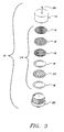
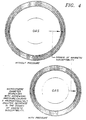
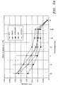

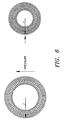

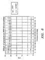
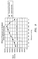

Classifications
-
- A—HUMAN NECESSITIES
- A61—MEDICAL OR VETERINARY SCIENCE; HYGIENE
- A61K—PREPARATIONS FOR MEDICAL, DENTAL OR TOILETRY PURPOSES
- A61K9/00—Medicinal preparations characterised by special physical form
- A61K9/10—Dispersions; Emulsions
- A61K9/127—Liposomes
-
- A—HUMAN NECESSITIES
- A61—MEDICAL OR VETERINARY SCIENCE; HYGIENE
- A61K—PREPARATIONS FOR MEDICAL, DENTAL OR TOILETRY PURPOSES
- A61K41/00—Medicinal preparations obtained by treating materials with wave energy or particle radiation ; Therapies using these preparations
- A61K41/0028—Disruption, e.g. by heat or ultrasounds, sonophysical or sonochemical activation, e.g. thermosensitive or heat-sensitive liposomes, disruption of calculi with a medicinal preparation and ultrasounds
-
- A—HUMAN NECESSITIES
- A61—MEDICAL OR VETERINARY SCIENCE; HYGIENE
- A61K—PREPARATIONS FOR MEDICAL, DENTAL OR TOILETRY PURPOSES
- A61K41/00—Medicinal preparations obtained by treating materials with wave energy or particle radiation ; Therapies using these preparations
- A61K41/0052—Thermotherapy; Hyperthermia; Magnetic induction; Induction heating therapy
-
- A—HUMAN NECESSITIES
- A61—MEDICAL OR VETERINARY SCIENCE; HYGIENE
- A61K—PREPARATIONS FOR MEDICAL, DENTAL OR TOILETRY PURPOSES
- A61K47/00—Medicinal preparations characterised by the non-active ingredients used, e.g. carriers or inert additives; Targeting or modifying agents chemically bound to the active ingredient
- A61K47/50—Medicinal preparations characterised by the non-active ingredients used, e.g. carriers or inert additives; Targeting or modifying agents chemically bound to the active ingredient the non-active ingredient being chemically bound to the active ingredient, e.g. polymer-drug conjugates
- A61K47/69—Medicinal preparations characterised by the non-active ingredients used, e.g. carriers or inert additives; Targeting or modifying agents chemically bound to the active ingredient the non-active ingredient being chemically bound to the active ingredient, e.g. polymer-drug conjugates the conjugate being characterised by physical or galenical forms, e.g. emulsion, particle, inclusion complex, stent or kit
- A61K47/6905—Medicinal preparations characterised by the non-active ingredients used, e.g. carriers or inert additives; Targeting or modifying agents chemically bound to the active ingredient the non-active ingredient being chemically bound to the active ingredient, e.g. polymer-drug conjugates the conjugate being characterised by physical or galenical forms, e.g. emulsion, particle, inclusion complex, stent or kit the form being a colloid or an emulsion
- A61K47/6911—Medicinal preparations characterised by the non-active ingredients used, e.g. carriers or inert additives; Targeting or modifying agents chemically bound to the active ingredient the non-active ingredient being chemically bound to the active ingredient, e.g. polymer-drug conjugates the conjugate being characterised by physical or galenical forms, e.g. emulsion, particle, inclusion complex, stent or kit the form being a colloid or an emulsion the form being a liposome
-
- A—HUMAN NECESSITIES
- A61—MEDICAL OR VETERINARY SCIENCE; HYGIENE
- A61K—PREPARATIONS FOR MEDICAL, DENTAL OR TOILETRY PURPOSES
- A61K47/00—Medicinal preparations characterised by the non-active ingredients used, e.g. carriers or inert additives; Targeting or modifying agents chemically bound to the active ingredient
- A61K47/50—Medicinal preparations characterised by the non-active ingredients used, e.g. carriers or inert additives; Targeting or modifying agents chemically bound to the active ingredient the non-active ingredient being chemically bound to the active ingredient, e.g. polymer-drug conjugates
- A61K47/69—Medicinal preparations characterised by the non-active ingredients used, e.g. carriers or inert additives; Targeting or modifying agents chemically bound to the active ingredient the non-active ingredient being chemically bound to the active ingredient, e.g. polymer-drug conjugates the conjugate being characterised by physical or galenical forms, e.g. emulsion, particle, inclusion complex, stent or kit
- A61K47/6921—Medicinal preparations characterised by the non-active ingredients used, e.g. carriers or inert additives; Targeting or modifying agents chemically bound to the active ingredient the non-active ingredient being chemically bound to the active ingredient, e.g. polymer-drug conjugates the conjugate being characterised by physical or galenical forms, e.g. emulsion, particle, inclusion complex, stent or kit the form being a particulate, a powder, an adsorbate, a bead or a sphere
- A61K47/6925—Medicinal preparations characterised by the non-active ingredients used, e.g. carriers or inert additives; Targeting or modifying agents chemically bound to the active ingredient the non-active ingredient being chemically bound to the active ingredient, e.g. polymer-drug conjugates the conjugate being characterised by physical or galenical forms, e.g. emulsion, particle, inclusion complex, stent or kit the form being a particulate, a powder, an adsorbate, a bead or a sphere the form being a microcapsule, nanocapsule, microbubble or nanobubble
-
- A—HUMAN NECESSITIES
- A61—MEDICAL OR VETERINARY SCIENCE; HYGIENE
- A61K—PREPARATIONS FOR MEDICAL, DENTAL OR TOILETRY PURPOSES
- A61K49/00—Preparations for testing in vivo
- A61K49/06—Nuclear magnetic resonance [NMR] contrast preparations; Magnetic resonance imaging [MRI] contrast preparations
- A61K49/18—Nuclear magnetic resonance [NMR] contrast preparations; Magnetic resonance imaging [MRI] contrast preparations characterised by a special physical form, e.g. emulsions, microcapsules, liposomes
- A61K49/1806—Suspensions, emulsions, colloids, dispersions
- A61K49/1815—Suspensions, emulsions, colloids, dispersions compo-inhalant, e.g. breath tests
-
- A—HUMAN NECESSITIES
- A61—MEDICAL OR VETERINARY SCIENCE; HYGIENE
- A61K—PREPARATIONS FOR MEDICAL, DENTAL OR TOILETRY PURPOSES
- A61K49/00—Preparations for testing in vivo
- A61K49/06—Nuclear magnetic resonance [NMR] contrast preparations; Magnetic resonance imaging [MRI] contrast preparations
- A61K49/18—Nuclear magnetic resonance [NMR] contrast preparations; Magnetic resonance imaging [MRI] contrast preparations characterised by a special physical form, e.g. emulsions, microcapsules, liposomes
- A61K49/1818—Nuclear magnetic resonance [NMR] contrast preparations; Magnetic resonance imaging [MRI] contrast preparations characterised by a special physical form, e.g. emulsions, microcapsules, liposomes particles, e.g. uncoated or non-functionalised microparticles or nanoparticles
- A61K49/1821—Nuclear magnetic resonance [NMR] contrast preparations; Magnetic resonance imaging [MRI] contrast preparations characterised by a special physical form, e.g. emulsions, microcapsules, liposomes particles, e.g. uncoated or non-functionalised microparticles or nanoparticles coated or functionalised microparticles or nanoparticles
-
- A—HUMAN NECESSITIES
- A61—MEDICAL OR VETERINARY SCIENCE; HYGIENE
- A61K—PREPARATIONS FOR MEDICAL, DENTAL OR TOILETRY PURPOSES
- A61K49/00—Preparations for testing in vivo
- A61K49/22—Echographic preparations; Ultrasound imaging preparations ; Optoacoustic imaging preparations
-
- A—HUMAN NECESSITIES
- A61—MEDICAL OR VETERINARY SCIENCE; HYGIENE
- A61K—PREPARATIONS FOR MEDICAL, DENTAL OR TOILETRY PURPOSES
- A61K49/00—Preparations for testing in vivo
- A61K49/22—Echographic preparations; Ultrasound imaging preparations ; Optoacoustic imaging preparations
- A61K49/222—Echographic preparations; Ultrasound imaging preparations ; Optoacoustic imaging preparations characterised by a special physical form, e.g. emulsions, liposomes
- A61K49/223—Microbubbles, hollow microspheres, free gas bubbles, gas microspheres
-
- A—HUMAN NECESSITIES
- A61—MEDICAL OR VETERINARY SCIENCE; HYGIENE
- A61K—PREPARATIONS FOR MEDICAL, DENTAL OR TOILETRY PURPOSES
- A61K49/00—Preparations for testing in vivo
- A61K49/22—Echographic preparations; Ultrasound imaging preparations ; Optoacoustic imaging preparations
- A61K49/222—Echographic preparations; Ultrasound imaging preparations ; Optoacoustic imaging preparations characterised by a special physical form, e.g. emulsions, liposomes
- A61K49/227—Liposomes, lipoprotein vesicles, e.g. LDL or HDL lipoproteins, micelles, e.g. phospholipidic or polymeric
-
- A—HUMAN NECESSITIES
- A61—MEDICAL OR VETERINARY SCIENCE; HYGIENE
- A61K—PREPARATIONS FOR MEDICAL, DENTAL OR TOILETRY PURPOSES
- A61K9/00—Medicinal preparations characterised by special physical form
- A61K9/10—Dispersions; Emulsions
- A61K9/127—Liposomes
- A61K9/1277—Processes for preparing; Proliposomes
- A61K9/1278—Post-loading, e.g. by ion or pH gradient
-
- A—HUMAN NECESSITIES
- A61—MEDICAL OR VETERINARY SCIENCE; HYGIENE
- A61K—PREPARATIONS FOR MEDICAL, DENTAL OR TOILETRY PURPOSES
- A61K9/00—Medicinal preparations characterised by special physical form
- A61K9/48—Preparations in capsules, e.g. of gelatin, of chocolate
- A61K9/50—Microcapsules having a gas, liquid or semi-solid filling; Solid microparticles or pellets surrounded by a distinct coating layer, e.g. coated microspheres, coated drug crystals
- A61K9/5005—Wall or coating material
- A61K9/5015—Organic compounds, e.g. fats, sugars
-
- G—PHYSICS
- G01—MEASURING; TESTING
- G01R—MEASURING ELECTRIC VARIABLES; MEASURING MAGNETIC VARIABLES
- G01R33/00—Arrangements or instruments for measuring magnetic variables
- G01R33/20—Arrangements or instruments for measuring magnetic variables involving magnetic resonance
- G01R33/44—Arrangements or instruments for measuring magnetic variables involving magnetic resonance using nuclear magnetic resonance [NMR]
- G01R33/48—NMR imaging systems
- G01R33/54—Signal processing systems, e.g. using pulse sequences ; Generation or control of pulse sequences; Operator console
- G01R33/56—Image enhancement or correction, e.g. subtraction or averaging techniques, e.g. improvement of signal-to-noise ratio and resolution
- G01R33/5601—Image enhancement or correction, e.g. subtraction or averaging techniques, e.g. improvement of signal-to-noise ratio and resolution involving use of a contrast agent for contrast manipulation, e.g. a paramagnetic, super-paramagnetic, ferromagnetic or hyperpolarised contrast agent
-
- A—HUMAN NECESSITIES
- A61—MEDICAL OR VETERINARY SCIENCE; HYGIENE
- A61M—DEVICES FOR INTRODUCING MEDIA INTO, OR ONTO, THE BODY; DEVICES FOR TRANSDUCING BODY MEDIA OR FOR TAKING MEDIA FROM THE BODY; DEVICES FOR PRODUCING OR ENDING SLEEP OR STUPOR
- A61M5/00—Devices for bringing media into the body in a subcutaneous, intra-vascular or intramuscular way; Accessories therefor, e.g. filling or cleaning devices, arm-rests
- A61M5/178—Syringes
- A61M5/31—Details
- A61M5/3145—Filters incorporated in syringes
Definitions
- This invention relates to the field of magnetic resonance imaging, more specifically to the use of stabilized gas filled microspheres as contrast media for magnetic resonance imaging (MRI).
- MRI magnetic resonance imaging
- Imaging techniques There are a variety of imaging techniques that have been used to diagnose disease in humans.
- One of the first imaging techniques employed was X-rays.
- X-rays the images produced of the patients' body reflect the different densities of body structures.
- contrast agents are employed to increase the density of tissues of interest as compared to surounding tissues to make the tissues of interest more visible on X-ray.
- Barium and iodinated contrast media are used extensively for X-ray gastrointestinal studies to visualize the esophagus, stomach, intestines and rectum.
- contrast agents are used for X-ray computed tomographic studies (that is, computer assisted tomography or CAT) to improve visualization of the gastrointestinal tract and to provide, for example, a contrast between the tract and the structures adjacent to it, such as the vessels or lymph nodes.
- CAT computer assisted tomography
- Such contrast agents permit one to increase the density inside the esophagus, stomach, intestines and rectum, and allow differentiation of the gastrointestinal system from surrounding structures.
- Magnetic resonance imaging is a relatively new imaging technique which, unlike X-rays, does not utilize ionizing radiation.
- MRI can make cross-sectional images of the body, however MRI has the additional advantage of being able to make images in any scan plane (i.e., axial, coronal, sagittal or orthogonal).
- scan plane i.e., axial, coronal, sagittal or orthogonal.
- MRI employs a magnetic field, radio frequency energy and magnetic field gradients to make images of the body.
- the contrast or signal intensity differences between tissues mainly reflect the T1 (longitudinal) and T2 (transverse) relaxation values and the proton density (effectively, the free water content) of the tissues.
- T1 longitudinal
- T2 transverse
- proton density effectively, the free water content
- paramagnetic contrast agents contain unpaired electrons which act as small local magnets within the main magnetic field to increase the rate of longitudinal (T1) and transverse (T2) relaxation.
- Most paramagnetic contrast agents are metal ions which in most cases are toxic. In order to decrease toxicity, these metal ions are generally chelated using ligands. The resultant paramagnetic metal ion complexes have decreased toxicity.
- Metal oxides most notably iron oxides, have also been tested as MRI contrast agents. While small particles of iron oxide, e.g., under 20 nm diameter, may have paramagnetic relaxation properties, their predominant effect is through bulk susceptibility. Therefore magnetic particles have their predominant effect on T2 relaxation.
- Nitroxides are another class of MRI contrast agent which are also paramagnetic. These have relatively low relaxivity and are generally less effective than paramagnetic ions as MRI contrast agents. All of these contrast agents can suffer from some toxic effects in certain use contexts and none of them are ideal for use as perfusion contrast agents by themselves.
- the existing MRI contrast agents suffer from a number of limitations.
- positive contrast agents are known to exhibit increased image noise arising from intrinsic peristaltic motions and motions from respiration or cardiovascular action.
- Positive contrast agents such as Gd-DTPA are subject to the further complication that the signal intensity depends upon the concentration of the agent as well as the pulse sequence used. Absorption of contrast agent from the gastrointestinal tract, for example, complicates interpretation of the images, particularly in the distal portion of the small intestine, unless sufficiently high concentrations of the paramagnetic species are used (Kornmesser et al., Magn. Reson. Imaging, 6:124 (1988)).
- Negative contrast agents by comparison, are less sensitive to variation in pulse sequence and provide more consistent contrast.
- Negative contrast agents typically exhibit superior contrast to fat, however on T1-weighted images, positive contrast agents exhibit superior contrast versus normal tissue. Since most pathological tissues exhibit longer T1 and T2 than normal tissue, they will appear dark on T1-weighted and bright on T2-weighted images. This would indicate that an ideal contrast agent should appear bright on T1-weighted images and dark on T2-weighted images. Many of the currently available MRI contrast media fail to meet these dual criteria.
- Toxicity is another problem with the existing contrast agents. With any drug there is some toxicity, the toxicity generally being dose related. With the ferrites there are often symptoms of nausea after oral administration, as well as flatulence and a transient rise in serum iron.
- the paramagnetic contrast agent Gd-DTPA is an organometallic complex of gadolinium coupled with the complexing agent diethylene triamine pentaacetic acid. Without coupling, the free gadolinium ion is highly toxic. Furthermore, the peculiarities of the gastrointestinal tract, for example, wherein the stomach secretes acids and the intestines release alkalines, raise the possibility of decoupling and separation of the free gadolinium or other paramagnetic agent from the complex as a result of these changes in pH during gastrointestinal use. Certainly, minimizing the dose of paramagnetic agents is important for minimizing any potential toxic effects.
- the present invention is directed, inter alia, to this important end.
- Widder et al . published application EP-A-0 324 938 discloses stabilized microbubble-type ultrasonic imaging agents produced from heat-denaturable biocompatible protein, e.g., albumin, hemoglobin, and collagen.
- microbubbles A Novel MR Susceptibility Contrast Agent.
- the microbubbles which are utilized comprise air coated with a shell of human albumin.
- the stabilized gas filled microspheres of the present invention are not suggested.
- any gas be stabilized with flexible compounds.
- Proteins such as albumin may be used to stabilize the bubbles but the resulting bubble shells may be brittle and inflexible. This is undesirable for several reasons.
- a brittle coating limits the capability of the bubble to expand and collapse. As the bubble encounters different pressure regions within the body (e.g., moving from the venous system into the arteries upon circulation through the heart) a brittle shell may break and the gas will be lost. This limits the effective period of time for which useful contrast can be obtained in vivo from the bubbles contrast agents. Also such brittle, broken fragments can be potentially toxic.
- the inflexible nature of brittle coatings such as albumin, and stiff resulting bubbles make it extremely difficult to measure pressure in vivo.
- oligolamellar liposomes which are multilamellar liposomes with increased aqueous space between bilayers or have liposomes nested within bilayers in a nonconcentric fashion, and thus contain internally separated bilayers.
- Their use as ultrasonic contrast agents to enhance ultrasonic imaging, and in monitoring a drug delivered in a liposome administered to a patient, is also described.
- stabilized gas filled microspheres are extremely effective, non-toxic contrast agents for MRI.
- the present invention is directed to a contrast medium useful for magnetic resonance imaging, said contrast medium comprising stabilized gas filled microspheres, wherein the gas is a biocompatible gas, e.g., nitrogen or perfluoropropane, but may also be derived from a gaseous precursor, e.g., perfluorooctylbromide, and the microspheres are stabilized by being formed from a stabilizing compound, e.g., a biocompatible lipid or polymer.
- the present invention may be carried out, often with considerable attendant advantage, by using gaseous precursors to form the gas of the gas filled microspheres. These gaseous precursors may be activated by a number of factors, but preferably are temperature activated.
- Such a gaseous precursor is a compound which, at a selected activation or transition temperature, changes phases from a liquid to a gas. Activation thus takes place by increasing the temperature of the compound from a point below, to a point above the activation or transition temperature.
- the lipid may be in the form of a monolayer or bilayer, and the mono- or bilayer lipids may be used to form a series of concentric mono-or bilayers.
- the lipid may be used to form a unilamellar liposome (comprised of one monolayer or bilayer lipid), an oligolamellar liposome (comprised of two or three monolayer or bilayer lipids) or a multilamellar liposome (comprised of more than three monolayer or bilayer lipids).
- the biocompatible lipid comprises a phospholipid.
- the contrast medium may include paramagnetic and/or superparamagnetic contrast agents, preferably encapsulated by the microspheres.
- the contrast medium may further comprise a liquid fluorocarbon compound, e.g., a perfluorocarbon, to further stablize the microspheres.
- the fluorocarbon liquid is encapsulated by the microspheres.
- the present invention also concerns a method for preparing stabilized gas or gaseous precursor filled lipid based microspheres for use as a magnetic resonance imaging contrast medium, comprising the step of agitating an aqueous suspension of the lipid (that is, the lipid stabilizing compound), in the presence of a gas or gasesous precursor, resulting in gas or gaseous precursor filled microspheres.
- the agitation step is carried out at a temperature below the gel to liquid crystalline phase transition temperature of the lipid in order to achieve a preferred end product.
- the gaseous precursor filled microsphere composition is generally maintained at a temperature at which the gaseous precursor is liquid until administration to the patient. At the time of administration the temperature may, if desired, be raised to activate the gaseous precursor to form a gas and the resultant gas filled microsphere then administered to the patient. Alternatively, the gaseous precursor filled microspheres may, if desired, be administered without raising the temperature, and the gaseous precursor allowed to form a gas as a result of the temperature of the patient. The composition may be agitated, if necessary, prior to administration.
- the present invention further pertains to a method of providing an image of an internal region of a patient, especially an image of the vasculature (including the cardiovascular region), particularly during perfusion, and of the gastrointestinal region of said patient, said method comprising (i) administering to the patient the foregoing contrast medium, and (ii) scanning the patient using magnetic resonance imaging to obtain visible images of said region.
- the present invention also encompasses a method for diagnosing the presence of diseased tissue in a patient, especially in the vasculature (including the cardiovascular region), particularly during perfusion, and in the gastrointestinal region of said patient, said method comprising (i) administering to the patient the foregoing contrast medium, and (ii) scanning the patient using magnetic resonance imaging to obtain visible images of any diseased tissue in the region.
- the present invention is directed, inter alia, to stabilized gas filled microspheres. While not intending to be bound by any particular theory of operation, the present invention is believed to rely, at least in part, on the fact that gas, liquid and solid phases have different magnetic susceptibilities. At the interface of gas and water, for example, the magnetic domains are altered and this results in dephasing of the spins of, e.g ., the hydrogen nuclei. In imaging this is seen as a decrease in signal intensity adjacent to the gas/water interface. This effect is more marked on T2 weighted images and most prominent on gradient echo pulse sequences. The effect is increased by using narrow bandwidth extended read-out pulse sequences. The longer the echo time on a gradient echo pulse sequence, the greater the effect ( i.e ., the greater the degree and size of signal loss).
- the stabilized gas filled microspheres of the present invention are believed to rely on this phase magnetic susceptibility difference, as well as on the other characteristics described in more detail herein, to provide high performance level magnetic resonance imaging contrast agents.
- the microspheres are formed from, i.e., created out of, a matrix of stabilizing compounds which permit the gas filled microspheres to be established and thereafter retain their size and shape for the period of time required to be useful in magnetic resonance imaging.
- These stabilizing compounds are most typically those which have a hydrophobic/hydrophilic character which allows them to form monolayers or bilayers, etc., and microspheres, in the presence of water.
- water, saline or some other water-based medium often referred to hereafter as a diluent, is generally an aspect of the stabilized gas filled microsphere contrast medium of the present invention.
- the stabilizing compound may, in fact, be a mixture of compounds which contribute various desirable attributes to the stabilized microspheres.
- compounds which assist in the dissolution or dispersion of the fundamental stabilizing compound have been found advantageous.
- a further element of the stabilized microspheres is a gas, which can be a gas at the time the microspheres are made, or can be a gaseous precursor which, responsive to an activating factor, such as temperature, is transformed from the liquid phase to the gas phase.
- the microspheres of the invention encapsulate a gas and/or gaseous precursor.
- gas filled and/or gaseous precursor filled means that the microspheres to which the present invention is directed, have an interior volume that is comprised of at least about 10% gas and gasesous precursor, preferably at least about 25% gas and gaseous precursor, more preferably at least about 50% gas and gaseous precursor, even more preferably at least about 75% gas and gaseous precursor, and most preferably at least about 90% gas and gaseous precursor.
- the interior microsphere volume comprise at least about 10% gas, preferably at least about 25%, 50%, 75%, and most preferably at least about 90% gas.
- gases include, for example, air, nitrogen, carbon dioxide, oxygen, argon, fluorine, xenon, neon, helium, or any and all combinations thereof. Of such gases, nitrogen is preferred.
- various fluorinated gaseous compounds such as various perfluorocarbon, hydrofluorocarbon, and sulfur hexafluoride gases may be utilized in the preparation of the gas filled microspheres.
- the gases discussed in Quay, published application WO 93/05819, including the high "Q" factor gases described therein, the disclosures of which are hereby incorporated herein by reference in their entirety, may be employed.
- paramagnetic gases or gases such as 17 O may be used.
- gases perfluorocarbons and sulfur hexafluoride are preferred.
- Suitable perfluorcarbon gases include, for example, perfluorobutane, perfluorocyclobutane, perfluoromethane, perfluoroethane, perfluoropropane, and perfluoropentane, most preferably perfluoropropane.
- gases include, for example, perfluorobutane, perfluorocyclobutane, perfluoromethane, perfluoroethane, perfluoropropane, and perfluoropentane, most preferably perfluoropropane.
- gases such as a perfluorocarbon gas and another type of gas such as oxygen, etc. Indeed, it is believed that a combination of gases may be particularly useful in magnetic resonance imaging applications.
- Table 3 in the Examples below shows the R2 (1/T2/mmol/L.sec-1) for different gases in lipid microspheres (the higher the R2 relaxation values, the more effective as an MRI contrast medium). As Table 3 shows, there may be dramatic differences in the relaxivity of different gas filled microspheres.
- gas and gaseous precursor filled microspheres be made from stabilizing compounds, it is preferred that a rather highly stable gas be utilized as well.
- highly stable gas is meant a gas selected from those gases which will have low (limited) solubility and diffusability in aqueous media. Gases such as perfluorocarbons are less diffusible and relatively insoluble and as such are easier to stabilize into the form of bubbles in aqueous media.
- gaseous precursors are an optional embodiment of the present invention.
- perfluorocarbons have been found to be suitable for use as gaseous precursors.
- a given perfluorocarbon may be used as a gaseous precursor, i.e ., in the liquid state when the microspheres of the present invention are first made, or may be used as a gas directly, i.e ., in the gas state, to make the gas and gaseous precursor filled microspheres.
- a perfluorocarbon is a gas or liquid depends, of course, on its liquid/gas phase transition temperature, or boiling point.
- one of the more preferred perfluorocarbons is perfluoropentane, which has a liquid/gas phase transition temperature or boiling point of 27°C, which means that it will be a liquid at ordinary room temperature, but will become a gas in the environment of the human body, where the temperature will be above its liquid/gas phase transition temperature or boiling point.
- perfluoropentane is a gaseous precursor.
- there is perfluorobutane and perflurohexane the next closest homologs of perfluoropentane.
- liquid/gas phase transition temperature of perfluorobutane is 4°C and that of perfluorohexane is 57°C, making the former potentially a gaseous precursor, but generally more useful as a gas, while the latter would generally be a gaseous precursor, except under unusual circumstances, because of its high boiling point.
- fluorinated compound especially a perfluorocarbon compound
- a fluorinated compound which will be in the liquid state at the temperature of use of the microspheres of the present invention, to assist or enhance the stability of said gas and gaseous precursor filled microspheres.
- fluorinated compounds include various liquid fluorinated compounds, such as fluorinated surfactants manufactured by the DuPont Company (Wilmington, DE), e.g., ZonylTM, as well as liquid perfluorocarbons.
- fluorinated compounds are perfluorocarbons.
- Suitable perfluorocarbons useful as additional stabilizing agents include perfluorooctylbromide (PFOB), perfluorodecalin, perfluorododecalin, perfluorooctyliodide, perfluorotripropylamine, and perfluorotributylamine.
- PFOB perfluorooctylbromide
- perfluorodecalin perfluorododecalin
- perfluorooctyliodide perfluorotripropylamine
- perfluorotributylamine perfluorotributylamine.
- perfluorocarbons over six carbon atoms in length will not be gaseous, i.e ., in the gas state, but rather will be liquids, i.e ., in the liquid state, at normal human body temperature.
- These compounds may, however, additionally be utilized in preparing the stabilized gas and gaseous precursor filled microspheres used in the present invention
- this perfluorocarbon is perfluorooctylbromide or perfluorohexane, which is in the liquid state at room temperature.
- the gas which is present may be, e.g ., nitrogen or perfluoropropane, or may be derived from a gaseous precursor, which may also be a perfluorocarbon, e.g. , perfluoropentane.
- the microspheres of the present invention would be prepared from a mixture of perfluorocarbons, which for the examples given, would be perfluoropropane (gas) or perfluoropentane (gaseous precursor) and perfluorooctylbromide (liquid).
- liquid fluorinated fluorinated copuond is situated at the interface between the gas and the membrane surface of the microsphere.
- a further stabilizing layer of liquid fluorinated compound on the internal surface of the stabilizing compound, e.g ., a biocompatible lipid used to form the microsphere, and this perfluorocarbon layer also serves the purpose of preventing the gas from diffusing through the microsphere membrane.
- a gaseous precursor within the context of the present invention, is a liquid at the temperature of manufacture and/or storage, but becomes a gas at least at or during the time of use.
- a liquid fluorinated compound such as a perfluorocarbon
- a gas or gasesous precursor ordinarily used to make the microspheres of the present invention
- a gas or gaseous precursor such as a perfluorocarbon gaseous precursor, e.g., perfluoropentane
- a perfluorocarbon which remains liquid after administration to a patient, i.e. , whose liquid to gas phase transition temperature is above the body temperature of the patient, e.g. , perfluoroctylbromide.
- biocompatible gas or gaseous precursor may be used to form the stabilized gas and gaseous precursor filled microspheres.
- biocompatible is meant a gas or gaseous precursor which, when introduced into the tissues of a human patient, will not result in any degree of unacceptable toxicity, including allergenic responses and disease states, and preferably are inert.
- a gas or gaseous precursor should also be suitable for making gas and gaseous precursor filled microspheres, as described herein.
- the size of the gas or gaseous precursor filled microspheres becomes stabilized when the stabilizing compounds described herein are employed; and the size of the microspheres can then be adjusted for the particular intended MRI end use.
- magnetic resonance imaging of the vasculature may require microspheres that are no larger that about 30 ⁇ in diameter, and that are preferably smaller, e.g. , no larger than about 12 ⁇ in diameter.
- the size of the gas filled microspheres can be adjusted, if desired, by a variety of procedures including microemulsification, vortexing, extrusion, filtration, sonication, homogenization, repeated freezing and thawing cycles, extrusion under pressure through pores of defined size, and similar methods.
- the microspheres are generally under 30 ⁇ in mean diameter, and are preferably under about 12 ⁇ in mean diameter.
- the microspheres can be appreciably under a micron, even under 100 nm diameter.
- the microspheres can be much larger, e.g., up to a millimeter in size, but microspheres between 20 ⁇ and 100 ⁇ in mean diameter are preferred.
- the embodiments of the present invention may also include, with respect to their preparation, formation and use, gaseous precursors that can be activated by temperature. Further below is set out a table listing a series of gaseous precursors which undergo phase transitions from liquid to gaseous states at relatively close to normal body temperature (37° C) or below, and the size of the emulsified droplets that would be required to form a microbubble of a maximum size of 10 microns.
- gaseous precursors that may be used to form microspheres of defined size.
- the list is not intended to be limiting, since it is possible to use other gaseous precursors for that purpose.
- virtually any liquid can be used to make gaseous precursors so long as it is capable of undergoing a phase transition to the gas phase upon passing through the appropriate temperature, so that at least at some point in use it provides a gas.
- Suitable gaseous precursors for use in the present invention are the following: hexafluoro acetone, isopropyl acetylene, allene, tetrafluoro-allene, boron trifluoride, isobutane, 1,2-butadiene, 2,3-butadiene, 1,3-butadiene, 1,2,3-trichloro-2-fluoro-1,3-butadiene, 2-methyl-1,3-butadiene, hexafluoro-1,3-butadiene, butadiyne, 1-fluorobutane, 2-methyl-butane, decafluorobutane, 1-butene, 2-butene, 2-methyl-1-butene, 3-methyl-1-butene, perfluoro-1-butene, perfluoro-2-butene, 4-phenyl-3-butene-2-one, 2-methyl-1-butene-3-yne, butyl nitrate, 1-buty
- the perfluorocarbons are preferred for use as the gas or gaseous precursors, as well as additional stabilizing components. Included in such perfluorocarbon compositions are saturated perfluorocarbons, unsaturated perfluorocarbons, and cyclic perfluorocarbons.
- the saturated perfluorocarbons which are usually preferred, have the formula C n F 2n+2 , where n is from 1 to 12, preferably 2 to 10, more preferably 4 to 8, and most preferably 5.
- saturated perfluorocarbons are the following: tetrafluoromethane, hexafluoroethane, octafluoropropane, decafluorobutane, dodecafluoropentane, perfluorohexane, and perfluoroheptane.
- Cyclic perfluorocarbons which have the formula C n F 2n , where n is from 3 to 8, preferably 3 to 6, may also be preferred, and include, e.g. , hexafluorocyclopropane, octafluorocyclobutane, and decafluorocyclopentane.
- the gas entrapped in the microsphere has a solubility less than that of oxygen, i.e.
- the gas entrapped in the microsphere possesses a solubility in water less than that of air; and even more preferably, the gas entrapped in the microsphere possesses a solubility in water less than that of nitrogen.
- One or more stabilizing compounds are employed to form the microspheres, and to assure continued encapsulation of the gases or gaseous precursors. Even for relatively insoluble, non-diffusible gases such as perfluoropropane or sulfur hexafluoride, improved microsphere preparations are obtained when one or more stabilizing compounds are utilized in the formation of the gas and gaseous precursor filled microspheres. These compounds maintain the stability and the integrity of the microspheres with regard to their size, shape and/or other attributes,
- stable or “stabilized”, as used herein, means that the microspheres are substantially resistant to degradation, i.e. , are resistant to the loss of microsphere structure or encapsulated gas or gaseous precursor for a useful period of time.
- the microspheres of the invention have a good shelf life, often retaining at least about 90 percent by volume of its original structure for a period of at least about two or three weeks under normal ambient conditions, although it is preferred that this period be at least a month, more at least preferably two months, even more preferably at least six months, still more preferably eighteen months, and most preferably three years.
- the gas and gaseous precursor filled microspheres typically have a good shelf life, sometimes even under adverse conditions, such as temperatures and pressures which are above or below those experienced under normal ambient conditions.
- the stability of the microspheres of the present invention is attributable, at least in part, to the materials from which said microspheres are made, and it is often not necessary to employ additional stabilizing additives, although it is optional and often preferred to do so; and such additional stabilizing agents and their characteristics are explained in more detail herein.
- the materials from which the microspheres used in the present invention are constructed are preferably biocompatible lipid or polymer materials, and of these, the biocompatible lipids are especially preferred.
- these microspheres may be conveniently made on site.
- the lipids and polymers employed in preparing the microspheres of the invention are biocompatible.
- biocompatible is meant a lipid or polymer which, when introduced into the tissues of a human patient, will not result in any degree of unacceptable toxicity, including allergenic responses and disease states.
- the lipids or polymers are inert.
- lipid materials For the biocompatible lipid materials, it is preferred that such lipid materials be what is often referred to as "amphiphilic” in nature (i.e ., polar lipid), by which is meant any composition of matter which has, on the one hand, lipophilic, i.e., hydrophobic properties, while on the other hand, and at the same time, having lipophobic, i.e. , hydrophilic properties.
- Hydrophilic groups may be charged moieties or other groups having an affinity for water.
- Natural and synthetic phospholipids are examples of lipids useful in preparing the stabilized microspheres used in the present invention. They contain charged phosphate "head” groups which are hydrophilic, attached to long hydrocarbon tails, which are hydrophobic. This structure allows the phospholipids to achieve a single bilayer (unilamellar) arrangement in which all of the water-insoluble hydrocarbon tails are in contact with one another, leaving the highly charged phosphate head regions free to interact with a polar aqueous environment. It will be appreciated that a series of concentric bilayers are possible, i.e. , oligolamellar and multilamellar, and such arrangements are also contemplated to be an aspect of the present invention. The ability to form such bilayer arrangements is one feature of the lipid materials useful in the present invention.
- the lipid may alternatively be in the form of a monolayer, and the monolayer lipids may be used to form a single monolayer (unilamellar) arrangement.
- the monolayer lipid may be used to form a series of concentric monolayers, i.e. , oligolamellar or multilamellar, and such arrangements are also considered to be within the scope of the invention.
- phase transition temperature is the temperature at which a lipid bilayer will convert from a gel state to a liquid crystalline state.
- lipids used in the present invention by incorporating at least a small amount, i.e ., about 1 to about 10 mole percent of the total lipid, of a negatively charged lipid into the lipid from which the gas and gaseous precursor filled microspheres are to be formed.
- Suitable negatively charged lipids include, e.g ., phosphatidylserine, phosphatidic acid, and fatty acids.
- Such negatively charged lipids provide added stability by counteracting the tendency of the microspheres to rupture by fusing together, i.e ., the negatively charged lipids tend to establish a uniform negatively charged layer on the outer surface of the microsphere, which will be repulsed by a similarly charged outer layer on the other microspheres. In this way, the microspheres will tend to be prevented from coming into touching proximity with each other, which would often lead to a rupture of the membrane or skin of the respective microspheres and consolidation of the contacting microspheres into a single, larger microsphere. A continuation of this process of consolidation will, of course, lead to significant degradation of the microspheres.
- the lipid material or other stabilizing compound used to form the microspheres is also preferably flexible, by which is meant, in the context of gas and gaseous precursor filled microspheres, the ability of a structure to alter its shape, for example, in order to pass through an opening having a size smaller than the microsphere.
- the lipids used may be of either natural, synthetic or semi-synthetic origin.
- Lipids which may be used to prepare the gas and gaseous precursor filled microspheres used in the present invention include but are not limited to: lipids such as fatty acids, lysolipids, phosphatidylcholine with both saturated and unsaturated lipids including dioleoylphosphatidylcholine; dimyristoylphosphatidylcholine; dipentadecanoylphosphatidylcholine; dilauroylphosphatidylcholine; dipalmitoylphosphatidylcholine (DPPC); distearoylphosphatidylcholine (DSPC); phosphatidylethanolamines such as dioleoylphosphatidylethanolamine and dipalmitoylphosphatidylethanolamine (DPPE); phosphatidylserine; phosphatidylglycerol; phosphatidylinositol; sphingolipids such as sphingomyelin; glycolipids such as
- ceramides non-ionic liposomes including niosomes such as polyoxyethylene fatty acid esters, polyoxyethylene fatty alcohols, polyoxyethylene fatty alcohol ethers, polyoxyethylated sorbitan fatty acid esters, glycerol polyethylene glycol oxystearate, glycerol polyethylene glycol ricinoleate, ethoxylated soybean sterols, ethoxylated castor oil, polyoxyethylene-polyoxypropylene polymers, and polyoxyethylene fatty acid stearates; sterol aliphatic acid esters including cholesterol sulfate, cholesterol butyrate, cholesterol iso-butyrate, cholesterol palmitate, cholesterol stearate, lanosterol acetate, ergosterol palmitate, and phytosterol n-butyrate; sterol esters of sugar acids including cholesterol glucuroneide, lanos
- cationic lipids such as DOTMA, N-[1-(2,3-dioleoyloxy)propyl]-N,N,N-trimethylammoium chloride; DOTAP, 1,2-dioleoyloxy-3-(trimethylammonio)gropane; and DOTB, 1,2-dioleoyl-3-(4'-trimethyl-ammonio)butanoyl-sn-glycerol may be used.
- DOTMA N-[1-(2,3-dioleoyloxy)propyl]-N,N,N-trimethylammoium chloride
- DOTAP 1,2-dioleoyloxy-3-(trimethylammonio)gropane
- DOTB 1,2-dioleoyl-3-(4'-trimethyl-ammonio)butanoyl-sn-glycerol
- the molar ratio of cationic lipid to non-cationic lipid in the liposome may be, for example, 1:1000, 1:100, preferably, between 2:1 to 1:10, more preferably in the range between 1:1 to 1:2.5 and most preferably 1:1 (ratio of mole amount cationic lipid to mole amount non-cationic lipid, e.g., DPPC).
- a wide variety of lipids may comprise the non-cationic lipid when cationic lipid is used to construct the microsphere.
- this non-cationic lipid is dipalmitoylphosphatidylcholine, dipalmitoylphosphatidylethanolamine or dioleoylphosphatidylethanolamine.
- lipids bearing cationic polymers such as polylysine or polyarginine, as well as alkyl phosphonates, alkyl phosphinates, and alkyl phosphites, may also be used to construct the microspheres.
- the most preferred lipids are phospholipids, preferably DPPC, DPPE, DPPA and DSPC, and most preferably DPPC.
- examples of saturated and unsaturated fatty acids that may be used to prepare the stabilized microspheres used in the present invention, in the form of gas and gaseous precursor filled mixed micelles, may include molecules that may contain preferably between 12 carbon atoms and 22 carbon atoms in either linear or branched form. Hydrocarbon groups consisting of isoprenoid units and/or prenyl groups can be used as well.
- saturated fatty acids examples include, but are not limited to, lauric, myristic, palmitic, and stearic acids; examples of unsaturated fatty acids that may be used are, but are not limited to, lauroleic, physeteric, myristoleic, palmitoleic, petroselinic, and oleic acids; examples of branched fatty acids that may be used are, but are not limited to, isolauric, isomyristic, isopalmitic, and isostearic acids.
- gas and gaseous precursor filled mixed micelles can also be composed of 5 carbon isoprenoid and prenyl groups.
- the biocompatible polymers useful as stabilizing compounds for preparing the gas and gaseous precursor filled microspheres used in the present invention can be of either natural, semi-synthetic (modified natural) or synthetic origin.
- polymer denotes a compound comprised of two or more repeating monomeric units, and preferably 10 or more repeating monomeric units.
- semi-synthetic polymer or modified natural polymer
- exemplary natural polymers suitable for use in the present invention include naturally occurring polysaccharides.
- Such polysaccharides include, for example, arabinans, fructans, fucans, galactans, galacturonans, glucans, mannans, xylans (such as, for example, inulin), levan, fucoidan, carrageenan, galatocarolose, pectic acid, pectin, amylose, pullulan, glycogen, amylopectin, cellulose, dextran, pustulan, chitin, agarose, keratan, chondroitan, dermatan, hyaluronic acid, alginic acid, xanthan gum, starch and various other natural homopolymer or heteropolymers such as those containing one or more of the following aldoses, ketoses, acids or amines: erythrose, threose, ribose, arabinose, xylose, lyxose, allose, altrose, glucose, mannose, gulose, idose, galacto
- Exemplary semi-synthetic polymers include carboxymethylcellulose, hydroxymethylcellulose, hydroxypropylmethylcellulose, methylcellulose, and methoxycellulose.
- Exemplary synthetic polymers suitable for use in the present invention include polyethylenes (such as, for example, polyethylene glycol, polyoxyethylene, and polyethylene terephthlate), polypropylenes (such as, for example, polypropylene glycol), polyurethanes (such as, for example, polyvinyl alcohol (PVA), polyvinylchloride and polyvinylpyrrolidone), polyamides including nylon, polystyrene, polylactic acids, fluorinated hydrocarbons, fluorinated carbons (such as, for example, polytetrafluoroethylene), and polymethylmethacrylate, and derivatives thereof.
- the polymer employed when intended to be used in the gastrointestinal tract, is one which has a relatively high water binding capacity.
- a polymer with a high water binding capacity binds a large amount of free water, enabling the polymer to carry a large volume of liquid through the gastrointestinal tract, thereby filling and distending the tract.
- the filled and distended gastrointestinal tract permits a clearer picture of the region.
- the polymer employed is also one which is not substantially degraded within and absorbed from the gastrointestinal region.
- polymers are preferably such that they are capable of displacing air and minimizing the formation of large air bubbles within the polymer composition.
- the stabilizing compound from which the stabilized gas and gaseous precursor filled microspheres are formed comprises three components: (1) a neutral (e.g., nonionic or zwitterionic) lipid, (2) a negatively charged lipid, and (3) a lipid bearing a hydrophilic polymer.
- a neutral (e.g., nonionic or zwitterionic) lipid e.g., a negatively charged lipid
- a lipid bearing a hydrophilic polymer e.g., lipid bearing a hydrophilic polymer.
- the amount of said negatively charged lipid will be greater than 1 mole percent of total lipid present, and the amount of lipid bearing a hydrophilic polymer will be greater than 1 mole percent of total lipid present.
- said negatively charged lipid be a phosphatidic acid.
- the lipid bearing a hydrophilic polymer will desirably be a lipid covalently bound to said polymer, and said polymer will preferably have a weight average molecular weight of from about 400 to about 100,000.
- Said hydrophilic polymer is preferably selected from the group consisting of polyethyleneglycol, polypropyleneglycol, polyvinylalcohol, and polyvinylpyrrolidone and copolymers thereof.
- the PEG or other polymer may be bound to the DPPE or other lipid through a covalent linkage, such as through an amide, carbamate or amine linkage.
- ester, ether, thioester, thioamide or disulfide (thioester) linkages may be used with the PEG or other polymer to bind the polymer to, for example, cholesterol or other phospholipids.
- the hydrophilic polymer is polyethyleneglycol
- a lipid bearing such a polymer will be said to be "PEGylated", which has reference to the abbreviation for polyethyleneglycol: "PEG”.
- Said lipid bearing a hydrophilic polymer is preferably dipalmitoylphosphatidylethanolamine-polyethyleneglycol 5000, i.e., a dipalmitoylphosphatidylethanolamine lipid having a polyethyleneglycol polymer of a mean weight average molecular weight of about 5000 attached thereto (DPPE-PEG5000); or distearoyl-phosphatidylethanolamine-polyethyleneglycol 5000.
- Preferred embodiments of the microsphere contemplated by the present invention would include, e.g. , 77.5 mole percent dipalmitoylphophatidylcholine (DPPC), with 12.5 mole percent of dipalmitoylphosphatidic acid (DPPA), and with 10 mole percent of dipalmitoylphosphatidylethanolamine-polyethyleneglycol-5000 (DPPE/PEG5000). These compositions in a 82/10/8 ratio of mole percentages, respectively, is also preferred.
- the DPPC component is effectively neutral, since the phosphtidyl portion is negatively charged and the choline portion is positively charged.
- the DPPA component which is negatively charged, is added to enhance stabilization in accordance with the mechanism described further above regarding negatively charged lipids as an additional agent.
- the third component, DPPE/PEG provides a PEGylated material bound to the lipid membrane or skin of the microsphere by the DPPE moiety, with the PEG moiety free to surround the microsphere membrane or skin, and thereby form a physical barrier to various enzymatic and other endogenous agents in the body whose function is to degrade such foreign materials. It is also theorized that the PEGylated material, because of its structural similarity to water, is able to defeat the action of the macrophages of the human immune system, which would otherwise tend to surround and remove the foreign object. The result is an increase in the time during which the stabilized microspheres can function as MRI contrast media.
- compositions of matter in addition to the biocompatible lipids and polymers described above, provided that the microspheres so prepared meet the stability and other criteria set forth herein.
- These compositions may be basic and fundamental, i.e. , form the primary basis for creating or establishing the stabilized gas and gaseous precursor filled microspheres.
- they may be auxiliary, i.e. , act as subsidiary or supplementary agents which either enhance the functioning of the basic stabilizing compound or compounds, or else contribute some desired property in addition to that afforded by the basic stabilizing compound.
- a given compound is a basic or an auxiliary agent, since the functioning of the compound in question is determined empirically, i.e. , by the results produced with respect to producing stabilized microspheres.
- these basic and auxiliary compounds may function, it has been observed that the simple combination of a biocompatible lipid and water or saline when shaken will often give a cloudy solution subsequent to autoclaving for sterilization. Such a cloudy solution may function a contrast agent, but is aesthetically objectionable and may imply instability in the form of undissolved or undispersed lipid particles.
- propylene glycol may be added to remove this cloudiness by facilitating dispersion or dissolution of the lipid particles.
- the propylene glycol may also function as a thickening agent which improves microsphere formation and stabilization by increasing the surface tension on the microsphere membrane or skin. It is possible that the propylene glycol further functions as an additional layer that coats the membrane or skin of the microsphere, thus providing additional stabilization.
- further basic or auxiliary stabilizing compounds there are conventional surfactants which may be used; see D'Arrigo U.S. Patents Nos. 4,684,479 and 5,215,680.
- Additional auxiliary and basic stabilizing compounds include such agents as peanut oil, canola oil, olive oil, safflower oil, corn oil, or any other oil commonly known to be ingestible which is suitable for use as a stabilizing compound in accordance with the requirements and instructions set forth in the instant specification.
- gas and gaseous precursor filled microspheres used in the present invention may be controlled according to size, solubility and heat stability by choosing from among the various additional or auxiliary stabilizing agents described herein. These agents can affect these parameters of the microspheres not only by their physical interaction with the lipid coatings, but also by their ability to modify the viscosity and surface tension of the surface of the gas and gaseous precursor filled microsphere.
- the gas and gaseous precursor filled microspheres used in the present invention may be favorably modified and further stabilized, for example, by the addition of one or more of a wide variety of (a) viscosity modifiers, including, but not limited to carbohydrates and their phosphorylated and sulfonated derivatives; and polyethers, preferably with molecular weight ranges between 400 and 100,000; di- and trihydroxy alkanes and their polymers, preferably with molecular weight ranges between 200 and 50,000; (b) emulsifying and/or solubilizing agents may also be used in conjunction with the lipids to achieve desired modifications and further stabilization; such agents include, but are not limited to, acacia, cholesterol, diethanolamine, glyceryl monostearate, lanolin alcohols, lecithin, mono- and di-glycerides, monoethanolamine, oleic acid, oleyl alcohol, poloxamer (e.g.
- a particularly desired component of the stabilized microspheres is an aqueous environment of some kind, which induces the lipid, because of its hydrophobic/hydrophilic nature, to form microspheres, the most stable configuration which it can achieve in such an environment.
- the diluents which can be employed to create such an aqueous environment include, but are not limited to water, either deionized or containing any number of dissolved salts, etc., which will not interfere with creation and maintenance of the stabilized microspheres or their use as MRI contrast agents; and normal saline and physiological saline.
- the stabilized gas filled microsphere based contrast medium of the invention may further comprise additional contrast agents such as conventional contrast agents, which may serve to increase the efficacy of the contrast medium for MRI.
- additional contrast agents such as conventional contrast agents, which may serve to increase the efficacy of the contrast medium for MRI.
- contrast agents are well known to those skilled in the art and include paramagnetic and superparamagnetic contrast agents.
- Exemplary paramagnetic contrast agents suitable for use in the subject invention include stable free radicals (such as, for example, stable nitroxides), as well as compounds comprising transition, lanthanide and actinide elements, which may, if desired, be in the form of a salt or may be covalently or noncovalently bound to complexing agents (including lipophilic derivatives thereof) or to proteinaceous macromolecules.
- stable free radicals such as, for example, stable nitroxides
- compounds comprising transition, lanthanide and actinide elements which may, if desired, be in the form of a salt or may be covalently or noncovalently bound to complexing agents (including lipophilic derivatives thereof) or to proteinaceous macromolecules.
- lanthanide and actinide elements include Gd(III), Mn(II), Cu(II), Cr(III), Fe(II), Fe(III), Co(II), Er(II), Ni(II), Eu(III) and Dy(III). More preferably, the elements include Gd(III), Mn(II), Cu(II), Fe(II), Fe(III), Eu(III) and Dy(III), especially Mn(II) and Gd(III).
- These elements may, if desired, be in the form of a salt, such as a manganese salt, e.g. , manganese chloride, manganese carbonate, manganese acetate, and organic salts of manganese such as manganese gluconate and manganese hydroxylapatite; and such as an iron salt, e.g. , iron sulfides and ferric salts such as ferric chloride.
- a salt such as a manganese salt, e.g. , manganese chloride, manganese carbonate, manganese acetate, and organic salts of manganese such as manganese gluconate and manganese hydroxylapatite
- an iron salt e.g. , iron sulfides and ferric salts such as ferric chloride.
- complexing agents include, for example, diethylenetriamine-pentaacetic acid (DTPA), ethylenediaminetetraacetic acid (EDTA), 1,4,7,10-tetraazacyclododecane-N,N',N',N'''-tetraacetic acid (DOTA), 1,4,7,10-tetraazacyclododecane-N,N',N''-triacetic acid (DO3A), 3,6,9-triaza-12-oxa-3,6,9-tricarboxymethylene-10-carboxy-13-phenyltridecanoic acid (B-19036), hydroxybenzylethylene-diamine diacetic acid (HBED), N,N'-bis(pyridoxyl-5-phosphate)ethylene diamine, N,N'
- the complexing agents are EDTA, DTPA, DOTA, DO3A and kryptands, most preferably DTPA.
- Preferable lipophilic complexes thereof include alkylated derivatives of the complexing agents EDTA, DOTA, etc., for example, EDTA-DDP, that is, N,N'-bis-(carboxy-decylamidomethyl-N-2,3-dihydroxypropyl)-ethylenediamine-N,N'-diacetate; EDTA-ODP, that is N,N'-bis-(carboxy-octadecylamido-methyl-N-2,3-dihydroxypropyl)-ethylenediamine-N,N'-diacetate; EDTA-LDP N,N'-Bis-(carboxy-laurylamidomethyl-N-2,3-dihydroxypropyl)-ethylenediamine-N,N'-diacetate; etc.; such as those
- proteinaceous macromolecules include albumin, collagen, polyarginine, polylysine, polyhistidine, ⁇ -globulin and ⁇ -globulin. More preferably, the proteinaceous macromolecules comprise albumin, polyarginine, polylysine, and polyhistidine.
- Suitable complexes thus include Mn(II)-DTPA, Mn(II)-EDTA, Mn(II)-DOTA, Mn(II)-DO3A, Mn(II)-kryptands, Gd(III)-DTPA, Gd(III)-DOTA, Gd(III)-DO3A, Gd(III)-kryptands, Cr(III)-EDTA, Cu(II)-EDTA, or iron-desferrioxamine, especially Mn(II)-DTPA or Gd(III)-DTPA.
- Nitroxides are paramagnetic contrast agents which increase both T1 and T2 relaxation rates by virtue of one unpaired electron in the nitroxide molecule.
- the paramagnetic effectiveness of a given compound as an MRI contrast agent is at least partly related to the number of unpaired electrons in the paragmagnetic nucleus or molecule, specifically to the square of the number of unpaired electrons. For example, gadolinium has seven unpaired electrons and a nitroxide molecule has only one unpaired electron; thus gadolinium is generally a much stronger MRI contrast agent than a nitroxide.
- effective correlation time another important parameter for assessing the effectiveness of contrast agents, confers potential increased relaxivity to the nitroxides.
- the relaxation rate may increase dramatically.
- the tumbling rate e.g ., by attaching the paramagnetic contrast agent to a large structure, it will tumble more slowly and thereby more effectively transfer energy to hasten relaxation of the water protons.
- the electron spin relaxation time is rapid and will limit the extent to which slow rotational correlation times can increase relaxivity.
- the electron spin correlation times are more favorable and tremendous increases in relaxivity may be attained by slowing the rotational correlation time of these molecules.
- the gas filled microspheres of the present invention are ideal for attaining the goals of slowed rotational correlation times and resultant improvement in relaxivity.
- the nitroxides may be designed to coat the perimeters of the gas filled microspheres, e.g. , by making alkyl derivatives thereof, that the resulting correlation times can be optimized.
- the resulting contrast medium of the present invention may be viewed as a magnetic sphere, a geometric configuration which maximizes relaxivity.
- the nitroxides may be alkylated or otherwise derivitized, such as the nitroxides 2,2,5,5-tetramethyl-1-pyrrolidinyloxy, free radical, and 2,2,6,6-tetramethyl-1-piperidinyloxy, free radical (TMPO),
- Exemplary superparamagnetic contrast agents suitable for use in the subject invention include metal oxides and sulfides which experience a magnetic domain, ferro- or ferrimagnetic compounds, such as pure iron, magnetic iron oxide (such as magnetite), - ⁇ -Fe 2 O 3 , manganese ferrite, cobalt ferrite and nickel ferrite.
- ferro- or ferrimagnetic compounds such as pure iron, magnetic iron oxide (such as magnetite), - ⁇ -Fe 2 O 3 , manganese ferrite, cobalt ferrite and nickel ferrite.
- contrast agents such as the paramagnetic and superparamagnetic contrast agents described above, may be employed as a component within the microspheres or in the contrast medium comprising the microspheres. They may be entrapped within the internal space of the microspheres, administered as a solution with the microspheres or incorporated into the stabilizing compound forming the microsphere wall.
- the paramagnetic or superparamagnetic agents may be delivered as alkylated or other derivatives incorporated into the stabilizing compound, especially the lipidic walls of the microspheres.
- the nitroxides 2,2,5,5-tetramethyl-1-pyrrolidinyloxy, free radical and 2,2,6,6-tetramethyl-1-piperidinyloxy, free radical can form adducts with long chain fatty acids at the positions of the ring which are not occupied by the methyl groups, via a number of different linkages, e.g. , an acetyloxy group.
- Such adducts are very amenable to incorporation into the stabilizing compounds, especially those of a lipidic nature, which form the walls of the microspheres of the present invention.
- Mixtures of any one or more of the paramagnetic agents and/or superparamagnetic agents in the contrast media may similarly be used.
- paramagnetic and superparamagnetic agents described above may also be coadministered separately, if desired.
- the gas filled microspheres used in the present invention may not only serve as effective carriers of the superparamagnetic agents, e.g. , iron oxides, but also appear to magnify the effect of the susceptibility contrast agents.
- superparamagnetic contrast agents include metal oxides, particularly iron oxides but including manganese oxides, and as iron oxides, containing varying amounts of manganese, cobalt and nickel which experience a magnetic domain. These agents are nano or microparticles and have very high bulk susceptibilities and transverse relaxation rates.
- the larger particles e.g., 100 nm diameter, have much higher R2 relaxivities than R1 relaxivities but the smaller particles, e.g., 10 to 15 nm diameter have somewhat lower R2 relaxivities, but much more balanced R1 nd R2 values.
- the smallest particles e.g., monocrystalline iron oxide particles, 3 to 5 nm in diameter, have lower R2 relaxivities, but probably the most balanced R1 and R2 relaxation rates.
- Ferritin can also be formulated to encapsulate a core of very high relaxation rate superparamagnetic iron. It has been discovered that stabilized gas filled microspheres used in the present invention can increase the efficacy and safety of these conventional iron oxide based MRI contrast agents.
- the iron oxides may simply be incorporated into the stabilizing compounds from which the microspheres are made. Particularly, the iron oxides may be incorporated into the walls of the lipid based microspheres, e.g., adsorbed onto the surfaces of the microspheres, or entrapped within the interior of the microspheres as described in U.S. Patent 5,088,499, issued February 18, 1992. Although there is no intention to limit the present invention to any particular theory as to its mode of action, it is believed that the microspheres increase the efficacy of the superparamagnetic contrast agents by several mechanisms. First, it is believed that the microspheres function so as to increase the apparent magnetic concentration of the iron oxide particles.
- microspheres increase the apparent rotational correlation time of the MRI contrast agents, both paramagnetic and superparamagnetic agents, so that relaxation rates are increased.
- microspheres appear to operate by way of a novel mechanism which increases the apparent magnetic domain of the contrast medium and is believed to operate in the manner described immediately below.
- the microspheres may be thought of as flexible spherical domains of differing susceptibility from the suspending medium, i.e., the aqueous suspension of the contrast medium and the gastrointestinal fluids in the case of gastrointestinal administration, and blood or other body fluids in the cases of intravascular injection or injection into another body space.
- the suspending medium i.e., the aqueous suspension of the contrast medium and the gastrointestinal fluids in the case of gastrointestinal administration, and blood or other body fluids in the cases of intravascular injection or injection into another body space.
- ferrites or iron oxide particles it should be noted that these agents have a particle size dependent effect on contrast, i.e ., it depends on the particle diameter of the iron oxide particle. This phenomenon is very common and is often referred to as the "secular" relaxation of the water molecules.
- this relaxation mechanism is dependent upon the effective size of the molecular complex in which a paramagnetic atom, or paramagnetic molecule, or molecules, may reside.
- a few large particles will generally have a much greater effect than a larger number of much smaller particles, primarily due to a larger correlation time. If one were to make the iron oxide particles very large however, they might be toxic and embolize the lungs or activate the complement cascade system. Furthermore, it is not the total size of the particle that matters, but particularly the diameter of the particle at its edge or outer surface.
- the domain of magnetization or susceptibility effect falls off exponentially from the surface of the particle. Generally speaking, in the case of dipolar (through space) relaxation mechanisms, this exponential fall off exhibits an r 6 dependence.
- the contrast agents may function as an effectively much larger sphere wherein the effective domain of magnetization is determined by the diameter of the microsphere and is maximal at the surface of the microsphere.
- These agents afford the advantage of flexibility, i.e. , compliance. While rigid microspheres might lodge in the lungs or other organs and cause toxic reactions, these flexible microspheres slide through the capillaries much more easily.
- the microsphere based contrast media of the present invention can be used to measure pressures non-invasively by NMR in vivo .
- the magnetic domain depends upon the diameter of the microspheres.
- 1/T2* the effect on the non-refocused relaxation rate
- signal intensity one can then infer the effects on pressure non-invasivley in vivo .
- these magnetically active microspheres may be used for estimating pressure by magnetic resonance imaging.
- the microspheres increase the bulk susceptibility and accordingly increase T 2 relaxation but even more so for T 2 * relaxation. Because the effects of static field gradients are mainly compensated in spin echo experiments (by virtue of the 180° Radiofrequency refocusing pulse) the effect of the bubbles is less marked on T 2 than T 2 * weighted pulse sequences where static field effects are not compensated.
- Increasing pressure results in loss of microsphere or microsphere disruption (for more soluble gases) as well as a decrease in microsphere diameter. Accordingly 1/T 2 decreases with increasing pressure. After release of pressure some of the remaining microspheres reexpand and 1/T 2 increases again slightly.
- Microspheres composed of about 80% PFP with 20% air show enhanced stability and a slight fall in 1/T 2 with pressure which returns to baseline after release of pressure (i.e., the micropheres are stable but show a slight 1/T 2 pressure).
- the micropheres are stable but show a slight 1/T 2 pressure.
- Signal intensity increases with increasing pressure (1/T 2 * decreases with increased pressure).
- the duration of exposure to pressure is much less and the nitrogen microspheres return nearly to baseline after pressure release (i.e. there is very little loss of microspheres). Accordingly the signal intensity on gradient echo falls back nearly to baseline at return to ambient pressure.
- the bubbles can either be designed to fall apart with increasing pressure or to be stable but decrease bubble diameter with increasing pressure. Because an MRI bubble radius affects 1/T 2 *, this relationship can be used to estimate pressure by MRI.
- the present invention further provides a method for determining the pressure in localized tissue of a patient comprising administering to the localized tissue gas filled microspheres, scanning said localized tissue using magnetic resonance imaging to ascertain 1/T2, 1/T2 * or signal intensity values, and comparing said 1/T2, 1/T2* or signal intensity values to other known 1/T2, 1/T2* or signal intensity values to estimate the pressure in said localized tissue.
- the known 1/T2, 1/T2*, or signal intensity values may be a preadministration (prior to administration to the patient) 1/T2, 1/T2*, or signal intensity value taken at a known pressure, temperature and/or microsphere radius, or may be a 1/T2, 1/T2* or signal intensity value taken at another localized tissue of a patient.
- a pressure estimate may be made of the absolute pressure in the localized tissue of interest, or of a change in pressure between the localized tissue of interest and another localized tissue.
- the present invention additionally provides a method for determining the temperature in localized tissue of a patient comprising administering to the localized tissue gas filled microspheres, scanning said localized tissue using magnetic resonance imaging to ascertain 1/T2, 1/T2* or signal intensity values, and comparing said 1/T2, 1/T2* or signal intensity values to other known 1/T2, 1/T2* or signal intensity values to estimate the temperature in said localized tissue.
- the stabilized gas filled microspheres used in the present invention may be prepared by a number of suitable methods. These are described below separately for the case where the microspheres are gas filled, and where they are gaseous precursor filled, although microspheres having both a gas and gaseous precursor are part of the present invention.
- a preferred embodiment comprises the steps of agitating an aqueous solution comprising a stabilizing compound, preferably a lipid, in the presence of a gas at a temperature below the gel to liquid crystalline phase transition temperature of the lipid to form gas filled microspheres.
- agitating means any motion that shakes an aqueous solution such that gas is introduced from the local ambient environment into the aqueous solution.
- the shaking must be of sufficient force to result in the formation of microspheres, particularily stabilized microspheres.
- the shaking may be by swirling, such as by vortexing, side-to-side, or up-and-down motion. Different types of motion may be combined.
- the shaking may occur by shaking the container holding the aqueous lipid solution, or by shaking the aqueous solution within the container without shaking the container itself.
- the shaking may occur manually or by machine.
- Mechanical shakers that may be used include, for example, a shaker table such as a VWR Scientific (Cerritos, CA) shaker table, or a Wig-L-Bu® Shaker from Crescent Dental Mfg. Ltd., Lyons, Ill., which has been found to give excellent results. It is a preferred embodiment of the present invention that certain modes of shaking or vortexing be used to make stable microspheres within a preferred size range. Shaking is preferred, and it is preferred that this shaking be carried out using the Wig-L-Bug® mechanical shaker. In accordance with this preferred method, it is preferred that a reciprocating motion be utilized to generate the gas filled microspheres.
- the motion be reciprocating in the form of an arc. It is still more preferred that the motion be reciprocating in the form of an arc between about 2° and about 20°, and yet further preferred that the arc be between about 5° and about 8°. It is most preferred that the motion is reciprocating between about 6° and about 7°, most particularly about 6.5°. It is contemplated that the rate of reciprocation, as well as the arc thereof, is critical to determining the amount and size of the gas filled microspheres formed. It is a preferred embodiment of the present invention that the number of reciprocations, i.e., full cycle oscillations, be within the range of about 1000 and about 20,000 per minute.
- the number of reciprocations or oscillations will be between 2500 and 8000.
- the Wig-L-Bug® referred to above, is a mechanical shaker which provides 2000 pestle strikes every 10 seconds, i.e., 6000 oscillations every minute.
- the number of oscillations is dependent upon the mass of the contents being agitated, with the larger the mass, the fewer the number of oscillations).
- Another means for producing shaking includes the action of gas emitted under high velocity or pressure.
- Vigorous shaking is defined as at least about 60 shaking motions per minute, and is preferred. Vortexing at least 60-300 revolutions per minute is more preferred. Vortexing at 300-1800 revolutions per minute is most preferred.
- the formation of gas filled microspheres upon shaking can be detected visually.
- concentration of lipid required to form a desired stabilized microsphere level will vary depending upon the type of lipid used, and may be readily determined by routine experimentation.
- the concentration of 1,2-dipalimitoyl-phosphatidylcholine (DPPC) used to form stabilized microspheres according to the methods of the present invention is about 0.1 mg/ml to about 30 mg/ml of saline solution, more preferably from about 0.5 mg/ml to about 20 mg/ml of saline solution, and most preferably from about 1 mg/ml to about 10 mg/ml of saline solution.
- DPPC 1,2-dipalimitoyl-phosphatidylcholine
- the concentration of distearoylphosphatidylcholine (DSPC) used in preferred embodiments is about 0.1 mg/ml to about 30 mg/ml of saline solution, more preferably from about 0.5 mg/ml to about 20 mg/ml of saline solution, and most preferably from about 1 mg/ml to about 10 mg/ml of saline solution.
- liquid crystalline shaking gas instillation processes and vacuum drying gas instillation processes, such as those described in U.S. Serial No. 076,250, filed June 11, 1993, which is incorporated herein by reference, in its entirety.
- the stabilized microspheres which are to be gas filled may be prepared prior to gas installation using any one of a variety of conventional liposome preparatory techniques which will be apparent to those skilled in the art.
- These techniques include freeze-thaw, as well as techniques such as sonication, chelate dialysis, homogenization, solvent infusion, microemulsification, spontaneous formation, solvent vaporization, French pressure cell technique, controlled detergent dialysis, and others, each involving preparing the microspheres in various fashions in a solution containing the desired active ingredient so that the therapeutic, cosmetic or other agent is encapsulated in, enmeshed in, or attached the resultant polar-lipid based microsphere. See, e.g ., Madden et al., Chemistry and Physics of Lipids, 1990 53, 37-46, the disclosure of which is hereby incorporated herein by reference in its entirety.
- the gas filled microspheres prepared in accordance with the methods described above range in size from below a micron to over 100 ⁇ in size.
- the agitation or shaking step yields gas filled microspheres with little to no residual anhydrous lipid phase (Bangham, A.D., Standish, M.M, & Watkins, J.C. (1965) J. Mol. Biol. 13, 238 - 252) present in the remainder of the solution.
- the resulting gas filled microspheres remain stable on storage at room temperature for a year or even longer.
- the size of gas filled microspheres can be adjusted, if desired, by a variety of procedures including microemulsification, vortexing, extrusion, filtration, sonication, homogenization, repeated freezing and thawing cycles, extrusion under pressure through pores of defined size, and similar methods. It may also be desirable to use the microspheres of the present invention as they are formed, without any attempt at further modification of the size thereof.
- the gas filled microspheres may be sized by a simple process of extrusion through filters; the filter pore sizes control the size distribution of the resulting gas filled microspheres.
- the filter pore sizes control the size distribution of the resulting gas filled microspheres.
- the gas filled microspheres have a very narrow size distribution centered around 7 - 9 ⁇ m. After filtration, these stabilized gas filled microspheres remain stable for over 24 hours.
- the sizing or filtration step may be accomplished by the use of a filter assembly when the suspension is removed from a sterile vial prior to use, or even more preferably, the filter assembly may be incorporated into the syringe itself during use.
- the method of sizing the microspheres will then comprise using a syringe comprising a barrel, at least one filter, and a needle; and will be carried out by a step of extracting which comprises extruding said microspheres from said barrel through said filter fitted to said syringe between said barrel and said needle, thereby sizing said microspheres before they are administered to a patient in the course of using the microspheres as MRI contrast agents in accordance with the present invention.
- the step of extracting may also comprise drawing said microspheres into said syringe, where the filter will function in the same way to size the microspheres upon entrance into the syringe.
- Another alternative is to fill such a syringe with microspheres which have already been sized by some other means, in which case the filter now functions to ensure that only microspheres within the desired size range, or of the desired maximum size, are subsequently administered by extrusion from the syringe.
- Typical of the devices which can be used for carrying out the sizing or filtration step is the syringe and filter combination shown in Figure 2 .
- This device consists of a cascade filter housing 10 , which may be fitted directly onto a syringe 12 , comprising a barrel 4 and a plunger 6 , thereby allowing cascade filtration at the point of use.
- FIG. 3 shows further detail regarding the filter.
- the filter housing 10 comprises a cascade filter assembly 24 , incorporated between a lower collar 22 , having male threads, and a female collar 14 , having female threads.
- the lower collar 22 is fitted with a Luer lock that allows it to be readily secured to the syringe 12 and the upper collar 14 is fitted with a needle 26 .
- An exploded view of the cascade filter assembly 24 is also shown in Figure 3 . It comprises two successive filters 16 and 20 , with filter 20 being disposed upstream of filter 16 .
- the upstream filter 20 is a "NUCLEPORE" 10 ⁇ m filter and the downstream filter 16 is a "NUCLEPORE" 8 ⁇ m filter.
- Two 0.15 mm metallic mesh discs 15 are preferably installed on either side of the filter 16 .
- the filters 16 and 20 are spaced apart a minimum of 150 ⁇ m by means of a TeflonTM O-ring, 18 .
- the stabilizing compound solution or suspension is extruded through a filter and the said solution or suspension is heat sterilized prior to shaking.
- gas filled microspheres Once gas filled microspheres are formed, they may be filtered for sizing as described above.
- a mixing vessel such as a vial or syringe may be filled with a filtered stabilizing compound, especially lipid suspension, and the suspension may then be sterilized within the mixing vessel, for example, by autoclaving. Gas may be instilled into the lipid suspension to form gas filled microspheres by shaking the sterile vessel.
- the sterile vessel is equipped with a filter positioned such that the gas filled microspheres pass through the filter before contacting a patient.
- the first step of this preferred method decreases the amount of unhydrated compound by breaking up the dried compound and exposing a greater surface area for hydration.
- the filter has a pore size of about 0.1 to about 5 ⁇ m, more preferably, about 0.1 to about 4 ⁇ m, even more preferably, about 0.1 to about 2 ⁇ m, and most preferably, about 1 ⁇ m. Unhydrated compound, especially lipid, appears as amorphous clumps of non-uniform size and is undesirable.
- the second step, sterilization provides a composition that may be readily administered to a patient for MRI imaging.
- sterilization is accomplished by heat sterilization, preferably, by autoclaving the solution at a temperature of at least about 100°C, and more preferably, by autoclaving at about 100°C to about 130°C, even more preferably, about 110°C to about 130°C, even more preferably, about 120°C to about 130°C, and most preferably, about 130°C.
- heating occurs for at least about 1 minute, more preferably, about 1 to about 30 minutes, even more preferably, about 10 to about 20 minutes, and most preferably, about 15 minutes.
- first and second steps may be reversed, or only one of the two steps employed.
- sterilization occurs by a process other than heat sterilization at a temperature which would cause rupture of the gas filled microspheres
- sterilization may occur subsequent to the formation of the gas filled microspheres, and is preferred.
- gamma radiation may be used before and/or after gas filled microspheres are formed.
- gaseous precursors contained in the lipid-based microspheres that can, upon activation by temperature, light, or pH, or other properties of the tissues of a host to which it is administered, undergo a phase transition from a liquid entrapped in the lipid-based microspheres, to a gaseous state, expanding to create the stabilized, gas filled microspheres used in the present invention.
- This technique is described in detail in patent applications Serial Nos. 08/160,232, filed November 30, 1993, and 08/159,687, filed November 30, 1993, both of which are incorporated herein by reference in their entirety.
- the techniques for preparing gasesous precursor filled microspheres are generally similar to those described for the preparation of gas filled microspheres herein, except that a gaseous precursor is substituted for the gas.
- the preferred method of activating the gaseous precursor is by temperature.
- Activation or transition temperature refers to the boiling point of the gaseous precursor, the temperature at which the liquid to gaseous phase transition of the gaseous precursor takes place.
- Useful gaseous precursors are those gases which have boiling points in the range of about -100° C to 70° C.
- the activation temperature is particular to each gaseous precursor.
- An activation temperature of about 37° C, or human body temperature, is preferred for gaseous precursors of the present invention.
- a liquid gaseous precursor is activated to become a gas at 37° C.
- the gaseous precursor may be in liquid or gaseous phase for use in the methods of the present invention.
- the methods of preparing the MRI contrast agents used in the present invention may be carried out below the boiling point of the gaseous precursor such that a liquid is incorporated into a microsphere.
- the said methods may be performed at the boiling point of the gaseous precursor such that a gas is incorporated into a microsphere.
- liquid precursors may be emulsified using a microfluidizer device chilled to a low temperature.
- the boiling points may also be depressed using solvents in liquid media to utilize a precursor in liquid form.
- the methods may be performed where the temperature is increased throughout the process, whereby the process starts with a gaseous precursor as a liquid and ends with a gas.
- the gaseous precursor may be selected so as to form the gas in situ in the targeted tissue or fluid, in vivo upon entering the patient or animal, prior to use, during storage, or during manufacture.
- the methods of producing the temperature-activated gaseous precursor-filled microspheres may be carried out at a temperature below the boiling point of the gaseous precursor.
- the gaseous precursor is entrapped within a microsphere such that the phase transition does not occur during manufacture. Instead, the gaseous precursor-filled microspheres are manufactured in the liquid phase of the gaseous precursor. Activation of the phase transition may take place at any time as the temperature is allowed to exceed the boiling point of the precursor. Also, knowing the amount of liquid in a droplet of liquid gaseous precursor, the size of the microspheres upon attaining the gaseous state may be determined.
- the gaseous precursors may be utilized to create stable gas filled microspheres which are pre-formed prior to use.
- the gaseous precursor is added to a container housing a suspending and/or stabilizing medium at a temperature below the liquid-gaseous phase transition temperature of the respective gaseous precursor. As the temperature is then exceeded, and an emulsion is formed between the gaseous precursor and liquid solution, the gaseous precursor undergoes transition from the liquid to the gaseous state.
- the gas displaces the air in the head space above the liquid suspension so as to form gas filled lipid spheres which entrap the gas of the gaseous precursor, ambient gas (e.g ., air) or coentrap gas state gaseous precursor and ambient air.
- This phase transition can be used for optimal mixing and stabilization of the MRI contrast medium.
- the gaseous precursor, perfluorobutane can be entrapped in the biocompatible lipid or other stabilizing compound, and as the temperature is raised, beyond 4° C (boiling point of perfluorobutane) stabilizing compound entrapped fluorobutane gas results.
- the gaseous precursor fluorobutane can be suspended in an aqueous suspension containing emulsifying and stabilizing agents such as glycerol or propylene glycol and vortexed on a commercial vortexer. Vortexing is commenced at a temperature low enough that the gaseous precursor is liquid and is continued as the temperature of the sample is raised past the phase transition temperature from the liquid to gaseous state. In so doing, the precursor converts to the gaseous state during the microemulsification process. In the presence of the appropriate stabilizing agents, surprisingly stable gas filled microspheres result.
- emulsifying and stabilizing agents such as glycerol or propylene glycol
- the gaseous precursors may be selected to form a gas filled microsphere in vivo or may be designed to produce the gas filled microsphere in situ, during the manufacturing process, on storage, or at some time prior to use.
- the maximum size of the microbubble may be estimated by using the ideal gas law, once the transition to the gaseous state is effectuated.
- the gas phase is assumed to form instantaneously and no gas in the newly formed microsphere has been depleted due to diffusion into the liquid (generally aqueous in nature).
- an emulsion of a stabilizing compound such as a lipid, and a gaseous precursor, containing liquid droplets of defined size may be formulated, such that upon reaching a specific temperature, the boiling point of the gaseous precursor, the droplets will expand into gas filled microspheres of defined size.
- the defined size represents an upper limit to the actual size because factors such as gas diffusion into solution, loss of gas to the atmosphere, and the effects of increased pressure are factors for which the ideal gas law cannot account.
- the amount (e.g ., number of moles) of liquid precursor as well as the volume of liquid precursor, a priori may be calculated, which when converted to a gas, will expand into a microsphere of known volume.
- the calculated volume will reflect an upper limit to the size of the gas filled microsphere, assuming instantaneous expansion into a gas filled microsphere and negligible diffusion of the gas over the time of the expansion.
- microspheres of the desired size for use as MRI contrast agents in the present invention and with a knowledge of the volume and especially the radius of the stabilizing compound/precursor liquid droplets, one can use appropriately sized filters in order to size the gaseous precursor droplets to the appropriate diameter sphere.
- a representative gaseous precursor may be used to form a microsphere of defined size, for example, 10 ⁇ diameter.
- the microsphere is formed in the bloodstream of a human being, thus the typical temperature would be 37° C or 310° K.
- the typical temperature would be 37° C or 310° K.
- 7.54 x 10 -17 moles of gaseous precursor would be required to fill the volume of a 10 ⁇ diameter microsphere.
- Equation (C) an emulsion of lipid droplets with a radius of 0.0272 ⁇ or a corresponding diameter of 0.0544 ⁇ need be formed to make a gaseous precursor filled microsphere with an upper limit of a 10 ⁇ microsphere.
- an emulsion of this particular size could be easily achieved by the use of an appropriately sized filter.
- the size of the filter would also suffice to remove any possible bacterial contaminants and, hence, can be used as a sterile filtration as well.
- This embodiment for preparing gas filled microspheres used as MRI contrast agents in the methods of the present invention may be applied to all gaseous precursors activated by temperature.
- depression of the freezing point of the solvent system allows the use gaseous precursors which would undergo liquid-to-gas phase transitions at temperatures below 0° C.
- the solvent system can be selected to provide a medium for suspension of the gaseous precursor. For example, 20% propylene glycol miscible in buffered saline exhibits a freezing point depression well below the freezing point of water alone. By increasing the amount of propylene glycol or adding materials such as sodium chloride, the freezing point can be depressed even further.
- K f M a RT o 2 /1000 ⁇ H fus K f is referred to as the molal freezing point and is equal to 1.86 degrees per unit of molal concentration for water at one atmosphere pressure.
- the above equation may be used to accurately determine the molal freezing point of gaseous-precursor filled microsphere solutions used in the present invention.
- the above equation can be applied to estimate freezing point depressions and to determine the appropriate concentrations of liquid or solid solute necessary to depress the solvent freezing temperature to an appropriate value.
- Methods of preparing the temperature activated gaseous precursor-filled microspheres include:
- Freeze drying is useful to remove water and organic materials from the stabilizing compounds prior to the shaking gas instillation method. Drying-gas instillation methods may be used to remove water from microspheres. By pre-entrapping the gaseous precursor in the dried microspheres ( i.e. , prior to drying) after warming, the gaseous precursor may expand to fill the microsphere. Gaseous precursors can also be used to fill dried microspheres after they have been subjected to vacuum.
- the drying chamber can be slowly filled with the gaseous precursor in its gaseous state, e.g ., perfluorobutane can be used to fill dried microspheres composed of dipalmitoylphosphatidylcholine (DPPC) at temperatures between 4° C (the boiling point of perfluorobutane) and below 40° C, the phase transition temperature of the biocompatible lipid. In this case, it would be most preferred to fill the microspheres at a temperature about 4° C to about 5° C.
- DPPC dipalmitoylphosphatidylcholine
- Preferred methods for preparing the temperature activated gaseous precursor-filled microspheres comprise shaking an aqueous solution having a stabilizing compound such as a biocompatible lipid in the presence of a gaseous precursor at a temperature below the gel state to liquid crystalline state phase transition temperature of the lipid.
- the present invention also contemplates the use of a method for preparing gaseous precursor-filled microspheres comprising shaking an aqueous solution comprising a stabilizing compound such as a biocompatible lipid in the presence of a gaseous precursor, and separating the resulting gaseous precursor-filled microspheres for MRI imaging use.
- Microspheres prepared by the foregoing methods are referred to herein as gaseous precursor-filled microspheres prepared by a gel state shaking gaseous precursor instillation method.
- aqueous-filled liposomes of the prior art are routinely formed at a temperature above the phase transition temperature of the lipids used to make them, since they are more flexible and thus useful in biological systems in the liquid crystalline state. See, for example, Szoka and Papahadjopoulos, Proc. Natl. Acad. Sci. 1978 , 75 , 4194-4198.
- the microspheres made according to preferred embodiments described herein are gaseous precursor-filled, which imparts greater flexibility, since gaseous precursors after gas formation are more compressible and compliant than an aqueous solution.
- the gaseous precursor-filled microspheres may be utilized in biological systems when formed at a temperature below the phase transition temperature of the lipid, even though the gel phase is more rigid.
- the methods contemplated by the present invention provide for shaking an aqueous solution comprising a stabilizing compound such as a biocompatible lipid in the presence of a temperature activated gaseous precursor.
- Shaking as used herein, is defined as a motion that agitates an aqueous solution such that gaseous precursor is introduced from the local ambient environment into the aqueous solution. Any type of motion that agitates the aqueous solution and results in the introduction of gaseous precursor may be used for the shaking.
- the shaking must be of sufficient force to allow the formation of a suitable number of microspheres after a period of time.
- the shaking is of sufficient force such that microspheres are formed within a short period of time, such as 30 minutes, and preferably within 20 minutes, and more preferably, within 10 minutes.
- the shaking may be by microemulsifying, by microfluidizing, for example, swirling (such as by vortexing), side-to-side, or up and down motion.
- sonication may be used in addition to the shaking methods set forth above.
- different types of motion may be combined.
- the shaking may occur by shaking the container holding the aqueous lipid solution, or by shaking the aqueous solution within the container without shaking the container itself. Further, the shaking may occur manually or by machine.
- Mechanical shakers that may be used include, for example, a shaker table, such as a VWR Scientific (Cerritos, CA) shaker table, a microfluidizer, Wig-L-BugTM (Crescent Dental Manufacturing, Inc., Lyons, IL), which has been found to give particularly good results, and a mechanical paint mixer, as well as other known machines.
- Another means for producing shaking includes the action of gaseous precursor emitted under high velocity or pressure. It will also be understood that preferably, with a larger volume of aqueous solution, the total amount of force will be correspondingly increased. Vigorous shaking is defined as at least about 60 shaking motions per minute, and is preferred. Vortexing at least 1000 revolutions per minute, an example of vigorous shaking, is more preferred. Vortexing at 1800 revolutions per minute is most preferred.
- the formation of gaseous precursor-filled microspheres upon shaking can be detected by the presence of a foam on the top of the aqueous solution. This is coupled with a decrease in the volume of the aqueous solution upon the formation of foam.
- the final volume of the foam is at least about two times the initial volume of the aqueous lipid solution; more preferably, the final volume of the foam is at least about three times the initial volume of the aqueous solution; even more preferably, the final volume of the foam is at least about four times the initial volume of the aqueous solution; and most preferably, all of the aqueous lipid solution is converted to foam.
- the required duration of shaking time may be determined by detection of the formation of foam. For example, 10 ml of lipid solution in a 50 ml centrifuge tube may be vortexed for approximately 15-20 minutes or until the viscosity of the gaseous precursor-filled microspheres becomes sufficiently thick so that it no longer clings to the side walls as it is swirled. At this time, the foam may cause the solution containing the gaseous precursor-filled microspheres to raise to a level of 30 to 35 ml.
- the concentration of stabilizing compound, especially lipid required to form a preferred foam level will vary depending upon the type of stabilizing compound such as biocompatible lipid used, and may be readily determined by one skilled in the art, once armed with the present disclosure.
- the concentration of 1,2-dipalimitoylphosphatidylcholine (DPPC) used to form gaseous precursor-filled microspheres according to methods contemplated by the present invention is about 20 mg/ml to about 30 mg/ml saline solution.
- the concentration of distearoylphosphatidylcholine (DSPC) used in preferred embodiments is about 5 mg/ml to about 10 mg/ml saline solution.
- DPPC in a concentration of 20 mg/ml to 30 mg/ml, upon shaking, yields a total suspension and entrapped gaseous precursor volume four times greater than the suspension volume alone.
- the lipids and other stabilizing compounds used as starting materials, or the microsphere final products may be manipulated prior and subsequent to being subjected to the methods contemplated by the present invention.
- the stabilizing compound such as a biocompatible lipid may be hydrated and then lyophilized, processed through freeze and thaw cycles, or simply hydrated.
- the lipid is hydrated and then lyophilized, or hydrated, then processed through freeze and thaw cycles and then lyophilized, prior to the formation of gaseous precursor-filled microspheres.
- the presence of gas may also be provided by the local ambient atmosphere.
- the local ambient atmosphere may be the atmosphere within a sealed container, or in an unsealed container, may be the external environment.
- a gas may be injected into or otherwise added to the container having the aqueous lipid solution or into the aqueous lipid solution itself in order to provide a gas other than air.
- Gases that are not heavier than air may be added to a sealed container while gases heavier than air may be added to a sealed or an unsealed container.
- the present invention includes co-entrapment of air and/or other gases along with gaseous precursors.
- the preferred methods contemplated by the present invention are carried out at a temperature below the gel state to liquid crystalline state phase transition temperature of the lipid employed.
- gel state to liquid crystalline state phase transition temperature it is meant the temperature at which a lipid bilayer will convert from a gel state to a liquid crystalline state. See, for example, Chapman et al., J. Biol. Chem. 1974 , 249, 2512-2521.
- the stabilized microsphere precursors described above can be used in the same manner as the other stabilized microspheres used in the present invention, once activated by application to the tissues of a host, where such factors as temperature or pH may be used to cause generation of the gas. It is preferred that this embodiment is one wherein the gaseous precursors undergo phase transitions from liquid to gaseous states at near the normal body temperature of said host, and are thereby activated by the temperature of said host tissues so as to undergo transition to the gaseous phase therein. More preferably still, this method is one wherein the host tissue is human tissue having a normal temperature of about 37°C, and wherein the gaseous precursors undergo phase transitions from liquid to gaseous states near 37°C.
- All of the above embodiments involving preparations of the stabilized gas filled microspheres used in the present invention may be sterilized by autoclave or sterile filtration if these processes are performed before either the gas instillation step or prior to temperature mediated gas conversion of the temperature sensitive gaseous precursors within the suspension.
- one or more anti-bactericidal agents and/or preservatives may be included in the formulation of the contrast medium, such as sodium benzoate, all quaternary ammonium salts, sodium azide, methyl paraben, propyl paraben, sorbic acid, ascorbylpalmitate, butylated hydroxyanisole, butylated hydroxytoluene, chlorobutanol, dehydroacetic acid, ethylenediamine, monothioglycerol, potassium benzoate, potassium metabisulfite, potassium sorbate, sodium bisulfite, sulfur dioxide, and organic mercurial salts.
- sodium benzoate all quaternary ammonium salts
- sodium azide sodium azide
- methyl paraben propyl paraben
- sorbic acid ascorbylpalmitate
- butylated hydroxyanisole butylated hydroxytoluene
- chlorobutanol chlorobutanol
- dehydroacetic acid
- the contrast medium is generally stored as an aqueous suspension but in the case of dried microspheres or dried lipidic spheres the contrast medium may be stored as a dried powder ready to be reconstituted prior to use.
- novel stabilized gas filled microspheres useful as contrast media in magnetic resonance imaging (MRI) will be found to be suitable for use in all areas where MRI is employed.
- the stabilized microspheres are particularly useful for perfusion imaging and may also be utilized to obtain non-invasive pressure information in vivo.
- a method of imaging a patient generally, and/or in specifically diagnosing the presence of diseased tissue in a patient may be carried out by administering a contrast medium of the invention to a patient, and then scanning the patient using magnetic resonance imaging to obtain visible images of an internal region of a patient and/or of any diseased tissue in that region.
- region of a patient it is meant the whole patient or a particular area or portion of the patient.
- the contrast medium may be particularly useful in providing images of the cardiovascular region or the gastrointestinal region, but can also be employed more broadly such as in imaging the vasculature or in other ways as will be readily apparent to those skilled in the art.
- Cardiovacular region denotes the region of the patient defined by the heart and the vasculature leading directly to and from the heart.
- gastrointestinal region or gastrointestinal tract includes the region of a patient defined by the esophagus, stomach, small and large intestines and rectum.
- vasculature denotes the blood vessels (arteries, veins, etc.) in the body or in an organ or part of the body.
- the patient can be any type of mammal, but most preferably is a human.
- administration of the stabilized gas filled microspheres used in the present invention may be carried out in various fashions, such as intravascularly, orally, rectally, etc., using a variety of dosage forms.
- administration of the contrast medium of the invention is preferably carried out intravascularly.
- administration of the contrast medium of the invention is preferably carried out orally or rectally.
- the useful dosage to be administered and the particular mode of administration will vary depending upon the age, weight and the particular mammal and region thereof to be scanned, and the particular contrast medium of the invention to be employed. Typically, dosage is initiated at lower levels and increased until the desired contrast enhancement is achieved.
- the stabilized gas filled microspheres may be used to modify the relaxation behavior of the medium or to alter properties such as the viscosity, osmolarity or palatability (in the case of orally administered materials).
- the contrast medium can be used alone, or in combination with other diagnostic, therapeutic or other agents.
- Such other agents include excipients such as flavoring or coloring materials.
- the magnetic resonance imaging techniques which are employed are conventional and are described, for example, in D.M. Kean and M.A. Smith, Magnetic Resonance Imaging: Principles and Applications, (William and Wilkins, Baltimore 1986).
- Contemplated MRI techniques include, but are not limited to, nuclear magnetic resonance (NMR) and electronic spin resonance (ESR).
- the preferred imaging modality is NMR.
- the routes of administration and areas of usefulness of the gas filled microspheres are not limited merely to the blood volume space, i.e., the vasculature.
- MRI can be achieved with the gas filled microspheres used in the present invention if the microspheres are ingested by mouth so as to image the gastrointestinal tract.
- rectal administration of these stabilized gas microspheres can result in excellent imaging of the lower gastrointestinal tract including the rectum, descending colon, transverse colon, and ascending colon as well as the appendix. It may be possible to achieve imaging of the jejunum and conceivably the ileum via this rectal route.
- direct intraperitoneal administration may be achieved to visualize the peritoneum.
- the stabilized gas microspheres may be administered directly into the ear canals such that one can visualize the canals as well as the Eustachian tubes and, if a perforation exists, the inner ear. It is also contemplated that the stabilized gas microspheres may be administered intranasally to aid in the visualization of the nasal septum as well as the nasal sinuses by MRI.
- Other routes of administration of the microsphere contrast agents of the present invention, and tissue areas whose imaging is enhanced thereby include, but are not limited to 1) intranasally for imaging the nasal passages and sinuses including the nasal region and sinuses and sinusoids; 2) intranasally and orally for imaging the remainder of the respiratory tract, including the trachea, bronchus, bronchioles, and lungs; 3) intracochlearly for imaging the hearing passages and Eustachian tubes, tympanic membranes and outer and inner ear and ear canals; 4) intraocularly for imaging the regions associated with vision; 5) intraperitoneally to visualize the peritoneum; and 6) intravesicularly, i.e., through the bladder, to image all regions of the genitourinary tract via the areas thereof, including, but not limited to, the urethra, bladder, ureters, kidneys and renal vasculature and beyond, e.g ., to perform cystography or to confirm the presence
- the gas filled microspheres used in the present invention can monitor the temperature of localized tissue by the use of gaseous precursor-filled stabilized microspheres in MRI.
- the gaseous precursors are moved through their liquid-to-gas phase transition temperature, the magnetic domains are altered. This can be visualized on MRI as a grey scale image, or by mapping the bulk susceptibility of the tissue. With prior knowledge of the liquid-to-gas phase transition temperature, one can then determine the extent of heating of localized tissue by the visualization of the bubbles by MRI.
- the present invention includes a method for determining the temperature of localized tissue within the body of a human or animal subject by means of stabilized gas filled microspheres, comprising the steps of (a) preparing a stabilized micropheres precursor by agitating an aqueous suspension of a lipid in the presence of one or more gaseous precursors which undergo phase transitions from liquid to gaseous states in response to increased temperatures, optionally in the presence of a gas, whereby a said precursor comprising microspheres filled with liquid phase gaseous precursor is formed; (b) administering said stabilized microspheres precursor prepared in the preceding step to the tissue of said subject; (c) activation of said gaseous precursor by increasing its temperature so that it undergoes transition to the gaseous phase, together with contemporaneous magnetic resonance imaging of said tissue; and (d) observing that magnetic resonance image enhancement occurs at the phase transition temperature of the gaseous precursor, thereby determining the temperature of said localized tissue.
- the increase in temperature of said gaseous precursor can take place as a
- 1/T2 * will also decrease.
- T2 or T2 * weighted MR imaging pulse sequences and either directly, or indirectly monitoring 1/T2 * in different parts of the body, e.g ., arteries and veins, it will be possible to obtain non-invasive measurements of pressure.
- a method for determining the pressure of localized tissue within the body of a human or animal subject by means of stabilized gas filled microspheres would comprise the steps of (a) preparing said stabilized micropheres by agitating an aqueous suspension of a stabilizing compound in the presence of one or more gases and/or gaseous precursors, whereby said microspheres are formed; (b) administering said stabilized microspheres prepared in the preceding step to the tissue of said subject; (c) magnetic resonance imaging said tissue and observing the 1/T2 * , which is proportional to the radius of said microspheres, and the radii of said microspheres, which will correspondingly decrease with increasing pressure, also causing the 1/T2 * to decrease; (d) monitoring 1/T2 * in a different part of the body; and (e) by a comparison of the 1/T2 * values and the proper calculation, determining the pressure of said localized tissue.
- Figure 1 shows a representative photomicrograph of stabilized gas filled microspheres used in the present invention.
- the mean diameter of the microspheres is about 7 ⁇ .
- the microspheres are formed from one or more lipid monolayers or bilayers composed of saturated dipalmitoyl-phosphatidylcholine. It will be understood, however, that other lipids may also be utilized. It is preferred, nevertheless, that the alkyl groups of the lipids thus selected be saturated and that the chains be from 16 or 18 carbon atoms in length. Most preferred are alkyl chains having 16 carbon atoms.
- the resulting gas filled microspheres are quite stable, and even relatively diffusible and soluble gases, such as air, can be stabilized by such liposomal membranes.
- Figure 2 illustrates an apparatus for filtering and/or dispensing an MRI contrast medium comprising stabilized gas filled microspheres of the present invention.
- This device consists of a cascade filter which may be fitted directly onto a syringe, thereby allowing cascade filtration at the point of use.
- FIG. 3 shows further detail regarding the filter.
- the filter housing comprises a cascade filter assembly incorporated between a lower collar having male threads, and a female collar having female threads.
- the lower collar can be readily secured to the syringe and the upper collar is fitted with a needle.
- An exploded view of the cascade filter assembly is also shown in Figure 3 .
- It comprises two successive filters and in a preferred embodiment, the upstream filter is a "NUCLEPORE" 10 ⁇ m filter and the downstream filter is a "NUCLEPORE" 8 ⁇ m filter.
- Two 0.15 mm metallic mesh discs are preferably installed on either side of the downstream filter.
- the filters and are spaced apart a minimum of 150 ⁇ m by means of a TeflonTM O-ring.
- Figure 4 is a diagrammatic representation of the effect of the signal intensity of the microspheres on aqueous samples.
- High speed fast GRASS imaging was performed on a 1.5 Tesla GE Signa, (Milwaukee, Wis).
- Samples containing 5, 2.5 and 1.25 mg per ml of lipid entrapping air, xenon and neon, i.e ., all three concentrations for each sample, were prepared.
- the gas filled microspheres were prepared from an aqueous suspension of 5 mg per ml of dipalmitoylphosphatidylcholine in a buffer prepared from 8 parts normal saline with 1 part glycerol and 1 part propylene glycol.
- a 30 ml glass bottle with a rubber stopper was filled with each respective gas and vortexed for 10 minutes using a Vortex Genie-2 (Scientific Industries Inc., Bohemia, New York) at the highest power setting, 6.5.
- the resultant stabilized microspheres were sized using an Accusizer optical particle sizer (Particle Sizing Systems, Santa Barbara, Calf.) and a Reichert-Jung Model 150 optical microscope (Cambridge Instruments, Inc., Buffalo, New York).
- Half of the microspheres from each sample were extruded by hand using a syringe through an 8 ⁇ filter (Nuclepore, Pleasanton, Calf.).
- the microspheres prepared without extrusion had a mean size of about 20 ⁇ .
- microspheres which were extruded had a mean size of about 10 ⁇ .
- a portion of each of the samples of stabilized microspheres was then put into 25 cc syringes, partially filling the syringes.
- a further portion of each sample was diluted 1:2 with normal saline, and a still further portion of each sample was diluted 1:4 with normal saline.
- the samples containing the nonextruded microspheres were larger than the extruded microspheres and the larger microspheres were noted to rise very rapidly towards the top of the solution after vigorous shaking.
- One Sprague Dawley rat (approx. 500 gms) was anesthetized with 0.55 mL of 8:5:2, v:v:v, xylazine 20 mg mL -1 , Ketamine 10 mg mL -1 , and Acepromazine 100 mg mL -1 .
- Magnetic resonance imaging was performed on a Bruker Biospec 4.7 Tesla magnet (Bruker Industries, Boston, Massachusetts) equipped with a Gradient Insert and Radiofrequency (RF) coil.
- Precontrast imaging was performed with a GRASS Imaging gradient echo fast imaging (GEF1) with a time of repetition (TR) of 32 msec and a time of echo (TE) of 8 msec.
- GEF1 GRASS Imaging gradient echo fast imaging
- the sedated rat was placed head first into the magnet probe and pre-contrast spin-echo images were obtained using the gradient insert and RF coil.
- the data was stored as a 128 x 128 matrix.
- the rat was catheterized with a 23 gauge butterfly catheter via the tail vein.
- 2.0 mL of gas filled microspheres were injected via slow IV push (duration approximately 10 sec).
- the complete image required approximately 8 seconds.
- the image obtained with gas filled microspheres was significant for a profound darkening of the blood vessels in the brain, presumably outlining the circulations through the meningeal circulation.
- delayed imaging i.e ., after approximately 20 seconds, revealed restoration of the signal intensity through the blood vessels, indicative of the ability of this contrast agent to act as a first pass susceptibility agent.
- Example 2 The procedures as described above in Example 1 were carried out. To a 20 cc syringe was added 10 cc of a control mixture consisting of glycerol:normal saline (1:2, v:v). To a second syringe was added 2.5 mL of gas filled microspheres mixed with 7.5 mL of a glycerol:normal saline (1:2, v:v) mixture. Relative to the control, the gas filled microspheres sample was significant for producing a darkening of the MRI image at the top of the scan, indicative of a susceptibility agent that has floated to the top.
- Vigorous shaking of the gas filled microspheres sample was then found to be significant for producing a more uniform darkening of the test sample imaging relative to the control. After five minutes, the sample was then rescanned. This time the test sample was found to be significant for the same darkening of the image at the top relative to control, once again indicative of activity as a susceptibility agent.
- a 20 ml solution of 10 mg per ml lipid in an 8:1:1 vol normal saline: glycerol: propylene glycol was prepared using an 82 mole percent dipalmitoylphosphatidylcholine (DPPC): 10 mole percent dipalmitoylpshosphatidic acid (DPPA): 8 mole percent dipalmitoylphosphatidylethanolamine-PEG 8,000 mixtures of lipids (all lipids from Avanti Polar Lipids, Alabaster, Ala).
- DPPC dipalmitoylphosphatidylcholine
- DPPA dipalmitoylpshosphatidic acid
- the lipids were shaken on a vortexer [VWR Genie-2 (120V, 0.5 amp, 60 Hz.) Scientific Industries, Inc., Bohemia, New York] for 10 minutes creating a foam height of about 100 cc.
- the above was then mixed with a 0.5% by weight suspension of xanthan gum in 800 cc of water to yield a final volume of about 900 cc.
- the resulting contrast medium composed of gas filled microspheres formulated from the foregoing lipids was judged to have good contrast and to be suitable for ingestion for contrast of the gastrointestinal (GI) tract.
- GI gastrointestinal
- Example 3 The procedures in Example 3 above were repeated except that distearoylphosphatidylcholine (DSPC) was used instead of DPPC.
- DSPC distearoylphosphatidylcholine
- the resulting micorpsheres was judged to be superior to the DPPC micropsheres and again highly suitable for ingestion as a GI MRI contrast agent.
- Samples of the microsphere based GI contrast media prepared in Examples 3-5 were assessed as MRI contrast agents by scanning phantoms in clinical MRI imaging devices. Three different devices were tested, 0.5 and 1.5 Tesla GE Signa magnets (GE Medical Systems, Milwaukee, Wis.) and a 4.7 Tesla Bruker, Biospec II (Bruker, Billerica, Mass.).
- the T2 weighted spin echo pulse sequences were repeated using a bandwidth of 32 kilohertz.
- Gradient echo pulse sequences were also tested using a TR of 50 msec and TE of 5, 10, 15, 20, 25, 30 and 35 msec using a flip angle of 30 degrees and a bandwidth of 8 kilohertz; the foregoing was repeated with a bandwidth of 32 kilohertz.
- the resultant images showed field-strength dependent contrast with decreased signal in the phantom due to the stabilized microspheres most evident on the 4.7 Tesla magnet and in turn more evident on the images at 1.5 Tesla than at 0.5 Tesla.
- the contrast effect was progressively more pronounced on more highly T2 weighted images, i.e ., the longer the echo time the greater the degree of signal loss. Little effect was shown on the T1 weighted images at 0.5 Tesla but progressively more signal loss was evident even on the T1 weighted images at 1.5, and even more at 4.7 Tesla.
- the spin echo images changing the bandwidth had no appreciable effect; but on the gradient echo images the degree of signal loss was much more pronounced on the extended read-out, i.e.
- Microspheres were prepared as in Example 4 except that a solution of 0.75 millimolar manganese chloride was added to the suspension of xanthan gum and this was then mixed with the microspheres.
- MR imaging was repeated as in Example 6, and it was found that the degree of signal darkening, i.e ., contrast effect, was even more marked than before.
- the contrast was, furthermore, found to be biphasic with increased signal intensity shown on the very shortest echo time T1 weighted images.
- a phantom containing a solution of 0.75 millimolar manganese chloride was scanned; the degree of contrast caused by the manganese chloride solution alone was much less than that for the combined microsphere and paramagnetic particle suspension.
- gaseous precursor based suspensions can be formulated so as to be more palatable and easily tolerated by the patient.
- a precursor GI contrast agent formulation was prepared by mixing a 20 mg per ml concentration of lipids as described in Example 4 with 20 mg per ml of peanut oil and 600 - 1200 ⁇ L of bromochlorofluoromethane, an amount sufficient amount to generate approximately 0.15 L - 0.30 L of gas.
- the above was mixed by vortexing for 10 minutes, although sonication could also have been used, and in turn 100 cc of the gaseous precursor emulsion was then mixed vigorously with 750 cc of a 0.85 % suspension of xanthan gum which contained 3 % by weight propyleneglycol and 3 % by weight glycerol.
- the above was then scanned by magnetic resonance imaging at room temperature. On MRI the precursor suspension had little contrast compared to a control suspension of 0.85% xanthan gum in water.
- a sample of the precursor contrast medium was then placed in a water bath at 40°C and shaken intermittently. Bubbles were noted to form in the emulsion. Because of the xanthan gum viscous suspension, the bubbles appeared to remain within the suspension rather than immediately floating to the top. On MRI the contrast medium was found to have similar contrast as that in Example 6.
- a 5 ml solution of 5 mg per ml lipid comprising 8:1:1 volume ratio of normal saline: glycerol: propylene glycol was prepared using a mixture of 77.5 mole percent DPPC + 12.5 mole percent DPPA + 10 mole percent of dipalmitoyl-phosphatidylethanolamine-polyethyleneglycol (DPPE-PEG 5000), a lipid covalently bound to a hydrophilic polymer. Air was evacuated from the 18 ml glass vial entrapping the lipids and the vial was filled with nitrogen to ambient pressure. The material was autoclaved at 121°C and elevated pressure for 15 minutes and allowed to cool to room temperature.
- the vial was shaken on a Wig-L-Bug brand shaker for 2 minutes, yielding a thick foam of about 12 cc volume of gas filled microspheres.
- the size of the gas filled microspheres was determined by Accusizer and found to be as follows: mean size about 5 ⁇ with 99.9% of the particles below 15 microns in size.
- Example 10 was substantially repeated except that instead of nitrogen the head space in the vial about the lipids was filled with perfluorobutane (decafluorobutane) to ambient pressure, autoclaved and shaken as described above, yielding a volume of gas filled microspheres of about 16 cc.
- the resultant size of the gas filled microspheres was found to be mean size about 5 ⁇ and 99.9% cut-off at about 25 ⁇ .
- a portion of these perfluorobutane microspheres was then placed in a 3 cc syringe and subjected to a single injection through a filter with 8 ⁇ filter pore size.
- the mean size of the resultant microspheres was about 3 to 4 ⁇ with 99.9% at about 11 ⁇ or below.
- Example 11 The procedures above in Example 11 were repeated with octaperfluoro-cyclobutane, with substantially the same results being obtained as in Example 11.
- Example 11 The procedures above in Example 11 were repeated, except that instead of the diluent 8:1:1 normal saline: glycerol: propyleneglycol, normal saline with 5% by weight polyvinylalcohol (PVA, weight average molecular weight about 5,000) was used as the diluent. The same procedure was then followed for formation of the perfluorobutane filled microspheres. The mean size of the gas filled microspheres was about 3 to 4 ⁇ , but the 99.9% cut-off was even smaller at about 11 ⁇ without the filtration step.
- PVA polyvinylalcohol
- Example 11 The procedures above in Example 11 were repeated except that 50 microliters of dodecaperfluoropentane (perfluoropentane, boiling point about 27°C) was injected into the vial containing the liquid lipid suspension. In this case the air head space was not removed and the injection of the perfluoropentane was performed at -20°C.
- the vial was then autoclaved at 121°C and elevated pressure for 15 minutes.
- the vial was then placed in a 30°C incubator and the temperature allowed to equilibrate.
- the sample was shaken for 2 minutes on the Wig-L-Bug brand shaker and the entire vial was then filled with foam. A portion of this foam was withdrawn and it was noted that the contents within the vial were under increased pressure.
- this foam was sized yielding a mean size of 5.8 ⁇ , a 95% cut-off of 19.1 ⁇ and a 99.9% cut-off of about 75 ⁇ .
- the mean size was about 3 to 4 ⁇ with a 99.9% cut-off at about 10 ⁇ .
- the mean size of the resultant microspheres was larger than when the microspheres were prepared under pressure with the head space comprising air.
- Samples of gas filled microspheres prepared by agitating aqueous lipid solutions comprising 5 mg per mL of DPPC, DPPA and DPPE-PEG5000 in a mole ratio of 82%:10%:8%, respectively, in separate atmospheres of oxygen (O 2 ), air, nitrogen (N 2 ), xenon (rubidium enriched hyperpolarized), neon, argon, sulfur hexafluoride (SHF or SF 6 ), perfluoropropane (PFP) and perfluorobutane (PFB) gas.
- the samples were agitated using a Wig-L-BugTM at 3300 RPM for 60 seconds.
- the resultant gas-filled liposome samples were then suspended in 4% methyl cellulose in normal saline, to prevent the liposomes from floating to the top during imaging experiments.
- FIG. 1 shows a diagram of the effect of pressure on gas-filled liposome size.
- Figure 7 shows the effect on 1/T2 of 2.5% by volume neon and
- Figure 8 shows the effect on 1/T2 of 2.5% by volume PFP upon exposure to pressure.
- Figure 9 shows the effect on signal intensity in nitrogen gas-filled liposomes using a gradient echo pulse sequence upon exposure to pressure.
- Ten cc of a suspension of gas-filled liposomes comprising of 20 mole % PFP, 70 mole % air and 10 mole % 17 O 2 gas is injected into a patient with an aortic coarct.
- Gradient echo pulse sequences are performed by MRI and the signal intensities are measured as well as 1/T 2 * across the coarct.
- a pressure gradient is estimated by magnetic resonance imaging.
Abstract
Description
- This application is a continuation-in-part of copending application Serial No. 08/212,553, filed March 11, 1994, the disclosures of which are hereby incorporated herein by reference, in their entirety, and priority to which is hereby claimed.
- Copending application Serial No. 08/076,250, filed June 11, 1993, which is a continuation-in-part of U.S. Serial Nos. 716,899 and 717,084, each filed June 18, 1991, which in turn are continuations-in-part of U.S. Serial No. 569,828, filed August 20, 1990, which in turn is a continuation-in-part of U.S. Serial No. 455,707, filed December 22, 1989, discloses therapeutic drug delivery systems comprising gas filled microspheres containing a therapeutic agent, with particular emphasis on the use of ultrasound techniques to monitor and determine the presence of said microspheres in a patient's body, and then to rupture said microspheres in order to release said therapeutic agent in the region of the patient's body where said microspheres are found.
- Copending application Serial No. 08/076,239, filed June 11, 1993, which has the identical parentage of preceding applications as Serial No. 08/076,250 set out immediately above, discloses methods and apparatus for preparing gas filled microspheres suitable for use as contrast agents for ultrasonic imaging or as drug delivery agents.
- Copending applications Serial No. 08/307,305, filed September 16, 1994, and copending applications Serial No. 08/159,687 and Serial No. 08/160,232, both of which were filed November 30, 1993, which in turn are continuation-in-parts, respectively, of applications Serial No. 08/076,239 and U.S. Serial No. 08/076,250, both of which were filed June 11, 1993, disclose novel therapeutic delivery systems and methods of preparing gas and gaseous precursor filled microspheres and multiphase lipid and gas compositions useful in diagnostic and therapeutic applications.
- Benefit of the filing dates of applications Serial Nos. 08/307,305, 08/159,687, 08/160,232, 08/076,239 and 08/076,250, and their parentage, is hereby claimed, and they are incorporated herein by reference in their entirety.
- Reference is also made to application Serial No. 07/507,125, filed April 10, 1990, which discloses the use of biocompatible polymers, either alone or in admixture with one or more contrast agents such as paramagnetic, superparamagnetic or proton density contrast agents. The polymers or polymer/contrast agent admixtures may optionally be admixed with one or more biocompatible gases to increase the relaxivity of the resultant preparation.
- This invention relates to the field of magnetic resonance imaging, more specifically to the use of stabilized gas filled microspheres as contrast media for magnetic resonance imaging (MRI).
- There are a variety of imaging techniques that have been used to diagnose disease in humans. One of the first imaging techniques employed was X-rays. In X-rays, the images produced of the patients' body reflect the different densities of body structures. To improve the diagnostic utility of this imaging technique, contrast agents are employed to increase the density of tissues of interest as compared to surounding tissues to make the tissues of interest more visible on X-ray. Barium and iodinated contrast media, for example, are used extensively for X-ray gastrointestinal studies to visualize the esophagus, stomach, intestines and rectum. Likewise, these contrast agents are used for X-ray computed tomographic studies (that is, computer assisted tomography or CAT) to improve visualization of the gastrointestinal tract and to provide, for example, a contrast between the tract and the structures adjacent to it, such as the vessels or lymph nodes. Such contrast agents permit one to increase the density inside the esophagus, stomach, intestines and rectum, and allow differentiation of the gastrointestinal system from surrounding structures.
- Magnetic resonance imaging (MRI) is a relatively new imaging technique which, unlike X-rays, does not utilize ionizing radiation. Like computer assisted tomography (CAT), MRI can make cross-sectional images of the body, however MRI has the additional advantage of being able to make images in any scan plane (i.e., axial, coronal, sagittal or orthogonal). Unfortunately, the full utility of MRI as a diagnostic modality for the body is hampered by the need for new or better contrast agents. Without suitable agents, it is often difficult using MRI to differentiate the target tissue from adjacent tissues. If better contrast agents were available, the overall usefulness of MRI as an imaging tool would improve, and the diagnostic accuracy of this modality would be greatly enhanced.
- MRI employs a magnetic field, radio frequency energy and magnetic field gradients to make images of the body. The contrast or signal intensity differences between tissues mainly reflect the T1 (longitudinal) and T2 (transverse) relaxation values and the proton density (effectively, the free water content) of the tissues. In changing the signal intensity in a region of a patient by the use of a contrast medium, several possible approaches are available. For example, a contrast medium could be designed to change either the T1, the T2 or the proton density.
- In the past, attention has mainly been focused on paramagnetic contrast media for MRI. Paramagnetic contrast agents contain unpaired electrons which act as small local magnets within the main magnetic field to increase the rate of longitudinal (T1) and transverse (T2) relaxation. Most paramagnetic contrast agents are metal ions which in most cases are toxic. In order to decrease toxicity, these metal ions are generally chelated using ligands. The resultant paramagnetic metal ion complexes have decreased toxicity. Metal oxides, most notably iron oxides, have also been tested as MRI contrast agents. While small particles of iron oxide, e.g., under 20 nm diameter, may have paramagnetic relaxation properties, their predominant effect is through bulk susceptibility. Therefore magnetic particles have their predominant effect on T2 relaxation. Nitroxides are another class of MRI contrast agent which are also paramagnetic. These have relatively low relaxivity and are generally less effective than paramagnetic ions as MRI contrast agents. All of these contrast agents can suffer from some toxic effects in certain use contexts and none of them are ideal for use as perfusion contrast agents by themselves.
- The existing MRI contrast agents suffer from a number of limitations. For example, positive contrast agents are known to exhibit increased image noise arising from intrinsic peristaltic motions and motions from respiration or cardiovascular action. Positive contrast agents such as Gd-DTPA are subject to the further complication that the signal intensity depends upon the concentration of the agent as well as the pulse sequence used. Absorption of contrast agent from the gastrointestinal tract, for example, complicates interpretation of the images, particularly in the distal portion of the small intestine, unless sufficiently high concentrations of the paramagnetic species are used (Kornmesser et al., Magn. Reson. Imaging, 6:124 (1988)). Negative contrast agents, by comparison, are less sensitive to variation in pulse sequence and provide more consistent contrast. However at high concentrations, particulates such as ferrites can cause magnetic susceptibility artifacts which are particularly evident, for example, in the colon where the absorption of intestinal fluid occurs and the superparamagnetic material may be concentrated. Negative contrast agents typically exhibit superior contrast to fat, however on T1-weighted images, positive contrast agents exhibit superior contrast versus normal tissue. Since most pathological tissues exhibit longer T1 and T2 than normal tissue, they will appear dark on T1-weighted and bright on T2-weighted images. This would indicate that an ideal contrast agent should appear bright on T1-weighted images and dark on T2-weighted images. Many of the currently available MRI contrast media fail to meet these dual criteria.
- Toxicity is another problem with the existing contrast agents. With any drug there is some toxicity, the toxicity generally being dose related. With the ferrites there are often symptoms of nausea after oral administration, as well as flatulence and a transient rise in serum iron. The paramagnetic contrast agent Gd-DTPA is an organometallic complex of gadolinium coupled with the complexing agent diethylene triamine pentaacetic acid. Without coupling, the free gadolinium ion is highly toxic. Furthermore, the peculiarities of the gastrointestinal tract, for example, wherein the stomach secretes acids and the intestines release alkalines, raise the possibility of decoupling and separation of the free gadolinium or other paramagnetic agent from the complex as a result of these changes in pH during gastrointestinal use. Certainly, minimizing the dose of paramagnetic agents is important for minimizing any potential toxic effects.
- New and/or better contrast agents useful in magnetic resonance imaging are needed. The present invention is directed, inter alia, to this important end.
- In the work on MRI contrast agents described above for application Serial No. 07/507,125, filed April 10, 1990, it has been disclosed how gas can be used in combination with polymer compositions and paramagnetic or superparamagnetic agents as MRI contrast agents. Therein it has been shown how the gas stabilized by said polymers would function as an effective susceptibility contrast agent to decrease signal intensity on T2 weighted images; and that such systems are particularly effective for use as gastrointestinal MRI contrast media.
- Widder et al. published application EP-A-0 324 938 discloses stabilized microbubble-type ultrasonic imaging agents produced from heat-denaturable biocompatible protein, e.g., albumin, hemoglobin, and collagen.
- There is also mentioned a presentation believed to have been made by Moseley et al., at a 1991 Napa, California meeting of the Society for Magnetic Resonance in Medicine, which is summarized in an abstract entitled "Microbubbles: A Novel MR Susceptibility Contrast Agent". The microbubbles which are utilized comprise air coated with a shell of human albumin. The stabilized gas filled microspheres of the present invention are not suggested.
- For intravascular use, however, the inventors have found that for optimal results, it is advantageous that any gas be stabilized with flexible compounds. Proteins such as albumin may be used to stabilize the bubbles but the resulting bubble shells may be brittle and inflexible. This is undesirable for several reasons. Firstly, a brittle coating limits the capability of the bubble to expand and collapse. As the bubble encounters different pressure regions within the body (e.g., moving from the venous system into the arteries upon circulation through the heart) a brittle shell may break and the gas will be lost. This limits the effective period of time for which useful contrast can be obtained in vivo from the bubbles contrast agents. Also such brittle, broken fragments can be potentially toxic. Additionally the inflexible nature of brittle coatings such as albumin, and stiff resulting bubbles make it extremely difficult to measure pressure in vivo.
- Quay published application WO 93/05819 discloses that gases with high Q numbers are ideal for forming stable gases, but the disclosure is limited to stable gases, rather than their stabilization and encapsulation, as in the present invention. In a preferred embodiment described on page 31, sorbitol is used to increase viscosity, which in turn is said to extend the life of a microbubble in solution. Also, it is not an essential requirement of the present invention that the gas involved have a certain Q number or diffusibility factor.
- Lanza et al. published application WO 93/20802 discloses acoustically reflective oligolamellar liposomes, which are multilamellar liposomes with increased aqueous space between bilayers or have liposomes nested within bilayers in a nonconcentric fashion, and thus contain internally separated bilayers. Their use as ultrasonic contrast agents to enhance ultrasonic imaging, and in monitoring a drug delivered in a liposome administered to a patient, is also described.
- D'Arrigo U.S. Patents 4,684,479 and 5,215,680 disclose gas-in-liquid emulsions and lipid-coated microbubbles, respectively.
- In accordance with the present invention it has been discovered that stabilized gas filled microspheres are extremely effective, non-toxic contrast agents for MRI.
- The present invention is directed to a contrast medium useful for magnetic resonance imaging, said contrast medium comprising stabilized gas filled microspheres, wherein the gas is a biocompatible gas, e.g., nitrogen or perfluoropropane, but may also be derived from a gaseous precursor, e.g., perfluorooctylbromide, and the microspheres are stabilized by being formed from a stabilizing compound, e.g., a biocompatible lipid or polymer. The present invention may be carried out, often with considerable attendant advantage, by using gaseous precursors to form the gas of the gas filled microspheres. These gaseous precursors may be activated by a number of factors, but preferably are temperature activated. Such a gaseous precursor is a compound which, at a selected activation or transition temperature, changes phases from a liquid to a gas. Activation thus takes place by increasing the temperature of the compound from a point below, to a point above the activation or transition temperature. The lipid may be in the form of a monolayer or bilayer, and the mono- or bilayer lipids may be used to form a series of concentric mono-or bilayers. Thus, the lipid may be used to form a unilamellar liposome (comprised of one monolayer or bilayer lipid), an oligolamellar liposome (comprised of two or three monolayer or bilayer lipids) or a multilamellar liposome (comprised of more than three monolayer or bilayer lipids). Preferably, the biocompatible lipid comprises a phospholipid. Optionally, the contrast medium may include paramagnetic and/or superparamagnetic contrast agents, preferably encapsulated by the microspheres. Also, optionally, the contrast medium may further comprise a liquid fluorocarbon compound, e.g., a perfluorocarbon, to further stablize the microspheres. Preferably the fluorocarbon liquid is encapsulated by the microspheres.
- The present invention also concerns a method for preparing stabilized gas or gaseous precursor filled lipid based microspheres for use as a magnetic resonance imaging contrast medium, comprising the step of agitating an aqueous suspension of the lipid (that is, the lipid stabilizing compound), in the presence of a gas or gasesous precursor, resulting in gas or gaseous precursor filled microspheres. Desirably, the agitation step is carried out at a temperature below the gel to liquid crystalline phase transition temperature of the lipid in order to achieve a preferred end product.
- Where a gaseous precursor is used, the gaseous precursor filled microsphere composition is generally maintained at a temperature at which the gaseous precursor is liquid until administration to the patient. At the time of administration the temperature may, if desired, be raised to activate the gaseous precursor to form a gas and the resultant gas filled microsphere then administered to the patient. Alternatively, the gaseous precursor filled microspheres may, if desired, be administered without raising the temperature, and the gaseous precursor allowed to form a gas as a result of the temperature of the patient. The composition may be agitated, if necessary, prior to administration.
- The present invention further pertains to a method of providing an image of an internal region of a patient, especially an image of the vasculature (including the cardiovascular region), particularly during perfusion, and of the gastrointestinal region of said patient, said method comprising (i) administering to the patient the foregoing contrast medium, and (ii) scanning the patient using magnetic resonance imaging to obtain visible images of said region.
- Finally, the present invention also encompasses a method for diagnosing the presence of diseased tissue in a patient, especially in the vasculature (including the cardiovascular region), particularly during perfusion, and in the gastrointestinal region of said patient, said method comprising (i) administering to the patient the foregoing contrast medium, and (ii) scanning the patient using magnetic resonance imaging to obtain visible images of any diseased tissue in the region.
- These and other aspects of the invention will become more apparent from the following detailed description when taken in conjunction with the following drawings.
-
- Figure 1 is a photomicrograph of stabilized gas filled microspheres having an approximate mean diameter of 10 microns.
- Figure 2 is a diagrammatic representation of an apparatus for filtering and/or dispensing an MRI contrast medium comprising stabilized gas filled microspheres of the present invention.
- Figure 3 is an exploded view of a portion of the apparatus of Figure 2.
- Figure 4 is a graphic representation of the effect of gas filled microsphere diameter on the domain of magnetization surrounding said microsphere. The domain of magnetization (i.e., domain of magnetic susceptibility) is shown by the region traversed by the arrows. As is shown in the figure, the susceptibility effect or magnetic domain caused by the microsphere has a relationship to microsphere diameter. Microsphere diameter decreases with increasing pressure, causing a proportionately greater decrease in the domain of magnetic susceptibility. For elastic pressure sensitive microspheres, this phenomenon of microsphere diameter/susceptibility dynamics can be utilized to non-invasively assess pressure in vivo by MRI.
- Figure 5 is a graphic representation of the relationship between 1/T2 (in seconds) versus gas concentrations for several different gases. Samples of gas filled microspheres were prepared using different gases. The gas filled microspheres were then scanned by magnetic resonance using a Brinker Biospec II 4.7 Tesla scanner (Bruker, Billerica, MA). T2 measurements were performed by scanning the samples with Spin Echo Sequences TR = 800 msec and TE = 30, 45, 60, 75 and 90 msec and gradient echo sequences for signal intensity measurements with TR = 60 msec, TE = 8 with a 40% flip. Figure 5A shows the 1/T2 versus gas concentrations for perfluoropropane (PFP), neon, oxygen, air, and nitrogen gas. Figure 5B shows the 1/T2 versus gas concentrations for xenon, argon, sulfur hexafluoride (SHF), and perfluorobutane (PFB) gas.
- Figure 6 provides a diagram of the effect of pressure on gas filled microsphere size.
- Figure 7 is a graph showing the effect of pressure on 1/T2 (in seconds) of gas filled microspheres containing 2.5% by volume of neon, using ascending and decending pressures of 0, 50, 100, 150 200, 250, and 300 mm Hg.
- Figure 8 is a graph showing the effect of pressure on 1/T2 (in seconds) of gas filled microspheres containing 2.5% by volume of perfluoropropane (PFP), using ascending and decending pressures of 0, 50, 100, 150 200, 250, and 300 mm Hg.
- Figure 9 is a graphical representation of the effect of pressure on signal intensity of a gradient echo pulse sequence using nitrogen gas filled microspheres, applying ascending and decending pressures of 0, 50, 100, 150 200, 250, and 300 mm Hg.
-
- The present invention is directed, inter alia, to stabilized gas filled microspheres. While not intending to be bound by any particular theory of operation, the present invention is believed to rely, at least in part, on the fact that gas, liquid and solid phases have different magnetic susceptibilities. At the interface of gas and water, for example, the magnetic domains are altered and this results in dephasing of the spins of, e.g., the hydrogen nuclei. In imaging this is seen as a decrease in signal intensity adjacent to the gas/water interface. This effect is more marked on T2 weighted images and most prominent on gradient echo pulse sequences. The effect is increased by using narrow bandwidth extended read-out pulse sequences. The longer the echo time on a gradient echo pulse sequence, the greater the effect (i.e., the greater the degree and size of signal loss).
- The stabilized gas filled microspheres of the present invention are believed to rely on this phase magnetic susceptibility difference, as well as on the other characteristics described in more detail herein, to provide high performance level magnetic resonance imaging contrast agents. The microspheres are formed from, i.e., created out of, a matrix of stabilizing compounds which permit the gas filled microspheres to be established and thereafter retain their size and shape for the period of time required to be useful in magnetic resonance imaging. These stabilizing compounds are most typically those which have a hydrophobic/hydrophilic character which allows them to form monolayers or bilayers, etc., and microspheres, in the presence of water. Thus, water, saline or some other water-based medium, often referred to hereafter as a diluent, is generally an aspect of the stabilized gas filled microsphere contrast medium of the present invention.
- The stabilizing compound may, in fact, be a mixture of compounds which contribute various desirable attributes to the stabilized microspheres. For example, compounds which assist in the dissolution or dispersion of the fundamental stabilizing compound have been found advantageous. A further element of the stabilized microspheres is a gas, which can be a gas at the time the microspheres are made, or can be a gaseous precursor which, responsive to an activating factor, such as temperature, is transformed from the liquid phase to the gas phase. The various aspects of the stabilized gas filled contrast media of the present invention will now be described, starting with the gases which comprise the microspheres.
- The microspheres of the invention encapsulate a gas and/or gaseous precursor. The term "gas filled and/or gaseous precursor filled", as used herein, means that the microspheres to which the present invention is directed, have an interior volume that is comprised of at least about 10% gas and gasesous precursor, preferably at least about 25% gas and gaseous precursor, more preferably at least about 50% gas and gaseous precursor, even more preferably at least about 75% gas and gaseous precursor, and most preferably at least about 90% gas and gaseous precursor. In use, where the presence of gas is important, it is preferred that the interior microsphere volume comprise at least about 10% gas, preferably at least about 25%, 50%, 75%, and most preferably at least about 90% gas.
- Any of the various biocompatible gases and gaseous precursors may be employed in the gas and gaseous precursor filled microspheres of the present invention. Such gases include, for example, air, nitrogen, carbon dioxide, oxygen, argon, fluorine, xenon, neon, helium, or any and all combinations thereof. Of such gases, nitrogen is preferred. Likewise, various fluorinated gaseous compounds, such as various perfluorocarbon, hydrofluorocarbon, and sulfur hexafluoride gases may be utilized in the preparation of the gas filled microspheres. Also, the gases discussed in Quay, published application WO 93/05819, including the high "Q" factor gases described therein, the disclosures of which are hereby incorporated herein by reference in their entirety, may be employed. Further, paramagnetic gases or gases such as 17O may be used. Of all of the gases, perfluorocarbons and sulfur hexafluoride are preferred. Suitable perfluorcarbon gases include, for example, perfluorobutane, perfluorocyclobutane, perfluoromethane, perfluoroethane, perfluoropropane, and perfluoropentane, most preferably perfluoropropane. Also preferred are a mixture of different types of gases, such as a perfluorocarbon gas and another type of gas such as oxygen, etc. Indeed, it is believed that a combination of gases may be particularly useful in magnetic resonance imaging applications. Table 3 in the Examples below shows the R2 (1/T2/mmol/L.sec-1) for different gases in lipid microspheres (the higher the R2 relaxation values, the more effective as an MRI contrast medium). As Table 3 shows, there may be dramatic differences in the relaxivity of different gas filled microspheres.
- Notwithstanding the requirement that the gas and gaseous precursor filled microspheres be made from stabilizing compounds, it is preferred that a rather highly stable gas be utilized as well. By highly stable gas is meant a gas selected from those gases which will have low (limited) solubility and diffusability in aqueous media. Gases such as perfluorocarbons are less diffusible and relatively insoluble and as such are easier to stabilize into the form of bubbles in aqueous media.
- The use of gaseous precursors is an optional embodiment of the present invention. In particular, perfluorocarbons have been found to be suitable for use as gaseous precursors. As the artisan will appreciate, a given perfluorocarbon may be used as a gaseous precursor, i.e., in the liquid state when the microspheres of the present invention are first made, or may be used as a gas directly, i.e., in the gas state, to make the gas and gaseous precursor filled microspheres. Whether such a perfluorocarbon is a gas or liquid depends, of course, on its liquid/gas phase transition temperature, or boiling point. For example, one of the more preferred perfluorocarbons is perfluoropentane, which has a liquid/gas phase transition temperature or boiling point of 27°C, which means that it will be a liquid at ordinary room temperature, but will become a gas in the environment of the human body, where the temperature will be above its liquid/gas phase transition temperature or boiling point. Thus, under normal circumstance, perfluoropentane is a gaseous precursor. As further examples, there is perfluorobutane and perflurohexane, the next closest homologs of perfluoropentane. The liquid/gas phase transition temperature of perfluorobutane is 4°C and that of perfluorohexane is 57°C, making the former potentially a gaseous precursor, but generally more useful as a gas, while the latter would generally be a gaseous precursor, except under unusual circumstances, because of its high boiling point.
- Another aspect of the present invention is the use of a fluorinated compound, especially a perfluorocarbon compound, which will be in the liquid state at the temperature of use of the microspheres of the present invention, to assist or enhance the stability of said gas and gaseous precursor filled microspheres. Such fluorinated compounds include various liquid fluorinated compounds, such as fluorinated surfactants manufactured by the DuPont Company (Wilmington, DE), e.g., Zonyl™, as well as liquid perfluorocarbons. Preferably the fluorinated compounds are perfluorocarbons. Suitable perfluorocarbons useful as additional stabilizing agents include perfluorooctylbromide (PFOB), perfluorodecalin, perfluorododecalin, perfluorooctyliodide, perfluorotripropylamine, and perfluorotributylamine. In general, perfluorocarbons over six carbon atoms in length will not be gaseous, i.e., in the gas state, but rather will be liquids, i.e., in the liquid state, at normal human body temperature. These compounds may, however, additionally be utilized in preparing the stabilized gas and gaseous precursor filled microspheres used in the present invention. Preferably this perfluorocarbon is perfluorooctylbromide or perfluorohexane, which is in the liquid state at room temperature. The gas which is present may be, e.g., nitrogen or perfluoropropane, or may be derived from a gaseous precursor, which may also be a perfluorocarbon, e.g., perfluoropentane. In that case, the microspheres of the present invention would be prepared from a mixture of perfluorocarbons, which for the examples given, would be perfluoropropane (gas) or perfluoropentane (gaseous precursor) and perfluorooctylbromide (liquid). Although not intending to be bound by any theory, it is believed that the liquid fluorinated fluorinated copuond is situated at the interface between the gas and the membrane surface of the microsphere. There is thus formed a further stabilizing layer of liquid fluorinated compound on the internal surface of the stabilizing compound, e.g., a biocompatible lipid used to form the microsphere, and this perfluorocarbon layer also serves the purpose of preventing the gas from diffusing through the microsphere membrane. A gaseous precursor, within the context of the present invention, is a liquid at the temperature of manufacture and/or storage, but becomes a gas at least at or during the time of use.
- Thus, it has been discovered that a liquid fluorinated compound, such as a perfluorocarbon, when combined with a gas or gasesous precursor ordinarily used to make the microspheres of the present invention, may confer an added degree of stability not otherwise obtainable with the gas or gaseous precursor alone. Thus, it is within the scope of the present invention to utilize a gas or gaseous precursor, such as a perfluorocarbon gaseous precursor, e.g., perfluoropentane, together with a perfluorocarbon which remains liquid after administration to a patient, i.e., whose liquid to gas phase transition temperature is above the body temperature of the patient, e.g., perfluoroctylbromide.
- Any biocompatible gas or gaseous precursor may be used to form the stabilized gas and gaseous precursor filled microspheres. By "biocompatible" is meant a gas or gaseous precursor which, when introduced into the tissues of a human patient, will not result in any degree of unacceptable toxicity, including allergenic responses and disease states, and preferably are inert. Such a gas or gaseous precursor should also be suitable for making gas and gaseous precursor filled microspheres, as described herein.
- The size of the gas or gaseous precursor filled microspheres becomes stabilized when the stabilizing compounds described herein are employed; and the size of the microspheres can then be adjusted for the particular intended MRI end use. For example, magnetic resonance imaging of the vasculature may require microspheres that are no larger that about 30µ in diameter, and that are preferably smaller, e.g., no larger than about 12µ in diameter. The size of the gas filled microspheres can be adjusted, if desired, by a variety of procedures including microemulsification, vortexing, extrusion, filtration, sonication, homogenization, repeated freezing and thawing cycles, extrusion under pressure through pores of defined size, and similar methods.
- For intravascular use the microspheres are generally under 30µ in mean diameter, and are preferably under about 12µ in mean diameter. For targeted intravascular use, e.g., to bind to a certain tissue such as a tumor, the microspheres can be appreciably under a micron, even under 100 nm diameter. For enteric, i.e., gastrointestinal use the microspheres can be much larger, e.g., up to a millimeter in size, but microspheres between 20µ and 100µ in mean diameter are preferred.
- As noted above, the embodiments of the present invention may also include, with respect to their preparation, formation and use, gaseous precursors that can be activated by temperature. Further below is set out a table listing a series of gaseous precursors which undergo phase transitions from liquid to gaseous states at relatively close to normal body temperature (37° C) or below, and the size of the emulsified droplets that would be required to form a microbubble of a maximum size of 10 microns.
Physical Characteristics of Gaseous Precursors and Diameter of Emulsified Droplet to Form a 10 µm Microsphere Compound Molecular Weight Boiling Point (° C) Density Diameter (µm) of emulsified droplet to make 10 micron microsphere perfluoro pentane 288.04 57.73 1.7326 2.9 1-fluorobutane 76.11 32.5 6.7789 1.2 2-methyl butane (isopentane) 72.15 27.8 0.6201 2.6 2-methyl 1-butene 70.13 31.2 0.6504 2.5 2-methyl-2-butene 70.13 38.6 0.6623 2.5 1-butene-3-yne-2-methyl 66.10 34.0 0.6801 2.4 3-methyl-1-butyne 68.12 29.5 0.6660 2.5 octafluoro cyclobutane 200.04 -5.8 1.48 2.8 decafluoro butane 238.04 -2 1.517 3.0 hexafluoro ethane 138.01 -78.1 1.607 2.7 - There is also set out below a list composed of potential gaseous precursors that may be used to form microspheres of defined size. However, the list is not intended to be limiting, since it is possible to use other gaseous precursors for that purpose. In fact, for a variety of different applications, virtually any liquid can be used to make gaseous precursors so long as it is capable of undergoing a phase transition to the gas phase upon passing through the appropriate temperature, so that at least at some point in use it provides a gas. Suitable gaseous precursors for use in the present invention are the following: hexafluoro acetone, isopropyl acetylene, allene, tetrafluoro-allene, boron trifluoride, isobutane, 1,2-butadiene, 2,3-butadiene, 1,3-butadiene, 1,2,3-trichloro-2-fluoro-1,3-butadiene, 2-methyl-1,3-butadiene, hexafluoro-1,3-butadiene, butadiyne, 1-fluorobutane, 2-methyl-butane, decafluorobutane, 1-butene, 2-butene, 2-methyl-1-butene, 3-methyl-1-butene, perfluoro-1-butene, perfluoro-2-butene, 4-phenyl-3-butene-2-one, 2-methyl-1-butene-3-yne, butyl nitrate, 1-butyne, 2-butyne, 2-chloro-1,1,1,4,4,4-hexafluoro-butyne, 3-methyl-1-butyne, perfluoro-2-butyne, 2-bromo-butyraldehyde, carbonyl sulfide, crotononitrile, cyclobutane, methyl-cyclobutane, octafluoro-cyclobutane, perfluoro-cyclobutene, 3-chlorocyclopentene, octafluorocyclopentene, cyclopropane, 1,2-dimethylcyclopropane, 1,1-dimethylcyclopropane, 1,2-dimethylcyclopropane, ethylcyclopropane, methylcyclopropane, diacetylene, 3-ethyl-3-methyl diaziridine, 1,1,1-trifluorodiazoethane, dimethyl amine, hexafluorodimethylamine, dimethylethylamine, bis-(dimethylphosphine)amine, perfluorohexane, 2,3-dimethyl-2-norbornane, perfluorodimethylamine, dimethyloxonium chloride, 1,3-dioxolane-2-one, 4-methyl-1,1,1,2-tetrafluoroethane, 1,1,1-trifluoroethane, 1,1,2,2-tetrafluoroethane, 1,1,2-trichloro-1,2,2-trifluoroethane, 1,1-dichloroethane, 1,1-dichloro-1,2,2,2-tetrafluoroethane, 1,2-difluoroethane, 1-chloro-1,1,2,2,2-pentafluoroethane, 2-chloro-1,1-difluoroethane, 1,1-dichloro-2-fluoroethane, 1-chloro-1,1,2,2-tetrafluoroethane, 2-chloro-1,1-difluoroethane, chloroethane, chloropentafluoroethane, dichlorotrifluoroethane, fluoroethane, hexafluoroethane, nitropentafluoroethane, nitrosopentafluoroethane, perfluoroethylamine, ethyl vinyl ether, 1,1-dichloroethane, 1,1-dichloro-1,2-difluoroethane, 1,2-difluoroethane,methane,trifluoromethanesulfonylchloride, trifluoromethanesulfonylfluoride,bromodifluoronitrosomethane, bromofluoromethane, bromochlorofluoromethane, bromotrifluoromethane, chlorodifluoronitromethane, chlorodinitromethane, chlorofluoromethane, chlorotrifluoromethane, chlorodifluoromethane, dibromodifluoromethane, dichlorodifluoromethane, dichlorofluoromethane, difluoromethane, difluoroiodomethane, disilanomethane, fluoromethane, iodomethane, iodotrifluoromethane, nitrotrifluoromethane, nitrosotrifluoromethane, tetrafluoromethane, trichlorofluoromethane, trifluoromethane, 2-methylbutane, methyl ether, methyl isopropyl ether, methyllactate, methylnitrite, methylsulfide, methyl vinyl ether, neon, neopentane, nitrogen (N2), nitrous oxide, 1,2,3-nonadecane-tricarboxylic acid-2-hydroxytrimethylester, 1-nonene-3-yne, oxygen (O2), 1,4-pentadiene, n-pentane, perfluoropentane, 4-amino-4-methylpentan-2-one, 1-pentene, 2-pentene (cis), 2-pentene (trans), 3-bromopent-1-ene, perfluoropent-1-ene, tetrachlorophthalic acid, 2,3,6-trimethylpiperidine, propane, 1,1,1,2,2,3-hexafluoropropane, 1,2-epoxypropane, 2,2-difluoropropane, 2-aminopropane, 2-chloropropane, heptafluoro-1-nitropropane, heptafluoro-1-nitrosopropane, perfluoropropane, propene, hexafluoropropane, 1,1,1,2,3,3-hexafluoro-2,3 dichloropropane, 1-chloropropane, chloropropane-(trans), 2-chloropropane, 3-fluoropropane, propyne, 3,3,3-trifluoropropyne, 3-fluorostyrene, sulfur hexafluoride, sulfur (di)-decafluoride(S2F10), 2,4-diaminotoluene, trifluoroacetonitrile, trifluoromethyl peroxide, trifluoromethyl sulfide, tungsten hexafluoride, vinyl acetylene, vinyl ether, and xenon.
- The perfluorocarbons, as already indicated, are preferred for use as the gas or gaseous precursors, as well as additional stabilizing components. Included in such perfluorocarbon compositions are saturated perfluorocarbons, unsaturated perfluorocarbons, and cyclic perfluorocarbons. The saturated perfluorocarbons, which are usually preferred, have the formula CnF2n+2, where n is from 1 to 12, preferably 2 to 10, more preferably 4 to 8, and most preferably 5. Examples of suitable saturated perfluorocarbons are the following: tetrafluoromethane, hexafluoroethane, octafluoropropane, decafluorobutane, dodecafluoropentane, perfluorohexane, and perfluoroheptane. Cyclic perfluorocarbons, which have the formula CnF2n, where n is from 3 to 8, preferably 3 to 6, may also be preferred, and include, e.g., hexafluorocyclopropane, octafluorocyclobutane, and decafluorocyclopentane.
- It is part of the present invention to optimize the utility of the microspheres by using gases of limited solubility. By limited solubility, is meant the ability of the gas to diffuse out of the microspheres by virtue of its solubility in the surrounding aqueous medium. A greater solubility in the aqueous medium imposes a gradient with the gas in the microsphere such that the gas will have a tendency to diffuse out of said microsphere. A lesser solubility in the aqueous milieu, will, on the other hand, decrease or eliminate the gradient between the microsphere and the interface such that the diffusion of the gas out of the microsphere will be impeded. Preferably, the gas entrapped in the microsphere has a solubility less than that of oxygen, i.e., 1 part gas in 32 parts water. See Matheson Gas Data Book, 1966, Matheson Company Inc. More preferably, the gas entrapped in the microsphere possesses a solubility in water less than that of air; and even more preferably, the gas entrapped in the microsphere possesses a solubility in water less than that of nitrogen.
- One or more stabilizing compounds are employed to form the microspheres, and to assure continued encapsulation of the gases or gaseous precursors. Even for relatively insoluble, non-diffusible gases such as perfluoropropane or sulfur hexafluoride, improved microsphere preparations are obtained when one or more stabilizing compounds are utilized in the formation of the gas and gaseous precursor filled microspheres. These compounds maintain the stability and the integrity of the microspheres with regard to their size, shape and/or other attributes,
- The terms "stable" or "stabilized", as used herein, means that the microspheres are substantially resistant to degradation, i.e., are resistant to the loss of microsphere structure or encapsulated gas or gaseous precursor for a useful period of time. Typically, the microspheres of the invention have a good shelf life, often retaining at least about 90 percent by volume of its original structure for a period of at least about two or three weeks under normal ambient conditions, although it is preferred that this period be at least a month, more at least preferably two months, even more preferably at least six months, still more preferably eighteen months, and most preferably three years. Thus, the gas and gaseous precursor filled microspheres typically have a good shelf life, sometimes even under adverse conditions, such as temperatures and pressures which are above or below those experienced under normal ambient conditions.
- The stability of the microspheres of the present invention is attributable, at least in part, to the materials from which said microspheres are made, and it is often not necessary to employ additional stabilizing additives, although it is optional and often preferred to do so; and such additional stabilizing agents and their characteristics are explained in more detail herein. The materials from which the microspheres used in the present invention are constructed are preferably biocompatible
lipid or polymer materials, and of these, the biocompatible lipids are especially preferred. In addition, because of the ease of formulation, i.e., the ability to produce the microspheres just prior to administration, these microspheres may be conveniently made on site. - The lipids and polymers employed in preparing the microspheres of the invention are biocompatible. By "biocompatible" is meant a lipid or polymer which, when introduced into the tissues of a human patient, will not result in any degree of unacceptable toxicity, including allergenic responses and disease states. Preferably the lipids or polymers are inert.
- For the biocompatible lipid materials, it is preferred that such lipid materials be what is often referred to as "amphiphilic" in nature (i.e., polar lipid), by which is meant any composition of matter which has, on the one hand, lipophilic, i.e., hydrophobic properties, while on the other hand, and at the same time, having lipophobic, i.e., hydrophilic properties.
- Hydrophilic groups may be charged moieties or other groups having an affinity for water. Natural and synthetic phospholipids are examples of lipids useful in preparing the stabilized microspheres used in the present invention. They contain charged phosphate "head" groups which are hydrophilic, attached to long hydrocarbon tails, which are hydrophobic. This structure allows the phospholipids to achieve a single bilayer (unilamellar) arrangement in which all of the water-insoluble hydrocarbon tails are in contact with one another, leaving the highly charged phosphate head regions free to interact with a polar aqueous environment. It will be appreciated that a series of concentric bilayers are possible, i.e., oligolamellar and multilamellar, and such arrangements are also contemplated to be an aspect of the present invention. The ability to form such bilayer arrangements is one feature of the lipid materials useful in the present invention.
- The lipid may alternatively be in the form of a monolayer, and the monolayer lipids may be used to form a single monolayer (unilamellar) arrangement. Alternatively, the monolayer lipid may be used to form a series of concentric monolayers, i.e., oligolamellar or multilamellar, and such arrangements are also considered to be within the scope of the invention.
- It has also been found advantageous to achieving the stabilized microspheres of the present invention that they be prepared at a temperature below the gel to liquid crystalline phase transition temperature of a lipid used as the stabilizing compound. This phase transition temperature is the temperature at which a lipid bilayer will convert from a gel state to a liquid crystalline state. See, for example, Chapman et al., J. Biol. Chem. 1974 249, 2512-2521.
- It is believed that, generally, the higher the gel state to liquid crystalline state phase transition temperature, the more impermeable the gas and gaseous precursor filled microspheres are at any given temperature. See Derek Marsh, CRC Handbook of Lipid Bilayers (CRC Press, Boca Raton, FL 1990), at p. 139 for main chain melting transitions of saturated diacyl-sn-glycero-3-phosphocholines. The gel state to liquid crystalline state phase transition temperatures of various lipids will be readily apparent to those skilled in the art and are described, for example, in Gregoriadis, ed., Liposome Technology, Vol. I, 1-18 (CRC Press, 1984). The following table lists some of the representative lipids and their phase transition temperatures:
Saturated Diacyl sn-Glycero(3)Phosphocholines: Main Chain Phase Transition Temperatures Carbons in Acyl Chains Main Phase Transition Temperature °C 1,2-(12:0) -1.0 1,2-(13:0) 13.7 1,2-(14:0) 23.5 1,2-(15:0) 34.5 1,2-(16:0) 41.4 1,2-(17:0) 48.2 1,2-(18:0) 55.1 1,2-(19:0) 61.8 1,2-(20:0) 64.5 1,2-(21:0) 71.1 1,2-(22:0) 74.0 1,2-(23:0) 79.5 1,2-(24:0) 80.1 *Derek Marsh, "CRC Handbook of Lipid Bilayers", CRC Press, Boca Raton, Florida (1990), page 139. - It has been found possible to enhance the stability of the microspheres used in the present invention by incorporating at least a small amount, i.e., about 1 to about 10 mole percent of the total lipid, of a negatively charged lipid into the lipid from which the gas and gaseous precursor filled microspheres are to be formed. Suitable negatively charged lipids include, e.g., phosphatidylserine, phosphatidic acid, and fatty acids. Such negatively charged lipids provide added stability by counteracting the tendency of the microspheres to rupture by fusing together, i.e., the negatively charged lipids tend to establish a uniform negatively charged layer on the outer surface of the microsphere, which will be repulsed by a similarly charged outer layer on the other microspheres. In this way, the microspheres will tend to be prevented from coming into touching proximity with each other, which would often lead to a rupture of the membrane or skin of the respective microspheres and consolidation of the contacting microspheres into a single, larger microsphere. A continuation of this process of consolidation will, of course, lead to significant degradation of the microspheres.
- The lipid material or other stabilizing compound used to form the microspheres is also preferably flexible, by which is meant, in the context of gas and gaseous precursor filled microspheres, the ability of a structure to alter its shape, for example, in order to pass through an opening having a size smaller than the microsphere.
- In selecting a lipid for preparing the stabilized microspheres used in the present invention, a wide variety of lipids will be found to be suitable for their construction. Particularly useful are any of the materials or combinations thereof known to those skilled in the art as suitable for liposome preparation. The lipids used may be of either natural, synthetic or semi-synthetic origin.
- Lipids which may be used to prepare the gas and gaseous precursor filled microspheres used in the present invention include but are not limited to: lipids such as fatty acids, lysolipids, phosphatidylcholine with both saturated and unsaturated lipids including dioleoylphosphatidylcholine; dimyristoylphosphatidylcholine; dipentadecanoylphosphatidylcholine; dilauroylphosphatidylcholine; dipalmitoylphosphatidylcholine (DPPC); distearoylphosphatidylcholine (DSPC); phosphatidylethanolamines such as dioleoylphosphatidylethanolamine and dipalmitoylphosphatidylethanolamine (DPPE); phosphatidylserine; phosphatidylglycerol; phosphatidylinositol; sphingolipids such as sphingomyelin; glycolipids such as ganglioside GM1 and GM2; glucolipids; sulfatides;glycosphingolipids;phosphatidic acids such as dipalymitoylphosphatidic acid (DPPA) ; palmitic acid; stearic acid; arachidonic acid; oleic acid; lipids bearing polymers such as polyethyleneglycol, i.e., PEGylated lipids, chitin, hyaluronic acid or polyvinylpyrrolidone; lipids bearing sulfonated mono-, di-, oligo- or polysaccharides; cholesterol, cholesterol sulfate and cholesterol hemisuccinate; tocopherol hemisuccinate; lipids with ether and ester-linked fatty acids; polymerized lipids (a wide variety of which are well known in the art); diacetyl phosphate; dicetyl phosphate; stearylamine; cardiolipin; phospholipids with short chain fatty acids of 6-8 carbons in length; synthetic phospholipids with asymmetric acyl chains (e.g., with one acyl chain of 6 carbons and another acyl chain of 12 carbons); ceramides; non-ionic liposomes including niosomes such as polyoxyethylene fatty acid esters, polyoxyethylene fatty alcohols, polyoxyethylene fatty alcohol ethers, polyoxyethylated sorbitan fatty acid esters, glycerol polyethylene glycol oxystearate, glycerol polyethylene glycol ricinoleate, ethoxylated soybean sterols, ethoxylated castor oil, polyoxyethylene-polyoxypropylene polymers, and polyoxyethylene fatty acid stearates; sterol aliphatic acid esters including cholesterol sulfate, cholesterol butyrate, cholesterol iso-butyrate, cholesterol palmitate, cholesterol stearate, lanosterol acetate, ergosterol palmitate, and phytosterol n-butyrate; sterol esters of sugar acids including cholesterol glucuroneide, lanosterol glucuronide, 7-dehydrocholesterol glucuronide, ergosterol glucuronide, cholesterol gluconate, lanosterol gluconate, and ergosterol gluconate; esters of sugar acids and alcohols including lauryl glucuronide, stearoyl glucuronide, myristoyl glucuronide, lauryl gluconate, myristoyl gluconate, and stearoyl gluconate; esters of sugars and aliphatic acids including sucrose laurate, fructose laurate, sucrose palmitate, sucrose stearate, glucuronic acid, gluconic acid, accharic acid, and polyuronic acid; saponins including sarsasapogenin, smilagenin, hederagenin, oleanolic acid, and digitoxigenin; glycerol dilaurate, glycerol trilaurate, glycerol dipalmitate, glycerol and glycerol esters including glycerol tripalmitate, glycerol distearate, glycerol tristearate, glycerol dimyristate, glycerol trimyristate; longchain alcohols including n-decyl alcohol, lauryl alcohol, myristyl alcohol, cetyl alcohol, and n-octadecyl alcohol; 6-(5-cholesten-3β-yloxy)-1-thio-β-D-galactopyranoside; digalactosyldiglyceride; 6-(5-cholesten-3β-yloxy) hexyl-6-amino-6-deoxy-1-thio-β-D-galactopyranoside; 6-(5-cholesten-3β-yloxy)hexyl-6-amino-6-deoxyl-1-thio-α-D-mannopyranoside; 12-(((7'-diethylaminocoumarin-3-yl)carbonyl)methylamino)-octadecanoic acid; N-[12-(((7'-diethylaminocoumarin-3-yl)carbonyl)methyl-amino) octadecanoyl]-2-aminopalmitic acid; cholesteryl)4'-trimethylammonio)butanoate; N-succinyldioleoylphosphatidylethanol-amine; 1,2-dioleoyl-sn-glycerol;1,2-dipalmitoyl-sn-3-succinylglycerol; 1,3-dipalmitoyl-2-succinylglycerol;1-hexadecyl-2-palmitoylglycerophosphoethanolamine and palmitoylhomocysteine, and/or combinations thereof.
- If desired, a variety of cationic lipids such as DOTMA, N-[1-(2,3-dioleoyloxy)propyl]-N,N,N-trimethylammoium chloride; DOTAP, 1,2-dioleoyloxy-3-(trimethylammonio)gropane; and DOTB, 1,2-dioleoyl-3-(4'-trimethyl-ammonio)butanoyl-sn-glycerol may be used. In general the molar ratio of cationic lipid to non-cationic lipid in the liposome may be, for example, 1:1000, 1:100, preferably, between 2:1 to 1:10, more preferably in the range between 1:1 to 1:2.5 and most preferably 1:1 (ratio of mole amount cationic lipid to mole amount non-cationic lipid, e.g., DPPC). A wide variety of lipids may comprise the non-cationic lipid when cationic lipid is used to construct the microsphere. Preferably, this non-cationic lipid is dipalmitoylphosphatidylcholine, dipalmitoylphosphatidylethanolamine or dioleoylphosphatidylethanolamine. In lieu of cationic lipids as described above, lipids bearing cationic polymers such as polylysine or polyarginine, as well as alkyl phosphonates, alkyl phosphinates, and alkyl phosphites, may also be used to construct the microspheres.
- The most preferred lipids are phospholipids, preferably DPPC, DPPE, DPPA and DSPC, and most preferably DPPC.
- In addition, examples of saturated and unsaturated fatty acids that may be used to prepare the stabilized microspheres used in the present invention, in the form of gas and gaseous precursor filled mixed micelles, may include molecules that may contain preferably between 12 carbon atoms and 22 carbon atoms in either linear or branched form. Hydrocarbon groups consisting of isoprenoid units and/or prenyl groups can be used as well. Examples of saturated fatty acids that are suitable include, but are not limited to, lauric, myristic, palmitic, and stearic acids; examples of unsaturated fatty acids that may be used are, but are not limited to, lauroleic, physeteric, myristoleic, palmitoleic, petroselinic, and oleic acids; examples of branched fatty acids that may be used are, but are not limited to, isolauric, isomyristic, isopalmitic, and isostearic acids. In addition, to the saturated and unsaturated groups, gas and gaseous precursor filled mixed micelles can also be composed of 5 carbon isoprenoid and prenyl groups.
- The biocompatible polymers useful as stabilizing compounds for preparing the gas and gaseous precursor filled microspheres used in the present invention can be of either natural, semi-synthetic (modified natural) or synthetic origin. As used herein, the term polymer denotes a compound comprised of two or more repeating monomeric units, and preferably 10 or more repeating monomeric units. The phrase semi-synthetic polymer (or modified natural polymer), as employed herein, denotes a natural polymer that has been chemically modified in some fashion. Exemplary natural polymers suitable for use in the present invention include naturally occurring polysaccharides. Such polysaccharides include, for example, arabinans, fructans, fucans, galactans, galacturonans, glucans, mannans, xylans (such as, for example, inulin), levan, fucoidan, carrageenan, galatocarolose, pectic acid, pectin, amylose, pullulan, glycogen, amylopectin, cellulose, dextran, pustulan, chitin, agarose, keratan, chondroitan, dermatan, hyaluronic acid, alginic acid, xanthan gum, starch and various other natural homopolymer or heteropolymers such as those containing one or more of the following aldoses, ketoses, acids or amines: erythrose, threose, ribose, arabinose, xylose, lyxose, allose, altrose, glucose, mannose, gulose, idose, galactose, talose, erythrulose, ribulose, xylulose, psicose, fructose, sorbose, tagatose, mannitol, sorbitol, lactose, sucrose, trehalose, maltose, cellobiose, glycine, serine, threonine, cysteine, tyrosine, asparagine, glutamine, aspartic acid, glutamic acid, lysine, arginine, histidine, glucuronic acid, gluconic acid, glucaric acid, galacturonic acid, mannuronic acid, glucosamine, galactosamine, and neuraminic acid, and naturally occurring derivatives thereof. Exemplary semi-synthetic polymers include carboxymethylcellulose, hydroxymethylcellulose, hydroxypropylmethylcellulose, methylcellulose, and methoxycellulose. Exemplary synthetic polymers suitable for use in the present invention include polyethylenes (such as, for example, polyethylene glycol, polyoxyethylene, and polyethylene terephthlate), polypropylenes (such as, for example, polypropylene glycol), polyurethanes (such as, for example, polyvinyl alcohol (PVA), polyvinylchloride and polyvinylpyrrolidone), polyamides including nylon, polystyrene, polylactic acids, fluorinated hydrocarbons, fluorinated carbons (such as, for example, polytetrafluoroethylene), and polymethylmethacrylate, and derivatives thereof. Methods for the preparation of such polymer-based microspheres will be readily apparent to those skilled in the art, once armed with the present disclosure, when the present disclosure is coupled with information known in the art, such as that described and referred to in Unger, U.S. Patent No. 5,205,290, the disclosures of which are hereby incorporated herein by reference, in their entirety.
- Preferably, when intended to be used in the gastrointestinal tract, the polymer employed is one which has a relatively high water binding capacity. When used, for example, in the gastrointestinal region, a polymer with a high water binding capacity binds a large amount of free water, enabling the polymer to carry a large volume of liquid through the gastrointestinal tract, thereby filling and distending the tract. The filled and distended gastrointestinal tract permits a clearer picture of the region. In addition, where imaging of the gastrointestinal region is desired, preferably the polymer employed is also one which is not substantially degraded within and absorbed from the gastrointestinal region. Minimization of metabolism and absorption within the gastrointestinal tract is preferable, so as to avoid the removal of the contrast agent from the tract as well as avoid the formation of gas within the tract as a result of this degradation. Moreover, particularly where gastrointestinal usage is contemplated, polymers are preferably such that they are capable of displacing air and minimizing the formation of large air bubbles within the polymer composition.
- Particularly preferred embodiments of the present invention include microspheres wherein the stabilizing compound from which the stabilized gas and gaseous precursor filled microspheres are formed comprises three components: (1) a neutral (e.g., nonionic or zwitterionic) lipid, (2) a negatively charged lipid, and (3) a lipid bearing a hydrophilic polymer. Preferably, the amount of said negatively charged lipid will be greater than 1 mole percent of total lipid present, and the amount of lipid bearing a hydrophilic polymer will be greater than 1 mole percent of total lipid present. It is also preferred that said negatively charged lipid be a phosphatidic acid. The lipid bearing a hydrophilic polymer will desirably be a lipid covalently bound to said polymer, and said polymer will preferably have a weight average molecular weight of from about 400 to about 100,000. Said hydrophilic polymer is preferably selected from the group consisting of polyethyleneglycol, polypropyleneglycol, polyvinylalcohol, and polyvinylpyrrolidone and copolymers thereof. The PEG or other polymer may be bound to the DPPE or other lipid through a covalent linkage, such as through an amide, carbamate or amine linkage. Alternatively, ester, ether, thioester, thioamide or disulfide (thioester) linkages may be used with the PEG or other polymer to bind the polymer to, for example, cholesterol or other phospholipids. Where the hydrophilic polymer is polyethyleneglycol, a lipid bearing such a polymer will be said to be "PEGylated", which has reference to the abbreviation for polyethyleneglycol: "PEG". Said lipid bearing a hydrophilic polymer is preferably dipalmitoylphosphatidylethanolamine-polyethyleneglycol 5000, i.e., a dipalmitoylphosphatidylethanolamine lipid having a polyethyleneglycol polymer of a mean weight average molecular weight of about 5000 attached thereto (DPPE-PEG5000); or distearoyl-phosphatidylethanolamine-polyethyleneglycol 5000.
- Preferred embodiments of the microsphere contemplated by the present invention would include, e.g., 77.5 mole percent dipalmitoylphophatidylcholine (DPPC), with 12.5 mole percent of dipalmitoylphosphatidic acid (DPPA), and with 10 mole percent of dipalmitoylphosphatidylethanolamine-polyethyleneglycol-5000 (DPPE/PEG5000). These compositions in a 82/10/8 ratio of mole percentages, respectively, is also preferred. The DPPC component is effectively neutral, since the phosphtidyl portion is negatively charged and the choline portion is positively charged. Consequently, the DPPA component, which is negatively charged, is added to enhance stabilization in accordance with the mechanism described further above regarding negatively charged lipids as an additional agent. The third component, DPPE/PEG, provides a PEGylated material bound to the lipid membrane or skin of the microsphere by the DPPE moiety, with the PEG moiety free to surround the microsphere membrane or skin, and thereby form a physical barrier to various enzymatic and other endogenous agents in the body whose function is to degrade such foreign materials. It is also theorized that the PEGylated material, because of its structural similarity to water, is able to defeat the action of the macrophages of the human immune system, which would otherwise tend to surround and remove the foreign object. The result is an increase in the time during which the stabilized microspheres can function as MRI contrast media.
- It is also contemplated to be a part of the present invention to prepare stabilized gas and gaseous precursor filled microspheres using compositions of matter in addition to the biocompatible lipids and polymers described above, provided that the microspheres so prepared meet the stability and other criteria set forth herein. These compositions may be basic and fundamental, i.e., form the primary basis for creating or establishing the stabilized gas and gaseous precursor filled microspheres. On the other hand, they may be auxiliary, i.e., act as subsidiary or supplementary agents which either enhance the functioning of the basic stabilizing compound or compounds, or else contribute some desired property in addition to that afforded by the basic stabilizing compound.
- However, it is not always possible to determine whether a given compound is a basic or an auxiliary agent, since the functioning of the compound in question is determined empirically, i.e., by the results produced with respect to producing stabilized microspheres. As examples of how these basic and auxiliary compounds may function, it has been observed that the simple combination of a biocompatible lipid and water or saline when shaken will often give a cloudy solution subsequent to autoclaving for sterilization. Such a cloudy solution may function a contrast agent, but is aesthetically objectionable and may imply instability in the form of undissolved or undispersed lipid particles. Thus, propylene glycol may be added to remove this cloudiness by facilitating dispersion or dissolution of the lipid particles. The propylene glycol may also function as a thickening agent which improves microsphere formation and stabilization by increasing the surface tension on the microsphere membrane or skin. It is possible that the propylene glycol further functions as an additional layer that coats the membrane or skin of the microsphere, thus providing additional stabilization. As examples of such further basic or auxiliary stabilizing compounds, there are conventional surfactants which may be used; see D'Arrigo U.S. Patents Nos. 4,684,479 and 5,215,680.
- Additional auxiliary and basic stabilizing compounds include such agents as peanut oil, canola oil, olive oil, safflower oil, corn oil, or any other oil commonly known to be ingestible which is suitable for use as a stabilizing compound in accordance with the requirements and instructions set forth in the instant specification.
- In addition, compounds used to make mixed micelle systems may be suitable for use as basic or auxiliary stabilizing compounds, and these include, but are not limited to: lauryltrimethylammonium bromide (dodecyl-), cetyltrimethylammonium bromide (hexadecyl-), myristyltrimethylammonium bromide (tetradecyl-), alkyldimethylbenzylammonium chloride (alkyl=C12,C14,C16,), benzyldimethyldodecylammonium bromide/chloride, benzyldimethyl hexadecylammonium bromide/ chloride, benzyldimethyl tetradecylammonium bromide/chloride, cetyldimethylethylammonium bromide/chloride, or cetylpyridinium bromide/chloride.
- It has been found that the gas and gaseous precursor filled microspheres used in the present invention may be controlled according to size, solubility and heat stability by choosing from among the various additional or auxiliary stabilizing agents described herein. These agents can affect these parameters of the microspheres not only by their physical interaction with the lipid coatings, but also by their ability to modify the viscosity and surface tension of the surface of the gas and gaseous precursor filled microsphere. Accordingly, the gas and gaseous precursor filled microspheres used in the present invention may be favorably modified and further stabilized, for example, by the addition of one or more of a wide variety of (a) viscosity modifiers, including, but not limited to carbohydrates and their phosphorylated and sulfonated derivatives; and polyethers, preferably with molecular weight ranges between 400 and 100,000; di- and trihydroxy alkanes and their polymers, preferably with molecular weight ranges between 200 and 50,000; (b) emulsifying and/or solubilizing agents may also be used in conjunction with the lipids to achieve desired modifications and further stabilization; such agents include, but are not limited to, acacia, cholesterol, diethanolamine, glyceryl monostearate, lanolin alcohols, lecithin, mono- and di-glycerides, monoethanolamine, oleic acid, oleyl alcohol, poloxamer (e.g., poloxamer 188, poloxamer 184, and poloxamer 181), polyoxyethylene 50 stearate, polyoxyl 35 castor oil, polyoxyl 10 oleyl ether, polyoxyl 20 cetostearyl ether, polyoxyl 40 stearate, polysorbate 20, polysorbate 40, polysorbate 60, polysorbate 80, propylene glycol diacetate, propylene glycol monostearate, sodium lauryl sulfate, sodium stearate, sorbitan mono-laurate, sorbitan mono-oleate, sorbitan mono-palmitate, sorbitan monostearate, stearic acid, trolamine, and emulsifying wax; (c) suspending and/or viscosity-increasing agents that may be used with the lipids include, but are not limited to, acacia, agar, alginic acid, aluminum mono-stearate, bentonite, magma, carbomer 934P, carboxymethylcellulose, calcium and sodium and sodium 12, carrageenan, cellulose, dextran, gelatin, guar gum, locust bean gum, veegum, hydroxyethyl cellulose, hydroxypropyl methylcellulose, magnesium-aluminum-silicate, methylcellulose, pectin, polyethylene oxide, povidone, propylene glycol alginate, silicon dioxide, sodium alginate, tragacanth, xanthum gum, α-d-gluconolactone, glycerol and mannitol; (d) synthetic suspending agents may also be utilized such as polyethyleneglycol (PEG), polyvinylpyrrolidone (PVP), polyvinylalcohol (PVA), polypropylene glycol, and polysorbate; and (e) tonicity raising agents may be included; such agents include but are not limited to sorbitol, propyleneglycol and glycerol.
- As mentioned earlier, where the microspheres are lipid in nature, a particularly desired component of the stabilized microspheres is an aqueous environment of some kind, which induces the lipid, because of its hydrophobic/hydrophilic nature, to form microspheres, the most stable configuration which it can achieve in such an environment. The diluents which can be employed to create such an aqueous environment include, but are not limited to water, either deionized or containing any number of dissolved salts, etc., which will not interfere with creation and maintenance of the stabilized microspheres or their use as MRI contrast agents; and normal saline and physiological saline.
- In a further embodiment of the present invention, the stabilized gas filled microsphere based contrast medium of the invention may further comprise additional contrast agents such as conventional contrast agents, which may serve to increase the efficacy of the contrast medium for MRI. Many such contrast agents are well known to those skilled in the art and include paramagnetic and superparamagnetic contrast agents.
- Exemplary paramagnetic contrast agents suitable for use in the subject invention include stable free radicals (such as, for example, stable nitroxides), as well as compounds comprising transition, lanthanide and actinide elements, which may, if desired, be in the form of a salt or may be covalently or noncovalently bound to complexing agents (including lipophilic derivatives thereof) or to proteinaceous macromolecules.
- Preferable transition, lanthanide and actinide elements include Gd(III), Mn(II), Cu(II), Cr(III), Fe(II), Fe(III), Co(II), Er(II), Ni(II), Eu(III) and Dy(III). More preferably, the elements include Gd(III), Mn(II), Cu(II), Fe(II), Fe(III), Eu(III) and Dy(III), especially Mn(II) and Gd(III).
- These elements may, if desired, be in the form of a salt, such as a manganese salt, e.g., manganese chloride, manganese carbonate, manganese acetate, and organic salts of manganese such as manganese gluconate and manganese hydroxylapatite; and such as an iron salt, e.g., iron sulfides and ferric salts such as ferric chloride.
- These elements may also, if desired, be bound, e.g., covalently or noncovalently, to complexing agents (including lipophilic derivatives thereof) or to proteinaceous macromolecules. Preferable complexing agents include, for example, diethylenetriamine-pentaacetic acid (DTPA), ethylenediaminetetraacetic acid (EDTA), 1,4,7,10-tetraazacyclododecane-N,N',N',N'''-tetraacetic acid (DOTA), 1,4,7,10-tetraazacyclododecane-N,N',N''-triacetic acid (DO3A), 3,6,9-triaza-12-oxa-3,6,9-tricarboxymethylene-10-carboxy-13-phenyltridecanoic acid (B-19036), hydroxybenzylethylene-diamine diacetic acid (HBED), N,N'-bis(pyridoxyl-5-phosphate)ethylene diamine, N,N'-diacetate (DPDP), 1,4,7-triazacyclononane-N,N',N''-triacetic acid (NOTA), 1,4,8,11-tetraazacyclotetradecane-N,N'N'',N'''-tetraacetic acid (TETA), kryptands (that is, macrocyclic complexes), and desferrioxamine. More preferably, the complexing agents are EDTA, DTPA, DOTA, DO3A and kryptands, most preferably DTPA. Preferable lipophilic complexes thereof include alkylated derivatives of the complexing agents EDTA, DOTA, etc., for example, EDTA-DDP, that is, N,N'-bis-(carboxy-decylamidomethyl-N-2,3-dihydroxypropyl)-ethylenediamine-N,N'-diacetate; EDTA-ODP, that is N,N'-bis-(carboxy-octadecylamido-methyl-N-2,3-dihydroxypropyl)-ethylenediamine-N,N'-diacetate; EDTA-LDP N,N'-Bis-(carboxy-laurylamidomethyl-N-2,3-dihydroxypropyl)-ethylenediamine-N,N'-diacetate; etc.; such as those described in U.S. Serial No. 887,290, filed May 22, 1992, the disclosures of which are hereby incorporated herein by reference in its entirety. Preferable proteinaceous macromolecules include albumin, collagen, polyarginine, polylysine, polyhistidine, γ-globulin and β-globulin. More preferably, the proteinaceous macromolecules comprise albumin, polyarginine, polylysine, and polyhistidine.
- Suitable complexes thus include Mn(II)-DTPA, Mn(II)-EDTA, Mn(II)-DOTA, Mn(II)-DO3A, Mn(II)-kryptands, Gd(III)-DTPA, Gd(III)-DOTA, Gd(III)-DO3A, Gd(III)-kryptands, Cr(III)-EDTA, Cu(II)-EDTA, or iron-desferrioxamine, especially Mn(II)-DTPA or Gd(III)-DTPA.
- Nitroxides are paramagnetic contrast agents which increase both T1 and T2 relaxation rates by virtue of one unpaired electron in the nitroxide molecule. The paramagnetic effectiveness of a given compound as an MRI contrast agent is at least partly related to the number of unpaired electrons in the paragmagnetic nucleus or molecule, specifically to the square of the number of unpaired electrons. For example, gadolinium has seven unpaired electrons and a nitroxide molecule has only one unpaired electron; thus gadolinium is generally a much stronger MRI contrast agent than a nitroxide. However, effective correlation time, another important parameter for assessing the effectiveness of contrast agents, confers potential increased relaxivity to the nitroxides. When the effective correlation time is very close to the proton Larmour frequency, the relaxation rate may increase dramatically. When the tumbling rate is slowed, e.g., by attaching the paramagnetic contrast agent to a large structure, it will tumble more slowly and thereby more effectively transfer energy to hasten relaxation of the water protons. In gadolinium, however, the electron spin relaxation time is rapid and will limit the extent to which slow rotational correlation times can increase relaxivity. For nitroxides, however, the electron spin correlation times are more favorable and tremendous increases in relaxivity may be attained by slowing the rotational correlation time of these molecules. The gas filled microspheres of the present invention are ideal for attaining the goals of slowed rotational correlation times and resultant improvement in relaxivity. Although not intending to be bound by any particular theory of operation, it is contemplated that since the nitroxides may be designed to coat the perimeters of the gas filled microspheres, e.g., by making alkyl derivatives thereof, that the resulting correlation times can be optimized. Moreover, the resulting contrast medium of the present invention may be viewed as a magnetic sphere, a geometric configuration which maximizes relaxivity.
- If desired, the nitroxides may be alkylated or otherwise derivitized, such as the nitroxides 2,2,5,5-tetramethyl-1-pyrrolidinyloxy, free radical, and 2,2,6,6-tetramethyl-1-piperidinyloxy, free radical (TMPO),
- Exemplary superparamagnetic contrast agents suitable for use in the subject invention include metal oxides and sulfides which experience a magnetic domain, ferro- or ferrimagnetic compounds, such as pure iron, magnetic iron oxide (such as magnetite), -γ-Fe2O3, manganese ferrite, cobalt ferrite and nickel ferrite.
- The contrast agents, such as the paramagnetic and superparamagnetic contrast agents described above, may be employed as a component within the microspheres or in the contrast medium comprising the microspheres. They may be entrapped within the internal space of the microspheres, administered as a solution with the microspheres or incorporated into the stabilizing compound forming the microsphere wall.
- For example, if desired, the paramagnetic or superparamagnetic agents may be delivered as alkylated or other derivatives incorporated into the stabilizing compound, especially the lipidic walls of the microspheres. In particular, the nitroxides 2,2,5,5-tetramethyl-1-pyrrolidinyloxy, free radical and 2,2,6,6-tetramethyl-1-piperidinyloxy, free radical, can form adducts with long chain fatty acids at the positions of the ring which are not occupied by the methyl groups, via a number of different linkages, e.g., an acetyloxy group. Such adducts are very amenable to incorporation into the stabilizing compounds, especially those of a lipidic nature, which form the walls of the microspheres of the present invention.
- Mixtures of any one or more of the paramagnetic agents and/or superparamagnetic agents in the contrast media may similarly be used.
- The paramagnetic and superparamagnetic agents described above may also be coadministered separately, if desired.
- The gas filled microspheres used in the present invention may not only serve as effective carriers of the superparamagnetic agents, e.g., iron oxides, but also appear to magnify the effect of the susceptibility contrast agents. Superparamagnetic contrast agents include metal oxides, particularly iron oxides but including manganese oxides, and as iron oxides, containing varying amounts of manganese, cobalt and nickel which experience a magnetic domain. These agents are nano or microparticles and have very high bulk susceptibilities and transverse relaxation rates. The larger particles, e.g., 100 nm diameter, have much higher R2 relaxivities than R1 relaxivities but the smaller particles, e.g., 10 to 15 nm diameter have somewhat lower R2 relaxivities, but much more balanced R1 nd R2 values. The smallest particles, e.g., monocrystalline iron oxide particles, 3 to 5 nm in diameter, have lower R2 relaxivities, but probably the most balanced R1 and R2 relaxation rates. Ferritin can also be formulated to encapsulate a core of very high relaxation rate superparamagnetic iron. It has been discovered that stabilized gas filled microspheres used in the present invention can increase the efficacy and safety of these conventional iron oxide based MRI contrast agents.
- The iron oxides may simply be incorporated into the stabilizing compounds from which the microspheres are made. Particularly, the iron oxides may be incorporated into the walls of the lipid based microspheres, e.g., adsorbed onto the surfaces of the microspheres, or entrapped within the interior of the microspheres as described in U.S. Patent 5,088,499, issued February 18, 1992. Although there is no intention to limit the present invention to any particular theory as to its mode of action, it is believed that the microspheres increase the efficacy of the superparamagnetic contrast agents by several mechanisms. First, it is believed that the microspheres function so as to increase the apparent magnetic concentration of the iron oxide particles. Second, it is believed that the microspheres increase the apparent rotational correlation time of the MRI contrast agents, both paramagnetic and superparamagnetic agents, so that relaxation rates are increased. Finally, the microspheres appear to operate by way of a novel mechanism which increases the apparent magnetic domain of the contrast medium and is believed to operate in the manner described immediately below.
- The microspheres may be thought of as flexible spherical domains of differing susceptibility from the suspending medium, i.e., the aqueous suspension of the contrast medium and the gastrointestinal fluids in the case of gastrointestinal administration, and blood or other body fluids in the cases of intravascular injection or injection into another body space. When considering ferrites or iron oxide particles, it should be noted that these agents have a particle size dependent effect on contrast, i.e., it depends on the particle diameter of the iron oxide particle. This phenomenon is very common and is often referred to as the "secular" relaxation of the water molecules. Described in more physical terms, this relaxation mechanism is dependent upon the effective size of the molecular complex in which a paramagnetic atom, or paramagnetic molecule, or molecules, may reside. One physical explanation may be described in the following Solomon-Bloembergen equations which define the paramagnetic contributions to the T1 and T2 relaxation times of a spin 1/2 nucleus with gyromagnetic ratio g perturbed by a paramagnetic ion:
- S = electron spin quantum number;
- g = electronic g factor;
- β = Bohr magneton;
- ω 1 and ω s (= 657 w1) = Larmor angular precession frequencies for
the
nuclear spins and electron spins; - r = ion-nucleus distance;
- A = hyperfine coupling constant;
- τc and τe = correlation times for the dipolar and
scalar
interactions, respectively; and - h = Planck's constant. See, e.g., Solomon, I. Phys. Rev. 99, 559 (1955) and Bloembergen, N. J. Chem. Phys. 27, 572, 595 (1957)
-
- A few large particles will generally have a much greater effect than a larger number of much smaller particles, primarily due to a larger correlation time. If one were to make the iron oxide particles very large however, they might be toxic and embolize the lungs or activate the complement cascade system. Furthermore, it is not the total size of the particle that matters, but particularly the diameter of the particle at its edge or outer surface. The domain of magnetization or susceptibility effect falls off exponentially from the surface of the particle. Generally speaking, in the case of dipolar (through space) relaxation mechanisms, this exponential fall off exhibits an r6 dependence. Literally interpreted, a water molecule that is 4 angstroms away from a paramagnetic surface will be influenced 64 times less than a water molecule that is 2 angstroms away from the same paramagnetic surface. The ideal situation in terms of maximizing the contrast effect would be to make the iron oxide particles hollow, flexible and as large as possible. Up until now it has not been possible to do this; furthermore, these benefits have probably been unrecognized until now. By coating the inner or outer surfaces of the microspheres with the contrast agents, even though the individual contrast agents, e.g., iron oxide nanoparticles or paramagnetic ions, are relatively small structures, the effectiveness of the contrast agents may be greatly enhanced. In so doing, the contrast agents may function as an effectively much larger sphere wherein the effective domain of magnetization is determined by the diameter of the microsphere and is maximal at the surface of the microsphere. These agents afford the advantage of flexibility, i.e., compliance. While rigid microspheres might lodge in the lungs or other organs and cause toxic reactions, these flexible microspheres slide through the capillaries much more easily.
- Furthermore, the microsphere based contrast media of the present invention can be used to measure pressures non-invasively by NMR in vivo. As noted above, the magnetic domain depends upon the diameter of the microspheres. As the microspheres encounter regions of higher pressure in vivo, due to their flexibility, they decrease in diameter and relaxivity then decreases. By measuring 1/T2* (the effect on the non-refocused relaxation rate) or signal intensity one can then infer the effects on pressure non-invasivley in vivo.
- Specifically, as shown in the accompanying figures,
these magnetically active microspheres may be used for estimating pressure by magnetic resonance imaging. The microspheres increase the bulk susceptibility and accordingly increase T2 relaxation but even more so for T2* relaxation. Because the effects of static field gradients are mainly compensated in spin echo experiments (by virtue of the 180° Radiofrequency refocusing pulse) the effect of the bubbles is less marked on T2 than T2* weighted pulse sequences where static field effects are not compensated. Increasing pressure results in loss of microsphere or microsphere disruption (for more soluble gases) as well as a decrease in microsphere diameter. Accordingly 1/T2 decreases with increasing pressure. After release of pressure some of the remaining microspheres reexpand and 1/T2 increases again slightly. Microspheres composed of about 80% PFP with 20% air show enhanced stability and a slight fall in 1/T2 with pressure which returns to baseline after release of pressure (i.e., the micropheres are stable but show a slight 1/T2 pressure). When gradient echo images are obtained and signal intensity measured these effects are much more marked. Signal intensity increases with increasing pressure (1/T2* decreases with increased pressure). Because the experiment is performed relatively quickly (it takes less than a tenth the time to perform the gradient echo images than to measure T2). The duration of exposure to pressure is much less and the nitrogen microspheres return nearly to baseline after pressure release (i.e. there is very little loss of microspheres). Accordingly the signal intensity on gradient echo falls back nearly to baseline at return to ambient pressure. Thus, for measurement of pressure by MRI or ultrasound the bubbles can either be designed to fall apart with increasing pressure or to be stable but decrease bubble diameter with increasing pressure. Because an MRI bubble radius affects 1/T2*, this relationship can be used to estimate pressure by MRI. - Thus the present invention further provides a method for determining the pressure in localized tissue of a patient comprising administering to the localized tissue gas filled microspheres, scanning said localized tissue using magnetic resonance imaging to ascertain 1/T2, 1/T2* or signal intensity values, and comparing said 1/T2, 1/T2* or signal intensity values to other known 1/T2, 1/T2* or signal intensity values to estimate the pressure in said localized tissue. The known 1/T2, 1/T2*, or signal intensity values may be a preadministration (prior to administration to the patient) 1/T2, 1/T2*, or signal intensity value taken at a known pressure, temperature and/or microsphere radius, or may be a 1/T2, 1/T2* or signal intensity value taken at another localized tissue of a patient. Thus, after a comparison of the 1/T2, 1/T2* or signal intensity values, a pressure estimate may be made of the absolute pressure in the localized tissue of interest, or of a change in pressure between the localized tissue of interest and another localized tissue.
- Similarly, as one skilled in the art would recognize, once armed with the present disclosure, the foregoing process for measuring pressure may also be advantageously employed in determining the temperature in localized tissue of a patient. Thus, the present invention additionally provides a method for determining the temperature in localized tissue of a patient comprising administering to the localized tissue gas filled microspheres, scanning said localized tissue using magnetic resonance imaging to ascertain 1/T2, 1/T2* or signal intensity values, and comparing said 1/T2, 1/T2* or signal intensity values to other known 1/T2, 1/T2* or signal intensity values to estimate the temperature in said localized tissue.
- The stabilized gas filled microspheres used in the present invention may be prepared by a number of suitable methods. These are described below separately for the case where the microspheres are gas filled, and where they are gaseous precursor filled, although microspheres having both a gas and gaseous precursor are part of the present invention.
- A preferred embodiment comprises the steps of agitating an aqueous solution comprising a stabilizing compound, preferably a lipid, in the presence of a gas at a temperature below the gel to liquid crystalline phase transition temperature of the lipid to form gas filled microspheres. The term agitating, and variations thereof, as used herein, means any motion that shakes an aqueous solution such that gas is introduced from the local ambient environment into the aqueous solution. The shaking must be of sufficient force to result in the formation of microspheres, particularily stabilized microspheres. The shaking may be by swirling, such as by vortexing, side-to-side, or up-and-down motion. Different types of motion may be combined. Also, the shaking may occur by shaking the container holding the aqueous lipid solution, or by shaking the aqueous solution within the container without shaking the container itself.
- Further, the shaking may occur manually or by machine. Mechanical shakers that may be used include, for example, a shaker table such as a VWR Scientific (Cerritos, CA) shaker table, or a Wig-L-Bu® Shaker from Crescent Dental Mfg. Ltd., Lyons, Ill., which has been found to give excellent results. It is a preferred embodiment of the present invention that certain modes of shaking or vortexing be used to make stable microspheres within a preferred size range. Shaking is preferred, and it is preferred that this shaking be carried out using the Wig-L-Bug® mechanical shaker. In accordance with this preferred method, it is preferred that a reciprocating motion be utilized to generate the gas filled microspheres. It is even more preferred that the motion be reciprocating in the form of an arc. It is still more preferred that the motion be reciprocating in the form of an arc between about 2° and about 20°, and yet further preferred that the arc be between about 5° and about 8°. It is most preferred that the motion is reciprocating between about 6° and about 7°, most particularly about 6.5°. It is contemplated that the rate of reciprocation, as well as the arc thereof, is critical to determining the amount and size of the gas filled microspheres formed. It is a preferred embodiment of the present invention that the number of reciprocations, i.e., full cycle oscillations, be within the range of about 1000 and about 20,000 per minute. More preferably, the number of reciprocations or oscillations will be between 2500 and 8000. The Wig-L-Bug®, referred to above, is a mechanical shaker which provides 2000 pestle strikes every 10 seconds, i.e., 6000 oscillations every minute. Of course, the number of oscillations is dependent upon the mass of the contents being agitated, with the larger the mass, the fewer the number of oscillations). Another means for producing shaking includes the action of gas emitted under high velocity or pressure.
- It will also be understood that preferably, with a larger volume of aqueous solution, the total amount of force will be correspondingly increased. Vigorous shaking is defined as at least about 60 shaking motions per minute, and is preferred. Vortexing at least 60-300 revolutions per minute is more preferred. Vortexing at 300-1800 revolutions per minute is most preferred. The formation of gas filled microspheres upon shaking can be detected visually. The concentration of lipid required to form a desired stabilized microsphere level will vary depending upon the type of lipid used, and may be readily determined by routine experimentation. For example, in preferred embodiments, the concentration of 1,2-dipalimitoyl-phosphatidylcholine (DPPC) used to form stabilized microspheres according to the methods of the present invention is about 0.1 mg/ml to about 30 mg/ml of saline solution, more preferably from about 0.5 mg/ml to about 20 mg/ml of saline solution, and most preferably from about 1 mg/ml to about 10 mg/ml of saline solution. The concentration of distearoylphosphatidylcholine (DSPC) used in preferred embodiments is about 0.1 mg/ml to about 30 mg/ml of saline solution, more preferably from about 0.5 mg/ml to about 20 mg/ml of saline solution, and most preferably from about 1 mg/ml to about 10 mg/ml of saline solution.
- In addition to the simple shaking methods described above, more elaborate, but for that reason less preferred, methods can also be employed, e.g., liquid crystalline shaking gas instillation processes, and vacuum drying gas instillation processes, such as those described in U.S. Serial No. 076,250, filed June 11, 1993, which is incorporated herein by reference, in its entirety. When such processes are used, the stabilized microspheres which are to be gas filled, may be prepared prior to gas installation using any one of a variety of conventional liposome preparatory techniques which will be apparent to those skilled in the art. These techniques include freeze-thaw, as well as techniques such as sonication, chelate dialysis, homogenization, solvent infusion, microemulsification, spontaneous formation, solvent vaporization, French pressure cell technique, controlled detergent dialysis, and others, each involving preparing the microspheres in various fashions in a solution containing the desired active ingredient so that the therapeutic, cosmetic or other agent is encapsulated in, enmeshed in, or attached the resultant polar-lipid based microsphere. See, e.g., Madden et al., Chemistry and Physics of Lipids, 1990 53, 37-46, the disclosure of which is hereby incorporated herein by reference in its entirety.
- The gas filled microspheres prepared in accordance with the methods described above range in size from below a micron to over 100µ in size. In addition, it will be noted that after the extrusion and sterilization procedures, the agitation or shaking step yields gas filled microspheres with little to no residual anhydrous lipid phase (Bangham, A.D., Standish, M.M, & Watkins, J.C. (1965) J. Mol. Biol. 13, 238 - 252) present in the remainder of the solution. The resulting gas filled microspheres remain stable on storage at room temperature for a year or even longer.
- The size of gas filled microspheres can be adjusted, if desired, by a variety of procedures including microemulsification, vortexing, extrusion, filtration, sonication, homogenization, repeated freezing and thawing cycles, extrusion under pressure through pores of defined size, and similar methods. It may also be desirable to use the microspheres of the present invention as they are formed, without any attempt at further modification of the size thereof.
- The gas filled microspheres may be sized by a simple process of extrusion through filters; the filter pore sizes control the size distribution of the resulting gas filled microspheres. By using two or more cascaded, i.e., a stacked set of filters, e.g., 10µ followed by 8µ, the gas filled microspheres have a very narrow size distribution centered around 7 - 9 µm. After filtration, these stabilized gas filled microspheres remain stable for over 24 hours.
- The sizing or filtration step may be accomplished by the use of a filter assembly when the suspension is removed from a sterile vial prior to use, or even more preferably, the filter assembly may be incorporated into the syringe itself during use. The method of sizing the microspheres will then comprise using a syringe comprising a barrel, at least one filter, and a needle; and will be carried out by a step of extracting which comprises extruding said microspheres from said barrel through said filter fitted to said syringe between said barrel and said needle, thereby sizing said microspheres before they are administered to a patient in the course of using the microspheres as MRI contrast agents in accordance with the present invention. The step of extracting may also comprise drawing said microspheres into said syringe, where the filter will function in the same way to size the microspheres upon entrance into the syringe. Another alternative is to fill such a syringe with microspheres which have already been sized by some other means, in which case the filter now functions to ensure that only microspheres within the desired size range, or of the desired maximum size, are subsequently administered by extrusion from the syringe.
- Typical of the devices which can be used for carrying out the sizing or filtration step, is the syringe and filter combination shown in Figure 2. This device consists of a cascade filter housing 10, which may be fitted directly onto a syringe 12, comprising a barrel 4 and a plunger 6, thereby allowing cascade filtration at the point of use.
- Figure 3 shows further detail regarding the filter. The filter housing 10 comprises a cascade filter assembly 24, incorporated between a lower collar 22, having male threads, and a female collar 14, having female threads. The lower collar 22 is fitted with a Luer lock that allows it to be readily secured to the syringe 12 and the upper collar 14 is fitted with a needle 26. An exploded view of the cascade filter assembly 24 is also shown in Figure 3. It comprises two successive filters 16 and 20, with filter 20 being disposed upstream of filter 16. In a preferred embodiment, the upstream filter 20 is a "NUCLEPORE" 10 µm filter and the downstream filter 16 is a "NUCLEPORE" 8 µm filter. Two 0.15 mm metallic mesh discs 15 are preferably installed on either side of the filter 16. In a preferred embodiment, the filters 16 and 20 are spaced apart a minimum of 150 µm by means of a Teflon™ O-ring, 18.
- In preferred embodiments, the stabilizing compound solution or suspension is extruded through a filter and the said solution or suspension is heat sterilized prior to shaking. Once gas filled microspheres are formed, they may be filtered for sizing as described above. These steps prior to the formation of gas filled microspheres provide the advantages, for example, of reducing the amount of unhydrated stabilizing compound, and thus providing a significantly higher yield of gas filled microspheres, as well as and providing sterile gas filled microspheres ready for administration to a patient. For example, a mixing vessel such as a vial or syringe may be filled with a filtered stabilizing compound, especially lipid suspension, and the suspension may then be sterilized within the mixing vessel, for example, by autoclaving. Gas may be instilled into the lipid suspension to form gas filled microspheres by shaking the sterile vessel. Preferably, the sterile vessel is equipped with a filter positioned such that the gas filled microspheres pass through the filter before contacting a patient.
- The first step of this preferred method, extruding the stabilizing, especially lipid, solution through a filter, decreases the amount of unhydrated compound by breaking up the dried compound and exposing a greater surface area for hydration. Preferably, the filter has a pore size of about 0.1 to about 5 µm, more preferably, about 0.1 to about 4 µm, even more preferably, about 0.1 to about 2 µm, and most preferably, about 1 µm. Unhydrated compound, especially lipid, appears as amorphous clumps of non-uniform size and is undesirable.
- The second step, sterilization, provides a composition that may be readily administered to a patient for MRI imaging. Preferably, sterilization is accomplished by heat sterilization, preferably, by autoclaving the solution at a temperature of at least about 100°C, and more preferably, by autoclaving at about 100°C to about 130°C, even more preferably, about 110°C to about 130°C, even more preferably, about 120°C to about 130°C, and most preferably, about 130°C. Preferably, heating occurs for at least about 1 minute, more preferably, about 1 to about 30 minutes, even more preferably, about 10 to about 20 minutes, and most preferably, about 15 minutes.
- If desired, alternatively the first and second steps, as outlined above, may be reversed, or only one of the two steps employed.
- Where sterilization occurs by a process other than heat sterilization at a temperature which would cause rupture of the gas filled microspheres, sterilization may occur subsequent to the formation of the gas filled microspheres, and is preferred. For example, gamma radiation may be used before and/or after gas filled microspheres are formed.
- In addition to the aforementioned embodiments, one can also use gaseous precursors contained in the lipid-based microspheres that can, upon activation by temperature, light, or pH, or other properties of the tissues of a host to which it is administered, undergo a phase transition from a liquid entrapped in the lipid-based microspheres, to a gaseous state, expanding to create the stabilized, gas filled microspheres used in the present invention. This technique is described in detail in patent applications Serial Nos. 08/160,232, filed November 30, 1993, and 08/159,687, filed November 30, 1993, both of which are incorporated herein by reference in their entirety. The techniques for preparing gasesous precursor filled microspheres are generally similar to those described for the preparation of gas filled microspheres herein, except that a gaseous precursor is substituted for the gas.
- The preferred method of activating the gaseous precursor is by temperature. Activation or transition temperature, and like terms, refer to the boiling point of the gaseous precursor, the temperature at which the liquid to gaseous phase transition of the gaseous precursor takes place. Useful gaseous precursors are those gases which have boiling points in the range of about -100° C to 70° C. The activation temperature is particular to each gaseous precursor. An activation temperature of about 37° C, or human body temperature, is preferred for gaseous precursors of the present invention. Thus, a liquid gaseous precursor is activated to become a gas at 37° C. However, the gaseous precursor may be in liquid or gaseous phase for use in the methods of the present invention. The methods of preparing the MRI contrast agents used in the present invention may be carried out below the boiling point of the gaseous precursor such that a liquid is incorporated into a microsphere. In addition, the said methods may be performed at the boiling point of the gaseous precursor such that a gas is incorporated into a microsphere. For gaseous precursors having low temperature boiling points, liquid precursors may be emulsified using a microfluidizer device chilled to a low temperature. The boiling points may also be depressed using solvents in liquid media to utilize a precursor in liquid form. Further, the methods may be performed where the temperature is increased throughout the process, whereby the process starts with a gaseous precursor as a liquid and ends with a gas.
- The gaseous precursor may be selected so as to form the gas in situ in the targeted tissue or fluid, in vivo upon entering the patient or animal, prior to use, during storage, or during manufacture. The methods of producing the temperature-activated gaseous precursor-filled microspheres may be carried out at a temperature below the boiling point of the gaseous precursor. In this embodiment, the gaseous precursor is entrapped within a microsphere such that the phase transition does not occur during manufacture. Instead, the gaseous precursor-filled microspheres are manufactured in the liquid phase of the gaseous precursor. Activation of the phase transition may take place at any time as the temperature is allowed to exceed the boiling point of the precursor. Also, knowing the amount of liquid in a droplet of liquid gaseous precursor, the size of the microspheres upon attaining the gaseous state may be determined.
- Alternatively, the gaseous precursors may be utilized to create stable gas filled microspheres which are pre-formed prior to use. In this embodiment, the gaseous precursor is added to a container housing a suspending and/or stabilizing medium at a temperature below the liquid-gaseous phase transition temperature of the respective gaseous precursor. As the temperature is then exceeded, and an emulsion is formed between the gaseous precursor and liquid solution, the gaseous precursor undergoes transition from the liquid to the gaseous state. As a result of this heating and gas formation, the gas displaces the air in the head space above the liquid suspension so as to form gas filled lipid spheres which entrap the gas of the gaseous precursor, ambient gas (e.g., air) or coentrap gas state gaseous precursor and ambient air. This phase transition can be used for optimal mixing and stabilization of the MRI contrast medium. For example, the gaseous precursor, perfluorobutane, can be entrapped in the biocompatible lipid or other stabilizing compound, and as the temperature is raised, beyond 4° C (boiling point of perfluorobutane) stabilizing compound entrapped fluorobutane gas results. As an additional example, the gaseous precursor fluorobutane, can be suspended in an aqueous suspension containing emulsifying and stabilizing agents such as glycerol or propylene glycol and vortexed on a commercial vortexer. Vortexing is commenced at a temperature low enough that the gaseous precursor is liquid and is continued as the temperature of the sample is raised past the phase transition temperature from the liquid to gaseous state. In so doing, the precursor converts to the gaseous state during the microemulsification process. In the presence of the appropriate stabilizing agents, surprisingly stable gas filled microspheres result.
- Accordingly, the gaseous precursors may be selected to form a gas filled microsphere in vivo or may be designed to produce the gas filled microsphere in situ, during the manufacturing process, on storage, or at some time prior to use.
- As a further embodiment of this invention, by preforming the liquid state of the gaseous precursor into an aqueous emulsion and maintaining a known size, the maximum size of the microbubble may be estimated by using the ideal gas law, once the transition to the gaseous state is effectuated. For the purpose of making gas filled microspheres from gaseous precursors, the gas phase is assumed to form instantaneously and no gas in the newly formed microsphere has been depleted due to diffusion into the liquid (generally aqueous in nature). Hence, from a known liquid volume in the emulsion, one actually would be able to predict an upper limit to the size of the gaseous microsphere.
- Pursuant to the present invention, an emulsion of a stabilizing compound such as a lipid, and a gaseous precursor, containing liquid droplets of defined size may be formulated, such that upon reaching a specific temperature, the boiling point of the gaseous precursor, the droplets will expand into gas filled microspheres of defined size. The defined size represents an upper limit to the actual size because factors such as gas diffusion into solution, loss of gas to the atmosphere, and the effects of increased pressure are factors for which the ideal gas law cannot account.
- The ideal gas law and the equation for calculating the increase in volume of the gas bubbles on transition from the liquid to gaseous states is as follows:
P = pressure in atmospheres
V = volume in liters
n = moles of gas
T = temperature in ° K
R = ideal gas constant = 22.4 L atmospheres deg-1 mole-1 - With knowledge of volume, density, and temperature of the liquid in the emulsion of liquids, the amount (e.g., number of moles) of liquid precursor as well as the volume of liquid precursor, a priori, may be calculated, which when converted to a gas, will expand into a microsphere of known volume. The calculated volume will reflect an upper limit to the size of the gas filled microsphere, assuming instantaneous expansion into a gas filled microsphere and negligible diffusion of the gas over the time of the expansion.
- Thus, for stabilization of the precursor in the liquid state in an emulsion wherein the precursor droplet is spherical, the volume of the precursor droplet may be determined by the equation:
r = radius of the sphere - Thus, once the volume is predicted, and knowing the density of the liquid at the desired temperature, the amount of liquid (gaseous precursor) in the droplet may be determined. In more descriptive terms, the following can be applied:
Converting back to a liquid volume
Solving for the diameter of the liquid droplet, - As a further means of preparing microspheres of the desired size for use as MRI contrast agents in the present invention, and with a knowledge of the volume and especially the radius of the stabilizing compound/precursor liquid droplets, one can use appropriately sized filters in order to size the gaseous precursor droplets to the appropriate diameter sphere.
- A representative gaseous precursor may be used to form a microsphere of defined size, for example, 10µ diameter. In this example, the microsphere is formed in the bloodstream of a human being, thus the typical temperature would be 37° C or 310° K. At a pressure of 1 atmosphere and using the equation in (A), 7.54 x 10⁻¹⁷ moles of gaseous precursor would be required to fill the volume of a 10µ diameter microsphere.
- Using the above calculated amount of gaseous precursor, and 1-fluorobutane, which possesses a molecular weight of 76.11, a boiling point of 32.5° C and a density of 0.7789 grams/mL-1 at 20° C, further calculations predict that 5.74 x 10⁻¹⁵ grams of this precursor would be required for a 10µ microsphere. Extrapolating further, and armed with the knowledge of the density, equation (B) further predicts that 8.47 x 10⁻¹⁶ mLs of liquid precursor are necessary to form a microsphere with an upper limit of 10µ.
- Finally, using equation (C), an emulsion of lipid droplets with a radius of 0.0272µ or a corresponding diameter of 0.0544µ need be formed to make a gaseous precursor filled microsphere with an upper limit of a 10µ microsphere.
- An emulsion of this particular size could be easily achieved by the use of an appropriately sized filter. In addition, as seen by the size of the filter necessary to form gaseous precursor droplets of defined size, the size of the filter would also suffice to remove any possible bacterial contaminants and, hence, can be used as a sterile filtration as well.
- This embodiment for preparing gas filled microspheres used as MRI contrast agents in the methods of the present invention may be applied to all gaseous precursors activated by temperature. In fact, depression of the freezing point of the solvent system allows the use gaseous precursors which would undergo liquid-to-gas phase transitions at temperatures below 0° C. The solvent system can be selected to provide a medium for suspension of the gaseous precursor. For example, 20% propylene glycol miscible in buffered saline exhibits a freezing point depression well below the freezing point of water alone. By increasing the amount of propylene glycol or adding materials such as sodium chloride, the freezing point can be depressed even further.
- The selection of appropriate solvent systems may be explained by physical methods as well. When substances, solid or liquid, herein referred to as solutes, are dissolved in a solvent, such as water based buffers for example, the freezing point is lowered by an amount that is dependent upon the composition of the solution. Thus, as defined by Wall, one can express the freezing point depression of the solvent by the following equation:
- xa = mole fraction of the solvent
- xb = mole fraction of the solute
- ΔHfus = heat of fusion of the solvent
- To = Normal freezing point of the solvent
-
- The normal freezing point of the solvent results from solving the equation. If xb is small relative to xa, then the above equation may be rewritten:
- Ma = Molecular weight of the solvent, and
- m = molality of the solute in moles per 1000 grams.
-
- Thus, substituting for the fraction xb:
- Hence, the above equation can be applied to estimate freezing point depressions and to determine the appropriate concentrations of liquid or solid solute necessary to depress the solvent freezing temperature to an appropriate value.
- Methods of preparing the temperature activated gaseous precursor-filled microspheres include:
- (a) vortexing an aqueous suspension of gaseous precursor-filled microspheres used in the present invention; variations on this method include optionally autoclaving before shaking, optionally heating an aqueous suspension of gaseous precursor and lipid, optionally venting the vessel containing the suspension, optionally shaking or permitting the gaseous precursor microspheres to form spontaneously and cooling down the gaseous precursor filled microsphere suspension, and optionally extruding an aqueous suspension of gaseous precursor and lipid through a filter of about 0.22 µ, alternatively, filtering may be performed during in vivo administration of the resulting microspheres such that a filter of about 0.22 µ is employed;
- (b) a microemulsification method whereby an aqueous suspension of gaseous precursor-filled microspheres of the present invention are emulsified by agitation and heated to form microspheres prior to administration to a patient; and
- (c) forming a gaseous precursor in lipid suspension by heating, and/or agitation, whereby the less dense gaseous precursor-filled microspheres float to the top of the solution by expanding and displacing other microspheres in the vessel and venting the vessel to release air; and
- (d) in any of the above methods, utilizing a sealed vessel to hold the aqueous suspension of gaseous precursor and stabilizing compound such as biocompatible lipid, said suspension being maintained at a temperature below the phase transition temperature of the gaseous precursor, followed by autoclaving to move the temperature above the phase transition temperature, optionally with shaking, or permitting the gaseous precursor microspheres to form spontaneously, whereby the expanded gaseous precursor in the sealed vessel increases the pressure in said vessel, and cooling down the gas filled microsphere suspension.
-
- Freeze drying is useful to remove water and organic materials from the stabilizing compounds prior to the shaking gas instillation method. Drying-gas instillation methods may be used to remove water from microspheres. By pre-entrapping the gaseous precursor in the dried microspheres (i.e., prior to drying) after warming, the gaseous precursor may expand to fill the microsphere. Gaseous precursors can also be used to fill dried microspheres after they have been subjected to vacuum. As the dried microspheres are kept at a temperature below their gel state to liquid crystalline temperature, the drying chamber can be slowly filled with the gaseous precursor in its gaseous state, e.g., perfluorobutane can be used to fill dried microspheres composed of dipalmitoylphosphatidylcholine (DPPC) at temperatures between 4° C (the boiling point of perfluorobutane) and below 40° C, the phase transition temperature of the biocompatible lipid. In this case, it would be most preferred to fill the microspheres at a temperature about 4° C to about 5° C.
- Preferred methods for preparing the temperature activated gaseous precursor-filled microspheres comprise shaking an aqueous solution having a stabilizing compound such as a biocompatible lipid in the presence of a gaseous precursor at a temperature below the gel state to liquid crystalline state phase transition temperature of the lipid. The present invention also contemplates the use of a method for preparing gaseous precursor-filled microspheres comprising shaking an aqueous solution comprising a stabilizing compound such as a biocompatible lipid in the presence of a gaseous precursor, and separating the resulting gaseous precursor-filled microspheres for MRI imaging use. Microspheres prepared by the foregoing methods are referred to herein as gaseous precursor-filled microspheres prepared by a gel state shaking gaseous precursor instillation method.
- Conventional, aqueous-filled liposomes of the prior art are routinely formed at a temperature above the phase transition temperature of the lipids used to make them, since they are more flexible and thus useful in biological systems in the liquid crystalline state. See, for example, Szoka and Papahadjopoulos, Proc. Natl. Acad. Sci. 1978, 75, 4194-4198. In contrast, the microspheres made according to preferred embodiments described herein are gaseous precursor-filled, which imparts greater flexibility, since gaseous precursors after gas formation are more compressible and compliant than an aqueous solution. Thus, the gaseous precursor-filled microspheres may be utilized in biological systems when formed at a temperature below the phase transition temperature of the lipid, even though the gel phase is more rigid.
- The methods contemplated by the present invention provide for shaking an aqueous solution comprising a stabilizing compound such as a biocompatible lipid in the presence of a temperature activated gaseous precursor. Shaking, as used herein, is defined as a motion that agitates an aqueous solution such that gaseous precursor is introduced from the local ambient environment into the aqueous solution. Any type of motion that agitates the aqueous solution and results in the introduction of gaseous precursor may be used for the shaking. The shaking must be of sufficient force to allow the formation of a suitable number of microspheres after a period of time. Preferably, the shaking is of sufficient force such that microspheres are formed within a short period of time, such as 30 minutes, and preferably within 20 minutes, and more preferably, within 10 minutes. The shaking may be by microemulsifying, by microfluidizing, for example, swirling (such as by vortexing), side-to-side, or up and down motion. In the case of the addition of gaseous precursor in the liquid state, sonication may be used in addition to the shaking methods set forth above. Further, different types of motion may be combined. Also, the shaking may occur by shaking the container holding the aqueous lipid solution, or by shaking the aqueous solution within the container without shaking the container itself. Further, the shaking may occur manually or by machine. Mechanical shakers that may be used include, for example, a shaker table, such as a VWR Scientific (Cerritos, CA) shaker table, a microfluidizer, Wig-L-Bug™ (Crescent Dental Manufacturing, Inc., Lyons, IL), which has been found to give particularly good results, and a mechanical paint mixer, as well as other known machines. Another means for producing shaking includes the action of gaseous precursor emitted under high velocity or pressure. It will also be understood that preferably, with a larger volume of aqueous solution, the total amount of force will be correspondingly increased. Vigorous shaking is defined as at least about 60 shaking motions per minute, and is preferred. Vortexing at least 1000 revolutions per minute, an example of vigorous shaking, is more preferred. Vortexing at 1800 revolutions per minute is most preferred.
- The formation of gaseous precursor-filled microspheres upon shaking can be detected by the presence of a foam on the top of the aqueous solution. This is coupled with a decrease in the volume of the aqueous solution upon the formation of foam. Preferably, the final volume of the foam is at least about two times the initial volume of the aqueous lipid solution; more preferably, the final volume of the foam is at least about three times the initial volume of the aqueous solution; even more preferably, the final volume of the foam is at least about four times the initial volume of the aqueous solution; and most preferably, all of the aqueous lipid solution is converted to foam.
- The required duration of shaking time may be determined by detection of the formation of foam. For example, 10 ml of lipid solution in a 50 ml centrifuge tube may be vortexed for approximately 15-20 minutes or until the viscosity of the gaseous precursor-filled microspheres becomes sufficiently thick so that it no longer clings to the side walls as it is swirled. At this time, the foam may cause the solution containing the gaseous precursor-filled microspheres to raise to a level of 30 to 35 ml.
- The concentration of stabilizing compound, especially lipid required to form a preferred foam level will vary depending upon the type of stabilizing compound such as biocompatible lipid used, and may be readily determined by one skilled in the art, once armed with the present disclosure. For example, in preferred embodiments, the concentration of 1,2-dipalimitoylphosphatidylcholine (DPPC) used to form gaseous precursor-filled microspheres according to methods contemplated by the present invention is about 20 mg/ml to about 30 mg/ml saline solution. The concentration of distearoylphosphatidylcholine (DSPC) used in preferred embodiments is about 5 mg/ml to about 10 mg/ml saline solution.
- Specifically, DPPC in a concentration of 20 mg/ml to 30 mg/ml, upon shaking, yields a total suspension and entrapped gaseous precursor volume four times greater than the suspension volume alone. DSPC in a concentration of 10 mg/ml, upon shaking, yields a total volume completely devoid of any liquid suspension volume and contains entirely foam.
- It will be understood by one skilled in the art, once armed with the present disclosure, that the lipids and other stabilizing compounds used as starting materials, or the microsphere final products, may be manipulated prior and subsequent to being subjected to the methods contemplated by the present invention. For example, the stabilizing compound such as a biocompatible lipid may be hydrated and then lyophilized, processed through freeze and thaw cycles, or simply hydrated. In preferred embodiments, the lipid is hydrated and then lyophilized, or hydrated, then processed through freeze and thaw cycles and then lyophilized, prior to the formation of gaseous precursor-filled microspheres.
- According to the methods contemplated by the present invention, the presence of gas, such as and not limited to air, may also be provided by the local ambient atmosphere. The local ambient atmosphere may be the atmosphere within a sealed container, or in an unsealed container, may be the external environment. Alternatively, for example, a gas may be injected into or otherwise added to the container having the aqueous lipid solution or into the aqueous lipid solution itself in order to provide a gas other than air. Gases that are not heavier than air may be added to a sealed container while gases heavier than air may be added to a sealed or an unsealed container. Accordingly, the present invention includes co-entrapment of air and/or other gases along with gaseous precursors.
- As already described above in the section dealing with the stabilizing compound, the preferred methods contemplated by the present invention are carried out at a temperature below the gel state to liquid crystalline state phase transition temperature of the lipid employed. By "gel state to liquid crystalline state phase transition temperature", it is meant the temperature at which a lipid bilayer will convert from a gel state to a liquid crystalline state. See, for example, Chapman et al., J. Biol. Chem. 1974, 249, 2512-2521.
- Hence, the stabilized microsphere precursors described above, can be used in the same manner as the other stabilized microspheres used in the present invention, once activated by application to the tissues of a host, where such factors as temperature or pH may be used to cause generation of the gas. It is preferred that this embodiment is one wherein the gaseous precursors undergo phase transitions from liquid to gaseous states at near the normal body temperature of said host, and are thereby activated by the temperature of said host tissues so as to undergo transition to the gaseous phase therein. More preferably still, this method is one wherein the host tissue is human tissue having a normal temperature of about 37°C, and wherein the gaseous precursors undergo phase transitions from liquid to gaseous states near 37°C.
- All of the above embodiments involving preparations of the stabilized gas filled microspheres used in the present invention, may be sterilized by autoclave or sterile filtration if these processes are performed before either the gas instillation step or prior to temperature mediated gas conversion of the temperature sensitive gaseous precursors within the suspension. Alternatively, one or more anti-bactericidal agents and/or preservatives may be included in the formulation of the contrast medium, such as sodium benzoate, all quaternary ammonium salts, sodium azide, methyl paraben, propyl paraben, sorbic acid, ascorbylpalmitate, butylated hydroxyanisole, butylated hydroxytoluene, chlorobutanol, dehydroacetic acid, ethylenediamine, monothioglycerol, potassium benzoate, potassium metabisulfite, potassium sorbate, sodium bisulfite, sulfur dioxide, and organic mercurial salts. Such sterilization, which may also be achieved by other conventional means, such as by irradiation, will be necessary where the stabilized micropheres are used for imaging under invasive circumstances, e.g., intravascularly or intraperitonealy. The appropriate means of sterilization will be apparent to the artisan instructed by the present description of the stabilized gas filled microspheres and their use. The contrast medium is generally stored as an aqueous suspension but in the case of dried microspheres or dried lipidic spheres the contrast medium may be stored as a dried powder ready to be reconstituted prior to use.
- The novel stabilized gas filled microspheres, useful as contrast media in magnetic resonance imaging (MRI), will be found to be suitable for use in all areas where MRI is employed. However, the stabilized microspheres are particularly useful for perfusion imaging and may also be utilized to obtain non-invasive pressure information in vivo.
- In accordance with the present invention there is provided a method of imaging a patient generally, and/or in specifically diagnosing the presence of diseased tissue in a patient. The imaging process of the present invention may be carried out by administering a contrast medium of the invention to a patient, and then scanning the patient using magnetic resonance imaging to obtain visible images of an internal region of a patient and/or of any diseased tissue in that region. By region of a patient, it is meant the whole patient or a particular area or portion of the patient. The contrast medium may be particularly useful in providing images of the cardiovascular region or the gastrointestinal region, but can also be employed more broadly such as in imaging the vasculature or in other ways as will be readily apparent to those skilled in the art. Cardiovacular region, as that phrase is used herein, denotes the region of the patient defined by the heart and the vasculature leading directly to and from the heart. The phrase gastrointestinal region or gastrointestinal tract, as used herein, includes the region of a patient defined by the esophagus, stomach, small and large intestines and rectum. The phrase vasculature, as used herein, denotes the blood vessels (arteries, veins, etc.) in the body or in an organ or part of the body. The patient can be any type of mammal, but most preferably is a human.
- As one skilled in the art would recognize, administration of the stabilized gas filled microspheres used in the present invention may be carried out in various fashions, such as intravascularly, orally, rectally, etc., using a variety of dosage forms. When the region to be scanned is the cardiovascular region, administration of the contrast medium of the invention is preferably carried out intravascularly. When the region to be scanned is the gastrointestinal region, administration of the contrast medium of the invention is preferably carried out orally or rectally. The useful dosage to be administered and the particular mode of administration will vary depending upon the age, weight and the particular mammal and region thereof to be scanned, and the particular contrast medium of the invention to be employed. Typically, dosage is initiated at lower levels and increased until the desired contrast enhancement is achieved. Various combinations of the stabilized gas filled microspheres may be used to modify the relaxation behavior of the medium or to alter properties such as the viscosity, osmolarity or palatability (in the case of orally administered materials). In carrying out the magnetic resonance imaging method of the present invention, the contrast medium can be used alone, or in combination with other diagnostic, therapeutic or other agents. Such other agents include excipients such as flavoring or coloring materials. The magnetic resonance imaging techniques which are employed are conventional and are described, for example, in D.M. Kean and M.A. Smith, Magnetic Resonance Imaging: Principles and Applications, (William and Wilkins, Baltimore 1986). Contemplated MRI techniques include, but are not limited to, nuclear magnetic resonance (NMR) and electronic spin resonance (ESR). The preferred imaging modality is NMR. As noted above, the routes of administration and areas of usefulness of the gas filled microspheres are not limited merely to the blood volume space, i.e., the vasculature. MRI can be achieved with the gas filled microspheres used in the present invention if the microspheres are ingested by mouth so as to image the gastrointestinal tract. Alternatively, rectal administration of these stabilized gas microspheres can result in excellent imaging of the lower gastrointestinal tract including the rectum, descending colon, transverse colon, and ascending colon as well as the appendix. It may be possible to achieve imaging of the jejunum and conceivably the ileum via this rectal route. As well, direct intraperitoneal administration may be achieved to visualize the peritoneum. It is also contemplated that the stabilized gas microspheres may be administered directly into the ear canals such that one can visualize the canals as well as the Eustachian tubes and, if a perforation exists, the inner ear. It is also contemplated that the stabilized gas microspheres may be administered intranasally to aid in the visualization of the nasal septum as well as the nasal sinuses by MRI.
- Other routes of administration of the microsphere contrast agents of the present invention, and tissue areas whose imaging is enhanced thereby include, but are not limited to 1) intranasally for imaging the nasal passages and sinuses including the nasal region and sinuses and sinusoids; 2) intranasally and orally for imaging the remainder of the respiratory tract, including the trachea, bronchus, bronchioles, and lungs; 3) intracochlearly for imaging the hearing passages and Eustachian tubes, tympanic membranes and outer and inner ear and ear canals; 4) intraocularly for imaging the regions associated with vision; 5) intraperitoneally to visualize the peritoneum; and 6) intravesicularly, i.e., through the bladder, to image all regions of the genitourinary tract via the areas thereof, including, but not limited to, the urethra, bladder, ureters, kidneys and renal vasculature and beyond, e.g., to perform cystography or to confirm the presence of ureteral reflux.
- It has also been discovered that it is possible to use the gas filled microspheres used in the present invention to monitor the temperature of localized tissue by the use of gaseous precursor-filled stabilized microspheres in MRI. As already described, the magnetic domains of the spin = 1/2 sensitive nuclei are altered at the interface between the gas and the surrounding aqueous-based media, e.g., blood. It has been discovered that, as the gaseous precursors are moved through their liquid-to-gas phase transition temperature, the magnetic domains are altered. This can be visualized on MRI as a grey scale image, or by mapping the bulk susceptibility of the tissue. With prior knowledge of the liquid-to-gas phase transition temperature, one can then determine the extent of heating of localized tissue by the visualization of the bubbles by MRI. Thus, the present invention includes a method for determining the temperature of localized tissue within the body of a human or animal subject by means of stabilized gas filled microspheres, comprising the steps of (a) preparing a stabilized micropheres precursor by agitating an aqueous suspension of a lipid in the presence of one or more gaseous precursors which undergo phase transitions from liquid to gaseous states in response to increased temperatures, optionally in the presence of a gas, whereby a said precursor comprising microspheres filled with liquid phase gaseous precursor is formed; (b) administering said stabilized microspheres precursor prepared in the preceding step to the tissue of said subject; (c) activation of said gaseous precursor by increasing its temperature so that it undergoes transition to the gaseous phase, together with contemporaneous magnetic resonance imaging of said tissue; and (d) observing that magnetic resonance image enhancement occurs at the phase transition temperature of the gaseous precursor, thereby determining the temperature of said localized tissue. The increase in temperature of said gaseous precursor can take place as a result of natural heating by said tissue to which it has been administered, or it can be the result of heating by the use of ultrasound, microwave energy, or other sources of energy applied to said localized tissue.
- Although MRI is currently capable of being used to measure vascular flow rates, to date there has been no suggestion of its use for measuring pressures within the bodies of humans or other mammals. It is yet another aspect of the present invention to accomplish such an objective by using the gas filled microspheres of the present invention and MRI technology. It is contemplated that by using flexible gas filled microspheres as described herein, that it will be possible to non-invasively measure pressures in vivo by MRI. Since 1/T2* is proportional to the radius of the microspheres to the third power or even potentially the sixth power, and since the gas filled microspheres function as elastic bubbles, as they encounter higher regions of pressure, the bubbles will compress and their radii will correspondingly decrease with increasing pressure. And, as a result, 1/T2* will also decrease. Thus, by using T2 or T2* weighted MR imaging pulse sequences and either directly, or indirectly monitoring 1/T2* in different parts of the body, e.g., arteries and veins, it will be possible to obtain non-invasive measurements of pressure.
- Accordingly, a method for determining the pressure of localized tissue within the body of a human or animal subject by means of stabilized gas filled microspheres, would comprise the steps of (a) preparing said stabilized micropheres by agitating an aqueous suspension of a stabilizing compound in the presence of one or more gases and/or gaseous precursors, whereby said microspheres are formed; (b) administering said stabilized microspheres prepared in the preceding step to the tissue of said subject; (c) magnetic resonance imaging said tissue and observing the 1/T2*, which is proportional to the radius of said microspheres, and the radii of said microspheres, which will correspondingly decrease with increasing pressure, also causing the 1/T2* to decrease; (d) monitoring 1/T2* in a different part of the body; and (e) by a comparison of the 1/T2* values and the proper calculation, determining the pressure of said localized tissue.
- As already mentioned further above, Figure 1 shows a representative photomicrograph of stabilized gas filled microspheres used in the present invention. As shown in this photomicrograph, the mean diameter of the microspheres is about 7µ. The microspheres are formed from one or more lipid monolayers or bilayers composed of saturated dipalmitoyl-phosphatidylcholine. It will be understood, however, that other lipids may also be utilized. It is preferred, nevertheless, that the alkyl groups of the lipids thus selected be saturated and that the chains be from 16 or 18 carbon atoms in length. Most preferred are alkyl chains having 16 carbon atoms. The resulting gas filled microspheres are quite stable, and even relatively diffusible and soluble gases, such as air, can be stabilized by such liposomal membranes.
- Figure 2 illustrates an apparatus for filtering and/or dispensing an MRI contrast medium comprising stabilized gas filled microspheres of the present invention. This device consists of a cascade filter which may be fitted directly onto a syringe, thereby allowing cascade filtration at the point of use.
- Figure 3 shows further detail regarding the filter. The filter housing comprises a cascade filter assembly incorporated between a lower collar having male threads, and a female collar having female threads. The lower collar can be readily secured to the syringe and the upper collar is fitted with a needle. An exploded view of the cascade filter assembly is also shown in Figure 3. It comprises two successive filters and in a preferred embodiment, the upstream filter is a "NUCLEPORE" 10 µm filter and the downstream filter is a "NUCLEPORE" 8 µm filter. Two 0.15 mm metallic mesh discs are preferably installed on either side of the downstream filter. In a preferred embodiment, the filters and are spaced apart a minimum of 150 µm by means of a Teflon™ O-ring.
- Figure 4 is a diagrammatic representation of the effect of the signal intensity of the microspheres on aqueous samples. High speed fast GRASS imaging was performed on a 1.5 Tesla GE Signa, (Milwaukee, Wis). Samples containing 5, 2.5 and 1.25 mg per ml of lipid entrapping air, xenon and neon, i.e., all three concentrations for each sample, were prepared. Briefly, the gas filled microspheres were prepared from an aqueous suspension of 5 mg per ml of dipalmitoylphosphatidylcholine in a buffer prepared from 8 parts normal saline with 1 part glycerol and 1 part propylene glycol. A 30 ml glass bottle with a rubber stopper was filled with each respective gas and vortexed for 10 minutes using a Vortex Genie-2 (Scientific Industries Inc., Bohemia, New York) at the highest power setting, 6.5. The resultant stabilized microspheres were sized using an Accusizer optical particle sizer (Particle Sizing Systems, Santa Barbara, Calf.) and a Reichert-Jung Model 150 optical microscope (Cambridge Instruments, Inc., Buffalo, New York). Half of the microspheres from each sample were extruded by hand using a syringe through an 8µ filter (Nuclepore, Pleasanton, Calf.). The microspheres prepared without extrusion had a mean size of about 20µ. The microspheres which were extruded had a mean size of about 10µ. A portion of each of the samples of stabilized microspheres was then put into 25 cc syringes, partially filling the syringes. A further portion of each sample was diluted 1:2 with normal saline, and a still further portion of each sample was diluted 1:4 with normal saline. The samples containing the nonextruded microspheres were larger than the extruded microspheres and the larger microspheres were noted to rise very rapidly towards the top of the solution after vigorous shaking.
- The invention is further demonstrated in the following working examples representing actual reductions to practice of the present invention. The examples, however, are not intended to in any way limit the scope of the present invention.
- One Sprague Dawley rat (approx. 500 gms) was anesthetized with 0.55 mL of 8:5:2, v:v:v, xylazine 20 mg mL -1, Ketamine 10 mg mL-1, and Acepromazine 100 mg mL-1. Magnetic resonance imaging was performed on a Bruker Biospec 4.7 Tesla magnet (Bruker Industries, Boston, Massachusetts) equipped with a Gradient Insert and Radiofrequency (RF) coil. Precontrast imaging was performed with a GRASS Imaging gradient echo fast imaging (GEF1) with a time of repetition (TR) of 32 msec and a time of echo (TE) of 8 msec. The sedated rat was placed head first into the magnet probe and pre-contrast spin-echo images were obtained using the gradient insert and RF coil. The following parameters were employed: Field of View = 4 cm, Slice = 3 mm, TE = 5 msec, TR = 32 msec, Number of acquisitions = 1. The data was stored as a 128 x 128 matrix.
- The rat was catheterized with a 23 gauge butterfly catheter via the tail vein. Upon determining the patency of the vein, 2.0 mL of gas filled microspheres were injected via slow IV push (duration approximately 10 sec). The complete image required approximately 8 seconds. The image obtained with gas filled microspheres was significant for a profound darkening of the blood vessels in the brain, presumably outlining the circulations through the meningeal circulation. It is further noted that delayed imaging, i.e., after approximately 20 seconds, revealed restoration of the signal intensity through the blood vessels, indicative of the ability of this contrast agent to act as a first pass susceptibility agent.
- The procedures as described above in Example 1 were carried out. To a 20 cc syringe was added 10 cc of a control mixture consisting of glycerol:normal saline (1:2, v:v). To a second syringe was added 2.5 mL of gas filled microspheres mixed with 7.5 mL of a glycerol:normal saline (1:2, v:v) mixture. Relative to the control, the gas filled microspheres sample was significant for producing a darkening of the MRI image at the top of the scan, indicative of a susceptibility agent that has floated to the top. Vigorous shaking of the gas filled microspheres sample was then found to be significant for producing a more uniform darkening of the test sample imaging relative to the control. After five minutes, the sample was then rescanned. This time the test sample was found to be significant for the same darkening of the image at the top relative to control, once again indicative of activity as a susceptibility agent.
- A 20 ml solution of 10 mg per ml lipid in an 8:1:1 vol normal saline: glycerol: propylene glycol was prepared using an 82 mole percent dipalmitoylphosphatidylcholine (DPPC): 10 mole percent dipalmitoylpshosphatidic acid (DPPA): 8 mole percent dipalmitoylphosphatidylethanolamine-PEG 8,000 mixtures of lipids (all lipids from Avanti Polar Lipids, Alabaster, Ala). The lipids were shaken on a vortexer [VWR Genie-2 (120V, 0.5 amp, 60 Hz.) Scientific Industries, Inc., Bohemia, New York] for 10 minutes creating a foam height of about 100 cc. The above was then mixed with a 0.5% by weight suspension of xanthan gum in 800 cc of water to yield a final volume of about 900 cc. The resulting contrast medium composed of gas filled microspheres formulated from the foregoing lipids was judged to have good contrast and to be suitable for ingestion for contrast of the gastrointestinal (GI) tract.
- The procedures in Example 3 above were repeated except that distearoylphosphatidylcholine (DSPC) was used instead of DPPC. The resulting micorpsheres was judged to be superior to the DPPC micropsheres and again highly suitable for ingestion as a GI MRI contrast agent.
- The procedures used above in Examples 3 and 4 were repeated, except that nitrogen gas was used instead of air. The resulting microspheres were judged to be somewhat more stable that those which utilized air, i.e., a larger amount of the foam height was retained upon incubation at 40°C for the DPPC microspheres and at 50°C for the DSPC microspheres, when nitrogen gas was used to prepare the microspheres, than when air was used.
- Samples of the microsphere based GI contrast media prepared in Examples 3-5 were assessed as MRI contrast agents by scanning phantoms in clinical MRI imaging devices. Three different devices were tested, 0.5 and 1.5 Tesla GE Signa magnets (GE Medical Systems, Milwaukee, Wis.) and a 4.7 Tesla Bruker, Biospec II (Bruker, Billerica, Mass.). T1 weighted pulse sequences were tested including T1 weighted spin echo sequences, TR = 250 msec / TE = 12 msec, T2 weighted fast spin echo pulse sequences (0.5 and 1.5 Tesla only) TR = 4000 msec with TE =19 (echo train = 4) and 105 msec (echo train = 16) and T2 weighted spin echo pulse sequences TR = 2,500 msec and TE = 25, 50, 75, 100, 125 and 150 msec using a bandwidth of 8 kilohertz. The T2 weighted spin echo pulse sequences were repeated using a bandwidth of 32 kilohertz. Gradient echo pulse sequences were also tested using a TR of 50 msec and TE of 5, 10, 15, 20, 25, 30 and 35 msec using a flip angle of 30 degrees and a bandwidth of 8 kilohertz; the foregoing was repeated with a bandwidth of 32 kilohertz.
- The resultant images showed field-strength dependent contrast with decreased signal in the phantom due to the stabilized microspheres most evident on the 4.7 Tesla magnet and in turn more evident on the images at 1.5 Tesla than at 0.5 Tesla. The contrast effect was progressively more pronounced on more highly T2 weighted images, i.e., the longer the echo time the greater the degree of signal loss. Little effect was shown on the T1 weighted images at 0.5 Tesla but progressively more signal loss was evident even on the T1 weighted images at 1.5, and even more at 4.7 Tesla. On the spin echo images changing the bandwidth had no appreciable effect; but on the gradient echo images the degree of signal loss was much more pronounced on the extended read-out, i.e., narrow bandwidth 8 kilohertz images, than on the 32 kilohertz images. Greater contrast was shown on the gradient echo images in general than on the spin echo images. The contrast effect caused by the gas filled microspheres was about the same for the air and nitrogen gas filled microsphere preparations.
- Microspheres were prepared as in Example 4 except that a solution of 0.75 millimolar manganese chloride was added to the suspension of xanthan gum and this was then mixed with the microspheres. MR imaging was repeated as in Example 6, and it was found that the degree of signal darkening, i.e., contrast effect, was even more marked than before. The contrast was, furthermore, found to be biphasic with increased signal intensity shown on the very shortest echo time T1 weighted images. By comparison, a phantom containing a solution of 0.75 millimolar manganese chloride was scanned; the degree of contrast caused by the manganese chloride solution alone was much less than that for the combined microsphere and paramagnetic particle suspension.
- In the foregoing examples of gastrointestinal MRI stabilized gas filled microsphere based contrast agents, the microspheres would be preformed prior to ingestion of the contrast agent. While this is highly effective, gaseous precursor based suspensions can be formulated so as to be more palatable and easily tolerated by the patient.
- A precursor GI contrast agent formulation was prepared by mixing a 20 mg per ml concentration of lipids as described in Example 4 with 20 mg per ml of peanut oil and 600 - 1200 µL of bromochlorofluoromethane, an amount sufficient amount to generate approximately 0.15 L - 0.30 L of gas. The above was mixed by vortexing for 10 minutes, although sonication could also have been used, and in turn 100 cc of the gaseous precursor emulsion was then mixed vigorously with 750 cc of a 0.85 % suspension of xanthan gum which contained 3 % by weight propyleneglycol and 3 % by weight glycerol. The above was then scanned by magnetic resonance imaging at room temperature. On MRI the precursor suspension had little contrast compared to a control suspension of 0.85% xanthan gum in water.
- A sample of the precursor contrast medium was then placed in a water bath at 40°C and shaken intermittently. Bubbles were noted to form in the emulsion. Because of the xanthan gum viscous suspension, the bubbles appeared to remain within the suspension rather than immediately floating to the top. On MRI the contrast medium was found to have similar contrast as that in Example 6.
- The same procedure as in Example 8 was used, except that 1,1 dichloro-1-fluoroethane was used in a sufficient quantity to generate 150 mLs of gas upon undergoing a phase transition at 38°C. ( MW 116.95, bp 38°C, density = 1.25 g/mL).
- A 5 ml solution of 5 mg per ml lipid comprising 8:1:1 volume ratio of normal saline: glycerol: propylene glycol was prepared using a mixture of 77.5 mole percent DPPC + 12.5 mole percent DPPA + 10 mole percent of dipalmitoyl-phosphatidylethanolamine-polyethyleneglycol (DPPE-PEG 5000), a lipid covalently bound to a hydrophilic polymer. Air was evacuated from the 18 ml glass vial entrapping the lipids and the vial was filled with nitrogen to ambient pressure. The material was autoclaved at 121°C and elevated pressure for 15 minutes and allowed to cool to room temperature. The vial was shaken on a Wig-L-Bug brand shaker for 2 minutes, yielding a thick foam of about 12 cc volume of gas filled microspheres. The size of the gas filled microspheres was determined by Accusizer and found to be as follows: mean size about 5µ with 99.9% of the particles below 15 microns in size.
- Example 10 was substantially repeated except that instead of nitrogen the head space in the vial about the lipids was filled with perfluorobutane (decafluorobutane) to ambient pressure, autoclaved and shaken as described above, yielding a volume of gas filled microspheres of about 16 cc. The resultant size of the gas filled microspheres was found to be mean size about 5µ and 99.9% cut-off at about 25µ. A portion of these perfluorobutane microspheres was then placed in a 3 cc syringe and subjected to a single injection through a filter with 8µ filter pore size. The mean size of the resultant microspheres was about 3 to 4µ with 99.9% at about 11µ or below.
- The procedures above in Example 11 were repeated with octaperfluoro-cyclobutane, with substantially the same results being obtained as in Example 11.
- The procedures above in Example 11 were repeated, except that instead of the diluent 8:1:1 normal saline: glycerol: propyleneglycol, normal saline with 5% by weight polyvinylalcohol (PVA, weight average molecular weight about 5,000) was used as the diluent. The same procedure was then followed for formation of the perfluorobutane filled microspheres. The mean size of the gas filled microspheres was about 3 to 4µ, but the 99.9% cut-off was even smaller at about 11µ without the filtration step.
- The procedures above in Example 11 were repeated except that 50 microliters of dodecaperfluoropentane (perfluoropentane, boiling point about 27°C) was injected into the vial containing the liquid lipid suspension. In this case the air head space was not removed and the injection of the perfluoropentane was performed at -20°C. The vial was then autoclaved at 121°C and elevated pressure for 15 minutes. The vial was then placed in a 30°C incubator and the temperature allowed to equilibrate. The sample was shaken for 2 minutes on the Wig-L-Bug brand shaker and the entire vial was then filled with foam. A portion of this foam was withdrawn and it was noted that the contents within the vial were under increased pressure. A portion of this foam was sized yielding a mean size of 5.8µ, a 95% cut-off of 19.1µ and a 99.9% cut-off of about 75µ. When a portion of this foam was extruded through the syringe with the 8µ filter, the mean size was about 3 to 4µ with a 99.9% cut-off at about 10µ.
- When the above procedures were repeated, except that the vial head space comprising air was evacuated prior to shaking the gaseous perfluoropentane, the mean size of the resultant microspheres was larger than when the microspheres were prepared under pressure with the head space comprising air.
- Samples of gas filled microspheres prepared by agitating aqueous lipid solutions comprising 5 mg per mL of DPPC, DPPA and DPPE-PEG5000 in a mole ratio of 82%:10%:8%, respectively, in separate atmospheres of oxygen (O2), air, nitrogen (N2), xenon (rubidium enriched hyperpolarized), neon, argon, sulfur hexafluoride (SHF or SF6), perfluoropropane (PFP) and perfluorobutane (PFB) gas. The samples were agitated using a Wig-L-Bug™ at 3300 RPM for 60 seconds. The resultant gas-filled liposome samples were then suspended in 4% methyl cellulose in normal saline, to prevent the liposomes from floating to the top during imaging experiments.
- Portions of each sample were then placed in plastic syringes and held within a radial array phantom holder which included tubing, a pressure gauge and a syringmamometer, for magnetic resonance imaging. Samples containing 20%, 10%, 5%, 2.5%, 1.25% and 0.625% by volume of each gas were then scanned by magnetic resonance using a Brinker Biospec II 4.7 Tesla scanner (Bruker, Billerica, MA). T2 measurements were performed by scanning the samples with Spin Echo Sequences TR = 800 msec and TE = 30, 45, 60, 75 and 90 msec and gradient echo sequences for signal intensity measurements with TR = 60 msec, TE = 8 with a 40% flip. Signal intensities were measured by selecting region of interest on the CRT monitor. For T2 measurements the signal intensity data was plotted and the R2 (1/T2/mmol/L.sec-1) was determined for each gas by using the standard gas law to determine the millimolar concentrations of the gas and fitting the T/T2 data versus concentration.
- The effect on the various gases on R2 and liposome sizes are shown in the table below. The relationships between 1/T2 versus gas concentrations for the different gases are shown in Figures 5A and 5B.
Size Distribution and Relaxivity Gas Number Weighted Distribution Volume WeightedR2 Distribution N2 6.96 +/- 0.63 31.08 +/- 7.42 474.6 +/- 59.9 SF6 4.31 +/- 0.13 44.25 +/- 1.23 319.3 +/- 42.5 Xenon 7.02 +/- 1.19 160.90 +/- 92.46 191.2 +/- 30.8 Argon 8.14 +/- 0.49 41.45 +/- 13.02 55.29 +/- 41.3 Air 6.05 +/- 1.05 23.28 +/- 0.41 1510.4 +/- 0.41 PFP 4.24 +/- 0.72 49.88 +/- 11.11 785 +/- 31.8 O2 7.26 +/- 0.98 30.99 +/- 3.90 732.4 +/-73.9 Neon 7.92 +/- 0.71 26.20 +/- 1.03 595.1 +/- 97.2 PFB 5.88 +/- 0.36 51.25 +/- 3.97 580.1 +/- 45.5 - The gas filled microspheres of Example 15 were scanned during exposure to pressures of 0, 50, 100, 150, 200, 250, and 300 mm of Hg. The results are shown in Figures 6-9. Specifically, Figure 6 shows a diagram of the effect of pressure on gas-filled liposome size. Figure 7 shows the effect on 1/T2 of 2.5% by volume neon and Figure 8 shows the effect on 1/T2 of 2.5% by volume PFP upon exposure to pressure. Figure 9 shows the effect on signal intensity in nitrogen gas-filled liposomes using a gradient echo pulse sequence upon exposure to pressure.
- Ten cc of a suspension of gas-filled liposomes comprising of 20 mole % PFP, 70 mole % air and 10 mole % 17O2 gas is injected into a patient with an aortic coarct. Gradient echo pulse sequences are performed by MRI and the signal intensities are measured as well as 1/T2 * across the coarct. By lobking at the increase in signal intensity or decrease in 1/T2* across the coarct a pressure gradient is estimated by magnetic resonance imaging.
- The disclosures of each patent, patent application and publication cited or described in this document are hereby incorporated herein by reference, in their entirety.
- Various modification of the invention, in addition to those described herein, will be apparent to those skilled in the art from the foregoing description. Such modifications are also intended to fall within the scope of the appended claims.
Claims (29)
- A contrast medium for magnetic resonance imaging, said contrast medium comprising gas filled microspheres prepared from at least one biocompatible lipid selected from the group consisting of dipalmitoylphosphatidylcholine, dipalmitoylphosphatidylethanolamine, and dipalmitoylphosphatidic acid and a contrast agent selected from the group consisting of paramagnetic and superparamagnetic agents.
- A contrast medium according to Claim 1 wherein the polymer polyethylene glycol is bound to said dipalmitoylphosphatidylethanolamine.
- A contrast medium according to Claim 1 wherein the lipid comprises dipalmitoylphosphatidylethanolamine and phosphatidic acid in a total amount of from 0.5 to 30 mole percent.
- A contrast medium according to Claim 1 wherein the lipid comprises about 77.5 mole percent dipalmitoyl-phophatidylcholine, about 12.5 mole percent of dipalmitoylphosphatidic acid, and about 10 mole percent of dipalmitoylphosphatidylethanolamine-polyethyleneglycol 5000.
- A contrast medium according to Claim 1 wherein the lipid comprises about 82 mole percent dipalmitoylphophatidyl-choline, about 10 mole percent of dipalmitoylphosphatidic acid, and about 8 mole percent of dipalmitoylphosphatidylethanolamine-polyethyleneglycol 5000.
- A contrast medium according to Claim 1 wherein the contrast agent is a paramagnetic agent.
- A contrast medium according to Claim 6 wherein the paramagnetic agent comprises a paramagnetic ion selected from the group consisting of transition, lanthanide and actinide elements.
- A contrast medium according to Claim 7 wherein the paramagnetic ion is selected from the group consisting of Gd(III), Mn(II), Cu(II), Cr(III), Fe(II), Fe(III), Co(II), Er(II), Ni(II), Eu(III) and Dy(III).
- A contrast medium according to Claim 8 wherein the paramagnetic ion is Mn(II).
- A contrast medium according to Claim 6 wherein the paramagnetic ion is in the form of a salt.
- A contrast medium according to Claim 6 wherein the paramagnetic ion is bound to a complexing agent or a proteinaceous macromolecule.
- A contrast medium according to Claim 6 wherein the paramagnetic agent comprises a nitroxide.
- A contrast medium according to Claim 1 wherein the contrast agent is a superparamagnetic agent.
- A contrast medium according to Claim 13 wherein the superparamagnetic agent comprises a metal oxide or metal sulfide.
- A contrast medium according to Claim 14 wherein the superparamagnetic agent comprises a metal oxide wherein the metal is iron.
- A contrast medium according to Claim 13 wherein the superparamagnetic agent is ferritin, iron, magnetic iron oxide, γ-Fe2O3, manganese ferrite, cobalt ferrite and nickel ferrite.
- A contrast medium according to Claim 1 further comprising compounds selected from the group consisting of ingestible oils; mixed micelle systems compounds; viscosity modifiers; emulsifying and/or solubilizing agents; suspending or viscosity-increasing agents; synthetic suspending agents; and tonicity-raising agents.
- A contrast medium according to claim 1 wherein the gas is derived at least in part from a gaseous precursor.
- A contrast medium according to claim 1 wherein the microspheres are prepared from a composition comprising dipalmitoylphosphatidylcholine, glycerol and propylene glycol.
- A method for preparing a contrast medium for magnetic resonance imaging, comprising the steps of (1) agitating an aqueous suspension of a biocompatible lipid selected from the group consisting of dipalmitoylphosphatidylcholine, dipalmitoylphosphatidyl-ethanolamine, and dipalmitoylphosphatidic acid in the presence of a gas, until gas filled microspheres result, and (2) mixing said gas filled microspheres with a contrast agent selected from the group consisting of paramagnetic and superparamagnetic agents.
- A method according to claim 20 wherein the step of agitating comprises shaking or vortexing.
- A method according to claim 21 wherein the shaking comprises a reciprocating motion in the form of an arc.
- A method according to claim 22 wherein the arc is between 2° and 20°, and the number of reciprocations per minute is between 1000 and 20,000.
- A method according to claim 23 wherein the arc is between 60° and 7°, and the number of reciprocations per minute is between 5000 and 8000.
- A method according to any of claims 20 to 23 wherein the step of agitating is carried out at a temperature below the gel to liquid crystalline phase transition temperature of the biocompatible lipid.
- A method according to any of claims 20 to 25 wherein the step of agitating is carried out in the additional presence of a gas.
- The use of the gas filled microspheres of any one of claims 1 to 19 for the manufacture of a contrast medium for use in a method for providing an image in the internal region of a patient.
- The use of the gas filled microspheres of any one of claims 1 to 19 for the manufacture of a contrast medium for use in a method for determining the pressure in a localised tissue of a patient.
- The use of the gas filled microspheres of any one of claims 1 to 19 for the manufacture of a contrast medium for use in a method for determining the temperature in a localised tissue of a patient.
Applications Claiming Priority (5)
| Application Number | Priority Date | Filing Date | Title |
|---|---|---|---|
| US21255394A | 1994-03-11 | 1994-03-11 | |
| US212553 | 1994-03-11 | ||
| US401974 | 1995-03-09 | ||
| US08/401,974 US5922304A (en) | 1989-12-22 | 1995-03-09 | Gaseous precursor filled microspheres as magnetic resonance imaging contrast agents |
| EP95914685A EP0797433A4 (en) | 1994-03-11 | 1995-03-10 | Gas filled microspheres as magnetic resonance imaging contrast agents |
Related Parent Applications (1)
| Application Number | Title | Priority Date | Filing Date |
|---|---|---|---|
| EP95914685A Division EP0797433A4 (en) | 1994-03-11 | 1995-03-10 | Gas filled microspheres as magnetic resonance imaging contrast agents |
Publications (2)
| Publication Number | Publication Date |
|---|---|
| EP1593395A2 true EP1593395A2 (en) | 2005-11-09 |
| EP1593395A3 EP1593395A3 (en) | 2005-11-16 |
Family
ID=26907250
Family Applications (2)
| Application Number | Title | Priority Date | Filing Date |
|---|---|---|---|
| EP95914685A Ceased EP0797433A4 (en) | 1994-03-11 | 1995-03-10 | Gas filled microspheres as magnetic resonance imaging contrast agents |
| EP05075094A Withdrawn EP1593395A3 (en) | 1994-03-11 | 1995-03-10 | Gas filled microspheres as magnetic resonance imaging contrast agents |
Family Applications Before (1)
| Application Number | Title | Priority Date | Filing Date |
|---|---|---|---|
| EP95914685A Ceased EP0797433A4 (en) | 1994-03-11 | 1995-03-10 | Gas filled microspheres as magnetic resonance imaging contrast agents |
Country Status (5)
| Country | Link |
|---|---|
| US (2) | US5922304A (en) |
| EP (2) | EP0797433A4 (en) |
| JP (1) | JPH09510204A (en) |
| AU (1) | AU2157395A (en) |
| WO (1) | WO1995024184A1 (en) |
Families Citing this family (153)
| Publication number | Priority date | Publication date | Assignee | Title |
|---|---|---|---|---|
| US5776429A (en) * | 1989-12-22 | 1998-07-07 | Imarx Pharmaceutical Corp. | Method of preparing gas-filled microspheres using a lyophilized lipids |
| US6088613A (en) * | 1989-12-22 | 2000-07-11 | Imarx Pharmaceutical Corp. | Method of magnetic resonance focused surgical and therapeutic ultrasound |
| US5874062A (en) * | 1991-04-05 | 1999-02-23 | Imarx Pharmaceutical Corp. | Methods of computed tomography using perfluorocarbon gaseous filled microspheres as contrast agents |
| US7083572B2 (en) * | 1993-11-30 | 2006-08-01 | Bristol-Myers Squibb Medical Imaging, Inc. | Therapeutic delivery systems |
| US5736121A (en) * | 1994-05-23 | 1998-04-07 | Imarx Pharmaceutical Corp. | Stabilized homogenous suspensions as computed tomography contrast agents |
| US6033645A (en) * | 1996-06-19 | 2000-03-07 | Unger; Evan C. | Methods for diagnostic imaging by regulating the administration rate of a contrast agent |
| US6821506B2 (en) * | 1995-06-08 | 2004-11-23 | Barnes-Jewish Hospital | Site specific binding system, imaging compositions and methods |
| GB9619758D0 (en) * | 1996-09-23 | 1996-11-06 | Nycomed Imaging As | Method |
| JP2000511449A (en) * | 1996-06-03 | 2000-09-05 | マリンクロット・インコーポレイテッド | Ultrasonic apparatus and method for calibrating control reagents and apparatus therefor |
| EP0979071B1 (en) * | 1997-04-30 | 2007-11-07 | Point Biomedical Corporation | Microparticles useful as ultrasonic contrast agents and for delivery of drugs into the bloodstream |
| US6416740B1 (en) * | 1997-05-13 | 2002-07-09 | Bristol-Myers Squibb Medical Imaging, Inc. | Acoustically active drug delivery systems |
| WO1999007415A1 (en) * | 1997-08-12 | 1999-02-18 | Bracco Research S.A. | Administrable compositions and methods for magnetic resonance imaging |
| GB9717542D0 (en) * | 1997-08-19 | 1997-10-22 | Nycomed Imaging As | Process |
| US20010003580A1 (en) * | 1998-01-14 | 2001-06-14 | Poh K. Hui | Preparation of a lipid blend and a phospholipid suspension containing the lipid blend |
| ATE276745T1 (en) * | 1998-04-09 | 2004-10-15 | Amersham Health As | USE OF PARTICLE CONTRAST AGENTS IN DIAGNOSTIC IMAGING FOR THE INVESTIGATION OF PHYSIOLOGICAL PARAMETERS |
| EP1031354A3 (en) | 1999-01-19 | 2003-02-05 | Rohm And Haas Company | Polymeric MRI Contrast agents |
| US6318146B1 (en) * | 1999-07-14 | 2001-11-20 | Wisconsin Alumni Research Foundation | Multi-imaging modality tissue mimicking materials for imaging phantoms |
| WO2001012069A1 (en) * | 1999-08-13 | 2001-02-22 | Point Biomedical Corporation | Hollow microspheres with controlled fragility for medical use |
| EP1202671A4 (en) | 1999-08-13 | 2004-11-10 | Point Biomedical Corp | Microparticles useful as ultrasonic contrast agents and for lymphatic system |
| FR2800635B1 (en) * | 1999-11-05 | 2002-07-26 | Bio Merieux | COMPOSITE NANOSPHERES, DERIVATIVE CONJUGATES, METHOD OF PREPARATION AND USES THEREOF |
| US20040009229A1 (en) * | 2000-01-05 | 2004-01-15 | Unger Evan Charles | Stabilized nanoparticle formulations of camptotheca derivatives |
| JP2003525682A (en) * | 2000-03-06 | 2003-09-02 | シメッド ライフ システムズ インコーポレイテッド | Embolic agent visible under ultrasound |
| WO2001067955A2 (en) * | 2000-03-13 | 2001-09-20 | Medi-Physics, Inc. | Diagnostic procedures using direct injection of gaseous hyperpolarized 129xe and associated systems and products |
| EP1139109A1 (en) * | 2000-03-28 | 2001-10-04 | Bracco International B.V. | A method for magnetic resonance imaging of the lung |
| US7527807B2 (en) * | 2000-06-21 | 2009-05-05 | Cubist Pharmaceuticals, Inc. | Compositions and methods for increasing the oral absorption of antimicrobials |
| WO2002001242A2 (en) * | 2000-06-28 | 2002-01-03 | The Regents Of The University Of Minnesota | Imaging methods for visualizing implanted living cells |
| US8565860B2 (en) | 2000-08-21 | 2013-10-22 | Biosensors International Group, Ltd. | Radioactive emission detector equipped with a position tracking system |
| US8489176B1 (en) | 2000-08-21 | 2013-07-16 | Spectrum Dynamics Llc | Radioactive emission detector equipped with a position tracking system and utilization thereof with medical systems and in medical procedures |
| US8909325B2 (en) | 2000-08-21 | 2014-12-09 | Biosensors International Group, Ltd. | Radioactive emission detector equipped with a position tracking system and utilization thereof with medical systems and in medical procedures |
| US6797257B2 (en) | 2001-06-26 | 2004-09-28 | The Board Of Trustees Of The University Of Illinois | Paramagnetic polymerized protein microspheres and methods of preparation thereof |
| US20030044354A1 (en) * | 2001-08-16 | 2003-03-06 | Carpenter Alan P. | Gas microsphere liposome composites for ultrasound imaging and ultrasound stimulated drug release |
| US7505811B2 (en) | 2001-11-19 | 2009-03-17 | Dune Medical Devices Ltd. | Method and apparatus for examining tissue for predefined target cells, particularly cancerous cells, and a probe useful in such method and apparatus |
| US7720532B2 (en) * | 2004-03-23 | 2010-05-18 | Dune Medical Ltd. | Clean margin assessment tool |
| US8721565B2 (en) | 2005-08-04 | 2014-05-13 | Dune Medical Devices Ltd. | Device for forming an effective sensor-to-tissue contact |
| US8019411B2 (en) | 2002-01-04 | 2011-09-13 | Dune Medical Devices Ltd. | Probes, systems, and methods for examining tissue according to the dielectric properties thereof |
| US7809425B2 (en) | 2003-07-24 | 2010-10-05 | Dune Medical Devices Ltd. | Method and apparatus for examining a substance, particularly tissue, to characterize its type |
| US8032211B2 (en) | 2002-01-04 | 2011-10-04 | Dune Medical Devices Ltd. | Probes, systems, and methods for examining tissue according to the dielectric properties thereof |
| US8116845B2 (en) | 2005-08-04 | 2012-02-14 | Dune Medical Devices Ltd. | Tissue-characterization probe with effective sensor-to-tissue contact |
| US20030224038A1 (en) * | 2002-02-21 | 2003-12-04 | Page Catherine J. | Cross-linkable phosphonate-containing supramolecular complexes and their use for delivery of therapeutic and diagnostic agents |
| WO2003073968A2 (en) | 2002-02-28 | 2003-09-12 | Gmp Vision Solutions, Inc. | Device and method for monitoring aqueous flow within the eye |
| DK1487499T3 (en) * | 2002-03-07 | 2009-07-20 | Univ Carnegie Mellon | Contrast agents for magnetic resonance imaging and related methods |
| US7998704B2 (en) * | 2002-03-07 | 2011-08-16 | Carnegie Mellon University | Methods for magnetic resonance imaging |
| US7462366B2 (en) | 2002-03-29 | 2008-12-09 | Boston Scientific Scimed, Inc. | Drug delivery particle |
| US20090060992A1 (en) * | 2002-05-08 | 2009-03-05 | University Of Central Florida Research Foundation, Inc., | Preparation of magneto-vesicles with DOPE/DDAB layers |
| CA2418229A1 (en) * | 2002-05-16 | 2003-11-16 | Rohan Dharmakumar | Microbubble construct for sensitivity enhanced mr manometry |
| US20030215394A1 (en) * | 2002-05-17 | 2003-11-20 | Short Robert E. | Microparticles having a matrix interior useful for ultrasound triggered delivery of drugs into the bloodstream |
| US6919068B2 (en) * | 2002-05-17 | 2005-07-19 | Point Biomedical Corporation | Method of preparing gas-filled polymer matrix microparticles useful for echographic imaging |
| US6975896B2 (en) * | 2002-05-23 | 2005-12-13 | Koninklijke Philips Electronics N.V. | Fiducial markers for MRI |
| US20040076582A1 (en) * | 2002-08-30 | 2004-04-22 | Dimatteo Kristian | Agent delivery particle |
| US7842377B2 (en) | 2003-08-08 | 2010-11-30 | Boston Scientific Scimed, Inc. | Porous polymeric particle comprising polyvinyl alcohol and having interior to surface porosity-gradient |
| US8012454B2 (en) | 2002-08-30 | 2011-09-06 | Boston Scientific Scimed, Inc. | Embolization |
| US7198867B2 (en) * | 2002-09-17 | 2007-04-03 | Diffusion Science, Inc. | Electrochemical generation, storage and reaction of hydrogen and oxygen |
| US20040067591A1 (en) * | 2002-10-04 | 2004-04-08 | Wisconsin Alumni Research Foundation | Tissue mimicking elastography phantoms |
| US7883490B2 (en) | 2002-10-23 | 2011-02-08 | Boston Scientific Scimed, Inc. | Mixing and delivery of therapeutic compositions |
| DE10260372B4 (en) | 2002-12-13 | 2007-01-04 | Schering Ag | Magnetic resonance imaging apparatus and method for spatially resolved imaging |
| US7623908B2 (en) | 2003-01-24 | 2009-11-24 | The Board Of Trustees Of The University Of Illinois | Nonlinear interferometric vibrational imaging |
| DK1590006T3 (en) * | 2003-02-04 | 2010-12-20 | Bracco Suisse Sa | Ultrasound contrast agents and methods for producing them |
| US20040185108A1 (en) * | 2003-03-18 | 2004-09-23 | Short Robert E. | Method of preparing gas-filled polymer matrix microparticles useful for delivering drug |
| US20040258760A1 (en) * | 2003-03-20 | 2004-12-23 | Wheatley Margaret A. | Isolated nanocapsule populations and surfactant-stabilized microcapsules and nanocapsules for diagnostic imaging and drug delivery and methods for their production |
| US20040247624A1 (en) * | 2003-06-05 | 2004-12-09 | Unger Evan Charles | Methods of making pharmaceutical formulations for the delivery of drugs having low aqueous solubility |
| US7217410B2 (en) * | 2003-06-17 | 2007-05-15 | The Board Of Trustees Of The Universtiy Of Illinois | Surface modified protein microparticles |
| US7198777B2 (en) * | 2003-06-17 | 2007-04-03 | The Board Of Trustees Of The University Of Illinois | Optical contrast agents for optically modifying incident radiation |
| US7479112B2 (en) * | 2003-08-26 | 2009-01-20 | Cardiac Pacemakers, Inc. | Acoustic physiological sensor |
| US7976823B2 (en) | 2003-08-29 | 2011-07-12 | Boston Scientific Scimed, Inc. | Ferromagnetic particles and methods |
| WO2005039646A1 (en) * | 2003-10-27 | 2005-05-06 | Takeshi Johkoh | Imaging agent for magnetic resonance tissue perfusion imaging method and apparatus |
| GB0329884D0 (en) * | 2003-12-23 | 2004-01-28 | Glaxo Group Ltd | Method |
| US7610074B2 (en) * | 2004-01-08 | 2009-10-27 | The Board Of Trustees Of The University Of Illinois | Multi-functional plasmon-resonant contrast agents for optical coherence tomography |
| US9470801B2 (en) | 2004-01-13 | 2016-10-18 | Spectrum Dynamics Llc | Gating with anatomically varying durations |
| US8586932B2 (en) | 2004-11-09 | 2013-11-19 | Spectrum Dynamics Llc | System and method for radioactive emission measurement |
| US7968851B2 (en) | 2004-01-13 | 2011-06-28 | Spectrum Dynamics Llc | Dynamic spect camera |
| WO2007010534A2 (en) | 2005-07-19 | 2007-01-25 | Spectrum Dynamics Llc | Imaging protocols |
| US9040016B2 (en) | 2004-01-13 | 2015-05-26 | Biosensors International Group, Ltd. | Diagnostic kit and methods for radioimaging myocardial perfusion |
| US8571881B2 (en) | 2004-11-09 | 2013-10-29 | Spectrum Dynamics, Llc | Radiopharmaceutical dispensing, administration, and imaging |
| US7176466B2 (en) | 2004-01-13 | 2007-02-13 | Spectrum Dynamics Llc | Multi-dimensional image reconstruction |
| US7736671B2 (en) | 2004-03-02 | 2010-06-15 | Boston Scientific Scimed, Inc. | Embolization |
| US7904145B2 (en) | 2004-03-23 | 2011-03-08 | Dune Medical Devices Ltd. | Clean margin assessment tool |
| US9750425B2 (en) | 2004-03-23 | 2017-09-05 | Dune Medical Devices Ltd. | Graphical user interfaces (GUI), methods and apparatus for data presentation |
| US8173176B2 (en) | 2004-03-30 | 2012-05-08 | Boston Scientific Scimed, Inc. | Embolization |
| US7970455B2 (en) * | 2004-05-20 | 2011-06-28 | Spectrum Dynamics Llc | Ingestible device platform for the colon |
| US7311861B2 (en) | 2004-06-01 | 2007-12-25 | Boston Scientific Scimed, Inc. | Embolization |
| EP1778957A4 (en) | 2004-06-01 | 2015-12-23 | Biosensors Int Group Ltd | Radioactive-emission-measurement optimization to specific body structures |
| EP2347827B1 (en) * | 2004-08-30 | 2019-12-25 | Bracco Diagnostics Inc. | Improved containers for pharmaceuticals, particularly for use in radioisotope generators |
| US8000773B2 (en) | 2004-11-09 | 2011-08-16 | Spectrum Dynamics Llc | Radioimaging |
| US9943274B2 (en) | 2004-11-09 | 2018-04-17 | Spectrum Dynamics Medical Limited | Radioimaging using low dose isotope |
| EP1827505A4 (en) | 2004-11-09 | 2017-07-12 | Biosensors International Group, Ltd. | Radioimaging |
| US8615405B2 (en) | 2004-11-09 | 2013-12-24 | Biosensors International Group, Ltd. | Imaging system customization using data from radiopharmaceutical-associated data carrier |
| US9316743B2 (en) | 2004-11-09 | 2016-04-19 | Biosensors International Group, Ltd. | System and method for radioactive emission measurement |
| WO2008059489A2 (en) | 2006-11-13 | 2008-05-22 | Spectrum Dynamics Llc | Radioimaging applications of and novel formulations of teboroxime |
| US8425550B2 (en) | 2004-12-01 | 2013-04-23 | Boston Scientific Scimed, Inc. | Embolic coils |
| GB2422310B (en) * | 2004-12-02 | 2006-12-27 | Kaizen Matsumoto | Carcinogen solvents in reducing the carcinogen concentration of cells |
| US7586618B2 (en) | 2005-02-28 | 2009-09-08 | The Board Of Trustees Of The University Of Illinois | Distinguishing non-resonant four-wave-mixing noise in coherent stokes and anti-stokes Raman scattering |
| US7727555B2 (en) | 2005-03-02 | 2010-06-01 | Boston Scientific Scimed, Inc. | Particles |
| US7858183B2 (en) | 2005-03-02 | 2010-12-28 | Boston Scientific Scimed, Inc. | Particles |
| EP1890596B1 (en) | 2005-03-29 | 2013-10-02 | Dune Medical Devices Ltd. | Electromagnetic sensors for tissue characterization |
| US7725169B2 (en) | 2005-04-15 | 2010-05-25 | The Board Of Trustees Of The University Of Illinois | Contrast enhanced spectroscopic optical coherence tomography |
| US7963287B2 (en) | 2005-04-28 | 2011-06-21 | Boston Scientific Scimed, Inc. | Tissue-treatment methods |
| US8968781B2 (en) * | 2005-04-29 | 2015-03-03 | Cubist Pharmaceuticals, Inc. | Therapeutic compositions |
| US9463426B2 (en) | 2005-06-24 | 2016-10-11 | Boston Scientific Scimed, Inc. | Methods and systems for coating particles |
| US8837793B2 (en) | 2005-07-19 | 2014-09-16 | Biosensors International Group, Ltd. | Reconstruction stabilizer and active vision |
| US8007509B2 (en) | 2005-10-12 | 2011-08-30 | Boston Scientific Scimed, Inc. | Coil assemblies, components and methods |
| US8101197B2 (en) | 2005-12-19 | 2012-01-24 | Stryker Corporation | Forming coils |
| US8152839B2 (en) | 2005-12-19 | 2012-04-10 | Boston Scientific Scimed, Inc. | Embolic coils |
| US7947368B2 (en) | 2005-12-21 | 2011-05-24 | Boston Scientific Scimed, Inc. | Block copolymer particles |
| US7787129B2 (en) | 2006-01-31 | 2010-08-31 | The Board Of Trustees Of The University Of Illinois | Method and apparatus for measurement of optical properties in tissue |
| AU2007244705A1 (en) * | 2006-04-27 | 2007-11-08 | Barnes-Jewish Hospital | Detection and imaging of target tissue |
| US8894974B2 (en) | 2006-05-11 | 2014-11-25 | Spectrum Dynamics Llc | Radiopharmaceuticals for diagnosis and therapy |
| US8414927B2 (en) | 2006-11-03 | 2013-04-09 | Boston Scientific Scimed, Inc. | Cross-linked polymer particles |
| US9275451B2 (en) | 2006-12-20 | 2016-03-01 | Biosensors International Group, Ltd. | Method, a system, and an apparatus for using and processing multidimensional data |
| US7688071B2 (en) * | 2007-01-31 | 2010-03-30 | Schlumberger Technology Corporation | NMR measurement of wax appearance in fluids |
| US8147423B2 (en) * | 2007-03-01 | 2012-04-03 | Dune Medical Devices, Ltd. | Tissue-characterization system and method |
| DE102007015598A1 (en) * | 2007-03-29 | 2008-10-02 | Heinrich-Heine-Universität Düsseldorf | Use of fluorochemical compounds for diagnostic purposes using imaging techniques |
| JP2009039225A (en) * | 2007-08-07 | 2009-02-26 | Aloka Co Ltd | Imaging capsule |
| US8521253B2 (en) | 2007-10-29 | 2013-08-27 | Spectrum Dynamics Llc | Prostate imaging |
| US8983580B2 (en) | 2008-01-18 | 2015-03-17 | The Board Of Trustees Of The University Of Illinois | Low-coherence interferometry and optical coherence tomography for image-guided surgical treatment of solid tumors |
| US8115934B2 (en) | 2008-01-18 | 2012-02-14 | The Board Of Trustees Of The University Of Illinois | Device and method for imaging the ear using optical coherence tomography |
| US7751057B2 (en) | 2008-01-18 | 2010-07-06 | The Board Of Trustees Of The University Of Illinois | Magnetomotive optical coherence tomography |
| US8338788B2 (en) | 2009-07-29 | 2012-12-25 | Spectrum Dynamics Llc | Method and system of optimized volumetric imaging |
| WO2011030249A1 (en) | 2009-09-14 | 2011-03-17 | Koninklijke Philips Electronics N.V. | Apparatus and method for measuring the internal pressure of an examination object |
| CN102821685B (en) | 2010-02-16 | 2016-08-03 | 杜克大学 | For using system, method, composition and the equipment of the lung in vivo nuclear magnetic resonance of perfluor admixture of gas |
| EP2423700A1 (en) * | 2010-08-30 | 2012-02-29 | Koninklijke Philips Electronics N.V. | Apparatus, computer-implemented method, and computer program product for calculating temperature in accordance with MRI transverse relaxometry data |
| WO2012136813A2 (en) | 2011-04-07 | 2012-10-11 | Universitetet I Oslo | Agents for medical radar diagnosis |
| US9872911B2 (en) | 2011-12-16 | 2018-01-23 | Massachusetts Institute Of Technology | Alpha-aminoamidine polymers and uses thereof |
| WO2013151682A1 (en) | 2012-04-06 | 2013-10-10 | Children's Medical Center Corporation | Process for forming microbubbles with high oxygen content and uses thereof |
| US10418143B2 (en) | 2015-08-05 | 2019-09-17 | Lockheed Martin Corporation | Perforatable sheets of graphene-based material |
| US10653824B2 (en) | 2012-05-25 | 2020-05-19 | Lockheed Martin Corporation | Two-dimensional materials and uses thereof |
| US9834809B2 (en) * | 2014-02-28 | 2017-12-05 | Lockheed Martin Corporation | Syringe for obtaining nano-sized materials for selective assays and related methods of use |
| US10376845B2 (en) | 2016-04-14 | 2019-08-13 | Lockheed Martin Corporation | Membranes with tunable selectivity |
| CN103059073A (en) * | 2012-12-14 | 2013-04-24 | 江苏省海安石油化工厂 | Method for preparing water-loss mannitol oleate |
| WO2014164621A1 (en) | 2013-03-12 | 2014-10-09 | Lockheed Martin Corporation | Method for forming filter with uniform aperture size |
| WO2014144364A1 (en) | 2013-03-15 | 2014-09-18 | Children's Medical Center Corporation | Gas-filled stabilized particles and methods of use |
| TWI552761B (en) * | 2013-05-03 | 2016-10-11 | 博信生物科技股份有限公司 | An lipid-based micro/nano-bubble, and an optimized preparing method and equipment thereof |
| CN103394102B (en) * | 2013-05-22 | 2018-08-07 | 博信生物科技股份有限公司 | A kind of lipid is micro-/preparation method and preparation facilities of nanometer bubble and its optimization |
| US9572918B2 (en) | 2013-06-21 | 2017-02-21 | Lockheed Martin Corporation | Graphene-based filter for isolating a substance from blood |
| EP3099645A4 (en) | 2014-01-31 | 2017-09-27 | Lockheed Martin Corporation | Processes for forming composite structures with a two-dimensional material using a porous, non-sacrificial supporting layer |
| WO2015143357A2 (en) * | 2014-03-21 | 2015-09-24 | Boston Scientific Scimed, Inc. | Devices and methods for treating a lung |
| AU2015311978A1 (en) | 2014-09-02 | 2017-05-11 | Lockheed Martin Corporation | Hemodialysis and hemofiltration membranes based upon a two-dimensional membrane material and methods employing same |
| MX2017005700A (en) * | 2014-10-30 | 2018-03-23 | Lantheus Medical Imaging Inc | Lipid encapsulated gas microsphere compositions and related methods. |
| EA201791437A1 (en) | 2014-12-31 | 2017-12-29 | Лантеус Медикал Имэджинг, Инк. | COMPOSITIONS OF MICROSPHERE WITH GAS DISTRIBUTED IN LIPID AND RELATED METHODS |
| MX2018001559A (en) | 2015-08-06 | 2018-09-27 | Lockheed Corp | Nanoparticle modification and perforation of graphene. |
| WO2017083834A1 (en) * | 2015-11-14 | 2017-05-18 | The Regents Of The University Of Colorado, A Body Corporate | Temperature sensitive contrast for magnetic resonance imaging |
| CA3020686A1 (en) | 2016-04-14 | 2017-10-19 | Lockheed Martin Corporation | Method for treating graphene sheets for large-scale transfer using free-float method |
| SG11201808961QA (en) | 2016-04-14 | 2018-11-29 | Lockheed Corp | Methods for in situ monitoring and control of defect formation or healing |
| EP3442786A4 (en) | 2016-04-14 | 2020-03-18 | Lockheed Martin Corporation | Two-dimensional membrane structures having flow passages |
| WO2017180134A1 (en) | 2016-04-14 | 2017-10-19 | Lockheed Martin Corporation | Methods for in vivo and in vitro use of graphene and other two-dimensional materials |
| JP2019521055A (en) | 2016-04-14 | 2019-07-25 | ロッキード・マーチン・コーポレーション | Selective interface relaxation of graphene defects |
| JP2019515936A (en) | 2016-05-04 | 2019-06-13 | ランセウス メディカル イメージング, インコーポレイテッド | Methods and devices for preparing ultrasound contrast agents |
| US9789210B1 (en) | 2016-07-06 | 2017-10-17 | Lantheus Medical Imaging, Inc. | Methods for making ultrasound contrast agents |
| WO2018158195A1 (en) | 2017-02-28 | 2018-09-07 | Lument Ab | Per-oral negative contrast agent for abdominal ct |
| WO2018160752A1 (en) | 2017-02-28 | 2018-09-07 | Children's Medical Center Corporation | Stimuli-responsive particles encapsulating a gas and methods of use |
| US10814021B2 (en) | 2017-12-15 | 2020-10-27 | University Of Washington | Paramagnetic boron-doped graphene quantum dots and their application for safe magnetic resonance imaging |
| US20210106699A1 (en) * | 2018-03-29 | 2021-04-15 | Case Western Reserve University | Stabilized nanobubbles and microbubbles for diagnostic and therapeutic application |
| US20210299284A1 (en) * | 2018-08-09 | 2021-09-30 | The Regents Of The University Of California | Method and material to acquire magnetic resonance imaging data |
| CN112618734A (en) * | 2020-12-24 | 2021-04-09 | 南京恒远科技开发有限公司 | Liposome microbubble preparation for injection and preparation process thereof |
Citations (6)
| Publication number | Priority date | Publication date | Assignee | Title |
|---|---|---|---|---|
| US4544545A (en) * | 1983-06-20 | 1985-10-01 | Trustees University Of Massachusetts | Liposomes containing modified cholesterol for organ targeting |
| US4996041A (en) * | 1988-08-19 | 1991-02-26 | Toshiyuki Arai | Method for introducing oxygen-17 into tissue for imaging in a magnetic resonance imaging system |
| WO1991009629A1 (en) * | 1989-12-22 | 1991-07-11 | Unger Evan C | Liposomes as contrast agents for ultrasonic imaging |
| US5352435A (en) * | 1989-12-22 | 1994-10-04 | Unger Evan C | Ionophore containing liposomes for ultrasound imaging |
| WO1994028874A1 (en) * | 1993-06-11 | 1994-12-22 | Unger Evan C | Novel therapeutic delivery systems |
| WO1994028797A1 (en) * | 1993-06-11 | 1994-12-22 | Unger Evan C | Methods of preparing gas-filled liposomes |
Family Cites Families (234)
| Publication number | Priority date | Publication date | Assignee | Title |
|---|---|---|---|---|
| US3015128A (en) * | 1960-08-18 | 1962-01-02 | Southwest Res Inst | Encapsulating apparatus |
| NL302030A (en) | 1962-12-21 | 1900-01-01 | ||
| US3291843A (en) | 1963-10-08 | 1966-12-13 | Du Pont | Fluorinated vinyl ethers and their preparation |
| BE661981A (en) * | 1964-04-03 | |||
| US3594326A (en) * | 1964-12-03 | 1971-07-20 | Ncr Co | Method of making microscopic capsules |
| US3968203A (en) * | 1965-10-01 | 1976-07-06 | Jerome G. Spitzer | Aerosol astringent composition |
| US3488714A (en) * | 1966-09-19 | 1970-01-06 | Dow Chemical Co | Formed laminate structure and method of preparation |
| US3615972A (en) * | 1967-04-28 | 1971-10-26 | Dow Chemical Co | Expansible thermoplastic polymer particles containing volatile fluid foaming agent and method of foaming the same |
| US3532500A (en) * | 1967-07-25 | 1970-10-06 | Eastman Kodak Co | Light sensitive vesicular composition comprising an azido-s-triazine compound |
| US3557294A (en) | 1967-10-12 | 1971-01-19 | Allied Chem | Fluorinated ethers as inhalation convulsants |
| US3479811A (en) * | 1967-11-29 | 1969-11-25 | Dow Chemical Co | Yarn and method of making the same |
| US3732172A (en) * | 1968-02-28 | 1973-05-08 | Ncr Co | Process for making minute capsules and prefabricated system useful therein |
| US3650831A (en) * | 1969-03-10 | 1972-03-21 | Armour Dial Inc | Method of cleaning surfaces |
| US4027007A (en) * | 1970-12-09 | 1977-05-31 | Colgate-Palmolive Company | Antiperspirants formulated with borax |
| US3873564A (en) * | 1971-03-03 | 1975-03-25 | Synvar Ass | 2-Imidazolinyl-3-oxide-1-oxypropionic acid |
| US4108806A (en) * | 1971-12-06 | 1978-08-22 | The Dow Chemical Company | Thermoplastic expandable microsphere process and product |
| US4179546A (en) * | 1972-08-28 | 1979-12-18 | The Dow Chemical Company | Method for expanding microspheres and expandable composition |
| US3960583A (en) * | 1974-05-02 | 1976-06-01 | Philadelphia Quartz Company | Method of preparing modified hollow, largely spherical particles by spray drying |
| CH588887A5 (en) * | 1974-07-19 | 1977-06-15 | Battelle Memorial Institute | |
| US3945956A (en) * | 1975-06-23 | 1976-03-23 | The Dow Chemical Company | Polymerization of styrene acrylonitrile expandable microspheres |
| US4138383A (en) * | 1975-11-24 | 1979-02-06 | California Institute Of Technology | Preparation of small bio-compatible microspheres |
| GB1523965A (en) | 1976-03-19 | 1978-09-06 | Ici Ltd | Pharmaceutical compositions containing steroids |
| US4162282A (en) * | 1976-04-22 | 1979-07-24 | Coulter Electronics, Inc. | Method for producing uniform particles |
| GB1599881A (en) * | 1977-02-02 | 1981-10-07 | Millington A R | Preparation for diagnostic radiology |
| CH624011A5 (en) * | 1977-08-05 | 1981-07-15 | Battelle Memorial Institute | |
| CH621479A5 (en) * | 1977-08-05 | 1981-02-13 | Battelle Memorial Institute | |
| US4192859A (en) * | 1978-09-29 | 1980-03-11 | E. R. Squibb & Sons, Inc. | Contrast media containing liposomes as carriers |
| US4310506A (en) * | 1979-02-22 | 1982-01-12 | California Institute Of Technology | Means of preparation and applications of liposomes containing high concentrations of entrapped ionic species |
| US4276885A (en) * | 1979-05-04 | 1981-07-07 | Rasor Associates, Inc | Ultrasonic image enhancement |
| US4265251A (en) * | 1979-06-28 | 1981-05-05 | Rasor Associates, Inc. | Method of determining pressure within liquid containing vessel |
| US4310505A (en) * | 1979-11-08 | 1982-01-12 | California Institute Of Technology | Lipid vesicles bearing carbohydrate surfaces as lymphatic directed vehicles for therapeutic and diagnostic substances |
| US4342826A (en) * | 1980-02-04 | 1982-08-03 | Collaborative Research, Inc. | Immunoassay products and methods |
| US4421562A (en) * | 1980-04-13 | 1983-12-20 | Pq Corporation | Manufacturing process for hollow microspheres |
| US4344929A (en) * | 1980-04-25 | 1982-08-17 | Alza Corporation | Method of delivering drug with aid of effervescent activity generated in environment of use |
| US4315514A (en) * | 1980-05-08 | 1982-02-16 | William Drewes | Method and apparatus for selective cell destruction |
| US4331654A (en) * | 1980-06-13 | 1982-05-25 | Eli Lilly And Company | Magnetically-localizable, biodegradable lipid microspheres |
| US4657756A (en) * | 1980-11-17 | 1987-04-14 | Schering Aktiengesellschaft | Microbubble precursors and apparatus for their production and use |
| US4442843A (en) * | 1980-11-17 | 1984-04-17 | Schering, Ag | Microbubble precursors and methods for their production and use |
| US4681119A (en) * | 1980-11-17 | 1987-07-21 | Schering Aktiengesellschaft | Method of production and use of microbubble precursors |
| AU545866B2 (en) | 1980-11-17 | 1985-08-01 | Schering Aktiengesellschaft | Microbubble precursors and methods for their production and use |
| US4420442A (en) * | 1981-04-13 | 1983-12-13 | Pq Corporation | Manufacturing process for hollow microspheres |
| US4533254A (en) * | 1981-04-17 | 1985-08-06 | Biotechnology Development Corporation | Apparatus for forming emulsions |
| EP0068961A3 (en) * | 1981-06-26 | 1983-02-02 | Thomson-Csf | Apparatus for the local heating of biological tissue |
| US4534899A (en) * | 1981-07-20 | 1985-08-13 | Lipid Specialties, Inc. | Synthetic phospholipid compounds |
| US4426330A (en) * | 1981-07-20 | 1984-01-17 | Lipid Specialties, Inc. | Synthetic phospholipid compounds |
| US4569836A (en) * | 1981-08-27 | 1986-02-11 | Gordon Robert T | Cancer treatment by intracellular hyperthermia |
| EP0088773B1 (en) * | 1981-09-23 | 1987-09-09 | M.B. Fillers Pty. Ltd. | Hollow, bilayered silicate microspheres |
| DE3141641A1 (en) * | 1981-10-16 | 1983-04-28 | Schering Ag, 1000 Berlin Und 4619 Bergkamen | ULTRASONIC CONTRAST AGENTS AND THEIR PRODUCTION |
| BR8107560A (en) | 1981-11-19 | 1983-07-05 | Luiz Romariz Duarte | ULTRASONIC STIMULATION OF BONE FRACTURE CONSOLIDATION |
| US4540629A (en) * | 1982-04-08 | 1985-09-10 | Pq Corporation | Hollow microspheres with organosilicon-silicate walls |
| DE3225848A1 (en) | 1982-07-07 | 1984-01-19 | Schering AG, 1000 Berlin und 4709 Bergkamen | PREPARATION OF CORTICOIDS FOR TOPICAL APPLICATION |
| FR2534487B1 (en) | 1982-10-15 | 1988-06-10 | Dior Christian Parfums | METHOD FOR THE HOMOGENEIZATION OF HYDRATED LIPIDAL LAMELLAR PHASE DISPERSIONS, AND SUSPENSIONS OBTAINED THEREBY |
| EP0111386B1 (en) * | 1982-10-26 | 1987-11-19 | University Of Aberdeen | Ultrasound hyperthermia unit |
| US4603044A (en) * | 1983-01-06 | 1986-07-29 | Technology Unlimited, Inc. | Hepatocyte Directed Vesicle delivery system |
| US4731239A (en) * | 1983-01-10 | 1988-03-15 | Gordon Robert T | Method for enhancing NMR imaging; and diagnostic use |
| US4572203A (en) * | 1983-01-27 | 1986-02-25 | Feinstein Steven B | Contact agents for ultrasonic imaging |
| US4718433A (en) * | 1983-01-27 | 1988-01-12 | Feinstein Steven B | Contrast agents for ultrasonic imaging |
| US4586511A (en) * | 1983-03-04 | 1986-05-06 | Children's Hospital Medical Center | Methods and compositions for detecting and imaging a gas in an animal by nuclear magnetic resonance |
| US4775522A (en) * | 1983-03-04 | 1988-10-04 | Children's Hospital Research Foundation, A Division Of Children's Hospital Medical Center | NMR compositions for indirectly detecting a dissolved gas in an animal |
| US4981692A (en) * | 1983-03-24 | 1991-01-01 | The Liposome Company, Inc. | Therapeutic treatment by intramammary infusion |
| US5141738A (en) * | 1983-04-15 | 1992-08-25 | Schering Aktiengesellschaft | Ultrasonic contrast medium comprising gas bubbles and solid lipophilic surfactant-containing microparticles and use thereof |
| DE3313946A1 (en) * | 1983-04-15 | 1984-10-18 | Schering AG, 1000 Berlin und 4709 Bergkamen | MICROPARTICLES AND GAS BUBBLES CONTAINING ULTRASONIC CONTRASTING AGENTS |
| US4485193A (en) | 1983-05-10 | 1984-11-27 | The Dow Chemical Company | Expandable synthetic resinous thermoplastic particles, method for the preparation thereof and the application therefor |
| US4900540A (en) * | 1983-06-20 | 1990-02-13 | Trustees Of The University Of Massachusetts | Lipisomes containing gas for ultrasound detection |
| US4615879A (en) * | 1983-11-14 | 1986-10-07 | Vanderbilt University | Particulate NMR contrast agents for gastrointestinal application |
| FR2563725B1 (en) * | 1984-05-03 | 1988-07-15 | Dory Jacques | APPARATUS FOR EXAMINING AND LOCATING ULTRASONIC TUMORS WITH A LOCALIZED HYPERTHERMAL TREATMENT DEVICE |
| JPH0753661B2 (en) * | 1984-03-08 | 1995-06-07 | フアレス フアーマスーチカル リサーチ エヌブイ | Pro-liposome composition and method of making an aqueous dispersion of liposomes |
| GB8407557D0 (en) * | 1984-03-23 | 1984-05-02 | Hayward J A | Polymeric lipsomes |
| US4728575A (en) * | 1984-04-27 | 1988-03-01 | Vestar, Inc. | Contrast agents for NMR imaging |
| US5008109A (en) | 1984-05-25 | 1991-04-16 | Vestar, Inc. | Vesicle stabilization |
| US5008050A (en) * | 1984-06-20 | 1991-04-16 | The Liposome Company, Inc. | Extrusion technique for producing unilamellar vesicles |
| US4620546A (en) * | 1984-06-30 | 1986-11-04 | Kabushiki Kaisha Toshiba | Ultrasound hyperthermia apparatus |
| SE8403905D0 (en) | 1984-07-30 | 1984-07-30 | Draco Ab | LIPOSOMES AND STEROID ESTERS |
| US4789501A (en) * | 1984-11-19 | 1988-12-06 | The Curators Of The University Of Missouri | Glass microspheres |
| US4921706A (en) * | 1984-11-20 | 1990-05-01 | Massachusetts Institute Of Technology | Unilamellar lipid vesicles and method for their formation |
| US4830858A (en) * | 1985-02-11 | 1989-05-16 | E. R. Squibb & Sons, Inc. | Spray-drying method for preparing liposomes and products produced thereby |
| US4689986A (en) * | 1985-03-13 | 1987-09-01 | The University Of Michigan | Variable frequency gas-bubble-manipulating apparatus and method |
| US5186922A (en) | 1985-03-15 | 1993-02-16 | See/Shell Biotechnology, Inc. | Use of biodegradable microspheres labeled with imaging energy constrast materials |
| US4663161A (en) | 1985-04-22 | 1987-05-05 | Mannino Raphael J | Liposome methods and compositions |
| WO1986006959A1 (en) * | 1985-05-22 | 1986-12-04 | Liposome Technology, Inc. | Liposome inhalation method and system |
| EP0216730B1 (en) | 1985-08-12 | 1991-01-23 | Battelle Memorial Institute | Porous spherical glass filtrating beads and method for the manufacturing thereof |
| US4684479A (en) * | 1985-08-14 | 1987-08-04 | Arrigo Joseph S D | Surfactant mixtures, stable gas-in-liquid emulsions, and methods for the production of such emulsions from said mixtures |
| US4938947A (en) * | 1985-11-01 | 1990-07-03 | Centre National De La Recherche Scientifique (Cnrs) | Aerosol composition for in vivo imaging |
| US4987154A (en) * | 1986-01-14 | 1991-01-22 | Alliance Pharmaceutical Corp. | Biocompatible, stable and concentrated fluorocarbon emulsions for contrast enhancement and oxygen transport in internal animal use |
| US4927623A (en) * | 1986-01-14 | 1990-05-22 | Alliance Pharmaceutical Corp. | Dissolution of gas in a fluorocarbon liquid |
| US4865836A (en) * | 1986-01-14 | 1989-09-12 | Fluoromed Pharmaceutical, Inc. | Brominated perfluorocarbon emulsions for internal animal use for contrast enhancement and oxygen transport |
| EP0231091B1 (en) | 1986-01-24 | 1993-03-31 | Children's Hospital Medical Center | Stable emulsions of highly fluorinated organic compound |
| US4737323A (en) * | 1986-02-13 | 1988-04-12 | Liposome Technology, Inc. | Liposome extrusion method |
| US4834964A (en) | 1986-03-07 | 1989-05-30 | M.R.I., Inc. | Use of charged nitroxides as NMR image enhancing agents for CSF |
| JPH0751496B2 (en) * | 1986-04-02 | 1995-06-05 | 武田薬品工業株式会社 | Manufacturing method of liposome |
| DE3614657A1 (en) | 1986-04-30 | 1987-11-05 | Dornier Medizintechnik | LIPID VESICLES CONTAINING PHARMAKA, METHOD FOR THE PRODUCTION AND INTRODUCTION THEREOF IN THE BODY OF A LIVING BEING AND RELEASE OF THE PHARMACA CONTAINING IN THE LIPID VESICLES |
| FR2602774B1 (en) * | 1986-07-29 | 1990-10-19 | Atta | NOVEL POLYHYDROXYLATED AND PERFLUOROALKYLATED AMPHIPHILIC MOLECULES HAVING SURFACTANT PROPERTIES |
| IL79559A0 (en) | 1986-07-29 | 1986-10-31 | Univ Ramot | Contrast agents for nmr medical imaging |
| US4728578A (en) * | 1986-08-13 | 1988-03-01 | The Lubrizol Corporation | Compositions containing basic metal salts and/or non-Newtonian colloidal disperse systems and vinyl aromatic containing polymers |
| US4776991A (en) * | 1986-08-29 | 1988-10-11 | The United States Of America As Represented By The Secretary Of The Navy | Scaled-up production of liposome-encapsulated hemoglobin |
| US4781871A (en) * | 1986-09-18 | 1988-11-01 | Liposome Technology, Inc. | High-concentration liposome processing method |
| US4769241A (en) | 1986-09-23 | 1988-09-06 | Alpha Therapeutic Corporation | Apparatus and process for oxygenation of liquid state dissolved oxygen-carrying formulation |
| ZW11287A1 (en) * | 1986-11-04 | 1989-01-25 | Aeci Ltd | Process for the production of an explosive |
| DE3637926C1 (en) | 1986-11-05 | 1987-11-26 | Schering Ag | Ultrasonic manometry in a liquid using microbubbles |
| US5049388A (en) * | 1986-11-06 | 1991-09-17 | Research Development Foundation | Small particle aerosol liposome and liposome-drug combinations for medical use |
| US4863717A (en) * | 1986-11-10 | 1989-09-05 | The State Of Oregon Acting By And Through The State Board Of Higher Education On Behalf Of The University Of Oregon | Methods for circumventing the problem of free radial reduction associated with the use of stable nitroxide free radicals as contrast agents for magnetic reasonance imaging |
| US4933121A (en) * | 1986-12-10 | 1990-06-12 | Ciba Corning Diagnostics Corp. | Process for forming liposomes |
| DK175531B1 (en) | 1986-12-15 | 2004-11-22 | Nexstar Pharmaceuticals Inc | Delivery vehicle with amphiphil-associated active ingredient |
| CA1321048C (en) * | 1987-03-05 | 1993-08-10 | Robert W. J. Lencki | Microspheres and method of producing same |
| US5219538A (en) | 1987-03-13 | 1993-06-15 | Micro-Pak, Inc. | Gas and oxygen carrying lipid vesicles |
| US5000960A (en) * | 1987-03-13 | 1991-03-19 | Micro-Pak, Inc. | Protein coupling to lipid vesicles |
| ES2058714T3 (en) | 1987-06-23 | 1994-11-01 | Nycomed Innovation Ab | IMPROVEMENTS INTRODUCED IN IMAGE FORMATION BY MAGNETIC RESONANCE. |
| US5354549A (en) | 1987-07-24 | 1994-10-11 | Nycomed Imaging As | Iodinated esters |
| US5036003A (en) * | 1987-08-21 | 1991-07-30 | Monsanto Company | Antibodies to VPF |
| US4844882A (en) * | 1987-12-29 | 1989-07-04 | Molecular Biosystems, Inc. | Concentrated stabilized microbubble-type ultrasonic imaging agent |
| IE61591B1 (en) | 1987-12-29 | 1994-11-16 | Molecular Biosystems Inc | Concentrated stabilized microbubble-type ultrasonic imaging agent and method of production |
| US5425366A (en) | 1988-02-05 | 1995-06-20 | Schering Aktiengesellschaft | Ultrasonic contrast agents for color Doppler imaging |
| IE66912B1 (en) | 1988-02-05 | 1996-02-07 | Schering Ag | Ultrasonic contrast agents process for their preparation and their use as diagnostic and therapeutic agents |
| US4898734A (en) * | 1988-02-29 | 1990-02-06 | Massachusetts Institute Of Technology | Polymer composite for controlled release or membrane formation |
| DE3812816A1 (en) | 1988-04-16 | 1989-11-02 | Lawaczeck Ruediger Dipl Phys P | METHOD FOR SOLUBILIZING LIPOSOMES AND / OR BIOLOGICAL MEMBRANES AND THE USE THEREOF |
| US5171755A (en) | 1988-04-29 | 1992-12-15 | Hemagen/Pfc | Emulsions of highly fluorinated organic compounds |
| US4893624A (en) * | 1988-06-21 | 1990-01-16 | Massachusetts Institute Of Technology | Diffuse focus ultrasound hyperthermia system |
| US4993415A (en) * | 1988-08-19 | 1991-02-19 | Alliance Pharmaceutical Corp. | Magnetic resonance imaging with perfluorocarbon hydrides |
| DE3828905A1 (en) | 1988-08-23 | 1990-03-15 | Schering Ag | MEDIALLY COMPOSED OF CAVITATE OR CLATHRATE MAKING HOST / GUEST COMPLEX AS A CONTRAST |
| US5045304A (en) * | 1988-08-31 | 1991-09-03 | Wayne State University | Contras agent having an imaging agent coupled to viable granulocytes for use in magnetic resonance imaging of abscess and a method of preparing and using same |
| US5410516A (en) | 1988-09-01 | 1995-04-25 | Schering Aktiengesellschaft | Ultrasonic processes and circuits for performing them |
| US4957656A (en) * | 1988-09-14 | 1990-09-18 | Molecular Biosystems, Inc. | Continuous sonication method for preparing protein encapsulated microbubbles |
| IL91664A (en) | 1988-09-28 | 1993-05-13 | Yissum Res Dev Co | Ammonium transmembrane gradient system for efficient loading of liposomes with amphipathic drugs and their controlled release |
| FR2637182B1 (en) | 1988-10-03 | 1992-11-06 | Lvmh Rech | COMPOSITIONS BASED ON HYDRATED LIPID LAMID PHASES OR LIPOSOMES CONTAINING AN ECDYSTEROID, PREFERABLY ECDYSTERONE, OR A DERIVATIVE THEREOF; AND COSMETIC, PHARMACEUTICAL, ESPECIALLY DERMATOLOGICAL, SERICULTURE OR PHYTOSANITARY COMPOSITIONS INCORPORATING THE SAME |
| DE68912139T2 (en) * | 1988-11-09 | 1994-04-28 | Evan C Unger | LIPOSOMAL RADIOLOGICAL CONTRAST AGENTS. |
| US5114703A (en) * | 1989-05-30 | 1992-05-19 | Alliance Pharmaceutical Corp. | Percutaneous lymphography using particulate fluorocarbon emulsions |
| DE69002767T2 (en) | 1989-06-22 | 1994-03-17 | Atta | FLUORINE AND PHOSPHORUS AMPHIPHILIC MOLECULES WITH SURFACE-ACTIVE PROPERTIES. |
| US5019370A (en) * | 1989-07-10 | 1991-05-28 | University Of Kentucky Research Foundation | Biodegradable, low biological toxicity radiographic contrast medium and method of x-ray imaging |
| US5194266A (en) | 1989-08-08 | 1993-03-16 | Liposome Technology, Inc. | Amphotericin B/cholesterol sulfate composition and method |
| US5562608A (en) | 1989-08-28 | 1996-10-08 | Biopulmonics, Inc. | Apparatus for pulmonary delivery of drugs with simultaneous liquid lavage and ventilation |
| US5013556A (en) * | 1989-10-20 | 1991-05-07 | Liposome Technology, Inc. | Liposomes with enhanced circulation time |
| US5620689A (en) | 1989-10-20 | 1997-04-15 | Sequus Pharmaceuuticals, Inc. | Liposomes for treatment of B-cell and T-cell disorders |
| US5149319A (en) | 1990-09-11 | 1992-09-22 | Unger Evan C | Methods for providing localized therapeutic heat to biological tissues and fluids |
| US6088613A (en) * | 1989-12-22 | 2000-07-11 | Imarx Pharmaceutical Corp. | Method of magnetic resonance focused surgical and therapeutic ultrasound |
| US5733572A (en) * | 1989-12-22 | 1998-03-31 | Imarx Pharmaceutical Corp. | Gas and gaseous precursor filled microspheres as topical and subcutaneous delivery vehicles |
| US5230882A (en) | 1989-12-22 | 1993-07-27 | Unger Evan C | Liposomes as contrast agents for ultrasonic imaging and methods for preparing the same |
| US5334381A (en) | 1989-12-22 | 1994-08-02 | Unger Evan C | Liposomes as contrast agents for ultrasonic imaging and methods for preparing the same |
| US5580575A (en) | 1989-12-22 | 1996-12-03 | Imarx Pharmaceutical Corp. | Therapeutic drug delivery systems |
| US5585112A (en) | 1989-12-22 | 1996-12-17 | Imarx Pharmaceutical Corp. | Method of preparing gas and gaseous precursor-filled microspheres |
| US5209720A (en) | 1989-12-22 | 1993-05-11 | Unger Evan C | Methods for providing localized therapeutic heat to biological tissues and fluids using gas filled liposomes |
| US5228446A (en) | 1989-12-22 | 1993-07-20 | Unger Evan C | Gas filled liposomes and their use as ultrasonic contrast agents |
| US5305757A (en) | 1989-12-22 | 1994-04-26 | Unger Evan C | Gas filled liposomes and their use as ultrasonic contrast agents |
| US5123414A (en) * | 1989-12-22 | 1992-06-23 | Unger Evan C | Liposomes as contrast agents for ultrasonic imaging and methods for preparing the same |
| DE4004430A1 (en) | 1990-02-09 | 1991-08-14 | Schering Ag | CONSTRUCTED POLYALDEHYDE CONSTITUENTS |
| US5186992A (en) | 1990-03-12 | 1993-02-16 | The Bentley-Harris Manufacturing Company | Braided product and method of making same |
| IN172208B (en) | 1990-04-02 | 1993-05-01 | Sint Sa | |
| US5556610A (en) | 1992-01-24 | 1996-09-17 | Bracco Research S.A. | Gas mixtures useful as ultrasound contrast media, contrast agents containing the media and method |
| US5445813A (en) | 1992-11-02 | 1995-08-29 | Bracco International B.V. | Stable microbubble suspensions as enhancement agents for ultrasound echography |
| US5578292A (en) | 1991-11-20 | 1996-11-26 | Bracco International B.V. | Long-lasting aqueous dispersions or suspensions of pressure-resistant gas-filled microvesicles and methods for the preparation thereof |
| US5672585A (en) | 1990-04-06 | 1997-09-30 | La Jolla Cancer Research Foundation | Method and composition for treating thrombosis |
| US5368840A (en) * | 1990-04-10 | 1994-11-29 | Imarx Pharmaceutical Corp. | Natural polymers as contrast media for magnetic resonance imaging |
| US5358702A (en) | 1990-04-10 | 1994-10-25 | Unger Evan C | Methoxylated gel particle contrast media for improved diagnostic imaging |
| US5078994A (en) * | 1990-04-12 | 1992-01-07 | Eastman Kodak Company | Microgel drug delivery system |
| JPH03297475A (en) | 1990-04-16 | 1991-12-27 | Ken Ishihara | Controlling method for emission of medicine by means of resonance sound wave |
| US5190982A (en) | 1990-04-26 | 1993-03-02 | Hoechst Aktiengesellschaft | Ultrasonic contrast agents, processes for their preparation and the use thereof as diagnostic and therapeutic agents |
| US5205287A (en) | 1990-04-26 | 1993-04-27 | Hoechst Aktiengesellschaft | Ultrasonic contrast agents, processes for their preparation and the use thereof as diagnostic and therapeutic agents |
| US5137928A (en) * | 1990-04-26 | 1992-08-11 | Hoechst Aktiengesellschaft | Ultrasonic contrast agents, processes for their preparation and the use thereof as diagnostic and therapeutic agents |
| AU636481B2 (en) | 1990-05-18 | 1993-04-29 | Bracco International B.V. | Polymeric gas or air filled microballoons usable as suspensions in liquid carriers for ultrasonic echography |
| US5196348A (en) | 1990-06-11 | 1993-03-23 | Air Products And Chemicals, Inc. | Perfluoro-crown ethers in fluorine magnetic resonance spectroscopy of biopsied tissue |
| US5315997A (en) | 1990-06-19 | 1994-05-31 | Molecular Biosystems, Inc. | Method of magnetic resonance imaging using diamagnetic contrast |
| US5215680A (en) | 1990-07-10 | 1993-06-01 | Cavitation-Control Technology, Inc. | Method for the production of medical-grade lipid-coated microbubbles, paramagnetic labeling of such microbubbles and therapeutic uses of microbubbles |
| IL95743A (en) * | 1990-09-19 | 1993-02-21 | Univ Ramot | Method of measuring blood flow |
| EP0504340B1 (en) | 1990-10-05 | 1995-06-21 | BRACCO International B.V. | Method for the preparation of stable suspensions of hollow gas-filled microspheres suitable for ultrasonic echography |
| US5487390A (en) | 1990-10-05 | 1996-01-30 | Massachusetts Institute Of Technology | Gas-filled polymeric microbubbles for ultrasound imaging |
| JP3337214B2 (en) | 1990-12-20 | 2002-10-21 | アーチ・ディベロップメント・コーポレーション | Regulation of gene expression by ionizing radiation |
| DE4100470A1 (en) * | 1991-01-09 | 1992-07-16 | Byk Gulden Lomberg Chem Fab | Echo contrast agent |
| US5107842A (en) | 1991-02-22 | 1992-04-28 | Molecular Biosystems, Inc. | Method of ultrasound imaging of the gastrointestinal tract |
| DE69215722T3 (en) | 1991-03-22 | 2001-03-08 | Katsuro Tachibana | Amplifiers for ultrasound therapy of diseases and liquid pharmaceutical compositions containing them |
| GB9106673D0 (en) | 1991-03-28 | 1991-05-15 | Hafslund Nycomed As | Improvements in or relating to contrast agents |
| GB9106686D0 (en) | 1991-03-28 | 1991-05-15 | Hafslund Nycomed As | Improvements in or relating to contrast agents |
| US5205290A (en) | 1991-04-05 | 1993-04-27 | Unger Evan C | Low density microspheres and their use as contrast agents for computed tomography |
| US5874062A (en) | 1991-04-05 | 1999-02-23 | Imarx Pharmaceutical Corp. | Methods of computed tomography using perfluorocarbon gaseous filled microspheres as contrast agents |
| US5496535A (en) | 1991-04-12 | 1996-03-05 | Alliance Pharmaceutical Corp. | Fluorocarbon contrast media for use with MRI and radiographic imaging |
| US5147631A (en) | 1991-04-30 | 1992-09-15 | Du Pont Merck Pharmaceutical Company | Porous inorganic ultrasound contrast agents |
| WO1992021382A1 (en) | 1991-06-03 | 1992-12-10 | Holmes, Michael, John | Improvements in or relating to contrast agents |
| JP2868335B2 (en) | 1991-06-13 | 1999-03-10 | 富士通株式会社 | Switching system and disconnection notification method in switching system |
| US5198255A (en) | 1991-07-08 | 1993-03-30 | General Mills, Inc. | Stability improvements of oat R-T-E cereals by high intensity microwave heating |
| GB9116610D0 (en) | 1991-08-01 | 1991-09-18 | Danbiosyst Uk | Preparation of microparticles |
| US5409688A (en) | 1991-09-17 | 1995-04-25 | Sonus Pharmaceuticals, Inc. | Gaseous ultrasound contrast media |
| MX9205298A (en) | 1991-09-17 | 1993-05-01 | Steven Carl Quay | GASEOUS ULTRASOUND CONTRASTING MEDIA AND METHOD FOR SELECTING GASES TO BE USED AS ULTRASOUND CONTRASTING MEDIA |
| ES2145011T5 (en) | 1991-09-17 | 2008-02-01 | Ge Healthcare As | GAS CONTRAST MEANS FOR ECOGRAPHIES. |
| US5362477A (en) | 1991-10-25 | 1994-11-08 | Mallinckrodt Medical, Inc. | 19F magnetic resonance imaging agents which include a nitroxide moiety |
| US5196183A (en) | 1991-12-04 | 1993-03-23 | Sterling Winthrop Inc. | Contrast agents for ultrasound imaging |
| GB9200387D0 (en) | 1992-01-09 | 1992-02-26 | Nycomed As | Improvements in or relating to contrast agents |
| GB9200391D0 (en) * | 1992-01-09 | 1992-02-26 | Nycomed As | Improvements in or relating to contrast agents |
| GB9200388D0 (en) | 1992-01-09 | 1992-02-26 | Nycomed As | Improvements in or relating to contrast agents |
| IL104084A (en) | 1992-01-24 | 1996-09-12 | Bracco Int Bv | Long-lasting aqueous suspensions of pressure-resistant gas-filled microvesicles their preparation and contrast agents consisting of them |
| WO1993015722A1 (en) | 1992-02-07 | 1993-08-19 | Syntex (Usa) Inc. | Controlled delivery of pharmaceuticals from preformed porous microparticles |
| JP3325300B2 (en) | 1992-02-28 | 2002-09-17 | 株式会社東芝 | Ultrasound therapy equipment |
| CN1084410A (en) * | 1992-03-06 | 1994-03-30 | 奈科姆成像有限公司 | Relate to the improvement of contrast agent |
| US5247935A (en) | 1992-03-19 | 1993-09-28 | General Electric Company | Magnetic resonance guided focussed ultrasound surgery |
| US5858399A (en) | 1992-04-09 | 1999-01-12 | Northwestern University | Acoustically reflective liposomes and methods to make and use the same |
| WO1993020802A1 (en) | 1992-04-09 | 1993-10-28 | Northwestern University | Acoustically reflective liposomes and methods to make and use the same |
| US5339814A (en) | 1992-04-14 | 1994-08-23 | Lasker Sigmund E | Process for visualizing tissue metabolism using oxygen-17 |
| US5846516A (en) | 1992-06-03 | 1998-12-08 | Alliance Pharmaceutial Corp. | Perfluoroalkylated amphiphilic phosphorus compounds: preparation and biomedical applications |
| DE4221256C2 (en) | 1992-06-26 | 1997-07-10 | Lancaster Group Ag | Galenic composition for topical use |
| DE4229299C1 (en) | 1992-09-02 | 1994-01-13 | Mtu Friedrichshafen Gmbh | Throttle valve arrangement for an exhaust pipe of an internal combustion engine |
| DK0660724T3 (en) * | 1992-09-16 | 1998-11-02 | Nycomed Imaging As | Preparations for contrast agents |
| DE4232755A1 (en) * | 1992-09-26 | 1994-03-31 | Schering Ag | Microparticle preparations made from biodegradable copolymers |
| US5552155A (en) | 1992-12-04 | 1996-09-03 | The Liposome Company, Inc. | Fusogenic lipsomes and methods for making and using same |
| US5558855A (en) | 1993-01-25 | 1996-09-24 | Sonus Pharmaceuticals | Phase shift colloids as ultrasound contrast agents |
| HUT72323A (en) | 1993-01-25 | 1996-04-29 | Sonus Pharma Inc | Phase shift colloids as ultrasound contrast agents |
| FR2700952B1 (en) | 1993-01-29 | 1995-03-17 | Oreal | New cosmetic or dermopharmaceutical compositions in the form of aqueous gels modified by the addition of expanded microspheres. |
| DE69433723T3 (en) | 1993-02-22 | 2008-10-30 | Abraxis Bioscience, Inc., Los Angeles | PROCESS FOR IN VIVO ADMINISTRATION OF BIOLOGICAL SUBSTANCES AND COMPOSITIONS USED THEREFROM |
| US5362478A (en) | 1993-03-26 | 1994-11-08 | Vivorx Pharmaceuticals, Inc. | Magnetic resonance imaging with fluorocarbons encapsulated in a cross-linked polymeric shell |
| WO1994021303A1 (en) | 1993-03-16 | 1994-09-29 | Alliance Pharmaceutical Corp. | Fluorocarbon compositions containing a visible or fluorescent label |
| US5701899A (en) | 1993-05-12 | 1997-12-30 | The Board Of Regents Of The University Of Nebraska | Perfluorobutane ultrasound contrast agent and methods for its manufacture and use |
| US5716597A (en) | 1993-06-04 | 1998-02-10 | Molecular Biosystems, Inc. | Emulsions as contrast agents and method of use |
| AU683485B2 (en) | 1993-07-02 | 1997-11-13 | Molecular Biosystems, Inc. | Method for making encapsulated gas microspheres from heat denatured protein in the absence of oxygen gas |
| US5855865A (en) | 1993-07-02 | 1999-01-05 | Molecular Biosystems, Inc. | Method for making encapsulated gas microspheres from heat denatured protein in the absence of oxygen gas |
| US5565215A (en) | 1993-07-23 | 1996-10-15 | Massachusettes Institute Of Technology | Biodegradable injectable particles for imaging |
| DE69434119T3 (en) | 1993-07-30 | 2011-05-05 | Imcor Pharmaceutical Co., San Diego | STABILIZED MICROGAS BLOWER COMPOSITIONS FOR ECHOGRAPHY |
| US5433204A (en) | 1993-11-16 | 1995-07-18 | Camilla Olson | Method of assessing placentation |
| CA2185810A1 (en) | 1994-03-28 | 1995-10-05 | Jo Klaveness | Liposomes |
| US5545396A (en) | 1994-04-08 | 1996-08-13 | The Research Foundation Of State University Of New York | Magnetic resonance imaging using hyperpolarized noble gases |
| AU2432795A (en) | 1994-05-03 | 1995-11-29 | Molecular Biosystems, Inc. | Composition for ultrasonically quantitating myocardial perfusion |
| US5571797A (en) | 1994-05-11 | 1996-11-05 | Arch Development Corporation | Method of inducing gene expression by ionizing radiation |
| US5502094A (en) | 1994-05-20 | 1996-03-26 | Minnesota Mining And Manufacturing Company | Physiologically acceptable emulsions containing perfluorocarbon ether hydrides and methods for use |
| US5736121A (en) | 1994-05-23 | 1998-04-07 | Imarx Pharmaceutical Corp. | Stabilized homogenous suspensions as computed tomography contrast agents |
| US5562893A (en) | 1994-08-02 | 1996-10-08 | Molecular Biosystems, Inc. | Gas-filled microspheres with fluorine-containing shells |
| US5509896A (en) | 1994-09-09 | 1996-04-23 | Coraje, Inc. | Enhancement of thrombolysis with external ultrasound |
| US5540909A (en) | 1994-09-28 | 1996-07-30 | Alliance Pharmaceutical Corp. | Harmonic ultrasound imaging with microbubbles |
| US5569448A (en) | 1995-01-24 | 1996-10-29 | Nano Systems L.L.C. | Sulfated nonionic block copolymer surfactants as stabilizer coatings for nanoparticle compositions |
| US5556372A (en) | 1995-02-15 | 1996-09-17 | Exogen, Inc. | Apparatus for ultrasonic bone treatment |
| US5660815A (en) * | 1995-04-28 | 1997-08-26 | Molecular Biosystems, Inc. | Water soluble fluorinated fatty acid sulfonate derivatives useful as magnetic resonance imaging agents |
| US5560364A (en) | 1995-05-12 | 1996-10-01 | The Board Of Regents Of The University Of Nebraska | Suspended ultra-sound induced microbubble cavitation imaging |
| US5558092A (en) | 1995-06-06 | 1996-09-24 | Imarx Pharmaceutical Corp. | Methods and apparatus for performing diagnostic and therapeutic ultrasound simultaneously |
| US5804162A (en) | 1995-06-07 | 1998-09-08 | Alliance Pharmaceutical Corp. | Gas emulsions stabilized with fluorinated ethers having low Ostwald coefficients |
| US5606973A (en) | 1995-06-07 | 1997-03-04 | Molecular Biosystems, Inc. | Liquid core microdroplets for ultrasound imaging |
| US5897851A (en) | 1995-06-07 | 1999-04-27 | Sonus Pharmaceuticals, Inc. | Nucleation and activation of a liquid-in-liquid emulsion for use in ultrasound imaging |
| US5648098A (en) | 1995-10-17 | 1997-07-15 | The Board Of Regents Of The University Of Nebraska | Thrombolytic agents and methods of treatment for thrombosis |
| US5840023A (en) | 1996-01-31 | 1998-11-24 | Oraevsky; Alexander A. | Optoacoustic imaging for medical diagnosis |
| US5976501A (en) | 1996-06-07 | 1999-11-02 | Molecular Biosystems, Inc. | Use of pressure resistant protein microspheres encapsulating gases as ultrasonic imaging agents for vascular perfusion |
| WO1999007415A1 (en) * | 1997-08-12 | 1999-02-18 | Bracco Research S.A. | Administrable compositions and methods for magnetic resonance imaging |
-
1995
- 1995-03-09 US US08/401,974 patent/US5922304A/en not_active Expired - Lifetime
- 1995-03-10 AU AU21573/95A patent/AU2157395A/en not_active Abandoned
- 1995-03-10 JP JP7523574A patent/JPH09510204A/en active Pending
- 1995-03-10 WO PCT/US1995/002782 patent/WO1995024184A1/en not_active Application Discontinuation
- 1995-03-10 EP EP95914685A patent/EP0797433A4/en not_active Ceased
- 1995-03-10 EP EP05075094A patent/EP1593395A3/en not_active Withdrawn
-
1999
- 1999-03-19 US US09/272,468 patent/US6315981B1/en not_active Expired - Fee Related
Patent Citations (6)
| Publication number | Priority date | Publication date | Assignee | Title |
|---|---|---|---|---|
| US4544545A (en) * | 1983-06-20 | 1985-10-01 | Trustees University Of Massachusetts | Liposomes containing modified cholesterol for organ targeting |
| US4996041A (en) * | 1988-08-19 | 1991-02-26 | Toshiyuki Arai | Method for introducing oxygen-17 into tissue for imaging in a magnetic resonance imaging system |
| WO1991009629A1 (en) * | 1989-12-22 | 1991-07-11 | Unger Evan C | Liposomes as contrast agents for ultrasonic imaging |
| US5352435A (en) * | 1989-12-22 | 1994-10-04 | Unger Evan C | Ionophore containing liposomes for ultrasound imaging |
| WO1994028874A1 (en) * | 1993-06-11 | 1994-12-22 | Unger Evan C | Novel therapeutic delivery systems |
| WO1994028797A1 (en) * | 1993-06-11 | 1994-12-22 | Unger Evan C | Methods of preparing gas-filled liposomes |
Also Published As
| Publication number | Publication date |
|---|---|
| EP0797433A1 (en) | 1997-10-01 |
| EP1593395A3 (en) | 2005-11-16 |
| AU2157395A (en) | 1995-09-25 |
| US6315981B1 (en) | 2001-11-13 |
| JPH09510204A (en) | 1997-10-14 |
| WO1995024184A1 (en) | 1995-09-14 |
| EP0797433A4 (en) | 1997-12-03 |
| US5922304A (en) | 1999-07-13 |
Similar Documents
| Publication | Publication Date | Title |
|---|---|---|
| US5922304A (en) | Gaseous precursor filled microspheres as magnetic resonance imaging contrast agents | |
| US6088613A (en) | Method of magnetic resonance focused surgical and therapeutic ultrasound | |
| EP1017425B1 (en) | Improved methods for diagnostic imaging involving the use of a contrast agent and a coronary vasodilator | |
| AU700799B2 (en) | Gas filled microspheres as computed tomography contrast agents | |
| EP0977597B1 (en) | Improved methods for diagnostic imaging using a contrast agent and a vasodilator | |
| US5736121A (en) | Stabilized homogenous suspensions as computed tomography contrast agents | |
| US20040265393A1 (en) | Non-invasive intravascular thrombolysis using modified ultrasound techniques |
Legal Events
| Date | Code | Title | Description |
|---|---|---|---|
| PUAI | Public reference made under article 153(3) epc to a published international application that has entered the european phase |
Free format text: ORIGINAL CODE: 0009012 |
|
| PUAL | Search report despatched |
Free format text: ORIGINAL CODE: 0009013 |
|
| AC | Divisional application: reference to earlier application |
Ref document number: 0797433 Country of ref document: EP Kind code of ref document: P |
|
| AK | Designated contracting states |
Kind code of ref document: A2 Designated state(s): DE FR GB |
|
| AK | Designated contracting states |
Kind code of ref document: A3 Designated state(s): DE FR GB |
|
| 17P | Request for examination filed |
Effective date: 20060424 |
|
| AKX | Designation fees paid |
Designated state(s): DE FR GB |
|
| 17Q | First examination report despatched |
Effective date: 20070405 |
|
| STAA | Information on the status of an ep patent application or granted ep patent |
Free format text: STATUS: THE APPLICATION IS DEEMED TO BE WITHDRAWN |
|
| 18D | Application deemed to be withdrawn |
Effective date: 20081216 |