-
The present invention relates to inclusion compounds comprising a protein and an
encapsulating organic compound (EOC), which will also be referred to as proteophore, in a
1:1 stoichiometrie. In one embodiment, the EOC is a dendrimer, resulting in a dendrimer-protein
inclusion compound (DPIC). In a further embodiment, the EOC is a macrocyclic
structure resulting in a macrocycle-protein inclusion compound (MPIC). The encapsulating
compounds, as well as the resulting inclusion compounds, are water soluble and lend
themselves for the controlled release of the protein to a target, preferably in a living body, in
particular a mammal.
-
Proteins are large and unstable molecules. A large amount of proteins is known which show
an important pharmacological activity. Examples include insulin, interferon, growth
hormones and blood forming factors. In general, proteins are applied to mammals and humans
by injection. To date, it is not possible to apply pharmacologically active proteins orally or
transdermally. Following the injection, the proteins are readily attacked and often partially or
totally eliminated by the immune system, various enzymes or kidney filtration. In addition,
the protein can be toxic or cause allergic reactions.
-
In order to overcome the in-vivo elimination, several techniques were developed in order to
ensure a controlled release of the respective protein. Examples include modification of the
protein sequence, pegylation, proteinylation, binding to albumin, glycosylation, formulation
into hydrogels or encapsulation by microparticles, nanoparticles, dendrimers.
-
Supramolecular chemistry is directed towards the synthesis and analysis of inclusion
compounds in which two or more components are associated through complete enclosure of
one set of molecules in a suitable structure formed by another.
-
In such chemical host-guest systems, one molecular entity is complementary to a different,
second entity. Complementarity can occur in shape or physicochemical properties or in a
combination of both. In the case of shape complementarity, the host molecule forms a cavity
of size similar to the guest molecule. In such topologically well-defined cases, where the
cavity is an inherent structural feature of a single molecule, the host is termed cavitand, and
the host-guest aggregate cavitate. Typically, the stoichiometry of the supramolecular system is
1:1. Nevertheless, various types of complex stoichiometries are known. Guest molecules are
typically smaller in size than the corresponding host compound.
-
Examples for small host-guest systems are for instance complexes formed between crown
ethers and sodium or potassium ions. Well-known examples for synthetic organic host-guest
molecules are the complexation of aromatic compounds such as nitrophenol by cyclodextrin
carbohydrates. Cyclodextrins come in various ring sizes, the larger of which can accomodate
bicyclic structures such as naphthalene and derivatives. Fullerene molecules are of spherical
shape and accomodate free space of a diameter of 0.7 nm.
-
Biomacromolecules such as starch can form inclusion complexes with small guests such as
iodine by filling a channel-like volume in the interior of a helix. DNA double helices are
known to accomodate rigid aromatic compounds by means of intercalation. Serum albumin is
a well-studied example of a protein that can be loaded with several molecules of fatty acids.
-
Comparatively little research has been undertaken to investigate the complexation of
biomacromolecules such as proteins by synthetic host compounds.
-
A large number of molecular medicines are based on proteins or peptides of which group
insulin, interferons, growth hormones and blood factors are among the most widely used
therapeutics.
-
Protein therapeutics are known to suffer from various drawbacks. Proteins are inherently
instable macromolecules as their bioactivity depends upon the correct three-dimensional
positioning of its polypeptide chain. External factors such as solvents, surfaces, agitation,
temperature or pH may effect the conformational equilibrium and result in partial or total
unfolding, denaturation, agglomeration or precipitation. Proteins of non-human origin or
proteins containing non-human amino acid sequences are highly immunogenic. Antibody
formation is even notable for human proteins such as insulin if frequently administered by
injection. Biomolecules may be cleared from circulation too fast or too slow for a given
therapeutic application and may exhibit a narrow therapeutic window. Proteins can be
degraded by endogenous proteases. Most therapeutic biomolecules need to be administered
parenterally, often imposing the need for lifetime daily injections on the patient. At this point
in time there are no approved protein formulations for oral or pulmonary delivery. Only in a
few cases has it been possible to direct the therapeutic protein to the diseased tissue or cell
type or to deliver the protein in an intracellular fashion.
-
Therefore it is highly desirable to develop molecularly defined delivery vehicles to enhance
the therapeutic benefit of protein-based medication. Specifically, proteins need to be
efficiently encapsulated for protection and released from the encapsulating agent for
bioactivity.
-
Proteins are composed of condensated amino acid sequences that fold into a compact three-dimensional
arrangement, often of globular shape. The diameter for globular proteins typicaly
ranges from 1 to 10 nm. Proteins can form complexes comprised of several identical or
different subunits, and several proteins can associate to form even larger complexes. A protein
encapsulating agent has to provide a well-hydrated internal volume of a similar size and
approximately spherical or ellipsoidal or channel-like shape. The water content is an
important molecular property, as most biomolecules depend on a hydration shell for
bioactivity.
-
Polymers have proven highly useful in delivery of therapeutic molecular biological material
to humans. Linear or branched water soluble polymers can occupy a volume of similar size or
greater than a protein molecule.
-
Polyethyleneglycol (PEG) is a polymer of low toxicitiy and immunogenicity. Various
therapeutic proteins have been covalently conjugated to PEG by a process called PEGylation
and successfully applied in molecular therapy (Harris JM, Chess RB, Nature (2003) 214-21).
-
An advantage of protein PEGylation is the improvement of pharmacokinetic properties of the
conjugate in circulation. Unconjugated proteins such as interferon alfa-2a are cleared rapidly
within 2.5 h in rats, the corresponding PEGylated interferons circulate with a half-life of t1/2=
3.4 h (linear 5 kDa PEG monoconjugate) up to 23 h (dipegylated with 2 x 20 kDa PEG). This
effect is attributed to reduced renal filtration. Kidney filtration is partially a size-exclusion
process, and enhancing the hydrodynamic radius of a protein by PEGylation can significantly
reduce its rate of clearance. PEG is strongly hydrated (2-3 water molecules per ethyleneoxide
unit) and therefore displays a high apparent molecular weight in size exclusion
chromatography studies. Due to its high conformational flexibility and hydration, PEG
molecules appear 5-10 times as large as proteins of similar molecular mass.
-
PEG is widely used to render surfaces protein adsorption resistant and to precipitate proteins
from aqueous solution, corroborating the notion that PEG does not physicochemically bind to
protein.
-
The property of PEG to fold randomly and to occupy a large molecular volume explains also
for the second therapeutic benefit of PEGylation, namely the reduction of immunogenicity of
proteins. This effect is most pronounced for proteins of non-human origin and is likely to be
achieved by imposing a steric shield in the vicinity of the immunogenic epitope and thereby
preventing recognition by the immune system.
-
The shielding effect may be enhanced by employing branched PEG. Polyethylene glycol with
a low degree of branching is known from US 5,643,575 and the 2003 catalogue of Nektar
Therapeutics. WO 01/21197 mentions branched monosubstituted insulin-PEG conjugates.
-
The steric shielding mechanism explains for the observed reduction of bioactivity of
PEGylated proteins. Covalent conjugation of protein side chains close to an epitope generally
may impair the ability of the protein to bind to its receptor. Care has to be taken to identify a
reactive protein side chain that is in a distal position to the region of the protein surface that is
mediating receptor binding or enzymatic activity. For this reason, PEG monoconjugation is
preferred over multiple conjugation. Nevertheless even for monoconjugates, various
regioisomers are obtained in various ratios.
-
Steric shielding may also be enhanced by conjugating the protein to more than one PEG
molecule. Multiple PEGylation leads to an apparent increase in hydrodynamic volume and
serves better to protect the protein from antibody recognition or protease attack. The approach
is compromised by loss of bioactivity, loss of therapeutic activity per gram of material and by
increasing the risk of protein inactivation by conformational destabilization.
-
It is a challenge to obtain a homogeneous product from the reaction of protein with PEG
reagent. If a PEG reagent is reacted with a given protein under equimolecular conditions or
added in slight excess, mono-, bis-, tris- and oligo-conjugations are commonly obtained. The
reason is that protein surfaces display various functional groups of similar reactivities. The
difficulties in analysis of such mixtures is aggravated by the fact that PEG in itself is a
polydisperse molecule. Polydispersity relates to the fact that PEG cannot be obtained as a
molecule of precise chain length beyond a degree of polymerization (dp) of 12 ethyleneglycol
units. Typical PEGs of MW 5 kDa or 20 kDa exhibit polydispersities of 1.01 up to 1.20
respectively.
-
Non-water soluble polymers such as poly(lactide-co-glycolide, PLG) may form nano- or
microparticles if precipitated from aqueous solution under certain conditions. The formed
particles are not water-soluble but are suspended in aqueous solution. Proteins present in the
aqueous phase may be entrapped inside these non-covalent assemblies. Proteins are released
as the particles degrade. Such hydrogels are successfully used in slow release formulations of
therapeutic proteins such as growth hormone (Tracy MA, Biotechnol Prod 14 (1998) 108-15).
-
Proteins or polypeptides may be incorporated in polymeric material by carrying out the
polymerization step in the presence of the biomolecule. Insulin has been loaded to n-butylcyanoacrylate
nanoparticles in this fashion (WO 96/31231). Polymerizing monomers are
highly reactive molecular species and the process usually requires organic solvents.
Biomacromolecules may suffer structural modification or degradation under such conditions.
-
Typical encapsulation methods involving prepolymerized entities are water-in-oil-in-water
(w/o/w) double emulsion/solvent evaporation or the solid-in-oil-in-water (s/o/w) technique.
The encapsulation process involves organic solvents such as methylene chloride, heat and
sonication or homogenization and therefore can lead to inactivation of the encapsulated
material.
-
An alternative method is based on polymer crosslinking. Proteins may be permanently
entrapped in polymers if the crosslinking step is carried out in the presence of the protein.
Protein, monomers and crosslinker are mixed and polymerized. Such polymers are not soluble
per se. Crosslinked polymers are constituted of a network of polymer chains. Within this
network various pores and cavities and channels exist in a random fashion, some of which
may be sufficiently large to allow for diffusion of protein into, through or out of the polymer.
The degree of crosslinking has a strong effect on diffusion into and effusion from the
polymer. Products from such crosslinking are called hydrogels, as they can be produced from
water-soluble, well-hydrated components and exhibit considerable swelling behaviour.
-
All of these methods of preparation may have severely detrimental effects on the protein
integrity and bioactivity. As a portion of the protein material is inactivated during the particle
preparation process, and it is difficult to quantify the remaining bioactivity of the entrapped
protein sample.
-
Even after encapsulation, the protein is put under stress as the protein is forced to make tight
contact with the more hydrophobic polymer molecules. This again may cause additional
denaturing and loss of activity. Additionally, the molecular architecture of the polymer
network imposes mechanical and physicochemical stress on the protein. The protein may be
dehydrated or denatured by aggregation or contact to internal surfaces, and it is difficult to
analyze the protein's bioactivity after encapsulation.
-
The release of proteins from the entrapment can be achieved by diffusion, a chemical or
enzymatic reaction leading to degradation of the polymer or solvent activation (through
osmosis or swelling) or a combination of mechanisms. For therapeutic applications, effusion,
swelling or biodegradation mechanisms take place in vivo and are difficult to control.
-
Liposomes can form small unilamellar vesicles or large, multilamellar assemblies (Refs). The
encapsulation of drugs in liposomes has been studied extensively and is applied in molecular
therapy. WO 03/030829 describes liposome-encapsulated insulin formulations. Typical
techniques such as mixing the drug with the lipid in an organic solvent, addition of an
aqueous medium and subsequent removal of the organic solvent or dialysis of mixed lipid-detergent
micelles are not readily applied to protein encapsulation due to protein denaturation
by solvent or detergent. A more suitable approach is lipid film hydration. Liposomes are
formed by hydrating and dispensing a previously dried film of lipid. Liposomes are not per se
water-soluble but can be homogeneously distributed in water by means of dispersion. If
protein is present in the hydration solution it becomes both associated on the surface and
entrapped in the interior of the liposomes. The process reduces the exposure of protein to
denaturing conditions but is of little encapsulation efficiency.
-
Dendrimers are well-defined polymeric structures. Dendrimers are based on repeating
hyperbranched structures emanating from a central core (US 4,507,466). Typical dendrimers
are based on polyamidoamine (PAMAM), polyethylene imine (PEI), polypropylene imine or
polylysine. These synthetic macromolecules are assembled in a stepwise fashion, with each
reaction cycle adding another layer of branches (dubbed "generation"). Dendrimers are
synthetically accessed by stepwise, divergent "bottom-up" or convergent "top-down"
synthesis. Central structural component is the core unit from which hyperbranched
dendrimers extend in a radially symmetric fashion. The core may provide at least two reactive
groups for dendrimer conjugation, it may also be of heterofunctional nature and protecting
groups may be used. In the latter case, the dendrimer may be assembled, and a guest
compound may be subsequently conjugated to an anilin core by means of orthogonal
chemistries (WO 88/01180). The core and dendrimers form the interior or backbone of a
dendrimer. As a consquence of the spherical symmetry supported by sterical crowding, the
terminal groups of the hyperbranches are defining the exterior. In higher generation
dendrimers, the terminal branches form rather dense shells and flexible internal voids have
been discovered. It is understood, that for a given dendrimer these cavities are filled up by
backfolded end groups and tightly coordinated solvent molecules. Dendrimers are related to
micelles, similary well suited to complex hydrophobic compounds. But in contrast they
exhibit higher structural order because of their monomolecular nature and the absence of a
dynamic equilibrium of various species. Synthetic compounds can only diffuse into
dendrimers if certain structural requirement such as conformational rigidity and flatness as
well as charge distribution such as affinity to tertiary amines are met. Various apolar
compounds such as pyrene or naphthalene have been encapsulated in dendrimers, but the
number of trapped guests as well as their molecular interaction with the dendrimer interior are
rater undefined and frequently substoichiometric.
-
In US 5,714,166 and WO 95/24221, dendrimer-protein conjugates are revealed. PAMAM
dendrimers of G4 are covalently coupled through their terminal functional groups to insulin,
fluorescently labeled insulin, avidin, monoclonal antibodies and bradykinin. The reactive
groups used for conjugation are only present at the surface of the dendrimers, and therefore
any covalent adduct generated by the teached method will be associated with the dendrimer
exterior. Sterical "congestion" of the dendrimeric terminal groups is a prerequisite to the
formation of internal void space. In a scanning transmission electron micrograph study, it was
observed that PAMAM dendrimers undergo a morphological change at the G9 stage. Surface
congestion created a hollow interior surrounded by a dense rim. The G4 dendrimers used for
protein conjugation do not contain such voids. Furthermore it is apparent from molecular size
comparison, that a 3 nm sized insulin may not be encapsulated in a dense, 4 nm-sized
generation 4 PAMAM dendrimer. Hemoglobin has a diameter of 5.5 nm, and PAMAM
dendrimers of G5, G6 and G7 exhibit diamters of 5.3 nm, 6.7 and 8.0 nm respectively.
Macromolecules such as peptides and proteins are per se excluded from diffusion through the
dense molecular packing and entering the interior of such dendrimers. As the dendrimer
surface is rather densely clustered, pore sizes are too small to allow for an entry of a protein
into the dendrimer interior. For these reasons, macromolecular protein or polypeptide guests
have not been encapsulated in dendrimers, neither has the non-covalent encapsulation of
proteins been demonstrated.
-
PAMAM dendrimers contain free amine groups on their surfaces and readily associate with
DNA through electrostatic interactions.
-
WO 01/07469 details water-soluble polypeptide dendrimers constituted of ornithin and
glycine amino acids. The patent application also teaches the non-covalent encapsulation of an
oligosaccharide, heparin, by dendrimerization of the dendrimer core in presence of heparin
under mild conditions. The oligosaccharide is released from the dendrimer by light-induced
cleavage of UV-labile bonds within the dendritic backbone. The core structure used here was
tris(2-maleimidoethyl)amine. Presynthesized polypeptide dendrimers, containing a free thiol
group were incubated in DMF in the presence of heparin. This approach is unlikely to be
applicable to proteins as substantial side reactions between the maleimido core and the protein
will occur, furthermore steric competition will prevent an efficient encapsulation as either full
formation of the tri-dendritic structure is prevented or the protein will not be entrapped. The
example does not teach how to generate a complex of well-defined stoichiometry.
-
There is a continuous need for techniques and devices which allow for an effective
encapsulation of proteins in order to ensure a controlled delivery and, if appropriate, release
of pharmacologically active proteins. The encapsulation should not alter the proteins'
structure and properties and should efficiently protect the protein from attacks by the immune
system and enzymes of the individual to which the protein is administered. Furthermore, the
protein should enable an efficient release of the encapsulated protein, in case this is desired.
-
This object is attained by a protein encapsulated covalently or non-covalently by an
encapsulating organic compound (EOC) wherein the protein and the encapsulating organic
compound are present in 1:1 stoichiometry.
-
Appropriate EOCs are water soluble.
-
The EOCs contain several, i.e. at least 2, molecule chains of an appropriate length which
chains can arrange such that a cavity is formed which can accommodate the protein and
protect it from the action of enzymes, antibodies and the like. The molecule chains will
hereinafter be referred to as "encapsulating molecular chains" EMC. The EMCs can be
directly connected with each other, or via a chemical unit, often one or more so-called
branching units (see below).
-
In the following, EMCs according to the present invention will be defined. This definition
applies every time EMCs will be mentioned in the present application in a general form,
either in connection wit a general formula or in any other context.
-
The EMCs contain hydrophilic groups, in an appropriate ratio and amount with respect to
hydrophobic groups which may be present in the EOC, to render the latter water soluble.
-
The EMCs are built up from linear, branched or cyclical alkyl chains. To render the
hydrophobic alkyl chains more hydrophilic, hetero atoms like but not limited to S, N, O may
be present within the chain. Further appropriate groups which can be present in the EMCs
include (-S-S)-, amide -C(O)NH- or C(O)NR-, -S-succinimido, amino (-NR-), carboxylic
ester (-C(O)O-), sulfonamide (-S(O)2-NR-, carbamate (-O-C(O)-NR-), carbonate (-O-C(O)-O-),
sulfone (-S(O)2-), ether (-O-), oxime (-CR=N-O-), hydrazone (-CR=N-NR-), urea (-NR-C(O)-NR-),
thiourea (-NR-C(S)-NR-), carbohydrate, glyceryl, phosphate (-O-P(O)(OR)O-),
phosphonate (-P(O)(OR)O-), saturated and nonsaturated (hetero)cyclic compounds. Non-limiting
examples of R include H, linear, branched or cyclical alkyl groups which may
contain further functional groups or hetero atoms. In addition to the afore-mentioned groups,
further groups known to the person skilled in the art can be present in the EMCs.
-
Example for preferred groups in the EMCs comprise oxyalkylene groups (i.e. oxyethylene (-OCH2CH2)-,
oxypropylene groups (-OCH2CH(CH3))- and oxybutylene groups) and amide
groups (-C(O)NH)-. It is preferred if the EMCs comprise oxyethylene groups (-OCH2CH2)-and
amide groups (-C(O)NH)-.
-
In one embodiment of the present invention, the EMCs comprises at least one amino acid unit
in its chain. In the context of the present invention, "amino acid unit" means an amino acid,
preferably a naturally occurring amino acid like lysine, which is connected to at least one
further binding partner, for example a further amino acid, by its amino and/or its carboxy
function. The amino acid may be modified, e.g. carry one or more substituents.
-
The EMCs can carry one or more substituents (capping groups or modifiers C) on their
backbone. Appropiate capping groups are sterically demanding groups. The capping groups
will in particular be present if the EMCs require sterically demanding groups forcing them
into a certain conformation necessary for the creation of the cavity enclosing the protein. In
many cases, the EMCs according to the present invention are not rigid, and the subunits of the
EMCs may rotate around the bonds of the chain and occupy a spatial position in accordance
with the sterical requirements (which, in general, will be the position with the lowest energy).
The capping groups can avoid a too close approaching of the EMCs and an opening of the
cavity which may result in an insufficient encapsulating of the protein and an insufficient
protection from the attack of enzymes, antibodies or the like. Furthermore, the protein may
totally leave the cavity through the gap resulting from the movement of the EMCs.
-
The capping groups C are built up from linear, branched or cyclical alkyl chains. To render
the hydrophobic alkyl chains more hydrophilic, hetero atoms like but not limited to S, N, O
may be present within the chain. Further appropriate groups which can be present in the
capping groups include (-S-S)-, amide -C(O)NH- or C(O)NR-, -S-succinimido, amino (-NR-),
carboxylic ester (-C(O)O-), sulfonamide (-S(O)2-NR-, carbamate (-O-C(O)-NR-), carbonate (-O-C(O)-O-),
sulfone (-S(O)2-), ether (-O-), oxime (-CR=N-O-), hydrazone (-CR=N-NR-),
urea (-NR-C(O)-NR-), thiourea (-NR-C(S)-NR-), carbohydrate, glyceryl, phosphate (-O-P(O)(OR)O-),
phosphonate (-P(O)(OR)O-), saturated and nonsaturated (hetero)cyclic
compounds. Non-limiting examples of R include H, linear, branched or cyclical alkyl groups
which may contain further functional groups or hetero atoms. In addition to the afore-mentioned
groups, further groups known to the person skilled in the art can be present in the
EMCs.
-
In one embodiment of the present invention, the capping units comprise at least one amino
acid unit in its chain. In the context of the present invention, "amino acid unit" means an
amino acid, preferably a naturally occurring amino acid like lysine, which is connected to at
least one further binding partner, for example a further amino acid, by its amino and/or its
carboxy function. The amino acid may be modified, e.g. carry one or more substituents.
-
It is preferred if the capping groups comprise oxyalkylene groups (i.e. oxyethylene
(-OCH2CH2)-, oxypropylene groups (-OCH2CH(CH3))- and oxybutylene groups) and amide
groups (-C(O)NH)-. It is even more preferred if the capping groups comprise oxyethylene
groups (-OCH2CH2)- and amide groups (-C(O)NH)-, in an appropriate ratio and amount, in
order to obtain capping groups with the desired hydrophilicity which may be higher or lower
than the hydrophilicity of the EMCs.
-
The capping groups in the EMC can contain one or more functional groups from those cited
above. The functional groups present in a given capping group can be identical or different.
Each of the cited groups can be present only once or several times. The capping groups
present in a given EMC can be identical or different.
-
In one embodiment of the present invention, the capping groups do not have a high branching
degree. This will in particular be the case if the EOCs according to the present invention have
a high number of EMCs.
-
In a further embodiment of the present invention, the capping groups are highly branched
molecules having preferably a branching degree of 2, 3, 4, 5 or 6. A branching degree of 2
means that the principal chain connected to the encapsulating unit splits up into 2 subchains,
whereas in the case of a branching degree of 3, the main chain splits up into 3 subchains, etc.
-
The subchains may themselves also be branched. In the context of the present invention, this
case will be referred to as "subbranched" (The modifiers are subbranched, i.e. their main
chain contains subchains which themselves are branched.) For example, in the case of a
branching degree of 2, each of the 2 subchains can be subbranched to a subbranching degree
of 2, meaning that also each of the 2 subchains into which the main chain (principal chain)
splits up itself splits up into 2 subchains. In such case, the branching degree will be designated
as 2(2).
-
When an EOC according to the present invention is substituted by capping groups which are
highly branched, a dendritic structure will result.
-
In one embodiment of the present invention, the encapsulation is realized by an EOC-protein
inclusion compound EPIC according to the formula (I) in which the EMCs are connected to
each other by one branching unit B, resulting in an EOC of the structure according to the
formula (II) in which a cavity is formed.
-
In the formulae (I) and (II), the symbols have the following meanings:
- B:
- branching unit (basic unit, core) containing at least one branching center Bc and at
least two branching functional groups Bfg connected to or capable of reacting with an
encapsulating unit EMC;
- EMC:
- encapsulating molecular chain;
- L:
- linker containing at least one functional group Lfg which is connected to the protein P
or capable of connecting with functional groups present on the protein P under the
formation of a chemical bond;
- l:
- 1, 2, 3, 4, 5, 6, 7, 8, 9, 10, 11 or 12, preferably 3, 4, 5, 6, 7 or 8, in particular 2,3,4,5 or
6;
- P:
- pharmacologically active protein.
-
The EMCs have been defined beforehand. In the following, the groups B, L and P according
to the formulae (I) and (II) will be defined. This definition applies every time B, L and P will
be mentioned in the present application in a general form, either in connection with a general
formula or in any other context.
-
The EOCs according to the present invention may comprise more than 2 EMCs, for example
2, 3, 4 ,5 ,6 ,7, 8, 9, 10, 11, 12 or 13. In the formulae below, some preferred embodiments are
shown.
-
The branching units B can be regarded as the basic unit or core of the EMCs according to the
present invention.
-
The EMCs are linked by an EMC functional group to at least one branching unit B. B
contains at least one branching center Bc. Examples of Bc include units like >CH- or >C< and
the respective analogues wherein H is replaced by an organic group; >N-; >P-. The centers
Bc can directly be linked to the branching functional groups (see below), or can be linked to
at least one organic chain.
-
Examples for appropriate organic chains include linear, branched or cyclical alkyl chains.
Hetero atoms like but not limited to S, N, O may be present within the chain. Further
appropriate groups which can be present in B include (-S-S)-, amide -C(O)NH- or C(O)NR-,
-S-succinimido, amino (-NR-), carboxylic ester (-C(O)O-), sulfonamide (-S(O)2-NR-,
carbamate (-O-C(O)-NR-), carbonate (-O-C(O)-O-), sulfone (-S(O)2-), ether (-O-), oxime (-CR=N-O-),
hydrazone (-CR=N-NR-), urea (-NR-C(O)-NR-), thiourea (-NR-C(S)-NR-),
carbohydrate, glyceryl, phosphate (-O-P(O)(OR)O-), phosphonate (-P(O)(OR)O-), saturated
and nonsaturated (hetero)cyclic compounds. Non-limiting examples of R include H, linear,
branched or cyclical alkyl groups which may contain further functional groups or hetero
atoms. In addition to the afore-mentioned groups, further groups known to the person skilled
in the art can be present in B.
-
B can contain one or more groups chosen from those cited above. The groups can be identical
or different. Each of the cited groups can be present only once or several times. In a preferred
embodiment of the present invention, B comprises at least one amino acid unit, preferably of a
naturally occurring amino acid like lysine. It is even more preferred if B contains a unit
composed of several amino acid units.
-
In general, B will be a branched structure containing one or more of the above mentioned
groups and having a certain length, in accordance with the steric requirements of the protein
to be encapsulated. B will comprise one or more branching center. Furthermore, B will
contain at least two branching functional groups Bfg allowing for the attachment of the
EMCs.
-
Examples for suitable bond species formed between B and the EMC include the following: (-S-S)-,
S-succinimido, amide -C(O)NH- or C(O)NR-, -S-succinimido, amino (-NR-),
carboxylic ester (-C(O)O-), sulfonamide (-S(O)2-NR-), carbamate (-O-C(O)-NR-), carbonate
(-O-C(O)-O-), ether (-O-), oxime (-CR=N-O-), hydrazone (-CR=N-NR-), urea (-NR-C(O)-NR-),
thiourea (-NR-C(S)-NR-), phosphate (-O-P(O)(OR)O-), phosphonate (-P(O)(OR)O-).
-
Non-limiting examples of R include H, linear, branched or cyclical alkyl groups which may
contain further functional groups or hetero atoms or aryl groups.
-
As will be apparent to the person skilled in the art from the foregoing, the functional groups
present on the EMC and B (Bfg) of the EOC will be transformed (and changed) into a bond
species. When, in the context of the present invention, reference is made to "functional group"
present on B and the EMC, this refers to the functional groups as such, as well as to the bond
species formed by reaction between the groups.
-
In the context of the present invention, "bond" or "chemical bond" refers to the attraction
forces as such between two or more atoms (e.g. a "covalent bond"), whereas "bond species"
denotes the chemical bond and the atoms in the vicinity which is involved in the binding
process (e.g.-S-S-).
-
As the bond species depicted beforehand are formed by the reaction between functional
groups (which functional groups can be identical or different), the EMCs and B before
forming the bond species, both contain functional groups which are capable of reacting with
each other under the formation of an appropriate chemical bond, preferably one of the
bonds mentioned beforehand.
-
Examples for appropriate branching functional groups Bfg and EMC functional groups
comprise amino (-NRH), carboxylic acid (-C(O)OH) and derivatives, sulfonic acid (-S(O)2-OH)
and derivatives, carbonate (-O-C(O)-O-) and derivativs, hydroxyl (-OH), aldehyde (-CHO),
ketone (-CRO), hydrazine (H2N-NR-), isocyanate (-NCO), isothiocyanate (-NCS),
phosphoric acid (-O-P(O)(OR)OH) and derivatives, phosphonic acid (-P(O)(OR)OH) and
derivatives, haloacetyl, alkyl halides, maleimide, acryloyl, arylating agents like aryl fluorides,
hydroxylamine, disulfides like pyridyl disulfide, vinyl sulfone, vinyl ketone, diazoalkanes,
diazoacetyl compounds, epoxide, oxirane, aziridine, Non-limiting examples of R include H,
linear, branched or cyclical alkyl groups which may contain further functional groups or
hetero atoms or aryl groups.
-
In a preferred embodiment of the present invention branching functional groups and EMC
functional groups comprise amino, carboxylic acid and derivatives, hydrazine,
hydroxylamine, thiol, aldehyde, hydroxyl, carbonate, maleimide or haloacetyl groups.
-
As already mentioned, B can contain two or more Bfgs. This is in particular the case when the
EOC comprises more than four, for example 5, 6, 7, 8 or more EMCs.
-
Likewise B can contain one or more branching centers Bc.
-
A further essential constituent of the EOCs according to the present invention is the linker L
which serves to establish a chemical bond between the protein P and the EOC, by the reaction
between appropriate functional groups Lfg on the linker L and the protein. The chemical bond
can be a covalent bond or a non-covalent bond, for example a coordinative bond. Preferably,
the EOC has 1 or 2 linkers.
-
In the context of the present invention, several sorts of linkers can be employed:
Non-cleavable linker: a linker containing no selectively cleavable bonds.
Cleavable linker: a linker containing a bond that can be selectively cleaved by a cleavage
reagent (TCEP, TFA, DTT, enzyme, or a buffer).
Traceless linker: a linker that upon cleavage releases protein in such a fashion that the protein
is not associated with a remaining linker cleavage product.
Prodrug linker: a cleavable linker containing a bond that can be selectively cleaved under in-vivo
conditions, for instance in the presence of endogeneous enzymes or other endogeneous
reagents, or solely in aqueous buffer.
Traceless prodrug linkers: linkers having both the characteristics of prodrug linkers and
traceless linkers.
-
Depending on the therapeutic application, the protein may need to be permanently
encapsulated, and therefore non-cleavable stable linkers may be employed. This is
exemplified in the preparation of hemoglobin-EOC conjugates. Hemoglobin requires the
diffusion of oxygen through the EOC but the protein does not need to be released from its
encapsulation for bioactivity. Corresponding linkers are known in the art (Hermanson GT,
Bioconjugate Techniques, Academic Press San Diego, 1996).
-
In many cases, the release of the protein from the EOC/EPIC is mandatory for its bioactivity.
One example is insulin which, in order to bind to its receptor, must diffuse out of the shielding
EOC. Protein release may be achieved by cleaving the covalent tether between protein and
EOC.
-
Cleavable linkers are known in the art (see Hermanson).
-
The linker L can react with any appropriate functional group Pfg present on the protein P,
preferably with those mentioned below.
-
Examples for suitable bond species formed between the protein P and the linker L include the
following: (-S-S)-, S-succinimido, amide -C(O)NH- or C(O)NR-, -S-succinimido, amino (-NR-),
carboxylic ester (-C(O)O-), sulfonamide (-S(O)2-NR-), carbamate (-O-C(O)-NR-),
carbonate (-O-C(O)-O-), ether (-O-), oxime (-CR=N-O-), hydrazone (-CR=N-NR-), urea (-NR-C(O)-NR-),
thiourea (-NR-C(S)-NR-), phosphate (-O-P(O)(OR)O-), phosphonate (-P(O)(OR)O-).
Non-limiting examples of R include H, linear, branched or cyclical alkyl
groups which may contain further functional groups or hetero atoms or aryl groups.
-
In the context of the present invention, "bond" or "chemical bond" refers to the attraction
forces as such between two or more atoms (e.g. a "covalent bond"), whereas "bond species"
denotes the chemical bond and the atoms in the vicinity which is involved in the binding
process (e.g.-S-S-).
-
As the bond species depicted beforehand are formed by the reaction between functional
groups (which functional groups can be identical or different), the EOC, before reacting with
the protein, and the protein, before reacting with the EOC, both contain functional groups
which are capable of reacting with each other under the formation of an appropriate chemical
bond, preferably one of the bonds mentioned beforehand.
-
Examples for appropriate protein functional groups Pfg which are part of the amino acids
forming the natural (i.e. non-modified) protein are amino, thiol, hydroxyl, phenol, imidazole,
amide, indole, carboxylic acid and guanidino groups.
-
In a preferred embodiment of the present invention Pfgs comprise amino, imidazole and thiol
groups.
-
Examples for appropriate linker functional groups comprise amino (-NRH), carboxylic acid (-C(O)OH)
and derivatives, sulfonic acid (-S(O)2-OH) and derivatives, carbonate (-O-C(O)-O-)
and derivatives, hydroxyl (-OH), aldehyde (-CHO), ketone (-CRO), isocyanate (-NCO),
isothiocyanate (-NCS), haloacetyl, alkyl halides, maleimide, acryloyl, arylating agents like
aryl fluorides, disulfides like pyridyl disulfide, vinyl sulfone, vinyl ketone, diazoalkanes,
diazoacetyl compounds, epoxide, oxirane, aziridine, Non-limiting examples of R include H,
linear, branched or cyclical alkyl groups which may contain further functional groups or
hetero atoms or aryl groups.
-
In a preferred embodiment of the present invention Lfgs comprise carbamate, carbonate, thiol,
thioether, succinimidyl, amide and disulfide.
-
As will be apparent to the person skilled in the art from the foregoing, the functional groups
present on the protein P (Pfg) and the linker L (Lfg) of the EOC will be transformed (and
changed) into a chemical bond. When, in the context of the present invention, reference is
made to "functional group" present on the linker L and the protein P, this refers to the
functional groups as such, as well as to the bond species formed by reaction between the
groups.
-
A part of the present invention are traceless double prodrug linker structures and their EPICs
resulting in a novel mechanism of cleavage and subsequent release of the protein from the
EOC.
-
Many widely applied and commercially available protein linker reagents cleave in such a
fashion, that part of the linker remains conjugated to the protein. As such linker fragments are
of low molecular weight and if the site of conjugation does not involve an amino acid that is
essential for receptor binding, the bioactivity of the therapeutic protein may be fully or
partially retained.
-
More advantageous are cleavable, traceless linkers that release the protein in an unmodified
form under in vivo conditions such as neutral pH without the addition of chemical or
biological cleaving agents. Examples are double prodrugs which are based on linker moieties
which are cleaved in a two-step process in vivo. WO 99/30727A1, which is incorporated
herein by reference, reveals conjugates containing a PEG moiety, a double prodrug linker and
protein. The advantage of such systems is that the protein is released in an unmodified form.
The linker cleavage process is traceless, the protein end product of the cleavage step do not
contain remnants of the linker structure. In a first, rate-determining step one bond is
hydrolyzed. This is typically an ester bond, such as in a phenol ester, and hydrolysis may
occur by enzymatic attack (lipases) or autohydrolysis or a combination of both. The resulting
free phenol is instable and rapidly rearranges for instance through 1,4- or 1,6-arylelimination,
and cleavage of a carbamate releases the protein, CO2 and an instable aromatic moiety.
-
Furthermore, linker are known to the person skilled in the art that can be cleaved in such a
fashion that after cleavage no parts of the linker remain at the EOC.
-
Thus, a preferred embodiment of the present invention are traceless prodrug linkers which
contain an ester functionality, in particular a phenol ester functionality, and a carbamate
functionality.
-
Examples for suitable linker reagents are those according to the formulae (1), (2), (5), (6) and
(7).
-
The most preferred linker reagent is the linker reagent according to the formula (11) below
(traceless prodrug linker).
-
Lee S, Greenwald RB, McGuire J, Yang K, Shi C, Bioconjugate Chem 12 (2001) 163-9,
which is incorporated herein by reference, reviewed double prodrug linkers employing 1,6-elimination
for releasable PEG-protein conjugates. The use of such or related linkers for
EOC-protein conjugates is within the scope of the present invention.
-
After the cleavage of the linker, a EPIC results in which the protein is not connected to the
EOC via a linker (a chemical bond), but the protein is held within the cavity defined by the
EOC. The resulting EPICs are an object of the present invention. It depends on the release
kinetics of the protein if the respective EPIC having no bond between the protein and the
EOC can be isolated as such.
-
In the EPICs according to the present invention, the protein can be encapsulated entirely or
partially by the EOC. It is preferred to encapsulate the protein entirely, i.e. the cavity is of a
size sufficiently large to accept the entire protein therein.
-
The EPICs according to the present invention can, in principle, accommodate any protein
which has a physiological or pharmacological activity. These are known to the person skilled
in the art. Important proteins can be found in standard text books which are known to the
skilled artisan.
-
Relevant therapeutic proteins and polypeptides which can be encapsulated according to the
present invention are: ACTH, adenosine deaminase, agalsidase, albumin, alfa-1 antitrypsin
(AAT), alfa-1 proteinase inhibitor (API), alteplase, anistreplase, ancrod serine protease,
antibodies (monoclonal or polyclonal, and fragments or fusions), antithrombin III,
antitrypsins, aprotinin, asparaginases, biphalin, bone-morphogenic proteins, calcitonin
(salmon), collagenase, DNase, endorphins, enfuvirtide, enkephalins, erythropoietins, factor
VIIa, factor VIII, factor VIIIa, factor IX, fibrinolysin, fusion proteins, follicle-stimulating
hormones, granulocyte colony stimulating factor (G-CSF), galactosidase, glucagon,
glucocerebrosidase, granulocyte macrophage colony stimulating factor (GM-CSF),
gonadotropin chorionic (hCG), hemoglobins, hepatitis B vaccines, hirudin, hyaluronidases,
idurnonidase, immune globulins, influenza vaccines, interleukins (1 alfa, 1 beta, 2, 3, 4, 6, 10,
11, 12), IL-1 receptor antagonist (rhIL-1ra), insulins, interferons (alfa 2a, alfa 2b, alfa 2c, beta
1a, beta 1b, gamma 1a, gamma 1b), keratinocyte growth factor (KGF), lactase, leuprolide,
levothyroxine, luteinizing hormone, lyme vaccine, natriuretic peptide, pancrelipase, papain,
parathyroid hormone, PDGF, pepsin, platelet activating factor acetylhydrolase (PAF-AH),
prolactin, protein C, octreotide, secretin, sermorelin, superoxide dismutase (SOD),
somatropins (growth hormone), somatostatin, streptokinase, sucrase, tetanus toxin fragment,
tilactase, thrombins, thymosin, thyroid stimulating hormone, thyrotropin, tumor necrosis
factor (TNF), TNF receptor-IgG Fc, tissue plasminogen activator (tPA), TSH, urate oxidase,
urokinase, vaccines.
-
Preferred proteins are antibodies, calcitonin, G-CSF, GM-CSF, erythropoietins, hemoglobins,
interleukins, insulins, interferons, SOD, somatropin, TNF, TNF-receptor-IgG Fc.
-
The most preferred proteins are erythropoietins, interferons, insulins, somatropins and
hemoglobins.
-
It is understood that the invention is not restricted to therapeutic proteins. Protection from
aggressive environments is also desirable for other proteins such as amylases, proteases,
peptidases, xylanases, lipases, lipoxygenases, cellulases, pectinases, phytases,
oxidoreductases applied in industrial processes such as food and animal feed applications, as
cleaning compounds in laundry detergents, dishwashing detergents, in the manufacture of
chemicals such as alcohol, steroids and antibiotics, amino acids, proteins, trigylcerides,
phospholipids, and for textile, leather and fur applications, especially in the prebleaching of
pulp.
-
All proteins, in particular those cited beforehand, can be encapsulated in a macrocyclic
structure according to the present invention, to result in an MPIC, or in a dendrimer resulting
in a DPIC.
-
The size of the cavity in the EOCs (i.e. the proteophors, macrocyclic structures and
dendrimers) according to the present invention needs to be adapted to the proteins diameter.
The size should be larger than the diameter of the smallest sphere that can be drawn around a
correctly folded protein. From this diameter estimation, the length of the corresponding
molecular chain in the EOC host can be calculated. In order to encapsulate insulin
(approximately 3 nm diameter), a chain of at least 5 nm length that can fold into a halfcyclic
conformation needs to be present in the EOC.
-
In a preferred embodiment of the present invention, the EMCs according to the formula (II)
contain capping groups (C) which are arranged such that a dendritic structure of the EOC
results. When such an EMC encloses a protein, dendrimer-protein inclusion compounds DPIC
according to the formula (V) wherein a dendrimer (VI) encapsulates the protein result. Such
DPICs are a part of the present invention. In the following formulae (V) and (VI) C denotes a
capping group as defined beforehand and the other symbols have the meanings defined for
formula (I).
-
Embodiments of the capped EOCs having three and four EMCs are shown in the formulae
(VII) and (VIII) below.
-
The DPICs according to the general formulae (V) to (VIII) show a 1:1 ratio
protein/dendrimer. The DPICs are soluble in water. In general, the cavity of the dendrimer
also comprises water, in addition to the protein.
-
Dendrimers are known to the person skilled in the art. Reference is made to: Dendrimer II
Architecture, Nanostructure and Supramolecular Chemistry, Springer Verlag 2000, F. Vögtle
Editor. Dendrimers are based on repeating hyperbranched structures emanating from a central
core (US 4,507,466). These synthetic macromolecules are assembled in a stepwise fashion,
with each reaction cycle adding another layer of branches (dubbed "generation"). Dendrimers
are synthetically accessed by stepwise, divergent "bottom-up" or convergent "top-down"
synthesis. Central structural component is the core unit from which hyperbranched
dendrimers extend in a radially symmetric fashion.
-
The dendrimers according to the present invention may contain, in the capping groups, centers
branching into two, three, four, or more directions, preferably two.
-
The length of dendritic chains may be identical or vary between chains of one dendrimer.
Preferred chain lengths for individual dendrimers are up to 5000 bonds.
-
By the choice of appropriate capping groups, it is perfectly possible to protect the
encapsulated protein form the attack of e.g. antibodies or the elimination by the kidney or the
liver.
-
The capping groups C have been defined above, which definition also applies here.
-
In all formulae (V) to (VIII) shown beforehand, an EMC can carry one capping group C (as
shown in the formulae) or more than one, i.e. 2, 3, 4, 5, 6, 7, 8, 9, 10, 11, 12 or even more
capping groups C.
-
In a preferred embodiment of the present invention, some capping groups in dendrimers
according to the invention contain branched heterofunctional units carrying at least one thio-succinimido
moiety. Thio-succinimido groups are the result of a reaction between a
maleimido and a thiol group and can be obtained under mild conditions, proving useful for the
synthesis of hyperbranched or dendritic structures. Further units which can be used in the
synthesis of the dendrimers according to the present invention comprise tris-(2-maleimidoethyl)amine
and hydroxysuccinimide ester (EP-A 0 618 192).
-
Most useful for the divergent assembly of hyperbranches or dendrimers are heterofunctional
reagents carrying both a maleimido as well as a protected thiol group. Such reagents may be
employed for repeated stepwise synthesis in a similar fashion as for instance protected amino
acids or protected nucleosides.
-
It was found that the dendrimers according to the present invention can efficiently be formed
of multidentate compounds containing only one maleimide group and a number of protected
thiols, in a divergent synthesis approach. The monomaleimido-tetrathio-dendrimer
compounds according to the present invention are of the general formula M-A-(S-Pg)n, with
the following meanings: M: maleimido, A: spacer, S: sulfur, Pg: thiol protecting group, n: 2 to
200.
-
Suitable thiol protecting groups: benzyl, 4-methoxybenzyl, 2,4-dimethoxybenzyl-, 2,4,6-trimethoxybenzyl-(Tmob),
4,4'-dimethoxyphenylmethyl-(diMpm), trityl-, 4-methoxytrityl-(Mmt),
4,4'-dimethoxytrithyl- (DMTr), 4,4',4"-trimethoxytrityl- (TMTr), tert.-butyl-MeCONHCH2-
(Acm), PhCH2CONHCH2- (PhAcm), MeOCOS- (Scm), BzlOCOS- (SZ)
PhN(Me)COS- (Snm), TrtS-, 2-pyridinesulfenyl-, 2-(3-nitropyridinesulfenyl), 4,5,6-trimethoxy-2,3-dihydro-7-benzofuranylmethyl-
(Tmbf), 2-(2,4-dinitrophenyl)ethyl- (Dnpe),
9H-xanthen-9-yl- (Xan), 2-methoxy-9H-xanthen-9-yl- (Moxan), Fmoc-S-sulfonate.
-
Multidentate compounds are water soluble and the conjugation reactions will not compromise
the biomolecule's structural integrity or bioactivity.
-
According to the present invention, the core of the DPIC is formed by the protein to be
encapsulated which is connected (conjugated) to the polymer backbone by a suitable linker, in
general one of the linkers listed above.
-
After the cleavage of the linker, a DPIC results in which the protein is not connected to the
dendrimer via a linker (a chemical bond), but the protein is held within the cavity defined by
the dendrimer. The resulting DPICs are an object of the present invention. It depends on the
release kinetics of the protein if the DPIC having no bond between the protein and the
dendrimer can be isolated as such.
-
In a further embodiment of the present invention, the EOCs comprise a second branching
group B identical or different from the first branching group B to which the EMCs are
connected, resulting in a cavity which is horizontally locked and vertically open. Thus, the
encapsulation is realized by a macrocycle-protein inclusion compound MPIC according to the
formula (IX)
containing a protein P and a macrocyclic structure encapsulating the protein totally or
partially according to the general formula (X)
wherein the symbols in formulae (IX) and (X) have the following meanings:
- B:
- branching unit (basic unit, core) containing at least one branching center Bc and at
least two branching functional groups Bfg connected to or capable of reacting with an
encapsulating unit EMC;
- EMC:
- encapsulating molecular chain;
- L:
- linker containing at least one functional group Lfg which is connected to the protein P
or capable of connecting with functional groups present on the protein P under the
formation of a chemical bond;
- l:
- 1, 2, 3, 4, 5, 6, 7, 8 or 9, preferably 1, 2, 3, 4 or 5, in particular 1, 2 or 3;
- P:
- pharmacologically active protein.
-
The MPICs according to the general formula (IX) show a 1:1 ratio protein/macrocyclic
structure. The MPICs are soluble in water. In general, the cavity of the macrocyclic structure
also comprises water, in addition to the protein.
-
The EMCs contain hydrophilic groups, in an appropriate ratio and amount with respect to
hydrophobic groups which may be present in the macrocyclic structure, to render the latter
water soluble.
-
B, EMC, L and P are in accordance with the definitions given above.
-
The macrocyclic structure and the MPIC according to the present invention comprise at least
two EMCs (l=1). The macrocyclic structure can however comprise 3 (l=2), 4 (l=3), 5 (l=4), 6
(l=5) or even up to 10 EMCs. Some embodiments are depicted in the formulae (XI) and (XII)
below.
-
The EMCs can all be identical, partly identical (partly different) or entirely different from
each other.
-
The EMCs can carry capping groups C, as defined above, on their backbone. Appropiate
capping groups are sterically demanding groups. The macrocyclic structures of the present
invention are not rigid, and the EMCs, due to the rotation around the bonds connecting them
to the branching units B, may swing to one side, resulting in a staggering and crowding on
one side of the macrocyclic structure, and opening of one or more sides of the cavity. Capping
groups prevent sterical proximity of the EMCs. If the EMCs come too close to one another,
insufficient protection of the encapsulated protein from the attack of enzymes, antibodies or
the like may result. Furthermore, the protein may leave the cavity (if the linker is broken)
through the gap, resulting in an undesired release kinetics of the protein. In case capping
groups are present, the following structures (XIII) and (XIV) result.
-
The figures (XV) and (XVI) below show macrocyclic structures having capping groups on the
three and four EMCs of the respective macrocyclic structure.
-
In all formulae (XI) to (XVI) shown beforehand, an EMC can carry one capping group C (as
shown in the formulae) or more than one, i.e. 2, 3, 4, 5 or even more capping groups C.
-
Examples for EPICs, DPICs and MPICs wherein the linker has been cleaved and the protein
is not held within the cavity by covalent bonds are depicted in the formulae (XVII) to (XX)
below, which include examples in which traces of the linker remain at the EOC, dendrimer or
macrocyclic structure, and examples wherein the linker has totally been removed.
Modified proteins containing a linker are a part of the present invention. Within this
embodiment, it is preferred if the linker is a prodrug linker or a traceless linker, more
preferably traceless prodrug linker.
-
After the cleavage of the linker, a MPIC results in which the protein is not connected to the
macrocyclic structure via a linker (a chemical bond), but the protein is held within the cavity
defined by the macrocyclic structure. The resulting MPICs are an object of the present
invention. It depends on the release kinetics of the protein if the MPIC having no bond
between the protein and the macrocyclic structure can be isolated as such.
-
The EPICs, MPICs and the DPICs of the present invention are synthesized from the protein
and the EOC, macrocyclic structure and dendrimer, respectively, by a combination of solid-phase
and solution synthesis methods known to the person skilled in the art.
-
The host molecule may be equipped with the linker moiety and be attached to the protein in
one single reaction step (convergent synthesis). Alternatively, the linker-protein conjugate
may be reacted with the branching unit B contained in the EOC, dendrimer or macrocyclic
backbone structure. In another, divergent manifestation of the process, protein-linker-branching
unit conjugate is reacted with presynthesized EOCs, macrocyclic structures or
dendrimers. In an even more divergent approach, the dendrimers, macrocyclic structures or
EOCs are assembled in a stepwise fashion in an extension of the central protein-linker-branching
unit structure.
-
Efficient encapsulation is demonstrated by antibody binding studies. Antibodies against
therapeutic proteins are high-affinity, high-selectivity probes. Steric shielding of the protein
prevents access of the antibodies to the epitopes for molecular recognition. Antibody binding
may be conveniently and reliably measured by methods known to the person skilled in the art,
preferably immunoprecipitation or, as exemplified here, by label-free surface plasmon
resonance scanning. In a study involving various insulin derivatives including insulins
conjugated to different PEG reagents, and three monoclonal anti-insulins, complete
prevention of antibody recognition was only achieved if insulin was complexed with a
macrocyclic structure according to the present invention. In order to eliminate any bias by
insulin release, non-cleavable covalent PEG or EOC conjugates were employed respectively.
-
The resulting EOC self-organizes into a biomolecule-containing void by conformationally
folding around the protein. This arrangement may be driven by sterical constraints or by
chemical reactions or both.
-
Eventually, the linker is cleaved, and a host-guest complex is obtained. Linker cleavage may
be performed in vitro to generate a non-covalent complex. Alternatively, linker cleavage may
occur in a prodrug approach in vivo after administration. Dissociation kinetics of the complex
may be governed by linker hydrolysis or protein effusion through the molecular matrix of the
EOC, dendrimer, or macrocyclic structure, or a combination of both.
-
In the presence of the protein, the complex is characterized by a well-defined cavity-forming
chemical structure and precise stoichiometry. After protein release, the host molecule may
adopt various conformations due to its structural flexibility, for which reason protein release
is essentially irreversible.
-
The formulae (XXI) to (XXIV) below show DPICs and MPICs wherein the bond between the
linker and the protein has been cleaved; in two cases, traces of the linker remain at the EOC,
dendrimer or macrocyclic structure, and in two cases the linker has totally been removed.
-
The present invention also relates to method for selectively delivering a protein to a target,
which method comprises
- providing an encapsulated protein;
- bringing the encapsulated protein into contact with a body liquid containing the target.
-
The encapsulated protein can be formulated into a drug, optionally together with one or more
pharmaceutically acceptable carriers. The drug can contain one or more encapsulated protein
types.
-
Drug containing at least one encapsulated protein according to the present invention and
optionally one or more pharmaceutically acceptable carriers are also an object of the present
invention
-
The present invention will now be illustrated by the following, non-limiting examples.
Examples
Materials and methods
Materials
-
Fmoc-amino acids, resins and TBTU were purchased from Novabiochem and are named
according to the catalogue. Fmoc-Ado-OH was obtained from Neosystem (France) and Fmoc-PP-OH
from Polypure (Norway). All additional chemicals were purchased from Sigma
Aldrich. Human insulin was from ICN Biomedicals (USA). Maleimide-PEG5k, Maleimide-PEG20k
and Maleimide-PEG2x20k were obtained from Nektar.
Reaction medium
-
Solid phase synthesis was performed on TentaGel TGR or Sieber amide resin with a loading
of 0.2 mmol/g or 0.5 mmol/g, respectively. Syringes equipped with polypropylene frits were
used as reaction vessels.
Standard coupling cycle for Fmoc-protected amino acids
-
For Fmoc protecting-group removal, resin was repeatedly (three times, 4 min each) agitated
with 2/2/96 (v/v/v) piperidine/DBU/DMF and repeatedly (six times) washed with DMF.
Coupling of Fmoc-protected amino acids to resin was achieved by agitating the resin with 3
equivalents (eq) of Fmoc-amino acid, 3 eq TBTU and 6 eq DIEA in DMF for 60 min.
Finally, the resin was repeatedly (five times) washed with DMF.
Standard cleavage protocol for TentaGel TGR resin
-
Upon completed synthesis, resin was washed with DCM, dried in vacuo and treated with 95/5
(v/v) TFA/TES. After evaporation, compounds were purified by preparative RP-HPLC
(Waters 600).
Analysis
-
Mass spectrometry (MS) was performed on a waters ZQ 4000 ESI instrument and spectra
were, if necessary, interpreted by waters software MaxEnt.
Size exclusion chromatography were performed using a Waters 600 systems equipped with
either a Superdex 75 or Superdex 200 column (Pharmacia) or manually using a PD10 column
(Pharmacia).
I - Synthesis of bifunctional linkers
I-1) Synthesis of linker 1
-
-
0.7 g TentaGel TGR resin was soaked in THF and incubated for 60 min with a solution of 4
ml of 0.5 M chloroformic acid-4-nitrophenyl ester and 0.5 M DIEA in THF, washed with
THF and dried. 4 ml of a suspension of 0.3 M cystamine-dihydrochloride and 0.7 M DIEA in
DMSO were added to the resin and agitated for 90 min. Resin was washed with DMSO and
DMF, and 3 eq maleimidopropionic acid and 3 eq DIC in DMF were added and agitated for
60 min.
After washing the resin with DMF and DCM, compound 1 was cleaved from the resin and
purified by RP-HPLC.
MS (MW calculated) 1: 346 g/mol (346.4 g/mol)
I-2) Synthesis of linker 2
-
-
Cystamine-dihydrochloride was suspended in 1/1 (v/v) DMSO/DMF and mixed with 2 eq
maleimidopropionic acid, 2 eq DIC and 2 eq DIEA. The suspension was agitated for 2 h at
room temperature (RT) and after acidification with formic acid, compound 2 was purified by
RP-HPLC.
MS (MW calculated) 2: 454 g/mol (454.5 g/mol)
I-3) Synthesis of linker 5
-
-
Cysteamine hydrochloride was dissolved in TFA and 0.5 eq Mmt-chloride were added. After
30 min, TFA was removed under nitrogen flow and the residue was taken up in pyridine.
After adding a solution of 0.2 M Na2CO3, product was extracted with ether and dried over
Na2SO4. Following filtration, solvents were removed using a rotary evaporator and compound
3 was obtained as a highly viscous oil.
-
3 was reacted with 4-hydroxymethyl-phenoxyacetic acid (HMPA) and 1 eq HOBT/DIC for 30
min. After purification by RP-HPLC, compound 4 was neutralized by DIEA and lyophilized.
-
4 was reacted with 5 eq chloroformic acid-4-nitrophenyl ester and 10 eq DIEA in dioxane for
2 h. Subsequent purification by RP-HPLC gave product 5.
MS [MNa]+ (MW+Na calculated) 5: 700 g/mol (701 g/mol)
I-4) Synthesis of linker 6
-
-
300 mg of 4-hydroxy-3-methoxy-phenylacetic acid and 570 µl DIEA were dissolved in 5 ml
DCM and added to 0.5 g 2-chlorotrityl-chloride resin (1.5 mmol/g). The suspension was
agitated for 1 h at RT and resin was washed with DCM. Resin was resuspended in a solution
of 190 mg 3-maleimidopropionic acid, 333 mg MSNT and 73 µl N-methyl imidazole in 3 ml
DCM and agitated for 1h. After washing of the resin with DCM, cleavage was performed by
agitation of the resin for 30 min in 10 ml 4/1 (v/v) DCM/TFA. Solvent was removed under
nitrogen flow and compound 6 was purified by RP-HPLC.
MS [MNa]+ (MW+Na calculated): 356 g/mol (356 g/mol)
I-5) Synthesis of linker 7
-
-
512 mg Fmoc-3-aminopropionic acid and 570 µl DIEA were dissolved in 5 ml DCM and
added to 0.5 g 2-chlorotrityl-chloride resin (1.5 mmol/g). The suspension was agitated for 1 h
at RT and resin was washed with DCM. After Fmoc-removal with 96/2/2 (v/v/v)
DMF/DBU/piperidine and washing of the resin with THF, 4 ml of a solution of 0.5 M
chloroformic acid-4-nitrophenyl ester and 0.5 M DIEA in THF were incubated with the resin
for 30 min. Resin was washed with THF and DMF and agitated for 30 min with a solution of
1 M cystamine in DMF and washed with DMF. Subsequently, resin was agitated for 15 min
in a solution of 2/1/1 (v/v/v) DMF/acetic anhydride/pyridine. After washing the resin with
DMF and DCM, compound 7 was cleaved by agitation for 30 min in 10 ml of 4/1 (v/v)
DCM/TFA. After evaporation of solvent under nitrogen flow, product 7 was purified by RP-HPLC.
MS (MW calculated): 309 g/mol (309 g/mol)
I-6) Synthesis of linker 11
-
-
Mmt-chloride (1 eq) and mercaptopropionic acid (1.1 eq) were taken up in TFA and incubated
for 30 min. TFA was removed under nitrogen flow. The product was dissolved in pyridine,
diluted in water, acidified by acetic acid and extracted in ether. The ether phase was separated
and dried over Na2SO4. Solvent was removed and product 8 was purified by RP-HPLC.
-
Compound 8 and DMAP (2.5 eq) were dissolved in dry DCM and 2 eq of EDC HCl were
added at 0 °C. The solution was stirred for 14 hat RT and washed with sodium acetate buffer
(0.25 M, pH 4.5). The organic phase was dried over Na2SO4, concentrated and compound 9
was purified by silica gel column chromatography using heptane/acetic acid ester (1/1) as
mobile phase.
MS (MW calculated) 9: 479 g/mol (479.7 g/mol)
-
Compound 9, 4-hydroxy-3-methoxy benzylalcohol (7 eq) and DMAP (7 eq) were refluxed in
DCM for 2 h under nitrogen atmosphere. After neutralization with acetic acid, the solution
was concentrated and compound 10 was purified by RP-HPLC:
MS [M+Na]+ (MW+Na calculated) 10: 537 g/mol (537,6 g/mol)
-
Compound 10, chloroformic acid-4-nitrophenyl ester (10 eq), and DIEA (20 eq) were stirred
in dry dioxane for 3 h at 40 °C under nitrogen atmosphere. After addition of acetic acid (25
eq) the mixture was concentrated and compound 11 purified by RP-HPLC:
MS [M+Na]+ (MW+Na calculated) 11: 702 g/mol (702.7 g/mol)
II - Synthesis of bifunctional macrocyclic structures
II-1) Synthesis of bis-maleimido-macrocyclic structure 18
-
-
Employing the standard protocol for solid-phase synthesis, the amino acids Fmoc-Cys(S-tBu)-OH,
Fmoc-Lys(Fmoc)-OH, two units of Fmoc-PP-OH and Fmoc-Cys(Trt)-OH were coupled
to TGR resin. After final Fmoc-removal, resin was treated with 2/1/1 (v/v/v) DMF/acetic
anhydride/pyridine for 15 min. Compound 12 was cleaved from the resin and purified by RP-HPLC.
MS (MW calculated) 12: 3024.8 g/mol (3025.8 g/mol)
-
Employing the standard protocol for solid-phase synthesis, the amino acids Fmoc-Lys(Mtt)-OH,
Fmoc-Lys(Fmoc) and two units of Fmoc-PP-OH were coupled to TGR resin. After final
Fmoc removal, the resin was treated for 30 min with 5 eq maleimidopropionic acid and 5 eq
DIC in DMF. Removal of the Mtt-protecting group was achieved by repeated washing of the
resin with 99/1 (v/v) DCM/TFA until the solution remained colorless. Resin was washed with
DCM, 1% DIEA in DMF (briefly) and DMF. Subsequently, resin was incubated for 25 min
with a solution of 0.5 M bromoacetic acid and 0.5 M DIC in DMF.
After cleavage, compound 13 was purified by RP-HPLC.
MS (MW calculated) 13: 3095.2 g/mol (3095.5 g/mol)
-
12 and 13 were mixed at an equimolar ratio and concentration was adjusted to 35 µM by
addition of 0.1 % aqueous TFA. The pH of the solution was adjusted to 7.5 with 0.1 M
phosphate buffer (pH 7.5). After 20 min the pH of the mixture was brought to 2.0 by addition
of formic acid, and product 14 was purified by RP-HPLC and lyophilized.
MS (MW calculated) 14: 6120 g/mol (6121.1 g/mol).
-
2 eq 14 were mixed with 1 eq Ac-Cys-Lys-Cys-NH2 (obtained according to the standard
protocol for solid phase synthesis) at a concentration of 350 µM and the pH of the solution
was adjusted to 8.0 with phosphate buffer. After 90 min the reaction was quenched by
acidification with acetic acid, and product 15 was purified by RP-HPLC.
MS (MW calculated) 15: 12472 g/mol (12474.2 g/mol)
-
S-tBu protecting groups in 15 were removed by reduction with 100 mM DTT in phosphate
buffer (pH 8.0). After 3 h the pH of the solution was adjusted to 2.0, and compound 16 was
purified by RP-HPLC.
MS (MW calculated) 16: 12295 g/mol (12297.8 g/mol)
-
16 was mixed with 1 eq Ac-Dpr(Mal)-Lys-Dpr(Mal)-NH2 (obtained according to the standard
protocol for solid phase synthesis using Fmoc-Dpr(ivDde)-OH) and concentrations were
adjusted by addition of water to 15 µM. Subsequently, pH was adjusted to 7.5 with 0.1 M
phosphate buffer (pH 7.5) and stirred for 20 min. The pH of the solution was brought to 2.0
by formic acid, and compound 17 was purified by RP-HPLC and lyophilized.
MS (MW calculated) 17: 12955 g/mol (12959.5 g/mol)
-
17 was dissolved in DMF and agitated for 30 min with a solution of 20 eq
maleimidopropionic acid, 30 eq DIEA and 20 eq DIC in DMF. Subsequently, the solution
was acidified with formic acid, diluted with water and 18 was purified by RP-HPLC and
lyophilized.
MS (MW calculated) 18: 13257 g/mol (13261.7 g/mol)
II-2) Synthesis of bis-thiol-macrocyclic structure 19
-
-
17 was adjusted to a concentration of 300 µM in 0.1 M phosphate buffer (pH 7.0) and 15 eq
SPDP in DMSO were added. The resulting suspension was agitated for 20 min at RT. 10 mM
TCEP were added, and the cocktail was agitated for another 20 min at RT. Product 19 was
purified by RP-HPLC.
MS (MW calculated) 19: 13132 g/mol (13135,7 g/mol)
II-3) Synthesis of bis-maleimido-macrocyclic structures 25a and 25b
-
-
Fmoc-Lys(Mtt)-OH, Fmoc-Lys(Fmoc)-OH and Fmoc-PP-OH were coupled according to the
standard protocol for solid phase synthesis. After Fmoc removal, resin was incubated with 6
eq maleimidopropionic acid and 6 eq DIC for 30 min. Following cleavage from resin,
compound 20 was purified by RP-HPLC.
MS (MW calculated) 20: 1774 g/mol (1775 g/mol)
-
Compounds 21a and 21b were obtained by solid phase synthesis according to protocol II-1)
for product 12.
MS (MW calculated) 21a: 1825 g/mol (1826 g/mol)
MS (MW calculated) 21b: 3024 g/mol (3026 g/mol)
-
Reactions of educts 20 and 21a to product 22a, and educts 20 and 21b to product 22b were
performed according to protocol II-1) in analogy to the reaction of compounds 12 and 13 to
product 14.
MS (MW calculated) 22a: 3600 g/mol (3601 g/mol)
MS (MW calculated) 22b: 4800 g/mol (4801 g/mol)
-
Compounds 22a or 22b, respectively, were dissolved in DMF and pH was adjusted to 8.0
with DIEA. 6 eq Maleimidopropionic acid and 6 eq DIC were added and mixtures were
incubated for 30 min to yield compounds 23a and 23b, respectively.Purification was
performed by RP-HPLC.
MS (MW calculated) 23a: 3752 g/mol (3753 g/mol)
MS (MW calculated) 23b: 4950 g/mol (4952 g/mol)
-
2 eq of compound 23a or 23b respectively were mixed with 1 eq Ac-Cys-Lys-Cys-amide
(obtained according to the standard protocol for solid phase synthesis). After adjusting the pH
to 8.0 by addition of 0.5 M phosphate buffer (pH 8.0) the solution was agitated for 10 min.
The reaction was quenched by addition of 10 eq DTT. After lyophilization the residue was
taken up in 1:1 (v/v) acetonitrile/50 mM phosphate buffer (pH 8.0). Removal of the S-tBu
protecting group by reduction with 50 mM DTT for 2 h yielded products 24a and 24b,
respectively. Purification was by RP-HPLC.
MS (MW calculated) 24a: 7719 g/mol (7722 g/mol)
MS (MW calculated) 24b: 10120 g/mol (10121 g/mol)
-
Compounds 24a or 24b, respectively, were subjected to two additional reaction steps to yield
25a or 25b, respectively, according to protocol II-1 and in analogy to the reaction of
compounds 16 and 17 to product 18.
MS (MW calculated) 25a: 8686 g/mol (8686 g/mol)
MS (MW calculated) 25b: 11085 g/mol (11085 g/mol)
II-4) Synthesis of bis-maleimido-core-macrocyclic structure 31
-
-
Compound 26 was obtained according to the standard protocol for solid phase synthesis. The
amino acids Fmoc-Cys(StBu)-OH, Fmoc-Lys(Fmoc)-OH, Fmoc-PP-OH, Fmoc-Lys(Boc)-OH,
Fmoc-PP-OH and Fmoc-Cys(Mmt) were coupled to Sieber amide resin. After final
Fmoc-removal, resin was treated with a solution of 2/1/1 (v/v/v) DMF/acetic acid
anhydride/pyridine for 15 min, washed with dichloromethane and dried in vacuo. Cleavage
was performed by repeated treatment (15 times) of the resin for 2 min with a solution of
97/1/2 (v/v/v) dichloromethane/TFA/TES. Collected supernatant was mixed and buffered
with 1 eq pyridine (vs. TFA). After concentrating the mixture, compound 26 was purified by
RP-HPLC.
MS (MW calculated) 26: 3482 g/mol (3482 g/mol)
-
Compound 27 was obtained according to the standard protocol for solid phase synthesis.
Starting from Sieber amide resin, the amino acid sequence Fmoc-Lys(Mtt)-OH, Fmoc-Lys(Fmoc)-OH,
Fmoc-PP-OH, Fmoc-Lys(Boc)-OH, Fmoc-PP-OH and Fmoc-Lys(Boc)-OH
was assembled. After final Fmoc-removal, the resin was reacted for 30 min with 6 eq
maleimidopropionic acid and 6 eq DIC, washed with dichloromethane and dried in vacuo.
Cleavage was performed by repeated (15 times) treatment of the resin for 2 min with a
solution of 97/1/2 (v/v/v) dichloromethane/TFA/TES. Collected supernatant was mixed and
buffered with 1 eq pyridine versus TFA. After concentrating the mixture, compound 27 was
purified by RP-HPLC.
MS (MW calculated) 27: 3888 g/mol (3887 g/mol)
-
Compound 28 was obtained from educts 26 and 27 in analogy to the synthesis of 24 from
educts 20 and 21 according to protocol II-3.
Cyclization of 28 to 29 was performed in analogy to cyclization of compound 16 according to
protocol II-1.
For analysis, a sample was treated with TFA to effect removal of the Boc-protecting groups.
MS (MW calculated without Boc-groups) 29: 14717 g/mol (14720 g/mol)
-
29 were reacted with 10 eq 6 and 10 eq DIC in DMF for 30min. Product was purified by RP-HPLC
and lyophilized. Boc-protecting groups were removed by incubation for 30 min in 1/1
(v/v) TFA/DCM. Solvent was evaporated under nitrogen flow. Extraction of the residue with
water and subsequent lyophilization yielded 30.
MS (MW calculated): 15352 g/mol (15360 g/mol)
-
Product 31 was obtained by reacting 30 with 30 eq 7 and 30 eq DIC in DMF for 45 min.
Product was purified by RP-HPLC and lyophilized.
MS (MW calculated): 18851 g/mol (18847 g/mol)
II-5) Synthesis of bis-maleimido-core-macrocyclic structure 34
-
-
Compound 32 was obtained according to the standard protocol for solid phase synthesis. The
amino acids Fmoc-Cys(StBu)-OH, Fmoc-Lys(Fmoc)-OH, Fmoc-PP-OH and Fmoc-Cys(Mmt)-OH
were coupled to Sieber amide resin. After final Fmoc-removal, the resin was
incubated with a solution of 2/1/1 (v/v/v) DMF/acetic acid anhydride/pyridine for 15 min,
washed with dichloromethane, and dried in vacuo. Cleavage from the resin was afforded by
treatment with 97/1/2 (v/v/v) Dichlormethan/TFA/TES for 30 min. After concentration,
product 32 was purified by RP-HPLC
-
Compound 33 was obtained according to the standard protocol for solid phase synthesis. The
amino acids Fmoc-Lys(Mtt)-OH, Fmoc-Lys(Fmoc)-OH, Fmoc-PP-OH and Fmoc-Cys(Trt)-OH
were coupled with Sieber amide resin. After final Fmoc-removal, the resin was incubated
with 6 eq maleimidopropionic acid and 6 eq DIC for 30 min, washed with dichloromethane,
and dried in vacuo. Cleavage from the resin was afforded by treatment with 99/1/ (v/v)
dichloromethane/TFA for 15 min and repeated washing with DCM. Pooled eluates were
buffered with 0.75 eq pyridine (versus TFA) and solvent was removed in vacuo. Product was
purified by RP-HPLC.
MS (MW calculated) 33: 2466 g/mol (2466 g/mol)
-
Synthesis of 34 was performed in analogy to the cyclization of compound 25 according to
protocol II-3, except for the use of Ac-Lys (Mal)-Lys-Lys(Mal)-NH2 instead of Ac-Dpr(Mal)-Lys-Dpr(Mal)-NH2.
For analysis, a sample of 34 was treated with 48/50/2 (v/v/v)
TFA/DCM/TES for 10 min to effect removal of the Trt-protecting groups.
MS (MW calculated without Trt groups) 34: 9183 g/mol (9183 g/mol)
III - Synthesis of proteophore-capping reagents
III-1) Synthesis of Mal-PEG4x5k (37)
-
-
Compound 35 was obtained according to the standard protocol for solid phase synthesis. The
amino acids Fmoc-Lys(Boc)-OH, Fmoc-Lys(Fmoc)-OH and Fmoc-Lys(Fmoc)-OH were
coupled to TGR resin, and Mmt-3-mercaptopropionic (8) acid was used as the terminal
building block. After resin cleavage, product was purified by RP-HPLC.
MS (MW calculated) 35: 881.5 g/mol (882 g/mol)
-
Compound 35 and 4.1 eq Maleimide-PEG5k were dissolved in 0.1 M sodium phosphate
buffer (pH 7.0) and stirred for 30 min. Excess Maleimide-PEG5k was reacted with
mercaptoethanol and product 36 was purified by RP-HPLC and lyophilized.
-
Compound 36 was taken up in DMF, 6 eq maleimidopropionic acid and 6 eq DIC in DMF
were added and the mixture was agitated for 30 min. The product was purified by RP-HPLC
and characterized by size exclusion chromatography (Superdex 200 column, flow rate: 0.75
ml/min)
SEC (retention time) 36: 16 min
III-2) Synthesis of Mal-PP4x1.25k (38)
-
-
Compound 38 was obtained according to the standard protocol for solid phase synthesis. The
amino acids Fmoc-Dpr(ivDde)-OH, Fmoc-Lys(Fmoc)-OH, Fmoc-Lys(Fmoc)-OH and two
units of Fmoc-PP-OH were coupled to TGR resin. After final Fmoc-removal, resin was
incubated with a solution of 2/1/1 (v/v/v) DMF/acetic acid anhydride/pyridine for 15 min.
Removal of the ivDde-protecting group was afforded by repeatedly agitating (three times) the
resin for 5 min with 98/2 (v/v) DMF/hydrazine. The resin was washed with DMF and treated
for 30 min with a solution of 6 eq maleimidopropionic acid and 6 eq DIC in DMF.
After cleavage from the resin the product was purified by RP-HPLC.
MS (MW calculated) 38: 5600 g/mol (5603 g/mol)
IV - Synthesis of a Hemogobin MPIC
-
IV-1) Modification of Hb Cys93(β) with linker 1
-
Human hemoglobin (Hb) was adjusted to a concentration of 10 mg/ml in 0.1 M sodium
phosphate buffer (pH 7.5), 5 eq 1 were added and the mixture was agitated for 30 min at RT.
Excess 1 was removed by size exclusion chromatography (PD10 column).
MS alpha-subunit, unmodified (MW calculated): 15121 g/mol (15127 g/mol)
MS beta-subunit (MW calculated): 16208 g/mol (16214 g/mol)
-
Cleavage of the disulfide bond of the conjugated linker was effected by reduction of the
modified Hb for 30 min in 5 mM TCEP (pH 7.5). Product 39 was purified by SEC (Superdex
200).
MS alpha subunit, unmodified (MW calculated) 39: 15121 g/mol (15127 g/mol)
MS beta subunit (MW calculated) 39: 16090 g/mol (16096 g/mol)
IV-2) Conjugation to bis-maleimido-macrocycic structure 18
-
Compound 39 was adjusted to a concentration of 20 µM in 0.1 M phosphate buffer (pH 7.5)
and 2 eq of 18 were added. After incubation for 20 min at RT, the resulting Hb MPIC 40 was
purified by SEC (Superdex 200 column). Figure 1 displays size exclusion chromatograms of
native Hb, 39, the reaction mixture of 39 and Hb, and purified product 40.
MS alpha subunit, unmodified (MW calculated) 40: 15122 g/mol (15127 g/mol)
MS crosslinked beta subunits (MW calculated) 40: 45432 g/mol (45454 g/mol)
V - Synthesis of a cleavable Hemoglobin MPIC
-
V-1) Modification of Hb Cys93(β) with linker 2
-
Hb was adjusted to a concentration of 10 mg/ml in 0.1 M sodium phosphate buffer (pH 7.5).
After addition of 10 eq 2 the solution was agitated for 30 min at RT. Hb-conjugate 41 was
purified by SEC (Superdex 200).
MS alpha subunit, unmodified (MW calculated) 41: 15123 g/mol (15127 g/mol)
MS beta subunit (MW calculated) 41: 16318 g/mol (16322 g/mol)
V-2) Conjugation to bis-thiol-macrocyclic structure 19
-
The concentration of 41 was adjusted to 20 µM in 0.1 M phosphate buffer (pH 7.5). After
addition of 2 eq 19 the solution was incubated for 45 min at RT. The Hemoglobin MPIC 42
was purified by SEC (Superdex 200).
MS alpha subunit (MW calculated) 42: 15125 g/mol (15127 g/mol)
MS beta subunit (MW calculated) 42: 45776 g/mol (45780 g/mol)
-
In order to prove the reversibility of the conjugation, product 42 was treated with 5 mM TCEP
in 0.1 M phosphate buffer (pH 7.5). The quantitative release of Hb from the MPIC was
assessed by LC/MS.
MS beta subunit (MW calculated) 42: 16091 g/mol (16096 g/mol)
-
Figure 2 displays size exclusion chromatograms of
41, 42 and of Hb (
44) released from
42.
VI - Synthesis of a Hemoglobin MPIC with capping reagents
-
-
Structural element 37' represents the succinimidyl-containing product of the Michael addition
of the neighboring proteophore thiol to the maleimido group of 37.
VI-1) Conjugation of 39 to bis-maleimide-macrocyclic structure 31
-
The conjugation of 39 to 31 was performed according to protocol IV-2 in analogy to the
preparation of 40. Disulfide-moieties associated with the proteophore were reduced by
incubation in 10 mM TCEP (pH 7.5) for 30 min. Product 45 was purified by SEC (Superdex
200 column).
VI-2) Modification of Hemoglobin MPIC with Mal-PEG4x5k (37)
-
Compound 45 was reacted with 30 eq 37 for 1 h at RT in 50 mM sodium phosphate buffer
(pH 7.0) and purified by SEC (Superdex 200, flow rate: 0.75 ml/min).
SEC (retention time) 46: 11.2 min
VII - Synthesis of an insulin MPIC
VII-1) Synthesis of N
αA1
-Boc-insulin 47
-
-
Insulin was dissolved in DMSO and reacted with 1.1 eq (t-BOC)2O for 60 min to yield NαA1-Boc-insulin
47. Purification was achieved by RP-HPLC. Enzymatic digestion of 47 with
endo-GluC and subsequent characterisation of the resulting fragments by LCMS confirmed
regioselective modification of the amino terminus of the A chain of insulin.
MS (MW calculated) 47: 5907 g/mol (5907 g/mol)
VII-2) Synthesis of NαA1-Boc,NαB1,NεB29-bis-(thiollinker)-insulin 49
-
-
47 was dissolved in DMSO and incubated with 10 eq of activated linker 5 for 5 h at pH 8-9.
The pH was adjusted by addition of DIEA, if necessary. Subsequent RP-HPLC purification
afforded NαA1-Boc,NαB1,NεB29-bis-(Mmt-thiollinker)-insulin 48. After lyophilization, the
Mmt-protecting group was removed by incubation for 30 min with 1/99 (v/v) TFA/DCM and
product NαA1-Boc,NαB1,NεB29-bis-(thiollinker)-insulin 49 was purified by RP-HPLC.
MS (MW calculated) 49: 6450 g/mol (6442 g/mol)
VII-3) Synthesis of macrocyclic structure-insulin conjugates 50a and 50b
-
-
A solution of 49 (20 µM) in 3/1 (v/v) 25 mM phosphate buffer (pH 7.5)/acetonitrile was
reacted with 1.1 eq of 25a or 25b, respectively, for 15 min to yield 50a or 50b, respectively.
Purification was achieved by RP-HPLC.
MS (MW calculated) 50a: 15128 g/mol (15128 g/mol)
MS (MW calculated) 50b: 17527 g/mol (17527 g/mol)
VII-4) Release of insulin from insulin MPIC 50a/50b
-
-
Insulin was released from conjugates 50a or 50b, respectively, by cleavage of the linker
moieties by incubation with 1:1 (v/v) DCM/TFA for 15 min. These conditions also effected
removal of the Boc-protection group on the α -Aminogruppe of the insulin A-chain.
-
Educts 50a and 50b, linker cleavage mixtures containing released insulin and remaining
macrocyclic structures 52a and 52b were characterized by SEC (Superdex 200, flow rate:
0.75 ml/min).
-
Figure 3 displays size exclusion chromatograms of
50a and products of the linker cleavage
procedure. Three peaks were detected and identified by LCMS analysis. The peak at retention
time 23.2 min contains insulin, the peak at 21.6 min is EOC
52a. The peak at retention time
20.7 min corresponds to EOC-insulin-monoconjugate.
VIII - Synthesis of insulin MPIC 57, 58 and 59
VIII-1) Synthesis of NαB1,NεB29-bis-(mercaptopropionyl)-insulin 54
-
-
240 µl of a solution containing 0.64 M DIC, 0.56 M 8 and 0.29 M N-hydroxysuccinimide in
DMF were preincubated for 30 min. 15 mg 47 were dissolved in 1 ml 1/1 (v/v) DMSO/water
and mixed with 40 µl DIEA. The mixtures were combined and incubated for 1 h. Compound
53 was purified by RP-HPLC.
MS (MW calculated) 53: 6630 g/mol (6627 g/mol)
-
After lyophilization, Mmt- and Boc-protection groups were removed by incubation with
2/48/50 (v/v/v) TES/TFA/DCM for 30 min. Product 54 was purified by RP-HPLC.
MS (MW calculated) 54: 5981 g/mol (5983 g/mol)
VIII-2) Synthesis of Insulin MPIC 56
-
-
150 µl of a 3 mM solution of compound
54 in 1/1 (v/v) water/acetonitrile were mixed with
270 µl of a 1.5 mM solution of macrocyclic structure
34 in 1/1 (v/v) water/acetonitrile and
diluted with 20/80 (v/v) acetonitrile/water to a total volume of 22 ml. Subsequently, pH was
adjusted to 7.5 by addition of 0.5 M phosphate buffer pH 8.0.
After 15 min incubation, compound
55 was purified by RP-HPLC and lyophilized.
-
Removal of Trt-protecting groups from compound 55 was effected by incubation for 30 min
in 2/58/40 (v/v/v) TES/TFA/DCM. A two-step purification procedure employing RP-HPLC
and SEC (Superdex 75, flow rate: 0.75 ml/min) yielded product 56.
MS (MW calculated) 56: 15167 g/mol (15166 g/mol)
VIII-3) Synthesis of modified Insulin MPIC 57, 58 and 59
-
-
Structural element
R represents the succinimidyl-containing product of the Michael addition
of the neighboring proteophore thiol to the maleimido group of 37, 38 or N-ethyl maleimide,
respectively. Aliquots (60 nmol) of compound
56 were reacted with 10 eq N-ethyl-maleimide,
or
37 or
38, respectively, for 15 min in 500 µl 1/4 (v/v) acetonitrile/100 mM phosphate buffer
(pH 8.0). Subsequent purification by SEC (Superdex 200, flow rate: 0.75 ml/min) yielded
products
57, 58 or
59, respectively.
| Compound | Retention time |
| 57 | 20.8 min |
| 58 | 16.8 min |
| 59 | 13.5 min |
IX - Synthesis of Insulin MPIC 64 and 65
IX-1) Synthesis of N αB1, N εB29 -bis-(thiollinker)-insulin 61
-
-
47 was mixed with 4 eq 11 in DMSO. The solution was adjusted to pH 8.0 by addition of
DIEA and stirred for 2.5 h at RT under nitrogen atmosphere. RP-HPLC-purification and
lyophilization gave compound 60.
MS (MW calculated) 60: 6991 g/mol (6991 g/mol)
-
Removal of Trt-protection of 60 was afforded by stirring the compound in TFA/TES 95/5
(v/v) for 10 min at RT. The solution was dried under nitrogen flow. The residue was
incubated for 30 min at RT in 1/1 TFA/DCM (v/v) and solvent was removed by a nitrogen
flow. Product 61 was purified by RP-HPLC.
MS (MW calculated) 61: 6445 g/mol (6445 g/mol)
IX-2) Synthesis of Insulin MPIC 62
-
-
1.8 ml of a solution of compound
61 (200 µM) in 1/1 (v/v) water/acetonitrile were mixed with
180 µl of a 1.5 mM solution of macrocyclic structure
34 in 1/1 (v/v) water/acetonitrile and
diluted with 80/20 (v/v) water/acetonitrile to a total volume of 24 ml. Subsequently, the pH
was adjusted to 7.5 by addition of 0.5 M phosphate buffer (pH 8.0). After incubating for 15
min,
62 was purified by RP-HPLC and lyophilized.
-
Detritylation of compound 62 was afforded by incubation for 30 min with 2/58/40 (v/v/v)
TES/TFA/DCM. A two-step purification procedure employing RP-HPLC and SEC (Superdex
200, flow rate: 0.75 ml/min) yielded product 63.
MS (MW calculated) 63: 15518 g/mol (15526 g/mol)
IX-3) Synthesis of modified Insulin MPIC 64 and 65
-
-
Structural element R represents the succinimidyl-containing product of the Michael addition
of the neighboring proteophore thiol to the maleimido group of compound 37 or N-ethyl-maleimide,
respectively.
-
Aliquots (9 nmol) of compound
63 were incubated with 15 eq of N-ethyl-maleimide or
37,
respectively, for 3 min in 70µl 1/4 (v/v) acetonitrile/phosphate buffer 100mM (pH 7.5).
Subsequent purification by SEC (Superdex 200, flow rate: 0.75 ml/min) gave products
64 or
65, respectively.
| Compound | Retention time |
| 64 | 20.2 min |
| 65 | 13.3 min |
IX-4) Release of insulin from Insulin MPIC 64 and 65
-
Release of insulin from Insulin MPIC
64 or
65, respectively, was effected by linker hydrolysis
in aqueous buffer. A ca. 10 µM solution of conjugate was incubated in 10 mM HEPES buffer
(pH 7.4), 150 mM NaCl, 3 mM EDTA and 0.005 % Tween. Samples were taken at time
intervals and analyzed by HPLC and UV detection. The peak correlating with the retention
time of native insulin was integrated and plotted against reaction time, and curve-fitting
software was applied to estimate the corresponding halftime of release. Under these
conditions, insulin was released from
64 with a halftime of approximately 150 min and from
compound
65 with a halftime of ca 230 min.
X - Synthesis of reference insulin conjugates 68-75
X-1) Synthesis of N
εB29
-mercaptopropionyl-insulin 67
-
-
150 µl of a solution of 0.64 M DIC, 0.56 M 8 and 0.29 M N-hydroxy succinimide in DMF
were prepared and incubated for 30 min. 18 mg insulin were dissolved in 1.6 ml 20/30/50
(v/v/v) DMSO/DMF/water and 50 µl DIEA were added, followed by addition of 50 µl of the
preincubated cocktail containing compound 8. After incubation for 30 min another 100 µl of
preactivated 8 were added. After incubation for 30 min product 66 was purified by RP-HPLC.
Acetonitrile was removed in vacuo and cleavage of the Mmt-protecting group was
afforded by addition of 98/2 (v/v) TFA/TES until an intensive yellow colour was observed.
Product 67 was obtained by RP-HPLC purification.
MS (MW calculated) 67: 5893 g/mol (5896 g/mol)
X-2) Synthesis of mono- and bis-pegylated insulin compounds 68-75
-
-
Structural element R represents the succinimidyl-containing product of the Michael addition
of a thiol to the maleimido group of compound 37, N-ethyl-maleimide, Maleimide-PEG5k,
Maleimide-PEG20k or Maleimide-PEG2x20k, respectively. Thiols are associated with the
modified insulins 54 or 67, respectively.
-
71 µl of a solution of 67 (464 µM) in 1/4 (v/v) acetonitrile/water were incubated with 3.3 µl
of N-ethyl maleimide (100 mM, 5 eq) in 1/1 (v/v) acetonitrile/water and 10 µl 0.5 M
phosphate buffer (pH 8.0) for 3 min. Purification of compound 68 was afforded by SEC
(Superdex 200).
-
71 µl of a solution of 67 (464 µM) in 1/4 (v/v) acetonitrile/water were mixed with 7 µl of a
solution of 10 mM Maleimide-PEG5k (1 eq) in 1/4 (v/v) acetonitrile/water and 10 µl of 0.5 M
phosphate buffer (pH 8.0) and incubated for 15 min. Compound 69 was purified by SEC
(Superdex 200).
-
Compounds 70 and 71 were obtained according to the procedure used for the conjugation of
69 to 37 or Maleimide-PEG20k, respectively.
-
43 µl of a solution of 67 (464 µM) in 1/4 (v/v) acetonitrile/water were mixed with 22 µl of a
solution of 2 mM Maleimide-PEG2x20k (2 eq) in 1/4 (v/v) acetonitrile/water and 10µl of 0.5
M phosphate buffer (pH 8.0) and incubated for 15 min. Compound 72 was purified by SEC
(Superdex 200).
-
11.4 µl of a solution of 54 (2.9 mM) in 1/4 (v/v) acetonitrile/water were mixed with 50 µl 1/1
(v/v) acetonitrile/water, 7 µl of a 100 mM solution of N-ethyl maleimide (10eq) and 10µl of
0.5 M phosphate buffer (pH 8.0) and incubated for 3 min. Compound 73 was purified by SEC
(Superdex 200).
-
11.4 µl of a solution of 54 (2.9 mM) in 1/4 (v/v) acetonitrile/water were mixed with 50 µl 1/1
(v/v) acetonitrile/water, 15 µl of a 10 mM solution of Maleimide-PEG5k (2eq) in 1/4 (v/v)
acetonitrile/water and 10µl of 0.5 M phosphate buffer (pH 8.0) and incubated for 15 min.
Compound 74 was purified by SEC (Superdex 200, flow rate: 0.75 ml/min). Compound 75
was obtained accordingly through reaction of 74 with Maleimide-PEG20k.
-
Preparative SEC of compounds
68-75 yielded fractions of 1.4-2.2 ml. Concentrations were
determined by measuring UV extinction at 275 nm, assuming an average coefficient of
extinction of ε
275=7500.
| Compound | E275 / mOD | Concentration / µMol | Retention time /min |
| 68 | 97 | 12.9 | 23.0 |
| 69 | 102 | 13.6 | 18.8 |
| 70 | 33 | 4.4 | 16.1 |
| 71 | 60 | 8.0 | 15.9 |
| 72 | 38 | 5.0 | 14.3 |
| 73 | 100 | 13.3 | 23.3 |
| 74 | 75 | 10.0 | 17.8 |
| 75 | 59 | 7.9 | 14.1 |
XI) Synthesis of Insulin DPICs
XI-1) Synthesis of dendron building block 77
-
-
76 was obtained according to the standard solid-phase synthesis protocol. The amino acid
sequence Fmoc-Dpr(ivDde)-OH, Fmoc-Lys(Fmoc)-OH, Fmoc-Ado-OH, Fmoc-Lys(Fmoc)-OH,
Fmoc-Ado-OH and Boc-βAla-OH were coupled to Sieber amide resin.
-
Resin was treated with DMF/hydrazine 98/2 (v/v), washed and agitated with 5 eq
maleimidopropionic acid and 5 eq DIC in DMF for 30 min. Product was cleaved from resin
with TFA/TES/water 95/3/2 (v/v/v). After evaporation of solvent, the residue was taken up in
3/1 (v/v) DMF/collidine and reacted for 30 min with a solution of 15 eq 8 preactivated for 15
min with 10 eq DIC in DMF. After acidification with acetic acid, product 77 was purified by
RP-HPLC.
MS (MW calculated) 77: 3233,7 g/mol (3236,0 g/mol).
XI-2) Synthesis of Insulin dendrimers 78, 79 and 80
-
-
Structural element 77' represents the succinimidyl-containing product of the Michael addition
of a thiol group to the maleimido group of compound 77 after Mmt protecting group removal.
Thiol groups are either associated with the insulin-linker conjugate or with a dendron-insulin
conjugate.
XI-2-1) Synthesis of 1st generation insulin-NεB29-Mpa-dendrimer (78)
-
2.9 mg 67 were dissolved in 100 µl 4/1 acetonitrile/water and mixed with 180 µl of a solution
of 77 (3.4 mM) in 4/1 acetonitrile/water. 20 µl of 0.5 M phosphate buffer (pH 7.5) were added
and the solution was treated for 30 min with in an ultrasonic bath. The cocktail was mixed
with 300 µl TFA and 10 µl TES agitated for 30 s. After dilution with 1/1 acetonitrile/water,
compound 78 was purified by RP-HPLC. Yield: 43 %.
MS (MW calculated) 78: 8035 g/mol (8042 g/mol).
XI-2-2) Synthesis of 2nd generation insulin-NεB29-Mpa-dendrimer (79)
-
350 µl of a solution of 78 (420 µM) in 4/1 acetonitrile/water were mixed with 150 µl of a 5.2
mM solution of dendron 77 in 4/1 acetonitrile/water. 20 µl of 0.5 M phosphate buffer (pH 7.5)
were added and the solution was treated for 30 min in an ultrasonic bath. The cocktail was
mixed with 500 µl TFA and 20 µl TES and agitated for 30 s. After dilution with 1/1
acetonitrile/water, compound 79 was purified by RP-HPLC.
Yield: 40%
MS (MW calculated) 79: 16621 g/mol (16622 g/mol).
XI-2-3) Synthesis of 3rd generation insulin-NεB29-Mpa-dendrimer (80)
-
46 µl of a solution of 79 (235 µM) in 4/1 acetonitrile/water were mixed with 60 µl of a 5.2
mM solution of dendron 77 in 4/1 acetonitrile/water. 20 µl of 0.5 M phosphate buffer (pH 7.5)
were added and the solution was treated for 30 min in an ultrasonic bath. The cocktail was
mixed with 100 µl TFA and 5 µl TES and agitated for 30 s. After dilution with 1/1
water/acetonitrile compound 80 was purified by HPLC.
Yield: 38%
MS (MW calculated) 80: 50967 g/mol (50967 g/mol)
X-3) Synthesis of end-capped dendrimers 81, 82, 83 and 84 based on 2nd generation
insulin-NεB29-Mpa-dendrimer
-
-
Structural element R represents the succinimidyl-containing product of the Michael addition
of a thiol group of 77' to the maleimido group of compound 37, 38, N-ethyl-maleimide, or
Maleimide-PEG5k, respectively.
XI-3-1) Synthesis of 81
-
70 µl of a solution of 79 (230 µM) in 1/1 acetonitrile/water were mixed with 10 µl of a 24
mM solution of N-ethyl-maleimide in acetonitrile. pH was adjusted to 7.5 with 0.5 M
phosphate buffer (pH 7.5) and the solution was incubated for 30 min at RT. Purification by
SEC (Superdex 200, flow rate: 0.75 ml/min) gave compound 81.
SEC (retention time) 81: 20.28 min
MS (MW calculated) 81: 18629 g/mol (18624 g/mol)
XI-3-2) Synthesis of 82
-
70 µl of a solution of 79 (230 µM) in 1/1 acetonitrile/water were mixed with 3 mg 38. The
pH was adjusted to 7.5 with 0.5 M phosphate buffer (pH 7.5) and the solution was incubated
for 30 min at RT. Purification by SEC (Superdex 200, flow rate: 0.75 ml/min) yielded
compound 82.
SEC (retention time) 82: 14.5 min
XI-3-3) Synthesis of 83
-
70 µl of a solution of 79 (230 µM) in 1/1 acetonitrile/water were mixed with 2.6 mg
Maleimide-PEG5k. The pH was adjusted to 7.5 with 0.5 M phosphate buffer (pH 7.5) and the
solution was incubated for 30 min at RT. Purification by SEC (Superdex 200, flow rate: 0.75
ml/min) gave compound 83.
SEC (retention time) 83: 12.9 min
XI-3-4) Synthesis of 84
-
115 µl of a solution of 79 (65 µM) in 1/1 acetonitrile/water were mixed with 5 mg 37. The
pH was adjusted to 7.5 with 0.5 M phosphate buffer (pH 7.5) and the solution was incubated
for 30 min at RT. Purification by SEC (Superdex 200, flow rate: 0.75 ml/min) gave
compound 84.
SEC (retention time) 84: 10.9 min
XI-4) Synthesis of cleavable Insulin dendrimeres 87, 88, and 89
-
-
Structural element 77' represents the succinimidyl-containing product of the Michael addition
of a thiol group to the maleimido group of compound 77 after Mmt protecting group removal.
-
Thiol groups are either associated with the insulin-linker conjugate or with a dendron-insulin
conjugate.
XI-4-1) Synthesis of NαA1-thiollinker-insulin 86
-
-
Insulin in DMSO was mixed with a solution of 0.9 eq 11 in DMSO. The resulting solution
was adjusted to pH 8.0 with DIEA and stirred for 1.5 h at RT. RP-HPLC purification gave
Mmt-protected intermediate 85.
MS (MW calculated) 85: 6350 g/mol (6350 g/mol)
-
Regioselectivity of the monoconjugation was determined by reduction of 85 with DTT (10
mM) in 0.5 M phosphate buffer pH 8.0 for 1 h at RT and subsequent analysis of the insulin
A- and B-chains by LC-MS.
-
After lyophilization, compound 85 was mixed with 95:5 (v/v) TFA/triethylsilane and stirred
for 5 min. Volatiles were removed under nitrogen flow and 86 was purified by RP-HPLC and
lyophilized.
MS (MW calculated) 86: 6077 g/mol (6077 g/mol)
XI-4-2) Synthesis of 1st generation NαA1-thiollinker-insulin-dendrimer 87
-
Synthesis of compound 87 was performed in analogy to the synthesis of compound 78.
Yield: 27 %
MS (MW calculated) 87: 8218 g/mol (8223 g/mol).
XI-4-3) Synthesis of 2nd generation NαA1-thiollinker-insulin-dendrimer 88
-
Synthesis of compound 88 was performed in analogy to the synthesis of compound 79.
Yield: 17 %
MS (MW calculated) 88: 16809 g/mol (16809 g/mol).
XI-4-4) Synthesis of 3rd generation NαA1-thiollinker-insulin-dendrimer 89
-
Synthesis of compound 89 was performed in analogy to the synthesis of compound 80.
Yield: 44%
MS (MW calculated) 89: 51117 g/mol (51155 g/mol).
XI-5) Synthesis of end-capped dendrimers 90 and 91 based on 1st or 2nd generation
NαA1-thiollinker-insulin-dendrimers
XI-5-1) Synthesis of 90
-
-
Structural element 37' represents the succinimidyl-containing product of the Michael addition
of a thiol group to the maleimido group of compound 37. Thiol groups are associated with a
generation 1 dendron-insulin conjugate.
-
50 µl of a solution of 87 (450 µM) in 1/1 acetonitrile/water were mixed with 3.5 mg of 37.
The pH was adjusted to 7.5 with 0.5 M phosphate buffer (pH 7.5) and the solution was
incubated for 30 min at RT. Purification by SEC (Superdex 200, flow rate 0.75 ml/min) gave
compound 90.
SEC (retention time ) 90: 13.0 min
XI-5-2) Synthesis of 91
-
-
Structural element 37' represents the succinimidyl-containing product of the Michael addition
of a thiol group to the maleimido group of compound 37. Thiol groups are associated with a
generation 2 dendron-insulin conjugate.
-
75 µl of a solution of 88 (206 µM) in 1/1 acetonitrile/water were mixed with 9 mg of 37.
The pH of the solution was adjusted to 7.5 with 0.5 M phosphate buffer (pH 7.5) and the
solution was incubated for 30 min at RT. Purification by SEC (Superdex 200, flow rate 0.75
ml/min) yielded compound 91.
SEC (retention time) 91: 10.9 min
XI-6) Release of insulin from Insulin DPIC 90 and 91
-
SEC-eluates of
90 and
91 were incubated with 10 mM HEPES buffer (pH 7.4), 150 mM
NaCl, 3 mM EDTA and 0.005 % Tween at 37 °C, and the amount of free insulin was
quantified by HPLC using UV detection at 215 nm (Figure 5).
XII - Analysis of efficiency of encapsulation
-
Analysis was performed on a BIAcore 2000 surface plasmon resonance instrument using
CM5 sensor chips (Biacore).
XII-1) Preparation of sensor chips
-
A sensor chip was mounted to the instrument and RAMFc (rabbit anti mouse Fc antibody,
BIAcore) was immobilized according to Karlsson et al. (J. Immun. Meth, 200, 1997, 121-133)
using EDC/NHS activation. Capping of activated, unreacted surface carboxy groups was
effected by ethanolamine.
XII-2) Interaction analysis between proteophor-encapsulated insulin and anti-insulin
antibodies
-
Insulin and insulin conjugates 68-75, 57-59 and 81-84 were subject to analysis.
-
For sample application, dissociation and regeneration, standard flow buffer was used
containing 10 mM HEPES (pH 7.4), 150 mM NaCl, 3 mM EDTA, 0,005 % Tween 20.
-
Three murine anti insulin monoclonal antibodies (Advanced ImmunoChemical Inc., clones
C7C9, 8E2, or 7F8, respectively,) were loaded onto prespecified sensor chip areas by
injection of 15 µl of a solution of 30µg/ml. A fourth sensor area was used for reference
purposes. After equilibration for 2.5 min, 150 µl of a 100 nM solution of insulin or insulin
conjugate were injected and flowed across all four sensor areas. A dissociation phase of 3 min
was followed by removal of the anti-insulins by injection of 60 µl glycine buffer (pH 2.0) and
regeneration of the sensor chip surface. Encapsulation efficiency was measured by recording
the refractive index units (RU) of each of the four sensor areas before dissociation.
-
Highly efficient encapsulation of insulin was achieved by conjugating the protein to
proteophore structures 84, 58 or 59, respectively, as evidenced by suppression of antibody
binding.
Abbreviations:
-
- Boc
- t-butyloxycarbonyl
- DBU
- 1,3-diazabicyclo[5.4.0]andecene
- DCM
- dichloromethane
- (iv)Dde
- 1-(4,4-dimethyl-2,6-dioxo-cyclohexyliden)3-methyl-butyl
- DIC
- diisopropylcarbodiimide
- DIEA
- diisopropylethylamine
- DMAP
- dimethylamino-pyridine
- DMF
- N,N-dimethylformamide
- DMSO
- dimethylsulfoxide
- Dpr
- diaminopropionic acid
- DTT
- dithiothreitol
- EDC-HCl
- 1-Ethyl-3-(3'-dimethylaminopropyl) carbodiimide hydrochloride
- Endo-GluC
- endoproteinase-GluC
- eq
- stoichiometric equivalent
- Fmoc
- 9-fluorenylmethoxycarbonyl
- Fmoc-PP-OH
- Fmoc-aminoethyl-undecaethyleneoxide-propionic acid
- Fmoc-Ado-OH
- Fmoc-8-amino-3,6-dioxaoctanoic acid
- Hb
- hemoglobin (human)
- HEPES
- N-(2-hydroxyethyl) piperazine-N'-(2-ethanesulfonic acid)
- HOBT
- N-hydroxybenzotriazole
- LCMS
- mass spectrometry-coupled liquid chromatography
- Mal
- maleimidopropionyl
- Mmt
- 4-methoxytrityl
- Mpa
- mercaptopropionyl
- MS
- mass spectrum
- MSNT
- 1-(mesitylen-2-sulfonyl)-3-nitro-1H-1,2,4-triazole
- Mtt
- 4-methyltrityl
- MW
- molecular mass
- NHS
- N-hydroxy succinimide
- RP-HPLC
- reversed-phase high pressure liquid chromatography
- RT
- room temperature
- RU
- response units
- SEC
- size exclusion chromatography
- SPDP
- succinimidyl 3-(2-pyridyldithio) propionate
- S-tBu
- t-butylthio
- Suc
- succinimidopropionyl
- TBTU
- O-(benzotriazol-1-yl)-N,N,N',N'-tetramethyluronium tetrafluoroborate
- TCEP
- tricarboxyethylphosphine
- TES
- triethylsilane
- TFA
- trifluoroacetic acid
- THF
- tetrahydrofurane
- Trt
- trityl, triphenylmethyl
- UV
- ultraviolet


 wherein the symbols in formulae (IX) and (X) have the following meanings:


 Modified proteins containing a linker are a part of the present invention. Within this embodiment, it is preferred if the linker is a prodrug linker or a traceless linker, more preferably traceless prodrug linker.


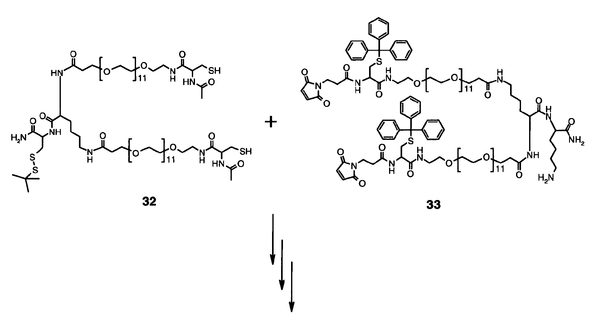


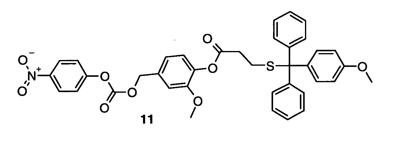

 wherein the symbols in formulae (IX) and (X) have the following meanings:


 wherein the symbols in formulae (II), (VI), (X), and (XIV) have the following meanings: