BACKGROUND OF THE INVENTION
Field of the Invention
-
The invention relates to a method of analyzing
a probe carrier using the Time-of-Flight Secondary
Ion Mass Spectrometry, particularly to a method of
analyzing the state of probes immobilized in a matrix
pattern on the probe carrier by the Time-of-Flight
Secondary Ion Mass Spectrometry, for example a method
for imaging a number of matrixes on the surface of
the probe carrier on which nucleic acid probes are
immobilized, or a method for quantitatively analyzing
the nucleic acid probes constituting the matrixes.
The invention also relates to a method for detecting
and analyzing target nucleic acids using a so-called
nucleic acid chip on which a plurality of nucleic
acid probes are disposed on a substrate in a matrix
pattern.
Related Background Art
-
Nucleic acid chips such as DNA chips and RNA
chips as examples of probe carriers have been used
for genomic analysis or analyzing expression of genes.
The analysis results are expected to provide
important indices for diagnosis, prognosis and
determination of therapeutic policy of cancers,
hereditary diseases, life style diseases and
infectious diseases.
-
Several methods are known for preparing the
nucleic acid chip. For example, representative
methods for preparing DNA chips include a successive
synthesis method of the DNA probe on the substrate
using photolithography (such as in U.S. Patent No.
5,405,783), and an immobilizing method of previously
synthesized DNA or cDNA (complementary DNA) by
feeding it on the substrate (such as in U.S. Patent
No. 5,601,980, Japanese Patent Application Laid-Open
No. H11-187900 and Science Vol. 270, 467, 1995).
-
Usually, the nucleic acid chip is prepared by
any of the methods described above. A desired object
can be attained by detecting formation/unformation of
a hybrid of the nucleic probe and a target nucleic
acid on the nucleic acid chip by some methods after
the chip and a solution containing a nucleic acid to
be detected, or a target nucleic acid, has been left
in a hybridization condition.
-
It is crucial for assuring reliability, or a
quantitative property and reproducibility, of the
analysis, to determine the quantity of either the
nucleic acid probe or the target nucleic acid
hybridized with the nucleic acid probe, or both
quantities existing in the matrix, or the density of
the nucleic acid probe and hybridized target nucleic
acid. It is also important from the viewpoint of
ensuring the quantitative property and reliability to
be informed of actual configuration (imaging) of the
matrix form (shape, size and state).
-
Suppose that no physical address indicating the
positions of the matrixes is formed on the substrate
for forming the chip. Then, the analysis portion of
the chip cannot be distinguished depending on the
detection methods due to the absence of the physical
address as will be particularly described hereinafter,
when the chip is prepared by supplying a probe
solution as fine droplets using, for example, an ink-jet
method. For solving such problem, the position
of the matrix should be visualized by the detection
method itself employed.
-
However, the nucleic acid probe or a hybrid
between the nucleic acid probe and the target nucleic
acid on the chip is in principle formed as a
monolayer of molecules, such analysis basically
requires a quite high sensitivity of surface analysis
techniques.
-
While such highly sensitive surface analysis
techniques known in the art include a method for
labeling the nucleic acid probe and/or the target
nucleic acid with an isotope, this method is not
always commonly used since it is complex and
dangerous while requiring special facilities and
equipment.
-
Labeling the nucleic acid probe and/or the
target nucleic acid with a fluorescent substance is
considered to be an option. While a fluorescence
hybridization method for labeling the target nucleic
acid with a fluorescent substance is widely known in
the art, this method involves many problems such as
unstability of fluorescent pigments, quenching and
nonspecific adsorption of the fluorescent pigment on
the surface of the substrate. Therefore, these
problems should be solved before quantitative
determination of the nucleic acid probe and hybrid.
-
While the other high sensitivity surface
analysis methods that are generally used include an
ATR method using FT-IR (Fourier Transform IR
spectroscopy) and XPS (X-ray Photoelectron
Spectroscopy), these methods are not always sensitive
enough for using for a quantitative analysis of the
hybrid on the nucleic acid chip, and for imaging of
the nucleic acid chip. In particular, when a
commonly used glass plate is used as the substrate of
the nucleic acid chip, there arise problems such as
absorption of IR light by the glass plate in FT-IR
(ATR), and charge up in XPS. Therefore, these
methods cannot be considered to be effective methods.
-
U.S. Patent No. 5,821,060 discloses a DNA
detection method by laser resonance ionization (RIS:
Resonance Ionization Spectroscopy) as another high
sensitivity surface analysis method. A target
element is ionized and detected by irradiating a
laser beam having a wavelength corresponding to the
ionization energy of the target element emitted from
the surface of the sample. While a method using a
laser beam and a method using ions have been
disclosed for permitting elements to be emitted from
the surface of the sample, these methods involve a
problem that only specified elements can be detected.
-
Dynamic Secondary Ion Mass Spectroscopy
(Dynamic-SIMS) is another option of the high
sensitivity surface analysis method. However, little
information of the chemical structure is obtained
from mass spectra since organic compounds are
decomposed to small fragment ions or particles in the
process for forming the secondary ions in this method,
which is not suitable for the analysis of organic
substances such as nucleic acid related substances.
-
On the contrary, the Time-of-Flight Secondary
Ion Mass Spectrometry (TOF-SIMS) is used as an
analytical method for investigating what kinds of
atoms or molecules are existing on the uppermost
surface of a solid sample, and has the following
features: detection ability of minute components as
small as 109 atoms/cm2 (corresponding to 1/105 of the
uppermost atomic monolayer); availability for both
organic substances and inorganic substances;
measurability of all atoms and compounds existing on
the surface; and capability of secondary ion imaging
from the substances existing on the surface of the
sample.
-
The principle of the method will be briefly
described hereinafter.
-
Constituting components on the surface are
emitted in vacuum by a sputtering phenomenon by
irradiating a high-speed ion (primary ion) beam onto
the surface of the solid sample in high vacuum.
Positively or negatively charged ions (secondary
ions) emitted by irradiation are converged in one
direction by an electric field, and are detected at a
position a given distance apart. While the secondary
ions having various masses are depending on the
surface composition of the sample emitted by
sputtering, the masses of the emitted secondary ions
can be analyzed by measuring the time lapse (time-of-flight)
from emission to detection of the secondary
ions, since a lighter ion fly with larger velocities
while a heavier ion fly with smaller velocities.
-
A little information on the chemical structure
is obtained from mass spectra in usual dynamic-SIMS
since organic compounds are decomposed to small
fragment ions or particle by ionization as described
above. However, the dose of the irradiated primary
ions is so small in TOF-SIMS that the organic
compounds are ionized while maintaining the chemical
structures to enable the structure of the organic
compound to be determined from the mass spectra.
Since only the secondary ions generated at the
uppermost surface of the solid sample are emitted in
vacuum, information on the uppermost surface (with a
depth of several Å) of the sample may be obtained.
-
The TOF-SIMS apparatus is roughly categorized
into two types of sector type and reflectron type.
One of the differences between these analysis methods
is an electrical grounding method of a holder for
fixing the analyzed sample. While the apparatus of
the sector type is constructed so that the secondary
ions are guided to the mass spectrometer by applying
several kilovolts of positive or negative voltage on
the holder in the apparatus of the reflection type,
the holder is grounded, and the secondary ions are
guided to the mass spectrometer by applying several
to several tens kilovolts of positive or negative
voltages on a secondary ion emitting electrode.
-
While positive primary ions are often used in
the TOF-SIMS method, positive secondary ions and
negative secondary ions are emitted irrespective of
the polarity of the primary ions. The secondary
electrons are generated by irradiation of the primary
ions under general conditions of measurements
irrespective of the polarity of the primary ions, and
the amount of the generated secondary electrons are
larger than the amount of the primary ions.
Consequently, the surface of the analyzed sample
tends to be positively charged to arise a defective
measurement when electrification is in excess (so-called
charge-up phenomenon). Positive
electrification seems to be maximum when the negative
secondary ions from an insulator are measured using
an apparatus of the sector type considering the
construction of the apparatus (because all of the
secondary electrons generated are directed toward the
secondary ion emitting electrode on which the
positive voltage is applied).
-
Most of the apparatus of the sector type and
reflectron type are equipped with a pulse electron
gun for neutralizing positive electrification as
described above. Specifically, electrification is
neutralized by the electron gun by irradiating the
analyzed sample with an electron beam from the pulse
electron gun for a given time during a period from
irradiation of the primary ions (sub- to several
nanoseconds of pulses) and measurement of the time-of-flight
of the positive or negative secondary ions
to the succeeding irradiation of the secondary ions.
Application of the voltage to the sample holder in
the apparatus of the sector type, and application of
the voltage to the secondary ion-emitting electrode
in the apparatus of the reflectron type are suspended
during the irradiation time of the electron beam to
the analyzed sample, and the holder or electrode is
grounded.
-
Although the positive electrification is
relaxed (or quenched) by this method to enable the
insulator to be analyzed, the margin for neutralizing
electrification becomes narrowest because positive
electrification tends to be the largest by the same
reason as described above when the negative secondary
ions are measured in the measurement of the insulator
using the apparatus of the sector type. Anyhow,
using the apparatus of the reflectron type comprising
the electrically grounded sample holder is (usually)
more advantageous than using the apparatus of the
sector type for avoiding the charge-up phenomenon.
When the conductivity of the analyzed sample, for
example a glass, is low (in other words, resistivity
or permittivity is high), the apparatus of the
reflectron type may be suitable for the measurements.
-
Since the TOF-SIMA method is a highly sensitive
measuring method, irrespective of the type of the
apparatus used such as the reflectron type or the
sector type, oligonucleotides formed as a molecular
monolayer on, for example, a gold substrate on which
the influence of charge-up is small may be analyzed.
(Proceeding of the 12th International Conference on
Secondary Ion Mass Spectrometry 951, 1999). This
literature describes the analytical results of DNA
and PNA +immobilized on a substrate by the TOF-SIMS.
According to this report, examples of the fragment
ions detected by the TOF-SIMS method include PO2 - and
PO3 - ions derived from phosphate backbones, and
(thymine-H)- ions derived from bases in the DNA probe,
and (thymine-H)- ions in the PNA probe.
-
However, there arise the following problems
that formation/unformation of hybrids of the target
DNA cannot be specifically detected by the following
two reasons when obtaining desired gene information
is attempted by detecting the target DNA by the TOF-SIMS
method using generally used DNA chips:
- (1) only a quite thin layer in the vicinity of
the surface is detected by the TOF-SIMS method; and
- (2) the fragment ion species generated from the
probe DNA and target DNA are the same with each other.
-
-
For solving these problems, PNA is immobilized
on a solid phase as a prove to form a hybrid between
the PNA and the target nucleic acid (J. C. Feldner et
al., SIMS XIII International Conference; 11 November,
2001, Nara). According to the method, the hybrid is
confirmed to be formed between the PNA probe and the
target nucleic acid by detecting the fragment ions
from the phosphate backbone, since the peptide-nucleic
acid has no phosphate backbone although the
base of the peptide nucleic acid is the same as that
of DNA.
-
However, acquiring gene information using the
chip having the peptide-nucleic acid as a probe is
not practical due to its high preparation cost, since
the peptide-nucleic acid is expensive. Detection is
relatively easy by using DNA as a probe since PO2 - and
PO3 - ions have relatively high efficiency and parent
structure of PO2 - and PO3 - ions are bonded to all the
nucleotides. However, detection of the fragment ions
of the base is often relatively difficult due to the
disadvantageous number and relatively low ionization
efficiency of the fragment ions, when four kinds of
bases are randomly contained in the nucleic acid
probe including the cases using PNA as the probe.
Accordingly, a method for labeling the target nucleic
acid capable of detection of the fragment ions with
higher detection and quantitatively detecting
efficiency over the foregoing art have been desired.
-
Japanese Patent Application Laid-Open No. 61-11665
discloses a nucleic acid base detector for
detection of the fragment ions, wherein non-metallic
elements such as S, Br and I, or metallic elements
such as Ag, Au, Pt, Os and Hg are introduced into
nucleic acid fragments separated by electrophoresis,
liquid chromatography or high speed gel filtration
depending on the molecular weight of the fragments,
and these elements are identified by atomic
absorption spectroscopy, plasma emission spectroscopy
or mass spectroscopy. However, there are no detailed
descriptions of the mass spectrometer as well as the
method for introducing the halogen atoms in the
nucleic acid.
-
On the other hand, the matrix itself on which
nucleic acid probe regions are specifically formed
should be analyzed from the point of view of
quantitatively determining the nucleic acid hybrid on
the chip. However, there are often no methods for
locating the matrix (the spot on which discharged DNA
is bonded) on the substrate when a solution of a
previously synthesized DNA probe is discharged and
immobilized on the surface of substrate by an ink-jet
method, which is a method for producing the DNA chip
described in Japanese Patent Application Laid-Open No.
H11-187900. It is desirable in this case to analyze
the fragment ions in an imaged spot after imaging the
probe or hybrid in the spot by the TOF-SIMS method.
However, no such methods are described in Japanese
Patent Application Laid-Open No. H11-187900.
Although Japanese Patent Application Laid-Open No.
61-11665 has mentioned the mass spectroscopic method
as described above, there are no descriptions at all
on the imaging method of the hybrid on the chip using
the TOF-SIMS method and quantitative analysis of the
target nucleic acid.
-
Accordingly, a novel method for detecting the
probe and/or the target substance have been desired,
whereby the detection efficiency is further improved
over the foregoing art, and quantitative detection of
the fragment ions are made possible.
SUMMARY OF THE INVENTION
-
An object of the invention is to provide a
method of analyzing a probe carrier that is capable
of more precisely analyzing the state of probes
immobilized on the probe carrier, for example,
imaging of the locations of the probes and
quantitative analysis thereof.
-
Another object of the invention is to provide a
method of analyzing a measuring sample that is
capable of accurate imaging of disposing locations or
quantitative analysis of a complex between a probe
formed on a measuring sample obtained by a reaction
of a probe carrier with a sample and a target
substance, more specifically a hybrid formed between
a nucleic acid probe and a target nucleic acid.
-
The method of the invention is applicable to
substances which recognize each other and form a
complex, either one of which can be immobilized on a
carrier, and into either one or both of which a
marker having a high ionization efficiency, for
example, halogen atoms can be introduced as a marker.
Examples of such complex forming substances include
proteins such as antigens, antibodies and enzymes, an
enzyme and a substrate specifically binding to the
enzyme, or mutually complementary nucleic acids.
-
The inventors have investigated the problems
involved in imaging a hybrid between a nucleic acid
probe and a target nucleic acid at a region having a
relatively large area on a carrier having a
relatively large resistivity, and in quantitatively
determining the hybrid, using the TOF-SIMS.
-
In an aspect, the invention provides a method
of detecting at least one of a probe and a target
substance capable of specifically binding to the
probe disposed on a substrate, the method comprising
the steps of preparing a substrate having at least
one of a probe and a target substance specifically
bonded to the probe disposed on a surface thereof;
and measuring the surface of the substrate by the
Time-of-Flight Secondary Ion Mass Spectrometry (TOF-SIMS),
wherein the probe and/or the target substance
is labeled with a marker substance capable of forming
a fragment ion that is not formed by fragmentation of
the at least one of the probe and the target
substance.
-
According to the invention, it is possible to
effect imaging of disposing locations or quantitative
analysis of probes immobilized on a carrier as a
probe carrier and/or a complex formed between the
probe and a target substance (a hybrid between a
nucleic acid probe and a target substance when the
target substance is a nucleic acid) with the probes
or the hybrid being immobilized on the carrier.
-
In another aspect, the invention provides a
method comprising reacting a sample with a probe
carrier having a number of probe-immobilized regions
disposed independently in a matrix pattern on a
carrier and analyzing an analysis sample obtained by
the reaction, wherein a target substance in the
sample capable of specifically binding to the probe
is labeled with a halogen atom and
formation/unformation of a complex obtained by the
reaction between the probe and the target substance
is detected by measuring the halogen atom by the
Time-of-Flight Secondary Ion Mass Spectrometry (TOF-SIMS).
-
According to the invention, it is possible to
effect, for example, imaging of disposing locations
or quantitative analysis with accuracy of
formation/unformation of a complex between a probe
formed in a measuring sample obtained by reacting a
sample with a probe-immobilized probe carrier and a
target substance (a hybrid between a nucleic acid
probe and a target substance when the target
substance is a nucleic acid) with the probes or the
hybrid being immobilized on the carrier.
-
In a different aspect, the invention provides a
method of analyzing a probe carrier having a number
of probes disposed in a matrix pattern in a probe-immobilized
region on the carrier by the Time-of-Flight
Secondary Ion Mass Spectrometry, wherein the
probes are labeled with halogen atoms and fragment
ions of the halogen atoms are detected to analyze the
state of probes.
-
According to the construction of the invention,
the analysis of the state of probes immobilized on
the probe carrier, for example imaging of disposing
locations and quantitative analysis thereof can be
more accurately performed.
-
Specifically, the construction above permits
imaging and quantitative analysis of the nucleic acid
probe at the same time by analyzing the halogen atoms
of the halogen labeled nucleic acid on the probe
carrier by the Time-of-Flight Secondary Ion Mass
Spectrometry.
-
In a further different aspect, the invention
provides a method of analyzing a nucleic acid chip
comprising a plurality of nucleic acid probes
disposed in a matrix pattern on a substrate, the
method comprising the steps of hybridizing the
nucleic acid probes with target nucleic acids in a
sample to form hybrids; and simultaneously analyzing
the nucleic acid probes and the target nucleic acids
in the state of the hybrids, wherein the nucleic acid
probes and the target nucleic acids are labeled with
different marker substances of prescribed numbers and
then analyzing the individual marker substances by
the Time-of-Flight Secondary Ion Mass Spectrometry,
thereby analyzing the labeled nucleic acid probes and
the labeled target nucleic acids.
-
In the above method, it is preferable that the
marker substance is neither a substance constituting
the nucleic acid probe, nor a substance constituting
the target nucleic acid. Specifically, it is
preferable to select a substance capable of
generating secondary ions that are distinctly
distinguishable from secondary ions derived from the
substance constituting the nucleic acid prove, and
from secondary ions derived from the substance
constituting the target nucleic acid.
-
According to the analysis method so constructed
as described above, the probe nucleic acid and the
target nucleic acid hybridized on the nucleic acid
chip are previously labeled with different marker
substances, for example halogen atoms. Consequently,
the probe nucleic acid and the target nucleic acid
can be independently imaged while independently
quantifying them based on the measurement of the
secondary ions derived from differently labeled
marker substances using the Time-of-Flight Secondary
Ion Mass Spectrometry.
BRIEF DESCRIPTION OF THE DRAWINGS
-
- Fig. 1A is a view showing the result of imaging
of sequence No. 1 using 79Br- ions in Example 2;
- Fig. 1B is a view showing the result of imaging
of sequence No. 1 using 81Br- ions in Example 2;
- Fig. 2A is a view illustrating a method for
continuous pattern irradiation with primary ions in a
spot fashion for imaging;
- Fig. 2B is a view illustrating a method for
non-continuous pattern irradiation with primary ions
in a spot fashion for imaging, the numbers given in
Fig. 2B showing an irradiation order of the
irradiation spot;
- Fig. 3A is a view showing the result of imaging
of sequence No. 1 using 79Br- ions in Example 2;
- Fig. 3B is a view showing the result of imaging
of sequence No. 1 using 81Br- ions in Example 2;
- Fig. 3C is a view showing the result of imaging
of sequence No. 2 using 79Br- ions in Example 2;
- Fig. 3D is a view showing the result of imaging
of sequence No. 2 using 81Br- ions in Example 2;
- Fig. 3E is a view showing the result of imaging
of sequence No. 3 using 79Br- ions in Example 2;
- Fig. 3F is a view showing the result of imaging
of sequence No. 3 using 81Br- ions in Example 2;
- Fig. 4A is a view showing the result of imaging
using F- ions in Example 2;
- Fig. 4B is a view showing the result of imaging
using 79Br- ions in Example 2;
- Fig. 4C is a view showing the result of imaging
using 81Br- ions in Example 2;
- Fig. 5 is a graphical representation showing
plots of the measured values of marker F- ions of the
nucleic acid probe, and marker 79Br- ions and 81Br-
ions of the target DNA, respectively, after
hybridization against the nucleic acid probe
concentration in the nucleic acid probe solution to
be spotted on the nucleic acid chip based on the
results of quantitative analysis in Example 2; and
- Fig. 6 is a graphical representation showing
plots of the measured values of marker F- ions of the
nucleic acid probe, and marker 79Br- ions and 81Br-
ions of the target DNA, respectively, after
hybridization against the target DNA concentration of
the sample solution to be hybridized with the nucleic
acid probe on the nucleic acid chip based on the
results of quantitative analysis in Example 3.
-
DESCRIPTION OF THE PREFERRED EMBODIMENTS
-
In the invention, the state of probes
immobilized on a probe carrier or a target substance
specifically bonded to the probes, for example, the
location or quantity thereof is analyzed using probe
and/or the target substance labeled with a marker
substance capable of generating fragment ions, which
are not generated by fragmentation of the probe or
target substance.
-
Specifically, a substance having a high
ionization efficiency, favorably ion fragments
derived from halogen atoms, may be detected by the
Time-of-Flight Secondary Ion Mass Spectrometry (TOF-SIMS).
-
In other words, probes on the probe carrier
and/or a target substance is preferably labeled with
a prescribed number of halogen atoms for imaging and
analysis of the probe carrier using TOF-SIMS, and
fragment ions of the halogen atom are detected and
analyzed by TOF-SIMS.
-
The probes and/or the target substance is
labeled with a marker substance capable of generating
fragment ions that are not generated by fragmentation
of the probes and/or the target substance.
-
The methods available are as follows:
- (1) The target substance is labeled preferably
with a prescribed number of the halogen atoms for
imaging and analysis of a hybrid using TOF-SIMS, and
the fragment ions of the halogen atoms are detected
by TOF-SIMS.
- (2) The state of probes immobilized on the
carrier, for example the location and quantity
thereof, is analyzed by detecting the fragment ions
derived from the halogen atoms labeled on the probe
by the Time-of-Flight Secondary Ion Mass Spectrometry
(TOF-SIMS). In other words, the probe on the probe
carrier is labeled preferably with a prescribed
number of the halogen atoms for imaging and analysis
of the probe carrier using TOF-SIMS, and the fragment
ions of the halogen atoms are detected and analyzed
by TOF-SIMS.
- (3) A probe nucleic acid and the target nucleic
acid, respectively, are labeled with different marker
substances of prescrived number for imaging and
quantitative analysis of the hybrid formed using TOF-SIMS,
and fragment ions of mutually different
substances are detected and analyzed by TOF-SIMS.
The marker substance available is preferably
different from the constituting elements such as the
probe nucleic acid, the substances constituting the
target nucleic acid and the nucleic acid, since the
secondary ions derived from the marker substance can
be distinctly extinguished from the fragment ions
derived from the nucleic acid. In addition, the
probe nucleic acid and the target nucleic acid,
respectively, are preferably labeled with prescribed
numbers of the marker substances in order to
quantitatively determine respective nucleic acids.
-
-
Examples of the marker substances are, although
not restrictive, halogen atoms such as fluorine,
chlorine, bromine and iodine.
-
Improvements of detection sensitivity can be
expected by using fragment ions of the halogen atoms
having relatively high ionization efficiency, and the
effects of charge-up can be excluded by reducing the
intensity of the primary ion in proportion to the
degree of reduction of the intensity. Accordingly,
large area imaging on a high resistivity substrate is
possible by combining with a method to be described
hereinafter.
-
The marker substance used in the invention
preferably has a high ionization efficiency than the
fragments contained in the probe and the target
substance in TOF-SIMS analysis. When both the probe
and the target substance are labeled, they are
preferably labeled with different marker substances
from each other.
-
The problems arising from detecting only the
nucleic acid's own fragment ions as described above
can be solved by labeling the nucleic acid with a
prescribed number of halogen atoms particularly when
the method of the invention is applied to the nucleic
acid, and the nucleic acid can be analyzed with
improved quantitative accuracy.
-
Further, the problems arising from detecting
only the nucleic acid's own fragment ions as
described above can be also solved by labeling the
nucleic acid with a prescribed number of the halogen
atoms, and the nucleic acid can be analyzed with
improved quantitative accuracy. The number of
labeling with the halogen atoms is not particularly
restricted. Any positions and methods for labeling
are available, so long as they are applicable and do
not inhibit complexes from being formed (hybrid
complex (hybridization) when the target substance is
a nucleic acid) between the probe and the target
substance from being formed thereafter.
-
Practically, one position is labeled in one
nucleotide molecule. Accordingly, the prescribed
number of the halogen atoms used as a marker is
desirably an arbitrary number from 1 to the number of
the nucleotides in the probe nucleic acid and the
target nucleic acid. For example, the more desirable
number is 1 to 5 when the nucleic acid is a synthetic
oligonucleotide considering the labor and cost of
labeling, and the degree of ionization efficiency of
the halogen atoms.
-
When introduction of the marker is attempted by
taking advantage of an enzymatic elongation reaction
such as a PCR method, the number of the introduced
marker is restricted due to steric hindrance when the
marker is a relatively large molecule such as a
fluorescent pigment. On the contrary, the halogen
atom induces substantially no steric hindrance. For
example, all the same kind of bases (for example
adenine) can be labeled in an elongation product by
using a nucleic acid base unit substituted with
halogen atoms for the elongation reaction.
Accordingly, selecting the halogen atom as the marker
is desirable for enabling the number of the markers
to be quantitatively determined in addition to the
sensitivity and method for introducing the marker.
-
The secondary ions of the fluorine, chlorine,
bromine and iodine atoms can be detected with high
sensitivity in the analysis by TOF-SIMS. Since the
four kinds of the halogen atoms can be introduced in
the target nucleic acid according to the method to be
described hereinafter, these halogen atoms may be
favorably utilized in the invention.
-
The probe immobilized on the carrier is able to
recognize a specific target substance and to form a
complex with the target substance. When the target
substance is a nucleic acid, the probe can be
specifically bonded to the target nucleic acid by a
complementary sequence of the nucleic acid probe with
the target nucleic acid. The probe immobilized on
the carrier should be able to be specifically bonded
to a specified target substance, and the method of
the invention is principally applicable to not only
the nucleic acid, but also to substances capable of
labeling with halogen atoms, for example proteins
such as antigens, antibodies and enzymes substrates,
and substrates specifically bonded to the enzymes.
-
Any methods known in the art may be used for
immobilizing the nucleic acid probe on the carrier in
the invention. In an example of the probe
immobilized on the carrier, a binding site with the
carrier is formed with interposition of a linker, if
necessary, at a part of the nucleic acid probe
comprising oligonucleotides having base sequences
capable of hybridizing with the target nucleic acid,
and the probe is linked to the surface of the carrier
at binding sites with the carrier. The position of
the binding site with the carrier so constructed as
described above in the nucleic acid probe molecules
is not particularly restricted, so long as a desired
hybridization reaction is not impaired.
-
Independent regions immobilizing the probe, for
example many dots, are arranged in a matrix pattern
with a given space in the probe carrier of the
invention. Such probe carrier includes a probe array,
microchip nucleic acid chip and the like.
-
On the other hand, the probe has a structure
capable of being bonded to the surface of the carrier,
and the probe is desirably immobilized through this
structure. Preferably, the structure of the probe
capable of bonded to the surface of the carrier is
formed by introducing at least one of organic
functional groups such as an amino group, a thiol
group, a carboxylic group, a hydroxyl group, an acid
halide (haloformyl group; -COX), a halide group (-X),
an aziridine group, a maleimide group, a succimide
group, an isothiocyanate group, a sulfonyl chloride
group (-SO2Cl), an aldehyde group (formyl group;
-CHO), a hydrazine group and an acetamide iodide
group.
-
Immobilization of the probe by covalent bonds
are possible by a treatment required for the surface
of the carrier, or by a treatment for forming a
maleimide group for the thiol group, an epoxy group,
aldehyde group or N-hydroxysuccimide for the amino
group, depending on the structure required for
binding the probe on the carrier.
-
The probe is desirably bonded to the surface of
the substrate by the covalent bond considering the
stability of the probe.
-
Examples of the combination of the probe with
the target substance include a combination between
the nucleic acid probe and the target nucleic acid,
and a combination capable of forming a complex
selected from proteins such as antigens, antibodies
and enzymes, and substrates capable of specifically
binding to the enzyme.
-
The nucleic acid probes used in the invention
are not particularly restricted, and any nucleic acid
probes are available so long as they are able to
recognize the target nucleic acid. However, the
nucleic acid probe is desirably selected from DNA,
RNA, PNA (peptide-nucleic acid), cDNA (complementary
DNA), cRNA (complementary RNA) and PCR amplification
products (from cDNA). Preferably, a nucleic acid
probe comprising at least one of them may be
immobilized on the carrier.
-
The target nucleic acid used in the invention
is desirably DNA, RNA, PNA (peptide nucleic acid),
cDNA (complementary DNA), cRNA (complementary RNA)
and PCR amplification products (from cDNA)
considering the method for labeling with the halogen
atoms to be described hereinafter. A sample
containing the target nucleic acid comprising at
least one of them may be used for analysis. The
target nucleic acid may be a synthetic nucleic acid,
or a natural nucleic acid derived from animals, human,
plants, microorganisms and the like.
-
The imaging method of the measuring sample (a
probe carrier prepared by required treatments after
allowing to react with a sample: the "nucleic acid
chip" will be described hereinafter as a
representative) of the invention comprises:
sequentially irradiating the primary ions onto a
portion on the surface of the nucleic acid surface
having a given area as a pulse spot having a
relatively smaller area than the area above; and
analyzing the secondary ions emitted by the pulse
irradiation by the Time-of-Flight Secondary Ion Mass
Spectrometry for every pulse irradiation. For
excluding the effect of charge-up, it is quite
effective and desirable that the pulses of the
primary ions are irradiated as a non-continuous
pattern, and the results of the mass spectroscopic
analysis obtained are reconstructed for imaging based
on the pattern of non-continuous irradiation of the
primary pulse.
-
It has been considered to be desirable that the
area of the imaging region is, for example, larger
than 300 µm × 300 µm or more considering the size of
the spot, or the detection efficiency, when the
nucleic acid chip is imaged and the nucleic acid on
the nucleic acid chip is quantitatively analyzed by
TOF-SIMS. However, the effect of charge-up becomes
large for obtaining an image by sequential scanning
(raster scanning) of the beam in a given direction as
is used in television picture tubes, when the
diameter of the primary beam is adjusted to 5 µm for
obtaining a required resolution and the area of 300
µm × 300 µm is scanned with the beam on the substrate
having a relatively high resistivity such as a glass
that is frequently used as a substrate for preparing
the DNA chip. Consequently, good images could not be
obtained.
-
Accordingly, the primary ions are sequentially
irradiated as a pulse spot to the portion having a
given area on the surface of the nucleic acid chip so
that the spot has a relatively smaller area than the
area above, and the secondary ions emitted by pulse
irradiation are analyzed by time-of-flight mass
spectroscopy for every pulse irradiation in the
invention. For excluding the effect of charge up, it
is quite effective and desirable that the primary
pulse is irradiated based on a non-continuous pattern,
and the results of the mass spectroscopic analysis
obtained are reconstructed based on the pattern of
primary pulse irradiation.
-
Examples of the non-continuous pattern include
a random pattern and a programmed non-continuous
pattern.
-
Simple examples of the continuous pattern and
non-continuous pattern are as follows.
-
Fig. 2A shows an example of the continuous
irradiation pattern, while Fig. 2B shows an example
of the non-continuous irradiation pattern. Suppose
that the primary ion pulse is irradiated on 5 × 5
spots each having a rectangular area, then a pattern
obtained by sequentially irradiating all the spots
from one spot to a neighboring spot is a continuous
pattern as shown in Fig. 2A. On the other hand, a
pattern obtained by sequentially irradiating all the
spots from one spot to at least a non-neighboring
spot is a non-continuous pattern.
-
It is possible to form the non-continuous
pattern in a random order. However, since
neighboring spots may be continuously irradiated by
this method, a non-continuous pattern according to a
special program can be formed. An algorithm may be
appropriately employed for the desirable pattern so
that the neighboring spots are not continuously
irradiated, or the spots on neighboring columns and
rows are not continuously irradiated.
-
Examples of the halogen atoms include fluorine,
chlorine, bromine and iodine atoms. Fragment ions of
the fluorine, chlorine, bromine and iodine atoms can
be detected by TOF-SIMS. These four kinds of the
halogen atoms can be introduced into the nucleic acid
probe by the methods to be described hereinafter.
-
The nucleic acid probes in each matrix of the
probe carrier may be labeled with different halogen
atoms with each other.
-
When the sample is unpredictable whether it
contains a target substance or not, the sample is
labeled with the halogen atom at first, and a probe
is allowed to react with the sample. Then, a complex
is formed when the sample contains a target substance
capable of being recognized by the probe, and the
complex can be detected using the marker halogen atom.
-
The fragment ions of the fluorine, chlorine,
bromine and iodine can be detected by TOF-SIMS.
These halogen atoms can be efficiently utilized in
the invention since the four kinds of the halogen
atoms can be also introduced into the target nucleic
acid by the methods to be described hereinafter.
-
While the method for introducing the halogen
atom in the target nucleic acid is not particularly
restricted, and an example of the method known in the
art is to permit the halogen atom to be bonded to the
nucleic acid base of the target nucleic acid, which
can be favorably used in the invention. The halogen
atom is desirably bonded at a position that does not
inhibit a hybrid of the target nucleic acid from
being formed when the target nucleic acid forms the
hybrid with the nucleic acid probe. Such binding
sites are the 5-position of the pyrimidine base and
the 8-position of the purine base. However, it is
not always required that the halogen atoms in all the
nucleotide bases of the target nucleic acid are
bonded to these positions.
-
While the method for introducing the halogen
atom into the nucleic acid prove is not restricted,
and the method well known in the art is to allow the
halogen atom to be bonded to the nucleotide base of
the nucleic acid probe. This method can be favorably
used in the invention. It is desirable that the
halogen atom is bonded to the position that does not
inhibit a hybrid of the nucleic acid probe from being
formed when the nucleic acid probe forms the hybrid
with the target nucleic acid. Such binding sites are
the 5-position of the pyrimidine base and the 8-position
of the purine base. However, it is not
always required that the halogen atoms in all the
nucleotide bases of the nucleic acid probe are bonded
to these positions.
-
In a practical method for introducing the
halogen atom in the nucleic acid probe or target
nucleic acid when the nucleic acid is a synthetic DNA,
a synthetic unit binding the halogen unit, or 2'-deoxyribonucleoside-3'-phosphoroamidite
represented
by the following structural formula may be used for
synthesizing the DNA using an automatic DNA
synthesizer:
wherein X represents a halogen atom, DMTO represents
a dimethoxytrityl group, iPr represents an isopropyl
group, and CNEt represents a 2-cyanoethyl group.
-
When the nucleic acid is a synthetic RNA, on
the other hand, a synthetic unit binding the halogen
atom, or ribonucleoside-3'-phosphoroamidite, may be
used for synthesizing the RNA using an RNA automatic
synthesizer. Examples of such synthetic unit include
the following compound:
wherein X represents a halogen atom (F, Cl, Br or I),
DMTO represents a dimethoxytrityl group, iPr
represents an isopropyl group, CNEt represents a 2-cyanoethyl
group, ME represents a methyl group, and
TBDMS represents a t-butyldimethoxyxylyl group).
-
When the nucleic acid is a synthetic PNA, A
synthetic unit binding the halogen atom, or a peptide
analogue binding a nucleic acid base may be favorably
used for synthesizing PNA using a PNA automatic
synthesizer.
-
In an example for introducing the halogen atom
when the nucleic acid is cDNA, 2'-deoxyribonucleoside-5'-triphosphate
binding the
halogen atom may be used for elongating a cDNA with a
reverse transcriptase.
-
In an example for introducing the halogen atom
when the nucleic acid is a DNA derived from a genome
DNA, 2'-deoxyribonucleoside-5'- phosphate binding the
halogen atom may be used for elongating the DNA with
DNA polymerase. For introducing the halogen atom
into the cRNA when the nucleic acid is cRNA, on the
other hand, ribonucleoside-5'-triphosphate binding
the halogen atom may be used for elongating the cRNA
with RNA polymerase.
-
A PCR reaction, or a RT-PCR (reverse
transcription PCR) reaction can be used for the
elongation reaction. A marker may be introduced
together with the nucleotide monomer used for the
elongation reaction, or the marker may be introduced
in the synthesis of a primer used for the reaction.
-
The nucleic acid probe or target nucleic acid
species of the nucleic acid chip used in the
invention are not particularly restricted, and DNA,
RNA, PNA (peptide-nucleic acid), cDNA (complementary
DNA), cRNA (complementary RNA), oligodeoxynucleoside,
oligoribonucleotide and the like may be used.
-
Other examples of the target substance
available in the analysis method of the invention
include metals such as Au, Ag, Cu, Ni, Co, Cr, Al, Ta,
Pt, Pd, Zn, Sn, Ru and Rh, and metal complexes
thereof including organic (metal) complexes. The
method described in Science, Vol. 262, 1025, 1993 may
be used, for example, for introducing the organic
metal complex.
[Examples]
-
The invention will be described in detail with
reference to examples. Although these examples
constitute a part of the best mode for carrying out
the invention, the invention is not restricted to
these examples.
(Example 1) Preparation of Nucleic Acid Probe Chip
-
The nucleic acid probe chip was prepared
according to Japanese Patent Application Laid-Open No.
H11-187900.
(1) Cleaning of substrate
-
Synthetic quartz substrates (25.4 mm × 25.4 mm
× 1 mm) were placed on a rack and soaked in a
ultrasonic wave detergent (GPII produced by Blanson)
diluted to 10% with water overnight. The substrate
was washed with the detergent for 20 minutes using a
ultrasonic wave followed by washing with water to
remove the detergent. After rinsing with pure water,
the substrate was further treated with the ultrasonic
wave for 20 minutes in a vessel filled with pure
water. Then, the substrate was soaked in a 1N
aqueous sodium hydroxide solution previously heated
at 80°C for 10 minutes, followed by washing with
water and pure water to subject the substrate to the
next step.
(2) Surface treatment
-
A 1% by weight aqueous solution of a silane
coupling reagent binding amino groups (N-β-(aminoethyl)-γ-aminopropyltrimethoxy
silane: KBM603
produced by Shin-Etsu Chemical Co.) was stirred for 2
hours at room temperature to hydrolyze intermolecular
methoxy groups in the silane compound. After soaking
the substrate obtained in (1) for 1 hour at room
temperature, the substrate was washed with pure water
and dried by blowing nitrogen gas onto both surfaces
of the substrate. Then, the substrate was baked for
1 hour in an oven heated at 120°C to finally
introduce the amino group on the surface of the
substrate.
-
Subsequently, 2.7 mg of N-maleimidecaproyloxysuccimide
(EMCS produced by
DOJINDO LABORATORIES.) was dissolved in a 1:1
solution of dimethylsulfoxide (DMSO) and ethanol in a
concentration of 0.3 mg/ml. The quartz substrate
after subjecting to the silane coupling treatment was
soaked in this EMCS solution for 2 hours at room
temperature, and the amino group bonded on the
surface of the substrate was allowed to react with
the succimide group in the EMCS solution by the
silane coupling treatment. The maleimide group
derived from EMCS is bonded on the surface of the
substrate by this treatment. The substrate after
pulling up from the EMCS solution was sequentially
washed with the mixed solution of DMSO and ethanol,
and ethanol, followed by drying by blowing nitrogen
gas.
(3) Synthesis of probe DNA
-
A single strand nucleic acid (40-mer of dT) of
sequence No: 1 was synthesized by requesting to a DNA
synthesis company (BEX). A thiol group (SH) was
introduced in the 5'-terminal of the single strand
DNA of sequence No. 1 by using a thiol modifier (Glen
Research) in the synthesis step. Deprotection and
recovering of DNA were performed by a usual method,
and the product was purified by HPLC (High
Performance Liquid Chromatography). A series of
steps from synthesis to purification were requested
to the synthesis company.
(4) Discharge of DNA by thermal jet printer and
binding to substrate
-
The single strand DNA of sequence No: 1 was
dissolved in a solution containing 7.5% by weight of
glycerin, 7.5% by weight of urea, 7.5% by weight of
thiodiglycol and 1% by weight of acetylene alcohol
(trade name: Acetylenol EH produced by Kawaken Fine
Chemical Co.) in a concentration of 8 µm. A printer
head BC-50 (manufactured by Canon Inc.) for a bubble
jet printer BJF-850 (manufactured by Canon Inc.)
using a bubble jet method as a kind of thermal jet
methods was reassembled so that several hundred
microliters of the solution is discharged. This head
was mounted on a discharge drawing machine
reassembled so as to be able to discharge on the
quartz substrate. Injected in a reassembled tank of
the head was several hundred microliters of the DNA
solution, and the solution was spotted on a substrate
treated with EMCS using the discharge drawing machine.
The discharge volume during spotting was 4
picoliter/drop, and the solution was discharged at
200 dpi, or 127 µm pitch, in a 10 mm × 10 mm range of
spotting at the center of the substrate. The
diameter of the dot spotted under the condition above
was about 50 µm.
-
After completing to spot, the substrate was
allowed to stand still in a moisturizing chamber for
30 minutes to allow the maleimide group on the
surface of the glass plate to react with the thiol
group at the terminal of the nucleic acid probe.
After washing the substrate with pure water, it was
stored in a 50 mM phosphate buffer solution (pH = 7.0,
named as solution A hereinafter) containing 1M of
NaCl.
(Example 2) Imaging and Analysis by Hybridization and
TOF-SIMS
(1) Synthesis of model target nucleic acid
-
-
A model terget nucleic acid (40-mer of dT;
sequence No: 2) labeled with five bromine atoms was
synthesized (BEX). Five bromine labeled nucleotides
at the 5'-end were introduced during the synthesis
with an automatic synthesizer using 8-bromo-3'-deoxyadenosine
phosphoroamidite represented by the
structure above. Deprotection and recovering of the
DNA was performed by a usual method, and the product
was purified by HPLC. A series of steps from the
synthesis to purification was requested to a
synthesis company. A(Br) in the sequence denotes
bromine labeled deoxyadenosine. The 8-position of
adenine as a marker position is known not to inhibit
hybridization.
(2) Blocking and hybridization
-
The chip prepared in Example 1 was soaked in
solution A containing 2% bovine thymus albumin (BSA).
After blocking the surface of the chip (for non-specific
adsorption of nucleic acids and the like),
the chip was soaked in solution A in which the target
nucleic acid of sequence No: 2 is dissolved in a
concentration of 50 nM for hybridization at 45°C for
15 hours. Then, after rinsing the chip with pure
water (at room temperature), it was dried by blowing
nitrogen gas, followed by storage in a vacuum
desiccator before used for TOF-SIMS.
(3) Analysis by TOF-SIMS
-
The DNA chip after hybridization was imaged and
analyzed using the TOF-SIMS IV apparatus manufactured
by ION TOF Co.
-
The conditions of the apparatus were as
follows:
- (Primary ion)
primary ion: 25kV, Ga+ random scanning mode
primary ion pulse frequency: 2.5 kHz (400
µsec/shot)
primary ion pulse width: 1 ns
primary ion beam diameter: 5 µm - (Secondary ion) imaging by reconstruction of the
primary ion irradiation pattern
secondary ion detection mode: negative
measuring region: 300 µm × 300 µm
pixel number of secondary ion: 128 × 128
integration times: 256 -
(4) Results
-
Figs. 1A and 1B show the result of imaging on
bromine ions from the data obtained after an analysis
of the hybridized DNA chip in (2) by TOF-SIMS under
the conditions above. Figs. 1A and 1B were obtained
using
79Br
- ion and
81Br
- ion.
| Ionic species | Counts |
| 79Br- ion | 1250 |
| 81Br- ion | 1285 |
| Total | 2663 |
-
Table 1 shows counts of each one spot obtained
from Figs. 1A and 1B. Figs. 1A and 1B, and Table 1
show that the spot of the bromine labeled target DNA
forming a hybrid with the nucleic acid probe on the
DNA chip may be quantitatively analyzed by Br imaging.
(Example 3) Imaging and Analysis of Bromine Labeled
Target DNA Derived from Genome
(1) Preparation of nucleic acid chip for detecting
target DNA derived from genome
-
Nature Biotechnology Vol. 18, 483, 2000
describes preparation of an oligonucleotide chip for
detecting exon 7 of the genome DNA of two cell lines
HSC4 and HSC5 of oral cavity epidermoid carcinoma,
and detection of fluorescence labeled DNA derived
from the exon.
-
The oligonucleotide chip was prepared in this
example according to the method above to synthesize a
DNA using bromine in place of a fluorescent marker,
followed by hybridization using the DNA.
-
The actual procedure thereof will be described
below.
(Synthesis of DNA probe and preparation of chip)
-
-
The DNA of sequence No: 3, having a base
sequence complementary to a part of the base sequence
of exon 7 of HSC4 (the part containing codon No. 248)
above and carrying a thiol group at the 5' terminal
for binding to the substrate, was synthesized as in
Example 1, and a DNA chip was prepared by the same
method as in Example 1 using the DNA.
(2) Synthesis of bromine labeled DNA derived from
genome
-
5-bromo-2'-deoxyuridine triphosphate (Br-dUTP)
-
An exon 7 portion was synthesized from the
genome of HSC4 by a PCR reaction using PCR primers of
sequence Nos: 4 and 5. Subjected to PCR
amplification by repeating 40 cycles of 94°C (30
seconds) and 60°C (45 seconds) were 50 µL of PCR
mixtures containing 20 ng of genome DNA and 0.4 µM
each of sense and ant-sense primers. The nucleotides
obtained were designed to have a chain length of 171
nucleotides.
-
Subsequently, 0.2 µm of anti-sense primer
(sequence No: 4) and 10 µm of 5-bromo-2'-deoxyuridine
triphosphate (Sigma-Aldrich Japan Co.) as a kind of
the bromine labeled nucleotide having the structure
shown above were subjected to ssPCR (single strand
PCR) using a part of the amplification products as
primers. The PCR cycles were 25 cycles of 96°C (30
seconds), 50°C (30 seconds) and 60°C (4 minutes).
The bromine labeled single strand DNA obtained was
purified by gel filtration.
(3) Blocking and hybridization
-
After blocking the chip prepared in (1) above
by the same method as in Example 1, the chip was
rinsed with pure water and used for hybridization
below. The chip after blocking was soaked in a SSPE
solution (0.9M NaCl, 60 mM NaH2PO4, 6 mM EDTA)
containing 20% of formamide six times followed by
heating at 80°C for 10 minutes. The solution
contained the DNA derived from genome synthesized as
(2) above. Then, the chip was subjected to
hybridization at 45°C for 15 hours, followed by
washing with the SSPE solution twice at 55°C. Then,
the chip was gently rinsed with pure water (at room
temperature) followed by drying by blowing nitrogen
gas to store in a desiccator before using in TOF-SIMS.
(4) Imaging and analysis by TOF-SIMS
-
The DNA chip after hybridization under the same
condition as in Example 2 was imaged and analyzed by
TOF-SIMS.
-
The numerical data obtained are shown in Table
2.
| Ionic species | Counts |
| 79Br- ion | 542 |
| 81Br- ion | 620 |
| Total | 1162 |
-
Table 2 shows that hybridization of the target
DNA derived from the genome and labeled with bromine
on the DNA chip can be quantitatively determined by
TOF-SIMS.
-
Labeling of the cDNA derived from mRNA with the
halogen atom, and imaging and analysis thereof by
TOF-SIMS are also possible by approximately the same
method as in this example.
(Example 4) Analysis of Binding of Probe
-
The surface of the substrate was (1) washed and
(2) treated by the same procedure in Example 1.
(3) synthesis of nucleic acid probe DNA
-
Single strand nucleic acids with sequence Nos:
6 to 8 were synthesized by requesting to the DNA
synthesis company (BEX). In the sequence, base T
represents usual 2'-deoxythymidine, and U(Br)
represents 5-bromo-2'-deoxyuridine, which were
introduced in the synthesis step using the
phosphoroamidite (Glen Research) shown below.
-
The terminal U(Br) was introduced using a (CPG)
column (Glen Research) to which U(Br) shown below is
immobilized. Bromine introduced the 5-position is
known not to inhibit hybridization.
-
The thiol group was introduced to the 5'-terminal
of DNA by using a thiol modifier (Glen
Research) in the synthesis step. The DNA was
deprotected and recovered by a usual method, HPLC was
used for purification. A series of steps from the
synthesis to purification were requested to the
synthesis company.
(4) Discharge of DNA by thermal jet printer and
binding to substrate
-
The single strand DNAs of sequence Nos: 6 to 8
were each dissolved at a concentration of 8 µM in a
solution containing 7.5% by weight of glycerin, 7.5%
by weight of urea, 7.5% by weight of thiodiglycol and
1% by weight of acetylene alcohol (trade name:
Acetylenol EH produced by Kawaken Fine Chemical Co.).
-
Using the discharge drawing machine used in
Example 1, 100 µL each of the DNA solutions was
filled in the reconstructed tank, and the three
sheets of the substrate treated with EMCS were
mounted on the discharge drawing machine, and the
three kinds of the DNA solutions were spotted on one
sheet each of the three substrates. The discharge
volume during spotting was 4 picoliter/drop, and the
solution was discharged at 200 dpi, or 127 µm pitch,
in a 10 mm × 10 mm range of spotting at the center of
the substrate. The diameter of the dot spotted under
the condition above was about 50 µm.
-
After completing to spot, the substrate was
allowed to stand still in a moisturizing chamber for
30 minutes to allow the maleimide group on the
surface of the glass plate to react with the thiol
group at the terminal of the nucleic acid probe.
Each substrate was washed with pure water, and was
stored in pure water. Immediately before analysis
by TOF-SIMS, the DNA bonded substrate (DNA chip) was
dried by blowing nitrogen gas, and was further dried
in a vacuum desiccator.
(Example 5) Imaging and Analysis by TOF-SIMS
(1) The DNA chips prepared in Example 4 were imaged
and analyzed using TOF-SIMS IV apparatus manufactured
by ION TOF Co. The conditions of the apparatus are
summarized below:
-
- (Primary ion)
Primary ion: 25 kV Ga+, random scan mode
Primary ion pulse frequency: 2.5 kHz (400
µsec/shot)
Primary ion pulse width: 1 ns
Primary ion beam diameter: 5 µm
- (Secondary ion) imaging by reconstruction on the
irradiation pattern of the primary ion
Secondary ion detection mode: negative
Measuring region: 300 µm × 300 µm
Pixel number of secondary ion images: 128 × 128
Integration time:
-
(2) Results
-
The DNA chips prepared in Example 4 were
analyzed by the TOF-SIMS IV apparatus under the
conditions above, and the bromide ion was imaged from
the data obtained. The results obtained are shown in
Figs. 3A to 3F. Figs. 3A and 3B are images of
sequence No: 6, Figs. 3C and 3D are images of
sequence No: 7, and Figs. 3E and 3F are images of
sequence No: 8. Figs 3A, 3C and 3E are derived from
79Br
- ion, while Figs 3B, 3D and 3F are derived from
81Br
- ion.
| Ionic species | Counts |
| | Sequence No: 6 | Sequence No: 7 | Sequence No: 8 |
| 79Br- ion | 1342 | 805 | 273 |
| 81Br- ion | 1321 | 800 | 224 |
| Total | 2663 | 1605 | 497 |
-
Table 3 shows the counts of one spot from each
of Figs. 3A to 3F. Figs. 3A to 3F, and Table 3 show
that imaging by bromine as a target substance of the
spot of the bromine labeled DNA on the DNA chip as
well as quantitative determination of bromine are
possible, although is a relative value.
(Example 6) Imaging and Analysis of Bromine Labeled
DNA Chip Derived from Genome
(1) Preparation of bromine labeled DNA chip derived
from genome
-
DNA was synthesized according to the detection
method of the fluorescence labeled DNA described in
Nature Biotechnology Vol. 18, 438, 2000, cited in
Example 3, wherein the marker was replaced from a
fluorescence substance to bromine. Then, a DNA chip
was prepared using the DNA according to the method
described in Science Vol. 270, 467, 1995 (this
reference relates to a method for preparing a cDNA
chip).
-
An actual procedure of the method will be
described below.
(1) Synthesis of Bromine labeled DNA derived from
genome
-
5-bromo-2'-deoxyuridine triphosphate (Br-dUTP)
-
The exon 7 part was synthesized from the genome
of HSC4 by a PCR reaction using the PCR primers of
sequence Nos: 4 and 5 used in Example 3 (common to
HSC4 and HSC5: requested to BEX Research Co.).
-
A PCR mixture (50 µl) containing 20 ng of a
genome DNA and 0.4 µM each of sense or anti-sense
primers were amplified by PCR by repeating 40 cycles
of 94°C (30 seconds) and 60°C (45 seconds). The DNA
obtained was designed to have a chain length of 171
nucleotides.
-
Then, 0.2 µM of a sense primer (sequence No: 4)
and 10 µM of 5-bromo-2'-deoxyuridine triphosphate
(Sigma Aldrich Japan Co.) as a kind of bromine
labeled nucleotide having the structure shown above
was subjected to ssPCR (single strand PCR) using a
part of the amplification product as a template. The
PCR was performed by 25 cycles of 96°C (30 seconds),
50°C (30 seconds) and 60°C (4 minutes). The bromine
labeled single strand DNA obtained was purified by
gel filtration.
(2) Preparation of DNA chip
-
A DNA chip was prepared by discharging the
bromine labeled single strand DNA on a slide glass as
a substrate on which polylysine was coated (Sigma
Aldrich Japan Co.) in place of the EMCS treated
substrate using the bubble jet method by the same
method as in Example 4. After allowing the substrate
on which the DNA solution was discharged to stand
still in a moisturizing vessel for 2 hours, it was
washed with pure water followed by washing with pure
water. Then, the substrate was dried by blowing
nitrogen gas and, after drying at 100°C for 1 hour by
heating, the substrate was stored in a vacuum
desiccator before used for analysis by TOF-AIMS.
(3) Imaging and analysis by TOF-SIMS
-
The DNA chip in (2) was imaged and analyzed by
TOF-SIMS under the same condition as in Example 4.
-
Only the numerical data obtained are shown in
Table 4.
| Ionic species | Counts |
| 79Br- ion | 2652 |
| 81Br- ion | 2420 |
| Total | 5072 |
-
Table 4 shows that the DNA chip comprising the
bromine labeled nucleic acid probe derived from the
genome can be quantitatively determined by TOF-SIMS.
-
By approximately the same method as in this
example, labeling with the halogen atom and imaging
and quantitative analysis by TOF-SIMS are also
possible with respect to the cDNA derived from mRNA.
(Example 7) Preparation of Nucleic Acid Probe Array
-
Cleaning (1) and surface treatment (2) of the
substrate were performed by the same procedure as in
Example 1.
(3) Synthesis of probe DNA
-
A single strand nucleic acid having the
following sequence No: 9 (a nucleic acid having five
molecules of 5-fluoro-3'-deoxyuridine U(F) linked at
the 3'-end of 35-mer of dT) with a base length of 40
was synthesized by requesting to the DNA synthesis
company (BEX). A thiol base (SH) was introduced at
the 5'-terminal of sequence No: 9 single strand DNA
in the synthesis step using a thiol modifier (Glen
Research). After the synthesis of DNA, the DNA was
deprotected and recovered by usual method, and
purified by HPLC. A series of steps from the
synthesis to purification were requested to the
synthesis company.
-
U(F) was introduced at the 3'-terminal using
phosphoroamidite (Glen Research) having the structure
shown below.
-
The 5-position as the fluorine substitution
site introduced in place of thymine is known not to
affect hybridization, and the same hybridization as
in 40-mer of dT is possible.
(4) Discharge of DNA and binding to substrate by
thermal jet printer
-
The single strand DNA of sequence No: 9
described in (3) was dissolved in a solution
containing 7.5% by weight of glycerin, 7.5% by weight
of urea, 7.5% by weight of thiodiglycol and 1% by
weight of acetylene alcohol (trade name: Acetylenol
EH produced by Kawaken Fine Chemical Co.) in each
final concentration of 10 µM, 5 µM, 2.5 µM, 1.25 µM
and 0.625 µM.
-
Using the discharge drawing machine used in
Example 1, 100 µL each of the DNA solutions was
filled in the reconstructed tank, and the EMCS
treated substrate was mounted on the discharge
drawing machine, and the single strand DNA solutions
were spotted on the surface of the EMSC treated
substrate. The discharge volume during spotting was
4 picoliter/drop, and the solution was discharged at
200 dpi, or 127 µm pitch, in a 10 mm × 10 mm range of
spotting at the center of the substrate. The
diameter of the dot spotted under the condition above
was about 50 µm.
-
After completing to spot, the substrate was
allowed to stand still in a moisturizing chamber for
30 minutes to allow the maleimide group on the
surface of the substrate to react with the sulphanyl
group (-SH) at the 5'-terminal of the nucleic acid
probe to immobilize the DNA probe. Subsequently,
each substrate was washed with pure water, and was
stored in a 50 mM phosphate buffer solution (pH = 7,
solution A above) containing 1M NaCl. Immediately
before analysis by TOF-SIMS, the DNA bonded substrate
(DNA chip) was dried by blowing nitrogen gas, and was
further dried in a vacuum desiccator.
(Example 8) Hybridization Reaction, and Imaging and
Quantitative Analysis by TOF-SIMS
(1) Synthesis of model target nucleic acid
-
A model target nucleic acid (sequence No: 10
below; 40-mer of dA) comprising, at the 5'-terminal
side, five adenine bases modified with the bromine
atoms was synthesized by requesting to the synthesis
company (BEX). The five bromine modified bases at
the 5'-terminal side were introduced using 8-bromo-3'-deoxyadenosine
phosphoroamidite (Glen Research)
having the structure shown below in the synthesis
step using an automatic synthesizer. The nucleic
acid was deprotected and recovered by the usual
method, and was purified by HPLC. A series of steps
from the synthesis to purification were requested to
the synthesis company. A(Br) in the sequence denotes
deoxyadenosine modified with bromine. The 8-position
of adenine as a modification site is known not to
inhibit hybridization.
(2) Blocking and hybridization
-
The DNA chip prepared in Example 7 was soaked
in solution A containing 2% bovine thymus albumin
(BSA) at room temperature for 3 hours. After
blocking the surface of the chip (for non-specific
adsorption of nucleic acids), the chip was rinsed
with solution A. The chip was soaked in solution A
in which the model target nucleic acid of sequence
No: 10 was dissolved in a concentration of 50 nM to
effect hybridization at 45°C for 15 hours. Then,
after rinsing the chip with pure water (at room
temperature), it was dried by blowing nitrogen gas,
and was stored in a vacuum desiccator before use for
analysis by TOF-SIMS.
(3) Analysis by TOF-SIMS
-
The DNA chip after hybridization was imaged and
analyzed using the TOF-SIMS IV apparatus manufactured
by ION TOF Co.
-
The apparatus and conditions used for the
measurement are summarized below:
- (Primary ion)
primary ion: 25 kV, Ga+, random scan mode
primary ion pulse frequency: 2.5 kHz (400
µsec/shot)
primary ion pulse width: 1 ns
primary ion diameter: 5 µm - (Secondary ion) Imaging by reconstruction of the
primary ion irradiation pattern
secondary ion detection mode: negative
measuring region: 300 µm × 300 µm
pixel number of secondary ion image: 128 × 128
number of integration: 256 -
(4) Results
-
The DNA chip, prepared from the DNA solution
with a nucleic acid probe concentration of 5 µm used
for hybridization in (2), was analyzed by the TOF-SIMS
IV apparatus under the condition above. The
fluorine ion derived from the probe DNA, and the
bromine ions derived from the target DNA were
subjected to two dimensional imaging based on the
data obtained. The results are shown in Figs. 4A to
4C. Fig. 4A shows an imaging picture of the fluorine
ion (F-), and Figs. 4B and 4C show imaging pictures
of the 79Br- ion and 81Br- ion, respectively.
-
Five kind of DNA chips prepared from the DNA
solutions having different nucleic acid probe
concentrations, respectively, described in Example 7
were hybridized. Fig. 5 shows the plots of the
counts of the fluorine ion, 79Br- ion and 81Br- ion,
respectively, detected by TOF-SIMS from one spot on
each chip against the concentration of the nucleic
acid probe used.
-
Figs. 4A to 4C show that the nucleic acid probe
on the nucleic acid chip, and the target nucleic acid
forming a hybrid with the nucleic acid probe can be
simultaneously and independently imaged after forming
the hybrid by taking advantage of marker atoms
labeled in the nucleic acid chip. The hybrid itself
containing both of the nucleic acid probe and the
target nucleic acid may be imaged by integration of
the image, although this method is not shown in the
drawing. In addition, fragments derived from the
phosphate backbone of each nucleic acid, and
fragments derived from the nucleic acid base may be
also observed.
-
Fig. 5 shows that the amount of the immobilized
probe nucleic acid and the target nucleic acid on
each spot can be simultaneously and independently
quantified.
(Example 9) Imaging and Quantitative Analysis of
Hybrids from Samples Having Different Target Nucleic
Acid Concentrations
-
The bromine labeled target DNA described in
Example 8 was hybridized with the DNA chip prepared
from the DNA solution with the probe nucleic acid
concentration of 10 µM as described in Example 7
under the conditions of the target nucleic acid
concentrations of 500 nM, 50 nM, 5 nM, 1nM and 0.2 nM,
respectively. The DNA chips after hybridization were
imaged and quantitatively analyzed by TOF-SIMS.
-
The counts of the fluorine ion, 79Br- ion and
81Br- ion detected by TOF-SIMS were plotted against
the target nucleic acid concentrations based on the
quantitative analysis results. The result of
plotting is shown in Fig. 6. Fig. 6 shows that the
probe concentrations (the counts of the fluorine ion)
on the DNA chips are approximately constant among the
substrates, in contrast, according to the target
nucleic acid concentrations used while the changes of
the quantity of the hybrid can be quantified from the
counts of the bromine ion.
(Example 10) Imaging and Quantitative Analysis after
Hybridization against Target DNA Derived from Genome:
Model System
(1) Preparation of the nucleic acid chip for
detecting the target DNA derived from the genome
-
The fluorine labeled oligonucleotide chip was
prepared according to the method for detecting the
fluorescence labeled DNA described in Nature
Biotechnology Vol. 18, 438, 2000 cited in Example 3.
A model target logic nucleotide labeled with bromine
was also synthesized, and the oligonucleotide chip
and the model target oligonucleotide were hybridized.
-
The detailed procedure thereof will be
described below:
(1) Synthesis of fluorine labeled DNA probe and
preparation of DNA chip
-
The DNA of sequence No: 11 was prepared as a
fluorine labeled nucleic acid probe by the same
method as described in Example 7. The DNA, into
which a sulfanyl group is introduced at the 5'-terminal
for immobilizing to the substrate and to
which five fluorine labeled deoxyuridine molecules
were bonded in place of the thymine molecules,
comprises a base sequence complementary to a part of
the base sequence contained in exon 7 of HSC4 (the
part containing codon No. 248). A DNA chip was
prepared using the DNA of sequence No: 11 by the same
procedure as in Example 7. The concentration of the
probe DNA in the solution used for preparing the chip
was 10 µM.
(2) Synthesis and hybridization of bromine labeled
model target DNA derived from genome
-
A labeled model DNA of sequence No: 12 was
synthesized by the same method as described in
Example 8 as the bromine labeled model target DNA.
The labeled model DNA had a sequence complementary to
the base sequence of the DNA of sequence No: 11, and
in total of five deoxyadenosine labeled with bromine
were bonded to the DNA in place of the adenosine.
The bromine labeled model target DNA was hybridized
with the DNA chip described in (1) above under the
same condition as in Example 8 (the target DNA
concentration of 50 nm), and the chip was analyzed by
TOF-SIMS after hybridization.
(3) Imaging and quantitative analysis by TOF-SIMS
-
The chip was subjected to blocking and
hybridization under the same condition as in Example
8, and the DNA chip after hybridization was imaged
and quantitatively analyzed by TOF-SIMS.
-
The data of counts of the fluorine ion,
79Br
-
ion and
81Br
- ion, respectively, obtained from the
results of the quantitative analysis are shown in
Table 5.
| F- ion | 3150 |
| 79Br- ion | 1450 |
| 81Br- ion | 1432 |
-
Table 5 shows approximately the same results as
the results of analysis under the same hybridization
and analysis conditions in Example 8 and Example 9.
It was confirmed from the analytical method of the
invention that the probe DNA and the target DNA can
be independently analyzed by independently labeling
with the halogen atoms with respect to a set of the
base sequences in which four kinds of the bases are
mixed together, as in the probe DNA and the target
DNA of practical uses.
(Example 11) Imaging and Quantitative Analysis of
Target DNA Derived from Genome after Hybridization
-
Imaging and quantitative analysis of the
practical target DNA derived from genome will be
described in this example using the DNA chip for
analyzing the target DNA derived from the genome
prepared in Example 10.
(1) Preparation of bromine labeled target DNA derived
from genome
-
5-bromo-2'-deoxyuridine triphosphate (Br-dUTP)
-
The exon 7 part was synthesized from the HSC4
genome by a PCR reaction using PCR primers of
sequence Nos: 4 and 5 (requested to BEX). A PCR
mixture (50 µL) containing 20 ng of a genome DNA and
0.4 µM each of a sense primer or an anti-sense primer
was amplified by PCR by repeating 40 cycles of 94°C
(30 seconds) and 60°C (45 second) reactions. The
amplification product obtained was designed to have a
length of 171 nucleotides.
-
Then, the 0.2 µM of a sense primer (sequence
NO: 12) and 10 µM of 5-bromo-2'-deoxyuridine
triphosphate (Sigma Aldrich Japan Co.) as a kind of
the bromine labeled nucleotide having the structure
shown above were used for ssPCR (single strand PCR)
by adding the other three kinds of nucleic acid bases.
The PCR cycles were 25 cycles of 96°C (30 seconds),
50°C (30 seconds) and 60°C (4 minutes). The bromine
labeled single strand DNA was purified by gel
filtration. All the thymine bases were replaced with
bromine labeled uridine in the chain elongated from
the sense primer.
(2) Blocking and hybridization
-
The DNA chip prepared in Example 10 was rinsed
with pure water after blocking by the same method as
in Example 7, and used for the following
hybridization procedure.
-
The DNA chip after blocking was soaked six
times in the SSPE solution (0.9M NaCl, 60 mM NaH2PO4,
6 mM EDTA) containing 20% of formamide. The solution
contained the bromine labeled single strand DNA
derived from the genome dissolved in a concentration
of 10 nM. The solution was heated at 80°C for 10
minutes followed by hybridization at 45°C for 15
hours. The DNA chip was washed with the SSPE
solution twice at 55°C thereafter using the SSPE
solution followed by gently rinsing with pure water
(at room temperature). After drying the DNA chip
after hybridization by blowing nitrogen gas, it was
stored in a vacuum desiccator before use for analysis
by TOF-SIMS.
(3) Imaging and quantitative analysis by TOF-SIMS
-
The DNA chip after hybridization was imaged and
quantitatively analyzed by TOF-SIMS under the same
condition as in Example 8.
-
Table 6 shows the data of counts of the
fluorine ion,
79Br
- ion and
81Br
- ion obtained from the
quantitative analysis data.
| F- ion | 1267 |
| 79Br- ion | 502 |
| 81Br- ion | 555 |
-
Table 6 shows that both the probe DNA and
target DNA immobilized on the DNA chip can be
independently detected and analyzed by TOF-SIMS, by
applying the analysis method of the invention after
hybridization of the bromine labeled target DNA
derived from the genome on the DNA comprising the
fluorine labeled probe.
-
The cDNA derived from the mRNA can be also
independently imaged and analyzed by TOF-SIMS of the
nucleic acid probe and target DNA after hybridization
by applying approximately the same analysis method as
in this example, by preparing the nucleic acid probe
labeled with the halogen atoms and a chip thereof,
and by PCR amplification of the target DNA labeled
with a different halogen atom.


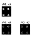
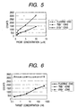
 wherein X represents a halogen atom, DMTO represents a dimethoxytrityl group, iPr represents an isopropyl group, and CNEt represents a 2-cyanoethyl group.


 5-bromo-2'-deoxyuridine triphosphate (Br-dUTP)


 5-bromo-2'-deoxyuridine triphosphate (Br-dUTP)


 5-bromo-2'-deoxyuridine triphosphate (Br-dUTP)


 wherein X represents the halogen atom, DMTO represents a dimethoxytrityl group, iPr represents an isopropyl group, and CNEt represents a 2-cyanoethyl group.