Background of the Invention
-
Percutaneous transluminal coronary angioplasty (PTCA) is widely used
as the primary treatment modality in many patients with coronary artery disease.
PTCA can relieve myocardial ischemia in patients with coronary artery disease
by reducing lumen obstruction and improving coronary flow. The use of this
surgical procedure has grown rapidly, with 39,000 procedures performed in
1983, nearly 150,000 in 1987, 200,000 in 1988, 250,000 in 1989, and
over 500,000 PTCAs per year are estimated by 1994 (Popma et al., Amer. J.
Med., 88: 16N-24N (1990); Fanelli et al, Amer. Heart Jour., 119: 357-368
(1990); Johnson et al., Circulation, 78 (Suppl. II): II-82 (1988)). Stenosis
following PTCA remains a significant problem, with from 25% to 35% of the
patients developing restenosis within 1 to 3 months. Restenosis results in
significant morbidity and mortality and frequently necessitates further
interventions such as repeat angioplasty or coronary bypass surgery. As of 1993,
no surgical intervention or post-surgical treatment has proven effective in
preventing restenosis.
-
The processes responsible for stenosis after PTCA are not completely
understood but may result from a complex interplay among several different
biologic agents and pathways. Viewed in histological sections, restenotic lesions
may have an overgrowth of smooth muscle cells in the intimal layers of the
vessel (Johnson et al., Circulation, 78 (Suppl. II): II-82 (1988)). Several possible
mechanisms for smooth muscle cell proliferation after PTCA have been
suggested (Popma et al., Amer. J. Med., 88: 16N-24N (1990); Fanelli et al,
Amer. Heart Jour., 119: 357-368 (1990); Liu et al., Circulation, 79: 1374-1387
(1989); Clowes et al., Circ. Res., 56: 139-145 (1985)).
-
Compounds that reportedly suppress smooth muscle proliferation in
vitro (Liu et al., Circulation, 79: 1374-1387 (1989); Goldman et al.,
Atherosclerosis, 65: 215-225 (1987); Wolinsky et al., JACC, 15 (2): 475-481
(1990)) may have undesirable pharmacological side effects when used in vivo.
Heparin is an example of one such compound, which reportedly inhibits smooth
muscle cell proliferation in vitro but when used in vivo has the potential adverse
side effect of inhibiting coagulation. Heparin peptides, while having reduced
anti-coagulant activity, have the undesirable pharmacological property of having
a short pharmacological half-life. Attempts have been made to solve such
problems by using a double balloon catheter, i.e., for regional delivery of the
therapeutic agent at the angioplasty site (e.g., Nabel et al., Science, 244: 1342-1344
(1989); U.S. Patent No. 4,824,436), and by using biodegradable materials
impregnated with a drug, i.e., to compensate for problems of short half-life
(e.g., Middlebrook et al., Biochem. Pharm., 38 (18): 3101-3110 (1989); U.S.
Patent No. 4,929,602).
-
At least five considerations would, on their face, appear to preclude use
of inhibitory drugs to prevent stenosis resulting from overgrowth of smooth
muscle cells. First, inhibitory agents may have systemic toxicity that could
create an unacceptable level of risk for patients with cardiovascular disease.
Second, inhibitory agents might interfere with vascular wound healing following
surgery and that could either delay healing or weaken the structure or elasticity
of the newly healed vessel wall. Third, inhibitory agents which kill smooth
muscle cells could damage surrounding endothelium and/or other medial smooth
muscle cells. Dead and dying cells also release mitogenic agents that might
stimulate additional smooth muscle cell proliferation and exacerbate stenosis.
Fourth, delivery of therapeutically effective levels of an inhibitory agent may be
problematic from several standpoints: namely, a) delivery of a large number of
molecules into the intercellular spaces between smooth muscle cells may be
necessary, i.e., to establish favorable conditions for allowing a therapeutically
effective dose of molecules to cross the cell membrane; b) directing an inhibitory
drug into the proper intracellular compartment, i.e., where its action is exerted,
may be difficult to control; and, c) optimizing the association of the inhibitory
drug with its intracellular target, e.g, a ribosome, while minimizing intercellular
redistribution of the drug, e.g. to neighboring cells, may be difficult. Fifth,
because smooth muscle cell proliferation takes place over several weeks it would
appear a priori that the inhibitory drugs should also be administered over several
weeks, perhaps continuously, to produce a beneficial effect.
-
As is apparent from the foregoing, many problems remain to be solved in
the use of inhibitory drugs to effectively treat smooth muscle cell proliferation.
Thus, there is a need for a method to inhibit or reduce stenosis due to
proliferation of vascular smooth muscle cells following traumatic injury to
vessels such as occurs during vascular surgery. There is also a need to deliver
compounds to vascular smooth muscle cells which exert inhibitory effects over
extended periods of time.
Summary of the Invention
-
The present invention provides a therapeutic method comprising the
administration of at least one therapeutic agent to a procedurally traumatized,
e.g., by an angioplasty procedure, mammalian vessel. Preferably, the
therapeutic agent is a cytoskeletal inhibitor. Preferred cytoskeletal inhibitors in
the practice of the present invention, include, for example, taxol and analogs or
derivatives thereof such as taxotere, or a cytochalasin, such as cytochalasin B,
cytochalasin C, cytochalasin D, or analogs or derivatives thereof. The
administration of a therapeutic agent of the invention is effective to biologically
stent the vessel, inhibit or reduce vascular remodeling of the vessel, inhibit or
reduce vascular smooth muscle cell proliferation, or any combination thereof.
The administration of the therapeutic agent preferably is carried out during the
procedure which traumatizes the vessel, e.g., during the angioplasty or other
vascular surgical procedure. The invention also provides therapeutic
compositions and dosage forms adapted for use in the present method, as well as
kits containing them.
-
Thus, one embodiment of the invention provides a method for
biologically stenting a traumatized mammalian blood vessel. As used herein,
"biological stenting" means the fixation of the vascular lumen in a dilated state
near its maximal systolic diameter. The method comprises the administration of
an effective amount of a cytoskeletal inhibitor to the blood vessel. Preferably,
the cytoskeletal inhibitor is dispersed in a pharmaceutically acceptable liquid
carrier, e.g., about 0.1 to about 10 µg for cytochalasin B/ml of vehicle, and
preferably administered locally via a catheter. Preferably, a portion of the
amount administered penetrates to at least about 6 to 9 cell layers of the inner
tunica media of the vessel and so is effective to biologically stent the vessel.
Another preferred embodiment of the invention is a cytochalasin or analog
thereof dispersed in a pharmaceutically acceptable liquid carrier at about 0.001
to about 25 µg per ml of aqueous vehicle.
-
Preferred catheter administration conditions include employing a catheter
to deliver about 4 to about 25 ml of a composition comprising the cytoskeletal
inhibitor dispersed or dissolved in a pharmaceutically acceptable liquid vehicle.
The cytoskeletal inhibitor is delivered at a hub pressure of about 3 to about 8
atm, more preferably about 4 to about 5 atm, for about 0.5 to about 5 minutes,
more preferably for about 0.7 to about 3 minutes. Preferred hydrostatic head
pressures for catheter administration include about 0.3 to about 1.0 atm, more
preferably about 0.5 to about 0.75 atm. The amount of therapeutic agent is
controlled so as to allow vascular smooth muscle cells to continue to synthesize
protein, which is required to repair minor cell trauma, and to secrete interstitial
matrix, thereby facilitating the fixation of the vascular lumen in a dilated state
near its maximal systolic diameter, i.e., to provide a biological stent of the
vessel. Preferably, the therapeutic agent is administered directly or
substantially directly to the traumatized area of the vascular smooth muscle
tissue.
-
The invention further provides a method for inhibiting or reducing
vascular remodeling of a traumatized mammalian blood vessel, by administering
an effective amount of a cytoskeletal inhibitor to the traumatized blood vessel.
-
As described hereinbelow, a dose response study showed that
cytochalasin B had a two logarithmic therapeutic index (TI). A large therapeutic
index allows the diffusion of therapeutic levels of the agent from the delivery
system, e.g., an implantable device, without toxicity to cells immediately
adjacent to the exit port of the system. Moreover, even at the maximum
concentration of cytochalasin B in a liquid vehicle, there was little or no toxicity
observed in cells adjacent to the delivery system. It was also found that
cytochalasin B and taxol both inhibit intimal proliferation in vessels subjected to
a procedural vascular trauma. This inhibition results in a more rapid and
complete endothelialization of the vessel wall following the trauma.
-
Thus, the invention further provides a method for inhibiting or reducing
diminution in vessel lumen volume in a procedurally traumatized mammalian
blood vessel. The method comprises administering to the blood vessel of a
mammal an effective amount of cytoskeletal inhibitor, wherein the cytoskeletal
inhibitor is in substantially pure substantially crystalline form and wherein the
crystals are of a size which results in sustained release of the cytoskeletal
inhibitor. Preferably, the crystals are of a size of about 0.1 micron to about 10
mm, preferably about 1 micron to about 25 micron, in size. Methods to
determine the size of crystals useful for sustained release are well known to the
art. Preferably, the cytoskeletal inhibitor is administered in situ, by means of an
implantable device, wherein the cytoskeletal inhibitor is releasably embedded in,
coated on, or embedded in and coated on, the implantable device. Preferably, the
crystalline cytoskeletal inhibitor is releasably embedded in, or dispersed in, a
adventitial wrap, e.g., a silicone membrane. For example, a preferred
therapeutic implantable device of the invention comprises about 5 to about 70,
preferably about 7 to about 50, and more preferably about 10 to about 30, weight
percent of a cytochalasin, e.g., cytochalasin B or an analog thereof, per weight
percent of the adventitial wrap. Another preferred therapeutic implantable
device of the invention comprises about 1 to about 70, preferably about 2 to
about 50, and more preferably about 3 to about 10, weight percent of taxol or an
analog thereof per weight percent of the adventitial wrap. Alternatively, a
preferred therapeutic implantable device of the invention comprises about 30 to
about 70, preferably about 30 to about 60, and more preferably about 30 to about
50, weight percent of taxol or an analog thereof per weight percent of the
adventitial wrap. Alternatively, the crystalline cytoskeletal inhibitor may be
suspended in a vehicle which yields a solution comprising the crystals, i.e., it is a
saturated solution.
-
The invention also provides a method for inhibiting or reducing
diminution in vessel lumen volume in a procedurally traumatized mammalian
blood vessel. The method comprises administering to the blood vessel an
emulsion or microemulsion comprising a cytoskeletal inhibitor. The amount of
the cytoskeletal inhibitor is effective to inhibit or reduce diminution in vessel
lumen area of the mammalian blood vessel. Preferably, the emulsion or
microemulsion is in sustained release dosage form. Preferably, the
microemulsion is about 5 nm to about 1000 nm, and more preferably about 30
nm to about 300 nm, in diameter.
-
The invention further provides a therapeutic method. The method
comprises administering to a procedurally traumatized mammalian blood vessel
a sustained release dosage form comprising microparticles or nanoparticles
comprising a cytoskeletal inhibitor, e.g., cytochalasin, taxol, or analogs thereof.
The sustained release dosage form is administered via an implantable device
which is not a catheter, preferably not a catheter used to perform bloodless
angioplasty. The amount administered is effective inhibit or reduce diminution
in vessel lumen area of the mammalian blood vessel. The sustained release
dosage form preferably comprises microparticles of 4 to about 50 microns in
diameter. The sustained release dosage form can also preferably comprise about
2 to about 50, and more preferably greater than 3 and less than 10, microns in
diameter. For nanoparticles, preferred sizes include about 10 to about 5000,
more preferably about 20 to about 500, and more preferably about 50 to about
200, nanometers.
-
Also provided is a method comprising administering to a procedurally
traumatized mammalian blood vessel a substantially pure substantially solid
form of a cytochalasin or an analog thereof effective inhibit or reduce diminution
in vessel lumen area of the mammalian blood vessel. Preferred solid forms
include, but are not limited to, microparticles or nanoparticles comprising the
cytochalasin or analog thereof, crystals or microcrystals of the cytochalasin or
analog thereof, microparticles or nanoparticles comprising crystals or
microcrystals of the cytochalasin or analog thereof, or a gel or paste comprising
the cytochalasin or analog thereof.
-
Also provided is a kit comprising, separately packaged, at least one
implantable device adapted for the in situ delivery, preferably local delivery, of
at least one cytoskeletal inhibitor to a site in the lumen of a traumatized
mammalian vessel and at least one unit dosage form of the cytoskeletal inhibitor
adapted for delivery by said device. The delivery of the unit dosage form to the
traumatized vessel via the device is effective to biologically stent the vessel,
inhibit or reduce the vascular remodeling of the vessel, inhibit or reduce vascular
smooth muscle cell proliferation, or any combination thereof.
-
Further provided is a kit comprising, separately packaged, an implantable
device adapted for the delivery of at least one therapeutic agent to a site in the
lumen of a traumatized mammalian vessel and a unit dosage form comprising an
emulsion or microemulsion comprising at least one cytoskeletal inhibitor. The
delivery of the unit dosage form via the device to the traumatized mammalian
vessel is effective to inhibit or reduce diminution in vessel lumen diameter in the
vessel.
-
The invention also provides a kit comprising, separately packaged, an
implantable device adapted for the delivery of at least one therapeutic agent to a
site in the lumen of a traumatized mammalian vessel and a unit dosage form
comprising an amount of nucroparticles or nanoparticles comprising taxol or an
analog thereof. Preferably, the kit also comprises a second unit dosage form
comprising a pharmaceutically acceptable liquid carrier vehicle for dispersing
said microparticles or said nanoparticles prior to delivery. The delivery of the
dispersed microparticles or nanoparticles to the traumatized mammalian vessel is
effective to inhibit or reduce diminution in vessel lumen diameter in the vessel.
-
Yet another embodiment of the invention is a pharmaceutical
composition suitable for administration by means of an implantable device. The
composition comprises an amount of a substantially pure solid cytochalasin
effective to inhibit or reduce stenosis or restenosis of a mammalian vessel
traumatized by a surgical
procedure; and a pharmaceutically acceptable release matrix for said
cytochalasin or analog thereof. Preferably, the release matrix comprises a gel,
paste or membrane, e.g., a silicone membrane.
-
The invention also provides a pharmaceutical composition comprising an
amount of a substantially pure solid cytochalasin or analog thereof effective to
inhibit or reduce stenosis or restenosis of a mammalian vessel traumatized by a
surgical procedure. Preferably the cytochalasin or analog thereof is in crystalline
form.
-
Further provided is a pharmaceutical composition comprising an
emulsion or microemulsion comprising an amount of a cytochalasin or an analog
thereof effective to inhibit or reduce stenosis or restenosis of a mammalian
vessel traumatized by a surgical procedure.
-
The invention also provides therapeutic devices. One embodiment of the
invention comprises a therapeutic shunt comprising an amount of a cytoskeletal
inhibitor effective to inhibit stenosis or reduce restenosis following placement of
the therapeutic shunt. Another embodiment of the invention comprises
therapeutic artificial graft comprising an amount of a cytochalasin or analog
thereof to inhibit stenosis or reduce restenosis following placement of the
graft. Yet another embodiment of the invention comprises a therapeutic
adventitial wrap comprising an amount of a cytoskeletal inhibitor effective to
inhibit stenosis or reduce restenosis following placement of the wrap.
-
The invention also provides a therapeutic method comprising inhibiting
diminution of vessel lumen diameter by administering to a traumatized vessel of
a mammal an effective amount of a cytoskeletal inhibitor. The cytoskeletal
inhibitor is administered via an implantable device, wherein the implantable
device is not a catheter which has a first and a second expansile member, i.e.,
balloons, which are disposed on opposite sides of the vessel area to be treated in
order to isolate the portion of the vessel to be treated prior to cytoskeletal
inhibitor administration. Preferably, the isolated portion of the vessel is not
washed to remove blood prior to cytoskeletal inhibitor administration
("bloodless angioplasty"). "Isolated," as used above, does not mean occlusive
contact of the actual treatment area by the catheter balloon, which is preferred in
the practice of the present invention. Moreover, bloodless angioplasty, such as
that described in Slepian, U.S. Patent No. 5,328,471, i.e., in which the region to
be treated is washed, may introduce trauma or further trauma to the vessel, may
increase the potential for complications and is not necessary to achieve a
beneficial effect.
-
Also provided is a method comprising administering to a mammalian
blood vessel a dosage form of a cytochalasin or an analog thereof in a non-liquid
vehicle or matrix effective inhibit or reduce diminution in vessel lumen area of
the mammalian blood vessel. Preferably the dosage form is a substantially solid
dosage form. The non-liquid vehicle or matrix preferably includes, but is not
limited to, a gel, paste, or a membrane, but does not include microparticles,
nanoparticles, and the like, which comprises the cytochalasin or analog thereof.
-
Further provided is a kit comprising, preferably separately packaged, an
implantable device adapted for the delivery of at least one therapeutic agent to a
site in the lumen of a traumatized mammalian vessel and a unit dosage form
comprising at least one cytoskeletal inhibitor, wherein the administration of at
least a portion of the unit dosage form is effective to inhibit or reduce diminution
in vessel lumen diameter of the vessel. The device is not a catheter which has a
first and a second expansile member which are disposed on opposite sides of the
region to be treated so as to isolate a portion of the vessel to be treated prior to
administration or wherein the isolated portion of the vessel is not washed to
remove blood prior to administration.
-
Yet another embodiment of the invention is a pharmaceutical
composition suitable for administration by means of an implantable device. The
composition comprises an amount of a cytochalasin or analog thereof effective
to inhibit or reduce stenosis or restenosis of a mammalian vessel traumatized by
a surgical procedure and a pharmaceutically acceptable non-liquid release matrix
for said cytochalasin. Preferably, the release matrix comprises a gel, paste or
membrane.
-
Also provided is a unit dosage form. The unit dosage form comprises a
vial comprising about 10 to about 30 ml of about 0.001 µg to about 25 µg of a
cytoskeletal inhibitor, preferably a cytochalasin, per ml of liquid vehicle,
wherein the unit dosage form is adapted for delivery via an implantable device,
and wherein the vial is labeled for use in treating or inhibiting stenosis or
restenosis. Preferably, the unit dosage form comprises a vial comprising about
10 to about 30 ml of about 0.01 µg to about 10 µg of cytochalasin B per ml of
liquid vehicle. Thus, the volume present in a vial may be greater than, or about
the same as, the volume present in the implantable device. Likewise, the volume
present in the implantable device may be greater than, or about the same as, the
volume administered. Similarly, the volume administered may be greater than,
or about the same as, the volume which has a beneficial effect.
-
Further provided is a unit dosage comprising a vial comprising a
cytostatic amount of a cytoskeletal inhibitor in a pharmaceutically acceptable
liquid vehicle. Preferably, the cytoskeletal inhibitor comprises a cytochalasin,
taxol, or an analog thereof.
Brief Description of the Drawings
-
- FIGURE 1A is a photomicrograph of vascular smooth muscle cells of a
24-year-old male patient.
- FIGURE 1B is a photomicrograph of vascular smooth muscle cells in an
artery of a 24-year-old male patient with vascular smooth muscle binding protein
bound to the cell surface and membrane. The patient received the vascular
smooth muscle binding protein by i.v. administration 4 days before the arterial
tissue was prepared for histology.
- FIGURE 2 depicts a first scheme for chemical coupling of a therapeutic
agent to a vascular smooth muscle binding protein.
- FIGURE 3 depicts a second scheme for chemical coupling of a
therapeutic agent to a vascular smooth muscle binding protein.
- FIGURE 4A graphically depicts experimental data showing rapid
binding of vascular smooth muscle binding protein to marker-positive test cells
in vitro.
- FIGURE 4B graphically depicts experimental data showing rapid binding
of vascular smooth muscle binding protein to vascular smooth muscle cells in
vitro.
- FIGURE 5A presents graphically experimental data showing undesirable
cytotoxicity of even low levels of therapeutic conjugate (i.e., RA-NR-AN-01),
and the free RA therapeutic agent, when vascular smooth muscle cells were
treated for 24 hours in vitro.
- FIGURE 5B graphically presents experimental data showing the effects
of RA-NR-AN-01 therapeutic conjugate on metabolic activity of marker-positive
and -negative cells. The data show undesirable nonspecific cytotoxicity of the
conjugate for all these cells in a 24 hour treatment in vitro. The non-specificity
results from extracellular hydrolysis of the coupling ligand which exposes the
tested cells to free drug.
- FIGURE 6A graphically depicts experimental data showing undesirable
nonspecific cytotoxicity of PE-NR-AN-01 therapeutic conjugate for marker-positive
and marker-negative test cells after 24 hours of treatment in vitro, even
though the 24 hour treatment was followed by an overnight recovery period prior
to testing the metabolic activity.
- FIGURE 6B depicts experimental data showing nonspecific cytotoxicity
of the free PE therapeutic agent on marker-positive and -negative test cells after
24 hours of treatment in vitro.
- FIGURE 7A graphically presents experimental data showing that a short
5 minute "pulse" treatment, i.e., instead of 24 hours, followed by exposure to
[3H]leucine, with free RA therapeutic agent being nonspecifically cytotoxic, i.e.,
for control HT29 marker-negative cells, but, in contrast, the RA-NR-AN-01
therapeutic conjugate is not cytotoxic in this "pulse" treatment.
- FIGURE 7B presents graphically experimental data showing that free RA
therapeutic agent is nonspecifically cytotoxic for control HT29 marker-negative
cells, even in a 5' "pulse" treatment followed by a 24 hour recovery period prior
to [3H]leucine exposure, but, in contrast, the RA-NR-AN-01 therapeutic
conjugate is not cytotoxic to cells.
- FIGURE 7C presents graphically results of experiments showing that
"pulse" treatment of cells in vitro with the RA-NR-AN-01 therapeutic conjugate
inhibits cellular activity in marker-positive A375 cells, as measured by protein
synthesis.
- FIGURE 7D presents graphically experimental data showing that "pulse"
treatment of cells in vitro with the RA-NR-AN-01 therapeutic conjugate did not
exert long-lasting inhibitory effects on cellular activity in marker-positive cells,
since protein synthesis in A375 cells was not inhibited when the cells were
allowed an overnight recovery period prior to testing in vitro.
- FIGURE 8A presents graphically experimental data showing that while a
"pulse" treatment of cells in vitro with free RA therapeutic agent was non-specifically
cytotoxic, the RA-NR-AN-01 therapeutic conjugate did not exert
long-lasting inhibitory effects on cellular activity in vascular smooth muscle
cells, as evidenced by metabolic activity in BO54 cells that were allowed a 48
hour recovery period prior to testing.
- FIGURE 8B graphically depicts experimental data similar to those
presented in FIGURE 8A, above, but using a second marker-positive cell type,
namely A375, the data show that "pulse" treatment with the RA-NR-AN-01
therapeutic conjugate did not exert long-lasting inhibitory effects on cellular
activity, as measured by metabolic activity in A375 cells that were allowed a 48
hour recovery period prior to testing.
- FIGURE 8C graphically depicts results similar to those presented in
FIGURE 8A and FIGURE 8B, above, but using a marker-negative control cell
type, namely HT29. The results show that the "pulse" treatment with the RA-NR-AN-01
therapeutic conjugate did not exert long-lasting inhibitory effects on
the cellular activity of marker-negative control cells, as measured by metabolic
activity in HT29 cells that were allowed a 48 hour recovery period prior to
testing.
- FIGURE 9A shows stenosis due to intimal smooth muscle cell
proliferation in a histological section of an untreated artery 5 weeks after
angioplasty in an animal model.
- FIGURE 9B shows inhibition of stenosis in a histological section of an
artery treated with therapeutic conjugate at 5 weeks after angioplasty in an
animal model.
- FIGURE 10A graphically depicts experimental data comparing protein
synthesis and DNA synthesis inhibition capability of suramin with respect to
vascular smooth muscle cells.
- FIGURE 10B graphically depicts experimental data comparing protein
synthesis and DNA synthesis inhibition capability of staurosporin with respect to
vascular smooth muscle cells.
- FIGURE 10C graphically depicts experimental data comparing protein
synthesis and DNA synthesis inhibition capability of nitroglycerin with respect
to vascular smooth muscle cells.
- FIGURE 10D graphically depicts experimental data comparing protein
synthesis and DNA synthesis inhibition capability of cytochalasin B with respect
to vascular smooth muscle cells.
- FIGURE 11 shows a tangential section parallel to the inner surface of a
smooth muscle cell which is magnified 62,500 times and is characterized by
numerous endocytic vesicles, several of which contain antibody coated gold
beads in the process of being internalized by the cell in vitro.
- FIGURE 12 shows a smooth muscle cell which is magnified 62,500
times and is characterized by a marked accumulation of gold beads in lysosomes
at 24 hours following exposure of the cell to the beads in vitro.
- FIGURE 13 shows a smooth muscle cell which is magnified 62,500
times and is characterized by accumulation of gold beads in lysosomes in vivo.
- FIGURE 14 depicts an in vivo dose response study of the effects of
cytochalasin B on the luminal area of pig femoral arteries.
- FIGURE 15 is a graph depicting the inhibition of smooth muscle cell
proliferation in traumatized vessels over time by cytochalasin B (CB) or taxol
(TAX) administered in silicone wraps (SW).
- FIGURE 16 is a graph depicting the inhibition of smooth muscle cell
proliferation in traumatized vessels over time by 10% or 30% wt/wt CB in SW
or 5% wt/wt TAX in SW.
- FIGURE 17 is a graph depicting the inhibition of smooth muscle cell
proliferation in traumatized vessels over time by CB or TAX in silicone, CB in a
collagen gel supported by a bovine collagen mesh (CG-CM) or CB in a pluronic
gel supported by a bovine collagen mesh (PG-CW).
-
Detaited Description of the Invention
Definitions
-
"Therapeutic conjugate" means a vascular smooth muscle or an
interstitial matrix binding protein coupled (e.g., optionally through a linker
moiety) to a therapeutic agent. Therapeutic conjugates of the invention are
obtained by coupling a vascular smooth muscle binding protein to a therapeutic
agent. In the therapeutic conjugate, the vascular smooth muscle binding protein
performs the function of targeting the therapeutic conjugate to vascular smooth
muscle cells or pericytes, and the therapeutic agent performs the function of
inhibiting the cellular activity or proliferation of the smooth muscle cell or
pericyte.
-
"Therapeutic agent" includes any moiety capable of exerting a
therapeutic or prophylactic effect in the present method.
-
"Target" and "marker" are used interchangeably in describing the present
conjugates to mean a molecule recognized in a specific manner by the matrix or
vascular smooth muscle binding protein, e.g., an antigen, polypeptide antigen or
cell surface carbohydrate (e.g., a glycolipid, glycoprotein, or proteoglycan) that
is expressed on the cell surface membranes of a vascular smooth muscle cell or a
matrix structure.
-
"Epitope" is used to refer to a specific site within the "target" molecule
that is bound by the matrix or smooth muscle binding protein, e.g., a sequence of
three or more amino acids or saccharides.
-
"Coupled" is used to mean covalent or non-covalent chemical association
(i.e., hydrophobic association as through van der Waals forces or charge-charge
interactions) of the matrix or vascular smooth muscle binding protein with the
therapeutic agent, including by chelation. Preferably, the binding proteins are
associated with the therapeutic agents by means of covalent bonding.
-
"Linker" means a moiety that couples the matrix or smooth muscle
binding protein to a therapeutic agent, e.g., as derived from an organic chemical
coupling agent.
-
As used herein, "substantially" pure means at least about 90%, preferably
at least about 98%, and more preferably at least about 99%, free of contaminants
when assayed by methods conventionally employed by the art.
-
As used herein, "substantially" solid or crystalline means at least about
90%, preferably at least about 98%, and more preferably at least about 99%, free
of non-solid or non-crystalline forms or phases when assayed by methods
conventionally employed by the art.
-
"Migration" of smooth muscle cells means movement of these cells in
vivo from the medial layers of a vessel into the intima, which may also be
studied in vitro by following the motion of a cell from one location to another
(e.g., using time-lapse cinematography or a video recorder and manual counting
of smooth muscle cell migration out of a defined area in the tissue culture over
time).
-
"Proliferation" means an increase in cell number, i.e., by mitosis of the
cells. As used herein "smooth muscle cells" does not refer to neoplastic vascular
smooth muscle cells, i.e., cancer cells.
-
"Implantable device" means any material that is capable of retaining and
releasing a therapeutic agent so as to deliver it in situ in a controlled fashion to a
mammalian vessel. An implantable device includes devices which are placed in
the lumen of the vessel, e.g., an indwelling catheter or stent, or on the exterior of
a vessel, e.g., an adventitial wrap, mesh or covering, or which become a part of
the vessel itself, for example to replace a portion of a diseased or traumatized
vessel, e.g., a synthetic graft. The implantable device may comprise the
therapeutic agent in a form which is releasably embedded in and/or coated on the
device. The therapeutic agent may also be releasably embedded in and/or coated
on a pharmaceutically acceptable release carrier matrix, which may be applied to
and/or embedded in the device or administered directly to a vessel. For example,
a matrix useful in the practice of the invention includes, but is not limited to,
microparticles, nanoparticles, a gel, a paste, or a permeable membrane. An
implantable device may be implanted for a limited amount of time, e.g., catheter
or infusion needle delivery of a therapeutic agent, or for a prolonged period of
time, e.g., a stent or graft. Vessels, into which the implantable device of the
invention may be inserted, include, but are not limited to, coronary, femoral,
carotid and peripheral vessels.
-
"Abnormal or pathological or inappropriate" with respect to an activity or
proliferation means division, growth or migration of cells, but not cancer cells,
that occurs more rapidly or to a significantly greater extent than typically occurs
in a normally functioning cell of the same type or in lesions not found in healthy
tissue.
-
"Expressed" means mRNA transcription and translation with resultant
synthesis, glycosylation, and/or secretion of a polypeptide by a cell, e.g., CSPG
synthesized by a vascular smooth muscle cell or pericyte.
-
"Vascular remodeling" means a diminution in vessel lumen volume,
diameter or area that is not the result of neointimal thickening or smooth muscle
cell proliferation, and which generally occurs after a procedural vascular trauma.
Thus, a reduction in the area ("constriction") circumscribed by the internal
elastic lamina or membrane (IEL) without significant amounts of neointimal
formation is termed "vascular remodeling." See Isner, Circ., 89, 2937 (1994).
The luminal cross-sectional area of a vessel can be measured by direct
planimetering, e.g., by intravascular ultrasound (IVUS) or at necropsy. As used
herein, "vascular remodeling" does not include compensatory enlargement of a
vessel which accompanies neointimal proliferation so as to accomodate the
intimal increase. This compensatory enlargement has also been referred to as
"positive" vascular remodeling.
-
"Sustained release" means a dosage form designed to release a
therapeutic agent therefrom for a time period from about 0.0005 to about 180,
preferably from about 1-3 to about 150, and more preferably from about 30 to
about 120, days. Release over a longer time period is also contemplated as
"sustained release" in the context of the present invention. Moreover, it is
contemplated that the invention can be practiced with a locally or systemically
administered sustained release dosage form.
-
"Dosage form" includes a formulation comprising a free (non-targeted or
non-binding partner associated) therapeutic agent, as well as a sustained release
formulation comprising a therapeutic agent. For example, sustained release
formulations can comprise microparticles or nanoparticles, microemulsions,
biodegradable or non-biodegradable polymeric materials, or any combination
thereof, comprising a therapeutic agent dispersed therein, as well as crystalline
forms of the therapeutic agent. A targeted or binding partner associated dosage
form of the invention includes a sustained release therapeutic formulation
comprising microparticles or nanoparticles, microemulsions, and/or
biodegradable or non-biodegradable polymeric materials. The sustained release
dosage form is linked to one or more binding proteins or peptides, so as to
deliver a therapeutic agent dispersed therein to a target cell population which
binds to the binding protein or peptide.
-
"Cytochalasin" includes a fungal metabolite exhibiting an inhibitory
effect on target cellular metabolism, including prevention of contraction or
migration of vascular smooth muscle cells. Preferably, cytochalasins inhibit the
polymerization of monomeric actin (G-actin) to polymeric form (F-actin),
thereby inhibiting cell functions requiring cytoplasmic micro filaments.
Cytochalasins typically are derived from phenylalanine (cytochalasins),
tryptophan (chaetoglobosins), or leucine (aspochalasins), resulting in a benzyl,
indol-3-yl methyl or isobutyl group, respectively, at position C-3 of a substituted
perhydroisoindole-1-one moiety (Formula I or II).
-
The perhydroisoindole moiety in turn contains an 11-, 13- or 14-atom
carbocyclic- or oxygen-containing ring linked to positions C-8 and C-9. All
naturally occurring cytochalasins contain a methyl group at C-5; a methyl or
methylene group at C-12; and a methyl group at C-14 or C-16. Exemplary
cytochalasins include cytochalasin A, cytochalasin B, cytochalasin C,
cytochalasin D, cytochalasin E, cytochalasin F, cytochalasin G, cytochalasin H,
cytochalasin J, cytochalasin K, cytochalasin L, cytochalasin M, cytochalasin N,
cytochalasin O, cytochalasin P, cytochalasin Q, cytochalasin R, cytochalasin S,
chaetoglobosin A, chaetoglobosin B, chaetoglobosin C, chaetoglobosin D,
chaetoglobosin E, chaetoglobosin F, chaetoglobosin G, chaetoglobosin J,
chaetoglobosin K, deoxaphomin, proxiphomin, protophomin, zygosporin D,
zygosporin E, zygosporin F, zygosporin G, aspochalasin B, aspochalasin C,
aspochalasin D and the like, as well as functional equivalents and derivatives
thereof. Certain cytochalasin derivatives are set forth in Japanese Patent Nos. 72
01,925; 72 14,219; 72 08,533; 72 23,394; 72 01924; and 72 04,164.
Cytochalasin B is used in this description as a typical cytochalasin.
-
As referred to herein, "taxol" includes taxol as well as functional analogs,
equivalents or derivatives thereof. For example, derivatives and analogs of
taxol include, but are not limited to, taxotere, baccatin, 10-deacetyltaxol, 7-xylosyl-10-deacetyltaxol,
cephalomannine, 10-deacetyl-7-epitaxol, 7 epitaxol,
10-deacetylbaccatin III, 10-deacetylcephaolmannine and analogs or derivatives
disclosed in Kingston et al. (New Trends in Nat. Prod. Chem., 26,219 (1986)),
Bringli et al. (WO 93/17121), Golik et al. (EPA 639577), Kelly et al. (WO
95/20582), and Cassady and Dourous (eds., In: Anticancer Agents Based on
Natural Product Models, Academic Press, NY (1980)), the disclosures of which
are incorporated by reference herein. Methods for preparing taxol and numerous
analogs and derivatives thereof are well known to the art.
-
"Macrocyclic trichothecene" is intended to mean any one of the group of
structurally related sesquiterpenoid macrocyclic mycotoxins produced by several
species of fungi and characterized by the 12,13-epoxytrichothec-9-ene basic
structure, e.g., verrucarins and roridins that are the products of secondary
metabolism in the soil fungi Myrothecium verrucaria and Myrothecium
roridium.
-
There are two broad classes of trichothecenes: those that have only a
central sesquiterpenoid structure and those that have an additional macrocyclic
ring (simple and macrocyclic trichothecenes, respectively). The simple
trichothecenes may be subdivided into three groups (i.e., Group A, B, and C) as
described in U.S. Patent Nos. 4,744,981 and 4,906,452 (incorporated herein by
reference). Representative examples of Group A simple trichothecenes include:
scirpene, roridin C, dihydrotrichothecene, scirpen-4, 8-diol, verrucarol,
scirpentriol, T-2 tetraol, pentahydroxyscirpene, 4-deacetylneosolaniol,
trichodermin, deacetylcalonectrin, calonectrin, diacetylverrucarol,
4-monoacetoxyscirpenol, 4,15-diacetoxyscirpenol, 7-hydroxydiacetoxyscirpenol,
8-hydroxydiacetoxy-scirpenol (neosolaniol), 7,8-dihydroxydiacetoxyscirpenol,
7-hydroxy-8-acetyldiacetoxyscirpenol, 8-acetylneosolaniol, NT-1, NT-2, HT-2,
T-2, and acetyl T-2 toxin. Representative examples of Group B simple
trichothecenes include: trichothecolone, trichothecin, deoxynivalenol,
3-acetyldeoxynivalenol, 5-acetyldeoxynivalenol, 3,15-diacetyldeoxynivalenol,
nivalenol, 4-acetylnivalenol (fusarenon-X), 4,15-idacetylnivalenol,
4,7,15-triacetylnivalenol, and tetra-acetylnivalenol. Representative examples of
Group C simple trichothecenes include: crotocol and crotocin. Representative
macrocyclic trichothecenes include verrucarin A, verrucarin B, verrucarin J
(Satratoxin C), roridin A, roridin D, roridin E (satratoxin D), roridin H,
satratoxin F, satratoxin G, satratoxin H, vertisporin, mytoxin A, mytoxin C,
mytoxin B, myrotoxin A, myrotoxin B, myrotoxin C, myrotoxin D, roritoxin A,
roritoxin B, and roritoxin D. In addition, the general "trichothecene"
sesquiterpenoid ring structure is also present in compounds termed "baccharins"
isolated from the higher plant Baccharis megapotamica, and these are described
in the literature, for instance as disclosed by Jarvis et al. (Chemistry of
Alleopathy, ACS Symposium Series No. 268: ed. A.C. Thompson, 1984, pp.
149-159) and Jarvis & Mazzola (Acc. Chem. Res. 15:338-395, 1982)).
Trichothecenes are also produced by soil fungi of the class Fungi imperfecti
(Bamburg, J.R. Proc. Molec. Subcell. Biol. 8:41-110, 1983))
-
"Staurosporin" includes staurosporin, a protein kinase C inhibitor of the
following formula (III),
as well as diindoloalkaloids having one of the following general structures:
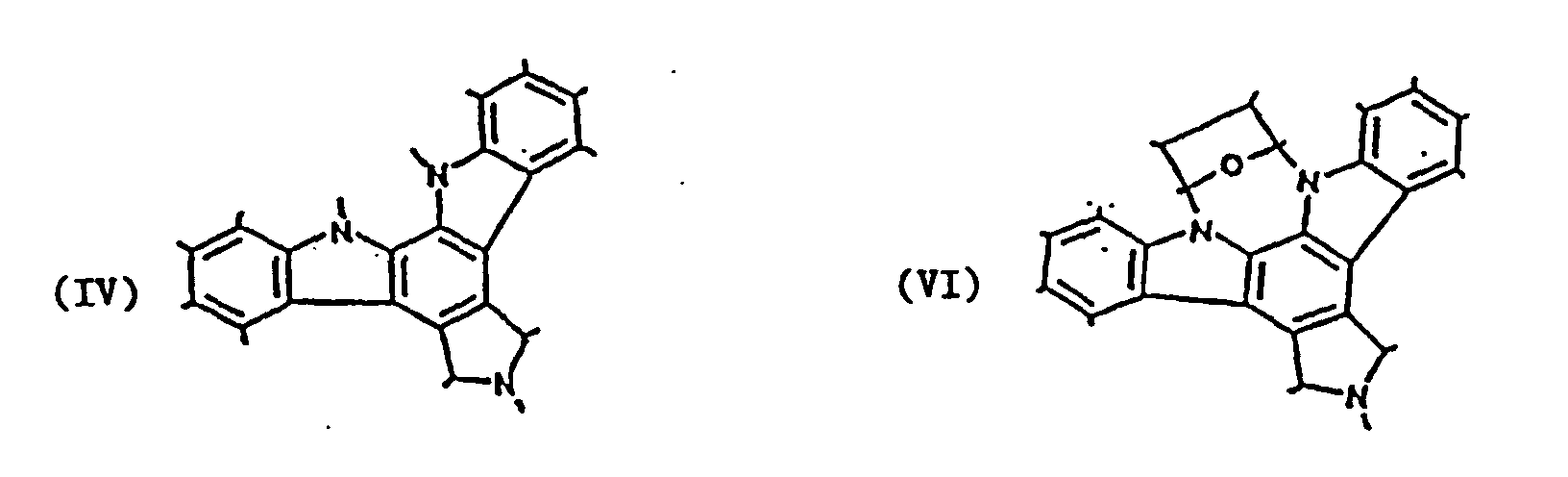

More specifically, the term "staurosporin" includes K-252 (see, for example,
Japanese Patent Application No. 62,164,626), BMY-41950 (U.S. Patent No.
5,015,578), UCN-01 (U.S. Patent No. 4,935,415), TAN-999 (Japanese Patent
Application No. 01,149,791), TAN-1030A (Japanese Patent Application No.
01,246,288), RK-286C (Japanese Patent Application No. 02,258,724) and
functional equivalents and derivatives thereof. Derivatives of staurosporin
include those discussed in Japanese Patent Application Nos. 03,72,485;
01,143,877; 02,09,819 and 03,220,194, as well as in PCT International
Application Nos. WO 89 07,105 and WO 91 09,034 and European Patent
Application Nos. EP 410,389 and EP 296,110. Derivatives of K-252, a natural
product, are known. See, for example, Japanese Patent Application Nos.
63,295,988; 62,240,689; 61,268,687; 62,155,284; 62,155,285; 62,120,388 and
63,295,589, as well as PCT International Application No. WO 88 07,045 and
European Patent Application No. EP 323,171.
-
As referred to herein, smooth muscle cells and pericytes include those
cells derived from the medial layers of vessels and adventitial vessels which
proliferate in intimal hyperplastic vascular sites following injury, such as that
caused during PTCA. Characteristics of smooth muscle cells include a
histological morphology (under light microscopic examination) of a spindle
shape with an oblong nucleus located centrally in the cell with nucleoli present
and myofibrils in the sarcoplasm. Under electron microscopic examination,
smooth muscle cells have long slender mitochondria in the juxtanuclear
sarcoplasm, a few tubular elements of granular endoplasmic reticulum, and
numerous clusters of free ribosomes. A small Golgi complex may also be
located near one pole of the nucleus. The majority of the sarcoplasm is occupied
by thin, parallel myofilaments that may be, for the most part, oriented to the
long axis of the muscle cell. These actin containing myofibrils may be arranged
in bundles with mitochondria interspersed among them. Scattered through the
contractile substance of the cell may also be oval dense areas, with similar dense
areas distributed at intervals along the inner aspects of the plasmalemma.
-
Characteristics of pericytes include a histological morphology (under
light microscopic examination) characterized by an irregular cell shape.
Pericytes are found within the basement membrane that surrounds vascular
endothelial cells and their identity may be confirmed by positive
immuno-staining with antibodies specific for alpha smooth muscle actin (e.g.,
anti-alpha-sml, Biomakor, Rehovot, Israel), HMW-MAA, and pericyte
ganglioside antigens e.g., MAb 3G5 (Schlingemann et al., Am. J. Pathol., 136:
1393-1405 (1990)); and, negative immune-staining with antibodies to
cytokeratins (i.e., epithelial and fibroblast markers) and von Willdebrand factor
(i.e., an endothelial marker). Both vascular smooth muscle cells and pericytes
are positive by immunostaining with the NR-AN-01 monoclonal antibody.
-
As used herein, the term "procedural vascular trauma" includes the
effects of surgical/mechanical interventions into mammalian vasculature, but
does not include vascular trauma due to the organic vascular pathologies, i.e.,
diseases and infections.
-
Thus, procedural vascular traumas within the scope of the present
treatment method include (1) organ transplantation, such as heart, kidney, liver
and the like, e.g., involving vessel anastomosis; (2) vascular surgery, e.g.,
coronary bypass surgery, biopsy, heart valve replacement, atheroectomy,
thrombectomy, and the like; (3) transcatheter vascular therapies (TVT) including
angioplasty, e.g., laser angioplasty and PTCA procedures, employing balloon
catheters, and indwelling catheters; (4) vascular grafting using natural or
synthetic materials, such as in saphenous vein coronary bypass grafts, dacron
and venous grafts used for peripheral arterial reconstruction, etc.; (5) placement
of a mechanical shunt, e.g., a PTFE hemodialysis shunt used for arteriovenous
communications; and (6) placement of an intravascular stent, which may be
metallic, plastic or a biodegradable polymer. See U.S. patent application Serial
No. 08/389,712, filed February 15, 1995, which is incorporated by reference
herein. For a general discussion of implantable devices and biomaterials from
which they can be formed, see H. Kambic et al.,
"Biomaterials in Artificial Organs", Chem. Eng. News, 30 (April 14, 1986), the
disclosure of which is incorporated by reference herein.
Therapeutic Agents Falling Within the Scope of the Invention
-
Therapeutic agents useful in the practice of the invention include agents
which biologically stent a vessel and/or reduce or inhibit vascular remodeling
and/or inhibit or reduce vascular smooth muscle cell proliferation following a
procedural vascular trauma. The therapeutic agents of the invention are selected
to inhibit a cellular activity of a vascular smooth muscle cell, e.g., proliferation,
migration, increase in cell volume, increase in extracellular matrix synthesis
(e.g., collagens, proteoglycans, and the like), or secretion of extracellular matrix
materials by the cell.
-
Preferably, the therapeutic agent is: a) a "cytostatic agent" which acts to
prevent or delay cell division in proliferating cells by inhibiting replication of
DNA or by inhibiting spindle fiber formation and the like; b) an inhibitor of
migration of vascular smooth muscle cells from the medial wall into the intima,
e.g., an "anti-migratory agent" e.g., a cytochalasin; c) as an inhibitor of the
intracellular increase in cell volume (i.e., the tissue volume occupied by a cell; a
"cytoskeletal inhibitor"); d) an inhibitor that blocks cellular protein synthesis
and/or secretion or organization of extracellular matrix (i.e., an "anti-matrix
agent"); or any combination thereof
-
Representative examples of "cytostatic agents" include, e.g., modified
toxins, methotrexate, adriamycin, radionuclides (e.g., see Fritzberg et al., U.S.
Patent No. 4,897,255), protein kinase inhibitors (e.g., staurosporin), taxol or
analogs thereof (e.g., taxotere), inhibitors of specific enzymes (such as the
nuclear enzyme DNA topoisomerase II and DNA polymerase, RNA polymerase,
adenyl guanyl cyclase), superoxide dismutase inhibitors, terminal
deoxynucleotidyl- transferase, reverse transcriptase, antisense oligonucleotides
that suppress smooth muscle cell proliferation and the like, which when
delivered into a cellular
compartment at an appropriate dosage will act to impair proliferation of a
smooth muscle cell or pericyte without killing the cell.
-
Representative examples of "anti-migratory agents" include inhibitors
(i.e., agonists and antagonists, and competitive or non-competitive inhibitors) of
chemotactic factors and their receptors (e.g., complement chemotaxins such as
C5a, C5a desarg or C4a; extracellular matrix factors, e.g., collagen degradation
fragments), or of intracellular cytoskeletal proteins involved in locomotion
(e.g., actin, cytoskeletal elements, and phosphatases and kinases involved in
locomotion). Representative examples of useful therapeutic agents in this
category of anti-migratory agents include caffeic acid derivatives and nilvadipine
(a calcium antagonist), and steroid hormones. Preferred anti-migratory
therapeutic agents are the cytochalasins.
-
Representative examples of "cytoskeletal inhibitors", a subset of
cytostatic agents, include colchicine, vinblastin, cytochalasins, taxol, or analogs
or derivatives thereof that act on microtubule and microfilament networks within
a cell. Preferred cytoskeletal inhibitors include cytochalasin B and taxol.
-
Representative examples of "anti-matrix agents" include inhibitors (i.e.,
agonists and antagonists and competitive and non-competitive inhibitors) of
matrix synthesis, secretion and assembly, organizational cross-linking (e.g.,
transglutaminases cross-linking collagen), and matrix remodeling (e.g.,
following wound healing). A representative example of a useful therapeutic
agent in this category of anti-matrix agents is colchicine, an inhibitor of secretion
of extracellular matrix. Another example is tamoxifen for which evidence exists
regarding its capability to organize and/or stabilize as well as diminish smooth
muscle cell proliferation following angioplasty. The organization or
stabilization may stem from the blockage of vascular smooth muscle cell
maturation into a pathologically proliferating form.
Identification of Therapeutic Agents Useful in the Practice of the Invention
-
The identification of therapeutic agents useful in the practice of the
invention may be determined by methods well known to the art. For example, a
therapeutic agent failing within the scope of the invention exhibits one or more
of the following characteristics. The agent:
- (i) results in retention of an expanded luminal cross-sectional area,
diameter or volume of a vessel following angioplasty (e.g., PTCA,
percutaneous transluminal angioplasty (PTA) or the like) or other trauma,
including atheroectomy (e.g., rotoblater, laser and the like), coronary
artery bypass procedures and the like;
- (ii) facilitates an initial increase in luminal cross-sectional area, diameter
or volume that does not result in or accentuate chronic stenosis of the
lumen;
- (iii) inhibits target cell contraction or migration; and
- (iv) is cytostatic.
Methods to measure luminal cross-sectional area, volume or diameter include,
but are not limited to, angiography, ultrasonic evaluation, fluoroscopic imaging,
fiber optic endoscopic examination or biopsy and histology.-
-
Preferably, a therapeutic agent employed herein will have all four
properties; however, the first and third are generally more important than the
second and fourth for practice of the present invention. It was found that
cytochalasin B administration can result in a biological stenting effect. The
biological stenting effect can be achieved using a single infusion of the
therapeutic agent into the traumatized region of the vessel wall at a dose
concentration ranging from about 0.1 micrograms/ml to about
10.0 micrograms/ml (Example 16).
-
In the case of therapeutic agents or dosage forms containing
anti-migratory or anti-matrix therapeutic agents, cell migration and cell
adherence in in vitro assays, respectively, may be used for determining the
concentration at which a therapeutically effective dosage will be reached in the
fluid space in the vessel wall created by an infusion catheter.
-
An agent useful in the sustained release embodiments of the present
invention exhibits one or more of the following characteristics. The agent
- (i) causes the retention of an expanded luminal diameter or cross-sectional
area following angioplasty (e.g., PTCA, percutaneous
transluminal angioplasty (PTA) or the like) or other trauma, including
atheroectomy (e.g., rotoblater, laser and the like), coronary artery bypass
procedures or the like;
- (ii) inhibits target cell proliferation (e.g., following 5 minute and 24 hour
exposure to the agent, in vitro vascular smooth muscle tissue cultures
demonstrate a level of inhibition of 3H-thymidine uptake and, preferably,
display relatively less inhibition of 3H-leucine uptake);
- (iii) at a dose sufficient to inhibit DNA synthesis, produces only mild to
moderate (e.g., grade 2 or 3 in the assays described below) morphological
cytotoxic effects;
- (iv) inhibits target cell contraction; and
- (v) is cytostatic.
-
-
Upon identification of a therapeutic agent exhibiting one or more of the
preceding properties, the agent is subjected to a second testing protocol that
involves longer exposure of vascular smooth muscle cells (VSMC) to the
therapeutic agent. For example, an agent useful in the sustained release
embodiments of the present invention also exhibits the following characteristics:
- (i) upon long term (e.g., 5 days) exposure, the agent produces the same
or similar in vitro effect on vascular smooth muscle tissue culture DNA
synthesis and protein synthesis, as described above for the 5 minute and
24 hour exposures; and
- (ii) at an effective dose in the long term in vitro assay for DNA synthesis
inhibition, the agent exhibits mild to moderate morphological cytotoxic
effects over a longer term (e.g., 10 days).
-
-
Further evaluation of potentially useful anti-proliferative agents is
conducted in an in vivo balloon traumatized pig femoral artery model.
Preferably, these agents demonstrate a 50% or greater inhibition of cell
proliferation in the tunica media vascular smooth muscle cells, as indicated by a
1 hour "BRDU flash labeling" prior to tissue collection and histological
evaluation (Example 13). If an agent is effective in this assay to inhibit intimal
smooth muscle proliferation by 50% or more with a single exposure, it does not
require administration in a sustained release dosage form.
-
Agents are evaluated for sustained release if the systemic toxicity and
potential therapeutic index appear to permit intravenous. administration to
achieve the 50% inhibition threshold, or if the agent is amenable to local delivery
to the vascular smooth muscle cells via sustained release at an effective anti-proliferative
dose. Agents are evaluated in a sustained release dosage form for
dose optimization and efficacy studies. Preferably, anti-proliferative agents
useful in the practice of the present invention decrease vascular stenosis by 50%
in balloon traumatized pig femoral arteries and, more preferably, decrease
vascular stenosis to a similar extent in pig coronary arteries.
-
Inhibition of cellular proliferation (i.e., DNA synthesis) is the primary
characteristic of agents useful in sustained release dosage forms. For example, a
preferred therapeutic agent exhibits a differential between 3H-leucine and 3H-thymidine
uptake so that it can be administered at cytostatic doses. Moreover,
cytotoxicity studies should indicate that prolonged exposure to the therapeutic
agent would not adversely impact the target cells. In addition, BRDU pulsing
should indicate that the therapeutic agent is effective to inhibit target cell
proliferation. Any convenient method for evaluating the capability of an agent
to inhibit cell proliferation may alternatively be employed, however.
Sustained Released Dosage Forms
-
Sustained release dosage forms of the invention may comprise
microparticles, nanoparticles or microemulsions having a therapeutic agent
dispersed therein or may comprise the therapeutic agent in pure, preferably
crystalline, solid form. For sustained release administration, microparticle
dosage forms comprising pure, preferably crystalline, therapeutic agents are
preferred. The therapeutic dosage forms of this aspect of the present invention
may be of any configuration suitable for sustained release. Preferred sustained
release therapeutic dosage forms exhibit one or more of the following
characteristics:
- microparticles (e.g., from about 0.01 micrometers to about 200
micrometers in diameter, preferably from about 0.5 to about 50 micrometers, and
more preferably from about 2 to about 15 micrometers) or nanoparticles (e.g.,
from about 0.01 nanometer to about 1000 nanometers in diameter, preferably
from about 50 to about 200 nanometers), free flowing powder structure;
- biodegradable structure designed to biodegrade over a period of time
preferably between from about 0.5 to about 180 days, preferably from about 1-3
to about 150 days, or non-biodegradable structure to allow therapeutic agent
diffusion to occur over a time period of between from about 0.5 to about 180
days, more preferably from about 30 to about 120 days;
- biocompatible with target tissue and the local physiological
environment into which the dosage form to be administered, including yielding
biocompatible biodegradation products;
- facilitate a stable and reproducible dispersion of therapeutic agent
therein, preferably to form a therapeutic agent-polymer matrix, with active
therapeutic agent release occurring by one or both of the following routes: (1)
diffusion of the therapeutic agent through the dosage form (when the therapeutic
agent is soluble in the shaped polymer or polymer mixture defining the
dimensions of the dosage form); or (2) release of the therapeutic agent as the
dosage form biodegrades; and/or
- for targeted dosage forms, having, preferably, from about 1 to about
10,000 binding protein/peptide to dosage form bonds and more preferably, a
maximum of about 1 binding peptide to dosage form bond per 150 square
angstroms of particle surface area. The total number of binding protein/peptide
to dosage form bonds depends upon the particle size used. The binding proteins
or peptides are capable of coupling to the particles of the therapeutic dosage
form through covalent ligand sandwich or non-covalent modalities as set forth
herein.
-
Nanoparticle sustained release therapeutic dosage forms are preferably
biodegradable and, optionally, bind to the vascular smooth muscle cells and
enter those cells, primarily by endocytosis. The biodegradation of the
nanoparticles occurs over time (e.g., 30 to 120 days) in prelysosomic vesicles
and lysosomes. Preferred larger microparticle therapeutic dosage forms of the
present invention release the therapeutic agents for subsequent target cell uptake
with only a few of the smaller microparticles entering the cell by phagocytosis.
A practitioner in the art will appreciate that the precise mechanism by which a
target cell assimilates and metabolizes a dosage form of the present invention
depends on the morphology, physiology and metabolic processes of those cells.
The size of the particle sustained release therapeutic dosage forms is also
important with respect to the mode of cellular assimilation. For example, the
smaller nanoparticles can flow with the interstitial fluid between cells and
penetrate the infused tissue. The larger microparticles tend to be more easily
trapped interstitially in the infused primary tissue, and thus are useful to deliver
anti-proliferative therapeutic agents.
-
Alternatively, the sustained release dosage form of the invention may
comprise an emulsion or a microemulsion having a therapeutic agent dispersed
therein. Microemulsions are generally defined as thermodynamically stable,
isotropically clear dispersions of two immiscible liquids stabilized by interfacial
films of surface-active molecules. Microemulsions can form spontaneously.
The formation of microemulsions. usually involves a combination of three to five
components, namely, an oil, water, a surfactant, a cosurfactant and an electrolyte.
In general, all pharmaceutical emulsions designed for parenteral administration
are of the oil-in-water (o/w) type. A parenteral drug microemulsion can be
useful for delivery of poorly water-soluble drugs, stabilization of hydrolytically
susceptible compounds, reduction of irritation from or toxicity of intravenously
administered drugs, preparation of sustained-release dosage forms, and directed
delivery of drugs to various organs.
-
The tendency to form either a water-in-oil (w/o) or an oil-in-water (o/w)
microemulsion is influenced by the properties of the oil and the surfactant.
Surfactants are conveniently classified on an empirical scale known as the
hydrophilic-lipophilic balance (HLB) which runs from 1 to 20. The HLB value
concept of, and determination thereof for, surfactants is disclosed by Milton J.
Rosen in "Surfactants & Interfacial Phenomena", J. Wiley & Sons, New York,
NY, 1978, pages 242-245 or by Kirk-Othmer, Encyclopedia of Chemical
Technology, 3rd Edition, Vol. 8, 1979, at pages 910-915.
-
In general, (w/o) microemulsions are formed using surfactants (or
emulsifiers) which have an HLB value in the range of about 3 to 6 while (o/w)
microemulsions are formed using surfactants which have an HLB value in the
range of about 8 to 18. It has long been recognized that low interfacial tension
contributes to the thermodynamic stability of microemulsions. To achieve this,
the surfactant preferably exhibits low solubility in both the oil and water phases,
and is preferentially absorbed at the water-oil interface with concomitant
lowering of interfacial tension. When interfacial tension is less than 2 x 10⁻²
dyn/cm, a stable microemulsion can form. General reviews of microemulsions
are provided by Bhargava et al., Pharm. Tech., 46-53, March 1987 and Kahlweit,
Science, 240, 617-621, 1988.
-
Microemulsions are typically substantially non-opaque, that is they are
transparent or opalescent when viewed by optical microscopic means. In the
undisturbed state, they are optically isotropic (non-birefringent) when examined
under polarized light. The dispersed phase typically comprises particles or
droplets which are normally between 5 and 200 nm in size and this gives rise to
their optical transparency. These particles may be spherical although other
structures are feasible.
-
The role of the cosurfactant, usually a short-chain alcohol, is to increase
the interfacial fluidity by penetrating the surfactant film and consequently
creating a disordered film due to the void space among surfactant molecules.
The use of a cosurfactant in microemulsions is however optional and alcohol-free
self-emulsifying emulsions and microemulsions have been described (see,
for instance, Pouton et al., Int. Journal of Pharmaceutics, 27, 335-348, 1985 and
Osborne et al., J. Disp. Sci. Tech., 9, 415-423, 1988).
-
There are many advantages to the use of a microemulsion over a
conventional emulsion (or macroemulsion) for drug transport (delivery).
Microemulsions can form spontaneously, without the need for a high input of
energy and are therefore easy to prepare and scale up for commercial
applications; they have thermodynamic stability due to their small particle size
and therefore have a long shelf life. They have an isotropically clear appearance
so that they may be monitored by spectroscopic means. They have a relatively
low viscosity and are therefore easy to transport and mix. They also have a large
interfacial area which accelerates surface reactions. They have a low interfacial
tension which permits flexible and high penetrating power. Also, they offer the
possibility of improved drug solubilization and protection against enzymatic
hydrolysis. In addition, microemulsions may undergo phase inversion upon
addition of an excess of the dispersed phase or in response to a temperature
change and this is a property of these systems that can affect drug release from
microemulsions both in vitro and in vivo.
-
The term "oil" is used herein in a general sense to identify a large class of
substances whether of mineral, vegetable, animal, essential, synthetic or edible
origin, as long as those substances are pharmaceutically acceptable. For
example, tri-fatty acid esters of glycerol having about 9-83, more preferably
about 21-60 and more preferably about 21-45 carbon atoms, are useful oils to
prepare microemulsions. Preferred triglycerides are short chain (9-15 carbon
atoms) and medium chain (21-45 carbon atoms) triglycerides. Thus, glycerol
triesters includes natural, edible oils such as canola, corn, olive, sunflower and
coconut oils, the decanoic esters, and chemically synthesized oils, e.g., triacetin,
1-oleyl-2,3-diacetyl glycerol and the like.
-
The alcohols that are useful in microemulsions include but are not limited
to ethanol; propylene glycol (as CH2OH-CH2-CH2OH and/or CH3-CHOH-CH2OH);
glycerol; C5-C12 mono- and di-saccharide sugars, for example,
dextrose, sucrose, fructose, in pure form, or in other forms , e.g., molasses,
brown sugar, invert sugar, refinery syrup, corn syrup; and sugar alcohols such as
sorbitol, xylitol and mannitol. The alcohols may be used individually or in
mixtures of two or more thereof. Moreover, these alcohols are preferentially
soluble in water rather than in the oils with which they are used.
-
The surfactants which may be useful to prepare microemulsions include
all those ionic and nonionic surfactants which are useful in orally ingestible
products, intended for use by humans, e.g., food, beverages, confections,
pharmaceuticals and dentifrice. The selection of the surfactants for use with a
particular oil depends on the HLB (hydrophile-lipophile balance) value of the
surfactants.
-
Various ionic (anionic) surfactants include myristic acid, palmitic acid,
stearic acid, oleic acid, monoglyceride ester of diacetyltartaric acid, diglyceride
ester of diacetyltartaric acid, monoglyceride ester of citric acid and salts thereof,
diglyceride ester of citric acid, monoglyceride ester of lactic acid, diglyceride
ester of lactic acid, dioctyl sodium sulfosuccinate monoglyceride ester of
phosphoric acid, diglyceride ester of phosphoric acid, lecithin, hydroxylated
lecithin. Various nonionic surfactants include polysorbates, sorbitan ester of
myristic acid, sorbitan ester of palmitic acid, sorbitan ester of stearic acid,
sorbitan ester of oleic acid, polyglycerol esters of myristic acid, polyglycerol
esters of palmitic acid, polyglycerol esters of stearic acid, polyglycerol esters of
oleic acid, monoglyceride ester of myristic acid, monoglyceride ester of palmitic
acid, monoglyceride ester of stearic acid, monoglyceride ester of oleic acid,
diglyceride ester of myristic acid, diglyceride ester of palmitic acid, diglyceride
ester of stearic acid, diglyceride ester of oleic acid, (ethoxy)n monoglyceride of
myristic acid, (ethoxy)n monoglyceride of palmitic acid, (ethoxy)n
monoglyceride of stearic acid, (ethoxy)n monoglyceride of oleic acid, wherein n
is a whole number of 10 to 30, (ethoxy)n diglyceride of myristic acid, (ethoxy)n
diglyceride of palmitic acid, (ethoxy)n diglyceride of stearic acid, (ethoxy)n
diglyceride of oleic acid, wherein n is a whole number of 10 to 30, sucrose ester
of myristic acid, ester of palmitic acid, ester of stearic acid, ester of oleic acid,
propylene glycol ester of myristic acid, ester of palmitic acid, ester of stearic acid
and ester of oleic acid.
-
Moreover, any of the conventional salts or surface-active derivatives can
be used, provided, of course, that they are pharmaceutically acceptable. One
skilled in the art can select a particular salt or derivative by conducting routine
tests, if necessary. In particular, the alkali metal salts and the taurocholate
derivatives are typical of such compounds.
-
A nonionic surfactant preferably includes an alkaline oxide condensate of
an organic compound which contains one or more hydroxyl groups. For
example, ethoxylated and/or propoxylated alcohols or esters or mixtures thereof
are commonly available and are well known to those skilled in the art. Suitable
surfactants include, but are not limited to, TYLOXAPOL; POLOXAMER 4070;
POLOXAMER 188; POLYOXYL 40 Stearate; POLYSORBATE 80, and
POLYSORBATE 20, as well as various compounds sold under the trade name
TWEEN (ICI America, Inc., Wilmington, Del., U.S.A.), and PLURONIC F-68
(trade name of BASF, Ludwigshafen, Germany for a copolymer of
polyoxyethylene and polyoxypropylene).
-
Some specific examples of surfactants useful in the invention can
include, bile salt, sodium cholate, a mixture of 80 wt % ethyleneglycol 1000
monocetylether and 20 wt % ethyleneglycol 400, and polyoxyethylenether. The
amount of surfactant in the invention is 5-10 wt % of the total amount of case
and capsule material. If the amount of surfactants is less than 0.5% wt %, the
emulsion cannot be formed.
-
Pharmaceutical compositions comprising a microemulsion preferably
also comprise a preservative, e.g., methyl-, ethyl-, propyl- and butylparaben
which are medically accepted for parenteral administration. However,
preservatives may not be required if the compositions can be sterilized by
autoclaving without essentially reducing their stability. If desired, the
pharmaceutical compositions of the present invention can also comprise an
osmotic pressure regulator such as mannitol or glycerin, glycerin being preferred
for parenteral administration and mannitol for oral administration. The
compositions of the present invention may also comprise an antioxidant, e.g., α-tocopherol.
-
The preparation of microemulsions is well known to the art. See, for
example, Wolf et al. (U.S. Patent No. 4,835,002); Lee et al. (U.S. Patent No.
5,362,424); Benita et al. (U.S. Patent No. 5,364,632); Owen et al. (WO
94/08604): Constantinides (WO 94/08605); Constantinides et al. (WO
94/19000); Constantinides et al. (WO 94/190001); and Constantinides et al. (WO
94/19003), the disclosures of which are incorporated by reference herein.
Preferred microemulsions for use in the invention are disclosed in U.S. Patent
No. 5,478,860, U.S. Patent No. 4,389,330 and U.S. Patent No. 5,407,609, the
disclosures of which are incorporated by reference herein.
-
Preferred sustained release dosage forms of the present invention
comprise biodegradable microparticles or nanoparticles. More preferably,
biodegradable microparticles or nanoparticles are formed of a polymer
containing matrix that biodegrades by random, nonenzymatic, hydrolytic
scissioning to release therapeutic agent, thereby forming pores within the
particulate structure.
-
Polymers derived from the condensation of alpha hydroxycarboxylic
acids and related lactones are preferred for use in the present invention. A
particularly preferred moiety is formed of a mixture of thermoplastic polyesters
(e.g., polylactide or polyglycolide) or a copolymer of lactide and glycolide
components, such as poly(lactide-co-glycolide). An exemplary structure, a
random poly(DL-lactide-co-glycolide), is shown below, with the values of x and
y being manipulable by a practitioner in the art to achieve desirable microparticle
or nanoparticle properties.
-
Other agents suitable for forming particulate dosage forms of the present
invention include polyorthoesters and polyacetals (Polymer Letters, 18:293
(1980) and polyorthocarbonates (U.S. Patent No. 4,093,709) and the like.
-
Preferred lactic acid/glycolic acid polymer containing matrix particles of
the present invention are prepared by emulsion-based processes, that constitute
modified solvent extraction processes, see, for example, processes described by
Cowsar et al., "Poly(Lactide-Co-Glycolide) Microcapsules for Controlled
Release of Steroids," Methods Enzymology, 112:101-116, 1985 (steroid
entrapment in microparticles); Eldridge et al., "Biodegradable and
Biocompatible Poly(DL-Lactide-Co-Glycolide) Microspheres as an Adjuvant for
Staphylococcal Enterotoxin B Toxoid Which Enhances the Level of Toxin-Neutralizing
Antibodies," Infection and Immunity, 59:2978-2986, 1991 (toxoid
entrapment); Cohen et al., "Controlled Delivery Systems for Proteins Based on
Poly(Lactic/Glycolic Acid) Microspheres," Pharmaceutical Research, 8(6):713-720,
1991 (enzyme entrapment); and Sanders et al., "Controlled Release of a
Luteinizing Hormone-Releasing Hormone Analogue from Poly(D,L-Lactide-Co-Glycolide)
Microspheres," J. Pharmaceutical Science, 73(9):1294-1297, 1984
(peptide entrapment).
-
In general, the procedure for forming particle dosage forms of the present
invention involves dissolving the polymer in a halogenated hydrocarbon solvent,
dispersing a therapeutic agent solution (preferably aqueous) therein, and adding
an additional agent that acts as a solvent for the halogenated hydrocarbon solvent
but not for the polymer. The polymer precipitates out from the polymer-halogenated
hydrocarbon solution onto droplets of the therapeutic agent
containing solution and entraps the therapeutic agent. Preferably the therapeutic
agent is substantially uniformly dispersed within the sustained release dosage
form of the present invention. Following particle formation, they are washed
and hardened with an organic solvent. Water washing and aqueous nonionic
surfactant washing steps follow, prior to drying at room temperature under
vacuum.
-
For biocoinpatibility purposes, particulate dosage forms, characterized by
a therapeutic agent dispersed in the matrix of the particles, are sterilized prior to
packaging, storage or administration. Sterilization may be conducted in any
convenient manner therefor. For example, the particles can be irradiated with
gamma radiation, provided that exposure to such radiation does not adversely
impact the structure or function of the therapeutic agent dispersed in the
therapeutic agent-polymer matrix or the binding protein/peptide attached thereto.
If the therapeutic agent or binding protein/peptide is so adversely impacted, the
particle dosage forms can be produced under sterile conditions.
-
Release of the therapeutic agent from the particle dosage forms of the
present invention can occur as a result of both diffusion and particle matrix
erosion. The biodegradation rate directly effects the kinetics of therapeutic agent
release. The biodegradation rate is regulable by alteration of the composition or
structure of the sustained release dosage form. For example, alteration of the
lactide/glycolide ratio in preferred dosage forms of the present invention can be
conducted, as described by Tice et al., "Biodegradable Controlled-Release
Parenteral Systems," Pharmaceutical Technology, pp. 26-35, 1984; by inclusion
of agents that alter the rate of polymer hydrolysis, such as citric acid and sodium
carbonate, as described by Kent et al., "Microencapsulation of Water Soluble
Active Polypeptides," U.S. Patent No. 4,675,189; by altering the loading of
therapeutic agent in the lactide/glycolide polymer, the degradation rate being
inversely proportional to the amount of therapeutic agent contained therein, by
judicious selection of an appropriate analog of a common family of therapeutic
agents that exhibit different potencies so as to alter said core loadings; and by
variation of particle size, as described by Beck et al., "Poly(DL-Lactide-Co-Glycolide)/Norethisterone
Microcapsules: An Injectable Biodegradable
Contraceptive," Biol. Reprod., 28:186-195,1983, or the like. All of the
aforementioned methods of regulating biodegradation rate influence the intrinsic
viscosity of the polymer containing matrix, thereby altering the hydration rate
thereof.
-
The preferred lactide/glycolide structure is biocompatible with the
mammalian physiological environment. Also, these preferred sustained release
dosage forms have the advantage that biodegradation thereof forms lactic acid
and glycolic acid, both normal metabolic products of mammals.
-
Functional groups required for binding of the protein/peptide to the
particle dosage form are optionally included in or on the particle matrix and are
attached to the non-degradable or biodegradable polymeric units. Functional
groups that are useful for this purpose include those that are reactive with
peptides, e.g., carboxyl groups, amine groups, sulfhydryl groups and the like.
Preferred binding enhancement moieties include the terminal carboxyl groups of
the preferred (lactide-glycolide) polymer containing matrix or the like.
-
Therapeutic agents useful in the sustained release dosage forms of the
present invention preferably are those that inhibit vascular smooth muscle cell
activity without killing the cells (i.e., cytostatic therapeutic agents). A cytostatic
agent can also be defined as a moiety capable of inhibiting one or more
pathological activities of the target cells for a time sufficient to achieve a
therapeutic benefit. Preferred therapeutic agents thus exhibit one or more of the
following capabilities: inhibition of DNA synthesis prior to protein synthesis
inhibition, or inhibition of migration of vascular smooth muscle cells into the
intima. These therapeutic agents do not significantly inhibit protein synthesis
(i.e., do not kill the target cells) and, therefore, facilitate cellular repair and
matrix production, which in turn acts to stabilize the vascular wall lesion caused
by angioplasty, by reducing smooth muscle cell proliferation.
-
Preferred therapeutic agents are protein kinase inhibitors, such as
staurosporin (staurosporine is available from Sigma Chemical Co., St. Louis,
Missouri), and cytoskeletal inhibitors such as the cytochalasins, e.g.,
cytochalasin B (Sigma Chemical Co.), and taxol, or analogs or functional
equivalents thereof. These compounds are cytostatic and have been shown to
exert minimal protein synthesis inhibition and cytotoxicity at concentrations at
which significant DNA synthesis inhibition occurs (see Example 8 and Figures
10A-10D). A useful protocol for identifying therapeutic agents useful in
sustained release dosage form embodiments of the present invention is set forth
in Example 8, for example.
-
To prepare one embodiment of the invention, a cytoskeletal inhibitor,
e.g., cytochalasin B, is incorporated into biodegradable poly (DL-lactide-co-glycolide)
microparticles or into nanoparticles. The microparticles are about 1 to
about 50 µ, preferably 4 µ to about 15 µ, and more preferably about 2 to about
15 µ, in diameter. The nanoparticles are about 5 to about 500 nanometers,
preferably about 10 to about 250 nanometers, and more preferably about 50 to
about 200 nanometers, in diameter. The microparticles or nanoparticles
comprising the therapeutic agent can be further embedded in or on an
implantable device, e.g., in a stent coating, or delivered in a suitable liquid
vehicle by an implantable device, e.g., via an infusion catheter. Preferably, the
sustained release dosage form is biodegradable and, preferably, biodegrades over
about 30-120 days. The sustained release dosage form is preferably
administered during the procedural vascular trauma.
-
A preferred sustained release dosage form of the invention comprises
biodegradable microparticles, preferably about 2 to about 15 µ in diameter, which
are tissue compatible and physically compatible with an implantable device, e.g.,
a needle infusion catheter or a micro infusion catheter. Another preferred
sustained release dosage form of the invention comprises biodegradable
nanoparticles, preferably about 50 to about 200 nanometers in diameter, which
are tissue compatible and physically compatible with an implantable device, e.g.,
a needle infusion catheter or a microinfusion catheter. To deliver the sustained
release dosage forms by catheter, catheter pore or hole sizes are preferably about
0.1 to about 8 micron, more preferably about 0.2 to about 0.8 micron, in
diameter.
-
The cellular concentration of the cytoskeletal inhibitor that is attained in
the tunica media and/or intima of the treated vessel is effective to inhibit
vascular smooth muscle cell proliferation and migration, e.g., a cellular
concentration at least about 0.1 µg/ml cytochalasin B is attained. The inhibition
of the smooth muscle cells results in a more rapid and complete re-endothelialization
after a procedural vascular trauma, e.g., intraventional
placement of the stent. The increased rate of re-endothelialization reduces loss
in luminal cross-sectional area or diameter and reduces decreases in blood flow.
-
Another preferred sustained release dosage form of the invention
comprises a pure, solid crystalline form of a therapeutic agent, preferably, of a
cytoskeletal inhibitor. This embodiment of the sustained release dosage form of
the present invention preferably further comprises a tissue-compatible
pharmaceutically acceptable matrix carrier that provides a supporting structure
for the crystals, e.g., a shaped body of silicone, collagen gel retained in a
collagen mesh, pluronic gel retained in a collagen mesh, or mannitol retained in
a shaped body of silicone. Thus, for example, sustained release dosage forms
comprising cytochalasin B and a pharmaceutical matrix carrier preferably
comprise about 5 to about 70%, more preferably about 7 to about 40%, and even
more preferably about 5 to about 30%, weight percent of cytochalasin B/weight
percent of the total matrix carrier-therapeutic agent sustained release dosage
form. Sustained release dosage forms comprising taxol and a pharmaceutical
matrix carrier preferably comprise about 1 to about 70%, more preferably about
2 to about 50%, and even more preferably about 3 to about 8%, weight percent
of taxovweight percent of the total matrix carrier-therapeutic agent sustained
release dosage form.
Identification and Preparation of Targeted Dosage Forms Useful in the Practice
of the Invention
-
Vascular smooth muscle cell binding proteins useful in the invention
bind to targets on the surface of vascular smooth muscle cells. A useful vascular
smooth muscle binding protein is a polypeptide, peptidic, or mimetic compound
(as described below) that is capable of binding to a target or marker on a surface
component of an intact or disrupted vascular smooth muscle cell. Such binding
allows for either release of therapeutic agent extracellularly in the immediate
interstitial matrix with subsequent diffusion of therapeutic agent into the
remaining intact smooth muscle cells and/or internalization by the cell into an
intracellular compartment of the entire targeted biodegradable moiety, thus
permitting delivery of the therapeutic agent thereto. It will be recognized that
specific targets, e.g., polypeptides or carbohydrates, proteoglycans and the like,
that are associated with the cell membranes of vascular smooth muscle cells are
useful for selecting (e.g., by cloning) or constructing (e.g., by genetic
engineering or chemical synthesis) appropriately specific vascular smooth
muscle binding proteins. Particularly useful "targets" are internalized by smooth
muscle cells, e.g., as membrane constituent antigen turnover occurs in renewal.
Internalization by cells may also occur by mechanisms involving
phagolysosomes, clathrin-coated pits, receptor-mediated redistribution or
endocytosis and the like.
-
Representative examples of useful vascular smooth muscle binding
proteins include antibodies (e.g., monoclonal and polyclonal antibodies), F(ab')2.
Fab', Fab, and Fv fragments and/or complementarity determining regions (CDR)
of those antibodies or functional equivalents thereof, (e.g., binding peptides and
the like)); growth factors, cytokines, and polypeptide hormones and the like; and
macromolecules recognizing extracellular matrix receptors (e.g., integrin and
fibronectin receptors and the like).
-
In a preferred embodiment, e.g., a "target" is exemplified by chondroitin
sulfate proteoglycans (CSPGs) synthesized by vascular smooth muscle cells and
pericytes, and a discrete portion (termed an epitope herein) of the CSPG
molecule having an apparent molecular weight of about 250 kD is especially
preferred as a target. The 250 kD target is an N-linked glycoprotein that is a
component of a larger 400 kD proteoglycan complex (Bumol et al., PNAS USA,
79: 1245-1249 (1982)). In one presently preferred embodiment of the invention,
a vascular smooth muscle binding protein is provided by the NR-AN-01
monoclonal antibody (a subculture of NR-ML-05) that binds to an epitope in a
vascular smooth muscle CSPG target molecule. The monoclonal antibody
designated NR-ML-05 reportedly binds a 250 kD CSPG synthesized by
melanoma cells (Morgan et al., U.S. Patent No. 4,897,255).
-
Smooth muscle cells and pericytes also reportedly synthesize a 250 kD
CSPG as well as other CSPGs (Schlingeman et al., supra). NR-ML-05 binding
to smooth muscle cells has been disclosed (Fritzberg et al., U.S.
Patent No. 4,879,225). The hybridoma, NR-ML-05, which secretes a
monoclonal antibody which binds to the 400 kD CSPG, has been deposited with
the American Type Culture Collection, Rockville, MD and granted Accession
No. 9350. NR-ML-05 is the parent of, and structurally and functionally
equivalent to, subclone NR-AN-01, disclosed herein.
-
It will be recognized that NR-AN-01 is just one example of a vascular
smooth muscle binding protein that specifically associates with the 400 kD
CSPG target, and that other binding proteins associating with this target and
other epitopes on this target (Bumol et al., PNAS USA, 79: 1245-1249 (1982))
are also useful in the therapeutic conjugates and methods of the invention. For
treating stenosis following vascular surgical procedures, e.g., PTCA, preferred
binding proteins, e.g., antibodies or fragments, for use in the practice of the
invention have a binding affinity of>104 liter/mole for the vascular smooth
muscle 250 kD CSPG, and also the ability to be bound to and internalized by
smooth muscle cells or pericytes.
-
Further, it will be recognized that the amino acid residues involved in the
multi-point kinetic association of the NR-AN-01 monoclonal antibody with a
CSPG marker antigenic epitope (i.e., the amino acids constituting the
complementarity determining regions) can be determined by computer-assisted
molecular modeling and by the use of mutants having altered antibody binding
affinity. The binding-site amino acids and three dimensional model of the
NR-AN-01 antigen binding site can serve as a molecular model for constructing
functional equivalents, e.g., short polypeptides ("minimal polypeptides"), that
have binding affinity for a CSPG synthesized by vascular smooth muscle cells
and pericytes.
-
Three-dimensional modeling is also useful to construct other functional
equivalents that mimic the binding of NR-AN-01 to its antigenic epitope, e.g.,
"mimetic" chemical compounds that mimic the three-dimensional aspects of
NR-AN-01 binding to its epitope in a CSPG target antigen. As used herein,
"minimal polypeptide" refers to an amino acid sequence of at least six amino
acids in length. As used herein, the term "mimetic" refers to an organic chemical
oligomer or polymer constructed to achieve the proper spacing for binding to the
amino acids of, for example, an NR-AN-01 CSPG target synthesized by vascular
smooth muscle cells or pericytes.
-
It is also envisioned that human monoclonal antibodies or "humanized"
murine antibodies which bind to a vascular smooth muscle binding protein are
useful in the therapeutic conjugates of their invention. For example, murine
monoclonal antibody may be "chimerized" by genetically recombining the
nucleotide sequence encoding the murine Fv region (i.e., containing the antigen
binding sites) with the nucleotide sequence encoding a human constant domain
region and an Fc region, e.g., in a manner similar to that disclosed in European
Patent Application No. 411,893. Some murine residues may also be retained
within the human variable region framework domains to ensure proper target site
binding characteristics. Humanized vascular smooth muscle binding partners
will be recognized to have the advantage of decreasing the immunoreactivity of
the antibody or polypeptide in the host recipient, and may be useful for
increasing the in vivo half-life and reducing the possibility of adverse immune
reactions tot he conjugate.
-
Also contemplated as useful binding peptides for sustained release
dosage forms adapted for restenosis treatment are those that localize to
intercellular stroma and matrix located between and among vascular smooth
muscle cells. Such binding peptides can deliver the therapeutic agent to the
interstitial space between the target cells. The therapeutic agent is released into
the interstitial spaces for subsequent uptake by the vascular smooth muscle cells.
Preferred binding peptides of this type are associated with epitopes on collagen,
extracellular glycoproteins, e.g., tenascin, reticulum and elastic fibers,
cytokeratin and other intercellular matrix components. Minimal peptides,
mimetic organic chemical compounds, human or humanized monoclonal
antibodies and the like that localize to intracellular stroma and matrix are also
useful as binding peptides in this embodiment of the present invention. These
binding peptides may be identified and constructed or isolated in accordance
with known techniques. In preferred embodiments of the present invention, the
interstitial matrix binding protein binds to a target epitope with an association
constant of at least about 10-4 M.
-
Representative "coupling" methods for linking the therapeutic agent
through covalent or non-covalent bonds to the vascular smooth muscle binding
protein include chemical cross-linkers and heterobifunctional cross-linking
compounds (i.e., "linkers") that react to form a bond between reactive groups
(such as hydroxyl, amino, amido, or sulfhydryl groups) in a therapeutic agent
and other reactive groups (of a similar nature) in the vascular smooth muscle
binding protein. This bond may be, for example, a peptide bond, disulfide bond,
thioester bond, amide bond, thioether bond, and the like.
-
In one illustrative example, conjugates of monoclonal antibodies with
drugs have been summarized by Morgan and Foon (Monoclonal Antibody
Therapy to Cancer: Preclinical Models and Investigations, Basic and Clinical
Tumor Immunology, Vol. 2, Kluwer Academic Publishers, Hingham, MA) and
by Uhr J. of Immunol. 133:i-vii, 1984). In another illustrative example where the
conjugate contains a radionuclide cytostatic agent, U.S. Patent No. 4,897,255,
Fritzberg et al., incorporated herein by reference, is instructive of coupling
methods that can be used to make the present conjugates.
-
The choice of coupling method will be influenced by the choice of
vascular smooth muscle binding protein or peptide, interstitial matrix binding
protein or peptide and therapeutic agent, and also by such physical properties as,
e.g., shelf life stability, and/or by biological properties, e.g., half-life in cells and
blood, intracellular compartmentalization route, and the like.
-
The physical and chemical character of the sustained release dosage
forms of the present invention permit several alternative modes of attachment of
the dosage forms to binding proteins or peptides. Dosage forms (sustained
release-type) of the present invention are capable of binding to binding
proteins/peptides through, for example, covalent linkages, intermediate ligand
sandwich attachment, or non-covalent adsorption or partial entrapment. When
the preferred poly-lactic/glycolic acid particles are formed with the therapeutic
agent dispersed therein, the uncharged polymer backbone is oriented both inward
(with the quasi lipophilic therapeutic agent contained therein) and outward,
along with a majority of the terminal carboxy groups. These surface carboxy
groups may serve as covalent attachment sites when activated by, for example, a
carbodiimide) for nucleophilic groups of the binding protein/peptide. Such
nucleophilic groups include lysine epsilon-amino groups (amide linkage), serine
hydroxyl groups (ester linkage) or cysteine mercaptan groups (thioester linkage).
Reactions with particular groups depend upon pH and the reduction state of the
reaction conditions.
-
For example, poly-lactic/glycolic acid particles having terminal
carboxylic acid groups can be reacted with N-hydroxybenztriazole in the
presence of a water soluble carbodiimide of the formula R-N=C=N-R' (wherein
R is a 3-dimethylaminopropyl group or the like and R' is an ethyl group or the
like). The benztriazole-derivatized particles (i.e., activated imidate-bearing
moieties) are then reacted with a protein/peptide nucleophilic moiety such as an
available epsilon- amino moiety. Alternatively, p-nitrophenol, tetrafluorophenol,
N-hydroxysuccinimide or like molecules are useful to form an active ester with
the terminal carboxy groups of poly-lactic/glycolic acid particles in the presence
of carbodiimide. Other binding protein/peptide nucleophilic moieties include
hydroxyl groups of serine, endogenous free thiols of cysteine, thiol groups
resulting from reduction of binding protein/peptide disulfide bridges using
reducing agents commonly employed for that purpose (e.g., cysteine,
dithiothreitol, mercaptoethanol and the like) and the like. Additionally, the
terminal carboxy groups of the poly- lactic/glycolic acid particles are activatable
by reaction with thionyl chloride to form an acyl chloride derivatized moiety.
The derivatized particles are then reacted with binding peptide/protein
nucleophilic groups to form targeted dosage forms of the present invention.
-
Direct conjugation of sustained release dosage form to binding protein or
peptide may disrupt binding protein/peptide recognition of the target cell.
Ligand sandwich attachment techniques are useful alternatives to achieve
sustained release dosage form attachment to binding protein/peptide. These
techniques involve the formation of a primary peptide or protein shell using a
protein that does not bind to the target cell population. Binding protein/peptide
is then bound to the primary peptide or protein shell to provide the resultant
particle with functional binding protein/peptide. An exemplary ligand sandwich
approach involves covalent attachment of avidin or streptavidin to the particles
through functional groups as described above with respect to the "direct" binding
approach. The binding protein or peptide is derivatized, preferably minimally,
via functionalized biotin (e.g., through active ester, hydrazide, iodoacetal,
maleimidyl or like functional groups). Ligand (i.e., binding peptide or protein/functionalized
biotin) attachment to the available biotin binding sites of the
avidin/streptavidin primary protein shell occurs through the use of a saturating
amount of biotinylated protein/peptide.
-
For example, poly-lactic/glycolic acid particles having terminal
carboxylic acid groups are activated with carbodiimide and subsequently reacted
with avidin or streptavidin. The binding protein or peptide is reacted with
biotinamidocaproate N-hydroxysuccinimide ester at a 1-3 molar offering of
biotin-containing compound to the binding protein/peptide to form a biotinylated
binding protein/peptide. A molar excess of the biotinylated binding
protein/peptide is incubated with the avidin-derivatized particles to form a
targeted dosage form of the present invention.
-
Alternatively, the particle carboxy groups are biotinylated (e.g., through
carbodiimide activation of the carboxy group and subsequent reaction with
amino alkyl biotinamide). The biotinylated particles are then incubated with a
saturating concentration (i.e., a molar excess) of avidin or streptavidin to form
protein coated particles having free biotin binding sites. These coated particles
are then capable of reaction with a molar excess of biotinylated binding protein
formed as described above. Another option involves avidin or streptavidin
bound binding peptide or protein attachment to biotinylated particles.
-
In addition, binding protein/peptide-particle attachment can be achieved
by adsorption of the binding peptide to the particle, resulting from the nonionic
character of the partially exposed polymer backbone of the particle. Under high
ionic strength conditions (e.g., 1.0 molar NaCl), hydrogen and hydrophobic
particle-binding protein/peptide binding are favored.
-
Moreover, binding protein/peptide may be partially entrapped in the
particle polymeric matrix upon formation thereof. Under these circumstances,
such entrapped binding protein/peptide provides residual selective binding
character to the particle. Mild particle formation conditions, e.g., those
employed by Cohen et al., Pharmaceutical Research, 8: 713-720 (1991), are
preferred so as to entrap the protein or peptide in the matrix. Entrapped binding
proteins are also useful in target cell reattachment of a partially degraded particle
that has undergone exocytosis. Binding proteins or peptides can be bound to
other polymeric particle dosage forms (e.g., non-biodegradable dosage forms)
having different exposed functional groups in accordance with the principles
discussed above.
-
Exemplary non-biodegradable polymers useful in the practice of the
present invention are polystyrenes, polypropylenes, styrene acrylic acid and
acrylate copolymers and the like. Such non-biodegradable polymers incorporate
or can be derivatized to incorporate functional groups for attachment of binding
protein/peptide, including carboxylic acid groups, aliphatic primary amino
groups, aromatic amino groups and hydroxyl groups.
-
Carboxylic acid functional groups are coupled to binding protein or
peptide using, for example, the reaction mechanisms set forth above for polylactic/glycolic
acid biodegradable polymeric particle dosage forms. Primary
amino functional groups are coupled by, for example, reaction thereof with
succinic anhydride to form a terminal carboxy moiety that can be bound to
binding peptide/protein as described above. Additionally, primary amino groups
can be activated with cyanogen bromide and form guanidine linkages with
binding protein/peptide primary amino groups. Aromatic amino functional
groups are, for example, diazotized with nitrous acid to form diazonium moieties
which react with binding protein/peptide tyrosines, thereby producing a diazo
bond between the non-biodegradable particle and the binding protein/peptide.
Hydroxyl functional groups are coupled to binding protein/peptide primary
amino groups by, for example, converting the hydroxyl moiety to a moiety
comprising a terminal carboxylic acid functional group. This conversion can be
accomplished through reaction with chloroacetic acid followed by reaction with
carbodiimide. Sandwich, adsorption and entrapment techniques, discussed
above with respect to biodegradable particles, are analogously applicable to non-biodegradable
particle-binding protein/peptide affixation.
Dosages, Formulation and Routes of Administration of the Therapeutic Agents
-
The amount of therapeutic agent administered is adjusted to treat vascular
traumas of differing severity. For example, smaller doses are sufficient to treat
lesser vascular trauma, e.g., to prevent vascular rejection following graft or
transplant, while larger doses are sufficient to treat more extensive vascular
trauma, e.g., to treat restenosis following angioplasty. Thus, to biologically
stent a traumatized vessel, a cytoskeletal inhibitor such as cytochalasin B is
administered at a systemic total dose of about 1 to about 24 ml, preferably about
1 to about 4 ml, at about 0.01 to about 10 µg cytochalasin B/ml of vehicle,
preferably about 0.1 to about 10 µg cytochalasin B/ml of vehicle, and more
preferably about 0.1 to about 8.0 µg cytochalasin B/ml of vehicle, although other
dosages may prove beneficial. In particular, lower concentrations of a
cytochalasin may exert a therapeutic effect when a non-aqueous solvent is
employed as the vehicle. The administration of a sytemic dose of cytochalasin B
results in about 5 to about 40, preferably about 8 to about 30, lambda of the
cytochalasin B-containing solution entering the interstitial space surrounding the
cells of the tunica media, and about 0.01 to about 4, preferably about 0.05 to
about 3, ml of the solution being exposed to the wall of the vessel by the
transport of the solution to the adventitia. Moreover, these dosages may also
exhibit anti-proliferative effects.
-
Administration of a therapeutic agent in accordance with the present
invention may be continuous or intermittent, depending, for example, upon the
recipient's physiological condition, whether the purpose of the administration is
therapeutic or prophylactic, and other factors known to skilled practitioners. The
administration of the agents of the invention may be essentially continuous over
a preselected period of time or may be in a series of spaced doses, e.g., either
before, during, or after procedural vascular trauma, before and during, before and
after, during and after, or before, during and after the procedural vascular trauma.
Moreover, the administration of the therapeutic agent is selected so as to not
further damage the traumatized vessel.
-
One or more suitable unit dosage forms comprising the therapeutic agent
of the invention, which may be formulated for sustained release, can be
administered by a variety of routes including oral, or parenteral, including by
rectal, transdermal, subcutaneous, intravenous, intramuscular, intrapulmonary
and intranasal routes. When the therapeutic agents of the invention are prepared
for oral administration, they are preferably combined with a pharmaceutically
acceptable carrier, diluent or excipient to form a pharmaceutical formulation,
preferably in unit dosage form. The total active ingredients in these formulations
can comprise from 0.1 to 99.9% by weight of the formulation. By
"pharmaceutically acceptable" it is meant the carrier, diluent, excipient, and/or
salt must be compatible with the other ingredients of the formulation, and not
deleterious to the recipient thereof.
-
Pharmaceutical formulations containing the therapeutic agent of the
invention can be prepared by procedures known in the art using well known and
readily available ingredients. For example, a cytochalasin can be formulated
with common excipients, diluents, or carriers, and formed into tablets, capsules,
suspensions, powders, and the like. Examples of excipients, diluents, and
carriers that are suitable for such formulations include the following fillers and
extenders, e.g., starch, sugars, mannitol, and silicic derivatives; binding agents,
for example, carboxymethyl cellulose, HPMC, and other cellulose derivatives,
alginates, gelatin, and polyvinyl-pyrrolidone; moisturizing agents such as
glycerol; disintegrating agents, e.g., calcium carbonate and sodium bicarbonate;
agents for retarding dissolution, for example, paraffin; resorption accelerators
such as quaternary ammonium compounds; surface active agents, e.g., cetyl
alcohol, glycerol monostearate; adsorptive carriers such as kaolin and bentonite;
and lubricants, for example, talc, calcium and magnesium stearate, and solid
polyethyl glycols. See Fondy et al. (WO 88/10259), the disclosure of which is
incorporated by reference herein..
-
For example, tablets or caplets containing a therapeutic agent of the
invention can include buffering agents, e.g., calcium carbonate, magnesium
oxide and magnesium carbonate. Caplets and tablets can also include inactive
ingredients such as cellulose, pregelatinized starch, silicon dioxide,
hydroxypropyl methylcellulose, magnesium stearate, microcrystalline cellulose,
starch, talc, titanium dioxide, benzoic acid, citric acid, corn starch, mineral oil,
polypropylene glycol, sodium phosphate, and zinc stearate, and the like. Hard or
soft gelatin capsules containing a therapeutic agent of the invention can contain
inactive ingredients, for example, gelatin, microcrystalline cellulose, sodium
lauryl sulfate, starch, talc, and titanium dioxide, and the like, as well as liquid
vehicles such as polyethylene glycols (PEGs) and vegetable oil.
-
The therapeutic agents of the invention can also be formulated as elixirs
or solutions for convenient oral administration or as solutions appropriate for
parenteral administration, for instance by intramuscular, subcutaneous or
intravenous routes. In the practice of certain embodiments of the present
invention, the therapeutic agent is dispersed in a pharmaceutically acceptable
carrier that is in liquid phase, and delivered via an implantable device, e.g., a
catheter. Useful pharmaceutically acceptable carriers for these purposes include
generally employed carriers, such as phosphate buffered saline solution, water,
emulsions (e.g., oil/water and water/oil emulsions) and wetting agents of various
types.
-
The pharmaceutical formulations of the therapeutic agents of the
invention can take the form of an aqueous or anhydrous solution or dispersion, or
alternatively the form of an emulsion or suspension. See Fondy et al.
(WO90/13293), the disclosure of which is incorporated by reference herein.
-
These formulations can contain pharmaceutically acceptable vehicles and
adjuvants which are well known in the prior art. It is possible, for example, to
prepare solutions using one or more organic solvent(s) that is/are acceptable
from the physiological standpoint, chosen, in addition to water, from solvents,
e.g., acetone, ethanol, isopropyl alcohol, glycol ethers such as the products sold
under the
name "Dowanol", polyglycols and polyethylene glycols, C1-C4 alkyl esters of
short-chain acids, preferably ethyl or isopropyl lactate, fatty acid triglycerides,
for example products marketed under the name "Miglyol", isopropyl myristate,
animal, mineral and vegetable oils and polysiloxanes.
-
The compositions according to the invention can also contain thickening
agents such as cellulose and/or cellulose derivatives. They can also contain
gums, e.g., xanthan, guar or carbo gum or gum arabic, or alternatively
polyethylene glycols, bentones and montmorillonites, and the like.
-
It is possible to add, if necessary, an adjuvant chosen from antioxidants,
surfactants, other preservatives, film-forming, keratolytic or comedolytic agents,
perfumes and colorings. Also, other active ingredients may be added, whether
for the conditions described or some other condition.
-
For example, among antioxidants, t-butylhydroquinone, butylated
hydroxyanisole, butylated hydroxytoluene and α-tocopherol and its derivatives
may be mentioned. The galenical forms chiefly conditioned for topical
application take the form of creams, milks, gels, dispersion or microemulsions,
lotions thickened to a greater or lesser extent, impregnated pads, ointments or
sticks, or alternatively the form of aerosol formulations in spray or foam form or
alternatively in the form of a cake of soap.
-
Additionally, the therapeutic agents are well suited to formulation as
sustained release dosage forms and the like. The formulations can be so
constituted that they release the active ingredient only or preferably in a
particular part of the intestinal tract, e.g., over a period of time. The coatings,
envelopes, and protective matrices may be made, for example, from polymeric
substances or waxes.
-
The local delivery of the therapeutic agents of the invention can be by a
variety of techniques which administer the agent at or near the traumatized
vascular site. Examples of site-specific or targeted local delivery techniques are
not intended to be limiting but to be illustrative of the techniques available.
Examples include local delivery catheters, such as an infusion catheter, an
indwelling catheter, or a needle catheter, stets, synthetic grafts, adventitial wraps,
shunts and stents or other implantable devices, site specific carriers, direct
injection, or direct applications.
-
Local delivery by an implant describes the surgical placement of a matrix
that contains the therapeutic agent into the lesion or traumatized area. The
implanted matrix releases the therapeutic agent by diffusion, chemical reaction,
or solvent activators. See, for example, Lange, Science, 249, 1527 (1990).
-
An example of targeted local delivery by an implant is the use of a stent.
Stents are designed to mechanically prevent the collapse and reocclusion of the
coronary arteries or other vessels. Incorporation of a therapeutic agent into the
stent can deliver the therapeutic agent directly to the lesion. Local delivery of
agents by this technique is described in Koh, Pharmaceutical Technology
(October, 1990).
-
For example, a metallic, plastic or biodegradable intravascular stent is
employed which comprises the therapeutic agent. The stent preferably
comprises a biodegradable coating, a porous or a permeable non-biodegradable
coating, or a biodegradable or non-biodegradable membrane or synthetic graft
sheath-like coating, e.g., PTFE, comprising the therapeutic agent. A more
preferred embodiment of the invention is a coated stent wherein the coating
comprises a sustained-release dosage form of the therapeutic agent. In an
alternative embodiment, a biodegradable stent may also have the therapeutic
agent impregnated therein, i.e., in the stent matrix.
-
A biodegradable stent with the therapeutic agent impregnated therein can
be further coated with a biodegradable coating or with a porous
non-biodegradable coating having the sustained release-dosage form of the
therapeutic agent dispersed therein. This stent can provide a differential release
rate of the therapeutic agent, i.e., there can be an initial faster release rate of the
therapeutic agent from the coating, followed by delayed release of the
therapeutic agent impregnated in the stent matrix, upon degradation of the stent
matrix. The intravascular stent also provides a mechanical means of providing
an increase in luminal area of a vessel. Furthermore, the placement of
intravascular stents comprising a therapeutic agent which is an inhibitor of
smooth muscle cell proliferation can also reduce or prevent intimal proliferation.
This inhibition of intimal smooth muscle cells and stroma produced by the
smooth muscle and pericytes can lead to a more rapid and complete
re-endothelization following the intraventional placement of the vascular stent.
The increased rate of re-endothelization and stabilization of the vessel wall
following stent placement can reduce the loss of luminal area and decreased
blood flow due to vascular smooth muscle cell proliferation which is one of the
primary causes of vascular stent failures.
-
Another example of targeted local delivery by an implant is the use of an
adventitial wrap. The wrap comprises a pharmaceutically acceptable carrier
matrix, e.g., a Pluronic gel which is free, or contained by a collagen mesh, which
gel has dispersed therein a therapeutic agent. One embodiment of the invention
is a pluronic gel (F-127, BASF) which is soluble at 4°C but solidifies at 37°C,
e.g., on contact with fluid or tissue in a human. To prepare a pluronic gel
containing wrap, 4 ml of phosphate buffer, pH 7.0 (Circ. Res., vol. 76, April
1995), was added to 1 g of pluronic gel F-127, which was mixed overnight at
4°C. The therapeutic agent was added to the mixture prior to local
administration. The mixture may be applied directly to a surgically exposed
artery wall, or may be applied to the surface of a bovine collagen mat (BioCore,
Inc., Topeka, KS), which is then wrapped around the artery and the edges joined
by sutures.
-
Another embodiment of the invention is the incorporation of the
therapeutic agent into the expanded nodal spaces of a PTFE (Impra, Inc., Tempe,
AZ) vascular graft-like membrane which can surround, or be placed on the
interior or on the exterior surface of, an interlumenal vascular stent, which
comprises metal or a biodegradable or nonbiodegradable polymer. The
therapeutic agent, or a sustained release dosage form of the therapeutic agent,
fills the nodal spaces of the PTFE membrane wall and/or coats the inner and/or
outer surfaces of the membrane.
-
Yet another embodiment of the invention is a mixture of a crystalline
form of a therapeutic agent in a bovine collagen gel (BioCore, Inc., Topeka, KS).
Crystals varied in size from about 0.1 micron to about 1 mm. Generally, the
crystals were pulverized to generate smaller sized crystals. This mixture is
applied directly to the surface of the artery, and the surrounding subcutaneous
tissues sutured around the vessel and the skin closed. Bovine collagen (BioCore,
Inc., Topeka, KS) is dissolved in sterile saline (1:1) and the crystalline
therapeutic agent added. Alternatively, the collagen gel is applied to a bovine
collagen mesh which is then wrapped around the vessel and the edges sutured to
hold the mesh in place. The bovine collagen mesh (BioCore, Inc.) is cut to
size, e.g., 1 cm x 1 cm, and the therapeutic agent-collagen gel mixture is applied
to the surface of the mesh.
-
A further embodiment of the invention comprises the entrapment of
crystalline therapeutic agent in about a 0.1 to about 3, preferably about 0.5 to
about 0.7, mm thick silicone membrane, e.g., silicone polymer Q-7 4840 (Dow
Corning, Midland, MI). The polymer (part A and B, 1:1) is mixed with a
spatula. An inert filler, e.g., mannitol, is powdered and sieved to a fraction 53-75
mesh size. Mannitol and therapeutic agent are mixed in predetermined
proportions and then levigated with the polymer to form a composite. The
composite is filled in a slab mold and compressed to 5000 psi. The composite is
then cured at 80°C for 2 hours. The composite membrane is then cut to size,
e.g., 1 cm x 1 cm, wrapped around the artery and held in place by suturing the
membrane edges together.
-
A therapeutic agent may also be coated onto the exterior of the wrap.
The wrap and/or the coating is preferably biodegradable. It is preferred that the
therapeutic agent is in sustained release dosage form.
-
Another example is a delivery system in which a polymer that contains
the therapeutic agent is injected into the area of the lesion in liquid form. The
polymer then solidifies or cures to form the implant which is retained in situ.
This technique is described in PCT WO 90/03768 (Donn, Apr. 19, 1990).
-
Another example is the delivery of a therapeutic agent by polymeric
endoluminal sealing. This technique uses a catheter to apply a polymeric
implant to the interior surface of the lumen. The therapeutic agent incorporated
into the biodegradable polymer implant and is thereby released at the surgical
site. This technique is described in PCT WO 90/01969 (Schindler, Aug. 23,
1989), the disclosure of which is incorporated by reference herein.
-
Yet another example of local delivery is by direct injection of vesicles or
microparticles into the lesion or artery wall adjacent to the lesion. These
microparticles may be composed of substances such as proteins, lipids,
carbohydrates or synthetic polymers. These microparticles have the therapeutic
agent incorporated throughout the microparticle or onto the microparticle as a
coating. Delivery systems incorporating microparticles are described in Lange,
Science, 249,1527 (1990) and Mathiowitz et al., J. App Poly. Sci., 26, 809
(1981).
-
For topical administration, the therapeutic agents may be formulated as is
known in the art for direct application to a target area. Conventional forms for
this purpose include wound dressings, coated bandages or other polymer
coverings, ointments, lotions, pastes, jellies, sprays, and aerosols. The percent
by weight of a therapeutic agent of the invention present in a topical formulation
will depend on various factors, but generally will be from 0.005% to 95% of the
total weight of the formulation, and typically 1-25% by weight.
Conditions Amenable to Treatment by the Method of the Invention
-
The therapeutic agents and dosage forms of the invention are useful to
treat or inhibit a diminution in vessel lumen volume, area and/or diameter
associated with a procedural vascular trauma. As used herein, "vessels" includes
mammalian vessels, e.g., coronary vessels as well as peripheral, femoral and
carotid vessels. It will be recognized that the therapeutic agents and dosage
forms (both free and sustained release) of the invention are not restricted in use
for therapy following angioplasty; rather, the usefulness of the therapeutic agents
and dosage forms will be proscribed by their ability to inhibit cellular activities
of smooth muscle cells and pericytes in the vascular wall. Thus, a vascular
trauma includes but is not limited to trauma associated with an interventional
procedure, such as angioplasty, placement of a stent, shunt, stet, synthetic or
natural graft, adventitial wrap, indwelling catheter or other implantable devices.
Grafts include synthetic therapeutic agent-treated grafts, e.g., impregnated or
coated grafts.
-
The therapeutic agents and dosage forms of the invention are also useful
in therapeutic modalities for enhancing the regrowth of endothelial cells in
injured vascular tissues and in other wound sites including epithelial wounds. In
these therapeutic modalities, the therapeutic agents, conjugates and dosage forms
of the invention find utility in inhibiting the migration and/or proliferation of
smooth muscle cells or pericytes. Smooth muscle cells and pericytes have been
implicated in the production of factors in vitro that inhibit endothelial cell
proliferation, and their proliferation can also result in a physical barrier to
establishing a continuous endothelium. Thus, the therapeutic agents, conjugates
and dosage forms of the invention find utility in promoting neo-angiogenesis and
increased re-endothelialization, e.g., during wound healing, vessel grafts and
following vascular surgery. The dosage forms may also release therapeutic
modalities that stimulate or accelerate up re-endothelialization of the damaged
vessel wall.
-
One embodiment of the present invention involves administration of a
therapeutic agent capable of inhibiting the ability of vascular smooth muscle
cells to contract. Exemplary agents useful in the practice of this aspect of the
present invention are those capable of causing a traumatized artery to lose
vascular tone, such that normal vascular hydrostatic pressure (i.e., blood
pressure) expands the flaccid vessel to or near to its maximal physiological
diameter. Loss of vascular tone may be caused by agents that interfere with the
formation or function of contractile proteins (e.g., actin, myosin, tropomyosin,
caldesmon, calponin or the like). This interference can occur directly or
indirectly through, for example, inhibition of calcium modulation,
phosphorylation or other metabolic pathways implicated in contraction of
vascular smooth muscle cells.
-
Inhibition of cellular contraction (i.e., loss of vascular tone) may operate
through two mechanisms to reduce the degree of vascular stenosis. First,
inhibition of cellular contraction for a prolonged period of time limits the
number of smooth muscle cells that migrate from the tunica media into the
intima, the thickening of which results in vascular luminal stenosis. Second,
inhibition of cellular contraction causes the smooth muscle wall to relax and
dilate under normal vascular hydrostatic pressure (i.e., blood pressure).
Therapeutic agents, e.g., the cytochalasins, inhibit smooth muscle cell
contraction without abolishing the protein synthesis necessary for traumatized,
post-angioplasty or other surgically- or disease-damaged, smooth muscle cells to
repair themselves. Protein synthesis is also necessary for the smooth muscle
cells to secrete matrix, which fixes or retains the lumen in a state near its
maximum systolic diameter as the vascular lesion stabilizes (i.e., a biologically-induced
stenting effect).
-
This biological stenting effect not only results in an expanded vessel
luminal cross-sectional area or diameter and increased blood flow rate through
the vessel, but also significantly reduces elastic recoil following angioplasty.
Elastic recoil is an acute closure of the vessel associated with vasospasm or early
relaxation of the muscular wall, due to trauma shock resulting from vessel overstretching
by a balloon catheter during angioplasty. This spasm of the tunica
media which leads to decreases in the luminal cross-sectional area may occur
within hours, days or weeks after the balloon dilation, as restoration of vascular
muscle wall tone occurs.
-
Recent observations during microscopic examination of atheroectomy
specimens suggest that elastic recoil may occur in up to 25% of angioplasty
procedures classified as successful, based on the initial post-procedure
angiogram. Because the biological stenting procedure relaxes the artery wall
following balloon angioplasty, the clinician can eliminate over-inflation and its
resultant trauma shock as a means to diminish or delay the vessel spasm or
elastic recoil. Reduction or elimination of over-inflation decreases trauma to the
muscular wall of the vessel, thereby reducing the determinants of smooth muscle
cell proliferation in the intima and, therefore, reducing the incidence or severity
of restenosis.
-
Biological stenting also decreases the incidence of thrombus formation.
In pig femoral arteries treated with cytochalasin B, for example, the incidence of
mural microthrombi was decreased as compared to the balloon traumatized
arteries that were not treated with the therapeutic agent. This phenomenon
appears to be a secondary benefit that may result from the increased blood flow
through the traumatized vessel, said benefit being obtained through the practice
of the present invention. In arteries treated with sustained release dosage forms
of cytochalasin B, cytochalasin B may also prevent the contraction and
organization of platelets which is required for thrombus formation.
-
Cytochalasins are exemplary therapeutic agents capable of generating a
biological stenting effect on vascular smooth muscle cells. Cytochalasins are
thought to inhibit both migration and contraction of vascular smooth muscle
cells by interacting with actin. The cytochalasins interact with the ends of
filamentous actin to inhibit the elongation of the actin filaments. Low doses of
cytochalasins (e.g., cytochalasin B) also disrupt microfilament networks of actin.
In vitro data indicate that after vascular smooth muscle cells clear cytochalasin
B, the cells regenerate enough polymerized actin to resume migration within
about 24 hours. In vivo assessments reveal that vascular smooth muscle cells
regain vascular tone within 2 to 4 days. It is during this recuperative period that
the lumen diameter fixation and biological stenting effect occurs.
-
The therapeutic agent may be targeted, but is preferably administered
directly to the traumatized vessel prior to, during or following the angioplasty or
other traumatic event. The biological stenting effect of cytochalasin B, for
example, is achievable using a single infusion of about 1 to about 24 ml,
preferably about 5 to about 15 ml, of a vehicle plus the therapeutic agent into the
traumatized region of the vessel wall at a dose concentration ranging from about
0.1 microgram of therapeutic agent/ml of vehicle to about 10.0 micrograms of
therapeutic agent/ml of vehicle.
-
Inhibition of vascular smooth muscle cell migration (from the tunica
media to the intima) has been demonstrated in the same dose range (Example
11); however, a sustained exposure of the vessel to the therapeutic agent is
preferable in order to maximize these anti-migratory effects. If the vascular
smooth muscle cells cannot migrate into the intima, they cannot proliferate there.
Should vascular smooth muscle cells migrate to the intima, a subsequently
administered anti-proliferative sustained release dosage form can inhibit intimal
proliferation. As a result, the sustained release dosage form of the present
invention, incorporating a cytochalasin or other anti-proliferative therapeutic
agent, can be administered in combination with a free therapeutic agent which is
preferably a cytoskeletal inhibitor. In this manner, a biological stenting effect, as
well as an anti-proliferative or anti-migratory effect, can be achieved in a single
dosing protocol.
Method of the Invention
-
The invention provides a method of treating a mammal having, or at risk
of, diminution in vessel lumen volume, area or diameter, e.g., stenosis or
restenosis of a blood vessel. The method comprises the administration of at least
one therapeutic agent in an amount effective to biologically stent a vessel, inhibit
or reduce vascular remodeling of a vessel, inhibit or reduce vascular smooth
muscle cell proliferation, or any combination thereof.
-
For the prevention of vessel lumen diminution associated with procedural
vascular trauma, the therapeutic agent can be administered before, during and/or
after the procedure, or any combination thereof. For example, for the prevention
of restenosis, a series of spaced doses of the therapeutic agent, optionally, in
sustained release dosage form, is preferably administered before, during and/or
after the traumatic procedure (e.g., angioplasty). The dose may also be delivered
locally, via an implantable device, e.g., a catheter, introduced into the afflicted
vessel during the procedure. Preferably, a sustained release dosage form is
administered via the implantable device during the traumatic procedure. After
the traumatic procedure is conducted, a series of follow-up doses can be
administered over time, preferably in a sustained release dosage form, for a time
sufficient to substantially reduce the risk of, or to prevent, restenosis. A
preferred therapeutic protocol duration for this purpose involves administration
from about 3 to about 26 weeks after angioplasty.
-
It will be recognized by those skilled in the art that
therapeutically/prophylactically effective dosages of the therapeutic agents and
dosage forms will be dependent on several factors, including, e.g.: a) the
binding affinity of the vascular smooth muscle binding protein, if any, in the
dosage form; b) the atmospheric pressure and duration of the pressure applied
during infusion; c) the time over which the therapeutic agent or dosage form
administered resides at the vascular site; d) the nature of the therapeutic agent
employed; e) the nature of the vascular trauma and therapy desired; f) for
sustained release dosage forms, the rate of release of the therapeutic agent from
the dosage form, and/or g) for sustained release dosage forms, the intercellular
and/or intracellular localization of the dosage form. Those skilled practitioners
trained to deliver drugs at therapeutically effective dosages (e.g., by monitoring
drug levels and observing clinical effects in patients) will determine the optimal
dosage for an individual patient based on experience and professional judgment.
Those skilled in the art will recognize that infiltration of the therapeutic agent
into the intimal layers of a traumatized vessel wall in free or sustained release
dosage form is subject to variation and will need to be determined on an
individual basis.
-
A therapeutically effective dosage of the therapeutic agent will be
typically reached when the concentration of therapeutic agent in the fluid space
between the balloons of the catheter is in the range of about 10-3 to 10-12M. It
will be recognized from the Examples provided herewith that therapeutic agents
and dosage forms of the invention may only need to be delivered in an anti-proliferative
therapeutic dosage sufficient to expose the inner 10%, preferably
the inner 20%, and more preferably the inner 99%, of the tunica media cells
lining the lumen to the therapeutic agent. This dosage can be determined
empirically, e.g., by a) infusing vessels from suitable animal model systems and
using immunohistochemical, fluorescent or electron microscopy methods to
detect the agent and its effects (e.g., such as exemplified in the EXAMPLES
below); and b) conducting suitable in vitro studies (see, for example,
EXAMPLES 3, 4, and 5, below).
-
For example, with respect to catheter delivery, it will be recognized by
those skilled in the art that therapeutically/prophylactically effective dosages of
the therapeutic agents and dosage forms will be dependent on factors including:
a) the atmospheric pressure applied during infusion; b) the time over which the
agent administered resides at the vascular site; c) the form of the therapeutic or
prophylactic agent employed; and/or d) the nature of the vascular trauma and
therapy desired. Catheters which may be useful in the practice of the invention
include catheters such as those disclosed in Just et al. (U.S. Patent No.
5,232,444), Abusio et al. (U.S. Patent No. 5,213,576), Shapland et al. (U.S.
Patent No. 5,282,785), Racchini et al. (U.S. Patent No. 5,458,568), Wolinsky
(U.S. Patent No. 4,824,436), Spears (U.S. Patent No. 4,512,762) and Shaffer et
al. (U.S. Patent No. 5,049,132), the disclosures of which are incorporated by
reference herein.
-
A therapeutically effective dosage is generally the pericellular agent
dosage in smooth muscle cell tissue culture, i.e., a dosage at which at a
continuous exposure results in a therapeutic effect between the toxic and
minimal effective doses. This therapeutic level is obtained in vivo by
determining the size, number and therapeutic agent concentration and release
rate required for particles infused between the smooth muscle cells of the artery
wall to maintain this pericellular therapeutic dosage. The dosage form should
release the therapeutic agent at a rate that approximates the pericellular dose of
the following exemplary therapeutic agents: from about 0.01 to about 100
micrograms/ml nitroglycerin, from about 1.0 to about 1000 micrograms/ml of
suramin, from about 0.001 to about 100 micrograms/ml for cytochalasin, and
from about 0.01 to about 105 nanograms/ml of staurosporin as well as from about
0.001 to about 100 micrograms/ml taxol. Thus, for cytochalasin B, the sytemic
dose results in about 5 to about 40, preferably about 8 to about 30, lambda of the
solution entering the interstitial space surrounding the cells of the tunica media,
and about 0.01 to about 4, preferably about 0.05 to about 3, ml of the solution
delivered to the wall of the vessel via the adventitia.
-
The administration of a cellular therapeutic dose of, for example,
cytochalasin B to vascular smooth muscle cells following balloon dilation
trauma can be achieved by replacing the entire volume of the tunica media with
the therapeutic agent so as to produce a biostenting effect, i.e., all cells are
exposed to a concentration of the therapeutic agent effective to biologically stent
the vessel. For example, a dose response study which employed swine femoral
arteries showed that if the entire tunica media was infused using an infusion
catheter with cytochalasin B in a range of about 0.1 µg/ml of vehicle to 10.0
µg/ml of vehicle, a biostenting effect resulted. That is, a more extensive
retention of the artery lumen size (diameter or cross-sectional area) was observed
relative to the artery lumen size produced by the dilating balloon. The
therapeutic effect had a threshold level of 0.1 µg/ml cytochalasin B with no
effect below this dose and no increase in therapeutic efficacy up to 10 µg/ml
cytochalasin B. Thus, cytochalasin B has a wide therapeutic index which ranges
from about 0.1 to about 10 µg/ml, with no evidence of toxicity at 10 µg/ml.
Ten µg/ml is the maximum saturation concentration of cytochalasin B in saline.
The therapeutic effect produced by cytochalasin B administration became more
apparent over the 3 to 8 weeks following the balloon trauma.
-
To achieve a cellular therapeutic dose to produce a biostenting effect, i.e.,
one where each cell of the tunica media is exposed to a therapeutic concentration
range, e.g., about 0.1 µg/ml to about 8.0 µg/ml of cytochalasin B, a catheter, e.g.,
a MIC2 or MIC3 catheter, is filled with a volume of the therapeutic agent in
solution and delivered at a hub pressure which does not damage the vessel. For
example, 9 to 24 ml (MIC2) or 5 to 10 ml (MIC3) of an 8.0 µg/ml cytochalasin
B solution is delivered at a hub pressure of 4 to 5 atmospheres for a total of 90
seconds infusion time. Delivery of the NR-ML-05 monoclonal antibody to the
tunica media under these conditions was achieved in both the swine femoral and
coronary models. With the hub pressure and exposure times held constant, the
amount of solution infused may vary because flow rate is determined by the
tightness of fit. If the flow rate is below the recommended range, the fit is too
tight to establish a uniform hydrostatic head, and therefore a uniform dose
around the vessel wall does not exist. If the flow rate is above the recommended
range, then the fit is too loose to establish the hydrostatic head required to force
the solution into the interstitial region of the tunica media, and the dose to the
individual cells is below the required therapeutic level.
-
It is also preferred that catheter administration of the therapeutic agent to
the tunica media and adventitia produces minimal or no damage to the vessel
wall by jetting or accentuating dissections produced by the PTCA procedure.
Moreover, it is preferred that the hydrostatic head pressure at the interface of the
infusion balloon and the vessel wall is between about 0.3 to about 1.5, preferably
about 0.4 to about 0.8, and more preferably about 0.5 to about 0.75, atmospheres,
so as to rapidly force the solution containing the therapeutic agent into the tunica
media interstitial space without rupturing the small vessels in the adventitia, the
origin of which is exposed to the hydrostatic head.
-
Preferably, the administration of the therapeutic agent results in uniform
delivery of the therapeutic agent to the tunica media. Moreover, the catheter
administration of the therapeutic agent results in the delivery of an effective
amount of the agent to the adventitia via the vasa vasorum, as well as the tunica
media. Preferably, the therapeutic agent is also uniformly distributed to the
adventitia. Catheter administration of a therapeutic agent, e.g., about 4 to about
24 ml of cytochalasin B at about 8.0 µg/ml, preferably results in a uniform
pattern of therapeutic agent delivery, at a depth of penetration to at least about
the inner 10%, more preferably to at least about the inner 20%, even more
preferably to the inner 100%, of the tunica media. By the time a therapeutic
agent diffused from the inner 10% of the tunica media to its outer limits in a
swine coronary study, it was found that the cells at the outer limits were exposed
to a therapeutic dose.
-
A preferred form of the therapeutic agent includes cytochalasin B in
sterile saline at a concentration of about 8.0 µg/ml, in 30 ml vials, which should
deliver the preferred cellular therapeutic dose described hereinabove needed to
treat a single lesion. Preferably, the amount is uniformly distributed to the inner
20% of the tunica media and uniformly distributed to the adventitia.
-
In another embodiment of the present invention, a solution of a
therapeutic agent is infused, in vivo or ex vivo, into the walls of isolated vessels
(arteries or veins) to be used for vascular grafts. In this embodiment of the
invention, the vessel that is to serve as the graft is excised or isolated and
subsequently distended by an infusion of a solution of a therapeutic agent.
Preferably the infusion is accomplished by a hub pressure of about 4 to about 5
atm for a time period of from about 1 to about 4 minutes, preferably for about 1
to about 2 minutes. This infusion regime will result in the penetration of an
efficacious dose of the therapeutic agent to the smooth muscle cells of the vessel
wall. Preferably, the therapeutic agent will be at a concentration of from about
0.1 µg/ml to about 8.0
µg/ml of infusate. Preferably, the therapeutic agent will be a cytochalasin, and
most preferably, the therapeutic agent employed will be cytochalasin B, or a
functionally equivalent analogue thereof.
-
It is known to those of ordinary skill in the art that peripheral vessels that
are used for vascular grafts in other peripheral sites or in coronary artery bypass
grafts, frequently fail due to post surgical stenosis. Since cytochalasin B
infusion maintains the vascular luminal area in surgically traumatized vessels by
virtue of its biological stenting activity, its administration in this process can
retard the ability of the vessel to contract, resulting in a larger luminal diameter
or cross-sectional area. Furthermore, it is an advantage of this embodiment of
the present invention that the administration of cytochalasin B in this manner can
prevent the constriction or spasm that frequently occurs after vascular grafts are
anastomosed to both their proximal and distal locations, that can lead to impaired
function, if not total failure, of vascular grafts. Thus, the vessel stenting
produced by cytochalasins should decrease the incidence of spasms, which can
occur from a few days to several months following the graft procedure.
-
For example, in another embodiment of the invention, the therapeutic
agents and dosage forms may be used in situations in which angioplasty is not
sufficient to open a blocked artery, such as those situations which require the
insertion of an intravascular stent or shunt or other implantable devices. Thus,
the invention also provides stents, stets, adventitial wraps, indwelling catheters,
synthetic grafts or shunts comprising the therapeutic agent. Useful therapeutic
agents in this embodiment of the invention include anti-proliferative agents, e.g.,
cytoskeletal inhibitors. A preferred cytoskeletal inhibitor is a cytochalasin, for
example, cytochalasin B or an analog thereof which is a functional equivalent of
cytochalasin B. Another preferred cytoskeletal inhibitor of the invention is taxol
or an analog of taxol which is a functional equivalent of taxol. Preferably, the
ani-proliferative agent is in sustained release dosage form.
-
Thus, an implantable device, e.g., an intravascular stent or shunt,
provides a mechanical means of providing an increase in luminal diameter of a
vessel, in addition to that provided via the biological stenting action and/or anti-proliferative
activity of the cytoskeletal inhibitor, such as cytochalasin B or
taxol, releasably embedded therein or administered in solution or suspension
during the interventional procedure. Furthermore, the placement of an
implantable device comprising a therapeutic agent which is an inhibitor of
smooth muscle cell proliferation provides an increased efficacy by reducing or
preventing intimal proliferation. This inhibition of intimal smooth muscle cells
and stroma produced by the smooth muscle allows for more rapid and complete
re-endothelization following the intraventional placement of the device.
Kits Comprising an Implantable Delivery Device and At Least One Therapeutic
Agent of the Invention
-
The invention provides a kit comprising packing material enclosing,
separately packaged, at least one implantable device adapted for the delivery of a
therapeutic agent, e.g., a catheter, an adventitial wrap, a valve, a stent, a stet, a
shunt or a synthetic graft, and at least one unit dosage form comprising the
therapeutic agent, as well as instruction means for their use, in accord with the
present methods. The unit dosage form may comprise an amount of at least one
of the present therapeutic agents effective to accomplish the therapeutic results
described herein when delivered locally and/or systemically. A preferred
embodiment of the invention is a kit comprising a catheter adapted for the local
delivery of at least one therapeutic agent to a site in the lumen of a mammalian
vessel, along with instruction means directing its use in accord with the present
invention. In a preferred aspect, the infusion catheter may be conveniently a
double balloon or quadruple balloon catheter with a permeable membrane.
Preferably, the therapeutic agent comprises a cytoskeletal inhibitor.
-
It is also envisioned that the kit of the invention comprises a non-catheter
delivery device, e.g., an adventitial wrap, a valve, stet, stent or shunt, for
systemic or local delivery. A valve, stent, wrap or shunt useful in the methods of
the invention can comprise a biodegradable coating or porous non-biodegradable
coating, e.g., a PTFE membrane, having dispersed therein one or more
therapeutic agents of the invention, preferably a sustained release dosage form of
the therapeutic agent.
-
Another embodiment of the invention is a kit comprising a device
adapted for the local delivery of at least two therapeutic agents, a unit dosage of
a first therapeutic agent, and a unit dosage of a second therapeutic agent, along
with instruction means directing their use in accord with the present invention.
The unit dosage forms of the first and second agents may be introduced via
discrete lumens of a catheter, or mixed together prior to introduction into a single
lumen of a catheter. If the unit dosage forms are introduced into discrete lumens
of a catheter, the delivery of the agents to the vessel can occur simultaneously or
sequentially. Moreover, a single lumen catheter may be employed to deliver a
unit dosage form of one agent, followed by the reloading of the lumen with
another agent and delivery of the other agent to the lumen of the vessel. Either
or both unit dosages can act to reduce the diminution in vessel lumen diameter at
the target site.
-
Alternatively, a unit dosage of one of the therapeutic agents may be
administered locally, e.g., via catheter, while a unit dosage of another therapeutic
agent is administered systemically, e.g., via oral administration.
-
The invention will be better understood by making reference to the
following specific examples.
EXAMPLE 1
Binding to Vascular Smooth Muscle Cells In the Blood Vessel Wall
In Vivo
-
FIGURE 1B illustrates the binding of NR-AN-01 (a murine
IgG2b MAb) to the smooth muscle cells in the vascular wall of an artery in a
24-year old male patient, 4 days after the i.v. administration of NR-AN-01.
FIGURE 1B is a photomicrograph of a histological section taken through the
medial region of an arterial wall of the patient after NR-AN-01 administration,
where the section was reacted ex vivo with HRP-conjugated goat anti-mouse
IgG. The reaction of the HRP-conjugate with NR-AN-01 MAb was visualized
by adding 4-chloro-1-naphthol or 3,3'-diaminobenzidine tetrahydrochloride as a
peroxidase substrate (chromogen). The reaction product of the substrate forms
an insoluble purple or dark brown precipitate at the reaction site (shown at #2,
FIGURE 1B). A counter stain was used to visualize collagenous extracellular
matrix material (shown at #2, FIGURE 1B) or cell nuclei (#1, FIGURE 1B).
Smooth muscle cells are visualized under microscopic examination as purple
stained cells (FIGURE 1A and FIGURE 1B). This photomicrograph (FIGURE
1B) demonstrates the ability of the MAb to specifically bind to human vascular
smooth muscle in vivo, and to be internalized by the cells and remain in the cells
for extended periods.
EXAMPLE 2
Therapeutic Conjugates Containing Trichothecene Therapeutic Agents
-
Conjugates of NR-AN-01 and Roridin A were constructed by chemically
coupling a hemisuccinate derivative of the trichothecene cytotoxin (as described
below) to a monoclonal antibody designated NR-AN-01. Two conjugates were
prepared, one coupled at the Roridin A 2' position and one at the 13' position.
Two schemes were used in this synthesis, as depicted in FIGURE 2 and
FIGURE 3. The conjugate was then purified from unreacted Roridin A by
PD-10 SEPHAROSE® column chromatography (Pharmacia; Piscataway, NJ),
analyzed by size exclusion high pressure liquid chromatography, and the column
fractions were characterized by SDS-PAGE and isoelectric focusing (IEF), as
described below.
-
FIGURE 2 shows diagrammatically the first reaction scheme for
synthesis of Roridin A hemisuccinyl succinimidate (RA-HS-NHS) through a two
step process with reagents: succinic anhydride, triethylamine (NEt3) and
dimethyl amino pyridine (DMAP) present in dichloromethane (CH2Cl2) at room
temperature (RT); and, N-hydroxysuccinimide (NHS) and dicyclohexyl
carbodiimide (DCC) reagents also in CH2Cl2 at RT.
-
FIGURE 3 shows diagrammatically the second reaction scheme for
synthesis of Roridin A hemisuccinyl succinimidate (RA-HS-NHS) through a five
step process with reagents: t-butyl dimethyl silyl chloride (TBMS-Cl) and
imidazole in dimethylformamide (DMF) at room temperature (RT); acetic
anhydride, triethylamine (TEA), and diethylaminopyridine in dichloromethane
(CH2Cl2) at RT; succinic anhydride, triethylamine (TEA) and
dimethylaminopyridine (DMAP) in (CH2Cl2) at RT; and, N-hydroxysuccinimide
(NHS) and dicyclohexyl carbodiimide (DCC) reagents.
Synthesis of 2' Roridin-A Hemisuccinic Acid (2):
-
To 0.5 g (0.94 mmol) of Roridin A, 15 ml of dichloromethane was added.
To this solution with stirring was added 0.104 g (1.04 mmol) of succinic
anhydride. To the reaction mixture, 0.2 ml of triethylamine in 5 ml
dichloromethane was added. To the homogeneous reaction mixture, a catalytic
amount of dimethylaminopyridine was added and stirred at room temperature
for 15 hours. Completion of the reaction was followed by thin layer
chromatography (CH2Cl2 : CH3OH = 9.7 : 0.3 with few drops of acetic acid). At
the end of the reaction, 0.3 ml of glacial acetic acid was added and the solvent
removed under reduced pressure. The dried crude residue was partitioned
between water and methylene chloride. The combined methylene chloride
extracts (3 x 50 ml) were dried over anhydrous sodium sulfate, solvent was
removed under vacuum and dried to yield 0.575 g (96%) of a crude mixture of
three compounds. Preparative C18 HPLC separation of the crude mixture
in 50% acetonitrile-water with 2% acetic acid yielded 0.36 g (60%) of 2 as a
white solid.
Synthesis of Succinimidyl 2'- Roridin A Hemisuccinate (3):
-
To 0.3 g (0.476 mmol) of 2' Roridin A hemisuccinic acid in 30 ml
dichloromethane, 0.055 g (0.478 mmol) N-hydroxysuccinimide was added. To
the clear reaction mixture, 0.108 g (0.524 mmol) dicyclohexylcarbodiimide was
added. The reaction mixture was stirred at room temperature for 6 hours.
Completion of the reaction was followed by TLC (CH2Cl2 : CH3OH = 9.7: 0.3
with a few drops of acetic acid) as a developing solvent. A few drops of glacial
acetic acid was added to the reaction mixture and the solvent was removed under
reduced pressure. To the dried residue dichloromethane was added and the
precipitated DCU was filtered. Solvent from the filtrate was removed under
reduced pressure to yield a white solid. From the crude product, 0.208 g (60%)
of 3 was purified by preparative HPLC in 50% acetonitrile with 2% acetic acid
as a mobile phase.
Synthesis of 13'-t-Butyldimethylsilyl Roridin A (4):
-
To 72.3 mg (0.136 mmol) of Roridin A in 0.5 ml dimethylformamide
solution, 0.055 g (0.367 mmol) t-butyldimethylsilyl chloride and 0.025 g
(0.368 mmol) of imidazole were added. The reaction mixture was stirred at
room temperature for 15 hours. Completion of the reaction was followed by
silica gel thin layer chromatography using 1% MeOH-CHCl3 as a developing
solvent. Solvent from the reaction mixture was removed in vacuo and dried.
The crude product was partitioned between water and methylene chloride.
Solvent from the combined methylene chloride extracts was removed under
reduced pressure and dried. The crude product was purified by flash
chromatography using EtOAc : Hexane (1:3) as an eluting solvent. Solvent from
the eluants was removed under reduced pressure to yield 0.66 g (75%) of 4 as a
solid.
Synthesis of 13'-t-Butyldimethylsilyl 2' Acetyl Roridin A (5):
-
To 0.1 g (0.155 mmol) of 13'-t-butyldimethylsilyl Roridin A in 10 ml
dichloromethane, 0.3 ml acetic anhydride, 0.2 ml triethylamine and a few
crystals of dimethylaminopyridine were added and stored at room temperature
for 2 hours. Completion of the reaction was followed by TLC in 1%
methanol-methylene chloride as a developing solvent. Solvent was removed
under reduced pressure and purified by a silica gel column using 1%
methanol-chloroform as an elution solvent. Solvent from the eluants was
removed under vacuum to yield 0.085 g (80%) of 5 as a solid.
Synthesis of 2' Acetyl Roridin A (6):
-
To 0.05 g (0.073 mmol) of 2' acetyl 13'-t-butyldimethylsilyl Roridin A
in 5 ml tetrahydrofuran, 0.3 ml of 1 M tetrabutyl-ammonium fluoride solution in
THF was added. The reaction mixture was stirred at room temperature
for 2 hours. Completion of the reaction was followed by silica gel thin layer
chromatography using 1% MeOH-CHCl3 as the developing solvent. Solvent
from the reaction mixture was removed under reduced pressure and dried. The
crude product was purified on a silica gel column using 1% CH3OH - CHCl3 as
an eluting solvent. Solvent from the combined eluants were removed under
vacuum to yield 0.020 g (48%) of 6 as a solid.
Synthesis of 2'-Acetyl 13'-hemisuccinyl Roridin A (7):
-
To 0.05 g (0.087 mmol) of 2'-acetyl Roridin A in 1 ml of
dichloromethane, 0.025 g (0.25 mmol) succinic anhydride and 35 ml of
triethylamine was added. A few crystals of dimethylaminopyridine was added as
a catalyst. The reaction mixture was stirred at room temperature for 24 hours.
Completion of the reaction was followed by thin layer chromatography using 5%
MeOH-CH2Cl2 as developing solvent. At the end of the reaction 30 ml of glacial
acetic acid was added. Solvent from the reaction mixture was removed under
reduced pressure and dried. The crude product was partitioned between water
and ethyl acetate. Solvent from the combined ethyl acetate fractions was
removed under reduced pressure. Crude product was purified by passing
through a silica gel column to yield 0.039 g (66%) of 7 as a white solid.
Synthesis of Succinimidyl 2'-Acetyl 13' - Roridin A Hemisuccinate (8):
-
To 0.036 g (0.0050 mmol) of 2'-acetyl 13'-Roridin A hemisuccinic acid
in 2 ml dichloromethane, 0.009 g (0.09 mmol) N-hydroxysuccinimide was
added. To a stirred solution, 0.012 g (0.059 mmol) dicyclohexylcarbodiimide
was added. The reaction mixture was stirred at room temperature for 8 hours.
Completion of the reaction was followed by silica gel thin layer chromatography
using 5% MeOH-CH2Cl2 as a developing solvent. A few drops of glacial acetic
acid was added to the reaction mixture. Solvent from the reaction mixture was
removed under reduced pressure and dried. The crude product was purified on a
silica gel column using 5% MeOH-CH2Cl2 as an eluting solvent. Solvent from
the combined eluants was removed under vacuum to yield 0.025 g (61%) of 8 as
a white solid.
Conjugation of Succinimidyl 2'-Roridin A Hemisuccinate (3) and
Succinimidyl 2'-Acetyl 13'-Roridin A Hemisuccinate (8) to NR-AN-01 Whole
Antibody (MAb):
-
Conjugation reactions were performed at pH 8.0 in borate buffer in the
presence of 25% dimethylsulfoxide (DMSO) solvent at room temperature with
gentle mixing for 45 minutes prior to purification by gel permeation
chromatography. The molar trichothecene drug precursor to antibody offerings
were 25:1 and 40:1 for the 2' and 13' Roridin A analogues (3 and 8),
respectively. Antibody concentration was 0.9 to 1.0 mg/ml during the
conjugation reaction.
A Typical 2' Analogue (3) Reaction with 25 mg of Antibody was as follows:
-
To 4.7 ml of 5.3 mg Ab/ml in phosphate buffered saline (i.e., PBS;
150 mM NaCl, 6.7 mM Phosphate, pH 7.3) was added 10 ml PBS and 5 ml of
borate buffer (0.5 M, pH 8.0). With stirring gently to the reaction
mixture, 6.3 ml of DMSO containing 1.37 mg of succinimidyl 2' Roridin A
hemisuccinate (3) was then added dropwise over a 15 second period.
Purification:
-
To purify, one ml reaction aliquots were applied to Pharmacia PD-14
Sepharose® columns equilibrated in PBS. The eluted conjugate was collected in
2.4 to 4.8 ml fractions. The PD-10 purified conjugate aliquots were then pooled
and concentrated on an Amicon PM-10 DiAflo® concentrator to 1.5 to 2.0 mg of
Ab/ml; sterile filtered through a 0.2µ Gelman Acrodisc® and filled into sterile
glass vials in 5 ml volume.
-
The 2' conjugate was quick frozen in liquid nitrogen and then stored at
-70°C until use. The 13' Roridin A NR-AN-01 conjugate was stored frozen or
refrigerated (i.e., 5-10°C).
Characterization of Conjugates:
-
Protein concentration was determined by BCA assay using the copper
reagent method (Pierce Chemical Corp.).
-
Assessment of degree of antibody derivatization was performed by first
hydrolyzing an aliquot of conjugate in 0.2 M carbonate, pH 10.3 for 4 hours (at
room temperature for 2' conjugate or at 37°C for the 13' conjugate) followed by
filtration through a PM-30 membrane. The filtrate was then assayed for Roridin
A on C-18 reverse phase HPLC using a mobile phase of 50:48:2 ratio
CH3CN:H2O:HOAC, respectively. A 1.32 correction factor was used to correct
for parallel macrocyclic ring decomposition that gives polar products during the
hydrolysis of the 13' conjugate.
-
Size exclusion chromatography on DuPont Zorbax® HPLC and
isoelectric focusing using Serva® gel plates (pH 3 to 10) were also performed.
No indication of aggregation was observed by HPLC.
-
Immunoassay of the Roridin A-antibody conjugates was performed by
either competitive ELISA using biotinylated-Ab with Streptavidin/Peroxidase
detection or by a competitive cell binding assay using 125I-labeled antibody.
Alternatively, immunoreactivity was measured under conditions of antigen
saturation in a cell binding assay wherein antibody was first trace labeled with
1-125 by the chloramine T method and then subsequently derivatized with 2'
and 13' Roridin A precursors.
EXAMPLE 3
Kinetics of Binding to Smooth Muscle Cells
-
For administration by i.v. catheter, it is desirable that the therapeutic
conjugates of the invention be administered in less than 3 to 5 minutes, so that
blood flow can be reestablished in the patient. Therefore, studies were
conducted to determine the binding kinetics of a smooth muscle binding protein
with a Ka of >109 liter/mole. Because human vascular smooth muscle cells grow
slowly in culture, and baboon smooth muscle cells were found to express the
human CSPG cell surface marker, BO54 baboon artery smooth muscle cells and
human A375 M/M (melanoma; ATCC #CRL1619) cells bearing CSPG surface
marker were used in many of the studies described in the Examples, below.
-
For the kinetic binding studies, A375 M/M and BO54 cells were seeded
in sterile 96 well microtiter plates at 2500 cells/well. Plates were wrapped in
aluminum foil, and incubated at 37°C overnight in a humidified atmosphere of
5% CO2/95% air. After approximately 18 hr, incubation plates were removed
and cells were fixed with 0.05% glutaraldehyde for 5 minutes to prevent
membrane turnover. Following fixation, the plates were exhaustively washed
with PBS containing 0.5% Tween-20®. Serial two-fold dilutions of an
NR-AN-01 therapeutic conjugate containing Roridin A were prepared at protein
concentrations of 10 mg/ml to 20 ng/ml, and each dilution was aliquoted into
two wells. The plates were incubated at 4°C with the NR-AN-01 for 5, 15, 30,
and 60 minutes, after which the unbound protein was removed by aspiration and
100 ml of CS buffer was added (5% chicken serum/ 0.5% Tween-20® in PBS)
to each well. CS buffer was removed and the NR-AN-01 therapeutic conjugate
bound to the cells was visualized by adding 100 ml of HRP-conjugated goat
anti-mouse IgG (Sigma Chemical Co., St. Louis, MO) to each well; incubating at
4'C for 1 hr.; washing with PBS/0.05% Tween® to remove unbound goat IgG;
and, adding 2,2'-azino-bis(3-ethylbenzthiazoline-6-sulfonic acid (ABTS)
chromogenic substrate (i.e., for HRP). After incubating for 30 minutes, the
amount of NR-AN-01 bound to the cells was quantified by measuring the
absorbance at 415 nm and 490 nm using an ELISA plate reader equipped for
data acquisition by a Compaq computer.
-
FIGURE 4A graphically depicts the results of in vitro studies in which
A375m/m marker-positive cells were held at 4'C (i.e., to prevent membrane
turnover) for 5 minutes (open squares, FIGURE 4A), 15 minutes (closed
diamonds, FIGURE 4A), 30 minutes (closed squares, FIGURE 4A) or 60
minutes (open diamonds, FIGURE 4A) with different concentrations ofNR-AN-01
(NRAN01 µg/ml). The binding of the NR-AN-01 MAb to the A375 cells
was quantified by washing to remove unbound antibody, adding HRP-conjugated
goat anti-mouse IgG to react with the cell-bound MAb, washing to
remove unbound goat second antibody, and adding 2,2'-azino-bis(3-ethylbenzthiazoline-6-sulfonic
acid) (ABTS) substrate for peroxidase. Color
development was monitored after 30 minutes at both 415 nm and 490 nm
(ABS415,490).
-
FIGURE 4B graphically depicts the results of in vitro studies conducted
in a manner similar to those described above in regard to FIGURE 4A, but using
BO54 marker-positive smooth muscle cells, i.e., instead of the A375 m/m cells.
-
The results presented in FIGURE 4A and FIGURE 4B show significant
binding of NR-AN-01 to A375 and BO54 cells within 5 minutes at 4°C, even at
the lowest dose of 20 ng/ml.
EXAMPLE 4
Effects of Roridin A and RA-NR-AN-01 Conjugates
-
The effects ofRoridin A (RA) and RA-NR-AN-01 conjugates on cellular
protein synthesis (i.e., by 3H-leucine incorporation) and metabolic activity (i.e.,
by mitochondrial MTT assay) were tested in the experiments detailed in
EXAMPLE 5 and EXAMPLE 6, below. The studies in EXAMPLE 4 include
experiments to determine the effects of long-term (i.e., 24 hour) treatment with
the agents. The studies in EXAMPLE 5 include experiments to determine the
effects of "pulse" (i.e., 5 minute) treatment on cells. In both studies, the cellular
specificity of the effects were evaluated by including "target" cells (i.e., cells
bearing the CSPG "marker") and non-target cells. For comparative purposes,
free-RA (i.e., uncoupled) was also included in the studies. The effects on
cellular protein synthesis or metabolic activity were evaluated either
immediately following the treatment, or a "recovery period" was allowed (i.e.,
involving incubation of the cells overnight at 37°C) to determine the long-term
effects of the agents on the cell populations.
Metabolic Effects After 24 Hours Exposure:
-
While it is known that monoclonal antibody-drug conjugates may have a
degree of specificity for cells bearing marker antigens when employed in vivo, it
has proven more difficult in many systems to demonstrate in vitro specificity of
action, especially with compounds that are lipophilic. Therefore, the present
experiments were conducted in which the inhibitory effects of the
NR-AN-01-Roridin A conjugate was tested on target and non-target cells over
24 hours. The results with RA-NR-AN-01 were compared to the effect of free
Roridin A over the same 24-hour period. A modified methyl-tetrazolium blue
(MTT) assay was utilized with 3-(4,5-dimethylthiazol-2-yl)-2,5-diphenyl
tetrazolium bromide (Sigma) to determine cellular metabolic activity. This assay
is thought to measure cellular mitochondrial dehydrogenase activity. For some
of these studies, M14 (melanoma) and BO54 (smooth muscle) cell lines were
used as marker-positive target cells and HT29 cells (colon carcinoma; ATCC
#HTB38) were used as the non-target specificity control. In other studies, A375
was used as a marker-positive cell. The HT29 and M14 cells were seeded in
96-well microtiter plates at a concentration of 5.0 x 10³ cells/well, and the BO54
cells were seeded at 2.5 x 10³ cells/well. Serial two-fold dilutions of free
Roridin A and 2'RA-HS-NR-AN-01 (i.e., Roridin A coupled through a
hemisuccinate (HS) coupling agent at the 2' position to NR-AN-01) were
prepared in DMEM over a range of protein concentrations from 20 mg/ml to 40
pg/ml. Test agents were added (in duplicate) to microtiter wells (100 ml/well),
and the plates were wrapped in aluminum foil and incubated at 37°C in a
humidified atmosphere consisting of 5% CO2/95% air for 24 hours. After
24 hours, medium was removed (by aspiration), fresh DMEM was added
(100 ml/well), and the cells were returned to incubate for an additional overnight
(i.e., 16-18 hours) "recovery period". At the end of the "recovery period"
cellular metabolic activity was determined by adding 20 ml to each well of a 5
mg/ml MTT solution. The plates were covered and incubated at 37°C for 4
hours and then the reaction was developed by adding 100 ml/well of 10%
SDS/0.1 N HCl. The dark blue solubilized formazan reaction product was
developed at room temperature after 16-18 hours and quantified using an ELISA
microtiter plate reader at an absorbance of 570 nm.
-
FIGURE 5A graphically depicts the results of in vitro studies in which
BO54 marker-positive smooth muscle cells were incubated with different
concentrations of RA-NR-AN-01 (NRAN01-RA; open squares, FIGURE 5A) or
free Roridin A (Free RA; closed diamonds, FIGURE 5A) for a period of 24
hours, washed, and then returned to culture for an additional 16-18 hour
overnight (o/n) recovery period prior to testing metabolic activity in an MTT
assay. The concentrations of Free RA and RA-NR-AN-01 are expressed as the
calculated concentration of Roridin A (in mg/ml plotted on a log scale) in the
assay (i.e., rather than the total mg/ml of NR-AN-01 protein in the assay), so
that direct comparisons could be made. The metabolic activity of the cells in the
MTT assay is presented as the percentage of the metabolic activity measured in a
control untreated culture of cells (i.e., % control).
-
FIGURE 5B graphically depicts the results of in vitro studies conducted
in a manner similar to those described above in regard to FIGURE 5A, but
comparing the effects of only RA-NR-AN-01 (NRAN01-RA) on three different
cell types: namely, BO54 marker-positive smooth muscle cells (BO54-NRAN01-RA;
open squares, FIGURE 5B); HT29 marker-negative control cells
(HT29-NRAN01-RA; closed diamonds, FIGURE 5B); and, M14 marker-positive
cells (M14-NRAN01-RA; closed squares, FIGURE 5B). As described
above in regard to FIGURE 5A, the concentrations in the present experiment are
expressed in terms of ug/ml of Roridin A. Metabolic activity of the cells is
expressed in a manner similar to that in FIGURE 5A, i.e., as the percentage of
activity measured in an untreated control culture of cells (% control).
-
The results presented in FIGURE 5A and FIGURE 5B show that
metabolic activity measured in the MTT assay was significantly decreased in all
populations of test cells, even 16-18 hours after a 24-hour incubation in either
free Roridin A or the 2' or 13' RA-NR-AN-01 conjugates. The effects of the
RA-NR-AN-01-conjugates appeared to be non-specifically inhibitory for both
target (BO54 and M14) and non-target (HT29) cells (FIGURES 5A and 5B).
The inhibitory effects were observed at a free Roridin A or RA-conjugate
concentration of >10 ng/ml.
-
For comparative purposes, a second study was conducted in which the
effects of Pseudomonas exotoxin (PE) conjugates on cells were evaluated in a
similar protocol. For these studies, target and non-target cells were treated with
PE or PE-NR-AN-01 for 24 hours, and then allowed a "recovery period" (as
above) before metabolic activity was tested in an MTT assay.
-
FIGURE 6A graphically depicts the results of in vitro studies conducted
in a manner similar to those described above in regard to FIGURE 5A, but
designed to study the metabolic effects of PE-NR-AN-01 (NRAN01-PE) on
cells, i.e., rather than RA-NR-AN-01. Three different cell types were utilized:
namely, BO54 marker-positive smooth muscle cells (BO54; open squares,
FIGURE 6A); HT29 marker-negative control cells (HT29; closed diamonds,
FIGURE 6A); and, M14 maker-positive cells (MT14; closed squares, FIGURE
6A). In this study, the concentration of conjugate is expressed in µg/ml NR-AN-01
protein (plotted on a log scale), and the metabolic activity is expressed as the
percentage of the MTT activity measured in an untreated control culture (%
control).
-
FIGURE 6B graphically depicts the results of in vitro studies conducted
in manner similar to those discussed above in regard to FIGURE 6A, but
designed to compare the effects obtained with free PE (PE) to those obtained
above, i.e., in FIGURE 6A, with PE-NR-AN-01. The cells, culture conditions,
calculations, and presentation of the results are the same as in FIGURE 6A,
above.
-
The results presented in FIGURE 6A and FIGURE 6B show that 24
hours exposure to PE-NR-AN-01 or free PE was non-specifically inhibitory to
cells at concentrations of >100 ng/ml.
-
While this type of non-specific inhibition was judged to be of potential
value for biological atheroectomy, it was not considered desirable for treatment
of restenosis following angioplasty where dead and dying cells may release
factors that stimulate smooth muscle proliferation.
EXAMPLE 5
Effects of Pulse-Treatment on Cellular Activity
-
Additional studies were conducted to evaluate the effects of a short-term,
i.e., 5 minute, exposure to a Roridin A-containing therapeutic conjugate on cells.
In these studies, both metabolic activity (measured in MTT assays) and cellular
protein synthesis (measured by 3H-leucine incorporation) were evaluated.
Effects After 5 Minutes of Exposure: Protein Synthesis
-
The effects of a 5-minute exposure to free Roridin A (RA) or a
therapeutic conjugate were evaluated. Roridin A-NR-AN-01 coupled through a
hemisuccinyl (HS) at either the 2' position (2'RA-HS-NR-AN-01) or the 13'
position (13'RA-HS-NR-AN-01) were employed. (In the case of
13'RA-HS-NR-AN-01, the 2' position of Roridin A was also acetylated.) The
RA, 2' or 13'RA-NR-AN-01 conjugates were diluted two fold in sterile DMEM
over a range of concentrations from 400 ng/ml to 780 pglml of Roridin A. (The
test samples were all normalized to Roridin A, so that direct comparisons could
be made of the effects at comparable doses.) Samples were aliquoted (in
duplicate) into duplicate microtiter plates at 100 ml/well and incubated at room
temperature for five minutes.
-
Both short-term and long-term effects of the test samples on
marker-positive A375 and marker-negative HT29 cells were determined. For
studying the short-term effects, 100 ml/well of [3H]-leucine (0.5 mCi/ml) was
added immediately after the 5-minute treatment with conjugate (or RA) and
protein synthesis was evaluated over a four-hour period. For determining the
long-term effects, the cells were treated for 5 minutes, washed, and then returned
to culture for a 24-hour "recovery" period in DMEM medium containing either
5% NBS/5% Serum Plus® (i.e., for A375 or HT29 cells) or 10% FBS (i.e., for
BO54 cells). At the end of the "recovery" period, the incubation medium was
removed (i.e., by aspiration) and 3H-leucine was added (as above). In both cases
(i.e., whether short-term or long-term), protein synthesis of the cells was
evaluated by incubating the cells with the 3H-leucine for 4 hours at 37°C in a
humidified chamber (as above), and all results are calculated by comparison with
non-treated cells (i.e., 100% control). After 4 hours the 3H-leucine was removed,
the cells were removed from the substrata by trypsin-treatment, aspirated (using
a PHD™ cell harvester (Cambridge Technology, Inc., Cambridge, MA)) and
collected by filtration on glass fiber filters. The glass fiber filters were dried and
radioactivity quantified by liquid scintillation spectroscopy in a Beckman liquid
scintillation counter.
-
FIGURE 7A graphically depicts the results of in vitro studies conducted
to investigate the effects on control HT29 marker-negative cells of a 5 minute
exposure to different concentrations of Roridin A (Free RA; open squares,
FIGURE 7A), or 2'RA-NR-AN-01 (2'RA-NRAN01; closed squares, FIGURE
7A), or 13'RA-NR-AN-01 (13'RA-NRAN01; closed triangles, FIGURE 7A)
conjugates. The concentrations of Free RA, 2'RA-NR-AN-01 or 13'NR-AN-01
are expressed as the calculated concentration of Roridin A in the assay (in µg/ml
plotted on a log scale), i.e., rather than the total µg/ml of NR-AN-01 protein, so
that direct comparisons of the results can be made. For these studies, the cells
were treated for 5 minutes, washed, and then returned to culture for 4 hours,
during which time cellular protein synthesis was evaluated by adding 0.5 mCi/ml
of 3H-leucine to the culture medium. At the end of the 4 hour period, cellular
proteins were collected and radioactivity was determined. The results are
expressed as the percentage of the radioactivity recorded in a control (non-treated)
HT29 cell culture (i.e., %control).
-
FIGURE 7B graphically depicts the results of in vitro studies
investigating the effects on control HT29 marker-negative cells of a 5 minute
expose to different concentrations of Free RA (open squares, FIGURE 7B),
2'RA-NRAN01 ( closed squares, FIGURE 7B), or 13'RA-NRAN01 ( closed
triangles, FIGURE 7B), as described above in regard to FIGURE 7A, but in the
present experiments the cells were incubated for a 16-18 hour recovery period
(i.e., overnight; o/n) prior to testing protein synthesis in a four hour 3H-leucine
protein synthesis assay. The results are presented in a manner similar to those
above in FIGURE 7A.
-
The results presented in FIGURE 7A and FIGURE 7B show the
short-term and long-term effects, respectively, ofRA, 2'RA-HS-NR-AN-01, and
13'RA-HS-NR-AN-01 on protein synthesis by HT29 control cells. The results
show a dose-response inhibition of cellular protein synthesis by the free
Roridin A, but not by RA-NR-AN-01, in HT29 cells. The inhibition triggered
by RA during the 5 minutes of incubation was still manifest after the 16-18
hours recovery period (FIGURE 7B). In contrast, treatment of non-target HT29
cells with 2'RA-HS-NR-AN-01 or 13'RA-HS-NR-AN-01 did not result in
detectable inhibition of protein synthesis. Thus, these results (in contrast to
those obtained above over 24 hours) seem to suggest a surprising degree of
specificity to the in vitro action of the NR-AN-01-conjugates when treatment
was delivered in a 5-minute "pulse". However, it was also possible that the
NR-AN-01-conjugate was inactive, and so additional experiments were
conducted to evaluate the effect of the conjugates on target cells.
-
FIGURE 7C graphically depicts the results of in vitro studies
investigating the effects on A375m/m marker-positive cells of a 5 minute
exposure to different concentrations of Free RA (open squares, FIGURE 7C),
2'RA-NR-AN-01 (closed squares, FIGURE 7C) or 13'RA-NR-AN-01 (closed
triangles, FIGURE 7C), as described above in regard to FIGURE 7A. In the
present studies, the A375 cells were incubated for 5 minutes in the test agent,
washed, and tested for protein synthesis over the next 4 hours by adding
0.5 mCi/ml 3H-leucine to the culture medium. The results of the experiments are
plotted in a manner similar to those described, above, in regard to FIGURE 7A.
-
FIGURE 7D graphically depicts the results of in vitro studies
investigating the effects on A375 m/ml marker-positive cells of a 5 minute
exposure to different concentrations of Free RA (open squares ,FIGURE 7D),
2'RA-NRAN01 (closed squares, FIGURE 7D), 13'RA-NRAN01 (closed
triangles, FIGURE 7D), as described above in regard to FIGURE 7B. In the
present studies, the A375 cells were incubated for 5 minutes in the test agent,
washed, and then returned to culture for a 16-18 hour recovery period (i.e.,
overnight; o/n Recovery), after which time protein synthesis was evaluated
during a 4 hour 3H-leucine protein synthesis assay. The results of the
experiments are plotted in a manner similar to those described above in regard
to FIGURE 7A.
-
The results presented in FIGURES 7C and FIGURE 7D show the
short-term and long-term effects, respectively, of RA, 2'RA-HS-NR-AN-01 and
13'-RA-HS-NR-AN-01 on protein synthesis by A375 target cells. Treatment of
target cells with either the 2' or 13'RA-NR-AN-01 therapeutic conjugate resulted
in a short-term inhibition of protein synthesis, i.e., observed immediately after
the 5-minute pulse treatment (FIGURE 7C). These findings, when combined
with the findings in FIGURE 7A and FIGURE 7B, above, suggest that the
RA-NR-AN-01 conjugates were active and that they were specifically inhibitory
for target cells but not non-target cells. Interestingly, when "pulse" treated target
cells were returned to culture no long-term inhibitory effects were observed
(FIGURE 7D). The results presented in FIGURES 7C and FIGURE 7D again
show that Roridin A is non-specifically inhibitory to test cells (i.e., in a manner
similar to FIGURE 7A and FIGURE 7B, above) and that its effect on the cells is
manifest even after a 16-18 hour recovery period. Thus, the specific effects of
the RA-NR-AN-01 conjugates on target cells during a "pulse" treatment appear
to be a property of the NR-AN-01 binding protein.
-
The results obtained with BO54 arterial smooth muscle cells were similar
to those obtained with the A375 cells, above, i.e., free Roridin A showed a
dose-response inhibition of protein synthesis in the short-term equated to be
60%, 66%, and 90% of control at 200 ng/ml, 100 ng/ml, and 50 ng/ml; and in
long-term the effects on protein synthesis were equated to be 27%, 46%, and
98% of control at the same dosages. In contrast, the 2' or 13'RA-NR-AN-01
showed only 10-20% inhibition for short- or long-term effects on protein
synthesis (i.e., >80% of control).
-
Thus, the results show a short-term specific reversible effect of
Roridin A-conjugated NR-AN-01 on target cells when delivered as a "pulse"
treatment. However, since only protein synthesis was evaluated in these
experiments, it was possible that cellular metabolic activity might be affected in
the cells as a result of the "pulse" treatment. Therefore, additional studies were
conducted in which cellular metabolic activity was evaluated following "pulse"
treatment.
Effects After 5 Minutes of Exposure: Metabolic Activity
-
MTT assays were conducted at 48 hours following a 5-minute exposure
of target and non-target cells to RA or RA-NR-AN-01 conjugates. Target cells
in these studies included BO54 and A375, and non-target cells included HT29
cells. Sterile 96 well microtiter plates were seeded with 2500 cells/well,
wrapped in aluminum foil and incubated in a humidified chamber containing
5% CO2/95% air for 16-18 hours. Serial two-fold dilutions of Roridin A (RA),
2'RA-HS-NR-AN-01 and 13'RA-HS-NR-AN-01 were prepared from 400 ng/ml
to 780 pg/ml, and 100 ml aliquots of the dilutions were dispensed into duplicate
wells. After 5 minutes exposure to the test samples, the cells were washed to
remove the test samples, and fresh medium was added. The cells were allowed
48 hours of recovery prior to testing: i.e., plates were incubated for 48 hours, and
then cellular metabolic activity was determined by adding 20 ml/well of a 5
mg/ml MTT solution. The plates were covered and incubated at 37°C for
4 hours and then the reaction was developed as described above (see EXAMPLE
4, above). The dark blue solubilized formazan reaction product was developed
at room temperature after a 16-18 hour incubation. The samples were quantified
using an ELISA microtiter plate reader at an absorbance of 570 nm.
-
FIGURE 8A graphically depicts the results of in vitro studies
investigating the effects on BO54 marker-positive smooth muscle cells of a 5
minute exposure to different concentrations of Roridin A (open squares,
FIGURE 8A), 2'RA-NR-AN-01 (NRAN01-2'RA; closed diamonds, FIGURE
8A), or 13' RA-NR-AN-01 (NRAN01-13'RA; closed squares, FIGURE 8A).
The experiments were conducted in a manner similar to those described above in
regard to FIGURE 7B, but metabolic activity was assayed by MTT assay, i.e.,
rather than protein synthesis as in FIGURE 7B, and cells were also given 48
hours to recover (rather than 24 hours, as in FIGURE 7B). The results of the
experiments are plotted in a manner similar to those described (above) in regard
to FIGURE 7A.
-
FIGURE 8B graphically depicts the results of in vitro studies
investigating the effects on A375 m/m marker-positive cells of a 5 minute
exposure to different concentrations of Roridin A (open squares, FIGURE 8B),
2'RA-NR-AN-01 (NRAN01-2'RA; closed diamonds, FIGURE 8B), 13'RA-NR-AN-01
(NRAN01-13'RA; closed squares, FIGURE 8B). The experiments were
conducted (and the results plotted) in a manner similar to those described above
in regard to FIGURE 8A.
-
FIGURE 8C graphically depicts the results of in vitro studies
investigating the effects on HT29 marker-negative cells of a 5 minute exposure
to different concentrations of Roridin A (open squares, FIGURE 8C), 2'RA-NR-AN-01
(NRAN01-2'RA; closed diamonds, FIGURE 8C), 13'RA-NR-AN-01
(NRAN01-13'RA; closed squares, FIGURE 8C). The experiments were
conducted (and the results plotted) in a manner similar to those described above
in regard to FIGURE 8A.
-
The results presented in FIGURES 8A-8C show slight differences
between the different RA-NR-AN-01 conjugates at the highest doses, but at the
lower doses the 2' and 13'RA-NR-AN-01 did not significantly inhibit target cell
(i.e., BO54 and A375) or non-target cell (i.e., HT29) metabolic activity over the
long-term (i.e., 48 hours). Thus, the results suggest that the short-term inhibition
of target cell protein synthesis (FIGURES 7C-7D, above) does not result in
long-term metabolic effects on the cells, as measurable in MTT assays. That
these assays were able to detect metabolic alterations in cells resulting from a 5
minute exposure is evidenced by the results obtained with free Roridin A. In this
case, free Roridin A was non-specifically inhibitory to target and non-target cell
types, even when the cells were exposed to the agent for only 5 minutes and then
returned to culture for the 48-hour recovery period (FIGURES 8A-8C).
-
Thus, the findings with free Roridin A suggest that the MTT assay was
capable of detecting metabolic alterations induced during a 5-minute exposure.
Taken together these finding suggest that RA-NR-AN-01 conjugates can
specifically inhibit target cell activity (i.e., protein synthesis) when administered
in a "pulse" treatment, and that these effects were reversible without significant
long-term effects on either protein synthesis or cellular metabolic activity (as
measured in an MTT assay). These in vitro properties of the RA-NR-AN-01
conjugates were judged to be highly useful for inhibition of smooth muscle cell
activity in vivo. Therefore, animal model studies were next conducted to
evaluate the effects of these therapeutic conjugates in vivo.
EXAMPLE 6
Determination of Infusion Conditions in an Animal Model
-
The therapeutic conjugates of the invention are useful for inhibiting
stenosis following vascular trauma or disease. In an illustrative example,
vascular trauma that is induced during angioplasty is treated during the surgical
procedure by removing the catheter used to perform the angioplasty, and
inserting a balloon infusion catheter into the vessel. The infusion catheter is
positioned with the instillation port (or, alternatively, a permeable membrane
region) in the traumatized area of the vessel, and then pressure is applied to
introduce the therapeutic conjugate. For example, an infusion catheter with two
balloons may be used, and when one balloon is inflated on either side of the
trauma site a fluid space is created that can be filled with a suitable infusion fluid
containing the therapeutic conjugate. It has been reported previously that
infusion of a horseradish peroxidase (HRP) marker enzyme at a pressure of 300
mm Hg over 45 seconds in dog or human coronary arteries resulted in
penetration of the HRP into the vessel wall (Goldman et al., supra). However,
HRP is a smaller molecule than NR-AN-01 and human and dog coronary arteries
are also considerably smaller than the carotid or femoral arteries in the present
domestic pig model system. Experiments were therefore conducted to
determine, in a domestic pig model system, the infusion conditions suitable for
delivery of a therapeutic conjugate to the vascular smooth muscle cells in carotid
and femoral arteries. Delivery conditions were monitored by evaluating the
penetration of the therapeutic conjugate into the vascular wall, and specific
binding of the therapeutic conjugate to the vascular smooth muscle cells in the
vessel wall.
-
Using an infusion catheter, the coronary and femoral arteries of domestic
pigs or non-human primates were infused with NR-AN-01 for 45 seconds to
3 minutes at multiple pressures in the range of about 0.4 atmospheres (300
mm Hg) to 3 atmospheres. After infusion, the vessels were flushed with sterile
saline and prepared for immunohistochemistry using HRP-conjugated goat
anti-mouse IgG to detect the NR-AN-01 mouse IgG in the vessel wall. It was
determined that full penetration was achieved of NR-AN-01 into these vessel
walls at a pressure of 3 atmospheres after 3 minutes.
-
Immunohistology was also used to determine which animal model
systems expressed the target antigen for NR-AN-01. Vascular tissue sections
from readily available experimental animal species were exposed to NR-AN-01,
washed, and reacted with HRP-conjugated goat anti-mouse IgG. Only
non-human primates and swine were found to share the 250 kD NR-AN-01
target antigen with man.
-
To determine whether NR-AN-01 could bind in a specific manner to its
target antigen in vivo, the coronary and femoral arteries of domestic pigs were
infused with therapeutic conjugates using an infusion catheter, the infusion sites
were flushed with sterile saline, the surgical sites were then closed, and the
animals were maintained for an additional 3-5 days. At the end of this time, the
vascular infusion sites were excised and prepared for immunohistology, once
again using goat anti-mouse IgG to identify NR-AN-01. NR-AN-01 was
identified in the vessel wall of swine coronary and femoral arteries 3-5 days after
surgery, and the NR-AN-01 appeared to be associated only with vascular smooth
muscle cells. These findings suggest that NR-AN-01 is capable of specifically
binding to its target antigen in vivo.
EXAMPLE 7
Inhibition of Vascular Smooth Muscle Cells In Vivo
-
Intimal smooth muscle proliferation that follows balloon catheter-induced
trauma is a good model to evaluate the therapeutic efficacy of
conjugates for inhibiting smooth muscle cell activity in vivo in response to
vascular trauma, including restenosis following angioplasty. Domestic pigs were
used to study the effects of NR-AN-01 (i.e., termed vascular smooth muscle
binding protein or simply VSMBP in these studies; and therapeutic conjugates
with Roridin A are termed VSMBP - RA). The events which normally follow
balloon angioplasty in the porcine artery have been described previously (Steele
et al., Circ. Res., 57: 105-112 (1985)). In these studies, dilation of the carotid
artery using an oversized balloon (balloon: artery ratio approximately 1.5:1)
resulted in complete endothelial denudation over an area of 1.5-2 cm in length.
Although this length of traumatic injury was selected in an attempt to minimize
thrombosis, there was still marked platelet deposition and thrombus formation.
The procedure also resulted in dissection through the internal elastic lamina into
the arterial media and necrosis of medial smooth muscle cells. Intimal
thickening due to smooth muscle proliferation was apparent 7 days after injury
and reached a mean maximum thickness of 85 mm at 14 days. The histological
appearance of this neointima is very similar to the proliferative neointimal tissue
of human restenosis (Schwartz et al., Circ., 82: 2190-2200 (1990)).
-
A single dose test protocol was conducted in domestic pigs with
NR-AN-01-Roridin A conjugates. Localized administration of the test
conjugates, i.e., through a catheter into a region of traumatized vessel confined
by temporary slip ligatures, was designed to reduce systemic toxicity while
providing a high level of exposure for the target smooth muscle cells. This
intra-artery route of administration in animal model studies simulates the
proposed route in human coronary arteries. The test protocol was designed as an
initial in vivo screening of intra-arteriolar, site specific, catheter administered,
vascular smooth muscle binding protein (VSMBP) conjugates.
-
Toxicity of free drug was also evaluated, i.e., for pathobiological effects
on arteriolar smooth muscle cells. The therapeutically effective dosage of the
Roridin A-NR-AN-01 conjugate was determined by in vitro studies, and the
proper intra-arteriolar administration pressure was determined by administering
free MAb and MAb conjugates to animals, as described above in Example 7.
-
Six domestic crossbred swine (Duroc X), weanling feeder pigs of
approximately 30 pounds body weight, were used in the experiment. The
animals were randomly assigned to the following treatment regimen where each
pig has four different treatments divided between the right and left carotid and
femoral arteries, one of which is a PBS control (Tables 1-3, below).
| GROUP NO. | TREATMENT GROUP | MATERIAL DESCRIPTION | |
| 1 | CONTROL, VSMBP | VSMBP, 200 µg/ml in PBS, pH 6.5 |
| 2 | CONTROL, PBS | PBS, pH 6.5, in injection sterile water |
| 3 | CONTROL, DRUG | Roridin A, 2.5 µg/ml in PBS, pH 6.5 |
| 4 | TEST, CONJUGATE | VSMBP-RA2' (200 µg/ml VSMBP & 2.5 µg/ml RA) |
| 5 | TEST, CONJUGATE | VSMBP-RA13' (200 µg/ml VSMBP & 3.1 µg/ml RA) |
| 6 | TEST, CONJ+RA | VSMBP-RA2' (200 µg/ml VSMBP & 2.5 µg/ml RA) PLUS free Roridin A (2.5 µg/ml) |
| 7 | TEST, CONJ+RA | VSMBP-RA13' (200 µg/ml VSMBP & 3.1 µg/ml RA) PLUS free Roridin A (2.5 µg/ml) |
Surgical Procedure:
-
Test conjugates and control compounds were administered as a single
intra-artery infusion at the site of endothelial denuding and trauma induced by a
balloon catheter. Both the carotid and femoral arteries were abraded over 1 cm
to 2 cm of endothelium by intraluminal passage of a 23 cm, size 3 (femoral) and
size 4 (carotid) Uresil Vascu-Flo® silicone occlusion balloon catheter (Uresil
Technology Center, Skokie, IL), sufficiently distended with saline to generate
slight resistance. This technique produced slight distension of the artery.
Following this treatment, proximal and distal slip ligatures, 3-0 silk, were placed
near the ends of the abraded region, and a size 8 French, Infant Feeding Catheter
(Cutter-Resiflex, Berkeley, CA) attached to an Inflation Pro® (USCI, C.R Bard,
Inc., Billerica, MA) pressure syringe was used to administer the test conjugates
and control compounds directly to the denuded segment at a pressure of three
atmospheres for three minutes. The slip ligatures were removed after the three
minute exposure period and arterial blood flow was re-established. In these
studies, branches of the femoral or carotid arteries were ligated with 00 silk
suture as required to attain pressurized infusion in the treated region. The largest
distal branch of the femoral artery (the saphenous artery) was incised and used as
an entry site for the catheters which were then passed into the main femoral
artery. Following this catheterization procedure in the main femoral artery, the
secondary branch was ligated. In these cases, ligation or incision was used to
allow entry of the catheters and the opening was then closed with 3 to 4 sutures
of 5-0 monosilamen polybutester (Novafil, D & G Monofil Inc., Monati, PR).
Follow-up Procedures:
-
Following surgery, the pigs were kept in 3 X 5 foot indoor runs with
cement floors during the quarantine and surgical recovery periods. They were
then transferred to indoor/outdoor pens for the remainder of the five week
healing period prior to collection of tissues for histology.
-
The animals recovered normally from surgery with no evidence of
hemorrhage or inflammation at the surgical sites. All six animals were
examined 5 to 6 days after treatment with a doppler stethoscope, and all arteries
in each of the animals were patent. Post treatment all animals had normal
appetite, activity and weight gain.
Gross Pathology and Histological Evaluation:
-
Five weeks following the traumatization and treatment of the arteries, the
animals were sedated with 0.6 ml Telazol® (tiletamine hydrochloride; A.H.
Robins Co., Richmond, VA) and 0.5 ml xylazine (Lloyd Laboratories,
Shenandoah, IA) per 30 lb body weight by intramuscular injection, heparinized
(i.v. 2 ml sodium heparin, 1000 units/ml), and euthanized by i.v. pentobarbital.
Both the right and left carotid and femoral arteries were removed with normal
vessel included both proximal and distal to the treated segment. The arteries
were measured and the location of ligatures and gross abnormalities noted. The
arteries were transected at 2 mm intervals and arranged in order in cryomolds
with O.C.T. (optimum cutting temperature) compound (Tissue Tek®, Miles
Laboratories Inc., Elkhart, IN) and frozen in liquid nitrogen. The blocks were
sectioned at 5 microns and stained with H&E, Massons Trichrome and Movats
Pentachrome for morphological studies. Sections were also used for
immunohistological staining of vascular smooth muscle.
-
Histological examination of the step sections of the arteries revealed
marked inhibition of intimal smooth muscle proliferation in the regions
traumatized and treated with RA-NR-AN-01 conjugates (Table 2). This
inhibition was evident even at sub-gross evaluation of the vessels. The
inhibition of intimal smooth muscle cell proliferation was produced with
minimal or no histological evidence of smooth muscle cell death in the artery
wall. A cross-sections of one such traumatized artery is provided in
FIGURES 9A and 9B.
| INTIMAL SMOOTH MUSCLE PROLIFERATION IN TRAUMATIZED AND TREATED PORCINE ARTERIES |
| TREATMENT | NO. ARTERIES EVALUATED | INTIMAL SMC HYPERTROPHY |
| | | ave. (range) |
| Control, MAB | 4 | 3.75 (3-4) |
| Control, PBS | 4 | 4 (4) |
| Control, RA | 2 | 4 (4) |
| Test, 2'RA |
| (High pressure) | 1 | 1 (1) |
| (Low pressure) | 1 | 3 (3) |
| Test, 13'RA |
| (High pressure) | 1 | 1 (1) |
| (Low pressure) | 1 | 1 (1) |
-
The results presented in FIGURE 9A show (at 160x magnification) a
cross-sectional of an untreated artery 5 weeks after angioplasty. Dominant
histological features of the artery include displacement of the endothelium
(see #1 in FIGURE 9A) away from the internal elastic lamina (see #2,
FIGURE 9A), apparently due to intimal smooth muscle proliferation (see #3,
FIGURE 9A).
-
The results presented in FIGURE 9B show (at 160x magnification) a
cross-section of a treated artery 5 weeks after angioplasty and infusion of the
RA-NR-AN-01 therapeutic conjugate. The vessel in this section was subjected
to greater mechanical stresses than the vessel shown in FIGURE 9A, with
multiple sites where the external elastic membrane was ruptured and associated
proliferation of smooth muscle cells in the outer layers of the media was
observed (i.e., see #4 in FIGURE 9B). Treatment with therapeutic conjugate
inhibited intimal hypertrophy, as evidenced by the lack of displacement of the
endothelium (see #1, FIGURE 9B) from the internal elastic lamina (see #2,
FIGURE 9B). Surprisingly, this inhibitory effect on intimal smooth muscle cells
was accomplished without inhibiting hypertrophy of medial smooth muscle cells
in the areas where the external elastic membrane was ruptured (see #4,
FIGURE 9B).
-
This is a highly fortunate result because wound healing proceeds in the
treated vessel without the adverse consequences of intimal hyperplasia and
stenosis, or necrosis of smooth muscle cells in the media.
-
In these histological studies, comparisons were also made of the
effectiveness of both the 2' and the 13' - Roridin A conjugate with the finding
that the 13' conjugate (i.e., 13'RA-HS-NR-AN-01) appeared to be more active in
inhibiting intimal hyperplasia of smooth muscle cells than the 2' conjugate (i.e.,
2' RA-HS-NR-AN-01). In this study, low pressure infusion of the 13' conjugate
appeared to inhibit smooth muscle proliferation more effectively than high
pressure and the 13' conjugate also appeared to be more effective than the
2' conjugate.
-
In FIGURE 9B, therapeutic conjugate administered at the site following
angioplasty resulted in approximately 95% inhibition of the smooth muscle
hypertrophy that restricted the lumen of the untreated vessel (FIGURE 9A).
Significantly, the therapeutic conjugate exerted its effects on the smooth muscle
cells migrating from the medial smooth muscle layers into the intima, without
affecting either endothelium, or producing any signs of necrosis (i.e., cell death)
in the smooth muscle cells in the medial layers of the arterial wall. Studies also
failed to show any histological signs of mononuclear infiltration or fibrosis such
as might result from toxic effects on the vessel wall. Also, visible signs of
healing were observed in the intimal layers of treated vessels and with re-growth
of endothelium observed, i.e., endothelial cells growing over the thin layer of
smooth muscle cells in the intima that lie between the endothelium and internal
elastic lamina (i.e., #1 and #2, FIGURE 9B). These combined histological
observations suggest the highly desirable features of wound healing, re-growth
of endothelium and improved vascular strength following treatment with a
therapeutic conjugate that inhibits smooth muscle hyperplasia in the intimal
layers of the vessel.
EXAMPLE 8
Vascular Smooth Muscle Cell In Vitro DNA and Protein Synthesis Inhibition
-
The ability of various therapeutic agents to inhibit DNA synthesis and
protein synthesis in vascular smooth muscle cells was tested. 3H-leucine and 3H-thymidine
uptake and cytotoxicity assays were conducted in accordance with the
following protocols.
5 minute exposure; 3H-leucine uptake: Vascular smooth muscle cells at 40,000
cells/ml were seeded in sterile 24 well plates at 1 ml/well. The plates were
incubated overnight at 37°C, 5% CO2, 95% air in a humidified atmosphere
(saturation). Log dilutions of the therapeutic agent of interest were incubated
with the vascular smooth muscle cells for 5 minutes or 24 hours. Samples of the
therapeutic agents were diluted in DMEM:F-12 medium (Whittaker Bioproducts,
Walkersville, Maryland) with 5% fetal bovine serum (FBS, Gibco BRL,
Gaithersburg, MD) and 5% Serum Plus@ (JRH Biosciences, Lenexa, KS).
Following therapeutic agent incubation, the solution was aspirated, and 1
ml/well of 0.5 microcurie/ml 3H-leucine in leucine-free DMEM (Dulbecco's
Modified Eagle's Medium) with 5% Serum Plus® was added. The plates were
re-incubated overnight at 37°C, 5% CO2 in a humidified atmosphere. The cells
were visually graded using an inverted microscope using a scoring scale to
determine viability and cell number. The 1 to 3 grade is based upon percent of
cell viability and number compared to control wells, with 3=100%, 2=70%-100%
and 1=0%-70%. A record of this scoring assisted in determining the
immediate cytotoxic effect of the therapeutic agents. The medium was then
aspirated, and the cells were washed twice with cold 5% TCA. 400 microliters
of 0.2 M NaOH was added per well, and the plates were incubated for two hours
at room temperature on a rotating platform. 200 microliters per well of the cell
solution was transferred into plastic scintillation vials (Bio-Rad Laboratories),
and 4 milliliters of Bio-Safe® II liquid scintillation fluid (Research Products
InterCorp., Mount Prospect, IL) was added prior to vortexing. Vials were
counted on a Beckman LS2800 liquid scintillation counter interfaced with
Beckman "Data Capture" software for conversion to a Lotus 1-2-3® file and
analysis using Lotus 1-2-3®.
5 minute exposure; 3H-thymidine uptake: Vascular smooth muscle cells were
incubated in complete medium with 5% FBS (Gibco) overnight at 37°C in a
humidified, 5% CO2 environment in sterile 24 well plates. The medium was
aspirated from the wells and serum free medium supplemented with growth
factors (DMEM: F-12 basal medium supplemented with growth factor cocktail,
catalog number I1884, which contains insulin (5 micrograms/ml), transfenin (5
micrograms/ml) and sodium selenite (5 nanograms/ml), available from Sigma
Chemical, St. Louis, Missouri) was added. Cells were incubated in this medium
for 24 hours. For a 5 minute therapeutic agent exposure, log dilutions of the
therapeutic agent were incubated with the cells in complete medium. After 5
minutes and medium aspiration, 1 ml/well of 1.0 microcurie/ml 3H-thymidine
dispersed in complete medium was added. The 24 hour exposure involved
incubation of the cells with 1 ml/well of 1.0 microcurie/ml of 3H-thymidine
dispersed in complete medium and log dilutions of the therapeutic agent being
tested. In both exposure trials, the cells were then incubated overnight at 37°C
in a humidified, 5% CO2 environment. The cells were visually scored for
viability and cell number. Cells were washed and prepared for transfer into
plastic scintillation vials as described for the 3H-leucine protocol. Vials were
counted on a Beckman LS2800 liquid scintillation counter interfaced with
Beckman "Data Capture" software for conversion to a Lotus 1-2-3® file and
analysis using Lotus 1-2-3®.
-
These protocols are amenable to use with other target cell populations,
especially adherent monolayer cell types.
Morphological Cytotoxicity Evaluation-Pulsed Exposure: Vascular smooth
muscle cells were seeded at 4.0 x 10⁴ cells/ml medium/well on a commercially
prepared four well slide (Nunc, Inc., Naperville, Illinois). Enough slides were
seeded to accommodate two pulsed exposure lengths (5 minutes and 24 hours)
and prescribed increment evaluation points (24 hours to 128 hours). All slides
were run in duplicate to reveal any assay anomalies. The therapeutic agent was
diluted in the same medium used in the 3H-leucine and 3H-thymidine assays.
Each four well slide was concentration bracketed to one log greater
concentration (well "B"), one log lower concentration (well "D") of the minimal
effective concentration (well "C"), as determined by the 3H-leucine and 3H-thymidine
assays described above. As a control for normal morphology, one
well (well "A") was left untreated (medium only). Incubation took place in a
37°C, 5% CO2 humidified incubator. After each of the two (5 minutes and 24
hours) exposure points, the therapeutic agent medium was aspirated from each
well, including the untreated well. One milliliter of fresh medium was then
added to replace the aspirated medium. Re-incubation followed until each of the
incremented evaluation points were achieved. At those points, the medium was
aspirated and subsequently replaced with 1 ml of 10% neutral buffered formalin
for one hour to allow for proper fixation. These fixed slides were stained by
hematoxylin (nuclear) and eosin (cytoplasmic) for morphologic evaluation and
grading.
Results: The results of the 24 hour 3H-leucine protein inhibition assay and the
24 hour 3H-thymidine DNA synthesis inhibition assay are shown in Figs. 10A-10D
for suramin, staurosporin, nitroglycerin and cytochalasin B, respectively.
All of the tested compounds showed an available therapeutic range (area under
the curve of 3H-leucine assay is greater than that resulting from the 3H-thymidine
assay), indicating usefulness in the practice of sustained release dosage form
embodiments of the present invention. More specifically, the compounds
inhibited the ability of vascular smooth muscle cells to undergo DNA synthesis
in the presence of 5% FBS to a greater extent than they inhibited protein
synthesis of vascular smooth muscle cells. The protein and DNA synthesis
inhibitory effects of suramin, staurosporin, nitroglycerin and cytochalasin B
during a 5 minute and 24 hour pulsed exposure are shown in Figure 10 A-D,
respectively.
EXAMPLE 9
Specific Binding and Internalization of Targeted
Particles by Vascular Smooth Muscle Cells
-
The ability of vascular smooth muscle cells to bind and internalize
particles coated with binding protein or peptide was demonstrated with
monoclonal antibody (NR-AN-01) coated gold beads both in vitro and in vivo.
The vascular smooth muscle cell tissue cultures (BO54), an antigen positive
control cell line (A375) and an antigen negative control cell line (HT29) were
incubated with 10 nm gold beads, with one group coated with NR-AN-01 and a
second, uncoated control group. The cells were exposed to the beads as
monolayer and cell suspension cultures, and were examined at six time points
(i.e., 1 minute, 5 minutes, 15 minutes, 30 minutes, 60 minutes and 24 hours) for
binding and internalization by electron microscopy.
-
Table 3 shows the results of the experimentation, indicating that the
binding to the cell surface is specific. The relative grading system used
throughout Table 3 represents a subjective assessment of particle binding,
wherein 0 = none; 1 = minimal; 2 = mild; 3 = moderate; and 4 = marked. If
aggregates of particles settled on the monolayer surface of both the smooth
muscle cells and the control cells, the particles were nonspecifically internalized
by macro and micro phagocytosis. When the cells were maintained in a cell
suspension, non-specific internalization was minimal or absent. Non-specific
adherence of gold beads devoid of NR-AN-01 to surface mucin produced by
HT29 cells was observed, resulting in modest non-specific internalization
thereof. Vascular smooth muscle cell uptake of NR-AN-01 targeted gold beads
was highly specific in cell suspension cultures.
-
FIGURE 11 shows a tangential section parallel to the inner surface of a
smooth muscle cell characterized by numerous endocytic vesicles, several of which
contain antibody coated gold beads in the process of being internalized by the cell.
These endocytic vesicles with particles attached to cell surface antigens were
stimulated to fuse with lysosomes at a higher than expected rate for normal cell
surface membrane recycling. The resultant marked accumulation of internalized
particles was observed at the 24 hour time point and is shown in FIGURE 12.
-
The targeted gold bead vascular smooth muscle cell surface binding,
internalization and lysosome concentration was observed in vivo as well. NR-AN-01
coated gold beads were infused via intravascular catheter, open ended with
treated area occluded proximally and distally with slip ligatures, at 3 atm pressure
applied for 3 minutes into the wall of a pig femoral artery immediately following
balloon trauma. The bead internalization rate varied with the degree of damage
sustained by the vascular smooth muscle cell during the balloon trauma. Cells with
minimal or no damage rapidly internalized the particles by endocytosis and
phagocytosis, concentrating the internalized particles in lysosomes. Cells that were
killed by the trauma exhibited surface bead binding. Cells that were damaged by the
trauma but survived were characterized by bead surface binding with delayed
internalization and lysosome concentration. FIGURE 13 shows particulate
concentration in the lysosomes in vivo at one week following bead administration.
EXAMPLE 10
Vascular Smooth Muscle In Vitro DNA and Protein
Synthesis Inhibition By Staurosporin and Cytochalasin
-
The ability of staurosporin and cytochalasin to inhibit in vitro DNA and
protein synthesis in vascular smooth muscle cells was tested. 3H-leucine and 3H-thymidine
uptake and cytotoxicity assays were conducted in accordance with the
following protocols.
Cultured Cells:
-
BO54 cells (baboon smooth muscle cells) were derived from explants of
aortic baboon smooth muscle cells. Cells were expanded in DMEM (Dulbecco's
Modified Eagle's Medium):F-12 medium (Whittaker Bioproducts, Walkersville,
Maryland) with 5% fetal bovine serum (FBS, Gibco) and 5% Serum Plus® (JRH
Biologicals) ("complete medium"), and a seed lot of cells was frozen in liquid
nitrogen for future use at passage seven.
5 Minute Exposure; Protein Synthesis Assay:
-
Vascular smooth muscle cells at 40,000-50,000 cells/ml were seeded and
processed as described in Example 8, "5 minute exposure;
3H-leucine uptake." Log
dilutions of staurosporin (200 ng/ml, 20 ng/ml, 2 ng/ml, 0.2 ng/ml and 0.02 ng/ml)
were dispersed in complete medium. For cytochalasin B, log dilutions at 20 µg/ml,
2.0 µg/ml, 0.2 µg/ml, 0.02 µg/ml and 0.002 µg/ml were dispersed in complete
medium. Complete medium was then added to the control wells. One ml/well of
each therapeutic agent dilution was added in quadruplicate wells, and the agent of
interest was incubated with the vascular smooth muscle cells for 5 min at room
temperature in a sterile ventilated hood. Following therapeutic agent incubation, the
wells were subsequently treated as described in Example 8, "5 minute exposure;
3H-leucine
uptake."
5 Minute Exposure; DNA Synthesis Assay: Vascular smooth muscle (BO54) cells
were seeded and processed in 24 well plates, as described above under "5 Minute
Exposure: Protein Synthesis Assay." After 5 min incubation with the test
therapeutic agent, the medium was aspirated and 1 ml/well of 1.0 µCi/ml
3H-thymidine
(rather than
3H-leucine) dispersed in complete medium was added. The
cells were then incubated overnight at 37°C in a humidified, 5% CO
2 environment.
The toxic effect of the therapeutic agent was then determined, as described in the
Protein Synthesis Assay, above.
24 and 120 Hour Exposure; Protein Synthesis Assay: Vascular smooth muscle
(BO54) cells at 20,000 cells/ml were seeded in sterile 24 well plates and incubated
in complete medium (1 ml/well) overnight at 37°C, 5% CO
2, 95% air in a
humidified atmosphere (saturation). Log dilutions of staurosporin (100 ng/ml,
10 ng/ml, 1 ng/ml, 0.1 ng/ml and 0.01 ng/ml) were dispersed sequentially in the two
media, as described below. For cytochalasin B, log dilutions at 10 µg/ml,
1.0 µg/ml, 0.1 µg/ml, 0.01 µg/ml and 0.001 µg/ml were dispersed sequentially in the
two media, as described below:
- Medium (1) = Complete medium; and
- Medium (2) = DMEM (leucine-free) with 0.5 µCi/ml 3H-leucine. Medium
-
-
(2) is used for the final 24 hour incubation period of the experiment.
More specifically, in the 24 hour assay, each therapeutic agent was diluted in
Medium (2), as noted above. Medium (1) was aspirated from the wells, and aliquots
of therapeutic agent dilutions in Medium (2) were added in quadruplicate to the
appropriate wells. Medium (2) was then added to the control wells.
-
In the 120 hour assay, each therapeutic agent was diluted in Medium (1), as
noted above. Medium (1) was aspirated from the wells, and aliquots of therapeutic
agent dilutions in Medium (1) were added in quadruplicate to the appropriate wells.
Medium (1) was then added to the control wells. The medium was changed every
24 hours, and fresh therapeutic agent was added to the test wells. At 96 hr, (i.e., the
fourth day), each therapeutic agent was diluted in Medium (2), as noted above.
Medium (1) was aspirated from the wells, and aliquots of therapeutic agent dilutions
in Medium (2) were added in quadruplicate to the appropriate wells. Medium (2)
was then added to the control wells.
-
The test agents in
3H-leucine (and controls) were incubated overnight at
37°C, 5% CO
2 in a humidified atmosphere. The toxic effect of the therapeutic
agents was then determined, as described in the 5 Minute Exposure: Protein
Synthesis Assay, described above. In addition, the changes in cells at each dilution
were photographed using a Zeiss microscope (Zeiss, West Germany) at 320X. The
medium was then aspirated, and the cells were processed with TCA, as described
above.
24 and 120 Hour Exposure; DNA Synthesis Assay: This assay was performed
according to the procedure described for "24 and 120 Hour Exposure; Protein
Synthesis Assay", except Medium (2) in this 24 & 120 hr DNA Synthesis Assay is:
- Medium (2) = Complete Medium with 1.0 µCi/ml 3H-thymidine.
Medium (2) is used in the final 24 hour incubation of the experiment.-
-
These protein and DNA synthesis assays are amenable for use with other
target cell populations, especially adherent monolayer cell types.
Results: The minimum effective dose (MED) of each agent was determined as a
percentage of the control that was treated with medium only; 50% of control values
was chosen as the cytotoxicity benchmark. At a 5 min exposure, staurosporin
demonstrated an MED of 100 ng/ml in the protein synthesis assay and 1 ng/ml in the
DNA assay. The 24 hour MED for staurosporin was 10 ng/ml in the protein
synthesis assay and 1 ng/ml in the DNA synthesis assay. Both assays gave an MED
of 1 ng/ml for a 120 hour exposure of staurosporin.
-
At a 5 minute exposure, cytochalasin B demonstrated an MED of 10 µg/ml
in the protein synthesis assay as well as in the DNA assay. The 24 hour MED for
cytochalasin B was 1.0 µg/ml in the protein synthesis assay and 0.1 µg/ml in the
DNA synthesis assay. Both assays gave an MED of approximately 0.1 µg/ml for a
120 hour exposure of staurosporin.
-
Cytochalasin C and cytochalasin D therapeutic agents were tested at 24 and
48 hour exposures using the same dilutions as described for cytochalasin B, above.
At 24 hours, cytochalasin C demonstrated an MED of 1.0 µg/ml in the protein
synthesis assay and an MED of 0.01 µg/ml in the DNA synthesis assay. At
48 hours, cytochalasin C demonstrated an MED of 0.1 µg/ml in the protein
synthesis assay and 0.01 µg/ml in the DNA synthesis assay. Cytochalasin D
demonstrated an MED of 1.0 µg/ml in the 24 hour protein synthesis assay and an
MED of 0.1 µg/ml in the 24 hr DNA synthesis assay. A 48 hour exposure to
cytochalasin D gave an MED ranging between 0.1 and 0.01 µg/ml in both the
protein synthesis and DNA synthesis assays.
EXAMPLE 11
Vascular Smooth Muscle Cell Migration Inhibition
-
Scratch assays to determine the extent of smooth muscle cell migration
inhibition by cytochalasin B were performed in accordance with the following
protocol:
- Vascular smooth muscle cells (BO54) were derived from explants of baboon
aortic smooth muscle, as described in Example 10. The cells were grown in flat
bottom, six well tissue culture plates, which hold about 5 ml of medium. The
vascular smooth muscle cells were plated at 200,000 cells/well and placed at 37°C
in a humidified 5% CO2 incubator for 18 hours. The wells were then scratched with
a sterile portion of a single edge razor blade that was held by clamp or pliers and
was brought aseptically into contact with the bottom of the well at a 90 ° angle. The
cells from a small area along the scratch were removed by a sterile cotton tipped
applicator while the blade was in contact with the bottom of the well. After
incubation, the presence of cells in the "scratched" area is indicative of cell
migration across the scratch line. A control incubation showed significant cellular
migration, and serves as the standard against which the migration of cells exposed to
the therapeutic agent is compared.
-
-
Briefly, a stock solution of cytochalasin B (Sigma Chemical Co.) in
dimethyl sulfoxide (DMSO) at 1 mg/ml was prepared. Test dilutions of
cytochalasin B or control medium were added. Each experiment included two sets
of plates:
- A set:
- Test agent exposure for 1, 3, 6, 8 and 10 days only; and
- B set:
- Test agent exposure for 1, 3, 6, 8 and 10 days, followed by a seven
day recovery time with control medium.
Both sets of plates were fixed (10% formalin in PBS) and stained (0.02% crystal
violet) at the end of the timed exposures. Test concentrations for cytochalasin B
were 1, 0.1 and 0.01 µg/ml, and a negative medium control was included. Fresh
medium and drug were supplied 3 times per week.
-
Table 4 shows the results of these experiments. In this Table, "M" indicates
Migration Grade, wherein - = no migration; +1 = minimal; +2 = mild; +3 =
moderate; and +4 = marked (maximum density; limit of cell contact inhibition)
migration of vascular smooth muscle cells into the cleared area adjacent to the
scratch. In this Table, "T" denotes a morphological Toxicity Grade, wherein - = no
toxicity; +1 = minimal; +2 = mild; +3 = moderate; and +4 = marked toxicity. The
migration results are expressed as "Grade in the Cleared Area of the Well / Grade in
an Undisturbed Region of the Well." The toxicity values represent a grade for all
cells in each well.
-
The data indicate that cytochalasin B inhibits the migration (+1 to +2) of
vascular smooth muscle cells into the cleared area adjacent to the scratch at a dose
of 0.1 µg/ml with only minimal (- to +1) morphological toxicity. The data also
show that the treated cells (0.1 µg/ml) regain the ability to migrate (+3 to +4)
following removal of the therapeutic agent, even after 10 days of continuous
exposure to the therapeutic agent.
EXAMPLE 12
Therapeutic Agent Cytotoxic Effects on Vascular Smooth
Muscle Cells - Pulse and Continuous Exposure
-
Vascular smooth muscle cells were exposed to a therapeutic agent in one of
two exposure formats:
- Pulse exposure: The pulse exposure protocol is described in Example 8 above (see
"Morphological Cytotoxicity Evaluation - Pulsed Exposure").
- Continuous exposure: The same methodology is used for continuous exposure
morphological cytotoxicity evaluation as for the pulse exposure, except that the test
wells were continuously exposed to therapeutic agent in medium during the
exposure period. The medium and therapeutic agent were aspirated from each well
daily, including from the untreated control well, and were replaced with 1 ml of
fresh medium and therapeutic agent (or medium alone for control wells). Re-incubation
followed, until each of the incremental evaluation points of the long term
continuous exposure protocol was achieved. These incremental evaluation time
points were at 6, 24, 48, 72, 96, 120, 168, 216 and 264 hours. At the designated
time period, the appropriate cells were fixed, stained and evaluated as in the pulse
exposure protocol. The results of a continuous exposure experiment are shown in
Table 5 for suramin, staurosporin and cytochalasin B. The 5 min and 24 hr data
presented in Table 5 are correlates of the data contained in Figures 10A, 10B and
10C.
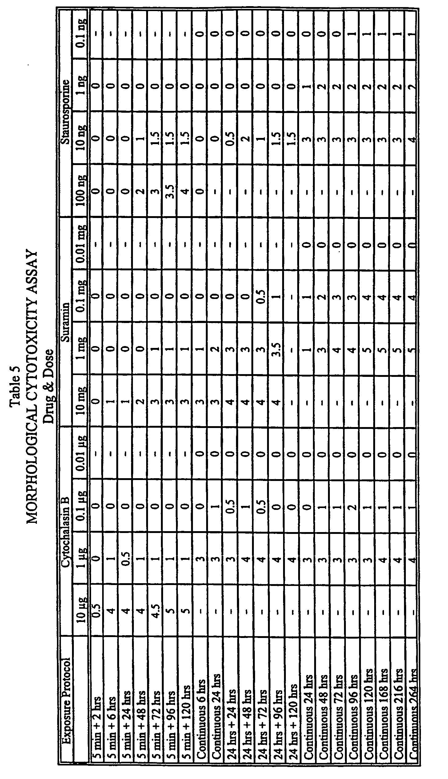
-
-
At an in vitro effective dosage, cytochalasin B (1 µg/ml; an anti-migration/contraction
effective dose) and staurosporin (1 ng/ml; an anti-proliferative
effective dose) exhibited a cytotoxicity grade of 1 (minimal) and 2 (mild),
respectively. Independent studies have indicated that a grade of 3 (moderate) or less
is preferred for a cytostatic, anti-proliferative agent of the present invention.
EXAMPLE 13
In Vivo BRDU Assay: Inhibition of Vascular Smooth Muscle
Cell Proliferation
-
BRDU assay: In vivo vascular smooth muscle proliferation was quantitated by
measuring incorporation of the base analog 5-bromo-2'-deoxyuridine (BRDU,
available from Sigma Chemical Co.) into DNA during cellular DNA synthesis and
proliferation. BRDU incorporation was demonstrated histochemically using
commercially available anti-BRDU monoclonal antibodies. The 1 hour pulse
labeling permits assessment of the number of cells undergoing division during the
pulse period.
-
The BRDU pulse labeling protocol described above is used as a standard
evaluation technique with in vivo pig vascular studies. Following surgical and
treatment procedures (discussed, for example, in Examples 7 and 11 herein) and a
post-surgical recovery period, pigs were sedated and pulsed with BRDU 1 hour
prior to tissue collection.
-
Briefly, the pigs were sedated with tiletamine hydrochloride and xylazine (as
in Example 7, "Gross Pathology and Histological Evaluation") by intramuscular
injection. BRDU was then administered intravenously via the lateral ear vein. Two
ml of BRDU at a concentration of 50 mg/ml was administered to each 30-40 lb pig.
One hour later, the pigs were sacrificed by intravenously administered pentobarbital.
Test artery segments were then removed (a segment included normal vessel located
proximally and, if possible, distally with respect to the treated artery segment). The
artery segments were transected at 2 mm intervals; arranged in order in cryomolds
with O.C.T. (optimum cutting temperature) compound (Tissue Tek®, Miles
Laboratories, Inc., Elkhart, IN); and frozen in liquid nitrogen. The blocks were
sectioned at 5 microns and immunohistologically stained to detect BRDU using the
following procedure.
BRDU-labeled cell detection: After BRDU (1 g BRDU diluted in 17 ml sterile
water and 3 ml 1 N NaOH) pulse labeling and test artery segment removal and
sectioning (as above), immunohistochemical staining with anti-BRDU monoclonal
antibody provides a visual means of determining a mitotic index over a specified
time period. The immunohistochemical staining method was performed as follows:
- 1) 5 µm sections of test artery were dehydrated in cold acetone (-20°C)
for 10 minutes;
- 2) Sections were mounted on glass microscope slides, and the slides
were dried in a 37°C oven for 10 minutes;
- 3) Slides were rehydrated in PBS for 10 minutes;
- 4) Slides were subjected to Feulgen's acid hydrolysis using 1 N HCl,
wherein two aliquots of 1 N HCl are preheated to 37°C and 60°C
prior to proceeding;
- 5) Slides were rinsed with 1 ml of 1 N HCl at 37°C for 1 min;
- 6) Slides were transferred to 60°C 1 N HCL for 15 min;
- 7) Slides were rinsed with 1 ml of 1 N HCl at 37°C for 1 min;
- 8) Slides were washed with room temperature PBS, using 3 changes of
PBS at 5 min intervals;
- 9) Endogenous, cross-reactive sites on the sections were blocked with
normal goat serum (1:25 in PBS) for 20 min;
- 10) Slides were washed with PBS, as in step 8;
- 11) Sections were incubated with mouse anti-BRDU antibody (DAKO
Corporation, Carpinteria, CA) at 10 µg/ml for 30 min;
- 12) Slides were washed with PBS, as in step 8;
- 13) Sections were incubated with horseradish peroxidase-labeled
(HRPO) goat anti-mouse IgG, (Jackson Immunoresearch
Laboratories, Inc., West Grove, PA; diluted 1:20 in PBS) and 4%
human AB serum for 30 min;
- 14) Slides were washed with PBS, as in step 8;
- 15) Sections were incubated with chromogen (3,3'-diaminobenzidine
(DAB; Sigma) at 5 mg/ml in 200 ml PBS) and 200 µl of 30% H2O2
for 10 min;
- 16) Slides were washed with PBS, as in step 8;
- 17) Samples were counterstained with Gill I hematoxylin (Gill I Lemer
Laboratories, Pittsburgh, PA; 30 dips);
- 18) Slides were washed with PBS, as in step 8; rinsed with a bluing
solution (1 gm lithium carbonate in 500 ml dH2O); washed with
deionized water; and
- 19) Test samples were then dehydrated, cleared and coverslipped.
-
-
At the conclusion of this procedure, a positive immunohistological stain
exhibits a brown color at the site(s) of reactivity.
-
Cytocidal agents inhibited BRDU uptake relative to a PBS control; however,
cytochalasin B and staurosporin inhibited BRDU uptake (i.e., cell proliferation)
without killing the vascular smooth muscle cells. The number of vascular smooth
muscle cells labeled with BRDU was assigned a grade at 400X magnification as
follows:
- 1 = ≤ 1/high power field (HPF);
- 2 = 2 to 5/HPF;
- 3 = > 5 to ≤ 10/HPF; and
- 4 = > 10/HPF.
-
-
Both cytochalasin B and staurosporin inhibited proliferation for 24 hours
following balloon trauma (grade 1), yielding a BRDU labeling grade equivalent to
that of a pre-trauma baseline (grade 1). PBS and monoclonal antibody controls
exhibited grade 2.5 to 4 BRDU labeling during the same time period. At 4 days
post-trauma, arteries treated with cytochalasin B or staurosporin, as well as PBS and
monoclonal antibody controls, exhibited a BRDU labeling grade of 4. The anti-proliferative,
non-cytocidal properties of cytochalasin B and staurosporin suggest
that these agents are amenable to sustained release dosage formulations for
reduction of vascular stenosis.
EXAMPLE 14
Direct Conjugation of NR-AN-01 Antibody to
Carboxylic Functional Groups of a Latex Particle
-
Antibody-coated latex particles (a model of an antibody-coated, sustained
release dosage form) may be obtained using the following aseptic technique:
Conjugation:
-
To 4 ml 0.05 M sodium borate, pH 8.5, containing 0.01% Tween-20®
(polyoxyethylene sorbitan monolaurate, Sigma) is added 0.5 ml PBS containing 5
mg NR-AN-01 monoclonal antibody. To this solution at room temperature is
added, with vortexing, 2.5 ml of an aqueous suspension containing 50 mg of 1 µm
diameter carboxylated latex particles. Immediately thereafter, 0.50 ml of water
containing 100 mg of freshly dissolved 1(3-dimethyl-aminopropyl)3-ethyl
carbodiimide HCl is added with vortexing. The solution is then incubated with
shaking for 1-2 hr at room temperature. The reaction mixture is then diluted with 50
ml of 50 mM phosphate buffer, pH 6.6, containing 0.2% gelatin stabilizer
(phosphate/gelatin buffer). The mixture is centrifuged at 40,000 x g for 2 hr at 4-10°C.
The supernatant is decanted, and the pellet is resuspended in 50 ml
phosphate/gelatin buffer using low level sonication for 10 sec. Centrifugation is
repeated, and the pellet is resuspended two times, followed by resuspension in the
phosphate/gelatin buffer. The conjugated particles are then lyophilized using
standard protocols and sorbitol excipients.
Characterization:
-
- (a) Sizing: Particle size homogeneity is assessed by laser anisotropy or, for
particles larger than 1 µm, by microscopic examination.
- (b) Specific Binding Assessment: Specific binding to smooth muscle cells
is determined by histological examination of tissue or cell pellet microtome slices
after incubation of protein/peptide conjugates with conjugated particles, with or
without blocker protein/peptide included in the incubation mixture. Preferred
detection techniques include second antibody assays (i.e., anti-mouse Ig) or
competition assays (i.e., radioscintigraphic detection in conjunction with
radioisotopically labeled protein/peptide conjugates).
- (c) Assessment of the extent of protein/peptide derivitization: This
determination is performed by coating the latex particles with radioisotopically
labeled antibody, followed by detection of radioactivity associated with the coated
particles.
-
-
The characterization of antibody-coated particles is described in Table 6.
| Characterization of NR-AN-01-Coated Latex Particles |
| Particle Diameter | Offering of Ab/Particle | µg Ab Bound/5 mg Latex | Ab Molecules Per Particle |
| 1.2 µm | 40,000 | 42 | 3520 |
| 1.2 µm | 84,000 | 66 | 5470 |
| 0.4 µm | 32,000 | 99 | 3160 |
| 0.4 µm | 64,000 | 140 | 4550 |
| 0.1 µm | 932 | 140 | 65 |
-
The particle aggregation effect of pH during antibody conjugation is
presented in Table 7.
| Effect of pH During Antibody Conjugation - Particle Aggregation |
| Particle Diameter | pH During Conjugation | Particle Aggregation |
| | | +Tween 20® | -Tween 20® |
| 1.2 µm | 8.5 | < 5% | <2.5% |
| 1.2 µm | 7.0 | ≈ 20% | ≈ 10% |
| 1.2 µm | 5.5 | 10% | 100% |
| 0.4 µm | 8.5 | < 10% | < 5% |
| 0.4 µm | 7.0 | ≈ 30% | ≈20% |
| 0.4 µm | 5.5 | 100% | 100% |
| 0.1 µm | 8.5 | < 20% | < 10% |
| 0.1 µm | 7.0 | = 50% | = 40% |
| 0.1 µm | 5.5 | 100% | 100% |
These data suggest that proteins or peptides may be directly conjugated with
sustained release dosage forms of the present invention. More specifically, polylactic/glycolic
acid particulates having terminal carboxylic acid groups will be
conjugated according to the procedure described herein or the alternative procedures
described in the specification hereof.
EXAMPLE 15
In Vivo Studies of Cytochalasin R
-
Biodistribution of Cytochalasin B. To determine the biodistribution of
cytochalasin B, mice were injected (i.p.) with 50 mg/kg cytochalasin B. Control
mice were injected with DMSO/Tween 20/carboxymethyl cellulose ("vehicle").
The mice were sacrificed at 3, 12, 24 and 72 hours after cytochalasin B or vehicle
administration. Organs were removed, homogenized, extracted and the amount of
cytochalasin B in tissues quantitated by HPLC. About 75% of the cytochalasin B
remained in the peritoneal cavity or at the injection site. Of the organs tested, the
highest amount of cytochalasin B was found in the liver. Subsequent analyses
showed that the maximum tolerated dose for cytochalsin B was 50 mg/kg, and that
this dose may be administered every second day.
-
Cytochalasin B was also administered intravenously to mice at 3.5 mg/kg (in
methanol or Tween 20/carboxymethyl cellulose). Mice were sacrificed at 2 minutes,
15 minutes, 30 minutes, 3 hours and 12 hours after cytochalasin B administration
and tissue extracts analyzed for cytochalasin by TLC. The maximal recovery of
cytochalasin B from tissue extracts was 32%. The data showed that cytochalasin B
was localized to the lung and the injection site, and that 3.5 mg/kg of cytochalasin B
resulted in no acute toxicity. By 12 hours after administration, there were very low
levels of cytochalasin B in tissues.
-
3H-cytochalasin B (2 µg; 30 µCi/µg) was injected i.v. into BALB/c mice
having urinary bladders that had been externally ligated. Animals were sacrificed at
15 minutes, 30 minutes, 2 hours or 16 hours post-injection. Organs were removed,
blotted, weighed, air dried and assayed for radioactivity. Fifty - 73% of the total
injected dose was accounted for in the tissues sampled (blood, heart, brain, muscle,
bone, lung, liver, spleen, stomach, kidney, intestines, and urinary bladder).
Clearance of 3H-cytochalasin B from the blood was extremely rapid with less than
1% of the injected dose in circulation by 15 minutes. Only liver, skeletal muscle
and intestines showed significant retention of 3H activity. All tissues had clearance
of 3H activity to below 1.5% injected dose per gram of tissue by 16 hours.
Cytochalasin B Metabolism. 3H-cytochalasin B at a dose of 1.5 or 8 µg/ml was
mixed with viable or non-viable human liver slices and media, and the amount of
3H-cytochalasin B in media or tissue assessed by HPLC (Tables 8 and 9).
-
The cytotoxity of
3H-cytochalasin B and its subsequent metabolites was also
assessed by evaluating dose dependent changes in mitochondrial function by
monitoring 3-[4, 5-dimethylthiazol-2yl]-2, 5-diphenyltetrazolium bromide (MTT)
activity following a 24 hour exposure of human liver slices to the test agent (Table
10).
| DISTRIBUTION OF 3H CYTOCHALASIN B MEDIA AND HUMAN LIVER TISSUE |
3H Cytochalasin B
(µg/ml) | Incubation System | Incubation Period at 37°C
(Hours) | % Activity in Media | % Activity in Tissue |
| 1.5 | Media/viable tissue | 1 | 84 | 16 |
| 1.5 | 4 | 84 | 16 |
| 1.5 | 24 | 91 | 9 |
| 8 | Media/viable tissue | 1 | 88 | 12 |
| 8 | 4 | 84 | 16 |
| 8 | 24 | 89 | 11 |
| 8 | Media/boiled tissue | 1 | 83 | 17 |
| 8 | 4 | 87 | 13 |
| 8 | 24 | 87 | 13 |
| HUMAN LIVER SLICES METABOLISM STUDY NATURE OF TRITIUM ACTIVITY EXTENT OF METABOLIC CONVERSION |
3H Cytochalasin B
(µg/ml) | Incubation System | Incubation Period
(Hours) | %Non extractable Activity | % Extractable Activity | % Metabolized of Extractable Activity |
| 8 | Media/viable tissue | 1 | 0.5 | 99.5 | 24.2 |
| 8 | 4 | 1 | 99.0 | 59.4 |
| 8 | 24 | 1.3 | 98.7 | 98.0 |
| 1.5 | Media/viable tissue | 1 | 0.5 | 99.5 | 43.0 |
| 1.5 | 4 | 0.6 | 99.4 | 77.0 |
| 1.5 | 24 | 0.4 | 99.6 | 98.7 |
| 8 | Media/boiled tissue | 1 | 0.5 | 99.5 | 5 |
| 8 | 4 | 1 | 99.0 | 5 |
| 8 | 24 | 1.3 | 98.7 | 5 |
| 8 | Media Only | 1 | NA | NA | | 5 |
| 8 | 4 | NA | NA | | 4 |
| 8 | 24 | NA | NA | | 5 |
| MTT Absorbance in Human Liver Slices Exposed to 3H-cytochalasin B for 24 hours. |
| Dose Level (µg/ml) | MTT Absorbance |
| 0 (control) | 0.639 +/- 0.188 |
| 0.1 | 0.844 +/- 0.014 |
| 1.5 | 0.841 +/- 0.081 |
| 8.0 | 0.850 +/- 0.082 |
| * MTT absorbance values reflect the mitochondrial viability in which high absorbance values represent viable mitochondria and while low absorbance values reflect nonfunctional mitochondria. Each value represents the mean +/- standard deviation optical density of triplicate liver samples. |
-
The results in Tables 8-9 indicated that 98% of the 3H-cytochalasin B was
metabolized within 24 hours of administration with greater than 80% of the total
reactivity being present in the media and less than 20% in the tissue. The results in
Table 10 indicated that 3H-cytochalasin B or its subsequent metabolites was not
cytotoxic.
-
To determine the metabolism of
3H-cytochalasin B in human blood,
3H-cytochalasin
B (8 µg/ml) was dissolved in saline; 1:1 dilution of saline and human
plasma; or 1:1 dilution of saline and human whole blood. The mixtures were
incubated for 20 hours at 37°C and then analyzed by HPLC for stability and
metabolism.
3H-cytochalasin B was not metabolized when mixed with either saline
or plasma. However,
3H-cytochalasin B was metabolized by human whole blood
with the metabolite having an HPLC retention time and profile consistent with that
seen in the human liver slice assay (see above).
Toxicity Studies. To determine the toxicity of cytochalasin B, rats (4 male and 4
female) were injected with 10 µg/ml cytochalasin B four times a day for 7 days
(Table 11). Data regarding body weight changes, food consumption, food
efficiency, hematology parameters, coagulation parameters, serum chemistry
parameters, and gross necropsy findings were collected. The only parameter which
suggested an adverse effect of cytochalasin B administration was an elevation of the
mean relative heart weight in treated female animals. There were no gross or
microscopic changes detected in the heart to account for the elevation. Thus, daily
administration of cytochalasin B to rats at a dose of 800 µg/kg (for a total injected
dose of 5600 µg/kg) may have an effect on heart weight of female (but not male)
rats. However, clotted blood was not routinely removed from the heart lumens prior
to weighing the heart.
| Group Number | Numher of Animals | Treatment | Dose Level
(µg/mL) | Route | Dosing Frequency | Dosing Duration |
| | Males | Females |
| |
| 1 | 4 | 4 | Biostent Control | 0 | IV | 4 times/day
(2 hrs apart) | 7 days |
| 2 | 4 | 4 | Cytochalasin B | 10 | IV | 4 times/day
(2 hrs apart) | 7 days |
-
To determine the toxicity of chronic cytochalasin B administration,
cytochalasin B was administered intravenously to Sprauge-Dawley rats for seven
days (Table 12). Data regarding food consumption, body weights, hematology
parameters, clinical chemistry parameters, coagulation profiles, organ weights,
clinical observations, gross necropsy findings and histopathology were collected. It
was found that chronic intravenous administration of cytochalasin B at doses up to
600 µg/kg/day did not result in any indication of adverse effects or toxicity.
| Group Number | Number of Animals | Treatment | Dose Level
(µg/mL) | Route | Dosing Frequency | Dosing Duration |
| | Males | Females |
| |
| 1 | 15 | 15 | Control | 0 | Intravenous injection | 4 times/day 4 times/day
(≈2 hrs apart) | 7 days |
| 2 | 10 | 10 | Cytochalasin B | 1.3 | 7 days |
| 3 | 10 | 10 | 3.9 | 7 days |
| 4 | 15 | 15 | 7.5 | 7 days |
| 5 | 15 | 15 | 7.5 | 7 days |
-
A similar study in dogs (Table 13), which collected data on food
consumption, body weights, hematology parameters, clinical chemistry parameters,
coagulation profiles, organ weights, clinical observations, thoracic cavity
ausculation, opthalmic examination, urinalysis, gross necropsy findings, and
histopathology (high dose group), found that intravenous administration of
cytochalasin B for seven consecutive days to beagle dogs at doses up to 648
µg/kg/day (a cumulative dose of 4,536 µg/kg) did not result in any indication of
adverse effects or toxicity.
| Group Number | Number of Animals | Treatment | Dose Level
(µg/mL) | Dose Volume (mL/kg); Flow Rate (mL/min) | Route | Dosing Frequency | Dosing Duration |
| | ♂ | ♀ |
| 1 | 6 | 6 | Control | 0 | 20;10 | IV | 4 times/day
(=6hrs apart) | 7 days |
| 2 | 4 | 4 | Cytochalasin B | 1.3 | 20;10 | IV | 4 times/day
(=6hrs apart) | 7 days |
| 3 | 4 | 4 | Cytochalasin B | 4.4 | 20;10 | IV | 4 times/day
(=6hrs apart) | 7 days |
| 4 | 6 | 6 | Cytochalasin B | 8.1 | 20;10 | IV | 4 times/day
(=6hrs apart) | 7 days |
| 5 | 6 | 6 | Cytochalasin B | 8.1 | 20;10 | IV | 4 times/day
(=6hrs apart) | 7 days |
| ♂ = males; ♀ = females; IV = intravenous infusion |
EXAMPLE 16
Biological Stenting of Balloon Traumatized Pig Arteries
-
Pig femoral arteries were traumatized as described in Example 7, and then
treated with cytochalasin B. About 1.5 to about 2 ml of cytochalasin B at 0.1 µg/ml
was infused into portions of the artery that had been separated from other portions
by ligatures. The artery was then pressurized for 3 minutes and the fluid aspirated.
Approximately 8 to about 30 lambda of the solution is retained in the interstitial
space surrounding the cells in the tunica media. Ten femoral arteries (two arteries
obtained from each of the 5 pigs that were treated according to the single dose
protocol described in Example 7) were then evaluated histologically at 4 days or 3
weeks after cytochalasin B adminstration. The maximal luminal area of each artery
was measured and calculated from digitized microscopic images by a BQ System IV
computerized morphometric analysis system (R & M Biometrics, Inc., Nashville,
TN). This experiment was repeated with 5 additional pigs (two arteries per pig;
cytochalasin B dose = 0.1 µg/ml, applied for 3 minutes at 1 atm pressure; same time
points). The data obtained from the two experiments were combined. Balloon
traumatized pig arteries that had been treated with cytochalasin B displayed a larger
luminal area at the 4 day and 3 week post-treatment time points, as compared to
arteries treated with other test agents or controls.
-
The luminal area of the traumatized and cytochalasin B-treated segments of
the arteries were also compared to the luminal area of the normal, untreated region
of the femoral artery proximal to the test area. The results showed that the lumen
area in the test region was approximately two times as large as the area of the
normal control segment of the same artery. The negative control agents, PBS and
monoclonal antibody NR-AN-01, showed no increase or a slight decrease in lumen
area as compared to the normal control segment of the same artery.
-
A cytochalasin B dose response study was then conducted on 10 pigs,
following the experimental protocol described in Example 7. Briefly, both femoral
arteries in each of 2 pigs were treated with one of the following doses of
cytochalasin B: 0.0 µg/ml (i.e., PBS negative control); 0.01 µg/ml; 0.10 µg/ml; 1.0
µg/ml; and 10.0 µg/ml as described above. The agent was delivered by intraluminal
catheter at 1 atm pressure for 3 minutes, and the arteries were evaluated 3 weeks
later by the morphometric analysis system described above. The ratio of treated
artery luminal area to proximal normal artery luminal area was determined as a
percent change in treated vs. normal area, or the artery lumen size (diameter or cross
sectional area) of treated arteries relative to to traumatized but untreated arteries. A
significant threshold effect was observed at doses from 0.1 µg/ml (about a 140%
increase) to 1.0 µg/ml (FIGURE 14). The subthreshold dose (0.01 µg/ml) and
negative control (PBS) exhibited about a ± 20% change in luminal area.
-
Electron micrographs revealed that within one hour of balloon trauma, there
was a depolymerization of the myofilaments in traumatized vessels, which are
contractive organelles. The depolymerization of the myofilaments is a normal
physiological response of vascular smooth muscle cells to trauma, and is the first
step in their transformation from a contractile to a secretory and migratory cell.
Treatment of traumatized swine arteries with about 0.1 to about 10.0 µg/ml of
cytochalasin B did not result in an increased rate, or more extensive
depolymerization, of the myofibrils. However, electron micrographs showed that
the myofilament reformation was retarded. Based on the sustained increase in
vessel diameter, the return to normal vascular contractility may be slowed more
extensively than is suggested by the retarded return to normal morphology.
-
The traumatized and treated areas of the artery did not undergo the
constriction or chronic geometrical (vascular) remodeling that normally occurred in
sham controls in the pig, and which has been described in man, following PTCA.
Cross sections of the artery showed a larger cross-sectional area of the total vessel,
obtained by measuring the ratio of the vessel area inside the external elastic lamina
(EEL) of the treated area to the mean of the total vessel area of the proximal and
distal regions of the same artery, compared to arteries traumatized but not treated
with cytochalasin B. In arteries that were traumatized with a torquable balloon that
damages the vessel wall without tearing the tunica media, there was a more uniform
intimal proliferation and the larger vessel size (area inside the EEL) resulted in a
larger luminal cross-sectional area and a significant decrease in restenosis. When
the vessels were extensively damaged and the tunica media was torn into, there was
extensive proliferation and thrombus remodeling that resulted in highly variable
intimal proliferation.
-
Thus, the administration of cytochalasin B to swine vascular smooth muscle
cells by infusion catheter following balloon dilation trauma resulted in a more
extensive retention of the artery lumen size (diameter or cross-sectional area) than
was produced by the dilating balloon. This effect was achieved by replacing the
entire interstitial fluid volume between cells of the tunica media with the therapeutic
agent.
-
Moreover, cytochalasin B has a wide therapeutic index which ranges from
about 0.1 to 10 µg/ml, with no evidence of toxicity at 10 µg/ml. Ten µg/ml is the
maximum saturation concentration of cytochalasin B in saline. Furthermore, the
effect produced by cytochalasin B administration became more apparent over the 3
to 8 weeks following the balloon trauma. These data suggest that cytochalasin B
acts as a "biological stent" when delivered to traumatized arteries.
-
Balloon traumatized swine femoral arteries from control and treated (0.1
µg/ml cytochalasin B) were evaluated at 1,4, 7, 14, or 21 days after intervention.
Morphometric analysis on frozen histologic sections of artery showed that
cytochalasin B treated arteries reached a state of sustained dilation which changed
very little between days 7 and 21. A two way analysis of variance indicated a
statistically significant difference (p<.05) in the artery lumen areas over three weeks
between the cytochalasin B treated and diluent control groups.
-
A dose-response study of balloon traumatized swine coronary arteries
showed that treatment with cytochalasin B at 0.1, 1.5 or 10.0 µg/ml (Table 14,
"Biostent") resulted in sustained arterial dilation of the coronary luminal area at
three weeks after intervention. Moreover, cytochalasin B administration did not
result in myocardial or arterial lesions attributable to the cytochalasin B as evaluated
histologically. No statistically and biologically significant changes attributable to
cytochalasin B were seen in clinical chemistry parameters, hematology paremeters,
body weights, blood pressure or electrocardiograms.
| SWINE CORONARY MORPHOMETRY DATA |
| Group Dose (µg/ml) | Days Post Surgery | Mean % Lumen Area | Std Dev | P-value Unpaired T-test |
| Saline |
| | 4 | 96.5 | 15.2 | --- |
| 0.1 µg/mL Biostent | 4 | 97.0 | 17.1 | 0.48 |
| 1.5 µg/mL Biostent | 4 | 101.7 | 6.8 | 0.28 |
| 10 µg/mL Biostent | 4 | 103.9 | 14.1 | 0.25 |
| Saline | 21 | 78.7 | 19.1 | --- |
| 0.1 µg/mL Biostent | 21 | 98.0 | 15.5 | 0.004 |
| 1.5 µg/mL Biostent | 21 | 100.9 | 18.5 | 0.002 |
| 10 µg/mL, Biostent | 21 | 110.9 | 19.2 | 0.004 |
-
Swine coronary arteries were also traumatized by embolectomy or over-sized
PTCA balloon, and then 8-16 ml of cytochalasin B at 8.0 µg/ml was infused into the
arterial wall with a MIC catheter to achieve a therapeutic dose. Controls included a
diluent control and a traumatized untreated control, and all animals were sacrificed 4
weeks after intervention and coronary arteries were fixed by perfusion.
Morphometry was performed on selected sections from proximal, treated and distal
segments of coronary arteries (Table 15).
| Swine Coronary Artery Study |
| Group | Neointimal Area | Luminal Area | Arterial Area |
| | Area | std | Area | std | Area | std |
| Untreated Control | 1.88 | 1.62 | 0.77 | 0.38 | 2.19 | 1.07 |
| Saline Treated Control | 1.39 | 1.13 | 0.73 | 0.37 | 1.88 | 0.96 |
| 8.0 µg/ml Cytochalasin B | 1.56 | 1.24 | 0.58 | 0.30 | 1.80 | 0.70 |
-
While the data shown in Table 15, in contrast to previous studies, showed
that the local delivery of cytochalasin B did not result in a statistically significant
increase in luminal area, the data does show that there was a trend toward beneficial
arterial remodeling as evidenced by larger arterial area bounded by the external
elastic lamina in the cytochalasin B treated arteries when compared to either of the
controls. The diameter of, or the area within, the EEL of the artery can be compared
to controls as an indicator of the degree of vascular remodeling. The lack of a
statistically significant increase in luminal area may be due to increased sample
variablility, increased neointimal formation, and/or increased degree of trauma.
-
In summary, these studies demonstrate that the administration of a
cytoskeletal inhibitor, such as cytochalasin B, in an amount which can biologically
stent a traumatized vessel may also be efficacious to inhibit or reduce the
proliferation of vascular smooth muscle cells.
EXAMPLE 17
Sustained Release Formulations of Cytochalasin B and Taxol
-
To determine the efficacy of the local, sustained release dosage forms of
cytochalasin B or taxol to inhibit restenosis, cytochalasin B or taxol in a supporting
structure, e.g., a "wrap," was applied to the adventitial tissue surrounding a balloon
traumatized rabbit carotid artery (Groups 1-11) or a balloon traumatized pig femoral
artery (Group 12) (Table 16).
-
The arteries in the animals in Group 1a and 1b were treated with 20 mg of
cytochalasin B in 1 g of a bovine collagen gel (BioCore, Inc., Topeka, KS) that was
supported by, or enclosed in, a bovine collagen mesh wrap (Skin Temp-Biosynthetic
Skin Dressing, BioCore, Inc., Topeka, KS). At 1 week post treatment, the
cytochalasin B treated artery in animal 1233 showed no intimal or adventital
proliferation. There was marked cell death in the outer zone of the tunica media
with heterophils infiltrating the tunica media. Heterophils were present outside the
wrap, but cytochalasin B inhibited heterophils and macrophages from infiltrating the
wrap. The artery of the control (1249) animal had moderate intimal proliferation,
and heterophils and histiocytes were infiltrating into the wrap. Cell death in the
tunica media was minimal.
-
At 2 weeks post-treatment, there was minimal intimal and adventital
proliferation in the cytochalasin B wrap-treated area of the artery (animal 1224).
The intima was loosely arranged and there was minimal heterophil infiltration.
Syncytial giant cells were present. In the artery of the control wrap animal (animal
1222), there was moderate intimal proliferation with heterophils and macrophage in
the wrap area. These cells were visible in the tunica media.
-
At three weeks post-treatment, there was no intimal proliferation observed in
the cytochalasin B wrap-treated area of the artery (1244). Heterophils and syncytial
giant cells were present around the wrap. There was significant necrosis of the cells
in the tunica media with infiltrating heterophils and macrophages. No endothelium
was present. In the control (animal 1232) artery, there was marked intimal
proliferation, with well organized adventitia and perivascular tissue. Heterophils
and macrophages were infiltrating the wrap. The cells in the tunica media were
viable and there was a mural thrombus in the vessel lumen. Thus, inhibition of
intimal proliferation was seen in the arteries of Group 1a and 1b animals treated
with cytochalasin B, however, there was significant reaction to the wrap material.
-
The arteries in the animals in Group 2a and 2b were treated with
cytochalasin B (30% wt/wt; 300 mg cytochalasin B/g silicone) in a silicone wrap
(Q-7 4840, Dow Coming, Midland, MI). One week post-trauma there was no
significant intimal or adventitial proliferation of smooth muscle cells (SMCs) or
mesenchymal tissue (animal 1229). There was significant necrosis of the SMCs in
the outer zone of the tunica media. In areas that appeared to have
been minimally traumatized by the torquable ballon there was minimal to no
cellular necrosis. This indicated that traumatized cells were more prone to die when
exposed to this dose of cytochalasin B but that this dose was not cytocidal to
minimally traumatized or normal SMCs. There was minimal mononuclear and
polymorphoneuclear cell infiltration into the tunica media. A few heterophils were
seen infiltrating from the vessel lumen.
-
In the control animal (1228), there was less cellular necrosis in the tunica
media, and the necrosis present was located in the inner zone rather than the outer
zone of the artery wall. Thus, cytochalasin B inhibition of cellular repair appears to
increase tunica media necrosis. The control also lacked tunica media or adventitial
proliferation and organization of the perivascular clot. Cellular infiltration in any
area was minimal.
-
Two weeks after initiating cytochalasin B treatment there was complete
inhibition of intimal proliferation and only minimal perivascular clot organization
which was primarily due to fibrin formation and not mesenchymal proliferation
(animal 1227). There was mild infiltration of polymorphonuclear cells and minimal
infiltration of mononuclear cells into the tunica media and adventitia. No
endothelium was present in the wrap area, except for a few small isolated foci. The
control artery (animal 1226) had moderate intimal proliferation and adventitial
proliferation with mesenchymal organization of the perivascular clot area. Foci of
endothelial proliferation were larger and more extensive in the control animal
compared to the cytochalasin B treated vessel.
-
With 3 weeks exposure (animal 1212) to cytochalasin B in a silicone wrap,
the vessel showed marked cell loss in the tunica media which was most severe in the
outer zone. Cellular infiltration in the tunica media and adventitia was minimal and
endothelilzation was only present in a few focal areas. There was moderate,
irregular intimal proliferation; however, the intimal cells and what few endothelial
cells that were present were unorganized and lack polarity. The inhibition of
migration by cytochalasin B resulted in this loss of organization or polarity. The
intimal proliferation was also mild in the control vessel (1230); however, the intima
was well organized and was almost completely endothelized. There was mininal
cell loss from the tunica media. Thus, significant intimal inhibition was seen in the
first two weeks in Group 2 treated animals.
-
The vessels in the animals in Group 3a and 3b were treated with 8 mg
cytochalasin B in 100 mg of a pluronic gel (F-127, BASF) that was supported by a 1
cm x 1 cm bovine collagen mesh wrap. One week after treatment, the cytochalasin
B treated artery of animal 1250 had mild intimal proliferation that was irregular in
thickness. There was approximately 30% re-endothelization and the tunica media
cells were viable with the most significant loss (mild) being in the inner zone of the
tunica media. There was a marked pyogranulomatous reaction to the pluronic gel in
the perivascular and adventitial region. Complete thrombosis of the control artery
from animal 1261 prevented its evaluation.
-
At two weeks, the pluronic gel with cytochalasin B stimulated a marked
pyogranulomatous reaction in the adventitial and perivascular tissue. There was
mild, irregular intimal proliferation and complete endothelization. The tunica media
cells were viable. There appeared to be a mild cell loss from the inner zone of the
tunica media. The artery of the control animal (1247) had mild, irregular intimal
proliferation, complete endothelization with plump endothelial cells and viable cells
in the tunica media. There was marked pyogranulomatous inflammatory reaction to
reminants of the pluronic gel.
-
At three weeks, the arteries of animals 1248 and 1246 showed reminants of
the collagen wrap; however, the wrap was less resolved in the cytochalasin B treated
animal (1248) than in the control (1246). This retardation of wrap resorbsion may
result from cytochalasin B inhibition of macrophage migration and function. Both
the treated and control had a moderate amount of intimal hyperplasia at 3 weeks, so
there was no significant amount of intimal inhibition by cytochalasin B when
administered in the pluronic gel. Foci of dystrophic mineralization were seen in the
cytochalasin B treated artery. Thus, no inhibitory effect on the intima was observed
in these animals (Group 3) at 1, 2 or 3 weeks after the initiation of treatment.
-
The arteries of the animals in Group 4a and b that were treated with 100 mg
cytochalasin B (10% wt/wt) in a 1 g silicone wrap had significant inhibition of
intimal proliferation at all time points. In the cytochalasin B wrap-treated artery of
animal 1259, there was no intimal proliferation or adventitial fibrosis present at one
week after treatment. Ectatic vessels were present in the adventitia and the
perivascular clot was unorganized and composed of only fibrin. There was marked
cell loss from the tunica media, especially in the outer zone. Heterophils were seen
infiltrating the tunica media. In the control artery (1206), there was no intimal
proliferation at 1 week; however, there was early fibrous organization of the
perivascular clot. Cellular loss from the tunical media was more diffuse than in the
cytochalasin B wrap which was most severe in the outer zone.
-
The cytochalasin B wrap-treated artery of animal 1253 had minimal intimal
proliferation at two weeks, compared to the control (1258) wrap-treated artery
which had maximal intimal proliferation. The intimal proliferation in the
cytochalasin B wrap-treated artery was irregular and appeared to be the result of
organizing mural thombi by infiltrating SMC. There was only loose thin layers of
platelets in the cytochalasin B wrap-treated artery, with margination of heterophils.
There was no endothelization in the cytochalasin B wrap-treated artery and <20%
endothelization in the control artery. The perivascular clot was unorganized and
remained fibinous in the cytochalasin B wrap-treated artery and well organized in
the control artery. The control artery had minimal cell loss from the tunica media
while the cytochalasin B wrap-treated artery had marked cell loss.
-
At three weeks, the cytochalasin B wrap-treated artery (1251) showed
minimal to no intimal proliferation. The intimal proliferation appeared to occur
where there was less cell loss from the tunica media. There was early re-endothelization
in the cytochalasin B wrap-treated artery, but the cells were often
rounded, loosely attached and only a few scattered foci were present (<10%). The
perivascular clot in the cytochalasin B wrap-treated artery was unorganized and still
consisted of fibrin, whereas the control artery was well organized with fibroblasts
and collagen matrix. The control artery was completely thrombosed,
there was marked initmal production in distal areas of the thrombus which were less
completely organized.
-
The arteries in the animals in Group 5a and b were treated with 50 mg taxol
in 1 g of a silicone wrap (5% wt/wt). This treatment showed marked inhibition of
intimal proliferation at all time points. The taxol wrap-treated artery (animal 1278 at
1 week) had no intimal or adventitial proliferation and the perivascular clot was
fibrinous and unorganized. There was a marked loss of tunica media cells and no
endothelial lining present. The control (1279) had mild intimal proliferation with
very early fibrosis of the fibrinous perivascular clot. The lumen was approximately
85% re-endothelized. Both the treated and control arteries had mild heterophil
infiltration into the tunica media and adventitia.
-
The artery from animal 1281, which had been treated for at two weeks with
taxol, had no intimal proliferation, minimal adventitial fribrosis and marked cell
necrosis in the tunica media with mild heterophil infiltration. Focal areas of
necrosis and dystrophic mineraliztion were present in the adventitia and perivascular
clot tissue. The artery from the control animal (1280) had moderate intimal
proliferation, with marked organization of the adventitia and perivascular clot. The
lumen was 100% re-endothelized and the tunica media SMCs were viable in the
control artery.
-
At three weeks, the taxol wrap-treated artery (1242) had no intimal
proliferation and was 50% re-endothelized with plump appearing endothelial cells.
There was minimal organization of the perivascular clot and marked cell loss from
the tunica media. There was mild infiltration of heterophils into the tunica media
and marginating on the vessel lumenal surface. The control artery (1234) had
marked intimal proliferation and fibrosis of the adventitia and perivascular clot.
Cells in the tunica were viable.
-
In Group 6a and 6b, taxol-treated arteries also had a marked inhibition 2
weeks after the wrap was removed. Animal 1276 had a taxol wrap for 2 weeks, then
the wrap was removed and the animal sacrificed 3 weeks later. Following the 3
week recovery period from the taxol wrap removal (1276) there was only minimal
intimal proliferation, except in a few focal areas that appeared to be thickened due to
SMC organization of mural thrombi, in this artery. The adventitia was well
organized and there was a significant cell loss in the tunica media but the cells
present were viable. The lumen was approximately 90% re-endothelized. The
control (1277) artery had marked intimal proliferation, well organized perivascular
and adventical tissue and was 100% re-endothelized.
-
The results observed for Group 7a animals demonstrated that cytochalasin B-treated
arteries (10% wt/wt) showed no intimal proliferation for 2 weeks. The
decrease in the release rate of the cytochalasin B, however, resulted in a mild intimal
proliferation by week three after wrap placement. At one week (1257), the arteries
showed no intimal proliferation, and a marked necrosis of tunical media SMCs with
moderate heterophil infiltration. There was no endothelium and heterophils and
macrophages were marginated along the lumen surface. Moreover, there was no
evidence of platelet aggregates adhering to vessel wall. At two weeks (1265), the
arteries were similar morphologically to the one week arteries. By three weeks
(1266), the arteries showed mild irregular intimal proliferation. Furthermore,
heterophils were rare in the tunica media, the lumen was 70% re-endothelized and
there was early fibrosis in the adventitia with unorganized perivascular clot still
present, in the treated arteries. This indicated that by 3 weeks the level of
therapeutic agent had fallen below therapeutic level within the artery wall; however,
there was still enough drug to have an inhibitory effect on clot organization
immediately adjacant to the wrap.
-
The arteries of Group 8a animals, which were treated with 10% cytochalasin
B for 2 weeks, then the wrap was removed and vessels evaluated 2 weeks later, had
variable intimal proliferation within and between animals. The artery of animal
1254 had variable intimal proliferation which ranged from none to mild, well
developed adventitial fibrosis, marked cell loss in the tunic media and focal areas of
dystrophic mineralization in the outer tunica media and adventitia. The mild intimal
proliferation areas were at the ends of the wrap area, suggesting an infiltration from
the adjacent untreated artery regions. The lumen was approximately 60% re-endothelized.
The artery of animal 1255 had mild to moderate intimal proliferation,
viable cells in the tunica media and well organized tissue in the adventitia. The
lumen was 100% re-endothelized. The artery in animal 1256 was completely
thrombosed. Proximal to the chronic thrombus in the area of the wrap was an acute
thrombus and there was moderate intimal proliferation.
-
While there was moderate intimal proliferation in the arteries of some
animals, the proliferation in these arteries was still less than the controls in Group
9b. The mannitol control silicone wraps were on the artery for two weeks following
balloon trauma and then removed and the animal necropsied and the artery
histologically evaluated 1 week following wrap removal. Two of the arteries (1267
and 1268) had moderate intimal proliferation with 100% re-endothelization and one
had maximum proliferation. The one with maximum intimal prolifertion had an
acute occluding thrombus present.
-
The arteries in the animals in Groups 10 and 11 were treated with 10 mg
cytochalasin B loaded in 1 g of a silicone wrap (1% wt/wt) that was applied to the
artery for 2 weeks, surgically removed, and histologically evaluated 2 or 4 weeks
later, respectively. No significant difference was seen by qualitative evaluation
between the test and control animals. Animals 1304 and 1305 had a cytochalasin B
(1%) wrap for 2 weeks which was then removed. Two weeks after the removal the
animal was sacrificed. The artery from animal 1304 showed moderate initmal
proliferation in most areas of the wrap, in areas of marked tunica media cell necrosis
and wall dystrophic mineralization the proliferation was mild. There was 100% re-endothelization
and no heterophils were present in the intima or tunica media. The
adventitia and perivascular clot area was well organized. The artery from animal
1305 was similar to the artery from animal 1304 morphologically. The artery from
animal 1306 showed marked intimal proliferation, no infiltrating heterophils in the
intima or tunica media and was 100% re-endothelized.
-
Animals 1307, 1308 and 1309 were exposed to a cytochalasin B (1%) wrap
for 2 weeks which was then removed. Four weeks after removal the animals were
sacrificed. The artery from animal 1307 had moderate initmal proliferation with
focal areas of thickening due to mural thrombus organization by SMCs. There was
significant loss of cells from the tunica media and the elastic elamina appear
collapsed. A few heterophils were present in the adventitia. There were areas or
sections in the wrap area with minimal intimal proliferation. The artery from animal
1308 showed moderate intimal proliferation with areas of marked cell loss in the
tunica media and dystrophic minearalization in the outer zone of the tunica media.
The vessel was 100% re-endothelized. The artery from animal 1309 had marked
intimal proliferation with a well organized aventiticia and perivascular region.
Animal 1311 was not evaluated due to thrombosis. The results of the artery from
animal 1312 were quite variable, with sections showing a range of intimal
proliferation, from mild to moderate. Endothelization appeared to be complete in
these arteries.
-
The arteries from Group 12 animals (pig femoral arteries) that were treated
with 30% wt/wt cytochalasin B loaded silicone wraps showed significantly inhibited
intimal proliferation for the first two weeks. While there was intimal proliferation in
the
arteries 3 weeks later, the proliferation was still less than the proliferation
observed for the controls.
-
In summary, intimal proliferation of traumatized pig arteries was
significantly inhibited with both cytochalasin B and taxol in sustained release
dosage form. The best controlled sustained release of therapeutic agent, without the
stimulation of secondary inflammatory reaction, was obtained with an adventitial
wrap material comprising silicone. The silicone wraps inhibited intimal
proliferation with 30% and 10% loadings of cytochalasin B; however, as the level of
release drops off between 2 and 3 weeks there was initiation of intimal proliferation.
When wraps were left in place for 2 weeks then surgically removed and the arteries
examined from 1 to 4 weeks later, there appeared to be an intimal proliferation
rebound effect. The rebound effect occurred when the intimal proliferation of the
artery treated with the therapeutic agent approaches, but is still less than, the intimal
proliferation in the control artery. The animal treated with taxol appeared to have
less of a rebound effect than the cytochalasin B treated arteries.
EXAMPLE 18
Delivery of Crystalline Cytochalasin B or Taxol
-
The in vivo tissue distribution of cytochalasin B administered in crystalline
form was evaluated in balloon traumatized swine femoral arteries after local
delivery. A femoral artery of a Yorkshire crossbred swine was balloon traumatized
by overinflation and rotation of a Vascu-Flo™ Silicone embolectomy catheter.
Balloon trauma was immediately followed by intravascular delivery of 10 µg/ml
3H-cytochalasin B crystals (Sigma Chemical Co., St. Louis, MO) in saline
(saturated) for three minutes under i atm of pressure. Blood flow was resumed in
the artery for five minutes prior to sacrifice of the animal. An analysis of the tissue
distribution of 3H-cytochalasin B showed that this method was effective at
delivering 31 ug of 3H-cytochalasin B which localized predominantly to the
adventitia. 3H-cytochalasin B was visualized histologically by the presence of
silver grains in an autoradiographic emulsion. Thus, these results showed that
crystalline cytochalasin B can be delivered locally to a vessel wall in vivo.
-
Another study employed twenty, male, Sprague-Dawley rats. The rats
underwent balloon trauma to their left carotid artery, followed by inter-arterial
infusion of a solution containing 1 mg crystalline cytochalasin B in 300 ml vehicle
(Hanks sterile salt solution with 0.5% Cremophor) or a diluent (saline) control.
Animals were sacrificed immediately after infusion, and 2, 4, 7 and 14 days post-trauma
and infusion. Post-sacrifice, the left and the right (control) carotid arteries
were removed. Samples of arteries were obtained for quantitation of 3H-cytochalasin
B by oxidation and scintillation counting, histopathology,
autoradiography and vascular morphometry. Histopathology documented uniform,
circumferential balloon trauma in the arterial wall of the left carotid arteries.
-
Autoradiographically, cytochalasin B crystals were present on day 0 in
intraluminal fibrin clots, adherent to the intima but rarely present in the adventitia.
By day 2, the number of crystals diminished compared to day 0, and by day 4
crystals were not detectable by autoradiography. The autoradiographic results
correlated closely with quantitative assessment of 3H-cytochalasin B by oxidation
and scintillation counts in which approximately 8 ug of cytochalasin B was present
over the treated length of artery on day 0 and slightly less than 2 ng was present by
day 2. However, one of the two animals sacrificed on day 4 still had cytochalasin B
levels above background. Morphometric analysis of left carotid arteries of
crystalline cytochalasin B treated rats compared to diluent treated rats showed no
statistically significant reduction in neointima formation. However, the five treated
rats had a higher mean luminal area and a smaller neointimal area than diluent
treated control.
-
Cytochalasin B and taxol were administered periadventially. Three groups
of seven adult male rats underwent balloon trauma of the left carotid artery
immediately followed by periadventitial placement of either cytochalasin B crystals
(7.8 - 11.8 mg/rat), taxol crystals (3.4 - 6.5 mg/rat), or no drug (control). The
cytochalasin B and taxol crystals were placed in a uniform pattern which covered
the surgically exposed surface of the carotid artery, followed by closure of
surrounding subcutaneous skin and tissues by sutures. Fourteen days later rats were
sacrificed and their carotid arteries processed for histologic and morphometric
analysis.
-
Two cytochalasin B treated animals died due to acute hemorrhage at the
surgical site and hypovolemic shock prior to the 14 day sacrifice point. Two
additional cytochalasin B treated and one taxol treated animal were sacrificed with
rapidly enlarging subcutaneous swelling and hemorrhage at the surgical site prior to
the 14 day sacrifice point. All animals treated with either cytochalasin B or taxol
crystals had significant toxicity at the surgical site which was characterized by
varying degrees of hemorrhage, necrosis of the vessel wall, necrosis of adjacent
skeletal muscle and inflammation. In addition, both the taxol and cytochalasin B
treated animals had a delay in post-surgical weight gain.
-
The three cytochalasin B treated, 6 taxol treated and 7 control animals which
survived to the 14 day sacrifice point were evaluated morphometrically. Taxol
treated animals had statistically significantly larger luminal areas and no neointimal
proliferation when compared to the balloon traumatized, untreated control animals
in a two-tailed t-test with p < 0.05. Cytochalasin B treated animals showed no
statistical difference from the controls in luminal area, neointimal area, medial area,
areas bounded by the internal and external elastic lamina or intimal to medial ratio.
-
To further evaluate the efficacy of crystalline taxol to inhibit neointimal
formation in rats, four groups of 5-6 adult male rats underwent balloon trauma of the
left carotid artery followed immediately by periadventitial delivery of 1, 0.1, 0.01 or
0 mg of taxol crystals in 500 mg of a pluronic polymer in gel matrix. Fourteen days
later, the rats were sacrificed, serum was collected and their carotid arteries were
processed for histologic and morphometric analysis.
-
Five animals (3-1 mg and 2 - 0.01 mg) died post-surgically due to technical
difficulties. Grossly, myonecrosis of the adjacent skeletal muscle (pale white
regions of the musculature) was present in 3/3, 1/5, 0/4 and 0/5 animals in the 1 mg,
0.1 mg, 0.01 mg and control groups, respectively. Histologically, myonecrosis was
confirmed in the adjacent skeletal muscle and in some regions of the tunica media of
the left carotid artery in the 1 mg treatment group but not in the other groups.
Morphometrically, there was no statistical significance in luminal area, neointimal
area, area bounded by the internal elastic lamina, area of the tunica media, area
bounded by the external elastic lamina or neointimal/medial ratio when compared
by analysis of variance using the excell data analysis software package.
-
Periadventitial treatment of rat carotid arteries with 1 mg taxol crystals in
500 mg of a pluronic gel resulted in gross myonecrosis of the adjacent musculature.
While the number of animals surviving in this group was too low to assess for
statistical significance in the reduction of neointimal formation, neointimal area was
38% less than that of control animals.
-
For animals treated with 0.1 and 0.01 mg taxol, a reduction in their
neointimal area and neointimal/medial ratio was observed when compared to control
animals, although this did not reach statistical significance given the small number
of animals per group. Moreover, animals in the lower dose groups showed no
(0.01 mg), minimal (0.1 mg) or limited (1.0 mg) toxicity, indicating that lower doses
may be efficacious and exhibit fewer adverse side effects than doses greater than
1.0 mg.
-
All publications and patents are incorporated by reference herein, as though
individually incorporated by reference, as long as they are not inconsistent with the
disclosure. The invention is not limited to the exact details shown and described,
for it should be understood that many variations and modifications may be made
while remaining within the spirit and scope of the invention defined by the claims.
-
On the following pages 137 - 150 preferred embodiments
of the present invention are disclosed.
- 1. A therapeutic method comprising administering to a mammalian blood
vessel a dosage form comprising a cytoskeletal inhibitor effective to inhibit
or reduce diminution in vessel lumen area of the mammalian blood vessel,
wherein the cytoskeletal inhibitor is in substantially crystalline form and
wherein the crystals are of a size which results in sustained release of the
cytoskeletal inhibitor.
- 2. The method of claim 1 wherein the crystals are microcrystals.
- 3. The method of claim 1 wherein the crystal size is about 0.1 micron to about
1 mm.
- 4. The method of claim 1 wherein the cytoskeletal inhibitor comprises taxol,
cytochalasin, or an analog thereof.
- 5. The method of claim 4 wherein the cytoskeletal inhibitor comprises an
analog of cytochalasin B.
- 6. The method of claim 4 wherein the cytoskeletal inhibitor comprises
cytochalasin B.
- 7. The method of claim 6 wherein the cytochalasin B comprises about 5 to
about 40 weight percent of the dosage form.
- 8. The method of claim 4 wherein the cytoskeletal inhibitor comprises an
analog of taxol.
- 9. The method of claim 4 wherein the cytoskeletal inhibitor comprises taxol.
- 10. The method of claim 9 wherein taxol comprises about 3 to about 50 weight
percent of the dosage form.
- 11. The method of claim 1 wherein the cytoskeletal inhibitor is administered via
an implantable device.
- 12. The method of claim 11 wherein the cytoskeletal inhibitor is releasably
embedded in, coated on, or embedded in and coated on, the implantable
device.
- 13. The method of claim 11 wherein the cytoskeletal inhibitor is releasably
embedded in microparticles, nanoparticles, or a mixture thereof.
- 14. The method of claim 13 wherein the microparticles or nanoparticles are
releasably embedded in, coated on, or embedded in and coated on, the
implantable device.
- 15. The method of claim 12 or 14 wherein the device is an adventitial wrap, an
artificial graft, a stent or a shunt.
- 16. The method of claim 1 wherein the cytoskeletal inhibitor is releasably
embedded in, coated on, or embedded in and coated on, a non-liquid matrix.
- 17. The method of claim 1 wherein the crystalline form is dispersed in a liquid
vehicle.
- 18. The method of claim 17 wherein the dispersed crystalline form is
administered by a catheter.
- 19. The method of claim 1 wherein the administration is before, during or after
procedural vascular trauma, or any combination thereof.
- 20. The method of claim 1 wherein the vessel is traumatized by angioplasty,
placement of a stent, placement of a shunt, or natural grafting, or any
combination thereof.
- 21. The method of claim 1 wherein the administration is local.
- 22. The method of claim 1 wherein the vessel is procedurally traumatized.
- 23. A therapeutic method comprising administering to a procedurally
traumatized mammalian blood vessel a sustained release dosage form
comprising microparticles or nanoparticles comprising taxol or an analog
thereof effective to inhibit or reduce diminution in vessel lumen area of the
mammalian blood vessel.
- 24. The method of claim 23 wherein sustained release dosage form comprises
microparticles of about 2 to about 50 microns in size.
- 25. The method of claim 23 wherein sustained release dosage form comprises
microparticles of about 0.5 to about 100 microns in size.
- 26. The method of claim 23 wherein the sustained release dosage form
comprises nanoparticles of about 50 to about 200 nanometers in size.
- 27. The method of claim 23 wherein the sustained release dosage form
comprises nanoparticles of about 0.1 to about 1000 nanometers in size.
- 28. The method of claim 23 wherein the sustained release dosage form is
administered via an implantable device.
- 29. The method of claim 28 wherein the sustained release dosage form is
releasably embedded in, coated on, or embedded in and coated on, the
implantable device.
- 30. The method of claim 29 wherein the device is an adventitial wrap, an
artificial graft, a stent or a shunt.
- 31. The method of claim 23 wherein the sustained release dosage form is
releasably embedded in, coated on, or embedded in and coated on, a non-liquid
matrix.
- 32. The method of claim 23 wherein the sustained release dosage form is
biodegradable.
- 33. The method of claim 23 wherein the administration is before, during or after
procedural vascular trauma, or any combination thereof.
- 34. The method of claim 23 wherein the vessel is traumatized by angioplasty,
placement of a stent, placement of a shunt, or natural grafting, or any
combination thereof.
- 35. The method of claim 23 wherein the microparticles or nanoparticles are
administered in solution via the implantable device.
- 36. The method of claim 35 wherein the device is a catheter.
- 37. A therapeutic method comprising administering to a mammalian blood
vessel a dosage form comprising a cytochalasin or an analog thereof in a
non-liquid matrix effective to inhibit or reduce diminution in vessel lumen
area of the mammalian blood vessel.
- 38. The method of claim 37 wherein the dosage form comprises microparticles
or nanoparticles comprising the cytochalasin or analog thereof.
- 39. The method of claim 37 wherein dosage form comprises crystals or
microcrystals of the cytochalasin or analog thereof.
- 40. The method of claim 37 wherein the matrix comprises a gel, membrane or
paste.
- 41. The method of claim 37 wherein the cytochalasin comprises an analog of
cytochalasin B.
- 42. The method of claim 37 wherein the cytochalasin comprises cytochalasin B.
- 43. The method of claim 37 wherein the cytochalasin or analog thereof is
administered via an implantable device.
- 44. The method of claim 43 wherein the cytochalasin or analog thereof is
releasably embedded in, coated on, or embedded in and coated on, the
implantable device, the matrix, or both.
- 45. The method of claim 44 wherein the device is an adventitial wrap, an
artificial graft, a stent or a shunt.
- 46. The method of claim 4 or 37 wherein the cytoskeletal inhibitor comprises
cytochalasin D or cytochalasin A.
- 47. A kit comprising an implantable device adapted for the delivery of at least
one therapeutic agent to a site in the lumen of a traumatized mammalian
vessel and a unit dosage form comprising an amount of microparticles or
nanoparticles comprising taxol or an analog thereof provided in a
pharmaceutically acceptable liquid carrier, and wherein at least a portion of
the unit dosage form is effective to inhibit or reduce diminution in vessel
lumen diameter in the vessel.
- 48. The kit of claim 47 wherein the device is a catheter.
- 49. A pharmaceutical composition suitable for delivery via an implantable
device, comprising:
- (a) an amount of a cytochalasin or an analog thereof effective to inhibit or
reduce stenosis or restenosis of a mammalian blood vessel traumatized by a
surgical procedure; and
- (b) a pharmaceutically acceptable non-liquid release matrix for said
cytochalasin.
- 50. The composition of claim 49 wherein the composition is coated on the
device.
- 51. The composition of claim 49 wherein the release matrix comprises a gel,
permeable membrane or paste.
- 52. The composition of claim 49 wherein the release matrix comprises
microparticles or nanoparticles comprising the cytochalasin or analog
thereof.
- 53. The composition of claim 49 wherein the cytochalasin comprises
cytochalasin B, cytochalasin D, cytochalasin A, cytochalasin B or an analog
of cytochalasin B.
- 54. The composition of claim 49 which is contained in a mesh.
- 55. The composition of claim 49 wherein the device is a shunt, a stent, an
artificial graft or an adventitial wrap.
- 56. A therapeutic shunt comprising an amount of a cytoskeletal inhibitor
effective to inhibit or reduce stenosis or restenosis following placement of
the therapeutic shunt.
- 57. The therapeutic shunt of claim 56 wherein the cytoskeletal inhibitor
comprises a cytochalasin, taxol, or an analog thereof.
- 58. A therapeutic adventitial wrap comprising an amount of a cytoskeletal
inhibitor effective to inhibit or reduce stenosis or restenosis following
placement of the therapeutic wrap.
- 59. The therapeutic adventitial wrap of claim 58 wherein the cytoskeletal
inhibitor comprises a cytochalasin, taxol, or an analog thereof.
- 60. A therapeutic artificial graft comprising an amount of a cytochalasin or an
analog thereof effective to inhibit or reduce stenosis or restenosis following
placement of the therapeutic graft.
- 61. The method of claim 37 wherein the vessel is procedurally traumatized.
- 62. A method for biologically stenting a traumatized mammalian blood vessel,
which method comprises administering to the blood vessel an amount of a
cyto skeletal inhibitor that is not a cytochalasin in a liquid vehicle effective
to biologically stent the vessel.
- 63. A method for biologically stenting a traumatized mammalian blood vessel,
which method comprises administering to the blood vessel an amount of
cytochalasin or an analog thereof in a liquid vehicle effective to biologically
stent the vessel, wherein the administration is via an implantable device
which delivers about 4 to about 25 ml.
- 64. The method of claim 62 wherein the cytoskeletal inhibitor comprises taxol or
an analog thereof.
- 65. The method of claim 63 wherein the cytochalasin comprises cytochalasin A,
cytochalasin D, cytochalasin B or an analog of cytochalasin B.
- 66. The method of claim 62 or 63 wherein at least a portion of the amount
administered penetrates to at least about 6 to 9 layers of the inner tunica
media of the vessel.
- 67. The method of claim 62 wherein the administration is via an implantable
device.
- 68. The method of claim 67 wherein the implantable device is a catheter which
delivers about 4 to about 25 ml.
- 69. The method of claim 63 or 68 wherein the administration is for about 1 to
about 5 minutes at about 0.3 atm to about 8 atm.
- 70. The method of claim 63 or 68 wherein the catheter pore size is about 0.1
microns to about 8 microns in diameter.
- 71. The method of claim 62 or 63 wherein the amount inhibits vascular smooth
muscle cell proliferation.
- 72. The method of claim 62 or 63 wherein the cytochalasin is at about 0.01 to
about 10 µg per ml of liquid vehicle.
- 73. The method of claim 62 or 63 wherein the cytochalasin is at about 1.0 to
about 10 µg per ml of liquid vehicle.
- 74. A kit comprising an implantable device adapted for the delivery of at least
one therapeutic agent to a site in the lumen of a traumatized mammalian
vessel and a unit dosage form comprising at least one cytoskeletal inhibitor
in a liquid vehicle, wherein the administration of at least a portion of the unit
dosage form to the vessel is effective to biologically stent the vessel.
- 75. The kit of claim 74 wherein the administration is sufficient for at least a
portion of the unit dosage form to penetrate to at least about the inner 6 to 9
cell layers of the inner tunica media of the vessel.
- 76. The kit of claim 74 wherein the cytoskeletal inhibitor comprises taxol, a
cytochalasin, or an analog thereof.
- 77. The kit of claim 76 wherein the cytoskeletal inhibitor comprises taxol or an
analog thereof.
- 78. The kit of claim 76 wherein the cytochalasin comprises cytochalasin A,
cytochalasin D, cytochalasin, or an analog of cytochalasin B.
- 79. The kit of claim 78 wherein the cytochalasin is at about 0.01 to about 10 µg
per ml of liquid vehicle.
- 80. The kit of claim 78 wherein the cytochalasin is at about 1.0 to about 10 µg
per ml of liquid vehicle.
- 81. The kit of claim 78 wherein the vehicle is aqueous.
- 82. The kit of claim 78 wherein the implantable device is a catheter.
- 83. The kit of claim 82 wherein the implantable device is a catheter which
delivers about 4 to about 25 ml of the unit dosage form.
- 84. The kit of claim 83 wherein the unit dosage form is administered for about 1
to about 5 minutes at about 0.3 atm to about 8 atm.
- 85. The kit of claim 82 wherein the catheter pore size is about 0.1 microns to
about 8 microns in diameter.
- 86. The kit of claim 78 wherein the unit dosage form comprises a vial
comprising about 10 to about 30 ml of about 0.01 µg to about 10 µg of
cytochalasin per ml of liquid vehicle.
- 87. The kit of claim 72 wherein the vial is labeled for use in treating or
inhibiting stenosis or restenosis.
- 88. A unit dosage form comprising a vial comprising about 10 to about 30 ml of
about 0.001 µg to about 25 µg of a cytochalasin or an analog thereof per ml
of liquid vehicle, wherein the unit dosage form is adapted for delivery via an
implantable device.
- 89. A unit dosage comprising a vial comprising a cytostatic amount of a
cytochalasin or an analog thereof in a pharmaceutically acceptable liquid
vehicle.
- 90. The unit dosage of claim 88 or 89 wherein the cytochalasin comprises
cytochalasin B, cytochalasin D or cytochalasin A.
- 91. The unit dosage of claim 88 or 89 wherein the cytochalasin comprises an
analog of cytochalasin B.
- 92. The unit dosage of claim 88, 89 or 91 wherein the vial comprises about 10 to
about 30 ml of vehicle comprising the a cytochalasin or an analog thereof.
- 93. The unit dosage of claim 91 wherein the vial comprises about 0.01 to about
10 µg of the analog.
- 94. A therapeutic method comprising inhibiting diminution of vessel lumen
diameter by administering to a traumatized vessel of a mammal an effective
amount of a dosage form comprising a cytoskeletal inhibitor, wherein the
cytoskeletal inhibitor is administered via an implantable device, and wherein
the implantable device is not a catheter having a first and a second expansile
member which are disposed on opposite sides of the portion of the
traumatized vessel to be treated so as to isolate the portion of the traumatized
vessel to be treated prior to cytoskeletal inhibitor administration.
- 95. The method of claim 94 wherein the isolated portion of the traumatized
vessel to be treated is not washed to remove blood prior to cytoskeletal
inhibitor administration.
- 96. The method of claim 94 wherein the cytoskeletal inhibitor comprises taxol, a
cytochalasin, or an analog thereof.
- 97. The method of claim 96 wherein the cytoskeletal inhibitor comprises taxol.
- 98. The method of claim 96 wherein the cytoskeletal inhibitor comprises an
analog of taxol.
- 99. The method of claim 96 wherein the cytoskeletal inhibitor comprises an
analog of cytochalasin B.
- 100. The method of claim 98 wherein the cytoskeletal inhibitor comprises
cytochalasin D, cytochalasin A or cytochalasin B.
- 101. The method of claim 94 wherein the effective amount inhibits or reduces
stenosis or restenosis.
- 103. The method of claim 94 wherein the device is a catheter.
- 104. The method of claim 94 wherein the dosage form is a sustained release
dosage form.
- 105. The method of claim 103 wherein the sustained release dosage form
comprises microparticles or nanoparticles comprising the cytoskeletal
inhibitor.
- 106. The method of claim 94 wherein the cytoskeletal inhibitor is administered in
a liquid vehicle.
- 106. The unit dosage form of claim 88 or 89 wherein the vial is labeled for use in
treating or inhibiting stenosis or restenosis.
- 107. A method for maintaining vessel luminal area following vascular trauma,
which method comprises administering to the vessel a dosage form
comprising a cytostatic amount of a therapeutic agent which does not exhibit
substantial cytotoxicity, wherein the therapeutic agent is a cytoskeletal
inhibitor, and wherein the amount inhibits or reduces vascular
remodeling of the vessel.
- 108. The method of claim 107 wherein the amount biologically stents a vessel.
- 109. The method of claim 107 wherein the cytoskeletal inhibitor is a
cytochalasin, taxol, or an analog thereof.
- 110. The method of claim 107 wherein the dosage form is a sustained release
dosage form.
- 111. A method to inhibit or reduce vascular remodeling of a traumatized
mammalian blood vessel, which method comprises administering to the
blood vessel a cytostatic amount of a cytoskeletal inhibitor effective to
inhibit or reduce vascular remodeling of the traumatized vessel, wherein the
vascular remodeling is characterized by constriction of the interior lumen
diameter or area of the vessel.
- 112. A therapeutic device comprising a stent overlayed with an artificial graft,
wherein the graft comprises an amount of a cytoskeletal inhibitor effective to
inhibit or reduce stenosis or restenosis following placement of the
therapeutic device.
- 113. The therapeutic device of claim 115 wherein the cytoskeletal inhibitor
comprises a cytochalasin, taxol, or an analog thereof.
- 114. A therapeutic artificial graft comprising an amount of a cytochalasin or an
analog thereof effective to inhibit or reduce stenosis or restenosis following
placement of the therapeutic graft.
- 115. The method of claim 37 wherein the vessel is procedurally traumatized.
-


