EP1302157A2 - Device for the determination of the intracerebral pressure gradient - Google Patents
Device for the determination of the intracerebral pressure gradient Download PDFInfo
- Publication number
- EP1302157A2 EP1302157A2 EP02022414A EP02022414A EP1302157A2 EP 1302157 A2 EP1302157 A2 EP 1302157A2 EP 02022414 A EP02022414 A EP 02022414A EP 02022414 A EP02022414 A EP 02022414A EP 1302157 A2 EP1302157 A2 EP 1302157A2
- Authority
- EP
- European Patent Office
- Prior art keywords
- catheter
- lumen
- pressure
- catheter tube
- sensors
- Prior art date
- Legal status (The legal status is an assumption and is not a legal conclusion. Google has not performed a legal analysis and makes no representation as to the accuracy of the status listed.)
- Withdrawn
Links
Images

Classifications
-
- A—HUMAN NECESSITIES
- A61—MEDICAL OR VETERINARY SCIENCE; HYGIENE
- A61B—DIAGNOSIS; SURGERY; IDENTIFICATION
- A61B5/00—Measuring for diagnostic purposes; Identification of persons
- A61B5/03—Detecting, measuring or recording fluid pressure within the body other than blood pressure, e.g. cerebral pressure; Measuring pressure in body tissues or organs
- A61B5/031—Intracranial pressure
-
- A—HUMAN NECESSITIES
- A61—MEDICAL OR VETERINARY SCIENCE; HYGIENE
- A61B—DIAGNOSIS; SURGERY; IDENTIFICATION
- A61B5/00—Measuring for diagnostic purposes; Identification of persons
- A61B5/0002—Remote monitoring of patients using telemetry, e.g. transmission of vital signals via a communication network
- A61B5/0031—Implanted circuitry
-
- A—HUMAN NECESSITIES
- A61—MEDICAL OR VETERINARY SCIENCE; HYGIENE
- A61M—DEVICES FOR INTRODUCING MEDIA INTO, OR ONTO, THE BODY; DEVICES FOR TRANSDUCING BODY MEDIA OR FOR TAKING MEDIA FROM THE BODY; DEVICES FOR PRODUCING OR ENDING SLEEP OR STUPOR
- A61M25/00—Catheters; Hollow probes
- A61M2025/0001—Catheters; Hollow probes for pressure measurement
- A61M2025/0002—Catheters; Hollow probes for pressure measurement with a pressure sensor at the distal end
-
- A—HUMAN NECESSITIES
- A61—MEDICAL OR VETERINARY SCIENCE; HYGIENE
- A61M—DEVICES FOR INTRODUCING MEDIA INTO, OR ONTO, THE BODY; DEVICES FOR TRANSDUCING BODY MEDIA OR FOR TAKING MEDIA FROM THE BODY; DEVICES FOR PRODUCING OR ENDING SLEEP OR STUPOR
- A61M2205/00—General characteristics of the apparatus
- A61M2205/33—Controlling, regulating or measuring
- A61M2205/3331—Pressure; Flow
- A61M2205/3344—Measuring or controlling pressure at the body treatment site
-
- A—HUMAN NECESSITIES
- A61—MEDICAL OR VETERINARY SCIENCE; HYGIENE
- A61M—DEVICES FOR INTRODUCING MEDIA INTO, OR ONTO, THE BODY; DEVICES FOR TRANSDUCING BODY MEDIA OR FOR TAKING MEDIA FROM THE BODY; DEVICES FOR PRODUCING OR ENDING SLEEP OR STUPOR
- A61M2210/00—Anatomical parts of the body
- A61M2210/06—Head
- A61M2210/0693—Brain, cerebrum
Definitions
- the intracranial pressure is usually only recorded by a single measuring point.
- the desirable position of the measuring sensor in the immediate vicinity of the pathological change can be reached in rare cases - and only by chance. Since so far only the general Brain pressure could be recorded, was the placement on the quasi-standardized Measuring locations usual.
- this is done by a catheter system in which at least two sensors are arranged in series, at least one sensor detecting the intracranial pressure in the damaged brain region and the second or more in the adjacent region of the brain measuring the intracranial pressure.
- the positioning is preferably done via neuronavigation or ultrasound, either using a separate sleeve in connection with an inserted stylet to create the access channel and then inserting the catheter system, or the catheter system contains a stylet and has an atraumatic catheter tip so that it can be inserted directly and can be positioned.
- All materials are CT, MRI and X-ray compatible, i.e. H. made of non-magnetic materials, such as B. titanium, ceramic or high polymer materials.
- a first intracranial pressure measurement probe can be produced in a single-lumen catheter with two piezoelectric sensors for intracranial pressure measurement.
- the first sensor is located at the tip of the catheter tube, the second at a distance behind it in the same lumen.
- the catheter tube itself is made of polyurethane, which is equipped with X-ray, CT and MRI contrast in the usual way, for example with the addition of tungsten powder. Furthermore, the catheter tube is signal-colored orange so that it can be easily recognized and identified during the operation, given the small diameter of 2 mm.
- the bolt kit is placed, as usual, and the part of the catheter according to the invention equipped with the sensors is placed in the head via a system with a sleeve and stylet. This is done using a neuronavigation system or using ultrasound, so that one sensor is placed in the damaged area of the brain, a second sensor is placed in the healthy brain or in the ventricle.
- the sensors are coupled to the measuring monitor via a corresponding adapter system, the pressure gradients accessible in this way and the changes over time can now be called up and documented.
- the catheter is provided with a scale and labeling.
- a second possibility for embodying the invention is based on a two-lumen system with two pressure sensors for measuring intracranial pressure, a large-lumen catheter tube at its distal end having a pressure sensor and an axial opening into which an inserted small-lumen catheter tube is provided at its distal end a pressure sensor. Both distal sensors form an atraumatic catheter tip in their starting position.
- This arrangement is also positioned, for example, via a sleeve with an internal stylet.
- Both catheter tubes each consist of a composite tube, the inner layer of which is made of polyethylene, the outer layer of which is made of silicone rubber.
- the two catheter tubes are also equipped with CT, MRI and X-ray contrast and are colored in different signal colors.
- the lumens of both catheter tubes are coupled via a proximally mounted valve, which is firmly attached to the large-lumen catheter tube and in the lumen of which the small-lumen catheter tube is tightly and axially fixed by means of a pinch seal.
- the seal is now opened, the required distance is established - checked by the markings on the catheter tubes - and the seal is closed again.
- the moving section of the small-lumen catheter tube is surrounded by a flexible tube protective cover, which secures the sterile surface of the moving tube section during the necessary change in position.
- both sensors are placed in series in one lumen of the catheter, in a second Lumen of the catheter is a rigid stylet that allows the catheter to be secure to stabilize and run without deviation.
- the stylet tip is so in the Catheter tip centrally located so that there is no axial deviation due to thrust during the implantation can come.
- the advantages of the catheter system according to the invention are numerous. First of all, it is possible that the measurement can take place at the site of the brain injury and in parallel in the area of the surrounding tissue. This makes it possible to make a concrete statement about the actual pressure conditions and especially about the pressure gradient. Furthermore, only one access channel is necessary in order to be able to carry out a measurement at several points with several sensors. It is particularly advantageous that the sensors can be positioned precisely, both in classic, manual handling and with the help of neuronavigation or ultrasound. This is possible either by using a rigid stylet in the catheter (see example 2) or by placing the catheter after inserting the access channel via the sleeve / stylet combination. Last but not least, the combination is secured with common aids, such as tunnel sleeves or cap screws or lumen-guided rigid guide wires.
- the diagnosis of the intracranial pressure profiles in the damaged brain area is carried out in a direct comparison with the intracranial pressure in the surrounding, healthy tissue and can thus be assessed locally.
- This enables the comparison of local and general intracranial pressure for the first time in the diagnosis of intracranial pressure, whereby the so-called gold standard of intracranial pressure, measured in the ventricle, is aimed at as general cerebral pressure in the sense of the best possible reference.
- the individual acquisition of the measurement data per sensor, as well as their processing z. B. in the form of pressure difference values leads to presentation of results that specifically improve the patient's outcome.
- the acquisition and evaluation of the measurement results is simple and adapted to the previous procedures. No additional hardware is required and only minimal software expenditure for the existing monitor systems. It is particularly advantageous for the diagnosis that, as described in Example 2, the position of the second sensor in the longitudinal axis of the catheter can be changed.
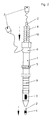
- Two sensors 2a, 3a for measuring intracranial pressure are integrated in the catheter tube 1a, the first sensor 2a being arranged at the distal end and the second sensor 3a being located proximally behind it.
- the sensors are located in housings made of non-magnetizable material, here ceramic.
- the scale 4a is MRI-contrasting, in this case titanium-colored, and is ring-shaped attached a length of 20 cm. The distance between the markings is 1 cm each.
- the sensors 2a, 3a are wired to the plugs 7a via a switch 6a.
- the hose rider 5a serves as a spacer or to fix the penetration depth of the catheter.
- the large-lumen catheter tube 1 has an axial one at the distal end Opening, proximally behind it is a sensor 3, a tube rider 5 and the ring-shaped scale 9 at intervals of 1 cm.
- the valve 6 is designed as an end piece, in which the electrical wiring of the sensor through a lateral lumen outlet of the valve 6 branches off to the monitor and in the central lumen of valve 6 the small-lumen catheter tube 2 or a rigid stylet may be positioned.
- the small-lumen catheter tube 2 has a conical tip with sensor 4 at the distal end.
- the two axially arranged catheter tubes form an atraumatic tip from the sensor housing of the small-lumen catheter tube 2 and the conical tip of the large-lumen catheter tube 1, which enables precise guidance and placement of the system in the brain.
- the valve 6 can be opened and the sensors 3, 4 can be axially displaced relative to one another. This movement can be tracked in a controlled manner via the scalings 9, 10.
- the connectors 7 can be coupled to the measuring device technology, the transmission of the measuring signals to the monitor via cable or - if the monitor is equipped accordingly - telemetrically, for example via radio , he follows. Corrections of the sensor position are also possible during the measurement.
- a protective cover 8 with one end on the valve 6 and the other end on the catheter tube 2 is fixedly mounted, which is preferably designed as a bellows or film tube made of transparent, flexible polymer material.
Abstract
Description
Der Stand der Technik bei der Messung des intrazerebralen Druckes ist in den Hirndruckdruckmesssonden
"Neurovent" der REHAU AG + Co samt Zubehör aktuell dargestellt. Hier
finden alle üblichen Mess- und Applikationstechniken Anwendung, die benötigt werden, um
einen mit einem Sensor versehenen Katheter nach den gebräuchlichen Regeln an den vorgesehenen
Orten zu platzieren, um die gewünschten Messwerte für den Hirndruck zu erhalten,
sowie grafisch darstellen zu können.
Dazu wird - nach der bisher geübten medizinischen Praxis - der Katheter meistens im Bereich
des vorderen Schädels, rechts oder links frontal, eingeführt und der Hirndruck an folgenden
Messorten bestimmt:
For this purpose - according to the previously practiced medical practice - the catheter is usually inserted in the area of the front skull, right or left frontal, and the intracranial pressure is determined at the following measuring locations:
Hier geht man von der im vielen Fällen unzutreffenden, aber medizinisch akzeptierten Annahme aus, dass der intrazerebrale Druck im Hirn an allen Punkten etwa in gleicher Größe anliegt. Für gewöhnlich wird daher der Hirndruck nur durch einen Einzelmesspunkt erfasst.Here one starts from the often incorrect but medically accepted assumption from that the intracerebral pressure in the brain is about the same size at all points is applied. Therefore, the intracranial pressure is usually only recorded by a single measuring point.
Es ist jedoch auch bekannt, dass das menschliche Hirn unter bestimmten Bedingungen an verschiedenen Messorten Druckdifferenzen, sog. Druckgradienten, aufweisen kann. Daher wird in Ausnahmefällen mit zwei Sensoren gearbeitet, von denen jeder in einem gesonderten Katheter platziert ist. Dies wird überwiegend im Rahmen von Studien durchgeführt und ist in der klinischen Praxis nicht üblich.However, it is also known to affect the human brain under certain conditions different measuring locations can have pressure differences, so-called pressure gradients. Therefore In exceptional cases, two sensors are used, each in a separate one Catheter is placed. This is mainly done within the framework of studies and is not common in clinical practice.
Die wünschenswerte Lage des Messsensors in unmittelbarer Nähe zur pathologischen Veränderung ist also in seltenen Fällen - und nur zufällig - zu erreichen. Da bisher nur der allgemeine Hirndruck erfasst werden konnte, war die Platzierung an den quasi genormten Messorten üblich. The desirable position of the measuring sensor in the immediate vicinity of the pathological change can be reached in rare cases - and only by chance. Since so far only the general Brain pressure could be recorded, was the placement on the quasi-standardized Measuring locations usual.
Besonders nachteilig ist, dass ohne Patientenrisiko eine Lagekorrektur des Sensors nachträglich
nicht möglich ist. Vielmehr muss der Katheter komplett entfernt und ein neuer gelegt
werden, um die Sterilitätsbedingungen zu sichern.
Nachteilig ist darüber hinaus, dass mehrere Sensoren nicht über einen Zugangskanal und in
variablen Abständen positionierbar sind. Wenn mehrere Sensoren zum Einsatz kommen
müssen, so geschieht dies in der Regel zwar über eine Kopfschraube, jedoch über mehrere
Katheter und damit über mehrere Zugangskanäle in der Hirnhaut.It is particularly disadvantageous that it is not possible to subsequently correct the position of the sensor without patient risk. Rather, the catheter must be removed completely and a new one inserted to ensure sterility conditions.
Another disadvantage is that several sensors cannot be positioned over an access channel and at variable intervals. If several sensors have to be used, this is usually done via a cap screw, but via several catheters and thus via several access channels in the meninges.
Aus diesen Nachteilen des Standes der Technik ergibt sich die Aufgabe, eine Vorrichtung bereit zu stellen, welche diese Negativpunkte überwindet und insbesondere die Möglichkeit bereit stellt, dass mindestens zwei Hirndruckmesssensoren über einen Zugang gleichzeitig an mindestens zwei verschiedenen Punkten im Hirn platziert werden können.From these disadvantages of the prior art, there is the task of a device to provide, which overcomes these negative points and in particular the possibility provides that at least two brain pressure measurement sensors have one access at the same time can be placed at at least two different points in the brain.
Das geschieht erfindungsgemäß durch ein Kathetersystem, in dem mindestens zwei Sensoren
in Reihe angeordnet sind, wobei mindestens ein Sensor den Hirndruck in der geschädigten
Hirnregion und der zweite oder weitere im angrenzenden Bereich des Hirns den Hirndruck
erfassen.
Die Positionierung geschieht vorzugsweise über Neuronavigation oder Ultraschall, wobei
entweder über eine separate Hülse in Verbindung mit einem eingebrachten Mandrin der Zugangskanal
erzeugt wird und anschließend das Kathetersystem eingebracht wird, oder das
Kathetersystem enthält einen Mandrin und verfügt über eine atraumatische Katheterspitze,
so dass es direkt eingeführt und positioniert werden kann.According to the invention, this is done by a catheter system in which at least two sensors are arranged in series, at least one sensor detecting the intracranial pressure in the damaged brain region and the second or more in the adjacent region of the brain measuring the intracranial pressure.
The positioning is preferably done via neuronavigation or ultrasound, either using a separate sleeve in connection with an inserted stylet to create the access channel and then inserting the catheter system, or the catheter system contains a stylet and has an atraumatic catheter tip so that it can be inserted directly and can be positioned.
Die Messung der absoluten Werte der Hirndrücke, sowie der zeitliche Verlauf der Differenzwerte, können nun die Grundlage für eine wesentlich wirksamere Therapiestrategie bilden, als dies bisher möglich war.The measurement of the absolute values of the brain pressures, as well as the time course of the difference values, can now form the basis for a much more effective therapy strategy, than was previously possible.
Alle Materialien sind CT-, MRT- und röntgentauglich, d. h. aus unmagnetischen Materialien, wie z. B. Titan, Keramik oder hochpolymeren Werkstoffen gefertigt.All materials are CT, MRI and X-ray compatible, i.e. H. made of non-magnetic materials, such as B. titanium, ceramic or high polymer materials.
Nachfolgend soll die Erfindung und einige Variationsmöglichkeiten davon, die nicht auf die beschriebenen Ausführungsbeispiele beschränkt sind, näher erläutert werden. The following is the invention and some possible variations thereof, which are not based on the described embodiments are limited, are explained in more detail.
Erfindungsgemäß lässt sich beispielsweise eine erste Hirndruckmesssonde in einem einlumigen
Katheter mit zwei piezoelektrischen Sensoren zur Hirndruckmessung herstellen. Dabei
ist der erste Sensor an der Spitze des Katheterschlauches, der zweite im Abstand dahinter
im selben Lumen angeordnet. Der Katheterschlauch selbst besteht aus Polyurethan,
das auf übliche Weise, beispielsweise mit Zusatz von Wolframpulver, röntgen-, CT- und
MRT-kontrastfähig ausgerüstet ist.
Weiterhin ist der Katheterschlauch signalfarben orange eingefärbt, damit er - bei dem geringen
Durchmesser von 2 mm - bei der Operation leicht erkennbar und identifizierbar ist.According to the invention, for example, a first intracranial pressure measurement probe can be produced in a single-lumen catheter with two piezoelectric sensors for intracranial pressure measurement. The first sensor is located at the tip of the catheter tube, the second at a distance behind it in the same lumen. The catheter tube itself is made of polyurethane, which is equipped with X-ray, CT and MRI contrast in the usual way, for example with the addition of tungsten powder.
Furthermore, the catheter tube is signal-colored orange so that it can be easily recognized and identified during the operation, given the small diameter of 2 mm.
Nach dem Öffnen des Schädels nahe dem geschädigten Bereich wird, wie üblich, der Bolt-Kit
gesetzt und über ein System mit Hülse und Mandrin der mit den Sensoren bestückte Teil
des erfindungsgemäßen Katheters im Kopf platziert. Das geschieht über ein Neuronavigationssystem
oder mittels Ultraschall und zwar so, dass im geschädigten Hirnbereich ein Sensor,
ein zweiter Sensor in der gesunden Hirnmasse oder im Ventrikel platziert werden.
Die Sensoren sind über ein entsprechendes Adaptersystem an den Messmonitor gekoppelt,
die so zugänglichen Druckgradienten der einzelnen Messstellen und deren zeitliche Veränderung
lassen sich nun abrufen und dokumentieren.
Um beim Implantieren des Katheters die einzelnen Abstände der Sensoren zu erkennen, ist
der Katheter zirkulär mit einer Skalierung und Beschriftung versehen.After opening the skull near the damaged area, the bolt kit is placed, as usual, and the part of the catheter according to the invention equipped with the sensors is placed in the head via a system with a sleeve and stylet. This is done using a neuronavigation system or using ultrasound, so that one sensor is placed in the damaged area of the brain, a second sensor is placed in the healthy brain or in the ventricle.
The sensors are coupled to the measuring monitor via a corresponding adapter system, the pressure gradients accessible in this way and the changes over time can now be called up and documented.
In order to recognize the individual distances between the sensors when implanting the catheter, the catheter is provided with a scale and labeling.
Eine zweite Möglichkeit, die Erfindung auszugestalten, geht von einem zweilumigen System
mit zwei Drucksensoren zur Hirndruckmessung aus, wobei ein großlumiger Katheterschlauch
an seinem distalen Ende über einen Drucksensor und eine axiale Öffnung verfügt,
in die ein eingebrachter kleinlumiger Katheterschlauch, versehen an seinem distalen Ende
mit einem Drucksensor, hineinragt. Beide distale Sensoren bilden in ihrer Ausgangsposition
eine atraumatische Katheterspitze. Auch diese Anordnung wird beispielsweise über eine
Hülse mit innenliegendem Mandrin positioniert. Beide Katheterschläuche bestehen jeweils
aus einem Verbundschlauch, wobei dessen innere Schicht aus Polyethylen, dessen äußere
Schicht aus Silikonkautschuk besteht. Die beiden Katheterschläuche sind ebenfalls CT-,
MRT- und röntgenkontrastfähig ausgerüstet und unterschiedlich signalfarben eingefärbt.
Die Lumen beider Katheterschläuche sind über ein proximal montiertes Ventil gekoppelt,
welches mit dem großlumigen Katheterschlauch fest montiert ist und in dessen Lumen der
kleinlumige Katheterschlauch durch eine Quetschdichtung dicht und axial fixiert ist. A second possibility for embodying the invention is based on a two-lumen system with two pressure sensors for measuring intracranial pressure, a large-lumen catheter tube at its distal end having a pressure sensor and an axial opening into which an inserted small-lumen catheter tube is provided at its distal end a pressure sensor. Both distal sensors form an atraumatic catheter tip in their starting position. This arrangement is also positioned, for example, via a sleeve with an internal stylet. Both catheter tubes each consist of a composite tube, the inner layer of which is made of polyethylene, the outer layer of which is made of silicone rubber. The two catheter tubes are also equipped with CT, MRI and X-ray contrast and are colored in different signal colors.
The lumens of both catheter tubes are coupled via a proximally mounted valve, which is firmly attached to the large-lumen catheter tube and in the lumen of which the small-lumen catheter tube is tightly and axially fixed by means of a pinch seal.
Im Falle einer erforderlichen Korrektur des Abstandes der Sensoren wird nun die Dichtung
geöffnet, der erforderliche Abstand hergestellt - kontrolliert durch die Markierungen der Katheterschläuche
- und die Dichtung wieder geschlossen.
Der bewegte Abschnitt des kleinlumigen Katheterschlauches ist für diese Handhabung mit
einer flexiblen Schlauchschutzhülle umgeben, welche die sterile Oberfläche des bewegten
Schlauchabschnittes während der notwendigen Positionsveränderung sichert.
Alternativ ist es auch möglich, den großlumigen Katheterschlauch über einen atraumatischen,
starren Mandrin vorab zu implantieren und nach dem Entfernen dieses Mandrins den
kleinlumigen Katheterschlauch zu installieren.If the distance between the sensors needs to be corrected, the seal is now opened, the required distance is established - checked by the markings on the catheter tubes - and the seal is closed again.
For this handling, the moving section of the small-lumen catheter tube is surrounded by a flexible tube protective cover, which secures the sterile surface of the moving tube section during the necessary change in position.
Alternatively, it is also possible to implant the large-lumen catheter tube beforehand using an atraumatic, rigid stylet and to install the small-lumen catheter tube after removing this stylet.
Eine weitere Möglichkeit der erfindungsgemäßen Ausführung sei hier noch erwähnt. Hier sind beide Sensoren in dem einem Lumen des Katheters in Reihe platziert, in einem zweiten Lumen des Katheters befindet sich ein starrer Mandrin, der es ermöglicht, den Katheter sicher zu stabilisieren und ohne Abweichung zu führen. Dabei ist die Mandrinspitze so in der Katheterspitze zentral angeordnet, dass es zu keiner axialen Abweichung durch Schub während der Implantation kommen kann.Another possibility of the embodiment according to the invention should be mentioned here. Here both sensors are placed in series in one lumen of the catheter, in a second Lumen of the catheter is a rigid stylet that allows the catheter to be secure to stabilize and run without deviation. The stylet tip is so in the Catheter tip centrally located so that there is no axial deviation due to thrust during the implantation can come.
Die Vorteile des erfindungsgemäßen Kathetersystems sind vielfältig. Zunächst ist es möglich,
dass die Messung am Ort der Hirnverletzung und parallel im Bereich des umgebenden
Gewebes erfolgen kann. Damit ist eine konkrete Aussage zu den tatsächlichen Druckverhältnissen
und besonders zum Druckgradienten möglich. Weiterhin ist nur ein Zugangskanal
nötig, um eine Messung an mehreren Punkten mit mehreren Sensoren durchführen zu können.
Besonders vorteilhaft ist, dass die Sensoren präzise positionierbar sind, sowohl im
klassischen, manuellen Handling, wie auch mit Hilfe der Neuronavigation oder des Ultraschalls.
Dies ist möglich entweder durch den Einsatz eines starren Mandrins im Katheter (sh.
Beispiel 2) oder durch die Platzierung des Katheters nach dem Einbringen des Zugangskanals
über die Kombination Hülse/Mandrin.
Nicht zuletzt ist die Kombination mit gebräuchlichen Hilfsmitteln gesichert, z.B. mit Tunnelungshülsen
oder Kopfschrauben oder lumengeführten starren Führungsdrähten.The advantages of the catheter system according to the invention are numerous. First of all, it is possible that the measurement can take place at the site of the brain injury and in parallel in the area of the surrounding tissue. This makes it possible to make a concrete statement about the actual pressure conditions and especially about the pressure gradient. Furthermore, only one access channel is necessary in order to be able to carry out a measurement at several points with several sensors. It is particularly advantageous that the sensors can be positioned precisely, both in classic, manual handling and with the help of neuronavigation or ultrasound. This is possible either by using a rigid stylet in the catheter (see example 2) or by placing the catheter after inserting the access channel via the sleeve / stylet combination.
Last but not least, the combination is secured with common aids, such as tunnel sleeves or cap screws or lumen-guided rigid guide wires.
Neben den Vorteilen bei der Platzierung sind die messtechnischen Vorteile bei der Anwendung des erfindungsgemäßen Kathetersystems erheblich. In addition to the advantages in terms of placement, there are the measurement advantages in use of the catheter system according to the invention considerably.
Zunächst ist es vorteilhaft, dass die Diagnostik der Hirndruckverläufe im geschädigten Hirnbereich
im direkten Vergleich mit dem Hirndruck im umgebenden, gesunden Gewebe erfolgt
und damit lokal bewertbar ist. Damit wird erstmals in der Hirndruckdiagnose der Vergleich
von lokalem und allgemeinem Hirndruck möglich, wobei vorzugsweise der sog. Goldstandard
des Hirndruckes, gemessen im Ventrikel, als allgemeiner Hirndruck im Sinne der bestmöglichen
Bezugsgröße, angestrebt wird. Die Einzelerfassung der Messdaten je Sensor,
sowie deren Verarbeitung z. B. in Form von Druckdifferenzwerten, führt zu Ergebnisdarstellungen,
die zielgerichtet den Outcome der Patienten verbessern.
Weiterhin ist die Erfassung und Auswertung der Messergebnisse einfach und den bisherigen
Verfahrensweisen angepasst. Es ist keine zusätzliche Hardware nötig und nur geringfügige
Aufwendungen an Software für die vorhandenen Monitorsysteme.
Für die Diagnose ist besonders vorteilhaft, dass, wie im Beispiel 2 beschrieben, die Lage
des zweiten Sensors in der Längsachse des Katheters veränderbar ist.First of all, it is advantageous that the diagnosis of the intracranial pressure profiles in the damaged brain area is carried out in a direct comparison with the intracranial pressure in the surrounding, healthy tissue and can thus be assessed locally. This enables the comparison of local and general intracranial pressure for the first time in the diagnosis of intracranial pressure, whereby the so-called gold standard of intracranial pressure, measured in the ventricle, is aimed at as general cerebral pressure in the sense of the best possible reference. The individual acquisition of the measurement data per sensor, as well as their processing z. B. in the form of pressure difference values, leads to presentation of results that specifically improve the patient's outcome.
Furthermore, the acquisition and evaluation of the measurement results is simple and adapted to the previous procedures. No additional hardware is required and only minimal software expenditure for the existing monitor systems.
It is particularly advantageous for the diagnosis that, as described in Example 2, the position of the second sensor in the longitudinal axis of the catheter can be changed.
Die aufgeführten Vorteile ergeben also letztlich optimale Behandlungszeiträume durch frühzeitige Einleitung in Therapiemaßnahmen sowie sinkende Belastungen für Arzt und Patienten und damit auch eine entsprechende Kostenoptimierung.The advantages listed thus ultimately result in optimal treatment periods through early ones Introduction to therapeutic measures and decreasing burdens for doctors and patients and thus also a corresponding cost optimization.
Im Folgenden wird anhand von zwei Figuren die Erfindung näher erläutert.
Dabei zeigt
Figur 1 die erfindungsgemäße Vorrichtung in der Ausführung eines Einlumen-KathetersFigur 2 die erfindungsgemäße Vorrichtung in der Ausführung eines Zweilumen-Katheters
It shows
- Figure 1 shows the device according to the invention in the design of a single-lumen catheter
- Figure 2 shows the device according to the invention in the design of a two-lumen catheter
zeigt eine einlumige Ausführung der Erfindung.
In den Katheterschlauch 1a sind zwei Sensoren 2a, 3a zur Hirndruckmessung integriert,
wobei der erste Sensor 2a am distalen Ende und der zweite Sensor 3a proximal dahinter
angeordnet sind. Die Sensoren befinden sich in Gehäusen aus nicht magnetisierbarem Material,
hier Keramik. shows a single-lumen embodiment of the invention.
Two
Die Skalierung 4a ist MRT-kontrastgebend, hier titanfarben, ausgeführt und ringförmig auf
eine Länge von 20 cm angebracht. Der Abstand der Markierungen beträgt je 1 cm. Die Sensoren
2a, 3a sind über eine Weiche 6a mit den Steckern 7a verkabelt. Der Schlauchreiter 5a
dient als Abstandshalter oder zur Fixierung der Eindringtiefe des Katheters.The
zeigt eine zweilumige Ausführung der Erfindung, wobei - für viele Fälle besonders vorteilhaft
- eine Anordnung gewählt wurde, die es ermöglicht, beide Sensoren axial gegen einander
zu bewegen: Der großlumige Katheterschlauch 1 verfügt am distalen Ende über eine axiale
Öffnung, proximal dahinter folgt ein Sensor 3, ein Schlauchreiter 5 und die ringförmige Skalierung
9 in Abständen von jeweils 1 cm. Das Ventil 6 ist als Endstück gestaltet, in welchem
durch einen seitlichen Lumenausgang des Ventils 6 die elektrische Verkabelung des Sensors
zum Monitor abzweigt und im zentralen Lumen des Ventils 6 der kleinlumige Katheterschlauch
2 oder unter Umständen ein starrer Mandrin positioniert sind.shows a two-lumen embodiment of the invention, being - particularly advantageous for many cases
- An arrangement was chosen that allows both sensors axially against each other
to move: The large-
Der kleinlumige Katheterschlauch 2 verfügt am distalen Ende über eine konisch ausgebildete
Spitze mit Sensor 4.
Die beiden axial angeordneten Katheterschläuche bilden aus dem Sensorgehäuse des
kleinlumigen Katheterschlauches 2 und der konischen Spitze des großlumigen Katheterschlauches
1 eine atraumatische Spitze, die eine präzise Führung und Platzierung des Systems
im Hirn ermöglicht. Ist die Positionierung erfolgt, so kann das Ventil 6 geöffnet und die
Sensoren 3, 4 gegeneinander axial verschoben werden. Diese Bewegung ist über die Skalierungen
9, 10 kontrolliert verfolgbar. Mit dem Verschluss des Ventils 6 und der damit erfolgten
Fixierung des kleinlumigen Katheterschlauches 2 können die Stecker 7 an die Messgerätetechnik
gekoppelt werden, wobei die Übertragung der Messsignale an den Monitor
über Kabel, oder - wenn der Monitor entsprechend ausgestattet ist - telemetrisch, beispielsweise
über Funk, erfolgt.
Korrekturen der Sensorposition sind auch während der Messung möglich.
Zu Sicherung der sterilen Oberfläche des bewegten Schlauchabschnittes des Katheterschlauches
2 ist eine Schutzhülle 8 mit einem Ende am Ventil 6 und dem anderen Ende am
Katheterschlauch 2 fest montiert, die vorzugsweise als Faltenbalg oder Folienschlauch aus
transparentem, flexiblen Polymermaterial gestaltet ist.The small-
The two axially arranged catheter tubes form an atraumatic tip from the sensor housing of the small-
Corrections of the sensor position are also possible during the measurement.
To secure the sterile surface of the moving tube section of the
Claims (11)
dadurch gekennzeichnet, dass der Katheter mindestens aus einem Katheterschlauch aus polymerem Material besteht, und über mindestens zwei Drucksensoren verfügt, die in Reihe angeordnet sind, wobei der eine Drucksensor den Hirndruck in der geschädigten Region, der zweite, oder weitere Drucksensoren den Hirndruck in den angrenzenden Bereichen erfassen und dass die Platzierung des Katheters und die Messung des Hirndruckes an beliebigen Stellen im Hirn erfolgt.Device for measuring intracranial pressure using a catheter,
characterized in that the catheter consists of at least one catheter tube made of polymeric material and has at least two pressure sensors which are arranged in series, one pressure sensor measuring the intracranial pressure in the damaged region, the second, or further pressure sensors measuring the intracranial pressure in the adjacent region Detect areas and that the placement of the catheter and the measurement of the intracranial pressure takes place anywhere in the brain.
Applications Claiming Priority (2)
| Application Number | Priority Date | Filing Date | Title |
|---|---|---|---|
| DE20116879U DE20116879U1 (en) | 2001-10-13 | 2001-10-13 | Device for determining the intracerebral pressure gradient |
| DE20116879U | 2001-10-13 |
Publications (2)
| Publication Number | Publication Date |
|---|---|
| EP1302157A2 true EP1302157A2 (en) | 2003-04-16 |
| EP1302157A3 EP1302157A3 (en) | 2005-04-13 |
Family
ID=7962860
Family Applications (1)
| Application Number | Title | Priority Date | Filing Date |
|---|---|---|---|
| EP02022414A Withdrawn EP1302157A3 (en) | 2001-10-13 | 2002-10-04 | Device for the determination of the intracerebral pressure gradient |
Country Status (2)
| Country | Link |
|---|---|
| EP (1) | EP1302157A3 (en) |
| DE (1) | DE20116879U1 (en) |
Cited By (1)
| Publication number | Priority date | Publication date | Assignee | Title |
|---|---|---|---|---|
| RU2447835C2 (en) * | 2010-05-20 | 2012-04-20 | Государственное учреждение здравоохранения "Кемеровская областная клиническая больница" | Device for studying subarachnoid space hydrodynamics |
Families Citing this family (2)
| Publication number | Priority date | Publication date | Assignee | Title |
|---|---|---|---|---|
| US8313442B2 (en) | 2009-10-21 | 2012-11-20 | Codman & Shurtleff, Inc. | Cerebral compliance monitoring |
| CN105962916B (en) * | 2016-06-08 | 2017-05-03 | 深圳北芯生命科技有限公司 | Fixture for manufacturing measurement catheter |
Citations (8)
| Publication number | Priority date | Publication date | Assignee | Title |
|---|---|---|---|---|
| DE4036355A1 (en) * | 1990-11-15 | 1992-05-21 | Messgeraetewerk Zwoenitz Gmbh | Medical pressure measurement catheter for internal use - has pressure sensor mounted in channel housing via sealing, incompressible, elastic coupling body |
| EP0694284A1 (en) * | 1994-07-18 | 1996-01-31 | Dräger Medical Electronics B.V. | Catheter for performing measurements in the esophagus and/or stomach at various positions |
| US5573007A (en) * | 1994-08-08 | 1996-11-12 | Innerspace, Inc. | Gas column pressure monitoring catheters |
| NL1005134C2 (en) * | 1997-01-30 | 1998-08-03 | Industrial Res Bv | Catheter with measuring insert |
| EP0879617A1 (en) * | 1997-05-21 | 1998-11-25 | Schneider (Europe) GmbH | Pressure monitoring guide wire and method for manufacturing such a guide wire |
| US6061587A (en) * | 1997-05-15 | 2000-05-09 | Regents Of The University Of Minnesota | Method and apparatus for use with MR imaging |
| EP1138343A1 (en) * | 2000-03-17 | 2001-10-04 | Integra Lifesciences Inc. | Improved ventricular catheter with reduced size connector and method of use |
| EP1142532A2 (en) * | 2000-04-03 | 2001-10-10 | Regents Of The University Of Minnesota | Intracranial pressure monitoring device for use in magnetic resonance imaging guided drug delivery |
-
2001
- 2001-10-13 DE DE20116879U patent/DE20116879U1/en not_active Expired - Lifetime
-
2002
- 2002-10-04 EP EP02022414A patent/EP1302157A3/en not_active Withdrawn
Patent Citations (8)
| Publication number | Priority date | Publication date | Assignee | Title |
|---|---|---|---|---|
| DE4036355A1 (en) * | 1990-11-15 | 1992-05-21 | Messgeraetewerk Zwoenitz Gmbh | Medical pressure measurement catheter for internal use - has pressure sensor mounted in channel housing via sealing, incompressible, elastic coupling body |
| EP0694284A1 (en) * | 1994-07-18 | 1996-01-31 | Dräger Medical Electronics B.V. | Catheter for performing measurements in the esophagus and/or stomach at various positions |
| US5573007A (en) * | 1994-08-08 | 1996-11-12 | Innerspace, Inc. | Gas column pressure monitoring catheters |
| NL1005134C2 (en) * | 1997-01-30 | 1998-08-03 | Industrial Res Bv | Catheter with measuring insert |
| US6061587A (en) * | 1997-05-15 | 2000-05-09 | Regents Of The University Of Minnesota | Method and apparatus for use with MR imaging |
| EP0879617A1 (en) * | 1997-05-21 | 1998-11-25 | Schneider (Europe) GmbH | Pressure monitoring guide wire and method for manufacturing such a guide wire |
| EP1138343A1 (en) * | 2000-03-17 | 2001-10-04 | Integra Lifesciences Inc. | Improved ventricular catheter with reduced size connector and method of use |
| EP1142532A2 (en) * | 2000-04-03 | 2001-10-10 | Regents Of The University Of Minnesota | Intracranial pressure monitoring device for use in magnetic resonance imaging guided drug delivery |
Cited By (1)
| Publication number | Priority date | Publication date | Assignee | Title |
|---|---|---|---|---|
| RU2447835C2 (en) * | 2010-05-20 | 2012-04-20 | Государственное учреждение здравоохранения "Кемеровская областная клиническая больница" | Device for studying subarachnoid space hydrodynamics |
Also Published As
| Publication number | Publication date |
|---|---|
| DE20116879U1 (en) | 2001-12-20 |
| EP1302157A3 (en) | 2005-04-13 |
Similar Documents
| Publication | Publication Date | Title |
|---|---|---|
| DE3504292C1 (en) | Instrument for endoscopic interventions, especially for percutaneous gallstone removal or gallbladder surgery | |
| DE69733249T2 (en) | DETERMINATION OF THE EXACT POSITION OF ENDOSCOPES | |
| EP1065987B1 (en) | Endoscope adapter which can be detected using computer assisted surgery | |
| DE60035811T2 (en) | Slit drainage catheter with stylet | |
| EP3641621B1 (en) | Endoscope device | |
| EP1319366A1 (en) | Magnetic navigation for a catheter | |
| DE4440346A1 (en) | Puncture instrument | |
| WO2002045588A1 (en) | Ultrasonic probe comprising a positioning device for examination devices and operation devices | |
| EP1731105A1 (en) | Device for establishing free transcutaneous access to an endoscopic surgical area | |
| DE10029737B4 (en) | Navigation of a medical instrument | |
| EP2185062B1 (en) | Catheter system having an optical probe and method for the application of an optical probe in a catheter system | |
| EP3585474A1 (en) | Device for drainage of the brain | |
| EP3979929B1 (en) | Surgical needle set for determining the position of a surgical instrument | |
| DE102005027677B4 (en) | Device for automatic replacement of instruments in minimally invasive procedures | |
| EP1075216B1 (en) | Magnetic resonance tomograph | |
| EP1302157A2 (en) | Device for the determination of the intracerebral pressure gradient | |
| DE10100756A1 (en) | Device for carrying out a percutaneous intervention in the area of the lung without damage to the pleural cavity has a flexible channel with two sluices at the proximal end | |
| WO2015007681A1 (en) | Device and method for connecting a medical instrument to a position-detecting system | |
| WO2003079915A2 (en) | Medical instrument for treating tissue by means of high-frequency current and medical system comprising such a medical instrument | |
| DE102016119065B4 (en) | Needle stockings with strap system | |
| DE10040164C2 (en) | Drainage and irrigation catheter with pressure measuring device | |
| EP0450111A1 (en) | Application of computer tomography, magnetic resonance tomography, ultrasonic imaging or other image processing for the guidance of endoscopic diagnostic and operative therapy | |
| WO2007087798A2 (en) | Aiming device for performing minimally invasive interventions | |
| DE102016006425A1 (en) | Guide catheter, method for positioning probes and use of guide catheter | |
| EP1199034A2 (en) | Device for linearly and/or rotatively guiding a probe or instrument |
Legal Events
| Date | Code | Title | Description |
|---|---|---|---|
| PUAI | Public reference made under article 153(3) epc to a published international application that has entered the european phase |
Free format text: ORIGINAL CODE: 0009012 |
|
| AK | Designated contracting states |
Designated state(s): AT BE BG CH CY CZ DE DK EE ES FI FR GB GR IE IT LI LU MC NL PT SE SK TR |
|
| AX | Request for extension of the european patent |
Extension state: AL LT LV MK RO SI |
|
| RAP1 | Party data changed (applicant data changed or rights of an application transferred) |
Owner name: RAUMEDIC AG |
|
| PUAL | Search report despatched |
Free format text: ORIGINAL CODE: 0009013 |
|
| AK | Designated contracting states |
Kind code of ref document: A3 Designated state(s): AT BE BG CH CY CZ DE DK EE ES FI FR GB GR IE IT LI LU MC NL PT SE SK TR |
|
| AX | Request for extension of the european patent |
Extension state: AL LT LV MK RO SI |
|
| AKX | Designation fees paid | ||
| STAA | Information on the status of an ep patent application or granted ep patent |
Free format text: STATUS: THE APPLICATION IS DEEMED TO BE WITHDRAWN |
|
| 18D | Application deemed to be withdrawn |
Effective date: 20051014 |
|
| REG | Reference to a national code |
Ref country code: DE Ref legal event code: 8566 |