EP1300713A2 - Method and apparatus for automated image analysis of biological specimens - Google Patents
Method and apparatus for automated image analysis of biological specimens Download PDFInfo
- Publication number
- EP1300713A2 EP1300713A2 EP02028849A EP02028849A EP1300713A2 EP 1300713 A2 EP1300713 A2 EP 1300713A2 EP 02028849 A EP02028849 A EP 02028849A EP 02028849 A EP02028849 A EP 02028849A EP 1300713 A2 EP1300713 A2 EP 1300713A2
- Authority
- EP
- European Patent Office
- Prior art keywords
- interest
- image
- slide
- stage
- objects
- Prior art date
- Legal status (The legal status is an assumption and is not a legal conclusion. Google has not performed a legal analysis and makes no representation as to the accuracy of the status listed.)
- Withdrawn
Links
- 238000000034 method Methods 0.000 title claims abstract description 125
- 238000010191 image analysis Methods 0.000 title claims description 14
- 238000012545 processing Methods 0.000 claims description 35
- 230000003287 optical effect Effects 0.000 claims description 23
- 238000000386 microscopy Methods 0.000 claims 4
- 238000004458 analytical method Methods 0.000 abstract description 31
- 238000012552 review Methods 0.000 abstract description 13
- 238000011156 evaluation Methods 0.000 abstract description 3
- 238000003745 diagnosis Methods 0.000 abstract description 2
- 210000004027 cell Anatomy 0.000 description 93
- 230000008569 process Effects 0.000 description 54
- 238000010586 diagram Methods 0.000 description 21
- 210000004881 tumor cell Anatomy 0.000 description 19
- 239000011159 matrix material Substances 0.000 description 15
- 230000000877 morphologic effect Effects 0.000 description 15
- 238000006243 chemical reaction Methods 0.000 description 11
- 238000011068 loading method Methods 0.000 description 11
- 238000005286 illumination Methods 0.000 description 10
- 239000000969 carrier Substances 0.000 description 9
- 238000001914 filtration Methods 0.000 description 8
- 238000012360 testing method Methods 0.000 description 8
- 238000001514 detection method Methods 0.000 description 7
- 102000002260 Alkaline Phosphatase Human genes 0.000 description 6
- 108020004774 Alkaline Phosphatase Proteins 0.000 description 6
- 239000000523 sample Substances 0.000 description 6
- 206010028980 Neoplasm Diseases 0.000 description 5
- 239000003153 chemical reaction reagent Substances 0.000 description 5
- 210000000805 cytoplasm Anatomy 0.000 description 5
- 238000003384 imaging method Methods 0.000 description 5
- 230000007246 mechanism Effects 0.000 description 5
- 210000000440 neutrophil Anatomy 0.000 description 5
- 238000010186 staining Methods 0.000 description 5
- WZUVPPKBWHMQCE-UHFFFAOYSA-N Haematoxylin Chemical compound C12=CC(O)=C(O)C=C2CC2(O)C1C1=CC=C(O)C(O)=C1OC2 WZUVPPKBWHMQCE-UHFFFAOYSA-N 0.000 description 4
- 230000010339 dilation Effects 0.000 description 4
- 230000003628 erosive effect Effects 0.000 description 4
- 230000001629 suppression Effects 0.000 description 4
- 230000032683 aging Effects 0.000 description 3
- WLDHEUZGFKACJH-UHFFFAOYSA-K amaranth Chemical compound [Na+].[Na+].[Na+].C12=CC=C(S([O-])(=O)=O)C=C2C=C(S([O-])(=O)=O)C(O)=C1N=NC1=CC=C(S([O-])(=O)=O)C2=CC=CC=C12 WLDHEUZGFKACJH-UHFFFAOYSA-K 0.000 description 3
- 238000013528 artificial neural network Methods 0.000 description 3
- 238000003556 assay Methods 0.000 description 3
- 239000006059 cover glass Substances 0.000 description 3
- 230000000694 effects Effects 0.000 description 3
- 239000000463 material Substances 0.000 description 3
- 230000001537 neural effect Effects 0.000 description 3
- 239000002244 precipitate Substances 0.000 description 3
- 238000002360 preparation method Methods 0.000 description 3
- 238000001228 spectrum Methods 0.000 description 3
- 238000013459 approach Methods 0.000 description 2
- 201000011510 cancer Diseases 0.000 description 2
- 230000008859 change Effects 0.000 description 2
- 239000003086 colorant Substances 0.000 description 2
- 238000012790 confirmation Methods 0.000 description 2
- 210000005069 ears Anatomy 0.000 description 2
- 239000012634 fragment Substances 0.000 description 2
- 229910052736 halogen Inorganic materials 0.000 description 2
- 150000002367 halogens Chemical class 0.000 description 2
- 238000002955 isolation Methods 0.000 description 2
- 210000005259 peripheral blood Anatomy 0.000 description 2
- 239000011886 peripheral blood Substances 0.000 description 2
- 238000003672 processing method Methods 0.000 description 2
- 210000001995 reticulocyte Anatomy 0.000 description 2
- 238000012549 training Methods 0.000 description 2
- 238000012546 transfer Methods 0.000 description 2
- 238000008940 Alkaline Phosphatase assay kit Methods 0.000 description 1
- 208000035473 Communicable disease Diseases 0.000 description 1
- 206010063045 Effusion Diseases 0.000 description 1
- 108010015776 Glucose oxidase Proteins 0.000 description 1
- 239000004366 Glucose oxidase Substances 0.000 description 1
- 208000003788 Neoplasm Micrometastasis Diseases 0.000 description 1
- 102000003992 Peroxidases Human genes 0.000 description 1
- 208000007660 Residual Neoplasm Diseases 0.000 description 1
- XUIMIQQOPSSXEZ-UHFFFAOYSA-N Silicon Chemical compound [Si] XUIMIQQOPSSXEZ-UHFFFAOYSA-N 0.000 description 1
- 230000002159 abnormal effect Effects 0.000 description 1
- 230000009471 action Effects 0.000 description 1
- 230000003213 activating effect Effects 0.000 description 1
- 230000004913 activation Effects 0.000 description 1
- 239000002390 adhesive tape Substances 0.000 description 1
- 239000012620 biological material Substances 0.000 description 1
- 230000005540 biological transmission Effects 0.000 description 1
- 210000004369 blood Anatomy 0.000 description 1
- 239000008280 blood Substances 0.000 description 1
- 210000001185 bone marrow Anatomy 0.000 description 1
- 210000001175 cerebrospinal fluid Anatomy 0.000 description 1
- 238000012512 characterization method Methods 0.000 description 1
- 238000004891 communication Methods 0.000 description 1
- 230000001268 conjugating effect Effects 0.000 description 1
- 238000010276 construction Methods 0.000 description 1
- 238000013016 damping Methods 0.000 description 1
- 238000007435 diagnostic evaluation Methods 0.000 description 1
- 238000006073 displacement reaction Methods 0.000 description 1
- 230000009977 dual effect Effects 0.000 description 1
- 229920001971 elastomer Polymers 0.000 description 1
- 239000000806 elastomer Substances 0.000 description 1
- 238000005516 engineering process Methods 0.000 description 1
- 238000006911 enzymatic reaction Methods 0.000 description 1
- 230000001605 fetal effect Effects 0.000 description 1
- 239000012530 fluid Substances 0.000 description 1
- 229940116332 glucose oxidase Drugs 0.000 description 1
- 235000019420 glucose oxidase Nutrition 0.000 description 1
- 108010046301 glucose peroxidase Proteins 0.000 description 1
- 238000009499 grossing Methods 0.000 description 1
- 238000007654 immersion Methods 0.000 description 1
- 230000002452 interceptive effect Effects 0.000 description 1
- 238000002372 labelling Methods 0.000 description 1
- 210000000265 leukocyte Anatomy 0.000 description 1
- 210000001165 lymph node Anatomy 0.000 description 1
- 230000008774 maternal effect Effects 0.000 description 1
- 230000035800 maturation Effects 0.000 description 1
- 239000012528 membrane Substances 0.000 description 1
- 230000008802 morphological function Effects 0.000 description 1
- IPSIPYMEZZPCPY-UHFFFAOYSA-N new fuchsin Chemical compound [Cl-].C1=CC(=[NH2+])C(C)=CC1=C(C=1C=C(C)C(N)=CC=1)C1=CC=C(N)C(C)=C1 IPSIPYMEZZPCPY-UHFFFAOYSA-N 0.000 description 1
- 150000007523 nucleic acids Chemical class 0.000 description 1
- 102000039446 nucleic acids Human genes 0.000 description 1
- 108020004707 nucleic acids Proteins 0.000 description 1
- 244000052769 pathogen Species 0.000 description 1
- 230000002093 peripheral effect Effects 0.000 description 1
- 238000007781 pre-processing Methods 0.000 description 1
- 238000003793 prenatal diagnosis Methods 0.000 description 1
- 239000000047 product Substances 0.000 description 1
- 238000007790 scraping Methods 0.000 description 1
- 230000035939 shock Effects 0.000 description 1
- 229910052710 silicon Inorganic materials 0.000 description 1
- 239000010703 silicon Substances 0.000 description 1
- 239000000758 substrate Substances 0.000 description 1
- 230000001131 transforming effect Effects 0.000 description 1
- 210000002700 urine Anatomy 0.000 description 1
- 230000003612 virological effect Effects 0.000 description 1
Images


Classifications
-
- G—PHYSICS
- G01—MEASURING; TESTING
- G01N—INVESTIGATING OR ANALYSING MATERIALS BY DETERMINING THEIR CHEMICAL OR PHYSICAL PROPERTIES
- G01N1/00—Sampling; Preparing specimens for investigation
- G01N1/28—Preparing specimens for investigation including physical details of (bio-)chemical methods covered elsewhere, e.g. G01N33/50, C12Q
- G01N1/30—Staining; Impregnating ; Fixation; Dehydration; Multistep processes for preparing samples of tissue, cell or nucleic acid material and the like for analysis
- G01N1/31—Apparatus therefor
- G01N1/312—Apparatus therefor for samples mounted on planar substrates
-
- G01N15/1433—
-
- G—PHYSICS
- G01—MEASURING; TESTING
- G01N—INVESTIGATING OR ANALYSING MATERIALS BY DETERMINING THEIR CHEMICAL OR PHYSICAL PROPERTIES
- G01N15/00—Investigating characteristics of particles; Investigating permeability, pore-volume, or surface-area of porous materials
- G01N15/10—Investigating individual particles
- G01N15/14—Electro-optical investigation, e.g. flow cytometers
- G01N15/1468—Electro-optical investigation, e.g. flow cytometers with spatial resolution of the texture or inner structure of the particle
-
- G—PHYSICS
- G02—OPTICS
- G02B—OPTICAL ELEMENTS, SYSTEMS OR APPARATUS
- G02B21/00—Microscopes
- G02B21/24—Base structure
- G02B21/241—Devices for focusing
- G02B21/244—Devices for focusing using image analysis techniques
-
- G—PHYSICS
- G02—OPTICS
- G02B—OPTICAL ELEMENTS, SYSTEMS OR APPARATUS
- G02B21/00—Microscopes
- G02B21/36—Microscopes arranged for photographic purposes or projection purposes or digital imaging or video purposes including associated control and data processing arrangements
- G02B21/365—Control or image processing arrangements for digital or video microscopes
- G02B21/367—Control or image processing arrangements for digital or video microscopes providing an output produced by processing a plurality of individual source images, e.g. image tiling, montage, composite images, depth sectioning, image comparison
-
- G—PHYSICS
- G06—COMPUTING; CALCULATING OR COUNTING
- G06T—IMAGE DATA PROCESSING OR GENERATION, IN GENERAL
- G06T7/00—Image analysis
- G06T7/60—Analysis of geometric attributes
-
- G—PHYSICS
- G06—COMPUTING; CALCULATING OR COUNTING
- G06V—IMAGE OR VIDEO RECOGNITION OR UNDERSTANDING
- G06V20/00—Scenes; Scene-specific elements
- G06V20/60—Type of objects
- G06V20/69—Microscopic objects, e.g. biological cells or cellular parts
-
- G—PHYSICS
- G06—COMPUTING; CALCULATING OR COUNTING
- G06V—IMAGE OR VIDEO RECOGNITION OR UNDERSTANDING
- G06V20/00—Scenes; Scene-specific elements
- G06V20/60—Type of objects
- G06V20/69—Microscopic objects, e.g. biological cells or cellular parts
- G06V20/693—Acquisition
-
- G—PHYSICS
- G01—MEASURING; TESTING
- G01N—INVESTIGATING OR ANALYSING MATERIALS BY DETERMINING THEIR CHEMICAL OR PHYSICAL PROPERTIES
- G01N15/00—Investigating characteristics of particles; Investigating permeability, pore-volume, or surface-area of porous materials
- G01N15/10—Investigating individual particles
- G01N15/14—Electro-optical investigation, e.g. flow cytometers
- G01N15/1468—Electro-optical investigation, e.g. flow cytometers with spatial resolution of the texture or inner structure of the particle
- G01N2015/1472—Electro-optical investigation, e.g. flow cytometers with spatial resolution of the texture or inner structure of the particle with colour
-
- G—PHYSICS
- G01—MEASURING; TESTING
- G01N—INVESTIGATING OR ANALYSING MATERIALS BY DETERMINING THEIR CHEMICAL OR PHYSICAL PROPERTIES
- G01N15/00—Investigating characteristics of particles; Investigating permeability, pore-volume, or surface-area of porous materials
- G01N15/10—Investigating individual particles
- G01N15/14—Electro-optical investigation, e.g. flow cytometers
- G01N2015/1488—Methods for deciding
-
- G—PHYSICS
- G01—MEASURING; TESTING
- G01N—INVESTIGATING OR ANALYSING MATERIALS BY DETERMINING THEIR CHEMICAL OR PHYSICAL PROPERTIES
- G01N15/00—Investigating characteristics of particles; Investigating permeability, pore-volume, or surface-area of porous materials
- G01N15/10—Investigating individual particles
- G01N15/14—Electro-optical investigation, e.g. flow cytometers
- G01N2015/1497—Particle shape
Abstract
Description
- Fig. 1
- is a perspective view of an apparatus for automated cell analysis.
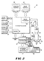
- Fig. 2
- is a block diagram of the apparatus shown in Fig. 1.
- Fig. 3
- is a block diagram of the microscope controller of Fig. 2.
- Fig.4
- is a plan view of the apparatus of Fig. 1 having the housing removed.
- Fig. 5
- is a side view of a microscope subsystem of the apparatus of Fig. 1.
- Fig. 6a
- is a top view of a slide carrier for use with the apparatus of Fig. 1.
- Fig. 6b
- is a bottom view of the slide carrier of Fig. 6a.
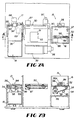
- Fig. 7a
- is a top view of an automated slide handling subsystem of the apparatus of Fig. 1.
- Fig. 7b
- is a partial cross-sectional view of the automated slide handling subsystem of Fig. 7a taken on line A-A.
- Fig. 8
- is an end view of the input module of the automated slide handling subsystem.
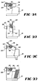
- Figs. 8a-8d
- illustrate the input operation of the automatic slide handling subsystem.
- Figs. 9a-9d
- illustrate the output operation of the automated slide handling subsystem.
- Fig. 10
- is a flow diagram of the procedure for automatically determining a scan area.
- Fig. 11
- shows the scan path on a prepared slide in the procedure of Fig. 10.
- Fig. 12
- illustrates an image of a field acquired in the procedure of Fig. 10.
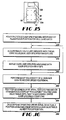
- Fig. 13A
- is a flow diagram of a preferred procedure for determining a focal position.
- Fig. 13B
- is a flow diagram of a preferred procedure for determining a focal position for neutrophils stained with Fast Red and counterstained with hemotoxylin.
- Fig. 14
- is a flow diagram of a procedure for automatically determining initial focus.
- Fig. 15
- shows an array of slide positions for use in the procedure of Fig. 14.
- Fig. 16
- is a flow diagram of a procedure for automatic focusing at a high magnification.
- Fig. 17A
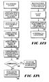
- is a flow diagram of an overview of the preferred process to locate and identify objects of interest in a stained biological specimen on a slide.
- Fig. 17B
- is a flow diagram of a procedure for color space conversion.
- Fig. 18
- is a flow diagram of a procedure for background suppression via dynamic thresholding.
- Fig. 19
- is a flow diagram of a procedure for morphological processing.
- Fig. 20
- is a flow diagram of a procedure for blob analysis.
- Fig. 21
- is a flow diagram of a procedure for image processing at a high magnification.
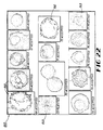
- Fig. 22
- illustrates a mosaic of cell images produced by the apparatus.
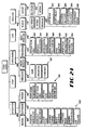
- Fig. 23
- is a flow diagram of a procedure for estimating the number of nucleated cells in a scan area.
- Fig. 24
- illustrates the apparatus functions available in a user interface of the apparatus.
The
R/R, R/G, R/B, G/G, G/B, G/R, B/B, B/G, B/R
Claims (6)
- A method for initially focusing an optical system of an automated microscopy system comprising the steps of:(a) positioning an optical system at an initial Z stage position;(b) acquiring an image and calculating a pixel variance about a pixel mean for the acquired image;(c) incrementing the position of the Z stage;(d) repeating steps (b) and (c) for a fixed number of iterations;(e) performing a least squares fit of the variance data to a known function; and(f) selecting the peak value of the least squares fit curve as an estimate of the best focal position.
- A method for initially focusing an optical system of an automated microscopy system comprising the steps of:(a) positioning an optical system at an initial Z stage position;(b) acquiring at low magnification an image of a slide having a stained biological specimen thereon and calculating a pixel variance about a pixel mean for the acquired image;(c) incrementing the position of the Z stage;(d) repeating steps (b) and (c) for a fixed number of coarse iterations to form a first set of variance data;(e) performing a least squares fit of the first set of variance data to a first function;(f) positioning the Z stage at a position near the peak of the first function;(g) repeating steps (b) and (c) for a fixed number of fine iterations to form a second set of variance data;(h) performing a least squares fit of the second set of variance data to a second function;(i) selecting the peak value of the least squares fit curve as an estimate of the best focal position; and(j) performing the above steps for an array of X-Y stage positions to form an array of focal positions and performing a least squares fit of the array of focal positions to yield a least squares fit focal plane.
- A method for focusing an optical system of an automated microscopy system at high magnification comprising the steps of:(a) positioning an optical system at an initial Z stage position;(b) acquiring an image and selecting a center pixel of a candidate object of interest;(c) defining a region of interest centered about the selected center pixel;(d) performing a fast fourier transform of said region of interest to identify frequency components for the region of interest and complex magnitudes for the frequency components;(e) computing a power value by summing the square of the complex magnitudes for the frequency components that are within the range of frequencies of 25% to 75% of a maximum frequency component for the fast fourier transform of the region of interest;(f) incrementing the position of the Z stage;(g) repeating steps (b) - (e) for a fixed number of iterations; and(h) selecting the Z stage position corresponding to the largest power value as the best focal position.
- A method for focusing an optical system of an automated microscopy system at high magnification comprising the steps of:(a) positioning an optical system at an initial Z stage position;(b) acquiring an image and selecting a center pixel of a candidate object of interest;(c) defining a region of interest centered about the selected center pixel;(d) applying a Hanning window function to the region of interest;(d) performing a fast fourier transform of said region of interest following the application of the Hanning window function to identify frequency components for the region of interest and complex magnitudes for the frequency components;(e) computing a power value by summing the square of the complex magnitudes for the frequency components for the fast fourier transform of the region of interest;(f) incrementing the position of the Z stage;(g) repeating steps (b) - (e) for a fixed number of iterations; and(h) selecting the Z stage position corresponding to the largest power value as the best focal position.
- Apparatus for automatic image analysis of a slide having a biological specimen, comprising:an optical system having an X-Y stage;means for scanning over a scan area of the slide at a plurality of locations at low magnification of the optical system;means for acquiring a low magnification image at each location in the scan area;a processor for processing each low magnification image to detect candidate objects of interest;means for storing X-Y coordinates of each location for each candidate object of interest;means for adjusting the optical system to a high magnification;means for repositioning the X-Y stage to the location for each candidate object of interest;means for acquiring a high magnification image of each candidate object of interest; anda storage device for storing each high magnification image.
- Apparatus for automatic image analysis of a slide having a biological specimen, comprising:an optical system having an X-Y stage;means for scanning over a scan area of the slide at a plurality of locations at low magnification of the optical system;means for acquiring a low magnification image at each location in the scan area;a processor for processing each low magnification image to detect candidate objects of interest;means for storing X-Y coordinates of each location for each candidate object of interest;means for adjusting the optical system to a high magnification;means for repositioning the X-Y stage to the center location for each candidate object of interest;means for acquiring a high magnification image of each candidate object of interest; anda storage device for storing each high magnification image.
Applications Claiming Priority (3)
| Application Number | Priority Date | Filing Date | Title |
|---|---|---|---|
| US2680595P | 1995-11-30 | 1995-11-30 | |
| US26805P | 1995-11-30 | ||
| EP96940930A EP0864082B1 (en) | 1995-11-30 | 1996-11-27 | Method for automated image analysis of biological specimens |
Related Parent Applications (1)
| Application Number | Title | Priority Date | Filing Date |
|---|---|---|---|
| EP96940930.9 Division | 1997-06-05 |
Publications (2)
| Publication Number | Publication Date |
|---|---|
| EP1300713A2 true EP1300713A2 (en) | 2003-04-09 |
| EP1300713A3 EP1300713A3 (en) | 2004-11-03 |
Family
ID=21833882
Family Applications (2)
| Application Number | Title | Priority Date | Filing Date |
|---|---|---|---|
| EP02028849A Withdrawn EP1300713A3 (en) | 1995-11-30 | 1996-11-27 | Method and apparatus for automated image analysis of biological specimens |
| EP96940930A Expired - Lifetime EP0864082B1 (en) | 1995-11-30 | 1996-11-27 | Method for automated image analysis of biological specimens |
Family Applications After (1)
| Application Number | Title | Priority Date | Filing Date |
|---|---|---|---|
| EP96940930A Expired - Lifetime EP0864082B1 (en) | 1995-11-30 | 1996-11-27 | Method for automated image analysis of biological specimens |
Country Status (8)
| Country | Link |
|---|---|
| US (12) | US6215892B1 (en) |
| EP (2) | EP1300713A3 (en) |
| JP (1) | JP2000501184A (en) |
| AT (1) | ATE236386T1 (en) |
| AU (1) | AU724393B2 (en) |
| CA (1) | CA2236268A1 (en) |
| DE (1) | DE69627183T2 (en) |
| WO (1) | WO1997020198A2 (en) |
Cited By (4)
| Publication number | Priority date | Publication date | Assignee | Title |
|---|---|---|---|---|
| WO2006024967A1 (en) * | 2004-09-02 | 2006-03-09 | 3D Histech Kft. | Focusing method for the high-speed digitalisation of microscope slides and slide displacing device, focusing optics, and optical rangefinder |
| US7783098B2 (en) | 1995-11-30 | 2010-08-24 | Carl Zeiss Microimaging Gmbh | Method and apparatus for automated image analysis of biological specimens |
| US8417015B2 (en) | 2007-08-06 | 2013-04-09 | Historx, Inc. | Methods and system for validating sample images for quantitative immunoassays |
| US8655037B2 (en) | 2007-05-14 | 2014-02-18 | Historx, Inc. | Compartment segregation by pixel characterization using image data clustering |
Families Citing this family (401)
| Publication number | Priority date | Publication date | Assignee | Title |
|---|---|---|---|---|
| JP5161052B2 (en) * | 2008-12-04 | 2013-03-13 | オリンパス株式会社 | Microscope system, specimen observation method and program |
| US6718053B1 (en) * | 1996-11-27 | 2004-04-06 | Chromavision Medical Systems, Inc. | Method and apparatus for automated image analysis of biological specimens |
| US6753161B2 (en) * | 1997-03-27 | 2004-06-22 | Oncosis Llc | Optoinjection methods |
| US6650357B1 (en) * | 1997-04-09 | 2003-11-18 | Richardson Technologies, Inc. | Color translating UV microscope |
| CA2318931A1 (en) * | 1998-02-03 | 1999-08-12 | Hakuju Institute For Health Science Co. Ltd. | Inspection method for microorganisms and the like, and unit therefor |
| US6350613B1 (en) * | 1998-03-07 | 2002-02-26 | Belton Dickinson & Co. | Determination of white blood cell differential and reticulocyte counts |
| US6373977B1 (en) * | 1998-03-22 | 2002-04-16 | Hewlett-Packard Company | Methods and apparatus for constructing a 3D model of a scene and rendering new views of the scene |
| US20080241848A1 (en) * | 1998-05-09 | 2008-10-02 | Ikonisys, Inc. | Methods for prenatal diagnosis of aneuploidy |
| US7901887B2 (en) | 1998-05-09 | 2011-03-08 | Ikonisys, Inc. | Automated cancer diagnostic methods using fish |
| US20090111101A1 (en) * | 1998-05-09 | 2009-04-30 | Ikonisys, Inc. | Automated Cancer Diagnostic Methods Using FISH |
| WO1999058972A1 (en) * | 1998-05-09 | 1999-11-18 | Ikonisys Inc. | Method and apparatus for computer controlled rare cell, including fetal cell, based diagnosis |
| US20040083085A1 (en) * | 1998-06-01 | 2004-04-29 | Zeineh Jack A. | Integrated virtual slide and live microscope system |
| US6606413B1 (en) * | 1998-06-01 | 2003-08-12 | Trestle Acquisition Corp. | Compression packaged image transmission for telemicroscopy |
| US6438261B1 (en) * | 1998-09-03 | 2002-08-20 | Green Vision Systems Ltd. | Method of in-situ focus-fusion multi-layer spectral imaging and analysis of particulate samples |
| ATE543093T1 (en) * | 1998-09-10 | 2012-02-15 | Wallac Oy | ANALYZER FOR A LARGE-AREA IMAGE |
| KR100549978B1 (en) * | 1999-02-08 | 2006-02-08 | 세이코 엡슨 가부시키가이샤 | An interface apparatus, method for controlling the same and information recording medium |
| CA2366524A1 (en) * | 1999-04-13 | 2000-10-19 | Chromavision Medical Systems, Inc. | Histological reconstruction and automated image analysis |
| US20040058401A1 (en) * | 1999-04-13 | 2004-03-25 | Blaise Bossy | Method for detecting rare event |
| US6847729B1 (en) | 1999-04-21 | 2005-01-25 | Fairfield Imaging Limited | Microscopy |
| US6937330B2 (en) | 1999-04-23 | 2005-08-30 | Ppd Biomarker Discovery Sciences, Llc | Disposable optical cuvette cartridge with low fluorescence material |
| US7151847B2 (en) | 2001-02-20 | 2006-12-19 | Cytokinetics, Inc. | Image analysis of the golgi complex |
| US6743576B1 (en) | 1999-05-14 | 2004-06-01 | Cytokinetics, Inc. | Database system for predictive cellular bioinformatics |
| US6651008B1 (en) | 1999-05-14 | 2003-11-18 | Cytokinetics, Inc. | Database system including computer code for predictive cellular bioinformatics |
| US6453060B1 (en) | 1999-06-29 | 2002-09-17 | Tri Path Imaging, Inc. | Method and apparatus for deriving separate images from multiple chromogens in a branched image analysis system |
| JP2003504627A (en) * | 1999-07-13 | 2003-02-04 | クロマビジョン メディカル システムズ インコーポレイテッド | Automatic detection of objects in biological samples |
| US6687395B1 (en) * | 1999-07-21 | 2004-02-03 | Surromed, Inc. | System for microvolume laser scanning cytometry |
| US6558623B1 (en) * | 2000-07-06 | 2003-05-06 | Robodesign International, Inc. | Microarray dispensing with real-time verification and inspection |
| US6979425B1 (en) * | 1999-10-04 | 2005-12-27 | Robodesign International, Inc. | High capacity microarray dispensing |
| US7369304B2 (en) * | 1999-10-29 | 2008-05-06 | Cytyc Corporation | Cytological autofocusing imaging systems and methods |
| US6483935B1 (en) * | 1999-10-29 | 2002-11-19 | Cognex Corporation | System and method for counting parts in multiple fields of view using machine vision |
| US20060073509A1 (en) * | 1999-11-18 | 2006-04-06 | Michael Kilpatrick | Method for detecting and quantitating multiple subcellular components |
| US7346200B1 (en) | 1999-11-18 | 2008-03-18 | Ikonisys, Inc. | Method and apparatus for computer controlled cell based diagnosis |
| WO2001040454A1 (en) * | 1999-11-30 | 2001-06-07 | Oncosis | Method and apparatus for selectively targeting specific cells within a cell population |
| WO2001054061A2 (en) * | 2000-01-20 | 2001-07-26 | Q3Dm, Corporation | Visual image processing method |
| DE50113521D1 (en) * | 2000-03-24 | 2008-03-13 | Lemnatec Gmbh | Automatic scoring of biological objects on the basis of dynamic color analysis with subsequent size and shape analysis |
| US7738688B2 (en) | 2000-05-03 | 2010-06-15 | Aperio Technologies, Inc. | System and method for viewing virtual slides |
| US7518652B2 (en) * | 2000-05-03 | 2009-04-14 | Aperio Technologies, Inc. | Method and apparatus for pre-focus in a linear array based slide scanner |
| US6711283B1 (en) | 2000-05-03 | 2004-03-23 | Aperio Technologies, Inc. | Fully automatic rapid microscope slide scanner |
| US7668362B2 (en) | 2000-05-03 | 2010-02-23 | Aperio Technologies, Inc. | System and method for assessing virtual slide image quality |
| WO2001088850A2 (en) * | 2000-05-17 | 2001-11-22 | University Of South Florida | Statistical image analysis |
| US6518554B1 (en) | 2000-05-24 | 2003-02-11 | Chromavision Medical Systems, Inc. | Reverse focusing methods and systems |
| US7025933B2 (en) * | 2000-07-06 | 2006-04-11 | Robodesign International, Inc. | Microarray dispensing with real-time verification and inspection |
| US6498863B1 (en) * | 2000-09-20 | 2002-12-24 | Media Cybernetics Inc. | Method, system, and product for analyzing a digitized image of an array to create an image of a grid overlay |
| CA2426871A1 (en) | 2000-10-24 | 2002-05-16 | Oncosis Llc | Method and device for selectively targeting cells within a three -dimensional specimen |
| WO2002064812A2 (en) * | 2000-10-30 | 2002-08-22 | Robodesign International, Inc. | High capacity microarray dispensing |
| US6787761B2 (en) * | 2000-11-27 | 2004-09-07 | Surromed, Inc. | Median filter for liquid chromatography-mass spectrometry data |
| AU2002217904A1 (en) * | 2000-11-28 | 2002-06-11 | Surromed, Inc. | Methods for efficiently minig broad data sets for biological markers |
| US7218764B2 (en) | 2000-12-04 | 2007-05-15 | Cytokinetics, Inc. | Ploidy classification method |
| US6993169B2 (en) | 2001-01-11 | 2006-01-31 | Trestle Corporation | System and method for finding regions of interest for microscopic digital montage imaging |
| US7155049B2 (en) | 2001-01-11 | 2006-12-26 | Trestle Acquisition Corp. | System for creating microscopic digital montage images |
| CA2436448A1 (en) * | 2001-02-02 | 2002-08-15 | Dana-Farber Cancer Institute, Inc. | Rare event detection system |
| US6922488B2 (en) * | 2001-02-16 | 2005-07-26 | International Business Machines Corporation | Method and system for providing application launch by identifying a user via a digital camera, utilizing an edge detection algorithm |
| US6956961B2 (en) | 2001-02-20 | 2005-10-18 | Cytokinetics, Inc. | Extracting shape information contained in cell images |
| US7016787B2 (en) | 2001-02-20 | 2006-03-21 | Cytokinetics, Inc. | Characterizing biological stimuli by response curves |
| US7219016B2 (en) | 2001-04-20 | 2007-05-15 | Yale University | Systems and methods for automated analysis of cells and tissues |
| US6943036B2 (en) * | 2001-04-30 | 2005-09-13 | Agilent Technologies, Inc. | Error detection in chemical array fabrication |
| USRE46351E1 (en) | 2001-05-10 | 2017-03-28 | Battelle Energy Alliance, Llc | Antibody profiling sensitivity through increased reporter antibody layering |
| US7695919B2 (en) * | 2001-05-10 | 2010-04-13 | Battelle Energy Alliance, Llc | Antibody profiling sensitivity through increased reporter antibody layering |
| USRE44031E1 (en) | 2001-05-10 | 2013-02-26 | Battelle Energy Alliance, Llc | Antibody profiling sensitivity through increased reporter antibody layering |
| US6989276B2 (en) | 2001-05-10 | 2006-01-24 | Battelle Energy Alliance, Llc | Rapid classification of biological components |
| WO2002098280A2 (en) * | 2001-06-04 | 2002-12-12 | Ikonisys Inc. | Method for detecting infectious agents using computer controlled automated image analysis |
| WO2002099734A1 (en) * | 2001-06-04 | 2002-12-12 | Ikonisys Inc. | Automated image analysis for detecting microalgae and other organisms in the environment |
| US6873915B2 (en) * | 2001-08-24 | 2005-03-29 | Surromed, Inc. | Peak selection in multidimensional data |
| US20030078739A1 (en) * | 2001-10-05 | 2003-04-24 | Surromed, Inc. | Feature list extraction from data sets such as spectra |
| US8722357B2 (en) * | 2001-11-05 | 2014-05-13 | Life Technologies Corporation | Automated microdissection instrument |
| US10156501B2 (en) | 2001-11-05 | 2018-12-18 | Life Technologies Corporation | Automated microdissection instrument for determining a location of a laser beam projection on a worksurface area |
| US8715955B2 (en) | 2004-09-09 | 2014-05-06 | Life Technologies Corporation | Laser microdissection apparatus and method |
| AU2002361618A1 (en) * | 2001-11-13 | 2003-05-26 | Chromavision Medical Systems, Inc. | A system for tracking biological samples |
| US7545949B2 (en) * | 2004-06-09 | 2009-06-09 | Cognex Technology And Investment Corporation | Method for setting parameters of a vision detector using production line information |
| US9092841B2 (en) * | 2004-06-09 | 2015-07-28 | Cognex Technology And Investment Llc | Method and apparatus for visual detection and inspection of objects |
| US20030165263A1 (en) * | 2002-02-19 | 2003-09-04 | Hamer Michael J. | Histological assessment |
| CA2484625A1 (en) * | 2002-05-09 | 2003-11-20 | Surromed, Inc. | Methods for time-alignment of liquid chromatography-mass spectrometry data |
| US20050037406A1 (en) * | 2002-06-12 | 2005-02-17 | De La Torre-Bueno Jose | Methods and apparatus for analysis of a biological specimen |
| US7272252B2 (en) * | 2002-06-12 | 2007-09-18 | Clarient, Inc. | Automated system for combining bright field and fluorescent microscopy |
| US6800249B2 (en) * | 2002-06-14 | 2004-10-05 | Chromavision Medical Systems, Inc. | Automated slide staining apparatus |
| US20040209237A1 (en) * | 2003-04-18 | 2004-10-21 | Medispectra, Inc. | Methods and apparatus for characterization of tissue samples |
| US20040208390A1 (en) * | 2003-04-18 | 2004-10-21 | Medispectra, Inc. | Methods and apparatus for processing image data for use in tissue characterization |
| US7309867B2 (en) * | 2003-04-18 | 2007-12-18 | Medispectra, Inc. | Methods and apparatus for characterization of tissue samples |
| US7282723B2 (en) * | 2002-07-09 | 2007-10-16 | Medispectra, Inc. | Methods and apparatus for processing spectral data for use in tissue characterization |
| AU2003255332A1 (en) * | 2002-07-30 | 2004-02-23 | Steinbeis-Transferzentrum Analytische Elektronenmikroskopie, Biomedizin, Biotechnologie-Heidelberg | Method and apparatus for multiple labeling detection and evaluation of a plurality of particles |
| US20040021666A1 (en) * | 2002-08-01 | 2004-02-05 | University Of Iowa Research Foundation | System and method for dynamically analyzing a mobile object |
| US20040115697A1 (en) * | 2002-08-09 | 2004-06-17 | Doxsey Stephen J. | Cancer diagnostics and prognostics |
| JPWO2004036283A1 (en) * | 2002-09-06 | 2006-02-16 | セレスター・レキシコ・サイエンシズ株式会社 | Microscope image processing system, microscope image processing method, program, and recording medium |
| JP2004101871A (en) | 2002-09-10 | 2004-04-02 | Olympus Corp | Photographing apparatus for microscope image |
| WO2004025569A2 (en) * | 2002-09-13 | 2004-03-25 | Arcturus Bioscience, Inc. | Tissue image analysis for cell classification and laser capture microdissection |
| US6884341B2 (en) * | 2002-10-02 | 2005-04-26 | G6 Science Corp. | Filter device to capture a desired amount of material |
| US6905594B2 (en) * | 2002-10-11 | 2005-06-14 | G6 Science Corp. | Filter apparatus and methods to capture a desired amount of material from a sample suspension for monolayer deposition, analysis or other uses |
| US7200252B2 (en) * | 2002-10-28 | 2007-04-03 | Ventana Medical Systems, Inc. | Color space transformations for use in identifying objects of interest in biological specimens |
| AU2006201607B2 (en) * | 2002-10-28 | 2008-10-30 | Ventana Medical Systems, Inc. | Color space transformations for use in identifying objects of interest in biological specimens |
| US7153691B2 (en) * | 2002-11-13 | 2006-12-26 | G6 Science Corp. | Method of identifying and assessing DNA euchromatin in biological cells for detecting disease, monitoring wellness, assessing bio-activity, and screening pharmacological agents |
| GB0226787D0 (en) * | 2002-11-18 | 2002-12-24 | Qinetiq Ltd | Measurement of mitotic activity |
| US20040105000A1 (en) * | 2002-11-29 | 2004-06-03 | Olymlpus Corporation | Microscopic image capture apparatus |
| US7130463B1 (en) * | 2002-12-04 | 2006-10-31 | Foveon, Inc. | Zoomed histogram display for a digital camera |
| GB0229734D0 (en) * | 2002-12-23 | 2003-01-29 | Qinetiq Ltd | Grading oestrogen and progesterone receptors expression |
| US20040150217A1 (en) * | 2003-01-23 | 2004-08-05 | Heffelfinger David M. | Identifying indicia and focusing target |
| US7116440B2 (en) | 2003-02-28 | 2006-10-03 | Aperio Technologies, Inc. | Image processing and analysis framework |
| US8712118B2 (en) * | 2003-04-10 | 2014-04-29 | Carl Zeiss Microimaging Gmbh | Automated measurement of concentration and/or amount in a biological sample |
| US20040202357A1 (en) | 2003-04-11 | 2004-10-14 | Perz Cynthia B. | Silhouette image acquisition |
| US6944333B2 (en) * | 2003-04-30 | 2005-09-13 | Ventana Medical Systems, Inc. | Color image compression via spectral decorrelation and elimination of spatial redundancy |
| US7701489B1 (en) | 2003-05-27 | 2010-04-20 | Apple Inc. | Method and apparatus for color correction |
| US20080235055A1 (en) * | 2003-07-17 | 2008-09-25 | Scott Mattingly | Laboratory instrumentation information management and control network |
| US8719053B2 (en) * | 2003-07-17 | 2014-05-06 | Ventana Medical Systems, Inc. | Laboratory instrumentation information management and control network |
| US7246012B2 (en) | 2003-07-18 | 2007-07-17 | Cytokinetics, Inc. | Characterizing biological stimuli by response curves |
| US7235353B2 (en) | 2003-07-18 | 2007-06-26 | Cytokinetics, Inc. | Predicting hepatotoxicity using cell based assays |
| US20050014217A1 (en) | 2003-07-18 | 2005-01-20 | Cytokinetics, Inc. | Predicting hepatotoxicity using cell based assays |
| WO2005010495A2 (en) * | 2003-07-22 | 2005-02-03 | Trestle Corporation | System and method for generating digital images of a microscope slide |
| US7369699B1 (en) | 2003-08-29 | 2008-05-06 | Apple Inc. | Methods and apparatuses for restoring color and enhancing electronic images |
| US8068988B2 (en) | 2003-09-08 | 2011-11-29 | Ventana Medical Systems, Inc. | Method for automated processing of digital images of tissue micro-arrays (TMA) |
| US20050136549A1 (en) * | 2003-10-30 | 2005-06-23 | Bioimagene, Inc. | Method and system for automatically determining diagnostic saliency of digital images |
| US7760927B2 (en) * | 2003-09-10 | 2010-07-20 | Bioimagene, Inc. | Method and system for digital image based tissue independent simultaneous nucleus cytoplasm and membrane quantitation |
| WO2005027015A2 (en) | 2003-09-10 | 2005-03-24 | Bioimagene, Inc. | Method and system for quantitatively analyzing biological samples |
| US7321791B2 (en) | 2003-09-23 | 2008-01-22 | Cambridge Research And Instrumentation, Inc. | Spectral imaging of deep tissue |
| US8634607B2 (en) * | 2003-09-23 | 2014-01-21 | Cambridge Research & Instrumentation, Inc. | Spectral imaging of biological samples |
| US20050095578A1 (en) * | 2003-10-31 | 2005-05-05 | Koller Manfred R. | Method and apparatus for cell permeabilization |
| EP1692292A1 (en) * | 2003-11-14 | 2006-08-23 | The State Of Israel-Ministry Of Agriculture & Rural Development | Transgenic disease resistant banana |
| US20050154286A1 (en) * | 2004-01-02 | 2005-07-14 | Neason Curtis G. | System and method for receiving and displaying information pertaining to a patient |
| US7425426B2 (en) * | 2004-03-15 | 2008-09-16 | Cyntellect, Inc. | Methods for purification of cells based on product secretion |
| US7423779B2 (en) | 2004-03-30 | 2008-09-09 | Omnivision Technologies, Inc. | Method and apparatus for automatic white balance |
| US7248360B2 (en) | 2004-04-02 | 2007-07-24 | Ppd Biomarker Discovery Sciences, Llc | Polychronic laser scanning system and method of use |
| WO2005119575A2 (en) | 2004-05-27 | 2005-12-15 | Aperio Technologies, Inc | Systems and methods for creating and viewing three dimensional virtual slides |
| US8243986B2 (en) | 2004-06-09 | 2012-08-14 | Cognex Technology And Investment Corporation | Method and apparatus for automatic visual event detection |
| US20050276445A1 (en) | 2004-06-09 | 2005-12-15 | Silver William M | Method and apparatus for automatic visual detection, recording, and retrieval of events |
| US8127247B2 (en) | 2004-06-09 | 2012-02-28 | Cognex Corporation | Human-machine-interface and method for manipulating data in a machine vision system |
| US8891852B2 (en) | 2004-06-09 | 2014-11-18 | Cognex Technology And Investment Corporation | Method and apparatus for configuring and testing a machine vision detector |
| US7653260B2 (en) * | 2004-06-17 | 2010-01-26 | Carl Zeis MicroImaging GmbH | System and method of registering field of view |
| US8582924B2 (en) * | 2004-06-30 | 2013-11-12 | Carl Zeiss Microimaging Gmbh | Data structure of an image storage and retrieval system |
| EP1612537B1 (en) * | 2004-06-30 | 2012-12-19 | Sysmex Corporation | Specimen preparation apparatus, specimen preparation/analysis system and specimen plate |
| US7323318B2 (en) | 2004-07-15 | 2008-01-29 | Cytokinetics, Inc. | Assay for distinguishing live and dead cells |
| CN101023358A (en) * | 2004-09-16 | 2007-08-22 | 皇家飞利浦电子股份有限公司 | An imaging method and apparatus for analysing objects |
| US8462384B2 (en) * | 2004-09-29 | 2013-06-11 | Apple Inc. | Methods and apparatuses for aesthetically enhanced image conversion |
| US7113625B2 (en) * | 2004-10-01 | 2006-09-26 | U.S. Pathology Labs, Inc. | System and method for image analysis of slides |
| KR100561872B1 (en) * | 2004-10-22 | 2006-03-17 | 삼성전자주식회사 | Apparatus and method for analyzing component using microscopic regions |
| US7720315B2 (en) * | 2004-11-12 | 2010-05-18 | Cognex Technology And Investment Corporation | System and method for displaying and using non-numeric graphic elements to control and monitor a vision system |
| US9292187B2 (en) | 2004-11-12 | 2016-03-22 | Cognex Corporation | System, method and graphical user interface for displaying and controlling vision system operating parameters |
| US7636449B2 (en) | 2004-11-12 | 2009-12-22 | Cognex Technology And Investment Corporation | System and method for assigning analysis parameters to vision detector using a graphical interface |
| EP2264512B1 (en) * | 2004-11-24 | 2014-06-04 | Battelle Memorial Institute | Method and apparatus for detection of rare cells |
| CN101916359B (en) * | 2005-01-27 | 2012-06-20 | 剑桥研究和仪器设备股份有限公司 | Methods and apparatus for classifying different parts of a sample into respective classes |
| US9910341B2 (en) | 2005-01-31 | 2018-03-06 | The Invention Science Fund I, Llc | Shared image device designation |
| US8902320B2 (en) | 2005-01-31 | 2014-12-02 | The Invention Science Fund I, Llc | Shared image device synchronization or designation |
| US20060174203A1 (en) | 2005-01-31 | 2006-08-03 | Searete Llc, A Limited Liability Corporation Of The State Of Delaware | Viewfinder for shared image device |
| US20060170956A1 (en) | 2005-01-31 | 2006-08-03 | Jung Edward K | Shared image devices |
| US9124729B2 (en) | 2005-01-31 | 2015-09-01 | The Invention Science Fund I, Llc | Shared image device synchronization or designation |
| US20060221197A1 (en) * | 2005-03-30 | 2006-10-05 | Jung Edward K | Image transformation estimator of an imaging device |
| US9489717B2 (en) | 2005-01-31 | 2016-11-08 | Invention Science Fund I, Llc | Shared image device |
| US8606383B2 (en) | 2005-01-31 | 2013-12-10 | The Invention Science Fund I, Llc | Audio sharing |
| US7876357B2 (en) | 2005-01-31 | 2011-01-25 | The Invention Science Fund I, Llc | Estimating shared image device operational capabilities or resources |
| US9325781B2 (en) | 2005-01-31 | 2016-04-26 | Invention Science Fund I, Llc | Audio sharing |
| US7920169B2 (en) | 2005-01-31 | 2011-04-05 | Invention Science Fund I, Llc | Proximity of shared image devices |
| US7907756B2 (en) * | 2005-01-31 | 2011-03-15 | Siemens Medical Solutions Usa, Inc. | System and method for validating an image segmentation algorithm |
| US9082456B2 (en) | 2005-01-31 | 2015-07-14 | The Invention Science Fund I Llc | Shared image device designation |
| GB0502511D0 (en) * | 2005-02-08 | 2005-03-16 | Medical Solutions Plc | Apparatus and method for image processing of specimen images for use in computer analysis thereof |
| US7782365B2 (en) | 2005-06-02 | 2010-08-24 | Searete Llc | Enhanced video/still image correlation |
| US8681225B2 (en) | 2005-06-02 | 2014-03-25 | Royce A. Levien | Storage access technique for captured data |
| US9621749B2 (en) | 2005-06-02 | 2017-04-11 | Invention Science Fund I, Llc | Capturing selected image objects |
| US9819490B2 (en) | 2005-05-04 | 2017-11-14 | Invention Science Fund I, Llc | Regional proximity for shared image device(s) |
| US8253821B2 (en) * | 2005-10-31 | 2012-08-28 | The Invention Science Fund I, Llc | Degradation/preservation management of captured data |
| US9167195B2 (en) | 2005-10-31 | 2015-10-20 | Invention Science Fund I, Llc | Preservation/degradation of video/audio aspects of a data stream |
| US20070222865A1 (en) | 2006-03-15 | 2007-09-27 | Searete Llc, A Limited Liability Corporation Of The State Of Delaware | Enhanced video/still image correlation |
| US9191611B2 (en) | 2005-06-02 | 2015-11-17 | Invention Science Fund I, Llc | Conditional alteration of a saved image |
| US9093121B2 (en) | 2006-02-28 | 2015-07-28 | The Invention Science Fund I, Llc | Data management of an audio data stream |
| US9076208B2 (en) | 2006-02-28 | 2015-07-07 | The Invention Science Fund I, Llc | Imagery processing |
| US9942511B2 (en) | 2005-10-31 | 2018-04-10 | Invention Science Fund I, Llc | Preservation/degradation of video/audio aspects of a data stream |
| US10003762B2 (en) | 2005-04-26 | 2018-06-19 | Invention Science Fund I, Llc | Shared image devices |
| US8964054B2 (en) | 2006-08-18 | 2015-02-24 | The Invention Science Fund I, Llc | Capturing selected image objects |
| US9001215B2 (en) | 2005-06-02 | 2015-04-07 | The Invention Science Fund I, Llc | Estimating shared image device operational capabilities or resources |
| US8072501B2 (en) * | 2005-10-31 | 2011-12-06 | The Invention Science Fund I, Llc | Preservation and/or degradation of a video/audio data stream |
| US7872675B2 (en) * | 2005-06-02 | 2011-01-18 | The Invention Science Fund I, Llc | Saved-image management |
| US8233042B2 (en) * | 2005-10-31 | 2012-07-31 | The Invention Science Fund I, Llc | Preservation and/or degradation of a video/audio data stream |
| US9967424B2 (en) | 2005-06-02 | 2018-05-08 | Invention Science Fund I, Llc | Data storage usage protocol |
| US9451200B2 (en) | 2005-06-02 | 2016-09-20 | Invention Science Fund I, Llc | Storage access technique for captured data |
| US8164622B2 (en) | 2005-07-01 | 2012-04-24 | Aperio Technologies, Inc. | System and method for single optical axis multi-detector microscope slide scanner |
| US8921102B2 (en) | 2005-07-29 | 2014-12-30 | Gpb Scientific, Llc | Devices and methods for enrichment and alteration of circulating tumor cells and other particles |
| WO2007013551A1 (en) * | 2005-07-29 | 2007-02-01 | Olympus Corporation | Light intensity measuring method and light intensity measuring device |
| US20070031043A1 (en) * | 2005-08-02 | 2007-02-08 | Perz Cynthia B | System for and method of intelligently directed segmentation analysis for automated microscope systems |
| US7627153B2 (en) * | 2005-08-04 | 2009-12-01 | Carl Zeiss Microimaging Gmbh | Repositioning inaccuracies in an automated imaging system |
| US20070091109A1 (en) * | 2005-09-13 | 2007-04-26 | Roscoe Atkinson | Image quality |
| JP4915071B2 (en) * | 2005-09-22 | 2012-04-11 | 株式会社ニコン | Microscope and virtual slide creation system |
| US7929738B2 (en) * | 2005-10-11 | 2011-04-19 | Olympus Corporation | Microscope apparatus and microscope system |
| US20070120980A1 (en) | 2005-10-31 | 2007-05-31 | Searete Llc, A Limited Liability Corporation Of The State Of Delaware | Preservation/degradation of video/audio aspects of a data stream |
| WO2013078476A1 (en) | 2011-11-27 | 2013-05-30 | Hologic, Inc. | System and method for generating a 2d image using mammography and/or tomosynthesis image data |
| AT502855B1 (en) * | 2005-11-30 | 2009-10-15 | Oridis Biomed Forschungs Und E | METHOD AND DEVICE FOR THE AUTOMATIC NON-DESTRUCTIVE ANALYSIS OF A VARIETY OF BIOLOGICAL SAMPLES |
| US20070135999A1 (en) * | 2005-12-13 | 2007-06-14 | Applied Spectral Imaging Ltd. | Method, apparatus and system for characterizing pathological specimen |
| WO2007080583A2 (en) * | 2006-01-10 | 2007-07-19 | Applied Spectral Imaging Ltd. | Methods and systems for analyzing biological samples |
| US7526116B2 (en) * | 2006-01-19 | 2009-04-28 | Luigi Armogida | Automated microscopic sperm identification |
| US7657070B2 (en) * | 2006-01-20 | 2010-02-02 | Sakura Finetek U.S.A., Inc. | Automated system of processing biological specimens and method |
| WO2007095590A2 (en) * | 2006-02-14 | 2007-08-23 | Intelliscience Corporation | Methods and systems for data analysis and feature recognition including detection of avian influenza virus |
| DE202007019497U1 (en) | 2006-02-15 | 2013-03-06 | Hologic, Inc. | Breast biopsy and needle localization using tomosynthesis systems |
| JP4917331B2 (en) * | 2006-03-01 | 2012-04-18 | 浜松ホトニクス株式会社 | Image acquisition apparatus, image acquisition method, and image acquisition program |
| HUP0600177A2 (en) * | 2006-03-03 | 2009-03-02 | 3D Histech Kft | Equipment for and method of digitizing slides by automated digital image recording system |
| US20070244844A1 (en) * | 2006-03-23 | 2007-10-18 | Intelliscience Corporation | Methods and systems for data analysis and feature recognition |
| US8625885B2 (en) | 2006-03-23 | 2014-01-07 | Intelliscience Corporation | Methods and systems for data analysis and feature recognition |
| US8150163B2 (en) * | 2006-04-12 | 2012-04-03 | Scanbuy, Inc. | System and method for recovering image detail from multiple image frames in real-time |
| US20070248943A1 (en) * | 2006-04-21 | 2007-10-25 | Beckman Coulter, Inc. | Displaying cellular analysis result data using a template |
| JP5048757B2 (en) | 2006-05-05 | 2012-10-17 | イェール・ユニバーシティー | Use of subcellular localization profiles as diagnostic or predictive indicators |
| US7532767B2 (en) * | 2006-05-31 | 2009-05-12 | Xerox Corporation | Removing ringing and blocking artifacts from JPEG compressed document images |
| US20080050739A1 (en) | 2006-06-14 | 2008-02-28 | Roland Stoughton | Diagnosis of fetal abnormalities using polymorphisms including short tandem repeats |
| WO2007147018A1 (en) * | 2006-06-14 | 2007-12-21 | Cellpoint Diagnostics, Inc. | Analysis of rare cell-enriched samples |
| US8372584B2 (en) | 2006-06-14 | 2013-02-12 | The General Hospital Corporation | Rare cell analysis using sample splitting and DNA tags |
| JP4911172B2 (en) * | 2006-07-12 | 2012-04-04 | 東洋紡績株式会社 | Analytical apparatus and use thereof |
| CA2657324A1 (en) * | 2006-07-13 | 2008-01-17 | Yale University | Methods for making cancer prognoses based on subcellular localization of biomarkers |
| SE530750C2 (en) * | 2006-07-19 | 2008-09-02 | Hemocue Ab | A measuring device, a method and a computer program |
| US8067245B2 (en) * | 2006-07-24 | 2011-11-29 | Medica Corporation | Automated microscope for blood cell analysis |
| DE102006038335A1 (en) * | 2006-08-15 | 2008-02-21 | Böcking, Alfred, Prof. Dr. med. | Analysis of cell for diagnosing cancer, comprises introducing cells on carrier, coloring the cells by first coloring means, creating and storing first digital image of the colored cells, and treating the cells with second coloring means |
| US8126205B2 (en) | 2006-09-25 | 2012-02-28 | Cambridge Research & Instrumentation, Inc. | Sample imaging and classification |
| KR100856411B1 (en) * | 2006-12-01 | 2008-09-04 | 삼성전자주식회사 | Method and apparatus for compensating illumination compensation and method and apparatus for encoding moving picture based on illumination compensation, and method and apparatus for encoding moving picture based on illumination compensation |
| JP4938428B2 (en) * | 2006-12-01 | 2012-05-23 | シスメックス株式会社 | Specimen image creation method and apparatus |
| US9070006B2 (en) * | 2006-12-19 | 2015-06-30 | Hologic, Inc. | Method and system for processing an image of a biological specimen |
| ITVA20060079A1 (en) * | 2006-12-19 | 2008-06-20 | St Microelectronics Srl | PIXEL CHROMATIC CLASSIFICATION METHOD AND ADAPTIVE IMPROVEMENT METHOD OF A COLOR IMAGE |
| US8244021B2 (en) | 2006-12-20 | 2012-08-14 | Ventana Medical Systems, Inc. | Quantitative, multispectral image analysis of tissue specimens stained with quantum dots |
| EP2104906A4 (en) * | 2007-01-05 | 2011-01-26 | Carl Zeiss Microimaging Ais Inc | System and method for analyzing tissue slides for observable pathologies |
| US7738094B2 (en) * | 2007-01-26 | 2010-06-15 | Becton, Dickinson And Company | Method, system, and compositions for cell counting and analysis |
| US8126267B2 (en) * | 2007-02-05 | 2012-02-28 | Albany Medical College | Methods and apparatuses for analyzing digital images to automatically select regions of interest thereof |
| US9355445B2 (en) * | 2007-03-01 | 2016-05-31 | Nec Corporation | Breast cancer pathological image diagnosis support system, breast cancer pathological image diagnosis support method, and recording medium recording breast cancer pathological image diagnosis support program |
| US8098956B2 (en) | 2007-03-23 | 2012-01-17 | Vantana Medical Systems, Inc. | Digital microscope slide scanning system and methods |
| US7576307B2 (en) * | 2007-04-30 | 2009-08-18 | General Electric Company | Microscope with dual image sensors for rapid autofocusing |
| US8179432B2 (en) * | 2007-04-30 | 2012-05-15 | General Electric Company | Predictive autofocusing |
| US8165363B2 (en) | 2007-05-04 | 2012-04-24 | Aperio Technologies, Inc. | System and method for quality assurance in pathology |
| US8494304B2 (en) * | 2007-05-11 | 2013-07-23 | Xerox Corporation | Punched hole detection and removal |
| US20080286881A1 (en) * | 2007-05-14 | 2008-11-20 | Apel William A | Compositions and methods for combining report antibodies |
| US8351674B2 (en) * | 2007-05-31 | 2013-01-08 | Battelle Energy Alliance, Llc | Image portion identification methods, image parsing methods, image parsing systems, and articles of manufacture |
| US8023714B2 (en) | 2007-06-06 | 2011-09-20 | Aperio Technologies, Inc. | System and method for assessing image interpretability in anatomic pathology |
| JP5153216B2 (en) * | 2007-06-08 | 2013-02-27 | キヤノン株式会社 | Image processing apparatus and image processing method |
| US8237099B2 (en) * | 2007-06-15 | 2012-08-07 | Cognex Corporation | Method and system for optoelectronic detection and location of objects |
| JP5593221B2 (en) * | 2007-06-15 | 2014-09-17 | ヒストロックス,インコーポレイテッド. | Method and system for standardizing microscope equipment |
| US20090155767A1 (en) * | 2007-06-29 | 2009-06-18 | Rimm David L | Methods for a predictive diagnostic test for tamoxifen |
| US9607372B2 (en) * | 2007-07-11 | 2017-03-28 | Hernani D. Cualing | Automated bone marrow cellularity determination |
| CA2596204C (en) * | 2007-08-07 | 2019-02-26 | Historx, Inc. | Method and system for determining an optimal dilution of a reagent |
| US7936913B2 (en) * | 2007-08-07 | 2011-05-03 | Nextslide Imaging Llc | Network image review in clinical hematology |
| US7978258B2 (en) | 2007-08-31 | 2011-07-12 | Historx, Inc. | Automatic exposure time selection for imaging tissue |
| JP5260919B2 (en) * | 2007-09-05 | 2013-08-14 | 浜松ホトニクス株式会社 | Blood test equipment |
| US20090067700A1 (en) * | 2007-09-10 | 2009-03-12 | Riverain Medical Group, Llc | Presentation of computer-aided detection/diagnosis (CAD) results |
| US8103085B1 (en) | 2007-09-25 | 2012-01-24 | Cognex Corporation | System and method for detecting flaws in objects using machine vision |
| US8330087B2 (en) * | 2007-10-16 | 2012-12-11 | Cambridge Research & Instrumentation, Inc. | Spectral imaging system with dynamic optical correction |
| US8712116B2 (en) * | 2007-10-17 | 2014-04-29 | Ffei Limited | Image generation based on a plurality of overlapped swathes |
| US8139831B2 (en) * | 2007-12-06 | 2012-03-20 | Siemens Aktiengesellschaft | System and method for unsupervised detection and gleason grading of prostate cancer whole mounts using NIR fluorscence |
| US20090161930A1 (en) * | 2007-12-19 | 2009-06-25 | Cytyc Corporation | System and method for processing and reading information on a biological specimen slide |
| JP4558047B2 (en) * | 2008-01-23 | 2010-10-06 | オリンパス株式会社 | Microscope system, image generation method, and program |
| US9235887B2 (en) | 2008-02-19 | 2016-01-12 | Elucid Bioimaging, Inc. | Classification of biological tissue by multi-mode data registration, segmentation and characterization |
| WO2009105530A2 (en) * | 2008-02-19 | 2009-08-27 | The Trustees Of The University Of Pennsylvania | System and method for automated segmentation, characterization, and classification of possibly malignant lesions and stratification of malignant tumors |
| US8008032B2 (en) | 2008-02-25 | 2011-08-30 | Cellective Dx Corporation | Tagged ligands for enrichment of rare analytes from a mixed sample |
| WO2009106081A1 (en) | 2008-02-29 | 2009-09-03 | Dako Denmark A/S | Systems and methods for tracking and providing workflow information |
| US8103081B2 (en) * | 2008-03-10 | 2012-01-24 | Cambridge Research & Instrumentation, Inc. | Classification of samples |
| US8175992B2 (en) | 2008-03-17 | 2012-05-08 | Intelliscience Corporation | Methods and systems for compound feature creation, processing, and identification in conjunction with a data analysis and feature recognition system wherein hit weights are summed |
| FI20085343A0 (en) * | 2008-04-22 | 2008-04-22 | Wallac Oy | Method and apparatus for punching sample cards |
| WO2012030313A1 (en) | 2008-04-25 | 2012-03-08 | James Winkelman | Method of determining a complete blood count and a white blood cell differential count |
| US9602777B2 (en) * | 2008-04-25 | 2017-03-21 | Roche Diagnostics Hematology, Inc. | Systems and methods for analyzing body fluids |
| US8199999B2 (en) * | 2008-06-17 | 2012-06-12 | Cambridge Research & Instrumentation, Inc. | Image classifier training |
| US20090324071A1 (en) * | 2008-06-30 | 2009-12-31 | Shengqi Yang | Color enhancement for graphic images |
| US8644580B2 (en) * | 2008-08-07 | 2014-02-04 | Cambridge Research & Instrumentation, Inc. | Detection of RNA in tissue samples |
| JP5269517B2 (en) * | 2008-08-14 | 2013-08-21 | 株式会社東芝 | Ultrasonic diagnostic apparatus, ultrasonic image processing apparatus, and ultrasonic image processing program |
| US8446463B2 (en) * | 2008-08-22 | 2013-05-21 | Genprime, Inc. | Apparatus, method and article to perform assays using assay strips |
| US8041139B2 (en) * | 2008-09-05 | 2011-10-18 | The Neat Company, Inc. | Method and apparatus for calculating the background color of an image |
| CA2737116C (en) * | 2008-09-16 | 2019-01-15 | Historx, Inc. | Reproducible quantification of biomarker expression |
| US8280134B2 (en) | 2008-09-22 | 2012-10-02 | Cambridge Research & Instrumentation, Inc. | Multi-spectral imaging including at least one common stain |
| JP5301232B2 (en) * | 2008-09-30 | 2013-09-25 | シスメックス株式会社 | Blood cell image display device, sample analysis system, blood cell image display method, and computer program |
| EP2340433B1 (en) * | 2008-10-23 | 2018-04-18 | Koninklijke Philips N.V. | Colour management for biological samples |
| EP3764085A3 (en) | 2008-10-24 | 2021-03-24 | Leica Biosystems Imaging Inc. | Whole slide fluorescence scanner |
| US10928398B2 (en) * | 2008-12-17 | 2021-02-23 | Cornell University | Method for double staining colocalized nuclear-markers in histological lymphoid or bone marrow tissue sample |
| KR20110106436A (en) * | 2009-01-09 | 2011-09-28 | 신텔렉트 인코포레이티드 | Genetic analysis of cells |
| JP2012514981A (en) | 2009-01-12 | 2012-07-05 | イントレクソン コーポレイション | Cell-mediated sectioning and migration of cell colonies |
| US8049811B2 (en) * | 2009-01-28 | 2011-11-01 | Board Of Regents, The University Of Texas System | Automatic focusing apparatus and method for digital images using automatic filter switching |
| US9567560B2 (en) * | 2009-02-26 | 2017-02-14 | National University Corporation Nagoya University | Incubated state evaluating device, incubated state evaluating method, incubator, and program |
| JP5321145B2 (en) | 2009-03-04 | 2013-10-23 | 日本電気株式会社 | Image diagnosis support apparatus, image diagnosis support method, image diagnosis support program, and storage medium thereof |
| AU2010222633B2 (en) | 2009-03-11 | 2015-05-14 | Sakura Finetek Usa, Inc. | Autofocus method and autofocus device |
| US8929630B2 (en) | 2009-03-27 | 2015-01-06 | Life Technologies Corporation | Systems and methods for assessing images |
| US8063385B2 (en) * | 2009-05-29 | 2011-11-22 | General Electric Company | Method and apparatus for ultraviolet scan planning |
| WO2010151761A2 (en) * | 2009-06-26 | 2010-12-29 | Cim Software Corporation | Method for identifying and tracking tissue samples and histological preparations |
| US20110029865A1 (en) * | 2009-07-31 | 2011-02-03 | Nellcor Puritan Bennett Llc | Control Interface For A Medical Monitor |
| US9002077B2 (en) * | 2009-08-10 | 2015-04-07 | Cambridge Research & Instrumentation, Inc. | Visualization of stained samples |
| US8463741B2 (en) * | 2009-09-04 | 2013-06-11 | Omnyx, LLC | Digital pathology system |
| US9410965B2 (en) * | 2009-09-17 | 2016-08-09 | Battelle Energy Alliance, Llc | Identification of discriminant proteins through antibody profiling, methods and apparatus for identifying an individual |
| US8969009B2 (en) * | 2009-09-17 | 2015-03-03 | Vicki S. Thompson | Identification of discriminant proteins through antibody profiling, methods and apparatus for identifying an individual |
| US10595954B2 (en) | 2009-10-08 | 2020-03-24 | Hologic, Inc. | Needle breast biopsy system and method for use |
| JP5394887B2 (en) * | 2009-10-29 | 2014-01-22 | オリンパス株式会社 | Microscope device and microscope observation method |
| US10509216B2 (en) * | 2012-12-26 | 2019-12-17 | Ventana Medical Systems, Inc. | Specimen processing systems and methods for aligning slides |
| US10746752B2 (en) | 2009-11-13 | 2020-08-18 | Ventana Medical Systems, Inc. | Opposables and automated specimen processing systems with opposables |
| EP2510494B1 (en) | 2009-12-11 | 2021-12-22 | Leica Biosystems Imaging, Inc. | Improved signal to noise ratio in digital pathology image analysis |
| US8765476B2 (en) * | 2009-12-22 | 2014-07-01 | Biocare Medical, Llc | Methods and systems for efficient automatic slide staining in immunohistochemistry sample processing |
| US9001200B2 (en) * | 2010-01-12 | 2015-04-07 | Bio-Rad Laboratories, Inc. | Cell characterization using multiple focus planes |
| US20110193950A1 (en) * | 2010-02-09 | 2011-08-11 | Stephen Liye Chen | Application of microscopic linear array scanner for measuring rate of change of live organisms |
| CN102781305B (en) * | 2010-03-09 | 2015-06-03 | 奥林巴斯株式会社 | Fluorescent endoscope device |
| US8462981B2 (en) | 2010-04-07 | 2013-06-11 | Cambridge Research & Instrumentation, Inc. | Spectral unmixing for visualization of samples |
| JP5622461B2 (en) * | 2010-07-07 | 2014-11-12 | オリンパス株式会社 | Image processing apparatus, image processing method, and image processing program |
| JP5884195B2 (en) * | 2010-07-21 | 2016-03-15 | ディオプシス、インコーポレーテッド | Method for analyzing OCT image and method for analyzing optic nerve |
| JP5324534B2 (en) * | 2010-07-29 | 2013-10-23 | 株式会社日立ハイテクノロジーズ | Inspection method and apparatus |
| WO2012019133A1 (en) | 2010-08-05 | 2012-02-09 | Cambridge Research & Instrumentation, Inc. | Enhancing visual assessment of samples |
| US10139613B2 (en) | 2010-08-20 | 2018-11-27 | Sakura Finetek U.S.A., Inc. | Digital microscope and method of sensing an image of a tissue sample |
| US8996570B2 (en) * | 2010-09-16 | 2015-03-31 | Omnyx, LLC | Histology workflow management system |
| US9076198B2 (en) * | 2010-09-30 | 2015-07-07 | Nec Corporation | Information processing apparatus, information processing system, information processing method, program and recording medium |
| CN103140757A (en) * | 2010-09-30 | 2013-06-05 | 日本电气株式会社 | Information processing apparatus, information processing system, information processing method, program, and recording medium |
| WO2012048154A1 (en) | 2010-10-06 | 2012-04-12 | Biocare Medical, Llc | Methods and systems for efficient processing of biological samples |
| US10114020B2 (en) | 2010-10-11 | 2018-10-30 | Mbio Diagnostics, Inc. | System and device for analyzing a fluidic sample |
| US9196047B2 (en) * | 2010-11-08 | 2015-11-24 | Manipal Institute Of Technology | Automated tuberculosis screening |
| US20120133600A1 (en) | 2010-11-26 | 2012-05-31 | Hologic, Inc. | User interface for medical image review workstation |
| US8673643B2 (en) | 2010-11-30 | 2014-03-18 | General Electric Company | Closed loop monitoring of automated molecular pathology system |
| WO2012080363A1 (en) * | 2010-12-15 | 2012-06-21 | Carl Zeiss Ag | Automated depiction of predetermined regions in series of sections |
| US9945763B1 (en) | 2011-02-18 | 2018-04-17 | Biocare Medical, Llc | Methods and systems for immunohistochemistry heat retrieval of biological samples |
| CN110353709A (en) | 2011-03-08 | 2019-10-22 | 霍洛吉克公司 | The system and method for dual intensity and/or radiography enhancing breast imaging |
| US8488111B2 (en) | 2011-04-15 | 2013-07-16 | Constitution Medical, Inc. | Measuring volume and constituents of cells |
| JP5372068B2 (en) * | 2011-05-20 | 2013-12-18 | キヤノン株式会社 | Imaging system, image processing apparatus |
| JP2013011856A (en) * | 2011-06-01 | 2013-01-17 | Canon Inc | Imaging system and control method thereof |
| US11047791B2 (en) | 2011-06-17 | 2021-06-29 | Roche Diagnostics Hematology, Inc. | Systems and methods for sample display and review |
| WO2012174404A1 (en) | 2011-06-17 | 2012-12-20 | The Trustees Of Columbia University In The City Of New York | Determination of cell chirality and diagnosis of disease therefrom |
| CN103998932B (en) | 2011-06-29 | 2017-06-06 | 中央研究院 | Capture, purifying and release using face coat to biological substance |
| JP5822345B2 (en) * | 2011-09-01 | 2015-11-24 | 島田 修 | Hall slide image creation device |
| US10001622B2 (en) * | 2011-10-25 | 2018-06-19 | Sanford Burnham Medical Research Institute | Multifunction autofocus system and method for automated microscopy |
| US9523682B2 (en) | 2011-11-16 | 2016-12-20 | Becton, Dickinson And Company | Methods and systems for detecting an analyte in a sample |
| ES2405938B1 (en) * | 2011-11-30 | 2014-09-24 | Celeromics Technologies, S.L. | PARTICLE COUNTING SYSTEM ADAPTABLE TO AN OPTICAL INSTRUMENT. |
| US9651499B2 (en) | 2011-12-20 | 2017-05-16 | Cognex Corporation | Configurable image trigger for a vision system and method for using the same |
| DK2798355T3 (en) * | 2011-12-30 | 2018-12-03 | Ventana Med Syst Inc | Automated analysis of circulating tumor cells |
| CN104135935A (en) | 2012-02-13 | 2014-11-05 | 霍罗吉克公司 | System and method for navigating a tomosynthesis stack using synthesized image data |
| US20130265411A1 (en) * | 2012-04-09 | 2013-10-10 | The Department Of Electrical Engineering, National Chang-Hua University Of Education | System and method for inspecting scraped surface of a workpiece |
| CN104541167B (en) * | 2012-06-14 | 2018-05-01 | 国家医疗保健研究所 | Method and its application for the immunocyte in quantitative tumor tissues |
| JP6172146B2 (en) * | 2012-07-04 | 2017-08-02 | ソニー株式会社 | Information processing apparatus, information processing method, program, and microscope system |
| US9020221B2 (en) * | 2012-07-13 | 2015-04-28 | Sony Corporation | Method and apparatus for automatic cancer diagnosis scoring of tissue samples |
| TW201404878A (en) * | 2012-07-27 | 2014-02-01 | Hsian-Chang Chen | Device for automatically rapidly analyzing biological cells and related method thereof |
| US9311520B2 (en) | 2012-08-08 | 2016-04-12 | Scanadu Incorporated | Method and apparatus for performing and quantifying color changes induced by specific concentrations of biological analytes in an automatically calibrated environment |
| US9528941B2 (en) | 2012-08-08 | 2016-12-27 | Scanadu Incorporated | Method and apparatus for determining analyte concentration by quantifying and interpreting color information captured in a continuous or periodic manner |
| US9285323B2 (en) | 2012-08-08 | 2016-03-15 | Scanadu Incorporated | Quantifying color changes of chemical test pads induced concentrations of biological analytes under different lighting conditions |
| CN102768271B (en) * | 2012-08-10 | 2014-06-04 | 爱威科技股份有限公司 | Sample analyzing method and comprehensive sample analyzer |
| JP2014048325A (en) * | 2012-08-29 | 2014-03-17 | Sony Corp | Information processor, information processing method, and information processing program |
| US9881371B2 (en) | 2012-10-24 | 2018-01-30 | Sony Corporation | System for visualization of a cancer diagnosis |
| EP2911791A4 (en) * | 2012-10-29 | 2016-11-02 | Mbio Diagnostics Inc | Biological particle identification system, cartridge and associated methods |
| US8942447B2 (en) | 2012-11-07 | 2015-01-27 | Sony Corporation | Method and apparatus for tissue region identification |
| JP6296457B2 (en) | 2013-01-11 | 2018-03-20 | ベクトン・ディキンソン・アンド・カンパニーBecton, Dickinson And Company | Low-cost clinical on-site assay device |
| JP2014149381A (en) * | 2013-01-31 | 2014-08-21 | Sony Corp | Image acquisition apparatus and image acquisition method |
| US10052631B2 (en) | 2013-03-05 | 2018-08-21 | Board Of Regents, The University Of Texas System | Microfluidic devices for the rapid and automated processing of sample populations |
| CA2905730C (en) * | 2013-03-15 | 2022-06-21 | Hologic, Inc. | System and method for reviewing and analyzing cytological specimens |
| EP2967479B1 (en) | 2013-03-15 | 2018-01-31 | Hologic Inc. | Tomosynthesis-guided biopsy in prone |
| JP6392309B2 (en) | 2013-03-15 | 2018-09-19 | ホロジック インコーポレイティッド | A system for navigating the tomosynthesis stack, including automatic focusing |
| US10088658B2 (en) | 2013-03-18 | 2018-10-02 | General Electric Company | Referencing in multi-acquisition slide imaging |
| US9064304B2 (en) | 2013-03-18 | 2015-06-23 | General Electric Company | Image quality assessment of microscopy images |
| DE102013103971A1 (en) | 2013-04-19 | 2014-11-06 | Sensovation Ag | Method for generating an overall picture of an object composed of several partial images |
| WO2015016960A1 (en) * | 2013-07-30 | 2015-02-05 | Express Diagnostics Int'l., Inc. | Universal assay reader |
| US10376880B2 (en) | 2013-07-30 | 2019-08-13 | Carehealth America Corporation | Lateral flow devices and methods of manufacture and use |
| US9536304B2 (en) * | 2013-08-30 | 2017-01-03 | Dairy Quality Inc. | Determining pathogens based on an image of somatic cells in a fluid sample |
| US9797899B2 (en) | 2013-11-06 | 2017-10-24 | Becton, Dickinson And Company | Microfluidic devices, and methods of making and using the same |
| EP3074754A4 (en) | 2013-11-13 | 2017-07-26 | Becton, Dickinson and Company | Microimager analysis system comprising optics and methods of use thereof |
| JP2017506367A (en) | 2013-11-15 | 2017-03-02 | マイクロスキャン テクノロジーズ,インク. | Geology scanner |
| US10007102B2 (en) | 2013-12-23 | 2018-06-26 | Sakura Finetek U.S.A., Inc. | Microscope with slide clamping assembly |
| JP6194791B2 (en) | 2013-12-27 | 2017-09-13 | 富士ゼロックス株式会社 | Image processing apparatus and program |
| WO2015102919A2 (en) * | 2013-12-30 | 2015-07-09 | Clarient Diagnostic Services, Inc. | Modular image analysis system and method |
| CA2937379C (en) | 2014-02-28 | 2022-08-09 | Hologic, Inc. | System and method for generating and displaying tomosynthesis image slabs |
| JP2015169991A (en) * | 2014-03-05 | 2015-09-28 | キヤノン株式会社 | Image processor, and image processing method |
| JP5768948B1 (en) * | 2014-03-27 | 2015-08-26 | コニカミノルタ株式会社 | Image processing apparatus and image processing program |
| TW201623605A (en) | 2014-04-01 | 2016-07-01 | 中央研究院 | Methods and systems for cancer diagnosis and prognosis |
| WO2015157246A2 (en) | 2014-04-07 | 2015-10-15 | Massachusetts Institute Of Technology | Use of microparticle additives to simultaneously enable artifact-free image registration, auto-focusing, and chromatic aberration correction in microscopy |
| AU2015282762B2 (en) * | 2014-06-30 | 2018-07-26 | Ventana Medical Systems, Inc. | Automated specimen processing systems and methods |
| TWI533025B (en) * | 2014-07-07 | 2016-05-11 | 億觀生物科技股份有限公司 | Portable microscope |
| US9291576B2 (en) * | 2014-07-11 | 2016-03-22 | Intel Corporation | Detection of defect in die |
| EP3180596A4 (en) | 2014-08-15 | 2018-09-26 | Scanadu Incorporated | Precision luxmeter methods for digital cameras to quantify colors in uncontrolled lighting environments |
| EP2998026B1 (en) | 2014-08-26 | 2024-01-17 | Academia Sinica | Collector architecture layout design |
| WO2016030897A1 (en) * | 2014-08-27 | 2016-03-03 | S.D. Sight Diagnostics Ltd | System and method for calculating focus variation for a digital microscope |
| WO2016029465A1 (en) * | 2014-08-29 | 2016-03-03 | 华为技术有限公司 | Image processing method and apparatus and electronic device |
| EP3206581B1 (en) | 2014-10-14 | 2018-09-19 | Becton, Dickinson and Company | Blood sample management using open cell foam |
| MX2016010432A (en) | 2014-10-14 | 2016-10-17 | Becton Dickinson Co | Blood sample management using open cell foam. |
| WO2016069794A1 (en) | 2014-10-28 | 2016-05-06 | Mikroscan Technologies, Inc. | Microdissection viewing system |
| JP6448996B2 (en) * | 2014-11-25 | 2019-01-09 | オリンパス株式会社 | Microscope system |
| US10393997B2 (en) | 2015-02-18 | 2019-08-27 | Abbott Laboratories | Methods, systems and devices for automatically focusing a microscope on a substrate |
| JP6759550B2 (en) * | 2015-03-04 | 2020-09-23 | ソニー株式会社 | Information processing equipment, programs, information processing methods and observation systems |
| JP6426832B2 (en) | 2015-03-10 | 2018-11-21 | ベクトン・ディキンソン・アンド・カンパニーBecton, Dickinson And Company | Microsample management system for biological fluid |
| AU2016251446A1 (en) | 2015-04-22 | 2017-09-07 | Société des Produits Nestlé S.A. | Biomarkers for predicting degree of weight loss in male subjects |
| CA2979313A1 (en) | 2015-04-22 | 2016-10-27 | Nestec S.A. | Biomarkers for predicting degree of weight loss in female subjects |
| US10282647B2 (en) | 2015-05-05 | 2019-05-07 | Massachusetts Institute Of Technology | Substrate pre-scanning for high throughput microscopy |
| CA3109854C (en) | 2015-09-01 | 2023-07-25 | Becton, Dickinson And Company | Depth filtration device for separating specimen phases |
| US10628736B2 (en) | 2015-09-24 | 2020-04-21 | Huron Technologies International Inc. | Systems and methods for barcode annotations for digital images |
| WO2017066635A1 (en) * | 2015-10-16 | 2017-04-20 | Mikroscan Technologies, Inc. | Systems, media, methods, and apparatus for enhanced digital microscopy |
| US9939623B2 (en) * | 2015-10-19 | 2018-04-10 | Molecular Devices, Llc | Microscope system with transillumination-based autofocusing for photoluminescence imaging |
| JP6898316B2 (en) * | 2015-10-23 | 2021-07-07 | ノバルティス・エイジーNovartis AG | Computer processing to support the extended version of AQUA |
| US9715721B2 (en) | 2015-12-18 | 2017-07-25 | Sony Corporation | Focus detection |
| US10107726B2 (en) | 2016-03-16 | 2018-10-23 | Cellmax, Ltd. | Collection of suspended cells using a transferable membrane |
| WO2017174696A1 (en) | 2016-04-06 | 2017-10-12 | Nestec S.A. | Biomarkers for predicting degree of weight loss |
| USD808038S1 (en) * | 2016-04-08 | 2018-01-16 | Clever Culture Systems Ag | Automated petri plate assessment system |
| CN109073454B (en) | 2016-04-20 | 2021-12-21 | 徕卡生物系统成像股份有限公司 | Digital pathology color calibration and verification |
| EP3475704A1 (en) | 2016-06-28 | 2019-05-01 | Nestec S.A. | Biomarkers of blood-brain barrier dysfunction |
| EP3267394A1 (en) * | 2016-07-04 | 2018-01-10 | Centre National De La Recherche Scientifique | Method and apparatus for real-time detection of polyps in optical colonoscopy |
| US11386552B2 (en) | 2016-08-01 | 2022-07-12 | Genprime, Inc. | System and method to interpret tests that change color to indicate the presence or non-presence of a compound |
| US10576475B2 (en) | 2016-09-15 | 2020-03-03 | Genprime, Inc. | Diagnostic assay strip cassette |
| EP3529591B1 (en) | 2016-10-20 | 2021-08-04 | Optina Diagnostics, Inc. | Method and system for detecting an anomaly within a biological tissue |
| US11280803B2 (en) | 2016-11-22 | 2022-03-22 | Sakura Finetek U.S.A., Inc. | Slide management system |
| TWI756365B (en) * | 2017-02-15 | 2022-03-01 | 美商脫其泰有限責任公司 | Image analysis systems and related methods |
| EP3600051B1 (en) | 2017-03-30 | 2024-05-01 | Hologic, Inc. | Method for synthesizing low-dimensional image data from high-dimensional image data using an object grid enhancement |
| CN110621231B (en) | 2017-03-30 | 2024-02-23 | 豪洛捷公司 | System and method for hierarchical multi-level feature image synthesis and representation |
| JP7174710B2 (en) | 2017-03-30 | 2022-11-17 | ホロジック, インコーポレイテッド | Systems and Methods for Targeted Object Augmentation to Generate Synthetic Breast Tissue Images |
| JP6543013B2 (en) * | 2017-06-13 | 2019-07-10 | オリンパス株式会社 | Endoscope device |
| WO2018228875A1 (en) | 2017-06-15 | 2018-12-20 | Nestec S.A. | Micro-rna biomarkers of blood-brain barrier dysfunction |
| WO2018236565A1 (en) | 2017-06-20 | 2018-12-27 | Hologic, Inc. | Dynamic self-learning medical image method and system |
| JP7009619B2 (en) | 2017-09-29 | 2022-01-25 | ライカ バイオシステムズ イメージング インコーポレイテッド | Double-pass macro image |
| US20190042934A1 (en) | 2017-12-01 | 2019-02-07 | Meenakshi Arunachalam | Methods and apparatus for distributed training of a neural network |
| WO2019165480A1 (en) * | 2018-02-26 | 2019-08-29 | Caliber Imaging & Diagnostics, Inc. | System and method for macroscopic and microscopic imaging ex-vivo tissue |
| WO2020093152A1 (en) * | 2018-11-05 | 2020-05-14 | Hamid Reza Tizhoosh | Systems and methods of managing medical images |
| JP2022510203A (en) * | 2018-11-29 | 2022-01-26 | ラ トローブ ユニバーシティ | Automation method to identify the structure |
| CN109709346B (en) * | 2018-12-07 | 2022-08-02 | 英华达(上海)科技有限公司 | Automated cell analysis device and method of operating the same |
| DE102018133188A1 (en) * | 2018-12-20 | 2020-06-25 | Carl Zeiss Microscopy Gmbh | DISTANCE DETERMINATION OF A SAMPLE LEVEL IN A MICROSCOPE SYSTEM |
| US10896316B2 (en) | 2019-02-04 | 2021-01-19 | Tokitae, LLC | Automated microscopy scanning systems and methods |
| EP3757648B1 (en) * | 2019-06-28 | 2023-08-30 | Associação Fraunhofer Portugal | Optical system attachable to a mobile device for scanning biological sample slides and operating method thereof |
| US20210345955A1 (en) * | 2020-05-05 | 2021-11-11 | Dave Engineering Llc | Portable real-time medical diagnostic device |
| CN114441512B (en) * | 2020-10-30 | 2024-02-23 | 深圳市瑞图生物技术有限公司 | Vaginal secretion detector, dry chemical detection device and dry chemical detection method |
| WO2022094732A1 (en) | 2020-11-24 | 2022-05-12 | Huron Technologies International Inc. | Systems and methods for generating encoded representations for multiple magnifications of image data |
| CN112924382B (en) * | 2021-01-28 | 2024-02-23 | 内蒙古昆明卷烟有限责任公司 | Cigarette strip package quality detection device |
| US11455724B1 (en) * | 2021-05-12 | 2022-09-27 | PAIGE.AI, Inc. | Systems and methods to process electronic images to adjust attributes of the electronic images |
Citations (4)
| Publication number | Priority date | Publication date | Assignee | Title |
|---|---|---|---|---|
| US4673973A (en) * | 1985-02-04 | 1987-06-16 | National Biomedical Research Foundation | Split-image, multi-power microscopic image display system and method |
| US4700298A (en) * | 1984-09-14 | 1987-10-13 | Branko Palcic | Dynamic microscope image processing scanner |
| DE3735091A1 (en) * | 1986-10-16 | 1988-04-28 | Olympus Optical Co | AUTOMATIC FOCUSING PROCESS |
| US5257182A (en) * | 1991-01-29 | 1993-10-26 | Neuromedical Systems, Inc. | Morphological classification system and method |
Family Cites Families (190)
| Publication number | Priority date | Publication date | Assignee | Title |
|---|---|---|---|---|
| US3072570A (en) * | 1956-11-02 | 1963-01-08 | Standard Oil Co | Lubricating oil composition |
| US3805028A (en) * | 1969-06-23 | 1974-04-16 | Bausch & Lomb | Methods of and apparatus for determining the quantity and physical parameters of objects |
| US3824393A (en) * | 1971-08-25 | 1974-07-16 | American Express Invest | System for differential particle counting |
| US4125828A (en) | 1972-08-04 | 1978-11-14 | Med-El Inc. | Method and apparatus for automated classification and analysis of cells |
| US3851972A (en) * | 1973-10-18 | 1974-12-03 | Coulter Electronics | Automatic method and system for analysis and review of a plurality of stored slides |
| US4011004A (en) * | 1975-02-06 | 1977-03-08 | Geometric Data Corporation | Pivotable stage for microscope system |
| FR2350596A2 (en) | 1976-05-04 | 1977-12-02 | Green James E | METHOD AND APPARATUS FOR THE ANALYSIS OF A FIELD ACCORDING TO TWO RESOLUTIONS |
| JPS5317081A (en) * | 1976-07-30 | 1978-02-16 | Sharp Corp | Production of i2l device |
| US4175860A (en) * | 1977-05-31 | 1979-11-27 | Rush-Presbyterian-St. Luke's Medical Center | Dual resolution method and apparatus for use in automated classification of pap smear and other samples |
| US4196265A (en) * | 1977-06-15 | 1980-04-01 | The Wistar Institute | Method of producing antibodies |
| US4210419A (en) * | 1978-02-17 | 1980-07-01 | California Institute Of Technology | Automated quantitative muscle biopsy analysis system |
| US4249825A (en) | 1979-05-14 | 1981-02-10 | The Trustees Of Boston University | Method and apparatus suitable for ocular blood flow analysis |
| US4342905A (en) | 1979-08-31 | 1982-08-03 | Nippon Kogaku K.K. | Automatic focusing device of a microscope |
| JPS5672845A (en) * | 1979-11-19 | 1981-06-17 | Hitachi Ltd | Detecting apparatus of examination position of sample |
| US4338024A (en) * | 1980-05-02 | 1982-07-06 | International Remote Imaging Systems, Inc. | Flow analyzer and system for analysis of fluids with particles |
| US4612614A (en) | 1980-09-12 | 1986-09-16 | International Remote Imaging Systems, Inc. | Method of analyzing particles in a fluid sample |
| US4393466A (en) | 1980-09-12 | 1983-07-12 | International Remote Imaging Systems | Method of analyzing particles in a dilute fluid sample |
| US4341802A (en) * | 1980-10-24 | 1982-07-27 | Provesto Corporation | Production of protein with reduced nucleic acid |
| BR8107605A (en) | 1981-11-23 | 1983-07-05 | Oce Nederland Bv | DIAZOTYPE MATERIAL |
| US4470043A (en) * | 1982-03-01 | 1984-09-04 | Squibb Vitatek Inc. | Non-fade bouncing ball display |
| JPS58154064A (en) * | 1982-03-08 | 1983-09-13 | Mitsubishi Rayon Co Ltd | Method for measuring percentage between lymph bead and t cell |
| US4513438A (en) * | 1982-04-15 | 1985-04-23 | Coulter Electronics, Inc. | Automated microscopy system and method for locating and re-locating objects in an image |
| EP0100785B1 (en) * | 1982-08-13 | 1986-10-29 | International Business Machines Corporation | High-performance vibration filter |
| US4939240A (en) * | 1983-03-04 | 1990-07-03 | Health Research, Inc. | Monoclonal antibodies to human breast carcinoma cells and their use in diagnosis and therapy |
| DE3340647A1 (en) | 1983-11-10 | 1985-05-23 | Will Wetzlar Gmbh, 6330 Wetzlar | Method for focusing a microscope and microscope for carrying out the method |
| JPH0616391B2 (en) * | 1984-07-13 | 1994-03-02 | 株式会社日立製作所 | Ion beam irradiation device |
| US4656594A (en) * | 1985-05-06 | 1987-04-07 | National Biomedical Research Foundation | Operator-interactive automated chromosome analysis system producing a karyotype |
| US4697594A (en) * | 1985-08-21 | 1987-10-06 | North American Philips Corporation | Displaying a single parameter image |
| US4710472A (en) | 1985-09-25 | 1987-12-01 | The United States Of America As Represented By The Secretary Of The Navy | Magnetic separation device |
| US4741043B1 (en) | 1985-11-04 | 1994-08-09 | Cell Analysis Systems Inc | Method of and apparatus for image analyses of biological specimens |
| US5008185A (en) | 1985-11-04 | 1991-04-16 | Cell Analysis Systems, Inc. | Methods and apparatus for the quantitation of nuclear proteins |
| US4777525A (en) * | 1985-12-23 | 1988-10-11 | Preston Jr Kendall | Apparatus and method for a multi-resolution electro-optical imaging, display and storage/retrieval system |
| US4821205A (en) * | 1986-05-30 | 1989-04-11 | Eaton Corporation | Seismic isolation system with reaction mass |
| JPS63256916A (en) * | 1987-04-15 | 1988-10-24 | Toshiba Corp | Electronic endoscope device |
| US5053344A (en) | 1987-08-04 | 1991-10-01 | Cleveland Clinic Foundation | Magnetic field separation and analysis system |
| US5202931A (en) * | 1987-10-06 | 1993-04-13 | Cell Analysis Systems, Inc. | Methods and apparatus for the quantitation of nuclear protein |
| US5085325A (en) * | 1988-03-08 | 1992-02-04 | Simco/Ramic Corporation | Color sorting system and method |
| US4965725B1 (en) | 1988-04-08 | 1996-05-07 | Neuromedical Systems Inc | Neural network based automated cytological specimen classification system and method |
| US5740270A (en) * | 1988-04-08 | 1998-04-14 | Neuromedical Systems, Inc. | Automated cytological specimen classification system and method |
| US4991223A (en) * | 1988-06-30 | 1991-02-05 | American Innovision, Inc. | Apparatus and method for recognizing image features using color elements |
| JP2622725B2 (en) * | 1988-07-25 | 1997-06-18 | 東亜医用電子株式会社 | Image processing method for extracting glandular cavity of gastric tissue |
| US6150089A (en) | 1988-09-15 | 2000-11-21 | New York University | Method and characterizing polymer molecules or the like |
| FR2638240B1 (en) * | 1988-10-21 | 1991-10-18 | Biocom Sa | PLATE FOR FAST AND MICROSCOPED INDEXED ANALYSIS OF FILTERS AND OTHER MULTI-SAMPLE CARRIERS AND METHOD OF ANALYSIS OF SUCH SAMPLES USING THE SAME |
| US4945220A (en) * | 1988-11-16 | 1990-07-31 | Prometrix Corporation | Autofocusing system for microscope having contrast detection means |
| US6099522A (en) * | 1989-02-06 | 2000-08-08 | Visx Inc. | Automated laser workstation for high precision surgical and industrial interventions |
| US5726009A (en) * | 1989-03-20 | 1998-03-10 | Anticancer, Inc. | Native-state method and system for determining viability and proliferative capacity of tissues in vitro |
| US5016173A (en) * | 1989-04-13 | 1991-05-14 | Vanguard Imaging Ltd. | Apparatus and method for monitoring visually accessible surfaces of the body |
| US5068909A (en) | 1989-05-18 | 1991-11-26 | Applied Imaging Corporation | Method and apparatus for generating quantifiable video displays |
| US5003165A (en) | 1989-05-25 | 1991-03-26 | International Remote Imaging Systems, Inc. | Method and an apparatus for selecting the best focal position from a plurality of focal positions |
| US4992660A (en) * | 1989-06-26 | 1991-02-12 | Jeol Ltd. | Scanning tunneling microscope |
| US5087965A (en) * | 1989-06-28 | 1992-02-11 | American Innovision, Inc. | Recognition of image colors using arbitrary shapes in color space |
| US5268966A (en) | 1989-08-10 | 1993-12-07 | International Remote Imaging Systems, Inc. | Method of differentiating particles based upon a dynamically changing threshold |
| US5123055A (en) | 1989-08-10 | 1992-06-16 | International Remote Imaging Systems, Inc. | Method and an apparatus for differentiating a sample of biological cells |
| US5107422A (en) * | 1989-10-02 | 1992-04-21 | Kamentsky Louis A | Method and apparatus for measuring multiple optical properties of biological specimens |
| US5222205A (en) * | 1990-03-16 | 1993-06-22 | Hewlett-Packard Company | Method for generating addresses to textured graphics primitives stored in rip maps |
| US5233684A (en) * | 1990-06-26 | 1993-08-03 | Digital Equipment Corporation | Method and apparatus for mapping a digital color image from a first color space to a second color space |
| JP2510771B2 (en) | 1990-07-25 | 1996-06-26 | 株式会社日立製作所 | Method and system for diagnosing activity of cultured organism |
| US5235522A (en) | 1990-10-10 | 1993-08-10 | Cell Analysis Systems, Inc. | Method and apparatus for automated analysis of biological specimens |
| US5051816A (en) | 1990-10-29 | 1991-09-24 | At&T Bell Laboratories | Pixel generator test set |
| WO1992009047A1 (en) * | 1990-11-07 | 1992-05-29 | Rutenberg Mark R | Inspection apparatus and method with inspection auditing for images presented on a display |
| US5655029A (en) * | 1990-11-07 | 1997-08-05 | Neuromedical Systems, Inc. | Device and method for facilitating inspection of a specimen |
| US5784162A (en) | 1993-08-18 | 1998-07-21 | Applied Spectral Imaging Ltd. | Spectral bio-imaging methods for biological research, medical diagnostics and therapy |
| US5991028A (en) | 1991-02-22 | 1999-11-23 | Applied Spectral Imaging Ltd. | Spectral bio-imaging methods for cell classification |
| US5231580A (en) * | 1991-04-01 | 1993-07-27 | The United States Of America As Represented By The Secretary Of The Department Of Health And Human Services | Automated method and apparatus for determining characteristics of nerve fibers |
| JP3324780B2 (en) * | 1991-05-16 | 2002-09-17 | オリンパス光学工業株式会社 | UV microscope |
| JPH06507496A (en) * | 1991-06-13 | 1994-08-25 | アボツト・ラボラトリーズ | Optical imaging for positioning and cell counting |
| US5548661A (en) * | 1991-07-12 | 1996-08-20 | Price; Jeffrey H. | Operator independent image cytometer |
| JPH0560616A (en) * | 1991-09-05 | 1993-03-12 | Matsushita Electric Ind Co Ltd | Method and apparatus for discriminating color |
| JP2714502B2 (en) * | 1991-09-18 | 1998-02-16 | キヤノン株式会社 | Moving stage device |
| US5428690A (en) * | 1991-09-23 | 1995-06-27 | Becton Dickinson And Company | Method and apparatus for automated assay of biological specimens |
| CA2077781A1 (en) * | 1991-09-23 | 1993-03-24 | James W. Bacus | Method and apparatus for automated assay of biological specimens |
| US5375177A (en) * | 1991-09-27 | 1994-12-20 | E. I. Du Pont De Nemours And Company | Method of identifying and characterizing a valid object by color |
| US5463470A (en) * | 1991-10-09 | 1995-10-31 | Fuji Photo Film Co., Ltd. | Methods of collecting photometric image data and determining light exposure by extracting feature image data from an original image |
| ATE154642T1 (en) * | 1991-10-25 | 1997-07-15 | Innogenetics Nv | MONOCLONAL ANTIBODIES AGAINST THE MICROTUBULUS-ASSOCIATED PROTEIN TAU. |
| US5371565A (en) * | 1991-11-15 | 1994-12-06 | Nikon Corporation | Focal-plane shutter device |
| US5331275A (en) * | 1991-12-09 | 1994-07-19 | Fujitsu Limited | Probing device and system for testing an integrated circuit |
| JP3030313B2 (en) * | 1992-02-12 | 2000-04-10 | 住特フェライト株式会社 | Continuous ultrasonic cleaning equipment |
| US5635402A (en) * | 1992-03-05 | 1997-06-03 | Alfano; Robert R. | Technique for determining whether a cell is malignant as opposed to non-malignant using extrinsic fluorescence spectroscopy |
| US5533628A (en) * | 1992-03-06 | 1996-07-09 | Agri Tech Incorporated | Method and apparatus for sorting objects by color including stable color transformation |
| US6069242A (en) | 1992-03-11 | 2000-05-30 | Institute Of Virology, Slovak Academy Of Sciences | MN gene and protein |
| EP0732637A3 (en) | 1992-04-30 | 1996-12-04 | Becton Dickinson Co | Method and apparatus for controlling coordinate displacements of a platform |
| US5376851A (en) | 1992-05-18 | 1994-12-27 | Electric Power Research Institute, Inc. | Variable reluctance motor with full and short pitch windings |
| JP3476482B2 (en) * | 1992-05-22 | 2003-12-10 | 株式会社トプコン | Morphological observation device |
| US5384862A (en) * | 1992-05-29 | 1995-01-24 | Cimpiter Corporation | Radiographic image evaluation apparatus and method |
| US5338924A (en) * | 1992-08-11 | 1994-08-16 | Lasa Industries, Inc. | Apparatus and method for automatic focusing of light using a fringe plate |
| US5254845A (en) | 1992-08-18 | 1993-10-19 | Applied Imaging International Limited | Automatic focusing system using focal length-changing element |
| US5889881A (en) | 1992-10-14 | 1999-03-30 | Oncometrics Imaging Corp. | Method and apparatus for automatically detecting malignancy-associated changes |
| US5317140A (en) * | 1992-11-24 | 1994-05-31 | Dunthorn David I | Diffusion-assisted position location particularly for visual pen detection |
| US5393976A (en) * | 1992-11-26 | 1995-02-28 | Kabushiki Kaisha Topcon | Apparatus for displaying a sample image |
| DE69420874T2 (en) | 1993-02-12 | 2000-05-04 | Eastman Kodak Co | Procedure for cross-device color image calibration and improvement |
| JP3129015B2 (en) * | 1993-02-16 | 2001-01-29 | 株式会社日立製作所 | Inspection method and apparatus for dyed particles |
| US5602941A (en) | 1993-05-21 | 1997-02-11 | Digital Equipment Corporation | Input modification system for multilevel dithering |
| FR2705655B1 (en) * | 1993-05-26 | 1995-08-25 | Aerospatiale | Machine for winding-deposited in simultaneous contact with a plurality of individual wires. |
| US5356110A (en) * | 1993-06-08 | 1994-10-18 | Newport Corporation | Pneumatic isolation systems for damping vertical, horizontal and rotational vibrations |
| US5483055A (en) | 1994-01-18 | 1996-01-09 | Thompson; Timothy V. | Method and apparatus for performing an automatic focus operation for a microscope |
| US5587833A (en) * | 1993-07-09 | 1996-12-24 | Compucyte Corporation | Computerized microscope specimen encoder |
| US5515172A (en) * | 1993-07-19 | 1996-05-07 | Xerox Corporation | Apparatus and method for enhanced color to color conversion |
| US5432871A (en) * | 1993-08-04 | 1995-07-11 | Universal Systems & Technology, Inc. | Systems and methods for interactive image data acquisition and compression |
| US5514571A (en) * | 1993-08-05 | 1996-05-07 | University Technologies International Inc. | Cyclin D1 negative regulatory activity |
| IL119767A (en) | 1993-08-13 | 1998-02-08 | Sophis View Tech Ltd | System and method for diagnosis of living tissue diseases |
| US5352613A (en) | 1993-10-07 | 1994-10-04 | Tafas Triantafillos P | Cytological screening method |
| US5450502A (en) * | 1993-10-07 | 1995-09-12 | Xerox Corporation | Image-dependent luminance enhancement |
| JP3039594B2 (en) | 1993-10-08 | 2000-05-08 | 株式会社日立製作所 | Staining reagent and method of use |
| US5544256A (en) * | 1993-10-22 | 1996-08-06 | International Business Machines Corporation | Automated defect classification system |
| US5469353A (en) | 1993-11-26 | 1995-11-21 | Access Radiology Corp. | Radiological image interpretation apparatus and method |
| US5409007A (en) * | 1993-11-26 | 1995-04-25 | General Electric Company | Filter to reduce speckle artifact in ultrasound imaging |
| JPH07212257A (en) * | 1994-01-21 | 1995-08-11 | Uniden Corp | Abnormal transmission stop circuit for transmitter |
| JP3134660B2 (en) | 1994-04-14 | 2001-02-13 | 松下電器産業株式会社 | Color conversion method and color conversion device |
| US5795723A (en) | 1994-05-06 | 1998-08-18 | Fred Hutchinson Cancer Research Center | Expression of neurogenic bHLH genes in primitive neuroectodermal tumors |
| US5595740A (en) * | 1994-05-16 | 1997-01-21 | University Of Florida | Cloning of non-IgA FC binding forms of the group B streptococcal beta antigens |
| US5625705A (en) * | 1994-06-03 | 1997-04-29 | Neuromedical Systems, Inc. | Intensity texture based classification system and method |
| US6463438B1 (en) * | 1994-06-03 | 2002-10-08 | Urocor, Inc. | Neural network for cell image analysis for identification of abnormal cells |
| US6301025B1 (en) * | 1994-06-24 | 2001-10-09 | Mgi Software Corporation | Method for performing a color space transformation |
| US5932872A (en) | 1994-07-01 | 1999-08-03 | Jeffrey H. Price | Autofocus system for scanning microscopy having a volume image formation |
| US5499097A (en) * | 1994-09-19 | 1996-03-12 | Neopath, Inc. | Method and apparatus for checking automated optical system performance repeatability |
| US5647025A (en) * | 1994-09-20 | 1997-07-08 | Neopath, Inc. | Automatic focusing of biomedical specimens apparatus |
| US5978497A (en) * | 1994-09-20 | 1999-11-02 | Neopath, Inc. | Apparatus for the identification of free-lying cells |
| US5966465A (en) | 1994-09-21 | 1999-10-12 | Ricoh Corporation | Compression/decompression using reversible embedded wavelets |
| US5846749A (en) | 1994-10-12 | 1998-12-08 | The Regents Of The University Of California | Quantitative measurement of tissue protein identified by immunohistochemistry and standardized protein determination |
| JP2856386B2 (en) * | 1994-10-26 | 1999-02-10 | 松下電器産業株式会社 | Color adjustment device and method |
| DE69417899T2 (en) * | 1994-11-17 | 1999-11-04 | Chemunex Maisons Alfort | Device and method for recognizing and counting rare mammalian cells |
| US5625709A (en) * | 1994-12-23 | 1997-04-29 | International Remote Imaging Systems, Inc. | Method and apparatus for identifying characteristics of an object in a field of view |
| US5619032A (en) * | 1995-01-18 | 1997-04-08 | International Remote Imaging Systems, Inc. | Method and apparatus for automatically selecting the best focal position from a plurality of focal positions for a focusing apparatus |
| US5623415A (en) | 1995-02-16 | 1997-04-22 | Smithkline Beecham Corporation | Automated sampling and testing of biological materials |
| US5646677A (en) | 1995-02-23 | 1997-07-08 | Motorola, Inc. | Method and apparatus for interactively viewing wide-angle images from terrestrial, space, and underwater viewpoints |
| US5586160A (en) | 1995-03-20 | 1996-12-17 | The Regents Of The University Of California | Automated analysis for microcalcifications in high resolution digital mammograms |
| US5671288A (en) * | 1995-05-31 | 1997-09-23 | Neopath, Inc. | Method and apparatus for assessing slide and specimen preparation quality |
| US6252979B1 (en) * | 1995-06-07 | 2001-06-26 | Tripath Imaging, Inc. | Interactive method and apparatus for sorting biological specimens |
| US5925802A (en) * | 1995-06-07 | 1999-07-20 | Estes; Don Mark | Functional reconstitution of SCID-bo mice with bovine fetal hematopoietic tissues |
| US5701172A (en) * | 1995-06-07 | 1997-12-23 | Gas Research Institute | Optical flowmeter |
| US5773459A (en) * | 1995-06-07 | 1998-06-30 | Sugen, Inc. | Urea- and thiourea-type compounds |
| US6117985A (en) | 1995-06-16 | 2000-09-12 | Stemcell Technologies Inc. | Antibody compositions for preparing enriched cell preparations |
| US5735387A (en) * | 1995-07-14 | 1998-04-07 | Chiron Diagnostics Corporation | Specimen rack handling system |
| US5690892A (en) | 1995-09-15 | 1997-11-25 | Accumed, Inc. | Cassette for use with automated specimen handling system |
| US6091842A (en) * | 1996-10-25 | 2000-07-18 | Accumed International, Inc. | Cytological specimen analysis system with slide mapping and generation of viewing path information |
| US6040139A (en) * | 1995-09-19 | 2000-03-21 | Bova; G. Steven | Laser cell purification system |
| US5642441A (en) * | 1995-10-24 | 1997-06-24 | Neopath, Inc. | Separation apparatus and method for measuring focal plane |
| AU724393B2 (en) * | 1995-11-30 | 2000-09-21 | Chromavision Medical Systems, Inc. | Method and apparatus for automated image analysis of biological specimens |
| US6718053B1 (en) * | 1996-11-27 | 2004-04-06 | Chromavision Medical Systems, Inc. | Method and apparatus for automated image analysis of biological specimens |
| US6418236B1 (en) * | 1999-06-24 | 2002-07-09 | Chromavision Medical Systems, Inc. | Histological reconstruction and automated image analysis |
| US6151405A (en) | 1996-11-27 | 2000-11-21 | Chromavision Medical Systems, Inc. | System and method for cellular specimen grading |
| US6330349B1 (en) | 1995-11-30 | 2001-12-11 | Chromavision Medical Systems, Inc. | Automated method for image analysis of residual protein |
| JP3744989B2 (en) | 1995-12-05 | 2006-02-15 | オリンパス株式会社 | Color classification apparatus and area correction method thereof |
| US6125194A (en) | 1996-02-06 | 2000-09-26 | Caelum Research Corporation | Method and system for re-screening nodules in radiological images using multi-resolution processing, neural network, and image processing |
| US5851186A (en) | 1996-02-27 | 1998-12-22 | Atl Ultrasound, Inc. | Ultrasonic diagnostic imaging system with universal access to diagnostic information and images |
| EP0891550A1 (en) | 1996-04-05 | 1999-01-20 | The Johns Hopkins University School Of Medicine | A method of enriching rare cells |
| US5911003A (en) * | 1996-04-26 | 1999-06-08 | Pressco Technology Inc. | Color pattern evaluation system for randomly oriented articles |
| US6006191A (en) | 1996-05-13 | 1999-12-21 | Dirienzo; Andrew L. | Remote access medical image exchange system and methods of operation therefor |
| US6031929A (en) * | 1996-07-18 | 2000-02-29 | University Of Pittsburgh | Image facsimile with real time image segmentation |
| US6031930A (en) * | 1996-08-23 | 2000-02-29 | Bacus Research Laboratories, Inc. | Method and apparatus for testing a progression of neoplasia including cancer chemoprevention testing |
| US6272235B1 (en) * | 1997-03-03 | 2001-08-07 | Bacus Research Laboratories, Inc. | Method and apparatus for creating a virtual microscope slide |
| US6396941B1 (en) * | 1996-08-23 | 2002-05-28 | Bacus Research Laboratories, Inc. | Method and apparatus for internet, intranet, and local viewing of virtual microscope slides |
| US6404906B2 (en) * | 1997-03-03 | 2002-06-11 | Bacus Research Laboratories,Inc. | Method and apparatus for acquiring and reconstructing magnified specimen images from a computer-controlled microscope |
| US5867598A (en) * | 1996-09-26 | 1999-02-02 | Xerox Corporation | Method and apparatus for processing of a JPEG compressed image |
| US5911327A (en) * | 1996-10-02 | 1999-06-15 | Nippon Steel Corporation | Method of automatically discriminating and separating scraps containing copper from iron scraps |
| US5731156A (en) * | 1996-10-21 | 1998-03-24 | Applied Imaging, Inc. | Use of anti-embryonic hemoglobin antibodies to identify fetal cells |
| US5880473A (en) * | 1997-07-28 | 1999-03-09 | Applied Imaging, Inc. | Multifluor-fluorescence in-situ hybridization (M-FISH) imaging techniques using multiple multiband filters with image registration |
| WO1998017992A2 (en) * | 1996-10-25 | 1998-04-30 | Applied Imaging, Inc. | Multifluor-fluorescence in situ hybridization (m-fish) imaging techniques using multiple multiband filters with image registration |
| US6009192A (en) * | 1996-12-19 | 1999-12-28 | Xerox Corporation | Color correction of a compressed image |
| US6169816B1 (en) * | 1997-05-14 | 2001-01-02 | Applied Imaging, Inc. | Identification of objects of interest using multiple illumination schemes and finding overlap of features in corresponding multiple images |
| US6259807B1 (en) | 1997-05-14 | 2001-07-10 | Applied Imaging Corp. | Identification of objects of interest using multiple illumination schemes and finding overlap of features in corresponding multiple images |
| US6072570A (en) * | 1997-07-24 | 2000-06-06 | Innotech | Image quality mapper for progressive eyeglasses |
| US6058322A (en) * | 1997-07-25 | 2000-05-02 | Arch Development Corporation | Methods for improving the accuracy in differential diagnosis on radiologic examinations |
| US6040208A (en) * | 1997-08-29 | 2000-03-21 | Micron Technology, Inc. | Angled ion implantation for selective doping |
| EP0902290B1 (en) * | 1997-09-11 | 2008-11-05 | Hitachi, Ltd. | Sample handling system for automatic analysers |
| US6011595A (en) * | 1997-09-19 | 2000-01-04 | Eastman Kodak Company | Method for segmenting a digital image into a foreground region and a key color region |
| US6141602A (en) | 1997-09-25 | 2000-10-31 | Hitachi, Ltd. | Specimen processing system |
| US6122400A (en) | 1997-09-26 | 2000-09-19 | Sarnoff Corporation | Compression encoder bit allocation utilizing colormetric-adaptive weighting as in flesh-tone weighting |
| US5920239A (en) | 1997-10-07 | 1999-07-06 | Scientific-Atlanta, Inc. | Multiple dwelling unit tap off including an equalizer circuit |
| US5888742A (en) | 1997-10-28 | 1999-03-30 | Incyte Pharmaceuticals, Inc. | Human phospholipid binding proteins |
| US6202829B1 (en) * | 1997-11-14 | 2001-03-20 | Bayer Corporation | Conveyor system for clinical test apparatus |
| EP1062515B1 (en) | 1998-02-12 | 2009-11-25 | Immunivest Corporation | Methods and reagents for the rapid and efficient isolation of circulating cancer cells |
| DE19819813C2 (en) | 1998-05-04 | 2000-11-02 | Olympus Diagnostica Gmbh | Use of a laboratory primary sample distributor for archiving |
| US6313452B1 (en) | 1998-06-10 | 2001-11-06 | Sarnoff Corporation | Microscopy system utilizing a plurality of images for enhanced image processing capabilities |
| US5978597A (en) | 1998-08-04 | 1999-11-02 | Eastman Kodak Company | One-time-use camera with break-off protuberance for film ejection |
| US6281874B1 (en) * | 1998-08-27 | 2001-08-28 | International Business Machines Corporation | Method and system for downloading graphic images on the internet |
| JP2000149283A (en) * | 1998-09-04 | 2000-05-30 | Sony Corp | Optical head and recording/reproducing device |
| US6226636B1 (en) * | 1998-11-20 | 2001-05-01 | Philips Electronics North America Corp. | System for retrieving images using a database |
| US6215894B1 (en) * | 1999-02-26 | 2001-04-10 | General Scanning, Incorporated | Automatic imaging and analysis of microarray biochips |
| CA2366524A1 (en) * | 1999-04-13 | 2000-10-19 | Chromavision Medical Systems, Inc. | Histological reconstruction and automated image analysis |
| WO2001015452A1 (en) * | 1999-07-13 | 2001-03-01 | Chromavision Medical Systems, Inc. | Device for applying accurate color thresholds in real time |
| WO2001057785A1 (en) | 2000-02-01 | 2001-08-09 | Chromavision Medical Systems, Inc. | Method and apparatus for automated image analysis of biological specimens |
| US6518554B1 (en) * | 2000-05-24 | 2003-02-11 | Chromavision Medical Systems, Inc. | Reverse focusing methods and systems |
| US6841388B2 (en) | 2000-12-05 | 2005-01-11 | Vysis, Inc. | Method and system for diagnosing pathology in biological samples by detection of infrared spectral markers |
| US7050087B2 (en) * | 2000-12-06 | 2006-05-23 | Bioview Ltd. | Data acquisition and display system and method |
| US6466690C1 (en) * | 2000-12-19 | 2008-11-18 | Bacus Res Lab Inc | Method and apparatus for processing an image of a tissue sample microarray |
| WO2003014795A1 (en) | 2001-08-06 | 2003-02-20 | Bioview Ltd. | Image focusing in fluorescent imaging |
| US6697509B2 (en) * | 2001-10-04 | 2004-02-24 | Chromavision Medical Systems, Inc. | Method and apparatus for scoring the uptake of markers in cells |
| US7636636B2 (en) * | 2001-10-12 | 2009-12-22 | Abbott Laboratories | Imaging microarrays |
| US7383134B2 (en) * | 2002-01-15 | 2008-06-03 | Piper James R | Method and/or system for analyzing biological samples using a computer system |
-
1996
- 1996-11-27 AU AU10869/97A patent/AU724393B2/en not_active Ceased
- 1996-11-27 US US08/758,436 patent/US6215892B1/en not_active Expired - Lifetime
- 1996-11-27 AT AT96940930T patent/ATE236386T1/en not_active IP Right Cessation
- 1996-11-27 EP EP02028849A patent/EP1300713A3/en not_active Withdrawn
- 1996-11-27 DE DE69627183T patent/DE69627183T2/en not_active Expired - Lifetime
- 1996-11-27 WO PCT/US1996/019166 patent/WO1997020198A2/en active IP Right Grant
- 1996-11-27 JP JP9520726A patent/JP2000501184A/en not_active Ceased
- 1996-11-27 CA CA002236268A patent/CA2236268A1/en not_active Abandoned
- 1996-11-27 EP EP96940930A patent/EP0864082B1/en not_active Expired - Lifetime
-
2000
- 2000-02-14 US US09/492,101 patent/US6553135B1/en not_active Expired - Fee Related
- 2000-07-12 US US09/616,817 patent/US6546123B1/en not_active Expired - Fee Related
-
2003
- 2003-02-03 US US10/358,114 patent/US7177454B2/en not_active Expired - Fee Related
- 2003-03-31 US US10/404,921 patent/US6920239B2/en not_active Expired - Fee Related
- 2003-12-05 US US10/728,698 patent/US7359536B2/en not_active Expired - Fee Related
-
2005
- 2005-04-26 US US11/115,847 patent/US7133545B2/en not_active Expired - Fee Related
-
2006
- 2006-11-07 US US11/594,374 patent/US7359548B2/en not_active Expired - Fee Related
-
2007
- 2007-02-13 US US11/674,652 patent/US7558415B2/en not_active Expired - Fee Related
-
2008
- 2008-04-14 US US12/102,665 patent/US7660449B2/en not_active Expired - Fee Related
- 2008-04-14 US US12/102,667 patent/US20080258061A1/en not_active Abandoned
- 2008-08-29 US US12/201,800 patent/US7783098B2/en not_active Expired - Fee Related
Patent Citations (5)
| Publication number | Priority date | Publication date | Assignee | Title |
|---|---|---|---|---|
| US4700298A (en) * | 1984-09-14 | 1987-10-13 | Branko Palcic | Dynamic microscope image processing scanner |
| US4673973A (en) * | 1985-02-04 | 1987-06-16 | National Biomedical Research Foundation | Split-image, multi-power microscopic image display system and method |
| DE3735091A1 (en) * | 1986-10-16 | 1988-04-28 | Olympus Optical Co | AUTOMATIC FOCUSING PROCESS |
| US5257182A (en) * | 1991-01-29 | 1993-10-26 | Neuromedical Systems, Inc. | Morphological classification system and method |
| US5257182B1 (en) * | 1991-01-29 | 1996-05-07 | Neuromedical Systems Inc | Morphological classification system and method |
Non-Patent Citations (1)
| Title |
|---|
| MCKEOGH L F ET AL: "A LOW-COST AUTOMATIC TRANSLATION AND AUTOFOCUSING SYSTEM FOR A MICROSCOPE" MEASUREMENT SCIENCE AND TECHNOLOGY, IOP PUBLISHING, BRISTOL, GB, vol. 6, no. 5, 1 May 1995 (1995-05-01), pages 583-587, XP000530244 ISSN: 0957-0233 * |
Cited By (5)
| Publication number | Priority date | Publication date | Assignee | Title |
|---|---|---|---|---|
| US7783098B2 (en) | 1995-11-30 | 2010-08-24 | Carl Zeiss Microimaging Gmbh | Method and apparatus for automated image analysis of biological specimens |
| WO2006024967A1 (en) * | 2004-09-02 | 2006-03-09 | 3D Histech Kft. | Focusing method for the high-speed digitalisation of microscope slides and slide displacing device, focusing optics, and optical rangefinder |
| US7663078B2 (en) | 2004-09-02 | 2010-02-16 | 3Dhistech Kft. | Focusing method for the high-speed digitalisation of microscope slides and slide displacing device, focusing optics, and optical rangefinder |
| US8655037B2 (en) | 2007-05-14 | 2014-02-18 | Historx, Inc. | Compartment segregation by pixel characterization using image data clustering |
| US8417015B2 (en) | 2007-08-06 | 2013-04-09 | Historx, Inc. | Methods and system for validating sample images for quantitative immunoassays |
Also Published As
| Publication number | Publication date |
|---|---|
| US6215892B1 (en) | 2001-04-10 |
| US20070053569A1 (en) | 2007-03-08 |
| US7359548B2 (en) | 2008-04-15 |
| US20080258061A1 (en) | 2008-10-23 |
| CA2236268A1 (en) | 1997-06-05 |
| EP0864082B1 (en) | 2003-04-02 |
| DE69627183D1 (en) | 2003-05-08 |
| US20080260233A1 (en) | 2008-10-23 |
| JP2000501184A (en) | 2000-02-02 |
| US20090060303A1 (en) | 2009-03-05 |
| AU724393B2 (en) | 2000-09-21 |
| WO1997020198A2 (en) | 1997-06-05 |
| US6920239B2 (en) | 2005-07-19 |
| WO1997020198A3 (en) | 1997-08-28 |
| US20070140540A1 (en) | 2007-06-21 |
| EP1300713A3 (en) | 2004-11-03 |
| US20050185832A1 (en) | 2005-08-25 |
| US7359536B2 (en) | 2008-04-15 |
| US7660449B2 (en) | 2010-02-09 |
| US7783098B2 (en) | 2010-08-24 |
| AU1086997A (en) | 1997-06-19 |
| US20030228038A1 (en) | 2003-12-11 |
| US20040066960A1 (en) | 2004-04-08 |
| ATE236386T1 (en) | 2003-04-15 |
| EP0864082A2 (en) | 1998-09-16 |
| US7177454B2 (en) | 2007-02-13 |
| US20040120562A1 (en) | 2004-06-24 |
| US6546123B1 (en) | 2003-04-08 |
| US7558415B2 (en) | 2009-07-07 |
| US6553135B1 (en) | 2003-04-22 |
| US7133545B2 (en) | 2006-11-07 |
| DE69627183T2 (en) | 2004-01-29 |
Similar Documents
| Publication | Publication Date | Title |
|---|---|---|
| EP0864082B1 (en) | Method for automated image analysis of biological specimens | |
| US7428325B2 (en) | Method and apparatus for automated image analysis of biological specimens | |
| US6671393B2 (en) | Automated method for image analysis of residual protein | |
| US7272252B2 (en) | Automated system for combining bright field and fluorescent microscopy | |
| US7226788B2 (en) | Automated slide staining apparatus | |
| EP1203343A1 (en) | Automated detection of objects in a biological sample | |
| US20050037406A1 (en) | Methods and apparatus for analysis of a biological specimen | |
| EP1256087A1 (en) | Method and apparatus for automated image analysis of biological specimens | |
| WO2001006446A1 (en) | Automated method for image analysis of residual protein |
Legal Events
| Date | Code | Title | Description |
|---|---|---|---|
| PUAI | Public reference made under article 153(3) epc to a published international application that has entered the european phase |
Free format text: ORIGINAL CODE: 0009012 |
|
| AC | Divisional application: reference to earlier application |
Ref document number: 0864082 Country of ref document: EP Kind code of ref document: P |
|
| AK | Designated contracting states |
Designated state(s): AT BE CH DE DK ES FI FR GB GR IE IT LI LU MC NL PT SE Kind code of ref document: A2 Designated state(s): AT BE CH DE DK ES FI FR GB GR IE IT LI LU MC NL PT SE |
|
| PUAL | Search report despatched |
Free format text: ORIGINAL CODE: 0009013 |
|
| AK | Designated contracting states |
Kind code of ref document: A3 Designated state(s): AT BE CH DE DK ES FI FR GB GR IE IT LI LU MC NL PT SE |
|
| RIC1 | Information provided on ipc code assigned before grant |
Ipc: 7G 01N 15/14 A |
|
| 17P | Request for examination filed |
Effective date: 20050408 |
|
| AKX | Designation fees paid |
Designated state(s): AT BE CH DE DK ES FI FR GB GR IE IT LI LU MC NL PT SE |
|
| STAA | Information on the status of an ep patent application or granted ep patent |
Free format text: STATUS: THE APPLICATION IS DEEMED TO BE WITHDRAWN |
|
| 18D | Application deemed to be withdrawn |
Effective date: 20060118 |