EP1286625B1 - Multipolar electrode system for radiofrequency ablation - Google Patents
Multipolar electrode system for radiofrequency ablation Download PDFInfo
- Publication number
- EP1286625B1 EP1286625B1 EP01939887A EP01939887A EP1286625B1 EP 1286625 B1 EP1286625 B1 EP 1286625B1 EP 01939887 A EP01939887 A EP 01939887A EP 01939887 A EP01939887 A EP 01939887A EP 1286625 B1 EP1286625 B1 EP 1286625B1
- Authority
- EP
- European Patent Office
- Prior art keywords
- electrode
- wires
- sets
- electrodes
- wire
- Prior art date
- Legal status (The legal status is an assumption and is not a legal conclusion. Google has not performed a legal analysis and makes no representation as to the accuracy of the status listed.)
- Expired - Lifetime
Links
Images


Classifications
-
- A—HUMAN NECESSITIES
- A61—MEDICAL OR VETERINARY SCIENCE; HYGIENE
- A61B—DIAGNOSIS; SURGERY; IDENTIFICATION
- A61B18/00—Surgical instruments, devices or methods for transferring non-mechanical forms of energy to or from the body
- A61B18/04—Surgical instruments, devices or methods for transferring non-mechanical forms of energy to or from the body by heating
- A61B18/12—Surgical instruments, devices or methods for transferring non-mechanical forms of energy to or from the body by heating by passing a current through the tissue to be heated, e.g. high-frequency current
- A61B18/14—Probes or electrodes therefor
- A61B18/1477—Needle-like probes
-
- A—HUMAN NECESSITIES
- A61—MEDICAL OR VETERINARY SCIENCE; HYGIENE
- A61B—DIAGNOSIS; SURGERY; IDENTIFICATION
- A61B18/00—Surgical instruments, devices or methods for transferring non-mechanical forms of energy to or from the body
- A61B2018/00636—Sensing and controlling the application of energy
- A61B2018/00642—Sensing and controlling the application of energy with feedback, i.e. closed loop control
- A61B2018/00654—Sensing and controlling the application of energy with feedback, i.e. closed loop control with individual control of each of a plurality of energy emitting elements
-
- A—HUMAN NECESSITIES
- A61—MEDICAL OR VETERINARY SCIENCE; HYGIENE
- A61B—DIAGNOSIS; SURGERY; IDENTIFICATION
- A61B18/00—Surgical instruments, devices or methods for transferring non-mechanical forms of energy to or from the body
- A61B2018/00636—Sensing and controlling the application of energy
- A61B2018/00696—Controlled or regulated parameters
- A61B2018/0075—Phase
-
- A—HUMAN NECESSITIES
- A61—MEDICAL OR VETERINARY SCIENCE; HYGIENE
- A61B—DIAGNOSIS; SURGERY; IDENTIFICATION
- A61B18/00—Surgical instruments, devices or methods for transferring non-mechanical forms of energy to or from the body
- A61B2018/00636—Sensing and controlling the application of energy
- A61B2018/00696—Controlled or regulated parameters
- A61B2018/00755—Resistance or impedance
-
- A—HUMAN NECESSITIES
- A61—MEDICAL OR VETERINARY SCIENCE; HYGIENE
- A61B—DIAGNOSIS; SURGERY; IDENTIFICATION
- A61B18/00—Surgical instruments, devices or methods for transferring non-mechanical forms of energy to or from the body
- A61B2018/00636—Sensing and controlling the application of energy
- A61B2018/00773—Sensed parameters
- A61B2018/00791—Temperature
- A61B2018/00797—Temperature measured by multiple temperature sensors
-
- A—HUMAN NECESSITIES
- A61—MEDICAL OR VETERINARY SCIENCE; HYGIENE
- A61B—DIAGNOSIS; SURGERY; IDENTIFICATION
- A61B18/00—Surgical instruments, devices or methods for transferring non-mechanical forms of energy to or from the body
- A61B2018/00636—Sensing and controlling the application of energy
- A61B2018/00773—Sensed parameters
- A61B2018/00875—Resistance or impedance
-
- A—HUMAN NECESSITIES
- A61—MEDICAL OR VETERINARY SCIENCE; HYGIENE
- A61B—DIAGNOSIS; SURGERY; IDENTIFICATION
- A61B18/00—Surgical instruments, devices or methods for transferring non-mechanical forms of energy to or from the body
- A61B18/04—Surgical instruments, devices or methods for transferring non-mechanical forms of energy to or from the body by heating
- A61B18/12—Surgical instruments, devices or methods for transferring non-mechanical forms of energy to or from the body by heating by passing a current through the tissue to be heated, e.g. high-frequency current
- A61B18/14—Probes or electrodes therefor
- A61B2018/1405—Electrodes having a specific shape
- A61B2018/1425—Needle
- A61B2018/143—Needle multiple needles
-
- A—HUMAN NECESSITIES
- A61—MEDICAL OR VETERINARY SCIENCE; HYGIENE
- A61B—DIAGNOSIS; SURGERY; IDENTIFICATION
- A61B18/00—Surgical instruments, devices or methods for transferring non-mechanical forms of energy to or from the body
- A61B18/04—Surgical instruments, devices or methods for transferring non-mechanical forms of energy to or from the body by heating
- A61B18/12—Surgical instruments, devices or methods for transferring non-mechanical forms of energy to or from the body by heating by passing a current through the tissue to be heated, e.g. high-frequency current
- A61B18/14—Probes or electrodes therefor
- A61B2018/1405—Electrodes having a specific shape
- A61B2018/1425—Needle
- A61B2018/1432—Needle curved
-
- A—HUMAN NECESSITIES
- A61—MEDICAL OR VETERINARY SCIENCE; HYGIENE
- A61B—DIAGNOSIS; SURGERY; IDENTIFICATION
- A61B18/00—Surgical instruments, devices or methods for transferring non-mechanical forms of energy to or from the body
- A61B18/04—Surgical instruments, devices or methods for transferring non-mechanical forms of energy to or from the body by heating
- A61B18/12—Surgical instruments, devices or methods for transferring non-mechanical forms of energy to or from the body by heating by passing a current through the tissue to be heated, e.g. high-frequency current
- A61B18/14—Probes or electrodes therefor
- A61B2018/1467—Probes or electrodes therefor using more than two electrodes on a single probe
Definitions
- the present invention relates to electrodes for radiofrequency ablation of tumors and the like, and in particular to a multipolar electrode system suitable for the ablation of liver tumors.
- Ablation of tumors uses heat or cold to kill tumor cells.
- cryosurgical ablation a probe is inserted during an open laparotomy and the tumor is frozen.
- radiofrequency ablation an electrode is inserted into the tumor and current passing from the electrode into the patient (to an electrical return typically being a large area plate on the patient's skin) destroys the tumor cells through resistive heating.
- a simple RFA electrode is a conductive needle having an uninsulated tip placed within the tumor.
- the needle is energized with respect to a large area contact plate on the patient's skin by an oscillating electrical signal of approximately 460 kHz.
- Current flowing radially from the tip of the needle produces a spherical or ellipsoidal zone of heating (depending on the length of the exposed needle tip) and ultimately a lesion within a portion of the zone having sufficient temperature to kill the tumor cells.
- the size of the lesion is limited by fall-off in current density away from the electrode (causing reduced resistive heating), loss of heat to the surrounding tissue, and limits on the amount of energy transferred to the tissue from the electrode.
- the electrode energy is limited to avoid charring, boiling and vaporization of the tissue next to the electrode, a condition that greatly increases the resistance between the electrode and the remainder of the tumor.
- the tissue next to the electrode chars first because of the high current densities close to the electrode and thus creates a bottleneck in energy transfer.
- a first method places temperature sensors in the tip of the electrode to allow more accurate monitoring of temperatures near the electrode and thereby to allow a closer approach to those energies just short of charring.
- a second method actively . cools the tip of the electrode with circulated coolant fluids within the electrode itself.
- a third method increases the area of the electrode using an umbrella-style electrode in which three or more electrode wires extend radially from the tip of the electrode shaft, after it has been positioned in the tumor. The greater surface area of the electrode reduces maximum current densities. The effect of all of these methods is to increase the amount of energy deposited into the tumor and thus to increase the lesion size allowing more reliable ablation of more extensive tumors.
- RFA in comparison to cryosurgical ablation is that it may be delivered percutaneously, without an incision, and thus with less trauma to the patient. In some cases, RFA is the only treatment the patient can withstand. Further, RFA can be completed while the patient is undergoing a CAT scan.
- RFA often fails to kill all of the tumor cells and, as a result, tumor recurrence rates of as high as 40% have been reported.
- US-A-5 735 847 discloses an ablation apparatus including a multiple antenna device coupled to an electromagnetic energy source that produces an electromagnetic energy ablation output.
- a primary antenna is provided with an insulation sleeve stopping short of the antenna's distal end.
- three secondary antennas are provided extending at angularly offset radial points around the primary antenna.
- a pair of further secondary antennas At the distal end of the primary antenna, offset axially along the primary antenna from the first set of secondary antennas, there is provided a pair of further secondary antennas, also positioned at angularly offset radial points around the primary antenna.
- an electrode assembly for ablating tumors in a patient comprising:
- the present inventors have modeled the heating zone of standard RFA electrodes and believe that the high recurrence rate currently associated with RFA may result in part from limitations in the lesion size and irregularities in the lesion shape that can be obtained with these electrodes.
- Current lesion sizes may be insufficient to encompass the entire volume of a typical hepatic tumor particularly in the presence of nearby blood vessels that act as heat sinks, carrying away energy to reduce the lesion size in their vicinity.
- the present inventors have adopted a multipolar electrode design that increase lesion size by "focusing" existing energy on the tumor volume between two or more electrodes.
- a more regular lesion area is created than is provided by a single umbrella electrode and the lesion produced is greater in volume than would be obtained by a comparable number of monopolar umbrella electrodes operating individually.
- the electrode assembly of present invention enables the ablation of a tumor in a patient by inserting a first wire electrode set and a second wire electrode set percutaneously at a tumor volume, so that the second wire electrode set is at a second location opposed to and at a predetermined separation from the first wire electrode set at a first location about the tumor volume.
- First and second electrically isolated wire umbrella electrodes sets extend radially at the first and second locations respectively to an extension radius; and a power supply is connected between the first and second electrode sets to induce a current flow between them through the tumor volume whereby current induced heating is concentrated in the tumor volume.
- the use of multiple radially displaced umbrella electrode sets communicating current between them delivers more energy to the tumor without necessarily increasing the total amount of energy delivered.
- the voltage may be an oscillating voltage waveform having substantial energy in the spectrum below 500 kHz and preferably below 100 kHz.
- the present inventors have further recognized that the impedance of tumor tissue differs significantly from that of regular tissue at frequencies below 100 kHz and especially below 10 kHz. This discovery can be exploited to preferentially ablate tumor tissue by proper selection of the frequency of the electrical energy.
- the temperature at the first or second electrode sets can be monitored and the voltage delivered to the electrodes controlled as a function of that temperature.
- the first and second electrode sets may comprise umbrella electrodes. Each electrode set has at least three electrode wires extending radially from a support shaft to a radius from the support shaft.
- the first and second locations may be separated by an amount less than six times (and preferably four times) the maximum radius to which the electrode wires are extended.
- the electrode sets may be separated by an amount that maximizes the useful size of the contiguous lesion volume.
- Use of the electrode assembly may involve placing an additional conductor in contact to provide a diffuse return path for current (for example), a conductive plate against the skin of the patient.
- Use of the electrode assembly may enable greater control over the current flow through the tumor, particularly in situations where inhomogeneities in the tissue would normally render one electrode much cooler than the other.
- inhomogeneities can include, for example, nearby blood vessels which carry heat away from nearby tissue.
- energy delivery at that electrode may be increased without changing the energy delivery at the other electrode.
- Use of the electrode assembly may also include the step of placing at least one third electrode percutaneously at a third location different from the first and second locations but adjacent to the tumor and offset from the center of the tumor volume and monitoring the temperature at the first, second and third electrodes.
- the electrode assembly may be part of a multi-electrode system that may define arbitrary volumes and accurately control temperature within those volumes for complete tumor ablation.
- a support shaft has a shaft tip and a shank portion having a predetermined separation from the shaft tip, and the support shaft is sized for percutaneous placement of the shaft tip adjacent to the first location and the shank portion adjacent to the second location, with the first and second wire electrode sets being extensible radially from the support shaft at the first and second locations.
- a power supply is connectable between the first and second electrodes sets to induce a current flow there between.
- a single shaft supporting the first and second wire sets in a predetermined separation corresponding to particular tissue characteristics and tumor sizes simplifies use of the method. Multiple different shafts with different separations can be provided for different tumor sizes.
- the ends of the electrode wire sets removed from the support shaft may be insulated.
- An insulating cover extends between the shaft and the shaft tip so as to prevent short circuit paths between the electrode sets through tissue and to the shaft.
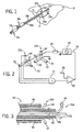
- Fig. 1 is a perspective view of two umbrella electrode assemblies providing first and second electrode wires deployed at opposite edges of a tumor to create a lesion encompassing the tumor by a passing current between the electrodes;
- Fig. 2 is a schematic representation of the electrodes of Fig. 1 as connected to a voltage controlled oscillator and showing temperature sensors on the electrode wires for feedback control of oscillator voltage;
- Fig. 3 is a fragmentary cross-sectional view of a tip of a combined electrode assembly providing for the first and second electrode wires of Fig. 1 extending from a unitary shaft arranging the wires of the first and second electrodes in concentric tubes and showing an insulation of the entire outer surface of the tubes and of the tips of the electrode wires;
- Fig. 4 is a simplified elevational cross-section of a tumor showing the first and second electrode positions and comparing the lesion volume obtained from two electrodes operating per the present invention, compared to the lesion volume obtained from two electrodes operating in a monopolar fashion;
- Fig. 5 is a figure similar to that of Fig. 2 showing electrical connection of the electrodes of Fig. 3 to effect a more complex control strategy employing temperature sensing from each of the first and second electrodes and showing the use of a third skin contact plate held in voltage between the two electrodes so as to provide independent current control for each of the two electrodes;
- Fig. 6 is a graph plotting resistivity in ohms-centimeters vs. frequency in Hz for tumorous and normal liver tissue, showing their separation in resistivity for frequencies below approximately 100 kHz;
- Fig. 7 is a figure similar to that of Fig. 5 showing yet another embodiment in which wires of each of the first and second electrodes are electrically isolated so that independent voltages or currents or phases of either can be applied to each wire to precisely tailor the current flow between that wire and the other electrodes; and
- Fig. 8 is a flow chart of a program as may be executed by the controller of Fig. 7 in utilizing its multi-electrode control.
- a liver 10 may include a tumor 12 about which a lesion 14 can be created by using two umbrella-type electrode assemblies 16a and 16b having a slight modification as will be disclosed below.
- Each electrode assembly 16a and 16b has a thin tubular metallic shaft 18a and 18b sized to be inserted percutaneously into the liver 10.
- the shafts 18a and 18b terminate, respectively, at shaft tips 20a and 20b from which project trifurcated electrodes 22a and 22b which are formed of wires 32.
- the wires 32 are extended by means of a plunger 24 remaining outside the body once the shafts 18a and 18b are properly located within the liver 10 and when extended, project by an extension radius separated by substantially equal angles around the shaft tips 20a and 20b.
- the exposed ends of the wires 32 are preformed into arcuate form so that when they are extended from the shafts 18a and 18b they naturally splay outward in a radial fashion.
- Umbrella electrode assemblies 16a and 16b of this type are well known in the art, but may be modified, by providing electrical insulation to all outer surfaces of the shafts 18a and 18b, in contrast to prior art umbrella electrode assemblies which leave the shaft tips 20a and 20b uninsulated, and by insulating the tips of the exposed portions of the wires 32. The purpose and effect of these modifications will be described further below.
- the first electrode 22a is positioned at one edge of the tumor 12 and the other electrode 22b positioned opposite the first electrode 22a across the tumor 12 center.
- edge refers generally to locations near the periphery of the tumor 12 and is not intended to be limited to positions either in or out of the tumor 12, whose boundaries in practice, may be irregular and not well known. Of significance is that a part of the tumor 12 is contained between the electrodes 22a and 22b.
- electrode 22a may be attached to a voltage controlled power oscillator 28 of a type well known in the art providing a settable frequency of alternating current power whose voltage amplitude (or current output) is controlled by an external signal.
- the return of the power oscillator 28 is connected to electrodes 22b also designated as a ground reference.
- power oscillator 28 When energized, power oscillator 28 induces a voltage between electrodes 22a and 22b causing current flow therebetween.
- each electrode 22a and 22b being referenced to a skin contact plate (not shown) would be expected to produce lesions 14a and 14b, respectively, per the prior art.
- Lesion 14c also has improved symmetry along the axis of separation of the electrodes 22a and 22b. Generally, it has been found preferable that the electrodes 22a and 22b are separated by 2.5 to 3 cm for typical umbrella electrodes or by less than four times their extension radius.
- temperature sensors 30, such as thermocouples, resistive or solid-state-type detectors, may be positioned at the distal ends of each of the exposed wires 32 of the tripartite electrodes 22a and 22b.
- the wires 32 may be small tubes holding small conductors and the temperature sensors 30 as described above.
- Commercially available umbrella-type electrode assemblies 16a and 16b currently include such sensors and wires connecting each sensor to a connector (not shown) in the plunger 24.
- the temperature sensors 30 in electrode 22a may be connected to a maximum determining circuit 34 that selects the maximum output signal from the three temperature sensors of electrode 22.
- the maximum determining circuit 34 may be discrete circuitry, such as may provide precision rectifiers joined to pass only the largest signal, or may be implemented in software by first converting the signals from the temperature sensors 30 to digital values and determining the maximum by means of an executed program on a microcontroller or the like.
- the maximum value of temperature from the temperature sensors 30 is passed by a comparator 36 (which also may be implemented in discrete circuitry or in software) which compares the maximum temperature to a predetermined desired temperature signal 38 such as may come from a potentiometer or the like.
- the desired temperature signal is typically set just below the point at which tissue boiling, vaporization or charring will occur.
- the output from the comparator 36 may be amplified and filtered according to well known control techniques to provide an amplitude input 39 to the power oscillator 28.
- the current between 22a and 22b will be limited to a point where the temperature at any one temperature sensors 30 approaches the predetermined desired temperature signal 38.
- current flowing between the electrodes 22a and 22b, measured as it flows from the power oscillator 28 through a current sensor 29, may be used as part of the feedback loop to limit current from the power oscillator 28 with or without the temperature control described above.
- the temperature sensors 30 of electrode 22b may also be provided to the maximum determining circuit 34 for more complete temperature monitoring.
- Other control methodologies may also be adopted including those provided for weighted averages of temperature readings or those anticipating temperature readings based on their trends according to techniques known to those of ordinary skill in the art.
- a unitary electrode 40 having a center tubular shaft 18c holding within its lumen, the wires 32 of first electrode 22a and a second concentric tubular shaft 42 positioned about shaft 18c and holding between its walls and shaft 18c wires 44 of the second electrode 22b.
- Wires 44 may be tempered and formed into a shape similar to that of wires 32 described above.
- Shafts 18c and 42 are typically metallic and thus are coated with insulating coatings 45 and 46, respectively, to ensure that any current flow is between the exposed wires 32 rather than the shafts 18c and 42.
- this insulating coating 46 is also applied to the tips of the shafts 18a and 18b of the electrode assemblies 16a and 16b of Fig. 1 to likewise ensure that current does not concentrate in a short circuit between the shafts 18a and 18b but in fact flows from the wires 32 of the wires of electrodes 22a and 22b.
- Kits of unitary electrode 40 each having different separations between first electrode 22a and second electrode 22a may be offered suitable for different tumor sizes and different tissue types.
- the wires 32 may include insulating coating 48 on their distal ends removed from shafts 18c and 42 so as to reduce high current densities associated with the ends of the wires 32.
- the wires of the first and second electrodes 22a and 22b are angularly staggered (unlike as shown in Fig. 2 ) so that an axial view of the electrode assembly reveals equally spaced non-overlapping wires 32.
- Such a configuration is also desired in the arrangement of Fig. 2 , although harder to maintain with two electrode assemblies 16a and 16b.
- the frequency of the power oscillator 28 may be preferentially set to a value much below the 450 kHz value used in the prior art. Referring to Fig. 6 , at less than 100 kHz and being most pronounced at frequencies below 10 kHz, the impedance of normal tissue increases to significantly greater than the impedance of tumor tissue. This difference in impedance is believed to be the result of differences in interstitial material between tumor and regular cell tissues although the present inventors do not wish to be bound by a particular theory. In any case, it is currently believed that the lower impedance of the tumorous tissue may be exploited to preferentially deposit energy in that tissue by setting the frequency of the power oscillator 28 at values near 10 kHz. Nevertheless, this frequency setting is not required in all embodiments of the present invention.
- the local environment of the electrodes 22a and 22b may differ by the presence of a blood vessel or the like in the vicinity of one electrode such as substantially reduces the heating of the lesion 14 in that area. Accordingly, it may be desired to increase the current density around one electrode 22a and 22b without changing the current density around the other electrode 22a and 22b.
- a skin contact plate 50 of a type used in the prior art yet employed in a different manner in the present invention.
- the term contact plate 50 may refer generally to any large area conductor intended but not necessarily limited to contact over a broad area at the patient's skin.
- the contact plate 50 may be referenced through a variable resistance (potentiometer) 52 to either of the output of power oscillator 28 or ground per switch 53 depending on the temperature of the electrodes 22a and 22b.
- switch 53 will connect the free end of variable resistance 52 to the output of the power oscillator 28 when the temperature sensors 30 indicate a higher temperature on electrode 22b than electrode 22a.
- switch 53 will connect the free end of variable resistance 52 to ground when the temperature sensors 30 indicate a lower temperature on electrode 22b than electrode 22a.
- the comparison of the temperatures of the electrodes 22a and 22b may be done via maximum determining circuits 34a and 34b, similar to that described above with respect to Fig. 2 .
- the switch 53 may be a comparator driven solid-state switch of a type well known in the art.
- the output of the maximum determining circuits 34a and 34b each connected respectively to the temperature sensors 30 of electrodes 22a and 22b may also be used to control the setting of the variable resistance 52.
- the maximum determining circuits 34a and 34b serve to reduce the resistance of variable resistance 52 as electrode 22b gets relatively hotter.
- the switch 53 connects the variable resistance 52 to ground
- the maximum determining circuits 34a and 34b serve to reduce the resistance of variable resistance 52 as electrode 22a gets relatively hotter.
- the action of the switch 53 and variable resistance 52 is thus generally to try to equalize the temperature of the electrodes 22a and 22b.
- electrode 22a If electrode 22a is close to a heat sink such as a blood vessel when electrode 22b is not, the temperature sensors 30 of electrode 22a will register a smaller value and thus the output of maximum determining circuit 34a will be lower than the output of maximum determining circuit 34b.
- variable resistance 52 may be implemented as a solid state devices according to techniques known in the art where the relative values of the outputs of maximum determining circuits 34a and 34b control the bias and hence resistance of a solid state device or a duty cycle modulation of a switching element or a current controlled voltage source providing the equalization described above.
- each wire 32 of electrodes 22a and 22b is electrically isolated within the electrode assemblies 16a and 16b and driven by separate feeds 53 through variable resistances 54 connected either to the power oscillator 28 or its return.
- Electrically isolated means in this context that there is not a conductive path between the electrodes 22a and 22b except through tissue prior to connection to the power supply or control electronics.
- a phase difference can also be employed between separate feeds 53 to further control the path of current flow between electrode wires 32.
- This phase difference could be created, e.g. by complex resistances that create a phase shift or by specialized waveform generators operating according to a computer program to produce an arbitrary switching pattern.
- the values of the resistances 54 are changed as will be described by a program operating on a controller 56.
- the variable resistances 54 may be implemented using solid-state devices such as MOSFET according to techniques known in the art.
- variable resistances 54 also controlled by a controller 56 may drive the contact plate 50.
- the controller 56 may receive the inputs from the temperature sensors 30 (described above) of each wire 32 as lines 58. This separate control of the voltages on the wires 32 allows additional control of current flows throughout the tumor 12 to be responsive to heat sinking blood vessels or the like near any one wire.
- one possible control algorithm scans the temperature sensors 30 as shown by process block 60. For each temperature sensor 30, if the temperature at that wire 32 is above a "ceiling value" below a tissue charring point, then the voltage at that wire is reduced. This "hammering down” process is repeated until all temperatures of all wires are below the ceiling value.
- the average temperature of the wires on each electrode 22a and 22b is determined and the voltage of the contact plate 50 is adjusted to incrementally equalize these average values.
- the voltage of the contact plate 50 is moved toward the voltage of the electrode 22 having the higher average.
- process block 64 the hammering down process of process block 60 is repeated to ensure that no wire has risen above its ceiling value.
- process block 66 one wire in sequence at each occurrence of process block 66 is examined and if its temperature is below a "floor value" below the ceiling value but sufficiently high to provide the desired power to the tumor, the voltage at that wire 32 is moved incrementally away from the voltage of the wires of the other electrode 22. Conversely, if the wire 32 is above the floor value, no action is taken.
- each wire 32 will have its temperature adjusted to be within the floor and ceiling range by separate voltage control.
- this process may be extended to an arbitrary number of electrodes 22 including a third electrode set 22c whose connections are not shown for clarity.
Abstract
Description
- The present invention relates to electrodes for radiofrequency ablation of tumors and the like, and in particular to a multipolar electrode system suitable for the ablation of liver tumors.
- Ablation of tumors, such as liver (hepatic) tumors, uses heat or cold to kill tumor cells. In cryosurgical ablation, a probe is inserted during an open laparotomy and the tumor is frozen. In radiofrequency ablation (RFA), an electrode is inserted into the tumor and current passing from the electrode into the patient (to an electrical return typically being a large area plate on the patient's skin) destroys the tumor cells through resistive heating.
- A simple RFA electrode is a conductive needle having an uninsulated tip placed within the tumor. The needle is energized with respect to a large area contact plate on the patient's skin by an oscillating electrical signal of approximately 460 kHz. Current flowing radially from the tip of the needle produces a spherical or ellipsoidal zone of heating (depending on the length of the exposed needle tip) and ultimately a lesion within a portion of the zone having sufficient temperature to kill the tumor cells. The size of the lesion is limited by fall-off in current density away from the electrode (causing reduced resistive heating), loss of heat to the surrounding tissue, and limits on the amount of energy transferred to the tissue from the electrode. The electrode energy is limited to avoid charring, boiling and vaporization of the tissue next to the electrode, a condition that greatly increases the resistance between the electrode and the remainder of the tumor. The tissue next to the electrode chars first because of the high current densities close to the electrode and thus creates a bottleneck in energy transfer.
- Several approaches have been developed to increase energy delivered to tissue without causing charring. A first method places temperature sensors in the tip of the electrode to allow more accurate monitoring of temperatures near the electrode and thereby to allow a closer approach to those energies just short of charring. A second method actively . cools the tip of the electrode with circulated coolant fluids within the electrode itself. A third method increases the area of the electrode using an umbrella-style electrode in which three or more electrode wires extend radially from the tip of the electrode shaft, after it has been positioned in the tumor. The greater surface area of the electrode reduces maximum current densities. The effect of all of these methods is to increase the amount of energy deposited into the tumor and thus to increase the lesion size allowing more reliable ablation of more extensive tumors.
- A major advantage of RFA in comparison to cryosurgical ablation is that it may be delivered percutaneously, without an incision, and thus with less trauma to the patient. In some cases, RFA is the only treatment the patient can withstand. Further, RFA can be completed while the patient is undergoing a CAT scan.
- Nevertheless, despite the improvements described above, RFA often fails to kill all of the tumor cells and, as a result, tumor recurrence rates of as high as 40% have been reported.
-
US-A-5 735 847 discloses an ablation apparatus including a multiple antenna device coupled to an electromagnetic energy source that produces an electromagnetic energy ablation output. In theFig. 8 arrangement, a primary antenna is provided with an insulation sleeve stopping short of the antenna's distal end. At the location of the distal end of the insulation sleeve three secondary antennas are provided extending at angularly offset radial points around the primary antenna. At the distal end of the primary antenna, offset axially along the primary antenna from the first set of secondary antennas, there is provided a pair of further secondary antennas, also positioned at angularly offset radial points around the primary antenna. - According to the present invention there is provided an electrode assembly for ablating tumors in a patient, the assembly comprising:
- (a) a support shaft sized for percutaneous placement, said shaft having an outer surface and a distal tip;
- (b) first and second wire electrode sets extensible radially from the shaft to an extension radius, the first wire electrode set being positionable at a first location adjacent to a tumour volume and offset axially along the support shaft from the second wire electrode set, which second wire electrode set is positionable at a second location offset from the first location about the tumour volume, the first and second electrode sets each comprising at least three wires positionable at angularly offset radial points around the support shaft;
- (c) a power supply connectable between the first and second electrode sets to induce a current flow between the first and second electrode sets whereby to concentrate current induced heating in the tumour volume;
- The present inventors have modeled the heating zone of standard RFA electrodes and believe that the high recurrence rate currently associated with RFA may result in part from limitations in the lesion size and irregularities in the lesion shape that can be obtained with these electrodes. Current lesion sizes may be insufficient to encompass the entire volume of a typical hepatic tumor particularly in the presence of nearby blood vessels that act as heat sinks, carrying away energy to reduce the lesion size in their vicinity.
- In order to overcome the energy limitations of current electrode designs, the present inventors have adopted a multipolar electrode design that increase lesion size by "focusing" existing energy on the tumor volume between two or more electrodes. By using axially displaced umbrella electrodes supported by an outwardly non-conductive shaft, a more regular lesion area is created than is provided by a single umbrella electrode and the lesion produced is greater in volume than would be obtained by a comparable number of monopolar umbrella electrodes operating individually.
- Specifically the electrode assembly of present invention enables the ablation of a tumor in a patient by inserting a first wire electrode set and a second wire electrode set percutaneously at a tumor volume, so that the second wire electrode set is at a second location opposed to and at a predetermined separation from the first wire electrode set at a first location about the tumor volume. First and second electrically isolated wire umbrella electrodes sets extend radially at the first and second locations respectively to an extension radius; and a power supply is connected between the first and second electrode sets to induce a current flow between them through the tumor volume whereby current induced heating is concentrated in the tumor volume.
- Use of the hereinafter described and illustrated embodiment provides a better shaped and substantially increased lesion volume while working within the energy limits imposed by local tissue boiling, vaporization and charring. The use of multiple radially displaced umbrella electrode sets communicating current between them delivers more energy to the tumor without necessarily increasing the total amount of energy delivered. The voltage may be an oscillating voltage waveform having substantial energy in the spectrum below 500 kHz and preferably below 100 kHz.
- The present inventors have further recognized that the impedance of tumor tissue differs significantly from that of regular tissue at frequencies below 100 kHz and especially below 10 kHz. This discovery can be exploited to preferentially ablate tumor tissue by proper selection of the frequency of the electrical energy.
- The temperature at the first or second electrode sets can be monitored and the voltage delivered to the electrodes controlled as a function of that temperature.
- It is thus possible to employ temperature feedback systems of the prior art with the present invention to increase, to the extent possible, the total energy delivered by the electrodes.
- The first and second electrode sets may comprise umbrella electrodes. Each electrode set has at least three electrode wires extending radially from a support shaft to a radius from the support shaft. The first and second locations may be separated by an amount less than six times (and preferably four times) the maximum radius to which the electrode wires are extended.
- The electrode sets may be separated by an amount that maximizes the useful size of the contiguous lesion volume.
- Use of the electrode assembly may involve placing an additional conductor in contact to provide a diffuse return path for current (for example), a conductive plate against the skin of the patient.
- Use of the electrode assembly may enable greater control over the current flow through the tumor, particularly in situations where inhomogeneities in the tissue would normally render one electrode much cooler than the other. Such inhomogeneities can include, for example, nearby blood vessels which carry heat away from nearby tissue. By using the conductive plate to augment current flow in one electrode, energy delivery at that electrode may be increased without changing the energy delivery at the other electrode.
- Use of the electrode assembly may also include the step of placing at least one third electrode percutaneously at a third location different from the first and second locations but adjacent to the tumor and offset from the center of the tumor volume and monitoring the temperature at the first, second and third electrodes.
- The electrode assembly may be part of a multi-electrode system that may define arbitrary volumes and accurately control temperature within those volumes for complete tumor ablation.
- In the preferred embodiment of electrode assembly a support shaft has a shaft tip and a shank portion having a predetermined separation from the shaft tip, and the support shaft is sized for percutaneous placement of the shaft tip adjacent to the first location and the shank portion adjacent to the second location, with the first and second wire electrode sets being extensible radially from the support shaft at the first and second locations. A power supply is connectable between the first and second electrodes sets to induce a current flow there between.
- A single shaft supporting the first and second wire sets in a predetermined separation corresponding to particular tissue characteristics and tumor sizes, simplifies use of the method. Multiple different shafts with different separations can be provided for different tumor sizes.
- The ends of the electrode wire sets removed from the support shaft may be insulated.
- It is an air with the electrode assembly of the invention to eliminate hot spots caused by high current densities at the tips of electrodes even in umbrella-type electrodes.
- An insulating cover extends between the shaft and the shaft tip so as to prevent short circuit paths between the electrode sets through tissue and to the shaft.
- The foregoing and other features of the invention will appear from the following description. In this description, reference is made to the accompanying drawings, which form a part hereof, and in which there is shown by way of illustration, a preferred embodiment of the invention. Such embodiment and its advantages do not define the scope of the invention, however, and reference must be made therefore to the claims for interpreting the scope of the invention. The
Fig.1 electrode assembly is not in accordance with the present invention due to the electrode sets being provided on separate shafts. -
Fig. 1 is a perspective view of two umbrella electrode assemblies providing first and second electrode wires deployed at opposite edges of a tumor to create a lesion encompassing the tumor by a passing current between the electrodes; -
Fig. 2 is a schematic representation of the electrodes ofFig. 1 as connected to a voltage controlled oscillator and showing temperature sensors on the electrode wires for feedback control of oscillator voltage; -
Fig. 3 is a fragmentary cross-sectional view of a tip of a combined electrode assembly providing for the first and second electrode wires ofFig. 1 extending from a unitary shaft arranging the wires of the first and second electrodes in concentric tubes and showing an insulation of the entire outer surface of the tubes and of the tips of the electrode wires; -
Fig. 4 is a simplified elevational cross-section of a tumor showing the first and second electrode positions and comparing the lesion volume obtained from two electrodes operating per the present invention, compared to the lesion volume obtained from two electrodes operating in a monopolar fashion; -
Fig. 5 is a figure similar to that ofFig. 2 showing electrical connection of the electrodes ofFig. 3 to effect a more complex control strategy employing temperature sensing from each of the first and second electrodes and showing the use of a third skin contact plate held in voltage between the two electrodes so as to provide independent current control for each of the two electrodes; -
Fig. 6 is a graph plotting resistivity in ohms-centimeters vs. frequency in Hz for tumorous and normal liver tissue, showing their separation in resistivity for frequencies below approximately 100 kHz; -
Fig. 7 is a figure similar to that ofFig. 5 showing yet another embodiment in which wires of each of the first and second electrodes are electrically isolated so that independent voltages or currents or phases of either can be applied to each wire to precisely tailor the current flow between that wire and the other electrodes; and -
Fig. 8 is a flow chart of a program as may be executed by the controller ofFig. 7 in utilizing its multi-electrode control. - Referring now to
Fig. 1 , aliver 10 may include atumor 12 about which alesion 14 can be created by using two umbrella-type electrode assemblies electrode assembly metallic shaft liver 10. Theshafts shaft tips electrodes wires 32. Thewires 32 are extended by means of aplunger 24 remaining outside the body once theshafts liver 10 and when extended, project by an extension radius separated by substantially equal angles around theshaft tips wires 32 are preformed into arcuate form so that when they are extended from theshafts -
Umbrella electrode assemblies shafts shaft tips wires 32. The purpose and effect of these modifications will be described further below. - The
first electrode 22a is positioned at one edge of thetumor 12 and theother electrode 22b positioned opposite thefirst electrode 22a across thetumor 12 center. The term "edge" as used herein refers generally to locations near the periphery of thetumor 12 and is not intended to be limited to positions either in or out of thetumor 12, whose boundaries in practice, may be irregular and not well known. Of significance is that a part of thetumor 12 is contained between theelectrodes - Referring now to
Figs. 1 and 2 ,electrode 22a may be attached to a voltage controlledpower oscillator 28 of a type well known in the art providing a settable frequency of alternating current power whose voltage amplitude (or current output) is controlled by an external signal. The return of thepower oscillator 28 is connected toelectrodes 22b also designated as a ground reference. When energized,power oscillator 28 induces a voltage betweenelectrodes - Referring now to
Fig. 4 , prior art operation of eachelectrode lesions 14a and 14b, respectively, per the prior art. By connecting the electrodes as shown inFig. 2 , however, with current flow therebetween, a substantiallylarger lesion 14c is created.Lesion 14c also has improved symmetry along the axis of separation of theelectrodes electrodes - Referring again to
Fig. 2 ,temperature sensors 30, such as thermocouples, resistive or solid-state-type detectors, may be positioned at the distal ends of each of the exposedwires 32 of thetripartite electrodes wires 32 may be small tubes holding small conductors and thetemperature sensors 30 as described above. Commercially available umbrella-type electrode assemblies plunger 24. - The
temperature sensors 30 inelectrode 22a may be connected to a maximum determiningcircuit 34 that selects the maximum output signal from the three temperature sensors of electrode 22. The maximum determiningcircuit 34 may be discrete circuitry, such as may provide precision rectifiers joined to pass only the largest signal, or may be implemented in software by first converting the signals from thetemperature sensors 30 to digital values and determining the maximum by means of an executed program on a microcontroller or the like. - The maximum value of temperature from the
temperature sensors 30 is passed by a comparator 36 (which also may be implemented in discrete circuitry or in software) which compares the maximum temperature to a predetermined desiredtemperature signal 38 such as may come from a potentiometer or the like. The desired temperature signal is typically set just below the point at which tissue boiling, vaporization or charring will occur. - The output from the
comparator 36 may be amplified and filtered according to well known control techniques to provide anamplitude input 39 to thepower oscillator 28. Thus it will be understood that the current between 22a and 22b will be limited to a point where the temperature at any onetemperature sensors 30 approaches the predetermined desiredtemperature signal 38. - While the
power oscillator 28 as described provides voltage amplitude control, it will be understood that current amplitude control may instead also be used. Accordingly, henceforth the terms voltage and current control as used herein should be considered interchangeable, being related by the impedance of the tissue between theelectrodes - Alternatively current flowing between the
electrodes power oscillator 28 through acurrent sensor 29, may be used as part of the feedback loop to limit current from thepower oscillator 28 with or without the temperature control described above. - In yet a further possible arrangement not shown, the
temperature sensors 30 ofelectrode 22b may also be provided to the maximum determiningcircuit 34 for more complete temperature monitoring. Other control methodologies may also be adopted including those provided for weighted averages of temperature readings or those anticipating temperature readings based on their trends according to techniques known to those of ordinary skill in the art. - Referring now to
Fig. 3 , the difficultly of positioning twoseparate electrode assemblies Fig. 1 may be reduced through the use of aunitary electrode 40 having a centertubular shaft 18c holding within its lumen, thewires 32 offirst electrode 22a and a second concentrictubular shaft 42 positioned aboutshaft 18c and holding between its walls andshaft 18c wiressecond electrode 22b.Wires 44 may be tempered and formed into a shape similar to that ofwires 32 described above.Shafts coatings wires 32 rather than theshafts - As mentioned above, this insulating
coating 46 is also applied to the tips of theshafts electrode assemblies Fig. 1 to likewise ensure that current does not concentrate in a short circuit between theshafts wires 32 of the wires ofelectrodes - Other similar shaft configurations for a
unitary electrode 40 may be obtained including those having side-by-side shafts - Kits of
unitary electrode 40 each having different separations betweenfirst electrode 22a andsecond electrode 22a may be offered suitable for different tumor sizes and different tissue types. - As mentioned briefly above, in either of the arrangements of
Figs. 1 and 3 thewires 32 may include insulatingcoating 48 on their distal ends removed fromshafts wires 32. - In a preferred embodiment, the wires of the first and
second electrodes Fig. 2 ) so that an axial view of the electrode assembly reveals equally spacednon-overlapping wires 32. Such a configuration is also desired in the arrangement ofFig. 2 , although harder to maintain with twoelectrode assemblies - The frequency of the
power oscillator 28 may be preferentially set to a value much below the 450 kHz value used in the prior art. Referring toFig. 6 , at less than 100 kHz and being most pronounced at frequencies below 10 kHz, the impedance of normal tissue increases to significantly greater than the impedance of tumor tissue. This difference in impedance is believed to be the result of differences in interstitial material between tumor and regular cell tissues although the present inventors do not wish to be bound by a particular theory. In any case, it is currently believed that the lower impedance of the tumorous tissue may be exploited to preferentially deposit energy in that tissue by setting the frequency of thepower oscillator 28 at values near 10 kHz. Nevertheless, this frequency setting is not required in all embodiments of the present invention. - Importantly, although such frequencies may excite nerve tissue, such as the heart, such excitation is limited by the present bi-polar design.
- Referring now to
Fig. 5 , the local environment of theelectrodes lesion 14 in that area. Accordingly, it may be desired to increase the current density around oneelectrode other electrode skin contact plate 50 of a type used in the prior art yet employed in a different manner in the present invention. As used herein, theterm contact plate 50 may refer generally to any large area conductor intended but not necessarily limited to contact over a broad area at the patient's skin. - In the embodiment of
Fig. 5 , thecontact plate 50 may be referenced through a variable resistance (potentiometer) 52 to either of the output ofpower oscillator 28 or ground perswitch 53 depending on the temperature of theelectrodes variable resistance 52 to the output of thepower oscillator 28 when thetemperature sensors 30 indicate a higher temperature onelectrode 22b thanelectrode 22a. Conversely, switch 53 will connect the free end ofvariable resistance 52 to ground when thetemperature sensors 30 indicate a lower temperature onelectrode 22b thanelectrode 22a. The comparison of the temperatures of theelectrodes maximum determining circuits Fig. 2 . Theswitch 53 may be a comparator driven solid-state switch of a type well known in the art. - The output of the
maximum determining circuits temperature sensors 30 ofelectrodes variable resistance 52. When theswitch 53 connects thevariable resistance 52 to the output of thepower oscillator 28, themaximum determining circuits variable resistance 52 aselectrode 22b gets relatively hotter. Conversely, when theswitch 53 connects thevariable resistance 52 to ground, themaximum determining circuits variable resistance 52 aselectrode 22a gets relatively hotter. The action of theswitch 53 andvariable resistance 52 is thus generally to try to equalize the temperature of theelectrodes - If
electrode 22a is close to a heat sink such as a blood vessel whenelectrode 22b is not, thetemperature sensors 30 ofelectrode 22a will register a smaller value and thus the output of maximum determiningcircuit 34a will be lower than the output of maximum determiningcircuit 34b. - The
variable resistance 52 may be implemented as a solid state devices according to techniques known in the art where the relative values of the outputs ofmaximum determining circuits - Referring now to
Fig. 7 , these principles may be applied to a system in which eachwire 32 ofelectrodes electrode assemblies separate feeds 53 throughvariable resistances 54 connected either to thepower oscillator 28 or its return. Electrically isolated means in this context that there is not a conductive path between theelectrodes separate feeds 53 to further control the path of current flow betweenelectrode wires 32. This phase difference could be created, e.g. by complex resistances that create a phase shift or by specialized waveform generators operating according to a computer program to produce an arbitrary switching pattern. The values of theresistances 54 are changed as will be described by a program operating on acontroller 56. For this purpose, thevariable resistances 54 may be implemented using solid-state devices such as MOSFET according to techniques known in the art. - Likewise, similar
variable resistances 54 also controlled by acontroller 56 may drive thecontact plate 50. - For the purpose of control, the
controller 56 may receive the inputs from the temperature sensors 30 (described above) of eachwire 32 aslines 58. This separate control of the voltages on thewires 32 allows additional control of current flows throughout thetumor 12 to be responsive to heat sinking blood vessels or the like near any one wire. - Referring to
Fig. 8 , one possible control algorithm scans thetemperature sensors 30 as shown byprocess block 60. For eachtemperature sensor 30, if the temperature at thatwire 32 is above a "ceiling value" below a tissue charring point, then the voltage at that wire is reduced. This "hammering down" process is repeated until all temperatures of all wires are below the ceiling value. - Next at
process block 62, the average temperature of the wires on eachelectrode contact plate 50 is adjusted to incrementally equalize these average values. The voltage of thecontact plate 50 is moved toward the voltage of the electrode 22 having the higher average. - Next at process block 64 the hammering down process of
process block 60 is repeated to ensure that no wire has risen above its ceiling value. - Next at
process block 66 one wire in sequence at each occurrence ofprocess block 66 is examined and if its temperature is below a "floor value" below the ceiling value but sufficiently high to provide the desired power to the tumor, the voltage at thatwire 32 is moved incrementally away from the voltage of the wires of the other electrode 22. Conversely, if thewire 32 is above the floor value, no action is taken. - Incrementally, each
wire 32 will have its temperature adjusted to be within the floor and ceiling range by separate voltage control. - As shown in
Fig. 7 , this process may be extended to an arbitrary number of electrodes 22 including a third electrode set 22c whose connections are not shown for clarity. - While this present invention has been described with respect to umbrella probes, it will be understood that most of its principles can be exploited using probes extensible radially from a shaft to an extension radius and energized in a bipolar configuration. Further it will be understood that the present invention is not limited to two electrode sets, but may be used with multiple electrode sets where current flow is between first and second sets of the electrodes. The number of wires of the umbrella electrodes is likewise not limited to three and commercially available probes suitable for use with the present invention include a 10 wire version. Further although the maximum temperatures of the electrodes were used for control in the above-described examples, it will be understood that the invention is equally amenable to control strategies that use average temperature or that also evaluate minimum temperatures.
- It is specifically intended that the present invention not be limited to the embodiments and illustrations contained herein, but modified forms of those embodiments including portions of the embodiments and combinations of elements of different embodiments as come within the scope of the following claims.
Claims (6)
- An electrode assembly (40) for ablating tumors in a patient, the assembly comprising:(a) a support shaft (18c) sized for percutaneous placement, said shaft having an outer surface and a distal tip;(b) first and second wire electrode sets (22a, 22b) extensible radially from the shaft (18c) to an extension radius, the first wire electrode set (22a) being positionable at a first location adjacent to a tumour volume and offset axially along the support shaft from the second wire electrode set (22b), which second wire electrode set is positionable at a second location offset from the first location about the tumour volume, the first and second electrode sets each comprising at least three wires (32) positionable at angularly offset radial point around the support shaft;(c) a power supply (28) connectable between the first and second electrode sets (22a, 22b) to induce a current flow between the first and second electrode sets whereby to concentrate current induced heating in the tumour volume;wherein the support shaft has an electrically insulating cover (46) on the outer surface between the first and second locations, said cover extending to the distal tip of the support shaft.
- The electrode assembly of claim 1, further comprising at least one temperature sensor (30) coupled to each of the first and second wire electrode sets (22a, 22b).
- The electrode assembly of claim 2, further comprising control means (34, 34a, 34b, 36) connected to said temperature sensors (30) to receive temperature level signals from each of the first and second electrode sets (22a, 22b) and to control the voltage level applied to the first and second electrode sets as a function of the temperature level.
- The electrode assembly as claim 3, wherein the electrode wires (32) in each of the first and second electrode sets (22a, 22b) arc electrically isolated, a said temperature sensor (30) is coupled to each of the wires in the electrode wire sets, and the control means monitors the temperature at each of the electrode wires and individually controls the voltage applied to the electrode wires.
- The electrode assembly of any one of the preceding claims, wherein the electrode wires (32) in the first electrode set (22a) are axially aligned with the electrodes wires in the second electrode set (22).
- The electrode assembly of any one of the preceding claims, wherein the electrode wires (32) of a respective electrode (22a, 22b) set are offset at substantially equal angles around the support shaft.
Applications Claiming Priority (3)
| Application Number | Priority Date | Filing Date | Title |
|---|---|---|---|
| US21010300P | 2000-06-07 | 2000-06-07 | |
| US210103P | 2000-06-07 | ||
| PCT/US2001/018088 WO2001093769A1 (en) | 2000-06-07 | 2001-06-05 | Multipolar electrode system for radiofrequency ablation |
Publications (2)
| Publication Number | Publication Date |
|---|---|
| EP1286625A1 EP1286625A1 (en) | 2003-03-05 |
| EP1286625B1 true EP1286625B1 (en) | 2010-12-22 |
Family
ID=22781587
Family Applications (1)
| Application Number | Title | Priority Date | Filing Date |
|---|---|---|---|
| EP01939887A Expired - Lifetime EP1286625B1 (en) | 2000-06-07 | 2001-06-05 | Multipolar electrode system for radiofrequency ablation |
Country Status (8)
| Country | Link |
|---|---|
| US (1) | US20020022864A1 (en) |
| EP (1) | EP1286625B1 (en) |
| JP (1) | JP5100947B2 (en) |
| AT (1) | ATE492231T1 (en) |
| AU (1) | AU2001265358A1 (en) |
| DE (1) | DE60143696D1 (en) |
| ES (1) | ES2356726T3 (en) |
| WO (1) | WO2001093769A1 (en) |
Cited By (1)
| Publication number | Priority date | Publication date | Assignee | Title |
|---|---|---|---|---|
| US10188457B2 (en) | 2003-09-12 | 2019-01-29 | Vessix Vascular, Inc. | Selectable eccentric remodeling and/or ablation |
Families Citing this family (143)
| Publication number | Priority date | Publication date | Assignee | Title |
|---|---|---|---|---|
| US7387628B1 (en) | 2000-09-15 | 2008-06-17 | Boston Scientific Scimed, Inc. | Methods and systems for focused bipolar tissue ablation |
| US7367974B2 (en) * | 2004-09-20 | 2008-05-06 | Wisconsin Alumni Research Foundation | Electrode array for tissue ablation |
| US7416549B2 (en) * | 2003-10-10 | 2008-08-26 | Boston Scientific Scimed, Inc. | Multi-zone bipolar ablation probe assembly |
| US7306595B2 (en) * | 2003-11-18 | 2007-12-11 | Boston Scientific Scimed, Inc. | System and method for tissue ablation |
| CA2560261A1 (en) * | 2004-03-18 | 2005-09-29 | St. Luke's Hospital | Method for the delivery of sustained release agents |
| WO2005110263A2 (en) * | 2004-05-11 | 2005-11-24 | Wisconsin Alumni Research Foundation | Radiofrequency ablation with independently controllable ground pad conductors |
| US20060074413A1 (en) * | 2004-06-28 | 2006-04-06 | Kamran Behzadian | Method and apparatus for substantial and uniform ablation about a linear bipolar array of electrodes |
| US20090125011A1 (en) * | 2004-06-28 | 2009-05-14 | Kamran Behzadian | Devices, Methods and Kits for Substantial and Uniform Ablation about a Linear Bipolar Array of Electrodes |
| US9974607B2 (en) | 2006-10-18 | 2018-05-22 | Vessix Vascular, Inc. | Inducing desirable temperature effects on body tissue |
| US9713730B2 (en) | 2004-09-10 | 2017-07-25 | Boston Scientific Scimed, Inc. | Apparatus and method for treatment of in-stent restenosis |
| US8396548B2 (en) | 2008-11-14 | 2013-03-12 | Vessix Vascular, Inc. | Selective drug delivery in a lumen |
| US20060089635A1 (en) * | 2004-10-22 | 2006-04-27 | Scimed Life Systems, Inc. | Methods and apparatus for focused bipolar tissue ablation using an insulated shaft |
| US8211104B2 (en) * | 2005-01-06 | 2012-07-03 | Boston Scientific Scimed, Inc. | Co-access bipolar ablation probe |
| US7601149B2 (en) | 2005-03-07 | 2009-10-13 | Boston Scientific Scimed, Inc. | Apparatus for switching nominal and attenuated power between ablation probes |
| EP1865870B8 (en) | 2005-03-28 | 2012-04-04 | Vessix Vascular, Inc. | Intraluminal electrical tissue characterization and tuned rf energy for selective treatment of atheroma and other target tissues |
| US7794458B2 (en) * | 2005-07-22 | 2010-09-14 | Boston Scientific Scimed, Inc. | Bipolar radio frequency ablation device with retractable insulator |
| US7799023B2 (en) * | 2005-09-26 | 2010-09-21 | Coherex Medical, Inc. | Compliant electrode for patent foramen ovale closure device |
| US7744596B2 (en) * | 2005-10-13 | 2010-06-29 | Boston Scientific Scimed, Inc. | Magnetically augmented radio frequency ablation |
| US20070100331A1 (en) * | 2005-10-27 | 2007-05-03 | Boston Scientific Scimed, Inc. | Systems and methods for organ tissue ablation |
| US7896874B2 (en) * | 2005-12-29 | 2011-03-01 | Boston Scientific Scimed, Inc. | RF ablation probes with tine valves |
| US8221405B2 (en) * | 2006-02-06 | 2012-07-17 | Coherex Medical, Inc. | Patent foramen ovale closure device and methods for determining RF dose for patent foramen ovale closure |
| EP2012695B1 (en) * | 2006-03-31 | 2015-07-22 | Cook Medical Technologies LLC | Electrosurgical cutting device |
| US8019435B2 (en) | 2006-05-02 | 2011-09-13 | Boston Scientific Scimed, Inc. | Control of arterial smooth muscle tone |
| US7938826B2 (en) * | 2006-05-30 | 2011-05-10 | Coherex Medical, Inc. | Methods, systems, and devices for closing a patent foramen ovale using mechanical structures |
| US8402974B2 (en) * | 2006-05-30 | 2013-03-26 | Coherex Medical, Inc. | Methods, systems, and devices for sensing, measuring, and controlling closure of a patent foramen ovale |
| EP2455036B1 (en) | 2006-10-18 | 2015-07-15 | Vessix Vascular, Inc. | Tuned RF energy and electrical tissue characterization for selective treatment of target tissues |
| CA2666663C (en) | 2006-10-18 | 2016-02-09 | Minnow Medical, Inc. | System for inducing desirable temperature effects on body tissue |
| US8496653B2 (en) | 2007-04-23 | 2013-07-30 | Boston Scientific Scimed, Inc. | Thrombus removal |
| US10448989B2 (en) | 2009-04-09 | 2019-10-22 | Virginia Tech Intellectual Properties, Inc. | High-frequency electroporation for cancer therapy |
| US8992517B2 (en) | 2008-04-29 | 2015-03-31 | Virginia Tech Intellectual Properties Inc. | Irreversible electroporation to treat aberrant cell masses |
| US9867652B2 (en) | 2008-04-29 | 2018-01-16 | Virginia Tech Intellectual Properties, Inc. | Irreversible electroporation using tissue vasculature to treat aberrant cell masses or create tissue scaffolds |
| US10245098B2 (en) | 2008-04-29 | 2019-04-02 | Virginia Tech Intellectual Properties, Inc. | Acute blood-brain barrier disruption using electrical energy based therapy |
| US11254926B2 (en) | 2008-04-29 | 2022-02-22 | Virginia Tech Intellectual Properties, Inc. | Devices and methods for high frequency electroporation |
| WO2009134876A1 (en) | 2008-04-29 | 2009-11-05 | Virginia Tech Intellectual Properties, Inc. | Irreversible electroporation to create tissue scaffolds |
| US10272178B2 (en) | 2008-04-29 | 2019-04-30 | Virginia Tech Intellectual Properties Inc. | Methods for blood-brain barrier disruption using electrical energy |
| US9198733B2 (en) | 2008-04-29 | 2015-12-01 | Virginia Tech Intellectual Properties, Inc. | Treatment planning for electroporation-based therapies |
| US9283051B2 (en) | 2008-04-29 | 2016-03-15 | Virginia Tech Intellectual Properties, Inc. | System and method for estimating a treatment volume for administering electrical-energy based therapies |
| US11272979B2 (en) | 2008-04-29 | 2022-03-15 | Virginia Tech Intellectual Properties, Inc. | System and method for estimating tissue heating of a target ablation zone for electrical-energy based therapies |
| US10238447B2 (en) | 2008-04-29 | 2019-03-26 | Virginia Tech Intellectual Properties, Inc. | System and method for ablating a tissue site by electroporation with real-time monitoring of treatment progress |
| US10702326B2 (en) | 2011-07-15 | 2020-07-07 | Virginia Tech Intellectual Properties, Inc. | Device and method for electroporation based treatment of stenosis of a tubular body part |
| US10117707B2 (en) | 2008-04-29 | 2018-11-06 | Virginia Tech Intellectual Properties, Inc. | System and method for estimating tissue heating of a target ablation zone for electrical-energy based therapies |
| WO2010056745A1 (en) | 2008-11-17 | 2010-05-20 | Minnow Medical, Inc. | Selective accumulation of energy with or without knowledge of tissue topography |
| US11638603B2 (en) | 2009-04-09 | 2023-05-02 | Virginia Tech Intellectual Properties, Inc. | Selective modulation of intracellular effects of cells using pulsed electric fields |
| US11382681B2 (en) | 2009-04-09 | 2022-07-12 | Virginia Tech Intellectual Properties, Inc. | Device and methods for delivery of high frequency electrical pulses for non-thermal ablation |
| WO2014143014A1 (en) | 2013-03-15 | 2014-09-18 | Triagenics, Llc | Therapeutic tooth bud ablation |
| US8551096B2 (en) | 2009-05-13 | 2013-10-08 | Boston Scientific Scimed, Inc. | Directional delivery of energy and bioactives |
| US8903488B2 (en) | 2009-05-28 | 2014-12-02 | Angiodynamics, Inc. | System and method for synchronizing energy delivery to the cardiac rhythm |
| US9895189B2 (en) | 2009-06-19 | 2018-02-20 | Angiodynamics, Inc. | Methods of sterilization and treating infection using irreversible electroporation |
| EP2467189A4 (en) * | 2009-08-20 | 2013-03-06 | Angiodynamics Inc | Multi-electrode energy delivery device and method of using the same |
| WO2011041322A1 (en) | 2009-09-30 | 2011-04-07 | Boston Scientific Scimed, Inc. | Medical probe with translatable co-access cannula |
| AU2010314930C1 (en) | 2009-11-05 | 2014-04-03 | Stratus Medical, LLC | Methods and systems for spinal radio frequency neurotomy |
| KR20130108067A (en) | 2010-04-09 | 2013-10-02 | 베식스 바스큘라 인코포레이티드 | Power generating and control apparatus for the treatment of tissue |
| US9192790B2 (en) | 2010-04-14 | 2015-11-24 | Boston Scientific Scimed, Inc. | Focused ultrasonic renal denervation |
| JP2013526940A (en) | 2010-05-21 | 2013-06-27 | ニンバス・コンセプツ・エルエルシー | Systems and methods for tissue ablation |
| US8473067B2 (en) | 2010-06-11 | 2013-06-25 | Boston Scientific Scimed, Inc. | Renal denervation and stimulation employing wireless vascular energy transfer arrangement |
| US9358365B2 (en) | 2010-07-30 | 2016-06-07 | Boston Scientific Scimed, Inc. | Precision electrode movement control for renal nerve ablation |
| US9084609B2 (en) | 2010-07-30 | 2015-07-21 | Boston Scientific Scime, Inc. | Spiral balloon catheter for renal nerve ablation |
| US9408661B2 (en) | 2010-07-30 | 2016-08-09 | Patrick A. Haverkost | RF electrodes on multiple flexible wires for renal nerve ablation |
| US9155589B2 (en) | 2010-07-30 | 2015-10-13 | Boston Scientific Scimed, Inc. | Sequential activation RF electrode set for renal nerve ablation |
| US9463062B2 (en) | 2010-07-30 | 2016-10-11 | Boston Scientific Scimed, Inc. | Cooled conductive balloon RF catheter for renal nerve ablation |
| WO2012051433A2 (en) | 2010-10-13 | 2012-04-19 | Angiodynamics, Inc. | System and method for electrically ablating tissue of a patient |
| TWI556849B (en) | 2010-10-21 | 2016-11-11 | 美敦力阿福盧森堡公司 | Catheter apparatus for renal neuromodulation |
| US8974451B2 (en) | 2010-10-25 | 2015-03-10 | Boston Scientific Scimed, Inc. | Renal nerve ablation using conductive fluid jet and RF energy |
| US9220558B2 (en) | 2010-10-27 | 2015-12-29 | Boston Scientific Scimed, Inc. | RF renal denervation catheter with multiple independent electrodes |
| US9028485B2 (en) | 2010-11-15 | 2015-05-12 | Boston Scientific Scimed, Inc. | Self-expanding cooling electrode for renal nerve ablation |
| US9089350B2 (en) | 2010-11-16 | 2015-07-28 | Boston Scientific Scimed, Inc. | Renal denervation catheter with RF electrode and integral contrast dye injection arrangement |
| US9668811B2 (en) | 2010-11-16 | 2017-06-06 | Boston Scientific Scimed, Inc. | Minimally invasive access for renal nerve ablation |
| US9326751B2 (en) | 2010-11-17 | 2016-05-03 | Boston Scientific Scimed, Inc. | Catheter guidance of external energy for renal denervation |
| US9060761B2 (en) | 2010-11-18 | 2015-06-23 | Boston Scientific Scime, Inc. | Catheter-focused magnetic field induced renal nerve ablation |
| US9192435B2 (en) | 2010-11-22 | 2015-11-24 | Boston Scientific Scimed, Inc. | Renal denervation catheter with cooled RF electrode |
| US9023034B2 (en) | 2010-11-22 | 2015-05-05 | Boston Scientific Scimed, Inc. | Renal ablation electrode with force-activatable conduction apparatus |
| US20120157993A1 (en) | 2010-12-15 | 2012-06-21 | Jenson Mark L | Bipolar Off-Wall Electrode Device for Renal Nerve Ablation |
| US9220561B2 (en) | 2011-01-19 | 2015-12-29 | Boston Scientific Scimed, Inc. | Guide-compatible large-electrode catheter for renal nerve ablation with reduced arterial injury |
| WO2013013156A2 (en) | 2011-07-20 | 2013-01-24 | Boston Scientific Scimed, Inc. | Percutaneous devices and methods to visualize, target and ablate nerves |
| JP6106669B2 (en) | 2011-07-22 | 2017-04-05 | ボストン サイエンティフィック サイムド,インコーポレイテッドBoston Scientific Scimed,Inc. | A neuromodulation system having a neuromodulation element that can be placed in a helical guide |
| US9078665B2 (en) | 2011-09-28 | 2015-07-14 | Angiodynamics, Inc. | Multiple treatment zone ablation probe |
| EP2765942B1 (en) | 2011-10-10 | 2016-02-24 | Boston Scientific Scimed, Inc. | Medical devices including ablation electrodes |
| EP2765940B1 (en) | 2011-10-11 | 2015-08-26 | Boston Scientific Scimed, Inc. | Off-wall electrode device for nerve modulation |
| US9420955B2 (en) | 2011-10-11 | 2016-08-23 | Boston Scientific Scimed, Inc. | Intravascular temperature monitoring system and method |
| US9364284B2 (en) | 2011-10-12 | 2016-06-14 | Boston Scientific Scimed, Inc. | Method of making an off-wall spacer cage |
| WO2013056266A2 (en) * | 2011-10-15 | 2013-04-18 | Diros Technology Inc. | Method and apparatus for precisely controlling the size and shape of radiofrequency ablations |
| EP2768563B1 (en) | 2011-10-18 | 2016-11-09 | Boston Scientific Scimed, Inc. | Deflectable medical devices |
| EP2768568B1 (en) | 2011-10-18 | 2020-05-06 | Boston Scientific Scimed, Inc. | Integrated crossing balloon catheter |
| WO2013070724A1 (en) | 2011-11-08 | 2013-05-16 | Boston Scientific Scimed, Inc. | Ostial renal nerve ablation |
| EP2779929A1 (en) | 2011-11-15 | 2014-09-24 | Boston Scientific Scimed, Inc. | Device and methods for renal nerve modulation monitoring |
| US9119632B2 (en) | 2011-11-21 | 2015-09-01 | Boston Scientific Scimed, Inc. | Deflectable renal nerve ablation catheter |
| US9265969B2 (en) | 2011-12-21 | 2016-02-23 | Cardiac Pacemakers, Inc. | Methods for modulating cell function |
| WO2013096920A1 (en) | 2011-12-23 | 2013-06-27 | Vessix Vascular, Inc. | Methods and apparatuses for remodeling tissue of or adjacent to a body passage |
| WO2013101452A1 (en) | 2011-12-28 | 2013-07-04 | Boston Scientific Scimed, Inc. | Device and methods for nerve modulation using a novel ablation catheter with polymeric ablative elements |
| US9050106B2 (en) | 2011-12-29 | 2015-06-09 | Boston Scientific Scimed, Inc. | Off-wall electrode device and methods for nerve modulation |
| US10660703B2 (en) | 2012-05-08 | 2020-05-26 | Boston Scientific Scimed, Inc. | Renal nerve modulation devices |
| US10321946B2 (en) | 2012-08-24 | 2019-06-18 | Boston Scientific Scimed, Inc. | Renal nerve modulation devices with weeping RF ablation balloons |
| CN104780859B (en) | 2012-09-17 | 2017-07-25 | 波士顿科学西美德公司 | Self-positioning electrode system and method for renal regulation |
| US10398464B2 (en) | 2012-09-21 | 2019-09-03 | Boston Scientific Scimed, Inc. | System for nerve modulation and innocuous thermal gradient nerve block |
| US10549127B2 (en) | 2012-09-21 | 2020-02-04 | Boston Scientific Scimed, Inc. | Self-cooling ultrasound ablation catheter |
| CN104869930B (en) | 2012-10-10 | 2020-12-25 | 波士顿科学国际有限公司 | Renal neuromodulation apparatus and methods |
| US9044575B2 (en) | 2012-10-22 | 2015-06-02 | Medtronic Adrian Luxembourg S.a.r.l. | Catheters with enhanced flexibility and associated devices, systems, and methods |
| WO2014163987A1 (en) | 2013-03-11 | 2014-10-09 | Boston Scientific Scimed, Inc. | Medical devices for modulating nerves |
| US9693821B2 (en) | 2013-03-11 | 2017-07-04 | Boston Scientific Scimed, Inc. | Medical devices for modulating nerves |
| US9808311B2 (en) | 2013-03-13 | 2017-11-07 | Boston Scientific Scimed, Inc. | Deflectable medical devices |
| US10265122B2 (en) | 2013-03-15 | 2019-04-23 | Boston Scientific Scimed, Inc. | Nerve ablation devices and related methods of use |
| WO2014149690A2 (en) | 2013-03-15 | 2014-09-25 | Boston Scientific Scimed, Inc. | Medical devices and methods for treatment of hypertension that utilize impedance compensation |
| EP2967734B1 (en) | 2013-03-15 | 2019-05-15 | Boston Scientific Scimed, Inc. | Methods and apparatuses for remodeling tissue of or adjacent to a body passage |
| US9655669B2 (en) * | 2013-05-06 | 2017-05-23 | Novocure Limited | Optimizing treatment using TTFields by changing the frequency during the course of long term tumor treatment |
| WO2014189794A1 (en) | 2013-05-18 | 2014-11-27 | Medtronic Ardian Luxembourg S.A.R.L. | Neuromodulation catheters with shafts for enhanced flexibility and control and associated devices, systems, and methods |
| JP2016523147A (en) | 2013-06-21 | 2016-08-08 | ボストン サイエンティフィック サイムド,インコーポレイテッドBoston Scientific Scimed,Inc. | Renal denervation balloon catheter with a riding-type electrode support |
| CN105473092B (en) | 2013-06-21 | 2019-05-17 | 波士顿科学国际有限公司 | The medical instrument for renal nerve ablation with rotatable shaft |
| US9707036B2 (en) | 2013-06-25 | 2017-07-18 | Boston Scientific Scimed, Inc. | Devices and methods for nerve modulation using localized indifferent electrodes |
| AU2014284558B2 (en) | 2013-07-01 | 2017-08-17 | Boston Scientific Scimed, Inc. | Medical devices for renal nerve ablation |
| US10660698B2 (en) | 2013-07-11 | 2020-05-26 | Boston Scientific Scimed, Inc. | Devices and methods for nerve modulation |
| WO2015006573A1 (en) | 2013-07-11 | 2015-01-15 | Boston Scientific Scimed, Inc. | Medical device with stretchable electrode assemblies |
| CN105682594B (en) | 2013-07-19 | 2018-06-22 | 波士顿科学国际有限公司 | Helical bipolar electrodes renal denervation dominates air bag |
| JP6122217B2 (en) | 2013-07-22 | 2017-04-26 | ボストン サイエンティフィック サイムド,インコーポレイテッドBoston Scientific Scimed,Inc. | Renal nerve ablation medical device |
| EP3024406B1 (en) | 2013-07-22 | 2019-06-19 | Boston Scientific Scimed, Inc. | Medical devices for renal nerve ablation |
| US10722300B2 (en) | 2013-08-22 | 2020-07-28 | Boston Scientific Scimed, Inc. | Flexible circuit having improved adhesion to a renal nerve modulation balloon |
| EP3041425B1 (en) | 2013-09-04 | 2022-04-13 | Boston Scientific Scimed, Inc. | Radio frequency (rf) balloon catheter having flushing and cooling capability |
| WO2015038947A1 (en) | 2013-09-13 | 2015-03-19 | Boston Scientific Scimed, Inc. | Ablation balloon with vapor deposited cover layer |
| CN105592778B (en) | 2013-10-14 | 2019-07-23 | 波士顿科学医学有限公司 | High-resolution cardiac mapping electrod-array conduit |
| US11246654B2 (en) | 2013-10-14 | 2022-02-15 | Boston Scientific Scimed, Inc. | Flexible renal nerve ablation devices and related methods of use and manufacture |
| CN105636537B (en) | 2013-10-15 | 2018-08-17 | 波士顿科学国际有限公司 | Medical instrument sacculus |
| US9770606B2 (en) | 2013-10-15 | 2017-09-26 | Boston Scientific Scimed, Inc. | Ultrasound ablation catheter with cooling infusion and centering basket |
| WO2015057961A1 (en) | 2013-10-18 | 2015-04-23 | Boston Scientific Scimed, Inc. | Balloon catheters with flexible conducting wires and related methods of use and manufacture |
| WO2015061457A1 (en) | 2013-10-25 | 2015-04-30 | Boston Scientific Scimed, Inc. | Embedded thermocouple in denervation flex circuit |
| JP6382989B2 (en) | 2014-01-06 | 2018-08-29 | ボストン サイエンティフィック サイムド,インコーポレイテッドBoston Scientific Scimed,Inc. | Medical device with tear resistant flexible circuit assembly |
| EP4059563B1 (en) | 2014-01-27 | 2023-09-27 | Medtronic Ireland Manufacturing Unlimited Company | Neuromodulation catheters having jacketed neuromodulation elements and related devices |
| CN106572881B (en) | 2014-02-04 | 2019-07-26 | 波士顿科学国际有限公司 | Substitution of the heat sensor on bipolar electrode is placed |
| US11000679B2 (en) | 2014-02-04 | 2021-05-11 | Boston Scientific Scimed, Inc. | Balloon protection and rewrapping devices and related methods of use |
| CN106232043B (en) | 2014-04-24 | 2019-07-23 | 美敦力阿迪安卢森堡有限公司 | Nerve modulation conduit and relevant system and method with braiding axle |
| AU2015259303B2 (en) | 2014-05-12 | 2021-10-28 | Arena, Christopher B. | Selective modulation of intracellular effects of cells using pulsed electric fields |
| AU2015287652B2 (en) * | 2014-07-11 | 2017-10-12 | Boston Scientific Scimed, Inc. | Ablation medical devices |
| US10799283B2 (en) | 2014-07-28 | 2020-10-13 | Boston Scientific Scimed, Inc. | Multiple lead electrode probe for controlled tissue ablation |
| WO2016100325A1 (en) | 2014-12-15 | 2016-06-23 | Virginia Tech Intellectual Properties, Inc. | Devices, systems, and methods for real-time monitoring of electrophysical effects during tissue treatment |
| US11007007B2 (en) | 2015-10-13 | 2021-05-18 | Biosense Webster (Israel) Ltd. | Self-centering multiray ablation catheter |
| CN105455897A (en) * | 2015-12-30 | 2016-04-06 | 迈德医疗科技(上海)有限公司 | Flexible radiofrequency ablation electrode |
| US10905492B2 (en) | 2016-11-17 | 2021-02-02 | Angiodynamics, Inc. | Techniques for irreversible electroporation using a single-pole tine-style internal device communicating with an external surface electrode |
| US11607537B2 (en) | 2017-12-05 | 2023-03-21 | Virginia Tech Intellectual Properties, Inc. | Method for treating neurological disorders, including tumors, with electroporation |
| US11925405B2 (en) | 2018-03-13 | 2024-03-12 | Virginia Tech Intellectual Properties, Inc. | Treatment planning system for immunotherapy enhancement via non-thermal ablation |
| US11311329B2 (en) | 2018-03-13 | 2022-04-26 | Virginia Tech Intellectual Properties, Inc. | Treatment planning for immunotherapy based treatments using non-thermal ablation techniques |
| EP3979938A4 (en) | 2019-06-06 | 2023-06-28 | TriAgenics, Inc. | Ablation probe systems |
| CN112022331B (en) * | 2020-08-31 | 2021-06-18 | 天津市鹰泰利安康医疗科技有限责任公司 | Irreversible electroporation ablation system |
| WO2023076940A1 (en) * | 2021-10-26 | 2023-05-04 | DIXI Neurolab, Inc. | Configurable multi-polar rf ablation probe |
| CN114601551B (en) * | 2022-03-18 | 2022-11-18 | 天津市鹰泰利安康医疗科技有限责任公司 | High-frequency irreversible electroporation treatment system |
| CN115349946B (en) * | 2022-10-19 | 2022-12-23 | 成都美创医疗科技股份有限公司 | Electrode assembly for treating basis cranii tumor, surgical internal cutter and surgical system |
Citations (1)
| Publication number | Priority date | Publication date | Assignee | Title |
|---|---|---|---|---|
| US5855576A (en) * | 1995-03-24 | 1999-01-05 | Board Of Regents Of University Of Nebraska | Method for volumetric tissue ablation |
Family Cites Families (10)
| Publication number | Priority date | Publication date | Assignee | Title |
|---|---|---|---|---|
| US5707349A (en) * | 1994-05-09 | 1998-01-13 | Somnus Medical Technologies, Inc. | Method for treatment of air way obstructions |
| US5588960A (en) * | 1994-12-01 | 1996-12-31 | Vidamed, Inc. | Transurethral needle delivery device with cystoscope and method for treatment of urinary incontinence |
| US5735847A (en) | 1995-08-15 | 1998-04-07 | Zomed International, Inc. | Multiple antenna ablation apparatus and method with cooling element |
| US6330478B1 (en) * | 1995-08-15 | 2001-12-11 | Rita Medical Systems, Inc. | Cell necrosis apparatus |
| DE19713797A1 (en) * | 1996-04-04 | 1997-10-09 | Valleylab Inc | Electrosurgical instrument for use in e.g. myoma necrosis |
| US6077257A (en) * | 1996-05-06 | 2000-06-20 | Vidacare, Inc. | Ablation of rectal and other internal body structures |
| US6050992A (en) * | 1997-05-19 | 2000-04-18 | Radiotherapeutics Corporation | Apparatus and method for treating tissue with multiple electrodes |
| AU753618B2 (en) * | 1998-03-06 | 2002-10-24 | Curon Medical, Inc. | Apparatus to electrosurgically treat esophageal sphincters |
| US6212433B1 (en) * | 1998-07-28 | 2001-04-03 | Radiotherapeutics Corporation | Method for treating tumors near the surface of an organ |
| ATE280617T1 (en) * | 1998-08-14 | 2004-11-15 | Leuven K U Res & Dev | LIQUID-COOLED WET ELECTRODE |
-
2001
- 2001-06-04 US US09/873,541 patent/US20020022864A1/en not_active Abandoned
- 2001-06-05 DE DE60143696T patent/DE60143696D1/en not_active Expired - Lifetime
- 2001-06-05 AU AU2001265358A patent/AU2001265358A1/en not_active Abandoned
- 2001-06-05 AT AT01939887T patent/ATE492231T1/en not_active IP Right Cessation
- 2001-06-05 EP EP01939887A patent/EP1286625B1/en not_active Expired - Lifetime
- 2001-06-05 JP JP2002501343A patent/JP5100947B2/en not_active Expired - Lifetime
- 2001-06-05 ES ES01939887T patent/ES2356726T3/en not_active Expired - Lifetime
- 2001-06-05 WO PCT/US2001/018088 patent/WO2001093769A1/en active Application Filing
Patent Citations (1)
| Publication number | Priority date | Publication date | Assignee | Title |
|---|---|---|---|---|
| US5855576A (en) * | 1995-03-24 | 1999-01-05 | Board Of Regents Of University Of Nebraska | Method for volumetric tissue ablation |
Cited By (1)
| Publication number | Priority date | Publication date | Assignee | Title |
|---|---|---|---|---|
| US10188457B2 (en) | 2003-09-12 | 2019-01-29 | Vessix Vascular, Inc. | Selectable eccentric remodeling and/or ablation |
Also Published As
| Publication number | Publication date |
|---|---|
| ES2356726T3 (en) | 2011-04-12 |
| JP5100947B2 (en) | 2012-12-19 |
| WO2001093769A1 (en) | 2001-12-13 |
| US20020022864A1 (en) | 2002-02-21 |
| AU2001265358A1 (en) | 2001-12-17 |
| EP1286625A1 (en) | 2003-03-05 |
| DE60143696D1 (en) | 2011-02-03 |
| JP2003534869A (en) | 2003-11-25 |
| ATE492231T1 (en) | 2011-01-15 |
Similar Documents
| Publication | Publication Date | Title |
|---|---|---|
| EP1286625B1 (en) | Multipolar electrode system for radiofrequency ablation | |
| US8486065B2 (en) | Radio-frequency ablation system and method using multiple electrodes | |
| US7520877B2 (en) | Radiofrequency ablation system using multiple prong probes | |
| US8216219B2 (en) | Device for electrosurgically destroying body tissue | |
| US10130415B2 (en) | Circumferential electrode array for tissue ablation | |
| EP2765939B1 (en) | Apparatus for precisely controlling the size and shape of radiofrequency ablations | |
| US6200314B1 (en) | RF ablation apparatus and method using unipolar and bipolar techniques | |
| AU2002341571A1 (en) | Radio-frequency ablation system using multiple electrodes | |
| ES2354110T3 (en) | RADIO FREQUENCY ABLATION SYSTEM THAT USES MULTIPLE ELECTRODES. |
Legal Events
| Date | Code | Title | Description |
|---|---|---|---|
| PUAI | Public reference made under article 153(3) epc to a published international application that has entered the european phase |
Free format text: ORIGINAL CODE: 0009012 |
|
| 17P | Request for examination filed |
Effective date: 20021209 |
|
| AK | Designated contracting states |
Kind code of ref document: A1 Designated state(s): AT BE CH CY DE DK ES FI FR GB GR IE IT LI LU MC NL PT SE TR |
|
| AX | Request for extension of the european patent |
Extension state: AL LT LV MK RO SI |
|
| RIN1 | Information on inventor provided before grant (corrected) |
Inventor name: MAHVI, DAVID, M. Inventor name: LEE, FRED, T., JR. Inventor name: WEBSTER, JOHN G. Inventor name: STAELIN, STEPHEN, T. Inventor name: TUNGJITKUSOLMUN, SUPAN Inventor name: HAMMERICH, DIETER |
|
| 17Q | First examination report despatched |
Effective date: 20040402 |
|
| RIN1 | Information on inventor provided before grant (corrected) |
Inventor name: TUNGJITKUSOLMUN, SUPAN Inventor name: LEE, FRED, T., JR. Inventor name: WEBSTER, JOHN G. Inventor name: HAMMERICH, DIETER Inventor name: MAHVI, DAVID, M. Inventor name: STAELIN, STEPHEN, T. |
|
| APBN | Date of receipt of notice of appeal recorded |
Free format text: ORIGINAL CODE: EPIDOSNNOA2E |
|
| APBR | Date of receipt of statement of grounds of appeal recorded |
Free format text: ORIGINAL CODE: EPIDOSNNOA3E |
|
| APAF | Appeal reference modified |
Free format text: ORIGINAL CODE: EPIDOSCREFNE |
|
| APBT | Appeal procedure closed |
Free format text: ORIGINAL CODE: EPIDOSNNOA9E |
|
| GRAP | Despatch of communication of intention to grant a patent |
Free format text: ORIGINAL CODE: EPIDOSNIGR1 |
|
| GRAS | Grant fee paid |
Free format text: ORIGINAL CODE: EPIDOSNIGR3 |
|
| GRAA | (expected) grant |
Free format text: ORIGINAL CODE: 0009210 |
|
| AK | Designated contracting states |
Kind code of ref document: B1 Designated state(s): AT BE CH CY DE DK ES FI FR GB GR IE IT LI LU MC NL PT SE TR |
|
| REG | Reference to a national code |
Ref country code: GB Ref legal event code: FG4D |
|
| REG | Reference to a national code |
Ref country code: CH Ref legal event code: EP |
|
| REG | Reference to a national code |
Ref country code: IE Ref legal event code: FG4D |
|
| REF | Corresponds to: |
Ref document number: 60143696 Country of ref document: DE Date of ref document: 20110203 Kind code of ref document: P |
|
| REG | Reference to a national code |
Ref country code: DE Ref legal event code: R096 Ref document number: 60143696 Country of ref document: DE Effective date: 20110203 |
|
| REG | Reference to a national code |
Ref country code: ES Ref legal event code: FG2A Ref document number: 2356726 Country of ref document: ES Kind code of ref document: T3 Effective date: 20110412 |
|
| REG | Reference to a national code |
Ref country code: NL Ref legal event code: VDEP Effective date: 20101222 |
|
| PG25 | Lapsed in a contracting state [announced via postgrant information from national office to epo] |
Ref country code: AT Free format text: LAPSE BECAUSE OF FAILURE TO SUBMIT A TRANSLATION OF THE DESCRIPTION OR TO PAY THE FEE WITHIN THE PRESCRIBED TIME-LIMIT Effective date: 20101222 Ref country code: CY Free format text: LAPSE BECAUSE OF FAILURE TO SUBMIT A TRANSLATION OF THE DESCRIPTION OR TO PAY THE FEE WITHIN THE PRESCRIBED TIME-LIMIT Effective date: 20101222 Ref country code: FI Free format text: LAPSE BECAUSE OF FAILURE TO SUBMIT A TRANSLATION OF THE DESCRIPTION OR TO PAY THE FEE WITHIN THE PRESCRIBED TIME-LIMIT Effective date: 20101222 Ref country code: SE Free format text: LAPSE BECAUSE OF FAILURE TO SUBMIT A TRANSLATION OF THE DESCRIPTION OR TO PAY THE FEE WITHIN THE PRESCRIBED TIME-LIMIT Effective date: 20101222 |
|
| PG25 | Lapsed in a contracting state [announced via postgrant information from national office to epo] |
Ref country code: GR Free format text: LAPSE BECAUSE OF FAILURE TO SUBMIT A TRANSLATION OF THE DESCRIPTION OR TO PAY THE FEE WITHIN THE PRESCRIBED TIME-LIMIT Effective date: 20110323 Ref country code: BE Free format text: LAPSE BECAUSE OF FAILURE TO SUBMIT A TRANSLATION OF THE DESCRIPTION OR TO PAY THE FEE WITHIN THE PRESCRIBED TIME-LIMIT Effective date: 20101222 Ref country code: PT Free format text: LAPSE BECAUSE OF FAILURE TO SUBMIT A TRANSLATION OF THE DESCRIPTION OR TO PAY THE FEE WITHIN THE PRESCRIBED TIME-LIMIT Effective date: 20110422 |
|
| PG25 | Lapsed in a contracting state [announced via postgrant information from national office to epo] |
Ref country code: NL Free format text: LAPSE BECAUSE OF FAILURE TO SUBMIT A TRANSLATION OF THE DESCRIPTION OR TO PAY THE FEE WITHIN THE PRESCRIBED TIME-LIMIT Effective date: 20101222 |
|
| PLBE | No opposition filed within time limit |
Free format text: ORIGINAL CODE: 0009261 |
|
| STAA | Information on the status of an ep patent application or granted ep patent |
Free format text: STATUS: NO OPPOSITION FILED WITHIN TIME LIMIT |
|
| PG25 | Lapsed in a contracting state [announced via postgrant information from national office to epo] |
Ref country code: DK Free format text: LAPSE BECAUSE OF FAILURE TO SUBMIT A TRANSLATION OF THE DESCRIPTION OR TO PAY THE FEE WITHIN THE PRESCRIBED TIME-LIMIT Effective date: 20101222 |
|
| 26N | No opposition filed |
Effective date: 20110923 |
|
| REG | Reference to a national code |
Ref country code: DE Ref legal event code: R097 Ref document number: 60143696 Country of ref document: DE Effective date: 20110923 |
|
| REG | Reference to a national code |
Ref country code: CH Ref legal event code: PL |
|
| PG25 | Lapsed in a contracting state [announced via postgrant information from national office to epo] |
Ref country code: LI Free format text: LAPSE BECAUSE OF NON-PAYMENT OF DUE FEES Effective date: 20110630 Ref country code: CH Free format text: LAPSE BECAUSE OF NON-PAYMENT OF DUE FEES Effective date: 20110630 |
|
| PG25 | Lapsed in a contracting state [announced via postgrant information from national office to epo] |
Ref country code: MC Free format text: LAPSE BECAUSE OF NON-PAYMENT OF DUE FEES Effective date: 20110630 |
|
| PG25 | Lapsed in a contracting state [announced via postgrant information from national office to epo] |
Ref country code: LU Free format text: LAPSE BECAUSE OF NON-PAYMENT OF DUE FEES Effective date: 20110605 |
|
| PG25 | Lapsed in a contracting state [announced via postgrant information from national office to epo] |
Ref country code: TR Free format text: LAPSE BECAUSE OF FAILURE TO SUBMIT A TRANSLATION OF THE DESCRIPTION OR TO PAY THE FEE WITHIN THE PRESCRIBED TIME-LIMIT Effective date: 20101222 |
|
| REG | Reference to a national code |
Ref country code: FR Ref legal event code: PLFP Year of fee payment: 16 |
|
| REG | Reference to a national code |
Ref country code: FR Ref legal event code: PLFP Year of fee payment: 17 |
|
| REG | Reference to a national code |
Ref country code: FR Ref legal event code: PLFP Year of fee payment: 18 |
|
| PGFP | Annual fee paid to national office [announced via postgrant information from national office to epo] |
Ref country code: FR Payment date: 20200512 Year of fee payment: 20 Ref country code: DE Payment date: 20200527 Year of fee payment: 20 Ref country code: IE Payment date: 20200609 Year of fee payment: 20 |
|
| PGFP | Annual fee paid to national office [announced via postgrant information from national office to epo] |
Ref country code: GB Payment date: 20200527 Year of fee payment: 20 Ref country code: IT Payment date: 20200512 Year of fee payment: 20 |
|
| PGFP | Annual fee paid to national office [announced via postgrant information from national office to epo] |
Ref country code: ES Payment date: 20200701 Year of fee payment: 20 |
|
| REG | Reference to a national code |
Ref country code: DE Ref legal event code: R071 Ref document number: 60143696 Country of ref document: DE |
|
| REG | Reference to a national code |
Ref country code: GB Ref legal event code: PE20 Expiry date: 20210604 |
|
| REG | Reference to a national code |
Ref country code: IE Ref legal event code: MK9A |
|
| PG25 | Lapsed in a contracting state [announced via postgrant information from national office to epo] |
Ref country code: GB Free format text: LAPSE BECAUSE OF EXPIRATION OF PROTECTION Effective date: 20210604 |
|
| PG25 | Lapsed in a contracting state [announced via postgrant information from national office to epo] |
Ref country code: IE Free format text: LAPSE BECAUSE OF EXPIRATION OF PROTECTION Effective date: 20210605 |
|
| PG25 | Lapsed in a contracting state [announced via postgrant information from national office to epo] |
Ref country code: ES Free format text: LAPSE BECAUSE OF EXPIRATION OF PROTECTION Effective date: 20210606 |