EP1285065B1 - Konstruierte fluoreszenzproteine mit langen wellenlängen - Google Patents
Konstruierte fluoreszenzproteine mit langen wellenlängen Download PDFInfo
- Publication number
- EP1285065B1 EP1285065B1 EP01937550A EP01937550A EP1285065B1 EP 1285065 B1 EP1285065 B1 EP 1285065B1 EP 01937550 A EP01937550 A EP 01937550A EP 01937550 A EP01937550 A EP 01937550A EP 1285065 B1 EP1285065 B1 EP 1285065B1
- Authority
- EP
- European Patent Office
- Prior art keywords
- fluorescent protein
- substitution
- protein
- engineered
- green fluorescent
- Prior art date
- Legal status (The legal status is an assumption and is not a legal conclusion. Google has not performed a legal analysis and makes no representation as to the accuracy of the status listed.)
- Expired - Lifetime
Links
- VRKMJCUABPDMDK-UHFFFAOYSA-O CCC[N-]C(N)[NH3+] Chemical compound CCC[N-]C(N)[NH3+] VRKMJCUABPDMDK-UHFFFAOYSA-O 0.000 description 1
Images


Classifications
-
- C—CHEMISTRY; METALLURGY
- C07—ORGANIC CHEMISTRY
- C07K—PEPTIDES
- C07K14/00—Peptides having more than 20 amino acids; Gastrins; Somatostatins; Melanotropins; Derivatives thereof
- C07K14/435—Peptides having more than 20 amino acids; Gastrins; Somatostatins; Melanotropins; Derivatives thereof from animals; from humans
- C07K14/43504—Peptides having more than 20 amino acids; Gastrins; Somatostatins; Melanotropins; Derivatives thereof from animals; from humans from invertebrates
- C07K14/43595—Peptides having more than 20 amino acids; Gastrins; Somatostatins; Melanotropins; Derivatives thereof from animals; from humans from invertebrates from coelenteratae, e.g. medusae
-
- C—CHEMISTRY; METALLURGY
- C07—ORGANIC CHEMISTRY
- C07K—PEPTIDES
- C07K2319/00—Fusion polypeptide
Definitions
- Fluorescent molecules are attractive as reporter molecules in many assay systems because of their high sensitivity and ease of quantification. Recently, fluorescent proteins have been the focus of much attention because they can be produced in vivo by biological systems, and can be used to trace intracellular events without the need to be introduced into the cell through microinjection or permeablization.
- the green fluorescent protein of Aequorea victoria is particularly interesting as a fluorescent protein.
- a cDNA for the protein has been cloned. (D.C. Prasher et al., "Primary structure of the Aequorea victoria green-fluorescent protein," Gene (1992) 111:229-33.) Not only can the primary amino acid sequence of the protein be expressed from the cDNA, but the expressed protein can fluoresce.
- Aequorea green fluorescent protein (“GFP”) is a stable, proteolysis-resistant single chain of 238 residues and has two absorption maxima at around 395 and 475 nm. The relative amplitudes of these two peaks is sensitive to environmental factors (W. W. Ward. Bioluminescence and Chemiluminescence (M. A. DeLuca and W. D. McElroy, eds) Academic Press pp. 235-242 (1981); W. W. Ward & S. H. Bokman Biochemistry 21:4535-4540 (1982); W. W. Ward et al. Photochem. Photobiol.
- the fluorophore results from the autocatalytic cyclization of the polypeptide backbone between residues Ser 65 and Gly 67 and oxidation of the - ⁇ bond of Tyr 66 (A. B. Cubitt et al. Trends Biochem. Sci. 20:448-455 (1995); C. W. Cody et al. Biochemistry 32:1212-1218 (1993); R. Heim et al. Proc. Natl. Acad. Sci. USA 91:12501-12504 (1994)).
- Mutation of Ser 65 to Thr simplifies the excitation spectrum to a single peak at 488 nm of enhanced amplitude (R. Heim et al. Nature 373:664-665 (1995)), which no longer gives signs of conformational isomers (A. B. Cubitt et al. Trends Biochem. Sci. 20:448-455 (1995)).
- Fluorescent proteins have been used as markers of gene expression, tracers of cell lineage and as fusion tags to monitor protein localization within living cells.
- Green fluorescent protein as a marker for gene expression
- Science 263:802-805 A.B. Cubitt et al., "Understanding, improving and using green fluorescent proteins," TIBS 20, November 1995, pp. 448-455.
- U.S. patent 5,491,084, M. Chalfie and D. Prasher Furthermore, engineered versions of Aequorea green fluorescent protein have been identified that exhibit altered fluorescence characteristics, including altered excitation and emission maxima, as well as excitation and emission spectra of different shapes.
- GFP fluorescence resonance energy transfer
- Biosensor applications include the use of differently colored GFPs for fluorescence resonance energy transfer (FRET) to monitor protein-protein interactions (Heim, 1999) or Ca2+ concentrations (Miyawaki et al ., 1999) , and receptor insertions within GFP surface loops to monitor ligand binding (Baird et al ., 1999; Doi & Yanagawa, 1999).
- FRET fluorescence resonance energy transfer
- GFP green fluorescent protein
- the two chromophore charge states have been found to be relevant to the pH sensitivity of the intact protein, and have been characterized crystallographically in terms of conformational changes in the vicinity of the phenolic end (Elsliger et al ., 1999), and spectroscopically using Raman studies (Bell et al ., 2000).
- the neutral form of the chromophore, band A absorbs around 400 nm in most variants, whereas the chromophore anion with the phenolic end deprotonated (band B) absorbs in the blue to green, depending on the particular mutations in the vicinity of the chromophore.
- WT GFP exhibits spectral characteristics that are consistent with two ground states characterized by a combination of bands A and B, the ratio of which is relatively invariant between pH 6 and 10 (Palm & Wlodawer, 1999; Ward et al ., 1982) . It has been suggested that an internal equilibrium exists where a proton is shared between the chromophore phenolate and the carboxylate of G1u222 over a broad range of pH (Brejc et al ., 1997; Palm et al ., 1997) .
- the chromophore of most variants titrates with a single pK a .
- the color emission and the chromophore pK a are strongly modulated by the protein surroundings (Llopis et al ., 1998).
- Glu222 is completely conserved among GFP homologs (Matz et al ., 1999), and its substitution by a glutamine has been shown to dramatically reduce efficiency of chromophore generation (Elsliger et al ., 1999).
- YFPs Protonation of Glu222 in S65T and in GFPs containing the T203Y mutation (YFPs) is generally thought to be responsible for lowering the chromophore pKa from that of WT to about 5.9 in GFP S65T (Elsliger et al ., 1999; Kneen et al ., 1998), and 5.2 -5.4 in YFP (GFP S65G/V68L/S72A/T203Y) (Ormo et al ., 1996; Wachter & Remington, 1999).
- the YFP chromophore pK a shows a strong dependence on the concentration of certain small anions such as chloride (Wachter & Remington, 1999), and increases in pK a from about 5.2 to 7.0 in the presence of 140 mM NaCl (Elsliger et al ., 1999).
- This sensitivity can be exploited to enable the creation of novel GFPs as biosensors to measure ions present both in the cytoplasm or in cellular compartments (Wachter & Remington, 1999) within living cells.
- the present invention includes the creation and use of novel GFP variants that permit the fluorescent measurement of a variety of ions, including halides such as chloride and iodide. These properties add variety and utility to the arsenal of biologically based fluorescent indicators. There is a need for engineered fluorescent proteins with varied fluorescent properties and with the ability to respond to ion concentrations via a change in fluorescence characteristics.
- This invention provides functional engineered fluorescent proteins with varied fluorescence characteristics that can be easily distinguished from currently existing green and blue fluorescent proteins.
- Such engineered fluorescent proteins enable the simultaneous measurement of two or more processes within cells and can be used as fluorescence energy donors or acceptors, as well as biosensors for detecting anions.
- Longer wavelength engineered fluorescent proteins are particularly useful because photodynamic toxicity and auto-fluorescence of cells are significantly reduced at longer wavelengths.
- the introduction of the substitution T203X, wherein X is an aromatic amino acid results in an increase in the excitation and emission wavelength maxima of Aequorea -related fluorescent proteins.
- this invention provides a nucleic acid molecule comprising a nucleotide sequence encoding a functional engineered fluorescent protein whose amino acid sequence is substantially identical to the amino acid sequence of Aequorea green fluorescent protein (SEQ ID NO:2) and which differs from SEQ ID NO:2 by at least an amino acid substitution located no more than about 0.5 nm from the chromophore of the engineered fluorescent protein, wherein the substitution alters the electronic environment of the chromophore, whereby the functional engineered fluorescent protein has a different fluorescent property than Aequorea green fluorescent protein.
- SEQ ID NO:2 Aequorea green fluorescent protein
- this invention provides a nucleic acid molecule comprising a nucleotide sequence encoding a functional engineered fluorescent protein whose amino acid sequence is substantially identical to the amino acid sequence of Aequorea green fluorescent protein (SEQ ID NO:2) and which differs from SEQ ID NO:2 by at least a substitution at T203 and, in particular, T203X, wherein X is an aromatic amino acid selected from H, Y, W or F, said functional engineered fluorescent protein having a different fluorescent property than Aequorea green fluorescent protein.
- the amino acid sequence further comprises a substitution at S65, wherein the substitution is selected from S65G, S65T, S65A, S65L, S65C, S65V and S65I.
- the amino acid sequence differs by no more than the substitutions S65T/T203H; S65T/T203Y; S72A/F64L/S65G/T203Y; S65G/V68L/Q69K/S72A/T203Y; S72A/S65G/V68L/T203Y; S65G/S72A/T203Y; or S65G/S72A/T203W.
- the amino acid sequence further comprises a substitution at Y66, wherein the substitution is selected from Y66H, Y66F, and Y66W.
- the amino acid sequence further comprises a mutation from Table A.
- the amino acid sequence further comprises a folding mutation.
- nucleotide sequence encoding the protein differs from the nucleotide sequence of SEQ ID NO:1 by the substitution of at least one codon by a preferred mammalian codon.
- nucleic acid molecule encodes a fusion protein wherein the fusion protein comprises a polypeptide of interest and the functional engineered fluorescent protein.
- this invention provides a nucleic acid molecule comprising a nucleotide sequence encoding a functional engineered fluorescent protein whose amino acid sequence is substantially identical to the amino acid sequence of Aequorea green fluorescent protein (SEQ ID NO:2) and which differs from SEQ ID NO:2 by at least an amino acid substitution at L42, V61, T62, V68, Q69, Q94, N121, Y145, H148, V150, F165, I167, Q183, N185, L220, E222 (not E222G), or V224, said functional engineered fluorescent protein having a different fluorescent property than Aequorea green fluorescent protein.
- amino acid substitution is:
- this invention provides an expression vector comprising expression control sequences operatively linked to any of the aforementioned nucleic acid molecules.
- this invention provides a recombinant host cell comprising the aforementioned expression vector.
- this invention provides a functional engineered fluorescent protein whose amino acid sequence is substantially identical to the amino acid sequence of Aequorea green fluorescent protein (SEQ ID N0:2) and which differs from SEQ ID NO:2 by at least an amino acid substitution located no more than about 0.5 nm from the chromophore of the engineered fluorescent protein, wherein the substitution alters the electronic environment of the chromophore, whereby the functional engineered fluorescent protein has a different fluorescent property than Aequorea green fluorescent protein.
- this invention provides a functional engineered fluorescent protein whose amino acid sequence is substantially identical to the amino acid sequence of Aequorea green fluorescent protein (SEQ ID NO:2) and which differs from SEQ ID NO:2 by at least the amino acid substitution at T203, and in particular, 'T203X, wherein X is an aromatic amino acid selected from H, Y, W or F, said functional engineered fluorescent protein having a different fluorescent property than Aequorea green fluorescent protein.
- the amino acid sequence further comprises a substitution at S65, wherein the substitution is selected from S65G, S65T, S65A, S65L, S65C, S65V and S65I.
- the amino acid sequence differs by no more than the substitutions S65T/T203H; S65T/T203Y; S72A/F64L/S65G/T203Y; S72A/S65G/V68L/T203Y; S65G/V68L/Q69K/S72A/T203Y; S65G/S72A/T203Y; or S65G/S72A/T203W.
- the amino acid sequence further comprises a substitution at Y66, wherein the substitution is selected from Y66H, Y66F, and Y66W.
- the amino acid sequence further comprises a folding mutation.
- the engineered fluorescent protein is part of a fusion protein wherein the fusion protein comprises a polypeptide of interest and the functional engineered fluorescent protein.
- this invention provides a functional engineered fluorescent protein whose amino acid sequence is substantially identical to the amino acid sequence of Aequorea green fluorescent protein (SEQ ID NO:2) and which differs from SEQ ID NO:2 by at least an amino acid substitution at L42, V61, T62, V68, Q69, Q94, N121, Y145, H148, V150, F165, I167, Q183, N185, L220, E222, or V224, said functional engineered fluorescent protein having a different fluorescent property than Aequorea green fluorescent protein.
- this invention provides a fluorescently labelled antibody comprising an antibody coupled to any of the aforementioned functional engineered fluorescent proteins.
- the fluorescently labelled antibody is a fusion protein wherein the fusion protein comprises the antibody fused to the functional engineered fluorescent protein.
- this invention provides a nucleic acid molecule comprising a nucleotide sequence encoding an antibody fused to a nucleotide sequence encoding a functional engineered fluorescent protein of this invention.
- this invention provides a fluorescently labelled nucleic acid probe comprising a nucleic acid probe coupled to a functional engineered fluorescent protein whose amino acid sequence of this invention.
- the fusion can be through a linker peptide.
- this invention provides a method for determining whether a mixture contains a target comprising contacting the mixture with a fluorescently labelled probe comprising a probe and a functional engineered fluorescent protein of this invention; and determining whether the target has bound to the probe.
- the target molecule is captured on a solid matrix.
- this invention provides a method for engineering a functional engineered fluorescent protein having a fluorescent property different than Aequorea green fluorescent protein, comprising substituting an amino acid that is located no more than 0.5 nm from any atom in the chromophore of an Aequorea -related green fluorescent protein with another amino acid; whereby the substitution alters a fluorescent property of the protein.
- the amino acid substitution alters the electronic environment of the chromophore.
- this invention provides a method for engineering a functional engineered fluorescent protein having a different fluorescent property than Aequorea green fluorescent protein comprising substituting amino acids in a loop domain of an Aequorea -related green fluorescent protein with amino acids so as to create a consensus sequence for phosphorylation or for proteolysis.
- this invention provides a method for producing fluorescence resonance energy transfer comprising providing a donor molecule comprising a functional engineered fluorescent protein this invention; providing an appropriate acceptor molecule for the fluorescent protein; and bringing the donor molecule and the acceptor molecule into sufficiently close contact to allow fluorescence resonance energy transfer.
- this invention provides a method for producing fluorescence resonance energy transfer comprising providing an acceptor molecule comprising a functional engineered fluorescent protein of this invention; providing an appropriate donor molecule for the fluorescent protein; and bringing the donor molecule and the acceptor molecule into sufficiently close contact to allow fluorescence resonance energy transfer.
- the donor molecule is a engineered fluorescent protein whose amino acid sequence comprises the substitution T203I and the acceptor molecule is an engineered fluorescent protein whose amino acid sequence comprises the substitution T203X, wherein X is an aromatic amino acid selected from H, Y, W or F, said functional engineered fluorescent protein having a different fluorescent property than Aequorea green fluorescent protein.
- this invention provides a crystal of a protein comprising a fluorescent protein with an amino acid sequence substantially identical to SEQ ID NO: 2, wherein said crystal diffracts with at least a 2.0 to 3.0 angstrom resolution.
- this invention provides computational method of designing a fluorescent protein comprising determining from a three dimensional model of a crystallized fluorescent protein comprising a fluorescent protein with a bound ligand, at least one interacting amino acid of the fluorescent protein that interacts with at least one first chemical moiety of the ligand, and selecting at least one chemical modification of the first chemical moiety to produce a second chemical moiety with a structure to either decrease or increase an interaction between the interacting amino acid and the second chemical moiety compared to the interaction between the interacting amino acid and the first chemical moiety.
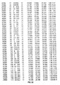
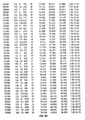
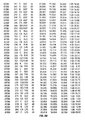
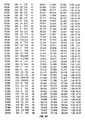
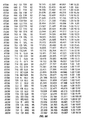
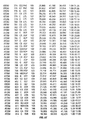
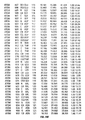
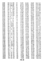
- this invention provides a computational method of modeling the three dimensional structure of a fluorescent protein comprising determining a three dimensional relationship between at least two atoms listed in the atomic coordinates of Figs. 5-1 to 5-28.
- this invention provides a device comprising a storage device and, stored in the device, at least 10 atomic coordinates selected from the atomic coordinates listed in Figs. 5-1 to 5-28.
- the storage device is a computer readable device that stores code that receives as input the atomic coordinates.
- the computer readable device is a floppy disk or a hard drive.
- this invention provides a nucleic acid molecule comprising a nucleotide sequence encoding a functional engineered fluorescent protein whose amino acid sequence is substantially identical to the amino acid sequence of Aequorea green fluorescent protein (SEQ ID NO:2) and which differs from SEQ ID NO:2 by at least one first substitution at position T203, wherein the substitution selected from the group consisting H, Y, W or F, and at least one second substitution at position H148.
- SEQ ID NO:2 Aequorea green fluorescent protein
- the present invention includes a method of determining the presence of an anion of interest in a sample, comprising the steps of introducing an engineered green fluorescent protein into a sample, said engineered green fluorescent protein comprising an amino acid sequence substantially identical to the amino acid sequence of Aequorea green fluorescent protein (SEQ ID NO:2) and which differs from SEQ ID NO:2 by at least one first substitution at position T203, wherein the substitution selected from the group consisting H, Y, W or F, and determining the fluorescence of said engineered green fluorescent protein in said sample.
- SEQ ID NO:2 Aequorea green fluorescent protein
- the invention includes a functional engineered fluorescent protein whose amino acid sequence is substantially identical to the amino acid sequence of Aequorea green fluorescent protein (SEQ ID NO:2) and which differs from SEQ ID NO:2 by at least one first substitution at position T203, wherein the substitution selected from the group consisting H, Y, W or F, and at least one second substitution at position H148, wherein said functional engineered fluorescent protein has a different fluorescent property than Aequorea green fluorescent protein.
- the invention includes a host cell comprising a functional engineered fluorescent protein whose amino acid sequence is substantially identical to the amino acid sequence of Aequorea green fluorescent protein (SEQ ID NO:2) and which differs from SEQ ID NO:2 by at least one first substitution at position T203, wherein the substitution selected from the group consisting H, Y, W or F, and at least one second substitution at position H148, wherein said functional engineered fluorescent protein has a different fluorescent property than Aequorea green fluorescent protein.
- SEQ ID NO:2 Aequorea green fluorescent protein
- Binding pair refers to two moieties (e.g. chemical or biochemical) that have an affinity for one another.
- binding pairs include antigen/antibodies, lectin/avidin, target polynucleotide/probe oligonucleotide, antibody/anti-antibody, receptor/ligand, enzyme/ligand and the like.
- One member of a binding pair refers to one moiety of the pair, such as an antigen or ligand.
- Nucleic acid refers to a deoxyribonucleotide or ribonucleotide polymer in either single- or double-stranded form, and, unless otherwise limited, encompasses known analogs of natural nucleotides that can function in a similar manner as naturally occurring nucleotides. It will be understood that when a nucleic acid molecule is represented by a DNA sequence, this also includes RNA molecules having the corresponding RNA sequence in which "U" replaces "T.”
- Recombinant nucleic acid molecule refers to a nucleic acid molecule which is not naturally occurring, and which comprises two nucleotide sequences which are not naturally joined together. Recombinant nucleic acid molecules are produced by artificial recombination, e.g., genetic engineering techniques or chemical synthesis.
- nucleotide sequence "encoding" a polypeptide means that the sequence, upon transcription and translation of mRNA, produces the polypeptide. This includes both the coding strand, whose nucleotide sequence is identical to mRNA and whose sequence is usually provided in the sequence listing, as well as its complementary strand, which is used as the template for transcription. As any person skilled in the art recognizes, this also includes all degenerate nucleotide sequences encoding the same amino acid sequence. Nucleotide sequences encoding a polypeptide include sequences containing introns.
- Expression control sequences refers to nucleotide sequences that regulate the expression of a nucleotide sequence to which they are operatively linked. Expression control sequences are "operatively linked” to a nucleotide sequence when the expression control sequences control and regulate the transcription and, as appropriate, translation of the nucleotide sequence.
- expression control sequences can include appropriate promoters, enhancers, transcription terminators, a start codon (i.e., ATG) in front of a protein-encoding gene, splicing signals for introns, maintenance of the correct reading frame of that gene to permit proper translation of the mRNA, and stop codons.
- Naturally-occurring refers to the fact that an object can be found in nature.
- a polypeptide or polynucleotide sequence that is present in an organism (including viruses) that can be isolated from a source in nature and which has not been intentionally modified by man in the laboratory is naturally-occurring.
- operably linked refers to a juxtaposition wherein the components so described are in a relationship permitting them to function in their intended manner.
- a control sequence "operably linked" to a coding sequence is ligated in such a way that expression of the coding sequence is achieved under conditions compatible with the control sequences, such as when the appropriate molecules (e.g., inducers and polymerases) are bound to the control or regulatory sequence(s).
- Control sequence refers to polynucleotide sequences which are necessary to effect the expression of coding and non-coding sequences to which they are ligated. The nature of such control sequences differs depending upon the host organism; in prokaryotes, such control sequences generally include promoter, ribosomal binding site, and transcription termination sequence; in eukaryotes, generally, such control sequences include promoters and transcription termination sequence.
- control sequences is intended to include, at a minimum, components whose presence can influence expression, and can also include additional components whose presence is advantageous, for example, leader sequences and fusion partner sequences.
- isolated polynucleotide refers a polynucleotide of genomic, cDNA, or synthetic origin or some combination there of, which by virtue of its origin the "isolated polynucleotide” (1) is not associated with the cell in which the "isolated polynucleotide” is found in nature, or (2) is operably linked to a polynucleotide which it is not linked to in nature.
- Polynucleotide refers to a polymeric form of nucleotides of at least 10 bases in length, either ribonucleotides or deoxynucleotides or a modified form of either type of nucleotide.
- the term includes single and double stranded forms of DNA.
- probe refers to a substance that specifically binds to another substance (a "target”).
- Probes include, for example, antibodies, nucleic acids, receptors and their ligands.
- Modulation refers to the capacity to either enhance or inhibit a functional property of biological activity or process (e.g., enzyme activity or receptor binding); such enhancement or inhibition may be contingent on the occurrence of a specific event, such as activation of a signal transduction pathway, and/or may be manifest only in particular cell types.
- modulator refers to a chemical (naturally occurring or non-naturally occurring), such as a synthetic molecule (e.g., nucleic acid, protein, non-peptide, or organic molecule), or an extract made from biological materials such as bacteria, plants, fungi, or animal (particularly mammalian) cells or tissues.
- Modulators can be evaluated for potential activity as inhibitors or activators (directly or indirectly) of a biological process or processes (e.g., agonist, partial antagonist, partial agonist, inverse agonist, antagonist, antineoplastic agents, cytotoxic agents, inhibitors of neoplastic transformation or cell proliferation, cell proliferation-promoting agents, and the like) by inclusion in screening assays described herein.
- the activity of a modulator may be known, unknown or partially known.
- test chemical refers to a chemical to be tested by one or more screening method(s) of the invention as a putative modulator.
- a test chemical is usually not known to bind to the target of interest.
- control test chemical refers to a chemical known to bind to the target (e.g., a known agonist, antagonist, partial agonist or inverse agonist).
- various predetermined concentrations of test chemicals are used for screening, such as .01 ⁇ M, .1 ⁇ M, 1.0 ⁇ M, and 10.0 ⁇ M.
- targets refers to a biochemical entity involved a biological process. Targets are typically proteins that play a useful role in the physiology or biology of an organism. A therapeutic chemical binds to target to alter or modulate its function. As used herein targets can include cell surface receptors, G-proteins, kinases, ion channels, phopholipases and other proteins mentioned herein.
- label refers to a composition detectable by spectroscopic, photochemical, biochemical, immunochemical, or chemical means.
- useful labels include 32 P, fluorescent dyes, fluorescent proteins, electron-dense reagents, enzymes (e.g., as commonly used in an ELISA), biotin, dioxigenin, or haptens and proteins for which antisera or monoclonal antibodies are available.
- polypeptides of this invention can be made as detectable labels, by e.g., incorporating a them as into a polypeptide, and used to label antibodies specifically reactive with the polypeptide.
- a label often generates a measurable signal, such as radioactivity, fluorescent light or enzyme activity, which can be used to quantitate the amount of bound label.
- nucleic acid probe refers to a nucleic acid molecule that binds to a specific sequence or sub-sequence of another nucleic acid molecule.
- a probe is preferably a nucleic acid molecule that binds through complementary base pairing to the full sequence or to a sub-sequence of a target nucleic acid. It will be understood that probes may bind target sequences lacking complete complementarity with the probe sequence depending upon the stringency of the hybridization conditions. Probes are preferably directly labelled as with isotopes, chromophores, lumiphores, chromogens, fluorescent proteins, or indirectly labelled such as with biotin to which a streptavidin complex may later bind. By assaying for the presence or absence of the probe, one can detect the presence or absence of the select sequence or sub-sequence.
- a "labeled nucleic acid probe” is a nucleic acid probe that is bound, either covalently, through a linker, or through ionic, van der Waals or hydrogen bonds to a label such that the presence of the probe may be detected by detecting the presence of the label bound to the probe.
- polypeptide and protein refers to a polymer of amino acid residues.
- the terms apply to amino acid polymers in which one or more amino acid residue is an artificial chemical analogue of a corresponding naturally occurring amino acid, as well as to naturally occurring amino acid polymers.
- recombinant protein refers to a protein that is produced by expression of a nucleotide sequence encoding the amino acid sequence of the protein from a recombinant DNA molecule.
- recombinant host cell refers to a cell that comprises a recombinant nucleic acid molecule.
- recombinant host cells can express genes that are not found within the native (non-recombinant) form of the cell.
- isolated refers to material which is substantially or essentially free from components which normally accompany it as found in its native state. Purity and homogeneity are typically determined using analytical chemistry techniques such as polyacrylamide gel electrophoresis or high performance liquid chromatography.
- a protein or nucleic acid molecule which is the predominant protein or nucleic acid species present in a preparation is substantially purified. Generally, an isolated protein or nucleic acid molecule will comprise more than 80% of all macromolecular species present in the preparation. Preferably, the protein is purified to represent greater than 90% of all macromolecular species present. More preferably the protein is purified to greater than 95%, and most preferably the protein is purified to essential homogeneity, wherein other macromolecular species are not detected by conventional techniques.
- naturally-occurring refers to the fact that an object can be found in nature.
- a polypeptide or polynucleotide sequence that is present in an organism (including viruses) that can be isolated from a source in nature and which has not been intentionally modified by man in the laboratory is naturally-occurring.
- antibody refers to a polypeptide substantially encoded by an immunoglobulin gene or immunoglobulin genes, or fragments thereof, which specifically bind and recognize an analyte (antigen).
- the recognized immunoglobulin genes include the kappa, lambda, alpha, gamma, delta, epsilon and mu constant region genes, as well as the myriad immunoglobulin variable region genes.
- Antibodies exist, e.g., as intact immunoglobulins or as a number of well characterized fragments produced by digestion with various peptidases. This includes, e.g., Fab' and F(ab)' 2 fragments.
- the term "antibody,” as used herein, also includes antibody fragments either produced by the modification of whole antibodies or those synthesized de novo using recombinant DNA methodologies.
- immunoassay refers to an assay that utilizes an antibody to specifically bind an analyte.
- the immunoassay is characterized by the use of specific binding properties of a particular antibody to isolate, target, and/or quantify the analyte.
- nucleic acid or polypeptide sequences refers to the residues in the two sequences which are the same when aligned for maximum correspondence.
- percentage of sequence identity is used in reference to proteins or peptides it is recognized that residue positions which are not identical often differ by conservative amino acid substitutions, where amino acids residues are substituted for other amino acid residues with similar chemical properties (e.g. charge or hydrophobicity) and therefore do not change the functional properties of the molecule.
- percent sequence identity may be adjusted upwards to correct for the conservative nature of the substitution. Means for making this adjustment are well known to those of skill in the art.
- Consatively modified variations of a particular nucleic acid sequence refers to those nucleic acids which encode identical or essentially identical amino acid sequences, or where the nucleic acid does not encode an amino acid sequence, to essentially identical sequences. Because of the degeneracy of the genetic code, a large number of functionally identical nucleic acids encode any given polypeptide. For instance, the codons CGU, CGC, CGA, CGG, AGA, and AGG all encode the amino acid arginine. Thus, at every position where an arginine is specified by a codon, the codon can be altered to any of the corresponding codons described without altering the encoded polypeptide.
- nucleic acid variations are "silent variations,” which are one species of “conservatively modified variations.” Every nucleic acid sequence herein which encodes a polypeptide also describes every possible silent variation.
- each codon in a nucleic acid except AUG, which is ordinarily the only codon for methionine
- each "silent variation" of a nucleic acid which encodes a polypeptide is implicit in each described sequence.
- nucleic acid molecule has the sequence of the binding partner of another nucleic acid molecule.
- sequence 5'-ATGC-3' is complementary to the sequence 5'-GCAT-3'.
- amino acid sequence or a nucleotide sequence is "substantially identical” or “substantially similar” to a reference sequence if the amino acid sequence or nucleotide sequence has at least 80% sequence identity with the reference sequence over a given comparison window.
- substantially similar sequences include those having, for example, at least 85% sequence identity, at least 90% sequence identity, at least 95% sequence identity or at least 99% sequence identity.
- Two sequences that are identical to each other are, of course, also substantially identical.
- a subject nucleotide sequence is "substantially complementary" to a reference nucleotide sequence if the complement of the subject nucleotide sequence is substantially identical to the reference nucleotide sequence.
- stringent conditions refers to a temperature and ionic conditions used in nucleic acid hybridization. Stringent conditions are sequence dependent and are different under different environmental parameters. Generally, stringent conditions are selected to be about 5°C to 20°C lower than the thermal melting point (T m ) for the specific sequence at a defined ionic strength and pH. The T m is the temperature (under defined ionic strength and pH) at which 50% of the target sequence hybridizes to a perfectly matched probe.
- allelic variants refers to polymorphic forms of a gene at a particular genetic locus, as well as cDNAs derived from mRNA transcripts of the genes and the polypeptides encoded by them.
- preferred mammalian codon refers to the subset of codons from among the set of codons encoding an amino acid that are most frequently used in proteins expressed in mammalian cells as chosen from the following list: Amino Acid Preferred codons for high level mammalian expression Gly GGC,GGG Glu GAG Asp GAC Val GUG,GUC Ala GCC,GCU Ser AGC,UCC Lys AAG Asn AAC Met AUG Ile AUC Thr ACC Trp UGG Cys UGC Tyr UAU,UAC Leu CUG Phe UUC Arg CGC,AGG,AGA Gln CAG His CAC Pro CCC
- Fluorescent molecules are useful in fluorescence resonance energy transfer ("FRET").
- FRET involves a donor molecule and an acceptor molecule.
- the emission spectrum of the donor should overlap as much as possible with the excitation spectrum of the acceptor to maximize the overlap integral.
- the quantum yield of the donor moiety and the extinction coefficient of the acceptor should likewise be as high as possible to maximize R 0 , the distance at which energy transfer efficiency is 50%.
- the excitation spectra of the donor and acceptor should overlap as little as possible so that a wavelength region can be found at which the donor can be excited efficiently without directly exciting the acceptor.
- Fluorescence arising from direct excitation of the acceptor is difficult to distinguish from fluorescence arising from FRET.
- the emission spectra of the donor and acceptor should overlap as little as possible so that the two emissions can be clearly distinguished.
- High fluorescence quantum yield of the acceptor moiety is desirable if the emission from the acceptor is to be measured either as the sole readout or as part of an emission ratio.
- One factor to be considered in choosing the donor and acceptor pair is the efficiency of fluorescence resonance energy transfer between them.
- the efficiency of FRET between the donor and acceptor is at least 10%, more preferably at least 50% and even more preferably at least 80%.
- fluorescent property refers to the molar extinction coefficient at an appropriate excitation wavelength, the fluorescence quantum efficiency, the shape of the excitation spectrum or emission spectrum, the excitation wavelength maximum and emission wavelength maximum, the ratio of excitation amplitudes at two different wavelengths, the ratio of emission amplitudes at two different wavelengths, the excited state lifetime, or the fluorescence anisotropy.
- a measurable difference in any one of these properties between wild-type Aequorea GFP and the mutant form is useful.
- a measurable difference can be determined by determining the amount of any quantitative fluorescent property, e.g., the amount of fluorescence at a particular wavelength, or the integral of fluorescence over the emission spectrum.
- excitation amplitude ratioing and “emission amplitude ratioing”, respectively
- emission amplitude ratioing are particularly advantageous because the ratioing process provides an internal reference and cancels out variations in the absolute brightness of the excitation source, the sensitivity of the detector, and light scattering or quenching by the sample.
- fluorescent protein refers to any protein capable of fluorescence when excited with appropriate electromagnetic radiation. This includes fluorescent proteins whose amino acid sequences are either naturally occurring or engineered (i.e., analogs or mutants). Many cnidarians use green fluorescent proteins ("GFPs") as energy-transfer acceptors in bioluminescence.
- GFPs green fluorescent proteins
- a variety of Aequorea -related fluorescent proteins having useful excitation and emission spectra have been engineered by modifying the amino acid sequence of a naturally occurring GFP from Aequorea victoria .
- D.C. Prasher et al. Gene, 111:229-233 (1992); R. Heim et al., Proc. Natl. Acad. Sci., USA , 91:12501-04 (1994); U.S. patent application 08/337,915, filed November 10, 1994; International application PCT/US95/14692, filed 11/10/95.
- a fluorescent protein is an " Aequorea -related fluorescent protein" if any contiguous sequence of 150 amino acids of the fluorescent protein has at least 85% sequence identity with an amino acid sequence, either contiguous or non-contiguous, from the 23 8 amino-acid wild-type Aequorea green fluorescent protein of Fig. 3 (SEQ ID NO:2). More preferably, a fluorescent protein is an Aequorea -related fluorescent protein if any contiguous sequence of 200 amino acids of the fluorescent protein has at least 95% sequence identity with an amino acid sequence, either contiguous or non-contiguous, from the wild type Aequorea green fluorescent protein of Fig. 3 (SEQ ID NO:2). Similarly, the fluorescent protein may be related to Renilla or Phialidium wild-type fluorescent proteins using the same standards.
- Aequorea -related fluorescent proteins include, for example and without limitation, wild-type (native) Aequorea victoria GFP (D.C. Prasher et al., "Primary structure of the Aequorea victoria green fluorescent protein," Gene, (1992) 1 11:229-33), whose nucleotide sequence (SEQ ID NO:1) and deduced amino acid sequence (SEQ ID NO:2) are presented in Fig. 3; allelic variants of this sequence, e.g., Q80R, which has the glutamine residue at position 80 substituted with arginine (M.
- Folding mutations improve the ability of fluorescent proteins to fold at higher temperatures, and to be more fluorescent when expressed in mammalian cells, but have little or no effect on the peak wavelengths of excitation and emission. It should be noted that these may be combined with mutations that influence the spectral properties of GFP to produce proteins with altered spectral and folding properties. Folding mutations include: F64L, V68L, S72A, and also T44A, F99S, Y145F, N146I, M153T or A, V163A, I167T, S175G, S205T and N212K.
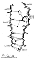
- loop domain refers to an amino acid sequence of an Aequorea -related fluorescent protein that connects the amino acids involved in the secondary structure of the eleven strands of the -barrel or the central -helix (residues 56-72) (see Fig. 1A and 1B).
- fluorescent protein moiety of a fluorescent protein is that portion of the amino acid sequence of a fluorescent protein which, when the amino acid sequence of the fluorescent protein substrate is optimally aligned with the amino acid sequence of a naturally occurring fluorescent protein, lies between the amino terminal and carboxy terminal amino acids, inclusive, of the amino acid sequence of the naturally occurring fluorescent protein.
- fluorescent proteins can be genetically fused to other target proteins and used as markers to identify the location and amount of the target protein produced. Accordingly, this invention provides fusion proteins comprising a fluorescent protein moiety and additional amino acid sequences. Such sequences can be, for example, up to about 15, up to about 50, up to about 150 or up to about 1000 amino acids long.
- the fusion proteins possess the ability to fluoresce when excited by electromagnetic radiation.
- the fusion protein comprises a polyhistidine tag to aid in purification of the protein.
- Fluorescent characteristics of Aequorea -related fluorescent proteins depend, in part, on the electronic environment of the chromophore.
- amino acids that are within about 0.5 nm of the chromophore influence the electronic environment of the chromophore. Therefore, substitution of such amino acids can produce fluorescent proteins with altered fluorescent characteristics.
- electron density tends to shift from the phenolate towards the carbonyl end of the chromophore. Therefore, placement of increasing positive charge near the carbonyl end of the chromophore tends to decrease the energy of the excited state and cause a red-shift in the absorbance and emission wavelength maximum of the protein. Decreasing positive charge near the carbonyl end of the chromophore tends to have the opposite effect, causing a blue-shift in the protein's wavelengths.
- Amino acids with charged (ionized D, E, K, and R), dipolar (H, N, Q, S, T, and uncharged D, E and K), and polarizable side groups are useful for altering the electronic environment of the chromophore, especially when they substitute an amino acid with an uncharged, nonpolar or non-polarizable side chain.
- amino acids with polarizable side groups alter the electronic environment least, and, consequently, are expected to cause a comparatively smaller change in a fluorescent property.
- Amino acids with charged side groups alter the environment most, and, consequently, are expected to cause a comparatively larger change in a fluorescent property.
- amino acids with charged side groups are more likely to disrupt the structure of the protein and to prevent proper folding if buried next to the chromophore without any additional solvation or salt bridging. Therefore charged amino acids are most likely to be tolerated and to give useful effects when they replace other charged or highly polar amino acids that are already solvated or involved in salt bridges.
- the structure of the protein may make selection of a larger amino acid, e.g., W, less appropriate.
- positions occupied by amino acids with charged or polar side groups that are unfavorably oriented may be substituted with amino acids that have less charged or polar side groups.
- an amino acid whose side group has a dipole oriented in one direction in the protein can be substituted with an amino acid having a dipole oriented in a different direction.
- Table B lists several amino acids located within about 0.5 nm from the chromophore whose substitution can result in altered fluorescent characteristics.
- the table indicates, underlined, preferred amino acid substitutions at the indicated location to alter a fluorescent characteristic of the protein.
- the table also provides codons for primers used in site-directed mutagenesis involving amplification. These primers have been selected to encode economically the preferred amino acids, but they encode other amino acids as well, as indicated, or even a stop codon, denoted by Z.
- the most efficient strategy is to screen the collection to identify mutants with the desired properties and then sequence their DNA to find out which of the possible substitutions is responsible.
- Codons are shown in double-stranded form with sense strand above, antisense strand below.
- amino acids with polar side groups that can be substituted with polarizable side groups include, for example, those in Table C.
- Original position and presumed role Change to Codon Q69 Terminates chain of H-bonding waters KREG RRg YYC Q94 H-bonds to carbonyl terminus of chromophore DEHKN Q VAS BTS Q183 Bridges Arg96 and center of chromophore bridge HY YAC RTG EK RAg YTC N185 Part of H-bond network near carbonyl of chromophore DEH N KQ VAS BTS
- an amino acid that is close to a second amino acid within about 0.5 nm of the chromophore can, upon substitution, alter the electronic properties of the second amino acid, in turn altering the electronic environment of the chromophore.
- Table D presents two such amino acids.
- the amino acids, L220 and V224, are close to E222 and oriented in the same direction in the ⁇ pleated sheet.
- One embodiment of the invention includes a nucleic acid molecule comprising a nucleotide sequence encoding a functional engineered fluorescent protein whose amino acid sequence is substantially identical to the amino acid sequence of Aequorea green fluorescent protein (SEQ ID NO:2) and which differs from SEQ ID N0:2 by at least a substitution at Q69, wherein the functional engineered fluorescent protein has a different fluorescent property than Aequorea green fluorescent protein.
- the substitution at Q69 is selected from the group of K, R, E and G.
- the Q69 substitution can be combined with other mutations to improve the properties of the protein, such as a functional mutation at S65.
- One embodiment of the invention includes a nucleic acid molecule comprising a nucleotide sequence encoding a functional engineered fluorescent protein whose amino acid sequence is substantially identical to the amino acid sequence of Aequorea green fluorescent protein (SEQ ID NO:2) and which differs from SEQ ID NO:2 by at least a substitution at E222, but not including E222G, wherein the functional engineered fluorescent protein has a different fluorescent property than Aequorea green fluorescent protein.
- the substitution at E222 is selected from the group of N and Q.
- the E222 substitution can be combined with other mutations to improve the properties of the protein, such as a functional mutation at F64.
- One embodiment of the invention includes a nucleic acid molecule comprising a nucleotide sequence encoding a functional engineered fluorescent protein whose amino acid sequence is substantially identical to the amino acid sequence of Aequorea green fluorescent protein (SEQ ID NO:2) and which differs from SEQ ID NO:2 by at least a substitution at Y145, wherein the functional engineered fluorescent protein has a different fluorescent property than Aequorea green fluorescent protein.
- the substitution at Y145 is selected from the group of W, C, F, L, E, H, K and Q.
- the Y145 substitution can be combined with other mutations to improve the properties of the protein, such as a Y66.
- the invention also includes computer related embodiments, including computational methods of using the crystal coordinates for designing new fluorescent protein mutations and devices for storing the crystal data, including coordinates.
- the invention includes a device comprising a storage device and, stored in the device, at least 10 atomic coordinates selected from the atomic coordinates listed in Figs. 5-1 to 5-28. More coordinates can be storage depending of the complexity of the calculations or the objective of using the coordinates (e.g. about 100,1,000, or more coordinates). For example, larger numbers of coordinates will be desirable for more detailed representations of fluorescent protein structure.
- the storage device is a computer readable device that stores code that it receives as input the atomic coordinates.
- the computer readable device can be a floppy disk or a hard drive.
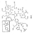
- the invention includes the use of X-ray crystallography and computer processing, to create a model of the crystal structure of YFP showing the relative location, and amino acids that interact with bound ions. This information is useful in identifying amino acids whose substitution alters the specificity and affinity of the binding site to various anions. Because the binding of the anion is close to the chromophore of YFP, binding results in a modulation of the fluorescent properties of YFP that can be used to monitor anion binding and therefore the concentration of the anion.
- the anion binding site found in YFP-H148Q exhibits many of the characteristics generally found in halide binding sites in other proteins.
- the binding site is amphiphilic in nature, with one side lined with polar and charged groups (Tyr203, the chromophore, Arg96, Gln69, and Gln183), and the other with hydrophobic residues (Ile152, Leu201, Val163, Val150, and Phe165).
- anion binding can be improved by creating more and or tighter binding interactions between the anion of interest and polar groups within the binding pocket.
- polar residues above For example, either directly substituting the polar residues above with more polar residues, or by substituting residues of different sizes, that may interact more effectively with the anion, can improve ion binding.
- the size and position of the chromophore may be altered by the substitution of S65 to G, A, C, V, L, I or T; Y66 may be altered by substitution to H, F or W; Q69 may be substituted to N or K; R96 to K; Q183 to N or K.
- the size and shape of the binding pocket may also be of particular importance due to the buried nature of the binding site for larger anions.
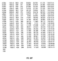
- TCA with a mean geometric diameter of 6.2 ⁇ (Halm & Frizzell, 1992), is apparently too large to interact with YFP to a measurable extent, whereas the somewhat smaller TFA does show weak binding (Table J). Improvements in the binding affinity of larger anions could thus be achieved via the substitution of amino acids lining the binding pocket with smaller residues, as outlined in Table F above, as well as increasing solvent accessibility as discussed below.
- mutations will typically be introduced in the YFP template protein via oligo-mediated site directed mutagenesis to create libraries of mutant proteins that typically have a 10 % probability of containing the wild-type amino acid residue and a 90% probability of containing one of the various mutant residues.
- This approach it is possible to rapidly screen libraries containing various combinations of mutants to identify the best combinations for a specific anion of interest. Typically this process can be repeated iteratively to ensure that sequence space around the binding pocket has been completely explored for any specific anion of interest.
- Recombinant production of a fluorescent protein involves expressing a nucleic acid molecule having sequences that encode the protein.
- the nucleic acid encodes a fusion protein in which a single polypeptide includes the fluorescent protein moiety within a longer polypeptide.
- the longer polypeptide can include a second functional protein, such as FRET partner or a protein having a second function (e.g., an enzyme, antibody or other binding protein).
- Nucleic acids that encode fluorescent proteins are useful as starting materials.
- the fluorescent proteins can be produced as fusion proteins by recombinant DNA technology. Recombinant production of fluorescent proteins involves expressing nucleic acids having sequences that encode the proteins. Nucleic acids encoding fluorescent proteins can be obtained by methods known in the art. Fluorescent proteins can be made by site-specific mutagenesis of other nucleic acids encoding fluorescent proteins, or by random mutagenesis caused by increasing the error rate of PCR of the original polynucleotide with 0.1 mM MnCl 2 and unbalanced nucleotide concentrations. See, e . g ., U.S. patent application 08/337,915, filed November 10, 1994 or International application PCT/US95/14692, filed 11/10/95.
- the nucleic acid encoding a green fluorescent protein can be isolated by polymerase chain reaction of cDNA from A. victoria using primers based on the DNA sequence of A. victoria green fluorescent protein, as presented in Fig. 3.
- PCR methods are described in, for example, U.S. Pat. No. 4,683,195; Mullis et al. (1987) Cold Spring Harbor Symp. Quant. Biol . 51:263; and Erlich, ed., PCR Technology, (Stockton Press, NY, 1989).
- the construction of expression vectors and the expression of genes in transfected cells involves the use of molecular cloning techniques also well known in the art. Sambrook et al., Molecular Cloning -- A Laboratory Manual, Cold Spring Harbor Laboratory, Cold Spring Harbor, NY, (1989) and Current Protocols in Molecular Biology, F.M. Ausubel et al., eds., (Current Protocols, a joint venture between Greene Publishing Associates, Inc. and John Wiley & Sons, Inc.).
- the expression vector can be adapted for function in prokaryotes or eukaryotes by inclusion of appropriate promoters, replication sequences, markers, etc.
- Nucleic acids used to transfect cells with sequences coding for expression of the polypeptide of interest generally will be in the form of an expression vector including expression control sequences operatively linked to a nucleotide sequence coding for expression of the polypeptide.
- the term "nucleotide sequence coding for expression of" a polypeptide refers to a sequence that, upon transcription and translation of mRNA, produces the polypeptide. This can include sequences containing, e . g ., introns.
- Expression control sequences are operatively linked to a nucleic acid sequence when the expression control sequences control and regulate the transcription and, as appropriate, translation of the nucleic acid sequence.
- expression control sequences can include appropriate promoters, enhancers, transcription terminators, a start codon (i . e ., ATG) in front of a protein-encoding gene, splicing signals for introns, maintenance of the correct reading frame of that gene to permit proper translation of the mRNA, and stop codons.
- Transformation of a host cell with recombinant DNA may be carried out by conventional techniques as are well known to those skilled in the art.
- the host is prokaryotic, such as E. coli
- competent cells which are capable of DNA uptake can be prepared from cells harvested after exponential growth phase and subsequently treated by the CaCl 2 method by procedures well known in the art.
- CaCl 2 or RbCl can be used. Transformation can also be performed after forming a protoplast of the host cell or by electroporation.
- Eukaryotic cells can also be co-transfected with DNA sequences encoding the fusion polypeptide of the invention, and a second foreign DNA molecule encoding a selectable phenotype, such as the herpes simplex thymidine kinase gene.
- Another method is to use a eukaryotic viral vector, such as simian virus 40 (SV40) or bovine papilloma virus, to transiently infect or transform eukaryotic cells and express the protein.
- a eukaryotic viral vector such as simian virus 40 (SV40) or bovine papilloma virus
- SV40 simian virus 40
- bovine papilloma virus bovine papilloma virus
- Techniques for the isolation and purification of either microbially or eukaryotically expressed polypeptides of the invention may be by any conventional means such as, for example, preparative chromatographic separations and immunological separations such as those involving the use of monoclonal or polyclonal antibodies or antigen.
- recombinant fluorescent proteins can be produced by expression of nucleic acid encoding for the protein in E. coli.
- Aequorea -related fluorescent proteins are best expressed by cells cultured between about 15°C and 30°C but higher temperatures (e.g. 37°C) are possible. After synthesis, these enzymes are stable at higher temperatures (e.g., 37°C) and can be used in assays at those temperatures.
- a variety of host-expression vector systems may be utilized to express fluorescent protein coding sequence. These include but are not limited to microorganisms such as bacteria transformed with recombinant bacteriophage DNA, plasmid DNA or cosmid DNA expression vectors containing a fluorescent protein coding sequence; yeast transformed with recombinant yeast expression vectors containing the fluorescent protein coding sequence; plant cell systems infected with recombinant virus expression vectors ( e . g ., cauliflower mosaic virus, CaMV; tobacco mosaic virus, TMU) or transformed with recombinant plasmid expression vectors ( e .
- microorganisms such as bacteria transformed with recombinant bacteriophage DNA, plasmid DNA or cosmid DNA expression vectors containing a fluorescent protein coding sequence
- yeast transformed with recombinant yeast expression vectors containing the fluorescent protein coding sequence e. g ., cauliflower mosaic virus, CaMV; tobacco mosaic virus, TMU
- any of a number of suitable transcription and translation elements including constitutive and inducible promoters, transcription enhancer elements, transcription terminators, etc. may be used in the expression vector (see, e.g., Bitter, et al ., Methods in Enzymology 153:516-544, 1987).
- inducible promoters such as pL of bacteriophage ⁇ , plac, ptrp, ptac (ptrp-lac hybrid promoter) and the like may be used.
- promoters derived from the genome of mammalian cells e.g., metallothionein promoter
- mammalian viruses e.g. , the retrovirus long terminal repeat; the adenovirus late promoter; the vaccinia virus 7.5K promoter
- Promoters produced by recombinant DNA or synthetic techniques may also be used to provide for transcription of the inserted fluorescent protein coding sequence.
- a number of expression vectors may be advantageously selected depending upon the use intended for the fluorescent protein expressed. For example, when large quantities of the fluorescent protein are to be produced, vectors which direct the expression of high levels of fusion protein products that are readily purified may be desirable. Those which are engineered to contain a cleavage site to aid in recovering fluorescent protein are preferred.
- yeast a number of vectors containing constitutive or inducible promoters may be used.
- Current Protocols in Molecular Biology Vol. 2, Ed. Ausubel, et al ., Greene Publish. Assoc. & Wiley Interscience, Ch. 13, 1988; Grant, et al ., Expression and Secretion Vectors for Yeast, in Methods in Enzymology, Eds. Wu & Grossman, 31987, Acad. Press, N.Y., Vol. 153, pp.516-544, 1987; Glover, DNA Cloning, Vol. II, IRL Press, Wash., D.C., Ch.
- yeast promoter such as ADH or LEU2 or an inducible promoter such as GAL may be used (Cloning in Yeast, Ch. 3, R. Rothstein In: DNA Cloning Vol. 11, A Practical Approach, Ed. DM Glover, IRL Press, Wash., D.C., 1986).
- vectors may be used which promote integration of foreign DNA sequences into the yeast chromosome.
- the expression of a fluorescent protein coding sequence may be driven by any of a number of promoters.
- viral promoters such as the 35S RNA and 19S RNA promoters of CaMV (Brisson, et al., Nature 310:511-514, 1984), or the coat protein promoter to TMV (Takamatsu, et al., EMBO J. 3:17-311, 1987) may be used; alternatively, plant promoters such as the small subunit of RUBISCO (Coruzzi, et al ., 1984, EMBO J.
- An alternative expression system which could be used to express fluorescent protein is an insect system.
- Autographa californica nuclear polyhedrosis virus (AcNPV) is used as a vector to express foreign genes.
- the virus grows in Spodoptera frugiperda cells.
- the fluorescent protein coding sequence may be cloned into non-essential regions (for example, the polyhedrin gene) of the virus and placed under control of an AcNPV promoter (for example the polyhedrin promoter).
- Successful insertion of the fluorescent protein coding sequence will result in inactivation of the polyhedrin gene and production of non-occluded recombinant virus (i.e. , virus lacking the proteinaceous coat coded for by the polyhedrin gene).
- Eukaryotic systems and preferably mammalian expression systems, allow for proper post-translational modifications of expressed mammalian proteins to occur.
- Eukaryotic cells which possess the cellular machinery for proper processing of the primary transcript, glycosylation, phosphorylation, and, advantageously secretion of the gene product should be used as host cells for the expression of fluorescent protein.
- host cell lines may include but are not limited to CHO, VERO, BHK, HeLa, COS, MDCK, Jurkat, HEK-293, and WI38.
- Mammalian cell systems which utilize recombinant viruses or viral elements to direct expression may be engineered.
- the fluorescent protein coding sequence may be ligated to an adenovirus transcription/translation control complex, e.g., the late promoter and tripartite leader sequence.
- This chimeric gene may then be inserted in the adenovirus genome by in vitro or in vivo recombination. Insertion in a non-essential region of the viral genome (e.g ., region E1 or E3) will result in a recombinant virus that is viable and capable of expressing the fluorescent protein in infected hosts ( e . g ., see Logan & Shenk, Proc.
- the vaccinia virus 7.5K promoter may be used.
- vectors based on bovine papilloma virus which have the ability to replicate as extrachromosomal elements (Sarver, et al., Mol. Cell.
- the plasmid replicates to about 100 to 200 copies per cell. Transcription of the inserted cDNA does not require integration of the plasmid into the host's chromosome, thereby yielding a high level of expression.
- These vectors can be used for stable expression by including a selectable marker in the plasmid, such as the neo gene.
- the retroviral genome can be modified for use as a vector capable of introducing and directing the expression of the fluorescent protein gene in host cells (Cone & Mulligan, Proc. Natl. Acad. Sci. USA, 81:6349-6353, 1984). High level expression may also be achieved using inducible promoters, including, but not limited to, the metallothionine IIA promoter and heat shock promoters.
- the invention can also include a localization sequence, such as a nuclear localization sequence, an endoplasmic reticulum localization sequence, a peroxisome localization sequence, a mitochondrial localization sequence, or a localized protein.
- Localization sequences can be targeting sequences which are described, for example, in "Protein Targeting", chapter 35 of Stryer, L., Biochemistry (4th ed.). W.H. Freeman, 1995.
- the localization sequence can also be a localized protein.
- Some important localization sequences include those targeting the nucleus (KKKRK), mitochondrion (amino terminal MLRTSSLFTRRVQPSLFRNILRLQST-), endoplasmic reticulum (KDEL at C-terminus, assuming a signal sequence present at N-terminus), peroxisome (SKF at C-terminus), prenylation or insertion into plasma membrane (CaaX, CC, CXC, or CCXX at C-terminus), cytoplasmic side of plasma membrane (fusion to SNAP-25), or the Golgi apparatus (fusion to furin).
- host cells can be transformed with the fluorescent protein cDNA controlled by appropriate expression control elements (e . g ., promoter, enhancer, sequences, transcription terminators, polyadenylation sites, etc .), and a selectable marker.
- expression control elements e . g ., promoter, enhancer, sequences, transcription terminators, polyadenylation sites, etc .
- the selectable marker in the recombinant plasmid confers resistance to the selection and allows cells to stably integrate the plasmid into their chromosomes and grow to form foci which in turn can be cloned and expanded into cell lines.
- engineered cells may be allowed to grow for 1-2 days in an enriched media, and then are switched to a selective media.
- a number of selection systems may be used, including but not limited to the herpes simplex virus thymidine kinase (Wigler, et al., Cell , 11: 223, 1977), hypoxanthine-guanine phosphoribosyltransferase (Szybalska & Szybalski, Proc. Natl. Acad. Sci.
- adenine phosphoribosyltransferase genes can be employed in tk - , hgprt - or aprt - cells respectively.
- antimetabolite resistance can be used as the basis of selection for dhfr, which confers resistance to methotrexate (Wigler, et al., Proc. Natl. Acad Sci. USA, 77: 3567, 1980; O'Hare, et al., Proc. Natl. Acad. Sci.
- gpt which confers resistance to mycophenolic acid
- neo which confers resistance to the aminoglycoside G-418
- hygro which confers resistance to hygromycin
- trpB which allows cells to utilize indole in place of tryptophan
- hisD which allows cells to utilize histinol in place of histidine
- ODC omithine decarboxylase
- DNA sequences encoding the fluorescence protein polypeptide of the invention can be expressed in vitro by DNA transfer into a suitable host cell.
- "Host cells” are cells in which a vector can be propagated and its DNA expressed.
- the term also includes any progeny of the subject host cell. It is understood that all progeny may not be identical to the parental cell since there may be mutations that occur during replication. However, such progeny are included when the term "host cell” is used. Methods of stable transfer, in other words when the foreign DNA is continuously maintained in the host, are known in the art.
- the expression vector can be transfected into a host cell for expression of the recombinant nucleic acid.
- Host cells can be selected for high levels of expression in order to purify the fluorescent protein fusion protein. E. coli is useful for this purpose.
- the host cell can be a prokaryotic or eukaryotic cell selected to study the activity of an enzyme produced by the cell.
- the linker peptide is selected to include an amino acid sequence recognized by the protease.
- the cell can be, e . g ., a cultured cell or a cell in vivo.
- a primary advantage of fluorescent protein fusion proteins is that they are prepared by normal protein biosynthesis, thus completely avoiding organic synthesis and the requirement for customized unnatural amino acid analogs.
- the constructs can be expressed in E. coli in large scale for in vitro assays. Purification from bacteria is simplified when the sequences include polyhistidine tags for one-step purification by nickel-chelate chromatography. Alternatively, the substrates can be expressed directly in a desired host cell for assays in situ .
- the invention provides a transgenic non-human animal that expresses a nucleic acid sequence which encodes the fluorescent protein.
- non-human animals comprise any non-human animal having nucleic acid sequence which encodes a fluorescent protein.
- Such non-human animals include vertebrates such as rodents, non-human primates, sheep, dog, cow, pig, amphibians, and reptiles. Preferred non-human animals are selected from the rodent family including rat and mouse, most preferably mouse.
- the "transgenic non-human animals” of the invention are produced by introducing "transgenes" into the germline of the non-human animal. Embryonal target cells at various developmental stages can be used to introduce transgenes. Different methods are used depending on the stage of development of the embryonic target cell. The zygote is the best target for micro-injection.
- the male pronucleus reaches the size of approximately 20 micrometers in diameter which allows reproducible injection of 1-2 pl of DNA solution.
- the use of zygotes as a target for gene transfer has a major advantage in that in most cases the injected DNA will be incorporated into the host gene before the first cleavage (Brinster et al., Proc. Natl. Acad. Sci. USA 82:4438-4442, 1985). As a consequence, all cells of the transgenic non-human animal will carry the incorporated transgene. This will in general also be reflected in the efficient transmission of the transgene to offspring of the founder since 50% of the germ cells will harbor the transgene. Microinjection of zygotes is the preferred method for incorporating transgenes in practicing the invention.
- transgenic is used to describe an animal which includes exogenous genetic material within all of its cells.
- a “transgenic” animal can be produced by cross-breeding two chimeric animals which include exogenous genetic material within cells used in reproduction. Twenty-five percent of the resulting offspring will be transgenic i . e ., animals which include the exogenous genetic material within all of their cells in both alleles. 50% of the resulting animals will include the exogenous genetic material within one allele and 25% will include no exogenous genetic material.
- Retroviral infection can also be used to introduce transgene into a non-human animal.
- the developing non-human embryo can be cultured in vitro to the blastocyst stage.
- the blastomeres can be targets for retro viral infection (Jaenich, R., Proc. Natl. Acad Sci USA 73:1260-1264, 1976).
- Efficient infection of the blastomeres is obtained by enzymatic treatment to remove the zona pellucida (Hogan, et al . (1986) in Manipulating the Mouse Embryo, Cold Spring Harbor Laboratory Press, Cold Spring Harbor, N.Y.).
- the viral vector system used to introduce the transgene is typically a replication-defective retro virus carrying the transgene (Jahner, et al., Proc. Natl. Acad. Sci. USA 82:6927-6931, 1985; Van der Putten, et al., Proc. Natl. Acad Sci USA 82:6148-6152, 1985). Transfection is easily and efficiently obtained by culturing the blastomeres on a monolayer of virus-producing cells (Van der Putten, supra ; Stewart, et al., EMBO J. 6:383-388, 1987). Alternatively, infection can be performed at a later stage. Virus or virus-producing cells can be injected into the blastocoele (D.
- transgenes are mosaic for the transgene since incorporation occurs only in a subset of the cells which formed the transgenic nonhuman animal. Further, the founder may contain various retro viral insertions of the transgene at different positions in the genome which generally will segregate in the offspring. In addition, it is also possible to introduce transgenes into the germ line, albeit with low efficiency, by intrauterine retro viral infection of the midgestation embryo (D. Jahner et al., supra ).
- ES cells are obtained from pre-implantation embryos cultured in vitro and fused with embryos (M. J. Evans et al. Nature 292:154-156, 1981; M.O. Bradley et al ., Nature 309: 255-258, 1984; Gossler, et al., Proc. Natl. Acad. Sci USA 83: 9065-9069, 1986; and Robertson et al., Nature 322:445-448, 1986).
- Transgenes can be efficiently introduced into the ES cells by DNA transfection or by retro virus-mediated transduction. Such transformed ES cells can thereafter be combined with blastocysts from a nonhuman animal. The ES cells thereafter colonize the embryo and contribute to the germ line of the resulting chimeric animal. (For review see Jaenisch, R., Science 240: 1468-1474, 1988).
- Transformed means a cell into which (or into an ancestor of which) has been introduced, by means of recombinant nucleic acid techniques, a heterologous nucleic acid molecule.
- Heterologous refers to a nucleic acid sequence that either originates from another species or is modified from either its original form or the form primarily expressed in the cell.
- Transgene means any piece of DNA which is inserted by artifice into a cell, and becomes part of the genome of the organism ( i . e ., either stably integrated or as a stable extrachromosomal element) which develops from that cell.
- a transgene may include a gene which is partly or entirely heterologous ( i . e ., foreign) to the transgenic organism, or may represent a gene homologous to an endogenous gene of the organism. Included within this definition is a transgene created by the providing of an RNA sequence which is transcribed into DNA and then incorporated into the genome.
- the transgenes of the invention include DNA sequences which encode which encodes the fluorescent protein which may be expressed in a transgenic non-human animal.
- transgenic as used herein additionally includes any organism whose genome has been altered by in vitro manipulation of the early embryo or fertilized egg or by any transgenic technology to induce a specific gene knockout.
- gene knockout refers to the targeted disruption of a gene in vivo with complete loss of function that has been achieved by any transgenic technology familiar to those in the art.
- transgenic animals having gene knockouts are those in which the target gene has been rendered nonfunctional by an insertion targeted to the gene to be rendered non-functional by homologous recombination.
- transgenic includes any transgenic technology familiar to those in the art which can produce an organism carrying an introduced transgene or one in which an endogenous gene has been rendered non-functional or "knocked out.”
- the proteins of this invention are useful in any methods that employ fluorescent proteins.
- the engineered fluorescent proteins of this invention are useful as fluorescent markers in the many ways fluorescent markers already are used. This includes, for example, coupling engineered fluorescent proteins to antibodies, nucleic acids or other receptors for use in detection assays, such as immunoassays or hybridization assays.
- the engineered fluorescent proteins of this invention are useful to track the movement of proteins in cells.
- a nucleic acid molecule encoding the fluorescent protein is fused to a nucleic acid molecule encoding the protein of interest in an expression vector.
- the protein of interest can be localized based on fluorescence.
- two proteins of interest are fused with two engineered fluorescent proteins having different fluorescent characteristics.
- the engineered fluorescent proteins of this invention are useful in systems to detect induction of transcription.
- a nucleotide sequence encoding the engineered fluorescent protein is fused to expression control sequences of interest and the expression vector is transfected into a cell. Induction of the promoter can be measured by detecting the expression and/or quantity of fluorescence.
- Such constructs can be used to follow signaling pathways from receptor to promoter.
- the engineered fluorescent proteins of this invention are useful in applications involving FRET. Such applications can detect events as a function of the movement of fluorescent donors and acceptor towards or away from each other.
- One or both of the donor/acceptor pair can be a fluorescent protein.
- a preferred donor and receptor pair for FRET based assays is a donor with a T203I mutation and an acceptor with the mutation T203X, wherein X is an aromatic amino acid-39, especially T203Y, T203W, or T203H.
- the donor contains the following mutations: S72A, K79R, Y145F, M153A and T203I (with a excitation peak of 395 nm and an emission peak of 511 nm) and the acceptor contains the following mutations S65G, S72A, K79R, and T203Y.
- This particular pair provides a wide separation between the excitation and emission peaks of the donor and provides good overlap between the donor emission spectrum and the acceptor excitation spectrum.
- Other red-shifted mutants, such as those described herein, can also be used as the acceptor in such a pair.
- FRET is used to detect the cleavage of a substrate having the donor and acceptor coupled to the substrate on opposite sides of the cleavage site.
- the donor/acceptor pair physically separate, eliminating FRET.
- Assays involve contacting the substrate with a sample, and determining a qualitative or quantitative change in FRET.
- the engineered fluorescent protein is used in a substrate for ⁇ -lactamase. Examples of such substrates are described in United States patent applications 08/407,544, filed March 20, 1995 and International Application PCT/US96/04059, filed March 20, 1996.
- an engineered fluorescent protein donor/acceptor pair are part of a fusion protein coupled by a peptide having a proteolytic cleavage site. Such tandem fluorescent proteins are described in United States patent application 08/594,575, filed January 31, 1996.
- FRET is used to detect changes in potential across a membrane.
- a donor and acceptor are placed on opposite sides of a membrane such that one translates across the membrane in response to a voltage change. This creates a measurable FRET.
- Such a method is described in United States patent application 08/481,977, filed June 7, 1995 and International Application PCT/US96/09652, filed June 6, 1996.
- the engineered proteins of this invention are useful in the creation of biosensors for determining the concentrations of ions within samples and living cells and transgenic organisms. Upon binding of an ion to the fluorescent protein, a change in at least one measurable fluorescent property of the engineered fluorescent protein occurs that provides the basis for determining the presence of the ion of interest.
- the engineered protein of this invention are useful in the creation of fluorescent substrates for protein kinases.
- Such substrates incorporate an amino acid sequence recognizable by protein kinases.
- the engineered fluorescent protein undergoes a change in a fluorescent property.
- Such substrates are useful in detecting and measuring protein kinase activity in a sample of a cell, upon transfection and expression of the substrate.
- the kinase recognition site is placed within about 20 amino acids of a terminus of the engineered fluorescent protein.
- the kinase recognition site also can be placed in a loop domain of the protein. (See, e.g. Figure 1B.)
- Methods for making fluorescent substrates for protein kinases are described in United States patent application 08/680,877, filed July 16, 1996.
- a protease recognition site also can be introduced into a loop domain. Upon cleavage, fluorescent property changes in a measurable fashion.
- the invention also includes a method of identifying a test chemical.
- the method includes contacting a test chemical a sample containing a biological entity labeled with a functional, engineered fluorescent protein or a polynucleotide encoding said functional, engineered fluorescent protein.
- fluorescence i.e. a fluorescent property
- Controls can be included to insure the specificity of the signal.
- Such controls include measurements of a fluorescent property in the absence of the test chemical, in the presence of a chemical with an expected activity (e.g., a known modulator) or engineered controls (e.g., absence of engineered fluorescent protein, absence of engineered fluorescent protein polynucleotide or the absence of operably linkage of the engineered fluorescent protein).
- a chemical with an expected activity e.g., a known modulator
- engineered controls e.g., absence of engineered fluorescent protein, absence of engineered fluorescent protein polynucleotide or the absence of operably linkage of the engineered fluorescent protein.
- the fluorescence in the presence of a test chemical can be greater or less than in the absence of said test chemical.
- the engineered fluorescent protein is used a reporter of gene expression
- the test chemical may up or down regulate gene expression.
- the polynucleotide encoding the functional, engineered fluorescent protein is operatively linked to a genomic polynucleotide or a re.
- the functional, engineered fluorescent protein is fused to second functional protein. This embodiment can be used to track localization of the second protein or to track protein-protein interactions using energy transfer.
- Fluorescence in a sample is measured using a fluorimeter.
- excitation radiation from an excitation source having a first wavelength passes through excitation optics.
- the excitation optics cause the excitation radiation to excite the sample.
- fluorescent proteins in the sample emit radiation which has a wavelength that is different from the excitation wavelength.
- Collection optics then collect the emission from the sample.
- the device can include a temperature controller to maintain the sample at a specific temperature while it is being scanned.
- a multi-axis translation stage moves a microtiter plate holding a plurality of samples in order to position different wells to be exposed.
- the multi-axis translation stage, temperature controller, auto-focusing feature, and electronics associated with imaging and data collection can be managed by an appropriately programmed digital computer.
- the computer also can transform the data collected during the assay into another format for presentation. This process can be miniaturized and automated to enable screening many thousands of compounds.
- YFP variants and revertants were prepared using the PCR-based QuikChangeTM Site-Directed Mutagenesis Kit (Stratagene, La Jolla, CA), according to the manufacturer's directions and using the YFP clone 10c as a template (Ormo et al ., 1996). Mutations were verified by sequencing the entire gene, and all GFP variants were expressed and purified as described (Ormo et al ., 1996).
- YFP-H148Q was concentrated to 15 mg/ml in 20 mM TRIS pH 7.9, and crystals were grown in hanging drops containing 5 ⁇ l protein and 5 ⁇ l mother liquor.
- the mother liquor contained 22% PEG 1550 at pH 5.5 in 100 mM sodium acetate and 90 mM MgCl 2 .
- the rod-shaped crystals were approximately 0.04 mm across and up to 1.0 mm long, and grew within 1.5 to 2 years at 4 C.
- One crystal was soaked in synthetic mother liquor containing the above ingredients without MgCl 2 but 100 mM potassium iodide, and 20% ethylene glycol for cryo-protection (referred to as iodide soak).
- the two data sets were processed with Denzo vl.9 and scaled using ScalePack (Otwinowski & Minor, 1997).
- the crystals are nearly isomorphous to YFP-H148G (Wachter et al., 1998) and GFP S65T crystals (Ormo et al ., 1996) previously described, and the YFP-H148G coordinate file 2yfp (Wachter et al ., 1998) was used as a model for phasing.
- a model for the anionic chromophore was obtained by semi-empirical molecular orbital calculations using AM1 in the program SPARTAN version 4.1 (Wavefunction Inc., Irvine, CA).
- An anomalous difference map was calculated from the data set derived from the iodide soak (anomalous data 65% complete), since iodine exhibits a significant anomalous signal at the in-house CuK ⁇ wavelength of 1.54 ⁇ . Heavy atom phases were approximated by subtraction of 90° from calculated protein phases using the program scaleit in the CCP4 program suite (Collaborative Computational Project N. 4, 1994). The anomalous difference map identified two iodide positions, one buried in the protein interior and one on the protein surface.
- the F o - F c maps clearly indicated the positions of the buried and surface iodides, at 11 and 5.5 rms deviations respectively, located in the centers of the two anomalous difference density peaks, though no positive difference density consistent with buried chloride binding was observed. Density for the Gln148 side chain was clearly visible early on, allowing for the modeling of the glutamine as a rotamer different from the original histidine.
- B-factors were refined using the default TNT B-factor correlation library. B-factor correlation values derived from His and Phe were used to model the chromophore atoms. Bound solvent molecules were added to the model where appropriate as judged from difference density and proximity of hydrogen bond partners. Before refining the occupancy of the bound halides, the B-factors for these halides were fixed. The thermal factor of the buried iodide was set to the average B-factor of the twelve atoms closest to it, 30 ⁇ 2 , ( Figure 5), and the thermal factor for the surface iodide was set to the average B-factor of the six closest solvent molecules bound to the protein surface, 39 ⁇ 2 . The last step in refinement was the refinement of the occupancy of the two bound halides.
- the chromophore pK a was determined from absorbance scans at varying anion concentrations. Absorbance scans were collected at room temperature between 250 and 600 nm (Shimadzu 2101 spectrophotometer) on 0.05 mg/ml YFP under two different pH conditions appropriate for the particular anion, chosen from a series of buffers (20 mM malic acid pH 5.8, malic acid pH 6.1, MES pH 6.4, HEPES pH 7.1).
- the following examples are provided by way of illustration, not by way of limitation.
- Cell paste was stored at-80°C overnight, then was resuspended in 50 mM HEPES pH 7.9, 0.3 M NaCl, 5 mM 2-mercaptoethanol, 0.1 mM phenylmethyl-sulfonylfluoride (PMSF), passed once through a French press at 10,000 psi, then centrifuged at 20 K rpm for 45 min. The supernatant was applied to a Ni-NTA-agarose column (Qiagen), followed by a wash with 20 mM imidazole, then eluted with 100 mM imidazole.
- PMSF phenylmethyl-sulfonylfluoride
- chymotryptic Sigma
- Crystals were 0.05 mm across and up to 1.0 mm long.
- Two crystal forms of wild-type GFP, unrelated to the present form, have been described by M. A. Perrozo, K. B. Ward, R. B. Thompson, & W. W. Ward. J. Biol. Chem . 203, 7713-7716 (1988).
- GFP The structure of GFP was determined by multiple isomorphous replacement and anomalous scattering (Table E), solvent flattening, phase combination and crystallographic refinement.
- Table E The most remarkable feature of the fold of GFP is an eleven stranded ⁇ -barrel wrapped around a single central helix (Fig. 1A and 1B), where each strand consists of approximately 9-13 residues.
- the barrel forms a nearly perfect cylinder 42 ⁇ long and 24 ⁇ in diameter.
- the N-terminal half of the polypeptide comprises three anti-parallel strands, the central helix, and then 3 more anti-parallel strands, the latter of which (residues 118-123) is parallel to the N-terminal strand (residues 11-23).
- the polypeptide backbone then crosses the "bottom" of the molecule to form the second half of the barrel in a five-strand Greek Key motif.
- the top end of the cylinder is capped by three short, distorted helical segments, while one short, very distorted helical segment caps the bottom of the cylinder.
- the main-chain hydrogen bonding lacing the surface of the cylinder very likely accounts for the unusual stability of the protein towards denaturation and proteolysis. There are no large segments of the polypeptide that could be excised while preserving the intactness of the shell around the chromophore. Thus it-would seem difficult to re-engineer GFP to reduce its molecular weight (J. Dopf & T.M. Horiagon Gene 173:39-43 (1996)) by a large percentage.
- chromophore faces a surprisingly large cavity, that occupies a volume of approximately 135 ⁇ 3 (B. Lee & F. M. Richards. J. Mol. Biol . 55:379-400 (1971)).
- the atomic radii were those of Lee & Richards, calculated using the program MS with a probe radius of 1.4 ⁇ . (M. L. Connolly, Science 221:709-713 (1983)).
- the cavity does not open out to bulk solvent.
- Four water molecules are located in the cavity, forming a chain of hydrogen bonds linking the buried side chains of Glu 222 and Gln 69 . Unless occupied, such a large cavity would be expected to de-stabilize the protein by several kcal/mol (S.
- Part of the volume of the cavity might be the consequence of the compaction resulting from cyclization and dehydration reactions.
- the cavity might also temporarily accommodate the oxidant, most likely O 2 (A. B. Cubitt et al. Trends Biochem. Sci. 20:448-455 (1995); R. Heim et al. Proc. Natl. Acad Sci. USA 91:12501-12504 (1994); S. Inouye & F.I. Tsuji. FEBS Lett .
- the opposite side of the chromophore is packed against several aromatic and polar side chains.
- Of particular interest is the intricate network of polar interactions with the chromophore (Fig. 2C).
- His 148 , Thr 203 and Ser 205 form hydrogen bonds with the phenolic hydroxyl;
- Arg 96 and Gln 94 interact with the carbonyl of the imidazolidinone ring and
- Glu 222 forms a hydrogen bond with the side chain of Thr 65 .
- Additional polar interactions such as hydrogen bonds to Arg 96 from the carbonyl of Thr 62 , and the side-chain carbonyl of Gln 183 , presumably stabilize the buried Arg 96 in its protonated form.
- Trp 57 the only tryptophan in GFP, is located 13 ⁇ to 15 ⁇ from the chromophore and the long axes of the two ring systems are nearly parallel. This indicates that efficient energy transfer to the latter should occur, and explains why no separate tryptophan emission is observable (D.C. Prasher et al. Gene 111:229-233 (1992).
- the two cysteines in GFP, Cys 48 and Cys 70 are 24 ⁇ apart, too distant to form a disulfide bridge. Cys 70 is buried, but Cys 48 should be relatively accessible to sulfhydryl-specific reagents.
- Thr 65 -Tyr 66 peptide bond is approximately in the ⁇ -helical conformation, while the peptide of Tyr 66 -Gly 67 appears to be tipped almost perpendicular to the helix axis by its interaction with Arg 96 .
- Arg 96 is important in generating the conformation required for cyclization, and possibly also for promoting the attack of Gly 67 on the carbonyl carbon of Thr 65 (A. B. Cubitt et al. Trends Biochem. Sci. 20:448-455 (1995)).
- T203I and E222G have profound but opposite consequences on the absorption spectrum (T. Ehrig et al. FEBS Letters 367:163-166 (1995)).
- T203I (with wild-type Ser 65 ) lacks the 475 nm absorbance peak usually attributed to the anionic chromophore and shows only the 395 nm peak thought to reflect the neutral chromophore (R. Heim et al. Proc. Natl. Acad Sci. USA 91:12501-12504 (1994); T.
- Gln 80 is a surface residue far removed from the chromophore, which explains why its accidental and ubiquitous mutation to Arg seems to have no obvious intramolecular spectroscopic effect (M. Chalfie et al. Science 263:802-805 (1994)).
- SESC Science and Engineering Research Council
- the final model contains residues 2-229 as the terminal residues are not visible in the electron density map, and the side chains of several disordered surface residues have been omitted. Density is weak for residues 156-158 and coordinates for these residues are unreliable. This disordering is consistent with previous analyses showing that residues 1 and 233-238 are dispensable but that further truncations may prevent fluorescence (J. Dopf & T.M. Horiagon. Gene 173:39-43 (1996)). The atomic model has been deposited in the Protein Data Bank (access code 1EMA).
- T203 Thr 203 mutants compared to S65T
- the absorbance spectrum of YFP is a function of NaCl concentration (Fig.7), with conversion of band B, the chromophore anion ( ⁇ max 514 nm), to band A, the neutral form ( ⁇ max 392 nm) upon addition of chloride. Since only the anion is fluorescent in the YFPs, suppression of fluorescence occurs concomitant with increasing [NaCl]. For YFP, a clean isosbestic point is observed (Fig. 7), whereas for YFP-H148Q, the isosbestic point is less well defined (Jayaraman et al ., 2000). The effect is fully reversible.
- the absorbance maximum of band A is blue-shifted by 20 nm (from 415 nm to 395 nm) upon addition of salt, though the absorbance maximum of band B is unaffected.
- the detailed binding equilibria and anion specificities are discussed below.
- the buried iodide refined to an occupancy of 0.60 with the thermal factor fixed at 30 ⁇ 2 , indicating that binding in the crystal at 100 mM iodide is not nearly as tight as in solution, where the binding constant is 2.7 mM (see below).
- This iodide is located 4.3 ⁇ away from the chromophore heterocyclic carbonyl oxygen, and is involved in a charge interaction with Arg96, with a distance of 4.1 ⁇ to NE2 of the guanidinium group (Fig. 9 and 10), a buried positive charge that is likely providing a large fraction of the anion binding energy.
- the iodide is hydrogen bonded to both the phenolic hydroxyl of Tyr203 and the side chain amide nitrogen of Gln69, with hydrogen bonding distances of 3.3 and 3.2 ⁇ respectively (Fig. 9 and 10). These distances are within the range of hydrogen bonding distances expected for iodide interacting with oxygen or nitrogen.
- a statistical database analysis of small molecule crystal structures found that the mean distance between iodide and a phenolic hydroxyl is 3.47 ⁇ , and between iodide and a sp 2 -hybridized nitrogen is 3.66 ⁇ (Steiner, 1998).
- the buried halide also interacts with the aromatic rings of the chromophore and Tyr203 (Fig. 9).
- Anions are often preferentially located in or near the plane of aromatic rings, since aromatic ring hydrogens carry a partial positive charge (Burley & Petsko, 1988).
- the iodide is not quite in the plane of either of the two ⁇ systems, but is located roughly equidistant from the 2 planes, offset from the center of the stacking interaction, 4.1 ⁇ from the aromatic CE1 of Tyr203 and 4.5 ⁇ from the aromatic CD2 of the chromophore (Fig. 9).
- halide binding site On the opposite side of the binding site, a series of hydrophobic residues line the halide binding site, consisting of Ile152, Leu201, Va1163, Va1150, and Phe165, all near van der Waals contact with the iodide. Both aromatic edge interactions with tyrosines and tryptophans, as well as apolar interactions with hydrophobic side chains are commonly found in other halide binding sites in proteins, such as in haloalkane dehalogenases (Pikkemaat et al ., 1999).
- the second, surface-bound iodide is hydrogen-bonded to the amide nitrogen of Trp 57 and several ordered solvent molecules. This exposed anion is 16 ⁇ from the chromophore phenolic oxygen, indicating that its influence on the chromophore charge state is negligible.
- the occupancy of this halide refines to 0.41 with the B-factor fixed at 39 ⁇ 2 , consistent weak binding as compared to the primary iodide adjacent to the chromophore.
- the anion-binding pocket near the chromophore appears to be empty in the apo-structure of YFP-H148Q, in spite of the fact that the crystals were grown in the presence of 180 mM chloride, followed by soaking in 200 mM chloride.
- the solution binding constant of 28 mM for chloride predicts that most of the pocket is occupied by chloride at the pH of the crystal mother liquor, 4.6 (see below).
- the volume of the internal cavity in YFP-H148Q is 55 ⁇ 3 , calculated using a probe with a radius of 1.2 ⁇ (Connolly, 1985).
- the iodide-containing cavity in the bound structure of YFP-H148Q is larger, 91 ⁇ 3 , to accommodate the rather large iodide which has a van der Waals volume of 42 ⁇ 3 .
- a series of conformational changes of various side chains lining the pocket are observed (Fig. 11). The largest movement is observed by Gln69, where the side chain amide has swung out from the center of the cavity, resulting in a 2.6 ⁇ movement of the NE2 which is hydrogen-bonded to the halide (Fig. 11 and 12).
- Gln183 NE2 has moved out by 1.0 ⁇ , though it is not clear whether it is a hydrogen bond donor to the iodide or Gln94 (NE2 and OE may be assigned oppositely).
- the apolar side chains of Leu201, Ile152, Val150, and Val163 (Fig. 11) all undergo movements to increase the cavity size in the presence of iodide, with their terminal carbons (CD1 for leucine and isoleucine, CG1 for valines) shifting by 2.4 ⁇ , 1.9 ⁇ , 1.6 ⁇ , and 1.2 ⁇ respectively.
- the aromatic ring plane of Phe165 has rotated by about 25°.
- the hydrogen bond between Tyr203 and the halide appears to be of major importance in the generation of a halide binding site with reasonably tight affinity (see mutational analysis below).
- the chromophore shift toward the halide may also serve to improve aromatic edge interactions with the anion.
- the carboxylate of Glu222 has rotated away from the chromophore ring nitrogen (distance increases from 3.3 to 3.6 ⁇ ), and is now involved in a tight hydrogen bond to Ser205 (Fig.12).
- the buried iodide site in YFP-H148Q identifies a small cavity that is present in a number of structures examined and does not vary much in size (Figure 13).
- the volume of this cavity is 21 ⁇ 3 in WT GFP (Brejc et al ., 1997), 19 ⁇ 3 in GFP S65T (Ormo et al., 1996) , 16 ⁇ 3 in YFP and YFP-H148G (Wachter et al ., 1998), and 21 ⁇ 3 in YFP-H148G soaked in 500 mM KBr, where the crystallographic analysis shows that the binding site is also empty (unpublished data).
- the cavities described above are too small to bind chloride, bromide, or iodide, whose van der Waals volumes range from 24.8 to 54 ⁇ 3 . Conformational changes are clearly necessary to allow for the interaction with any anions.
- the cavity In the apo-structure of YFP-H148Q, the cavity is somewhat larger, even in the absence of bound anions, with a volume of 55 ⁇ 3 .
- the cavity In this variant, the cavity is extended towards the chromophore, and the volume is increased by small movements of side chain atoms (0.4 ⁇ and 1.2 ⁇ ) lining the binding site (Gln69, Tyr203, Val150, Val163, Phe165, Arg96, His181), and the chromophore itself.
- strands 7 and 8 are connected via a turn centered on residue 158, whereas near the other cap of the barrel, strand 7 forms a ⁇ -bulge around residue 148, and main chain ⁇ -sheet interactions are disrupted (Ormo et al., 1996; Wachter et al., 1998). Instead, several ordered solvent molecules and side-chain contacts (His148 in YFP, Gln148 in YFP-H148Q) form a hydrogen bond network between the strands ( Figure 14). Upon substitution of His148 with Gln, the ⁇ -carbon of residue 166 is pulled in towards the center of the barrel by 0.94 ⁇ , and the ⁇ -carbon of residue 148 is pushed out by 0.94 ⁇ . These movements are compensated for by structural adjustments within the adjacent loop regions (1.4 ⁇ shift by the ⁇ -carbon of residue 172, and 0.94 ⁇ by the ⁇ -carbon of residue 157). None of these residues are involved in crystal contacts in either of the two structures.
- Lys166 Upon binding of iodide to YFP-H148Q, Lys166 is pushed back out from the center of the barrel by 1.0 ⁇ , and is located near its original position in YFP ( Figure 14). Likely, this shift in position occurs in response to the expansion of the buried cavity. Lys166 is not involved in a crystal contact in either of the two structures, whereas nearby Arg168 forms an intermolecular salt bridge with Asp149 upon iodide binding, but not without. The hydrogen bond of the Lys166 backbone oxygen to the side chain of Gln148 is not disrupted by this movement.
- YFP-H148Q shows that the Gln148 side chain is swung out towards the protein exterior ( Figure 9), unlike the original histidine imidazole that is hydrogen bonded to the chromophore hydroxyl (Wachter et al ., 1998), and constitutes a barrier to bulk solvent. Even before a structure was available, we predicted that Gln148 may be flipped out into the solvent (Elsliger et al ., 1999), since partial chromophore exposure to exterior solvent may explain the higher pK a of YFP-H148Q as compared to YFP (see Table K).
- the strong dependence of chromophore pKa on specific anion binding can be described by a linked binding equilibrium that considers the interaction between two different ligands, the anion that binds adjacent to Arg96, and the proton that binds to the phenolic end of the chromophore. Positive cooperativity is indicated by the fact that binding of the anion facilitates binding of the proton, raising the pK a of the chromophore.
- the binding constant for anion binding is therefore influenced by the amount of proton binding, and vice versa.
- k 1 for anion binding when the proton is on
- k 2 for anion binding when the proton is off.
- Our crystallographic analysis for YFP-H148Q is consistent with one relevant binding site for the anion, and a previous crystallographic analysis on S65T is consistent with one proton binding site on the chromophore (Elsliger et al ., 1999).
- the observed extent of anion binding is a function of pH, hence the macroscopic binding constants lie somewhere between the limiting values of k 1 and k 2 .
- the pK a of YFP was determined as a function of anion concentration for a large number of different ions.
- concentration of the particular anion of interest was varied between 0 and at least 150 mM (for the halides as high as 400 mM, Fig. 15), and ionic strength was controlled by the addition of potassium gluconate which does not interact with the YFPs (Wachter & Remington, 1999) .
- Results for interacting anions were fit to equation (1), and the microscopic binding constants k 1 , and k 2 where possible, were extracted from the curve fit ( Figure 15 and Table J). In general, small monovalent anions appear to show some interaction with YFP.
- TFA trifluoroacetic acid
- Divalent anions such as phosphate and sulfate, and larger monovalent anions such as gluconate, Good buffers (e.g. HEPES, PIPES), isethionate (2-hydroxyethanesulfonic acid), and TCA (trichloroacetic acid), do not interact (Table J), as indicated by a constant pK a of about 5.4 for YFP, essentially the same as when measurements are carried out in low ionic strength buffers without addition of salts (Wachter & Remington, 1999).
- Somewhat smaller monovalent anions that do not interact include phosphoric acid, bicarbonate, and acetate.
- YFP-H148Q binds Br - more strongly than Cl, whereas for YFP, the order is reversed, likely due to the larger binding site in YFP-H148Q.
- Revertant 1 (S65G/S72A/T203Y) and revertant 2 (S65G/V68L/S72A) exhibited well-behaved pH and halide titration behavior as is observed for the YFPs, and their pK a and k 1 for chloride and iodide binding are compared with those obtained for the YFPs in Table K. Reversion of residue 68 or residue 203 raises the chromophore pKa to 5.8 and 6.4 respectively.
- Revertants 3 S72A/T203Y and 4 (T203Y) were more difficult to analyze, since their titration behavior is similar to WT GFP.
- Their absorbance spectra exhibit a mixed ground state of bands A and B, and are nearly pH-independent above pH 6.5. Excitation of either band A or B leads to green fluorescence in these revertants, reminiscent of the excited-state deprotonation described for WT GFP (Chattoraj et al ., 1996).
- Addition of NaCl to 250 mM to revertants 3 and 4 at pH 6.5 changes the ratio of the two absorbance bands only to a small degree, resulting in roughly a 20% decrease of band B in favor of band A.
Claims (19)
- Nukleinsäuremolekül, umfassend eine Nukleotidsequenz, die für ein funktionelles manipuliertes fluoreszierendes Protein kodiert, dessen Aminosäuresequenz im Wesentlichen identisch mit der Aminosäuresequenz des grün-fluoreszierenden Proteins von Aequorea (SEQ ID NO:2) ist und die sich von SEQ ID NO:2 durchwobei das funktionelle manipulierte fluoreszierende Protein eine unterschiedliche fluoreszierende Eigenschaft aufweist als das grün-fluoreszierende Protein von Aquorea und eine Fluoreszenzmessung von Haliden ermöglicht.i) mindestens eine erste Substitution an der Position T203Y undii) mindestens eine zweite Substitution an der Position H 148 undiii) mindestens eine dritte Substitution an einer der folgenden Positionen: V150T oder A, V163S, T oder A, I152L oder F165Y unterscheidet,
- Nukleinsäuremolekül nach Anspruch 1, wobei die mindestens eine zweite Substitution an der Position H148 ausgewählt ist aus der Gruppe bestehend aus H148R, H148G, H148Q, H148A, H148N und H148K.
- Nukleinsäure nach Anspruch 1, wobei die mindestens eine dritte Substitution an der Position V150 V150T ist.
- Nukleinsäure nach Anspruch 1, wobei die mindestens eine dritte Substitution an der Position V163 V163S ist.
- Nukleinsäure nach Anspruch 1, wobei die mindestens eine dritte Substitution an der Position V163 V163T ist.
- Nukleinsäure nach Anspruch 1, wobei die mindestens eine dritte Substitution an der Position V163 V163A ist.
- Nukleinsäure nach Anspruch 1, wobei die mindestens eine dritte Substitution an der Position V150 V150A ist.
- Verfahren zur Bestimmung des Vorliegens eines Anions von Interesse in einer Probe, umfassend die Schritte1) Einführen eines manipulierten grün-fluoreszierenden Proteins in eine Probe, wobei das manipulierte grün-fluoreszierende Protein eine Aminosäuresequenz umfasst, die im Wesentlichen identisch mit der Aminosäuresequenz des grün-fluoreszierenden Proteins von Aequorea (SEQ ID NO:2) ist und sich von SEQ ID NO:2 durch mindestens eine erste Substitution an der Position T203Y, mindestens eine zweite Substitution an der Position H148 und mindestens eine dritte Substitution an einer der folgenden Positionen: V150T oder A, V163S, T oder A, I152L oder F165Y unterscheidet,
wobei das funktionelle manipulierte fluoreszierende Protein eine unterschiedliche fluoreszierende Eigenschaft aufweist als das grün-fluoreszierende Protein von Aequorea und eine Fluoreszenzmessung von Haliden ermöglicht,2) Bestimmung der Fluoreszenz des manipulierten grün-fluoreszierenden Proteins in der Probe. - Verfahren nach Anspruch 8, weiter umfassend den Schritt des Vergleichens der Fluoreszenz des manipulierten grün-fluoreszierenden Proteins in der Probe mit der Fluoreszenz einer Kontrolle des manipulierten grün-fluoreszierenden Proteins, das in eine Kontrollprobe, die das Anion von Interesse bei einer bekannten Konzentration umfasst, eingeführt wurde.
- Verfahren nach Anspruch 8, wobei die Probe mindestens eine lebende Zelle umfasst.
- Verfahren nach Anspruch 8, wobei das Anion von Interesse ein Halid ist.
- Verfahren nach Anspruch 8, wobei das manipulierte fluoreszierende Protein durch ein Nukleinsäuremolekül gemäß einem der Ansprüche 1 bis 3 kodiert wird.
- Funktionelles manipuliertes fluoreszierendes Protein, das durch ein Nukleinsäuremolekül gemäß einem der Ansprüche 1 bis 7 kodiert wird.
- Wirtszelle, umfassend ein funktionelles manipuliertes fluoreszierendes Protein gemäß Anspruch 13.
- Verfahren zum Screening der Wirkungen von Testverbindungen auf die Ionenkanalaktivität, umfassend die Schrittei) Bereitstellen einer Zelle, umfassenda) ein manipuliertes grün-fluoreszierendes Protein, wobei das manipulierte grün-fluoreszierende Protein eine Aminosäuresequenz umfasst, die im Wesentlichen identisch mit der Aminosäuresequenz des grün-fluoreszierenden Proteins von Aequorea (SEQ ID NO:2) ist und sich von SEQ ID NO:2 durch mindestens eine erste Substitution an der Position T203Y, mindestens eine zweite Substitution an der Position H148 und mindestens eine dritte Substitution an einer der folgenden Positionen: V150T oder A, V163S, T oder A, I152L oder F165Y unterscheidet,
wobei das funktionelle manipulierte fluoreszierende Protein eine unterschiedliche fluoreszierende Eigenschaft aufweist als das grün-fluoreszierende Protein von Aequorea und eine Fluoreszenzmessung von Haliden ermöglicht,b) einen Ionenkanal von Interesse,ii) In-Kontakt-Bringen der Zelle mit einer Testverbindung,iii) Bestimmen der Fluoreszenz von dem manipulierten grün-fluoreszierenden Protein. - Verfahren nach Anspruch 15, wobei das Verfahren weiter den Schritt des In-Kontakt-Bringens der Zelle mit einem bekannten Aktivator des Ionenkanals von Interesse umfasst.
- Verfahren nach Anspruch 15, weiter umfassend den Schritt des Vergleichens der Fluoreszenz von dem manipulierten grün-fluoreszierenden Protein in der Zelle mit der Fluoreszenz einer Kontrolle des manipulierten grün-fluoreszierenden Proteins, das in eine Kontrollzelle eingeführt wurde.
- Verfahren nach Anspruch 15, wobei der Ionenkanal von Interesse Halide transportiert.
- Verfahren nach Anspruch 15, wobei das manipulierte fluoreszierende Protein eines gemäß Anspruch 13 ist.
Applications Claiming Priority (3)
| Application Number | Priority Date | Filing Date | Title |
|---|---|---|---|
| US09/575,847 US6593135B2 (en) | 1996-08-16 | 2000-05-19 | Long wavelength engineered fluorescent proteins |
| US575847 | 2000-05-19 | ||
| PCT/US2001/016149 WO2001090147A2 (en) | 2000-05-19 | 2001-05-17 | Long wavelength engineered fluorescent proteins |
Publications (2)
| Publication Number | Publication Date |
|---|---|
| EP1285065A2 EP1285065A2 (de) | 2003-02-26 |
| EP1285065B1 true EP1285065B1 (de) | 2005-11-09 |
Family
ID=24301941
Family Applications (1)
| Application Number | Title | Priority Date | Filing Date |
|---|---|---|---|
| EP01937550A Expired - Lifetime EP1285065B1 (de) | 2000-05-19 | 2001-05-17 | Konstruierte fluoreszenzproteine mit langen wellenlängen |
Country Status (10)
| Country | Link |
|---|---|
| US (4) | US6593135B2 (de) |
| EP (1) | EP1285065B1 (de) |
| JP (1) | JP4427222B2 (de) |
| AT (1) | ATE309356T1 (de) |
| AU (1) | AU2001263269A1 (de) |
| CA (1) | CA2408302C (de) |
| DE (1) | DE60114804T2 (de) |
| DK (1) | DK1285065T3 (de) |
| ES (1) | ES2253384T3 (de) |
| WO (1) | WO2001090147A2 (de) |
Cited By (3)
| Publication number | Priority date | Publication date | Assignee | Title |
|---|---|---|---|---|
| US8236838B2 (en) | 2008-04-21 | 2012-08-07 | Institute For Oneworld Health | Compounds, compositions and methods comprising isoxazole derivatives |
| US8343976B2 (en) | 2009-04-20 | 2013-01-01 | Institute For Oneworld Health | Compounds, compositions and methods comprising pyrazole derivatives |
| US8796321B2 (en) | 2008-04-21 | 2014-08-05 | Path Drug Solutions | Compounds, compositions and methods comprising oxadiazole derivatives |
Families Citing this family (12)
| Publication number | Priority date | Publication date | Assignee | Title |
|---|---|---|---|---|
| US6803188B1 (en) * | 1996-01-31 | 2004-10-12 | The Regents Of The University Of California | Tandem fluorescent protein constructs |
| US6124128A (en) | 1996-08-16 | 2000-09-26 | The Regents Of The University Of California | Long wavelength engineered fluorescent proteins |
| US6593135B2 (en) | 1996-08-16 | 2003-07-15 | The State Of Oregon, Acting By And Through The State Board Of Higher Education On Behalf Of The University Of Oregon | Long wavelength engineered fluorescent proteins |
| JP3829252B2 (ja) * | 2001-06-08 | 2006-10-04 | 独立行政法人理化学研究所 | 蛍光蛋白質 |
| ATE509946T1 (de) * | 2002-11-12 | 2011-06-15 | Zakrytoe Aktsionernoe Obschestvo S Evrogens | Fluoreszenzproteine und chromoproteine aus nicht- aequorea-hydrozoa-spezies sowie verfahren zur verwendung davon |
| ES2399563T3 (es) * | 2005-11-04 | 2013-04-02 | Evrogen, Jsc | Proteínas verdes fluorescentes modificadas y método para la utilización de las mismas |
| EP1988161B1 (de) * | 2006-01-26 | 2012-03-07 | NEC Soft, Ltd. | Verfahren zur änderung der fluoreszenzwellenlänge eines fluoroproteins |
| KR100742217B1 (ko) | 2006-03-13 | 2007-07-24 | 학교법인 포항공과대학교 | 피에이치에 의한 에프알이티 효율조절을 통한항생펩타이드의 활성도 측정방법 |
| US20120122115A1 (en) * | 2006-04-25 | 2012-05-17 | Sayre Richard T | Bacterial quorum sensing biosensor |
| KR101834574B1 (ko) | 2009-01-29 | 2018-03-05 | 커먼웰쓰 사이언티픽 앤드 인더스트리얼 리서치 오가니제이션 | G 단백질 커플링 수용체 활성화 측정 |
| WO2014020933A1 (ja) * | 2012-07-31 | 2014-02-06 | アルバイオ株式会社 | 新規蛍光物質 |
| US11341384B2 (en) * | 2014-08-28 | 2022-05-24 | Banctec, Incorporated | Document processing system and method for associating metadata with a physical document while maintaining the integrity of its content |
Family Cites Families (34)
| Publication number | Priority date | Publication date | Assignee | Title |
|---|---|---|---|---|
| US640374A (en) * | 1899-03-02 | 1900-01-02 | John C Fleming | Reflector for electric or other lights. |
| US5085985A (en) * | 1987-11-12 | 1992-02-04 | Becton Dickinson & Co. | Monoclonal antibodies and their use in a method for monitoring subsets of activated T cells |
| GB8916806D0 (en) | 1989-07-22 | 1989-09-06 | Univ Wales Medicine | Modified proteins |
| WO1995007463A1 (en) | 1993-09-10 | 1995-03-16 | The Trustees Of Columbia University In The City Of New York | Uses of green fluorescent protein |
| US5491084A (en) | 1993-09-10 | 1996-02-13 | The Trustees Of Columbia University In The City Of New York | Uses of green-fluorescent protein |
| WO1995021191A1 (en) | 1994-02-04 | 1995-08-10 | William Ward | Bioluminescent indicator based upon the expression of a gene for a modified green-fluorescent protein |
| US5795737A (en) | 1994-09-19 | 1998-08-18 | The General Hospital Corporation | High level expression of proteins |
| US5777079A (en) | 1994-11-10 | 1998-07-07 | The Regents Of The University Of California | Modified green fluorescent proteins |
| US5625048A (en) | 1994-11-10 | 1997-04-29 | The Regents Of The University Of California | Modified green fluorescent proteins |
| CA2217700C (en) | 1995-01-31 | 2002-12-17 | Novo Nordisk A/S | A method of detecting biologically active substances |
| WO1996027027A1 (en) | 1995-03-02 | 1996-09-06 | Rutgers The State University Of New Jersey | Improved method for purifying green fluorescent protein |
| GB9504446D0 (en) | 1995-03-06 | 1995-04-26 | Medical Res Council | Improvements in or relating to gene expression |
| US5741657A (en) * | 1995-03-20 | 1998-04-21 | The Regents Of The University Of California | Fluorogenic substrates for β-lactamase and methods of use |
| US5661035A (en) * | 1995-06-07 | 1997-08-26 | The Regents Of The University Of California | Voltage sensing by fluorescence resonance energy transfer |
| US6342379B1 (en) * | 1995-06-07 | 2002-01-29 | The Regents Of The University Of California | Detection of transmembrane potentials by optical methods |
| DK0851874T3 (da) | 1995-09-22 | 2000-03-06 | Bioimage A S | Hidtil ukendte varianter af grønt fluorescerende protein, GFP |
| US5874304A (en) | 1996-01-18 | 1999-02-23 | University Of Florida Research Foundation, Inc. | Humanized green fluorescent protein genes and methods |
| US6803188B1 (en) | 1996-01-31 | 2004-10-12 | The Regents Of The University Of California | Tandem fluorescent protein constructs |
| US5804387A (en) | 1996-02-01 | 1998-09-08 | The Board Of Trustees Of The Leland Stanford Junior University | FACS-optimized mutants of the green fluorescent protein (GFP) |
| US6027881A (en) | 1996-05-08 | 2000-02-22 | The United States Of America As Represented By The Secretary Of The Department Of Health And Human Services | Mutant Aequorea victoria fluorescent proteins having increased cellular fluorescence |
| US5912137A (en) * | 1996-07-16 | 1999-06-15 | The Regents Of The University Of California | Assays for protein kinases using fluorescent |
| US5925558A (en) * | 1996-07-16 | 1999-07-20 | The Regents Of The University Of California | Assays for protein kinases using fluorescent protein substrates |
| US6593135B2 (en) | 1996-08-16 | 2003-07-15 | The State Of Oregon, Acting By And Through The State Board Of Higher Education On Behalf Of The University Of Oregon | Long wavelength engineered fluorescent proteins |
| US6124128A (en) | 1996-08-16 | 2000-09-26 | The Regents Of The University Of California | Long wavelength engineered fluorescent proteins |
| US6197928B1 (en) | 1997-03-14 | 2001-03-06 | The Regents Of The University Of California | Fluorescent protein sensors for detection of analytes |
| US5998204A (en) | 1997-03-14 | 1999-12-07 | The Regents Of The University Of California | Fluorescent protein sensors for detection of analytes |
| US6046925A (en) * | 1997-04-14 | 2000-04-04 | The Regents Of The University Of California | Photochromic fluorescent proteins and optical memory storage devices based on fluorescent proteins |
| US6008378A (en) | 1997-10-21 | 1999-12-28 | The Regents Of The University Of California | Synthetic molecules that specifically react with target sequences |
| US6140132A (en) | 1998-06-09 | 2000-10-31 | The Regents Of The University Of California | Fluorescent protein sensors for measuring the pH of a biological sample |
| US6150176A (en) * | 1998-06-09 | 2000-11-21 | The Regents Of The University Of California | Fluorescent protein sensors for measuring the pH of a biological sample |
| US6699687B1 (en) | 1999-05-21 | 2004-03-02 | The Regents Of The University Of California | Circularly permuted fluorescent protein indicators |
| US6469154B1 (en) | 1999-05-21 | 2002-10-22 | The Regents Of The University Of California | Fluorescent protein indicators |
| US7060793B2 (en) | 1999-05-21 | 2006-06-13 | The Regents Of The University Of California | Circularly permuted fluorescent protein indicators |
| WO2002077011A2 (en) * | 2001-03-12 | 2002-10-03 | The State Of Oregon Acting By And Through The State Board Of Higher Education On Behalf Of The University Of Oregon | Oxidation-reduction sensitive green fluorescent protein variants |
-
2000
- 2000-05-19 US US09/575,847 patent/US6593135B2/en not_active Expired - Lifetime
-
2001
- 2001-05-17 JP JP2001586334A patent/JP4427222B2/ja not_active Expired - Lifetime
- 2001-05-17 CA CA2408302A patent/CA2408302C/en not_active Expired - Lifetime
- 2001-05-17 AU AU2001263269A patent/AU2001263269A1/en not_active Abandoned
- 2001-05-17 ES ES01937550T patent/ES2253384T3/es not_active Expired - Lifetime
- 2001-05-17 WO PCT/US2001/016149 patent/WO2001090147A2/en active IP Right Grant
- 2001-05-17 AT AT01937550T patent/ATE309356T1/de not_active IP Right Cessation
- 2001-05-17 EP EP01937550A patent/EP1285065B1/de not_active Expired - Lifetime
- 2001-05-17 DK DK01937550T patent/DK1285065T3/da active
- 2001-05-17 DE DE60114804T patent/DE60114804T2/de not_active Expired - Lifetime
-
2003
- 2003-07-14 US US10/620,099 patent/US7544776B2/en not_active Expired - Fee Related
-
2009
- 2009-04-29 US US12/432,576 patent/US20100145024A1/en not_active Abandoned
-
2011
- 2011-01-21 US US13/011,432 patent/US8263412B2/en not_active Expired - Fee Related
Non-Patent Citations (1)
| Title |
|---|
| GALIETTA ET AL: "Green fluorescent protein-based halide indicators with improved chloride and iodide affinities.", FEBS LETTERS, vol. 499, 2001, pages 220 - 224 * |
Cited By (3)
| Publication number | Priority date | Publication date | Assignee | Title |
|---|---|---|---|---|
| US8236838B2 (en) | 2008-04-21 | 2012-08-07 | Institute For Oneworld Health | Compounds, compositions and methods comprising isoxazole derivatives |
| US8796321B2 (en) | 2008-04-21 | 2014-08-05 | Path Drug Solutions | Compounds, compositions and methods comprising oxadiazole derivatives |
| US8343976B2 (en) | 2009-04-20 | 2013-01-01 | Institute For Oneworld Health | Compounds, compositions and methods comprising pyrazole derivatives |
Also Published As
| Publication number | Publication date |
|---|---|
| DK1285065T3 (da) | 2006-03-27 |
| US8263412B2 (en) | 2012-09-11 |
| US20040014128A1 (en) | 2004-01-22 |
| DE60114804T2 (de) | 2006-08-10 |
| WO2001090147A3 (en) | 2002-05-02 |
| JP2004502410A (ja) | 2004-01-29 |
| EP1285065A2 (de) | 2003-02-26 |
| DE60114804D1 (de) | 2005-12-15 |
| JP4427222B2 (ja) | 2010-03-03 |
| US6593135B2 (en) | 2003-07-15 |
| AU2001263269A1 (en) | 2001-12-03 |
| WO2001090147A2 (en) | 2001-11-29 |
| CA2408302C (en) | 2013-07-02 |
| US7544776B2 (en) | 2009-06-09 |
| ES2253384T3 (es) | 2006-06-01 |
| CA2408302A1 (en) | 2001-11-29 |
| US20030013149A1 (en) | 2003-01-16 |
| ATE309356T1 (de) | 2005-11-15 |
| US20100145024A1 (en) | 2010-06-10 |
| US20110275107A1 (en) | 2011-11-10 |
Similar Documents
| Publication | Publication Date | Title |
|---|---|---|
| US8263412B2 (en) | Long wavelength engineered fluorescent proteins | |
| EP0886644B1 (de) | Langwellig fluorezierende proteine | |
| US6608189B1 (en) | Fluorescent protein sensors for measuring the pH of a biological sample | |
| US6469154B1 (en) | Fluorescent protein indicators | |
| US20030212265A1 (en) | Fluorescent protein sensors for measuring the pH of a biological sample | |
| WO2000071565A9 (en) | Fluorescent protein indicators | |
| US6699687B1 (en) | Circularly permuted fluorescent protein indicators | |
| EP1997885A1 (de) | Floureszierende Proteine und deren Verwendung | |
| AU767375B2 (en) | Long wavelength engineered fluorescent proteins | |
| AU2004200425A8 (en) | Long wavelength engineered fluorescent proteins | |
| MXPA98002972A (en) | Long wavelength engineered fluorescent proteins | |
| EP1908775A1 (de) | Fluoreszente Proteine aus den Phylum Ctenophora und ihre Verwendungen |
Legal Events
| Date | Code | Title | Description |
|---|---|---|---|
| PUAI | Public reference made under article 153(3) epc to a published international application that has entered the european phase |
Free format text: ORIGINAL CODE: 0009012 |
|
| 17P | Request for examination filed |
Effective date: 20021107 |
|
| AK | Designated contracting states |
Kind code of ref document: A2 Designated state(s): AT BE CH CY DE DK ES FI FR GB GR IE IT LI LU MC NL PT SE TR |
|
| AX | Request for extension of the european patent |
Extension state: AL LT LV MK RO SI |
|
| 17Q | First examination report despatched |
Effective date: 20031212 |
|
| GRAP | Despatch of communication of intention to grant a patent |
Free format text: ORIGINAL CODE: EPIDOSNIGR1 |
|
| GRAS | Grant fee paid |
Free format text: ORIGINAL CODE: EPIDOSNIGR3 |
|
| GRAA | (expected) grant |
Free format text: ORIGINAL CODE: 0009210 |
|
| AK | Designated contracting states |
Kind code of ref document: B1 Designated state(s): AT BE CH CY DE DK ES FI FR GB GR IE IT LI LU MC NL PT SE TR |
|
| PG25 | Lapsed in a contracting state [announced via postgrant information from national office to epo] |
Ref country code: AT Free format text: LAPSE BECAUSE OF FAILURE TO SUBMIT A TRANSLATION OF THE DESCRIPTION OR TO PAY THE FEE WITHIN THE PRESCRIBED TIME-LIMIT Effective date: 20051109 Ref country code: FI Free format text: LAPSE BECAUSE OF FAILURE TO SUBMIT A TRANSLATION OF THE DESCRIPTION OR TO PAY THE FEE WITHIN THE PRESCRIBED TIME-LIMIT Effective date: 20051109 Ref country code: BE Free format text: LAPSE BECAUSE OF FAILURE TO SUBMIT A TRANSLATION OF THE DESCRIPTION OR TO PAY THE FEE WITHIN THE PRESCRIBED TIME-LIMIT Effective date: 20051109 |
|
| REG | Reference to a national code |
Ref country code: GB Ref legal event code: FG4D |
|
| REG | Reference to a national code |
Ref country code: CH Ref legal event code: EP |
|
| REG | Reference to a national code |
Ref country code: IE Ref legal event code: FG4D |
|
| REF | Corresponds to: |
Ref document number: 60114804 Country of ref document: DE Date of ref document: 20051215 Kind code of ref document: P |
|
| PG25 | Lapsed in a contracting state [announced via postgrant information from national office to epo] |
Ref country code: GR Free format text: LAPSE BECAUSE OF FAILURE TO SUBMIT A TRANSLATION OF THE DESCRIPTION OR TO PAY THE FEE WITHIN THE PRESCRIBED TIME-LIMIT Effective date: 20060209 |
|
| REG | Reference to a national code |
Ref country code: SE Ref legal event code: TRGR |
|
| REG | Reference to a national code |
Ref country code: DK Ref legal event code: T3 |
|
| PG25 | Lapsed in a contracting state [announced via postgrant information from national office to epo] |
Ref country code: PT Free format text: LAPSE BECAUSE OF FAILURE TO SUBMIT A TRANSLATION OF THE DESCRIPTION OR TO PAY THE FEE WITHIN THE PRESCRIBED TIME-LIMIT Effective date: 20060410 |
|
| REG | Reference to a national code |
Ref country code: CH Ref legal event code: NV Representative=s name: SERVOPATENT GMBH |
|
| PG25 | Lapsed in a contracting state [announced via postgrant information from national office to epo] |
Ref country code: MC Free format text: LAPSE BECAUSE OF NON-PAYMENT OF DUE FEES Effective date: 20060531 |
|
| REG | Reference to a national code |
Ref country code: ES Ref legal event code: FG2A Ref document number: 2253384 Country of ref document: ES Kind code of ref document: T3 |
|
| ET | Fr: translation filed | ||
| PLBE | No opposition filed within time limit |
Free format text: ORIGINAL CODE: 0009261 |
|
| STAA | Information on the status of an ep patent application or granted ep patent |
Free format text: STATUS: NO OPPOSITION FILED WITHIN TIME LIMIT |
|
| 26N | No opposition filed |
Effective date: 20060810 |
|
| REG | Reference to a national code |
Ref country code: CH Ref legal event code: PFA Owner name: THE STATE OF OREGON ACTING BY AND THROUGH THE STA Free format text: THE STATE OF OREGON ACTING BY AND THROUGH THE STATE BOARD OF HIGHER EDUCATION ON BEHALF OF THE UNIVERSITY OF OREGON#1238 UNIVERSITY OF OREGON#EUGENE, OR 97403-1238 (US) -TRANSFER TO- THE STATE OF OREGON ACTING BY AND THROUGH THE STATE BOARD OF HIGHER EDUCATION ON BEHALF OF THE UNIVERSITY OF OREGON#1238 UNIVERSITY OF OREGON#EUGENE, OR 97403-1238 (US) |
|
| PG25 | Lapsed in a contracting state [announced via postgrant information from national office to epo] |
Ref country code: LU Free format text: LAPSE BECAUSE OF NON-PAYMENT OF DUE FEES Effective date: 20060517 Ref country code: TR Free format text: LAPSE BECAUSE OF FAILURE TO SUBMIT A TRANSLATION OF THE DESCRIPTION OR TO PAY THE FEE WITHIN THE PRESCRIBED TIME-LIMIT Effective date: 20051109 |
|
| PG25 | Lapsed in a contracting state [announced via postgrant information from national office to epo] |
Ref country code: CY Free format text: LAPSE BECAUSE OF FAILURE TO SUBMIT A TRANSLATION OF THE DESCRIPTION OR TO PAY THE FEE WITHIN THE PRESCRIBED TIME-LIMIT Effective date: 20051109 |
|
| REG | Reference to a national code |
Ref country code: FR Ref legal event code: PLFP Year of fee payment: 16 |
|
| REG | Reference to a national code |
Ref country code: FR Ref legal event code: PLFP Year of fee payment: 17 |
|
| REG | Reference to a national code |
Ref country code: FR Ref legal event code: PLFP Year of fee payment: 18 |
|
| REG | Reference to a national code |
Ref country code: CH Ref legal event code: PCAR Free format text: NEW ADDRESS: WANNERSTRASSE 9/1, 8045 ZUERICH (CH) |
|
| PGFP | Annual fee paid to national office [announced via postgrant information from national office to epo] |
Ref country code: FR Payment date: 20200414 Year of fee payment: 20 Ref country code: DK Payment date: 20200512 Year of fee payment: 20 Ref country code: IE Payment date: 20200513 Year of fee payment: 20 Ref country code: NL Payment date: 20200513 Year of fee payment: 20 Ref country code: DE Payment date: 20200506 Year of fee payment: 20 Ref country code: ES Payment date: 20200601 Year of fee payment: 20 Ref country code: CH Payment date: 20200515 Year of fee payment: 20 |
|
| PGFP | Annual fee paid to national office [announced via postgrant information from national office to epo] |
Ref country code: GB Payment date: 20200506 Year of fee payment: 20 Ref country code: SE Payment date: 20200512 Year of fee payment: 20 Ref country code: IT Payment date: 20200414 Year of fee payment: 20 |
|
| REG | Reference to a national code |
Ref country code: DE Ref legal event code: R071 Ref document number: 60114804 Country of ref document: DE |
|
| REG | Reference to a national code |
Ref country code: NL Ref legal event code: MK Effective date: 20210516 |
|
| REG | Reference to a national code |
Ref country code: DK Ref legal event code: EUP Expiry date: 20210517 |
|
| REG | Reference to a national code |
Ref country code: CH Ref legal event code: PL |
|
| REG | Reference to a national code |
Ref country code: GB Ref legal event code: PE20 Expiry date: 20210516 |
|
| REG | Reference to a national code |
Ref country code: IE Ref legal event code: MK9A |
|
| REG | Reference to a national code |
Ref country code: SE Ref legal event code: EUG |
|
| PG25 | Lapsed in a contracting state [announced via postgrant information from national office to epo] |
Ref country code: GB Free format text: LAPSE BECAUSE OF EXPIRATION OF PROTECTION Effective date: 20210516 Ref country code: IE Free format text: LAPSE BECAUSE OF EXPIRATION OF PROTECTION Effective date: 20210517 |
|
| REG | Reference to a national code |
Ref country code: ES Ref legal event code: FD2A Effective date: 20211105 |
|
| PG25 | Lapsed in a contracting state [announced via postgrant information from national office to epo] |
Ref country code: ES Free format text: LAPSE BECAUSE OF EXPIRATION OF PROTECTION Effective date: 20210518 |