EP1222896A2 - Arterial wall sealing device with positioning indicator - Google Patents
Arterial wall sealing device with positioning indicator Download PDFInfo
- Publication number
- EP1222896A2 EP1222896A2 EP02445001A EP02445001A EP1222896A2 EP 1222896 A2 EP1222896 A2 EP 1222896A2 EP 02445001 A EP02445001 A EP 02445001A EP 02445001 A EP02445001 A EP 02445001A EP 1222896 A2 EP1222896 A2 EP 1222896A2
- Authority
- EP
- European Patent Office
- Prior art keywords
- introducer assembly
- proximal end
- introducer
- blood
- distal end
- Prior art date
- Legal status (The legal status is an assumption and is not a legal conclusion. Google has not performed a legal analysis and makes no representation as to the accuracy of the status listed.)
- Granted
Links
- 238000007789 sealing Methods 0.000 title abstract description 16
- 238000000034 method Methods 0.000 claims abstract description 28
- 230000036772 blood pressure Effects 0.000 claims abstract description 27
- 210000004204 blood vessel Anatomy 0.000 claims abstract description 19
- 239000008280 blood Substances 0.000 claims abstract description 11
- 210000004369 blood Anatomy 0.000 claims abstract description 11
- 239000012530 fluid Substances 0.000 claims abstract description 11
- 238000004891 communication Methods 0.000 claims description 3
- KKJUPNGICOCCDW-UHFFFAOYSA-N 7-N,N-Dimethylamino-1,2,3,4,5-pentathiocyclooctane Chemical compound CN(C)C1CSSSSSC1 KKJUPNGICOCCDW-UHFFFAOYSA-N 0.000 abstract description 2
- 210000001367 artery Anatomy 0.000 description 25
- 238000011144 upstream manufacturing Methods 0.000 description 9
- 238000004458 analytical method Methods 0.000 description 3
- 238000012544 monitoring process Methods 0.000 description 3
- 208000027418 Wounds and injury Diseases 0.000 description 2
- 230000004872 arterial blood pressure Effects 0.000 description 2
- 238000013461 design Methods 0.000 description 2
- 238000001514 detection method Methods 0.000 description 2
- 230000017531 blood circulation Effects 0.000 description 1
- 230000006378 damage Effects 0.000 description 1
- 230000001419 dependent effect Effects 0.000 description 1
- 210000001105 femoral artery Anatomy 0.000 description 1
- 230000002439 hemostatic effect Effects 0.000 description 1
- 208000014674 injury Diseases 0.000 description 1
- 238000012986 modification Methods 0.000 description 1
- 230000004048 modification Effects 0.000 description 1
Images


Classifications
-
- A—HUMAN NECESSITIES
- A61—MEDICAL OR VETERINARY SCIENCE; HYGIENE
- A61B—DIAGNOSIS; SURGERY; IDENTIFICATION
- A61B17/00—Surgical instruments, devices or methods, e.g. tourniquets
- A61B17/0057—Implements for plugging an opening in the wall of a hollow or tubular organ, e.g. for sealing a vessel puncture or closing a cardiac septal defect
-
- A—HUMAN NECESSITIES
- A61—MEDICAL OR VETERINARY SCIENCE; HYGIENE
- A61B—DIAGNOSIS; SURGERY; IDENTIFICATION
- A61B17/00—Surgical instruments, devices or methods, e.g. tourniquets
- A61B2017/00017—Electrical control of surgical instruments
- A61B2017/00022—Sensing or detecting at the treatment site
-
- A—HUMAN NECESSITIES
- A61—MEDICAL OR VETERINARY SCIENCE; HYGIENE
- A61B—DIAGNOSIS; SURGERY; IDENTIFICATION
- A61B17/00—Surgical instruments, devices or methods, e.g. tourniquets
- A61B17/0057—Implements for plugging an opening in the wall of a hollow or tubular organ, e.g. for sealing a vessel puncture or closing a cardiac septal defect
- A61B2017/00637—Implements for plugging an opening in the wall of a hollow or tubular organ, e.g. for sealing a vessel puncture or closing a cardiac septal defect for sealing trocar wounds through abdominal wall
-
- A—HUMAN NECESSITIES
- A61—MEDICAL OR VETERINARY SCIENCE; HYGIENE
- A61B—DIAGNOSIS; SURGERY; IDENTIFICATION
- A61B17/00—Surgical instruments, devices or methods, e.g. tourniquets
- A61B17/0057—Implements for plugging an opening in the wall of a hollow or tubular organ, e.g. for sealing a vessel puncture or closing a cardiac septal defect
- A61B2017/00646—Type of implements
- A61B2017/00659—Type of implements located only on one side of the opening
-
- A—HUMAN NECESSITIES
- A61—MEDICAL OR VETERINARY SCIENCE; HYGIENE
- A61B—DIAGNOSIS; SURGERY; IDENTIFICATION
- A61B17/00—Surgical instruments, devices or methods, e.g. tourniquets
- A61B17/0057—Implements for plugging an opening in the wall of a hollow or tubular organ, e.g. for sealing a vessel puncture or closing a cardiac septal defect
- A61B2017/00672—Locating means therefor, e.g. bleed back lumen
-
- A—HUMAN NECESSITIES
- A61—MEDICAL OR VETERINARY SCIENCE; HYGIENE
- A61B—DIAGNOSIS; SURGERY; IDENTIFICATION
- A61B90/00—Instruments, implements or accessories specially adapted for surgery or diagnosis and not covered by any of the groups A61B1/00 - A61B50/00, e.g. for luxation treatment or for protecting wound edges
- A61B90/06—Measuring instruments not otherwise provided for
- A61B2090/064—Measuring instruments not otherwise provided for for measuring force, pressure or mechanical tension
Definitions
- the invention relates to sealing punctures in tissues of living bodies.
- the invention can be used, for example, when sealing punctures in the walls of arteries (such as following an angio or PTCA procedure) or other blood vessels.
- Fig. 9 shows an introducer 10A with a core pin 27A inserted.
- the introducer has a sidehole 28 at a desired distance from the tip.
- the introducer can be positioned in a single operation by monitoring the pressure on detector 30, and the length that the introducer protrudes into the vessel is defined by the length from the sidehole to the introducer tip.
- the introducer can be positioned by positioning hole 28 just inside the vessel wall by monitoring pressure on detector 30.
Abstract
Description
- The invention relates to sealing punctures in tissues of living bodies. The invention can be used, for example, when sealing punctures in the walls of arteries (such as following an angio or PTCA procedure) or other blood vessels.
- Background and various details of techniques of the kind mentioned above can be found in US Patent Application 09/704,726 entitled "Sealing Device and Wound Closure Device" and filed on November 3, 2000 by Dan Akerfeldt et al; US Patent No. 5,613,974 (Assigned to Perclose, Inc.); and US Patent No. 6,090,130 (assigned to Kensey Nash Corporation). The entire contents of this application and these two.
- In the course of using sealing devices or anchors that are inserted into an artery, it is helpful to detect the position of the various components with respect to the arterial wall. If an introducer is positioned based on feeling, there is a risk that the introducer pops out from the artery, and it is almost impossible to reintroduce it in an easy way. Ideally, the seal or anchor is deployed as close to the puncture hole as possible. If the seal or anchor is deployed too deep in the artery, the risk increases that the seal or anchor will be caught upstream in the artery before being seated on the puncture hole and/or cause injury to the inside of the artery wall.
- An introducer is normally 10-15 cm long, and during cauterization it is fully inserted. To seal the puncture hole, a seal needs to have a diameter larger than the introducer, e.g. > 3 mm. To be properly seated to the inside of the artery hole, the seal needs to be even larger, otherwise the seal may be pulled out by mistake. The femoral artery inside diameter is normally 5-10 mm in humans, and it is difficult to increase the seal width to more than 5 mm because, if the seal width is bigger, it is difficult to fit the seal into the arterial lumen without affecting the circularity of the lumen too much. The length of the seal can however be increased to achieve high pull out strength.
- The FemoSeal seal (described in 09/704,726) and Kensey Nash AngioSeal anchors described in 6,090,130) have a length of 10 mm, and consequently can get caught perpendicular in the 5-10mm arterial lumen if the position and direction of the seal or anchor inserted in the artery are not guided. As discussed above, the seal or anchor can also be caught in an artery branch upstream. The AngioSeal technique employs an anchor that can move around in the artery as its inner member. The anchor does not perform a sealing function (and is not a "seal" as this term is used in this patent specification) but instead anchors an outer member and the outer member performs the sealing function. The AngioSeal technique solves the problem of detecting the position of the various components relative to the vessel wall by detecting the vessel wall by introducing an indicator through the introducer. This is a tube that extends 3 cm distal of the introducer tip with a side hole positioned 1 mm distal from the introducer tip. By pulling the introducer back and forth, the tip can be positioned at a desired position from the vessel wall by looking at blood dripping out from the indicator. This can be done without losing the entrance into the artery. Then, the anchor can be deployed near the puncture hole, 1 cm upstream, and the risk of getting the anchor caught upstream is reduced.
- A technique used by Perclose (described in 5,613,974) is similar in that a channel through the device, with a side hole, is provided to visually detect blood emerging from the device handle to indicate the device position within the artery.
- The invention addresses and solves the problem of confirming that an inner seal is correctly positioned, utilizing an introducer assembly, and that it is performing its sealing function properly.
- The solution to the problem is achieved by providing means for observing a characteristic of blood at the proximal end of the introducer assembly.
- The inventive method is defined in
claim 1, and a system according to the invention is defined inclaim 7. - By means of the inventive method and device it is also possible to detect the arterial wall in order to position the introducer tip at the correct location inside the vessel.
- A method of such detection according to the invention is defined in
claim 12, and a system for detection is defined inclaim 14. - In preferred embodiments of the invention, the position of a distal end of an introducer assembly in tissue is determined using a pressure sensor. The pressure sensor is connected to the proximal end of the introducer assembly. The introducer assembly has a fluid path between its distal end and its proximal end. Measured blood pressure is outputted as an indication of the position of the distal end of the introducer assembly in the tissue. Proper positioning of an inner seal is confirmed by placing the introducer assembly such that its distal end is in tissue outside a puncture in a blood vessel wall and observing a characteristic of blood at the proximal end of the introducer assembly, by suitable means defined in dependent claims. In both techniques, a waveform of the blood pressure at the distal end of the introducer assembly may be displayed on a display to provide additional information to a surgeon as to the relative position of the components with respect to various tissues.
- A pressure transducer is not needed to confirm that the puncture is sealed, since the flow of blood can be observed from an output port in the introducer if the puncture is not sealed. The output port can be, for example, a hole in the proximal end of the introducer, a clear tube connected to the proximal end of the introducer, or the like. However, providing a pressure transducer, or a similar device, allows generation of pressure waveforms and thus provides additional information to the surgeon.
-
- Figures 1 to 7 illustrate the design and operation of a first embodiment of the invention;
- Figure 8 illustrates a second embodiment of the invention;
- Figure 9 illustrates a third embodiment of the invention; and
- Figure 10 illustrates a fourth embodiment of the invention.
-
- The invention provides an improved technique to detect the position of an introducer assembly in a blood vessel. According to this technique, the invention electronically (or optically) quantitatively measures the presence, amount (for example, absolute pressure), and/or waveform of blood pressure in the introducer, as opposed to visually detecting the presence of blood, as in the prior art. To accomplish this, a standard bedside blood pressure transducer is connected to the introducer's sidearm and the pressure (in for example mmHg)is displayed on a lab monitor.
- The invention also provides a technique to detect the proper sealing of a puncture in a blood vessel wall. In this technique, after deploying an inner seal, the blood pressure in the tissue immediately outside of the seal is measured. If the puncture is not sealed (if, for example, the inner seal is caught upstream), significant blood pressure will still be indicated, and the seal can then be manipulated and twisted until it is released in the artery and can then be positioned properly.
- When the inner seal is properly seated, the blood pressure will disappear. If the sealing is incomplete, a pressure will still be present, but at a lower level, and this indicates the need for, for example, harder tightening of the sealing elements.
- The measured pressure waveform is displayed on the monitor to give the surgeon information as to the positioning of the various components relative to one another and relative to the various tissues. In addition, the pressure waveform can be analyzed electronically to provide the surgeon with further information.
- Thus, the invention provides information regarding whether the introducer tip is in the artery, in the vessel wall, or outside the vessel. After closure of the puncture, the pressure information provided by the invention indicates if the inner seal is tight or leaks. A small leak from the artery can be distinguished from tissue oozing by observing and/or analyzing the pressure waveform (for example, a pulsed waveform shape suggests a small leak). An artery leak indicates the need for better tightening of the seal. Tissue oozing requires no further action.
- In a puncture closure device that has a seal inside the artery and a seal outside the artery (such as the FemoSeal seal), the invention can serve additional purposes. After deployment of the inner seal in the artery, the device is withdrawn until a resistance is felt. At that point, the inner seal should be seated over the inside of the puncture and the outer seal can then be deployed without risk of being deployed inside the artery. If the inner seal is caught upstream in the artery, without the invention, the surgeon may misinterpret the resistance (when the device is pulled) as an indication that the inner seal is seated over the puncture hole. However, with the invention, the fact that the inner seal is caught upstream in the artery will be detected by reading the pressure on the monitor.
- Figures 1 to 10 illustrate various embodiments of the invention. These figures illustrate use of the invention in conjunction with the FemoSeal seal. However, the invention can be used in connection with a wide variety of seals other than the FemoSeal seal.
- Figures 1 and 2 illustrate an
apparatus 1 having aninner seal 2, a pusher 3 (with a tip 25), and asuture 4. Fig. 3 illustrates a sidearm 9 (with a stopcock in a closed position), anintroducer 10, ahemostatic valve 11, and atube 12, positioned with respect toartery wall 7 intissue 14.Inner seal 2 performs two distinct functions. First,inner seal 2 seals the puncture in the blood vessel wall. Second,seal 2 holds an outer seal (not shown) in place. The above-identified patent application describes these components in further detail. - Fig. 4 shows
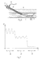
inner seal 2 deployed within the artery, sidearm 9 (with a stopcock in an open position), and apressure transducer 13. Thepressure transducer 13 is in fluid communication with thedistal tip 10A ofintroducer 10. Thus, in Fig. 4,transducer 13 senses normal arterial pressure. The pressure transducer can be any pressure transducer suitable for measuring blood pressure. - Fig. 4 also shows a
display 23 to display pressure waveforms to the surgeon. The display can be any type of display or monitor. Ananalysis circuit 21 is also provided, which analyses the pressure data to output additional information to the surgeon. - Fig. 5 shows the
inner seal 2 properly seated to a puncture hole. In Fig. 5,transducer 13 senses essentially no normal arterial pressure and no normal pressure waveforms in the tissue 14' immediately outside of the seal. Thus, by monitoring the pressure (actually lack of pressure) viatransducer 13,display 23, and/oranalysis circuit 21, the surgeon can confirm thatseal 2 is properly seated (that is, thatseal 2 mates with the inner wall of the blood vessel in a leak tight fashion). If the sealing were incomplete, a pressure will still be present, but at a lower level, and this indicates the need for harder tightening of the sealing elements. The technique shown in Fig. 5 can also be employed to determine whether a seal which seals the puncture from outside the artery is properly positioned. - Fig. 6 shows
seal 2 caught in an upstream branch. As described above, if the seal is caught upstream, blood pressure will still be indicated on the display (or monitor), and the seal can then be manipulated and twisted until it is released from the branch and can be seated to the puncture hole. In the Fig. 6 situation, when the surgeon pulls onsutures 4, the surgeon feels significant resistance and may wrongly believe (without use of the invention) that the seal is properly seated. - Fig. 7 illustrates examples of waveforms displayed on
display 23.Waveform 20 corresponds to the situation when an inner seal is inside the blood vessel and an introducer is positioned as shown in Fig. 4.Waveform 22 corresponds to the situation when the inner seal is positioned as shown in Fig. 5.Waveform 21 corresponds to the situation when the sealing is incomplete. - Figs. 8, 9, and 10 show second, third, and fourth embodiments of the invention.
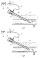
- Fig. 8 shows an
introducer 10 with acore pin 27 inserted. The core pin (a dilator) has one channel 26 (or an axial grove) that communicates atip opening 26a with thesidearm 9. With this technique, the introducer is pushed/pulled forward and backward until theopening 26a (or sidehole) is sealed by thevessel wall 7. This can be detected by a loss of pressure withpressure detector 30. Thedetector 30 is similar totransducer 13 and equipped with a display or audible indication of pressure, for example, an audible indication which varies as pressure varies. The detector is connectable, or adaptable, to thesidearm 9. Once it is confirmed that the introducer tip is positioned at the wall, the introducer can then be pushed forward into the vessel to a desired length. Then, the seal can be inserted into the introducer and deployed as close to the puncture site as possible. - Fig. 9 shows an
introducer 10A with acore pin 27A inserted. The introducer has a sidehole 28 at a desired distance from the tip. With this design, the introducer can be positioned in a single operation by monitoring the pressure ondetector 30, and the length that the introducer protrudes into the vessel is defined by the length from the sidehole to the introducer tip. For example, the introducer can be positioned by positioninghole 28 just inside the vessel wall by monitoring pressure ondetector 30. - The technique shown in Figure 10 is similar to the technique shown in Figure 5, except that in Figure 10, proper sealing of the
inner seal 2 is determined by observing the absence of blood flow fromoutput port 32. - The invention is, of course, not limited to these specific embodiments. Modifications and variations of the invention will occur to those skilled in the field, after receiving the above teachings. The invention is therefore defined by reference to the following claims.
Claims (17)
- A method for confirming correct positioning of an inner seal of a puncture in a blood vessel wall, comprising:(a) providing an introducer assembly having a distal end and a proximal end and a fluid path between the distal end and the proximal end;(b) placing the introducer assembly such that the distal end of the introducer assembly is in tissue outside a puncture in a blood vessel wall;(c) placing a seal inside the blood vessel; and(d) determining whether the puncture is sealed by said seal by observing a characteristic of blood at the proximal end of the introducer assembly.
- A method as claimed in claim 1, wherein step (c) includes:visually observing a flow of blood at the proximal end of the introducer assembly.
- A method as claimed in claim 1, wherein step (c) includes
measuring blood pressure at the proximal end of the introducer assembly with a pressure sensor and outputting an indication of measured blood pressure. - A method as claimed in claim 1, wherein step (c) includes
measuring blood pressure at the proximal end of the introducer assembly with a pressure sensor and displaying a numerical indication of measured blood pressure. - A method as claimed in claim 1, wherein step (c) includes
measuring blood pressure at the proximal end of the introducer assembly with a pressure sensor; and
displaying a blood pressure waveform on a display. - A method as claimed in any preceding claim, further comprising:after step (d), placing a seal outside the blood vessel.
- A system for confirming correct positioning of an inner seal of a puncture in a blood vessel wall, comprising:an introducer assembly (10, 10A, 11) having a distal (10A) end and a proximal end and a fluid path between the distal end and the proximal end, the introducer assembly being arranged such that the distal end of the introducer assembly is in tissue (14) outside a puncture in a blood vessel wall; andmeans (13, 21; 23; 30; 31, 32) for the observation of a characteristic of blood at the proximal end of the introducer assembly to determine whether the puncture is sealed by said seal inside the blood vessel.
- A system as claimed in claim 7, further comprising:an output port (32), in communication with said fluid path, to visually observe a flow of blood from said fluid path.
- A system as claimed in claim 7, further comprising:a pressure sensor (13), connected to the proximal end of the introducer assembly; anda display (23) to display an indication of blood pressure measured by the pressure sensor.
- A system as claimed in claim 7, further comprising:a pressure sensor (13), connected to the proximal end of the introducer assembly; anda display (23) to display a numerical indication of blood pressure measured by the pressure sensor.
- A system as claimed in claim 7, further comprising:a pressure sensor (13) connected to the proximal end of the introducer assembly; anda display (23), which receives signals from the pressure sensor, to display a blood pressure waveform.
- A method for detecting the position of a distal end of an introducer assembly in tissue containing a blood vessel, comprising:(a) providing an introducer assembly having a distal end and a proximal end and a fluid path between the distal end and the proximal end;(b) providing a pressure sensor to measure blood pressure at the proximal end of the introducer assembly;(c) positioning the introducer assembly such that the distal end of the introducer assembly is in tissue containing a blood vessel; and(d) measuring the blood pressure at the proximal end of the introducer assembly using the pressure sensor and outputting said blood pressure as an indication of the position of the distal end of the introducer assembly.
- A method as claimed in claim 12, wherein step (d) includes displaying a blood pressure waveform on a display.
- A system for detecting the position of a distal end of an introducer assembly in tissue containing a blood vessel, comprising:(a) an introducer assembly (10, 10A, 11) having a distal end and a proximal end and a fluid path between the distal end and the proximal end; and(b) a pressure sensor (13), coupled to the proximal end of the introducer assembly, to measure blood pressure and output an indication of blood pressure at the distal end of the introducer assembly.
- A system as claimed in claim 14, further comprising:a display (23) to display a waveform of blood pressure measured by the pressure sensor.
- A system as claimed in claim 14 wherein the introducer assembly comprises a core pin (27) with a channel (26) as said fluid path.
- A system as claimed in claim 14 wherein the introducer assembly comprises:an introducer (33) with a side hole (28) at a distal end of the introducer; anda core pin (27A), insertable into the introducer, with a channel (26) as said fluid path, the channel being in communication with the side hole of the introducer when the core pin is inserted into the introducer.
Applications Claiming Priority (2)
| Application Number | Priority Date | Filing Date | Title |
|---|---|---|---|
| US26089501P | 2001-01-12 | 2001-01-12 | |
| US260895P | 2001-01-12 |
Publications (3)
| Publication Number | Publication Date |
|---|---|
| EP1222896A2 true EP1222896A2 (en) | 2002-07-17 |
| EP1222896A3 EP1222896A3 (en) | 2003-12-03 |
| EP1222896B1 EP1222896B1 (en) | 2005-08-31 |
Family
ID=22991095
Family Applications (1)
| Application Number | Title | Priority Date | Filing Date |
|---|---|---|---|
| EP02445001A Expired - Lifetime EP1222896B1 (en) | 2001-01-12 | 2002-01-09 | Arterial wall sealing device with positioning indicator |
Country Status (6)
| Country | Link |
|---|---|
| US (3) | US6682489B2 (en) |
| EP (1) | EP1222896B1 (en) |
| JP (1) | JP4233256B2 (en) |
| AT (1) | ATE303100T1 (en) |
| DE (1) | DE60205780T2 (en) |
| ES (1) | ES2248510T3 (en) |
Cited By (11)
| Publication number | Priority date | Publication date | Assignee | Title |
|---|---|---|---|---|
| CN105105800A (en) * | 2015-06-26 | 2015-12-02 | 段书华 | Intelligent surgery transport device for interventional therapy on congenital heart disease |
| US10154835B2 (en) | 2013-05-09 | 2018-12-18 | Essential Medical, Inc. | Vascular closure device with conforming plug member |
| US10159571B2 (en) | 2012-11-21 | 2018-12-25 | Corquest Medical, Inc. | Device and method of treating heart valve malfunction |
| US10307167B2 (en) | 2012-12-14 | 2019-06-04 | Corquest Medical, Inc. | Assembly and method for left atrial appendage occlusion |
| US10314594B2 (en) | 2012-12-14 | 2019-06-11 | Corquest Medical, Inc. | Assembly and method for left atrial appendage occlusion |
| US10383611B2 (en) | 2011-10-25 | 2019-08-20 | Essential Medical, Inc. | Instrument and methods for surgically closing percutaneous punctures |
| US10813630B2 (en) | 2011-08-09 | 2020-10-27 | Corquest Medical, Inc. | Closure system for atrial wall |
| US10842626B2 (en) | 2014-12-09 | 2020-11-24 | Didier De Canniere | Intracardiac device to correct mitral regurgitation |
| US11364024B2 (en) | 2013-12-23 | 2022-06-21 | Teleflex Life Sciences Limited | Vascular closure device |
| US11419592B2 (en) | 2013-03-15 | 2022-08-23 | Teleflex Life Sciences Limited | Vascular closure devices and methods of use |
| US11576663B2 (en) | 2015-06-26 | 2023-02-14 | Teleflex Life Sciences Limited | Vascular closure device with removable guide member |
Families Citing this family (107)
| Publication number | Priority date | Publication date | Assignee | Title |
|---|---|---|---|---|
| EP1211983B1 (en) * | 1999-09-13 | 2007-03-07 | Rex Medical, LP | Vascular closure |
| US8083766B2 (en) * | 1999-09-13 | 2011-12-27 | Rex Medical, Lp | Septal defect closure device |
| US7942888B2 (en) | 1999-09-13 | 2011-05-17 | Rex Medical, L.P. | Vascular hole closure device |
| US7267679B2 (en) * | 1999-09-13 | 2007-09-11 | Rex Medical, L.P | Vascular hole closure device |
| US7341595B2 (en) * | 1999-09-13 | 2008-03-11 | Rex Medical, L.P | Vascular hole closure device |
| US7662161B2 (en) * | 1999-09-13 | 2010-02-16 | Rex Medical, L.P | Vascular hole closure device |
| US6846319B2 (en) | 2000-12-14 | 2005-01-25 | Core Medical, Inc. | Devices for sealing openings through tissue and apparatus and methods for delivering them |
| US6623509B2 (en) | 2000-12-14 | 2003-09-23 | Core Medical, Inc. | Apparatus and methods for sealing vascular punctures |
| US6896692B2 (en) | 2000-12-14 | 2005-05-24 | Ensure Medical, Inc. | Plug with collet and apparatus and method for delivering such plugs |
| US6890343B2 (en) | 2000-12-14 | 2005-05-10 | Ensure Medical, Inc. | Plug with detachable guidewire element and methods for use |
| US8083768B2 (en) | 2000-12-14 | 2011-12-27 | Ensure Medical, Inc. | Vascular plug having composite construction |
| US7285097B2 (en) * | 2001-01-12 | 2007-10-23 | Radi Medical System Ab | Technique to confirm correct positioning with respect to arterial wall |
| ES2248510T3 (en) * | 2001-01-12 | 2006-03-16 | Radi Medical Systems Ab | CLOSURE DEVICE OF ARTERIAL PERFORATIONS WITH INDICATION OF POSITIONING. |
| US20080109030A1 (en) * | 2001-04-24 | 2008-05-08 | Houser Russell A | Arteriotomy closure devices and techniques |
| US20110144661A1 (en) * | 2001-04-24 | 2011-06-16 | Houser Russell A | Tissue closure devices, device and systems for delivery, kits and methods therefor |
| US8992567B1 (en) | 2001-04-24 | 2015-03-31 | Cardiovascular Technologies Inc. | Compressible, deformable, or deflectable tissue closure devices and method of manufacture |
| US8961541B2 (en) * | 2007-12-03 | 2015-02-24 | Cardio Vascular Technologies Inc. | Vascular closure devices, systems, and methods of use |
| US8398675B2 (en) * | 2002-10-25 | 2013-03-19 | Radi Medical Systems Ab | Absorbable medical sealing device with retaining assembly having at least two loops |
| EP1442708B1 (en) * | 2003-01-14 | 2008-10-15 | Radi Medical Systems Ab | Introducer sheath |
| US8382793B2 (en) * | 2003-01-14 | 2013-02-26 | Radi Medical Systems Ab | Introducer sheath |
| US7223266B2 (en) | 2003-02-04 | 2007-05-29 | Cardiodex Ltd. | Methods and apparatus for hemostasis following arterial catheterization |
| WO2004093649A2 (en) * | 2003-04-22 | 2004-11-04 | Sub-Q, Inc. | Puncture closure systeme with pin and pull technique |
| US7850654B2 (en) * | 2003-04-24 | 2010-12-14 | St. Jude Medical Puerto Rico B.V. | Device and method for positioning a closure device |
| US20050085773A1 (en) * | 2003-10-15 | 2005-04-21 | Forsberg Andrew T. | Method and apparatus for locating vascular punctures |
| US7361183B2 (en) | 2003-10-17 | 2008-04-22 | Ensure Medical, Inc. | Locator and delivery device and method of use |
| US8852229B2 (en) * | 2003-10-17 | 2014-10-07 | Cordis Corporation | Locator and closure device and method of use |
| US8128652B2 (en) * | 2003-11-13 | 2012-03-06 | St. Jude Medical Puerto Rico Llc | Method and apparatus for sealing an internal tissue puncture incorporating a block and tackle |
| US20050107820A1 (en) * | 2003-11-13 | 2005-05-19 | Forsberg Andrew T. | Vascular puncture depth locator |
| US7597705B2 (en) * | 2003-12-03 | 2009-10-06 | St. Jude Medical Puerto Rico Llc | Vascular puncture seal anchor nest |
| US7621937B2 (en) | 2003-12-03 | 2009-11-24 | St. Jude Medical Puerto Rico LC | Vascular sealing device with high surface area sealing plug |
| JP4589643B2 (en) * | 2004-03-17 | 2010-12-01 | テルモ株式会社 | In vivo tissue closure device |
| US7648493B2 (en) * | 2004-04-20 | 2010-01-19 | St. Jude Medical Puerto Rico Llc | Method and apparatus for locating vascular punctures |
| US20050267520A1 (en) * | 2004-05-12 | 2005-12-01 | Modesitt D B | Access and closure device and method |
| US20050267521A1 (en) * | 2004-05-13 | 2005-12-01 | St. Jude Medical Puerto Rico B.V. | Collagen sponge for arterial sealing |
| US7678133B2 (en) | 2004-07-10 | 2010-03-16 | Arstasis, Inc. | Biological tissue closure device and method |
| US20060058844A1 (en) * | 2004-09-13 | 2006-03-16 | St. Jude Medical Puerto Rico B.V. | Vascular sealing device with locking system |
| JP5068662B2 (en) | 2004-11-22 | 2012-11-07 | カーディオデックス リミテッド | Heat treatment technology for varicose veins |
| US20060116602A1 (en) * | 2004-12-01 | 2006-06-01 | Alden Dana A | Medical sensing device and system |
| US7618436B2 (en) * | 2005-04-12 | 2009-11-17 | St. Jude Medical Puerto Rico Llc | Tissue puncture closure device with scroll gear transmission tamping system |
| EP1871241B1 (en) * | 2005-04-22 | 2012-12-19 | Rex Medical, L.P. | Closure device for left atrial appendage |
| US8926654B2 (en) | 2005-05-04 | 2015-01-06 | Cordis Corporation | Locator and closure device and method of use |
| US20060276836A1 (en) * | 2005-06-07 | 2006-12-07 | Bergin Patrick J | Hemostatic wire guided bandage and method of use |
| US20080015481A1 (en) * | 2005-05-04 | 2008-01-17 | Bergin Patrick J | Hemostatic bandage and method of use |
| US7622628B2 (en) * | 2005-05-04 | 2009-11-24 | Innovasa Corporation | Hemostatic wire guided bandage and method of use |
| US8088144B2 (en) | 2005-05-04 | 2012-01-03 | Ensure Medical, Inc. | Locator and closure device and method of use |
| US20060276838A1 (en) * | 2005-06-07 | 2006-12-07 | Wensel Jeffrey P | Vascular puncture sealing method, apparatus, and system |
| AU2006247355B2 (en) * | 2005-05-12 | 2013-01-10 | Arstasis, Inc. | Access and closure device and method |
| US7618438B2 (en) * | 2005-05-17 | 2009-11-17 | St. Jude Medical Puerto Rico Llc | Tissue puncture closure device with disengagable automatic tamping system |
| US8273094B2 (en) * | 2005-05-17 | 2012-09-25 | St. Jude Medical Puerto Rico Llc | Puncture locating device |
| US7135377B1 (en) * | 2005-05-20 | 2006-11-14 | Phoenix Precision Technology Corporation | Semiconductor package substrate with embedded resistors and method for fabricating same |
| US8382794B2 (en) * | 2006-01-04 | 2013-02-26 | St. Jude Medical Puerto Rico Llc | Balloon insertion apparatus and method of sealing a tissue puncture |
| US7850710B2 (en) * | 2006-05-23 | 2010-12-14 | St. Jude Medical Puerto Rico Llc | Puncture closure apparatuses, sealing plugs, and related methods |
| US7749248B2 (en) * | 2006-09-18 | 2010-07-06 | St. Jude Medical Puerto Rico Llc | Flexible tamping device |
| US20080262475A1 (en) * | 2007-04-20 | 2008-10-23 | Radi Medical Systems Ab | Adapter for an introducer |
| AU2008276658B2 (en) * | 2007-07-13 | 2013-10-03 | Rex Medical, Lp | Vascular hole closure device |
| US8366706B2 (en) * | 2007-08-15 | 2013-02-05 | Cardiodex, Ltd. | Systems and methods for puncture closure |
| US8333787B2 (en) | 2007-12-31 | 2012-12-18 | St. Jude Medical Puerto Rico Llc | Vascular closure device having a flowable sealing material |
| US8568445B2 (en) * | 2007-08-21 | 2013-10-29 | St. Jude Medical Puerto Rico Llc | Extra-vascular sealing device and method |
| US8310336B2 (en) | 2008-10-10 | 2012-11-13 | Masimo Corporation | Systems and methods for storing, analyzing, retrieving and displaying streaming medical data |
| US20090105744A1 (en) * | 2007-10-17 | 2009-04-23 | Modesitt D Bruce | Methods for forming tracts in tissue |
| EP2231030B1 (en) * | 2007-12-21 | 2019-02-27 | MicroVention, Inc. | System and method for locating detachment zone of a detachable implant |
| US9282953B2 (en) * | 2007-12-31 | 2016-03-15 | St. Jude Medical Puerto Rico Llc | Systems and methods for locating and closing a tissue puncture |
| US8840640B2 (en) | 2007-12-31 | 2014-09-23 | St. Jude Medical Puerto Rico Llc | Vascular closure device having an improved plug |
| US9226738B2 (en) | 2008-02-15 | 2016-01-05 | Rex Medical, L.P. | Vascular hole closure delivery device |
| US8920462B2 (en) | 2008-02-15 | 2014-12-30 | Rex Medical, L.P. | Vascular hole closure device |
| US8070772B2 (en) | 2008-02-15 | 2011-12-06 | Rex Medical, L.P. | Vascular hole closure device |
| US8491629B2 (en) | 2008-02-15 | 2013-07-23 | Rex Medical | Vascular hole closure delivery device |
| US8920463B2 (en) | 2008-02-15 | 2014-12-30 | Rex Medical, L.P. | Vascular hole closure device |
| US20110029013A1 (en) | 2008-02-15 | 2011-02-03 | Mcguckin James F | Vascular Hole Closure Device |
| US8968345B2 (en) * | 2008-03-24 | 2015-03-03 | Covidien Lp | Surgical introducer with indicators |
| WO2009149474A1 (en) | 2008-06-06 | 2009-12-10 | Vital Access Corporation | Tissue management methods, apparatus, and systems |
| CN102159126A (en) * | 2008-07-21 | 2011-08-17 | 阿尔斯塔西斯公司 | Devices, methods, and kits for forming tracts in tissue |
| AU2009274128A1 (en) * | 2008-07-21 | 2010-01-28 | Arstasis, Inc. | Devices and methods for forming tracts in tissue |
| AU2009288440B2 (en) * | 2008-08-26 | 2015-04-23 | St Jude Medical, Inc. | Device and sealing component for sealing punctures |
| US9179901B2 (en) | 2009-01-29 | 2015-11-10 | Vital Access Corporation | Vascular access ports and related methods |
| CA2751185C (en) | 2009-01-29 | 2018-07-03 | Vital Access Corporation | Vascular access ports and related methods |
| US11197952B2 (en) | 2009-01-29 | 2021-12-14 | Advent Access Pte. Ltd. | Vascular access ports and related methods |
| US8292918B2 (en) | 2009-02-20 | 2012-10-23 | Boston Scientific Scimed, Inc. | Composite plug for arteriotomy closure and method of use |
| US20100217309A1 (en) * | 2009-02-20 | 2010-08-26 | Boston Scientific Scimed, Inc. | Plug for arteriotomy closure and method of use |
| US8317824B2 (en) * | 2009-02-20 | 2012-11-27 | Boston Scientific Scimed, Inc. | Tissue puncture closure device |
| US8529598B2 (en) | 2009-02-20 | 2013-09-10 | Boston Scientific Scimed, Inc. | Tissue puncture closure device |
| US8052914B2 (en) * | 2009-02-20 | 2011-11-08 | Boston Scientific Scimed, Inc. | Modified plug for arteriotomy closure |
| US8375553B2 (en) | 2009-02-20 | 2013-02-19 | Boston Scientific Scimed, Inc. | Locking element for vascular closure device |
| US9913634B2 (en) * | 2009-02-20 | 2018-03-13 | Boston Scientific Scimed, Inc. | Locking element for vascular closure device |
| EP3605550A1 (en) | 2009-03-04 | 2020-02-05 | Masimo Corporation | Medical monitoring system |
| US10032002B2 (en) | 2009-03-04 | 2018-07-24 | Masimo Corporation | Medical monitoring system |
| US9323894B2 (en) | 2011-08-19 | 2016-04-26 | Masimo Corporation | Health care sanitation monitoring system |
| US10007758B2 (en) | 2009-03-04 | 2018-06-26 | Masimo Corporation | Medical monitoring system |
| WO2010118312A2 (en) * | 2009-04-09 | 2010-10-14 | Cardiovascular Systems, Inc. | Tissue closure devices, device and systems for delivery, kits and methods therefor |
| AU2010248821A1 (en) * | 2009-05-15 | 2011-12-01 | Arstasis, Inc. | Devices, methods and kits for forming tracts in tissue |
| CA2774958A1 (en) * | 2009-09-22 | 2011-03-31 | Arstasis, Inc. | Devices, methods, and kits for forming tracts in tissue |
| US8478384B2 (en) | 2010-01-19 | 2013-07-02 | Lightlab Imaging, Inc. | Intravascular optical coherence tomography system with pressure monitoring interface and accessories |
| WO2011100547A2 (en) | 2010-02-11 | 2011-08-18 | Boston Scientific Scimed, Inc. | Automatic vascular closure deployment devices and methods |
| US20120022562A1 (en) * | 2010-07-23 | 2012-01-26 | Boston Scientific Scimed, Inc. | Device to detect internal bleeding |
| US8597340B2 (en) | 2010-09-17 | 2013-12-03 | Boston Scientific Scimed, Inc. | Torque mechanism actuated bioabsorbable vascular closure device |
| US8758402B2 (en) | 2010-12-17 | 2014-06-24 | Boston Scientific Scimed, Inc. | Tissue puncture closure device |
| CA2836790C (en) | 2011-05-31 | 2019-04-23 | Desmond Adler | Multimodal imaging system, apparatus, and methods |
| WO2013019840A1 (en) | 2011-08-03 | 2013-02-07 | Lightlab Imaging, Inc. | Systems, methods and apparatus for determining a fractional flow reserve |
| US20130317438A1 (en) | 2012-05-25 | 2013-11-28 | Arstasis, Inc. | Vascular access configuration |
| US20130317481A1 (en) | 2012-05-25 | 2013-11-28 | Arstasis, Inc. | Vascular access configuration |
| US9554785B2 (en) | 2012-12-21 | 2017-01-31 | Essential Medical, Inc. | Vascular locating systems and methods of use |
| US9566443B2 (en) | 2013-11-26 | 2017-02-14 | Corquest Medical, Inc. | System for treating heart valve malfunction including mitral regurgitation |
| AU2015242353B2 (en) | 2014-04-04 | 2020-01-23 | St. Jude Medical Systems Ab | Intravascular pressure and flow data diagnostic systems, devices, and methods |
| CN108210017A (en) * | 2018-03-05 | 2018-06-29 | 江秀秀 | A kind of surgical clamp and the method for judging clamped organization type |
| US11504105B2 (en) | 2019-01-25 | 2022-11-22 | Rex Medical L.P. | Vascular hole closure device |
| US11350919B2 (en) | 2019-02-19 | 2022-06-07 | Teleflex Life Sciences Limited | Puncture locating system with blood pulsation indicator |
| US20220031353A1 (en) * | 2020-07-31 | 2022-02-03 | Arrow International Llc | Access sheath with valve assembly |
Citations (2)
| Publication number | Priority date | Publication date | Assignee | Title |
|---|---|---|---|---|
| US5613974A (en) | 1992-12-10 | 1997-03-25 | Perclose, Inc. | Apparatus and method for vascular closure |
| US6090130A (en) | 1991-11-08 | 2000-07-18 | Kensey Nash Corporation | Hemostatic puncture closure system including blood vessel locator and method of use |
Family Cites Families (25)
| Publication number | Priority date | Publication date | Assignee | Title |
|---|---|---|---|---|
| US4834108A (en) * | 1986-06-09 | 1989-05-30 | Manresa, Inc. | Blocking filter to prevent air flow into a fluid conduit to a transducer |
| US4890612A (en) * | 1987-02-17 | 1990-01-02 | Kensey Nash Corporation | Device for sealing percutaneous puncture in a vessel |
| US4852568A (en) * | 1987-02-17 | 1989-08-01 | Kensey Nash Corporation | Method and apparatus for sealing an opening in tissue of a living being |
| US5217671A (en) * | 1988-09-22 | 1993-06-08 | Terumo Kabushiki Kaisha | Method of making a tubular body |
| US4928693A (en) * | 1989-03-13 | 1990-05-29 | Schneider (Usa), Inc. | Pressure monitor catheter |
| US5222974A (en) * | 1991-11-08 | 1993-06-29 | Kensey Nash Corporation | Hemostatic puncture closure system and method of use |
| US5282827A (en) * | 1991-11-08 | 1994-02-01 | Kensey Nash Corporation | Hemostatic puncture closure system and method of use |
| US6350274B1 (en) * | 1992-05-11 | 2002-02-26 | Regen Biologics, Inc. | Soft tissue closure systems |
| US5460607A (en) * | 1992-09-30 | 1995-10-24 | Nippon Zeon Co., Ltd. | Balloon catheter |
| US5431639A (en) * | 1993-08-12 | 1995-07-11 | Boston Scientific Corporation | Treating wounds caused by medical procedures |
| NL9301526A (en) * | 1993-09-03 | 1995-04-03 | Cordis Europ | Device for hemostasis treatment after catheter surgery. |
| US5843124A (en) * | 1993-09-28 | 1998-12-01 | Hemodynamics, Inc. | Surface opening adhesive sealer |
| US5395353A (en) * | 1993-11-02 | 1995-03-07 | Vascular Technologies, Inc. | Guiding catheter with controllable perfusion ports |
| US6302898B1 (en) * | 1994-06-24 | 2001-10-16 | Advanced Closure Systems, Inc. | Devices for sealing punctures in body vessels |
| DE19514638C2 (en) * | 1995-04-20 | 1998-06-04 | Peter Dr Med Boekstegers | Device for the selective suction and retroinfusion of a fluid from or into body veins controlled by venous pressure |
| US6117144A (en) * | 1995-08-24 | 2000-09-12 | Sutura, Inc. | Suturing device and method for sealing an opening in a blood vessel or other biological structure |
| US5728132A (en) * | 1996-04-08 | 1998-03-17 | Tricardia, L.L.C. | Self-sealing vascular access device |
| US5855559A (en) * | 1997-02-14 | 1999-01-05 | Tricardia, Inc. | Hemostatic agent delivery device having built-in pressure sensor |
| US6193670B1 (en) | 1997-02-14 | 2001-02-27 | Tricardia, Llc | Hemostatic agent delivery device having built-in pressure sensor |
| US6733515B1 (en) * | 1997-03-12 | 2004-05-11 | Neomend, Inc. | Universal introducer |
| EP0879617B1 (en) * | 1997-05-21 | 2003-04-16 | Schneider (Europe) GmbH | Pressure monitoring guide wire and method for manufacturing such a guide wire |
| US5964782A (en) * | 1997-09-18 | 1999-10-12 | Scimed Life Systems, Inc. | Closure device and method |
| US6493670B1 (en) * | 1999-10-14 | 2002-12-10 | Ericsson Inc. | Method and apparatus for transmitting DTMF signals employing local speech recognition |
| US6508828B1 (en) * | 2000-11-03 | 2003-01-21 | Radi Medical Systems Ab | Sealing device and wound closure device |
| ES2248510T3 (en) * | 2001-01-12 | 2006-03-16 | Radi Medical Systems Ab | CLOSURE DEVICE OF ARTERIAL PERFORATIONS WITH INDICATION OF POSITIONING. |
-
2002
- 2002-01-09 ES ES02445001T patent/ES2248510T3/en not_active Expired - Lifetime
- 2002-01-09 AT AT02445001T patent/ATE303100T1/en not_active IP Right Cessation
- 2002-01-09 DE DE60205780T patent/DE60205780T2/en not_active Expired - Lifetime
- 2002-01-09 EP EP02445001A patent/EP1222896B1/en not_active Expired - Lifetime
- 2002-01-11 JP JP2002004868A patent/JP4233256B2/en not_active Expired - Fee Related
- 2002-01-11 US US10/042,247 patent/US6682489B2/en not_active Expired - Fee Related
-
2003
- 2003-11-12 US US10/704,556 patent/US7044916B2/en not_active Expired - Lifetime
-
2004
- 2004-03-12 US US10/798,438 patent/US7073509B2/en not_active Expired - Fee Related
Patent Citations (2)
| Publication number | Priority date | Publication date | Assignee | Title |
|---|---|---|---|---|
| US6090130A (en) | 1991-11-08 | 2000-07-18 | Kensey Nash Corporation | Hemostatic puncture closure system including blood vessel locator and method of use |
| US5613974A (en) | 1992-12-10 | 1997-03-25 | Perclose, Inc. | Apparatus and method for vascular closure |
Cited By (14)
| Publication number | Priority date | Publication date | Assignee | Title |
|---|---|---|---|---|
| US10813630B2 (en) | 2011-08-09 | 2020-10-27 | Corquest Medical, Inc. | Closure system for atrial wall |
| US10383611B2 (en) | 2011-10-25 | 2019-08-20 | Essential Medical, Inc. | Instrument and methods for surgically closing percutaneous punctures |
| US11589855B2 (en) | 2011-10-25 | 2023-02-28 | Teleflex Life Sciences Limited | Instrument and methods for surgically closing percutaneous punctures |
| US10485524B2 (en) | 2011-10-25 | 2019-11-26 | Essential Medical, Inc. | Instrument and methods for surgically closing percutaneous punctures |
| US10159571B2 (en) | 2012-11-21 | 2018-12-25 | Corquest Medical, Inc. | Device and method of treating heart valve malfunction |
| US10307167B2 (en) | 2012-12-14 | 2019-06-04 | Corquest Medical, Inc. | Assembly and method for left atrial appendage occlusion |
| US10314594B2 (en) | 2012-12-14 | 2019-06-11 | Corquest Medical, Inc. | Assembly and method for left atrial appendage occlusion |
| US11419592B2 (en) | 2013-03-15 | 2022-08-23 | Teleflex Life Sciences Limited | Vascular closure devices and methods of use |
| US10154835B2 (en) | 2013-05-09 | 2018-12-18 | Essential Medical, Inc. | Vascular closure device with conforming plug member |
| US11364024B2 (en) | 2013-12-23 | 2022-06-21 | Teleflex Life Sciences Limited | Vascular closure device |
| US11779320B2 (en) | 2013-12-23 | 2023-10-10 | Teleflex Life Sciences Limited | Vascular closure device |
| US10842626B2 (en) | 2014-12-09 | 2020-11-24 | Didier De Canniere | Intracardiac device to correct mitral regurgitation |
| CN105105800A (en) * | 2015-06-26 | 2015-12-02 | 段书华 | Intelligent surgery transport device for interventional therapy on congenital heart disease |
| US11576663B2 (en) | 2015-06-26 | 2023-02-14 | Teleflex Life Sciences Limited | Vascular closure device with removable guide member |
Also Published As
| Publication number | Publication date |
|---|---|
| US6682489B2 (en) | 2004-01-27 |
| DE60205780D1 (en) | 2005-10-06 |
| JP2002253556A (en) | 2002-09-10 |
| US7044916B2 (en) | 2006-05-16 |
| EP1222896B1 (en) | 2005-08-31 |
| US20040172059A1 (en) | 2004-09-02 |
| US20020095179A1 (en) | 2002-07-18 |
| US20040098046A1 (en) | 2004-05-20 |
| JP4233256B2 (en) | 2009-03-04 |
| DE60205780T2 (en) | 2006-05-18 |
| US7073509B2 (en) | 2006-07-11 |
| EP1222896A3 (en) | 2003-12-03 |
| ES2248510T3 (en) | 2006-03-16 |
| ATE303100T1 (en) | 2005-09-15 |
Similar Documents
| Publication | Publication Date | Title |
|---|---|---|
| EP1222896B1 (en) | Arterial wall sealing device with positioning indicator | |
| US7285097B2 (en) | Technique to confirm correct positioning with respect to arterial wall | |
| US7361183B2 (en) | Locator and delivery device and method of use | |
| AU2004233866B2 (en) | Device and method for positioning a closure device | |
| EP0664687B1 (en) | Vessel position locating device | |
| CA2152061C (en) | Surgical depth measuring instrument and method | |
| JP2904733B2 (en) | Apparatus for determining position of blood vessel or lumen wall of living body | |
| US10463838B2 (en) | Vascular access methods and devices | |
| US20070123781A1 (en) | Surgical anastomosis leak detection system | |
| US20130144331A1 (en) | Introducer sheath | |
| EP1442708B1 (en) | Introducer sheath | |
| US20240033449A1 (en) | Method and system for detecting leaks and/or verifying adequate closure following a medical procedure |
Legal Events
| Date | Code | Title | Description |
|---|---|---|---|
| PUAI | Public reference made under article 153(3) epc to a published international application that has entered the european phase |
Free format text: ORIGINAL CODE: 0009012 |
|
| AK | Designated contracting states |
Kind code of ref document: A2 Designated state(s): AT BE CH CY DE DK ES FI FR GB GR IE IT LI LU MC NL PT SE TR |
|
| AX | Request for extension of the european patent |
Free format text: AL;LT;LV;MK;RO;SI |
|
| PUAL | Search report despatched |
Free format text: ORIGINAL CODE: 0009013 |
|
| AK | Designated contracting states |
Kind code of ref document: A3 Designated state(s): AT BE CH CY DE DK ES FI FR GB GR IE IT LI LU MC NL PT SE TR |
|
| AX | Request for extension of the european patent |
Extension state: AL LT LV MK RO SI |
|
| 17P | Request for examination filed |
Effective date: 20040112 |
|
| 17Q | First examination report despatched |
Effective date: 20040420 |
|
| AKX | Designation fees paid |
Designated state(s): AT BE CH CY DE DK ES FI FR GB GR IE IT LI LU MC NL PT SE TR |
|
| GRAP | Despatch of communication of intention to grant a patent |
Free format text: ORIGINAL CODE: EPIDOSNIGR1 |
|
| GRAS | Grant fee paid |
Free format text: ORIGINAL CODE: EPIDOSNIGR3 |
|
| GRAA | (expected) grant |
Free format text: ORIGINAL CODE: 0009210 |
|
| AK | Designated contracting states |
Kind code of ref document: B1 Designated state(s): AT BE CH CY DE DK ES FI FR GB GR IE IT LI LU MC NL PT SE TR |
|
| PG25 | Lapsed in a contracting state [announced via postgrant information from national office to epo] |
Ref country code: IT Free format text: LAPSE BECAUSE OF FAILURE TO SUBMIT A TRANSLATION OF THE DESCRIPTION OR TO PAY THE FEE WITHIN THE PRESCRIBED TIME-LIMIT;WARNING: LAPSES OF ITALIAN PATENTS WITH EFFECTIVE DATE BEFORE 2007 MAY HAVE OCCURRED AT ANY TIME BEFORE 2007. THE CORRECT EFFECTIVE DATE MAY BE DIFFERENT FROM THE ONE RECORDED. Effective date: 20050831 Ref country code: AT Free format text: LAPSE BECAUSE OF FAILURE TO SUBMIT A TRANSLATION OF THE DESCRIPTION OR TO PAY THE FEE WITHIN THE PRESCRIBED TIME-LIMIT Effective date: 20050831 Ref country code: FI Free format text: LAPSE BECAUSE OF FAILURE TO SUBMIT A TRANSLATION OF THE DESCRIPTION OR TO PAY THE FEE WITHIN THE PRESCRIBED TIME-LIMIT Effective date: 20050831 |
|
| REG | Reference to a national code |
Ref country code: CH Ref legal event code: EP Ref country code: GB Ref legal event code: FG4D |
|
| REG | Reference to a national code |
Ref country code: IE Ref legal event code: FG4D |
|
| REF | Corresponds to: |
Ref document number: 60205780 Country of ref document: DE Date of ref document: 20051006 Kind code of ref document: P |
|
| PG25 | Lapsed in a contracting state [announced via postgrant information from national office to epo] |
Ref country code: DK Free format text: LAPSE BECAUSE OF FAILURE TO SUBMIT A TRANSLATION OF THE DESCRIPTION OR TO PAY THE FEE WITHIN THE PRESCRIBED TIME-LIMIT Effective date: 20051130 Ref country code: GR Free format text: LAPSE BECAUSE OF FAILURE TO SUBMIT A TRANSLATION OF THE DESCRIPTION OR TO PAY THE FEE WITHIN THE PRESCRIBED TIME-LIMIT Effective date: 20051130 |
|
| REG | Reference to a national code |
Ref country code: SE Ref legal event code: TRGR |
|
| REG | Reference to a national code |
Ref country code: CH Ref legal event code: NV Representative=s name: PATENTANWAELTE SCHAAD, BALASS, MENZL & PARTNER AG |
|
| PG25 | Lapsed in a contracting state [announced via postgrant information from national office to epo] |
Ref country code: IE Free format text: LAPSE BECAUSE OF NON-PAYMENT OF DUE FEES Effective date: 20060109 |
|
| PG25 | Lapsed in a contracting state [announced via postgrant information from national office to epo] |
Ref country code: MC Free format text: LAPSE BECAUSE OF NON-PAYMENT OF DUE FEES Effective date: 20060131 |
|
| PG25 | Lapsed in a contracting state [announced via postgrant information from national office to epo] |
Ref country code: PT Free format text: LAPSE BECAUSE OF FAILURE TO SUBMIT A TRANSLATION OF THE DESCRIPTION OR TO PAY THE FEE WITHIN THE PRESCRIBED TIME-LIMIT Effective date: 20060222 |
|
| REG | Reference to a national code |
Ref country code: ES Ref legal event code: FG2A Ref document number: 2248510 Country of ref document: ES Kind code of ref document: T3 |
|
| ET | Fr: translation filed | ||
| PLBE | No opposition filed within time limit |
Free format text: ORIGINAL CODE: 0009261 |
|
| STAA | Information on the status of an ep patent application or granted ep patent |
Free format text: STATUS: NO OPPOSITION FILED WITHIN TIME LIMIT |
|
| 26N | No opposition filed |
Effective date: 20060601 |
|
| REG | Reference to a national code |
Ref country code: IE Ref legal event code: MM4A |
|
| PG25 | Lapsed in a contracting state [announced via postgrant information from national office to epo] |
Ref country code: TR Free format text: LAPSE BECAUSE OF FAILURE TO SUBMIT A TRANSLATION OF THE DESCRIPTION OR TO PAY THE FEE WITHIN THE PRESCRIBED TIME-LIMIT Effective date: 20050831 |
|
| PG25 | Lapsed in a contracting state [announced via postgrant information from national office to epo] |
Ref country code: CY Free format text: LAPSE BECAUSE OF FAILURE TO SUBMIT A TRANSLATION OF THE DESCRIPTION OR TO PAY THE FEE WITHIN THE PRESCRIBED TIME-LIMIT Effective date: 20050831 |
|
| PGFP | Annual fee paid to national office [announced via postgrant information from national office to epo] |
Ref country code: LU Payment date: 20140203 Year of fee payment: 13 |
|
| PGFP | Annual fee paid to national office [announced via postgrant information from national office to epo] |
Ref country code: BE Payment date: 20140127 Year of fee payment: 13 Ref country code: NL Payment date: 20140126 Year of fee payment: 13 Ref country code: CH Payment date: 20140129 Year of fee payment: 13 |
|
| REG | Reference to a national code |
Ref country code: FR Ref legal event code: PLFP Year of fee payment: 14 |
|
| PGFP | Annual fee paid to national office [announced via postgrant information from national office to epo] |
Ref country code: DE Payment date: 20150128 Year of fee payment: 14 Ref country code: ES Payment date: 20150126 Year of fee payment: 14 |
|
| PGFP | Annual fee paid to national office [announced via postgrant information from national office to epo] |
Ref country code: FR Payment date: 20150119 Year of fee payment: 14 Ref country code: SE Payment date: 20150128 Year of fee payment: 14 Ref country code: GB Payment date: 20150127 Year of fee payment: 14 |
|
| PG25 | Lapsed in a contracting state [announced via postgrant information from national office to epo] |
Ref country code: BE Free format text: LAPSE BECAUSE OF NON-PAYMENT OF DUE FEES Effective date: 20150131 |
|
| REG | Reference to a national code |
Ref country code: DE Ref legal event code: R082 Ref document number: 60205780 Country of ref document: DE Representative=s name: HOFFMANN - EITLE PATENT- UND RECHTSANWAELTE PA, DE Ref country code: DE Ref legal event code: R081 Ref document number: 60205780 Country of ref document: DE Owner name: ST. JUDE MEDICAL COORDINATION CENTER BVBA, BE Free format text: FORMER OWNER: RADI MEDICAL SYSTEMS AB, UPPSALA, SE |
|
| REG | Reference to a national code |
Ref country code: NL Ref legal event code: V1 Effective date: 20150801 |
|
| REG | Reference to a national code |
Ref country code: CH Ref legal event code: PL |
|
| PG25 | Lapsed in a contracting state [announced via postgrant information from national office to epo] |
Ref country code: LU Free format text: LAPSE BECAUSE OF NON-PAYMENT OF DUE FEES Effective date: 20150109 |
|
| REG | Reference to a national code |
Ref country code: GB Ref legal event code: 732E Free format text: REGISTERED BETWEEN 20150827 AND 20150902 |
|
| REG | Reference to a national code |
Ref country code: ES Ref legal event code: PC2A Owner name: ST. JUDE MEDICAL COORDINATION CENTER BVBA Effective date: 20150923 |
|
| PG25 | Lapsed in a contracting state [announced via postgrant information from national office to epo] |
Ref country code: NL Free format text: LAPSE BECAUSE OF NON-PAYMENT OF DUE FEES Effective date: 20150801 |
|
| PG25 | Lapsed in a contracting state [announced via postgrant information from national office to epo] |
Ref country code: LI Free format text: LAPSE BECAUSE OF NON-PAYMENT OF DUE FEES Effective date: 20150131 Ref country code: CH Free format text: LAPSE BECAUSE OF NON-PAYMENT OF DUE FEES Effective date: 20150131 |
|
| REG | Reference to a national code |
Ref country code: FR Ref legal event code: TP Owner name: ST. JUDE MEDICAL COORDINATION CENTER BVBA, BE Effective date: 20160113 |
|
| REG | Reference to a national code |
Ref country code: DE Ref legal event code: R119 Ref document number: 60205780 Country of ref document: DE |
|
| GBPC | Gb: european patent ceased through non-payment of renewal fee |
Effective date: 20160109 |
|
| REG | Reference to a national code |
Ref country code: FR Ref legal event code: ST Effective date: 20160930 |
|
| PG25 | Lapsed in a contracting state [announced via postgrant information from national office to epo] |
Ref country code: GB Free format text: LAPSE BECAUSE OF NON-PAYMENT OF DUE FEES Effective date: 20160109 Ref country code: DE Free format text: LAPSE BECAUSE OF NON-PAYMENT OF DUE FEES Effective date: 20160802 |
|
| PG25 | Lapsed in a contracting state [announced via postgrant information from national office to epo] |
Ref country code: SE Free format text: LAPSE BECAUSE OF NON-PAYMENT OF DUE FEES Effective date: 20160110 Ref country code: FR Free format text: LAPSE BECAUSE OF NON-PAYMENT OF DUE FEES Effective date: 20160201 |
|
| PG25 | Lapsed in a contracting state [announced via postgrant information from national office to epo] |
Ref country code: ES Free format text: LAPSE BECAUSE OF NON-PAYMENT OF DUE FEES Effective date: 20160110 |
|
| REG | Reference to a national code |
Ref country code: ES Ref legal event code: FD2A Effective date: 20181205 |