EP1221299A2 - Method and apparatus for generating a twelve-lead ecg from fewer than ten electrodes - Google Patents
Method and apparatus for generating a twelve-lead ecg from fewer than ten electrodes Download PDFInfo
- Publication number
- EP1221299A2 EP1221299A2 EP01310732A EP01310732A EP1221299A2 EP 1221299 A2 EP1221299 A2 EP 1221299A2 EP 01310732 A EP01310732 A EP 01310732A EP 01310732 A EP01310732 A EP 01310732A EP 1221299 A2 EP1221299 A2 EP 1221299A2
- Authority
- EP
- European Patent Office
- Prior art keywords
- patient
- electrodes
- twelve
- electrode
- electrical signals
- Prior art date
- Legal status (The legal status is an assumption and is not a legal conclusion. Google has not performed a legal analysis and makes no representation as to the accuracy of the status listed.)
- Granted
Links
Images


Classifications
-
- A—HUMAN NECESSITIES
- A61—MEDICAL OR VETERINARY SCIENCE; HYGIENE
- A61B—DIAGNOSIS; SURGERY; IDENTIFICATION
- A61B5/00—Measuring for diagnostic purposes; Identification of persons
- A61B5/24—Detecting, measuring or recording bioelectric or biomagnetic signals of the body or parts thereof
- A61B5/316—Modalities, i.e. specific diagnostic methods
- A61B5/318—Heart-related electrical modalities, e.g. electrocardiography [ECG]
- A61B5/327—Generation of artificial ECG signals based on measured signals, e.g. to compensate for missing leads
-
- A—HUMAN NECESSITIES
- A61—MEDICAL OR VETERINARY SCIENCE; HYGIENE
- A61B—DIAGNOSIS; SURGERY; IDENTIFICATION
- A61B5/00—Measuring for diagnostic purposes; Identification of persons
- A61B5/24—Detecting, measuring or recording bioelectric or biomagnetic signals of the body or parts thereof
- A61B5/25—Bioelectric electrodes therefor
- A61B5/279—Bioelectric electrodes therefor specially adapted for particular uses
- A61B5/28—Bioelectric electrodes therefor specially adapted for particular uses for electrocardiography [ECG]
- A61B5/282—Holders for multiple electrodes
-
- A—HUMAN NECESSITIES
- A61—MEDICAL OR VETERINARY SCIENCE; HYGIENE
- A61B—DIAGNOSIS; SURGERY; IDENTIFICATION
- A61B5/00—Measuring for diagnostic purposes; Identification of persons
- A61B5/24—Detecting, measuring or recording bioelectric or biomagnetic signals of the body or parts thereof
- A61B5/30—Input circuits therefor
- A61B5/307—Input circuits therefor specially adapted for particular uses
- A61B5/308—Input circuits therefor specially adapted for particular uses for electrocardiography [ECG]
Definitions
- the invention relates to a method and apparatus for generating a twelve-lead electrocardiogram (ECG) from a plurality of fewer than ten electrodes for attachment to a patient in the standard ten-electrode, twelve-lead ECG positions.
- ECG electrocardiogram
- a standard, resting ECG is acquired with ten electrodes. Four of the ten electrodes are placed on the patient's limbs. Six of the ten electrodes are attached to the patient's chest over the heart. The signals acquired by the ten electrodes are amplified and processed to generate twelve channels of ECG data. The twelve channels or leads are generally split into two groups - the frontal plane leads (I, II, III, aVR, aVL, aVF) and the horizontal plane leads (V1, V2, V3, V4, V5, V6).
- a standard, resting ECG has several limitations.
- the electrodes, leadwires, and amplifiers necessary to acquire twelve channels of ECG data increase the cost of the ECG machine.
- Fourth, the amount of data representing twelve channels of ECG generally exceeds the maximum amount or bandwidth that typical telemetry units are able to transmit.
- U.S. Patent No. 4,850,370 provides a solution to some of the above-described limitations.
- the '370 Patent discloses a method of sensing and analyzing the electrical activity of the human heart by sensing the voltage signals generated by the heart between four electrodes located at key positions on the surface of the subject's body.
- a signal processing means produces electrocardiographic signals corresponding to the lead signals of a twelve-lead electrocardiogram.
- FIG. 3 illustrates the electrode placement for the method of generating a twelve-lead ECG from four electrodes as disclosed in the '370 Patent.
- the four electrodes are designated as E, A, S, and I (hereinafter referred to collectively as the "EASI electrodes").
- the E electrode is located at the front midline over the lower end of the sternum.
- the A electrode is located at the left mid-axillary line.
- the S electrode is located at the front midline over the upper end of the sternum.
- the I electrode is located at the right mid-axillary line.
- the EASI electrodes are coupled to a signal processor (not shown) having a first stage and a second stage.
- the first stage of the signal processor does not generate the twelve-lead ECG, but rather generates xyz vectorcardiographic signals.
- the twelve-lead ECG is then derived from the xyz vectorcardiographic signals in the second stage of the signal processor.
- each of the twelve leads generated from the EASI electrodes are mathematically generated. In other words, none of the leads are the same as the leads that would be generated from the electrical signals of a standard ten-electrode, twelve-lead ECG.
- the method of the '370 Patent has several limitations.
- Third, two of the four electrodes are placed directly over the sternum. The sternum is cracked for all open chest surgeries in a procedure called sternotomy. Thus, the clinician may not be able to attach the two electrodes directly over the sternum due to sternotomy wounds and bandages.
- the invention provides a method and apparatus for generating a twelve-lead ECG from fewer than ten electrodes for attachment to a patient in at least some of the standard ten-electrode, twelve-lead ECG positions.
- the apparatus is a device for acquiring and processing electrical signals produced by a patient's heart.
- the device includes fewer than ten electrodes for attachment to the patient. Each electrode is attached in a respective one of the standard ten-electrode, twelve-lead ECG positions.
- the device includes a signal processor connected to the electrodes. The signal processor acquires electrical signals from the electrodes and generates a twelve-lead ECG from the electrical signals. The signal processor generates less than twelve of the leads mathematically.
- a plurality of less than ten electrodes are attached to the patient.
- Each electrode is attached in a respective one of the standard ten-electrode, twelve-lead ECG positions. Electrical signals are acquired from the electrodes and a twelve-lead ECG is generated from the acquired electrical signals. But not all twelve leads are generated mathematically.
- the device employs multiple-linear regression using expansion-coefficient equations to mathematically generate fewer than twelve of the leads.
- the expansion-coefficient equations are determined either from ECGs from a hospital's general population, from a sub-population of the hospital's general population, or from ECGs previously acquired from the patient.
- the invention employs multiple-linear regression to generate the leads that are missing due to the use of fewer than ten electrodes. Stated differently, some of the leads (for the twelve lead ECG) are generated from a standard electrical manipulation of the signals acquired from the electrodes, while the remaining leads are generated mathematically by a signal processor.
- the invention further includes a telemetry unit to acquire electrical signals from the plurality of less than ten electrodes and to transmit the electrical signals to the signal processor to generate a twelve-lead ECG.
- FIG. 2 illustrates the electrode placement for a standard or resting ten-electrode, twelve-lead ECG (hereinafter "standard ECG").
- standard ECG twelve-lead ECG
- ten electrodes are attached to a patient's body.
- One electrode is attached to each of the patient's four limbs at the wrists and ankles. These electrodes are referred to as left arm (LA), right arm (RA), left leg (LL), and right leg (RL).
- the RL electrode generally serves as an electrical ground.
- the limb electrodes are attached at any point along the limb from the wrist or ankle towards the point of attachment of the limb to the trunk of the body.
- six electrodes are attached in standard positions on the chest around the heart.
- the ten electrodes are connected via leadwires and resistor networks to enough amplifiers to record twelve separate ECG channels or leads.
- the twelve leads are split into two groups: the frontal plane and the horizontal plane. If a straight line were drawn from the heart to each wrist and each ankle, the four lines would lie in the frontal plane. Similarly, if a straight line were drawn from the heart to each of the six electrodes placed on the patient's chest, the six lines would generally lie in the horizontal plane.
- the leads in the frontal plane are referred to as the frontal leads, the limb leads, or the Einthoven leads, and include leads I, II, III, aVR, aVL, and aVF.
- the leads in the horizontal plane are referred to as the horizontal leads, the precordial leads, the chest leads, or the unipolar leads, and include leads V1, V2, V3, V4, V5, and V6.
- the frontal leads are obtained with various permutations of the LA, RA, and LL electrodes, with the RL electrode serving as an electrical ground.
- the frontal leads are comprised of the potential between two of the limb electrodes: lead I corresponds to the potential between LA and RA, lead II corresponds to the potential between LL and RA, and lead III corresponds to the potential between LL and LA.
- Leads aVR, aVL, and aVF are comprised of the potential between one electrode and a reference input, the reference input being the average of two electrodes.
- lead aVF is the signal between LL and a reference input, where the reference input is the average of the potentials at electrodes RA and LA.
- the horizontal leads are obtained with various permutations of the six electrodes attached to the patient's chest, in addition to three of the four limb electrodes.
- Each of the six horizontal leads is comprised of the signal between the potential at the particular electrode placed on the patient's chest and the potential at Wilson's central terminal.
- Wilson's central terminal refers to the average potential between the RA, LA, and LL electrodes.
- lead V1 is the signal between electrode V1 and Wilson's central terminal.
- Cardiologists are trained to recognize the subtle characteristics of ECG waveforms and to correlate the subtle characteristics to specific cardiovascular events and conditions. In general, any ECG machine that does not generate the standard twelve-lead ECG is undesirable, because cardiologists depend on consistency for their interpretation of ECG waveforms. Accordingly, the preferred embodiments of the present invention provide a method and apparatus for generating a standard twelve-lead ECG from fewer than the ten electrodes.
- the method and apparatus of the present invention generates a plurality of leads that are the same as the leads that would be generated from the electrical signals of a standard ECG.
- the essence of the invention includes the use of any number of electrodes and any configuration of electrode placement, as long as fewer than ten electrodes are each attached to the patient in a respective one of the standard ECG positions, and a twelve-lead ECG is derived therefrom.
- FIG. 4 illustrates the electrode placement for six electrodes used to acquire electrical signals from the patient's heart.
- V1 is attached to the patient in approximately the fourth intercostal space at the right border of the patient's sternum.
- V5 is attached to the patient in approximately the fifth intercostal space at the patient's anterior axillary line.
- Four electrodes are attached to the patient's limbs. The electrodes attached to the right arm, left arm, and left leg acquire electrical signals, while the electrode attached to the right leg generally acts as an electrical ground.
- electrodes V2, V3, V4, and V6 are not attached to the patient.
- leads V2, V3, V4, and V6 corresponding to the omitted electrodes must be derived mathematically.
- leads I, II, III, aVR, aVL, aVF, V1, and V5 are generated in the same manner as in a standard ten-electrode, twelve-lead ECG.
- Electrodes V1 and V5 are the most preferred chest electrodes for a number of reasons.
- the information in the V1 lead is very important to clinicians.
- Lead V1 is, of course, most accurate if an electrode is placed directly in the V1 position.
- the positions of the V1 and V5 electrodes do not interfere with the clinician's access to the patient's chest as much as the positions of the V2, V3, and V4 electrodes.
- the clinician is still able to access the area of the chest closest to the patient's heart. For example, the clinician is able to use imaging probes in the area of the chest closest to the patient's heart.
- V2 V3, and V4 electrodes may not even be able to attach the V2, V3, and V4 electrodes, because the patient may have wounds or bandages from surgical procedures involving the heart in the area of the chest where the V2, V3, and V4 electrodes are normally positioned.
- electrode V1 is placed near the sternum, but not directly on the sternum, avoiding sternotomy wounds and bandages.
- FIG. 5 also illustrates the electrode placement for six electrodes used to acquire electrical signals from the patient's heart. Again, only two electrodes are attached to the patient's chest. V2 is attached to the patient in approximately the fourth intercostal space at the left border of the patient's sternum. V5 is attached to the patient in approximately the fifth intercostal space at the patient's anterior axillary line. Again, four electrodes are attached to the patient's limbs. The electrodes attached to the right arm, left arm, and left leg acquire electrical signals, while the electrode attached to the right leg generally acts as an electrical ground.
- electrodes V1, V3, V4, and V6 are not attached to the patient.
- leads V1, V3, V4, and V6 corresponding to the omitted electrodes must be derived mathematically. Again, only these four leads need to be derived mathematically.
- Eight of the leads namely leads I, II, III, aVR, aVL, aVF, V2, and V5, are generated in the same manner as in a standard ten-electrode, twelve-lead ECG.
- One disadvantage of the less preferred embodiment is that the mathematically derived V1 lead is not as accurate as the V1 lead generated from the use of the V1 electrode. Since the V1 lead is considered important to clinicians, the embodiment using the V2 and V5 electrodes is less desirable, even though the embodiment produces generally acceptable results.
- the four missing leads are generated using a mathematical method called multiple-linear regression.
- Multiple-linear regression is a technique used to compute a prediction of a given data set from associated members of other data sets.
- these "other" data sets are either ECGs from a hospital's general population, from a sub-population of the hospital's general population, or from ECGs previously acquired from the patient.
- the sub-populations of the hospital's general population are based on one or several parameters, such as sex, race, age, weight, height, or body habitus.
- Body habitus refers to a combination of body build, height, and weight.
- the four missing leads are calculated based on the relationship between the available leads and a data set of previously acquired ECGs.
- an algorithm uses the multiple-linear regression technique to generate the four missing leads.
- the input into the algorithm is a data set of previously acquired ECGs.
- the data set consists of previously acquired ECGs from a hospital's general population or from a sub-population of the hospital's general population.
- the data set consists of previously acquired ECGs from the particular patient.
- the data set consists of previously acquired ECGs that have been sampled at a rate of between 120 and 1000 times per second.
- the data set consists of twelve columns, corresponding to each of the twelve leads, with hundreds of thousands of rows of samples, corresponding to the voltage values for each of the twelve leads.
- the algorithm performs multiple-linear regression between the columns of lead data. Specifically, the algorithm performs multiple-linear regression between the columns of lead data corresponding to the electrodes that will be attached to the patient and between the columns of lead data corresponding to an electrode that will not be attached to the patient.
- the algorithm creates an equation from the previously-acquired ECG data defining the leads that will be missing, due to the omitted electrodes, as a function of the leads that will be available.
- the algorithm creates a first equation to determine the value of lead V2 based on the values of leads V1, V5, I, and II.
- the inputs to the algorithm are five columns of data corresponding to the previously-acquired ECG voltage values for leads V1, V2, V5, I, and II.
- the columns of data corresponding to leads V1, V5, I, and II are compared to the column of data corresponding to lead V2.
- the algorithm generates an equation relating the data in the V1, V5, I, and II lead columns to the data in the V2 lead column by minimizing the sum squared error between the five columns of data.
- an expansion-coefficient equation is derived for each of the leads corresponding to the omitted electrodes.
- a separate equation is derived for each of leads V2, V3, V4, and V6.
- the columns of data corresponding to leads V1, V5, I, and II are compared to the column of data corresponding to lead V3 to generate an expansion-coefficient equation for lead V3.
- the columns of data corresponding to leads V1, V5, I, and II are compared to the column of data corresponding to lead V4 to generate an expansion-coefficient equation for lead V4.
- the columns of data corresponding to leads V1, V5, I, and II are compared to the column of data corresponding to lead V6 to generate an expansion-coefficient equation for lead V6.
- the patient's ECG is acquired with only chest electrodes V1 and V5 and the four limb electrodes. From these six electrodes, eight leads are generated in the same manner as for a standard ECG. Four of the twelve leads, however, must be determined using the expansion-coefficient equations previously derived. Accordingly, the expansion-coefficient equations are used to derive the voltage values for the four missing leads for each individual sample. For example, the expansion-coefficient equation for lead V2 is used to derive the voltage value of lead V2 for each individual sample. In the most preferred embodiment, the voltage values for each sample of leads V3, V4, and V6 are derived in the same manner. With the addition of the data for the four derived leads, the data set comprises a complete twelve-lead ECG.
- FIG. 1 illustrates the apparatus 10 embodying the invention.
- the apparatus 10 is a patient monitoring or patient data acquisition device. While any device for acquiring ECG signals (such as, for example, a bedside monitor, transport monitor, or Holter monitor) is contemplated by the invention, the apparatus of the preferred embodiment employs a telemetry-based monitoring device.
- the apparatus 10 includes six electrodes 12 attachable to a patient 14, leadwires 16 coupled to the electrodes 12, a telemetry unit 18 coupled to the electrodes 12 by leadwires 16, a signal processor 20 wirelessly coupled to the telemetry unit 18, a telemetry monitor 22 coupled to the signal processor 20, and an ECG storage facility 24 coupled to the signal processor 20.
- FIG. 1 illustrates the apparatus 10 including a telemetry unit 18 wirelessly coupled to the signal processor 20 and telemetry monitor 22. Conventional methods of wireless transmission are used to transmit the electrical signals from the telemetry unit 18 to the receiver 28 in the signal processor 20.
- Biopotential signals are often processed by telemetry, a technique that provides a wireless link between the patient and the signal processing components.
- clinicians can monitor a patient while the patient has full mobility.
- Traditional methods of telemetry utilize from three to five electrodes, but are unable to acquire a twelve-lead ECG from these three to five electrodes.
- the limiting factor in telemetry is the bandwidth of the signal being transmitted to the signal processing components. Accordingly, traditional telemetry monitors are unable to support the bandwidth necessary to transmit the electrical signals representing an entire twelve-lead ECG.
- the bandwidth necessary to transmit the electrical signals representing the patient's ECG is reduced by at least half. Due to the reduced number of electrodes and the reduced bandwidth, the telemetry unit 18 is capable of monitoring the electrical activity of the patient's heart while the patient has full mobility. Moreover, the apparatus 10 is capable of acquiring more data from each particular patient or data from more than one patient.
- a first electrode (V1) is attached in approximately the fourth intercostal space at the right border of the patient's sternum.
- a second electrode (V5) is attached in approximately the fifth intercostal space at the patient's anterior axillary line.
- Four additional electrodes (LA, RA, LL, and G) are attached to the patient's limbs. Electrical signals are acquired from the six electrodes 12 and transmitted via leadwires 16 to the telemetry unit 18. The electrical signals are amplified and transmitted to the receiver 28 of the signal processor 20.
- reducing the number of electrodes 12 attached to the patient reduces the number of leadwires 16 and amplifiers (not shown) within the telemetry unit 18 necessary to acquire the electrical signals.
- a software module 26 within signal processor 20 uses an algorithm to calculate the expansion-coefficient equations for the four missing leads.
- the input to the algorithm is, most preferably, a data set of the patient's previously acquired ECGs.
- the data set is preferably stored in a hospital ECG storage facility 24 and accessed by the signal processor 20 of the ECG machine 10.
- the apparatus 10 then acquires an ECG for the patient.
- the telemetry unit 18 transmits the electrical signals from the electrodes 12 to a receiver 28 within the signal processor 20.
- the four missing leads are calculated by the software module 26 within signal processor 20 using the previously-derived expansion-coefficient equations. All twelve leads, including the four generated leads, are then displayed for the clinician on telemetry monitor 22.
Abstract
Description
- The invention relates to a method and apparatus for generating a twelve-lead electrocardiogram (ECG) from a plurality of fewer than ten electrodes for attachment to a patient in the standard ten-electrode, twelve-lead ECG positions.
- A standard, resting ECG is acquired with ten electrodes. Four of the ten electrodes are placed on the patient's limbs. Six of the ten electrodes are attached to the patient's chest over the heart. The signals acquired by the ten electrodes are amplified and processed to generate twelve channels of ECG data. The twelve channels or leads are generally split into two groups - the frontal plane leads (I, II, III, aVR, aVL, aVF) and the horizontal plane leads (V1, V2, V3, V4, V5, V6).
- A standard, resting ECG has several limitations. First, the six electrodes attached to the patient's chest inhibit the clinicians' access to the patient's chest. Second, clinicians may not be able to attach all six chest electrodes due to wounds or bandages on the patient's chest. The electrodes, leadwires, and amplifiers necessary to acquire twelve channels of ECG data increase the cost of the ECG machine. Fourth, the amount of data representing twelve channels of ECG generally exceeds the maximum amount or bandwidth that typical telemetry units are able to transmit.
- U.S. Patent No. 4,850,370 provides a solution to some of the above-described limitations. The '370 Patent discloses a method of sensing and analyzing the electrical activity of the human heart by sensing the voltage signals generated by the heart between four electrodes located at key positions on the surface of the subject's body. A signal processing means produces electrocardiographic signals corresponding to the lead signals of a twelve-lead electrocardiogram.
- FIG. 3 illustrates the electrode placement for the method of generating a twelve-lead ECG from four electrodes as disclosed in the '370 Patent. The four electrodes are designated as E, A, S, and I (hereinafter referred to collectively as the "EASI electrodes"). The E electrode is located at the front midline over the lower end of the sternum. The A electrode is located at the left mid-axillary line. The S electrode is located at the front midline over the upper end of the sternum. The I electrode is located at the right mid-axillary line.
- The EASI electrodes are coupled to a signal processor (not shown) having a first stage and a second stage. The first stage of the signal processor does not generate the twelve-lead ECG, but rather generates xyz vectorcardiographic signals. The twelve-lead ECG is then derived from the xyz vectorcardiographic signals in the second stage of the signal processor. As a result of the two stage signal processing, each of the twelve leads generated from the EASI electrodes are mathematically generated. In other words, none of the leads are the same as the leads that would be generated from the electrical signals of a standard ten-electrode, twelve-lead ECG.
- The method of the '370 Patent has several limitations. First, the four electrodes are placed in non-standard positions, i.e. positions different from the electrode positions for a standard ten-electrode, twelve-lead ECG. This requires clinicians to be trained specifically for the method of the '370 Patent. Second, twelve leads of ECG data are generated, but all twelve leads are generated mathematically. None of the twelve leads in the method of the '370 Patent are the same as the leads that would be generated from a standard ten-electrode, twelve-lead ECG. Rather, all twelve leads are mere approximations of the leads of a standard ten-electrode, twelve-lead ECG. Third, two of the four electrodes are placed directly over the sternum. The sternum is cracked for all open chest surgeries in a procedure called sternotomy. Thus, the clinician may not be able to attach the two electrodes directly over the sternum due to sternotomy wounds and bandages.
- In light of the limitations described above, a need exists for a method and apparatus for generating a twelve-lead ECG from less than ten electrodes for attachment to a patient in the standard ten-electrode, twelve-lead ECG positions.
- Accordingly, the invention provides a method and apparatus for generating a twelve-lead ECG from fewer than ten electrodes for attachment to a patient in at least some of the standard ten-electrode, twelve-lead ECG positions.
- The apparatus is a device for acquiring and processing electrical signals produced by a patient's heart. The device includes fewer than ten electrodes for attachment to the patient. Each electrode is attached in a respective one of the standard ten-electrode, twelve-lead ECG positions. The device includes a signal processor connected to the electrodes. The signal processor acquires electrical signals from the electrodes and generates a twelve-lead ECG from the electrical signals. The signal processor generates less than twelve of the leads mathematically.
- For the method of the invention, a plurality of less than ten electrodes are attached to the patient. Each electrode is attached in a respective one of the standard ten-electrode, twelve-lead ECG positions. Electrical signals are acquired from the electrodes and a twelve-lead ECG is generated from the acquired electrical signals. But not all twelve leads are generated mathematically.
- The device employs multiple-linear regression using expansion-coefficient equations to mathematically generate fewer than twelve of the leads. The expansion-coefficient equations are determined either from ECGs from a hospital's general population, from a sub-population of the hospital's general population, or from ECGs previously acquired from the patient. The invention employs multiple-linear regression to generate the leads that are missing due to the use of fewer than ten electrodes. Stated differently, some of the leads (for the twelve lead ECG) are generated from a standard electrical manipulation of the signals acquired from the electrodes, while the remaining leads are generated mathematically by a signal processor.
- The invention further includes a telemetry unit to acquire electrical signals from the plurality of less than ten electrodes and to transmit the electrical signals to the signal processor to generate a twelve-lead ECG.
- It is an advantage of the invention to reduce the number of electrodes, leadwires, and amplifiers necessary to acquire a twelve-lead ECG while still employing some of the standard electrode positions commonly known to clinicians.
- It is another advantage of the invention to provide a method of generating a twelve-lead ECG without generating all twelve leads mathematically.
- It is still another advantage of the invention to provide a method of electrode placement that allows for better access by clinicians to the patient's chest.
- It is still another advantage of the invention to provide a method of electrode attachment that avoids electrode placement over the sternum in order to avoid sternotomy wounds and bandages.
- It is still another advantage of the invention to reduce the bandwidth required to transmit the acquired electrical signals representing the patient's ECG from a telemetry unit to a signal processor.
- It is still another advantage of the invention to provide a telemetry system capable of monitoring the ECG of more than one patient.
- The invention will now be described in greater detail, by way of example, with reference to the drawings, in which:-
- FIG. 1 illustrates the apparatus embodying the invention.
- FIG. 2 illustrates the electrode placement for a standard or resting ten-electrode, twelve-lead ECG.
- FIG. 3 illustrates the electrode placement for the method of the '370 Patent.
- FIG. 4 illustrates the electrode placement for the most preferred embodiment of the invention.
- FIG. 5 illustrates the electrode placement for a less preferred embodiment of the invention.
-
- Before one embodiment of the invention is explained in full detail, it is to be understood that the invention is not limited in its application to the details of construction and the arrangement of components set forth in the following description or illustrated in the following drawings. The invention is capable of other embodiments and of being practiced or of being carried out in various ways. Also, it is to be understood that the phraseology and terminology used herein is for the purpose of description and should not be regarded as limiting. The use of "including" and "comprising" and variations thereof herein is meant to encompass the items listed thereafter and equivalents thereof as well as additional items.
- FIG. 2 illustrates the electrode placement for a standard or resting ten-electrode, twelve-lead ECG (hereinafter "standard ECG"). For a standard ECG, ten electrodes are attached to a patient's body. One electrode is attached to each of the patient's four limbs at the wrists and ankles. These electrodes are referred to as left arm (LA), right arm (RA), left leg (LL), and right leg (RL). The RL electrode generally serves as an electrical ground. In practice, the limb electrodes are attached at any point along the limb from the wrist or ankle towards the point of attachment of the limb to the trunk of the body. As shown in Figure 2, six electrodes are attached in standard positions on the chest around the heart. As is commonly known in the art, the ten electrodes are connected via leadwires and resistor networks to enough amplifiers to record twelve separate ECG channels or leads.
- The twelve leads are split into two groups: the frontal plane and the horizontal plane. If a straight line were drawn from the heart to each wrist and each ankle, the four lines would lie in the frontal plane. Similarly, if a straight line were drawn from the heart to each of the six electrodes placed on the patient's chest, the six lines would generally lie in the horizontal plane. The leads in the frontal plane are referred to as the frontal leads, the limb leads, or the Einthoven leads, and include leads I, II, III, aVR, aVL, and aVF. The leads in the horizontal plane are referred to as the horizontal leads, the precordial leads, the chest leads, or the unipolar leads, and include leads V1, V2, V3, V4, V5, and V6.
- The frontal leads are obtained with various permutations of the LA, RA, and LL electrodes, with the RL electrode serving as an electrical ground. The frontal leads are comprised of the potential between two of the limb electrodes: lead I corresponds to the potential between LA and RA, lead II corresponds to the potential between LL and RA, and lead III corresponds to the potential between LL and LA.
- Leads aVR, aVL, and aVF, referred to as the augmented leads, are comprised of the potential between one electrode and a reference input, the reference input being the average of two electrodes. For example, lead aVF is the signal between LL and a reference input, where the reference input is the average of the potentials at electrodes RA and LA.
- The horizontal leads are obtained with various permutations of the six electrodes attached to the patient's chest, in addition to three of the four limb electrodes. Each of the six horizontal leads is comprised of the signal between the potential at the particular electrode placed on the patient's chest and the potential at Wilson's central terminal. Wilson's central terminal refers to the average potential between the RA, LA, and LL electrodes. For example, lead V1 is the signal between electrode V1 and Wilson's central terminal.
- Cardiologists are trained to recognize the subtle characteristics of ECG waveforms and to correlate the subtle characteristics to specific cardiovascular events and conditions. In general, any ECG machine that does not generate the standard twelve-lead ECG is undesirable, because cardiologists depend on consistency for their interpretation of ECG waveforms. Accordingly, the preferred embodiments of the present invention provide a method and apparatus for generating a standard twelve-lead ECG from fewer than the ten electrodes.
- The method and apparatus of the present invention generates a plurality of leads that are the same as the leads that would be generated from the electrical signals of a standard ECG. Before the preferred embodiments of the invention are described, it should be understood that the essence of the invention includes the use of any number of electrodes and any configuration of electrode placement, as long as fewer than ten electrodes are each attached to the patient in a respective one of the standard ECG positions, and a twelve-lead ECG is derived therefrom.
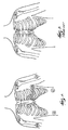
- The most preferred electrode placement is illustrated in FIG. 4. Specifically, FIG. 4 illustrates the electrode placement for six electrodes used to acquire electrical signals from the patient's heart. Preferably, only two electrodes are attached to the patient's chest. V1 is attached to the patient in approximately the fourth intercostal space at the right border of the patient's sternum. V5 is attached to the patient in approximately the fifth intercostal space at the patient's anterior axillary line. Four electrodes are attached to the patient's limbs. The electrodes attached to the right arm, left arm, and left leg acquire electrical signals, while the electrode attached to the right leg generally acts as an electrical ground.
- In the most preferred embodiment, electrodes V2, V3, V4, and V6 are not attached to the patient. Thus, leads V2, V3, V4, and V6 corresponding to the omitted electrodes must be derived mathematically. However, unlike the known prior art, only these four leads need to be derived mathematically. Eight of the leads, namely leads I, II, III, aVR, aVL, aVF, V1, and V5, are generated in the same manner as in a standard ten-electrode, twelve-lead ECG.
- Electrodes V1 and V5 are the most preferred chest electrodes for a number of reasons. First, the information in the V1 lead is very important to clinicians. Lead V1 is, of course, most accurate if an electrode is placed directly in the V1 position. Second, the positions of the V1 and V5 electrodes do not interfere with the clinician's access to the patient's chest as much as the positions of the V2, V3, and V4 electrodes. Even with the V1 and V5 electrodes in place, the clinician is still able to access the area of the chest closest to the patient's heart. For example, the clinician is able to use imaging probes in the area of the chest closest to the patient's heart. Third, the clinician may not even be able to attach the V2, V3, and V4 electrodes, because the patient may have wounds or bandages from surgical procedures involving the heart in the area of the chest where the V2, V3, and V4 electrodes are normally positioned. Fourth, electrode V1 is placed near the sternum, but not directly on the sternum, avoiding sternotomy wounds and bandages.
- A less preferred embodiment for the electrode placement of the invention is illustrated in FIG. 5. FIG. 5 also illustrates the electrode placement for six electrodes used to acquire electrical signals from the patient's heart. Again, only two electrodes are attached to the patient's chest. V2 is attached to the patient in approximately the fourth intercostal space at the left border of the patient's sternum. V5 is attached to the patient in approximately the fifth intercostal space at the patient's anterior axillary line. Again, four electrodes are attached to the patient's limbs. The electrodes attached to the right arm, left arm, and left leg acquire electrical signals, while the electrode attached to the right leg generally acts as an electrical ground.
- As illustrated in FIG. 5, electrodes V1, V3, V4, and V6 are not attached to the patient. Thus, leads V1, V3, V4, and V6 corresponding to the omitted electrodes must be derived mathematically. Again, only these four leads need to be derived mathematically. Eight of the leads, namely leads I, II, III, aVR, aVL, aVF, V2, and V5, are generated in the same manner as in a standard ten-electrode, twelve-lead ECG. One disadvantage of the less preferred embodiment is that the mathematically derived V1 lead is not as accurate as the V1 lead generated from the use of the V1 electrode. Since the V1 lead is considered important to clinicians, the embodiment using the V2 and V5 electrodes is less desirable, even though the embodiment produces generally acceptable results.
- In both preferred embodiments, the four missing leads are generated using a mathematical method called multiple-linear regression. Multiple-linear regression is a technique used to compute a prediction of a given data set from associated members of other data sets. In the present invention, these "other" data sets are either ECGs from a hospital's general population, from a sub-population of the hospital's general population, or from ECGs previously acquired from the patient. Preferably, the sub-populations of the hospital's general population are based on one or several parameters, such as sex, race, age, weight, height, or body habitus. Body habitus refers to a combination of body build, height, and weight. In general, the four missing leads are calculated based on the relationship between the available leads and a data set of previously acquired ECGs.
- Preferably, an algorithm uses the multiple-linear regression technique to generate the four missing leads. The input into the algorithm is a data set of previously acquired ECGs. Preferably, the data set consists of previously acquired ECGs from a hospital's general population or from a sub-population of the hospital's general population. Most preferably, the data set consists of previously acquired ECGs from the particular patient.
- Preferably, the data set consists of previously acquired ECGs that have been sampled at a rate of between 120 and 1000 times per second. Thus, the data set consists of twelve columns, corresponding to each of the twelve leads, with hundreds of thousands of rows of samples, corresponding to the voltage values for each of the twelve leads. The algorithm performs multiple-linear regression between the columns of lead data. Specifically, the algorithm performs multiple-linear regression between the columns of lead data corresponding to the electrodes that will be attached to the patient and between the columns of lead data corresponding to an electrode that will not be attached to the patient. The algorithm creates an equation from the previously-acquired ECG data defining the leads that will be missing, due to the omitted electrodes, as a function of the leads that will be available.
- For example, in the most preferred embodiment, the algorithm creates a first equation to determine the value of lead V2 based on the values of leads V1, V5, I, and II. The inputs to the algorithm are five columns of data corresponding to the previously-acquired ECG voltage values for leads V1, V2, V5, I, and II. The columns of data corresponding to leads V1, V5, I, and II are compared to the column of data corresponding to lead V2. The algorithm generates an equation relating the data in the V1, V5, I, and II lead columns to the data in the V2 lead column by minimizing the sum squared error between the five columns of data. The generated equation is in the following form:
- In the same manner, an expansion-coefficient equation is derived for each of the leads corresponding to the omitted electrodes. In the most preferred embodiment, a separate equation is derived for each of leads V2, V3, V4, and V6. Thus, the columns of data corresponding to leads V1, V5, I, and II are compared to the column of data corresponding to lead V3 to generate an expansion-coefficient equation for lead V3. Similarly, the columns of data corresponding to leads V1, V5, I, and II are compared to the column of data corresponding to lead V4 to generate an expansion-coefficient equation for lead V4. Finally, the columns of data corresponding to leads V1, V5, I, and II are compared to the column of data corresponding to lead V6 to generate an expansion-coefficient equation for lead V6.
- In the most preferred embodiment, once the expansion-coefficient equations are derived, the patient's ECG is acquired with only chest electrodes V1 and V5 and the four limb electrodes. From these six electrodes, eight leads are generated in the same manner as for a standard ECG. Four of the twelve leads, however, must be determined using the expansion-coefficient equations previously derived. Accordingly, the expansion-coefficient equations are used to derive the voltage values for the four missing leads for each individual sample. For example, the expansion-coefficient equation for lead V2 is used to derive the voltage value of lead V2 for each individual sample. In the most preferred embodiment, the voltage values for each sample of leads V3, V4, and V6 are derived in the same manner. With the addition of the data for the four derived leads, the data set comprises a complete twelve-lead ECG.
- FIG. 1 illustrates the
apparatus 10 embodying the invention. In the preferred embodiment, theapparatus 10 is a patient monitoring or patient data acquisition device. While any device for acquiring ECG signals (such as, for example, a bedside monitor, transport monitor, or Holter monitor) is contemplated by the invention, the apparatus of the preferred embodiment employs a telemetry-based monitoring device. Theapparatus 10 includes six electrodes 12 attachable to apatient 14, leadwires 16 coupled to the electrodes 12, a telemetry unit 18 coupled to the electrodes 12 byleadwires 16, asignal processor 20 wirelessly coupled to the telemetry unit 18, atelemetry monitor 22 coupled to thesignal processor 20, and anECG storage facility 24 coupled to thesignal processor 20. - Before the telemetry system embodied in the
apparatus 10 of FIG. 1 is described, it should be understood that the invention could also be implemented in a bed-side monitor. For the bed-side monitor, the electrodes 12 would be coupled directly to thesignal processor 20 byleadwires 16 in a conventional manner. Conversely, FIG. 1 illustrates theapparatus 10 including a telemetry unit 18 wirelessly coupled to thesignal processor 20 and telemetry monitor 22. Conventional methods of wireless transmission are used to transmit the electrical signals from the telemetry unit 18 to thereceiver 28 in thesignal processor 20. - Biopotential signals are often processed by telemetry, a technique that provides a wireless link between the patient and the signal processing components. Thus, clinicians can monitor a patient while the patient has full mobility. Traditional methods of telemetry utilize from three to five electrodes, but are unable to acquire a twelve-lead ECG from these three to five electrodes. The limiting factor in telemetry is the bandwidth of the signal being transmitted to the signal processing components. Accordingly, traditional telemetry monitors are unable to support the bandwidth necessary to transmit the electrical signals representing an entire twelve-lead ECG.
- In the preferred embodiments of the present invention, four, rather than eight or more, channels of ECG data are transmitted from the telemetry unit 18 to the
signal processor 20. As a result, the bandwidth necessary to transmit the electrical signals representing the patient's ECG is reduced by at least half. Due to the reduced number of electrodes and the reduced bandwidth, the telemetry unit 18 is capable of monitoring the electrical activity of the patient's heart while the patient has full mobility. Moreover, theapparatus 10 is capable of acquiring more data from each particular patient or data from more than one patient. - Referring to FIGS. 1 and 4, according to the method of the most preferred embodiment of the invention, six electrodes 12 are attached to the patient. A first electrode (V1) is attached in approximately the fourth intercostal space at the right border of the patient's sternum. A second electrode (V5) is attached in approximately the fifth intercostal space at the patient's anterior axillary line. Four additional electrodes (LA, RA, LL, and G) are attached to the patient's limbs. Electrical signals are acquired from the six electrodes 12 and transmitted via
leadwires 16 to the telemetry unit 18. The electrical signals are amplified and transmitted to thereceiver 28 of thesignal processor 20. - In both preferred embodiments, reducing the number of electrodes 12 attached to the patient reduces the number of
leadwires 16 and amplifiers (not shown) within the telemetry unit 18 necessary to acquire the electrical signals. Preferably, only sixleadwires 16 and four amplifiers within the telemetry unit 18 are necessary for the preferred embodiments of the invention. - A
software module 26 withinsignal processor 20 uses an algorithm to calculate the expansion-coefficient equations for the four missing leads. The input to the algorithm is, most preferably, a data set of the patient's previously acquired ECGs. The data set is preferably stored in a hospitalECG storage facility 24 and accessed by thesignal processor 20 of theECG machine 10. - The
apparatus 10 then acquires an ECG for the patient. The telemetry unit 18 transmits the electrical signals from the electrodes 12 to areceiver 28 within thesignal processor 20. The four missing leads are calculated by thesoftware module 26 withinsignal processor 20 using the previously-derived expansion-coefficient equations. All twelve leads, including the four generated leads, are then displayed for the clinician ontelemetry monitor 22. - For the sake of good order, various aspects of the invention are set out in the following clauses:-
- 1. A device for acquiring and processing electrical signals produced by a
patient's heart, the device comprising:
- a plurality of less than ten electrodes (12) each for attachment to the patient (14) in a one of the standard ten-electrode, twelve-lead electrocardiogram electrode positions; and
- a signal processor (20) connected to the plurality of less than ten electrodes (12) for acauiring electrical signals from the electrodes (12) and for generating a twelve-lead electrocardiogram from the electrical signals.
- 2. The device of clause 1 wherein the plurality of electrodes (12) includes a one of (a) an electrode for attachment to the patient in approximately the fourth intercostal space at the right border of the patient's sternum and (b) an electrode for attachment to the patient in approximately the fourth intercostal space at the left border of the patient's sternum.
- 3. The device of
clause 2 wherein the electrode (12) is attachable to adjust for the patient's gender. - 4. The device of clause 1 wherein the plurality of electrodes (12) includes an electrode (12) attachable in approximately the fifth intercostal space at the patient's anterior axillary line.
- 5. The device of clause 1 wherein the plurality of electrodes (12) includes an electrode (12) attachable to the patient's left arm.
- 6. The device of clause 1 wherein the plurality of electrodes (12) includes an electrode (12) attachable to the patient's right arm.
- 7. The device of clause 1 wherein the plurality of electrodes (12) includes an electrode (12) attachable to the patient's left leg.
- 8. The device of clause 1 wherein the plurality of electrodes (12) includes an electrode (12) attachable to a ground.
- 9. The device of clause 1 wherein the generated twelve-lead electrocardiogram includes leads I, II, III, aVR, aVL, aVF, V1, V2, V3, V4, V5, and V6.
- 10. The device of clause 9 wherein leads I, II, III, aVR, aVL, aVF, V1 or V2, and V5 generated from the electrical signals are the same as leads I, II, III, aVR, aVL, aVF, V1 or V2, and V5 that would be generated from the electrical signals of a standard ten-electrode, twelve-lead electrocardiogram.
- 11. The device of clause 9 wherein the signal processor (20) includes a software module (26) for mathematically generating leads V1 or V2, V3, V4, and V6 from the electrical signals.
- 12. The device of clause 11 wherein the software module (26) generates the leads mathematically from the electrical signals by the signal processor (20) using multiple-linear regression.
- 13. The device of clause 12 wherein the software module (26) uses expansion coefficients in the multiple-linear regression to generate the leads.
- 14. The device of clause 13 wherein the expansion coefficients are determined from a data set of electrocardiograms from the general population.
- 15. The device of clause 13 wherein the expansion coefficients are determined from a data set of electrocardiograms from the same patient (14).
- 16. The device of clause 1 and further comprising a telemetry monitor (22) coupled to the electrodes and to the signal processor (20).
- 17. The device of
clause 16 wherein the device acquires electrical signals from more than one patient (14). - 18. A method of acquiring and processing electrical signals produced by a
patient's heart, the method comprising the acts of:
- attaching a plurality of less than ten electrodes (12) to the patient, the act of attaching including attaching each of the electrodes in a one of the standard ten-electrode, twelve-lead electrocardiogram positions;
- acquiring electrical signals from the electrodes; and
- generating a twelve-lead electrocardiogram from the acquired electrical signals.
- 19. The method of clause 18 wherein the act of attaching a plurality of electrodes (12) further comprises the act of attaching an electrode (12) in one of (a) approximately the fourth intercostal space at the right border of the patient's sternum and (b) approximately the fourth intercostal space at the left border of the patient's sternum.
- 20. The method of clause 18 wherein the act of attaching the electrode (12) further comprises the act of adjusting the electrode position to account for the patient's gender.
- 21. The method of clause 18 wherein the act of attaching a plurality of electrodes (12) further comprises the act of attaching an electrode (12) in approximately the fifth intercostal space at the patient's anterior axillary line.
- 22. The method of clause 18 wherein the act of attaching a plurality of electrodes (12) further comprises the act of attaching an electrode (12) to the patient's left arm.
- 23. The method of clause 18 wherein the act of attaching a plurality of electrodes (12) further comprises the act of attaching an electrode (12) to the patient's right arm.
- 24. The method of clause 18 wherein the act of attaching a plurality of electrodes (12) further comprises the act of attaching an electrode t(12) o the patient's left leg.
- 25. The method of clause 18 wherein the act of attaching a plurality of electrodes (12) further comprises the act of attaching an electrode (12) to a ground.
- 26. The method of clause 18 wherein the act of generating a twelve-lead electrocardiogram further comprises the act of generating leads I, II, III, aVR, aVL, aVF, V1, V2, V3, V4, V5, and V6.
- 27. The method of
clause 26 wherein leads I, II, III, aVR, aVL, aVF, V1 or V2, and V5 that would be generated from the electrical signals are the same as leads I, II, III, aVR, aVL, aVF, V1 or V2, and V5 generated from the electrical signals of a standard ten-electrode, twelve-lead electrocardiogram. - 28. The method of
clause 26 wherein the act of generating a twelve-lead electrocardiogram further comprises the act of mathematically generating leads V1 or V2, V3, V4, and V6 from the electrical signals. - 29. The method of
clause 28 wherein the act of generating the leads further comprises the act of generating the leads using multiple-linear regression. - 30. The method of clause 29 wherein the act of generating the leads further comprises the act of using expansion coefficients in the multiple-linear regression.
- 31. The method of clause 30 wherein the act of generating the leads further comprises the act of determining the expansion coefficients from a data set of electrocardiograms from the general population.
- 32. The method of clause 30 wherein the act of generating the leads further comprises the act of determining the expansion coefficients from a data set of electrocardiograms from the same patient.
- 33. The method of clause 18 wherein the act of acquiring the electrical signals further comprises the act of acquiring the electrical signals with a signal processor (20) coupled to a telemetry monitor (22).
- 34. The method of clause 33 wherein the act of acquiring the electrical signals from the electrodes further comprises the act of acquiring electrical signals from the electrodes of more than one patient.
- 35. An device for acquiring and processing electrical signals produced by a
patient's heart, the device comprising:
- a plurality of less than ten electrodes (12) for attachment to the patient (14), at least two of the electrodes (12) attachable to the patient's chest in two of the standard ten-electrode, twelve-lead electrocardiograph positions and at least two of the electrodes attachable to the patient's limbs in two of the standard ten-electrode, twelve-lead electrocardiograph positions; and
- a signal processor (20) connected to the electrodes (12) for acquiring electrical signals from the electrodes (12) and for generating a twelve-lead electrocardiogram from the electrical signals.
- 36. The device of clause 35 wherein the electrodes (12) attachable to the patient's chest include an electrode (12) attachable in one of (a) approximately the fourth intercostal space at the right border of the patient's sternum and (b) approximately the fourth intercostal space at the left border of the patient's sternum.
- 37. The device of clause 35 wherein the electrodes (12) attachable to the patient's chest include an electrode (12) attachable in approximately the fifth intercostal space at the patient's anterior axillary line.
- 38. The device of clause 35 wherein the electrodes (12) attachable to the patient's limbs include an electrode (12) attachable to the patient's left arm.
- 39. The device of clause 35 wherein the electrodes (12) attachable to the patient's limbs include an electrode (12) attachable to the patient's right arm.
- 40. The device of clause 35 wherein the electrodes (12) attachable to the patient's limbs include an electrode (12) attachable to the patient's left leg.
- 41. The device of clause 35 wherein the plurality of electrodes (12) includes an electrode (12) attachable to a ground.
- 42. The device of clause 35 wherein the generated twelve-lead electrocardiogram including leads I, II, III, aVR, aVL, aVF, V1, V2, V3, V4, V5, and V6.
- 43. The device of clause 42 wherein leads I, II, III, aVR, aVL, aVF, V1 or V2, and V5 generated from the electrical signals are the same as leads I, II, III, aVR, aVL, aVF, V1 or V2, and V5 that would be generated from the electrical signals of a standard ten-electrode, twelve-lead electrocardiogram.
- 44. The device of clause 42 wherein the signal processor includes a software module (26) for mathematically generating leads V1 or V2, V3, V4, and V6 from the electrical signals.
- 45. The device of clause 44 wherein the software module (26) generates the leads mathematically from the electrical signals by the signal processor (20) using multiple-linear regression.
- 46. The device of clause 45 wherein the software module (26) uses expansion coefficients in the multiple-linear regression to generate the leads.
- 47. The device of clause 46 wherein the expansion coefficients are determined from a data set of electrocardiograms from the general population.
- 48. The device of clause 46 wherein the expansion coefficients are determined from a data set of electrocardiograms from the same patient (14).
- 49. The device of clause 35 wherein the electrodes (12) attachable to the patient's chest are attachable to adjust for the patient's gender.
- 50. The device of clause 35 and further comprising a telemetry monitor (22) coupled to the electrodes (12) and to the signal processor )20).
- 51. The device of clause 50 wherein the device acquires electrical signals from the electrodes (12) of more than one patient (14).
- 52. A method of acquiring and processing electrical signals produced by a
patient's heart, the method comprising the acts of:
- attaching a plurality of less than ten electrodes (12) to the patient (14), the act of attaching including attaching at least two of the electrodes (12) to the patient's chest in two of the standard ten-electrode, twelve-lead electrocardiogram positions and attaching at least two of the electrodes (12) to the patient's limbs in two of the standard ten-electrode twelve-lead electrocardiogram positions;
- acquiring electrical signals from the electrodes(12); and
- generating a twelve-lead electrocardiogram from the acquired electrical signals.
- 53. The method of clause 52 wherein the act of attaching electrodes (12) to the patient's chest includes the act of attaching an electrode (12) in one of (a) approximately the fourth intercostal space at the right border of the patient's sternum and (b) approximately the fourth intercostal space at the left border of the patient's sternum.
- 54. The method of clause 52 wherein the act of attaching electrodes (12) to the patient's chest includes the act of attaching an electrode (12) in approximately the fifth intercostal space at the patient's anterior axillary line.
- 55. The method of clause 52 wherein the act of attaching electrodes (12) to the patient's limbs includes the act of attaching an electrode (12) to the patient's left arm.
- 56. The method of clause 52 wherein the act of attaching electrodes (12) to the patient's limbs includes the act of attaching an electrode (12) to the patient's right arm.
- 57. The method of clause 52 wherein the act of attaching electrodes (12) to the patient's limbs includes the act of attaching an electrode (12) to the patient's left leg.
- 58. The method of clause 52 wherein the act of attaching a plurality of electrodes (12) to the patient includes the act of attaching an electrode (12) to a ground.
- 59. The method of clause 52 wherein the act of generating a twelve-lead electrocardiogram further comprises the act of generating leads I, II, III, aVR, aVL, aVF, V1, V2, V3, V4, V5, and V6.
- 60. The method of clause 59 wherein leads I, II, III, aVR, aVL, aVF, V1 or V2, and V5 generated from the electrical signals are the same as leads I, II, III, aVR, aVL, aVF, V1, and V5 that would be generated from the electrical signals of a standard ten-electrode, twelve-lead electrocardiograph.
- 61. The method of clause 60 wherein the act of generating a twelve-lead electrocardiogram further comprises the act of mathematically generating leads V1 or V2, V3, V4, and V6 from the electrical signals.
- 62. The method of clause 61 wherein the act of generating the leads further comprises the act of using expansion coefficients in the multiple-linear regression.
- 63. The method of clause 62 wherein the act of generating the leads further comprises the act of determining the expansion coefficients from a data set of electrocardiograms from the general population.
- 64. The method of clause 62 wherein the act of generating the leads further comprises the act of determining the expansion coefficients from a data set of electrocardiograms from the same patient.
- 65. The method of clause 52 wherein the act of attaching the electrodes (12) to the patient's chest further comprises the act of adjusting the electrode (12) positions to account for the patient's gender.
- 66. The method of clause 52 wherein the act of acquiring the electrical signals further comprises the act of acquiring the electrical signals with a signal processor (20) coupled to a telemetry monitor (22).
- 67. The method of clause 66 wherein the act of acquiring the electrical signals from the electrodes (12) further comprises the act of acquiring electrical signals from the electrodes (12) of more than one patient (14).
- 68. A method of generating a twelve-lead electrocardiogram from electrical
data acquired from a patient's heart, the method comprising the acts of:
- attaching a plurality of less than ten electrodes (12) to the patient (14), the act of attaching including attaching at least two of the electrodes (12) to the patient's chest in two of the standard ten-electrode, twelve-lead electrocardiogram positions and attaching at least two of the electrodes (12) to the patient's limbs in two of the standard ten-electrode, twelve-lead electrocardiogram positions;
- acquiring a first and second channel of data from the electrodes (12) attached to the patient's chest;
- acquiring a third and fourth channel of data from the electrodes (12) attached to the patient's limbs;
- generating a twelve-lead electrocardiogram from the acquired channels of data.
- 69. The method of clause 68 wherein the act of attaching electrodes (12) to the patient's chest further comprises the act of attaching an electrode (12) in one of (a) approximately the fourth intercostal space at the right border of the patient's sternum and (b) approximately the fourth intercostal space at the left border of the patient's sternum.
- 70. The method of clause 68 wherein the act of attaching electrodes (12) to the patient's chest further comprises the act of attaching an electrode (12) in approximately the fifth intercostal space at the patient's anterior axillary line.
- 71. The method of clause 68 wherein the act of attaching electrodes (12) to the patient's limbs further comprises the act of attaching an electrode (12) to the patient's left arm.
- 72. The method of clause 68 wherein the act of attaching electrodes (12) to the patient's limbs further comprises the act of attaching an electrode (12) to the patient's right arm.
- 73. The method of clause 68 wherein the act of attaching electrodes (12) to the patient's limbs further comprises the act of attaching an electrode (12) to the patient's left leg.
- 74. The method of clause 68 wherein the act of attaching a plurality of electrodes (12) further comprises the act of attaching an electrode (12) to a ground.
- 75. The method of clause 68 and further comprising the act of coupling four amplifiers to the electrodes (12).
- 76. The method of clause 75 and further comprising the act of coupling a signal processor (20) to the amplifiers.
- 77. The method of clause 76 and further comprising the act of coupling the signal processor (20) to a telemetry monitor (22).
- 78. The method of
clause 77 wherein the act of acquiring the channels of data further comprises the act of acquiring channels of data from more than one patient (14). - 79. The method of clause 68 wherein the act of generating a twelve-lead electrocardiogram further comprises the act of generating leads I, II, III, aVR, aVL, aVF, V1, V2, V3, V4, V5, and V6.
- 80. The method of clause 79 wherein leads I, II, III, aVR, aVL, aVF, V1 or V2, and V5 generated from the electrical signals are the same as leads I, II, III, aVR, aVL, aVF, V1 or V2, and V5 that would be generated from the electrical signals of a standard ten-electrode, twelve-lead electrocardiogram.
- 81. The method of clause 80 wherein the act of generating a twelve-lead electrocardiogram further comprises the act of mathematically generating leads V1 or V2, V3, V4, and V6 from the electrical signals.
- 82. The method of clause 81 wherein the act of generating the leads further comprises the act of generating the leads using multiple-linear regression.
- 83. The method of clause 82 wherein the act of generating the leads further comprises the act of using expansion coefficients in the multiple-linear regression.
- 84. The method of clause 83 wherein the act of generating the leads further comprises the act of determining the expansion coefficients from a data set of electrocardiograms from the general population.
- 85. The method of clause 83 wherein the act of generating the leads further comprises the act of determining the expansion coefficients from a data set of electrocardiograms from the same patient.
- 86. The method of clause 68 wherein the act of attaching the electrodes (12) to the patient's chest further comprises the act of adjusting the electrode (12) positions to account for the patient's gender.
- 87. An electrocardiograph device for acquiring and processing electrical
data from a patient's heart, the device comprising:
- a plurality of less than ten electrodes (12) attachable to the patient (14),
- at least two of the plurality of electrodes (12) attachable to the patient's chest in two of the standard ten-electrode twelve-lead electrocardiograph positions, the output of the electrodes (12) attachable to the patient's chest being a first and second channel of data, and
- at least two of the plurality of electrodes (12) attachable to the patient's limbs in two of the standard ten-electrode twelve-lead electrocardiograph positions, the output of the electrodes (12) attachable to the patient's limbs being a third and fourth channel of data; and
- a signal processor (20) for acquiring the channels of data and for generating a twelve-lead electrocardiogram from the acquired channels of data.
- 88. The device of clause 87 wherein the electrodes (12) attachable to the patient's chest include an electrode (12) attachable in one of (a) approximately the fourth intercostal space at the right border of the patient's sternum and (b) approximately the fourth intercostal space at the left border of the patient's sternum.
- 89. The device of clause 87 wherein the electrodes (12) attachable to the patient's chest include an electrode (12) attachable in approximately the fifth intercostal space at the patient's anterior axillary line.
- 90. The device of clause 87 wherein the electrodes (12) attachable to the patient's limbs include an electrode (12) attachable to the patient's left arm.
- 91. The device of clause 87 wherein the electrodes (12) attachable to the patient's limbs include an electrode (12) attachable to the patient's right arm.
- 92. The device of clause 87 wherein the electrodes (12) attachable to the patient's limbs include an electrode (12) attachable to the patient's left leg.
- 93. The device of clause 87 wherein the plurality of electrodes (12) include an electrode (12) attachable to a ground.
- 94. The device of clause 87 and further comprising four amplifiers coupled to the electrodes (12).
- 95. The device of clause 87 and further comprising a telemetry monitor (22) coupled to the electrodes and to the signal processor (20).
- 96. The device of clause 95 wherein the device acquires channels of data from more than one patient (14).
- 97. The device of clause 87 wherein the generated twelve-lead electrocardiogram includes leads I, II, III, aVR, aVL, aVF, V1, V2, V3, V4, V5, and V6.
- 98. The device of clause 97 wherein leads I, II, III, aVR, aVL, aVF, V1 or V2, and V5 generated from the electrical signals are the same as leads I, II, III, aVR, aVL, aVF, V1 or V2, and V5 that would be generated from the electrical signals of a standard ten-electrode, twelve-lead electrocardiogram.
- 99. The device of clause 97 wherein the signal processor (20) includes a software module (26) for mathematically generating leads V1 or V2, V3, V4, and V6 from the electrical signals.
- 100. The device of clause 99 wherein the software module (26) generates the leads mathematically from the electrical signals by the signal processor (20) using multiple-linear regression.
- 101. The device of clause 100 wherein the software module (26) expansion coefficients in the multiple-linear regression to generate the leads.
- 102. The device of clause 101 wherein the expansion coefficients are determined from a data set of electrocardiograms from the general population.
- 103. The device of clause 101 wherein the expansion coefficients are determined from a data set of electrocardiograms from the same patient (14).
- 104. The device of clause 87 wherein the electrodes (12) attachable to the patient's chest are adjustable for the patient's gender.
-
Claims (10)
- A device for acquiring and processing electrical signals produced by a patient's heart, the device comprising:a plurality of less than ten electrodes (12) each for attachment to the patient (14) in a one of the standard ten-electrode, twelve-lead electrocardiogram electrode positions; anda signal processor (20) connected to the plurality of less than ten electrodes (12) for acquiring electrical signals from the electrodes (12) and for generating a twelve-lead electrocardiogram from the electrical signals.
- The device of claim 1 wherein the plurality of electrodes (12) includes a one of (a) an electrode for attachment to the patient in approximately the fourth intercostal space at the right border of the patient's sternum and (b) an electrode for attachment to the patient in approximately the fourth intercostal space at the left border of the patient's sternum.
- A method of acquiring and processing electrical signals produced by a patient's heart, the method comprising the acts ofattaching a plurality of less than ten electrodes (12) to the patient, the act of attaching including attaching each of the electrodes in a one of the standard ten-electrode, twelve-lead electrocardiogram positions;acquiring electrical signals from the electrodes; andgenerating a twelve-lead electrocardiogram from the acquired electrical signals.
- The method of claim 3 wherein the act of attaching a plurality of electrodes (12) further comprises the act of attaching an electrode (12) in one of (a) approximately the fourth intercostal space at the right border of the patient's sternum and (b) approximately the fourth intercostal space at the left border of the patient's sternum.
- An device for acquiring and processing electrical signals produced by a patient's heart, the device comprising:a plurality of less than ten electrodes (12) for attachment to the patient (14), at least two of the electrodes (12) attachable to the patient's chest in two of the standard ten-electrode, twelve-lead electrocardiograph positions and at least two of the electrodes attachable to the patient's limbs in two of the standard ten-electrode, twelve-lead electrocardiograph positions; anda signal processor (20) connected to the electrodes (12) for acquiring electrical signals from the electrodes (12) and for generating a twelve-lead electrocardiogram from the electrical signals.
- The device of claim 5 wherein the electrodes (12) attachable to the patient's chest include an electrode (12) attachable in one of (a) approximately the fourth intercostal space at the right border of the patient's sternum and (b) approximately the fourth intercostal space at the left border of the patient's sternum.
- A method of acquiring and processing electrical signals produced by a patient's heart, the method comprising the acts of:attaching a plurality of less than ten electrodes (12) to the patient (14), the act of attaching including attaching at least two of the electrodes (12) to the patient's chest in two of the standard ten-electrode, twelve-lead electrocardiogram positions and attaching at least two of the electrodes (12) to the patient's limbs in two of the standard ten-electrode twelve-lead electrocardiogram positions;acquiring electrical signals from the electrodes(12); andgenerating a twelve-lead electrocardiogram from the acquired electrical signals.
- The method of claim 52 wherein the act of attaching electrodes (12) to the patient's chest includes the act of attaching an electrode (12) in one of (a) approximately the fourth intercostal space at the right border of the patient's sternum and (b) approximately the fourth intercostal space at the left border of the patient's sternum.
- A method of generating a twelve-lead electrocardiogram from electrical data acquired from a patient's heart, the method comprising the acts of:attaching a plurality of less than ten electrodes (12) to the patient (14), the act of attaching including attaching at least two of the electrodes (12) to the patient's chest in two of the standard ten-electrode, twelve-lead electrocardiogram positions and attaching at least two of the electrodes (12) to the patient's limbs in two of the standard ten-electrode, twelve-lead electrocardiogram positions;acquiring a first and second channel of data from the electrodes (12) attached to the patient's chest;acquiring a third and fourth channel of data from the electrodes (12) attached to the patient's limbs; andgenerating a twelve-lead electrocardiogram from the acquired channels of data.
- An electrocardiograph device for acquiring and processing electrical data from a patient's heart, the device comprising:a plurality of less than ten electrodes (12) attachable to the patient (14),at least two of the plurality of electrodes (12) attachable to the patient's chest in two of the standard ten-electrode twelve-lead electrocardiograph positions, the output of the electrodes (12) attachable to the patient's chest being a first and second channel of data, andat least two of the plurality of electrodes (12) attachable to the patient's limbs in two of the standard ten-electrode twelve-lead electrocardiograph positions, the output of the electrodes (12) attachable to the patient's limbs being a third and fourth channel of data; anda signal processor (20) for acquiring the channels of data and for generating a twelve-lead electrocardiogram from the acquired channels of data.
Applications Claiming Priority (2)
| Application Number | Priority Date | Filing Date | Title |
|---|---|---|---|
| US752140 | 1991-08-29 | ||
| US09/752,140 US6636761B2 (en) | 2000-12-29 | 2000-12-29 | Method and apparatus for generating a twelve-lead ECG from fewer than ten electrodes |
Publications (3)
| Publication Number | Publication Date |
|---|---|
| EP1221299A2 true EP1221299A2 (en) | 2002-07-10 |
| EP1221299A3 EP1221299A3 (en) | 2004-04-21 |
| EP1221299B1 EP1221299B1 (en) | 2008-01-30 |
Family
ID=25025059
Family Applications (1)
| Application Number | Title | Priority Date | Filing Date |
|---|---|---|---|
| EP01310732A Expired - Lifetime EP1221299B1 (en) | 2000-12-29 | 2001-12-20 | Method and apparatus for generating a twelve-lead ecg from fewer than ten electrodes |
Country Status (4)
| Country | Link |
|---|---|
| US (2) | US6636761B2 (en) |
| EP (1) | EP1221299B1 (en) |
| JP (1) | JP4027091B2 (en) |
| DE (1) | DE60132655T2 (en) |
Cited By (9)
| Publication number | Priority date | Publication date | Assignee | Title |
|---|---|---|---|---|
| US6643539B2 (en) | 2000-08-03 | 2003-11-04 | Siemens Medical Solutions Usa, Inc. | Electrocardiogram system for synthesizing leads and providing an accuracy measure |
| US6690967B2 (en) | 2000-08-03 | 2004-02-10 | Draeger Medical System, Inc. | Electrocardiogram system for synthesizing leads and providing an accuracy measure |
| WO2005011492A2 (en) * | 2003-07-31 | 2005-02-10 | Del Mar Reynolds Medical Limited | Reduced electrode electrocardiography system |
| WO2005013817A1 (en) * | 2003-08-07 | 2005-02-17 | Charité-Universitäts- Medezin Berlin | Ecg system and method for the large-surface measurement of ecg signals |
| US6931271B2 (en) | 2002-08-12 | 2005-08-16 | Draeger Medical Systems, Inc | System for adaptively deriving ECG chest lead signal data |
| GB2458389A (en) * | 2005-04-14 | 2009-09-23 | Justin Pisani | Wearable physiological monitoring device |
| US7881778B2 (en) | 2006-09-28 | 2011-02-01 | The General Electric Company | Floating physiological data acquisition system with expandable ECG and EEG |
| CN102342830A (en) * | 2010-07-30 | 2012-02-08 | 日本光电工业株式会社 | Derived electrocardiogram generating system and derived electrocardiogram generating method |
| US11523766B2 (en) | 2020-06-25 | 2022-12-13 | Spacelabs Healthcare L.L.C. | Systems and methods of analyzing and displaying ambulatory ECG data |
Families Citing this family (50)
| Publication number | Priority date | Publication date | Assignee | Title |
|---|---|---|---|---|
| US6636761B2 (en) * | 2000-12-29 | 2003-10-21 | Ge Medical Systems Information Technologies, Inc. | Method and apparatus for generating a twelve-lead ECG from fewer than ten electrodes |
| US6716165B1 (en) * | 2001-03-02 | 2004-04-06 | Ge Medical Systems Information Technologies, Inc. | Patient telemetry device and leadset designs for providing antenna diversity |
| US8069418B2 (en) * | 2002-07-31 | 2011-11-29 | Draeger Medical Systems, Inc | Medical information system and user interface supporting treatment administration |
| US20060235317A1 (en) | 2003-02-26 | 2006-10-19 | Daming Wei | Electrocardiograph device having additional lead function and method for obtaining additional-lead electrocardiogram |
| JP4777326B2 (en) * | 2003-02-26 | 2011-09-21 | 大名 魏 | Electrocardiograph with additional lead function and method for deriving additional lead electrocardiogram |
| JP2004266399A (en) * | 2003-02-28 | 2004-09-24 | Hirona Gi | Medical information telemeter system |
| JP4830266B2 (en) * | 2004-05-14 | 2011-12-07 | 日本光電工業株式会社 | Standard 12-lead ECG construction method and ECG inspection apparatus |
| JP4470063B2 (en) | 2004-08-27 | 2010-06-02 | 大名 魏 | Derived 12-lead ECG construction method and monitoring device |
| US20060058591A1 (en) * | 2004-09-16 | 2006-03-16 | Memtec Corporation | First-response portable recorder and automated report generator |
| US8005531B2 (en) | 2006-09-29 | 2011-08-23 | The General Electric Company | Method and apparatus with reduced electrode system specific ECG interpretation |
| EP2293717A1 (en) * | 2008-06-18 | 2011-03-16 | Koninklijke Philips Electronics N.V. | Electrocardiograph for magnetic resonance imaging and electrode patch for same |
| US11375938B2 (en) | 2008-08-14 | 2022-07-05 | Ticker Medical Ltd | Miniature ECG data acquisition device |
| US8082025B2 (en) * | 2008-08-14 | 2011-12-20 | David Amitai | ECG data acquisition device |
| US9351654B2 (en) | 2010-06-08 | 2016-05-31 | Alivecor, Inc. | Two electrode apparatus and methods for twelve lead ECG |
| US8509882B2 (en) | 2010-06-08 | 2013-08-13 | Alivecor, Inc. | Heart monitoring system usable with a smartphone or computer |
| CN102106728A (en) * | 2011-01-27 | 2011-06-29 | 孙迎春 | Four electrode plates-based 18-lead electrocardiogram tracing method for exercise electrocardiogram |
| US20130281815A1 (en) * | 2012-04-18 | 2013-10-24 | The Board Of Trustees Of The University Of Arkansas | Wearable remote electrophysiological monitoring system |
| US10932720B2 (en) | 2011-03-08 | 2021-03-02 | Nanowear Inc. | Smart materials, dry textile sensors, and electronics integration in clothing, bed sheets, and pillow cases for neurological, cardiac and/or pulmonary monitoring |
| US8923957B2 (en) * | 2011-07-15 | 2014-12-30 | Verathon Inc. | Data conversion in ECG techniques |
| WO2014074913A1 (en) | 2012-11-08 | 2014-05-15 | Alivecor, Inc. | Electrocardiogram signal detection |
| WO2014107700A1 (en) | 2013-01-07 | 2014-07-10 | Alivecor, Inc. | Methods and systems for electrode placement |
| US9254092B2 (en) | 2013-03-15 | 2016-02-09 | Alivecor, Inc. | Systems and methods for processing and analyzing medical data |
| US9247911B2 (en) | 2013-07-10 | 2016-02-02 | Alivecor, Inc. | Devices and methods for real-time denoising of electrocardiograms |
| JP6118229B2 (en) * | 2013-10-28 | 2017-04-19 | 日本光電工業株式会社 | Electrocardiogram measurement apparatus, derived electrocardiogram generation method, and derived electrocardiogram generation program |
| US9420956B2 (en) | 2013-12-12 | 2016-08-23 | Alivecor, Inc. | Methods and systems for arrhythmia tracking and scoring |
| TWI637723B (en) | 2014-12-09 | 2018-10-11 | 國立交通大學 | Method and system for generating 12-lead electrocardiography signals using differential voltages of three leads |
| US11111593B2 (en) | 2015-01-16 | 2021-09-07 | Nanowear Inc. | Large scale manufacturing of hybrid nanostructured textile sensors |
| US10131993B2 (en) | 2015-01-16 | 2018-11-20 | Nanowear, Inc. | Large scale manufacturing of hybrid nanostructured textile sensors |
| CN107530020B (en) | 2015-04-14 | 2021-09-24 | 皇家飞利浦有限公司 | Method and system for ECG-based cardiac ischemia detection |
| US9839363B2 (en) | 2015-05-13 | 2017-12-12 | Alivecor, Inc. | Discordance monitoring |
| EP3445236A4 (en) * | 2015-10-16 | 2019-12-18 | Enrique Saldivar | Device and method of using hexaxial electrocardiograph |
| JP6724348B2 (en) * | 2015-11-27 | 2020-07-15 | 東洋紡株式会社 | Electrocardiogram measurement method and electrocardiogram measurement clothing |
| US10602946B2 (en) * | 2015-12-30 | 2020-03-31 | Vectracor, Inc. | Mobile cardiac monitoring device |
| US10231623B2 (en) | 2016-02-04 | 2019-03-19 | Nanowear Inc. | Roll-to-roll printing process for manufacturing a wireless nanosensor |
| US10617356B2 (en) | 2016-03-15 | 2020-04-14 | Anhui Huami Information Technology Co., Ltd. | Garment and cardiac data processing |
| CN105832328A (en) * | 2016-03-15 | 2016-08-10 | 安徽华米信息科技有限公司 | Processing method and device of electrocardiogram data, and garment |
| US10123741B2 (en) | 2016-11-30 | 2018-11-13 | Huami Inc. | Cardiac condition detection |
| US10959634B2 (en) | 2017-05-02 | 2021-03-30 | Nanowear Inc. | Wearable congestive heart failure management system |
| CN107440706A (en) * | 2017-05-23 | 2017-12-08 | 北京蓬阳丰业医疗设备有限公司 | Based on 9 electrode synchronous collection methods in wilson electrocardiographic lead systems |
| CN107440707A (en) * | 2017-05-23 | 2017-12-08 | 北京蓬阳丰业医疗设备有限公司 | 18 based on wilson electrocardiographic lead systems, which lead/21, leads dynamic electrocardiogram drawing method |
| CN107440710A (en) * | 2017-05-23 | 2017-12-08 | 北京蓬阳丰业医疗设备有限公司 | 4 electrodes based on wilson electrocardiographic lead systems are to 7 electrode synchronous collection methods |
| CN107440711A (en) * | 2017-05-23 | 2017-12-08 | 北京蓬阳丰业医疗设备有限公司 | 8 electrodes and 10 electrode synchronous collection methods based on wilson electrocardiographic lead systems |
| TWI669096B (en) * | 2017-07-13 | 2019-08-21 | 國立臺灣大學 | Versatile monitoring device with a determining carotid artery pressure function |
| JP6795003B2 (en) * | 2018-03-08 | 2020-12-02 | 東洋紡株式会社 | Method of manufacturing electrodes and wiring, clothes with electrodes and wiring |
| US20210113108A1 (en) * | 2019-10-22 | 2021-04-22 | Sensesemi Technologies Private Limited | Method, system and device for generating electrocardiogram with fewer number of probes |
| JP2020192391A (en) * | 2020-08-26 | 2020-12-03 | 東洋紡株式会社 | Electrocardiogram measurement method |
| CN113208600B (en) * | 2021-05-17 | 2024-02-27 | 北京蓬阳丰业科技有限公司 | Method and equipment for pushing and deriving leads V7 and V8 based on electrocardio monopole leads V6 and V9 |
| CN113208599B (en) * | 2021-05-17 | 2024-02-27 | 北京蓬阳丰业科技有限公司 | Method and equipment for pushing lead V3R, V R based on electrocardio monopole leads V1 and V5R |
| CN113223702B (en) * | 2021-05-17 | 2022-05-03 | 北京蓬阳丰业科技有限公司 | Method and equipment for deriving leads V3 and V4 based on electrocardio unipolar leads V2 and V5 |
| CN115886831B (en) * | 2022-09-05 | 2023-09-26 | 荣耀终端有限公司 | Multi-lead electrocardiograph detection method and electronic equipment |
Citations (3)
| Publication number | Priority date | Publication date | Assignee | Title |
|---|---|---|---|---|
| US5058598A (en) * | 1990-08-03 | 1991-10-22 | Nicklas John M | Method and apparatus for synthesizing leads of an electrocardiogram |
| WO1999055228A1 (en) * | 1998-04-30 | 1999-11-04 | Medtronic Physio-Control Manufacturing Corp. | Reduced lead set for detecting acute cardiac ischemic conditions |
| US6119035A (en) * | 1998-03-26 | 2000-09-12 | Hewlett-Packard Company | Method and system for synthesizing the 12-lead electrocardiogram |
Family Cites Families (10)
| Publication number | Priority date | Publication date | Assignee | Title |
|---|---|---|---|---|
| US4850370A (en) | 1987-07-22 | 1989-07-25 | Dower Gordon E | Method and apparatus for sensing and analyzing electrical activity of the human heart |
| EP0818016B1 (en) | 1995-02-09 | 1999-05-26 | Gordon Ewbank Dower | Apparatus and method for monitoring activity of the human heart |
| US5913828A (en) * | 1996-10-29 | 1999-06-22 | Hewlett-Packard Company | Method and apparatus for distinguishing pacing pulses in an EKG using conduction velocity calculations |
| US5794624A (en) * | 1997-01-31 | 1998-08-18 | Hewlett-Packard Company | Method and system for the fast determination of EKG waveform morphology |
| US6006125A (en) * | 1998-02-12 | 1999-12-21 | Unilead International Inc. | Universal electrocardiogram sensor positioning device and method |
| US6167258A (en) * | 1998-10-09 | 2000-12-26 | Cleveland Medical Devices Inc. | Programmable wireless data acquisition system |
| JP4587008B2 (en) | 2000-07-24 | 2010-11-24 | 大名 魏 | Standard 12-lead ECG construction method and ECG inspection device |
| ATE414468T1 (en) | 2000-08-03 | 2008-12-15 | Draeger Medical Systems Inc | ELECTROCARDIOLOGICAL SYSTEM FOR SYNTHESIZING LEADS AND DELIVERING AN ACCURATE MEASUREMENT |
| US6505067B1 (en) * | 2000-11-22 | 2003-01-07 | Medtronic, Inc. | System and method for deriving a virtual ECG or EGM signal |
| US6636761B2 (en) * | 2000-12-29 | 2003-10-21 | Ge Medical Systems Information Technologies, Inc. | Method and apparatus for generating a twelve-lead ECG from fewer than ten electrodes |
-
2000
- 2000-12-29 US US09/752,140 patent/US6636761B2/en not_active Expired - Lifetime
-
2001
- 2001-12-20 EP EP01310732A patent/EP1221299B1/en not_active Expired - Lifetime
- 2001-12-20 DE DE60132655T patent/DE60132655T2/en not_active Expired - Lifetime
- 2001-12-28 JP JP2001399036A patent/JP4027091B2/en not_active Expired - Lifetime
-
2003
- 2003-08-20 US US10/644,539 patent/US7136693B2/en not_active Expired - Lifetime
Patent Citations (3)
| Publication number | Priority date | Publication date | Assignee | Title |
|---|---|---|---|---|
| US5058598A (en) * | 1990-08-03 | 1991-10-22 | Nicklas John M | Method and apparatus for synthesizing leads of an electrocardiogram |
| US6119035A (en) * | 1998-03-26 | 2000-09-12 | Hewlett-Packard Company | Method and system for synthesizing the 12-lead electrocardiogram |
| WO1999055228A1 (en) * | 1998-04-30 | 1999-11-04 | Medtronic Physio-Control Manufacturing Corp. | Reduced lead set for detecting acute cardiac ischemic conditions |
Cited By (17)
| Publication number | Priority date | Publication date | Assignee | Title |
|---|---|---|---|---|
| US6690967B2 (en) | 2000-08-03 | 2004-02-10 | Draeger Medical System, Inc. | Electrocardiogram system for synthesizing leads and providing an accuracy measure |
| US6643539B2 (en) | 2000-08-03 | 2003-11-04 | Siemens Medical Solutions Usa, Inc. | Electrocardiogram system for synthesizing leads and providing an accuracy measure |
| US6931271B2 (en) | 2002-08-12 | 2005-08-16 | Draeger Medical Systems, Inc | System for adaptively deriving ECG chest lead signal data |
| EP2281505A1 (en) * | 2003-07-31 | 2011-02-09 | Spacelabs Healthcare Limited | Reduced electrode electrocardiography system |
| WO2005011492A2 (en) * | 2003-07-31 | 2005-02-10 | Del Mar Reynolds Medical Limited | Reduced electrode electrocardiography system |
| WO2005011492A3 (en) * | 2003-07-31 | 2005-04-07 | Del Mar Reynolds Medical Ltd | Reduced electrode electrocardiography system |
| US8295919B2 (en) | 2003-07-31 | 2012-10-23 | Spacelabs Healthcare, Llc | Reduced electrode electrocardiography system |
| US7974684B2 (en) | 2003-07-31 | 2011-07-05 | Spacelabs Healthcare, Llc | Reduced electrode electrocardiography system |
| WO2005013817A1 (en) * | 2003-08-07 | 2005-02-17 | Charité-Universitäts- Medezin Berlin | Ecg system and method for the large-surface measurement of ecg signals |
| GB2458389B (en) * | 2005-04-14 | 2010-03-24 | Justin Pisani | Wearable physiological monitoring device |
| GB2425181B (en) * | 2005-04-14 | 2010-02-03 | Justin Pisani | Wearable physiological monitoring device |
| GB2458389A (en) * | 2005-04-14 | 2009-09-23 | Justin Pisani | Wearable physiological monitoring device |
| US7881778B2 (en) | 2006-09-28 | 2011-02-01 | The General Electric Company | Floating physiological data acquisition system with expandable ECG and EEG |
| CN102342830A (en) * | 2010-07-30 | 2012-02-08 | 日本光电工业株式会社 | Derived electrocardiogram generating system and derived electrocardiogram generating method |
| EP2412308A3 (en) * | 2010-07-30 | 2012-10-10 | Daming Wei | Derived electrocardiogram generating system and derived electrocardiogram generating method |
| US8335560B2 (en) | 2010-07-30 | 2012-12-18 | Daming Wei | Derived electrocardiogram generating system and derived electrocardiogram generating method |
| US11523766B2 (en) | 2020-06-25 | 2022-12-13 | Spacelabs Healthcare L.L.C. | Systems and methods of analyzing and displaying ambulatory ECG data |
Also Published As
| Publication number | Publication date |
|---|---|
| DE60132655T2 (en) | 2009-01-29 |
| EP1221299A3 (en) | 2004-04-21 |
| US20040015089A1 (en) | 2004-01-22 |
| EP1221299B1 (en) | 2008-01-30 |
| US7136693B2 (en) | 2006-11-14 |
| US20020087088A1 (en) | 2002-07-04 |
| JP2002282229A (en) | 2002-10-02 |
| DE60132655D1 (en) | 2008-03-20 |
| US6636761B2 (en) | 2003-10-21 |
| JP4027091B2 (en) | 2007-12-26 |
Similar Documents
| Publication | Publication Date | Title |
|---|---|---|
| EP1221299B1 (en) | Method and apparatus for generating a twelve-lead ecg from fewer than ten electrodes | |
| US6052615A (en) | Method and apparatus for sensing and analyzing electrical activity of the human heart using a four electrode arrangement | |
| US6721591B2 (en) | Method of deriving standard 12-lead electrocardiogram and electrocardiogram monitoring apparatus | |
| US7647093B2 (en) | Apparatus and method for cordless recording and telecommunication transmission of three special ECG leads and their processing | |
| Feild et al. | Improved EASI coefficients: their derivation, values, and performance | |
| US5419337A (en) | Non-invasive multi-electrocardiographic apparatus and method of assessing acute ischaemic damage | |
| US4850370A (en) | Method and apparatus for sensing and analyzing electrical activity of the human heart | |
| US7881778B2 (en) | Floating physiological data acquisition system with expandable ECG and EEG | |
| US8005532B2 (en) | Electrocardiograph with extended lead function, and extended lead electrocardiogram deriving method | |
| WO1994001039A9 (en) | Wireless electrocardiographic system and wireless electrode assemblies | |
| EP0648089A1 (en) | Wireless electrocardiographic system and wireless electrode assemblies | |
| EP1217946B1 (en) | Method and apparatus for frank lead reconstruction from derived chest leads | |
| US6496720B1 (en) | Process for sensing and analyzing electrical activity of the human heart utilizing one lead system with an egg monitor designed for use with another lead system | |
| Nelwan | Evaluation of12-LeadElectrocardiogramReconstruction Methods forPatientMonitoring | |
| Wei | Deriving the 12-lead electrocardiogram from four standard leads based on the Frank Torso model | |
| KR20060116190A (en) | 3 apparatus and method for cordless recording and telecommunication transmission of three special ecg leads and their processing | |
| Dotsinsky et al. | Twelve-lead electrocardiogram obtained by eight leads | |
| WEI | TWELVE-LEAD ELECTROCARDIOGRAM TELEMONITORING | |
| Kligfield et al. | AHA/ACC/HRS Scientific Statement | |
| Lead | THE STANDARD 12-LEAD ELECTROCARDIOGRAM |
Legal Events
| Date | Code | Title | Description |
|---|---|---|---|
| PUAI | Public reference made under article 153(3) epc to a published international application that has entered the european phase |
Free format text: ORIGINAL CODE: 0009012 |
|
| AK | Designated contracting states |
Kind code of ref document: A2 Designated state(s): AT BE CH CY DE DK ES FI FR GB GR IE IT LI LU MC NL PT SE TR |
|
| AX | Request for extension of the european patent |
Free format text: AL;LT;LV;MK;RO;SI |
|
| PUAL | Search report despatched |
Free format text: ORIGINAL CODE: 0009013 |
|
| AK | Designated contracting states |
Kind code of ref document: A3 Designated state(s): AT BE CH CY DE DK ES FI FR GB GR IE IT LI LU MC NL PT SE TR |
|
| AX | Request for extension of the european patent |
Extension state: AL LT LV MK RO SI |
|
| RIC1 | Information provided on ipc code assigned before grant |
Ipc: 7A 61B 5/0428 A Ipc: 7A 61B 5/0408 B |
|
| 17P | Request for examination filed |
Effective date: 20041021 |
|
| AKX | Designation fees paid |
Designated state(s): DE FI FR GB SE |
|
| 17Q | First examination report despatched |
Effective date: 20060505 |
|
| GRAP | Despatch of communication of intention to grant a patent |
Free format text: ORIGINAL CODE: EPIDOSNIGR1 |
|
| RAP1 | Party data changed (applicant data changed or rights of an application transferred) |
Owner name: GE MEDICAL SYSTEMS INFORMATION TECHNOLOGIES, INC. |
|
| GRAS | Grant fee paid |
Free format text: ORIGINAL CODE: EPIDOSNIGR3 |
|
| GRAA | (expected) grant |
Free format text: ORIGINAL CODE: 0009210 |
|
| AK | Designated contracting states |
Kind code of ref document: B1 Designated state(s): DE FI FR GB SE |
|
| REG | Reference to a national code |
Ref country code: GB Ref legal event code: FG4D |
|
| REF | Corresponds to: |
Ref document number: 60132655 Country of ref document: DE Date of ref document: 20080320 Kind code of ref document: P |
|
| PG25 | Lapsed in a contracting state [announced via postgrant information from national office to epo] |
Ref country code: FI Free format text: LAPSE BECAUSE OF FAILURE TO SUBMIT A TRANSLATION OF THE DESCRIPTION OR TO PAY THE FEE WITHIN THE PRESCRIBED TIME-LIMIT Effective date: 20080130 |
|
| PG25 | Lapsed in a contracting state [announced via postgrant information from national office to epo] |
Ref country code: SE Free format text: LAPSE BECAUSE OF FAILURE TO SUBMIT A TRANSLATION OF THE DESCRIPTION OR TO PAY THE FEE WITHIN THE PRESCRIBED TIME-LIMIT Effective date: 20080430 |
|
| EN | Fr: translation not filed | ||
| PLBE | No opposition filed within time limit |
Free format text: ORIGINAL CODE: 0009261 |
|
| STAA | Information on the status of an ep patent application or granted ep patent |
Free format text: STATUS: NO OPPOSITION FILED WITHIN TIME LIMIT |
|
| 26N | No opposition filed |
Effective date: 20081031 |
|
| PG25 | Lapsed in a contracting state [announced via postgrant information from national office to epo] |
Ref country code: FR Free format text: LAPSE BECAUSE OF FAILURE TO SUBMIT A TRANSLATION OF THE DESCRIPTION OR TO PAY THE FEE WITHIN THE PRESCRIBED TIME-LIMIT Effective date: 20081121 |
|
| REG | Reference to a national code |
Ref country code: DE Ref legal event code: R079 Ref document number: 60132655 Country of ref document: DE Free format text: PREVIOUS MAIN CLASS: A61B0005042800 Ipc: A61B0005308000 |
|
| PGFP | Annual fee paid to national office [announced via postgrant information from national office to epo] |
Ref country code: GB Payment date: 20201123 Year of fee payment: 20 Ref country code: DE Payment date: 20201119 Year of fee payment: 20 |
|
| REG | Reference to a national code |
Ref country code: DE Ref legal event code: R071 Ref document number: 60132655 Country of ref document: DE |
|
| REG | Reference to a national code |
Ref country code: GB Ref legal event code: PE20 Expiry date: 20211219 |
|
| PG25 | Lapsed in a contracting state [announced via postgrant information from national office to epo] |
Ref country code: GB Free format text: LAPSE BECAUSE OF EXPIRATION OF PROTECTION Effective date: 20211219 |