EP1199354A1 - A method of forming a cell pattern on a surface, cellular networks and tissues based thereon - Google Patents
A method of forming a cell pattern on a surface, cellular networks and tissues based thereon Download PDFInfo
- Publication number
- EP1199354A1 EP1199354A1 EP00122915A EP00122915A EP1199354A1 EP 1199354 A1 EP1199354 A1 EP 1199354A1 EP 00122915 A EP00122915 A EP 00122915A EP 00122915 A EP00122915 A EP 00122915A EP 1199354 A1 EP1199354 A1 EP 1199354A1
- Authority
- EP
- European Patent Office
- Prior art keywords
- cells
- pattern
- matrix
- cell
- group
- Prior art date
- Legal status (The legal status is an assumption and is not a legal conclusion. Google has not performed a legal analysis and makes no representation as to the accuracy of the status listed.)
- Granted
Links
Images


Classifications
-
- C—CHEMISTRY; METALLURGY
- C12—BIOCHEMISTRY; BEER; SPIRITS; WINE; VINEGAR; MICROBIOLOGY; ENZYMOLOGY; MUTATION OR GENETIC ENGINEERING
- C12N—MICROORGANISMS OR ENZYMES; COMPOSITIONS THEREOF; PROPAGATING, PRESERVING, OR MAINTAINING MICROORGANISMS; MUTATION OR GENETIC ENGINEERING; CULTURE MEDIA
- C12N5/00—Undifferentiated human, animal or plant cells, e.g. cell lines; Tissues; Cultivation or maintenance thereof; Culture media therefor
- C12N5/0068—General culture methods using substrates
-
- C—CHEMISTRY; METALLURGY
- C12—BIOCHEMISTRY; BEER; SPIRITS; WINE; VINEGAR; MICROBIOLOGY; ENZYMOLOGY; MUTATION OR GENETIC ENGINEERING
- C12N—MICROORGANISMS OR ENZYMES; COMPOSITIONS THEREOF; PROPAGATING, PRESERVING, OR MAINTAINING MICROORGANISMS; MUTATION OR GENETIC ENGINEERING; CULTURE MEDIA
- C12N5/00—Undifferentiated human, animal or plant cells, e.g. cell lines; Tissues; Cultivation or maintenance thereof; Culture media therefor
- C12N5/06—Animal cells or tissues; Human cells or tissues
- C12N5/0602—Vertebrate cells
- C12N5/0618—Cells of the nervous system
-
- C—CHEMISTRY; METALLURGY
- C12—BIOCHEMISTRY; BEER; SPIRITS; WINE; VINEGAR; MICROBIOLOGY; ENZYMOLOGY; MUTATION OR GENETIC ENGINEERING
- C12N—MICROORGANISMS OR ENZYMES; COMPOSITIONS THEREOF; PROPAGATING, PRESERVING, OR MAINTAINING MICROORGANISMS; MUTATION OR GENETIC ENGINEERING; CULTURE MEDIA
- C12N2535/00—Supports or coatings for cell culture characterised by topography
- C12N2535/10—Patterned coating
Definitions
- a third aim of these studies is to be able to facilitate the integration of transplants and/or implants by "masking" the outer parts of these devices with a special array of cells which by their chemical and immunological nature as well as by their arrangement fit into the organism's body at the site in which the device is to be introduced.
- a stamp is produced by casting a silicon elastomer (polydimethyl siloxane, PDMS) in the desired pattern which is then coated with a solution of the biomolecule to be transferred. After contacting the "inked" stamp with the substrate surface the biomolecules self-assemble in the pre-given pattern.
- Kumar et al. and Mrksich et al. developed this method of producing patterns by stamping alkane thiols on gold substrates (Mrksich et al. 1996, PNAS USA, 93, 10775 - 10778, Mrksich et al. 1997, Exp. Cell. Res. 235, 305 - 313).
- Poly-D-lysine and laminin have been immobilised using microcontact printing on amino silane derivatised glass substrates with glutaraldehyde as a cross linker (Branch et al. 1998, Med. Biol. Eng. Comput., 36, 135 - 141) and sulfo-GMBS (Wheeler et al. 1999, J. Biomech. Eng., 121, 73 - 78), and the technique of microcontact printing has been used in neuronal cell guidance (Wheeler et al. 1999, ibid.; Branch et al. 2000, IEEE Transact. Biomed. Eng., 47, 3, 290 - 300).
- said whole tissue is derived from an organism's body.
- said whole tissue is derived from an organ selected from the group comprising brain, liver, kidney, muscle, skin, bone, lung and heart.
- said cells are organ slices.
- organ slices are preferably organotypic in that they mimic the arrangement of cells within an organ.
- said cells are brain slices.
- said pattern of cell-growth promoting molecules and/or cell-growth inhibiting molecules attached on said prepatterned surface allows for the guided growth and migration of cells, wherein preferably, said pattern of cell-growth promoting molecules and/or cell-growth inhibiting molecules mimics the arrangement of cells in an organ.
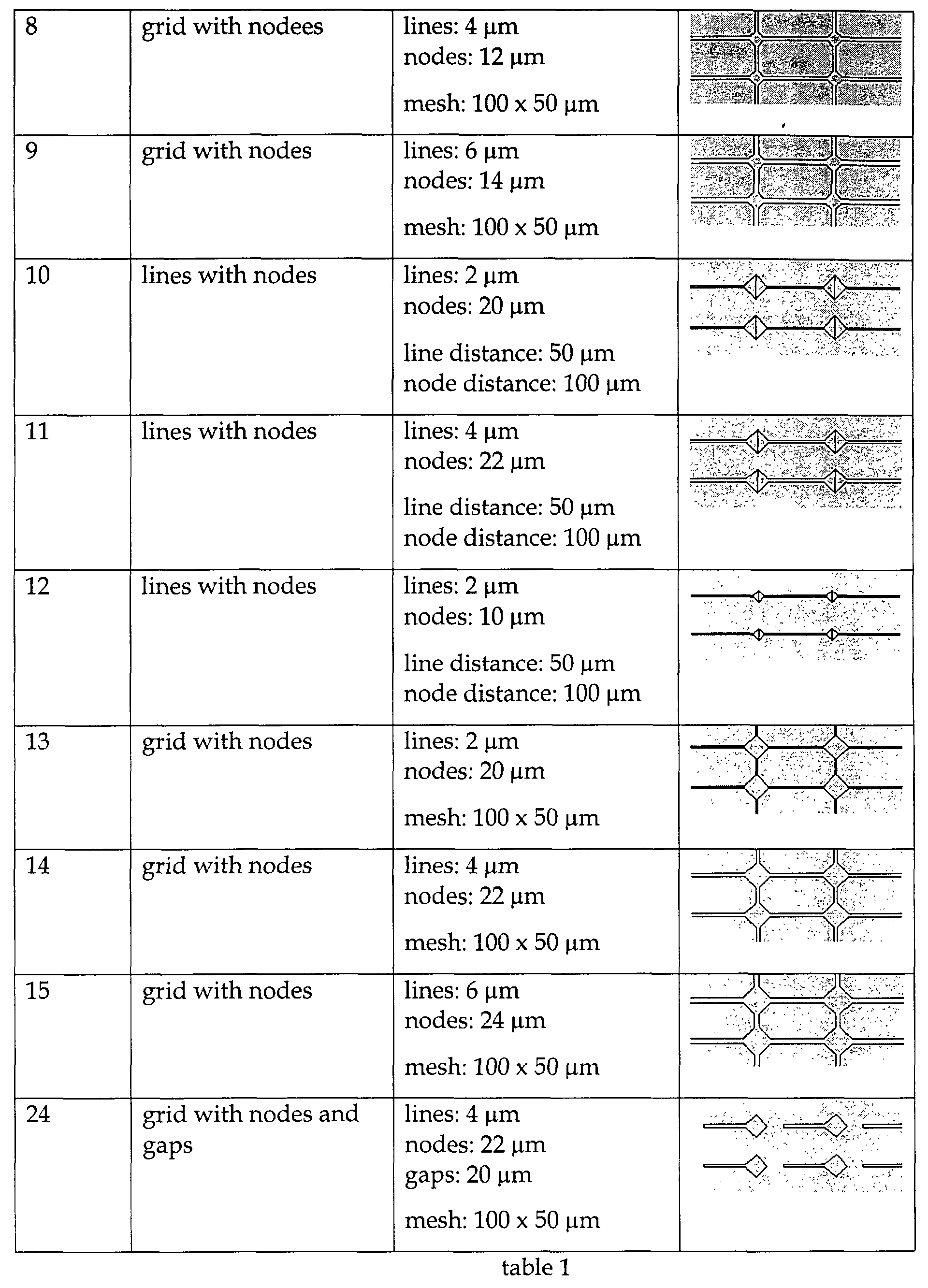
- said pattern of cell-growth promoting molecules and/or cell-growth inhibiting molecules has a structure with lines and nodes, wherein preferably, said lines have a width in the range from 1 - 8 micrometers and said nodes have a diameter in the range from 1 - 30 micrometers, more preferably, said lines have a width in the range from 1 - 6 micrometers and said nodes have a diameter in the range from 8 - 16 micrometers, and most preferably, said lines have a width in the range from 2 - 4 micrometers and said nodes have a diameter in the range from 10 - 14 micrometers.
- said pattern of cell-growth promoting molecules and/or cell-growth inhibiting molecules is formed by at least one layer of a substance selected from the group comprising polypeptide, polyethyleneimine and polystyrene wherein, preferably, said polypeptide is selected from the group comprising extracellular matrix proteins, poly-L-lysine and poly-ornithine, wherein, more preferably, said extracellular matrix proteins are selected from the group comprising laminin and fibronectin.
- the object is also solved by a method of forming a pattern of cells on a surface, said surface being prepatterned in having a pattern of cell-growth promoting molecules and/or cell-growth inhibiting molecules attached thereon, in particular according to any of the preceding claims, characterised in that cells are cultured on said prepatterned surface such that they form a pattern of cells on said surface, said cells being selected from the group comprising whole tissue and dissociated cells, further characterised in that said pattern of cells, after having been formed on said prepatterned surface, is transferred to a second surface in a transfer step. wherein preferably, said transfer step comprises the sequence:
- said matrix is a cell-compatible matrix.
- a cell-compatible matrix is a matrix that does not interfere with the viability of cells used.
- said matrix is a matrix composed of a material selected from the group comprising agarose, fibrin, collagen and cellulose.
- said matrix is a matrix composed of a curable material, wherein preferably, said curable material is selected from the group comprising agarose.
- said matrix is a matrix composed of a material capable of forming a gel, wherein, preferably, said material capable of forming a gel is selected from the group comprising fibrinogen and collagen.
- said second surface is selected from the group comprising surfaces of bioelectronical devices, sensors, electronical components, tissues, implants and transplants.
- “Sensors” are meant to include biosensors, optical sensors, amongst others.
- “Electronical components” can, for example, be field-effect transistors, multi-electrode arrays and the like.Transplants can be of any form known, i.e. autogenous (donor and receptor identical), syngenous (genetically identical donor and receptor), allogenous (donor and receptor belong to the same species) and xenogenous (donor and receptor belong to different species).
- said matrix (ab)) is achieved by increasing the temperature above the gel-transition temperature and/or addition of at least one gel-inducing component, wherein, preferably, said gel-inducing component is selected from the group comprising thrombin and other blood-coagulation factors.
- said releasing said pattern from said matrix is achieved by enzymatic degradation and/or lowering the temperature below the gel-transition temperature.
- the object of the present invention is also solved by a pattern of cells and/or an artificial tissue producable by a method according to the present invention up to, but exclusive of the transfer step.
- the object is furthermore solved by a pattern of cells and/or an artificial tissue on a surface producable by a method according to the present invention up to, but exclusive of the transfer step.
- the object is also solved by a pattern of cells and/or an artificial tissue on a surface producable by the method according to the present invention including the transfer step and various embodiments thereof.
- the object of the present invention is also solved by a combination of patterns of cells according to the present invention.
- the object is furthermore solved by a combination of artificial tissues according to the present invention.
- the object is furthermore solved by the use of a pattern of cells and/or an artificial tissue and/or a combination according to the present invention in a device selected from the group comprising sensors, technical substrates, tissues, implants and transplants.
- the inventors in making the present invention, could clearly demonstrate that it is possible to manipulate cells of organ slices onto a number of best suited patterns.
- whole tissue slices In using cells from whole tissue, extended, almost perfect patterns of cells, e. g. grids, could be achieved which were hitherto impossible.
- the use of whole tissue slices has many advantages over dissociated cultures in that, apart from the superficial layer, relatively minimal mechanical damage is being inflicted and the relative cytoarchitectural organisation of the tissue is preserved. The subsequently patterned cells will thus, reflect the relative positions of cells of the original slice.
- By choosing appropriate patterns of cell-growth promoting molecules and/or cell-growth inhibiting molecules on a surface it is possible to achieve almost any pattern of cells desired.
- the present inventors managed to further manipulate the almost perfect patterns of cells in that they could easily transfer these patterns onto any other surface desired.
- a transfer can, for example, be performed by embedding the pattern/artificial tissue in materials, which are compatible with the cells in that they do not interfere with the cells' viability.
- a material can for example be agarose which is poured in liquid form over the pattern of cells and thereafter cured by allowing it to cool.
- An alternative method would be the embedding of cells in a gel of fibrin and/or collagen.
- fibrinogen can be induced to form a gel of fibrin by the addition of thrombin. Modifications of this method, wherein prothrombin is added in combination with various factors from the blood-clotting cascade, e.g.
- fibrin-gels are also within the scope of the present invention.
- Methods of forming fibrin-gels are, for example, described in Schense et al., 2000, Nature Biotechnology, 18, 415-419 and Ye et al., 2000, European Journal of Cardio-thoracic Surgery, 17, 587-591 and are included herein by reference.
- Collagen-gels can be formed by self-assembly of collagen molecules upon warming cold neutral solutions of collagen. Such a method is, for example, described in O'Connor et al., 2000, Biosensors and Bioelectronics, 14, 871-881, and is explictly included herein by reference.
- all matrices are assembled on a pattern of cells/artificial tissue that has already been formed on a surface.
- the matrix is formed only after the formation of the pattern of cells/artificial tissue.
- the pattern of cells can then be tranferred elsewhere. Removal of the matrix can then, for example, be achieved by lowering the temperature below the gel-transition temperature, enzymatic degradation and the like.
- a new pattern of cells (grown on a prepatterned substrate) can be placed onto the same device and the measurements (of whatever kind that is to take place with this particular device) can be resumed.
- the pattern of cells used in one device can be transferred into sterile conditions (i. e. taken off the device) and then be returned to the incubator for a further period of culture.
- the present inventors using a combination of organotypic whole tissue and carefully designed prepatterned surfaces, managed to produce a huge variety of patterns of cells. According to any requirement (depending on the device) further patterns can be designed.
- the microcontact printing technique is discussed herebelow as a suitable technique for creating a surface which is prepatterned in having cell-growth promoting molecules and/or cell-growth inhibiting molecules attached thereon, the present invention is by no means intended to be restricted to this particular technique.
- Other techniques for creating prepatterned surfaces like photolithography, laser ablation techniques with lithographic masks etc. are envisaged, too.
- This incubation period was provided in order to allow damaged cells (as a result of slicing) to detach from the surfaces of the cut slices.
- the slices were then positioned onto controls (laminin-coated) and test substrates (laminin-patterned) using a small surface-polished spatula. Care has to be taken not to further damage the prepared slices.
- plasma clot or collagen was not used in immobilising the slices as either method would hinder cell migrations, particularly migrations onto ECM patterns. Furthermore, the roller tube technique was deemed unnecessary.
- a critical amount of medium was added such that the slices did not detach from the surface. After 2 - 3 days in culture, the slices should have undergone significant extent of migration and more medium could be added at that stage.
- Antimitotic agent cytosine ⁇ -D-arabinofuranoside (ARA-C, SIGMA C-6645) was added on day 4 or 5 to inhibit non-neuronal cells proliferation if and when necessary. If ARA-C was used, the culture medium was reverted back to ARA-C-free after 5 to 7 days.
- ARA-C cytosine ⁇ -D-arabinofuranoside
- Microstamps for the experiment were produced by photolithography and moulding. An electron beam writer transposed the different structures designed for the experiment to a chrome mask. Applying UV-photolithography, master moulds were produced out of spin coated 12.5 ⁇ m thick photoresist layers (AZ 4562, Clariant GmbH, Germany) on 0.6 mm thick silicon wafers (MEMC Electronic Materials, Germany). Polydimethylsiloxane (PDMS) microstamps were then fabricated curing Sylgard 182 (Dow Corning, Germany) in 10 ml eppendorf tubes for 48 hours at 55 °C upside down on the master moulds. After master mould release final curing was performed for 1 h at 110 °C .
- PDMS Polydimethylsiloxane
- stamps were stored in deionized water for a minumum of 24 hours. Prior to patterning, stamps were taken out of the water and sterilised in a 70 % ethanol bath for 1 minute. Inking took place for 30 seconds in 25 ⁇ g/ml ( ⁇ 0.25 ⁇ M) of laminin solution. The inked stamp was then dried in a soft nitrogen airstream and immediately pressed onto the substrate for 10 seconds. In all experiments, non tissue culture polystyrene petri dishes of 3 cm diameter (Greiner Labortechnik, Germany) were used.
- Brain slice medium was made from HAMS F10 (SIGMA; N1387) supplemented with 20-25 % foetal bovine serum (SIGMA, F7524) and 4mM glutamine (SIGMA, G7513).
- Laminin 1243217, Boehringer Mannheim GmbH, Germany was re-constituted in sterile PBS.
- Figure 1 shows the migration of organotypic brain stem neurons on laminin-coated tissue culture plastic.
- fig. 1A in culture, migrated neurons were clearly identifiable and growth of neurites had already begun.
- fig 1B On day 5 (fig 1B), these dendritic and axonal processess had grown to significant length.
- fig. 1C By 13 (fig. 1C) and 20 days (fig. 1D), the culture became confluent with all the neurons forming a diffused network on top of glial cells and other non-neuronal cells.
- Figure 2 shows the effectiveness by which neurite outgrowth and neuronal migration can be guided by using the technique of the present invention. Regardless of the pattern used, brain stem slices on laminin patterned substrates displayed a rapid growth of neurites, and these extended to the edge of the stamped patterns within two weeks. By the end of two weeks, the axons reached maturity and became thickened.
- Figure 3 shows the evaluation of a variety of extracellular matrix proteins patterns.
- Pattern 1 node 10 ⁇ m, track 2 ⁇ m;
- (fig. 3DA and 3DB) Pattern 4 node 10 ⁇ m, track 2 ⁇ m;
- fig. 3FA and 3FB Pattern 6, node 20 ⁇ m, track 4 ⁇ m.
- the figure shows the growth of neuronal processes on Pattern 1, 14 days old culture at progressively increasing magnification of x40 (fig. 4A), x100 (fig. 4B), x200 (fig. 4C).
- This perfect network of processes was obtained with pattern of the smallest track (2 ⁇ m).
- * indicate the same point of the substrate at their corresponding magnifications. With this particular pattern, however, very few visually identifiable neurons actually migrated to the pattern, which, in turn, is exclusively formed from neurite outgrowth.
- Patterns with node size of 10 - 14 ⁇ m and line width of 2 - 4 ⁇ m were the most appropriate for the confinement of neurons.
- Experiments using various line widths showed the best neuronal migration occurred with a 6 ⁇ m line width, where a perfect network of processes had occupied approximately the same area of the original stamp (10 mm diameter) used for the microcontact printing technique.
- It is known that cells tend to migrate towards the nodes (Klein et al., 1999, J. of Mat. Sci: Mat. in Med., 10, 721-729; Corey et al., 1991, J. of Neurosci. Res., 30, 300-307) and the same phenomenon was observed with brain sliced neurons. These neurons became immobilised once migrated onto the nodes. After a number of days in culture, the neuronal processes of the neurons were beginning to grow and were forming a simple linear neuronal network.
Abstract
Description
- In order to be able to study the functions of cells of various types so that their behaviour and spatial organisation in association with other cells of the same type can be better understood, it is necessary to be able to culture the cells under precisely controlled conditions. For a couple of years attempts have been undertaken to culture and grow cells on prepatterned substrates which guide the cell growth along the patterns on this substrate. This was done with the hindsight that, one day, one should be able to thereby build miniature biological electronic devices, incorporating live cells to make up a biological microcircuit. Another long-term goal of these studies is the capability of making artificial tissues suitable for implanting into an organism's body, thereby possibly replacing natural tissue of the same kind which is malfunctioning. A third aim of these studies is to be able to facilitate the integration of transplants and/or implants by "masking" the outer parts of these devices with a special array of cells which by their chemical and immunological nature as well as by their arrangement fit into the organism's body at the site in which the device is to be introduced.
- One way of achieving cultures of cells to that extent, i. e. cultures which show a specific intended special pattern is to grow the cells along surfaces on which, previously, patterns of a "guiding" molecule which promotes cell growth have been created, and where there are regions which do not promote cell growth. Various cell growth promoting molecules have been used:
- Mrksich et al. (1996, PNAS USA, 93, 10775-10778;1997, Exp. Cell Res., 235, 305-313) used alkanethiolate patterns on gold to control cell attachment to these substrates. By choosing an appropriately terminated alkanethiolate they succeeded in creating regions of cell growth promotion and cell growth inhibition. Corey et al.(1991, J. Neurosc. Res., 30, 300-307) managed to pattern neurons on polylysine-coated glass cover slips patterned by selective laser ablation so as to leave grids of polylysine with varying line widths, intrasection distances and nodal diameters.
- Matsuzawa et al.(1996, J. Neurosci. Meth., 69, 189-196) chemically attached a synthetic peptide derived from a neurite-outgrowth-promoting domain of the B2 chain of laminin.
- Others immobilised various other peptides (Matsuda et al., 1990, Trans. Am. Soc. Artif. Int. Organs; 36 (3): M559-63), or extracellular matrix proteins (ECM) such as laminin (Klein et al., 1999, J. Mat. Sci.: Mat. in Med.; 10: 721 - 727).
-
- Various techniques for attaching and patterning biomolecules on a surface have been used, including crosslinkers (Clemence et al., 1985, Bioconjugated Chem. 6: 411 - 417), silane coupling agents (Plueddemann E. 2 edn New York Plenum Press, 1991: 1 - 250), amongst others. One recently and successfully applied technique to attach proteins in a specific pattern to a substrate is the so-called microcontact printing technique. It is comparatively simple and universal for patterning biomolecules (Kumar et al., 1993, Appl. Phys. Lett., 63 (14), 2002 - 2004). In this technique a stamp is produced by casting a silicon elastomer (polydimethyl siloxane, PDMS) in the desired pattern which is then coated with a solution of the biomolecule to be transferred. After contacting the "inked" stamp with the substrate surface the biomolecules self-assemble in the pre-given pattern. Kumar et al. and Mrksich et al. developed this method of producing patterns by stamping alkane thiols on gold substrates (Mrksich et al. 1996, PNAS USA, 93, 10775 - 10778, Mrksich et al. 1997, Exp. Cell. Res. 235, 305 - 313). Poly-D-lysine and laminin have been immobilised using microcontact printing on amino silane derivatised glass substrates with glutaraldehyde as a cross linker (Branch et al. 1998, Med. Biol. Eng. Comput., 36, 135 - 141) and sulfo-GMBS (Wheeler et al. 1999, J. Biomech. Eng., 121, 73 - 78), and the technique of microcontact printing has been used in neuronal cell guidance (Wheeler et al. 1999, ibid.; Branch et al. 2000, IEEE Transact. Biomed. Eng., 47, 3, 290 - 300).
- All of the aforementioned studies used dissociated cell cultures, mainly of neural origin and achieved successful pattern formation only in some cases. It is not clear, however, whether the patterns of cells thus formed do represent a true picture as it would appear in nature nor whether they are of any use, e. g. for bioelectronic devices. Therefore, the conclusions to be drawn from these studies, e. g. in respect of the spatial arrangement of cells within an organ or the interactions between cells within an organ are only of limited use. Likewise, if one looks at current bioelectronic interface devices and cell modified interfaces there is a problem of reproducibility in creating these devices. It is still not possible to fully control and guide cell attachment and growth on surfaces. Since with current bioelectronic interface devices and cell modified interfaces, the cells are being cultured directly onto the surface of these devices, there is no guarantee that growth on every device will be successful, and therefore a lot of devices and a lot of starter cultures are required just to ensure that some substrates, after culturing, may actually display a cellular network which is useful. A related problem concerning implants is that these are often only of limited biocompatibility due to their bad integration, a rejection by the host or simply the toxicity of the substrates. Lining them with a pattern of cells which mimic the spatial organisation of cells within an organ would certainly enhance the biocompatibility of implants.
- Accordingly, one object of the present invention is to be able to control and guide cell growth on surfaces in a precise and hitherto unheard of manner. Another object of the present invention is to be able to limit the efforts in producing bioelectronic devices by reducing the number of starter cultures/substrates that will give a successful device. Another object of the present invention is to enhance that biocompatibility of implants and transplants.
- The object is solved by
- a method of forming a pattern of cells on a surface, said surface being prepatterned in having a pattern of cell-growth promoting molecules and/or cell-growth inhibiting molecules attached thereon, characterised in that cells are cultured on said prepatterned surface such that they form a pattern of cells on said surface, said cells being whole tissue.
-
- Preferably said whole tissue is derived from an organism's body.
- In one embodiment said whole tissue is derived from an organ selected from the group comprising brain, liver, kidney, muscle, skin, bone, lung and heart.
- It is preferred that said cells are organ slices.
- These organ slices are preferably organotypic in that they mimic the arrangement of cells within an organ.
- Preferably said cells are brain slices.
- In one embodiment said pattern of cell-growth promoting molecules and/or cell-growth inhibiting molecules attached on said prepatterned surface, allows for the guided growth and migration of cells, wherein preferably, said pattern of cell-growth promoting molecules and/or cell-growth inhibiting molecules mimics the arrangement of cells in an organ.
- It is preferred that said pattern of cell-growth promoting molecules and/or cell-growth inhibiting molecules has a structure with lines and nodes, wherein preferably, said lines have a width in the range from 1 - 8 micrometers and said nodes have a diameter in the range from 1 - 30 micrometers, more preferably, said lines have a width in the range from 1 - 6 micrometers and said nodes have a diameter in the range from 8 - 16 micrometers, and most preferably, said lines have a width in the range from 2 - 4 micrometers and said nodes have a diameter in the range from 10 - 14 micrometers.
- In one embodiment said pattern of cell-growth promoting molecules and/or cell-growth inhibiting molecules is formed by at least one layer of a substance selected from the group comprising polypeptide, polyethyleneimine and polystyrene wherein, preferably, said polypeptide is selected from the group comprising extracellular matrix proteins, poly-L-lysine and poly-ornithine, wherein, more preferably, said extracellular matrix proteins are selected from the group comprising laminin and fibronectin.
- The object is also solved by a method of forming a pattern of cells on a surface, said surface being prepatterned in having a pattern of cell-growth promoting molecules and/or cell-growth inhibiting molecules attached thereon, in particular according to any of the preceding claims, characterised in that cells are cultured on said prepatterned surface such that they form a pattern of cells on said surface, said cells being selected from the group comprising whole tissue and dissociated cells, further characterised in that said pattern of cells, after having been formed on said prepatterned surface, is transferred to a second surface in a transfer step.
wherein preferably, said transfer step comprises the sequence: - a) embedding said pattern of cells in a matrix,
- b) lifting said matrix including said pattern of cells from said prepatterned surface,
- c) contacting said pattern of cells embedded in said matrix with said second surface.
-
- In one embodiment said transfer step further comprises the sequence:
- d) releasing said pattern of cells from said matrix,
- e) removing said matrix from said pattern of cells.
-
- Preferably said matrix is a cell-compatible matrix.
- A cell-compatible matrix is a matrix that does not interfere with the viability of cells used.
- It is preferred that said matrix is a matrix composed of a material selected from the group comprising agarose, fibrin, collagen and cellulose.
- In one embodiment said matrix is a matrix composed of a curable material, wherein preferably, said curable material is selected from the group comprising agarose.
- In one embodiment said matrix is a matrix composed of a material capable of forming a gel, wherein, preferably, said material capable of forming a gel is selected from the group comprising fibrinogen and collagen.
- In one embodiment said second surface is selected from the group comprising surfaces of bioelectronical devices, sensors, electronical components, tissues, implants and transplants.
- "Sensors" are meant to include biosensors, optical sensors, amongst others. "Electronical components" can, for example, be field-effect transistors, multi-electrode arrays and the like.Transplants can be of any form known, i.e. autogenous (donor and receptor identical), syngenous (genetically identical donor and receptor), allogenous (donor and receptor belong to the same species) and xenogenous (donor and receptor belong to different species).
- In one embodiment said embedding is achieved
- aa) partially or fully covering said pattern of cells with said matrix in a liquid form, and
- ab) forming said matrix.
-
- It is preferred that forming said matrix (ab))is achieved by increasing the temperature above the gel-transition temperature and/or addition of at least one gel-inducing component, wherein, preferably, said gel-inducing component is selected from the group comprising thrombin and other blood-coagulation factors.
- It is preferred that said releasing said pattern from said matrix is achieved by enzymatic degradation and/or lowering the temperature below the gel-transition temperature.
- The object of the present invention is also solved by a pattern of cells and/or an artificial tissue producable by a method according to the present invention up to, but exclusive of the transfer step.
- The object is furthermore solved by a pattern of cells and/or an artificial tissue on a surface producable by a method according to the present invention up to, but exclusive of the transfer step.
- It is also solved by a pattern of cells and/or an artificial tissue producable by the method according to the present invention including the transfer step and various embodiments thereof.
- The object is also solved by a pattern of cells and/or an artificial tissue on a surface producable by the method according to the present invention including the transfer step and various embodiments thereof.
The object of the present invention is also solved by a combination of patterns of cells according to the present invention. - The object is furthermore solved by a combination of artificial tissues according to the present invention.
- It is also solved by a combination of patterns of cells and artificial tissues according to the present invention.
- The term "combination of patterns of cells" is meant to include any spatial arrangement of patterns of cells wherein these patterns are in proximity to each other. The same applies to "combination of artificial tissues".
- The object is furthermore solved by the use of a pattern of cells and/or an artificial tissue and/or a combination according to the present invention in a device selected from the group comprising sensors, technical substrates, tissues, implants and transplants.
- The inventors, in making the present invention, could clearly demonstrate that it is possible to manipulate cells of organ slices onto a number of best suited patterns. In using cells from whole tissue, extended, almost perfect patterns of cells, e. g. grids, could be achieved which were hitherto impossible. The use of whole tissue slices has many advantages over dissociated cultures in that, apart from the superficial layer, relatively minimal mechanical damage is being inflicted and the relative cytoarchitectural organisation of the tissue is preserved. The subsequently patterned cells will thus, reflect the relative positions of cells of the original slice. By means of the present invention it is possible to consistently achieve, e. g., outgrowth of neurons, neurites and filopodia from brain stem slices cultured on ECM protein structures of, e. g. grid- and line-shapes. By choosing appropriate patterns of cell-growth promoting molecules and/or cell-growth inhibiting molecules on a surface it is possible to achieve almost any pattern of cells desired.
- Furthermore, the present inventors managed to further manipulate the almost perfect patterns of cells in that they could easily transfer these patterns onto any other surface desired. Such a transfer can, for example, be performed by embedding the pattern/artificial tissue in materials, which are compatible with the cells in that they do not interfere with the cells' viability. Such a material can for example be agarose which is poured in liquid form over the pattern of cells and thereafter cured by allowing it to cool. An alternative method would be the embedding of cells in a gel of fibrin and/or collagen. For example, fibrinogen can be induced to form a gel of fibrin by the addition of thrombin. Modifications of this method, wherein prothrombin is added in combination with various factors from the blood-clotting cascade, e.g. factor X, are also within the scope of the present invention. Methods of forming fibrin-gels are, for example, described in Schense et al., 2000, Nature Biotechnology, 18, 415-419 and Ye et al., 2000, European Journal of Cardio-thoracic Surgery, 17, 587-591 and are included herein by reference.
Collagen-gels can be formed by self-assembly of collagen molecules upon warming cold neutral solutions of collagen. Such a method is, for example, described in O'Connor et al., 2000, Biosensors and Bioelectronics, 14, 871-881, and is explictly included herein by reference. - According to the present invention all matrices, independent of their nature, are assembled on a pattern of cells/artificial tissue that has already been formed on a surface. This means, that according to the present invention the matrix is formed only after the formation of the pattern of cells/artificial tissue. By means of the gel, the pattern of cells can then be tranferred elsewhere.
Removal of the matrix can then, for example, be achieved by lowering the temperature below the gel-transition temperature, enzymatic degradation and the like.
This makes the production of bioelectronic devices much more economical and allows for precise control of the characteristics of these devices since only successful patterns of cells will be used and transferred onto the surfaces of these devices. Once a perfect pattern of cells is formed, it can be stabilised using a matrix and removed from the substrate and transferred onto biosensors, such as field effect transistors etc. which in themselves might not be suited as substrates for cell-growth in the first place. With a "pre-cultured-situation" according to the present invention, only useful patterns of cells will be used and transferred onto the devices. Thus the number of devices that are required can be better controlled. This is true for all the other applications mentioned and gives a huge flexibility to possible applications. This "off-the-shelf-approach" is very useful when the device availability is limited, the device itself is not so well suited for cell-growth and culture, or when the further manipulation of cells on these devices causes irreversible damage to the cells. In this case a new pattern of cells (grown on a prepatterned substrate) can be placed onto the same device and the measurements (of whatever kind that is to take place with this particular device) can be resumed. Alternatively, the pattern of cells used in one device can be transferred into sterile conditions (i. e. taken off the device) and then be returned to the incubator for a further period of culture. - The present inventors, using a combination of organotypic whole tissue and carefully designed prepatterned surfaces, managed to produce a huge variety of patterns of cells. According to any requirement (depending on the device) further patterns can be designed. Although the microcontact printing technique is discussed herebelow as a suitable technique for creating a surface which is prepatterned in having cell-growth promoting molecules and/or cell-growth inhibiting molecules attached thereon, the present invention is by no means intended to be restricted to this particular technique. Other techniques for creating prepatterned surfaces like photolithography, laser ablation techniques with lithographic masks etc. are envisaged, too.
- Further advantages and aspects of the present invention can be taken from the following examples and figures, although the invention is by no means limited thereto. In the figures,
- Fig. 1 shows the migration of organotypic brain stem neurons on laminin-coated tissue culture plastic;
- Fig. 2 shows the growth of neurites on laminin tracks;
- Fig. 3 shows the evaluation of a variety of extacellular matrix protein patterns, and
- Fig. 4 shows the growth of neuronal processes.
-
- Using the present invention a huge variety of patterns of laminin on a surface could be successfully employed in the present invention. These patterns, in which a precise control of the cell-growth could be achieved, are shown in table 1


- Upon extraction of the whole embryonic 15 - 18 days old Sprague-Dawley rat brains, the medulla and pons were removed by a transverse section through the rostral pons and the cerebellum removed by sectioning the peduncles. Coronal sections of brain stem slices were harvested in sterile conditions in chilled brain slice culture medium (pH 7.4). These slices, which were cut using McIlwain Tissue Chopper, were 250 µm thick. The thus prepared slices (usually about 6 to 12 slices per rat brain stem) were placed in an incubator at 37°C and a 5 % CO2 enriched atmosphere for 4 - 5 hours minimum. This incubation period was provided in order to allow damaged cells (as a result of slicing) to detach from the surfaces of the cut slices. After this incubation period, the slices were then positioned onto controls (laminin-coated) and test substrates (laminin-patterned) using a small surface-polished spatula. Care has to be taken not to further damage the prepared slices. Unlike the method described by Gähwiler (1997, Trends in Neurosci., 20 (10), 471-477) plasma clot or collagen was not used in immobilising the slices as either method would hinder cell migrations, particularly migrations onto ECM patterns. Furthermore, the roller tube technique was deemed unnecessary. Instead, in all culture dishes, a critical amount of medium was added such that the slices did not detach from the surface. After 2 - 3 days in culture, the slices should have undergone significant extent of migration and more medium could be added at that stage. Antimitotic agent, cytosine β-D-arabinofuranoside (ARA-C, SIGMA C-6645), was added on day 4 or 5 to inhibit non-neuronal cells proliferation if and when necessary. If ARA-C was used, the culture medium was reverted back to ARA-C-free after 5 to 7 days.
- Microstamps for the experiment were produced by photolithography and moulding. An electron beam writer transposed the different structures designed for the experiment to a chrome mask. Applying UV-photolithography, master moulds were produced out of spin coated 12.5 µm thick photoresist layers (AZ 4562, Clariant GmbH, Germany) on 0.6 mm thick silicon wafers (MEMC Electronic Materials, Germany). Polydimethylsiloxane (PDMS) microstamps were then fabricated curing Sylgard 182 (Dow Corning, Germany) in 10 ml eppendorf tubes for 48 hours at 55 °C upside down on the master moulds. After master mould release final curing was performed for 1 h at 110 °C . In order to increase the stamp hydrophilicity, PDMS stamps were stored in deionized water for a minumum of 24 hours. Prior to patterning, stamps were taken out of the water and sterilised in a 70 % ethanol bath for 1 minute. Inking took place for 30 seconds in 25 µg/ml (∼0.25 µM) of laminin solution. The inked stamp was then dried in a soft nitrogen airstream and immediately pressed onto the substrate for 10 seconds. In all experiments, non tissue culture polystyrene petri dishes of 3 cm diameter (Greiner Labortechnik, Germany) were used.
- Brain slice medium was made from HAMS F10 (SIGMA; N1387) supplemented with 20-25 % foetal bovine serum (SIGMA, F7524) and 4mM glutamine (SIGMA, G7513). Laminin (1243217, Boehringer Mannheim GmbH, Germany) was re-constituted in sterile PBS.
- Figure 1 shows the migration of organotypic brain stem neurons on laminin-coated tissue culture plastic. On day 3 (fig. 1A) in culture, migrated neurons were clearly identifiable and growth of neurites had already begun. On day 5 (fig 1B), these dendritic and axonal processess had grown to significant length. By 13 (fig. 1C) and 20 days (fig. 1D), the culture became confluent with all the neurons forming a diffused network on top of glial cells and other non-neuronal cells.
- Figure 2 shows the effectiveness by which neurite outgrowth and neuronal migration can be guided by using the technique of the present invention. Regardless of the pattern used, brain stem slices on laminin patterned substrates displayed a rapid growth of neurites, and these extended to the edge of the stamped patterns within two weeks. By the end of two weeks, the axons reached maturity and became thickened.
- Figure 3 shows the evaluation of a variety of extracellular matrix proteins patterns. (fig. 3AA and 3AB) Pattern 1, node 10µm, track 2µm; (fig. 3BA and 3BB) Pattern 2, node 10µm, track 4µm); (fig. 3CA and 3CB) Pattern 3, node 10µm, track 6µm; (fig. 3DA and 3DB) Pattern 4, node 10µm, track 2µm; (fig. 3EA and 3EB) Pattern 5, node 10µm, track 2µm; (fig. 3FA and 3FB) Pattern 6, node 20µm, track 4µm. During early days in culture, the migrated neurons and growing processes were clearly visible with little overlapping on all the patterns. These cultures continued to mature and by 10 days or more, the original shapes of the laminin stamped patterns were identifiable.
- Figure 4 shows that nearly perfect patterns could be achieved using a 2 µm line width (node diameter = 10 µm) and a perfect grid structure could be formed. The figure shows the growth of neuronal processes on Pattern 1, 14 days old culture at progressively increasing magnification of x40 (fig. 4A), x100 (fig. 4B), x200 (fig. 4C). This perfect network of processes was obtained with pattern of the smallest track (2µm). * indicate the same point of the substrate at their corresponding magnifications. With this particular pattern, however, very few visually identifiable neurons actually migrated to the pattern, which, in turn, is exclusively formed from neurite outgrowth. Patterns with node size of 10 - 14 µm and line width of 2 - 4 µm were the most appropriate for the confinement of neurons. Experiments using various line widths showed the best neuronal migration occurred with a 6 µm line width, where a perfect network of processes had occupied approximately the same area of the original stamp (10 mm diameter) used for the microcontact printing technique. It is known that cells tend to migrate towards the nodes (Klein et al., 1999, J. of Mat. Sci: Mat. in Med., 10, 721-729; Corey et al., 1991, J. of Neurosci. Res., 30, 300-307) and the same phenomenon was observed with brain sliced neurons. These neurons became immobilised once migrated onto the nodes. After a number of days in culture, the neuronal processes of the neurons were beginning to grow and were forming a simple linear neuronal network.
- The features of the present invention disclosed in the specification, the claims and/or in the accompaning drawings, may, both seperatly, and in any combination thereof, be material for realizing the invention, in various forms thereof.
Claims (40)
- A method of forming a pattern of cells on a surface, said surface being prepatterned in having a pattern of cell-growth promoting molecules and/or cell-growth inhibiting molecules attached thereon, characterised in that cells are cultured on said prepatterned surface such that they form a pattern of cells on said surface, said cells being whole tissue.
- A method according to claim 1 characterised in that said whole tissue is derived from an organism's body.
- A method according to any of claims 1 - 2 characterised in that said whole tissue is derived from an organ selected from the group comprising brain, liver, kidney, muscle, skin, bone, lung and heart.
- A method according to any of the preceding claims characterised in that said cells are organ slices.
- A method according to claim 4 characterised in that said cells are brain slices.
- A method according to any of the preceding claims characterised in that said pattern of cellgrowth promoting molecules and/or cell-growth inhibiting molecules attached on said prepatterned surface allows for the guided growth and migration of cells.
- A method according to claim 6 characterised in that said pattern of cell-growth promoting molecules and/or cell-growth inhibiting molecules mimics the arrangement of cells in an organ.
- A method according to any of claims 1 - 7 characterised in that said pattern of cell-growth promoting molecules and/or cell-growth inhibiting molecules has a structure with lines and nodes.
- A method according to claim 8 characterised in that said lines have a width in the range from 1 - 8 micrometers and said nodes have a diameter in the range from 1 - 30 micrometers.
- A method according to claim 9 characterised in that said lines have a width in the range from 1 - 6 micrometers and said nodes have a diameter in the range from 8 - 16 micrometers.
- A method according to claim 10 characterised in that said lines have a width in the range from 2 - 4 micrometers and said nodes have a diameter in the range from 10 - 14 micrometers.
- A method according to any of the preceding claims characterised in that said pattern of cell-growth promoting molecules and/or cell-growth inhibiting molecules is formed by at least one layer of a substance selected from the group comprising polypeptide, polyethyleneimine and polystyrene.
- A method according to claim 12 characterised in that said polypeptide is selected from the group comprising extracellular matrix proteins, poly-L-lysine and poly-ornithine.
- A method according to claim 13 characterised in that said extracellular matrix proteins are selected from the group comprising laminin and fibronectin.
- A method of forming a pattern of cells on a surface, said surface being prepatterned in having a pattern of cell-growth promoting molecules and/or cell-growth inhibiting molecules attached thereon, in particular according to any of the preceding claims, characterised in that cells are cultured on said prepatterned surface such that they form a pattern of cells on said surface, said cells being selected from the group comprising whole tissue and dissociated cells, further characterised in that said pattern of cells, after having been formed on said prepatterned surface, is transferred to a second surface in a transfer step.
- A method according to claim 15 characterised in that said transfer step comprises the sequence:a) embedding said pattern of cells in a matrix,b) lifting said matrix including said pattern of cells from said prepatterned surface,c) contacting said pattern of cells embedded in said matrix with said second surface.
- A method according to any of claims 15-16 characterised in that said transfer step further comprises the sequence:d) releasing said pattern of cells from said matrix,e) removing said matrix from said pattern of cells.
- A method according to any of claims 16-17 characterised in that said matrix is a cell-compatible matrix.
- A method according to any of claims 16-18 characterised in that said matrix is a matrix composed of a material selected from the group comprising agarose, fibrin, collagen and cellulose.
- A method according to any of claims 16 - 18 characterised in that said matrix is a matrix composed of a curable material.
- A method according to claim 20 characterised in that said curable material is selected from the group comprising agarose.
- A method according to any of claims 16-18 characterised in that said matrix is a matrix composed of a material capable of forming a gel.
- A method according to claim 22 characterised in that said material capable of forming a gel is selected from the group comprising fibrinogen and collagen.
- A method according to any of claims 15 - 21 characterised in that said second surface is selected from the group comprising surfaces of bioelectronical devices, sensors, electronical components, tissues, implants and transplants.
- A method according to any of claims 16 - 24 characterised in that said embedding is achieved byaa) partially or fully covering said pattern of cells with said matrix in a liquid form, andab) forming said matrix.
- A method according to claim 25 characterised in that forming said matrix (ab))is achieved by increasing the temperature above the gel-transition temperature and/or addition of at least one gel-inducing component.
- A method according to claim 26 characterised in that said gel-inducing component is selected from the group comprising thrombin and other blood-coagulation factors.
- A method according to any of claims 17 - 27 characterised in that said releasing said pattern from said matrix is achieved by enzymatic degradation and/or lowering the temperature below the gel-transition temperature.
- A pattern of cells producable by a method according to any of claims 1 - 14.
- A pattern of cells on a surface producable by a method according to any of claims 1 - 14.
- A pattern of cells producable by a method according to any of claims 1 - 28.
- A pattern of cells on a surface producable by a method according to any of claims 1 - 28.
- An artificial tissue producable by a method according to any of claims 1-14.
- An artificial tissue on a surface producable by a method according to any of claims 1-14.
- An artificial tissue producable by a method according to any of claims 1-28.
- An artificial tissue on a surface producable by a method according to any of claims 1-28.
- A combination of patterns of cells according to any of claims 29-32.
- A combination of artificial tissues according to any of claims 33-36.
- A combination of patterns of cells according to any of claims 29-32 and artificial tissues according to any of claims 33-36.
- Use of a pattern of cells according to any of claims 29 - 32 and/or an artificial tissue according to any of claims 33-36 and/or a combination according to any of claims 37-38 in a device selected from the group comprising sensors, technical substrates, tissues, implants and transplants.
Priority Applications (4)
| Application Number | Priority Date | Filing Date | Title |
|---|---|---|---|
| EP00122915A EP1199354B1 (en) | 2000-10-20 | 2000-10-20 | A method of forming a cell pattern on a surface |
| DE60019603T DE60019603T2 (en) | 2000-10-20 | 2000-10-20 | Method for forming a cell pattern on a surface |
| JP2001322549A JP2002355031A (en) | 2000-10-20 | 2001-10-19 | Method of forming cell pattern |
| US10/033,150 US6787358B2 (en) | 2000-10-20 | 2001-10-19 | Method of forming a cell pattern on a surface |
Applications Claiming Priority (1)
| Application Number | Priority Date | Filing Date | Title |
|---|---|---|---|
| EP00122915A EP1199354B1 (en) | 2000-10-20 | 2000-10-20 | A method of forming a cell pattern on a surface |
Publications (2)
| Publication Number | Publication Date |
|---|---|
| EP1199354A1 true EP1199354A1 (en) | 2002-04-24 |
| EP1199354B1 EP1199354B1 (en) | 2005-04-20 |
Family
ID=8170154
Family Applications (1)
| Application Number | Title | Priority Date | Filing Date |
|---|---|---|---|
| EP00122915A Expired - Lifetime EP1199354B1 (en) | 2000-10-20 | 2000-10-20 | A method of forming a cell pattern on a surface |
Country Status (4)
| Country | Link |
|---|---|
| US (1) | US6787358B2 (en) |
| EP (1) | EP1199354B1 (en) |
| JP (1) | JP2002355031A (en) |
| DE (1) | DE60019603T2 (en) |
Cited By (18)
| Publication number | Priority date | Publication date | Assignee | Title |
|---|---|---|---|---|
| WO2004034016A2 (en) | 2002-10-04 | 2004-04-22 | Noo Li Jeon | Microfluidic multi-compartment device for neuroscience research |
| US6844184B2 (en) | 2000-11-08 | 2005-01-18 | Surface Logix, Inc. | Device for arraying biomolecules and for monitoring cell motility in real-time |
| US6864065B2 (en) | 2000-11-08 | 2005-03-08 | Surface Logix, Inc. | Assays for monitoring cell motility in real-time |
| US6893851B2 (en) | 2000-11-08 | 2005-05-17 | Surface Logix, Inc. | Method for arraying biomolecules and for monitoring cell motility in real-time |
| US7033819B2 (en) | 2000-11-08 | 2006-04-25 | Surface Logix, Inc. | System for monitoring cell motility in real-time |
| US7033821B2 (en) | 2000-11-08 | 2006-04-25 | Surface Logix, Inc. | Device for monitoring cell motility in real-time |
| EP1686171A1 (en) * | 2003-10-17 | 2006-08-02 | Ikuo Morita | Method of constructing artificial cell tissue and base material therefor |
| US7326563B2 (en) | 2000-11-08 | 2008-02-05 | Surface Logix, Inc. | Device and method for monitoring leukocyte migration |
| US7374906B2 (en) | 2000-11-08 | 2008-05-20 | Surface Logix, Inc. | Biological assays using gradients formed in microfluidic systems |
| WO2013041803A1 (en) | 2011-09-19 | 2013-03-28 | Institut Curie | Device for guiding cell migration and method of guiding cell migration implementing such a device |
| WO2013041800A1 (en) | 2011-09-19 | 2013-03-28 | Institut Curie | Device for guiding cell migration and guiding method implementing such a device |
| US9657942B2 (en) | 2010-10-25 | 2017-05-23 | ADA-ES, Inc. | Hot-side method and system |
| US9884286B2 (en) | 2010-02-04 | 2018-02-06 | ADA-ES, Inc. | Method and system for controlling mercury emissions from coal-fired thermal processes |
| US9889405B2 (en) | 2012-04-11 | 2018-02-13 | ADA-ES, Inc. | Control of wet scrubber oxidation inhibitor and byproduct recovery |
| US10350545B2 (en) | 2014-11-25 | 2019-07-16 | ADA-ES, Inc. | Low pressure drop static mixing system |
| US10465137B2 (en) | 2011-05-13 | 2019-11-05 | Ada Es, Inc. | Process to reduce emissions of nitrogen oxides and mercury from coal-fired boilers |
| US10767130B2 (en) | 2012-08-10 | 2020-09-08 | ADA-ES, Inc. | Method and additive for controlling nitrogen oxide emissions |
| US11298657B2 (en) | 2010-10-25 | 2022-04-12 | ADA-ES, Inc. | Hot-side method and system |
Families Citing this family (32)
| Publication number | Priority date | Publication date | Assignee | Title |
|---|---|---|---|---|
| US8124036B1 (en) | 2005-10-27 | 2012-02-28 | ADA-ES, Inc. | Additives for mercury oxidation in coal-fired power plants |
| US20040156988A1 (en) * | 2002-08-26 | 2004-08-12 | Mehenti Neville Z. | Selective and alignment-free molecular patterning of surfaces |
| JP4201182B2 (en) * | 2003-05-20 | 2008-12-24 | 大日本印刷株式会社 | Cell culture substrate and method for producing the same |
| JP4554913B2 (en) * | 2003-11-14 | 2010-09-29 | 大日本印刷株式会社 | Patterning substrate and cell culture substrate |
| US20050266319A1 (en) * | 2004-01-28 | 2005-12-01 | Dai Nippon Printing Co., Ltd. | Patterning substrate and cell culture substrate |
| US20060019390A1 (en) * | 2004-01-28 | 2006-01-26 | Dai Nippon Printing Co., Ltd. | Patterning substrate and cell culture substrate |
| US20050255594A1 (en) * | 2004-01-28 | 2005-11-17 | Dai Nippon Printing Co., Ltd. | Patterning substrate and cell culture substrate |
| JP2004200178A (en) * | 2004-02-19 | 2004-07-15 | Sumitomo Electric Ind Ltd | Oxide superconductor and its manufacturing method |
| US7919305B2 (en) | 2004-02-19 | 2011-04-05 | Dai Nippon Printing Co., Ltd. | Method for manufacturing cell culture substrate |
| JP4456393B2 (en) | 2004-03-26 | 2010-04-28 | 大日本印刷株式会社 | Cell culture substrate manufacturing method and cell culture substrate manufacturing apparatus |
| JP4843793B2 (en) | 2004-09-08 | 2011-12-21 | 国立大学法人名古屋大学 | Production of cell culture and materials used for the production |
| US9249386B2 (en) | 2005-06-06 | 2016-02-02 | Dai Nippon Printing Co., Ltd. | Substrate for cell transfer |
| US8003380B2 (en) * | 2006-01-04 | 2011-08-23 | Agency For Science, Technology And Research | High throughput cell-based assays fabricated with integrated silicon and cell culture technologies |
| US8592139B2 (en) | 2006-11-10 | 2013-11-26 | Dai Nippon Printing Co., Ltd. | Test method using cells and test kit therefor |
| JP5261920B2 (en) * | 2006-11-10 | 2013-08-14 | 大日本印刷株式会社 | Test methods and test kits using cells |
| DE102007017502A1 (en) * | 2007-04-13 | 2008-10-16 | Aquagroup Ag | Electrochemically treated water, process and apparatus for its preparation and its use as a disinfectant |
| US20150125957A1 (en) | 2008-04-02 | 2015-05-07 | Manus J.P. Biggs | Cellular response to surface with nanoscale heterogeneous rigidity |
| WO2009123739A1 (en) * | 2008-04-02 | 2009-10-08 | The Trustees Of Columbia University In The City Of New York | Structures having an adjusted mechanical property |
| CA2788820C (en) | 2010-02-04 | 2021-09-21 | Michael Durham | Method and system for controlling mercury emissions from coal-fired thermal processes |
| US8524179B2 (en) | 2010-10-25 | 2013-09-03 | ADA-ES, Inc. | Hot-side method and system |
| EP2545334B8 (en) | 2010-03-10 | 2018-09-19 | ADA-ES, Inc. | Process for dilute phase injection of dry alkaline materials into a gas |
| US8784757B2 (en) | 2010-03-10 | 2014-07-22 | ADA-ES, Inc. | Air treatment process for dilute phase injection of dry alkaline materials |
| WO2013040544A2 (en) * | 2011-09-15 | 2013-03-21 | Cornell University | Biomedical implant for use in fluid shear stress environments |
| US9017452B2 (en) | 2011-11-14 | 2015-04-28 | ADA-ES, Inc. | System and method for dense phase sorbent injection |
| WO2017062417A1 (en) * | 2015-10-05 | 2017-04-13 | The Trustees Of Princetion University | Scaffolds for neural tissue and uses thereof |
| WO2013086149A1 (en) * | 2011-12-07 | 2013-06-13 | The Trustees Of Princeton University | Scaffolds for tissues and uses thereof |
| US10675138B2 (en) | 2011-12-07 | 2020-06-09 | The Trustees Of Princeton University | Scaffolds for soft tissue and uses thereof |
| JP2015517879A (en) * | 2012-05-30 | 2015-06-25 | ニューヨーク ユニバーシティ | Tissue repair device or scaffold |
| US8974756B2 (en) | 2012-07-25 | 2015-03-10 | ADA-ES, Inc. | Process to enhance mixing of dry sorbents and flue gas for air pollution control |
| KR101561744B1 (en) | 2013-12-19 | 2015-10-20 | 단국대학교 천안캠퍼스 산학협력단 | 3D cell implant patch and process for preparing the same |
| WO2016118349A1 (en) | 2015-01-21 | 2016-07-28 | The Trustees Of Princeton University | Patterning of fragile or non-planar surfaces for cell alignment |
| US11648135B2 (en) | 2017-09-13 | 2023-05-16 | Boston Scientific Scimed, Inc. | Coated stent |
Citations (5)
| Publication number | Priority date | Publication date | Assignee | Title |
|---|---|---|---|---|
| US5106743A (en) * | 1981-01-26 | 1992-04-21 | Trustees Of Boston University | Hydrogels capable of supporting cell growth |
| EP0529751A1 (en) * | 1991-08-09 | 1993-03-03 | W.R. Grace & Co.-Conn. | Cell culture substrate, test material for cell culture and preparations thereof |
| JPH06153928A (en) * | 1992-11-25 | 1994-06-03 | Yamato Kubota | Carrier for culturing primary cancerous cell and method for culturing primary cancerous cell using the same |
| US5776748A (en) * | 1993-10-04 | 1998-07-07 | President And Fellows Of Harvard College | Method of formation of microstamped patterns on plates for adhesion of cells and other biological materials, devices and uses therefor |
| US6103528A (en) * | 1998-04-17 | 2000-08-15 | Battelle Memorial Institute | Reversible gelling culture media for in-vitro cell culture in three-dimensional matrices |
-
2000
- 2000-10-20 DE DE60019603T patent/DE60019603T2/en not_active Expired - Lifetime
- 2000-10-20 EP EP00122915A patent/EP1199354B1/en not_active Expired - Lifetime
-
2001
- 2001-10-19 JP JP2001322549A patent/JP2002355031A/en not_active Abandoned
- 2001-10-19 US US10/033,150 patent/US6787358B2/en not_active Expired - Fee Related
Patent Citations (5)
| Publication number | Priority date | Publication date | Assignee | Title |
|---|---|---|---|---|
| US5106743A (en) * | 1981-01-26 | 1992-04-21 | Trustees Of Boston University | Hydrogels capable of supporting cell growth |
| EP0529751A1 (en) * | 1991-08-09 | 1993-03-03 | W.R. Grace & Co.-Conn. | Cell culture substrate, test material for cell culture and preparations thereof |
| JPH06153928A (en) * | 1992-11-25 | 1994-06-03 | Yamato Kubota | Carrier for culturing primary cancerous cell and method for culturing primary cancerous cell using the same |
| US5776748A (en) * | 1993-10-04 | 1998-07-07 | President And Fellows Of Harvard College | Method of formation of microstamped patterns on plates for adhesion of cells and other biological materials, devices and uses therefor |
| US6103528A (en) * | 1998-04-17 | 2000-08-15 | Battelle Memorial Institute | Reversible gelling culture media for in-vitro cell culture in three-dimensional matrices |
Non-Patent Citations (13)
| Title |
|---|
| BENYA P D ET AL: "DEDIFFERENTIATED CHONDROCYTES REEXPRESS THE DIFFERENTIATED COLLAGEN PHENOTYPE WHEN CULTURED IN AGAROSE GELS", CELL, vol. 30, no. 1, 1982, pages 215 - 224, XP001023689, ISSN: 0092-8674 * |
| BRANCH D W ET AL: "MICROSTAMP PATTERNS OF BIOMOLECULES FOR HIGH-RESOLUTION NEURAL NETWORKS", MEDICAL AND BIOLOGICAL ENGINEERING AND COMPUTING,GB,PETER PEREGRINUS LTD. STEVENAGE, vol. 36, no. 1, 1998, pages 135 - 141, XP000727982, ISSN: 0140-0118 * |
| COREY JOSEPH M ET AL: "Differentiated B104 neuroblastoma cells are a high-resolution assay for micropatterned substrates.", JOURNAL OF NEUROSCIENCE METHODS, vol. 75, no. 1, 1997, pages 91 - 97, XP000992308, ISSN: 0165-0270 * |
| DATABASE WPI Section Ch Week 200143, Derwent World Patents Index; Class B04, AN 1994-220492, XP002176115 * |
| HAMMARBACK J A ET AL: "GUIDANCE OF NEURITE OUTGROWTH BY PATHWAYS OF SUBSTRATUM-ADSORBED LAMININ", JOURNAL OF NEUROSCIENCE RESEARCH, vol. 13, no. 1-2, 1985, pages 213 - 220, XP000997726, ISSN: 0360-4012 * |
| HENDRICKSON DEAN A ET AL: "Phenotype and biological activity of neonatal equine chondrocytes cultured in a three-dimensional fibrin matrix.", AMERICAN JOURNAL OF VETERINARY RESEARCH, vol. 55, no. 3, 1994, pages 410 - 414, XP001023610, ISSN: 0002-9645 * |
| HO TZYY-CHANG ET AL: "Tissue culture of retinal pigment epithelium following isolation with a gelatin matrix technique.", EXPERIMENTAL EYE RESEARCH, vol. 64, no. 2, 1997, pages 133 - 139, XP001023547, ISSN: 0014-4835 * |
| KUEPPER JAN-HEINER ET AL: "Improved protocols for the isolation and in-situ cryopreservation of cell colonies.", CYTOTECHNOLOGY, vol. 21, no. 3, 1996, pages 225 - 229, XP001023600, ISSN: 0920-9069 * |
| NAGASHIMA MASABUMI ET AL: "Cortical neurite outgrowth and growth cone behaviors reveal developmentally regulated cues in spinal cord membranes.", JOURNAL OF NEUROBIOLOGY, vol. 39, no. 3, 5 June 1999 (1999-06-05), pages 393 - 406, XP000992295, ISSN: 0022-3034 * |
| O'CONNOR STEPHEN M ET AL: "Immobilization of neural cells in three-dimensional matrices for biosensor applications.", BIOSENSORS & BIOELECTRONICS., vol. 14, no. 10-11, January 2000 (2000-01-01), pages 871 - 881, XP001023978, ISSN: 0956-5663 * |
| SCHOLL M ET AL: "Ordered networks of rat hippocampal neurons attached to silicon oxide surfaces.", JOURNAL OF NEUROSCIENCE METHODS, vol. 104, no. 1, 2000, pages 65 - 75, XP000992294, ISSN: 0165-0270 * |
| SNOW D M ET AL: "SULFATED PROTEOGLYCANS IN ASTROGLIAL BARRIERS INHIBIT NEURITE OUTGROWTH IN-VITRO", EXPERIMENTAL NEUROLOGY, vol. 109, no. 1, 1990, pages 111 - 130, XP000992420, ISSN: 0014-4886 * |
| TAI HSIN-CHIEN ET AL: "Neurite outgrowth and growth cone morphology on micropatterned surfaces.", BIOTECHNOLOGY PROGRESS, vol. 14, no. 3, May 1998 (1998-05-01), pages 364 - 370, XP000992486, ISSN: 8756-7938 * |
Cited By (34)
| Publication number | Priority date | Publication date | Assignee | Title |
|---|---|---|---|---|
| US7033821B2 (en) | 2000-11-08 | 2006-04-25 | Surface Logix, Inc. | Device for monitoring cell motility in real-time |
| US6844184B2 (en) | 2000-11-08 | 2005-01-18 | Surface Logix, Inc. | Device for arraying biomolecules and for monitoring cell motility in real-time |
| US6864065B2 (en) | 2000-11-08 | 2005-03-08 | Surface Logix, Inc. | Assays for monitoring cell motility in real-time |
| US6893851B2 (en) | 2000-11-08 | 2005-05-17 | Surface Logix, Inc. | Method for arraying biomolecules and for monitoring cell motility in real-time |
| US7033819B2 (en) | 2000-11-08 | 2006-04-25 | Surface Logix, Inc. | System for monitoring cell motility in real-time |
| US7326563B2 (en) | 2000-11-08 | 2008-02-05 | Surface Logix, Inc. | Device and method for monitoring leukocyte migration |
| US7374906B2 (en) | 2000-11-08 | 2008-05-20 | Surface Logix, Inc. | Biological assays using gradients formed in microfluidic systems |
| EP1581612A2 (en) * | 2002-10-04 | 2005-10-05 | Noo Li Jeon | Microfluidic multi-compartment device for neuroscience research |
| EP2719756A1 (en) * | 2002-10-04 | 2014-04-16 | Noo Li Jeon | Microfluidic multi-compartment device for neuroscience research |
| EP1581612A4 (en) * | 2002-10-04 | 2009-10-21 | Noo Li Jeon | Microfluidic multi-compartment device for neuroscience research |
| WO2004034016A2 (en) | 2002-10-04 | 2004-04-22 | Noo Li Jeon | Microfluidic multi-compartment device for neuroscience research |
| EP1686171A1 (en) * | 2003-10-17 | 2006-08-02 | Ikuo Morita | Method of constructing artificial cell tissue and base material therefor |
| EP1686171A4 (en) * | 2003-10-17 | 2007-08-29 | Dainippon Printing Co Ltd | Method of constructing artificial cell tissue and base material therefor |
| US9884286B2 (en) | 2010-02-04 | 2018-02-06 | ADA-ES, Inc. | Method and system for controlling mercury emissions from coal-fired thermal processes |
| US10427096B2 (en) | 2010-02-04 | 2019-10-01 | ADA-ES, Inc. | Method and system for controlling mercury emissions from coal-fired thermal processes |
| US11213787B2 (en) | 2010-02-04 | 2022-01-04 | ADA-ES, Inc. | Method and system for controlling mercury emissions from coal-fired thermal processes |
| US10843130B2 (en) | 2010-02-04 | 2020-11-24 | ADA-ES, Inc. | Method and system for controlling mercury emissions from coal-fired thermal processes |
| US9657942B2 (en) | 2010-10-25 | 2017-05-23 | ADA-ES, Inc. | Hot-side method and system |
| US11298657B2 (en) | 2010-10-25 | 2022-04-12 | ADA-ES, Inc. | Hot-side method and system |
| US10124293B2 (en) | 2010-10-25 | 2018-11-13 | ADA-ES, Inc. | Hot-side method and system |
| US10730015B2 (en) | 2010-10-25 | 2020-08-04 | ADA-ES, Inc. | Hot-side method and system |
| US11118127B2 (en) | 2011-05-13 | 2021-09-14 | ADA-ES, Inc. | Process to reduce emissions of nitrogen oxides and mercury from coal-fired boilers |
| US10465137B2 (en) | 2011-05-13 | 2019-11-05 | Ada Es, Inc. | Process to reduce emissions of nitrogen oxides and mercury from coal-fired boilers |
| US10731095B2 (en) | 2011-05-13 | 2020-08-04 | ADA-ES, Inc. | Process to reduce emissions of nitrogen oxides and mercury from coal-fired boilers |
| WO2013041800A1 (en) | 2011-09-19 | 2013-03-28 | Institut Curie | Device for guiding cell migration and guiding method implementing such a device |
| WO2013041803A1 (en) | 2011-09-19 | 2013-03-28 | Institut Curie | Device for guiding cell migration and method of guiding cell migration implementing such a device |
| US10758863B2 (en) | 2012-04-11 | 2020-09-01 | ADA-ES, Inc. | Control of wet scrubber oxidation inhibitor and byproduct recovery |
| US10159931B2 (en) | 2012-04-11 | 2018-12-25 | ADA-ES, Inc. | Control of wet scrubber oxidation inhibitor and byproduct recovery |
| US11065578B2 (en) | 2012-04-11 | 2021-07-20 | ADA-ES, Inc. | Control of wet scrubber oxidation inhibitor and byproduct recovery |
| US9889405B2 (en) | 2012-04-11 | 2018-02-13 | ADA-ES, Inc. | Control of wet scrubber oxidation inhibitor and byproduct recovery |
| US10767130B2 (en) | 2012-08-10 | 2020-09-08 | ADA-ES, Inc. | Method and additive for controlling nitrogen oxide emissions |
| US11384304B2 (en) | 2012-08-10 | 2022-07-12 | ADA-ES, Inc. | Method and additive for controlling nitrogen oxide emissions |
| US10350545B2 (en) | 2014-11-25 | 2019-07-16 | ADA-ES, Inc. | Low pressure drop static mixing system |
| US11369921B2 (en) | 2014-11-25 | 2022-06-28 | ADA-ES, Inc. | Low pressure drop static mixing system |
Also Published As
| Publication number | Publication date |
|---|---|
| DE60019603T2 (en) | 2006-04-27 |
| JP2002355031A (en) | 2002-12-10 |
| US20020095219A1 (en) | 2002-07-18 |
| US6787358B2 (en) | 2004-09-07 |
| EP1199354B1 (en) | 2005-04-20 |
| DE60019603D1 (en) | 2005-05-25 |
Similar Documents
| Publication | Publication Date | Title |
|---|---|---|
| EP1199354B1 (en) | A method of forming a cell pattern on a surface | |
| Yeung et al. | Modulation of the growth and guidance of rat brain stem neurons using patterned extracellular matrix proteins | |
| Kageyama et al. | Spontaneous hair follicle germ (HFG) formation in vitro, enabling the large-scale production of HFGs for regenerative medicine | |
| Hsu et al. | Oriented Schwann cell growth on microgrooved surfaces | |
| JP4111446B2 (en) | Cultured cell constructs containing animal spheroids and their use | |
| Scholl et al. | Ordered networks of rat hippocampal neurons attached to silicon oxide surfaces | |
| Hannachi et al. | Fabrication of transferable micropatterned-co-cultured cell sheets with microcontact printing | |
| JP4567936B2 (en) | Support material for cell culture, cell co-culture method and co-culture cell sheet obtained therefrom | |
| Bhatia et al. | Microfabrication of hepatocyte/fibroblast co‐cultures: Role of homotypic cell interactions | |
| Miller et al. | Synergistic effects of physical and chemical guidance cues on neurite alignment and outgrowth on biodegradable polymer substrates | |
| De Silva et al. | Micro-patterning of animal cells on PDMS substrates in the presence of serum without use of adhesion inhibitors | |
| JPH10508522A (en) | In vitro tissue and organ equivalent model | |
| EP2358860A1 (en) | Spaced projection substrates and devices for cell culture | |
| CA2882802C (en) | Method for producing retinal pigment epithelial cell sheet | |
| Ophof et al. | Oral keratinocytes cultured on dermal matrices form a mucosa-like tissue | |
| Yoshizawa et al. | Ex vivo produced human conjunctiva and oral mucosa equivalents grown in a serum-free culture system | |
| WO1998051785A1 (en) | Co-cultivation of cells in a micropatterned configuration | |
| Kidambi et al. | Primary neuron/astrocyte co‐culture on polyelectrolyte multilayer films: a template for studying astrocyte‐mediated oxidative stress in neurons | |
| Klein et al. | Neuronal networks in vitro: formation and organization on biofunctionalized surfaces | |
| Sciancalepore et al. | Micropatterning control of tubular commitment in human adult renal stem cells | |
| Dai et al. | Nanostructured substrate fabricated by sectioning tendon using a microtome for tissue engineering | |
| KR20210092726A (en) | Composition for reconstructing human skin tissue having hair follicles, human skin tissue model animal, and method for preparing the same | |
| Hosseini et al. | Bio‐inspired microstructures in collagen type I hydrogel | |
| JP4336756B2 (en) | Animal cell culture constructs | |
| Schumacher et al. | Controlled formation of biological tubule systems in extracellular matrix gels in vitro |
Legal Events
| Date | Code | Title | Description |
|---|---|---|---|
| PUAI | Public reference made under article 153(3) epc to a published international application that has entered the european phase |
Free format text: ORIGINAL CODE: 0009012 |
|
| AK | Designated contracting states |
Kind code of ref document: A1 Designated state(s): AT BE CH CY DE DK ES FI FR GB GR IE IT LI LU MC NL PT SE Kind code of ref document: A1 Designated state(s): DE FR GB |
|
| AX | Request for extension of the european patent |
Free format text: AL;LT;LV;MK;RO;SI |
|
| 17P | Request for examination filed |
Effective date: 20020515 |
|
| 17Q | First examination report despatched |
Effective date: 20020725 |
|
| AKX | Designation fees paid |
Free format text: DE FR GB |
|
| GRAP | Despatch of communication of intention to grant a patent |
Free format text: ORIGINAL CODE: EPIDOSNIGR1 |
|
| RTI1 | Title (correction) |
Free format text: A METHOD OF FORMING A CELL PATTERN ON A SURFACE |
|
| GRAS | Grant fee paid |
Free format text: ORIGINAL CODE: EPIDOSNIGR3 |
|
| GRAA | (expected) grant |
Free format text: ORIGINAL CODE: 0009210 |
|
| RIN1 | Information on inventor provided before grant (corrected) |
Inventor name: OFFENHAEUSSER,ANDREAS Inventor name: KNOLL, WOLFGANG Inventor name: YASUDA,AKIOADVANCED TECHN.CENTER STUTTGART Inventor name: LAUER, LARS Inventor name: NELLES, GABRIELEADVANCED TECHN.CENTER STUTTGART Inventor name: YEUNG, CHI-KONG |
|
| AK | Designated contracting states |
Kind code of ref document: B1 Designated state(s): DE FR GB |
|
| REG | Reference to a national code |
Ref country code: GB Ref legal event code: FG4D |
|
| REG | Reference to a national code |
Ref country code: IE Ref legal event code: FG4D |
|
| REF | Corresponds to: |
Ref document number: 60019603 Country of ref document: DE Date of ref document: 20050525 Kind code of ref document: P |
|
| PLBE | No opposition filed within time limit |
Free format text: ORIGINAL CODE: 0009261 |
|
| STAA | Information on the status of an ep patent application or granted ep patent |
Free format text: STATUS: NO OPPOSITION FILED WITHIN TIME LIMIT |
|
| ET | Fr: translation filed | ||
| 26N | No opposition filed |
Effective date: 20060123 |
|
| PGFP | Annual fee paid to national office [announced via postgrant information from national office to epo] |
Ref country code: FR Payment date: 20121018 Year of fee payment: 13 Ref country code: DE Payment date: 20121102 Year of fee payment: 13 |
|
| PGFP | Annual fee paid to national office [announced via postgrant information from national office to epo] |
Ref country code: GB Payment date: 20121017 Year of fee payment: 13 |
|
| GBPC | Gb: european patent ceased through non-payment of renewal fee |
Effective date: 20131020 |
|
| PG25 | Lapsed in a contracting state [announced via postgrant information from national office to epo] |
Ref country code: GB Free format text: LAPSE BECAUSE OF NON-PAYMENT OF DUE FEES Effective date: 20131020 |
|
| REG | Reference to a national code |
Ref country code: FR Ref legal event code: ST Effective date: 20140630 |
|
| REG | Reference to a national code |
Ref country code: DE Ref legal event code: R119 Ref document number: 60019603 Country of ref document: DE Effective date: 20140501 |
|
| PG25 | Lapsed in a contracting state [announced via postgrant information from national office to epo] |
Ref country code: FR Free format text: LAPSE BECAUSE OF NON-PAYMENT OF DUE FEES Effective date: 20131031 Ref country code: DE Free format text: LAPSE BECAUSE OF NON-PAYMENT OF DUE FEES Effective date: 20140501 |