EP1078053B1 - METHODS FOR REGULATING HEMATOPOIESIS USING CpG-OLIGONUCLEOTIDES - Google Patents
METHODS FOR REGULATING HEMATOPOIESIS USING CpG-OLIGONUCLEOTIDES Download PDFInfo
- Publication number
- EP1078053B1 EP1078053B1 EP99928176A EP99928176A EP1078053B1 EP 1078053 B1 EP1078053 B1 EP 1078053B1 EP 99928176 A EP99928176 A EP 99928176A EP 99928176 A EP99928176 A EP 99928176A EP 1078053 B1 EP1078053 B1 EP 1078053B1
- Authority
- EP
- European Patent Office
- Prior art keywords
- cpg
- antigen
- subject
- virus
- cells
- Prior art date
- Legal status (The legal status is an assumption and is not a legal conclusion. Google has not performed a legal analysis and makes no representation as to the accuracy of the status listed.)
- Expired - Lifetime
Links
- JLCPHMBAVCMARE-UHFFFAOYSA-N [3-[[3-[[3-[[3-[[3-[[3-[[3-[[3-[[3-[[3-[[3-[[5-(2-amino-6-oxo-1H-purin-9-yl)-3-[[3-[[3-[[3-[[3-[[3-[[5-(2-amino-6-oxo-1H-purin-9-yl)-3-[[5-(2-amino-6-oxo-1H-purin-9-yl)-3-hydroxyoxolan-2-yl]methoxy-hydroxyphosphoryl]oxyoxolan-2-yl]methoxy-hydroxyphosphoryl]oxy-5-(5-methyl-2,4-dioxopyrimidin-1-yl)oxolan-2-yl]methoxy-hydroxyphosphoryl]oxy-5-(6-aminopurin-9-yl)oxolan-2-yl]methoxy-hydroxyphosphoryl]oxy-5-(6-aminopurin-9-yl)oxolan-2-yl]methoxy-hydroxyphosphoryl]oxy-5-(6-aminopurin-9-yl)oxolan-2-yl]methoxy-hydroxyphosphoryl]oxy-5-(6-aminopurin-9-yl)oxolan-2-yl]methoxy-hydroxyphosphoryl]oxyoxolan-2-yl]methoxy-hydroxyphosphoryl]oxy-5-(5-methyl-2,4-dioxopyrimidin-1-yl)oxolan-2-yl]methoxy-hydroxyphosphoryl]oxy-5-(4-amino-2-oxopyrimidin-1-yl)oxolan-2-yl]methoxy-hydroxyphosphoryl]oxy-5-(5-methyl-2,4-dioxopyrimidin-1-yl)oxolan-2-yl]methoxy-hydroxyphosphoryl]oxy-5-(5-methyl-2,4-dioxopyrimidin-1-yl)oxolan-2-yl]methoxy-hydroxyphosphoryl]oxy-5-(6-aminopurin-9-yl)oxolan-2-yl]methoxy-hydroxyphosphoryl]oxy-5-(6-aminopurin-9-yl)oxolan-2-yl]methoxy-hydroxyphosphoryl]oxy-5-(4-amino-2-oxopyrimidin-1-yl)oxolan-2-yl]methoxy-hydroxyphosphoryl]oxy-5-(4-amino-2-oxopyrimidin-1-yl)oxolan-2-yl]methoxy-hydroxyphosphoryl]oxy-5-(4-amino-2-oxopyrimidin-1-yl)oxolan-2-yl]methoxy-hydroxyphosphoryl]oxy-5-(6-aminopurin-9-yl)oxolan-2-yl]methoxy-hydroxyphosphoryl]oxy-5-(4-amino-2-oxopyrimidin-1-yl)oxolan-2-yl]methyl [5-(6-aminopurin-9-yl)-2-(hydroxymethyl)oxolan-3-yl] hydrogen phosphate Polymers Cc1cn(C2CC(OP(O)(=O)OCC3OC(CC3OP(O)(=O)OCC3OC(CC3O)n3cnc4c3nc(N)[nH]c4=O)n3cnc4c3nc(N)[nH]c4=O)C(COP(O)(=O)OC3CC(OC3COP(O)(=O)OC3CC(OC3COP(O)(=O)OC3CC(OC3COP(O)(=O)OC3CC(OC3COP(O)(=O)OC3CC(OC3COP(O)(=O)OC3CC(OC3COP(O)(=O)OC3CC(OC3COP(O)(=O)OC3CC(OC3COP(O)(=O)OC3CC(OC3COP(O)(=O)OC3CC(OC3COP(O)(=O)OC3CC(OC3COP(O)(=O)OC3CC(OC3COP(O)(=O)OC3CC(OC3COP(O)(=O)OC3CC(OC3COP(O)(=O)OC3CC(OC3COP(O)(=O)OC3CC(OC3COP(O)(=O)OC3CC(OC3CO)n3cnc4c(N)ncnc34)n3ccc(N)nc3=O)n3cnc4c(N)ncnc34)n3ccc(N)nc3=O)n3ccc(N)nc3=O)n3ccc(N)nc3=O)n3cnc4c(N)ncnc34)n3cnc4c(N)ncnc34)n3cc(C)c(=O)[nH]c3=O)n3cc(C)c(=O)[nH]c3=O)n3ccc(N)nc3=O)n3cc(C)c(=O)[nH]c3=O)n3cnc4c3nc(N)[nH]c4=O)n3cnc4c(N)ncnc34)n3cnc4c(N)ncnc34)n3cnc4c(N)ncnc34)n3cnc4c(N)ncnc34)O2)c(=O)[nH]c1=O JLCPHMBAVCMARE-UHFFFAOYSA-N 0.000 title abstract description 66
- 230000011132 hemopoiesis Effects 0.000 title abstract description 38
- 238000000034 method Methods 0.000 title abstract description 31
- 230000001105 regulatory effect Effects 0.000 title abstract description 13
- 108091034117 Oligonucleotide Proteins 0.000 claims abstract description 186
- 108091007433 antigens Proteins 0.000 claims description 213
- 102000036639 antigens Human genes 0.000 claims description 213
- 239000000427 antigen Substances 0.000 claims description 210
- 208000015181 infectious disease Diseases 0.000 claims description 56
- 241000700605 Viruses Species 0.000 claims description 55
- 150000007523 nucleic acids Chemical class 0.000 claims description 49
- 230000028993 immune response Effects 0.000 claims description 48
- 239000002773 nucleotide Substances 0.000 claims description 43
- 125000003729 nucleotide group Chemical group 0.000 claims description 43
- 108020004707 nucleic acids Proteins 0.000 claims description 37
- 102000039446 nucleic acids Human genes 0.000 claims description 37
- 206010028980 Neoplasm Diseases 0.000 claims description 31
- 238000004519 manufacturing process Methods 0.000 claims description 27
- 239000003814 drug Substances 0.000 claims description 24
- 230000002458 infectious effect Effects 0.000 claims description 23
- 201000011510 cancer Diseases 0.000 claims description 22
- 108090000623 proteins and genes Proteins 0.000 claims description 18
- 230000001939 inductive effect Effects 0.000 claims description 16
- 241000894006 Bacteria Species 0.000 claims description 15
- 206010020751 Hypersensitivity Diseases 0.000 claims description 14
- 208000006673 asthma Diseases 0.000 claims description 14
- 150000002632 lipids Chemical class 0.000 claims description 13
- 230000004048 modification Effects 0.000 claims description 13
- 238000012986 modification Methods 0.000 claims description 13
- RYYWUUFWQRZTIU-UHFFFAOYSA-K thiophosphate Chemical compound [O-]P([O-])([O-])=S RYYWUUFWQRZTIU-UHFFFAOYSA-K 0.000 claims description 13
- 230000003612 virological effect Effects 0.000 claims description 13
- 239000013566 allergen Substances 0.000 claims description 12
- 239000002671 adjuvant Substances 0.000 claims description 11
- 239000002502 liposome Substances 0.000 claims description 11
- 102000004169 proteins and genes Human genes 0.000 claims description 11
- 108091028043 Nucleic acid sequence Proteins 0.000 claims description 8
- 125000002467 phosphate group Chemical group [H]OP(=O)(O[H])O[*] 0.000 claims description 8
- 241000233866 Fungi Species 0.000 claims description 7
- 238000001246 colloidal dispersion Methods 0.000 claims description 7
- 239000000284 extract Substances 0.000 claims description 6
- 150000004676 glycans Chemical class 0.000 claims description 6
- 239000000693 micelle Substances 0.000 claims description 6
- 229920001282 polysaccharide Polymers 0.000 claims description 6
- 239000005017 polysaccharide Substances 0.000 claims description 6
- 229910019142 PO4 Inorganic materials 0.000 claims description 5
- 239000011324 bead Substances 0.000 claims description 4
- NAGJZTKCGNOGPW-UHFFFAOYSA-K dioxido-sulfanylidene-sulfido-$l^{5}-phosphane Chemical compound [O-]P([O-])([S-])=S NAGJZTKCGNOGPW-UHFFFAOYSA-K 0.000 claims description 4
- 239000010452 phosphate Substances 0.000 claims description 4
- XLYOFNOQVPJJNP-UHFFFAOYSA-N water Substances O XLYOFNOQVPJJNP-UHFFFAOYSA-N 0.000 claims description 4
- 229930186217 Glycolipid Natural products 0.000 claims description 3
- 208000030961 allergic reaction Diseases 0.000 claims description 3
- 150000001720 carbohydrates Chemical class 0.000 claims description 3
- 239000000839 emulsion Substances 0.000 claims description 3
- 239000004005 microsphere Substances 0.000 claims description 3
- NBIIXXVUZAFLBC-UHFFFAOYSA-K phosphate Chemical compound [O-]P([O-])([O-])=O NBIIXXVUZAFLBC-UHFFFAOYSA-K 0.000 claims description 3
- 239000002088 nanocapsule Substances 0.000 claims description 2
- 244000045947 parasite Species 0.000 claims description 2
- 208000007502 anemia Diseases 0.000 abstract description 38
- 210000000987 immune system Anatomy 0.000 abstract description 36
- 230000035921 thrombopoiesis Effects 0.000 abstract description 2
- 229940046168 CpG oligodeoxynucleotide Drugs 0.000 description 263
- CTMZLDSMFCVUNX-VMIOUTBZSA-N cytidylyl-(3'->5')-guanosine Chemical compound O=C1N=C(N)C=CN1[C@H]1[C@H](O)[C@H](OP(O)(=O)OC[C@@H]2[C@H]([C@@H](O)[C@@H](O2)N2C3=C(C(N=C(N)N3)=O)N=C2)O)[C@@H](CO)O1 CTMZLDSMFCVUNX-VMIOUTBZSA-N 0.000 description 142
- 108020004414 DNA Proteins 0.000 description 122
- 241000699670 Mus sp. Species 0.000 description 101
- 210000004027 cell Anatomy 0.000 description 95
- 210000001772 blood platelet Anatomy 0.000 description 75
- 206010043554 thrombocytopenia Diseases 0.000 description 70
- 210000000952 spleen Anatomy 0.000 description 65
- GHASVSINZRGABV-UHFFFAOYSA-N Fluorouracil Chemical compound FC1=CNC(=O)NC1=O GHASVSINZRGABV-UHFFFAOYSA-N 0.000 description 52
- 208000037265 diseases, disorders, signs and symptoms Diseases 0.000 description 52
- 229960002949 fluorouracil Drugs 0.000 description 52
- 230000001965 increasing effect Effects 0.000 description 49
- 241001465754 Metazoa Species 0.000 description 41
- 230000003393 splenic effect Effects 0.000 description 41
- 230000004044 response Effects 0.000 description 40
- 229940046166 oligodeoxynucleotide Drugs 0.000 description 39
- 238000011282 treatment Methods 0.000 description 39
- 210000001151 cytotoxic T lymphocyte Anatomy 0.000 description 33
- 238000002347 injection Methods 0.000 description 33
- 239000007924 injection Substances 0.000 description 33
- 210000003743 erythrocyte Anatomy 0.000 description 32
- 210000002360 granulocyte-macrophage progenitor cell Anatomy 0.000 description 30
- 206010041660 Splenomegaly Diseases 0.000 description 29
- LOKCTEFSRHRXRJ-UHFFFAOYSA-I dipotassium trisodium dihydrogen phosphate hydrogen phosphate dichloride Chemical compound P(=O)(O)(O)[O-].[K+].P(=O)(O)([O-])[O-].[Na+].[Na+].[Cl-].[K+].[Cl-].[Na+] LOKCTEFSRHRXRJ-UHFFFAOYSA-I 0.000 description 28
- 239000002953 phosphate buffered saline Substances 0.000 description 28
- 210000003719 b-lymphocyte Anatomy 0.000 description 27
- 201000010099 disease Diseases 0.000 description 27
- 210000004989 spleen cell Anatomy 0.000 description 27
- 208000035475 disorder Diseases 0.000 description 25
- 108010058846 Ovalbumin Proteins 0.000 description 24
- 230000000694 effects Effects 0.000 description 24
- 229940092253 ovalbumin Drugs 0.000 description 24
- 230000003394 haemopoietic effect Effects 0.000 description 23
- 241001430294 unidentified retrovirus Species 0.000 description 23
- 102000004127 Cytokines Human genes 0.000 description 22
- 108090000695 Cytokines Proteins 0.000 description 22
- 210000002865 immune cell Anatomy 0.000 description 22
- 238000007634 remodeling Methods 0.000 description 20
- 241000282326 Felis catus Species 0.000 description 19
- 210000001185 bone marrow Anatomy 0.000 description 19
- 238000001727 in vivo Methods 0.000 description 19
- 241000271566 Aves Species 0.000 description 18
- 230000014509 gene expression Effects 0.000 description 18
- 230000006698 induction Effects 0.000 description 18
- 210000004443 dendritic cell Anatomy 0.000 description 17
- 241000283690 Bos taurus Species 0.000 description 16
- 238000011740 C57BL/6 mouse Methods 0.000 description 16
- 241000699666 Mus <mouse, genus> Species 0.000 description 16
- 210000000612 antigen-presenting cell Anatomy 0.000 description 16
- 238000003556 assay Methods 0.000 description 16
- 102100037850 Interferon gamma Human genes 0.000 description 15
- 108010074328 Interferon-gamma Proteins 0.000 description 15
- 210000001744 T-lymphocyte Anatomy 0.000 description 15
- 241000251539 Vertebrata <Metazoa> Species 0.000 description 14
- 210000004369 blood Anatomy 0.000 description 14
- 239000008280 blood Substances 0.000 description 14
- 244000052769 pathogen Species 0.000 description 14
- 244000005700 microbiome Species 0.000 description 13
- 239000013612 plasmid Substances 0.000 description 13
- 208000026935 allergic disease Diseases 0.000 description 12
- 229940079593 drug Drugs 0.000 description 12
- 238000007912 intraperitoneal administration Methods 0.000 description 12
- 210000003924 normoblast Anatomy 0.000 description 12
- 150000003839 salts Chemical class 0.000 description 12
- 241000282472 Canis lupus familiaris Species 0.000 description 11
- 102000013462 Interleukin-12 Human genes 0.000 description 11
- 108010065805 Interleukin-12 Proteins 0.000 description 11
- 239000003795 chemical substances by application Substances 0.000 description 11
- 230000007423 decrease Effects 0.000 description 11
- 238000000338 in vitro Methods 0.000 description 11
- 210000000265 leukocyte Anatomy 0.000 description 11
- 230000001717 pathogenic effect Effects 0.000 description 11
- 210000001519 tissue Anatomy 0.000 description 11
- 239000013598 vector Substances 0.000 description 11
- 241000251468 Actinopterygii Species 0.000 description 10
- 241000725585 Chicken anemia virus Species 0.000 description 10
- 108010002386 Interleukin-3 Proteins 0.000 description 10
- 102000000646 Interleukin-3 Human genes 0.000 description 10
- 210000000601 blood cell Anatomy 0.000 description 10
- 235000019688 fish Nutrition 0.000 description 10
- 229940076264 interleukin-3 Drugs 0.000 description 10
- 239000000203 mixture Substances 0.000 description 10
- 108090000765 processed proteins & peptides Proteins 0.000 description 10
- UHOVQNZJYSORNB-UHFFFAOYSA-N Benzene Chemical compound C1=CC=CC=C1 UHOVQNZJYSORNB-UHFFFAOYSA-N 0.000 description 9
- 241000287828 Gallus gallus Species 0.000 description 9
- 102000004889 Interleukin-6 Human genes 0.000 description 9
- 108090001005 Interleukin-6 Proteins 0.000 description 9
- 241001529936 Murinae Species 0.000 description 9
- 238000004820 blood count Methods 0.000 description 9
- 235000013330 chicken meat Nutrition 0.000 description 9
- 230000001332 colony forming effect Effects 0.000 description 9
- 230000001225 therapeutic effect Effects 0.000 description 9
- 229960005486 vaccine Drugs 0.000 description 9
- 241000702421 Dependoparvovirus Species 0.000 description 8
- 108010017213 Granulocyte-Macrophage Colony-Stimulating Factor Proteins 0.000 description 8
- 102100039620 Granulocyte-macrophage colony-stimulating factor Human genes 0.000 description 8
- 102000001554 Hemoglobins Human genes 0.000 description 8
- 108010054147 Hemoglobins Proteins 0.000 description 8
- 230000007815 allergy Effects 0.000 description 8
- 230000006870 function Effects 0.000 description 8
- 210000003958 hematopoietic stem cell Anatomy 0.000 description 8
- 210000002540 macrophage Anatomy 0.000 description 8
- 210000005259 peripheral blood Anatomy 0.000 description 8
- 239000011886 peripheral blood Substances 0.000 description 8
- 230000000638 stimulation Effects 0.000 description 8
- 238000002255 vaccination Methods 0.000 description 8
- 208000035143 Bacterial infection Diseases 0.000 description 7
- 206010065553 Bone marrow failure Diseases 0.000 description 7
- 241000709661 Enterovirus Species 0.000 description 7
- 241000283086 Equidae Species 0.000 description 7
- 206010021245 Idiopathic thrombocytopenic purpura Diseases 0.000 description 7
- 108010002616 Interleukin-5 Proteins 0.000 description 7
- 102000000743 Interleukin-5 Human genes 0.000 description 7
- 241001494479 Pecora Species 0.000 description 7
- 208000031981 Thrombocytopenic Idiopathic Purpura Diseases 0.000 description 7
- 201000003710 autoimmune thrombocytopenic purpura Diseases 0.000 description 7
- 230000000973 chemotherapeutic effect Effects 0.000 description 7
- 150000001875 compounds Chemical class 0.000 description 7
- 230000003247 decreasing effect Effects 0.000 description 7
- 230000002950 deficient Effects 0.000 description 7
- 230000001419 dependent effect Effects 0.000 description 7
- 238000011161 development Methods 0.000 description 7
- 230000018109 developmental process Effects 0.000 description 7
- 239000002158 endotoxin Substances 0.000 description 7
- 210000001165 lymph node Anatomy 0.000 description 7
- 210000003593 megakaryocyte Anatomy 0.000 description 7
- 239000000126 substance Substances 0.000 description 7
- 238000013518 transcription Methods 0.000 description 7
- 230000035897 transcription Effects 0.000 description 7
- 208000035473 Communicable disease Diseases 0.000 description 6
- 241000714165 Feline leukemia virus Species 0.000 description 6
- 241000282324 Felis Species 0.000 description 6
- 241001631646 Papillomaviridae Species 0.000 description 6
- 230000001580 bacterial effect Effects 0.000 description 6
- 208000022362 bacterial infectious disease Diseases 0.000 description 6
- 230000015572 biosynthetic process Effects 0.000 description 6
- 230000015556 catabolic process Effects 0.000 description 6
- OPTASPLRGRRNAP-UHFFFAOYSA-N cytosine Chemical compound NC=1C=CNC(=O)N=1 OPTASPLRGRRNAP-UHFFFAOYSA-N 0.000 description 6
- 230000006378 damage Effects 0.000 description 6
- 238000006731 degradation reaction Methods 0.000 description 6
- 208000005098 feline infectious peritonitis Diseases 0.000 description 6
- 201000004108 hypersplenism Diseases 0.000 description 6
- 231100000518 lethal Toxicity 0.000 description 6
- 230000001665 lethal effect Effects 0.000 description 6
- 210000004698 lymphocyte Anatomy 0.000 description 6
- 230000001404 mediated effect Effects 0.000 description 6
- 229940124276 oligodeoxyribonucleotide Drugs 0.000 description 6
- 239000002243 precursor Substances 0.000 description 6
- 102000004196 processed proteins & peptides Human genes 0.000 description 6
- 201000009225 splenic sequestration Diseases 0.000 description 6
- 210000004988 splenocyte Anatomy 0.000 description 6
- 210000000130 stem cell Anatomy 0.000 description 6
- 238000007801 sublethal irradiation Methods 0.000 description 6
- RWQNBRDOKXIBIV-UHFFFAOYSA-N thymine Chemical class CC1=CNC(=O)NC1=O RWQNBRDOKXIBIV-UHFFFAOYSA-N 0.000 description 6
- 230000001052 transient effect Effects 0.000 description 6
- 241000701161 unidentified adenovirus Species 0.000 description 6
- 239000003981 vehicle Substances 0.000 description 6
- 208000030507 AIDS Diseases 0.000 description 5
- 208000023275 Autoimmune disease Diseases 0.000 description 5
- 238000011725 BALB/c mouse Methods 0.000 description 5
- 241000710780 Bovine viral diarrhea virus 1 Species 0.000 description 5
- 241000283707 Capra Species 0.000 description 5
- 241000701022 Cytomegalovirus Species 0.000 description 5
- 241000701081 Equid alphaherpesvirus 1 Species 0.000 description 5
- 206010073306 Exposure to radiation Diseases 0.000 description 5
- 238000012413 Fluorescence activated cell sorting analysis Methods 0.000 description 5
- 208000032843 Hemorrhage Diseases 0.000 description 5
- -1 IL- 4 Proteins 0.000 description 5
- 108090000978 Interleukin-4 Proteins 0.000 description 5
- 102000004388 Interleukin-4 Human genes 0.000 description 5
- 241001505332 Polyomavirus sp. Species 0.000 description 5
- 241000288906 Primates Species 0.000 description 5
- 241000700159 Rattus Species 0.000 description 5
- 238000004458 analytical method Methods 0.000 description 5
- 230000012010 growth Effects 0.000 description 5
- 210000000777 hematopoietic system Anatomy 0.000 description 5
- 208000018706 hematopoietic system disease Diseases 0.000 description 5
- 238000011534 incubation Methods 0.000 description 5
- 239000012678 infectious agent Substances 0.000 description 5
- 230000007246 mechanism Effects 0.000 description 5
- 239000002609 medium Substances 0.000 description 5
- 150000004713 phosphodiesters Chemical class 0.000 description 5
- 229920000642 polymer Polymers 0.000 description 5
- 230000003389 potentiating effect Effects 0.000 description 5
- 238000002360 preparation method Methods 0.000 description 5
- 230000035755 proliferation Effects 0.000 description 5
- 230000001172 regenerating effect Effects 0.000 description 5
- 230000004043 responsiveness Effects 0.000 description 5
- 239000002904 solvent Substances 0.000 description 5
- QTBSBXVTEAMEQO-UHFFFAOYSA-N Acetic acid Chemical compound CC(O)=O QTBSBXVTEAMEQO-UHFFFAOYSA-N 0.000 description 4
- 229920001817 Agar Polymers 0.000 description 4
- 108020000946 Bacterial DNA Proteins 0.000 description 4
- 241000282465 Canis Species 0.000 description 4
- 241000991587 Enterovirus C Species 0.000 description 4
- 241000283073 Equus caballus Species 0.000 description 4
- 241000713800 Feline immunodeficiency virus Species 0.000 description 4
- 241000701047 Gallid alphaherpesvirus 2 Species 0.000 description 4
- 241000725303 Human immunodeficiency virus Species 0.000 description 4
- 108090000176 Interleukin-13 Proteins 0.000 description 4
- XEEYBQQBJWHFJM-UHFFFAOYSA-N Iron Chemical compound [Fe] XEEYBQQBJWHFJM-UHFFFAOYSA-N 0.000 description 4
- 241000186779 Listeria monocytogenes Species 0.000 description 4
- 206010024641 Listeriosis Diseases 0.000 description 4
- 101710116435 Outer membrane protein Proteins 0.000 description 4
- 241000702670 Rotavirus Species 0.000 description 4
- 241000714474 Rous sarcoma virus Species 0.000 description 4
- 101710172711 Structural protein Proteins 0.000 description 4
- 241000282898 Sus scrofa Species 0.000 description 4
- DRTQHJPVMGBUCF-XVFCMESISA-N Uridine Chemical compound O[C@@H]1[C@H](O)[C@@H](CO)O[C@H]1N1C(=O)NC(=O)C=C1 DRTQHJPVMGBUCF-XVFCMESISA-N 0.000 description 4
- 206010000210 abortion Diseases 0.000 description 4
- 231100000176 abortion Toxicity 0.000 description 4
- 239000008272 agar Substances 0.000 description 4
- MWPLVEDNUUSJAV-UHFFFAOYSA-N anthracene Chemical compound C1=CC=CC2=CC3=CC=CC=C3C=C21 MWPLVEDNUUSJAV-UHFFFAOYSA-N 0.000 description 4
- 244000144974 aquaculture Species 0.000 description 4
- 208000010668 atopic eczema Diseases 0.000 description 4
- 210000002960 bfu-e Anatomy 0.000 description 4
- 230000004663 cell proliferation Effects 0.000 description 4
- 230000001413 cellular effect Effects 0.000 description 4
- 238000002512 chemotherapy Methods 0.000 description 4
- HVYWMOMLDIMFJA-DPAQBDIFSA-N cholesterol Chemical compound C1C=C2C[C@@H](O)CC[C@]2(C)[C@@H]2[C@@H]1[C@@H]1CC[C@H]([C@H](C)CCCC(C)C)[C@@]1(C)CC2 HVYWMOMLDIMFJA-DPAQBDIFSA-N 0.000 description 4
- 230000002596 correlated effect Effects 0.000 description 4
- 230000016396 cytokine production Effects 0.000 description 4
- 230000009089 cytolysis Effects 0.000 description 4
- 230000000120 cytopathologic effect Effects 0.000 description 4
- 230000007812 deficiency Effects 0.000 description 4
- 230000003111 delayed effect Effects 0.000 description 4
- 241001493065 dsRNA viruses Species 0.000 description 4
- 235000013601 eggs Nutrition 0.000 description 4
- 230000000925 erythroid effect Effects 0.000 description 4
- UYTPUPDQBNUYGX-UHFFFAOYSA-N guanine Chemical compound O=C1NC(N)=NC2=C1N=CN2 UYTPUPDQBNUYGX-UHFFFAOYSA-N 0.000 description 4
- 230000003308 immunostimulating effect Effects 0.000 description 4
- 230000001506 immunosuppresive effect Effects 0.000 description 4
- 238000001990 intravenous administration Methods 0.000 description 4
- 230000000813 microbial effect Effects 0.000 description 4
- 229920001184 polypeptide Polymers 0.000 description 4
- 244000144977 poultry Species 0.000 description 4
- 235000013594 poultry meat Nutrition 0.000 description 4
- 230000001681 protective effect Effects 0.000 description 4
- 230000005855 radiation Effects 0.000 description 4
- 210000002966 serum Anatomy 0.000 description 4
- 235000015170 shellfish Nutrition 0.000 description 4
- 238000007920 subcutaneous administration Methods 0.000 description 4
- 208000011580 syndromic disease Diseases 0.000 description 4
- 238000012546 transfer Methods 0.000 description 4
- 230000014616 translation Effects 0.000 description 4
- 238000013519 translation Methods 0.000 description 4
- GFFGJBXGBJISGV-UHFFFAOYSA-N Adenine Chemical compound NC1=NC=NC2=C1N=CN2 GFFGJBXGBJISGV-UHFFFAOYSA-N 0.000 description 3
- 229930024421 Adenine Natural products 0.000 description 3
- 241001118702 Border disease virus Species 0.000 description 3
- 241000282693 Cercopithecidae Species 0.000 description 3
- 241000710777 Classical swine fever virus Species 0.000 description 3
- 102000007644 Colony-Stimulating Factors Human genes 0.000 description 3
- 108010071942 Colony-Stimulating Factors Proteins 0.000 description 3
- 241000450599 DNA viruses Species 0.000 description 3
- RTZKZFJDLAIYFH-UHFFFAOYSA-N Diethyl ether Chemical compound CCOCC RTZKZFJDLAIYFH-UHFFFAOYSA-N 0.000 description 3
- 241001598169 Equid alphaherpesvirus 3 Species 0.000 description 3
- 241000725578 Equid gammaherpesvirus 2 Species 0.000 description 3
- 241000230501 Equine herpesvirus sp. Species 0.000 description 3
- 102000003951 Erythropoietin Human genes 0.000 description 3
- 108090000394 Erythropoietin Proteins 0.000 description 3
- 241000588724 Escherichia coli Species 0.000 description 3
- 241000282412 Homo Species 0.000 description 3
- 241000713772 Human immunodeficiency virus 1 Species 0.000 description 3
- 208000019758 Hypergammaglobulinemia Diseases 0.000 description 3
- 206010062016 Immunosuppression Diseases 0.000 description 3
- 108010050904 Interferons Proteins 0.000 description 3
- 102000014150 Interferons Human genes 0.000 description 3
- 108010002350 Interleukin-2 Proteins 0.000 description 3
- 102000000588 Interleukin-2 Human genes 0.000 description 3
- 241000713666 Lentivirus Species 0.000 description 3
- 108700018351 Major Histocompatibility Complex Proteins 0.000 description 3
- 241000712079 Measles morbillivirus Species 0.000 description 3
- 101710163270 Nuclease Proteins 0.000 description 3
- 108091081548 Palindromic sequence Proteins 0.000 description 3
- 241000710778 Pestivirus Species 0.000 description 3
- 241000286209 Phasianidae Species 0.000 description 3
- 241000712909 Reticuloendotheliosis virus Species 0.000 description 3
- 241000607142 Salmonella Species 0.000 description 3
- 241000277331 Salmonidae Species 0.000 description 3
- 206010039491 Sarcoma Diseases 0.000 description 3
- 241000194017 Streptococcus Species 0.000 description 3
- 230000005867 T cell response Effects 0.000 description 3
- 241000710924 Togaviridae Species 0.000 description 3
- 230000004913 activation Effects 0.000 description 3
- 229960000643 adenine Drugs 0.000 description 3
- 230000000172 allergic effect Effects 0.000 description 3
- 230000000692 anti-sense effect Effects 0.000 description 3
- QVGXLLKOCUKJST-UHFFFAOYSA-N atomic oxygen Chemical compound [O] QVGXLLKOCUKJST-UHFFFAOYSA-N 0.000 description 3
- 230000027455 binding Effects 0.000 description 3
- 230000033228 biological regulation Effects 0.000 description 3
- 230000000740 bleeding effect Effects 0.000 description 3
- 230000037396 body weight Effects 0.000 description 3
- 238000004113 cell culture Methods 0.000 description 3
- 230000001684 chronic effect Effects 0.000 description 3
- KRKNYBCHXYNGOX-UHFFFAOYSA-N citric acid Chemical compound OC(=O)CC(O)(C(O)=O)CC(O)=O KRKNYBCHXYNGOX-UHFFFAOYSA-N 0.000 description 3
- 229940047120 colony stimulating factors Drugs 0.000 description 3
- 230000000875 corresponding effect Effects 0.000 description 3
- 229940104302 cytosine Drugs 0.000 description 3
- 230000004069 differentiation Effects 0.000 description 3
- 210000003013 erythroid precursor cell Anatomy 0.000 description 3
- 229940105423 erythropoietin Drugs 0.000 description 3
- 238000002474 experimental method Methods 0.000 description 3
- 239000003925 fat Substances 0.000 description 3
- 238000001943 fluorescence-activated cell sorting Methods 0.000 description 3
- 238000009472 formulation Methods 0.000 description 3
- 239000012634 fragment Substances 0.000 description 3
- 210000003714 granulocyte Anatomy 0.000 description 3
- 208000007475 hemolytic anemia Diseases 0.000 description 3
- 230000036039 immunity Effects 0.000 description 3
- 230000007813 immunodeficiency Effects 0.000 description 3
- 238000010348 incorporation Methods 0.000 description 3
- 238000001802 infusion Methods 0.000 description 3
- 239000004615 ingredient Substances 0.000 description 3
- 230000005764 inhibitory process Effects 0.000 description 3
- 229940079322 interferon Drugs 0.000 description 3
- 230000003834 intracellular effect Effects 0.000 description 3
- 230000000670 limiting effect Effects 0.000 description 3
- 108020004999 messenger RNA Proteins 0.000 description 3
- 210000001616 monocyte Anatomy 0.000 description 3
- 210000000066 myeloid cell Anatomy 0.000 description 3
- 208000004235 neutropenia Diseases 0.000 description 3
- 210000000056 organ Anatomy 0.000 description 3
- 239000002245 particle Substances 0.000 description 3
- 239000013600 plasmid vector Substances 0.000 description 3
- OXCMYAYHXIHQOA-UHFFFAOYSA-N potassium;[2-butyl-5-chloro-3-[[4-[2-(1,2,4-triaza-3-azanidacyclopenta-1,4-dien-5-yl)phenyl]phenyl]methyl]imidazol-4-yl]methanol Chemical compound [K+].CCCCC1=NC(Cl)=C(CO)N1CC1=CC=C(C=2C(=CC=CC=2)C2=N[N-]N=N2)C=C1 OXCMYAYHXIHQOA-UHFFFAOYSA-N 0.000 description 3
- 230000008569 process Effects 0.000 description 3
- 238000011084 recovery Methods 0.000 description 3
- 230000008929 regeneration Effects 0.000 description 3
- 238000011069 regeneration method Methods 0.000 description 3
- 230000010076 replication Effects 0.000 description 3
- 238000011160 research Methods 0.000 description 3
- 210000002345 respiratory system Anatomy 0.000 description 3
- 241000894007 species Species 0.000 description 3
- 230000004936 stimulating effect Effects 0.000 description 3
- 230000020382 suppression by virus of host antigen processing and presentation of peptide antigen via MHC class I Effects 0.000 description 3
- 238000002560 therapeutic procedure Methods 0.000 description 3
- 229940113082 thymine Drugs 0.000 description 3
- 238000010361 transduction Methods 0.000 description 3
- 230000026683 transduction Effects 0.000 description 3
- 241001529453 unidentified herpesvirus Species 0.000 description 3
- 241000712461 unidentified influenza virus Species 0.000 description 3
- 241001455214 Acinonyx jubatus Species 0.000 description 2
- 241000701242 Adenoviridae Species 0.000 description 2
- 241000701386 African swine fever virus Species 0.000 description 2
- 241000219496 Alnus Species 0.000 description 2
- 241000272517 Anseriformes Species 0.000 description 2
- 241000712892 Arenaviridae Species 0.000 description 2
- 235000003261 Artemisia vulgaris Nutrition 0.000 description 2
- 240000006891 Artemisia vulgaris Species 0.000 description 2
- 206010003645 Atopy Diseases 0.000 description 2
- 244000075850 Avena orientalis Species 0.000 description 2
- 241000714230 Avian leukemia virus Species 0.000 description 2
- 241000713838 Avian myeloblastosis virus Species 0.000 description 2
- 230000003844 B-cell-activation Effects 0.000 description 2
- 241001227615 Bovine foamy virus Species 0.000 description 2
- 241000714266 Bovine leukemia virus Species 0.000 description 2
- 206010006187 Breast cancer Diseases 0.000 description 2
- 101100289995 Caenorhabditis elegans mac-1 gene Proteins 0.000 description 2
- 244000281762 Chenopodium ambrosioides Species 0.000 description 2
- 235000000509 Chenopodium ambrosioides Nutrition 0.000 description 2
- 235000005490 Chenopodium botrys Nutrition 0.000 description 2
- 208000017667 Chronic Disease Diseases 0.000 description 2
- 206010010356 Congenital anomaly Diseases 0.000 description 2
- 108091029430 CpG site Proteins 0.000 description 2
- 240000005109 Cryptomeria japonica Species 0.000 description 2
- 241000252233 Cyprinus carpio Species 0.000 description 2
- 102000053602 DNA Human genes 0.000 description 2
- 201000004624 Dermatitis Diseases 0.000 description 2
- 241000712471 Dhori virus Species 0.000 description 2
- 241001115402 Ebolavirus Species 0.000 description 2
- 108010042407 Endonucleases Proteins 0.000 description 2
- 102000004533 Endonucleases Human genes 0.000 description 2
- 206010014950 Eosinophilia Diseases 0.000 description 2
- 241000713730 Equine infectious anemia virus Species 0.000 description 2
- 108050001049 Extracellular proteins Proteins 0.000 description 2
- 241000714174 Feline sarcoma virus Species 0.000 description 2
- 241000192125 Firmicutes Species 0.000 description 2
- 241000710781 Flaviviridae Species 0.000 description 2
- 241000710198 Foot-and-mouth disease virus Species 0.000 description 2
- WSFSSNUMVMOOMR-UHFFFAOYSA-N Formaldehyde Chemical compound O=C WSFSSNUMVMOOMR-UHFFFAOYSA-N 0.000 description 2
- 108091006027 G proteins Proteins 0.000 description 2
- 108091000058 GTP-Binding Proteins 0.000 description 2
- 208000015872 Gaucher disease Diseases 0.000 description 2
- 108700039691 Genetic Promoter Regions Proteins 0.000 description 2
- 241000713813 Gibbon ape leukemia virus Species 0.000 description 2
- 102000003886 Glycoproteins Human genes 0.000 description 2
- 108090000288 Glycoproteins Proteins 0.000 description 2
- 102000004269 Granulocyte Colony-Stimulating Factor Human genes 0.000 description 2
- 108010017080 Granulocyte Colony-Stimulating Factor Proteins 0.000 description 2
- NYHBQMYGNKIUIF-UUOKFMHZSA-N Guanosine Chemical compound C1=NC=2C(=O)NC(N)=NC=2N1[C@@H]1O[C@H](CO)[C@@H](O)[C@H]1O NYHBQMYGNKIUIF-UUOKFMHZSA-N 0.000 description 2
- WZUVPPKBWHMQCE-UHFFFAOYSA-N Haematoxylin Chemical compound C12=CC(O)=C(O)C=C2CC2(O)C1C1=CC=C(O)C(O)=C1OC2 WZUVPPKBWHMQCE-UHFFFAOYSA-N 0.000 description 2
- 208000032759 Hemolytic-Uremic Syndrome Diseases 0.000 description 2
- 241000711549 Hepacivirus C Species 0.000 description 2
- 241000700721 Hepatitis B virus Species 0.000 description 2
- 241000709721 Hepatovirus A Species 0.000 description 2
- 241000700586 Herpesviridae Species 0.000 description 2
- 101001046686 Homo sapiens Integrin alpha-M Proteins 0.000 description 2
- 241000713673 Human foamy virus Species 0.000 description 2
- 241000701044 Human gammaherpesvirus 4 Species 0.000 description 2
- 241000714192 Human spumaretrovirus Species 0.000 description 2
- 102000018251 Hypoxanthine Phosphoribosyltransferase Human genes 0.000 description 2
- 108010091358 Hypoxanthine Phosphoribosyltransferase Proteins 0.000 description 2
- 241000248484 Ichthyophthirius Species 0.000 description 2
- 241000700723 Ictalurid herpesvirus 1 Species 0.000 description 2
- 206010061598 Immunodeficiency Diseases 0.000 description 2
- 208000029462 Immunodeficiency disease Diseases 0.000 description 2
- 108060003951 Immunoglobulin Proteins 0.000 description 2
- 208000026350 Inborn Genetic disease Diseases 0.000 description 2
- 241000711450 Infectious bronchitis virus Species 0.000 description 2
- 241000702626 Infectious bursal disease virus Species 0.000 description 2
- 241000711804 Infectious hematopoietic necrosis virus Species 0.000 description 2
- 241000710921 Infectious pancreatic necrosis virus Species 0.000 description 2
- 102100022338 Integrin alpha-M Human genes 0.000 description 2
- 102100022297 Integrin alpha-X Human genes 0.000 description 2
- 102000003814 Interleukin-10 Human genes 0.000 description 2
- 108090000174 Interleukin-10 Proteins 0.000 description 2
- 241000701377 Iridoviridae Species 0.000 description 2
- 241000721662 Juniperus Species 0.000 description 2
- 101001056675 Klebsiella pneumoniae Ferric aerobactin receptor Proteins 0.000 description 2
- 102000004058 Leukemia inhibitory factor Human genes 0.000 description 2
- 108090000581 Leukemia inhibitory factor Proteins 0.000 description 2
- 241000186781 Listeria Species 0.000 description 2
- 241000712899 Lymphocytic choriomeningitis mammarenavirus Species 0.000 description 2
- 241000282553 Macaca Species 0.000 description 2
- 241000713821 Mason-Pfizer monkey virus Species 0.000 description 2
- 241000713862 Moloney murine sarcoma virus Species 0.000 description 2
- 241000711386 Mumps virus Species 0.000 description 2
- 241000714177 Murine leukemia virus Species 0.000 description 2
- 101001076402 Mus musculus Interleukin-13 Proteins 0.000 description 2
- 206010062207 Mycobacterial infection Diseases 0.000 description 2
- 241001644525 Nastus productus Species 0.000 description 2
- 241000795633 Olea <sea slug> Species 0.000 description 2
- 241000712464 Orthomyxoviridae Species 0.000 description 2
- 241000150218 Orthonairovirus Species 0.000 description 2
- 241000282320 Panthera leo Species 0.000 description 2
- 241000282372 Panthera onca Species 0.000 description 2
- 241000282373 Panthera pardus Species 0.000 description 2
- 241000711504 Paramyxoviridae Species 0.000 description 2
- 208000002606 Paramyxoviridae Infections Diseases 0.000 description 2
- 241000150350 Peribunyaviridae Species 0.000 description 2
- 241000713137 Phlebovirus Species 0.000 description 2
- NBIIXXVUZAFLBC-UHFFFAOYSA-N Phosphoric acid Chemical compound OP(O)(O)=O NBIIXXVUZAFLBC-UHFFFAOYSA-N 0.000 description 2
- 241000700625 Poxviridae Species 0.000 description 2
- 241000125945 Protoparvovirus Species 0.000 description 2
- 241000711798 Rabies lyssavirus Species 0.000 description 2
- 208000001647 Renal Insufficiency Diseases 0.000 description 2
- 241000702247 Reoviridae Species 0.000 description 2
- 241000725643 Respiratory syncytial virus Species 0.000 description 2
- 241000711931 Rhabdoviridae Species 0.000 description 2
- 241000606701 Rickettsia Species 0.000 description 2
- 241000283984 Rodentia Species 0.000 description 2
- 241000710799 Rubella virus Species 0.000 description 2
- 238000011579 SCID mouse model Methods 0.000 description 2
- 206010070834 Sensitisation Diseases 0.000 description 2
- 241001529934 Simian T-lymphotropic virus 3 Species 0.000 description 2
- 241000713656 Simian foamy virus Species 0.000 description 2
- 241000700584 Simplexvirus Species 0.000 description 2
- 208000001203 Smallpox Diseases 0.000 description 2
- 244000062793 Sorghum vulgare Species 0.000 description 2
- 241000713896 Spleen necrosis virus Species 0.000 description 2
- 229930182558 Sterol Natural products 0.000 description 2
- 241000282887 Suidae Species 0.000 description 2
- 208000000389 T-cell leukemia Diseases 0.000 description 2
- 208000028530 T-cell lymphoblastic leukemia/lymphoma Diseases 0.000 description 2
- 102000036693 Thrombopoietin Human genes 0.000 description 2
- 108010041111 Thrombopoietin Proteins 0.000 description 2
- 201000007023 Thrombotic Thrombocytopenic Purpura Diseases 0.000 description 2
- 241000218636 Thuja Species 0.000 description 2
- 241000589886 Treponema Species 0.000 description 2
- ISAKRJDGNUQOIC-UHFFFAOYSA-N Uracil Chemical compound O=C1C=CNC(=O)N1 ISAKRJDGNUQOIC-UHFFFAOYSA-N 0.000 description 2
- 208000024780 Urticaria Diseases 0.000 description 2
- 241000700618 Vaccinia virus Species 0.000 description 2
- 206010047115 Vasculitis Diseases 0.000 description 2
- 241000711970 Vesiculovirus Species 0.000 description 2
- 241000711825 Viral hemorrhagic septicemia virus Species 0.000 description 2
- 208000036142 Viral infection Diseases 0.000 description 2
- 241000714205 Woolly monkey sarcoma virus Species 0.000 description 2
- 230000005856 abnormality Effects 0.000 description 2
- 239000002253 acid Substances 0.000 description 2
- 108700010877 adenoviridae proteins Proteins 0.000 description 2
- 230000005875 antibody response Effects 0.000 description 2
- 239000002246 antineoplastic agent Substances 0.000 description 2
- 238000009360 aquaculture Methods 0.000 description 2
- 239000012298 atmosphere Substances 0.000 description 2
- 229940125717 barbiturate Drugs 0.000 description 2
- HNYOPLTXPVRDBG-UHFFFAOYSA-N barbituric acid Chemical compound O=C1CC(=O)NC(=O)N1 HNYOPLTXPVRDBG-UHFFFAOYSA-N 0.000 description 2
- 210000003651 basophil Anatomy 0.000 description 2
- DRTQHJPVMGBUCF-PSQAKQOGSA-N beta-L-uridine Natural products O[C@H]1[C@@H](O)[C@H](CO)O[C@@H]1N1C(=O)NC(=O)C=C1 DRTQHJPVMGBUCF-PSQAKQOGSA-N 0.000 description 2
- 239000011230 binding agent Substances 0.000 description 2
- 210000002798 bone marrow cell Anatomy 0.000 description 2
- 208000015322 bone marrow disease Diseases 0.000 description 2
- 210000004556 brain Anatomy 0.000 description 2
- 239000006172 buffering agent Substances 0.000 description 2
- 239000000969 carrier Substances 0.000 description 2
- 230000010261 cell growth Effects 0.000 description 2
- 239000006285 cell suspension Substances 0.000 description 2
- 108091092356 cellular DNA Proteins 0.000 description 2
- 230000004700 cellular uptake Effects 0.000 description 2
- WIIZWVCIJKGZOK-RKDXNWHRSA-N chloramphenicol Chemical compound ClC(Cl)C(=O)N[C@H](CO)[C@H](O)C1=CC=C([N+]([O-])=O)C=C1 WIIZWVCIJKGZOK-RKDXNWHRSA-N 0.000 description 2
- 229960005091 chloramphenicol Drugs 0.000 description 2
- OSASVXMJTNOKOY-UHFFFAOYSA-N chlorobutanol Chemical compound CC(C)(O)C(Cl)(Cl)Cl OSASVXMJTNOKOY-UHFFFAOYSA-N 0.000 description 2
- 235000012000 cholesterol Nutrition 0.000 description 2
- 208000037976 chronic inflammation Diseases 0.000 description 2
- 208000037893 chronic inflammatory disorder Diseases 0.000 description 2
- 230000005757 colony formation Effects 0.000 description 2
- 229940127089 cytotoxic agent Drugs 0.000 description 2
- 230000034994 death Effects 0.000 description 2
- 235000014113 dietary fatty acids Nutrition 0.000 description 2
- 208000009190 disseminated intravascular coagulation Diseases 0.000 description 2
- 239000003937 drug carrier Substances 0.000 description 2
- 239000012636 effector Substances 0.000 description 2
- 238000005516 engineering process Methods 0.000 description 2
- 239000003623 enhancer Substances 0.000 description 2
- 230000007613 environmental effect Effects 0.000 description 2
- 210000003979 eosinophil Anatomy 0.000 description 2
- 230000010437 erythropoiesis Effects 0.000 description 2
- 210000003527 eukaryotic cell Anatomy 0.000 description 2
- 102000013165 exonuclease Human genes 0.000 description 2
- 229930195729 fatty acid Natural products 0.000 description 2
- 239000000194 fatty acid Substances 0.000 description 2
- 150000004665 fatty acids Chemical class 0.000 description 2
- MHMNJMPURVTYEJ-UHFFFAOYSA-N fluorescein-5-isothiocyanate Chemical compound O1C(=O)C2=CC(N=C=S)=CC=C2C21C1=CC=C(O)C=C1OC1=CC(O)=CC=C21 MHMNJMPURVTYEJ-UHFFFAOYSA-N 0.000 description 2
- 102000034356 gene-regulatory proteins Human genes 0.000 description 2
- 108091006104 gene-regulatory proteins Proteins 0.000 description 2
- 208000016361 genetic disease Diseases 0.000 description 2
- 230000002068 genetic effect Effects 0.000 description 2
- 239000003102 growth factor Substances 0.000 description 2
- 239000001963 growth medium Substances 0.000 description 2
- 210000003709 heart valve Anatomy 0.000 description 2
- 238000005534 hematocrit Methods 0.000 description 2
- 208000034737 hemoglobinopathy Diseases 0.000 description 2
- 230000023597 hemostasis Effects 0.000 description 2
- 206010020718 hyperplasia Diseases 0.000 description 2
- 239000003326 hypnotic agent Substances 0.000 description 2
- 230000000147 hypnotic effect Effects 0.000 description 2
- 230000001900 immune effect Effects 0.000 description 2
- 230000003053 immunization Effects 0.000 description 2
- 238000002649 immunization Methods 0.000 description 2
- 102000018358 immunoglobulin Human genes 0.000 description 2
- 239000003018 immunosuppressive agent Substances 0.000 description 2
- 238000000099 in vitro assay Methods 0.000 description 2
- 230000008595 infiltration Effects 0.000 description 2
- 238000001764 infiltration Methods 0.000 description 2
- 206010022000 influenza Diseases 0.000 description 2
- 230000010354 integration Effects 0.000 description 2
- 230000003993 interaction Effects 0.000 description 2
- 238000007918 intramuscular administration Methods 0.000 description 2
- 229910052742 iron Inorganic materials 0.000 description 2
- 201000006370 kidney failure Diseases 0.000 description 2
- 208000032839 leukemia Diseases 0.000 description 2
- 206010024378 leukocytosis Diseases 0.000 description 2
- 239000007788 liquid Substances 0.000 description 2
- 210000004185 liver Anatomy 0.000 description 2
- 208000019423 liver disease Diseases 0.000 description 2
- 244000144972 livestock Species 0.000 description 2
- 229920002521 macromolecule Polymers 0.000 description 2
- 230000035800 maturation Effects 0.000 description 2
- VNWKTOKETHGBQD-UHFFFAOYSA-N methane Chemical compound C VNWKTOKETHGBQD-UHFFFAOYSA-N 0.000 description 2
- 229920000609 methyl cellulose Polymers 0.000 description 2
- 239000001923 methylcellulose Substances 0.000 description 2
- 208000027531 mycobacterial infectious disease Diseases 0.000 description 2
- 201000000050 myeloid neoplasm Diseases 0.000 description 2
- 239000013642 negative control Substances 0.000 description 2
- 230000007935 neutral effect Effects 0.000 description 2
- 210000000440 neutrophil Anatomy 0.000 description 2
- 230000002560 nonimmunologic effect Effects 0.000 description 2
- 239000000346 nonvolatile oil Substances 0.000 description 2
- 150000007530 organic bases Chemical class 0.000 description 2
- 229910052760 oxygen Inorganic materials 0.000 description 2
- 239000001301 oxygen Substances 0.000 description 2
- 238000004806 packaging method and process Methods 0.000 description 2
- 230000002093 peripheral effect Effects 0.000 description 2
- 239000008194 pharmaceutical composition Substances 0.000 description 2
- 230000004962 physiological condition Effects 0.000 description 2
- 238000007747 plating Methods 0.000 description 2
- 210000001778 pluripotent stem cell Anatomy 0.000 description 2
- 208000007232 portal hypertension Diseases 0.000 description 2
- 239000003755 preservative agent Substances 0.000 description 2
- 239000000047 product Substances 0.000 description 2
- 230000002062 proliferating effect Effects 0.000 description 2
- 230000000644 propagated effect Effects 0.000 description 2
- 150000003212 purines Chemical class 0.000 description 2
- 150000003230 pyrimidines Chemical class 0.000 description 2
- 230000004223 radioprotective effect Effects 0.000 description 2
- 230000009467 reduction Effects 0.000 description 2
- 230000002829 reductive effect Effects 0.000 description 2
- 230000001177 retroviral effect Effects 0.000 description 2
- 238000012552 review Methods 0.000 description 2
- 206010039073 rheumatoid arthritis Diseases 0.000 description 2
- 230000008313 sensitization Effects 0.000 description 2
- 239000007787 solid Substances 0.000 description 2
- 239000000243 solution Substances 0.000 description 2
- 238000010911 splenectomy Methods 0.000 description 2
- 150000003432 sterols Chemical class 0.000 description 2
- 235000003702 sterols Nutrition 0.000 description 2
- 238000003860 storage Methods 0.000 description 2
- 230000004083 survival effect Effects 0.000 description 2
- 208000024891 symptom Diseases 0.000 description 2
- 238000003786 synthesis reaction Methods 0.000 description 2
- ZEMGGZBWXRYJHK-UHFFFAOYSA-N thiouracil Chemical compound O=C1C=CNC(=S)N1 ZEMGGZBWXRYJHK-UHFFFAOYSA-N 0.000 description 2
- 229950000329 thiouracil Drugs 0.000 description 2
- 210000001541 thymus gland Anatomy 0.000 description 2
- 238000004448 titration Methods 0.000 description 2
- 238000001890 transfection Methods 0.000 description 2
- 230000032258 transport Effects 0.000 description 2
- 230000001960 triggered effect Effects 0.000 description 2
- 239000013638 trimer Substances 0.000 description 2
- 210000004881 tumor cell Anatomy 0.000 description 2
- 238000011144 upstream manufacturing Methods 0.000 description 2
- DRTQHJPVMGBUCF-UHFFFAOYSA-N uracil arabinoside Natural products OC1C(O)C(CO)OC1N1C(=O)NC(=O)C=C1 DRTQHJPVMGBUCF-UHFFFAOYSA-N 0.000 description 2
- 229940045145 uridine Drugs 0.000 description 2
- 230000002477 vacuolizing effect Effects 0.000 description 2
- 239000013603 viral vector Substances 0.000 description 2
- 230000003442 weekly effect Effects 0.000 description 2
- DGVVWUTYPXICAM-UHFFFAOYSA-N β‐Mercaptoethanol Chemical compound OCCS DGVVWUTYPXICAM-UHFFFAOYSA-N 0.000 description 2
- DIGQNXIGRZPYDK-WKSCXVIASA-N (2R)-6-amino-2-[[2-[[(2S)-2-[[2-[[(2R)-2-[[(2S)-2-[[(2R,3S)-2-[[2-[[(2S)-2-[[2-[[(2S)-2-[[(2S)-2-[[(2R)-2-[[(2S,3S)-2-[[(2R)-2-[[(2S)-2-[[(2S)-2-[[(2S)-2-[[2-[[(2S)-2-[[(2R)-2-[[2-[[2-[[2-[(2-amino-1-hydroxyethylidene)amino]-3-carboxy-1-hydroxypropylidene]amino]-1-hydroxy-3-sulfanylpropylidene]amino]-1-hydroxyethylidene]amino]-1-hydroxy-3-sulfanylpropylidene]amino]-1,3-dihydroxypropylidene]amino]-1-hydroxyethylidene]amino]-1-hydroxypropylidene]amino]-1,3-dihydroxypropylidene]amino]-1,3-dihydroxypropylidene]amino]-1-hydroxy-3-sulfanylpropylidene]amino]-1,3-dihydroxybutylidene]amino]-1-hydroxy-3-sulfanylpropylidene]amino]-1-hydroxypropylidene]amino]-1,3-dihydroxypropylidene]amino]-1-hydroxyethylidene]amino]-1,5-dihydroxy-5-iminopentylidene]amino]-1-hydroxy-3-sulfanylpropylidene]amino]-1,3-dihydroxybutylidene]amino]-1-hydroxy-3-sulfanylpropylidene]amino]-1,3-dihydroxypropylidene]amino]-1-hydroxyethylidene]amino]-1-hydroxy-3-sulfanylpropylidene]amino]-1-hydroxyethylidene]amino]hexanoic acid Chemical compound C[C@@H]([C@@H](C(=N[C@@H](CS)C(=N[C@@H](C)C(=N[C@@H](CO)C(=NCC(=N[C@@H](CCC(=N)O)C(=NC(CS)C(=N[C@H]([C@H](C)O)C(=N[C@H](CS)C(=N[C@H](CO)C(=NCC(=N[C@H](CS)C(=NCC(=N[C@H](CCCCN)C(=O)O)O)O)O)O)O)O)O)O)O)O)O)O)O)N=C([C@H](CS)N=C([C@H](CO)N=C([C@H](CO)N=C([C@H](C)N=C(CN=C([C@H](CO)N=C([C@H](CS)N=C(CN=C(C(CS)N=C(C(CC(=O)O)N=C(CN)O)O)O)O)O)O)O)O)O)O)O)O DIGQNXIGRZPYDK-WKSCXVIASA-N 0.000 description 1
- MZOFCQQQCNRIBI-VMXHOPILSA-N (3s)-4-[[(2s)-1-[[(2s)-1-[[(1s)-1-carboxy-2-hydroxyethyl]amino]-4-methyl-1-oxopentan-2-yl]amino]-5-(diaminomethylideneamino)-1-oxopentan-2-yl]amino]-3-[[2-[[(2s)-2,6-diaminohexanoyl]amino]acetyl]amino]-4-oxobutanoic acid Chemical compound OC[C@@H](C(O)=O)NC(=O)[C@H](CC(C)C)NC(=O)[C@H](CCCN=C(N)N)NC(=O)[C@H](CC(O)=O)NC(=O)CNC(=O)[C@@H](N)CCCCN MZOFCQQQCNRIBI-VMXHOPILSA-N 0.000 description 1
- WRIDQFICGBMAFQ-UHFFFAOYSA-N (E)-8-Octadecenoic acid Natural products CCCCCCCCCC=CCCCCCCC(O)=O WRIDQFICGBMAFQ-UHFFFAOYSA-N 0.000 description 1
- 108091032973 (ribonucleotides)n+m Proteins 0.000 description 1
- NHBKXEKEPDILRR-UHFFFAOYSA-N 2,3-bis(butanoylsulfanyl)propyl butanoate Chemical compound CCCC(=O)OCC(SC(=O)CCC)CSC(=O)CCC NHBKXEKEPDILRR-UHFFFAOYSA-N 0.000 description 1
- LDGWQMRUWMSZIU-LQDDAWAPSA-M 2,3-bis[(z)-octadec-9-enoxy]propyl-trimethylazanium;chloride Chemical compound [Cl-].CCCCCCCC\C=C/CCCCCCCCOCC(C[N+](C)(C)C)OCCCCCCCC\C=C/CCCCCCCC LDGWQMRUWMSZIU-LQDDAWAPSA-M 0.000 description 1
- ASJSAQIRZKANQN-CRCLSJGQSA-N 2-deoxy-D-ribose Chemical compound OC[C@@H](O)[C@@H](O)CC=O ASJSAQIRZKANQN-CRCLSJGQSA-N 0.000 description 1
- LQJBNNIYVWPHFW-UHFFFAOYSA-N 20:1omega9c fatty acid Natural products CCCCCCCCCCC=CCCCCCCCC(O)=O LQJBNNIYVWPHFW-UHFFFAOYSA-N 0.000 description 1
- 101710169336 5'-deoxyadenosine deaminase Proteins 0.000 description 1
- FVFVNNKYKYZTJU-UHFFFAOYSA-N 6-chloro-1,3,5-triazine-2,4-diamine Chemical group NC1=NC(N)=NC(Cl)=N1 FVFVNNKYKYZTJU-UHFFFAOYSA-N 0.000 description 1
- QSBYPNXLFMSGKH-UHFFFAOYSA-N 9-Heptadecensaeure Natural products CCCCCCCC=CCCCCCCCC(O)=O QSBYPNXLFMSGKH-UHFFFAOYSA-N 0.000 description 1
- 208000029483 Acquired immunodeficiency Diseases 0.000 description 1
- 241000186046 Actinomyces Species 0.000 description 1
- 108010085238 Actins Proteins 0.000 description 1
- 102000007469 Actins Human genes 0.000 description 1
- 102000055025 Adenosine deaminases Human genes 0.000 description 1
- 101000689231 Aeromonas salmonicida S-layer protein Proteins 0.000 description 1
- 241000120516 African horse sickness virus Species 0.000 description 1
- 241000209136 Agropyron Species 0.000 description 1
- 241000743339 Agrostis Species 0.000 description 1
- 240000005611 Agrostis gigantea Species 0.000 description 1
- 241001135972 Aleutian mink disease virus Species 0.000 description 1
- 208000035285 Allergic Seasonal Rhinitis Diseases 0.000 description 1
- 206010027654 Allergic conditions Diseases 0.000 description 1
- 241000710929 Alphavirus Species 0.000 description 1
- 241000223600 Alternaria Species 0.000 description 1
- 241000223602 Alternaria alternata Species 0.000 description 1
- NLXLAEXVIDQMFP-UHFFFAOYSA-N Ammonia chloride Chemical compound [NH4+].[Cl-] NLXLAEXVIDQMFP-UHFFFAOYSA-N 0.000 description 1
- 241000252073 Anguilliformes Species 0.000 description 1
- 241000743857 Anthoxanthum Species 0.000 description 1
- 240000004178 Anthoxanthum odoratum Species 0.000 description 1
- 235000014251 Anthoxanthum odoratum Nutrition 0.000 description 1
- 102000006306 Antigen Receptors Human genes 0.000 description 1
- 108010083359 Antigen Receptors Proteins 0.000 description 1
- 241000269350 Anura Species 0.000 description 1
- 241000473391 Archosargus rhomboidalis Species 0.000 description 1
- 240000005528 Arctium lappa Species 0.000 description 1
- 241001167018 Aroa Species 0.000 description 1
- 241000508787 Arrhenatherum Species 0.000 description 1
- 241000508786 Arrhenatherum elatius Species 0.000 description 1
- 235000003826 Artemisia Nutrition 0.000 description 1
- 235000004355 Artemisia lactiflora Nutrition 0.000 description 1
- 206010003497 Asphyxia Diseases 0.000 description 1
- 206010003591 Ataxia Diseases 0.000 description 1
- 241000701061 Ateline gammaherpesvirus 2 Species 0.000 description 1
- 206010003694 Atrophy Diseases 0.000 description 1
- 241000972773 Aulopiformes Species 0.000 description 1
- 235000005781 Avena Nutrition 0.000 description 1
- 235000007319 Avena orientalis Nutrition 0.000 description 1
- 241000701802 Aviadenovirus Species 0.000 description 1
- 241000711404 Avian avulavirus 1 Species 0.000 description 1
- 241001213911 Avian retroviruses Species 0.000 description 1
- 241000700663 Avipoxvirus Species 0.000 description 1
- 241000193738 Bacillus anthracis Species 0.000 description 1
- 241001148536 Bacteroides sp. Species 0.000 description 1
- 241000219429 Betula Species 0.000 description 1
- 235000003932 Betula Nutrition 0.000 description 1
- 241000219430 Betula pendula Species 0.000 description 1
- 235000009109 Betula pendula Nutrition 0.000 description 1
- 241000219495 Betulaceae Species 0.000 description 1
- 241000702628 Birnaviridae Species 0.000 description 1
- 241000237519 Bivalvia Species 0.000 description 1
- 241000228405 Blastomyces dermatitidis Species 0.000 description 1
- 241000238658 Blattella Species 0.000 description 1
- 241000238657 Blattella germanica Species 0.000 description 1
- 102000004506 Blood Proteins Human genes 0.000 description 1
- 108010017384 Blood Proteins Proteins 0.000 description 1
- 208000019838 Blood disease Diseases 0.000 description 1
- 241000120506 Bluetongue virus Species 0.000 description 1
- 241000589969 Borreliella burgdorferi Species 0.000 description 1
- 239000011547 Bouin solution Substances 0.000 description 1
- 108091003079 Bovine Serum Albumin Proteins 0.000 description 1
- 241000701083 Bovine alphaherpesvirus 1 Species 0.000 description 1
- 241000711895 Bovine orthopneumovirus Species 0.000 description 1
- 241000701822 Bovine papillomavirus Species 0.000 description 1
- 241000621124 Bovine papular stomatitis virus Species 0.000 description 1
- 241000701922 Bovine parvovirus Species 0.000 description 1
- 241001506128 Bovine rotavirus strain NCDV/G6 Species 0.000 description 1
- 208000003174 Brain Neoplasms Diseases 0.000 description 1
- 208000026310 Breast neoplasm Diseases 0.000 description 1
- 241000209200 Bromus Species 0.000 description 1
- 241000743756 Bromus inermis Species 0.000 description 1
- 206010066091 Bronchial Hyperreactivity Diseases 0.000 description 1
- 208000027312 Bursal disease Diseases 0.000 description 1
- 102100021943 C-C motif chemokine 2 Human genes 0.000 description 1
- 101710155857 C-C motif chemokine 2 Proteins 0.000 description 1
- 238000011752 CBA/J (JAX™ mouse strain) Methods 0.000 description 1
- 210000001266 CD8-positive T-lymphocyte Anatomy 0.000 description 1
- 208000008889 California Encephalitis Diseases 0.000 description 1
- 241000589994 Campylobacter sp. Species 0.000 description 1
- 241000222122 Candida albicans Species 0.000 description 1
- 241000701931 Canine parvovirus Species 0.000 description 1
- 241000700664 Capripoxvirus Species 0.000 description 1
- 108090000565 Capsid Proteins Proteins 0.000 description 1
- 241000879755 Caracal Species 0.000 description 1
- 201000009030 Carcinoma Diseases 0.000 description 1
- 208000009458 Carcinoma in Situ Diseases 0.000 description 1
- 241000710190 Cardiovirus Species 0.000 description 1
- 241000700199 Cavia porcellus Species 0.000 description 1
- 241000238366 Cephalopoda Species 0.000 description 1
- 102100023321 Ceruloplasmin Human genes 0.000 description 1
- 206010008342 Cervix carcinoma Diseases 0.000 description 1
- 241000723437 Chamaecyparis Species 0.000 description 1
- 241000711969 Chandipura virus Species 0.000 description 1
- 102000001326 Chemokine CCL4 Human genes 0.000 description 1
- 108010055165 Chemokine CCL4 Proteins 0.000 description 1
- 241001502567 Chikungunya virus Species 0.000 description 1
- 241000606153 Chlamydia trachomatis Species 0.000 description 1
- 208000006332 Choriocarcinoma Diseases 0.000 description 1
- 241000193468 Clostridium perfringens Species 0.000 description 1
- 241000193449 Clostridium tetani Species 0.000 description 1
- 241000223205 Coccidioides immitis Species 0.000 description 1
- 108091026890 Coding region Proteins 0.000 description 1
- 102000029816 Collagenase Human genes 0.000 description 1
- 108060005980 Collagenase Proteins 0.000 description 1
- 206010009944 Colon cancer Diseases 0.000 description 1
- 241000204955 Colorado tick fever virus Species 0.000 description 1
- 206010010947 Coordination abnormal Diseases 0.000 description 1
- 241000711573 Coronaviridae Species 0.000 description 1
- 241000186227 Corynebacterium diphtheriae Species 0.000 description 1
- 241000186249 Corynebacterium sp. Species 0.000 description 1
- 241000709687 Coxsackievirus Species 0.000 description 1
- 208000020406 Creutzfeldt Jacob disease Diseases 0.000 description 1
- 208000003407 Creutzfeldt-Jakob Syndrome Diseases 0.000 description 1
- 208000010859 Creutzfeldt-Jakob disease Diseases 0.000 description 1
- 241000699800 Cricetinae Species 0.000 description 1
- 241000150230 Crimean-Congo hemorrhagic fever orthonairovirus Species 0.000 description 1
- MIKUYHXYGGJMLM-GIMIYPNGSA-N Crotonoside Natural products C1=NC2=C(N)NC(=O)N=C2N1[C@H]1O[C@@H](CO)[C@H](O)[C@@H]1O MIKUYHXYGGJMLM-GIMIYPNGSA-N 0.000 description 1
- 201000007336 Cryptococcosis Diseases 0.000 description 1
- 241000221204 Cryptococcus neoformans Species 0.000 description 1
- 241000723198 Cupressus Species 0.000 description 1
- 244000301850 Cupressus sempervirens Species 0.000 description 1
- 101710112752 Cytotoxin Proteins 0.000 description 1
- NYHBQMYGNKIUIF-UHFFFAOYSA-N D-guanosine Natural products C1=2NC(N)=NC(=O)C=2N=CN1C1OC(CO)C(O)C1O NYHBQMYGNKIUIF-UHFFFAOYSA-N 0.000 description 1
- HMFHBZSHGGEWLO-SOOFDHNKSA-N D-ribofuranose Chemical compound OC[C@H]1OC(O)[C@H](O)[C@@H]1O HMFHBZSHGGEWLO-SOOFDHNKSA-N 0.000 description 1
- 241000209210 Dactylis Species 0.000 description 1
- 240000004585 Dactylis glomerata Species 0.000 description 1
- 241000238557 Decapoda Species 0.000 description 1
- 241000725619 Dengue virus Species 0.000 description 1
- 241000710829 Dengue virus group Species 0.000 description 1
- 241000238710 Dermatophagoides Species 0.000 description 1
- 241000238713 Dermatophagoides farinae Species 0.000 description 1
- 206010012735 Diarrhoea Diseases 0.000 description 1
- 241000723298 Dicentrarchus labrax Species 0.000 description 1
- 241000255925 Diptera Species 0.000 description 1
- 208000000655 Distemper Diseases 0.000 description 1
- 238000002965 ELISA Methods 0.000 description 1
- 241000710945 Eastern equine encephalitis virus Species 0.000 description 1
- 241001466953 Echovirus Species 0.000 description 1
- 208000006586 Ectromelia Diseases 0.000 description 1
- 241000607471 Edwardsiella tarda Species 0.000 description 1
- 241000508725 Elymus repens Species 0.000 description 1
- 241000196324 Embryophyta Species 0.000 description 1
- 101100491986 Emericella nidulans (strain FGSC A4 / ATCC 38163 / CBS 112.46 / NRRL 194 / M139) aromA gene Proteins 0.000 description 1
- 206010014584 Encephalitis california Diseases 0.000 description 1
- 241000710188 Encephalomyocarditis virus Species 0.000 description 1
- 206010014733 Endometrial cancer Diseases 0.000 description 1
- 206010014759 Endometrial neoplasm Diseases 0.000 description 1
- 241000194032 Enterococcus faecalis Species 0.000 description 1
- 241001495410 Enterococcus sp. Species 0.000 description 1
- 241000988559 Enterovirus A Species 0.000 description 1
- 241000709691 Enterovirus E Species 0.000 description 1
- 206010066919 Epidemic polyarthritis Diseases 0.000 description 1
- 241000701089 Equid alphaherpesvirus 4 Species 0.000 description 1
- 208000000832 Equine Encephalomyelitis Diseases 0.000 description 1
- 241000186810 Erysipelothrix rhusiopathiae Species 0.000 description 1
- 108700039887 Essential Genes Proteins 0.000 description 1
- 108060002716 Exonuclease Proteins 0.000 description 1
- 241000282323 Felidae Species 0.000 description 1
- 241000725579 Feline coronavirus Species 0.000 description 1
- 241000701915 Feline panleukopenia virus Species 0.000 description 1
- 241001280522 Feline picornavirus Species 0.000 description 1
- 241000879809 Felis margarita Species 0.000 description 1
- 241000234642 Festuca Species 0.000 description 1
- 241000234645 Festuca pratensis Species 0.000 description 1
- 241000711950 Filoviridae Species 0.000 description 1
- 241000710831 Flavivirus Species 0.000 description 1
- 241000604777 Flavobacterium columnare Species 0.000 description 1
- 208000004262 Food Hypersensitivity Diseases 0.000 description 1
- 208000007212 Foot-and-Mouth Disease Diseases 0.000 description 1
- 208000000666 Fowlpox Diseases 0.000 description 1
- 206010017533 Fungal infection Diseases 0.000 description 1
- 206010017553 Furuncle Diseases 0.000 description 1
- 241000605986 Fusobacterium nucleatum Species 0.000 description 1
- 102000030782 GTP binding Human genes 0.000 description 1
- 241000701063 Gallid alphaherpesvirus 1 Species 0.000 description 1
- 208000005577 Gastroenteritis Diseases 0.000 description 1
- 108700028146 Genetic Enhancer Elements Proteins 0.000 description 1
- 241000856850 Goose coronavirus Species 0.000 description 1
- 241001506229 Goose reovirus Species 0.000 description 1
- 206010018690 Granulocytosis Diseases 0.000 description 1
- 241000057766 Gymnostoma chamaecyparis Species 0.000 description 1
- 208000031886 HIV Infections Diseases 0.000 description 1
- 241000606768 Haemophilus influenzae Species 0.000 description 1
- 206010061192 Haemorrhagic fever Diseases 0.000 description 1
- 241000150562 Hantaan orthohantavirus Species 0.000 description 1
- 241000713858 Harvey murine sarcoma virus Species 0.000 description 1
- 241000590002 Helicobacter pylori Species 0.000 description 1
- 108010006464 Hemolysin Proteins Proteins 0.000 description 1
- 241000700739 Hepadnaviridae Species 0.000 description 1
- 208000005176 Hepatitis C Diseases 0.000 description 1
- 208000005331 Hepatitis D Diseases 0.000 description 1
- 208000009889 Herpes Simplex Diseases 0.000 description 1
- 208000007514 Herpes zoster Diseases 0.000 description 1
- 241000701020 Herpesvirus sylvilagus Species 0.000 description 1
- 241000226709 Hesperocyparis arizonica Species 0.000 description 1
- 241001290232 Hesperocyparis macrocarpa Species 0.000 description 1
- 241000228404 Histoplasma capsulatum Species 0.000 description 1
- 241000744855 Holcus Species 0.000 description 1
- 240000003857 Holcus lanatus Species 0.000 description 1
- 241001272567 Hominoidea Species 0.000 description 1
- 101000599048 Homo sapiens Interleukin-6 receptor subunit alpha Proteins 0.000 description 1
- 101000916628 Homo sapiens Macrophage colony-stimulating factor 1 Proteins 0.000 description 1
- 206010020460 Human T-cell lymphotropic virus type I infection Diseases 0.000 description 1
- 241000714260 Human T-lymphotropic virus 1 Species 0.000 description 1
- 241000714259 Human T-lymphotropic virus 2 Species 0.000 description 1
- 241000701085 Human alphaherpesvirus 3 Species 0.000 description 1
- 241000701024 Human betaherpesvirus 5 Species 0.000 description 1
- 244000309469 Human enteric coronavirus Species 0.000 description 1
- 241000713340 Human immunodeficiency virus 2 Species 0.000 description 1
- 241000701806 Human papillomavirus Species 0.000 description 1
- 241000829111 Human polyomavirus 1 Species 0.000 description 1
- 241000726041 Human respirovirus 1 Species 0.000 description 1
- 241000430519 Human rhinovirus sp. Species 0.000 description 1
- 241000617996 Human rotavirus Species 0.000 description 1
- XQFRJNBWHJMXHO-RRKCRQDMSA-N IDUR Chemical compound C1[C@H](O)[C@@H](CO)O[C@H]1N1C(=O)NC(=O)C(I)=C1 XQFRJNBWHJMXHO-RRKCRQDMSA-N 0.000 description 1
- DGAQECJNVWCQMB-PUAWFVPOSA-M Ilexoside XXIX Chemical compound C[C@@H]1CC[C@@]2(CC[C@@]3(C(=CC[C@H]4[C@]3(CC[C@@H]5[C@@]4(CC[C@@H](C5(C)C)OS(=O)(=O)[O-])C)C)[C@@H]2[C@]1(C)O)C)C(=O)O[C@H]6[C@@H]([C@H]([C@@H]([C@H](O6)CO)O)O)O.[Na+] DGAQECJNVWCQMB-PUAWFVPOSA-M 0.000 description 1
- 208000004467 Infectious Canine Hepatitis Diseases 0.000 description 1
- 206010061218 Inflammation Diseases 0.000 description 1
- 241000712431 Influenza A virus Species 0.000 description 1
- 108010002352 Interleukin-1 Proteins 0.000 description 1
- 102000000589 Interleukin-1 Human genes 0.000 description 1
- 108090000177 Interleukin-11 Proteins 0.000 description 1
- 102000003815 Interleukin-11 Human genes 0.000 description 1
- 108090000172 Interleukin-15 Proteins 0.000 description 1
- 102000013691 Interleukin-17 Human genes 0.000 description 1
- 108050003558 Interleukin-17 Proteins 0.000 description 1
- 102100037792 Interleukin-6 receptor subunit alpha Human genes 0.000 description 1
- 108010002586 Interleukin-7 Proteins 0.000 description 1
- 102000000704 Interleukin-7 Human genes 0.000 description 1
- 108010002335 Interleukin-9 Proteins 0.000 description 1
- 102000015696 Interleukins Human genes 0.000 description 1
- 108010063738 Interleukins Proteins 0.000 description 1
- 241000701460 JC polyomavirus Species 0.000 description 1
- 241000710842 Japanese encephalitis virus Species 0.000 description 1
- 241000721668 Juniperus ashei Species 0.000 description 1
- 241000592238 Juniperus communis Species 0.000 description 1
- 241000701646 Kappapapillomavirus 2 Species 0.000 description 1
- 241000120527 Kemerovo virus Species 0.000 description 1
- 208000008839 Kidney Neoplasms Diseases 0.000 description 1
- 102100020880 Kit ligand Human genes 0.000 description 1
- 241000588915 Klebsiella aerogenes Species 0.000 description 1
- 241000588747 Klebsiella pneumoniae Species 0.000 description 1
- 241000710912 Kunjin virus Species 0.000 description 1
- ZDXPYRJPNDTMRX-VKHMYHEASA-N L-glutamine Chemical compound OC(=O)[C@@H](N)CCC(N)=O ZDXPYRJPNDTMRX-VKHMYHEASA-N 0.000 description 1
- 229930182816 L-glutamine Natural products 0.000 description 1
- 201000009908 La Crosse encephalitis Diseases 0.000 description 1
- 241000712902 Lassa mammarenavirus Species 0.000 description 1
- 241000589248 Legionella Species 0.000 description 1
- 208000007764 Legionnaires' Disease Diseases 0.000 description 1
- 241000222732 Leishmania major Species 0.000 description 1
- 241000700563 Leporipoxvirus Species 0.000 description 1
- 241000589902 Leptospira Species 0.000 description 1
- 206010024503 Limb reduction defect Diseases 0.000 description 1
- 241000209082 Lolium Species 0.000 description 1
- 244000100545 Lolium multiflorum Species 0.000 description 1
- 240000004296 Lolium perenne Species 0.000 description 1
- 206010058467 Lung neoplasm malignant Diseases 0.000 description 1
- 108010074338 Lymphokines Proteins 0.000 description 1
- 102000008072 Lymphokines Human genes 0.000 description 1
- 206010025323 Lymphomas Diseases 0.000 description 1
- 208000030289 Lymphoproliferative disease Diseases 0.000 description 1
- 241000721701 Lynx Species 0.000 description 1
- 241000711828 Lyssavirus Species 0.000 description 1
- 108060004872 MIF Proteins 0.000 description 1
- 241000316144 Macrodon ancylodon Species 0.000 description 1
- 241000124008 Mammalia Species 0.000 description 1
- 241001115401 Marburgvirus Species 0.000 description 1
- 208000006758 Marek Disease Diseases 0.000 description 1
- 241000701244 Mastadenovirus Species 0.000 description 1
- 102000018697 Membrane Proteins Human genes 0.000 description 1
- 108010052285 Membrane Proteins Proteins 0.000 description 1
- 241000710185 Mengo virus Species 0.000 description 1
- 102000003792 Metallothionein Human genes 0.000 description 1
- 108090000157 Metallothionein Proteins 0.000 description 1
- 241000713869 Moloney murine leukemia virus Species 0.000 description 1
- 206010027906 Monocytosis Diseases 0.000 description 1
- 241000712045 Morbillivirus Species 0.000 description 1
- 241000713333 Mouse mammary tumor virus Species 0.000 description 1
- 208000003445 Mouth Neoplasms Diseases 0.000 description 1
- 241000204795 Muraena helena Species 0.000 description 1
- 241000711466 Murine hepatitis virus Species 0.000 description 1
- 241000711941 Murine orthopneumovirus Species 0.000 description 1
- 241000711408 Murine respirovirus Species 0.000 description 1
- 241001135960 Murine rotavirus Species 0.000 description 1
- 241000710908 Murray Valley encephalitis virus Species 0.000 description 1
- 101100340754 Mus musculus Il3 gene Proteins 0.000 description 1
- 101001033276 Mus musculus Interleukin-3 Proteins 0.000 description 1
- 101001002703 Mus musculus Interleukin-4 Proteins 0.000 description 1
- 241000186367 Mycobacterium avium Species 0.000 description 1
- 241000187484 Mycobacterium gordonae Species 0.000 description 1
- 241000186364 Mycobacterium intracellulare Species 0.000 description 1
- 241000186363 Mycobacterium kansasii Species 0.000 description 1
- 208000031888 Mycoses Diseases 0.000 description 1
- 208000014767 Myeloproliferative disease Diseases 0.000 description 1
- 241001457453 Nairobi sheep disease virus Species 0.000 description 1
- 241000588653 Neisseria Species 0.000 description 1
- 241000588652 Neisseria gonorrhoeae Species 0.000 description 1
- 206010029260 Neuroblastoma Diseases 0.000 description 1
- 244000061176 Nicotiana tabacum Species 0.000 description 1
- 235000002637 Nicotiana tabacum Nutrition 0.000 description 1
- 241001126259 Nippostrongylus brasiliensis Species 0.000 description 1
- 241000714209 Norwalk virus Species 0.000 description 1
- 101710141454 Nucleoprotein Proteins 0.000 description 1
- 102000011931 Nucleoproteins Human genes 0.000 description 1
- 108010061100 Nucleoproteins Proteins 0.000 description 1
- 241000710944 O'nyong-nyong virus Species 0.000 description 1
- QWZRZYWLWTWVLF-UHFFFAOYSA-N O.OP(O)=O Chemical compound O.OP(O)=O QWZRZYWLWTWVLF-UHFFFAOYSA-N 0.000 description 1
- 108010075205 OVA-8 Proteins 0.000 description 1
- 239000005642 Oleic acid Substances 0.000 description 1
- ZQPPMHVWECSIRJ-UHFFFAOYSA-N Oleic acid Natural products CCCCCCCCC=CCCCCCCCC(O)=O ZQPPMHVWECSIRJ-UHFFFAOYSA-N 0.000 description 1
- 241000725177 Omsk hemorrhagic fever virus Species 0.000 description 1
- 241001327682 Oncorhynchus mykiss irideus Species 0.000 description 1
- 208000001388 Opportunistic Infections Diseases 0.000 description 1
- 241000702259 Orbivirus Species 0.000 description 1
- 241000713112 Orthobunyavirus Species 0.000 description 1
- 241000700629 Orthopoxvirus Species 0.000 description 1
- 241000702244 Orthoreovirus Species 0.000 description 1
- 241000283973 Oryctolagus cuniculus Species 0.000 description 1
- 241000237502 Ostreidae Species 0.000 description 1
- 206010033128 Ovarian cancer Diseases 0.000 description 1
- 206010061535 Ovarian neoplasm Diseases 0.000 description 1
- 241001474977 Palla Species 0.000 description 1
- 241000282579 Pan Species 0.000 description 1
- 206010061902 Pancreatic neoplasm Diseases 0.000 description 1
- 206010033799 Paralysis Diseases 0.000 description 1
- 241000700639 Parapoxvirus Species 0.000 description 1
- 206010033976 Paravaccinia Diseases 0.000 description 1
- 241001465379 Parietaria judaica Species 0.000 description 1
- 241000721464 Parietaria officinalis Species 0.000 description 1
- 241000701945 Parvoviridae Species 0.000 description 1
- 241001330453 Paspalum Species 0.000 description 1
- 241001330451 Paspalum notatum Species 0.000 description 1
- 241000606860 Pasteurella Species 0.000 description 1
- 206010034107 Pasteurella infections Diseases 0.000 description 1
- 229930182555 Penicillin Natural products 0.000 description 1
- JGSARLDLIJGVTE-MBNYWOFBSA-N Penicillin G Chemical compound N([C@H]1[C@H]2SC([C@@H](N2C1=O)C(O)=O)(C)C)C(=O)CC1=CC=CC=C1 JGSARLDLIJGVTE-MBNYWOFBSA-N 0.000 description 1
- 241000238661 Periplaneta Species 0.000 description 1
- 241000238675 Periplaneta americana Species 0.000 description 1
- 241000745991 Phalaris Species 0.000 description 1
- 244000081757 Phalaris arundinacea Species 0.000 description 1
- 241000746981 Phleum Species 0.000 description 1
- 241000746983 Phleum pratense Species 0.000 description 1
- 241000709664 Picornaviridae Species 0.000 description 1
- 206010035148 Plague Diseases 0.000 description 1
- 241001127637 Plantago Species 0.000 description 1
- 244000239204 Plantago lanceolata Species 0.000 description 1
- 235000010503 Plantago lanceolata Nutrition 0.000 description 1
- 206010035226 Plasma cell myeloma Diseases 0.000 description 1
- 241000224016 Plasmodium Species 0.000 description 1
- 241000223960 Plasmodium falciparum Species 0.000 description 1
- 241000223821 Plasmodium malariae Species 0.000 description 1
- 241001505293 Plasmodium ovale Species 0.000 description 1
- 241000223810 Plasmodium vivax Species 0.000 description 1
- 241001600434 Plectroglyphidodon lacrymatus Species 0.000 description 1
- 241000711902 Pneumovirus Species 0.000 description 1
- 241000209048 Poa Species 0.000 description 1
- 241000136254 Poa compressa Species 0.000 description 1
- 241000209049 Poa pratensis Species 0.000 description 1
- 229920002732 Polyanhydride Polymers 0.000 description 1
- 229920001710 Polyorthoester Polymers 0.000 description 1
- ZLMJMSJWJFRBEC-UHFFFAOYSA-N Potassium Chemical compound [K] ZLMJMSJWJFRBEC-UHFFFAOYSA-N 0.000 description 1
- 208000027954 Poultry disease Diseases 0.000 description 1
- 241000710884 Powassan virus Species 0.000 description 1
- 208000024777 Prion disease Diseases 0.000 description 1
- 206010060862 Prostate cancer Diseases 0.000 description 1
- 208000000236 Prostatic Neoplasms Diseases 0.000 description 1
- 241000589516 Pseudomonas Species 0.000 description 1
- 241000282374 Puma concolor Species 0.000 description 1
- KDCGOANMDULRCW-UHFFFAOYSA-N Purine Natural products N1=CNC2=NC=NC2=C1 KDCGOANMDULRCW-UHFFFAOYSA-N 0.000 description 1
- 102000013009 Pyruvate Kinase Human genes 0.000 description 1
- 108020005115 Pyruvate Kinase Proteins 0.000 description 1
- 241000219492 Quercus Species 0.000 description 1
- 244000274906 Quercus alba Species 0.000 description 1
- 235000009137 Quercus alba Nutrition 0.000 description 1
- 208000015634 Rectal Neoplasms Diseases 0.000 description 1
- 206010038389 Renal cancer Diseases 0.000 description 1
- 241000186812 Renibacterium salmoninarum Species 0.000 description 1
- 241000702263 Reovirus sp. Species 0.000 description 1
- 208000018569 Respiratory Tract disease Diseases 0.000 description 1
- 206010038997 Retroviral infections Diseases 0.000 description 1
- 241000712907 Retroviridae Species 0.000 description 1
- 206010039085 Rhinitis allergic Diseases 0.000 description 1
- 206010051497 Rhinotracheitis Diseases 0.000 description 1
- PYMYPHUHKUWMLA-LMVFSUKVSA-N Ribose Natural products OC[C@@H](O)[C@@H](O)[C@@H](O)C=O PYMYPHUHKUWMLA-LMVFSUKVSA-N 0.000 description 1
- 241000713124 Rift Valley fever virus Species 0.000 description 1
- 241000711897 Rinderpest morbillivirus Species 0.000 description 1
- 241000710942 Ross River virus Species 0.000 description 1
- 241000710801 Rubivirus Species 0.000 description 1
- 244000004774 Sabina virginiana Species 0.000 description 1
- 235000008691 Sabina virginiana Nutrition 0.000 description 1
- 241000282695 Saimiri Species 0.000 description 1
- 241001354013 Salmonella enterica subsp. enterica serovar Enteritidis Species 0.000 description 1
- 241000277293 Salvelinus alpinus Species 0.000 description 1
- 241000714213 San Miguel sea lion virus Species 0.000 description 1
- 241001135555 Sandfly fever Sicilian virus Species 0.000 description 1
- 241000242680 Schistosoma mansoni Species 0.000 description 1
- 241000209056 Secale Species 0.000 description 1
- 244000082988 Secale cereale Species 0.000 description 1
- 235000007238 Secale cereale Nutrition 0.000 description 1
- 241000710961 Semliki Forest virus Species 0.000 description 1
- 238000012300 Sequence Analysis Methods 0.000 description 1
- 241000713311 Simian immunodeficiency virus Species 0.000 description 1
- 241000702677 Simian rotavirus Species 0.000 description 1
- 241000710960 Sindbis virus Species 0.000 description 1
- 208000000453 Skin Neoplasms Diseases 0.000 description 1
- FAPWRFPIFSIZLT-UHFFFAOYSA-M Sodium chloride Chemical compound [Na+].[Cl-] FAPWRFPIFSIZLT-UHFFFAOYSA-M 0.000 description 1
- 235000011684 Sorghum saccharatum Nutrition 0.000 description 1
- 241000710888 St. Louis encephalitis virus Species 0.000 description 1
- 241000295644 Staphylococcaceae Species 0.000 description 1
- 241000191967 Staphylococcus aureus Species 0.000 description 1
- 240000006694 Stellaria media Species 0.000 description 1
- 108010039445 Stem Cell Factor Proteins 0.000 description 1
- 208000005718 Stomach Neoplasms Diseases 0.000 description 1
- 241001478880 Streptobacillus moniliformis Species 0.000 description 1
- 241000193985 Streptococcus agalactiae Species 0.000 description 1
- 241000194049 Streptococcus equinus Species 0.000 description 1
- 241000193998 Streptococcus pneumoniae Species 0.000 description 1
- 241000193996 Streptococcus pyogenes Species 0.000 description 1
- 241001505901 Streptococcus sp. 'group A' Species 0.000 description 1
- 241000193990 Streptococcus sp. 'group B' Species 0.000 description 1
- 238000000692 Student's t-test Methods 0.000 description 1
- 241000701093 Suid alphaherpesvirus 1 Species 0.000 description 1
- 241000700568 Suipoxvirus Species 0.000 description 1
- 206010042566 Superinfection Diseases 0.000 description 1
- 241000725681 Swine influenza virus Species 0.000 description 1
- 230000024932 T cell mediated immunity Effects 0.000 description 1
- 230000029662 T-helper 1 type immune response Effects 0.000 description 1
- 108700026226 TATA Box Proteins 0.000 description 1
- 241000712908 Tacaribe mammarenavirus Species 0.000 description 1
- 208000031320 Teratogenesis Diseases 0.000 description 1
- 208000024313 Testicular Neoplasms Diseases 0.000 description 1
- 206010057644 Testis cancer Diseases 0.000 description 1
- 210000000447 Th1 cell Anatomy 0.000 description 1
- 210000004241 Th2 cell Anatomy 0.000 description 1
- 101000748795 Thermus thermophilus (strain ATCC 27634 / DSM 579 / HB8) Cytochrome c oxidase polypeptide I+III Proteins 0.000 description 1
- RYYWUUFWQRZTIU-UHFFFAOYSA-N Thiophosphoric acid Chemical group OP(O)(S)=O RYYWUUFWQRZTIU-UHFFFAOYSA-N 0.000 description 1
- 208000005485 Thrombocytosis Diseases 0.000 description 1
- 102000006601 Thymidine Kinase Human genes 0.000 description 1
- 108020004440 Thymidine kinase Proteins 0.000 description 1
- 208000024770 Thyroid neoplasm Diseases 0.000 description 1
- YXFVVABEGXRONW-UHFFFAOYSA-N Toluene Chemical compound CC1=CC=CC=C1 YXFVVABEGXRONW-UHFFFAOYSA-N 0.000 description 1
- 241000223997 Toxoplasma gondii Species 0.000 description 1
- 235000021307 Triticum Nutrition 0.000 description 1
- 241000209140 Triticum Species 0.000 description 1
- 244000098338 Triticum aestivum Species 0.000 description 1
- 108060008682 Tumor Necrosis Factor Proteins 0.000 description 1
- 102000000852 Tumor Necrosis Factor-alpha Human genes 0.000 description 1
- 208000006105 Uterine Cervical Neoplasms Diseases 0.000 description 1
- 241000713152 Uukuniemi virus Species 0.000 description 1
- 206010046865 Vaccinia virus infection Diseases 0.000 description 1
- 241000700647 Variola virus Species 0.000 description 1
- 241000710959 Venezuelan equine encephalitis virus Species 0.000 description 1
- 241001494970 Vesicular exanthema of swine virus Species 0.000 description 1
- 241000711975 Vesicular stomatitis virus Species 0.000 description 1
- 241001135139 Vibrio ordalii Species 0.000 description 1
- 206010058874 Viraemia Diseases 0.000 description 1
- 108020000999 Viral RNA Proteins 0.000 description 1
- 208000010094 Visna Diseases 0.000 description 1
- 241000713325 Visna/maedi virus Species 0.000 description 1
- 241000710886 West Nile virus Species 0.000 description 1
- 241000710951 Western equine encephalitis virus Species 0.000 description 1
- 241000710772 Yellow fever virus Species 0.000 description 1
- 241000120645 Yellow fever virus group Species 0.000 description 1
- 206010048249 Yersinia infections Diseases 0.000 description 1
- 208000025079 Yersinia infectious disease Diseases 0.000 description 1
- 150000007513 acids Chemical class 0.000 description 1
- 230000009471 action Effects 0.000 description 1
- 230000003213 activating effect Effects 0.000 description 1
- 239000012190 activator Substances 0.000 description 1
- 239000004480 active ingredient Substances 0.000 description 1
- 230000002411 adverse Effects 0.000 description 1
- 238000001042 affinity chromatography Methods 0.000 description 1
- 208000037883 airway inflammation Diseases 0.000 description 1
- 125000000217 alkyl group Chemical group 0.000 description 1
- 201000010105 allergic rhinitis Diseases 0.000 description 1
- HMFHBZSHGGEWLO-UHFFFAOYSA-N alpha-D-Furanose-Ribose Natural products OCC1OC(O)C(O)C1O HMFHBZSHGGEWLO-UHFFFAOYSA-N 0.000 description 1
- 230000004075 alteration Effects 0.000 description 1
- 229910000147 aluminium phosphate Inorganic materials 0.000 description 1
- 238000005571 anion exchange chromatography Methods 0.000 description 1
- 230000003466 anti-cipated effect Effects 0.000 description 1
- 239000003146 anticoagulant agent Substances 0.000 description 1
- 229940041181 antineoplastic drug Drugs 0.000 description 1
- 101150037081 aroA gene Proteins 0.000 description 1
- 235000009052 artemisia Nutrition 0.000 description 1
- 239000000823 artificial membrane Substances 0.000 description 1
- 125000003118 aryl group Chemical group 0.000 description 1
- 239000010425 asbestos Substances 0.000 description 1
- 244000309743 astrovirus Species 0.000 description 1
- 230000037444 atrophy Effects 0.000 description 1
- 230000003416 augmentation Effects 0.000 description 1
- 230000003190 augmentative effect Effects 0.000 description 1
- 241000701792 avian adenovirus Species 0.000 description 1
- 229940065181 bacillus anthracis Drugs 0.000 description 1
- 244000052616 bacterial pathogen Species 0.000 description 1
- 230000000721 bacterilogical effect Effects 0.000 description 1
- 230000004888 barrier function Effects 0.000 description 1
- 230000008901 benefit Effects 0.000 description 1
- 229960000686 benzalkonium chloride Drugs 0.000 description 1
- CADWTSSKOVRVJC-UHFFFAOYSA-N benzyl(dimethyl)azanium;chloride Chemical compound [Cl-].C[NH+](C)CC1=CC=CC=C1 CADWTSSKOVRVJC-UHFFFAOYSA-N 0.000 description 1
- 201000009036 biliary tract cancer Diseases 0.000 description 1
- 208000020790 biliary tract neoplasm Diseases 0.000 description 1
- 239000003124 biologic agent Substances 0.000 description 1
- 230000004071 biological effect Effects 0.000 description 1
- 238000001574 biopsy Methods 0.000 description 1
- 230000002051 biphasic effect Effects 0.000 description 1
- 210000003969 blast cell Anatomy 0.000 description 1
- 230000023555 blood coagulation Effects 0.000 description 1
- 210000000988 bone and bone Anatomy 0.000 description 1
- KGBXLFKZBHKPEV-UHFFFAOYSA-N boric acid Chemical compound OB(O)O KGBXLFKZBHKPEV-UHFFFAOYSA-N 0.000 description 1
- 239000004327 boric acid Substances 0.000 description 1
- 238000009395 breeding Methods 0.000 description 1
- 230000001488 breeding effect Effects 0.000 description 1
- 230000036427 bronchial hyperreactivity Effects 0.000 description 1
- 210000001669 bursa of fabricius Anatomy 0.000 description 1
- 159000000007 calcium salts Chemical class 0.000 description 1
- 244000309466 calf Species 0.000 description 1
- 229940095731 candida albicans Drugs 0.000 description 1
- 125000002843 carboxylic acid group Chemical group 0.000 description 1
- 241001233037 catfish Species 0.000 description 1
- 125000002091 cationic group Chemical group 0.000 description 1
- 230000022131 cell cycle Effects 0.000 description 1
- 230000011712 cell development Effects 0.000 description 1
- 239000002771 cell marker Substances 0.000 description 1
- 238000001516 cell proliferation assay Methods 0.000 description 1
- 230000007969 cellular immunity Effects 0.000 description 1
- 230000030570 cellular localization Effects 0.000 description 1
- 230000007248 cellular mechanism Effects 0.000 description 1
- 230000036755 cellular response Effects 0.000 description 1
- 208000015114 central nervous system disease Diseases 0.000 description 1
- 201000010881 cervical cancer Diseases 0.000 description 1
- 238000006243 chemical reaction Methods 0.000 description 1
- 239000003153 chemical reaction reagent Substances 0.000 description 1
- 229940038705 chlamydia trachomatis Drugs 0.000 description 1
- 229960004926 chlorobutanol Drugs 0.000 description 1
- 150000001840 cholesterol esters Chemical class 0.000 description 1
- 210000003040 circulating cell Anatomy 0.000 description 1
- 235000020639 clam Nutrition 0.000 description 1
- 238000010367 cloning Methods 0.000 description 1
- 238000011260 co-administration Methods 0.000 description 1
- 238000000576 coating method Methods 0.000 description 1
- 229960002424 collagenase Drugs 0.000 description 1
- 239000000084 colloidal system Substances 0.000 description 1
- 208000029742 colonic neoplasm Diseases 0.000 description 1
- 239000006781 columbia blood agar Substances 0.000 description 1
- 230000000295 complement effect Effects 0.000 description 1
- 239000002299 complementary DNA Substances 0.000 description 1
- 239000007891 compressed tablet Substances 0.000 description 1
- 201000005332 contagious pustular dermatitis Diseases 0.000 description 1
- 238000007796 conventional method Methods 0.000 description 1
- 238000011443 conventional therapy Methods 0.000 description 1
- 201000003740 cowpox Diseases 0.000 description 1
- 239000000287 crude extract Substances 0.000 description 1
- 230000001086 cytosolic effect Effects 0.000 description 1
- 231100000433 cytotoxic Toxicity 0.000 description 1
- 239000002254 cytotoxic agent Substances 0.000 description 1
- 231100000599 cytotoxic agent Toxicity 0.000 description 1
- 230000001472 cytotoxic effect Effects 0.000 description 1
- 239000002619 cytotoxin Substances 0.000 description 1
- 230000000994 depressogenic effect Effects 0.000 description 1
- 239000000645 desinfectant Substances 0.000 description 1
- 229960004132 diethyl ether Drugs 0.000 description 1
- BOKOVLFWCAFYHP-UHFFFAOYSA-N dihydroxy-methoxy-sulfanylidene-$l^{5}-phosphane Chemical compound COP(O)(O)=S BOKOVLFWCAFYHP-UHFFFAOYSA-N 0.000 description 1
- 238000010790 dilution Methods 0.000 description 1
- 239000012895 dilution Substances 0.000 description 1
- PSLWZOIUBRXAQW-UHFFFAOYSA-M dimethyl(dioctadecyl)azanium;bromide Chemical compound [Br-].CCCCCCCCCCCCCCCCCC[N+](C)(C)CCCCCCCCCCCCCCCCCC PSLWZOIUBRXAQW-UHFFFAOYSA-M 0.000 description 1
- 150000002009 diols Chemical class 0.000 description 1
- 230000006806 disease prevention Effects 0.000 description 1
- 239000002552 dosage form Substances 0.000 description 1
- 231100000673 dose–response relationship Toxicity 0.000 description 1
- 238000012137 double-staining Methods 0.000 description 1
- 239000000428 dust Substances 0.000 description 1
- 230000013020 embryo development Effects 0.000 description 1
- 206010014599 encephalitis Diseases 0.000 description 1
- 201000002491 encephalomyelitis Diseases 0.000 description 1
- 229940092559 enterobacter aerogenes Drugs 0.000 description 1
- 230000013764 eosinophil chemotaxis Effects 0.000 description 1
- 210000000981 epithelium Anatomy 0.000 description 1
- 238000011156 evaluation Methods 0.000 description 1
- 230000003203 everyday effect Effects 0.000 description 1
- 239000013604 expression vector Substances 0.000 description 1
- 238000009313 farming Methods 0.000 description 1
- 230000008713 feedback mechanism Effects 0.000 description 1
- 230000009123 feedback regulation Effects 0.000 description 1
- 239000012894 fetal calf serum Substances 0.000 description 1
- 206010016629 fibroma Diseases 0.000 description 1
- 239000000945 filler Substances 0.000 description 1
- 238000000684 flow cytometry Methods 0.000 description 1
- 239000012909 foetal bovine serum Substances 0.000 description 1
- 235000020932 food allergy Nutrition 0.000 description 1
- 238000013467 fragmentation Methods 0.000 description 1
- 238000006062 fragmentation reaction Methods 0.000 description 1
- 230000037433 frameshift Effects 0.000 description 1
- 230000002538 fungal effect Effects 0.000 description 1
- 208000003512 furunculosis Diseases 0.000 description 1
- 108010074605 gamma-Globulins Proteins 0.000 description 1
- 206010017758 gastric cancer Diseases 0.000 description 1
- 210000001035 gastrointestinal tract Anatomy 0.000 description 1
- 238000001415 gene therapy Methods 0.000 description 1
- 229940029575 guanosine Drugs 0.000 description 1
- 210000004524 haematopoietic cell Anatomy 0.000 description 1
- 229940047650 haemophilus influenzae Drugs 0.000 description 1
- 230000012447 hatching Effects 0.000 description 1
- 230000001894 hemadsorption Effects 0.000 description 1
- 208000014951 hematologic disease Diseases 0.000 description 1
- 230000002489 hematologic effect Effects 0.000 description 1
- 239000003228 hemolysin Substances 0.000 description 1
- 230000002949 hemolytic effect Effects 0.000 description 1
- 230000002008 hemorrhagic effect Effects 0.000 description 1
- 208000006454 hepatitis Diseases 0.000 description 1
- 231100000283 hepatitis Toxicity 0.000 description 1
- 208000029570 hepatitis D virus infection Diseases 0.000 description 1
- 244000144980 herd Species 0.000 description 1
- IIRDTKBZINWQAW-UHFFFAOYSA-N hexaethylene glycol Chemical compound OCCOCCOCCOCCOCCOCCO IIRDTKBZINWQAW-UHFFFAOYSA-N 0.000 description 1
- 210000003630 histaminocyte Anatomy 0.000 description 1
- 102000053925 human CSF1 Human genes 0.000 description 1
- 239000000017 hydrogel Substances 0.000 description 1
- 230000009610 hypersensitivity Effects 0.000 description 1
- 238000007654 immersion Methods 0.000 description 1
- 230000005934 immune activation Effects 0.000 description 1
- 230000005965 immune activity Effects 0.000 description 1
- 239000012642 immune effector Substances 0.000 description 1
- 208000026278 immune system disease Diseases 0.000 description 1
- 230000002163 immunogen Effects 0.000 description 1
- 229940072221 immunoglobulins Drugs 0.000 description 1
- 229940088592 immunologic factor Drugs 0.000 description 1
- 239000000367 immunologic factor Substances 0.000 description 1
- 229940121354 immunomodulator Drugs 0.000 description 1
- 229940125721 immunosuppressive agent Drugs 0.000 description 1
- 229940124589 immunosuppressive drug Drugs 0.000 description 1
- 230000006057 immunotolerant effect Effects 0.000 description 1
- 230000001771 impaired effect Effects 0.000 description 1
- 239000007943 implant Substances 0.000 description 1
- 238000002513 implantation Methods 0.000 description 1
- 230000002779 inactivation Effects 0.000 description 1
- 208000033065 inborn errors of immunity Diseases 0.000 description 1
- 208000016290 incoordination Diseases 0.000 description 1
- 239000000411 inducer Substances 0.000 description 1
- 230000004054 inflammatory process Effects 0.000 description 1
- 230000028709 inflammatory response Effects 0.000 description 1
- 208000030603 inherited susceptibility to asthma Diseases 0.000 description 1
- 230000002401 inhibitory effect Effects 0.000 description 1
- 230000000977 initiatory effect Effects 0.000 description 1
- 239000002054 inoculum Substances 0.000 description 1
- 239000002919 insect venom Substances 0.000 description 1
- 238000003780 insertion Methods 0.000 description 1
- 230000037431 insertion Effects 0.000 description 1
- 238000002743 insertional mutagenesis Methods 0.000 description 1
- 238000007913 intrathecal administration Methods 0.000 description 1
- 238000010253 intravenous injection Methods 0.000 description 1
- 238000011835 investigation Methods 0.000 description 1
- QXJSBBXBKPUZAA-UHFFFAOYSA-N isooleic acid Natural products CCCCCCCC=CCCCCCCCCC(O)=O QXJSBBXBKPUZAA-UHFFFAOYSA-N 0.000 description 1
- 206010023332 keratitis Diseases 0.000 description 1
- 201000010666 keratoconjunctivitis Diseases 0.000 description 1
- 210000003734 kidney Anatomy 0.000 description 1
- 201000010982 kidney cancer Diseases 0.000 description 1
- 208000017169 kidney disease Diseases 0.000 description 1
- 206010023497 kuru Diseases 0.000 description 1
- 230000003902 lesion Effects 0.000 description 1
- 231100000636 lethal dose Toxicity 0.000 description 1
- 239000003446 ligand Substances 0.000 description 1
- 208000012987 lip and oral cavity carcinoma Diseases 0.000 description 1
- 201000007270 liver cancer Diseases 0.000 description 1
- 208000014018 liver neoplasm Diseases 0.000 description 1
- 241000238565 lobster Species 0.000 description 1
- 230000007774 longterm Effects 0.000 description 1
- 210000004072 lung Anatomy 0.000 description 1
- 201000005202 lung cancer Diseases 0.000 description 1
- 208000020816 lung neoplasm Diseases 0.000 description 1
- 230000000998 lymphohematopoietic effect Effects 0.000 description 1
- 210000005210 lymphoid organ Anatomy 0.000 description 1
- 230000002101 lytic effect Effects 0.000 description 1
- 230000036210 malignancy Effects 0.000 description 1
- 208000015486 malignant pancreatic neoplasm Diseases 0.000 description 1
- 230000008774 maternal effect Effects 0.000 description 1
- 239000011159 matrix material Substances 0.000 description 1
- 238000005259 measurement Methods 0.000 description 1
- 238000013160 medical therapy Methods 0.000 description 1
- 230000005541 medical transmission Effects 0.000 description 1
- 201000001441 melanoma Diseases 0.000 description 1
- 239000012528 membrane Substances 0.000 description 1
- 210000004379 membrane Anatomy 0.000 description 1
- 229910052751 metal Inorganic materials 0.000 description 1
- 239000002184 metal Substances 0.000 description 1
- 229910021645 metal ion Inorganic materials 0.000 description 1
- YACKEPLHDIMKIO-UHFFFAOYSA-N methylphosphonic acid Chemical compound CP(O)(O)=O YACKEPLHDIMKIO-UHFFFAOYSA-N 0.000 description 1
- 244000000010 microbial pathogen Species 0.000 description 1
- 239000003094 microcapsule Substances 0.000 description 1
- 238000007431 microscopic evaluation Methods 0.000 description 1
- 230000003278 mimic effect Effects 0.000 description 1
- 230000000116 mitigating effect Effects 0.000 description 1
- ZAHQPTJLOCWVPG-UHFFFAOYSA-N mitoxantrone dihydrochloride Chemical compound Cl.Cl.O=C1C2=C(O)C=CC(O)=C2C(=O)C2=C1C(NCCNCCO)=CC=C2NCCNCCO ZAHQPTJLOCWVPG-UHFFFAOYSA-N 0.000 description 1
- 238000010369 molecular cloning Methods 0.000 description 1
- 230000009456 molecular mechanism Effects 0.000 description 1
- 208000005871 monkeypox Diseases 0.000 description 1
- 210000005087 mononuclear cell Anatomy 0.000 description 1
- 210000000865 mononuclear phagocyte system Anatomy 0.000 description 1
- 238000010172 mouse model Methods 0.000 description 1
- 210000003643 myeloid progenitor cell Anatomy 0.000 description 1
- 230000002071 myeloproliferative effect Effects 0.000 description 1
- 208000009091 myxoma Diseases 0.000 description 1
- ZLDPNFYTUDQDMJ-UHFFFAOYSA-N n-octadecyloctadecan-1-amine;hydrobromide Chemical compound Br.CCCCCCCCCCCCCCCCCCNCCCCCCCCCCCCCCCCCC ZLDPNFYTUDQDMJ-UHFFFAOYSA-N 0.000 description 1
- 210000000822 natural killer cell Anatomy 0.000 description 1
- 230000000926 neurological effect Effects 0.000 description 1
- 230000002981 neuropathic effect Effects 0.000 description 1
- 230000009871 nonspecific binding Effects 0.000 description 1
- ZQPPMHVWECSIRJ-KTKRTIGZSA-N oleic acid Chemical compound CCCCCCCC\C=C/CCCCCCCC(O)=O ZQPPMHVWECSIRJ-KTKRTIGZSA-N 0.000 description 1
- 238000002515 oligonucleotide synthesis Methods 0.000 description 1
- 230000003204 osmotic effect Effects 0.000 description 1
- 230000002611 ovarian Effects 0.000 description 1
- 235000020636 oyster Nutrition 0.000 description 1
- 238000002559 palpation Methods 0.000 description 1
- 208000008443 pancreatic carcinoma Diseases 0.000 description 1
- 239000012188 paraffin wax Substances 0.000 description 1
- 230000003071 parasitic effect Effects 0.000 description 1
- 238000007911 parenteral administration Methods 0.000 description 1
- 201000005115 pasteurellosis Diseases 0.000 description 1
- 230000008506 pathogenesis Effects 0.000 description 1
- 230000001575 pathological effect Effects 0.000 description 1
- 230000037361 pathway Effects 0.000 description 1
- 229940049954 penicillin Drugs 0.000 description 1
- 210000003200 peritoneal cavity Anatomy 0.000 description 1
- 239000012466 permeate Substances 0.000 description 1
- 230000002085 persistent effect Effects 0.000 description 1
- 239000000546 pharmaceutical excipient Substances 0.000 description 1
- 239000000049 pigment Substances 0.000 description 1
- 229940118768 plasmodium malariae Drugs 0.000 description 1
- 229920001610 polycaprolactone Polymers 0.000 description 1
- 201000006401 polyclonal hypergammaglobulinemia Diseases 0.000 description 1
- 229920006149 polyester-amide block copolymer Polymers 0.000 description 1
- 229920001223 polyethylene glycol Polymers 0.000 description 1
- 108091033319 polynucleotide Proteins 0.000 description 1
- 102000040430 polynucleotide Human genes 0.000 description 1
- 239000002157 polynucleotide Substances 0.000 description 1
- 239000011148 porous material Substances 0.000 description 1
- 229910052700 potassium Inorganic materials 0.000 description 1
- 239000011591 potassium Substances 0.000 description 1
- 230000036515 potency Effects 0.000 description 1
- 208000028529 primary immunodeficiency disease Diseases 0.000 description 1
- 230000001566 pro-viral effect Effects 0.000 description 1
- 230000001737 promoting effect Effects 0.000 description 1
- 238000011321 prophylaxis Methods 0.000 description 1
- 101150002764 purA gene Proteins 0.000 description 1
- 238000000746 purification Methods 0.000 description 1
- 238000011002 quantification Methods 0.000 description 1
- 238000001959 radiotherapy Methods 0.000 description 1
- 230000009257 reactivity Effects 0.000 description 1
- 108020003175 receptors Proteins 0.000 description 1
- 102000005962 receptors Human genes 0.000 description 1
- 206010038038 rectal cancer Diseases 0.000 description 1
- 201000001275 rectum cancer Diseases 0.000 description 1
- 230000022532 regulation of transcription, DNA-dependent Effects 0.000 description 1
- 230000000241 respiratory effect Effects 0.000 description 1
- 208000023504 respiratory system disease Diseases 0.000 description 1
- 108091008146 restriction endonucleases Proteins 0.000 description 1
- 210000001995 reticulocyte Anatomy 0.000 description 1
- 238000010839 reverse transcription Methods 0.000 description 1
- 230000002441 reversible effect Effects 0.000 description 1
- 206010039083 rhinitis Diseases 0.000 description 1
- 229910052895 riebeckite Inorganic materials 0.000 description 1
- 235000019515 salmon Nutrition 0.000 description 1
- 238000009738 saturating Methods 0.000 description 1
- 238000000926 separation method Methods 0.000 description 1
- 238000013207 serial dilution Methods 0.000 description 1
- 238000007493 shaping process Methods 0.000 description 1
- 201000000849 skin cancer Diseases 0.000 description 1
- 229910052708 sodium Inorganic materials 0.000 description 1
- 239000011734 sodium Substances 0.000 description 1
- 230000009870 specific binding Effects 0.000 description 1
- 239000007921 spray Substances 0.000 description 1
- 230000006641 stabilisation Effects 0.000 description 1
- 238000011105 stabilization Methods 0.000 description 1
- 238000010561 standard procedure Methods 0.000 description 1
- 150000003431 steroids Chemical class 0.000 description 1
- 201000011549 stomach cancer Diseases 0.000 description 1
- 229940031000 streptococcus pneumoniae Drugs 0.000 description 1
- 239000006228 supernatant Substances 0.000 description 1
- 239000013589 supplement Substances 0.000 description 1
- 230000008093 supporting effect Effects 0.000 description 1
- 230000001629 suppression Effects 0.000 description 1
- 239000000725 suspension Substances 0.000 description 1
- 238000013268 sustained release Methods 0.000 description 1
- 239000012730 sustained-release form Substances 0.000 description 1
- 230000009885 systemic effect Effects 0.000 description 1
- 238000012353 t test Methods 0.000 description 1
- 230000008685 targeting Effects 0.000 description 1
- 201000003120 testicular cancer Diseases 0.000 description 1
- UWHCKJMYHZGTIT-UHFFFAOYSA-N tetraethylene glycol Chemical compound OCCOCCOCCOCCO UWHCKJMYHZGTIT-UHFFFAOYSA-N 0.000 description 1
- RTKIYNMVFMVABJ-UHFFFAOYSA-L thimerosal Chemical compound [Na+].CC[Hg]SC1=CC=CC=C1C([O-])=O RTKIYNMVFMVABJ-UHFFFAOYSA-L 0.000 description 1
- 229940033663 thimerosal Drugs 0.000 description 1
- 230000002537 thrombolytic effect Effects 0.000 description 1
- 208000008732 thymoma Diseases 0.000 description 1
- 201000002510 thyroid cancer Diseases 0.000 description 1
- 239000003104 tissue culture media Substances 0.000 description 1
- 231100000440 toxicity profile Toxicity 0.000 description 1
- 239000003053 toxin Substances 0.000 description 1
- 231100000765 toxin Toxicity 0.000 description 1
- 108700012359 toxins Proteins 0.000 description 1
- 230000007704 transition Effects 0.000 description 1
- 238000002054 transplantation Methods 0.000 description 1
- 150000003626 triacylglycerols Chemical class 0.000 description 1
- 201000008827 tuberculosis Diseases 0.000 description 1
- 230000029069 type 2 immune response Effects 0.000 description 1
- 241000990167 unclassified Simian adenoviruses Species 0.000 description 1
- 241000724775 unclassified viruses Species 0.000 description 1
- 210000000689 upper leg Anatomy 0.000 description 1
- 229940035893 uracil Drugs 0.000 description 1
- 208000007089 vaccinia Diseases 0.000 description 1
- 201000006266 variola major Diseases 0.000 description 1
- 201000000627 variola minor Diseases 0.000 description 1
- 208000014016 variola minor infection Diseases 0.000 description 1
- 231100000611 venom Toxicity 0.000 description 1
- 230000009385 viral infection Effects 0.000 description 1
- 239000000277 virosome Substances 0.000 description 1
- 239000001993 wax Substances 0.000 description 1
- 229940051021 yellow-fever virus Drugs 0.000 description 1
Images
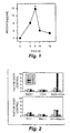
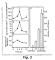


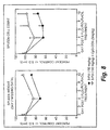

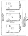


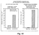
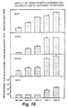

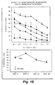
Classifications
-
- C—CHEMISTRY; METALLURGY
- C12—BIOCHEMISTRY; BEER; SPIRITS; WINE; VINEGAR; MICROBIOLOGY; ENZYMOLOGY; MUTATION OR GENETIC ENGINEERING
- C12N—MICROORGANISMS OR ENZYMES; COMPOSITIONS THEREOF; PROPAGATING, PRESERVING, OR MAINTAINING MICROORGANISMS; MUTATION OR GENETIC ENGINEERING; CULTURE MEDIA
- C12N15/00—Mutation or genetic engineering; DNA or RNA concerning genetic engineering, vectors, e.g. plasmids, or their isolation, preparation or purification; Use of hosts therefor
- C12N15/09—Recombinant DNA-technology
- C12N15/11—DNA or RNA fragments; Modified forms thereof; Non-coding nucleic acids having a biological activity
- C12N15/117—Nucleic acids having immunomodulatory properties, e.g. containing CpG-motifs
-
- A—HUMAN NECESSITIES
- A61—MEDICAL OR VETERINARY SCIENCE; HYGIENE
- A61K—PREPARATIONS FOR MEDICAL, DENTAL OR TOILETRY PURPOSES
- A61K31/00—Medicinal preparations containing organic active ingredients
- A61K31/70—Carbohydrates; Sugars; Derivatives thereof
-
- A—HUMAN NECESSITIES
- A61—MEDICAL OR VETERINARY SCIENCE; HYGIENE
- A61K—PREPARATIONS FOR MEDICAL, DENTAL OR TOILETRY PURPOSES
- A61K39/00—Medicinal preparations containing antigens or antibodies
- A61K39/39—Medicinal preparations containing antigens or antibodies characterised by the immunostimulating additives, e.g. chemical adjuvants
-
- A—HUMAN NECESSITIES
- A61—MEDICAL OR VETERINARY SCIENCE; HYGIENE
- A61K—PREPARATIONS FOR MEDICAL, DENTAL OR TOILETRY PURPOSES
- A61K39/00—Medicinal preparations containing antigens or antibodies
- A61K2039/555—Medicinal preparations containing antigens or antibodies characterised by a specific combination antigen/adjuvant
- A61K2039/55511—Organic adjuvants
- A61K2039/55555—Liposomes; Vesicles, e.g. nanoparticles; Spheres, e.g. nanospheres; Polymers
-
- A—HUMAN NECESSITIES
- A61—MEDICAL OR VETERINARY SCIENCE; HYGIENE
- A61K—PREPARATIONS FOR MEDICAL, DENTAL OR TOILETRY PURPOSES
- A61K39/00—Medicinal preparations containing antigens or antibodies
- A61K2039/555—Medicinal preparations containing antigens or antibodies characterised by a specific combination antigen/adjuvant
- A61K2039/55511—Organic adjuvants
- A61K2039/55561—CpG containing adjuvants; Oligonucleotide containing adjuvants
-
- A—HUMAN NECESSITIES
- A61—MEDICAL OR VETERINARY SCIENCE; HYGIENE
- A61K—PREPARATIONS FOR MEDICAL, DENTAL OR TOILETRY PURPOSES
- A61K39/00—Medicinal preparations containing antigens or antibodies
- A61K2039/555—Medicinal preparations containing antigens or antibodies characterised by a specific combination antigen/adjuvant
- A61K2039/55511—Organic adjuvants
- A61K2039/55566—Emulsions, e.g. Freund's adjuvant, MF59
-
- A—HUMAN NECESSITIES
- A61—MEDICAL OR VETERINARY SCIENCE; HYGIENE
- A61K—PREPARATIONS FOR MEDICAL, DENTAL OR TOILETRY PURPOSES
- A61K39/00—Medicinal preparations containing antigens or antibodies
-
- C—CHEMISTRY; METALLURGY
- C12—BIOCHEMISTRY; BEER; SPIRITS; WINE; VINEGAR; MICROBIOLOGY; ENZYMOLOGY; MUTATION OR GENETIC ENGINEERING
- C12N—MICROORGANISMS OR ENZYMES; COMPOSITIONS THEREOF; PROPAGATING, PRESERVING, OR MAINTAINING MICROORGANISMS; MUTATION OR GENETIC ENGINEERING; CULTURE MEDIA
- C12N2310/00—Structure or type of the nucleic acid
- C12N2310/30—Chemical structure
- C12N2310/31—Chemical structure of the backbone
- C12N2310/315—Phosphorothioates
-
- C—CHEMISTRY; METALLURGY
- C12—BIOCHEMISTRY; BEER; SPIRITS; WINE; VINEGAR; MICROBIOLOGY; ENZYMOLOGY; MUTATION OR GENETIC ENGINEERING
- C12N—MICROORGANISMS OR ENZYMES; COMPOSITIONS THEREOF; PROPAGATING, PRESERVING, OR MAINTAINING MICROORGANISMS; MUTATION OR GENETIC ENGINEERING; CULTURE MEDIA
- C12N2310/00—Structure or type of the nucleic acid
- C12N2310/30—Chemical structure
- C12N2310/34—Spatial arrangement of the modifications
- C12N2310/345—Spatial arrangement of the modifications having at least two different backbone modifications
-
- Y—GENERAL TAGGING OF NEW TECHNOLOGICAL DEVELOPMENTS; GENERAL TAGGING OF CROSS-SECTIONAL TECHNOLOGIES SPANNING OVER SEVERAL SECTIONS OF THE IPC; TECHNICAL SUBJECTS COVERED BY FORMER USPC CROSS-REFERENCE ART COLLECTIONS [XRACs] AND DIGESTS
- Y02—TECHNOLOGIES OR APPLICATIONS FOR MITIGATION OR ADAPTATION AGAINST CLIMATE CHANGE
- Y02A—TECHNOLOGIES FOR ADAPTATION TO CLIMATE CHANGE
- Y02A50/00—TECHNOLOGIES FOR ADAPTATION TO CLIMATE CHANGE in human health protection, e.g. against extreme weather
- Y02A50/30—Against vector-borne diseases, e.g. mosquito-borne, fly-borne, tick-borne or waterborne diseases whose impact is exacerbated by climate change
Definitions
- the present invention relates to methods for regulating hematopoiesis using CpG containing oligonucleotides.
- the invention relates to methods of treating thrombopoiesis and anemia by regulating hematopoiesis.
- the invention also relates to methods of regulating immune system remodeling by administering CpG oligonucleotides to manipulate hematopoiesis.
- hematopoietic cytokines are currently in clinical trials as treatments to prevent or reduce such complications.
- Hematopoietic development is considered to be regulated by two categories of factors.
- One category includes colony-stimulating factors (CSFs), which promote colony formation and proliferation of cells of various lineages.
- CSFs colony-stimulating factors
- Another is potentiators, which potentiate maturation or differentiation.
- Megakaryocyte-CSFs reportedly include IL-3, granulocyte-macrophage colony-stimulating factor (GM-CSF) and stem cell factor (SCF)
- Megakaryocyte potentiators reportedly include IL-6, IL-7, IL-11, erythropoietin (EPO) and leukemia inhibitory factor (LIF).
- EPO erythropoietin
- LIF leukemia inhibitory factor
- Platelet production is a terminal phenomenon in the development of megakaryocytes in vivo.
- Thrombopoietin (TPO) was reported to posses both Meg-CSF
- a fraction extracted from BCG (designated MY-1) was shown to exhibit anti-tumor activity in vivo, augment NK cell activity and trigger type I and type II IFN release from murine spleen cells or human peripheral blood lymphocytes (PBL) in vitro.
- PBL peripheral blood lymphocytes
- Bacterial DNA, some viral DNA, and invertebrate DNA seem to differ structurally from vertebrate DNA.
- Bacterial DNA has the expected frequency of CpG dinucleotides of 1:16.
- mammalian DNA exhibits CpG suppression and has only about one-fourth as many CpG as predicted by random base usage.
- the usage of the 5'-Pu-Pu-CpG-Pyr-Pyr-3' motif is even more suppressed in mammals compared with the genome of Escherichia coli. Krieg, AM et al. (1995) Nature 374:546-549.
- CpG DNA The immunogenicity of proteinaceous natural and recombinant purified antigens is poor unless aided by adjuvants. Because of the apparent recognition and response to foreign DNA by the immune system, the potential of CpG DNA to serve as an adjuvant was previously tested. Mice were challenged subcutaneously with liposome-encapsulated ovalbumin (used as antigen) and CpG-ODN (used as adjuvant) using a protocol described by Lipford et al. Lipford, GB et al. (1997) Eur J Immunol 27:3420-3426. The mice which were co-administered CpG-ODN developed strong peptide-specific cytotoxic T lymphocyte (CTL) activity in the draining lymph nodes (LNs).
- CTL cytotoxic T lymphocyte
- the Th1-promoting adjuvanticity of CpG-ODN may be useful for the redirection to protective, or even curative, responses in Th2-driven disorders.
- a model is the CpG-ODN modulation of Th2 driven airway inflammation in a murine model of asthma induced with Schistosoma mansoni eggs. Airway eosinophilia, Th2 cytokine induction, IgE production and bronchial hyperreactivity were prevented by CpG-ODN co-administration with egg sensitization. Additionally, egg sensitized mice treated at day 7 post sensitization with CpG-ODN and antigen were protected from airway eosinophilia.
- the phenotype of the response post CpG intervention was Th1-like although the initial response to L. major challenge was Th2-like.
- CpG-ODN triggers the release of IL-12 into the serum post injection and IL-12 is a known inducer of Th1 differentiation.
- the wave of IL-12 is transient, however, peaking at 2-4 h and returning to near baseline by 24 h.
- Experimental treatment with anti-IL-4 or IL-12 following with L. major is effective only within the first 3 days; at later time points, even multiple injections of IL-12 fail to influence the course of infection. Since CpG-ODN effectively stimulate NK cells to produce IFN- ⁇ , and since there is evidence that IFN- ⁇ can redirect Th2 responses in vitro and in vivo, this may be a possible explanation. However, the actual cellular and molecular mechanism of the curative effects are yet poorly understood.
- Splenomegaly is a well-recognized phenomenon accompanying some oligonucleotide injections.
- Branda et al. observed that mice developed massive splenomegaly and polyclonal hypergammaglobulinemia within 2 days after intravenous injection of a phosphorothioate oligomer that is antisense to a portion of the rev region of the HIV-1 genome.
- mice with antisense to the initiation region of the env gene resulted in (i) increased spleen cell numbers, primarily due to an increase in splenic B cells, (ii) increased class II MHC expression on B cells, (iii) increased RNA and DNA synthesis, and (iv) increased numbers of immunoglobulin (Ig)-producing cells.
- Ig immunoglobulin-producing cells.
- Zhao et al. concluded that phosphorothioate ODN induce splenomegaly due to B cell proliferation. Zhao, Q et al. (1996) Biochem Pharmacol 51:173-182. In a follow-up study Zhao et al. found administration of the 27-mer-phosphorothioate oligonucleotide into mice resulted in splenomegaly and an increase in IgM production 48 hr post-administration. Zhao, Q et al. (1996) Biochem Pharmacol 52:1537-1544.
- Agrawal et al. evaluated the in vivo toxicological effects of phosphorothioate oligodeoxynucleotides (PS oligo).
- PS oligo phosphorothioate oligodeoxynucleotides
- Agrawal, S et al. (1997) Antisense Nucleic Acid Drug Dev 7:575-584.
- Oligodeoxynucleotides were administrated intravenously to male and female rats at doses of 3, 10, and 30 mg/kg/day for 14 days. Rats were killed on day 15, blood samples were collected for hematology and clinical chemistry determinations, and tissues, including lymph nodes, spleens, livers, and kidneys, were subjected to pathologic examinations.
- the toxicity profiles of four oligodeoxynucleotides were very similar, but differed in magnitude.
- hematology parameters included thrombocytopenia, anemia, and neutropenia. Dose-dependent enlargements of spleen, liver, and kidney were observed. Pathologic studies showed a generalized hyperplasia of the reticuloendothelial system in the tissues examined.
- Hematopoietic development is considered to be regulated by colony-stimulating factors, which promote colony formation and proliferation of cells of various primitive lineages, and potentiators, which potentiate maturation or differentiation into various blood cells.
- colony-stimulating factors which promote colony formation and proliferation of cells of various primitive lineages
- potentiators which potentiate maturation or differentiation into various blood cells.
- the observation of splenomegaly is explained by direct ODN B cell mitogenicity in a sequence specific manner.
- Zhao et al. administered to mice a 27-mer phosphorothioate oligonucleotide (sequence 5'-TCG TCG CTG TCT CCG CTT CTT CTT GCC-3' SEQ ID NO:109), which had previously been shown to cause splenomegaly and hypergammaglobulinemia upon in vivo administration in mice, and studied the pattern and kinetics of cytokine production at both the splenic mRNA and serum protein levels. Zhao et al.
- IL-12 has been shown to be released in response to CpG-ODN and is an inducer of IFN- ⁇ . Control of intracellular bacterial infections requires IFN- ⁇ both for establishing a Th1 T-cell response and for activating macrophages to kill the bacteria. Murray et al.
- mice deficient in IFN- ⁇ produces an immune response characterized by a Th2 T-cell phenotype, florid bacterial growth, and death.
- Murray, PJ et al. (1998) Blood 91:2914-2924. They reported that IFN- ⁇ -deficient mice infected with mycobacteria also undergo a dramatic remodeling of the hematopoietic system. Myeloid cell proliferation proceeds unchecked throughout the course of mycobacterial infection, resulting in a transition to extramedullary hematopoiesis.
- IFN- ⁇ may be a key cytokine in the coordinate regulation of immune effector cells and myelopoiesis.
- IL-3/GM-CSF/IL-5 (Th0 and Th2 cytokines) produced by activated T cells play a major role in expansion of hematopoietic cells in emergency
- IL-13 a recently identified Th2 cytokine, shares some, but not all, IL-4 functions, including inhibition of monocyte and macrophage activation, stimulation of human B cells, and induction of growth and differentiation of mouse bone marrow cells in vitro .
- rIL-13 recombinant mouse IL-13
- boLB/c mice After purification by anion exchange chromatography, rIL-13 was administered in the peritoneal cavity of BALB/c mice via osmotic pump for 7 days. Spleens from the rIL-13-treated mice were significantly enlarged compared with control spleens due to increased cellularity. In particular, increased numbers of immature erythroblasts and megakaryocytes were observed in splenic sections after rIL-13 treatment.
- Spleen cells from rIL-13-treated mice showed greatly increased responsiveness in vitro to recombinant forms of mouse IL-3, mouse granulocyte-macrophage CSF, or human CSF-1 and, to a lesser extent, to mouse IL-4 or IL-13.
- the rIL-13-treated mice also showed significant increases in CFU-E, CFU-C, and erythroid burst colonies in the spleen, further indicating the presence of increased numbers of hematopoietic precursors. Hematologic analyses indicated that rIL-13 treatment induced slight anemia and striking monocytosis.
- spleen cells from rIL-13-treated mice produced significantly more IL-6 upon LPS stimulation.
- Th2 driven responses are strongly predisposing for extramedullary hematopoiesis.
- CpG-ODN injection is Th1-biasing and Th2-suppressive.
- IFN- ⁇ the hallmark Th1 cytokine
- IL-13 a Th2 cytokine has been shown to induce hematopoiesis.
- ODN administration has been shown to lead to thrombocytopenia, anemia, and neutropenia.
- IL-12 a central cytokine in CpG-ODN effects, induces thrombocytopenia.
- the phenomenon of splenomegaly has been repeatedly correlated with B cell mitogenicity of ODN, suggesting that the ODN induces splenomegaly through B cell activation rather than hematopoiesis.
- Hematopoiesis may be used to treat immune system disorders.
- CpG oligonucleotide for inducing an antigen-specific immune response is based on the finding that a CpG oligonucleotide can be used to induce remodeling of the immune system by regulating hematopoiesis.
- a CpG oligonucleotide and antigen are administered together to a subject an initial immune response occurs. It has been discovered according to the invention that this initial immune response declines rapidly and a new immune response develops after approximately 48 hours. Unexpectedly, when antigen is administered 48 hours or more after the administration of CpG an antigen specific immune response will be mounted to the antigen. This immune response is due to a repopulation of lymph nodes and/or spleen with primed immune cells.
- a CpG oligonucletoide for the manufacture of a medicament for inducing an antigen-specific immune response by administering to a subject an oligonucleotide, having a sequence including at least the following formula: 5' X 1 CGX 2 3' wherein the oligonucleotide includes at least 8 nucleotides wherein C and G are unmethylated and wherein X 1 and X 2 are nucleotides, and exposing the subject to an antigen at least 3 days after the oligonucleotide is administered to the subject to produce an antigen-specific immune response.
- the subject may be exposed to the antigen at least 48 hours after the CpG oligonucleotide is administered to the subject. It has been discovered that immune system remodeling begins to occur within 48 hours of CpG administration. It has also been discovered that the primed immune cells are still capable of responding to antigen even 30 days after CpG administration.
- the antigen is administered at least 4 days after the oligonucleotide is administered to the subject. In another embodiment the antigen is administered at least 7 days after the oligonucleotide is administered to the subject. In another embodiment the antigen is administered at least 15 days after the oligonucleotide is administered to the subject. In yet another embodiment the antigen is administered at least 30 days after the oligonucleotide is administered to the subject.
- the antigen may be any type of antigen known in the art.
- the antigen may be cells, cell extracts, proteins, peptides, polysaccharides, polysaccharide conjugates, lipids, glycolipids, carbohydrate, viral extracts, viruses, bacteria, fungi, parasites, and allergens.
- the antigen may be a nucleic acid encoding an antigen.
- the antigen is an allergen and the CpG oligonucleotide is used for the manufacture of a medicament for treating allergy.
- the antigen is derived from an infectious organism selected from the group consisting of infectious bacteria, infectious viruses, and infectious fungi and the CpG oligonucleotide is used for the manufacture of a medicament for treating an infectious disease.
- the subject is exposed to an antigen.
- the subject may be actively exposed to the antigen.
- the antigen may be delivered in conjunction with a colloidal dispersion system.
- the colloidal dispersion system is selected from the group consisting of macromolecular complexes, nanocapsules, microspheres, beads, and lipid-based systems in another embodiment.
- a lipid-based system is preferably selected from the group consisting of oil-in-water emulsions, micelles, mixed micelles, and liposomes.
- the antigen may be administered in conjunction with an adjuvant.
- the subject may also be passively exposed to the antigen.
- the subject is a subject at risk of developing cancer.
- the subject is at risk of developing an allergic reaction.
- the subject is an asthmatic.
- the antigen specific immune response is a Th1 type immune response in another embodiment.
- the subject is a vertebrate animal.
- the subject is a human.
- the subject is a nonhuman vertebrate animal.
- the vertebrate nonhuman animal is selected from the group consisting of a dog, cat, horse, cow, pig, sheep, goat, chicken, primate, fish, rat, and mouse.
- Hematopoiesis may be treated by administering a CpG oligonucleotide to a subject having or at risk of developing a hematopoietic disorder.
- a hematopoietic disorder is a disorder involving a loss or decrease in numbers of one or more hematopoietic cells.
- Hematopoietic cells include erythrocytes, leukocytes and platelets.
- Platelet counts may be increased in a subject having thrombocytopenia by administering to a subject having thrombocytopenia an oligonucleotide, having a sequence including at least the following formula: 5' X 1 CGX 2 3' wherein the oligonucleotide includes at least 8 nucleotides wherein C and G are unmethylated and wherein X 1 and X 2 are nucleotides, in an amount effective to increase platelet counts in the subject.
- the thrombocytopenia is a non-chemotherapeutic induced thrombocytopenia.
- a subject at risk of developing thrombocytopenia can be treated by administering to a subject at risk of developing thrombocytopenia an oligonucletoide, having a sequence including at least the following formula: 5' X 1 CGX 2 3' wherein the oligonucleotide includes at least 8 nucleotides wherein C and G are unmethylated and wherein X 1 and X 2 are nucleotides, in an amount effective to prevent a decrease in platelet counts ordinarily expected under platclet-depleting conditions in The subject when the subject is exposed to platelet-depleting conditions.
- the oligonucleotide is administered in an amount effective to increase platelet counts in the subject by at least 10,000 platelets per microliter. In another embodiment the oligonucleotide is administered in an amount effective to increase platelet counts in the subject by at least 20,000 platelets per microliter. In yet another embodiment the oligonucleotide is administered to the subject in an amount effective to increase the platelet counts in the subject by 100 percent.
- the thrombocytopenia is any type of thrombocytopenia known in the art.
- the thrombocytopenia is a drug-induced thrombocytopenia.
- the thrombocytopenia is due to an autoimmune disorder such as idiopathic thrombocytopenic purpura.
- the thrombocytopenia is a thrombocytopenia resulting from accidental radiation exposure.
- the thrombocytopenia is a thrombocytopenia resulting from therapeutic radiation exposure in yet another embodiment.
- Anemia can be treated by administering to a subject having anemia an oligonucleotide, having a sequence including at least the following formula: 5' X 1 CGX 2 3' wherein the oligonucleotide includes at least 8 nucleotides wherein C and G are unmethylated and wherein X 1 and X 2 are nucleotides, in an amount effective to induce erythropoiesis in the subject.
- the oligonucleotide is administered in an amount effective to increase erythroblast counts in the subject by at least 10 percent. In another embodiment the oligonucleotide is administered in an amount effective to increase erythroblast counts in the subject by at least 20 percent. According to yet another embodiment the oligonucleotide is administered to the subject in an amount effective to increase erythroblast counts in the subject by 100 percent.
- the anemia can be any type of anemia known in the art.
- the anemia is a drug-induced anemia.
- the anemia is selected from the group consisting of an immunohemolytic disorder, genetic disorders such as hemoglobinopathy and inherited hemolytic anemia; inadequate production despite adequate iron stores; chronic disease such as kidney failure; and chronic inflammatory disorder such as rheumatoid arthritis.
- the subject having or at risk of having a hematopoietic disorder is a vertebrate animal.
- the subject is a human.
- the subject is a dog.
- the subject is a nonhuman vertebrate animal selected from the group consisting of a cat, horse, cow, pig, sheep, goat, chicken, primate, fish, rat, and mouse.
- the CpG oligonucleotide is an oligonucleotide, having a sequence including at least the following formula: 5' X 1 CGX 2 3'.
- the oligonucleotide is 8 to 100 nucleotides in length. In other embodiments the oligonucleotide is 8 to 30 nucleotides in length.
- the oligonucleotide is a stabilized oligonucleotide.
- the oligonucleotide includes a phosphate backbone modification which is a phosphorothioate or phosphorodithioate modification.
- the phosphate backbone modification occurs at the 5' end of the oligonucleotide.
- the phosphate backbone modification occurs at the 3' end of the oligonucleotide.
- the CpG oligonucleotide has a sequence including at least the following formula: 5' X 1 X 2 CGX 3 X 4 3' wherein X 1 X 2 are nucleotides selected from the group consisting of:GpT, GpG, GpA, ApA, ApT, ApG, CpT, CpA, CpG, TpA, TpT, and TpG; and X 3 X 4 are nucleotides selected from the group consisting of: TpT, CpT, ApT, TpG, ApG, CpG, TpC, ApC, CpC, TpA, ApA, and CpA.
- the CpG oligonucleotide has a sequence including at least the following formula: 5' TCNTX 1 X 2 CGX 3 X 4 3' wherein X 1 , X 2 , X 3 , and X 4 are nucleotides, N is a nucleic acid sequence composed of from about 0-25 nucleotides.
- X 1 X 2 are nucleotides selected from the group consisting of: GpT, GpG, GpA and ApA and X 3 X 4 are nucleotides selected from the group consisting of: TpT and CpT in another embodiment.
- Hematopoiesis refers to the generation of blood cells.
- the process of generating new blood cells is controlled through the complex interaction of immune factors such as interleukin and CSF. Using these factors the immune system is able to regulate the levels of each of the cellular components in blood in response to physiological changes.
- Erythrocytes, leukocytes and platelets are the essential cells of the human hematopoietic system.
- the primary function of erythrocytes, also known as red blood cells, is to transport hemoglobin, which in turn carries oxygen from the lungs to tissues. Oxygenated hemoglobin gives the erythrocytes a red color.
- Leukocytes also referred to as myeloid cells, are a heterogenous group of cells that mediate immune responses and which include granulocytes, including eosinophils, basophils, and neutrophils; monocytes; and T and B lymphocytes. These cells are found predominately in the blood, bone marrow, lymphoid organs and epithelium.
- Leukocytes are referred to as white blood cells because of a lack of natural pigment which gives the cells a whitish or transparent appearance. Platelets play a role in hemostasis, or the regulation of bleeding.
- disorders of the hematopoietic system vary depending on the factor causing the disorder as well as the cell type affected.
- CpG containing oligonucleotides can regulate hematopoieses to inhibit loss of blood cells in response to physiological disorders caused by genetic abnormalities, environmental factors or medical therapies. It has also been discovered that hematopoiesis can be manipulated using CpG oligonucleotides to induce immune system remodeling in order to stimulate an antigen specific immune response.
- the process of immune system remodeling is based on the generation of immune cells in response to a stimulus in preparation for generating a strong antigen specific immune response.
- the stimulus is a CpG oligonucleotide. It has been discovered that when a CpG oligonucleotide is administered to a subject, after an initial delay, the immune system of the subject undergoes a repopulation event to produce a population of immune cells which are primed to generate an antigen specific response. This renewed population of cells remains in the body for an extensive period of time. When the primed cells encounter antigen the cells respond to the antigen by producing an antigen specific immune response. In fact the antigen is capable of producing an immune specific response even in the absence of an adjuvant Ordinarily the administration of antigen in the absence of an adjuvant would not produce a specific immune response.
- CpG when CpG is administered to a subject, CpG activates the circulating immune cells, causing them to mature into mature active immune cells. If CpG is administered at the same time or slightly before or after an antigen then the circulating immune cells will likely contact the antigen and develop a specific immune response against that antigen. After a period of about 24 hours, the circulating immune cells will no longer be capable of mounting an antigen specific immune response because the circulating cells have already been activated and matured. It has been found according to the invention, however, that approximately two days after the administration of CpG the subject's immune system has been repopulated with immune cells which are capable of being matured and activated in response to antigen.
- the immune system is capable of generating an antigen specific immune response, which may be even of a greater magnitude than the immune response which is generated in response to antigen administration at the same time as CpG.
- the remodeled immune system encompasses a population of cells which are capable of responding to antigen. It has been demonstrated according to the invention that this population of cells is capable of responding to antigen for long periods of time. For instance, administration of an antigen at time periods of greater than 30 days after the CpG administration can still produce an antigen specific response.
- the invention encompasses the use of CpG oligonucleotides for the manufacture of a medicament for generating an antigen specific immune response by administering CpG to induce immune remodeling to prepare for exposure to an antigen.
- the subject may be intentionally exposed to the antigen two days or more after being administered CpG in order to develop an immunity to a specific antigen.
- the subject may also be exposed passively to an antigen, causing a specific immune response to develop against an antigen to which the subject is exposed from the environment.
- the immune system can be manipulated to be in an active state ready to respond to invading substances, such as pathogens.
- CpG oligonucleotides for the manufacture of a medicament for inducing immune system remodeling of the invention is a use of the CpG oligonucleotides for the manufacture of a medicament for inducing an antigen-specific immune response, by administering to a subject an oligonucleotide, having a sequence including at least the following formula: 5' X 1 CGX 2 3' wherein the oligonucleotide includes at least 8 nucleotides wherein C and G are unmethylated and wherein X 1 and X 2 are nucleotides, and exposing the subject to an antigen at least 3 days alter the oligonucleotide is administered to the subject to produce an antigen-specific immune response.
- an "antigen” as used herein is a molecule capable of provoking an immune response.
- Antigens include but are not limited to cells, cell extracts, polysaccharides, polysaccharide conjugates, lipids, glycolipids, carbohydrate, peptides, proteins, viruses, and viral extracts.
- the term antigen broadly includes any type of molecule which is recognized by a host immune system as being foreign.
- Antigens include but are not limited to cancer antigens, microbial antigens, and allergens.
- the CpG oligonucleotides of the invention are useful for treating cancer by stimulating an antigen specific immune response against an antigen.
- a "cancer antigen” as used herein is a compound, such as a peptide, associated with a tumor or cancer cell surface and which is capable of provoking an immune response when expressed on the surface of an antigen presenting cell in the context of an MHC molecule.
- Cancer antigens can be prepared from cancer cells either by preparing crude extracts of cancer cells, for example, as described in Cohen, et al., 1994, Cancer Research , 54:1055, by partially purifying the antigens, by recombinant technology, or by de novo synthesis of known antigens.
- Cancer antigens include antigens that are recombinately an immunogenic portion of or a whole tumor or cancer. Such antigens can be isolated or prepared recombinady or by any other means known in the art. Cancers or tumors include but are not limited to biliary tract cancer, brain cancer, breast cancer; cervical cancer; choriocarcinoma; colon cancer, endometrial cancer; esophogeal cancer; gastric cancer; intraepithelial neoplasms; lymphomas; liver cancer; lung cancer (e.g.
- melanoma neuroblastomas
- oral cancer ovarian cancer; pancreas cancer; prostate cancer; rectal cancer; sarcomas; skin cancer; testicular cancer; thyroid cancer; and renal cancer, as well as other carcinomas and sarcomas.
- the CpG oligonucleotides of the invention are also useful for treating infectious diseases.
- An infectious disease as used herein, is a disease arising from the presence of a foreign microorganism in the body.
- CpG is used to stimulate an antigen specific immune response which can activate a T or B cell response against an antigen of the microorganism.
- the the Cph oligonucleotides are used in the same way as described above for the tumor except that the antigen is specific for a microorganism using a microbial antigen.
- a "microbial antigen” as used herein is an antigen of a microorganism and includes but is not limited to infectious virus, infectious bacteria, and infectious fungi.
- antigens include the intact microorganism as well as natural isolates and fragments or derivatives thereof and also synthetic compounds which are identical to or similar to natural microorganism antigens.
- a compound is similar to a natural microorganism antigen if it induces an immune response (humoral and/or cellular) to a natural microorganism antigen.
- antigens are used routinely in the art and are well known to those of ordinary skill in the art.
- Retroviridae e.g. human immunodeficiency viruses, such as HIV-1 (also referred to as HTLV-III, LAV or HTLV-III/LAV, or HIV-III; and other isolates, such as HIV-LP; Picornaviridae (e.g. polio viruses, hepatitis A virus; enteroviruses, human Coxsackie viruses, rhinoviruses, echoviruses); Calciviridae (e.g. strains that cause gastroenteritis); Togaviridae (e.g. equine encephalitis viruses, rubella viruses); Flaviviridae (e.g.
- Coronaviridae e.g. coronaviruses
- Rhabdoviridae e.g. vesicular stomatitis viruses, rabies viruses
- Filoviridae e.g. ebola viruses
- Paramyxoviridae e.g. parainfluenza viruses, mumps virus, measles virus, respiratory syncytial virus
- Orthomyxoviridae e.g. influenza viruses
- Bunyaviridae e.g. Hantaan viruses, bunya viruses, phleboviruses and Nairo viruses
- Arena viridae hemorrhagic fever viruses
- Reoviridae e.g.
- reoviruses reoviruses, orbiviurses and rotaviruses
- Birnaviridae Hepadnaviridae (Hepatitis B virus); Parvovirida (parvoviruses); Papovaviridae (papilloma viruses, polyoma viruses); Adenoviridae (most adenoviruses); Herpesviridae (herpes simplex virus (HSV) 1 and 2, varicella zoster virus, cytomegalovirus (CMV), herpes virus; Poxviridae (variola viruses, vaccinia viruses, pox viruses); and Iridoviridae (e.g. African swine fever virus); and unclassified viruses (e.g.
- infectious bacteria examples include but are not limited to: Helicobacter pyloris , Borrelia burgdorferi, Legionella pneumophilia, Mycobacteria sps (e.g. M. tuberculosis, M. avium, M. intracellulare, M. kansaii, M.
- infectious fungi examples include: Cryptococcus neoformans, Histoplasma capsulatum, Coccidioides immitis, Blastomyces dermatitidis, Chlamydia trachomatis, Candida albicans .
- Other infectious organisms i.e., protists
- Plasmodium such as Plasmodium falciparum, Plasmodium malariae, Plasmodium ovale , and Plasmodium vivax and Toxoplasma gondii.
- the Cph oligonucleotides of the invention are also useful for treating allergic diseases.
- the methods are accomplished in the same way as described above for the tumor immunothcrapy and treatment of infectious diseases except that the antigen is specific for an allergen.
- allergic diseases are generally treated by the injection of small doses of antigen followed by subsequent increasing dosage of antigen. It is believed that this procedure produces a memory immune response to prevent further allergic reactions. These methods, however, are associated with the risk of side effects such as an allergic response. The methods of the invention avoid these problems.
- allergen refers to a substance (antigen) that can induce an allergic or asthmatic response in a susceptible subject.
- the list of allergens is enormous and can include pollens, insect venoms, animal dander dust, fungal spores and drugs (e.g. penicillin).
- Examples of natural, animal and plant allergens include but are not limited to proteins specific to the following genuses: Canine (Canis familiaris) ; Dermatophagoides (e.g. Dermatophagoides farinae ); Felis (Felis domesticus); Ambrosia (Ambrosia artemiisfolia ; Lolium (e.g.
- Lolium perenne or Lolium multiflorum Cryptomeria ( Cryptomeria japonica ); Alternaria (Alternaria alternata ); Alder ; Alnus (Alnus gultinoasa ); Betula ( Betula verrucosa ); Quercus (Quercus alba ); Olea (Olea europa ); Artemisia ( Artemisia vulgaris ); Plantago (e.g. Plantago lanceolata ); Parietaria (e.g. Parietaria officinalis or Parietaria judaica ); Blattella (e.g. Blattella germanica ); Apis (e.g.
- Apis multiflorum Cupressus (e.g. Cupressus sempervirens, Cupressus arizonica and Cupressus macrocarpa ); Juniperus (e.g. Juniperus sabinoides, Juniperus virginiana, Juniperus communis and Juniperus ashei ); Thuya (e.g. Thuya orientalis ); Chamaecyparis (e.g. Chamaecyparis obtusa ); Periplaneta (e.g. Periplaneta americana ); Agropyron (e.g. Agropyron repens ) ; Secale (e.g. Secale cereale ); Triticum (e.g.
- Triticum aestivum Dactylis (e.g. Dactylis glomerata ) ; Festuca (e.g. Festuca elatior ); Poa (e.g. Poa pratensis or Poa compressa ); Avena (e.g. Avena sativa ); Holcus (e.g. Holcus lanatus ); Anthoxanthum (e.g. Anthoxanthum odoratum ); Arrhenatherum (e.g. Arrhenatherum elatius ); Agrostis (e.g. Agrostis alba ); Phleum (e.g. Phleum pratense ); Phalaris (e.g. Phalaris arundinacea ); Paspalum (e.g. Paspalum notatum ); Sorghum (e.g. Sorghum halepensis ); and Bromus (e.g. Bromus inermis ).
- Allergic conditions include but are not limited to eczema, allergic rhinitis or coryza, hay fever, bronchial asthma, urticaria (hives) and food allergies, and other atopic conditions.
- a subject having an allergic reaction is a subject that has or is at risk of developing an allergy.
- Th1 The other major type of immune response is termed as Th2 immune response, which is associated with more of an antibody immune response and with the production of IL-4, IL-5 and IL-10.
- Th2 immune response the other major type of immune response.
- allergic diseases are mediated by Th2 type immune responses and autoimmune diseases by Th1 immune response.
- an effective dose of a CpG oligonucleotide can be administered to a subject to treat or prevent an allergy.
- Th2 cytokines especially IL-4 and IL-5 are elevated in the airways of asthmatic subjects. These cytokines, especially IL-4 and IL-5 are elevated in the airways of asthmatic subjects. These cytokines promote important aspects of the asthmatic inflammatory response, including IgE isotope switching, eosinophil chemotaxis and activation and mast cell growth. Th1 cytokines, especially IFN- ⁇ and IL-12, can suppress the formation of Th2 clones and production of Th2 cytokines.
- “Asthma” refers to a disorder of the respiratory system characterized by inflammation, narrowing of the airways and increased reactivity of the airways to inhaled agents. Asthma is frequently, although not exclusively associated with atopic or allergic symptoms.
- the antigen is taken up by an antigen presenting cell (APC) such as a dendritic cell in the repopulated immune system.
- APC antigen presenting cell
- the APC then processes and presents the antigen on its cell surface to produce a cytotoxic T lymphocyte (CTL) response by interacting with T lymphocytes or an antibody response by interacting with B lymphocytes.
- CTL cytotoxic T lymphocyte
- the antigen is exposed to the immune cells 48 hours after adding CpG.
- the subject's immune cells are exposed to the antigen 60 hours after the CpG.
- the subject's immune cells are exposed to the antigen at least 3, 4, 5, 6, 7, 8, 9, 10, 11, 12, 13, 14, 15, 16, 17,18, 19, 20, 21, 22, 23, 24, 25, 26, 27, 28, 29, 30, 31, 32, 33, 34, 35, 36, 37, 38, 39, or 40 days after the CpG.
- a “subject” shall mean a human or vertebrate animal including but not limited to a dog, cat, horse, cow, pig, sheep, goat, chicken, primate, e.g., monkey, fish (aquaculture species), e.g. salmon, rat, and mouse.
- Nonhuman vertebrates are also capable of developing cancer, infections, allergies, and asthma.
- the CpG oligonucleotides of the invention are useful for treating infections of animals.
- the term "treat” or “treating” when used with respect to an infectious disease refers to a prophylactic treatment which increases the resistance of a subject to infection with a pathogen or, in other words, decreases the likelihood that the subject will become infected with the pathogen.
- Many vaccines for the treatment of non-human vertebrates are disclosed in Bennett, K. Compendium of Veterinary Products, 3rd ed. North American Compendiums, Inc., 1995.
- antigens include infectious microbes such as virus, bacteria and fungi and fragments thereof, derived from natural sources or synthetically.
- infectious virus of both human and non-human vertebrates include retroviruses, RNA viruses and DNA viruses.
- This group of retroviruses includes both simple retroviruses and complex retroviruses.
- the simple retroviruses include the subgroups of B-type retroviruses, C-type retroviruses and D-type retroviruses.
- An example of a B-type retrovirus is mouse mammary tumor virus (MMTV).
- the C-type retroviruses include subgroups C-type group A (including Rous sarcoma virus (RSV), avian leukemia virus (ALV), and avian myeloblastosis virus (AMV)) and C-type group B (including murine leukemia virus (MLV), feline leukemia virus (FeLV), murine sarcoma virus (MSV), gibbon ape leukemia virus (GALV), spleen necrosis virus (SNV), reticuloendotheliosis virus (RV) and simian sarcoma virus (SSV)).
- the D-type retroviruses include Mason-Pfizer monkey virus (MPMV) and simian retrovirus type 1 (SRV-1).
- the complex retroviruses include the subgroups of lentiviruses, T-cell leukemia viruses and the foamy viruses.
- Lentiviruses include HIV-1, but also include HIV-2, SIV, Visna virus, feline immunodeficiency virus (FIV), and equine infectious anemia virus (EIAV).
- the T-cell leukemia viruses include HTLV-I, HTLV-II, simian T-cell leukemia virus (STLV), and bovine leukemia virus (BLV).
- the foamy viruses include human foamy virus (HFV), simian foamy virus (SFV) and bovine foamy virus (BFV). The foregoing list is illustrative, and is not intended to be limiting.
- the family Bunyaviridae including the genus Bunyvirus (Bunyamwera and related viruses, California encephalitis group viruses), the genus Phlebovirus (Sandfly fever Sicilian virus, Rift Valley fever virus), the genus Nairovirus (Crimean-Congo hemorrhagic fever virus, Kenya sheep disease virus), and the genus Uukuvirus (Uukuniemi and related viruses); the family Orthomyxoviridae, including the genus Influenza virus (Influenza virus type A, many human subtype
- Illustrative DNA viruses that are antigens in vertebrate animals include, but are not limited to: the family Poxviridae, including the genus Orthopoxvirus (Variola major, Variola minor, Monkey pox Vaccinia, Cowpox, Buffalopox, Rabbitpox, Ectromelia), the genus Leporipoxvirus (Myxoma, Fibroma), the genus Avipoxvirus (Fowlpox, other avian poxvirus), the genus Capripoxvirus (sheeppox, goatpox), the genus Suipoxvirus (Swinepox), the genus Parapoxvirus (contagious postular dermatitis virus, pseudocowpox, bovine papular stomatitis virus); the family Iridoviridae (African swine fever virus, Frog viruses 2 and 3, Lymphocystis virus of fish); the family Herpesviridae
- Gram negative and gram positive bacteria serve as antigens in vertebrate animals.
- Such gram positive bacteria include, but are not limited to those bacteria discussed above as well as Pasteurella species, Staphylococci species, and Streptococcus species.
- Gram negative bacteria include, but are not limited to, Escherichia coli, Pseudomonas species, and Salmonella species. Salmonella enteritidis is an important pathogen in the commercial layer industry, as ovarian colonization of layers may result in maternally transmitted Salmonella in table eggs.
- the CpG oligonucleotides of the preferred embodiments are particularly well suited for treatment of birds such as hens, chickens, turkeys, ducks, geese, quail, and pheasant. Birds are prime targets for many types of infections including AIDS or immunodeficiency virus.
- Hatching birds are exposed to pathogenic microorganisms shortly after birth. Although these birds are initially protected against pathogens by maternal derived antibodies, this protection is only temporary, and the bird's own immature immune system must begin to protect the bird against the pathogens. It is often desirable to prevent infection in young birds when they are most susceptible. It is also desirable to prevent against infection in older birds, especially when the birds are housed in closed quarters, leading to the rapid spread of disease. Thus, it is desirable to administer the CpG oligonucleotide of the invention to birds to enhance an antigen-specific immune response when antigen is present.
- CIAV chicken infectious anemia virus
- CIAV infection results in a clinical disease, characterized by anemia, hemorrhage and immunosuppression, in young susceptible chickens. Atrophy of the thymus and of the bone marrow and consistent lesions of CIAV-infected chickens are also characteristic of CIAV infection. Lymphocyte depletion in the thymus, and occasionally in the bursa of Fabricius, results in immunosuppression and increased susceptibility to secondary viral, bacterial, or fungal infections which then complicate the course of the disease. The immunosuppression may cause aggravated disease after infection with one or more of Marek's disease virus (MDV), infectious bursal disease virus, reticuloendotheliosis virus, adenovirus, or reovirus.
- MDV Marek's disease virus
- Vaccination of birds, like other vertebrate animals can be performed at any age. Normally, vaccinations are performed at up to 12 weeks of age for a live microorganism and between 14-18 weeks for an inactivated microorganism or other type of vaccine. For in ovo vaccination, vaccination can be performed in the last quarter of embryo development.
- the vaccine may be administered subcutaneously, by spray, orally, intraocularly, intratracheally, nasally, in ovo or by other methods described herein.
- the CpG oligonucleotide of the invention can be administered to birds and other non-human vertebrates using routine vaccination schedules and the antigen is administered after an appropriate time period as described herein.
- Cattle and livestock are also susceptible to infection. Disease which affect these animals can produce severe economic losses, especially amongst cattle.
- the methods of the invention can be used to protect against infection in livestock, such as cows, horses, pigs, sheep, and goats.
- Bovine viral diarrhea virus (BVDV) is a small enveloped positive-stranded RNA virus and is classified, along with hog cholera virus (HOCV) and sheep border disease virus (BDV), in the pestivirus genus.
- HOCV hog cholera virus
- BDV sheep border disease virus
- Pestiviruses were previously classified in the Togaviridae family, some studies have suggested their reclassification within the Flaviviridae family along with the flavivirus and hepatitis C virus (HCV) groups (Francki, et al., 1991).
- BVDV which is an important pathogen of cattle can be distinguished, based on cell culture analysis, into cytopathogenic (CP) and noncytopathogenic (NCP) biotypes.
- CP cytopathogenic
- NCP noncytopathogenic
- the NCP biotype is more widespread although both biotypes can be found in cattle. If a pregnant cow becomes infected with an NCP strain, the cow can give birth to a persistently infected and specifically immunotolerant calf that will spread virus during its lifetime. The persistently infectected cattle can succumb to mucosal disease and both biotypes can then be isolated from the animal.
- Clinical manifestations can include abortion, teratogenesis, and respiratory problems, mucosal disease and mild diarrhea.
- severe thrombocytopenia associated with herd epidemics, that may result in the death of the animal has been described and strains associated with this disease seem more virulent than the classical BVDVs.
- Equine herpesviruses comprise a group of antigenically distinct biological agents which cause a variety of infections in horses ranging from subclinical to fatal disease. These include Equine herpesvirus-1 (EHV-1), a ubiquitous pathogen in horses. EHV-1 is associated with epidemics of abortion, respiratory tract disease, and central nervous system disorders. Primary infection of upper respiratory tract of young horses results in a febrile illness which lasts for 8 to 10 days. Immunologically experienced mares may be reinfected via the respiratory tract without disease becoming apparent, so that abortion usually occurs without warning. The neurological syndrome is associated with respiratory disease or abortion and can affect animals of either sex at any age, leading to incoordination, weakness and posterior paralysis (Telford, E. A. R.
- EHV's include EHV-2, or equine cytomegalovirus, EHV-3, equine coital exanthema virus, and EHV-4, previously classified as EHV-1 subtype 2.
- Sheep and goats can be infected by a variety of dangerous microorganisms including visna-maedi.
- Cats both domestic and wild, are susceptible to infection with a variety of microorganisms.
- feline infectious peritonitis is a disease which occurs in both domestic and wild cats, such as lions, leopards, cheetahs, and jaguars.
- the CpG oligonucleotides of the invention can be used to vaccinate cats to prevent them against infection.
- FeLV feline leukemia virus
- FeSV feline sarcoma virus
- RD-114 endogenous type C oncomavirus
- FeSFV feline syncytia-forming virus
- FeLV is the most significant pathogen, causing diverse symptoms, including lymphoreticular and myeloid neoplasms, anemias, immune mediated disorders, and an immunodeficiency syndrome which is similar to human acquired immune deficiency syndrome (AIDS).
- AIDS human acquired immune deficiency syndrome
- FeLV-AIDS a particular replication-defective FeLV mutant, designated FeLV-AIDS, has been more particularly associated with immunosuppressive properties.
- feline T-lymphotropic lentivirus also referred to as feline immunodeficiency
- Characteristics of FIV have been reported in Yamamoto et aL (1988) Leukemia, December Supplement 2:204S-215S; Yamamoto et aL (1988) Am. J. Vet. Res. 49:1246-1258; and Ackley et aL (1990) J. Virol. 64:5652-5655. Cloning and sequence analysis of FIV have been reported in Olmsted et al. (1989) Proc. NatL Acad. Sci. USA 86:2448-2452 and 86:4355-4360.
- Feline infectious peritonitis is a sporadic disease occurring unpredictably in domestic and wild Felidae. While FIP is primarily a disease of domestic cats, it has been diagnosed in lions, mountain lions, leopards, cheetahs, and the jaguar. Smaller wild cats that have been afflicted with FIP include the lynx and caracal, sand cat, and pallas cat. In domestic cats, the disease occurs predominantly in young animals, although cats of all ages are susceptible. A peak incidence occurs between 6 and 12 months of age. A decline in incidence is noted from 5 to 13 years of age, followed by an increased incidence in cats 14 to 15 years old.
- the fish immune system has many features similar to the mammalian immune system, such as the presence of B cells, T cells, lymphokines, complement, and immunoglobulins. Fish have lymphocyte subclasses with roles that appear similar in many respects to those of the B and T cells of mammals. Vaccines can be administered orally or by immersion or injection.
- Aquaculture species include but are not limited to fin-fish, shellfish, and other aquatic animals.
- Fin-fish include all vertebrate fish, which may be bony or cartilaginous fish, such as, for example, salmonids, carp, catfish, yellowtail, seabream, and seabass.
- Salmonids are a family of fin-fish which include trout (including rainbow trout), salmon, and Arctic char.
- shellfish include, but are not limited to, clams, lobster, shrimp, crab, and oysters.
- Other cultured aquatic animals include, but are not limited to eels, squid, and octopi.
- Polypeptides of viral aquaculture pathogens include but are not limited to glycoprotein (G) or nucleoprotein (N) of viral hemorrhagic septicemia virus (VHSV); G or N proteins of infectious hematopoietic necrosis virus (IHNV); VP1, VP2, VP3 or N structural proteins of infectious pancreatic necrosis virus (IPNV); G protein of spring viremia of carp (SVC); and a membrane-associated protein, tegumin or capsid protein or glycoprotein of channel catfish virus (CCV).
- G glycoprotein
- N nucleoprotein
- IHNV infectious hematopoietic necrosis virus
- IPNV infectious pancreatic necrosis virus
- SVC spring viremia of carp
- CMV channel catfish virus
- Polypeptides of bacterial pathogens include but are not limited to an iron-regulated outer membrane protein, (IROMP), an outer membrane protein (OMP), and an A-protein of Aeromonis salmonicida which causes furunculosis, p57 protein of Renibacterium salmoninarum which causes bacterial kidney disease (BKD), major surface associated antigen (msa), a surface expressed cytotoxin (mpr), a surface expressed hemolysin (ish), and a flagellar antigen of Yersiniosis; an extracellular protein (ECP), an iron-regulated outer membrane protein (IROMP), and a structural protein of Pasteurellosis; an OMP and a flagellar protein of Vibrosis anguillarum and V.
- IROMP iron-regulated outer membrane protein
- OMP outer membrane protein
- Vibrosis anguillarum and V.
- ordalii a flagellar protein, an OMP protein aroA, and purA of Edwardsiellosis ictaluri and E. tarda; and surface antigen of Ichthyophthirius; and a structural and regulatory protein of Cytophaga columnari; and a structural and regulatory protein of Rickettsia.
- Polypeptides of a parasitic pathogen include but are not limited to the surface antigens of Ichthyophthirius.
- the subject is exposed to the antigen.
- the term “exposed to” refers to either the active step of contacting the subject with an antigen or the passive exposure of the subject to the antigen in vivo .
- Methods for the active exposure of a subject to an antigen are well-known in the art.
- an antigen is administered directly to the subject by any means such as intravenous, intramuscular, oral, transdermal, mucosal, intranasal, intratracheal, or subcutaneous administration.
- the antigen can be administered systemically or locally. Methods for administering the antigen and the CpG are described in more detail below.
- a subject is passively exposed to an antigen if an antigen becomes available for exposure to the immune cells in the body.
- a subject may be passively exposed to an antigen, for instance, by entry of a foreign pathogen into the body or by the development of a tumor cell expressing a foreign antigen on its surface.
- the CpG oligonucleotide is an oligonucleotide of 8-100 nucleotides in length and/or has a phosphate modified backbone. It is also preferred that the oligonucleotide is not administered in conjunction with a first antigen.
- the uses of the CpG oligonucleotides in which a subject is passively exposed to an antigen can be particularly dependent on timing of CpG oligonucleotide administration. For instance, in a subject at risk of developing a cancer or an allergic or asthmatic response, the subject may be administered the CpG oligonucleotide on a regular basis when that risk is greatest, i.e., during allergy season or after exposure to a cancer causing agent.
- CpG oligonucleotide may be administered to travelers before they travel to foreign lands where they are at risk of exposure to infectious agents. Likewise the CpG oligonucleotide may be administered to soldiers or civilians at risk of exposure to biowarfare.
- the invention contemplates scheduled administration of CpG oligonucleotides.
- the oligonucleotides may be administered to a subject on a weekly or monthly basis.
- the CpG may be administered on a regular basis to maintain a primed immune system that will recognize the antigen immediately upon exposure and produce an antigen specific immune reponse.
- a subject at risk of exposure to an antigen is any subject who has a high probability of being exposed to an antigen and of developing an immune response to the antigen.
- the antigen is an allergen and the subject develops allergic responses to that particular antigen and the subject is exposed to the antigen, i.e., during pollen season, then that subject is at risk of exposure to the antigen. If such a subject is administered a CpG oligonucleotide on a monthly basis then they will maintain a primed set of immune cells which are capable of recognizing and reacting to an antigen.
- a subject at risk of developing a cancer can also be treated using the CpG oligonucleotides of the invention, by passive or active exposure to antigen following CpG.
- a subject at risk of developing a cancer is one who has a high probability of developing cancer. These subjects include, for instance, subjects having a genetic abnormality, the presence of which has been demonstrated to have a correlative relation to a higher likelihood of developing a cancer and subjects exposed to cancer causing agents such as tobacco, asbestos, or other chemical toxins.
- the subject at risk of developing a cancer is treated with CpG on a regular basis, such as monthly, the subject will maintain a primed set of immune cells which are capable of recognizing and producing an antigen specific immune reponse. If a tumor begins to form in the subject, the subject will develop a specific immune response against one or more of the tumor antigens.
- This aspect of the invention is particularly advantageous when the antigen to which the subject will be exposed is unknown. For instance, in soldiers at risk of exposure to biowarfare, it is generally not known what biological weapon to which the soldier might be exposed. A subject traveling to foreign countries may likewise not know what infectious agents they might come into contact with. By inducing immune system remodeling the immune system will be primed to respond to any antigen.
- the antigen may be delivered to the immune system of a subject alone or with a carrier.
- colloidal dispersion systems may be used to deliver antigen to the subject.
- a "colloidal dispersion system” refers to a natural or synthetic molecule, other than those derived from bacteriological or viral sources, capable of delivering to and releasing the antigen in a subject.
- Colloidal dispersion systems include macromolecular complexes, nanocapsuies, microspheres, beads, and lipid-based systems including oil-in-water emulsions, micelles, mixed micelles, and liposomes.
- a preferred colloidal system of the invention is a liposome.
- Liposomes are artificial membrane vessels which are useful as a delivery vector in vivo or in vitro . It has been shown that large unilamellar vessels (LUV), which range in size from 0.2 - 4.0 ⁇ can encapsulate large macromolecules within the aqueous interior and these macromolecules can be delivered to cells in a biologically active form (Fraley, et al., Trends Biochem. Sci ., 6:77 (1981)).
- LUV large unilamellar vessels
- Lipid formulations for transfection are commercially available from QIAGEN, for example as EFFECTENETM (a non-liposomal lipid with a special DNA condensing enhancer) and SUPER-FECTTM (a novel acting dendrimeric technology) as well as Gibco BRL, for example, as LIPOFECTINTM and LIPOFECTACETM, which are formed of cationic lipids such as N-[1-(2, 3 dioleyloxy)-propyl]-N, N, N-trimethylammonium chloride (DOTMA) and dimethyl dioctadecylammonium bromide (DDAB).
- DOTMA N-[1-(2, 3 dioleyloxy)-propyl]-N, N, N-trimethylammonium chloride
- DDAB dimethyl dioctadecylammonium bromide
- the antigen may be delivered to the subject in a nucleic acid molecule which encodes for the antigen such that the antigen must be expressed in vivo .
- the nucleic acid encoding the antigen is operatively linked to a gene expression sequence which directs the expression of the antigen nucleic acid within a eukaryotic cell.
- the "gene expression sequence” is any regulatory nucleotide sequence, such as a promoter sequence or promoter-enhancer combination, which facilitates the efficient transcription and translation of the antigen nucleic acid to which it is operatively linked.
- the gene expression sequence may, for example, be a mammalian or viral promoter, such as a constitutive or inducible promoter.
- Constitutive mammalian promoters include, but are not limited to the promoters for the following genes: hypoxanthine phosphoribosyl transferase (HPRT), adenosine deaminase, pyruvate kinase, ⁇ -actin promoter and other constitutive promoters.
- HPRT hypoxanthine phosphoribosyl transferase
- adenosine deaminase pyruvate kinase
- ⁇ -actin promoter ⁇ -actin promoter
- Exemplary viral promoters which function constitutively in eukaryotic cells include, for example, promoters from the simian virus, papilloma virus, adenovirus, human immunodeficiency virus (HIV), rous sarcoma virus, cytomegalovirus, the long terminal repeats (LTR) of moloney leukemia virus and other retroviruses, and the thymidine kinase promoter of herpes simplex virus.
- Other constitutive promoters are known to those of ordinary skill in the art.
- the promoters useful as gene expression sequences of the invention also include inducible promoters. Inducible promoters are expressed in the presence of an inducing agent. For example, the metallothionein promoter is induced to promote transcription and translation in the presence of certain metal ions. Other inducible promoters are known to those of ordinary skill in the art.
- the gene expression sequence shall include, as necessary, 5' non-transcribing and 5' non-translating sequences involved with the initiation of transcription and translation, respectively, such as a TATA box, capping sequence, CAAT sequence, and the like.
- 5' non-transcribing sequences will include a promoter region which includes a promoter sequence for transcriptional control of the operably joined antigen nucleic acid.
- the gene expression sequences optionally include enhancer sequences or upstream activator sequences as desired.
- the antigen nucleic acid is operatively linked to the gene expression sequence.
- the antigen nucleic acid sequence and the gene expression sequence are said to be "operably linked” when they are covalently linked in such a way as to place the expression or transcription and/or translation of the antigen coding sequence under the influence or control of the gene expression sequence.
- Two DNA sequences are said to be operably linked if induction of a promoter in the 5' gene expression sequence results in the transcription of the antigen sequence and if the nature of the linkage between the two DNA sequences does not (1) result in the introduction of a frame-shift mutation, (2) interfere with the ability of the promoter region to direct the transcription of the antigen sequence, or (3) interfere with the ability of the corresponding RNA transcript to be translated into a protein.
- a gene expression sequence would be operably linked to an antigen nucleic acid sequence if the gene expression sequence were capable of effecting transcription of that antigen nucleic acid sequence such that the resulting transcript is translated into the desired protein or polypeptide.
- the antigen nucleic acid of the invention may be delivered to the immune system alone or in association with a vector.
- a "vector" is any vehicle capable of facilitating the transfer of the antigen nucleic acid to the cells of the immune system and preferably APCs so that the antigen can be expressed and presented on the surface of an APC.
- the vector transports the nucleic acid to the immune cells with reduced degradation relative to the extent of degradation that would result in the absence of the vector.
- the vector optionally includes the above-described gene expression sequence to enhance expression of the antigen nucleic acid in APCs.
- the vectors useful in the invention include, but are not limited to, plasmids, phagemids, viruses, other vehicles derived from viral or bacterial sources that have been manipulated by the insertion or incorporation of the antigen nucleic acid sequences.
- Viral vectors are a preferred type of vector and include, but are not limited to nucleic acid sequences from the following viruses: retrovirus, such as moloney murine leukemia virus, harvey murine sarcoma virus, murine mammary tumor virus, and rouse sarcoma virus; adenovirus, adeno-associated virus; SV40-type viruses; polyoma viruses; Epstein-Barr viruses; papilloma viruses; herpes virus; vaccinia virus; polio virus; and RNA virus such as a retrovirus.
- retrovirus such as moloney murine leukemia virus, harvey murine sarcoma virus, murine mammary tumor virus, and rouse sarcoma virus
- retrovirus such as moloney murine leukemia virus, harvey murine sarcoma virus, murine mammary tumor virus, and rouse sarcoma virus
- adenovirus adeno
- Non-cytopathic viruses include retroviruses, the life cycle of which involves reverse transcription of genomic viral RNA into DNA with subsequent proviral integration into host cellular DNA. Retroviruses have been approved for human gene therapy trials. Most useful are those retroviruses that are replication-deficient (i.e., capable of directing synthesis of the desired proteins, but incapable of manufacturing an infectious particle). Such genetically altered retroviral expression vectors have general utility for the high-efficiency transduction of genes in vivo .
- a preferred virus for certain applications is the adeno-associated virus, a double-stranded DNA virus.
- the adeno-associated virus can be engineered to be replication - deficient and is capable of infecting a wide range of cell types and species. It further has advantages such as, heat and lipid solvent stability; high transduction frequencies in cells of diverse lineages, including hemopoietic cells; and lack of superinfection inhibition thus allowing multiple series of transductions.
- the adeno-associated virus can integrate into human cellular DNA in a site-specific manner, thereby minimizing the possibility of insertional mutagenesis and variability of inserted gene expression characteristic of retroviral infection.
- adeno-associated virus infections have been followed in tissue culture for greater than 100 passages in the absence of selective pressure, implying that the adeno-associated virus genomic integration is a relatively stable event.
- the adeno-associated virus can also function in an extrachromosomal fashion.
- Plasmid vectors have been extensively described in the art and are well-known to those of skill in the art. See e.g., Sanbrook et al., "Molecular Cloning: A Laboratory Manual," Second Edition, Cold Spring Harbor Laboratory Press, 1989. In the last few years, plasmid vectors have been found to be particularly advantageous for delivering genes to cells in vivo because of their inability to replicate within and integrate into a host genome. These plasmids, however, having a promoter compatible with the host cell, can express a peptide from a gene operatively encoded within the plasmid.
- Plasmids include pBR322, pUC 18, pUC 19, pRC/CMV, SV40, and pBlueScript. Other plasmids are well-known to those of ordinary skill in the art. Additionally, plasmids may be custom designed using restriction enzymes and ligation reactions to remove and add specific fragments of DNA.
- gene carrying plasmids can be delivered to the immune system using bacteria.
- Modified forms of bacteria such as Salmonella can be transfected with the plasmid and used as delivery vehicles.
- the bacterial delivery vehicles can be administered to a host subject orally or by other administration means.
- the bacteria deliver the plasmid to immune cells, e.g. dendritic cells, probably by passing through the gut barrier. High levels of immune protection have been established using this methodology.
- the CpG oligonucleotides of the invention are immune remodeling nucleic acid molecules.
- An "immune remodeling nucleic acid molecule” refers to a nucleic acid molecule, which contains an unmethylated cytosine-guanine dinucleotide sequence (i.e. "CpG DNA” or DNA containing a 5' cytosine followed by 3' guanosine and linked by a phosphate bond) and stimulates the repopulation of immune cells.
- An immune remodeling nucleic acid molecule can be double-stranded or single-stranded. Generally, double-stranded molecules are more stable in vivo , while single-stranded molecules have increased immune activity.
- nucleic acid or “oligonucleotide” means multiple nucleotides (i.e. molecules comprising a sugar (e.g. ribose or deoxyribose) linked to a phosphate group and to an exchangeable organic base, which is either a substituted pyrimidine (e.g. cytosine (C), thymine (T) or uracil (U)) or a substituted purine (e.g. adenine (A) or guanine (G)).
- substituted pyrimidine e.g. cytosine (C), thymine (T) or uracil (U)
- purine e.g. adenine (A) or guanine (G)
- the terms refer to oligoribonucleotides as well as oligodeoxyribonucleotides.
- the terms shall also include polynucleosides (i.e.
- Nucleic acid molecules can be obtained from existing nucleic acid sources (e.g. genomic or cDNA), but are preferably synthetic (e.g. produced by oligonucleotide synthesis). The entire CpG "oligonucleotide can be unmethylated or portions may be unmethylated but at lest the 5' CG 3' must be unmethylated.
- the invention provides a CpG oligonucleotide represented by the formula: 5'N 1 X 1 CGX 2 N 2 3' wherein at least one nucleotide separates consecutive CpGs; X 1 is adenine, guanine, or thymine; X 2 is cytosine, adenine, or thymine; N is any nucleotide and N 1 and N 2 are nucleic acid sequences composed of from about 0-25 N's.
- the invention provides an isolated CpG oligonucleotide represented by the formula: 5'N 1 X 1 X 2 CGX 3 X 4 N 2 3' wherein at least one nucleotide separates consecutive CpGs; X 1 X 2 is selected from the group consisting of GpT, GpA, ApA and ApT; X 3 X 4 is selected from the group consisting of TpT, CpT, TpC, and ApT; N is any nucleotide and N 1 and N 2 are nucleic acid sequences composed of from about 0-25 N's. In a preferred embodiment N 1 and N 2 of the nucleic acid do not contain a CCGG quadmer or more than one CCG or CGG trimer. In another preferred embodiment the CpG oligonucleotide has the sequence 5'TCN 1 TX 1 X 2 CGX 3 X 4 3'.
- the CpG oligonucleotides of the invention include X 1 X 2 selected from the group consisting of GpT, GpG, GpA and ApA and X 3 X 4 is selected from the group consisting of TpT, CpT and TpC.
- CpG containing oligonucleotides are preferably in the range of 8 to 30 bases in length.
- nucleic acids of any size greater than 8 nucleotides are capable of inducing immune remodeling if sufficient immunostimulatory motifs are present, since larger nucleic acids are degraded into oligonucleotides inside of cells.
- Preferred synthetic oligonucleotides do not include a CCGG quadmer or more than one CCG or CGG trimer at or near the 5' and/or 3' terminals.
- Stabilized oligonucleotides where the oligonucleotide incorporates a phosphate backbone modification, as discussed in more detail below are also preferred.
- the modification may be, for example, a phosphorothioate or phosphorodithioate modification.
- the phosphate backbone modification occurs at the 5' end of the nucleic acid for example, at the first two nucleotides of the 5' end of the oligonucleotide.
- the phosphate backbone modification may occur at the 3' end of the nucleic acid for example, at the last five nucleotides of the 3' end of the nucleic acid.
- the oligonucleotide may be completely or partially modified.
- the CpG Oligonucleotide is in the range of between 8 and 100 and more preferably between 8 and 30 nucleotides in size.
- CpG oligonucleotides can be produced on a large scale in plasmids, which after being administered to a subject are degraded into oligonucleotides.
- the CpG oligonucleotide may be directly administered to the subject or it may be administered in conjunction with a nucleic acid delivery complex.
- a "nucleic acid delivery complex” shall mean a nucleic acid molecule associated with (e.g. ionically or covalently bound to; or encapsulated within) a targeting means (e.g. a molecule that results in higher affinity binding to target cell (e.g. dendritic cell surfaces and/or increased cellular uptake by target cells).
- a targeting means e.g. a molecule that results in higher affinity binding to target cell (e.g. dendritic cell surfaces and/or increased cellular uptake by target cells).
- nucleic acid delivery complexes include nucleic acids associated with: a sterol (e.g. cholesterol), a lipid (e.g.
- a cationic lipid, virosome or liposome e.g. a cationic lipid, virosome or liposome
- a target cell specific binding agent e.g. a ligand recognized by target cell specific receptor
- Preferred complexes should be sufficiently stable in vivo to prevent significant uncoupling prior to internalization by the target cell. However, the complex should be cleavable under appropriate conditions within the cell so that the nucleic acid is released in a functional form.
- Palindromic sequence shall mean an inverted repeat (i.e. a sequence such as ABCDEE'D'C'B'A' in which A and A' are bases capable of forming the usual Watson-Crick base pairs. In vivo, such sequences may form double-stranded structures.
- the CpG oligonucleotide contains a palindromic sequence.
- a palindromic sequence used in this context refers to a palindrome in which the CpG is part of the palindrome, and preferably is the center of the palindrome. In another embodiment the CpG oligonucleotide is free of a palindrome.
- a CpG oligonucleotide that is free of a palindrome is one in which the CpG dinucleotide is not part of a palindrome.
- Such an oligonucleotide may include a palindrome in which the CpG is not part of the palindrome.
- a “stabilized nucleic acid molecule” shall mean a nucleic acid molecule that is relatively resistant to in vivo degradation (e.g. via an exo- or endo-nuclease). Stabilization can be a function of length or secondary structure. Unmethylated CpG oligonucleotides that are tens to hundreds of kbs long are relatively resistant to in vivo degradation. For shorter CpG. oligonucleotides, secondary structure can stabilize and increase their effect.
- an oligonucleotide For example, if the 3' end of an oligonucleotide has self-complementarity to an upstream region, so that it can fold back and form a sort of stem loop structure, then the oligonucleotide becomes stabilized and therefore exhibits more activity.
- Preferred stabilized oligonucleotides of the instant invention have a modified backbone. It has been demonstrated that modification of the oligonucleotide backbone provides enhanced activity of the CpG oligonucleotides when administered in vivo.
- CpG constructs including at least two phosphorothioate linkages at the 5' end of the oligodeoxyribonucleotide in multiple phosphorothioate linkages at the 3' end, preferably 5, provides maximal activity and protected the oligodeoxyribonucleotide from degradation by intracellular exo- and endo-nucleases.
- modified oligodeoxyribonucleotides include phosphodiester modified oligodeoxyribonucleotide, combinations of phosphodiester and phosphorothioate oligodeoxyribonucleotide, methylphosphonate, methylphosphorothioate, phosphorodithioate, and combinations thereof.
- phosphodiester modified oligodeoxyribonucleotide combinations of phosphodiester and phosphorothioate oligodeoxyribonucleotide, methylphosphonate, methylphosphorothioate, phosphorodithioate, and combinations thereof.
- Both phosphorothioate and phosphodiester oligonucleotides containing CpG motifs are active in APCs such as dendritic cells. However, based on the concentration needed to induce CpG specific effects, the nuclease resistant phosphorothioate backbone CpG oligonucleotides are more potent (2 ⁇ g/ml for the phosphorothioate vs. a total of 90 ⁇ g/ml for phosphodiester).
- oligonucleotides include: nonionic DNA analogs, such as alkyl- and aryl-phosphates (in which the charged phosphonate oxygen is replaced by an alkyl or aryl group), phosphodiester and alkylphosphotriesters, in which the charged oxygen moiety is alkylated. Oligonucleotides which contain diol, such as tetraethyleneglycol or hexaethyleneglycol, at either or both termini have also been shown to be substantially resistant to nuclease degradation.
- nucleic acid sequences of the invention which are useful for inducing immune remodeling are those broadly described above.
- Exemplary sequences include but are not limited to those sequences shown in Table 1 as well as TCCATGTCGCTCCTGATGCT (SEQ ID NO: 47), TCCATGTCGTTCCTGATGCT (SEQ ID NO: 48), TCGTCGTTTTGTCGTTTTGTCGTT (SEQ ID NO: 53), TCGTCGTTGTCGTTGTCGTT (SEQ ID NO: 89); TCGTCGTTTTGTCGTTGTCGTT (SEQ ID NO: 90), TCGTCGTTGTCGTTTTGTCGTT (SEQ ID NO: 91), GCGTGCGTTGTCGTTGTCGTT (SEQ ID NO: 92), TGTCGTTTGTCGTTTGTCGTT (SEQ ID NO: 94), TGTCGTTGTCGTTGTCGTT (SEQ ID NO: 96) TCGTCGTCGTCGTT (SEQ ID NO:97), TCCTGTCGTTCCTTGTCGTT (S
- the ability of a particular CpG oligonucleotide to induce immune system remodeling can be tested in various immune cell assays which assess the stimulation index of the oligonucleotide.
- the stimulation index of the CpG oligonucleotide with regard to B cell proliferation is at least about 5, preferably at least about 10, more preferably at least about 15 and most preferably at least about 20 as determined by incorporation of 3 H uridine in a murine B cell culture, which has been contacted with 20 ⁇ M of ODN for 20h at 37°C and has been pulsed with 1 ⁇ Ci of 3 H uridine; and harvested and counted 4h later as described in detail in copending PCT Published Patent Applications claiming priority to U.S. Serial Nos.
- the CpG oligonucleotides are used in one aspect of the invention to induce repopulation of immune cells and preferably APCs.
- An APC has its ordinary meaning in the art and includes, for instance, dendritic cells such as immature dendritic cells and precursor and progenitor dendritic cells, as were as mature dendritic cells which are capable of taking up and expressing antigen.
- dendritic cells such as immature dendritic cells and precursor and progenitor dendritic cells, as were as mature dendritic cells which are capable of taking up and expressing antigen.
- Such a population of APC or dendritic cells is referred to as a primed population of APCs or dendritic cells.
- CpG oligonucleotides can be administered to a subject alone prior to the administration of an antigen.
- the oligonucleotides can also be administered to a subject in conjunction with an antigen to provide an immediate antigen specific response.
- a second antigen which may be the same or different from the first antigen may then be administered to the subject at least two days after the administration of CpG.
- the term in conjunction with refers to the administration of the CpG oligonucleotide slightly before or slightly after or at the same time as the first antigen.
- the terms slightly before and slightly after refer to a time period of 24 hours and preferably 12 hours.
- the first antigen When the CpG oligonucleotide is administered in conjunction with a first antigen the first antigen will determine the specificity of the immediate immune response.
- the CpG oligonucleotide acts as an effective "danger signal" and causes the immune system to respond vigorously to new antigens in the area. This mode of action presumably results primarily from the stimulatory local effects of CpG oligonucleotide on dendritic cells and other "professional” antigen presenting cells, as well as from the co-stimulatory effects on B cells. This effect occurs immediately upon the administration of the CpG oligonucleotide and is distinct from the repopulation event seen after about two days.
- an effective amount of an appropriate CpG oligonucleotide alone or formulated as a nucleic acid delivery complex can be administered to a subject by any mode allowing the oligonucleotide to be taken up by the appropriate target cells (e.g. dendritic cells).
- Preferred routes of administration include but are not limited to oral, transdermal (e.g. via a patch), injection (subcutaneous, intravenous, parenteral, intraperitoneal, intrathecal, etc.), intranasal, intratracheal, and mucosal.
- An injection may be in a bolus or a continuous infusion.
- an effective amount of a CpG oligonucleotide refers to the amount necessary or sufficient to realize a desired biologic effect.
- an effective amount of an oligonucleotide containing at least one unmethylated CpG for treating an immune system deficiency could be that amount necessary to cause repopulation of the immune system, resulting in the development of an antigen specific immune response upon exposure to antigen.
- the effective amount for any particular application can vary depending on such factors as the disease or condition being treated, the particular CpG oligonucleotide being administered (e.g. the number of unmethylated CpG motifs or their location in the nucleic acid), the size of the subject, or the severity of the disease or condition.
- One of ordinary skill in the art can empirically determine the effective amount of a particular oligonucleotide without necessitating undue experimentation.
- the CpG oligonucleotides In addition to inducing immune system remodeling by regulating hematopoiesis, the CpG oligonucleotides induce hematopoiesis of specific immune cells such as platelets and erythroblasts. Such applications of the CpG oligonucleotides are useful for treating thrombocytopenia and anemia respectively.
- Thrombocytopenia is a disorder associated with a deficiency in platelets.
- Platelets which play an important role in blood coagulation, are derived by cytoplasmic fragmentation of the precursor stem cells, megakaryocytes, found in bone marrow. Arter formation, platelets leave the bone marrow and travel through the spleen and into the blood, with approximately one third of the platelets becoming sequestered in the spleen. The platelets which are transported to the blood, circulate for approximately seven to ten days. Platelets which are normally present in human blood at a concentration of 150,000-400,000 per microliter play a crucial role in hemostasis, or the regulation of bleeding. When the level of platelets falls below normal in a subject, the risk of hemorrhage increases in the subject.
- At least three physiological conditions are known to result in thrombocytopenia: a decreased production of platelets in the bone marrow; an increased splenic sequestration of platelets; or an accelerated destruction of platelets.
- conventional therapies in order to successfully treat thrombocytopenia one must first identify which mechanism is causing the decrease in platelet levels and then treat the subject by administering a drug or instituting a procedure which will eliminate the underlying cause of the platelet loss.
- a loss of platelets due to decreased production of bone marrow may be established by the examination of a bone marrow aspirate or biopsy which demonstrates a reduced number of megakaryocytes.
- a decreased production of bone marrow may result from myelosuppression as a consequence of gamma irradiation, therapeutic exposure to radiation, or cytotoxic drug treatment.
- Chemicals containing benzene or anthracene and even some commonly used drugs such as chloramphenicol, thiouracil, and barbiturate hypnotics can cause myelosuppression, resulting in thrombocytopenia.
- rare bone marrow disorders such as congenital amegakaryocytic hypoplasia and thrombocytopenia with absent radii (TAR syndrome) can selectively decrease megakaryocyte production, resulting in thrombocytopenia.
- Splenic sequestration of platelets can cause an increase in spleen size.
- Splenic sequestration can often be determined by bedside palpation to estimate splenic size.
- An increase in spleen size, or splenomegaly is typically caused by portal hypertension secondary to liver disease, splenic infiltration with tumor cells in myeloproliferative or lymphoproliferative disorders, or macrophage storage disorders such as Gauchers disease.
- Splenectomy is often used to increase platelet counts in cases of excessive splenic sequestration.
- Thrombocytopenia resulting from accelerated destruction of platelets is generally the cause of decreased levels of platelets in the blood when impaired production of bone marrow and splenic sequestration have been ruled out.
- the accelerated destruction of platelets is caused by either an immunologic disorder or a non-immunologic disorder.
- Immunologic thrombocytopenia can be caused, for example, by autoimmune disorders such as idiopathic thrombocytopenic purpura (ITP), viral or bacterial infections, and drugs.
- ITP idiopathic thrombocytopenic purpura
- Non-immunologic thrombocytopenia is caused by vasculitis, hemolytic uremic syndrome, thrombotic thrombocytopenic purpura (TTP), disseminated intravascular coagulation (DIC) and prosthetic cardiac valves.
- Chronic ITP is often treated with high doses of steroids, intravenous gamma globulins, splenectomy, and even immunosuppressive drugs. Each of these therapeutic modalities provides only temporary relief and is associated with serious side effects. Additionally, approximately 20 percent of the chronic ITP patients do not respond to any of the known treatments.
- thrombocytopenia is a disorder in which the platelet levels in the affected individual fall below a normal range of platelets for that individual.
- Thrombocytopenia includes infection-induced thrombocytopenia, treatment-induced thrombocytopenia, and physiologically-induced thrombocytopenia.
- Infection-induced thrombocytopenia is a disorder characterized by a low level of platelets in peripheral blood which is caused by an infectious agent such as a bacteria or virus.
- Treatment-induced thrombocytopenia is a disorder characterized by a low level of platelets in peripheral blood which is caused by therapeutic treatments such as gamma irradiation, therapeutic exposure to radiation, cytotoxic drugs, chemicals containing benzene or anthracene and even some commonly used drugs such as chloramphenicol, thiouracil, and barbiturate hypnotics.
- thrombocytopenia is a disorder characterized by a low level of platelets in peripheral blood which is caused by any mechanism other than infectious agents or therapeutic treatments causing thrombocytopenia.
- Factors causing physiologically-induced thrombocytopenia include, but are not limited to, rare bone marrow disorders such as congenital amegakaryocytic hypoplasia and thrombocytopenia with absent radii (TAR syndrome), an increase in spleen size, or splenomegaly, caused by portal hypertension secondary to liver disease, or macrophage storage disorders such as Gauchers disease, autoimmune disorders such as idiopathic thrombocytopenic purpura (ITP), vasculitis, hemolytic uremic syndrome, thrombotic thrombocytopenic purpura (TTP) disseminated intravascular coagulation (DIC) and prosthetic cardiac valves.
- rare bone marrow disorders such as congenital amegakaryocytic hypoplasia and thrombocytopenia with
- a subject having thrombocytopenia is a subject having any type of thrombocytopenia.
- the subject having thrombocytopenia is a subject having non-chemotherapeutic induced thrombocytopenia
- a subject having non-chemotherapeutic thrombocytopenia is a subject having any type of thrombocytopenia but who is not undergoing chemotherapy.
- the subject is a subject having chemotherapeutic induced thrombocytopenia, which includes any subject having thrombocytopenia and being treated with chemotherapeutic agents.
- "a subject at risk of developing thrombocytopenia" is a subject who has a high probability of acquiring or developing thrombocytopenia.
- a patient with a malignant tumor who is prescribed a chemotherapeutic treatment is at risk of developing treatment-induced thrombocytopenia and a subject who has an increased risk of exposure to infectious agents is at risk of developing infection-induced thrombocytopenia.
- the CpG oligonucleotides can be used in the manufacture of a medicament for increasing platelet counts in a subject having thrombocytopenia or subject at risk of developing thrombocytopenia by administering to the subject an oligonucleotide, having a sequence including at least the following formula: 5' X 1 CGX 2 3' wherein the oligonucleotide includes at least 8 nucleotides wherein C and G are unmethylated and wherein X 1 and X 2 are nucleotides, in an amount effective to increase platelet counts in the subject or in an amount effective to prevent a decrease in platelet counts ordinarily expected under platelet-depleting conditions in the subject when the subject is exposed to platelet-depleting conditions.
- An amount effective to increase platelet counts in the subject is an amount which causes an increase in the amount of circulating platelet levels.
- the actual levels of platelets achieved will vary depending on many variables such as the initial status of the immune system in the subject, i.e., whether the subject has mild to severe thrombocytopenia (e.g., resulting from an autoimmune disease or splenic sequestration). In general, the platelet levels of a subject who has severe thrombocytopenia will initially be very low. Any increase in the platelet levels of such a subject, even increases to a level which are still below a normal level, can be advantageous to the subject.
- the platelet levels of a subject at risk of developing thrombocytopenia are generally within a normal range.
- the oligonucleotide prevents the platelet levels of such a subject from decreasing to a level which would ordinarily occur when the subject is exposed to the condition causing thrombocytopenia.
- administering the oligonucleotide to the subject will inhibit to a medically significant extent, the decrease in platelet count that would otherwise occur in the absence of treatment according to the invention thereby preventing the development of thrombocytopenia to the extent that would ordinarily occur when the subject is exposed to the condition causing thrombocytopenia.
- the effective amount is one which prevents platelet levels from decreasing below a level of 50,000 platelets per microliter.
- An effective amount of an oligonucleotide for increasing platelet levels may be measured by any conventional method known in the art for measuring platelet levels or for measuring parameters which correlate with platelet levels. Platelet count is determined simply by obtaining a blood sample and counting the number of platelets per microliter of blood. Platelet levels also can correlate with bleeding time.
- a subject exposed to a thrombolytic triggering event when administered a CpG oligonucleotide the subject has an increased platelet count compared to a subject exposed to the thrombocytopenia triggering event but not treated with a CpG oligonucleotide.
- the response is particularly significant in a short period of time after the subject is exposed to the triggering event. For example, a significant increase in platelet counts is observed after four days.
- Anemia is a blood disorder associated with a decrease in levels of red blood cells or erythrocytes.
- Erythrocytes are derived from the same undifferentiated progenitor cell in the bone marrow as platelets, referred to as the pluripotent stem cell.
- the pluripotent stem cell can generate an erythroid burst forming unit which can in turn form an erythroid colony forming unit. These cells eventually differentiate into erythroblasts, followed by erythrocytes.
- the CpG oligonucleotides can be used in the manufacture of a medicament for treating anemia by administering to a subject having anemia an oligonucleotide, having a sequence including at least the following formula: 5' X 1 CGX 2 3' wherein the oligonucleotide includes at least 8 nucleotides wherein C and G are unmethylated and wherein X 1 and X 2 are nucleotides, in an amount effective to induce erythropoiesis in the subject.
- the amount of erythroblasts in a subject can be assessed by measuring the number of erythroblasts in bone marrow or by measuring the amount of erythrocytes in peripheral blood.
- the assay involving the measurement of erythrocytes in peripheral blood is more convenient and provides reasonable correlation to the number of erythroblasts.
- Anemia refers to a disease in which there is a loss in number of red blood cells and/ or hemoglobin concentration.
- An anemic subject usually experiences a reduction in blood cell mass and a corresponding decrease in the oxygen carrying capacity of the blood.
- Many types of underlying disease cause anemia. These are discussed in extensive detail in Harrisons Principles of Internal Medicine; Ed. Isselbachet et al; 13th edition; McGraw-Hill Inc, New York, 1994.
- Anemia includes; for instance but is not limited to, a drug-induced anemia, an immunohemolytic disorder, genetic disorders such as hemoglobinopathy and inherited hemolytic anemia; inadequate production despite adequate iron stores; chronic disease such as kidney failure; and chronic inflammatory disorder such as rheumatoid arthritis.
- a subject includes human and nonhuman vertebrates.
- the CpG oligonucleotides are useful for treating nonhuman platelet and other blood cell disorders.
- the most common canine immune-mediated diseases include immune-mediated hemolytic anemia and immune-mediated thrombocytopenia (TTP). Both of these disorders are triggered by antibodies that attack red blood cells or platelets, respectively. The antibodies cause destruction of the cells leading to depletion of red blood cells or platelets. These disorders can be life threatening in dogs.
- the invention contemplates the treatment of canine immune-mediated hemolytic disorders through the administration of CpG oligonucleotides.
- a method for assessing anemia in dogs is by determining blood cell counts.
- a low Packed Cell Volume (PCV) which can be assessed with a simple hematocrit, is indicative of anemia.
- the normal PCV for dogs is 40-59 and cats is 29-50.
- Anemias can be classified as either regenerative or non-regenerative.
- regenerative anemia an animal is cable of responding by releasing new reticulocytes into the circulation.
- non-regenerative anemia there are no or very few immature RBC's in the sample and the body continues to lose red blood cells but no new ones are produced.
- the invention is useful for treating both types of anemia but is particularly useful in treating non-regenerative anemia.
- the actual number of RBC's in a given quantity of blood of an animal may also be measured.
- the red blood cell count is measured as an actual number of cells found in a microliter ( ⁇ l). Although each laboratory has their own set of "normal" ranges for a RBC count, the average is 5.6-8.7 x 10 6 RBC's per microliter for dogs and 6.1-11.9 x 10 6 / ⁇ l for cats.
- the number of red blood cells may also be assessed by quantifying the amount of hemoglobin present.
- the normal hemoglobin level for a dog is 14-20 grams/deciliter and 9-15.6 g/dl for cats.
- the normal hematology values for dogs and cats are presented in the Table below.
- PCV Normal Hematology Values for Dogs and Cats Unit Canine Feline Hematocrit (PCV) % 40-59 29-50 Hemoglobin g/dl 14-20 9-15.6 Red BloodCell Count x 10 6 ⁇ l 5.6-8.7 6.1-11.9 White Blood Cell Count/ ⁇ l 6,000-17,000 4,900-20,000 Neutrophils/ ⁇ l 3,000-12,000 2,500-12,500 Lymphocytes/ ⁇ l 530-4,800 1,500-7,000 Monocytes/ ⁇ l 100-1800 0-850 Eosinophils/ ⁇ l 0-1,900 0-1,500 Basophils / ⁇ l ⁇ 100 ⁇ 100 Platelets/ ⁇ l 145-440 190-800
- Horses also develop hematopoietic disorders such as anemia.
- One anemic condition that horses develop is an exercise induced increase in the number of crenated or spiculated red blood cells as described in US Patent No. 4,500,530.
- the red blood cell spiculation results in destruction of the cells leading to sports anemia.
- the CpG oligonucleotides of the invention may be used to treat or prevent this disorder in animals undergoing exercise. For instance, horses may be administered CpG prior to or after a race to prevent anemia.
- the CpG oligonucleotide useful according to the invention is the CpG oligonucleotide described above.
- the preparations of the invention are administered in effective amounts.
- An effective amount of an oligonucleotide is that amount that will alone, or together with further doses, desirably modulate platelet or erythroblast levels such as by increasing the circulating level of platelets or erythroblasts of a subject. It is believed that doses ranging from 1 nanogram/kilogram to 100 milligrams /kilogram, depending upon the mode of administration, will be effective. The preferred range is believed to be between 0.1 and 10.0 mg/dose, particularly if given subcutaneously.
- the amount is in the range of 0.5 - 1.0 mg/dose.
- the effective amount is administered more than once.
- the effective amount is administered every day to every thirty days and, more preferably, every five to fifteen days. This regimen can be maintained for up to six months to one year, or even the life of a subject.
- the effective amount is administered once weekly for up to fifty-two weeks; more preferably, for up to thirty-two weeks, and even more preferably, for four to fourteen weeks.
- the absolute amount will depend upon a variety of factors (including whether the administration is in conjunction with other methods of treating thrombocytopenia or anemia, the number of doses and individual patient parameters including age, physical condition, size and weight) and can be determined with routine experimentation. It is preferred generally that a maximum dose be used, that is, the highest safe dose according to sound medical judgment.
- the therapy is discontinued for four to 52 weeks and restarted. Even more preferred, the therapy is restarted after eight to fourteen weeks.
- compositions of the invention are administered in pharmaceutically acceptable solutions, which may routinely contain pharmaceutically acceptable concentrations of salt, buffering agents, preservatives, compatible carriers, adjuvants, and optionally other therapeutic ingredients.
- the CpG oligonucleotides and antigens may be administered per se (neat) or in the form of a pharmaceutically acceptable salt.
- the salts should be pharmaceutically acceptable, but non-pharmaceutically acceptable salts may conveniently be used to prepare pharmaceutically acceptable salts thereof.
- Such salts include, but are not limited to, those prepared from the following acids: hydrochloric, hydrobromic, sulphuric, nitric, phosphoric, maleic, acetic, salicylic, p-toluene sulphonic, tartaric, citric, methane sulphonic, formic, malonic, succinic, naphthalene-2-sulphonic, and benzene sulphonic.
- such salts can be prepared as alkaline metal or alkaline earth salts, such as sodium, potassium or calcium salts of the carboxylic acid group.
- Suitable buffering agents include: acetic acid and a salt (1-2% w/v); citric acid and a salt (1-3% w/v); boric acid and a salt (0.5-2.5% w/v); and phosphoric acid and a salt (0.8-2% w/v).
- Suitable preservatives include benzalkonium chloride (0.003-0.03% w/v); chlorobutanol (0.3-0.9% w/v); parabens (0.01-0.25% w/v) and thimerosal (0.004-0.02% w/v).
- compositions of the invention contain an effective amount of a CpG oligonucleotide and antigens optionally included in a pharmaceutically-acceptable carrier.
- pharmaceutically-acceptable carrier means one or more compatible solid or liquid filler, dilutants or encapsulating substances which are suitable for administration to a human or other vertebrate animal.
- carrier denotes an organic or inorganic ingredient, natural or synthetic, with which the active ingredient is combined to facilitate the application.
- the components of the pharmaceutical compositions also are capable of being commingled with the compounds of the present invention, and with each other, in a manner such that there is no interaction which would substantially impair the desired pharmaceutical efficiency.
- compositions suitable for parenteral administration conveniently comprise sterile aqueous preparations, which can be isotonic with the blood of the recipient.
- acceptable vehicles and solvents are water, Ringer's solution, and isotonic sodium chloride solution.
- sterile, fixed oils are conventionally employed as a solvent or suspending medium.
- any bland fixed oil may be employed including synthetic mono- or di-glycerides.
- fatty acids such as oleic acid find use in the preparation of injectables.
- Carrier formulations suitable for subcutaneous, intramuscular, intraperitoneal, intravenous, etc. administrations may be found in Remington's Pharmaceutical Sciences, Mack Publishing Company, Easton, PA.
- the CpG oligonucleotides or antigens useful in the invention may be delivered in mixtures of more than one CpG oligonucleotide or antigen.
- a mixture may consist of several CpG oligonucleotides or antigens.
- a variety of administration routes are available. The particular mode selected will depend, of course, upon the particular CpG oligonucleotide or antigen selected, the particular condition being treated and the dosage required for therapeutic efficacy.
- the methods of administration generally speaking, may be practiced using any mode of administration that is medically acceptable, meaning any mode that produces effective levels of an immune response without causing clinically unacceptable adverse effects. Preferred modes of administration are discussed above.
- compositions may conveniently be presented in unit dosage form and may be prepared by any of the methods well known in the art of pharmacy. All methods include the step of bringing the compounds into association with a carrier which constitutes one or more accessory ingredients. In general, the compositions are prepared by uniformly and intimately bringing the compounds into association with a liquid carrier, a finely divided solid carrier, or both, and then, if necessary, shaping the product.
- Other delivery systems can include time-release, delay or sustained release delivery systems. Such systems can avoid repeated administrations of the compounds either CpG or antigen, increasing convenience to the subject and the physician.
- Many types of release delivery systems are available and known to those of ordinary skill in the art. They include polymer base systems such as poly(lactide-glycolide), copolyoxalates, polycaprolactones, polyesteramides, polyorthoesters, polyhydroxybutyric acid, and polyanhydrides. Microcapsules of the foregoing polymers containing drugs arc described in, for example, U.S. Patent 5,075,109.
- Delivery systems also include non-polymer systems that are: lipids including sterols such as cholesterol, cholesterol esters and fatty acids or neutral fats such as mono- di- and tri-glycerides; hydrogel release systems; sylastic systems; peptide based systems; wax coatings; compressed tablets using conventional binders and excipients; partially fused implants; and the like.
- Specific examples include, but are not limited to: (a) erosional systems in which an agent of the invention is contained in a form within a matrix such as those described in U.S. Patent Nos. 4,452,775, 4,675,189, and 5,736,152, and (b) diffusional systems in which an active component permeates at a controlled rate from a polymer such as described in U.S. Patent Nos. 3,854,480, 5,133,974 and 5,407,686.
- pump-based hardware delivery systems can be used, some of which are adapted for implantation.
- Example 1 CpG Oligonucleotides Induce Hematopoiesis.
- mice Female C57BL/6, BALB/c, CBA/J, C3H/HeJ and SCID mice were purchased from Harlan Winkelmann (Borchen, Germany), Charles River Wiga (Sulzfeld, Germany) or Bomholtgard Breeding and Research Centre Ltd. (Ry, Denmark). All animals were housed in specific pathogen-free conditions and were used at 8-12 weeks of age (18 to 21 g of body weight).
- ODN Phosphorothioate-stabilized oligonucleotides
- Listeria monocytogenes came from ATCC (American type culture collection strain 43251) and were grown in brain hear infusion (Difco, Detroit, USA) in overnight cultures. Number of bacteria was determined by OD 600 and checked by plating 10 ⁇ l aliquots of a serial 10-fold dilution on Columbia blood agar plates and counting the colony forming units after overnight incubation at 37°C.
- mice CpG-ODN were injected intraperitoneally (i.p.) in low endotoxin aqua ad injectable at 1-50 nmol/mouse, LPS was used at 10 ⁇ g/mouse. Negative control mice received injections with aqua ad injectable alone. Sublethal irradiation of mice (4 Gy) were performed using a 60 Co irradiator (Buchler, Braunschweig, Germany). For induction of ovalbumin (OVA)-specific cytotoxic T cells liposomes containing OVA were prepared as described. Lipford, GB et al. (1994) Vaccine 12:73-80.
- OVA ovalbumin
- Inocula containing liposome-entrapped OVA with QuilA as adjuvant was injected in the hind footpads of C57BL/6 mice and 4 days later draining lymph nodes were harvested. The lymph node cells were cultured for 4 days with 10 U/ml recombinant (r)IL-2 and CTL assays were performed as described. Lipford, GB et al. (1994) Vaccine 12:73-80.
- r recombinant
- CTL assays were performed as described. Lipford, GB et al. (1994) Vaccine 12:73-80.
- For Listeria infection 2.5-5x10 5 Listeria /mouse were inoculated intraperitoneally in a volume of 300 ⁇ l of brain heart infusion into sublethally irradiated mice (4 Gy) at day 14 post irradiation and survival was recorded for the following 30 days.
- ODN-protected mice received 10 nmol CpG-ODN (CG1) within 30 minutes after irradiation i.p., control mice were mock-treated (injection of aqua ad injectable). Each experiment performed had 3-10 mice per group per time point.
- CG1 CpG-ODN
- mice were killed by CO 2 asphyxiation. Selected tissues, including spleen, liver, lymph nodes and bone marrow were removed. For determination of splenomegaly, organs were trimmed of fat and contiguous tissues and weighted. The organ/body weight ratios were calculated. Tissues processed for microscopic evaluation were fixed in 10% neutral buffered formalin, embedded in paraffin, section (5 ⁇ m sections), mounted on slides and stained with hematoxylin and cosin (HE).
- HE hematoxylin and cosin
- Cytokines A purified preparation of murine (mu) recombinant (r) kit ligand (hisKL) was kindly provided by Dr. R. Mailhammer (GSF-Forschungstechnik, Kunststoff, Germany). It had been expressed in E. coli and purified by affinity chromatography as described.
- Murine recombinant interleukin 3 (IL-3) was produced by X63Ag8-653 myeloma cells transfected with retroviral vectors carrying the mouse IL-3 gene.
- a final concentration of 1% (v/v) X63Ag8-653 supernatant equaled the effect of 10 ng/ml purified mu IL-3 obtained from Bachem Biochemica (Heidelberg, Germany).
- Murine recombinant GM-CSF was a kind gift from Immunex (Seattle, WA, USA).
- Human r IL-6 was obtained from Genzyme (Boston, MA, USA).
- GM-CFU Quantification of GM-CFU .
- Individual spleen cell samples from mice were analyzed for GM-CFU by a soft agar colony assay as described previously. Staber, FG et al. (1982) Nature 298:79-82.
- the desired number of spleen cells (final concentration usually 3x10 5 to 1x10 6 per ml) was added to the agar medium mixture and 1 ml was added in triplicate to 35-mm-diameter culture plates (Greiner, Nürtingen, Germany).
- the culture medium was further complemented with 100 ⁇ l mu r hisKL (stock: 10 ⁇ g/ml), 100 ⁇ l mu r IL-3 (stock: I ⁇ g/ml), and 100 ⁇ l hu r IL-6 (stock: 100 ng/ml) and carefully mixed with a syringe fitted with a 1.4x40m needle.
- CFU-Assay Spleen colony forming units (CFU-S) were measured by the macroscopic spleen colony assay of Till and McCulloch.
- Female C57BL/6 mice at the age of 12 weeks were irradiated with 8 Gy ( 137 Cs), a potentially lethal dose which was found to give no formation of endogenous macroscopic spleen colonies.
- the irradiated mice were anaesthetized with diethylether and injected into the retro-orbital plexus with 2.5x10 5 spleen cells/200 ⁇ l/mouse derived from individual normal C57BL/6 mice or from mice sacrificed 6 days after i.p.
- CpG-ODN 10 nmol/mouse CpG-ODN
- mice group treated with CpG-ODN or vehicle, respectively Each donor spleen suspension was injected into 5 irradiated mice. Eleven days after transplantation, recipient mice were killed and their spleens were excised and placed in Bouin's fixative to determine the number of macroscopic visible spleen colonies.
- Flow Cytometry Cells (5x10 5 - 10 6 ) were washed in PBS containing 2% FCS and incubated for 10 min at 4°C with anti Fc ⁇ RII/III antibody from PharMingen (Hamburg, Germany) to block unspecific binding of the following antibody reagents.
- Monoclonal antibodies (mAb) used at 5-20 ⁇ g ml, including mAb against B220.
- CD3 Mac-1 and GR-1, FITC and PE-labeled Antibodies were purchased from PharMingen (Hamburg, Germany).
- Isotype controls included purified rat IgG2a, rat IgG2b, and Hamster IgG (all Pharmingen).
- FACS-Analysis was performed on a Coulter Epics XL flow cytometer (Krefeld, Germany), acquiring 10,000 events. FACS data were analyzed using WinMDI 2.6 FACS-software.
- CpG-ODN cause transient splenomegaly.
- Mice challenged i.p. with ODN display a dramatic splenomegaly (Fig. 1). Kinetically, spleen weight increases to a peak at day 6 and subsequently normalized.
- injection of CPG ODN CG1 or CG2 significantly induced splenomegaly, whereas in control non-CpG ODN injected animals spleens were not significantly different for mock injected animals.
- murine splenomegaly was induced in a CpG motif dependent manner and peaked at day 6 post injection.
- Fig. 1 shows the kinetics of increased spleen weight induced by CpG-ODN.
- CpG-ODN CG1
- CG1 CpG-ODN
- Spleens were removed at day 0, 4, 6 and 12, trimmed of contiguous tissues and weighed.
- Organ weight is presented as spleen weight (mg/total body weight (g) (means values of 5 C57BL/6 mice per group ⁇ SD).
- CpG ODN has been shown to induce B cell proliferation with a maximum between days 1-3 post challenge.
- Cell surface phenotyping of splenic cells by FACS-analysis revealed that the absolute frequency of B220 positive cells (used as B cell marker) was only marginally increased (Fig. 2). The most dramatic effect observed however was a transient but significant increase at day 6 in the B220-CD3 double negative compartment. Histologically, an increased number of large immature blasts and erythroblasts was detected with a maximum at day 6 suggesting increased hematopoietic activity.
- Fig. 2 shows changes in phenotype of spleen cells after stimulation with CpG-ODN.
- CpG-ODN CG1
- CpG-ODN CG1
- Spleens were removed at indicated time points and FACS-stained for B220/CD3 and GR-1/Mac-1 (double stainings). Increase of absolute cell number is presented as factor over day 0 control spleen cells (mean values of 3 individual C57BL/6 mice).
- GM-CFU granulocyte-macrophage progenitor cells
- Fig. 3 shows CpG-ODN induced changes in splenic cell number, number of splenic and BM GM-CFU.
- A Kinetics of CpG-ODN (CG1) induced changes in splenic cell count (mean values of 3 C57BL/6 mice per time point ⁇ SD).
- B Evaluation of hematopoietic progenitor cells in the spleens of CpG-ODN-treated mice. Graph display number of GM-CFU per spleen per time point (mean values of triplicate spleen cell cultures of 3 mice ⁇ SEM).
- C Frequency of GM-CFU in pooled bone-marrow cells from 3 mice per time point.
- D Increased number of GM-CFU in B220/CD3 double negative spleen cell fraction.
- Spleen cells from 4 non-treated C57BL/6 mice and 3 CpG-ODN (CG1)-injected mice ( ⁇ SEM) were pooled at day 6 post i.p. injection. A portion of these cells was depleted for B220+, CD4+ and CD8+ cells and both non-depleted and depleted (d) spleen cells were analyzed for GM-CFU by soft agar colony assay.
- Fig. 4 shows a dose titration of CpG-ODN.
- CpG-ODN CpG-ODN
- LPS 10 ⁇ g/mouse, black bars
- solvent aqua ad injectable, white bars
- GC-ODN dark grey bars
- Fig. 5 shows an increased number of BFU-E induced by CpG-ODN.
- Spleen cells of mice treated with ODN CG1 (black bars) or solvent control (aqua ad injectable, white bars) were plated in a methylcellulose-based colony assay at day 6 post injection and scored for growth of hemoglobin-containing erythroid colonies after an incubation period of 9 days in vitro (mean values of 5 C57BL/6 mice ⁇ SEM).
- CFU-S spleen colony-forming unit assay
- CFU-S exhibit many characteristics of primitive hematopoietic stem cells such as extensive proliferative capacity, the ability for self-renewal and the capability of generating spleen colonies containing cells of multiple hematopoietic lineages that can rescue animals from lethal irradiation.
- CFU-S contained in spleens of CpG-ODN treated mice.
- Fig. 6 shows a determination of spleen colony forming units of normal vs. CpG-ODN induced spleen cells (CFU-S Assay).
- CpG-ODN (CG1) induced splenic hematopoiesis leads to increased number of macroscopic visible colonies after injection into lethally irradiated mice.
- CpG-ODN mediate radioprotective effects in myelosuppression .
- Hematopoietic progenitor cells are considered as rather radioresistant.
- CpG-DON induce extramedullary hematopoiesis via mobilization of CFU-S to the spleen
- CpG-ODN could mediate radioprotective effects in sublethally irradiated mice.
- CpG challenge of sublethally irradiated mice (4 Gy) lead within day s to a 4 fold increase lead within 14 days to a 4 fold increase of splenic GM-CFU (Fig. 7A).
- mice were treated with CpG-ODN within 30 minutes after sublethal irradiation (4 Gy), allowed to recover for 18 days and thereafter immunized subcutaneously (s.c.) with ovalbumin (OVA) containing liposomes plus QuilA as adjuvant. After 4 days cells of draining lymph nodes were harvested, cultured for an additional four days and assayed for OVA specific CTL activity. As detailed in Fig. 7B lymphocytes from CpG-ODN treated irradiated mice displayed an enhanced CTL response compared to non-treated irradiated mice. Basically similar results were obtained in an infection model using L. Monocylogenes infection at day 14. Overall the data given in Fig.
- CpG-ODN compensate radiation induced damage of the lympho-hematopoietic system by accelerating regeneration from hematopoietic progenitor cells.
- Fig. 7 shows an increased number of CM-CFU and enhanced CTL function after ODN-injection correlates with increased resistance towards lethal listeriosis in sublethally irradiated mice.
- A Increased number of GM-CFU per 1 million cells (left panel) and GM-CFU per spleen (right panel) at day 14 after sublethal irradiation (4 Gy) and injection of CpG-ODN (CG1). Number of splenic GM-CFU of 3 mice per group ( ⁇ SEM) with (+) and without (-) ODN injection was compared to normal mice without irradiation.
- B OVA-specific primary CTL-response using ODN CG1 as adjuvant.
- CTL-function of ODN-treated (squares) and mock-treated (circles) mice immunized at day 18 post-sublethal irradiation was compared.
- the target cells were EL4 cells (dotted lines), or EL4 cells pulsed with the SIINFEKL peptide (solid lines) and specific lysis was measured by 51 Cr release (mean values ⁇ SD of three mice per group).
- C Increased resistance towards listeria infection in sublethally irradiated mice treated with CG1 (closed circles) compared to irradiation alone (open triangles). Mice were infected with 5x10 5 listeria at day 14 post irradiation and survival was recorded for 30 days.
- CpG-ODN extramedullary hematopoiesis induced by CpG-ODN are described and characterized.
- Mice challenged with CpG-ODN develop transient splenomegaly peaking at day 6 which is associated with increased splenic frequencies of B220/CD3 double negative cells.
- hematopoietic progenitor cells were detected by GM-CFU and BFU in vitro assays.
- CpG-ODN shorten the period of radiation induced myelosuppression by improving hematopoietic regeneration via enhanced CFU-S export to the spleen.
- recovery of cytotoxic T cell responses and resistance to bacterial infection developed earlier in time post sublethal irradiation.
- CpG-ODN activate DC and macrophages in vitro to secrete large amounts of hematopoietically active cytokines including IL-6, GM-CSF, IL-1, 1L-2 and TNF- ⁇ .
- Sparwasser, T et al. (1997) Nature 386:336-337; Sparwasser, T et al. (1997) Eur J Immunol 27:1671-1679; Sparwasser, T et al. (1998) Eur J Immunol 28:2045-2054; Lipford, GB et al. (1997) Eur J Immunol 27:3420-3426; Halpern, MD et al. (1996) Cell Immunol 167:72-78; Chace, JH et al.
- mice In peripheral blood of the mice however, changes were discrete in that leukocytosis and a slight reduction of numbers of erythrocytes and platelets were observed. Unlike humans, the spleen of mice accounts for a large portion of hematopoietic activity.
- bacterial stimuli LPS or complete Freud's adjuvant containing heat killed mycobacteria
- LPS partial Freud's adjuvant containing heat killed mycobacteria
- splenic hematopoiesis Apte, RN et al. (1976) J Cell Physiol 71-78; Staber, FG et al. (1980) Proc Natl Acad Sci USA 77:4322-4325; McNeill, TA (1970) Immunology 18:61-72
- macrophage derived hematopoietic growth factors that stimulate the generation and mobilization of blood cells necessary to combat bacterial infections
- CpG-ODN known to mimic the immunostimulatory effects of bacterial DNA (Krieg, AM et al. (1995) Nature 374:546-549) displayed the capacity to potentiate hematopoiesis. Furthermore, CpG-ODN was shown to enhance hematopoietic regeneration from myelosuppression as caused by sublethal irradiation. For example, irradiated and CpG-ODN treated mice exhibited increased numbers of splenic GM-CFU, mounted antigen specific CTL responses and displayed enhanced resistance to Listeria monocytogenes infection (Fig. 7).
- Table 8 shows increased spleen weight and number of GM-CFU after injection of CpG-ODN.
- CpG-ODN CG1, CG2
- nCG non-CpG ODN
- Example 2 CpG-ODN Induced Blood and Cell Resistance to 5-Fluorouracil (5-FU).
- mice Two groups of BALB/c mice, 9 mice each at 10 weeks of age, were injected intraperitoneally (i.p.) with 150 mg/kg of 5-FU in 200 ⁇ l of sterile phosphate buffered saline (PBS) on day 0.
- PBS sterile phosphate buffered saline
- Twenty-four hours later one group of 5-FU treated mice were administered 3 mg/kg CpG-ODN (CG1) in 200 ⁇ l sterile PBS; the other 5-FU treated group and the PBS-treated group received PBS alone.
- CpG-ODN CpG-ODN
Abstract
Description
| Normal Hematology Values for Dogs and Cats | ||
| Unit | Canine | Feline |
| Hematocrit (PCV) % | 40-59 | 29-50 |
| Hemoglobin g/dl | 14-20 | 9-15.6 |
| Red BloodCell Count x 106 µl | 5.6-8.7 | 6.1-11.9 |
| White Blood Cell Count/µl | 6,000-17,000 | 4,900-20,000 |
| Neutrophils/µl | 3,000-12,000 | 2,500-12,500 |
| Lymphocytes/µl | 530-4,800 | 1,500-7,000 |
| Monocytes/µl | 100-1800 | 0-850 |
| Eosinophils/µl | 0-1,900 | 0-1,500 |
| Basophils /µl | <100 | <100 |
| Platelets/µl | 145-440 | 190-800 |
| a) weight (mg/g bw) | b) GM-CFU/spleen x 103 | c) GM-CFU/106 cells | |
| Control | 3.92 ± 0.27 | 1.20 ± 0.43 | 7.75 ± 2.75 |
| CG1 | 6.84 ± 1.42 | 8.58 ± 2.52 | 28.50 ± 7.75 |
| GC | 4.36 ± 0.36 | 2.07 ± 0.57 | 12.50 ± 3.00 |
| | 6.91 ± 1.89 | 4.47 ± 0.87 | 13.50 ± 2.25 |
| nCG | 3.95 ± 0.31 | 1.13 ± 0.24 | 6.75 ± 1.50 |


Using CpG- Oligonucleotides
<151> 1998-05-14
<151> 1999-02-02
<211> 15
<212> DNA
<213> Artificial Sequence
<223> Synthetic Sequence
<211> 15
<212> DNA
<213> Artificial Sequence
<223> Synthetic Sequence
<211> 15
<212> DNA
<213> Artificial Sequence
<221> modified_base
<222> (7)...(7)
<223> m5c
<211> 15
<212> DNA
<213> Artificial Sequence
<223> Synthetic Sequence
<222> (13)...(13)
<223> m5c
<211> 15
<212> DNA
<213> Artificial Sequence
<223> Synthetic Sequence
<211> 20
<212> DNA
<213> Artificial Sequence
<223> Synthetic Sequence
<211> 20
<212> DNA
<213> SArtificial Sequence
<223> Synthetic Sequence
<211> 20
<212> DNA
<213> Artificial Sequence
<223> Synthetic Sequence
<222> (3)...(3)
<223> m5c
<222> (10)...(10)
<223> m5c
<222> (14)...(14)
<223> m5c
<211> 20
<212> DNA
<213> Artificial Sequence
<223> Synthetic Sequence
<222> (3)...(3)
<223> m5c
<211> 20
<212> DNA
<213> Artificial Sequence
<223> Synthetic Sequence
<222> (18)...(18)
<223> m5c
<211> 20
<212> DNA
<213> Artificial Sequence
<223> Synthetic Sequence
<211> 20
<212> DNA
<213> Artificial Sequence
<223> Synthetic Sequence
<211> 20
<212> DNA
<213> Artificial Sequence
<223> Synthetic Sequence
<211> 20
<212> DNA
<213> Artificial Sequence
<223> Synthetic Sequence
<211> 20
<212> DNA
<213> Artificial Sequence
<223> Synthetic Sequence
<211> 20
<212> DNA
<213> Artificial Sequence
<223> Synthetic Sequence
<222> (6)...(6)
<223> m5c
<211> 20
<212> DNA
<213> Artificial Sequence
<223> Synthetic Sequence
<222> (14)...(14)
<223> m5c
<211> 20
<212> DNA
<213> Artificial Sequence
<223> Synthetic Sequence
<211> 20
<212> DNA
<213> Artificial Sequence
<223> Synthetic Sequence
<211> 19
<212> DNA
<213> Artificial Sequence
<223> Synthetic Sequence
<211> 20
<212> DNA
<213> Artificial Sequence
<223> Synthetic Sequence
<211> 20
<212> DNA
<213> Artificial Sequence
<223> Synthetic Sequence
<222> (8)...(8)
<223> m5c
<211> 20
<212> DNA
<213> Artificial Sequence
<223> Synthetic Sequence
<222> (12)...(12)
<223> m5c
<211> 20
<212> DNA
<213> Artificial Sequence
<223> Synthetic Sequence
<211> 20
<212> DNA
<213> Artificial Sequence
<223> Synthetic Sequence
<211> 8
<212> DNA
<213> Artificial Sequence
<223> Synthetic Sequence
<211> 8
<212> DNA
<213> Artificial Sequence
<223> Synthetic Sequence
<213> Artificial Sequence
<223> Synthetic Sequence
<211> 8
<212> DNA
<213> Artificial Sequence
<223> Synthetic Sequence
<211> 8
<212> DNA
<213> Artificial Sequence
<223> Synthetic Sequence
<211> 7
<212> DNA
<213> Artificial Sequence
<223> Synthetic Sequence
<211> 8
<212> DNA
<213> Artificial Sequence
<223> Synthetic Sequence
<211> 9
<212> DNA
<213> Artificial Sequence
<223> Synthetic Sequence
<211> 8
<212> DNA
<213> Artificial Sequence
<223> Synthetic Sequence
<211> 20
<212> DNA
<213> Artificial Sequence
<223> Synthetic Sequence
<211> 20
<212> DNA
<213> Artificial Sequence
<223> Synthetic Sequence
<211> 20
<212> DNA
<213> Artificial Sequence
<223> Synthetic Sequence
<211> 20
<212> DNA
<213> Artificial Sequence
<223> Synthetic Sequence
<211> 21
<212> DNA
<213> Artificial Sequence
<223> Synthetic Sequence
<222> (3)...(3)
<223> m5c
<222> (11)...(11)
<223> m5c
<222> (15)...(15)
<223> m5c
<211> 20
<212> DNA
<213> Artificial Sequence
<223> Synthetic Sequence
<222> (14)...(14)
<223> m5c
<211> 15
<212> DNA
<213> Artificial Sequence
<223> Synthetic Sequence
<211> 20
<212> DNA
<213> Artificial Sequence
<223> Synthetic Sequence
<211> 20
<212> DNA
<213> Artificial Sequence
<223> Synthetic Sequence
<211> 20
<212> DNA
<213> Artificial Sequence
<223> Synthetic Sequence
<211> 20
<212> DNA
<213> Artificial Sequence
<223> Synthetic Sequence
<211> 20
<212> DNA
<213> Artificial Sequence
<223> Synthetic Sequence
<211> 20
<212> DNA
<213> Artificial Sequence
<223> Synthetic Sequence
<211> 20
<212> DNA
<213> Artificial Sequence
<223> Synthetic Sequence
<211> 20
<212> DNA
<213> Artificial Sequence
<223> Synthetic Sequence
<211> 20
<212> DNA
<213> Artificial Sequence
<223> Synthetic Sequence
<211> 20
<212> DNA
<213> Artificial Sequence
<223> Synthetic Sequence
<211> 19
<212> DNA
<213> Artificial Sequence
<223> Synthetic Sequence
<211> 19
<212> DNA
<213> Artificial Sequence
<223> Synthetic Sequence
<211> 15
<212> DNA
<213> Artificial Sequence
<223> Synthetic Sequence
<211> 15
<212> DNA
<213> Artificial Sequence
<223> Synthetic Sequence
<222> (7)...(7)
<223> m5c
<211> 20
<212> DNA
<213> Artificial Sequence
<223> Synthetic Sequence
<211> 20
<212> DNA
<213> Artificial Sequence
<223> Synthetic Sequence
<222> (8)...(8)
<223> m5c
<211> 24
<212> DNA
<213> Artificial Sequence
<223> Synthetic Sequence
<211> 18
<212> DNA
<213> Artificial Sequence
<223> Synthetic Sequence
<211> 18
<212> DNA
<213> Artificial Sequence
<223> Synthetic Sequence
<211> 24
<212> DNA
<213> Artificial Sequence
<223> Synthetic Sequence
<211> 24
<212> DNA
<213> Artificial Sequence
<223> Synthetic Sequence
<211> 24
<212> DNA
<213> Artificial Sequence
<223> Synthetic Sequence
<211> 24
<212> DNA
<213> Artificial Sequence
<223> Synthetic Sequence
<211> 24
<212> DNA
<213> Artificial Sequence
<223> Synthetic Sequence
<211> 24
<212> DNA
<213> Artificial Sequence
<223> Synthetic Sequence
<211> 15
<212> DNA
<213> Artificial Sequence
<223> Synthetic Sequence
<211> 15
<212> DNA
<213> Artificial Sequence
<223> Synthetic Sequence
<211> 12
<212> DNA
<213> Artificial Sequence
<223> Synthetic Sequence
<211> 17
<212> DNA
<213> Artificial Sequence
<223> Synthetic Sequence
<211> 17
<212> DNA
<213> Artificial Sequence
<223> Synthetic Sequence
<211> 18
<212> DNA
<213> Artificial Sequence
<223> Synthetic Sequence
<211> 20
<212> DNA
<213> Artificial Sequence
<223> Synthetic Sequence
<211> 20
<212> DNA
<213> Artificial Sequence
<223> Synthetic Sequence
<211> 21
<212> DNA
<213> Artificial Sequence
<223> Synthetic Sequence
<211> 19
<212> DNA
<213> Artificial Sequence
<223> Synthetic Sequence
<211> 19
<212> DNA
<213> Artificial Sequence
<223> Synthetic Sequence
<211> 20
<212> DNA
<213> Artificial Sequence
<223> Synthetic Sequence
<211> 20
<212> DNA
<213> Artificial Sequence
<223> Synthetic Sequence
<211> 20
<212> DNA
<213> Artificial Sequence
<223> Synthetic Sequence
<211> 20
<212> DNA
<213> Artificial Sequence
<223> Synthetic Sequence
<211> 21
<212> DNA
<213> Artificial Sequence
<223> Synthetic Sequence
<211> 21
<212> DNA
<213> Artificial Sequence
<223> Synthetic Sequence
<211> 20
<212> DNA
<213> Artificial Sequence
<223> Synthetic Sequence
<222> (8)...(8)
<223> m5c
<221> modified_base
<222> (17)...(17)
<223> m5c
<211> 20
<212> DNA
<213> Artificial Sequence
<223> Synthetic Sequence
<211> 20
<212> DNA
<213> Artificial Sequence
<223> Synthetic Sequence
<211> 20
<212> DNA
<213> Artificial Sequence
<223> Synthetic Sequence
<211> 20
<212> DNA
<213> Artificial Sequence
<223> Synthetic Sequence
<211> 20
<212> DNA
<213> Artificial Sequence
<223> Synthetic Sequence
<211> 24
<212> DNA
<213> Artificial Sequence
<223> Synthetic Sequence
<211> 22
<212> DNA
<213> Artificial Sequence
<223> Synthetic Sequence
<211> 21
<212> DNA
<213> Artificial Sequence
<223> Synthetic Sequence
<211> 20
<212> DNA
<213> Artificial Sequence
<223> Synthetic Sequence
<211> 21
<212> DNA
<213> Artificial Sequence
<223> Synthetic Sequence
<211> 25
<212> DNA
<213> Artificial Sequence
<223> Synthetic Sequence
<211> 19
<212> DNA
<213> Artificial Sequence
<223> Synthetic Sequence
<211> 14
<212> DNA
<213> Artificial Sequence
<223> Synthetic Sequence
<211> 13
<212> DNA
<213> Artificial Sequence
<223> Synthetic Sequence
<211> 20
<212> DNA
<213> Artificial Sequence
<223> Synthetic Sequence
<211> 20
<212> DNA
<213> Artificial Sequence
<223> Synthetic Sequence
<211> 6
<212> DNA
<213> Artificial Sequence
<223> Synthetic Sequence
<211> 7
<212> DNA
<213> Artificial Sequence
<223> Synthetic Sequence
<211> 20
<212> DNA
<213> Artificial Sequence
<223> Synthetic Sequence
<211> 18
<212> DNA
<213> Artificial Sequence
<223> Synthetic Sequence
<211> 20
<212> DNA
<213> Artificial Sequence
<223> Synthetic Sequence
<211> 20
<212> DNA
<213> Artificial Sequence
<223> Synthetic
<211> 18
<212> DNA
<213> Artificial Sequence
<223> Synthetic
<211> 20
<212> DNA
<213> Artificial Sequence
<223> Synthetic
<211> 27
<212> DNA
<213> Artificial Sequence
<223> Synthetic
Claims (19)
- Use of a CpG oligonucleotide having at least 8 nucleotides, for the manufacture of a medicament for inducing an antigen-specific immune response, wherein the CpG oligonucleotide is to be administered to a subject and the subject is to be exposed to an antigen at least 3 days after administration of the CpG oligonucleotide to the subject, to produce an antigen-specific immune response.
- A use as claimed in claim 1, wherein the antigen is to be administered at least 4, 7, 15 or 30 days after the CpG oligonucleotide is to be administered to the subject.
- A use as claimed in claim 1, wherein the antigen is a nucleic acid encoding an antigen.
- A use as claimed in claim 1, wherein the antigen is selected from the group consisting of cells, cell extracts, proteins, polysaccharides, polysaccharide conjugates, lipids, glycolipids, carbohydrate, viral extracts, viruses, bacteria, fungi, parasites, and allergens.
- A use as claimed in claim 1, wherein the antigen is an allergen.
- A use as claimed in claim 1, wherein the antigen is derived from an infectious organism selected from the group consisting of infectious bacteria, infectious viruses, and infectious fungi.
- A use as claimed in claim 1, wherein the subject is to be actively exposed to the antigen.
- A use as claimed in claim 7, wherein the antigen is to be delivered in conjunction with a colloidal dispersion system.
- A use as claimed in claim 8, wherein the colloidal dispersion system is selected from the group consisting of macromolecular complexes, nanocapsules, microspheres, beads, and lipid-based systems.
- A use as claimed in claim 9, wherein the lipid-based system is selected from the group consisting of oil-in-water emulsions, micelles, mixed micelles, and liposomes.
- A use as claimed in claim 7, wherein an adjuvant is to be administered in conjunction with the antigen.
- A use as claimed in claim 1, wherein the subject is to be passively exposed to the antigen.
- A use as claimed in claim 12, wherein the subject is at risk of developing cancer.
- A use as claimed in claim 12, wherein the subject is at risk of developing an allergic reaction.
- A use as claimed in claim 12, wherein the subject is an asthmatic.
- A use as claimed in claim 1, wherein the CpG oligonucleotide is 8 to 100 nucleotides in length.
- A use as claimed in claim 1, wherein the CpG oligonucleotide includes a phosphate backbone modification which is a phosphorothioate or phosphorodithioate modification, and wherein the phosphate modification preferably occurs at the 5' end of the CpG oligonucleotide or at the 3' end of the CpG oligonucleotide.
- A use as claimed in claim 1, wherein the CpG oligonucleotide has a sequence including at least the following formula:
wherein the CpG oligonucleotide has a sequence including at least the following formula: - A use as claimed in claim 1, wherein the CpG oligonucleotide has a sequence including at least the following formula:
Priority Applications (1)
| Application Number | Priority Date | Filing Date | Title |
|---|---|---|---|
| EP05108933A EP1674574A1 (en) | 1998-05-14 | 1999-05-14 | Methods for Regulating Hematopoiesis using CpG-Oligonucleotides |
Applications Claiming Priority (5)
| Application Number | Priority Date | Filing Date | Title |
|---|---|---|---|
| US8551698P | 1998-05-14 | 1998-05-14 | |
| US85516P | 1998-05-14 | ||
| US24165399A | 1999-02-02 | 1999-02-02 | |
| US241653 | 1999-02-02 | ||
| PCT/IB1999/001285 WO1999058118A2 (en) | 1998-05-14 | 1999-05-14 | METHODS FOR REGULATING HEMATOPOIESIS USING CpG-OLIGONUCLEOTIDES |
Related Child Applications (1)
| Application Number | Title | Priority Date | Filing Date |
|---|---|---|---|
| EP05108933A Division EP1674574A1 (en) | 1998-05-14 | 1999-05-14 | Methods for Regulating Hematopoiesis using CpG-Oligonucleotides |
Publications (2)
| Publication Number | Publication Date |
|---|---|
| EP1078053A2 EP1078053A2 (en) | 2001-02-28 |
| EP1078053B1 true EP1078053B1 (en) | 2005-09-28 |
Family
ID=26772811
Family Applications (2)
| Application Number | Title | Priority Date | Filing Date |
|---|---|---|---|
| EP05108933A Withdrawn EP1674574A1 (en) | 1998-05-14 | 1999-05-14 | Methods for Regulating Hematopoiesis using CpG-Oligonucleotides |
| EP99928176A Expired - Lifetime EP1078053B1 (en) | 1998-05-14 | 1999-05-14 | METHODS FOR REGULATING HEMATOPOIESIS USING CpG-OLIGONUCLEOTIDES |
Family Applications Before (1)
| Application Number | Title | Priority Date | Filing Date |
|---|---|---|---|
| EP05108933A Withdrawn EP1674574A1 (en) | 1998-05-14 | 1999-05-14 | Methods for Regulating Hematopoiesis using CpG-Oligonucleotides |
Country Status (10)
| Country | Link |
|---|---|
| US (5) | US20040030118A1 (en) |
| EP (2) | EP1674574A1 (en) |
| JP (1) | JP2002514397A (en) |
| AT (1) | ATE305507T1 (en) |
| AU (1) | AU760795B2 (en) |
| CA (1) | CA2328406A1 (en) |
| DE (1) | DE69927495T2 (en) |
| IL (3) | IL139646A0 (en) |
| NZ (1) | NZ508650A (en) |
| WO (1) | WO1999058118A2 (en) |
Cited By (3)
| Publication number | Priority date | Publication date | Assignee | Title |
|---|---|---|---|---|
| US8148341B2 (en) | 2005-07-01 | 2012-04-03 | Index Pharmaceuticals Ab | Method for modulating responsiveness to steroids |
| US8258107B2 (en) | 2005-07-01 | 2012-09-04 | Index Pharmaceuticals Ab | Immunostimulatory method |
| US8895522B2 (en) | 2005-10-28 | 2014-11-25 | Index Pharmaceuticals Ab | Composition and method for the prevention, treatment and/or alleviation of an inflammatory disease |
Families Citing this family (103)
| Publication number | Priority date | Publication date | Assignee | Title |
|---|---|---|---|---|
| US6727230B1 (en) * | 1994-03-25 | 2004-04-27 | Coley Pharmaceutical Group, Inc. | Immune stimulation by phosphorothioate oligonucleotide analogs |
| US6207646B1 (en) * | 1994-07-15 | 2001-03-27 | University Of Iowa Research Foundation | Immunostimulatory nucleic acid molecules |
| US6239116B1 (en) * | 1994-07-15 | 2001-05-29 | University Of Iowa Research Foundation | Immunostimulatory nucleic acid molecules |
| US20030026782A1 (en) * | 1995-02-07 | 2003-02-06 | Arthur M. Krieg | Immunomodulatory oligonucleotides |
| US7935675B1 (en) | 1994-07-15 | 2011-05-03 | University Of Iowa Research Foundation | Immunostimulatory nucleic acid molecules |
| US6429199B1 (en) * | 1994-07-15 | 2002-08-06 | University Of Iowa Research Foundation | Immunostimulatory nucleic acid molecules for activating dendritic cells |
| EP0879284B1 (en) * | 1996-01-30 | 2009-07-29 | The Regents of The University of California | Gene expression vectors which generate an antigen specific immune response and methods of using the same |
| EP0855184A1 (en) | 1997-01-23 | 1998-07-29 | Grayson B. Dr. Lipford | Pharmaceutical composition comprising a polynucleotide and an antigen especially for vaccination |
| US6406705B1 (en) * | 1997-03-10 | 2002-06-18 | University Of Iowa Research Foundation | Use of nucleic acids containing unmethylated CpG dinucleotide as an adjuvant |
| ATE370740T1 (en) | 1997-05-20 | 2007-09-15 | Ottawa Health Research Inst | METHOD FOR PRODUCING NUCLEIC ACID CONSTRUCTS |
| AU760549B2 (en) * | 1998-04-03 | 2003-05-15 | University Of Iowa Research Foundation, The | Methods and products for stimulating the immune system using immunotherapeutic oligonucleotides and cytokines |
| WO1999058118A2 (en) * | 1998-05-14 | 1999-11-18 | Cpg Immunopharmaceuticals Gmbh | METHODS FOR REGULATING HEMATOPOIESIS USING CpG-OLIGONUCLEOTIDES |
| ES2628744T3 (en) | 1998-05-22 | 2017-08-03 | Ottawa Hospital Research Institute | Methods and products to induce immunity in mucous membranes |
| US20030022854A1 (en) | 1998-06-25 | 2003-01-30 | Dow Steven W. | Vaccines using nucleic acid-lipid complexes |
| US6693086B1 (en) * | 1998-06-25 | 2004-02-17 | National Jewish Medical And Research Center | Systemic immune activation method using nucleic acid-lipid complexes |
| DK1104306T3 (en) | 1998-08-10 | 2006-05-22 | Antigenics Inc | Preparations of CpG and Saponin Adjuvants and Methods for Using Them |
| EP2204186B1 (en) * | 1999-02-17 | 2016-04-06 | CSL Limited | Immunogenic complexes and methods relating thereto |
| EP1176966B1 (en) * | 1999-04-12 | 2013-04-03 | THE GOVERNMENT OF THE UNITED STATES OF AMERICA, as represented by THE SECRETARY, DEPARTMENT OF HEALTH AND HUMAN SERVICES | Oligodeoxynucleotide and its use to induce an immune response |
| FR2795963A1 (en) * | 1999-07-08 | 2001-01-12 | Pasteur Merieux Serums Vacc | New polynucleotides are useful as vaccines for humans |
| EP1204425B1 (en) | 1999-08-19 | 2009-01-07 | Dynavax Technologies Corporation | Methods of modulating an immune response using immunostimulatory sequences and compositions for use therein |
| SK287400B6 (en) | 1999-09-25 | 2010-08-09 | University Of Iowa Research Foundation | Immunostimulatory nucleic acid composition and the use thereof for stimulating an immune response |
| US6949520B1 (en) * | 1999-09-27 | 2005-09-27 | Coley Pharmaceutical Group, Inc. | Methods related to immunostimulatory nucleic acid-induced interferon |
| EP1322655B1 (en) | 2000-01-14 | 2007-11-14 | The Government of the United States of America, as represented by the Secretary of the Department of Health and Human Services | Oligodeoxynucleotide and its use to induce an immune response |
| WO2001095935A1 (en) * | 2000-01-20 | 2001-12-20 | Ottawa Health Research Institute | Immunostimulatory nucleic acids for inducing a th2 immune response |
| US7585847B2 (en) * | 2000-02-03 | 2009-09-08 | Coley Pharmaceutical Group, Inc. | Immunostimulatory nucleic acids for the treatment of asthma and allergy |
| US20040131628A1 (en) * | 2000-03-08 | 2004-07-08 | Bratzler Robert L. | Nucleic acids for the treatment of disorders associated with microorganisms |
| US20020098199A1 (en) * | 2000-03-10 | 2002-07-25 | Gary Van Nest | Methods of suppressing hepatitis virus infection using immunomodulatory polynucleotide sequences |
| US20020028784A1 (en) * | 2000-03-10 | 2002-03-07 | Nest Gary Van | Methods of preventing and treating viral infections using immunomodulatory polynucleotide sequences |
| US7157437B2 (en) * | 2000-03-10 | 2007-01-02 | Dynavax Technologies Corporation | Methods of ameliorating symptoms of herpes infection using immunomodulatory polynucleotide sequences |
| US20010046967A1 (en) * | 2000-03-10 | 2001-11-29 | Gary Van Nest | Methods of preventing and treating respiratory viral infection using immunomodulatory polynucleotide |
| US20020107212A1 (en) * | 2000-03-10 | 2002-08-08 | Nest Gary Van | Methods of reducing papillomavirus infection using immunomodulatory polynucleotide sequences |
| US6653467B1 (en) * | 2000-04-26 | 2003-11-25 | Jcr Pharmaceutical Co., Ltd. | Medicament for treatment of Duchenne muscular dystrophy |
| ES2332444T3 (en) * | 2000-06-22 | 2010-02-05 | University Of Iowa Research Foundation | COMBINATION OF CPG AND ANTIBODIES DIRECTED AGAINST CD19, CD20, CD22 OR CD40 FOR THE TREATMENT OR PREVENTION OF CANCER. |
| WO2002002172A1 (en) * | 2000-06-30 | 2002-01-10 | Univ Jefferson | Dna palindrome - oligoguanylic acid compositions and uses thereof |
| KR100917101B1 (en) * | 2000-08-04 | 2009-09-15 | 도요 보세키 가부시키가이샤 | Flexible metal laminate and production method thereof |
| WO2002022809A2 (en) | 2000-09-15 | 2002-03-21 | Coley Pharmaceutical Gmbh | PROCESS FOR HIGH THROUGHPUT SCREENING OF CpG-BASED IMMUNO-AGONIST/ANTAGONIST |
| ATE398175T1 (en) * | 2000-12-08 | 2008-07-15 | Coley Pharmaceuticals Gmbh | CPG-TYPE NUCLEIC ACIDS AND METHODS OF USE THEREOF |
| US20030055014A1 (en) * | 2000-12-14 | 2003-03-20 | Bratzler Robert L. | Inhibition of angiogenesis by nucleic acids |
| US7087586B2 (en) * | 2001-04-24 | 2006-08-08 | Bioniche Life Sciences, Inc. | Oligonucleotide compositions and their use to induce differentiation of cells |
| CN100334228C (en) | 2001-06-21 | 2007-08-29 | 戴纳瓦克斯技术公司 | Cimeric immunomodulatory compounds and methods of using the same |
| US9777312B2 (en) | 2001-06-30 | 2017-10-03 | Enzo Life Sciences, Inc. | Dual polarity analysis of nucleic acids |
| ATE411054T1 (en) | 2001-08-17 | 2008-10-15 | Coley Pharm Gmbh | COMBINATION MOTIF-IMMUNO-STIMULATING OLIGONUCLEOTIDES WITH IMPROVED EFFECT |
| CA2492826C (en) * | 2001-09-14 | 2016-12-13 | Cytos Biotechnology Ag | Encapsulation of unmethylated cpg-containing oligonucleotides into virus-like particles: method of preparation and use |
| EP1425040A2 (en) * | 2001-09-14 | 2004-06-09 | Cytos Biotechnology AG | In vivo activation of antigen presenting cells for enhancement of immune responses induced by virus like particles |
| US20030139364A1 (en) * | 2001-10-12 | 2003-07-24 | University Of Iowa Research Foundation | Methods and products for enhancing immune responses using imidazoquinoline compounds |
| EP1499187B1 (en) | 2002-04-04 | 2015-06-17 | Zoetis Belgium S.A. | Immunostimulatory g,u-containing oligoribonucleotides |
| JP2006515266A (en) * | 2002-04-22 | 2006-05-25 | バイオニケ ライフ サイエンシーズ インコーポレイテッド | Oligonucleotide compositions and their use for modulating immune responses |
| CN1662253A (en) * | 2002-06-20 | 2005-08-31 | 赛托斯生物技术公司 | Packaged virus-like particles for use as adjuvants: method of preparation and use |
| US7807803B2 (en) | 2002-07-03 | 2010-10-05 | Coley Pharmaceutical Group, Inc. | Nucleic acid compositions for stimulating immune responses |
| US20040053880A1 (en) * | 2002-07-03 | 2004-03-18 | Coley Pharmaceutical Group, Inc. | Nucleic acid compositions for stimulating immune responses |
| US7569553B2 (en) * | 2002-07-03 | 2009-08-04 | Coley Pharmaceutical Group, Inc. | Nucleic acid compositions for stimulating immune responses |
| US7605138B2 (en) * | 2002-07-03 | 2009-10-20 | Coley Pharmaceutical Group, Inc. | Nucleic acid compositions for stimulating immune responses |
| US7576066B2 (en) | 2002-07-03 | 2009-08-18 | Coley Pharmaceutical Group, Inc. | Nucleic acid compositions for stimulating immune responses |
| SG167667A1 (en) * | 2002-07-03 | 2011-01-28 | Coley Pharm Group Inc | Nucleic acid compositions for stimulating immune responses |
| US20050209183A1 (en) * | 2002-07-25 | 2005-09-22 | Phenion Gmbh & Co. Kg | Cosmetic or pharmaceutical preparations comprising nucleic acids based on non-methylated CPG motifs |
| AR040996A1 (en) | 2002-08-19 | 2005-04-27 | Coley Pharm Group Inc | IMMUNE STIMULATING NUCLEIC ACIDS |
| BR0315810A (en) * | 2002-10-29 | 2005-09-13 | Coley Pharmaceutical Group Ltd | Use of cpg oligonucleotides in the treatment of hepatitis c virus infection |
| CA2502015A1 (en) * | 2002-12-11 | 2004-06-24 | Coley Pharmaceutical Group, Inc. | 5' cpg nucleic acids and methods of use |
| AU2004224762B2 (en) * | 2003-03-26 | 2009-12-24 | Kuros Us Llc | Packaging of immunostimulatory oligonucleotides into virus-like particles: method of preparation and use |
| CA2527756C (en) * | 2003-05-15 | 2014-05-06 | Japan Science And Technology Agency | Immunostimulating agents |
| JP2007524615A (en) * | 2003-06-20 | 2007-08-30 | コーリー ファーマシューティカル ゲーエムベーハー | Low molecular weight Toll-like receptor (TLR) antagonist |
| JP2009513532A (en) * | 2003-07-10 | 2009-04-02 | サイトス バイオテクノロジー アーゲー | Packaged virus-like particles |
| US20050013812A1 (en) * | 2003-07-14 | 2005-01-20 | Dow Steven W. | Vaccines using pattern recognition receptor-ligand:lipid complexes |
| MXPA06000619A (en) * | 2003-07-15 | 2006-04-11 | Hybridon Inc | Synergistic stimulation of the immune system using immunostimulatory oligonucleotides and/or immunomer compounds in conjunction with cytokines and/or chemotherapeutic agents or radiation therapy. |
| EP1663316A2 (en) * | 2003-09-25 | 2006-06-07 | Coley Pharmaceutical Group, Inc. | Nucleic acid lipophilic conjugates |
| UA88457C2 (en) * | 2003-10-30 | 2009-10-26 | Коли Фармасьютикал Гмбх | Immunostimulatory nucleic acid with enhanced immunostimulatory potency |
| US20050239733A1 (en) * | 2003-10-31 | 2005-10-27 | Coley Pharmaceutical Gmbh | Sequence requirements for inhibitory oligonucleotides |
| US20050100983A1 (en) * | 2003-11-06 | 2005-05-12 | Coley Pharmaceutical Gmbh | Cell-free methods for identifying compounds that affect toll-like receptor 9 (TLR9) signaling |
| AU2005230938A1 (en) * | 2004-02-19 | 2005-10-20 | Coley Pharmaceutical Gmbh | Immunostimulatory viral RNA oligonucleotides |
| TWI235440B (en) * | 2004-03-31 | 2005-07-01 | Advanced Semiconductor Eng | Method for making leadless semiconductor package |
| WO2006080946A2 (en) * | 2004-06-08 | 2006-08-03 | Coley Pharmaceutical Gmbh | Abasic oligonucleotide as carrier platform for antigen and immunostimulatory agonist and antagonist |
| EP2484374A1 (en) * | 2004-07-18 | 2012-08-08 | CSL Limited | Immuno stimulating complex and oligonucleotide formulations for inducing enhanced interferon-gamma responses |
| MY159370A (en) * | 2004-10-20 | 2016-12-30 | Coley Pharm Group Inc | Semi-soft-class immunostimulatory oligonucleotides |
| CA2598992A1 (en) * | 2005-02-24 | 2006-08-31 | Coley Pharmaceutical Group, Inc. | Immunostimulatory oligonucleotides |
| KR20080008350A (en) * | 2005-04-08 | 2008-01-23 | 콜레이 파마시티컬 그룹, 인코포레이티드 | Methods for treating infectious disease exacerbated asthma |
| US20060241076A1 (en) * | 2005-04-26 | 2006-10-26 | Coley Pharmaceutical Gmbh | Modified oligoribonucleotide analogs with enhanced immunostimulatory activity |
| EA200800268A1 (en) * | 2005-07-07 | 2008-06-30 | Коли Фармасьютикал Груп, Инк. | COMBINED THERAPY WITH ANTIBODY AGAINST CTLA-4 AND CONTAINING A CpG-MOTIVE SYNTHETIC OLYGESOXYNECLETOSIDE FOR THE TREATMENT OF MALIGNANT TUMOR |
| WO2007088423A2 (en) * | 2005-09-16 | 2007-08-09 | Coley Pharmaceutical Gmbh | Immunostimulatory single-stranded ribonucleic acid with phosphodiester backbone |
| BRPI0616069A2 (en) * | 2005-09-16 | 2011-06-07 | Coley Pharm Gmbh | modulation of immunomodulatory properties of small interfering ribonucleic acid (sirna) by nucleotide modification |
| BRPI0618857B1 (en) | 2005-11-25 | 2022-07-19 | Zoetis Belgium S.A | ISOLATED RNA OLIGONUCLEOTIDE, AND METHOD TO NEGATIVELY REGULATE IMMUNOSUPPRESSANT CD4+ REGULATORY CELLS |
| AU2006325225B2 (en) | 2005-12-14 | 2013-07-04 | Cytos Biotechnology Ag | Immunostimulatory nucleic acid packaged particles for the treatment of hypersensitivity |
| ES2526879T3 (en) * | 2006-02-15 | 2015-01-16 | Adiutide Pharmaceuticals Gmbh | Compositions and procedures for oligonucleotide formulations |
| US8541559B2 (en) | 2006-06-12 | 2013-09-24 | Cytos Biotechnology Ag | Process for producing aggregated oligonucleotides |
| KR101251707B1 (en) | 2006-09-27 | 2013-04-11 | 콜리 파마슈티칼 게엠베하 | CpG oligonucleotide analogs containing hydrophobic T analogs with enhanced immunostimulatory activity |
| WO2008057529A2 (en) * | 2006-11-06 | 2008-05-15 | Coley Pharmaceutical Group, Inc. | Peptide-based vaccine compositions to endogenous cholesteryl ester transfer protein (cetp) |
| DK3056214T3 (en) | 2008-06-27 | 2019-05-13 | Zoetis Services Llc | HOWEVER, UNKNOWN ADDITIONAL COMPOSITIONS |
| TWI351288B (en) * | 2008-07-04 | 2011-11-01 | Univ Nat Pingtung Sci & Tech | Cpg dna adjuvant in avian vaccines |
| EP2370451B1 (en) | 2008-12-02 | 2016-11-16 | Wave Life Sciences Japan, Inc. | Method for the synthesis of phosphorus atom modified nucleic acids |
| KR101856462B1 (en) | 2009-03-25 | 2018-05-10 | 보드 오브 리전츠, 더 유니버시티 오브 텍사스 시스템 | Compositions for stimulation of mammalian innate immune resistance to pathogens |
| SG177564A1 (en) | 2009-07-06 | 2012-02-28 | Ontorii Inc | Novel nucleic acid prodrugs and methods of use thereof |
| JP5868324B2 (en) | 2010-09-24 | 2016-02-24 | 株式会社Wave Life Sciences Japan | Asymmetric auxiliary group |
| KR101502200B1 (en) * | 2011-03-03 | 2015-03-13 | 충북대학교 산학협력단 | Composition for Stimulating Erythropoiesis Comprising Quercetin 3-O-β-(2"-galloyl)-rhamnopyranoside As Active Ingredient |
| RU2014105311A (en) | 2011-07-19 | 2015-08-27 | Уэйв Лайф Сайенсес Пте. Лтд. | METHODS FOR SYNTHESIS OF FUNCTIONALIZED NUCLEIC ACIDS |
| SG11201500243WA (en) | 2012-07-13 | 2015-04-29 | Shin Nippon Biomedical Lab Ltd | Chiral nucleic acid adjuvant |
| SG11201500239VA (en) | 2012-07-13 | 2015-03-30 | Wave Life Sciences Japan | Asymmetric auxiliary group |
| US9982257B2 (en) | 2012-07-13 | 2018-05-29 | Wave Life Sciences Ltd. | Chiral control |
| NZ733383A (en) | 2013-09-19 | 2022-12-23 | Zoetis Services Llc | Oil-based adjuvants |
| JPWO2015108047A1 (en) | 2014-01-15 | 2017-03-23 | 株式会社新日本科学 | Chiral nucleic acid adjuvant having immunity induction activity and immunity induction activator |
| US10149905B2 (en) | 2014-01-15 | 2018-12-11 | Shin Nippon Biomedical Laboratories, Ltd. | Chiral nucleic acid adjuvant having antitumor effect and antitumor agent |
| JPWO2015108046A1 (en) | 2014-01-15 | 2017-03-23 | 株式会社新日本科学 | Chiral nucleic acid adjuvant and antiallergic agent having antiallergic action |
| DK3094728T3 (en) | 2014-01-16 | 2022-05-16 | Wave Life Sciences Ltd | KIRALT DESIGN |
| WO2016044839A2 (en) | 2014-09-19 | 2016-03-24 | The Board Of Regents Of The University Of Texas System | Compositions and methods for treating viral infections through stimulated innate immunity in combination with antiviral compounds |
| PL3244920T3 (en) | 2015-01-16 | 2023-09-25 | Zoetis Services Llc | Foot-and-mouth disease vaccine |
Family Cites Families (88)
| Publication number | Priority date | Publication date | Assignee | Title |
|---|---|---|---|---|
| US2004A (en) * | 1841-03-12 | Improvement in the manner of constructing and propelling steam-vessels | ||
| US3906092A (en) * | 1971-11-26 | 1975-09-16 | Merck & Co Inc | Stimulation of antibody response |
| US5786189A (en) * | 1989-11-29 | 1998-07-28 | Smithkline Beecham Biologicals (S.A.) | Vaccine |
| US5212295A (en) * | 1990-01-11 | 1993-05-18 | Isis Pharmaceuticals | Monomers for preparation of oligonucleotides having chiral phosphorus linkages |
| US5506212A (en) * | 1990-01-11 | 1996-04-09 | Isis Pharmaceuticals, Inc. | Oligonucleotides with substantially chirally pure phosphorothioate linkages |
| US5248670A (en) * | 1990-02-26 | 1993-09-28 | Isis Pharmaceuticals, Inc. | Antisense oligonucleotides for inhibiting herpesviruses |
| US5512668A (en) * | 1991-03-06 | 1996-04-30 | Polish Academy Of Sciences | Solid phase oligonucleotide synthesis using phospholane intermediates |
| US5498410A (en) * | 1991-04-22 | 1996-03-12 | Gleich; Gerald J. | Method for the treatment of eosinophil-associated conditions with anionic polymers |
| US5359052A (en) * | 1991-08-05 | 1994-10-25 | Polish Academy Of Sciences | Chalcophospholanes useful in the synthesis of oligonucleoside phosphorothioates, phosphorodithioates and related selenates |
| US5599797A (en) * | 1991-10-15 | 1997-02-04 | Isis Pharmaceuticals, Inc. | Oligonucleotides having phosphorothioate linkages of high chiral purity |
| US5585479A (en) * | 1992-07-24 | 1996-12-17 | The United States Of America As Represented By The Secretary Of The Navy | Antisense oligonucleotides directed against human ELAM-I RNA |
| US5849719A (en) * | 1993-08-26 | 1998-12-15 | The Regents Of The University Of California | Method for treating allergic lung disease |
| EP0731809A4 (en) * | 1993-11-16 | 1998-10-21 | Genta Inc | Chirally enriched synthetic phosphonate oligomers |
| WO1995026204A1 (en) * | 1994-03-25 | 1995-10-05 | Isis Pharmaceuticals, Inc. | Immune stimulation by phosphorothioate oligonucleotide analogs |
| US6727230B1 (en) * | 1994-03-25 | 2004-04-27 | Coley Pharmaceutical Group, Inc. | Immune stimulation by phosphorothioate oligonucleotide analogs |
| US20030050263A1 (en) * | 1994-07-15 | 2003-03-13 | The University Of Iowa Research Foundation | Methods and products for treating HIV infection |
| PT772619E (en) * | 1994-07-15 | 2006-10-31 | Univ Iowa Res Found | OLIGONUCLEOTIDOS IMUNOMODULADORES |
| US6239116B1 (en) * | 1994-07-15 | 2001-05-29 | University Of Iowa Research Foundation | Immunostimulatory nucleic acid molecules |
| US6429199B1 (en) * | 1994-07-15 | 2002-08-06 | University Of Iowa Research Foundation | Immunostimulatory nucleic acid molecules for activating dendritic cells |
| US20030026782A1 (en) * | 1995-02-07 | 2003-02-06 | Arthur M. Krieg | Immunomodulatory oligonucleotides |
| US7935675B1 (en) * | 1994-07-15 | 2011-05-03 | University Of Iowa Research Foundation | Immunostimulatory nucleic acid molecules |
| US6207646B1 (en) * | 1994-07-15 | 2001-03-27 | University Of Iowa Research Foundation | Immunostimulatory nucleic acid molecules |
| CA2192674C (en) * | 1995-04-13 | 2001-02-13 | John Mcmichael | Methods for treating respiratory disease |
| US5750674A (en) * | 1995-05-23 | 1998-05-12 | Hybridon, Inc. | Methods and compounds for the stereoselective enrichment of oligonucleotide diastereomers and oligonucleotides thereby produced |
| US6040296A (en) * | 1995-06-07 | 2000-03-21 | East Carolina University | Specific antisense oligonucleotide composition & method for treatment of disorders associated with bronchoconstriction and lung inflammation |
| US7034007B1 (en) * | 1995-06-07 | 2006-04-25 | East Carolina University | Low adenosine anti-sense oligonucleotide, compositions, kit & method for treatment of airway disorders associated with bronchoconstriction, lung inflammation, allergy(ies) & surfactant depletion |
| US6025339A (en) * | 1995-06-07 | 2000-02-15 | East Carolina University | Composition, kit and method for treatment of disorders associated with bronchoconstriction and lung inflammation |
| US5780448A (en) * | 1995-11-07 | 1998-07-14 | Ottawa Civic Hospital Loeb Research | DNA-based vaccination of fish |
| EP0879284B1 (en) * | 1996-01-30 | 2009-07-29 | The Regents of The University of California | Gene expression vectors which generate an antigen specific immune response and methods of using the same |
| US20030078223A1 (en) * | 1996-01-30 | 2003-04-24 | Eyal Raz | Compositions and methods for modulating an immune response |
| WO1997040163A1 (en) * | 1996-04-19 | 1997-10-30 | Metin Colpan | Nucleic acid vaccination for parvoviral infections |
| US5856465A (en) * | 1996-05-24 | 1999-01-05 | Polska Akademia Nauk Centrum Badan Molekularnych I Makromolekularnych | Compositions and methods for the synthesis of chirally pure organophosphorus nucleoside derivatives |
| US5856462A (en) * | 1996-09-10 | 1999-01-05 | Hybridon Incorporated | Oligonucleotides having modified CpG dinucleosides |
| EP0930893B1 (en) * | 1996-10-11 | 2005-04-13 | The Regents of The University of California | Immunostimulatory polynucleotide/immunomodulatory molecule conjugates |
| EP0855184A1 (en) * | 1997-01-23 | 1998-07-29 | Grayson B. Dr. Lipford | Pharmaceutical composition comprising a polynucleotide and an antigen especially for vaccination |
| JP2001513776A (en) * | 1997-02-28 | 2001-09-04 | ユニバーシティ オブ アイオワ リサーチ ファウンデーション | Use of nucleic acids containing unmethylated CpG dinucleotides in the treatment of LPS-related disorders |
| US6406705B1 (en) * | 1997-03-10 | 2002-06-18 | University Of Iowa Research Foundation | Use of nucleic acids containing unmethylated CpG dinucleotide as an adjuvant |
| US6426334B1 (en) * | 1997-04-30 | 2002-07-30 | Hybridon, Inc. | Oligonucleotide mediated specific cytokine induction and reduction of tumor growth in a mammal |
| US20030104044A1 (en) * | 1997-05-14 | 2003-06-05 | Semple Sean C. | Compositions for stimulating cytokine secretion and inducing an immune response |
| ATE370740T1 (en) * | 1997-05-20 | 2007-09-15 | Ottawa Health Research Inst | METHOD FOR PRODUCING NUCLEIC ACID CONSTRUCTS |
| WO1999001154A1 (en) * | 1997-07-03 | 1999-01-14 | University Of Iowa Research Foundation | Method for inhibiting immunostimulatory dna associated responses |
| CA2302805A1 (en) * | 1997-09-05 | 1999-03-11 | The Regents Of The University Of California | Use of immunostimulatory oligonucleotides for preventing or reducing antigen-stimulated, granulocyte-mediated inflammation |
| US6749856B1 (en) * | 1997-09-11 | 2004-06-15 | The United States Of America, As Represented By The Department Of Health And Human Services | Mucosal cytotoxic T lymphocyte responses |
| US20050031638A1 (en) * | 1997-12-24 | 2005-02-10 | Smithkline Beecham Biologicals S.A. | Vaccine |
| JPH11209289A (en) * | 1998-01-22 | 1999-08-03 | Taisho Pharmaceut Co Ltd | Mucosal immunity inducer |
| AU760549B2 (en) * | 1998-04-03 | 2003-05-15 | University Of Iowa Research Foundation, The | Methods and products for stimulating the immune system using immunotherapeutic oligonucleotides and cytokines |
| JP2002513763A (en) * | 1998-05-06 | 2002-05-14 | ユニバーシティ オブ アイオワ リサーチ ファウンデーション | Methods for preventing and treating parasitic infections and related diseases using CPG oligonucleotides |
| WO1999058118A2 (en) * | 1998-05-14 | 1999-11-18 | Cpg Immunopharmaceuticals Gmbh | METHODS FOR REGULATING HEMATOPOIESIS USING CpG-OLIGONUCLEOTIDES |
| US6562798B1 (en) * | 1998-06-05 | 2003-05-13 | Dynavax Technologies Corp. | Immunostimulatory oligonucleotides with modified bases and methods of use thereof |
| US20030022854A1 (en) * | 1998-06-25 | 2003-01-30 | Dow Steven W. | Vaccines using nucleic acid-lipid complexes |
| CA2343052A1 (en) * | 1998-09-18 | 2000-03-30 | Dynavax Technologies Corporation | Methods of treating ige-associated disorders and compositions for use therein |
| US6558670B1 (en) * | 1999-04-19 | 2003-05-06 | Smithkline Beechman Biologicals S.A. | Vaccine adjuvants |
| US6514948B1 (en) * | 1999-07-02 | 2003-02-04 | The Regents Of The University Of California | Method for enhancing an immune response |
| US20050249794A1 (en) * | 1999-08-27 | 2005-11-10 | Semple Sean C | Compositions for stimulating cytokine secretion and inducing an immune response |
| SK287400B6 (en) * | 1999-09-25 | 2010-08-09 | University Of Iowa Research Foundation | Immunostimulatory nucleic acid composition and the use thereof for stimulating an immune response |
| US6949520B1 (en) * | 1999-09-27 | 2005-09-27 | Coley Pharmaceutical Group, Inc. | Methods related to immunostimulatory nucleic acid-induced interferon |
| US7223398B1 (en) * | 1999-11-15 | 2007-05-29 | Dynavax Technologies Corporation | Immunomodulatory compositions containing an immunostimulatory sequence linked to antigen and methods of use thereof |
| US7585847B2 (en) * | 2000-02-03 | 2009-09-08 | Coley Pharmaceutical Group, Inc. | Immunostimulatory nucleic acids for the treatment of asthma and allergy |
| ES2332444T3 (en) * | 2000-06-22 | 2010-02-05 | University Of Iowa Research Foundation | COMBINATION OF CPG AND ANTIBODIES DIRECTED AGAINST CD19, CD20, CD22 OR CD40 FOR THE TREATMENT OR PREVENTION OF CANCER. |
| US20020165178A1 (en) * | 2000-06-28 | 2002-11-07 | Christian Schetter | Immunostimulatory nucleic acids for the treatment of anemia, thrombocytopenia, and neutropenia |
| US20020198165A1 (en) * | 2000-08-01 | 2002-12-26 | Bratzler Robert L. | Nucleic acids for the prevention and treatment of gastric ulcers |
| US20020091097A1 (en) * | 2000-09-07 | 2002-07-11 | Bratzler Robert L. | Nucleic acids for the prevention and treatment of sexually transmitted diseases |
| WO2002022809A2 (en) * | 2000-09-15 | 2002-03-21 | Coley Pharmaceutical Gmbh | PROCESS FOR HIGH THROUGHPUT SCREENING OF CpG-BASED IMMUNO-AGONIST/ANTAGONIST |
| ATE398175T1 (en) * | 2000-12-08 | 2008-07-15 | Coley Pharmaceuticals Gmbh | CPG-TYPE NUCLEIC ACIDS AND METHODS OF USE THEREOF |
| US20030055014A1 (en) * | 2000-12-14 | 2003-03-20 | Bratzler Robert L. | Inhibition of angiogenesis by nucleic acids |
| US20030050268A1 (en) * | 2001-03-29 | 2003-03-13 | Krieg Arthur M. | Immunostimulatory nucleic acid for treatment of non-allergic inflammatory diseases |
| US20030148316A1 (en) * | 2001-08-01 | 2003-08-07 | Lipford Grayson B. | Methods and compositions relating to plasmacytoid dendritic cells |
| WO2003031573A2 (en) * | 2001-10-05 | 2003-04-17 | Coley Pharmaceutical Gmbh | Toll-like receptor 3 signaling agonists and antagonists |
| US20030139364A1 (en) * | 2001-10-12 | 2003-07-24 | University Of Iowa Research Foundation | Methods and products for enhancing immune responses using imidazoquinoline compounds |
| EP1499187B1 (en) * | 2002-04-04 | 2015-06-17 | Zoetis Belgium S.A. | Immunostimulatory g,u-containing oligoribonucleotides |
| WO2003103586A2 (en) * | 2002-06-05 | 2003-12-18 | Coley Pharmaceutical Group, Inc. | Method for treating autoimmune or inflammatory diseases with combinations of inhibitory oligonucleotides and small molecule antagonists of immunostimulatory cpg nucleic acids |
| US7569553B2 (en) * | 2002-07-03 | 2009-08-04 | Coley Pharmaceutical Group, Inc. | Nucleic acid compositions for stimulating immune responses |
| US20040053880A1 (en) * | 2002-07-03 | 2004-03-18 | Coley Pharmaceutical Group, Inc. | Nucleic acid compositions for stimulating immune responses |
| US7576066B2 (en) * | 2002-07-03 | 2009-08-18 | Coley Pharmaceutical Group, Inc. | Nucleic acid compositions for stimulating immune responses |
| AR040996A1 (en) * | 2002-08-19 | 2005-04-27 | Coley Pharm Group Inc | IMMUNE STIMULATING NUCLEIC ACIDS |
| BR0315810A (en) * | 2002-10-29 | 2005-09-13 | Coley Pharmaceutical Group Ltd | Use of cpg oligonucleotides in the treatment of hepatitis c virus infection |
| CA2504493C (en) * | 2002-11-01 | 2015-12-29 | The Government Of The United States Of America As Represented By The Secretary Of The Department Of Health And Human Services | Method of preventing infections from bioterrorism agents with immunostimulatory cpg oligonucleotides |
| JP2007524615A (en) * | 2003-06-20 | 2007-08-30 | コーリー ファーマシューティカル ゲーエムベーハー | Low molecular weight Toll-like receptor (TLR) antagonist |
| EP1663316A2 (en) * | 2003-09-25 | 2006-06-07 | Coley Pharmaceutical Group, Inc. | Nucleic acid lipophilic conjugates |
| US20050100983A1 (en) * | 2003-11-06 | 2005-05-12 | Coley Pharmaceutical Gmbh | Cell-free methods for identifying compounds that affect toll-like receptor 9 (TLR9) signaling |
| WO2005111057A2 (en) * | 2004-04-02 | 2005-11-24 | Coley Pharmaceutical Group, Inc. | Immunostimulatory nucleic acids for inducing il-10 responses |
| WO2006080946A2 (en) * | 2004-06-08 | 2006-08-03 | Coley Pharmaceutical Gmbh | Abasic oligonucleotide as carrier platform for antigen and immunostimulatory agonist and antagonist |
| JP2008506683A (en) * | 2004-07-18 | 2008-03-06 | コーリー ファーマシューティカル グループ, リミテッド | Methods and compositions for inducing innate immune responses |
| MY159370A (en) * | 2004-10-20 | 2016-12-30 | Coley Pharm Group Inc | Semi-soft-class immunostimulatory oligonucleotides |
| CA2598992A1 (en) * | 2005-02-24 | 2006-08-31 | Coley Pharmaceutical Group, Inc. | Immunostimulatory oligonucleotides |
| EA200800268A1 (en) * | 2005-07-07 | 2008-06-30 | Коли Фармасьютикал Груп, Инк. | COMBINED THERAPY WITH ANTIBODY AGAINST CTLA-4 AND CONTAINING A CpG-MOTIVE SYNTHETIC OLYGESOXYNECLETOSIDE FOR THE TREATMENT OF MALIGNANT TUMOR |
| BRPI0618857B1 (en) * | 2005-11-25 | 2022-07-19 | Zoetis Belgium S.A | ISOLATED RNA OLIGONUCLEOTIDE, AND METHOD TO NEGATIVELY REGULATE IMMUNOSUPPRESSANT CD4+ REGULATORY CELLS |
| ES2526879T3 (en) * | 2006-02-15 | 2015-01-16 | Adiutide Pharmaceuticals Gmbh | Compositions and procedures for oligonucleotide formulations |
-
1999
- 1999-05-14 WO PCT/IB1999/001285 patent/WO1999058118A2/en active Application Filing
- 1999-05-14 JP JP2000547969A patent/JP2002514397A/en active Pending
- 1999-05-14 EP EP05108933A patent/EP1674574A1/en not_active Withdrawn
- 1999-05-14 NZ NZ508650A patent/NZ508650A/en unknown
- 1999-05-14 IL IL13964699A patent/IL139646A0/en active IP Right Grant
- 1999-05-14 AT AT99928176T patent/ATE305507T1/en not_active IP Right Cessation
- 1999-05-14 DE DE69927495T patent/DE69927495T2/en not_active Expired - Lifetime
- 1999-05-14 CA CA002328406A patent/CA2328406A1/en not_active Abandoned
- 1999-05-14 EP EP99928176A patent/EP1078053B1/en not_active Expired - Lifetime
- 1999-05-14 AU AU45288/99A patent/AU760795B2/en not_active Ceased
-
2000
- 2000-11-13 IL IL139646A patent/IL139646A/en not_active IP Right Cessation
-
2003
- 2003-02-24 US US10/373,381 patent/US20040030118A1/en not_active Abandoned
-
2004
- 2004-06-25 US US10/876,965 patent/US20040235778A1/en not_active Abandoned
- 2004-06-25 US US10/876,892 patent/US20040235777A1/en not_active Abandoned
- 2004-06-25 US US10/877,369 patent/US20040234512A1/en not_active Abandoned
-
2006
- 2006-11-10 US US11/595,823 patent/US20070184465A1/en not_active Abandoned
-
2007
- 2007-12-06 IL IL187987A patent/IL187987A0/en unknown
Cited By (5)
| Publication number | Priority date | Publication date | Assignee | Title |
|---|---|---|---|---|
| US8148341B2 (en) | 2005-07-01 | 2012-04-03 | Index Pharmaceuticals Ab | Method for modulating responsiveness to steroids |
| US8258107B2 (en) | 2005-07-01 | 2012-09-04 | Index Pharmaceuticals Ab | Immunostimulatory method |
| US8569257B2 (en) | 2005-07-01 | 2013-10-29 | Index Pharmaceuticals Ab | Method for modulating responsiveness to steroids |
| US8592390B2 (en) | 2005-07-01 | 2013-11-26 | Index Pharmaceuticals Ab | Immunostimulatory method |
| US8895522B2 (en) | 2005-10-28 | 2014-11-25 | Index Pharmaceuticals Ab | Composition and method for the prevention, treatment and/or alleviation of an inflammatory disease |
Also Published As
| Publication number | Publication date |
|---|---|
| DE69927495D1 (en) | 2006-02-09 |
| EP1078053A2 (en) | 2001-02-28 |
| AU760795B2 (en) | 2003-05-22 |
| WO1999058118A2 (en) | 1999-11-18 |
| US20070184465A1 (en) | 2007-08-09 |
| US20040235778A1 (en) | 2004-11-25 |
| EP1674574A1 (en) | 2006-06-28 |
| JP2002514397A (en) | 2002-05-21 |
| US20040235777A1 (en) | 2004-11-25 |
| WO1999058118A3 (en) | 2000-01-13 |
| US20040030118A1 (en) | 2004-02-12 |
| US20040234512A1 (en) | 2004-11-25 |
| IL139646A0 (en) | 2002-02-10 |
| IL139646A (en) | 2008-03-20 |
| CA2328406A1 (en) | 1999-11-18 |
| AU4528899A (en) | 1999-11-29 |
| ATE305507T1 (en) | 2005-10-15 |
| NZ508650A (en) | 2003-05-30 |
| IL187987A0 (en) | 2008-03-20 |
| DE69927495T2 (en) | 2006-07-06 |
Similar Documents
| Publication | Publication Date | Title |
|---|---|---|
| EP1078053B1 (en) | METHODS FOR REGULATING HEMATOPOIESIS USING CpG-OLIGONUCLEOTIDES | |
| EP1067956B1 (en) | Methods and products for stimulating the immune system using immunotherapeutic oligonucleotides and cytokines | |
| AU764532B2 (en) | Stereoisomers of CpG oligonucleotides and related methods | |
| EP1446162B1 (en) | Combination motif immune stimulatory oligonucleotides with improved activity | |
| AU2002331643A1 (en) | Combination motif immune stimulatory oligonucleotides with improved activity | |
| CA2452909A1 (en) | Immunostimulatory oligodeoxynucleotides | |
| AU2008200016B2 (en) | Combination motif immune stimulatory oligonucleotides with improved activity |
Legal Events
| Date | Code | Title | Description |
|---|---|---|---|
| PUAI | Public reference made under article 153(3) epc to a published international application that has entered the european phase |
Free format text: ORIGINAL CODE: 0009012 |
|
| 17P | Request for examination filed |
Effective date: 20001212 |
|
| AK | Designated contracting states |
Kind code of ref document: A2 Designated state(s): AT BE CH CY DE DK ES FI FR GB GR IE IT LI LU MC NL PT SE |
|
| AX | Request for extension of the european patent |
Free format text: AL PAYMENT 20001212;LT PAYMENT 20001212;LV PAYMENT 20001212;MK PAYMENT 20001212;RO PAYMENT 20001212;SI PAYMENT 20001212 |
|
| RAP1 | Party data changed (applicant data changed or rights of an application transferred) |
Owner name: COLEY PHARMACEUTICAL GROUP, INC. Owner name: COLEY PHARMACEUTICAL GMBH |
|
| RAP1 | Party data changed (applicant data changed or rights of an application transferred) |
Owner name: COLEY PHARMACEUTICAL GROUP, INC. Owner name: COLEY PHARMACEUTICAL GMBH |
|
| 17Q | First examination report despatched |
Effective date: 20030130 |
|
| GRAP | Despatch of communication of intention to grant a patent |
Free format text: ORIGINAL CODE: EPIDOSNIGR1 |
|
| GRAS | Grant fee paid |
Free format text: ORIGINAL CODE: EPIDOSNIGR3 |
|
| RAP1 | Party data changed (applicant data changed or rights of an application transferred) |
Owner name: COLEY PHARMACEUTICAL GROUP, INC. Owner name: COLEY PHARMACEUTICAL GMBH |
|
| RIN1 | Information on inventor provided before grant (corrected) |
Inventor name: LIPFORD, GRAYSON,COLEY PHARMACEUTICAL GROUP, INC. Inventor name: WAGNER, HERMANNINSTITUTE OF MED. MICROBIO. |
|
| GRAA | (expected) grant |
Free format text: ORIGINAL CODE: 0009210 |
|
| AK | Designated contracting states |
Kind code of ref document: B1 Designated state(s): AT BE CH CY DE DK ES FI FR GB GR IE IT LI LU MC NL PT SE |
|
| AX | Request for extension of the european patent |
Extension state: AL LT LV MK RO SI |
|
| PG25 | Lapsed in a contracting state [announced via postgrant information from national office to epo] |
Ref country code: NL Free format text: LAPSE BECAUSE OF FAILURE TO SUBMIT A TRANSLATION OF THE DESCRIPTION OR TO PAY THE FEE WITHIN THE PRESCRIBED TIME-LIMIT Effective date: 20050928 Ref country code: LI Free format text: LAPSE BECAUSE OF FAILURE TO SUBMIT A TRANSLATION OF THE DESCRIPTION OR TO PAY THE FEE WITHIN THE PRESCRIBED TIME-LIMIT Effective date: 20050928 Ref country code: IT Free format text: LAPSE BECAUSE OF FAILURE TO SUBMIT A TRANSLATION OF THE DESCRIPTION OR TO PAY THE FEE WITHIN THE PRESCRIBED TIME-LIMIT;WARNING: LAPSES OF ITALIAN PATENTS WITH EFFECTIVE DATE BEFORE 2007 MAY HAVE OCCURRED AT ANY TIME BEFORE 2007. THE CORRECT EFFECTIVE DATE MAY BE DIFFERENT FROM THE ONE RECORDED. Effective date: 20050928 Ref country code: FI Free format text: LAPSE BECAUSE OF FAILURE TO SUBMIT A TRANSLATION OF THE DESCRIPTION OR TO PAY THE FEE WITHIN THE PRESCRIBED TIME-LIMIT Effective date: 20050928 Ref country code: CH Free format text: LAPSE BECAUSE OF FAILURE TO SUBMIT A TRANSLATION OF THE DESCRIPTION OR TO PAY THE FEE WITHIN THE PRESCRIBED TIME-LIMIT Effective date: 20050928 Ref country code: BE Free format text: LAPSE BECAUSE OF FAILURE TO SUBMIT A TRANSLATION OF THE DESCRIPTION OR TO PAY THE FEE WITHIN THE PRESCRIBED TIME-LIMIT Effective date: 20050928 Ref country code: AT Free format text: LAPSE BECAUSE OF FAILURE TO SUBMIT A TRANSLATION OF THE DESCRIPTION OR TO PAY THE FEE WITHIN THE PRESCRIBED TIME-LIMIT Effective date: 20050928 |
|
| REG | Reference to a national code |
Ref country code: GB Ref legal event code: FG4D |
|
| REG | Reference to a national code |
Ref country code: CH Ref legal event code: EP |
|
| REG | Reference to a national code |
Ref country code: IE Ref legal event code: FG4D |
|
| PG25 | Lapsed in a contracting state [announced via postgrant information from national office to epo] |
Ref country code: SE Free format text: LAPSE BECAUSE OF FAILURE TO SUBMIT A TRANSLATION OF THE DESCRIPTION OR TO PAY THE FEE WITHIN THE PRESCRIBED TIME-LIMIT Effective date: 20051228 Ref country code: GR Free format text: LAPSE BECAUSE OF FAILURE TO SUBMIT A TRANSLATION OF THE DESCRIPTION OR TO PAY THE FEE WITHIN THE PRESCRIBED TIME-LIMIT Effective date: 20051228 Ref country code: DK Free format text: LAPSE BECAUSE OF FAILURE TO SUBMIT A TRANSLATION OF THE DESCRIPTION OR TO PAY THE FEE WITHIN THE PRESCRIBED TIME-LIMIT Effective date: 20051228 |
|
| PG25 | Lapsed in a contracting state [announced via postgrant information from national office to epo] |
Ref country code: ES Free format text: LAPSE BECAUSE OF FAILURE TO SUBMIT A TRANSLATION OF THE DESCRIPTION OR TO PAY THE FEE WITHIN THE PRESCRIBED TIME-LIMIT Effective date: 20060108 |
|
| REF | Corresponds to: |
Ref document number: 69927495 Country of ref document: DE Date of ref document: 20060209 Kind code of ref document: P |
|
| PG25 | Lapsed in a contracting state [announced via postgrant information from national office to epo] |
Ref country code: PT Free format text: LAPSE BECAUSE OF FAILURE TO SUBMIT A TRANSLATION OF THE DESCRIPTION OR TO PAY THE FEE WITHIN THE PRESCRIBED TIME-LIMIT Effective date: 20060228 |
|
| NLV1 | Nl: lapsed or annulled due to failure to fulfill the requirements of art. 29p and 29m of the patents act | ||
| LTIE | Lt: invalidation of european patent or patent extension |
Effective date: 20050928 |
|
| REG | Reference to a national code |
Ref country code: CH Ref legal event code: PL |
|
| PG25 | Lapsed in a contracting state [announced via postgrant information from national office to epo] |
Ref country code: IE Free format text: LAPSE BECAUSE OF NON-PAYMENT OF DUE FEES Effective date: 20060515 |
|
| PG25 | Lapsed in a contracting state [announced via postgrant information from national office to epo] |
Ref country code: MC Free format text: LAPSE BECAUSE OF NON-PAYMENT OF DUE FEES Effective date: 20060531 |
|
| REG | Reference to a national code |
Ref country code: GB Ref legal event code: 732E |
|
| ET | Fr: translation filed | ||
| RAP2 | Party data changed (patent owner data changed or rights of a patent transferred) |
Owner name: COLEY PHARMACEUTICAL GMBH |
|
| PLBE | No opposition filed within time limit |
Free format text: ORIGINAL CODE: 0009261 |
|
| STAA | Information on the status of an ep patent application or granted ep patent |
Free format text: STATUS: NO OPPOSITION FILED WITHIN TIME LIMIT |
|
| 26N | No opposition filed |
Effective date: 20060629 |
|
| REG | Reference to a national code |
Ref country code: IE Ref legal event code: MM4A |
|
| PG25 | Lapsed in a contracting state [announced via postgrant information from national office to epo] |
Ref country code: LU Free format text: LAPSE BECAUSE OF NON-PAYMENT OF DUE FEES Effective date: 20060514 |
|
| PG25 | Lapsed in a contracting state [announced via postgrant information from national office to epo] |
Ref country code: CY Free format text: LAPSE BECAUSE OF FAILURE TO SUBMIT A TRANSLATION OF THE DESCRIPTION OR TO PAY THE FEE WITHIN THE PRESCRIBED TIME-LIMIT Effective date: 20050928 |
|
| PGFP | Annual fee paid to national office [announced via postgrant information from national office to epo] |
Ref country code: FR Payment date: 20110511 Year of fee payment: 13 |
|
| PGFP | Annual fee paid to national office [announced via postgrant information from national office to epo] |
Ref country code: GB Payment date: 20110421 Year of fee payment: 13 |
|
| PGFP | Annual fee paid to national office [announced via postgrant information from national office to epo] |
Ref country code: DE Payment date: 20110531 Year of fee payment: 13 |
|
| GBPC | Gb: european patent ceased through non-payment of renewal fee |
Effective date: 20120514 |
|
| REG | Reference to a national code |
Ref country code: FR Ref legal event code: ST Effective date: 20130131 |
|
| REG | Reference to a national code |
Ref country code: DE Ref legal event code: R119 Ref document number: 69927495 Country of ref document: DE Effective date: 20121201 |
|
| PG25 | Lapsed in a contracting state [announced via postgrant information from national office to epo] |
Ref country code: GB Free format text: LAPSE BECAUSE OF NON-PAYMENT OF DUE FEES Effective date: 20120514 Ref country code: FR Free format text: LAPSE BECAUSE OF NON-PAYMENT OF DUE FEES Effective date: 20120531 |
|
| PG25 | Lapsed in a contracting state [announced via postgrant information from national office to epo] |
Ref country code: DE Free format text: LAPSE BECAUSE OF NON-PAYMENT OF DUE FEES Effective date: 20121201 |