EP1041970B1 - Compositions of microspheres for wound healing - Google Patents
Compositions of microspheres for wound healing Download PDFInfo
- Publication number
- EP1041970B1 EP1041970B1 EP98952952.4A EP98952952A EP1041970B1 EP 1041970 B1 EP1041970 B1 EP 1041970B1 EP 98952952 A EP98952952 A EP 98952952A EP 1041970 B1 EP1041970 B1 EP 1041970B1
- Authority
- EP
- European Patent Office
- Prior art keywords
- microspheres
- wound
- cells
- composition
- group
- Prior art date
- Legal status (The legal status is an assumption and is not a legal conclusion. Google has not performed a legal analysis and makes no representation as to the accuracy of the status listed.)
- Expired - Lifetime
Links
Images


Classifications
-
- A—HUMAN NECESSITIES
- A61—MEDICAL OR VETERINARY SCIENCE; HYGIENE
- A61K—PREPARATIONS FOR MEDICAL, DENTAL OR TOILETRY PURPOSES
- A61K9/00—Medicinal preparations characterised by special physical form
- A61K9/14—Particulate form, e.g. powders, Processes for size reducing of pure drugs or the resulting products, Pure drug nanoparticles
-
- A—HUMAN NECESSITIES
- A61—MEDICAL OR VETERINARY SCIENCE; HYGIENE
- A61L—METHODS OR APPARATUS FOR STERILISING MATERIALS OR OBJECTS IN GENERAL; DISINFECTION, STERILISATION OR DEODORISATION OF AIR; CHEMICAL ASPECTS OF BANDAGES, DRESSINGS, ABSORBENT PADS OR SURGICAL ARTICLES; MATERIALS FOR BANDAGES, DRESSINGS, ABSORBENT PADS OR SURGICAL ARTICLES
- A61L24/00—Surgical adhesives or cements; Adhesives for colostomy devices
- A61L24/04—Surgical adhesives or cements; Adhesives for colostomy devices containing macromolecular materials
- A61L24/046—Surgical adhesives or cements; Adhesives for colostomy devices containing macromolecular materials obtained otherwise than by reactions only involving carbon-to-carbon unsaturated bonds
-
- A—HUMAN NECESSITIES
- A61—MEDICAL OR VETERINARY SCIENCE; HYGIENE
- A61K—PREPARATIONS FOR MEDICAL, DENTAL OR TOILETRY PURPOSES
- A61K9/00—Medicinal preparations characterised by special physical form
- A61K9/14—Particulate form, e.g. powders, Processes for size reducing of pure drugs or the resulting products, Pure drug nanoparticles
- A61K9/16—Agglomerates; Granulates; Microbeadlets ; Microspheres; Pellets; Solid products obtained by spray drying, spray freeze drying, spray congealing,(multiple) emulsion solvent evaporation or extraction
- A61K9/1605—Excipients; Inactive ingredients
- A61K9/1629—Organic macromolecular compounds
- A61K9/1635—Organic macromolecular compounds obtained by reactions only involving carbon-to-carbon unsaturated bonds, e.g. polyvinyl pyrrolidone, poly(meth)acrylates
-
- A—HUMAN NECESSITIES
- A61—MEDICAL OR VETERINARY SCIENCE; HYGIENE
- A61K—PREPARATIONS FOR MEDICAL, DENTAL OR TOILETRY PURPOSES
- A61K9/00—Medicinal preparations characterised by special physical form
- A61K9/14—Particulate form, e.g. powders, Processes for size reducing of pure drugs or the resulting products, Pure drug nanoparticles
- A61K9/16—Agglomerates; Granulates; Microbeadlets ; Microspheres; Pellets; Solid products obtained by spray drying, spray freeze drying, spray congealing,(multiple) emulsion solvent evaporation or extraction
- A61K9/1605—Excipients; Inactive ingredients
- A61K9/1629—Organic macromolecular compounds
- A61K9/1641—Organic macromolecular compounds obtained otherwise than by reactions only involving carbon-to-carbon unsaturated bonds, e.g. polyethylene glycol, poloxamers
-
- A—HUMAN NECESSITIES
- A61—MEDICAL OR VETERINARY SCIENCE; HYGIENE
- A61L—METHODS OR APPARATUS FOR STERILISING MATERIALS OR OBJECTS IN GENERAL; DISINFECTION, STERILISATION OR DEODORISATION OF AIR; CHEMICAL ASPECTS OF BANDAGES, DRESSINGS, ABSORBENT PADS OR SURGICAL ARTICLES; MATERIALS FOR BANDAGES, DRESSINGS, ABSORBENT PADS OR SURGICAL ARTICLES
- A61L24/00—Surgical adhesives or cements; Adhesives for colostomy devices
- A61L24/001—Use of materials characterised by their function or physical properties
- A61L24/0015—Medicaments; Biocides
-
- A—HUMAN NECESSITIES
- A61—MEDICAL OR VETERINARY SCIENCE; HYGIENE
- A61L—METHODS OR APPARATUS FOR STERILISING MATERIALS OR OBJECTS IN GENERAL; DISINFECTION, STERILISATION OR DEODORISATION OF AIR; CHEMICAL ASPECTS OF BANDAGES, DRESSINGS, ABSORBENT PADS OR SURGICAL ARTICLES; MATERIALS FOR BANDAGES, DRESSINGS, ABSORBENT PADS OR SURGICAL ARTICLES
- A61L24/00—Surgical adhesives or cements; Adhesives for colostomy devices
- A61L24/04—Surgical adhesives or cements; Adhesives for colostomy devices containing macromolecular materials
- A61L24/06—Surgical adhesives or cements; Adhesives for colostomy devices containing macromolecular materials obtained by reactions only involving carbon-to-carbon unsaturated bonds
-
- A—HUMAN NECESSITIES
- A61—MEDICAL OR VETERINARY SCIENCE; HYGIENE
- A61P—SPECIFIC THERAPEUTIC ACTIVITY OF CHEMICAL COMPOUNDS OR MEDICINAL PREPARATIONS
- A61P1/00—Drugs for disorders of the alimentary tract or the digestive system
- A61P1/02—Stomatological preparations, e.g. drugs for caries, aphtae, periodontitis
-
- A—HUMAN NECESSITIES
- A61—MEDICAL OR VETERINARY SCIENCE; HYGIENE
- A61P—SPECIFIC THERAPEUTIC ACTIVITY OF CHEMICAL COMPOUNDS OR MEDICINAL PREPARATIONS
- A61P17/00—Drugs for dermatological disorders
-
- A—HUMAN NECESSITIES
- A61—MEDICAL OR VETERINARY SCIENCE; HYGIENE
- A61P—SPECIFIC THERAPEUTIC ACTIVITY OF CHEMICAL COMPOUNDS OR MEDICINAL PREPARATIONS
- A61P17/00—Drugs for dermatological disorders
- A61P17/02—Drugs for dermatological disorders for treating wounds, ulcers, burns, scars, keloids, or the like
-
- A—HUMAN NECESSITIES
- A61—MEDICAL OR VETERINARY SCIENCE; HYGIENE
- A61L—METHODS OR APPARATUS FOR STERILISING MATERIALS OR OBJECTS IN GENERAL; DISINFECTION, STERILISATION OR DEODORISATION OF AIR; CHEMICAL ASPECTS OF BANDAGES, DRESSINGS, ABSORBENT PADS OR SURGICAL ARTICLES; MATERIALS FOR BANDAGES, DRESSINGS, ABSORBENT PADS OR SURGICAL ARTICLES
- A61L2300/00—Biologically active materials used in bandages, wound dressings, absorbent pads or medical devices
- A61L2300/60—Biologically active materials used in bandages, wound dressings, absorbent pads or medical devices characterised by a special physical form
- A61L2300/62—Encapsulated active agents, e.g. emulsified droplets
- A61L2300/622—Microcapsules
Definitions
- the present invention relates to compositions of microspheres for application to wounds, and to methods of use of said composition alone or in combination with other agents in the treatment of wounds.
- Wound healing is a complex process involving such factors as cells, extracellular matrix components and the cellular microenvironment. Essentially, all wound healing involves the repair or replacement of damaged tissues including but not limited to skin, muscle, neurologic tissue, bone, soft tissue, internal organs or vascular tissue. The precise nature of such repair or replacement depends upon the tissues involved, although all such processes involve certain basic principles. An important aspect of wound healing is the rate at which a wound gains tensile strength.
- Skin exhibits tension and extensibility. Skin tension is one of the determining factors in the response to a wound and varies with age and site. Skin has multiple layers, including keratin, epidermis and dermis and contains cells, a fibrous network composed of collagen and elastin and an amorphous ground substance which consists of protein polysaccharides, glycoproteins, globular proteins, salts and water. If only the epidermis is damaged, as in most minor injuries, keratinocytes migrate from the edge of the wound and eventually cover it, reforming the epidermis and keratin ( Knighton, D.R. and Fiegel, V.D., 1991, Invest. Radiol: 26:604-611 ).
- tissue If all skin layers are damaged or destroyed, new connective tissue, called granulation tissue, must first fill the wound space. This tissue is formed by deposition of extracellular matrix components, for example, collagen, by fibroblasts which migrate into the wound space. The synthesis and deposition of collagen is an important event in wound healing and the rate of collagen synthesis varies in different organs ( Haukipuro, K. et al., 1991, Ann. Surg. 213:75-80 ).
- the polymer microspheres are solid or hollow, insoluble in the carrier liquid and are of varying dimensions, not exceeding 1000nm (1 ⁇ m), with the preferred sizes ranging from 50 to 500 nm (0.05 ⁇ m to 0.5 ⁇ m) and the most preferred sizes ranging from 60 to 300 nm (0.06 ⁇ m to 0.3 ⁇ m).
- the fineness of the microspheres leads to higher specific area per unit weight and higher combination of microspheres to active substances without the disadvantage of conventional excipients which block skin pores.
- a suspension of the microspheres can be obtained onto which an active substance is adsorbed, another substance binding to the microsphere by chemical bonds, with the possibility of a third substance binding to the microspheres by electrostatic or ionic bonds (Col. 2, lines 58-63).
- the microspheres are hollow, they are both adsorbent and/or carriers of functional groups (Col. 3, lines 18-21).
- the microspheres contain pores, bonding with a pharmaceutical or cosmetic substance consists of adsorption into the pores (Col. 3, lines 26-28).
- U.S. Patent No. 5,264,207 discloses a composition of microspheres which act as carriers for pharmaceutical and/or cosmetic substances only and does not teach or suggest using only the microspheres themselves to enhance wound healing.
- PCT Application Nos. WO96/13164 and WO94/13333 both disclose microspheres made of a material which catalyzes the production or release of certain therapeutic substances.
- PCT Application No. WO96/13164 discloses polymeric nitric oxide adducts which release nitric oxide when directly applied to damaged tissue.
- PCT Application No. WO94/13333 discloses particles which are chemically modified to have free radical activity in the wound environment. Thus, neither reference teaches or suggests using the microspheres themselves as a therapeutic substance, without chemical modification of the microsphere material.
- microspheres The size of the microspheres was shown to influence the effect of microspheres as carriers for the class I alloantigen used in the activation of cytotoxic T lymphocytes ( Mescher, M.F., 1992, J. Immunol 149:2402-2405 ). The response was dependent on the class I antigen being presented on the optimum size of the microspheres of 4 or 5 ⁇ m diameter (4000nm or 5000nm). In other words, Mescher disclosed that the activation of T lymphocytes in vitro could not be achieved with microspheres alone but required the class I antigen to induce the activation. The antigen reacted optimally when bound to microspheres of 4 or 5 ⁇ m diameter, i.e. 4 to 5 times greater in size than microspheres used in U.S.
- Patent 5,264,207 issued to Bommelser, J., et al.
- Mescher There is no teaching or suggestion in Mescher of using microspheres alone without the active component, class I antigen, in the activation of wound healing.
- the role of microspheres and cytotoxic T lymphocytes, in wound healing and collagen synthesis is not at all taught or suggested by Mescher.
- Bommelser teaches away from the use of microspheres greater than 1000nm or 1 ⁇ m in size because larger microspheres would block skin pores.
- the present invention is neither inherent (because of the difference in the ingredients of the compositions of prior art) nor obvious (because the size of microspheres used would be expected from prior art to block skin pores) from the prior art.
- US 5,092,883 generally relates to materials for fostering formation of mammalian hard and soft connective tissue. Specifically, it is disclosed that biodegradable dextran beads (e.g. Sephadex) having a particle size of 40-120 ⁇ m exhibit varying positive or negative functional groups. The beads can be used for preparing a paste or slurry, or can be employed in a suspension. It is also disclosed that treatment of wounds with beads having a positive charge was significantly more successful than treatment with uncharged or negatively charged beads.
- biodegradable dextran beads e.g. Sephadex
- US 5,565,206 pertains to the field of dental applications and solves the problem of bacteria induced tooth decays in the mouth by providing a composition for oral hygiene comprising polystyrene microspheres having a diameter of 0.05 to 1 ⁇ m and a negative outer surface charge for electrostatic interaction with chlorhexidine.
- a polystyrene microsphere suspension is added to a chlorhexidine solution to make a final concentration of 0.54 mg/ml.
- the process of wound healing includes an initial proliferative phase promoting rapid cell metabolism and proliferation, disposal of debris, mobilization of fibroblasts and restoration of circulation. It is during this period that the wound is most susceptible to infection.
- the subsequent phase also referred to as the fibroplastic phase
- increasing tensile strength parallels the rise in collagen content of the wound.
- compositions including microspheres and genetically engineered stromal cells which express a gene product beneficial for successful and/or improved wound healing.
- the fibroblasts can be genetically engineered to express insulin growth factor (IGF) or anticoagulation gene products to reduce the risk of atherosclerosis, occlusion, or anti-inflammatory gene products to reduce the risk of failure to heal.
- IGF insulin growth factor
- a composition contained in a package for storing and mixing the composition from which the composition can be poured and applied on the surface of a wound said composition comprising:
- the pharmaceutical carrier is selected from the group consisting of aqueous medium, aerosol carrier, aqueous solvent for pharmacological agents, aqueous solvent for biologics, ointment and bandage.
- a container holding a composition for use in treating a wound according to the above embodiments.
- microspheres for promoting wound healing by using microspheres.
- microspheres of the particular size range described herein are able to promote wound healing without the further addition or inclusion of any drug or other therapeutic substance. Indeed, as described below, these microspheres do not degrade or undergo other chemical alteration in order to produce their therapeutic effect.
- the microspheres being part of the present invention can also be administered as adjuvant therapy to conventional therapies, e.g., radiation, chemotherapy, hormone, laser, high pressure or ozone therapy.
- the structure of these microspheres includes a core material and at least one type of charged surface group which is present at least on the exterior of the microsphere.
- materials include long-chain polymers such as polystyrene, polymethylmethacrylate (PMMA), silicone and derivatized polystyrene.
- surface groups include sulfate, poly-N-ethyl-4-vinylpyridinium bromide, protamine, protamine sulfate, protamine salts, polylysine, carboxyl and polystyrene. These surface groups may be present as part of the core material, or may be added later by such chemical processes as derivatization of the long-chain polymer.
- the term “derivatization” refers to the process of chemically altering, modifying or changing a molecule or a portion thereof.
- the microspheres produced from the polymer should be substantially insoluble in aqueous media, instead forming a suspension or dispersion in such media.
- wound or “lesion” includes any injury to any portion of the body of a subject including, but not limited to, acute conditions such as thermal bums, chemical bums, radiation bums, bums caused by excess exposure to ultraviolet radiation such as sunburn, damage to bodily tissues such as the perineum as a result of labor and childbirth, including injuries sustained during medical procedures such as episiotomies, trauma-induced injuries including cuts, those injuries sustained in automobile and other mechanical accidents, and those caused by bullets, knives and other weapons, and post-surgical injuries, as well as chronic conditions such as pressure sores, bedsores, conditions related to diabetes and poor circulation, and all types of acne.
- acute conditions such as thermal bums, chemical bums, radiation bums, bums caused by excess exposure to ultraviolet radiation such as sunburn, damage to bodily tissues such as the perineum as a result of labor and childbirth, including injuries sustained during medical procedures such as episiotomies, trauma-induced injuries including cuts, those injuries sustained in automobile and other mechanical accidents, and those caused
- Areas of the body which can be treated with the present invention include, but are not limited to, skin, muscle, neurologic tissue, bone, soft tissue, vascular tissue, and internal organs.
- subject refers to a human mammal or lower animal on whom the present invention is practiced.
- the term “promoting” includes accelerating and enhancing.
- reducing scarring includes preventing or decreasing excess scar formation such as keloids and hypertrophic scars, as well as decreasing the extent of scar tissue formation both externally such as on the skin of the subject, and internally such as adhesions.
- the method of the present invention may also be used cosmetically, to prevent excess scar formation in a cut or other wound to the skin such as the skin of the face, and to treat acne.
- the term “excess scar formation” includes any scarring which is cosmetically undesirable or unacceptable.
- microspheres can be beads, particles or globules which are either solid or hollow.
- these microspheres are dispersed in a pharmaceutically acceptable carrier medium in which the agents are substantially insoluble, as a suspension in aqueous medium for example, or in a non-aqueous medium such as an ointment, aerosol spray, or a bandage which may be occlusive or non-occlusive.
- the shape of the microspheres can be regular, such as spherical or elliptical, or regular non-spherical shapes; or the shape of the particles can be non-regular, so that the surface is not a single continuous curve or so that the surface is not smooth.
- microspheres can be a mixture of different polymers and can also be a mixture of different particles, beads or globules of different sizes.
- the agents can also have pores of different sizes.
- the long-chain polymer forming the agents can be cross-linked, which particularly favors the spherical shape of a microsphere, although such a shape can be obtained without cross-linking.
- An example of a method of manufacture for a cross-linked poly- ⁇ -alanine microsphere is given in U.S. Patent No. 5,077,058 , although it should be noted that this material would require further derivatization to obtain an overall surface charge of the microsphere.
- the particles can have chaotic irregular forms, particularly if the polymer is not cross-linked.
- the particle can have any form, such as coiled, globular, extended and random coil.
- the polymer should not be biochemically reactive.
- the polymer is non-biodegradable so that it would remain undegraded during the period required for healing of the wound.
- non-biodegradable refers to agents which are not biodegradable during the treatment period, which is the period required for treatment of the wound.
- the agents should have the following properties:
- microspheres are capable of forming mulpi-point contacts with a cellular membrane.
- the effect of the agents of the present invention appears to be directly related to the formation of multi-point contacts between the material of the agents and a portion of the cell such as the outer cell membrane, thereby forming an adherent surface for the cells to attach to.
- Such multi-point contacts are possible with many different polymers which permit charged groups to be accessible for interaction with molecules and portions of the outer cell membrane.
- the description below focuses on one type of agent, microspheres, it is understood that the present invention covers any material capable of forming such multi-point contacts.
- the microsphere has a diameter in a range of from about 0.01 microns to about 200 microns, preferably in a range of from about 1 to about 100 microns, and most preferably from about 2 to about 20 microns.
- the microsphere composition of the present invention comprises 0.001 to 25 percent by weight in suspension. Without desiring to be bound by any mechanism, it should be noted that these preferred ranges are the best size for enabling uptake of the microspheres by macrophages infiltrating the wound area.
- the microspheres appear to actually attract and activate the macrophages through contact with at least a portion of the macrophages, probably the molecules of the outer cell membrane of the macrophage.
- the anti-inflammatory and anti-bacterial effects observed for the microspheres are thus presumably indirect effects, obtained through the activation of the macrophages or other cells.
- microspheres Another important property of the microspheres is the charge of the surface groups.
- the overall charge carried by certain preferred examples of microspheres was measured as a Z or zeta potential by electrophoretic mobility (milliVolts) by a ZetaMaster (Malvern Instruments, United Kingdom).
- the range of Z potentials measured in certain embodiments exemplified herein was from -29.58 mV to -79.7 6mV.
- charged refers to a Z potential with an absolute value of at least about 1 mV, and preferably of at least about 10 mV, whether negative or positive.
- microspheres in the suspensions tested did not aggregate, coalesce, clump or undergo irreversible caking. Although the microspheres did settle somewhat over time, they were easily resuspended with gentle agitation.
- microspheres of the present invention are akin to a liquid bandage or device that can be applied on the contours of a wound or within the wound.
- the microsphere s are packaged in a sterile container under argon, neon or nitrogen at an optimum pH and varying concentration and volume by conventional methods.
- any one or more of the following kinds of pharmacological agents can be included in the therapeutic compositions along with the untreated microspheres: antiseptics, astringents, antifungal agents, antiviral agents, anticancer agents, antihistamines, antibiotics, blood coagulants for battlefield use, vitamin and mineral preparations, vitamins, minerals or anti-inflammatory agents.
- Biologically active substances which can be included in the therapeutic composition include, but are not limited to astringents, antibiotics, oxidants, proteolytic enzymes, collagen cross-link inhibitors (e.g. natural or synthetic diamines-cystamine, putrescine, spermidine, cadaverine and the like), amino acids, macrophage stimulating factors, or anesthetics.
- astringents antibiotics, oxidants, proteolytic enzymes, collagen cross-link inhibitors (e.g. natural or synthetic diamines-cystamine, putrescine, spermidine, cadaverine and the like), amino acids, macrophage stimulating factors, or anesthetics.
- Growth and regulatory factors may be added to the therapeutic composition to enhance the wound healing process by aiding in the formation of granulation tissue and re-epithelization, for example, but not limited to platelet-derived growth factor (PDGF), platelet-derived angiogenesis factor (PDAF), platelet-derived epidermal growth factor (PDEGF), transforming growth factor-beta (TGFB), platelet factor (PF-4), alpha and beta fibroblast growth factors ( ⁇ FGF and ⁇ FGF) growth hormone (GH), epidermal growth factor (EGF) or insulin growth factor (IGF).
- PDGF platelet-derived growth factor
- PDAF platelet-derived angiogenesis factor
- PEGF platelet-derived epidermal growth factor
- TGFB transforming growth factor-beta
- PF-4 platelet factor
- alpha and beta fibroblast growth factors ⁇ FGF and ⁇ FGF growth hormone
- GH epidermal growth factor
- IGF insulin growth factor
- Transforming growth factor-beta refers to a growing family of related dimeric proteins which regulate the proliferation and differentiation of many cell types ( Massague, 1990, Ann. Rev. Cell. Biol. 6:597-619 ).
- TGFB also induces the production of cartilage-specific macromolecules in muscle cells and chondrocytes.
- TGFB was found to inhibit the synthesis of collagen type II by chicken sternal chondrocytes ( Horton et al., 1989, J. Cell Physiol. 141:8-15 ) and by rat chondrocytes ( Rosen et al., 1988, J.
- TGFB has emerged as a prototypical inhibitor of the proliferation of most normal cell types in vitro and in vivo ( Alexandrow, A.G., et al., 1995, Cancer Res. 55: 1452-1457 ).
- TGFB 1 has been purified from human and porcine blood platelets and recombinant TGFB 1 is currently available ( Gentry et al., 1988, Mol. Cell. Biol. 7:3418-3427 ). Insulin alone is much less potent than IGF-I in stimulating collagen matrix synthesis.
- IGF-II stimulates DNA and RNA synthesis and is more potent than IGF-I in stimulating clonal growth in fetal cells ( McQuillan, et al., 1986, Biochem. J. 240:423-430 ).
- Epidermal growth factor alone has no effect on chondrocyte proliferation.
- EGF synergistically stimulates proliferation of chondrocytes ( Osborn, K.D., et al., 1989, J. Orthop. Res. 7:35-42 ).
- lipid peroxides have been demonstrated in activated macrophages, aggregated platelets, and subcellular particles isolated from injured tissue. Strong oxidants, drugs metabolized to free radicals, and ionizing radiation all induce prostaglandins, leukotrienes (eicosanoids) and/or lipid peroxidative increases. These increased eicosanoids and lipid peroxides may be one cause of tissue damage or necrosis and of the increase in the inflammatory reaction. The inclusion of anti-inflammatory drugs, e.g.
- non-steroidal anti-inflammatory drugs ibuprofen, indomethacin, aspirin, acetaminophen, naproxen, sulindac
- COX2 cyclooxygenase-2
- the inclusion of anti-oxidants e.g., vitamin E
- free radical scavengers e.g., reduced glutathione
- a number of enzymes are zinc-dependent notably DNA-polymerase and reverse-transcriptase. The effects of zinc depletion on wound healing are what one would expect if the amount of functions of these enzymes were depressed. Epithelial and fibroblast proliferation does not occur, since mitosis of these cells cannot take place without DNA polymerase and reverse-transcriptase.
- Zinc will be provided as a salt selected from the group consisting of zinc chloride, zinc carbonate and/or zinc gluconate.
- Vitamin A and E - Vitamin A can partially reverse the retarded development of granulation tissue and reduced amounts of collagen in wound healing.
- Vitamin E can be used to modify scar formation since its side effects are less than those of comparable amounts of steroid hormones. In high doses, vitamin E retards wound healing and collagen production but this effect is overcome in the presence of vitamin A.
- the therapeutic composition of the present invention may afford a vehicle for introducing genes and gene products in vivo to accelerate the wound healing process.
- the therapeutic composition includes the microspheres of the present invention and genetically engineered stromal cells (e.g. fibroblasts with or without other cells and/or elements found in loose connective tissue taken from the subject, including but not limited to, endothelial cells, pericytes, macrophages, monocytes; plasma cells, mast cells, adipocytes, etc.) which use genetically engineered to express anticoagulation gene products to reduce the risk of thromoembolisim, or anti-inflammatory gene products to reduce the risk of failure due to inflammatory reactions.
- stromal cells e.g. fibroblasts with or without other cells and/or elements found in loose connective tissue taken from the subject, including but not limited to, endothelial cells, pericytes, macrophages, monocytes; plasma cells, mast cells, adipocytes, etc.
- the presence of the anti-inflammatory gene products can bring about amelioration of the inflammatory reactions associated with diseases including but not limited to burned tissues, infections following surgery, diabetic neuropathic ulcers, pressure ulcers, venous stasis ulcers, burned tissues, infections following surgery, surgery wound breakdown, internal ulcers, hemorrhage, bone gangrene, pressure sores, decubitis, compromised amputation sites, non-healing traumatic wounds, cosmetics, after shave, dental work, chronic ulcers (of the diabetics, varicose vein, post stroke), destruction of tissue by radiation, spinal injury wounds, gynecological wounds, chemical wounds, vessel disease wounds, diabetic skin sores, diabetic feet, physical trauma, post plastic surgery suture sites,
- TNF tumor necrosis factor
- IL-2 interleukin-2
- the cells are engineered to express such gene products transiently and/or under inducible control during the initial phase of wound healing, or as a chimeric fusion protein anchored to the stromal cells, for example, a chimeric molecule composed of an intracellular and/or transmembrane domain of a receptor or receptor-like molecule, fused to the gene product as the extracellular domain.
- the stromal cells could be genetically engineered to express a gene for which a patient is deficient, or which would exert a therapeutic effect, e.g., HDL, apolipoprotein E, etc.
- the genes of interest engineered into the stromal cells need to be related to the disease being treated.
- the stromal cells can be engineered using a recombinant DNA construct containing the gene used to transform or transfect a host cell which is cloned and then clonally expanded in the culture system.
- the culture which expresses the active gene product could be implanted into an individual who is deficient for that product.
- genes that prevent or ameliorate symptoms of various types of vascular may be underexpressed or down regulated under disease conditions.
- expression of genes involved in preventing the following pathological conditions may be down-regulated, for example: thrombus formation, inflammatory reactions, and fibrosis and calcification of the valves.
- the level of gene activity may be increased by either increasing the level of gene product present or by increasing the level of the active gene product which is present in the three-dimensional microsphere culture system.
- Stromal cells may be designed specifically to meet the requirements of an individual patient, for example, the stromal cells may be genetically engineered to regulate one or more genes; or the regulation of gene expression may be transient or long-term; or the gene activity may be non-inducible or inducible.
- one or more copies of a normal target gene, or a portion of the gene that directs the production of a normal target gene protein product with target gene function may be inserted into human cells that populate the three-dimensional constructs using either non-inducible vectors including, but are not limited to, adenovirus, adeno-associated virus, and retrovirus vectors, or inducible promoters, including metallothionein, or heat shock protein, in addition to other particles that introduce DNA into cells, such as liposomes or direct DNA injection or in gold particles.
- non-inducible vectors including, but are not limited to, adenovirus, adeno-associated virus, and retrovirus vectors, or inducible promoters, including metallothionein, or heat shock protein, in addition to other particles that introduce DNA into cells, such as liposomes or direct DNA injection or in gold particles.
- the use of the stromal cell in the therapeutic composition in gene therapy has a number of advantages. Firstly, since the culture comprises eukaryotic cells, the gene product will be properly expressed and processed in culture to form an active product. Secondly, gene therapy techniques are useful only if the number of transfected cells can be substantially enhanced to be of clinical value, relevance, and utility; the three-dimensional cultures of the invention allow for expansion of the number of transfected cells and amplification (via cell division) of transfected cells.
- transkaryotic as used herein, suggests that the nuclei of the implanted cells have been altered by the addition of DNA sequences by stable or transient transfection.
- the cells can be engineered using any of the variety of vectors including, but not limited to, integrating viral vectors, e.g ., retrovirus vector or adeno-associated viral vectors, or non-integrating replicating vectors, e.g ., papilloma virus vectors, SV40 vectors, adenoviral vectors; or replication-defective viral vectors.
- integrating viral vectors e.g ., retrovirus vector or adeno-associated viral vectors
- non-integrating replicating vectors e.g ., papilloma virus vectors, SV40 vectors, adenoviral vectors
- replication-defective viral vectors e.g papilloma virus vectors, SV40 vectors, adenoviral vectors
- replication-defective viral vectors e.g papilloma virus vectors, SV40 vectors, adenoviral vectors
- replication-defective viral vectors e.g pap
- any promoter may be used to drive the expression of the inserted gene.
- viral promoters include but are not limited to the CMV promoter/enhancer, SV 40, papillomavirus, Epstein-Barr virus, elastin gene promoter and b-globulin. If transient expression is desired, such constitutive promoters are preferably used in a non-integrating and/or replication-defective vector.
- inducible promoters could be used to drive the expression of the inserted gene when necessary.
- inducible promoters include, but are not limited to, metallothionein and heat shock protein.
- transcriptional control regions that exhibit tissue specificity for connective tissues which have been described and could be used, include but are not limited to: elastin or elastase I gene control region which is active in pancreatic acinar cells ( Swit et al., 1984, Cell 38:639-646 ; Ornitz et al., 1986, Cold Spring Harbor Symp. Quant. Biol. 50:399-409 ; MacDonald, 1987, Hepatology 7:425-515 ).
- the deposition of elastin is correlated with specific physiological and developmental events in different tissues, including the vascular grafts. For example, in developing arteries, elastin deposition appears to be coordinated with changes in arterial pressure and mechanical activity.
- the transduction mechanisms that link mechanical activity to elastin expression involve cell-surface receptors. Once elastin-synthesizing cells are attached to elastin through cell-surface receptors, the synthesis of additional elastin and other matrix proteins may be influenced by exposure to stress or mechanical forces in the tissue (for example, the constant movement of the construct in the bioreactor) or other factors that influence cellular shape.
- the stromal cells used in the microsphere culture system of the invention may be genetically engineered to "knock out" expression of factors or surface antigens that promote clotting or rejection at the implant site. Negative modulatory techniques for the reduction of target gene expression levels or target gene product activity levels are discussed below. "Negative modulation”, as used herein, refers to a reduction in the level and/or activity of target gene product relative to the level and/or activity of the target gene product in the absence of the modulatory treatment.
- the expression of a gene native to stromal cell can be reduced or knocked out using a number of techniques, for example, expression may be inhibited by inactivating the gene completely (commonly termed "knockout") using the homologous recombination technique.
- an exon encoding an important region of the protein is interrupted by a positive selectable marker (for example neo), preventing the production of normal mRNA from the target gene and resulting in inactivation of the gene.
- a gene may also be inactivated by creating a deletion in part of a gene, or by deleting the entire gene. By using a construct with two regions of homology to the target gene that are far apart in the genome, the sequences intervening the two regions can be deleted. Mombaerts, P., et al., 1991, Proc. Nat. Acad. Sci. U.S.A. 88:3084-3087 .
- Antisense and ribozyme molecules which inhibit expression of the target gene can also be used in accordance with the invention to reduce the level of target gene activity.
- antisense RNA molecules which inhibit the expression of major histocompatibility gene complexes (HLA) shown to be most versatile with respect to immune responses.
- triple helix molecules can be utilized in reducing the level of target gene activity.
- the stromal cells can be genetically engineered to block gene expression necessary for the transition of smooth muscle cells to proliferate, migrate and to lead to development of neointimal hyperplasia, e.g., by antisense oligodeoxynucleotide blockade of expression of cell division cycle 2 kinase and proliferating cell nuclear antigen. Mann, M.J., et al., 1995, Proc. Natl. Acad. Sci. USA 92:4502-4506 .
- the use of the stromal cell culture in gene therapy has a number of advantages. Firstly, since the culture comprises eukaryotic cells, the gene product will be properly expressed and processed in culture to form an active product. Secondly, gene therapy techniques are useful only if the number of transfected cells can be substantially enhanced to be of clinical value, relevance, and utility; the stromal cell cultures of the invention allow for expansion of the number of transfected cells and amplification (via cell division) of transfected cells. For example, genetically engineered cells that express wound healing factors may be incorporated into the living stromal cultures used to make tendons and ligaments to enhance wound healing at the site of injury.
- transcriptional control regions that exhibit tissue specificity which have been described and could be used, include but are not limited to: elastase I gene control region which is active in pancreatic acinar cells ( Swift et al., 1984, Cell 38:639-646 ; Ornitz et al., 1986, Cold Spring Harbor Symp. Quant. Biol. 50:399-409 ; MacDonald,1987, Hepatology 7:42S-51S ); insulin gene control region which is active in pancreatic beta cells ( Hanahan, 1985, Nature 315:115-122 ); immunoglobulin gene control region which is active in lymphoid cells ( Grosschedl et al.,1984, Cell 38:647-658 ;.
- the breakdown of the foot in the diabetic is commonly due to a combination of neuropathy and infection, with or without some vascular impairment. If an ingrown toenail or ulcer occurs and remains untreated because of lack of pain sensation, the infection may spread throughout the foot, creating a gross infection that demands more blood supply than the impaired vessels can provide. The resulting gangrene may demand an amputation. Physicians sometimes get the impression that trophic ulcers in diabetics occur without loss of sensation. They are then surprised when breakdown occurs. The problem is that a foot may be vulnerable to damage long before gross sensory loss is noted.
- the therapeutic compositions of the present invention are designed to prevent the breakdown and to treat the ulcers associated with diabetics.
- Pressure ulcers continue to be a major healthcare problem especially for the elderly patient with limited mobility.
- the risk of death among geriatric patients increases fourfold when a pressure ulcer develops and sixfold when a ulcer does not heal.
- Development of pressure ulcers has been recognized as a source of malpractice liability for all who provide patient care. Courts have shown little sympathy for healthcare providers who permit such wounds to occur or persist.
- West v. Van-Care, Inc . (Case No. CV-91-617)
- an Alabama jury returned a verdict for $65 million when an elderly male developed a 10 inch, gangrenous pressure ulcer while residing at the court's nursing home.
- Surgery including surgery for, but not limited to the following organs and tissues: skin, breast, chest wall, pleura, lung and mediastinum, heart disease, disease of great vessels, peripheral arterial disease, gastrointestinal disease, stomach, colon, rectum and anus, appendix, liver, gallbladder and extrahepatic biliary system, pancreas, spleen, peritonitis and intraabdominal infection, abdominal wall, omentum, mesentery, and retroperitenium, abdominal wall hernias, pituitary and adrenal, pediatric surgery, thyroid and parathyroid, urology, gynecology, neurosurgery, orthopedics, amputations, hand, plastic and reconstructive surgery, oncological surgery, head surgery, orthopedic, musculoskeletal, genitourinary system, pediatric, gastrointestinal), internal wounds, plastic surgery, gynecological surgery, scar removal, laser treatment, rentgen radiation, radioactive radiation, ozone treatment, or heat treatment, are some procedures that result in wound
- Host defenses against infection can be local or systemic, nonspecific or specific, and humoral or cellular. A variety of normal functions act continually to reduce the body's bacterial burden and to expedite the healing processes.
- the prophylactic and therapeutic compositions of the present invention can be used to accelerate or enhance the natural immunological phenomena involved in dealing with potential invading pathogens, for example the combined protective effects of anatomic barriers, baseline cellular phagocytosis, digestion by phagocytic cells or effector mechanisms.
- first-, second-, third-, and fourth-degree bums are a very common injury, with estimates of at least two million individuals per year being burned severely enough to require medical attention.
- the classic description of first-, second-, third-, and fourth-degree bums is an anatomic one based on the depth of injury related to the skin anatomy.
- a first-degree burn has not penetrated the basal layer of the epithelium. In essence, the epithelium has not been breached. These injuries are typified by sunburns and essentially need little care, except possibly for a moisturizer.
- a second-degree burn extends from beneath the basal layer of the epithelium to, but not through, the entire epidermis. Epithelial cells lining the dermal adnexa remain viable and migrate to cover the surface of the wound.
- a third-degree burn extends completely beneath the dermis into the fat.
- a fourth-degree burn extends into muscle and bone and requires treatment in a specialized burn center.
- the therapeutic compositions of the present invention are suitable for accelerating wound healing in burn patients, in patients with inhalation injury, upper airways burns, lower airways bums or lung bums and in the management of related complications.
- the present invention is exemplified herein by the use of the composition comprising the microspheres which can be used to promote wound healing in general, as well as muscle regeneration.
- Wound healing and muscle regeneration both involve the repair of damaged tissue and the replacement of missing tissue.
- the migration and proliferation of specific types of cells must occur in an orderly and structured manner, which can be easily differentiated from the unrestrained growth of malignant tissues such as solid tumors.
- cells involved in wound healing and muscle repair must first become activated in order to perform their required roles in the healing process.
- the orderly, structured cell growth in proliferation which occurs in wound healing clearly demonstrates the presence of a highly organized regulatory process.
- the composition of the present invention do not appear to interfere with this complex, organized and structured process, since the microspheres clearly only quicken the pace of the overall healing process, as well as of specific steps within that process. However, unexpectedly the microspheres do not cause the cells to exhibit a state of continuous, unrestrained metabolic activation, indicating that normal regulatory processes are not affected. Thus, the composition of the present invention do not cause unrestrained cellular activation.
- the addition of microspheres with negatively charged groups may have a therapeutic effect on wound healing by serving as an additional surface for the attachment and plating of cells.
- the negatively charged groups enable the creation of multiple links between the solid surface of the microsphere and the cell membranes, which represent multi-point contacts between the material of the microsphere and the cell membrane. The formation of these links causes changes in the distribution and state of membrane ligands, cytoskeletal reorganization, activation of intracellular signal transduction and other biochemical changes, eventually leading to activation of the cell. Cell activation then leads to cell proliferation and production of growth factors, and of collagen and other components of the extracellular matrix. It should be noted that the present invention need not rely on any particular mechanism, since as demonstrated below, these microspheres clearly had a beneficial effect for wound treatment and healing in vivo.
- microspheres were tested in the Examples below. These microspheres were made of polystyrene, either with carboxyl or amino surface groups or without additional surface groups. The diameters of the microspheres ranged from about 0.1 to about 20 microns. The zeta potential of certain microspheres was also tested and demonstrated that the size of the sphere and the type of surface groups clearly had an effect on the amount of overall charge carried by each microsphere, which could have important effect on the ability of the microsphere to promote wound healing.
- microspheres Although certain specific types of microspheres are illustrated, it is understood that many other related types of microspheres could be used if the following characteristics were fulfilled.
- the microspheres must be made from material which is non-biodegradable preferable polystyrene.
- the microspheres must carry an overall negative charge.
- the size of the microspheres is from about 0.1 to about 200 microns in diameter.
- the microspheres should be derivatized with carboxyl surface groups, although other negatively charged groups may also be used.
- the microspheres of the present invention clearly induced an initial increase in creatine phosphokinase (CPK) activity of cultured myoblasts, as shown in Figure 1 .
- CPK creatine phosphokinase
- a primary culture of rat embryo skeletal muscle was prepared as described by Freshney [ R. J. Freshney, Culture of Animal Cells, Willey. 1986, p. 117. 170-172 ]. Briefly, the muscles were dissected free of skin and bone and 15 desegregated by warm trypsinization (0.25% trypsin at 36.5 °C). Contamination by fibroblasts was reduced by preplating cells for 1 hour in an incubator with 5% CO 2 , 37 °C, since fibroblasts adhere to tissue culture plates first.
- Myoblasts were then seeded on 35 mm Petri dishes at a concentration of 5,.000 cells per ml with 2 ml of media (Dulbecco modified Eagle medium: medium 199 at a 1:4 ratio), enriched by antibiotics, 10% vol/vol horse serum and 4% vol/vol chick embryonic extract.
- the chick embryonic extract was prepared from 10 day-old chick embryos according to R. J. Freshney, Culture of Animal Cells, Willey, 1986 .
- the antibiotics included amphotericin and gentamicin, diluted as 1:1000 from the standard initial concentration of 2.5 mg/ml. After 24 hours, the media was decanted and replaced with new media containing 20% vol/vol fetal horse serum and 1% vol/vol chick embryonic extract.
- the cultured cells were then either treated with microspheres, starting at the time of plating. in media for 4-8 days or with media alone.
- the microspheres were either carboxylated polystyrene of 1, 2 or 4.5 microns in diameter, or polystyrene alone at 4.5 microns in diameter.
- the concentration of microspheres was either 10 6 or 10 7 per ml of media, with similar results obtained for both concentrations (not shown).
- creatine phosphokinase activity was measured by a standard assay (" Creatine 'Kinase", Worthington Enzyme Manual, Worthington Biochemical Corporation, Freehold, N.J., USA, 1972. pp. 54-55 ). Results are shown in Figure 1 , as Units of CPK activity per mg of total cellular protein.
- Figure 1 clearly demonstrates the ability of the microspheres of the present invention to induce an initial increase of creatine phosphokinase activity, as compared to control cells.
- microsphere treated cells show an initial increase of CPK activity as compared to control cells. This increase is particularly pronounced at days 5 and 6 of treatment.
- CPK activity in control cells is beginning to achieve parity with that of microsphere-treated cells.
- both control and microsphere treated cells show similar levels of activity.
- Clearly microspheres promoted an initial increase of CPK activity in myoblasts, which leveled off after 8 days of treatment. Such increased CPK activity is correlated with biochemical maturation of myogenic cells.
- the microspheres promoted biochemical maturation of the cultured myoblasts.
- microspheres of the present invention were demonstrated to induce an initial increase in both cell proliferation and myoblast fusion, as compared to control (untreated) cells, as shown below.
- rat myoblasts Primary cultures of rat myoblasts were prepared as described in Example 1 above, except that the cells were grown on cover slips. Treated cells were incubated with microspheres in media, as further described below. while control cells were only given media. To determine the extent of cell proliferation, cells were fixed in ethanol/acetic acid (3:1) and then stained by hematoxilin-eosin. The stained cells were then counted in a light microscope. The mitotic index was calculated as the proportion of cells in mitosis counted per 1000 cells.
- polystyrene microspheres which had sulfate surface groups were used, with a diameter of 0.18 microns, and a concentration of 10 7 microspheres/ml of media.
- a 20-fold increase in the mitotic index was observed after treatment for 24 hours with microspheres as compared to control cells. Specifically, the mitotic index of control cells was 1.25 + 0.7%, while that of microsphere-treated cells was 24.6 + 1.0%. Thus, clearly microspheres promoted a large increase in the mitotic index of the myoblasts.
- microspheres The effect of microspheres on myoblast fusion was also examined. Results are given in Table 1. Generally, cells treated with microspheres exhibited about 150% fusion rate as compared to controls. However. the extent of this effect depended upon the type of microspheres and the length of treatment.
- microspheres tested are given in Table 1.
- the diameter of the microspheres is given in microns under "Diameter”.
- the surface groups on the polystyrene beads are given under "Surface Group”.
- Polystyrene beads without any further derivatization are "polystyrene”.
- Beads derivatized with either carboxyl or amino surface groups are described as “carboxy” and “amino”, respectively.
- concentration of beads is given as number of beads per ml of media under "Conc.”
- Cells were prepared, fixed and stained as for determining the rate of proliferation of myoblasts, described above. Cells were initially plated at the density given in Table 1 as cells per ml media, under the column "Initial Cells”. The measurements of myoblast fusion were made after the given number of days after treatment under "Days after Treatment”.
- the extent of fusion is calculated as the proportion of nuclei within multinuclear cells, or myoimplasts, related to the total amount of nuclei within the microscopic field, given as "Proportion of Fusion" for microsphere treated cells, and "Control Fusion” for control, untreated cells. At least 400 nuclei were counted for each experimental condition. The ratio of the extent of fusion in microsphere treated cells and control, untreated cells is given as "Relative Effect”. If no value is given for a particular slot in Table 1, the value is the same as that in the row above. Table 1. Effect of Microspheres on Fusion Myoblasts Diameter Surface Group Conc.
- microspheres As can be seen from Table 1. all of the different types of microspheres promoted myoblast cell fusion, although the extent of the effect depended upon the diameter of the microsphere. the surface group on the microsphere, the number of days after treatment and the concentration. Myoblast fusion occurs when muscle tissue is formed during embryogenesis, and is also a very important step in muscle regeneration and repair of damaged muscle tissue. Thus, the ability of microspheres to promote such fusion clearly indicates the potential of these microspheres to promote muscle regeneration, as demonstrated in Example 5 below:
- collagen synthesis and deposition is an important step in the process of wound healing. Furthermore, the amount of collagen deposited in the !wound is an important determinant of wound strength.
- the microspheres of the present invention clearly have a variety of effects on different cell types, as demonstrated in the preceding and following Examples, clearly one important determinant of the ability of a composition to promote wound healing is its effect on collagen synthesis and deposition.
- the microspheres of the present invention clearly promote collagen synthesis by cultured fibroblasts.
- Type I microspheres had a diameter of 4.5 microns, was made of carboxylated polystyrene and had a Z potential of about -29.96 mV.
- Type II microspheres had a diameter of 0.49 microns, were made of polystyrene alone and had a Z potential of about -34.5 mV.
- Type III microspheres had a diameter of 1.0 microns, were made of carboxylated polystyrene and had a Z potential of about -53.34 mV.
- the experimental method was as follows.
- Foreskin fibroblast cultures were grown in 75 cm 2 plastic flasks (Coming Glass Works, Coming, NY) in Dulbecco's modified Eagle medium (DMEM) containing 4.5 mg/ml glucose supplemented with 10% vol/vol fetal calf serum, 2 mM L-glutamine, 50 ug/ml gentamycin sulfate and 2.5 mg/ml amphotericin B. The cultures were incubated at 37°C in 5% CO 2 until confluent.
- DMEM Dulbecco's modified Eagle medium
- Fibroblasts were harvested using 0.25% trypsin/0.05% EDTA solution and subcultured in 24-well plates at a density of 200.000 cells/well with the same media for 24 hours, at which time treated cells were incubated with Type I, II or III microspheres. Control cells were incubated with media alone.
- Collagen synthesis was measured as follows. The cultured fibroblasts were preincubated in DMEM supplemented with 0.5% dialyzed fetal calf serum for 24 hours. Cells were labeled with 3 uCi 2,3- 3 H-proline or 3,4- 3 H-proline solution containing B-aminopropionitrile fumarate (BAPN) at a final concentration of 100 uM, in the presence ( Figure 2A ) or absence ( Figure 2B ) of 10 uM ascorbic acid as indicated. Ascorbic acid promotes collagen synthesis in fibroblasts and is an important stimulation factor.
- BAPN B-aminopropionitrile fumarate
- Type I and Type II microspheres were able to stimulate collagen synthesis above the level seen in control (untreated) fibroblasts, both in the presence ( Figure 2A ) and absence ( Figure 2B ) of ascorbic acid.
- Type I microspheres had a greater effect relative to Type II microspheres in the presence of ascorbic acid, although both types had a similar effect. in the absence of ascorbic acid.
- Type III microspheres did not have a detectable effect on collagen synthesis either in the presence or absence of ascorbic acid.
- Type I and Type II microspheres had an effect. While Type III microspheres did not, indicating that the specific size and material of the microspheres is important. Furthermore, both Type I and Type II microspheres elicited an effect even in the absence of ascorbic acid, indicating that these two types of microspheres can potentiate collagen synthesis even in the absence of other stimulatory factors. Thus, clearly both Type I and Type II microspheres have a substantial stimulatory effect on collagen synthesis.
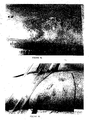
- rat myoblasts Primary cell cultures of rat myoblasts were prepared as described in Example 1 above. Cells were then incubated with polystyrene microspheres (treated cells) or without (control cells) for 48 hours. Cells were then fixed in 1% glutaraldehyde in phosphate buffered saline for 1-4 days, and rinsed in PBS. Cells were then transferred to a solution of 1% tannic acid and 1% guanidine HCI (1:1 ratio) in PBS for I hour. Specimens were post-fixed in 1% OSO 4 for 1 hour and dehydrated in graded ethanol and Freon 113 at room temperature. Specimens were then mounted on slides, coated with gold and examined in a JEOL T-300 scanning electron microscope at 2 kV.
- Figures 3A-3C illustrate the effect of the microspheres of the present invention on myoblast shape.
- the cell in Figure 3A has grown over the microsphere so that part of the cell surface is convex rather than flat.
- Figures 3B and 3C show cells extending pseudopodia from a portion of the cell on which the microsphere rests.
- the pseudopod of the cell in Figure 3C is particularly pronounced. showing that the microspheres clearly influence myoblast shape.
- the formation and extension of a pseudopod clearly requires changes in the cytoskeletal structure, demonstrating that the microspheres also affect the cytoskeleton of the cell.
- the formation of such pseudopodia may be important for the migration of cells into the wound area.
- the stimulation of such pseudopodia by the microspheres indicates their ability to promote another important step in the wound healing process.
- the following description is a general device and method for application of the agents for wound healing.
- the agents such as microspheres. are preferably applied repeatedly to the wound to be treated.
- the frequency of application, and the concentration applied is dependent on the severity of the symptoms and on the responsiveness of the subject to the treatment. Persons of ordinary skill in the art can easily determine optimum concentrations, dosing methodologies and repetition rates.
- the microspheres were applied to the wound to be treated about once per day, although of course other application rates are possible.
- the method includes the step of administering the agents such as microspheres, in a pharmaceutically acceptable carrier in which the agents are substantially insoluble, to a subject to be treated.
- pharmaceutically acceptable carriers include aqueous media for a suspension of agents, non aqueous media such as ointments, creams and aerosol-forming material, as well as bandages soaked in, or otherwise containing, media with the agents.
- the bandages can be occlusive or non-occlusive.
- the agents which are in a pharmaceutically acceptable carrier can be described as a dispersion of agents.
- the agents are administered according to an effective dosing methodology, preferably until a predefined endpoint is reached. such as the absence of clinical symptoms in the subject.
- a predefined endpoint such as the absence of clinical symptoms in the subject.
- the closure of the wound to be treated is an example of such an endpoint.
- the device of the present invention includes a composition with one or more agents and a pharmaceutically acceptable carrier for the agents, and a container for containing the composition.
- suitable containers include aerosol-dispersing pumps and spray cans.
- the agents such as microspheres, are preferably applied in a two step procedure. The microspheres are first applied in a dispersion to the wound, by dripping, spraying, painting, washing or by any other suitable method of topical application. Preferably, 30 sec to 2 minutes are allowed to elapse before the second step, in order to allow the microspheres to form initial contact with the wound.
- the second step includes applying an occlusive or non-occlusive bandage, or other suitable covering soaked in the liquid suspension containing the microspheres, to the wound.
- an occlusive or non-occlusive bandage, or other suitable covering soaked in the liquid suspension containing the microspheres, to the wound. This substantially reduces or eliminates absorption of the microspheres by the bandage or covering. This method was used both in rats and humans for wound healing as described in the Examples below.
- microspheres in the suspension did not aggregate, coalesce, clump or undergo irreversible caking. Although the microspheres did settle somewhat over time, they were easily resuspended with gentle agitation.
- the microspheres of the present invention promote various in vitro cell processes which are important for wound healing.
- in vitro and in vivo effects do not always correlate.
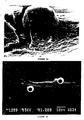
- Tibialis anterior muscle Male Wistar rats. weighing between 300 and 400 g. were anesthetized by nembutal (5 mg/kg of body weight). An excision injury to the lateral parts of the Tibialis anterior muscle was performed as follows. First, a longitudinal incision was made in the skin to expose the Tibialis anterior muscle. Next, the partial excision of this muscle was made by a transverse cut of the muscle fibers, along about half of the muscle width. The excised piece was then cut out of the muscle, leaving a gap of about 5 mm by 5 mm in the muscle. In all rats the same amount of excised tissue (80 ⁇ 10 mg) was removed from precisely the same location in the muscle. The wound area was then dressed with 2 micron polystyrene microspheres in saline for treated rats, and saline alone for control rats. The wound area was measured for between 3 and 15 days following injury.
- Figures 4A-4D show pictures of wound areas prepared as described above.
- Figure 4A shows the wound of the control rat immediately after injury
- Figure 4B shows the equivalent wound of the rat to be treated
- Figures 4C and 4D show the same rats five days after injury.
- the wound of the control rat was treated with saline alone, and still has not completely healed
- the wound of the treated rat, treated with microspheres has completely healed.
- the microspheres of the present invention promote faster wound healing.
- Figure 5 further illustrates the promotion of wound healing by the microspheres of the present invention.
- the wounds of control rats eventually heal, but at a much slower rate than the wounds of treated rats.
- the microspheres clearly increase the rate at which the wound area decreases and the wound heals.
- FIG. 6 compares the effect of the microspheres of the present invention on wound healing with tissue culture media and saline in rats. wounds were induced in rats as described above, and the rats were treated with saline alone ( Figure 6A ,), tissue culture media alone ( Figure 6B , x) saline plus microspheres ( Figure 6A ,) or tissue culture media plus microspheres ( Figure 6B ,).
- Example 6 The in vivo experiments described in Example 6 above clearly demonstrate that the microspheres of the present invention can promote wound healing and muscle regeneration in rats. Furthermore, the results of the toxicity studies in rats described in Example 7 show that the microspheres are substantially non-toxic. Therefore, studies were performed to determine the effect of the microspheres of the present invention on wound healing in humans. As described in detail below, case studies demonstrated that the microspheres clearly promoted wound healing in humans.
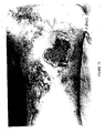
- the first case study was that of a 66-year old female with ulcers in the left leg which refused to heal. The patient also had cellulitis of the left leg and varicose veins in both legs. Ulcers on the inner thigh of the patient were treated with Milton 2% which is a corrosive chlorine salt in water.
- FIG. 7A shows the control wound at day 0, while Figure 7B shows the control wound after 4 months of treatment.
- Figure 7C shows the treated wound at day 0, while Figure 7D shows the treated wound after 4 months of treatment.
- Both the wounds treated with the microspheres of the present invention and those treated with Milton exhibited signs,of infection and other difficulties healing during the next four months.
- the wounds treated with microspheres had shown a significant improvement.
- the wound size had decreased and the wounds were clean, without signs of infection.
- the microspheres of the present invention exhibited greater efficacy in wound healing promotion than currently available treatments.
- Figures 7A and 7B the wound which had served as a control for Figure 7 above ( Figures 7A and 7B ) was treated with the same microspheres as those used to treat the wound in Figures 7C and 7D .
- the results are shown in Figures 8A and 8B .
- Figure 8A shows the wound at day 0 of treatment with microspheres
- Figure 8B shows the wound after 21 days of treatment.
- the extent of the wound has decreased, even after such a short time period.
- the wound was superficial and clean and as no longer producing exudations.
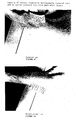
- the second case study w as that of a 52-year old female who had a year old infected wound on the front side of the left thigh.
- the wound was treated with 1% Milton for a week, debrided and then treated with the microspheres of Figures 7 and 8 for 10 days.
- Figure 9A shows the wound at day 0 of treatment
- Figure 9B shows the wound after 10 days of treatment.
- the wound After 10 days, the wound showed a significant improvement. It had decreased in extent to a small size, was clean and was no longer producing exudations, as can be seen from Figure 9B . Although the wound did not fully close during the relatively short treatment period, its effects had been significantly ameliorated.
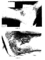
- the third case study w as that of a 19-year old male who was injured by a chemical spill in an industrial workplace accident.
- the chemicals in question sulfurides, caused severe bums and blistering on the right side of his neck and right hand.
- All wounds were treated with Silverol, a hydrogel with strong absorptive properties.
- the wounds on the right forearm were treated with the microspheres of case studies 1 and 2, while the remaining wounds were treated with Silverol.
- the results are shown in Figures 10A and 10B (control wound at day 0 and day 5, respectively), and in Figures 10C and 10D (treated wound at day 0 and day 5, respectively).
- the condition of the treated wound on the forearm had improved significantly over that of the remaining wounds which were not treated with microspheres.
- the wound on the forearm had completely healed after 5 days of treatment with microspheres.
- the remaining wounds which were treated with Silverol had not healed completely.
- the microspheres clearly promoted wound healing, demonstrating a greater efficacy than currently available treatments.
- the fourth case study was of a 52-year old female who had sustained second-degree bums on the buttocks from a hot bath. Wounds on the left buttock were treated with Silverol, while those on the right buttock were treated with microspheres of the previous case studies. The results are shown, for microsphere-treated wounds only, in Figures 11A (day 0) and 11B (day 7) of treatment.
- the wounds on the right buttock, which were ' treated, with microspheres had completely healed with good epithelial growth.
- the wounds on the left buttock, which were treated with Silverol had not completely healed and were closing relatively slowly.
- the microspheres were able to promote wound healing at a more rapid rate than conventional treatments.
- the fifth case study was of a 28 year old female w ho had suffered extensive and severe sunburn (data not shown). She was treated with the microspheres of the previous case studies. The patient reported both a significant reduction in discomfort and rapid healing of the sunburn.
- the microspheres used in the method and device of the present invention can both relieve discomfort and promote wound healing, although it should be noted that the relief of discomfort is probably a highly indirect effect of the microspheres rather than direct analgesia.
- the above patient report can only be inferred to include the apparent reduction in the sensation of discomfort from the sunburn.
- Such decreased discomfort probably does not demonstrate any ability of the microspheres to have a direct effect on the transmission of nerve impulses, or indeed to directly alter any of the many factors which lead to the sensation of discomfort. Instead, this effect is probably highly indirect, occurring as a result of the activation of macrophages, which in turn has anti inflammatory effects, leading to the decreased sensation of discomfort by the patient.
- the use of the agents, such as microspheres, according to the present invention has clearly been shown to have greater efficacy for the promotion of wound healing and muscle regeneration than currently available, prior art treatments.
- the method and device promotes, accelerates and enhances wound healing, as well as diminishing discomfort experienced by the subject.
- microspheres of the present invention induce an initial increase in a patient having undergone radical mastectomy followed by radiation therapy.
- FIG. 12A A fifty fouur year old breast cancer patient having stage II disease underwent surgery, followed by radiation therapy.
- the patient was able to take a course of chemotherapy and is currently disease free.
- the composition of the present invention was useful in the treatment of wounds in combination with radiation therapy and chemotherapy.
- the pharmacological agents and/or biologics are soluble in the pharmaceutical carrier but the microspheres are not and microspheres remain in suspension in the therapeutic composition.
Description
- The present invention relates to compositions of microspheres for application to wounds, and to methods of use of said composition alone or in combination with other agents in the treatment of wounds.
- Wound healing is a complex process involving such factors as cells, extracellular matrix components and the cellular microenvironment. Essentially, all wound healing involves the repair or replacement of damaged tissues including but not limited to skin, muscle, neurologic tissue, bone, soft tissue, internal organs or vascular tissue. The precise nature of such repair or replacement depends upon the tissues involved, although all such processes involve certain basic principles. An important aspect of wound healing is the rate at which a wound gains tensile strength.
- Skin exhibits tension and extensibility. Skin tension is one of the determining factors in the response to a wound and varies with age and site. Skin has multiple layers, including keratin, epidermis and dermis and contains cells, a fibrous network composed of collagen and elastin and an amorphous ground substance which consists of protein polysaccharides, glycoproteins, globular proteins, salts and water. If only the epidermis is damaged, as in most minor injuries, keratinocytes migrate from the edge of the wound and eventually cover it, reforming the epidermis and keratin (Knighton, D.R. and Fiegel, V.D., 1991, Invest. Radiol: 26:604-611).
- If all skin layers are damaged or destroyed, new connective tissue, called granulation tissue, must first fill the wound space. This tissue is formed by deposition of extracellular matrix components, for example, collagen, by fibroblasts which migrate into the wound space. The synthesis and deposition of collagen is an important event in wound healing and the rate of collagen synthesis varies in different organs (Haukipuro, K. et al., 1991, Ann. Surg. 213:75-80).
- The entire multi-step process of wound healing must be completed for successful would healing. If one or more of these components is missing, healing does not take place, the skin is not repaired and the wound remains open. Such open wounds can easily become infected, further, retarding the process of healing and leading to the formation of ulcers and sores on the skin. The process of wound healing is further inhibited in many patients by the presence of other complicating conditions, including, but not limited to diabetes or old age. Patients with such conditions often have skin wounds which ulcerate and refuse to heal, or only heal slowly after an extended period of time has elapsed.
- Various treatments have been used in order to accelerate the rate at which wounds heal (
U.S. Patent No. 4,772,591 ;U.S. Patent N. 4,590,212 ) and various pharmaceutical carriers have been employed to deliver chemotherapeutic agents to the wound, for example, creams, gels, powders and microspheres.U.S. Patent No. 5,264,207 discloses microspheres of a polymer, which act as carriers for one or more active pharmaceutical or cosmetic substances. The polymer microspheres are solid or hollow, insoluble in the carrier liquid and are of varying dimensions, not exceeding 1000nm (1µm), with the preferred sizes ranging from 50 to 500 nm (0.05µm to 0.5µm) and the most preferred sizes ranging from 60 to 300 nm (0.06µm to 0.3µm). The fineness of the microspheres leads to higher specific area per unit weight and higher combination of microspheres to active substances without the disadvantage of conventional excipients which block skin pores. Thus, according to the invention ofU.S. Patent No. 5,264,207 , a suspension of the microspheres can be obtained onto which an active substance is adsorbed, another substance binding to the microsphere by chemical bonds, with the possibility of a third substance binding to the microspheres by electrostatic or ionic bonds (Col. 2, lines 58-63). When the microspheres are hollow, they are both adsorbent and/or carriers of functional groups (Col. 3, lines 18-21). When the microspheres contain pores, bonding with a pharmaceutical or cosmetic substance consists of adsorption into the pores (Col. 3, lines 26-28). Thus,U.S. Patent No. 5,264,207 discloses a composition of microspheres which act as carriers for pharmaceutical and/or cosmetic substances only and does not teach or suggest using only the microspheres themselves to enhance wound healing. - Similarly,
PCT Application Nos. WO96/13164 WO94/13333 PCT Application No. WO96/13164 PCT Application No. WO94/13333 - The size of the microspheres was shown to influence the effect of microspheres as carriers for the class I alloantigen used in the activation of cytotoxic T lymphocytes (Mescher, M.F., 1992, J. Immunol 149:2402-2405). The response was dependent on the class I antigen being presented on the optimum size of the microspheres of 4 or 5µm diameter (4000nm or 5000nm). In other words, Mescher disclosed that the activation of T lymphocytes in vitro could not be achieved with microspheres alone but required the class I antigen to induce the activation. The antigen reacted optimally when bound to microspheres of 4 or 5µm diameter, i.e. 4 to 5 times greater in size than microspheres used in
U.S. Patent 5,264,207, issued to Bommelser, J., et al. There is no teaching or suggestion in Mescher of using microspheres alone without the active component, class I antigen, in the activation of wound healing. In fact, the role of microspheres and cytotoxic T lymphocytes, in wound healing and collagen synthesis is not at all taught or suggested by Mescher. Furthermore, Bommelser teaches away from the use of microspheres greater than 1000nm or 1µm in size because larger microspheres would block skin pores. Thus, the present invention is neither inherent (because of the difference in the ingredients of the compositions of prior art) nor obvious (because the size of microspheres used would be expected from prior art to block skin pores) from the prior art. -
US 5,092,883 generally relates to materials for fostering formation of mammalian hard and soft connective tissue. Specifically, it is disclosed that biodegradable dextran beads (e.g. Sephadex) having a particle size of 40-120 µm exhibit varying positive or negative functional groups. The beads can be used for preparing a paste or slurry, or can be employed in a suspension. It is also disclosed that treatment of wounds with beads having a positive charge was significantly more successful than treatment with uncharged or negatively charged beads. -
US 5,565,206 pertains to the field of dental applications and solves the problem of bacteria induced tooth decays in the mouth by providing a composition for oral hygiene comprising polystyrene microspheres having a diameter of 0.05 to 1 µm and a negative outer surface charge for electrostatic interaction with chlorhexidine. During preparation of the composition a polystyrene microsphere suspension is added to a chlorhexidine solution to make a final concentration of 0.54 mg/ml. - The process of wound healing includes an initial proliferative phase promoting rapid cell metabolism and proliferation, disposal of debris, mobilization of fibroblasts and restoration of circulation. It is during this period that the wound is most susceptible to infection. During the subsequent phase (also referred to as the fibroplastic phase) of wound healing, increasing tensile strength parallels the rise in collagen content of the wound. Thus, there has remained a need to develop compositions for wound healing such that they contain non-biodegradable microspheres and other extracellular components capable of promoting the proliferative phase and regulating the fibroplastic phase in situ.
- Hence, there must be a balance between promotion of the proliferative phase and the onset of the fibroplastic phase during wound healing in animals and human beings for different conditions including, but not limited to, burned tissues, infections following surgery, surgery wound breakdown, internal ulcers, hemorrhage, bone gangrene, pressure sores, decubitis, compromised amputation sites, non-healing traumatic wounds, cosmetics, after shave, dental work, chronic ulcers (of the diabetics, varicose vein, post stroke), destruction of tissue by radiation, spinal injury wounds, gynecological wounds, chemical wounds, vessel disease wounds, diabetic skin sores, diabetic feet, physical trauma, post plastic surgery suture sites, sunburns or episiotomies.
- It is therefore an object of the present invention to produce a therapeutic composition that can promote wound healing and that can function as a wound/lesion dressing when applied to the contours of and within the wound or lesion.
- It is another object of the present invention to provide a composition that can function as a wound dressing that will serve as a promoter of regeneration of muscle, skin, cartilage, neurologic tissue, soft tissue or vascular tissue.
- It is yet another object of the present invention to provide a composition that can function as a wound/lesion covering that is, when wet, adherent and will remain in contact with the wound/lesion site without the need of a potentially toxic adhesive.
- It is yet another object of the present invention to provide a composition that can serve as a medium for microspheres and a drug delivery system for pharmaceutical agents.
- It is yet another object of the present invention to provide a composition that will serve to promote myoblast fusion.
- It is another object of the present invention to provide a composition that will serve to promote collagen synthesis in vitro and in vivo.
- It is yet another object of the present invention to provide a composition that will prevent scar formation.
- It is another object of the present invention to provide a composition including microspheres and exogenous growth factors added to it that will serve to promote wound healing.
- It is yet another object of the present invention to provide a composition including microspheres and anti-inflammatory agents.
- It is yet another object of the present invention to provide a composition including microspheres and antihistaminic agents.
- It is yet another object of the present invention to provide a composition including microspheres and antibiotics.
- It is yet another object of the present invention to provide a composition including microspheres and vitamins.
- It is yet another object of the present invention to provide a composition including microspheres and minerals.
- It is yet another object of the present invention to provide a composition including microspheres and anticancer agents, antiviral agents and antifungal agents.
- It is yet another object of the present invention to provide a composition including microspheres with or without an active substance, that is applied as adjuvant therapy to surgery, radiation therapy, hormone therapy or chemotherapy.
- It is yet another object of the present invention to provide a composition including microspheres and an analgesic, anesthetic or astringent.
- It is yet another object of the present invention to provide a composition including microspheres and collagen.
- It is yet another object of the present invention to provide a composition including microspheres and one or more of the agents selected from the group consisting of anti-inflammatory, antihistaminic, antibiotic, antiseptic, antifungal, analgesic, anesthetic, minerals, vitamins, astringent agent and collagen.
- It is yet another object of the present invention to provide a composition including microspheres and stromal cells that will serve to promote wound healing.
- It is yet another object of the present invention to provide a composition including microspheres and genetically engineered stromal cells which express a gene product beneficial for successful and/or improved wound healing. For example, in the case of diabetic sores, the fibroblasts can be genetically engineered to express insulin growth factor (IGF) or anticoagulation gene products to reduce the risk of atherosclerosis, occlusion, or anti-inflammatory gene products to reduce the risk of failure to heal. Alila, H.et al., 1997, Human gene Ther. 8:1785-1795; and Pickering, J.G. et al., 1996, Semin. Interv. Cardiol. 1;84-88. In accordance with the present invention, there is provided a composition contained in a package for storing and mixing the composition from which the composition can be poured and applied on the surface of a wound, said composition comprising:
- about 0.001-25% by weight of non-biodegradable microspheres in suspension, wherein said microspheres:
- have a diameter in a range of from about 0.01 µm to about 200 µm;
- exhibit surface groups with a negative charge;
- are capable of forming multi-point contacts with a cellular membrane;
- are made from a material selected form the group consisting of polystyrene, derivatized polystyrene, poly-N-ethyl-4-vinyl-pyridinium bromide, polymethylacrylate and silicone;
- a pharmaceutical agent; and
- a pharmaceutically acceptable carrier for said microspheres, said microspheres being substantially insoluble in said carrier for use in treating a wound.
- According to one embodiment the pharmaceutical carrier is selected from the group consisting of aqueous medium, aerosol carrier, aqueous solvent for pharmacological agents, aqueous solvent for biologics, ointment and bandage.
- According to a further embodiment the pharmaceutical agent is chosen from the group consisting of:
- an antibiotic, the antibiotic preferably being selected from the group consisting of tetracycline, oxytetracycline, gentamycin neomycin sulfate, bacitracin, polymyxin B sulfate and gramaciclin,
- an antihistamine,
- an anti-inflammatory agent,
- an anticancer agent,
- an antiviral agent,
- an antifungal agent,
- a metal ion, preferably selected from the group consisting of zinc, magnesium, cobalt, iron copper and manganese,
- an astringent,
- an anesthetic, preferably selected from the group consisting of lidocaine, procaine and epinephrine,
- an analgesic, the analgesic preferably selected from the group consisting of morphine, heroin and fentanyl,
- a cycloxygenase-2 inhibitor,
- a vitamin,
- an amino acid,
- a collagen,
- a growth factor, preferably chosen from the group consisting of PDGF, PDAF, PDEGR, TBFB, PF-4, FGF, BFGF, GH, EGF and IGF,
- a proteolytic enzyme,
- one or more stromal cells,
- an immune enhancer, selected from the group consisting of L-arginine, nitric oxide, quadrol, muramyl dipeptide and other macrophage activating factors, or
- hyaluronic acid.
- In accordance with the present invention, there is provided a container holding a composition for use in treating a wound according to the above embodiments.
- For a more complete understanding of the present invention, and the advantages thereof, reference is now made to the following descriptions taken in conjunction with the accompanying drawings, in which:
-
FIG. 1 is a graph showing the ability of the microspheres of the present invention to increase creatinine phosphokinase activity; -
FIG. 2 is a plot illustrating the effect of the microspheres of the present invention on collagen synthesis; -
FIGS. 3A-3C illustrate the effect of the microspheres of the present invention on myoblast shape; -
FIGS. 4A-4D illustrate the ability of the microspheres to promote wound healing in rats; -
FIG. 5 is a graph at the rate at which wound area offigure 4 decreases; -
FIGS. 6A and 6B compare the effect of the microspheres of the present invention on wound healing with tissue culture media and saline in rats; -
FIGS. 7A-7D demonstrate the ability of the microspheres of the present invention to promote wound healing in a first human case study; -
FIGS. 8A and8B further demonstrate the efficacy of the present invention in the human case study offigures 7A-&D; -
FIGS. 9A and 9B demonstrate the efficacy of the present invention in a second human case study; -
FIGS. 10A-10D show the effect of the present invention in a third human case study; and -
FIGS. 11A and 11B show the efficacy of the present invention in a fourth human case study. -
FIGS 12A-12D describe the effect of the present invention in an individual when given in combination with radiation therapy and chemotherapy. - The present invention relates to therapeutic compositions for promoting wound healing by using microspheres. Unexpectedly, microspheres of the particular size range described herein are able to promote wound healing without the further addition or inclusion of any drug or other therapeutic substance. Indeed, as described below, these microspheres do not degrade or undergo other chemical alteration in order to produce their therapeutic effect. The microspheres being part of the present invention can also be administered as adjuvant therapy to conventional therapies, e.g., radiation, chemotherapy, hormone, laser, high pressure or ozone therapy.
- The structure of these microspheres includes a core material and at least one type of charged surface group which is present at least on the exterior of the microsphere. Examples of materials include long-chain polymers such as polystyrene, polymethylmethacrylate (PMMA), silicone and derivatized polystyrene. Examples of surface groups include sulfate, poly-N-ethyl-4-vinylpyridinium bromide, protamine, protamine sulfate, protamine salts, polylysine, carboxyl and polystyrene. These surface groups may be present as part of the core material, or may be added later by such chemical processes as derivatization of the long-chain polymer. Hereinafter the term "derivatization" refers to the process of chemically altering, modifying or changing a molecule or a portion thereof. The microspheres produced from the polymer should be substantially insoluble in aqueous media, instead forming a suspension or dispersion in such media.
- In order to further clarify the parameters of the present invention, a number of terms should be defined. Hereinafter, the terms "wound" or "lesion" includes any injury to any portion of the body of a subject including, but not limited to, acute conditions such as thermal bums, chemical bums, radiation bums, bums caused by excess exposure to ultraviolet radiation such as sunburn, damage to bodily tissues such as the perineum as a result of labor and childbirth, including injuries sustained during medical procedures such as episiotomies, trauma-induced injuries including cuts, those injuries sustained in automobile and other mechanical accidents, and those caused by bullets, knives and other weapons, and post-surgical injuries, as well as chronic conditions such as pressure sores, bedsores, conditions related to diabetes and poor circulation, and all types of acne. Areas of the body which can be treated with the present invention include, but are not limited to, skin, muscle, neurologic tissue, bone, soft tissue, vascular tissue, and internal organs. Hereinafter, the term "subject" refers to a human mammal or lower animal on whom the present invention is practiced.
- Hereinafter, the term "promoting" includes accelerating and enhancing. Hereinafter, "reducing scarring" includes preventing or decreasing excess scar formation such as keloids and hypertrophic scars, as well as decreasing the extent of scar tissue formation both externally such as on the skin of the subject, and internally such as adhesions. Finally, it should be noted that the method of the present invention may also be used cosmetically, to prevent excess scar formation in a cut or other wound to the skin such as the skin of the face, and to treat acne. In a cosmetic sense, the term "excess scar formation" includes any scarring which is cosmetically undesirable or unacceptable.
- Although the discussion below refers to specific types of microspheres, it should be noted that this is not intended to be limiting in any way. It will be appreciated to those skilled in the art that these microspheres, more generally, can be beads, particles or globules which are either solid or hollow. In preferred embodiments of the present invention, these microspheres are dispersed in a pharmaceutically acceptable carrier medium in which the agents are substantially insoluble, as a suspension in aqueous medium for example, or in a non-aqueous medium such as an ointment, aerosol spray, or a bandage which may be occlusive or non-occlusive. The shape of the microspheres can be regular, such as spherical or elliptical, or regular non-spherical shapes; or the shape of the particles can be non-regular, so that the surface is not a single continuous curve or so that the surface is not smooth.
- Furthermore, the microspheres can be a mixture of different polymers and can also be a mixture of different particles, beads or globules of different sizes. The agents can also have pores of different sizes.
- By way of example, the long-chain polymer forming the agents, can be cross-linked, which particularly favors the spherical shape of a microsphere, although such a shape can be obtained without cross-linking. An example of a method of manufacture for a cross-linked poly-β-alanine microsphere is given in
U.S. Patent No. 5,077,058 , although it should be noted that this material would require further derivatization to obtain an overall surface charge of the microsphere. - Alternatively, the particles can have chaotic irregular forms, particularly if the polymer is not cross-linked. The particle can have any form, such as coiled, globular, extended and random coil. Preferably, the polymer should not be biochemically reactive. The polymer is non-biodegradable so that it would remain undegraded during the period required for healing of the wound. Hereinafter, the term "non-biodegradable" refers to agents which are not biodegradable during the treatment period, which is the period required for treatment of the wound.
- At the very least, the agents should have the following properties:
- 1. They should be able to promote wound healing without significant chemical alteration or degradation; and
- 2. They should be substantially insoluble in aqueous media such as bodily fluids, and instead should form a suspension.
- The microspheres are capable of forming mulpi-point contacts with a cellular membrane.
- These characteristics are important because as further discussed below, the effect of the agents of the present invention appears to be directly related to the formation of multi-point contacts between the material of the agents and a portion of the cell such as the outer cell membrane, thereby forming an adherent surface for the cells to attach to. Such multi-point contacts are possible with many different polymers which permit charged groups to be accessible for interaction with molecules and portions of the outer cell membrane. Thus, although the description below focuses on one type of agent, microspheres, it is understood that the present invention covers any material capable of forming such multi-point contacts.
- As noted above, the microsphere has a diameter in a range of from about 0.01 microns to about 200 microns, preferably in a range of from about 1 to about 100 microns, and most preferably from about 2 to about 20 microns. The microsphere composition of the present invention comprises 0.001 to 25 percent by weight in suspension. Without desiring to be bound by any mechanism, it should be noted that these preferred ranges are the best size for enabling uptake of the microspheres by macrophages infiltrating the wound area. The microspheres appear to actually attract and activate the macrophages through contact with at least a portion of the macrophages, probably the molecules of the outer cell membrane of the macrophage. The anti-inflammatory and anti-bacterial effects observed for the microspheres are thus presumably indirect effects, obtained through the activation of the macrophages or other cells.
- Another important property of the microspheres is the charge of the surface groups. The overall charge carried by certain preferred examples of microspheres was measured as a Z or zeta potential by electrophoretic mobility (milliVolts) by a ZetaMaster (Malvern Instruments, United Kingdom). The range of Z potentials measured in certain embodiments exemplified herein was from -29.58 mV to -79.7 6mV. Hereinafter, the term "charged" refers to a Z potential with an absolute value of at least about 1 mV, and preferably of at least about 10 mV, whether negative or positive.
- The microspheres in the suspensions tested did not aggregate, coalesce, clump or undergo irreversible caking. Although the microspheres did settle somewhat over time, they were easily resuspended with gentle agitation.
- The microspheres of the present invention are akin to a liquid bandage or device that can be applied on the contours of a wound or within the wound. The microsphere s are packaged in a sterile container under argon, neon or nitrogen at an optimum pH and varying concentration and volume by conventional methods.
- Any one or more of the following kinds of pharmacological agents can be included in the therapeutic compositions along with the untreated microspheres: antiseptics, astringents, antifungal agents, antiviral agents, anticancer agents, antihistamines, antibiotics, blood coagulants for battlefield use, vitamin and mineral preparations, vitamins, minerals or anti-inflammatory agents.
- Biologically active substances which can be included in the therapeutic composition include, but are not limited to astringents, antibiotics, oxidants, proteolytic enzymes, collagen cross-link inhibitors (e.g. natural or synthetic diamines-cystamine, putrescine, spermidine, cadaverine and the like), amino acids, macrophage stimulating factors, or anesthetics.
- Growth and regulatory factors may be added to the therapeutic composition to enhance the wound healing process by aiding in the formation of granulation tissue and re-epithelization, for example, but not limited to platelet-derived growth factor (PDGF), platelet-derived angiogenesis factor (PDAF), platelet-derived epidermal growth factor (PDEGF), transforming growth factor-beta (TGFB), platelet factor (PF-4), alpha and beta fibroblast growth factors (αFGF and βFGF) growth hormone (GH), epidermal growth factor (EGF) or insulin growth factor (IGF).
- Platelet-derived growth factors stimulate the cascade systems involved in wound healing especially during the phases of granulation formation and re-epithelization. Transforming growth factor-beta refers to a growing family of related dimeric proteins which regulate the proliferation and differentiation of many cell types (Massague, 1990, Ann. Rev. Cell. Biol. 6:597-619). TGFB also induces the production of cartilage-specific macromolecules in muscle cells and chondrocytes. However, TGFB was found to inhibit the synthesis of collagen type II by chicken sternal chondrocytes (Horton et al., 1989, J. Cell Physiol. 141:8-15) and by rat chondrocytes (Rosen et al., 1988, J. Cell Physiol. 134:337-346). In fact, TGFB has emerged as a prototypical inhibitor of the proliferation of most normal cell types in vitro and in vivo (Alexandrow, A.G., et al., 1995, Cancer Res. 55: 1452-1457). TGFB1 has been purified from human and porcine blood platelets and recombinant TGFB1 is currently available (Gentry et al., 1988, Mol. Cell. Biol. 7:3418-3427). Insulin alone is much less potent than IGF-I in stimulating collagen matrix synthesis. IGF-II stimulates DNA and RNA synthesis and is more potent than IGF-I in stimulating clonal growth in fetal cells (McQuillan, et al., 1986, Biochem. J. 240:423-430). Epidermal growth factor alone has no effect on chondrocyte proliferation. Together with insulin, EGF synergistically stimulates proliferation of chondrocytes (Osborn, K.D., et al., 1989, J. Orthop. Res. 7:35-42).
- High levels of lipid peroxides have been demonstrated in activated macrophages, aggregated platelets, and subcellular particles isolated from injured tissue. Strong oxidants, drugs metabolized to free radicals, and ionizing radiation all induce prostaglandins, leukotrienes (eicosanoids) and/or lipid peroxidative increases. These increased eicosanoids and lipid peroxides may be one cause of tissue damage or necrosis and of the increase in the inflammatory reaction. The inclusion of anti-inflammatory drugs, e.g. non-steroidal anti-inflammatory drugs (ibuprofen, indomethacin, aspirin, acetaminophen, naproxen, sulindac) and specifically the cyclooxygenase-2 (COX2) inhibitors, in the prophylactic and/or therapeutic composition may prove useful in all injured tissues, for example, in lung, liver, radiation-induced injury in which fibrosis is a prominent consequence of prolonged or repeated injury. In addition, the inclusion of anti-oxidants (e.g., vitamin E) and free radical scavengers (e.g., reduced glutathione) may also prove useful.
- Zinc - It has been demonstrated that after trauma, surgical or otherwise, the amount of zinc in blood and tissues may fall to low levels. This is particularly noticeable in burned patients. Administration of zinc sufficient to restore normal levels of this trace element in blood and tissues increases rate of epithelization, rate of gain of wound strength, and synthesis of collagen and other proteins. A number of enzymes are zinc-dependent notably DNA-polymerase and reverse-transcriptase. The effects of zinc depletion on wound healing are what one would expect if the amount of functions of these enzymes were depressed. Epithelial and fibroblast proliferation does not occur, since mitosis of these cells cannot take place without DNA polymerase and reverse-transcriptase. Thus, although these cells may migrate normally, they do not multiply; hence adequate epithelization does not occur and collagen synthesis by the few fibroblasts migrating into the wound cannot supply the fibers needed to hold the wound together. In such a case raising blood and tissue zinc concentrations to normal will restore normal progression of wound healing. It is the object of the present invention that in patients in whom blood and tissue zinc levels are low, administration of zinc orally, parenterally or topically in the therapeutic composition will restore normal wound healing. Zinc will be provided as a salt selected from the group consisting of zinc chloride, zinc carbonate and/or zinc gluconate.
- Vitamin A and E - Vitamin A can partially reverse the retarded development of granulation tissue and reduced amounts of collagen in wound healing. Vitamin A applied topically enhances epithelization in wounds retarded by corticosteroid administration, but collagen deposition is affected by systemic vitamin A only.
- Vitamin E can be used to modify scar formation since its side effects are less than those of comparable amounts of steroid hormones. In high doses, vitamin E retards wound healing and collagen production but this effect is overcome in the presence of vitamin A.
- The therapeutic composition of the present invention may afford a vehicle for introducing genes and gene products in vivo to accelerate the wound healing process. For example, for diabetic sores, the therapeutic composition includes the microspheres of the present invention and genetically engineered stromal cells (e.g. fibroblasts with or without other cells and/or elements found in loose connective tissue taken from the subject, including but not limited to, endothelial cells, pericytes, macrophages, monocytes; plasma cells, mast cells, adipocytes, etc.) which use genetically engineered to express anticoagulation gene products to reduce the risk of thromoembolisim, or anti-inflammatory gene products to reduce the risk of failure due to inflammatory reactions. Once genetically engineered cells are applied into the wound site, the presence of the anti-inflammatory gene products, for example, peptides or polypeptides corresponding to the idiotype of neutralizing antibodies for tumor necrosis factor (TNF), interleukin-2 (IL-2) or other inflammatory cytokines, can bring about amelioration of the inflammatory reactions associated with diseases including but not limited to burned tissues, infections following surgery, diabetic neuropathic ulcers, pressure ulcers, venous stasis ulcers, burned tissues, infections following surgery, surgery wound breakdown, internal ulcers, hemorrhage, bone gangrene, pressure sores, decubitis, compromised amputation sites, non-healing traumatic wounds, cosmetics, after shave, dental work, chronic ulcers (of the diabetics, varicose vein, post stroke), destruction of tissue by radiation, spinal injury wounds, gynecological wounds, chemical wounds, vessel disease wounds, diabetic skin sores, diabetic feet, physical trauma, post plastic surgery suture sites, sunburns or episiotomies.
- Preferably, the cells are engineered to express such gene products transiently and/or under inducible control during the initial phase of wound healing, or as a chimeric fusion protein anchored to the stromal cells, for example, a chimeric molecule composed of an intracellular and/or transmembrane domain of a receptor or receptor-like molecule, fused to the gene product as the extracellular domain. In another embodiment, the stromal cells could be genetically engineered to express a gene for which a patient is deficient, or which would exert a therapeutic effect, e.g., HDL, apolipoprotein E, etc. The genes of interest engineered into the stromal cells need to be related to the disease being treated.
- The stromal cells can be engineered using a recombinant DNA construct containing the gene used to transform or transfect a host cell which is cloned and then clonally expanded in the culture system. The culture which expresses the active gene product, could be implanted into an individual who is deficient for that product. For example, genes that prevent or ameliorate symptoms of various types of vascular, may be underexpressed or down regulated under disease conditions. Specifically, expression of genes involved in preventing the following pathological conditions may be down-regulated, for example: thrombus formation, inflammatory reactions, and fibrosis and calcification of the valves. Thus, the level of gene activity may be increased by either increasing the level of gene product present or by increasing the level of the active gene product which is present in the three-dimensional microsphere culture system.
- Further, patients may be treated by gene replacement therapy during the fibroplastic phase of wound healing. Stromal cells may be designed specifically to meet the requirements of an individual patient, for example, the stromal cells may be genetically engineered to regulate one or more genes; or the regulation of gene expression may be transient or long-term; or the gene activity may be non-inducible or inducible. For example, one or more copies of a normal target gene, or a portion of the gene that directs the production of a normal target gene protein product with target gene function, may be inserted into human cells that populate the three-dimensional constructs using either non-inducible vectors including, but are not limited to, adenovirus, adeno-associated virus, and retrovirus vectors, or inducible promoters, including metallothionein, or heat shock protein, in addition to other particles that introduce DNA into cells, such as liposomes or direct DNA injection or in gold particles.
- The use of the stromal cell in the therapeutic composition in gene therapy has a number of advantages. Firstly, since the culture comprises eukaryotic cells, the gene product will be properly expressed and processed in culture to form an active product. Secondly, gene therapy techniques are useful only if the number of transfected cells can be substantially enhanced to be of clinical value, relevance, and utility; the three-dimensional cultures of the invention allow for expansion of the number of transfected cells and amplification (via cell division) of transfected cells.
- A variety of methods may be used to obtain the constitutive or transient expression of gene products engineered into the stromal cells. For example, the transkaryotic implantation technique described by Seldon, R.F., et al., 1987, Science 236:714-718 can be used. "Transkaryotic", as used herein, suggests that the nuclei of the implanted cells have been altered by the addition of DNA sequences by stable or transient transfection. The cells can be engineered using any of the variety of vectors including, but not limited to, integrating viral vectors, e.g., retrovirus vector or adeno-associated viral vectors, or non-integrating replicating vectors, e.g., papilloma virus vectors, SV40 vectors, adenoviral vectors; or replication-defective viral vectors. Where transient expression is desired, non-integrating vectors and replication defective vectors may be preferred, since either inducible or constitutive promoters can be used in these systems to control expression of the gene of interest. Alternatively, integrating vectors can be used to obtain transient expression, provided the gene of interest is controlled by an inducible promoter.
- Any promoter may be used to drive the expression of the inserted gene. For example, viral promoters include but are not limited to the CMV promoter/enhancer, SV 40, papillomavirus, Epstein-Barr virus, elastin gene promoter and b-globulin. If transient expression is desired, such constitutive promoters are preferably used in a non-integrating and/or replication-defective vector. Alternatively, inducible promoters could be used to drive the expression of the inserted gene when necessary. For example, inducible promoters include, but are not limited to, metallothionein and heat shock protein.
- Examples of transcriptional control regions that exhibit tissue specificity for connective tissues which have been described and could be used, include but are not limited to: elastin or elastase I gene control region which is active in pancreatic acinar cells (Swit et al., 1984, Cell 38:639-646; Ornitz et al., 1986, Cold Spring Harbor Symp. Quant. Biol. 50:399-409; MacDonald, 1987, Hepatology 7:425-515). The deposition of elastin is correlated with specific physiological and developmental events in different tissues, including the vascular grafts. For example, in developing arteries, elastin deposition appears to be coordinated with changes in arterial pressure and mechanical activity. The transduction mechanisms that link mechanical activity to elastin expression involve cell-surface receptors. Once elastin-synthesizing cells are attached to elastin through cell-surface receptors, the synthesis of additional elastin and other matrix proteins may be influenced by exposure to stress or mechanical forces in the tissue (for example, the constant movement of the construct in the bioreactor) or other factors that influence cellular shape.
- The stromal cells used in the microsphere culture system of the invention may be genetically engineered to "knock out" expression of factors or surface antigens that promote clotting or rejection at the implant site. Negative modulatory techniques for the reduction of target gene expression levels or target gene product activity levels are discussed below. "Negative modulation", as used herein, refers to a reduction in the level and/or activity of target gene product relative to the level and/or activity of the target gene product in the absence of the modulatory treatment. The expression of a gene native to stromal cell can be reduced or knocked out using a number of techniques, for example, expression may be inhibited by inactivating the gene completely (commonly termed "knockout") using the homologous recombination technique. Usually, an exon encoding an important region of the protein (or an
exon 51 to that region) is interrupted by a positive selectable marker (for example neo), preventing the production of normal mRNA from the target gene and resulting in inactivation of the gene. A gene may also be inactivated by creating a deletion in part of a gene, or by deleting the entire gene. By using a construct with two regions of homology to the target gene that are far apart in the genome, the sequences intervening the two regions can be deleted. Mombaerts, P., et al., 1991, Proc. Nat. Acad. Sci. U.S.A. 88:3084-3087. - Antisense and ribozyme molecules which inhibit expression of the target gene can also be used in accordance with the invention to reduce the level of target gene activity. For example, antisense RNA molecules which inhibit the expression of major histocompatibility gene complexes (HLA) shown to be most versatile with respect to immune responses. Still further, triple helix molecules can be utilized in reducing the level of target gene activity. These techniques are described in detail by L.G. Davis, et al., eds, Basic Methods in Molecular Biology, 2nd ed., Appleton & Lange, Norwalk, Conn. 1994.
- In another alternative, the stromal cells can be genetically engineered to block gene expression necessary for the transition of smooth muscle cells to proliferate, migrate and to lead to development of neointimal hyperplasia, e.g., by antisense oligodeoxynucleotide blockade of expression of
cell division cycle 2 kinase and proliferating cell nuclear antigen. Mann, M.J., et al., 1995, Proc. Natl. Acad. Sci. USA 92:4502-4506. - The use of the stromal cell culture in gene therapy has a number of advantages. Firstly, since the culture comprises eukaryotic cells, the gene product will be properly expressed and processed in culture to form an active product. Secondly, gene therapy techniques are useful only if the number of transfected cells can be substantially enhanced to be of clinical value, relevance, and utility; the stromal cell cultures of the invention allow for expansion of the number of transfected cells and amplification (via cell division) of transfected cells. For example, genetically engineered cells that express wound healing factors may be incorporated into the living stromal cultures used to make tendons and ligaments to enhance wound healing at the site of injury.
- Examples of transcriptional control regions that exhibit tissue specificity which have been described and could be used, include but are not limited to: elastase I gene control region which is active in pancreatic acinar cells (Swift et al., 1984, Cell 38:639-646; Ornitz et al., 1986, Cold Spring Harbor Symp. Quant. Biol. 50:399-409; MacDonald,1987, Hepatology 7:42S-51S); insulin gene control region which is active in pancreatic beta cells (Hanahan, 1985, Nature 315:115-122); immunoglobulin gene control region which is active in lymphoid cells (Grosschedl et al.,1984, Cell 38:647-658;.Adams et al., 1985, Nature 318:533-538; Alexander et al., 1987, Mol. Cell. Biol. 7:1436-1444); and myosin light chain-2 gene control region which is active in skeletal muscle (Shani, 1985, Nature 314:283-286).
- From 50-70% of all nontraumatic amputations in the United States is in the diabetics (A Report of the National Diabetes Board:NIH Publication 81-2284, 1980, p25). The breakdown of the foot in the diabetic is commonly due to a combination of neuropathy and infection, with or without some vascular impairment. If an ingrown toenail or ulcer occurs and remains untreated because of lack of pain sensation, the infection may spread throughout the foot, creating a gross infection that demands more blood supply than the impaired vessels can provide. The resulting gangrene may demand an amputation. Physicians sometimes get the impression that trophic ulcers in diabetics occur without loss of sensation. They are then surprised when breakdown occurs. The problem is that a foot may be vulnerable to damage long before gross sensory loss is noted. The therapeutic compositions of the present invention are designed to prevent the breakdown and to treat the ulcers associated with diabetics.
- Pressure ulcers continue to be a major healthcare problem especially for the elderly patient with limited mobility. The risk of death among geriatric patients increases fourfold when a pressure ulcer develops and sixfold when a ulcer does not heal. Development of pressure ulcers has been recognized as a source of malpractice liability for all who provide patient care. Courts have shown little sympathy for healthcare providers who permit such wounds to occur or persist. In West v. Van-Care, Inc. (Case No. CV-91-617), an Alabama jury returned a verdict for $65 million when an elderly male developed a 10 inch, gangrenous pressure ulcer while residing at the defendant's nursing home. There is therefore a need not only for better devices (e.g. mattresses) to prevent the formation of sores, but also a therapeutic composition that can be used in providing better skin care and accelerating the wound healing process in pressure ulcers.
- Surgery (including surgery for, but not limited to the following organs and tissues: skin, breast, chest wall, pleura, lung and mediastinum, heart disease, disease of great vessels, peripheral arterial disease, gastrointestinal disease, stomach, colon, rectum and anus, appendix, liver, gallbladder and extrahepatic biliary system, pancreas, spleen, peritonitis and intraabdominal infection, abdominal wall, omentum, mesentery, and retroperitenium, abdominal wall hernias, pituitary and adrenal, pediatric surgery, thyroid and parathyroid, urology, gynecology, neurosurgery, orthopedics, amputations, hand, plastic and reconstructive surgery, oncological surgery, head surgery, orthopedic, musculoskeletal, genitourinary system, pediatric, gastrointestinal), internal wounds, plastic surgery, gynecological surgery, scar removal, laser treatment, rentgen radiation, radioactive radiation, ozone treatment, or heat treatment, are some procedures that result in wounds that trigger the onset of host defenses. Host defenses against infection can be local or systemic, nonspecific or specific, and humoral or cellular. A variety of normal functions act continually to reduce the body's bacterial burden and to expedite the healing processes. The prophylactic and therapeutic compositions of the present invention can be used to accelerate or enhance the natural immunological phenomena involved in dealing with potential invading pathogens, for example the combined protective effects of anatomic barriers, baseline cellular phagocytosis, digestion by phagocytic cells or effector mechanisms.
- Bums are a very common injury, with estimates of at least two million individuals per year being burned severely enough to require medical attention. The classic description of first-, second-, third-, and fourth-degree bums is an anatomic one based on the depth of injury related to the skin anatomy. A first-degree burn has not penetrated the basal layer of the epithelium. In essence, the epithelium has not been breached. These injuries are typified by sunburns and essentially need little care, except possibly for a moisturizer. A second-degree burn extends from beneath the basal layer of the epithelium to, but not through, the entire epidermis. Epithelial cells lining the dermal adnexa remain viable and migrate to cover the surface of the wound. A third-degree burn extends completely beneath the dermis into the fat. A fourth-degree burn extends into muscle and bone and requires treatment in a specialized burn center. The therapeutic compositions of the present invention are suitable for accelerating wound healing in burn patients, in patients with inhalation injury, upper airways burns, lower airways bums or lung bums and in the management of related complications.
- The present invention is exemplified herein by the use of the composition comprising the microspheres which can be used to promote wound healing in general, as well as muscle regeneration. Wound healing and muscle regeneration both involve the repair of damaged tissue and the replacement of missing tissue. The migration and proliferation of specific types of cells must occur in an orderly and structured manner, which can be easily differentiated from the unrestrained growth of malignant tissues such as solid tumors. In particular, cells involved in wound healing and muscle repair must first become activated in order to perform their required roles in the healing process. Although the exact mechanism is not known, the orderly, structured cell growth in proliferation which occurs in wound healing clearly demonstrates the presence of a highly organized regulatory process.
- As demonstrated in the Examples given below, the composition of the present invention do not appear to interfere with this complex, organized and structured process, since the microspheres clearly only quicken the pace of the overall healing process, as well as of specific steps within that process. However, unexpectedly the microspheres do not cause the cells to exhibit a state of continuous, unrestrained metabolic activation, indicating that normal regulatory processes are not affected. Thus, the composition of the present invention do not cause unrestrained cellular activation.
- Without limiting the present invention to a particular mechanism, the addition of microspheres with negatively charged groups may have a therapeutic effect on wound healing by serving as an additional surface for the attachment and plating of cells. One explanation for the efficacy of the microspheres of the present invention is that the negatively charged groups enable the creation of multiple links between the solid surface of the microsphere and the cell membranes, which represent multi-point contacts between the material of the microsphere and the cell membrane. The formation of these links causes changes in the distribution and state of membrane ligands, cytoskeletal reorganization, activation of intracellular signal transduction and other biochemical changes, eventually leading to activation of the cell. Cell activation then leads to cell proliferation and production of growth factors, and of collagen and other components of the extracellular matrix. It should be noted that the present invention need not rely on any particular mechanism, since as demonstrated below, these microspheres clearly had a beneficial effect for wound treatment and healing in vivo.
- A number of different types of microspheres were tested in the Examples below. These microspheres were made of polystyrene, either with carboxyl or amino surface groups or without additional surface groups. The diameters of the microspheres ranged from about 0.1 to about 20 microns. The zeta potential of certain microspheres was also tested and demonstrated that the size of the sphere and the type of surface groups clearly had an effect on the amount of overall charge carried by each microsphere, which could have important effect on the ability of the microsphere to promote wound healing.
- Although certain specific types of microspheres are illustrated, it is understood that many other related types of microspheres could be used if the following characteristics were fulfilled.
- 1. They should be capable of forming multi-point contacts with cells or portions of cells thereof;
- 2. Their mechanism of action should not require chemical alteration or degradation; and
- 3. They should be substantially insoluble in aqueous media such as bodily fluids, and instead should form a suspension.
- Other preferable attributes include the following. First, the microspheres must be made from material which is non-biodegradable preferable polystyrene. Second, the microspheres must carry an overall negative charge. The size of the microspheres is from about 0.1 to about 200 microns in diameter. Preferably, the microspheres should be derivatized with carboxyl surface groups, although other negatively charged groups may also be used. The principles and operation of microspheres according to the present invention may be better understood with reference to the Examples, drawings and the accompanying description.
- The microspheres of the present invention clearly induced an initial increase in creatine phosphokinase (CPK) activity of cultured myoblasts, as shown in
Figure 1 . However, after eight days. the untreated and treated cells both demonstrate the same level of CPK activity, indicating that the induction of increased CPK activity by the microspheres of the present invention is temporary. The experimental method was as follows. - A primary culture of rat embryo skeletal muscle was prepared as described by Freshney [R. J. Freshney, Culture of Animal Cells, Willey. 1986, p. 117. 170-172]. Briefly, the muscles were dissected free of skin and bone and 15 desegregated by warm trypsinization (0.25% trypsin at 36.5 °C). Contamination by fibroblasts was reduced by preplating cells for 1 hour in an incubator with 5% CO2, 37 °C, since fibroblasts adhere to tissue culture plates first. Myoblasts were then seeded on 35 mm Petri dishes at a concentration of 5,.000 cells per ml with 2 ml of media (Dulbecco modified Eagle medium: medium 199 at a 1:4 ratio), enriched by antibiotics, 10% vol/vol horse serum and 4% vol/vol chick embryonic extract. The chick embryonic extract was prepared from 10 day-old chick embryos according to R. J. Freshney, Culture of Animal Cells, Willey, 1986. The antibiotics included amphotericin and gentamicin, diluted as 1:1000 from the standard initial concentration of 2.5 mg/ml. After 24 hours, the media was decanted and replaced with new media containing 20% vol/vol fetal horse serum and 1% vol/vol chick embryonic extract.
- The cultured cells were then either treated with microspheres, starting at the time of plating. in media for 4-8 days or with media alone. The microspheres were either carboxylated polystyrene of 1, 2 or 4.5 microns in diameter, or polystyrene alone at 4.5 microns in diameter. The concentration of microspheres was either 106 or 107 per ml of media, with similar results obtained for both concentrations (not shown). After 4, 5, 6, 7 or 8 days of treatment, creatine phosphokinase activity was measured by a standard assay ("Creatine 'Kinase", Worthington Enzyme Manual, Worthington Biochemical Corporation, Freehold, N.J., USA, 1972. pp. 54-55). Results are shown in
Figure 1 , as Units of CPK activity per mg of total cellular protein. -
Figure 1 clearly demonstrates the ability of the microspheres of the present invention to induce an initial increase of creatine phosphokinase activity, as compared to control cells. After 4 days of treatment, microsphere treated cells show an initial increase of CPK activity as compared to control cells. This increase is particularly pronounced atdays 5 and 6 of treatment. However, byday 7, CPK activity in control cells is beginning to achieve parity with that of microsphere-treated cells. Byday 8, both control and microsphere treated cells show similar levels of activity. Clearly microspheres promoted an initial increase of CPK activity in myoblasts, which leveled off after 8 days of treatment. Such increased CPK activity is correlated with biochemical maturation of myogenic cells. Thus, the microspheres promoted biochemical maturation of the cultured myoblasts. - The microspheres of the present invention were demonstrated to induce an initial increase in both cell proliferation and myoblast fusion, as compared to control (untreated) cells, as shown below.
- Primary cultures of rat myoblasts were prepared as described in Example 1 above, except that the cells were grown on cover slips. Treated cells were incubated with microspheres in media, as further described below. while control cells were only given media. To determine the extent of cell proliferation, cells were fixed in ethanol/acetic acid (3:1) and then stained by hematoxilin-eosin. The stained cells were then counted in a light microscope. The mitotic index was calculated as the proportion of cells in mitosis counted per 1000 cells.
- For the examination of cell proliferation, polystyrene microspheres which had sulfate surface groups were used, with a diameter of 0.18 microns, and a concentration of 107 microspheres/ml of media. A 20-fold increase in the mitotic index was observed after treatment for 24 hours with microspheres as compared to control cells. Specifically, the mitotic index of control cells was 1.25 + 0.7%, while that of microsphere-treated cells was 24.6 + 1.0%. Thus, clearly microspheres promoted a large increase in the mitotic index of the myoblasts.
- The effect of microspheres on myoblast fusion was also examined. Results are given in Table 1. Generally, cells treated with microspheres exhibited about 150% fusion rate as compared to controls. However. the extent of this effect depended upon the type of microspheres and the length of treatment.
- The types of microspheres tested are given in Table 1. The diameter of the microspheres is given in microns under "Diameter". The surface groups on the polystyrene beads are given under "Surface Group". Polystyrene beads without any further derivatization are "polystyrene". Beads derivatized with either carboxyl or amino surface groups are described as "carboxy" and "amino", respectively. The concentration of beads is given as number of beads per ml of media under "Conc."
- Cells were prepared, fixed and stained as for determining the rate of proliferation of myoblasts, described above. Cells were initially plated at the density given in Table 1 as cells per ml media, under the column "Initial Cells". The measurements of myoblast fusion were made after the given number of days after treatment under "Days after Treatment".
- The extent of fusion is calculated as the proportion of nuclei within multinuclear cells, or myoimplasts, related to the total amount of nuclei within the microscopic field, given as "Proportion of Fusion" for microsphere treated cells, and "Control Fusion" for control, untreated cells. At least 400 nuclei were counted for each experimental condition. The ratio of the extent of fusion in microsphere treated cells and control, untreated cells is given as "Relative Effect". If no value is given for a particular slot in Table 1, the value is the same as that in the row above.
Table 1. Effect of Microspheres on Fusion Myoblasts Diameter Surface Group Conc. Initial Cells Days after Treatment Proportion of Fusion Control Fusion Relative Enhancement 0.22 polystyrene 107 3*104 6 0.75±0.06 0.58±0.10 1.29 7 0.82±0.09 0.64±0.09 1.28 0.49 6 0.86±0.06 0.58±0.10 1.48 7 0.91±0.07 0.64±0.09 1.57 0.91 carboxy 108 5*105 5 0.84±0.06 0.64±0.09 1.31 4 0.63±0.09 0.51±0.06 1.23 1.12 amino 4*104 6 0.69±0.15 0.58±0.10 1.18 0.73±0.08 0.58±0.10 1.25 2.01 carboxy 107 5*105 5 0.84±0.096 0.64±0.09 1.31 4 0.63±0.06 0.51±0.06 1.23 4.58 106 5 0.72±0.09 0.64±0.09 1.12 4 0.67±0.09 0.51±0.09 1.31 10.85 5 0.68±0.08 0.64±0.09 1.06 4 0.60±0.10 0.51±0.09 1.17 - As can be seen from Table 1. all of the different types of microspheres promoted myoblast cell fusion, although the extent of the effect depended upon the diameter of the microsphere. the surface group on the microsphere, the number of days after treatment and the concentration. Myoblast fusion occurs when muscle tissue is formed during embryogenesis, and is also a very important step in muscle regeneration and repair of damaged muscle tissue. Thus, the ability of microspheres to promote such fusion clearly indicates the potential of these microspheres to promote muscle regeneration, as demonstrated in Example 5 below:
- As noted above in the Background section, collagen synthesis and deposition is an important step in the process of wound healing. Furthermore, the amount of collagen deposited in the !wound is an important determinant of wound strength. Thus, although the microspheres of the present invention clearly have a variety of effects on different cell types, as demonstrated in the preceding and following Examples, clearly one important determinant of the ability of a composition to promote wound healing is its effect on collagen synthesis and deposition.
- As shown in
Figures 2A and2B , the microspheres of the present invention clearly promote collagen synthesis by cultured fibroblasts. The largest effect is seen with Type I and Type II microspheres. Type I microspheres had a diameter of 4.5 microns, was made of carboxylated polystyrene and had a Z potential of about -29.96 mV. Type II microspheres had a diameter of 0.49 microns, were made of polystyrene alone and had a Z potential of about -34.5 mV. Type III microspheres had a diameter of 1.0 microns, were made of carboxylated polystyrene and had a Z potential of about -53.34 mV. The experimental method was as follows. - Foreskin fibroblast cultures were grown in 75 cm2 plastic flasks (Coming Glass Works, Coming, NY) in Dulbecco's modified Eagle medium (DMEM) containing 4.5 mg/ml glucose supplemented with 10% vol/vol fetal calf serum, 2 mM L-glutamine, 50 ug/ml gentamycin sulfate and 2.5 mg/ml amphotericin B. The cultures were incubated at 37°C in 5% CO2 until confluent. Fibroblasts were harvested using 0.25% trypsin/0.05% EDTA solution and subcultured in 24-well plates at a density of 200.000 cells/well with the same media for 24 hours, at which time treated cells were incubated with Type I, II or III microspheres. Control cells were incubated with media alone.
- Collagen synthesis was measured as follows. The cultured fibroblasts were preincubated in DMEM supplemented with 0.5% dialyzed fetal calf serum for 24 hours. Cells were labeled with 3
uCi 2,3-3H-proline or 3,4-3H-proline solution containing B-aminopropionitrile fumarate (BAPN) at a final concentration of 100 uM, in the presence (Figure 2A ) or absence (Figure 2B ) of 10 uM ascorbic acid as indicated. Ascorbic acid promotes collagen synthesis in fibroblasts and is an important stimulation factor. - After 24 hours of incubation the reaction was terminated and collagen was extracted from each well by the addition of 30 uI cold acetic acid (0.5 M) containing pepsin (final concentration 0.5 mg/ml), followed by gentle shaking at room temperature for 4 hours. After centrifugation, the cellular debris was discarded and 80 ul of collagen solution in 0.5 M acetic acid was added to each supernatant, with a final collagen concentration of about 200 mg/ml. Collagen was precipitated from each supernatant by the addition of 0.4 ml of 5.2M NaCI solution in 0.5 M acetic acid. After standing for 2 hours. precipitated collagen was separated by centrifugation for 15 minutes at 15,000 rpm. Next. the pellet was resuspended in 750 ul of 10 mM TRIS buffer, pH 7.4 containing 1 M NaCI. Collagen was precipitated by the addition of 750 ul TRIS buffer, pH 7.4 containing 5 M NaCI. After 2 hours the collagen was separated by centrifugation, redissolved in 0.5 M acetic acid and each sample was measured in a scintillation counter. Results are shown in
Figures 2A and2B , given as cpm per well. Data presented are an average of quadruplicate samples. - Both Type I and Type II microspheres were able to stimulate collagen synthesis above the level seen in control (untreated) fibroblasts, both in the presence (
Figure 2A ) and absence (Figure 2B ) of ascorbic acid. Type I microspheres had a greater effect relative to Type II microspheres in the presence of ascorbic acid, although both types had a similar effect. in the absence of ascorbic acid. Type III microspheres did not have a detectable effect on collagen synthesis either in the presence or absence of ascorbic acid. - One particularly interesting finding is that both Type I and Type II microspheres had an effect. while Type III microspheres did not, indicating that the specific size and material of the microspheres is important. Furthermore, both Type I and Type II microspheres elicited an effect even in the absence of ascorbic acid, indicating that these two types of microspheres can potentiate collagen synthesis even in the absence of other stimulatory factors. Thus, clearly both Type I and Type II microspheres have a substantial stimulatory effect on collagen synthesis.
- Primary cell cultures of rat myoblasts were prepared as described in Example 1 above. Cells were then incubated with polystyrene microspheres (treated cells) or without (control cells) for 48 hours. Cells were then fixed in 1% glutaraldehyde in phosphate buffered saline for 1-4 days, and rinsed in PBS. Cells were then transferred to a solution of 1% tannic acid and 1% guanidine HCI (1:1 ratio) in PBS for I hour. Specimens were post-fixed in 1% OSO4 for 1 hour and dehydrated in graded ethanol and Freon 113 at room temperature. Specimens were then mounted on slides, coated with gold and examined in a JEOL T-300 scanning electron microscope at 2 kV.
-
Figures 3A-3C illustrate the effect of the microspheres of the present invention on myoblast shape. The cell inFigure 3A has grown over the microsphere so that part of the cell surface is convex rather than flat.Figures 3B and3C show cells extending pseudopodia from a portion of the cell on which the microsphere rests. The pseudopod of the cell inFigure 3C is particularly pronounced. showing that the microspheres clearly influence myoblast shape. Furthermore, the formation and extension of a pseudopod clearly requires changes in the cytoskeletal structure, demonstrating that the microspheres also affect the cytoskeleton of the cell. The formation of such pseudopodia may be important for the migration of cells into the wound area. Thus, the stimulation of such pseudopodia by the microspheres indicates their ability to promote another important step in the wound healing process. - The following description is a general device and method for application of the agents for wound healing. The agents, such as microspheres. are preferably applied repeatedly to the wound to be treated. The frequency of application, and the concentration applied, is dependent on the severity of the symptoms and on the responsiveness of the subject to the treatment. Persons of ordinary skill in the art can easily determine optimum concentrations, dosing methodologies and repetition rates. In the present study, the microspheres were applied to the wound to be treated about once per day, although of course other application rates are possible.
- The method includes the step of administering the agents such as microspheres, in a pharmaceutically acceptable carrier in which the agents are substantially insoluble, to a subject to be treated. Examples of pharmaceutically acceptable carriers include aqueous media for a suspension of agents, non aqueous media such as ointments, creams and aerosol-forming material, as well as bandages soaked in, or otherwise containing, media with the agents. The bandages can be occlusive or non-occlusive. In any case, the agents which are in a pharmaceutically acceptable carrier can be described as a dispersion of agents.
- The agents are administered according to an effective dosing methodology, preferably until a predefined endpoint is reached. such as the absence of clinical symptoms in the subject. The closure of the wound to be treated is an example of such an endpoint.
- The device of the present invention includes a composition with one or more agents and a pharmaceutically acceptable carrier for the agents, and a container for containing the composition. Examples of suitable containers include aerosol-dispersing pumps and spray cans. One of ordinary skill in the art could easily select suitable containers for the composition. Regardless of the particular device used, the agents, such as microspheres, are preferably applied in a two step procedure. The microspheres are first applied in a dispersion to the wound, by dripping, spraying, painting, washing or by any other suitable method of topical application. Preferably, 30 sec to 2 minutes are allowed to elapse before the second step, in order to allow the microspheres to form initial contact with the wound. Preferably, the second step includes applying an occlusive or non-occlusive bandage, or other suitable covering soaked in the liquid suspension containing the microspheres, to the wound. This substantially reduces or eliminates absorption of the microspheres by the bandage or covering. This method was used both in rats and humans for wound healing as described in the Examples below.
- The microspheres in the suspension did not aggregate, coalesce, clump or undergo irreversible caking. Although the microspheres did settle somewhat over time, they were easily resuspended with gentle agitation.
- As noted above in Examples 1-4, the microspheres of the present invention, promote various in vitro cell processes which are important for wound healing. However, in vitro and in vivo effects do not always correlate.
- Therefore, in vivo experiments were performed to assess the ability of the microspheres to promote wound healing in rats. As shown in
Figures 4A-4D , the microspheres of the present invention clearly promote wound healing in rats.Figure 5 is a graph of the rate at which the wound area decreases, showing that the microspheres of the present invention increase the rate at which such a decrease occurs. Finally, Table 2 shows that the microspheres promote muscle regeneration in rats. The experimental method was as follows. - Male Wistar rats. weighing between 300 and 400 g. were anesthetized by nembutal (5 mg/kg of body weight). An excision injury to the lateral parts of the Tibialis anterior muscle was performed as follows. First, a longitudinal incision was made in the skin to expose the Tibialis anterior muscle. Next, the partial excision of this muscle was made by a transverse cut of the muscle fibers, along about half of the muscle width. The excised piece was then cut out of the muscle, leaving a gap of about 5 mm by 5 mm in the muscle. In all rats the same amount of excised tissue (80±10 mg) was removed from precisely the same location in the muscle. The wound area was then dressed with 2 micron polystyrene microspheres in saline for treated rats, and saline alone for control rats. The wound area was measured for between 3 and 15 days following injury.
-
Figures 4A-4D show pictures of wound areas prepared as described above.Figure 4A shows the wound of the control rat immediately after injury, whileFigure 4B shows the equivalent wound of the rat to be treated.Figures 4C and 4D show the same rats five days after injury. The wound of the control rat was treated with saline alone, and still has not completely healed By contrast, the wound of the treated rat, treated with microspheres, has completely healed. Thus, clearly the microspheres of the present invention promote faster wound healing. -
Figure 5 further illustrates the promotion of wound healing by the microspheres of the present invention. The wounds of control rats eventually heal, but at a much slower rate than the wounds of treated rats. Thus, the microspheres clearly increase the rate at which the wound area decreases and the wound heals. - Slides were prepared for histological analysis by making a biopsy punch of the wound area. Rats were sacrificed 4, 5, 6, 7, 8, 9, 13 or 14 days after injury and biopsies were taken for histological examination. The number of specialized myogenic cells incorporated into the newly formed or repaired muscle fibers was counted by determining the number of "new" nuclei. which represent activated myogenic cells. The nuclei of these cells are large, basophilic nuclei with dispersed chromatin and can be easily differentiated from the nuclei of existing myoblasts. Results are given in Table 2.
Table 2. Promotion of Muscle Regeneration by Microspheres Treatment Post-Surgical Day "New" Nuceli Per Slide "New" Nuclei Per Field "New" Nuclei Per Fiber M 4 422±67 53.8±22 9.5±3.5 C 4 117±37 14.6±10 5.9±1.4 M 5 350±84 43.8±13.5 8.6±1.8 C 5 110+31 14.1±4.6 4.8±1.2 M 6 1221±180 94±25 11.9±5 C 6 676±120 52±11 4.9±0.9 M 7 762±110 95±51 9.4±3.5 C 7 169±47 21.1±4.8 4.5±0.8 M 8 715±140 89.4±36 11±2.2 C 8 126±32 18.6±12 5.2±1.5 M 9 299±75 42.7±19 7.4±1.3 C 9 235±84 33±12 6.5±2.8 M 13 747±129 53.3±15 9.7±1.5 C 13 582±140 42±42 5±1.7 M 14 665±143 83±24 9.4±1.9 C 14 491±124 61±36 5.5±2.7 - As shown in Table 2, the microspheres of the present invention clearly promoted muscle regeneration, as measured by the number of "new" or incorporated nuclei in muscle fibers. The fact that such measurements were made on histological samples taken from rats treated in vivo also indicates that the microspheres promote muscle regeneration in vivo as well as in vitro. Finally,
Figure 6 compares the effect of the microspheres of the present invention on wound healing with tissue culture media and saline in rats. wounds were induced in rats as described above, and the rats were treated with saline alone (Figure 6A ,), tissue culture media alone (Figure 6B , x) saline plus microspheres (Figure 6A ,) or tissue culture media plus microspheres (Figure 6B ,). The rats were then photographed 4 days after wounding occurred. As can be seen fromFigures 6A and 6B , the microspheres were able to induce a much more rapid rate of wound healing regardless of whether the carrier was saline or tissue culture media. Thus, tissue culture media was not responsible for any part of the effect of the microspheres of the present invention on wound healing. - No toxic effect of a preparation containing microspheres was observed. Preliminary examination of treated rats 65 and 180 days after injury showed that none of the following organs exhibited signs of pathological changes: heart, liver, lungs, kidney. blood vessels, stomach, lymph nodes and brain.
Experiments with fluorescently-labeled microspheres showed that no signs of pathology were observed in treated rats. Furthermore, the microspheres did not penetrate into any of the above-referenced organs. No new growth was detected in the above-referenced organs. Finally, the microspheres were dispersed within the wound area but did not penetrate into regenerating muscle fibers. - The in vivo experiments described in Example 6 above clearly demonstrate that the microspheres of the present invention can promote wound healing and muscle regeneration in rats. Furthermore, the results of the toxicity studies in rats described in Example 7 show that the microspheres are substantially non-toxic. Therefore, studies were performed to determine the effect of the microspheres of the present invention on wound healing in humans. As described in detail below, case studies demonstrated that the microspheres clearly promoted wound healing in humans. The first case study was that of a 66-year old female with ulcers in the left leg which refused to heal. The patient also had cellulitis of the left leg and varicose veins in both legs. Ulcers on the inner thigh of the patient were treated with
Milton 2% which is a corrosive chlorine salt in water. Ulcers on the outer thigh of the patient were treated with 4.5 micron microspheres of the present invention made from polystyrene in tissue culture medium.Figure 7A shows the control wound atday 0, whileFigure 7B shows the control wound after 4 months of treatment.Figure 7C shows the treated wound atday 0, whileFigure 7D shows the treated wound after 4 months of treatment. Both the wounds treated with the microspheres of the present invention and those treated with Milton exhibited signs,of infection and other difficulties healing during the next four months. However, at the end of the treatment period, the wounds treated with microspheres had shown a significant improvement. The wound size had decreased and the wounds were clean, without signs of infection. Thus, even for wounds which were difficult to heal, due to complications such as infection, the microspheres of the present invention exhibited greater efficacy in wound healing promotion than currently available treatments. - As a further proof, the wound which had served as a control for
Figure 7 above (Figures 7A and7B ) was treated with the same microspheres as those used to treat the wound inFigures 7C and7D . The results are shown inFigures 8A and8B .Figure 8A shows the wound atday 0 of treatment with microspheres, whileFigure 8B shows the wound after 21 days of treatment. Clearly, the extent of the wound has decreased, even after such a short time period. Furthermore, the wound was superficial and clean and as no longer producing exudations. - The second case study w as that of a 52-year old female who had a year old infected wound on the front side of the left thigh. The wound was treated with 1% Milton for a week, debrided and then treated with the microspheres of
Figures 7 and8 for 10 days.Figure 9A shows the wound atday 0 of treatment, whileFigure 9B shows the wound after 10 days of treatment. - After 10 days, the wound showed a significant improvement. It had decreased in extent to a small size, was clean and was no longer producing exudations, as can be seen from
Figure 9B . Although the wound did not fully close during the relatively short treatment period, its effects had been significantly ameliorated. - The third case study w as that of a 19-year old male who was injured by a chemical spill in an industrial workplace accident. The chemicals in question, sulfurides, caused severe bums and blistering on the right side of his neck and right hand. For the first two days, all wounds were treated with Silverol, a hydrogel with strong absorptive properties. Next, the wounds on the right forearm were treated with the microspheres of
case studies Figures 10A and 10B (control wound atday 0 and day 5, respectively), and inFigures 10C and 10D (treated wound atday 0 and day 5, respectively). - After 5 days of treatment with microspheres, the condition of the treated wound on the forearm had improved significantly over that of the remaining wounds which were not treated with microspheres. The wound on the forearm had completely healed after 5 days of treatment with microspheres. By contrast, the remaining wounds which were treated with Silverol had not healed completely. Thus, the microspheres clearly promoted wound healing, demonstrating a greater efficacy than currently available treatments.
- The fourth case study was of a 52-year old female who had sustained second-degree bums on the buttocks from a hot bath. Wounds on the left buttock were treated with Silverol, while those on the right buttock were treated with microspheres of the previous case studies. The results are shown, for microsphere-treated wounds only, in
Figures 11A (day 0) and 11B (day 7) of treatment. - Seven days after beginning treatment, the wounds on the right buttock, which were ' treated, with microspheres, had completely healed with good epithelial growth. By contrast, the wounds on the left buttock, which were treated with Silverol, had not completely healed and were closing relatively slowly. Thus, the microspheres were able to promote wound healing at a more rapid rate than conventional treatments.
- The fifth case study was of a 28 year old female w ho had suffered extensive and severe sunburn (data not shown). She was treated with the microspheres of the previous case studies. The patient reported both a significant reduction in discomfort and rapid healing of the sunburn. Thus, the microspheres used in the method and device of the present invention can both relieve discomfort and promote wound healing, although it should be noted that the relief of discomfort is probably a highly indirect effect of the microspheres rather than direct analgesia.
- Indeed, it is worth mentioning that the above patient report can only be inferred to include the apparent reduction in the sensation of discomfort from the sunburn. Such decreased discomfort probably does not demonstrate any ability of the microspheres to have a direct effect on the transmission of nerve impulses, or indeed to directly alter any of the many factors which lead to the sensation of discomfort. Instead, this effect is probably highly indirect, occurring as a result of the activation of macrophages, which in turn has anti inflammatory effects, leading to the decreased sensation of discomfort by the patient.
- From these five case histories, coupled with the extensive evidence obtained from studies in rats, the use of the agents, such as microspheres, according to the present invention has clearly been shown to have greater efficacy for the promotion of wound healing and muscle regeneration than currently available, prior art treatments. The method and device promotes, accelerates and enhances wound healing, as well as diminishing discomfort experienced by the subject.
- With regard to the diminished discomfort, it should be noted that the patients in the above case studies also reported local reduction in pain and discomfort from the treated wounds, particularly the patient suffering from sunburn, probably an indirect effect of the microspheres through their (also indirect) anti-inflammatory action.
- Finally, although the data is not shown, an indirect bacteriostatic effect against infections of the wounds by Pseudomonas species was also noted in humans. The mechanism for both the indirect anti-inflammatory action and the indirect bacteriostatic effect is not clear, but is probably a result of a cellular effect involving the attraction and activation of macrophages. Regardless of the exact mechanism, the use of the microspheres according to the present invention clearly represents a significant improvement in the treatment of wounds.
- The microspheres of the present invention induce an initial increase in a patient having undergone radical mastectomy followed by radiation therapy.
- A fifty fouur year old breast cancer patient having stage II disease underwent surgery, followed by radiation therapy. The patient suffered from a necrotic lesion (
FIG. 12A ) and was advised not to have a course of chemotherapy to prevent the spread of her advanced disease, until the closure of necrotic wound was accomplished. The patient was treated withmicrospheres 2 times daily for 54 days. After 21 days of microsphere treatment, the patient's wound had improved and chemotherapy was started. until the closure of the wound was achieved. (FIG. 12B-12D ). The patient was able to take a course of chemotherapy and is currently disease free. Thus, the composition of the present invention was useful in the treatment of wounds in combination with radiation therapy and chemotherapy. - In the following examples, the pharmacological agents and/or biologics are soluble in the pharmaceutical carrier but the microspheres are not and microspheres remain in suspension in the therapeutic composition.
- a) A therapeutic composition made up of microspheres as described in Section 5.1 above, except that an antibiotic is added to the composition to prevent infections, for example, about 1-100 mg/ml neomycin sulfate, about 100-1000 Units/ml of bacitracin, about 10-100mg/ml oxytetracycline HCL, or about 1-10 mg/ml gramacidin.
- b) A therapeutic composition made up of microspheres as described in Section 5.1 above, except that a proteolytic enzyme is added to the composition to provide enzymatic debridement of pathogenic wounds
when necessary, for example, about 10-100 units/ml of collagenase or elastase, about 10-100 units/ml of streptokinase or about 10-100 units/ml of streptodornase. - c) A therapeutic composition made up of microspheres as described in Section 5.1 above, except that an astringent is added to the composition to provide an emergency or battlefield therapeutic composition, for example, about 75-250 mg/ml of alum, about 100-1000mg/ml of witch hazel, about 1-10% of povidine iodine, about 10-100mg/ml ozone as an oxygen base or 10-100mg/ml hydrogen peroxide.
- d) A therapeutic composition made up of microspheres as described in Section 5.1 above, except a diamine is added to the composition to reduce collagen cross-linking, for example, about 1-50mg/ml of putrescine, about 1-100mg/ml of spermidine or about 1-150mg/ml of cadaverine.
- e) A therapeutic composition made up of microspheres as described in Section 5.1 above, except that a growth factor is added to the aqueous composition to enhance natural healing processes and stimulate growth, for example, about 10-100 units/ml of PGDF, PDAF, PDEGE or PF-4, about 10-100 units/ml of FGF, about 10-100 units/ml of EGF, about 10-100 units/ml of TGF, or about 10-100 units/ml of IGF.
- f) A therapeutic composition made up of microspheres as described in Section 5.1 above, except an immune enhancer is added to the composition to stimulate the wound healing, for example, about 10-100 mg/ml L-arginine, about 5-50mg/ml nitric oxide, about 50-500mg/ml of quadrol, about 50-500mg/ml of muramyl dipeptide, about 10-100mg/ml of macrophage activating factor about 1-10mg/ml of hyaluronic acid.
- g) A therapeutic composition made up of microsphers as described in Section 5.1 above, except a metal ion is added to the composition, for example, about 1-10mg/ml zinc chloride, about 1-10mg/ml zinc gluconate, 1-100mg/ml magnesium gluconate, about 1-10mg /ml copper salts or peptides.
- h) A therapeutic composition made up of microspheres as described in Section 5.1 above, except a vitamin is added to the composition, for example, about 100-10 units/ml vitamin A, about 50-500mg/ml vitamin C or about 50-500 units/ml vitamin E.
- i) A therapeutic composition made up of microspheres as described in Section 5.1 above, except an analgesic is added to the composition for treating trauma wounds encountered in emergency or battlefield, for example, about 5-50mg/ml morphine sulfate, about 1-10mg/ml fentamyl citrate or 5-50mg/ml lidocaine hydrochloride.
- j) A therapeutic composition made up of microspheres as described in Section 5.1, above, administered in combination therapy with radiation therapy, laser treatment, high pressure therapy or ozone.
Claims (4)
- A composition contained in a package for storing and mixing the composition from which the composition can be poured and applied on the surface of a wound, said composition comprising:- about 0.001-25% by weight of non-biodegradable microspheres in suspension, wherein said microspheres:- have a diameter in a range of from about 0.01 µm to about 200 µm;- exhibit surface groups with a negative charge;- are capable of forming multi-point contacts with a cellular membrane;- are made from a material selected form the group consisting of polystyrene, derivatized polystyrene, poly-N-ethyl-4-vinyl-pyridinium bromide, polymethylacrylate and silicone;- a pharmaceutical agent; and- a pharmaceutically acceptable carrier for said microspheres, said microspheres being substantially insoluble in said carrier for use in treating a wound.
- The composition for use according to claim 1, wherein said pharmaceutical carrier is selected from the group consisting of aqueous medium, aerosol carrier, aqueous solvent for pharmacological agents, aqueous solvent for biologics, ointment and bandage.
- The composition for use according to any of the previous claims, wherein said pharmaceutical agent is chosen from the group consisting of:- an antibiotic, the antibiotic preferably being selected from the group consisting of tetracycline, oxytetracycline, gentamycin neomycin sulfate, bacitracin, polymyxin B sulfate and gramaciclin,- an antihistamine,- an anti-inflammatory agent,- an anticancer agent,- an antiviral agent,- an antifungal agent,- a metal ion, preferably selected from the group consisting of zinc, magnesium, cobalt, iron copper and manganese,- an astringent,- an anesthetic, preferably selected from the group consisting of lidocaine, procaine and epinephrine,- an analgesic, the analgesic preferably selected from the group consisting of morphine, heroin and fentanyl,- a cycloxygenase-2 inhibitor,- a vitamin,- an amino acid,- a collagen,- a growth factor, preferably chosen from the group consisting of PDGF, PDAF, PDEGR, TBFB, PF-4, FGF, BFGF, GH, EGF and IGF,- a proteolytic enzyme,- one or more stromal cells,- an immune enhancer, selected from the group consisting of L-arginine, nitric oxide, quadrol, muramyl dipeptide and other macrophage activating factors, or- hyaluronic acid.
- Device comprising a composition for use in treating a wound according to any of the claims 1-3.
Applications Claiming Priority (1)
| Application Number | Priority Date | Filing Date | Title |
|---|---|---|---|
| PCT/IB1998/001838 WO2000024378A1 (en) | 1998-10-23 | 1998-10-23 | Compositions of microspheres for wound healing |
Publications (3)
| Publication Number | Publication Date |
|---|---|
| EP1041970A1 EP1041970A1 (en) | 2000-10-11 |
| EP1041970A4 EP1041970A4 (en) | 2006-03-22 |
| EP1041970B1 true EP1041970B1 (en) | 2013-07-03 |
Family
ID=11004778
Family Applications (1)
| Application Number | Title | Priority Date | Filing Date |
|---|---|---|---|
| EP98952952.4A Expired - Lifetime EP1041970B1 (en) | 1998-10-23 | 1998-10-23 | Compositions of microspheres for wound healing |
Country Status (8)
| Country | Link |
|---|---|
| EP (1) | EP1041970B1 (en) |
| JP (1) | JP4653887B2 (en) |
| KR (1) | KR100683603B1 (en) |
| AU (1) | AU752255B2 (en) |
| CA (1) | CA2315115C (en) |
| ES (1) | ES2435847T3 (en) |
| IL (2) | IL136583A0 (en) |
| WO (1) | WO2000024378A1 (en) |
Cited By (1)
| Publication number | Priority date | Publication date | Assignee | Title |
|---|---|---|---|---|
| US11931481B2 (en) | 2016-02-16 | 2024-03-19 | The Regents Of The University Of California | Microporous annealed particle gel system |
Families Citing this family (31)
| Publication number | Priority date | Publication date | Assignee | Title |
|---|---|---|---|---|
| DE60034778T2 (en) | 1999-03-23 | 2008-01-31 | Hibernation Therapeutics Ltd., Townsville | STOP, PROTECT AND PRESERVE ORGANS |
| US20040018228A1 (en) * | 2000-11-06 | 2004-01-29 | Afmedica, Inc. | Compositions and methods for reducing scar tissue formation |
| US7465463B2 (en) * | 2002-09-04 | 2008-12-16 | Polyheal, Ltd. | Compositions comprising microspheres with anti-inflammatory properties for healing of ocular tissues |
| WO2006013309A1 (en) * | 2004-08-03 | 2006-02-09 | Biocompatibles Uk Limited | Drug delivery from embolic agents |
| WO2006083927A2 (en) | 2005-01-31 | 2006-08-10 | Coram Bioscience, Inc. | Antimicrobial agent |
| US8278359B2 (en) | 2005-02-25 | 2012-10-02 | Johnson & Johnson Consumer Companies, Inc. | Compositions containing amines and use thereof to treat acne or reduce the appearance of oil or pores on the skin |
| US8221046B2 (en) | 2005-02-25 | 2012-07-17 | Johnson & Johnson Consumer Companies, Inc. | Compositions containing amines and use thereof |
| US20060193814A1 (en) | 2005-02-25 | 2006-08-31 | Eduardo Ruvolo | Compositions for the treatment of signs of aging |
| US7547434B2 (en) | 2005-09-09 | 2009-06-16 | Johnson & Johnson Consumer Companies, Inc. | Compositions and methods for mitigating skin irritation |
| KR100720052B1 (en) * | 2005-10-07 | 2007-05-18 | (주) 차바이오텍 | Microsphere or microbead coated with nanoparticle containing growth factor for regenerating cartilaginous tissue |
| WO2007086637A1 (en) * | 2006-01-27 | 2007-08-02 | Prostemics Co., Ltd. | Mass producing method of growth factor using adipose derived adult stem cells |
| AU2007249200B2 (en) * | 2006-05-15 | 2013-12-19 | Virginia Commonwealth University | Methods and compositions for controlled and sustained production and delivery of peroxides |
| WO2007137321A1 (en) | 2006-05-29 | 2007-12-06 | Hibernation Therapeutics Limited | Improved tissue maintenance |
| EP2139316B1 (en) | 2006-07-25 | 2015-03-18 | Hibernation Therapeutics, a KF LLC | Trauma therapy |
| US20100119554A1 (en) | 2007-03-02 | 2010-05-13 | Hibernation Therapeutics Limited | Transplants |
| BRPI0809869A2 (en) | 2007-05-01 | 2014-09-30 | Oplon Bv | MEDICAL DEVICES, IMPLANTS AND CURATIVES FOR BIOCIDED WOUNDS |
| US20090177133A1 (en) * | 2008-01-04 | 2009-07-09 | Kristine Kieswetter | Reduced pressure dressing coated with biomolecules |
| NZ571665A (en) | 2008-09-30 | 2011-01-28 | Innate Therapeutics Ltd | Compositions and methods for treatment of radiation exposure utilising cross-linked muramyl dipeptide microparticles |
| US9999702B2 (en) | 2010-04-09 | 2018-06-19 | Kci Licensing Inc. | Apparatuses, methods, and compositions for the treatment and prophylaxis of chronic wounds |
| US8597264B2 (en) | 2011-03-24 | 2013-12-03 | Kci Licensing, Inc. | Apparatuses, methods, and compositions for the treatment and prophylaxis of chronic wounds |
| HUE047212T2 (en) | 2012-09-13 | 2020-04-28 | Polyheal Ltd | Improved wound healing compositions comprising microspheres |
| EP2908870B1 (en) | 2012-10-16 | 2018-05-23 | SurModics, Inc. | Wound packing device and methods |
| WO2014151107A1 (en) | 2013-03-15 | 2014-09-25 | Kcl Licensing, Inc. | Wound healing compositions |
| WO2015006830A1 (en) | 2013-07-17 | 2015-01-22 | Hts Therapeutics Pty Ltd | A method for organ arrest, protection and preservation and reducing tissue injury |
| KR102421923B1 (en) | 2014-07-17 | 2022-07-18 | 더 리전트 오브 더 유니버시티 오브 캘리포니아 | Controllable self-annealing microgel particles for biomedical applications |
| US10201457B2 (en) | 2014-08-01 | 2019-02-12 | Surmodics, Inc. | Wound packing device with nanotextured surface |
| US20160243023A1 (en) * | 2015-02-19 | 2016-08-25 | Elc Management Llc | Novel Skin Remodeling Strategy |
| CN110446508A (en) | 2016-12-29 | 2019-11-12 | 泰普治疗公司 | For treating the method and system at medical implant position |
| KR20200060997A (en) | 2018-11-23 | 2020-06-02 | (주)메디코스바이오텍 | Pharmaceutical Composition for Treatment of Wounds |
| WO2022264098A1 (en) * | 2021-06-17 | 2022-12-22 | Indian Institute Of Science | Nano and micro-particles to promote phagocytic activity of an immune cell |
| CN114225010A (en) * | 2021-12-30 | 2022-03-25 | 朗肽生物制药股份有限公司 | Oral ulcer membrane patch containing rh-bFGF and preparation method thereof |
Citations (4)
| Publication number | Priority date | Publication date | Assignee | Title |
|---|---|---|---|---|
| US5565206A (en) * | 1991-09-13 | 1996-10-15 | Gillette Canada Inc. | Polymeric particles for dental applications |
| US5667501A (en) * | 1992-12-08 | 1997-09-16 | University College Cardiff Consultants Limited | Wound dressings |
| US5766631A (en) * | 1993-09-21 | 1998-06-16 | Arnold; Peter Stuart | Wound implant materials |
| WO1998055108A1 (en) * | 1997-06-04 | 1998-12-10 | Polyheal Ltd. | Device and methods for wound treatment |
Family Cites Families (9)
| Publication number | Priority date | Publication date | Assignee | Title |
|---|---|---|---|---|
| FR2601371B1 (en) * | 1986-07-11 | 1989-05-12 | Merieux Inst | PROCESS FOR TREATING COLLAGEN WITH A VIEW TO, IN PARTICULAR, FACILITATING CROSS-LINKING AND COLLAGEN OBTAINED BY APPLICATION OF SAID PROCESS |
| JPS63160661A (en) * | 1986-12-24 | 1988-07-04 | テルモ株式会社 | Surgical composition |
| US5658894A (en) * | 1989-04-23 | 1997-08-19 | The Trustees Of The University Of Pennsylvania | Compositions for inhibiting restenosis |
| US4898734A (en) * | 1988-02-29 | 1990-02-06 | Massachusetts Institute Of Technology | Polymer composite for controlled release or membrane formation |
| ES2060791T3 (en) * | 1988-11-16 | 1994-12-01 | Advanced Polymer Systems Inc | CATIONIC COMPOSITIONS FOR TOPIC APPLICATION. |
| US5092883A (en) * | 1988-12-28 | 1992-03-03 | Eppley Barry L | Method for promoting soft connective tissue growth and repair in mammals |
| FR2649888B1 (en) * | 1989-07-18 | 1994-08-26 | Exsymol Sa | PRODUCTS FOR SKIN APPLICATIONS, WITH COSMETIC OR / AND THERAPEUTIC EFFECTS |
| US6197346B1 (en) * | 1992-04-24 | 2001-03-06 | Brown Universtiy Research Foundation | Bioadhesive microspheres and their use as drug delivery and imaging systems |
| GB9210574D0 (en) * | 1992-05-18 | 1992-07-01 | Ca Nat Research Council | Biotherapeutic cell-coated microspheres for wound/burn and prothesis implant applications |
-
1998
- 1998-10-23 EP EP98952952.4A patent/EP1041970B1/en not_active Expired - Lifetime
- 1998-10-23 WO PCT/IB1998/001838 patent/WO2000024378A1/en active IP Right Grant
- 1998-10-23 ES ES98952952T patent/ES2435847T3/en not_active Expired - Lifetime
- 1998-10-23 AU AU10486/99A patent/AU752255B2/en not_active Ceased
- 1998-10-23 IL IL13658398A patent/IL136583A0/en active IP Right Grant
- 1998-10-23 CA CA002315115A patent/CA2315115C/en not_active Expired - Fee Related
- 1998-10-23 JP JP2000577989A patent/JP4653887B2/en not_active Expired - Fee Related
- 1998-10-23 KR KR1020007006977A patent/KR100683603B1/en not_active IP Right Cessation
-
2000
- 2000-06-05 IL IL136583A patent/IL136583A/en not_active IP Right Cessation
Patent Citations (4)
| Publication number | Priority date | Publication date | Assignee | Title |
|---|---|---|---|---|
| US5565206A (en) * | 1991-09-13 | 1996-10-15 | Gillette Canada Inc. | Polymeric particles for dental applications |
| US5667501A (en) * | 1992-12-08 | 1997-09-16 | University College Cardiff Consultants Limited | Wound dressings |
| US5766631A (en) * | 1993-09-21 | 1998-06-16 | Arnold; Peter Stuart | Wound implant materials |
| WO1998055108A1 (en) * | 1997-06-04 | 1998-12-10 | Polyheal Ltd. | Device and methods for wound treatment |
Non-Patent Citations (2)
| Title |
|---|
| DATABASE EMBASE ELSEVIER SCIENCE PUBLISHERS, AMSTERDAM, NL; YOUNG H.L. ET AL: "Report of a prospective trial of dextranomer beads (Debrisan) and silicone foam elastomer (Silastic) dressings in surgical wounds" * |
| DATABASE MEDLINE US NATIONAL LIBRARY OF MEDICINE (NLM), BETHESDA, MD, US; BACIU C. ET AL: "Treatment of chronic post-traumatic infections of the locomotor apparatues with methyl-polymethacrylate pearls containing gentamicin" * |
Cited By (1)
| Publication number | Priority date | Publication date | Assignee | Title |
|---|---|---|---|---|
| US11931481B2 (en) | 2016-02-16 | 2024-03-19 | The Regents Of The University Of California | Microporous annealed particle gel system |
Also Published As
| Publication number | Publication date |
|---|---|
| AU752255B2 (en) | 2002-09-12 |
| ES2435847T3 (en) | 2013-12-23 |
| KR100683603B1 (en) | 2007-02-20 |
| CA2315115A1 (en) | 2000-05-04 |
| JP2002528407A (en) | 2002-09-03 |
| IL136583A0 (en) | 2001-06-14 |
| EP1041970A1 (en) | 2000-10-11 |
| IL136583A (en) | 2007-03-08 |
| CA2315115C (en) | 2007-03-20 |
| JP4653887B2 (en) | 2011-03-16 |
| KR20010033476A (en) | 2001-04-25 |
| EP1041970A4 (en) | 2006-03-22 |
| WO2000024378A1 (en) | 2000-05-04 |
| AU1048699A (en) | 2000-05-15 |
Similar Documents
| Publication | Publication Date | Title |
|---|---|---|
| EP1041970B1 (en) | Compositions of microspheres for wound healing | |
| US6086863A (en) | Compositions of microspheres for wound healing | |
| EP0526756B1 (en) | Composition for revitalizing scar tissue | |
| JP6618898B2 (en) | Tissue scaffold material for tissue regeneration and production method | |
| JP6309033B2 (en) | Wound healing | |
| US20120264689A1 (en) | Methods and compositions for skin regeneration | |
| CA2103938A1 (en) | Method and composition for revitalizing scar tissue | |
| CN108420787B (en) | Cell factor type wound repair drug lyotropic liquid crystal gel nano preparation and preparation method thereof | |
| Yao et al. | Acceleration of wound healing in traumatic ulcers by absorbable collagen sponge containing recombinant basic fibroblast growth factor | |
| JP2015013880A (en) | Use of anti-connexin polynucleotides for the treatment of surgical adhesions | |
| MXPA00006308A (en) | Compositions of microspheres for wound healing | |
| AU757968B2 (en) | Methods for increasing vascularization and promoting wound healing | |
| IL133162A (en) | Device and methods for wound treatment | |
| Cevher et al. | Bioengineered wound and burn healing substitutes: novel design for biomedical applications and general aspects. | |
| KR20230103530A (en) | Inflammatory state-specific drug release hydrogels cross-linked with cathepsin-cleavable peptides | |
| MXPA99011250A (en) | Device and methods for wound treatment | |
| Aijaz | Coencapsulation of Insulin-Producing Cells and Mesenchymal Stromal Cells in Pegda Hydrogels to Enhance Chronic Wound Healing | |
| EP1430899B1 (en) | Graft from submucosal intestinal tissue for repairing neurological tissue | |
| Abid | Acceleration of skin wound healing by application of galvanic stimulation in rabbits |
Legal Events
| Date | Code | Title | Description |
|---|---|---|---|
| PUAI | Public reference made under article 153(3) epc to a published international application that has entered the european phase |
Free format text: ORIGINAL CODE: 0009012 |
|
| 17P | Request for examination filed |
Effective date: 20000719 |
|
| AK | Designated contracting states |
Kind code of ref document: A1 Designated state(s): AT BE CH CY DE DK ES FI FR GB GR IE IT LI LU MC NL PT SE |
|
| A4 | Supplementary search report drawn up and despatched |
Effective date: 20060206 |
|
| 17Q | First examination report despatched |
Effective date: 20070209 |
|
| RIC1 | Information provided on ipc code assigned before grant |
Ipc: A61K 9/14 20060101AFI20121205BHEP Ipc: A61L 24/00 20060101ALI20121205BHEP Ipc: A61L 24/06 20060101ALI20121205BHEP Ipc: A61K 9/16 20060101ALI20121205BHEP Ipc: A61L 24/04 20060101ALI20121205BHEP |
|
| GRAP | Despatch of communication of intention to grant a patent |
Free format text: ORIGINAL CODE: EPIDOSNIGR1 |
|
| RAP1 | Party data changed (applicant data changed or rights of an application transferred) |
Owner name: POLYHEAL LTD. |
|
| RAP1 | Party data changed (applicant data changed or rights of an application transferred) |
Owner name: POLYHEAL LTD. |
|
| GRAS | Grant fee paid |
Free format text: ORIGINAL CODE: EPIDOSNIGR3 |
|
| GRAA | (expected) grant |
Free format text: ORIGINAL CODE: 0009210 |
|
| AK | Designated contracting states |
Kind code of ref document: B1 Designated state(s): AT BE CH CY DE DK ES FI FR GB GR IE IT LI LU MC NL PT SE |
|
| REG | Reference to a national code |
Ref country code: GB Ref legal event code: FG4D |
|
| REG | Reference to a national code |
Ref country code: AT Ref legal event code: REF Ref document number: 619296 Country of ref document: AT Kind code of ref document: T Effective date: 20130715 Ref country code: CH Ref legal event code: EP |
|
| REG | Reference to a national code |
Ref country code: IE Ref legal event code: FG4D |
|
| REG | Reference to a national code |
Ref country code: DE Ref legal event code: R096 Ref document number: 69843058 Country of ref document: DE Effective date: 20130822 |
|
| RAP2 | Party data changed (patent owner data changed or rights of a patent transferred) |
Owner name: POLYHEAL LTD. |
|
| REG | Reference to a national code |
Ref country code: AT Ref legal event code: MK05 Ref document number: 619296 Country of ref document: AT Kind code of ref document: T Effective date: 20130703 |
|
| REG | Reference to a national code |
Ref country code: NL Ref legal event code: VDEP Effective date: 20130703 |
|
| REG | Reference to a national code |
Ref country code: ES Ref legal event code: FG2A Ref document number: 2435847 Country of ref document: ES Kind code of ref document: T3 Effective date: 20131223 |
|
| PG25 | Lapsed in a contracting state [announced via postgrant information from national office to epo] |
Ref country code: AT Free format text: LAPSE BECAUSE OF FAILURE TO SUBMIT A TRANSLATION OF THE DESCRIPTION OR TO PAY THE FEE WITHIN THE PRESCRIBED TIME-LIMIT Effective date: 20130703 Ref country code: PT Free format text: LAPSE BECAUSE OF FAILURE TO SUBMIT A TRANSLATION OF THE DESCRIPTION OR TO PAY THE FEE WITHIN THE PRESCRIBED TIME-LIMIT Effective date: 20131104 Ref country code: CY Free format text: LAPSE BECAUSE OF NON-PAYMENT OF DUE FEES Effective date: 20130420 Ref country code: BE Free format text: LAPSE BECAUSE OF FAILURE TO SUBMIT A TRANSLATION OF THE DESCRIPTION OR TO PAY THE FEE WITHIN THE PRESCRIBED TIME-LIMIT Effective date: 20130703 Ref country code: SE Free format text: LAPSE BECAUSE OF FAILURE TO SUBMIT A TRANSLATION OF THE DESCRIPTION OR TO PAY THE FEE WITHIN THE PRESCRIBED TIME-LIMIT Effective date: 20130703 |
|
| PG25 | Lapsed in a contracting state [announced via postgrant information from national office to epo] |
Ref country code: GR Free format text: LAPSE BECAUSE OF FAILURE TO SUBMIT A TRANSLATION OF THE DESCRIPTION OR TO PAY THE FEE WITHIN THE PRESCRIBED TIME-LIMIT Effective date: 20131004 Ref country code: FI Free format text: LAPSE BECAUSE OF FAILURE TO SUBMIT A TRANSLATION OF THE DESCRIPTION OR TO PAY THE FEE WITHIN THE PRESCRIBED TIME-LIMIT Effective date: 20130703 Ref country code: NL Free format text: LAPSE BECAUSE OF FAILURE TO SUBMIT A TRANSLATION OF THE DESCRIPTION OR TO PAY THE FEE WITHIN THE PRESCRIBED TIME-LIMIT Effective date: 20130703 |
|
| PG25 | Lapsed in a contracting state [announced via postgrant information from national office to epo] |
Ref country code: CY Free format text: LAPSE BECAUSE OF NON-PAYMENT OF DUE FEES Effective date: 20130703 |
|
| PG25 | Lapsed in a contracting state [announced via postgrant information from national office to epo] |
Ref country code: DK Free format text: LAPSE BECAUSE OF FAILURE TO SUBMIT A TRANSLATION OF THE DESCRIPTION OR TO PAY THE FEE WITHIN THE PRESCRIBED TIME-LIMIT Effective date: 20130703 |
|
| PLBE | No opposition filed within time limit |
Free format text: ORIGINAL CODE: 0009261 |
|
| STAA | Information on the status of an ep patent application or granted ep patent |
Free format text: STATUS: NO OPPOSITION FILED WITHIN TIME LIMIT |
|
| PG25 | Lapsed in a contracting state [announced via postgrant information from national office to epo] |
Ref country code: MC Free format text: LAPSE BECAUSE OF FAILURE TO SUBMIT A TRANSLATION OF THE DESCRIPTION OR TO PAY THE FEE WITHIN THE PRESCRIBED TIME-LIMIT Effective date: 20130703 |
|
| REG | Reference to a national code |
Ref country code: CH Ref legal event code: PL |
|
| 26N | No opposition filed |
Effective date: 20140404 |
|
| REG | Reference to a national code |
Ref country code: DE Ref legal event code: R097 Ref document number: 69843058 Country of ref document: DE Effective date: 20140404 |
|
| REG | Reference to a national code |
Ref country code: IE Ref legal event code: MM4A |
|
| PG25 | Lapsed in a contracting state [announced via postgrant information from national office to epo] |
Ref country code: LI Free format text: LAPSE BECAUSE OF NON-PAYMENT OF DUE FEES Effective date: 20131031 Ref country code: CH Free format text: LAPSE BECAUSE OF NON-PAYMENT OF DUE FEES Effective date: 20131031 |
|
| PG25 | Lapsed in a contracting state [announced via postgrant information from national office to epo] |
Ref country code: IE Free format text: LAPSE BECAUSE OF NON-PAYMENT OF DUE FEES Effective date: 20131023 |
|
| PG25 | Lapsed in a contracting state [announced via postgrant information from national office to epo] |
Ref country code: LU Free format text: LAPSE BECAUSE OF NON-PAYMENT OF DUE FEES Effective date: 20131023 |
|
| REG | Reference to a national code |
Ref country code: FR Ref legal event code: PLFP Year of fee payment: 18 |
|
| REG | Reference to a national code |
Ref country code: FR Ref legal event code: PLFP Year of fee payment: 19 |
|
| PGFP | Annual fee paid to national office [announced via postgrant information from national office to epo] |
Ref country code: FR Payment date: 20161025 Year of fee payment: 19 Ref country code: DE Payment date: 20161028 Year of fee payment: 19 Ref country code: GB Payment date: 20161025 Year of fee payment: 19 |
|
| PGFP | Annual fee paid to national office [announced via postgrant information from national office to epo] |
Ref country code: IT Payment date: 20161025 Year of fee payment: 19 Ref country code: ES Payment date: 20161025 Year of fee payment: 19 |
|
| REG | Reference to a national code |
Ref country code: DE Ref legal event code: R119 Ref document number: 69843058 Country of ref document: DE |
|
| GBPC | Gb: european patent ceased through non-payment of renewal fee |
Effective date: 20171023 |
|
| REG | Reference to a national code |
Ref country code: FR Ref legal event code: ST Effective date: 20180629 |
|
| PG25 | Lapsed in a contracting state [announced via postgrant information from national office to epo] |
Ref country code: GB Free format text: LAPSE BECAUSE OF NON-PAYMENT OF DUE FEES Effective date: 20171023 Ref country code: DE Free format text: LAPSE BECAUSE OF NON-PAYMENT OF DUE FEES Effective date: 20180501 |
|
| PG25 | Lapsed in a contracting state [announced via postgrant information from national office to epo] |
Ref country code: FR Free format text: LAPSE BECAUSE OF NON-PAYMENT OF DUE FEES Effective date: 20171031 |
|
| PG25 | Lapsed in a contracting state [announced via postgrant information from national office to epo] |
Ref country code: IT Free format text: LAPSE BECAUSE OF NON-PAYMENT OF DUE FEES Effective date: 20171023 |
|
| REG | Reference to a national code |
Ref country code: ES Ref legal event code: FD2A Effective date: 20181221 |
|
| PG25 | Lapsed in a contracting state [announced via postgrant information from national office to epo] |
Ref country code: ES Free format text: LAPSE BECAUSE OF NON-PAYMENT OF DUE FEES Effective date: 20171024 |