EP0981549B2 - Use of an inhibitor substance for the improvement of neuronal regeneration - Google Patents
Use of an inhibitor substance for the improvement of neuronal regeneration Download PDFInfo
- Publication number
- EP0981549B2 EP0981549B2 EP98924307A EP98924307A EP0981549B2 EP 0981549 B2 EP0981549 B2 EP 0981549B2 EP 98924307 A EP98924307 A EP 98924307A EP 98924307 A EP98924307 A EP 98924307A EP 0981549 B2 EP0981549 B2 EP 0981549B2
- Authority
- EP
- European Patent Office
- Prior art keywords
- basal membrane
- assembly
- inhibitor substance
- ethyl
- lesion
- Prior art date
- Legal status (The legal status is an assumption and is not a legal conclusion. Google has not performed a legal analysis and makes no representation as to the accuracy of the status listed.)
- Expired - Lifetime
Links
Images


Classifications
-
- C—CHEMISTRY; METALLURGY
- C07—ORGANIC CHEMISTRY
- C07K—PEPTIDES
- C07K16/00—Immunoglobulins [IGs], e.g. monoclonal or polyclonal antibodies
- C07K16/18—Immunoglobulins [IGs], e.g. monoclonal or polyclonal antibodies against material from animals or humans
-
- A—HUMAN NECESSITIES
- A61—MEDICAL OR VETERINARY SCIENCE; HYGIENE
- A61K—PREPARATIONS FOR MEDICAL, DENTAL OR TOILETRY PURPOSES
- A61K31/00—Medicinal preparations containing organic active ingredients
-
- A—HUMAN NECESSITIES
- A61—MEDICAL OR VETERINARY SCIENCE; HYGIENE
- A61K—PREPARATIONS FOR MEDICAL, DENTAL OR TOILETRY PURPOSES
- A61K31/00—Medicinal preparations containing organic active ingredients
- A61K31/12—Ketones
- A61K31/122—Ketones having the oxygen directly attached to a ring, e.g. quinones, vitamin K1, anthralin
-
- A—HUMAN NECESSITIES
- A61—MEDICAL OR VETERINARY SCIENCE; HYGIENE
- A61K—PREPARATIONS FOR MEDICAL, DENTAL OR TOILETRY PURPOSES
- A61K31/00—Medicinal preparations containing organic active ingredients
- A61K31/16—Amides, e.g. hydroxamic acids
-
- A—HUMAN NECESSITIES
- A61—MEDICAL OR VETERINARY SCIENCE; HYGIENE
- A61K—PREPARATIONS FOR MEDICAL, DENTAL OR TOILETRY PURPOSES
- A61K31/00—Medicinal preparations containing organic active ingredients
- A61K31/16—Amides, e.g. hydroxamic acids
- A61K31/164—Amides, e.g. hydroxamic acids of a carboxylic acid with an aminoalcohol, e.g. ceramides
-
- A—HUMAN NECESSITIES
- A61—MEDICAL OR VETERINARY SCIENCE; HYGIENE
- A61K—PREPARATIONS FOR MEDICAL, DENTAL OR TOILETRY PURPOSES
- A61K31/00—Medicinal preparations containing organic active ingredients
- A61K31/185—Acids; Anhydrides, halides or salts thereof, e.g. sulfur acids, imidic, hydrazonic or hydroximic acids
- A61K31/19—Carboxylic acids, e.g. valproic acid
- A61K31/192—Carboxylic acids, e.g. valproic acid having aromatic groups, e.g. sulindac, 2-aryl-propionic acids, ethacrynic acid
-
- A—HUMAN NECESSITIES
- A61—MEDICAL OR VETERINARY SCIENCE; HYGIENE
- A61K—PREPARATIONS FOR MEDICAL, DENTAL OR TOILETRY PURPOSES
- A61K31/00—Medicinal preparations containing organic active ingredients
- A61K31/185—Acids; Anhydrides, halides or salts thereof, e.g. sulfur acids, imidic, hydrazonic or hydroximic acids
- A61K31/19—Carboxylic acids, e.g. valproic acid
- A61K31/194—Carboxylic acids, e.g. valproic acid having two or more carboxyl groups, e.g. succinic, maleic or phthalic acid
-
- A—HUMAN NECESSITIES
- A61—MEDICAL OR VETERINARY SCIENCE; HYGIENE
- A61K—PREPARATIONS FOR MEDICAL, DENTAL OR TOILETRY PURPOSES
- A61K31/00—Medicinal preparations containing organic active ingredients
- A61K31/185—Acids; Anhydrides, halides or salts thereof, e.g. sulfur acids, imidic, hydrazonic or hydroximic acids
- A61K31/19—Carboxylic acids, e.g. valproic acid
- A61K31/195—Carboxylic acids, e.g. valproic acid having an amino group
- A61K31/197—Carboxylic acids, e.g. valproic acid having an amino group the amino and the carboxyl groups being attached to the same acyclic carbon chain, e.g. gamma-aminobutyric acid [GABA], beta-alanine, epsilon-aminocaproic acid, pantothenic acid
- A61K31/198—Alpha-aminoacids, e.g. alanine, edetic acids [EDTA]
-
- A—HUMAN NECESSITIES
- A61—MEDICAL OR VETERINARY SCIENCE; HYGIENE
- A61K—PREPARATIONS FOR MEDICAL, DENTAL OR TOILETRY PURPOSES
- A61K31/00—Medicinal preparations containing organic active ingredients
- A61K31/275—Nitriles; Isonitriles
-
- A—HUMAN NECESSITIES
- A61—MEDICAL OR VETERINARY SCIENCE; HYGIENE
- A61K—PREPARATIONS FOR MEDICAL, DENTAL OR TOILETRY PURPOSES
- A61K31/00—Medicinal preparations containing organic active ingredients
- A61K31/33—Heterocyclic compounds
- A61K31/335—Heterocyclic compounds having oxygen as the only ring hetero atom, e.g. fungichromin
- A61K31/365—Lactones
- A61K31/366—Lactones having six-membered rings, e.g. delta-lactones
-
- A—HUMAN NECESSITIES
- A61—MEDICAL OR VETERINARY SCIENCE; HYGIENE
- A61K—PREPARATIONS FOR MEDICAL, DENTAL OR TOILETRY PURPOSES
- A61K31/00—Medicinal preparations containing organic active ingredients
- A61K31/33—Heterocyclic compounds
- A61K31/395—Heterocyclic compounds having nitrogen as a ring hetero atom, e.g. guanethidine or rifamycins
- A61K31/40—Heterocyclic compounds having nitrogen as a ring hetero atom, e.g. guanethidine or rifamycins having five-membered rings with one nitrogen as the only ring hetero atom, e.g. sulpiride, succinimide, tolmetin, buflomedil
- A61K31/401—Proline; Derivatives thereof, e.g. captopril
-
- A—HUMAN NECESSITIES
- A61—MEDICAL OR VETERINARY SCIENCE; HYGIENE
- A61K—PREPARATIONS FOR MEDICAL, DENTAL OR TOILETRY PURPOSES
- A61K31/00—Medicinal preparations containing organic active ingredients
- A61K31/33—Heterocyclic compounds
- A61K31/395—Heterocyclic compounds having nitrogen as a ring hetero atom, e.g. guanethidine or rifamycins
- A61K31/40—Heterocyclic compounds having nitrogen as a ring hetero atom, e.g. guanethidine or rifamycins having five-membered rings with one nitrogen as the only ring hetero atom, e.g. sulpiride, succinimide, tolmetin, buflomedil
- A61K31/4015—Heterocyclic compounds having nitrogen as a ring hetero atom, e.g. guanethidine or rifamycins having five-membered rings with one nitrogen as the only ring hetero atom, e.g. sulpiride, succinimide, tolmetin, buflomedil having oxo groups directly attached to the heterocyclic ring, e.g. piracetam, ethosuximide
-
- A—HUMAN NECESSITIES
- A61—MEDICAL OR VETERINARY SCIENCE; HYGIENE
- A61K—PREPARATIONS FOR MEDICAL, DENTAL OR TOILETRY PURPOSES
- A61K31/00—Medicinal preparations containing organic active ingredients
- A61K31/33—Heterocyclic compounds
- A61K31/395—Heterocyclic compounds having nitrogen as a ring hetero atom, e.g. guanethidine or rifamycins
- A61K31/435—Heterocyclic compounds having nitrogen as a ring hetero atom, e.g. guanethidine or rifamycins having six-membered rings with one nitrogen as the only ring hetero atom
- A61K31/44—Non condensed pyridines; Hydrogenated derivatives thereof
- A61K31/4402—Non condensed pyridines; Hydrogenated derivatives thereof only substituted in position 2, e.g. pheniramine, bisacodyl
-
- A—HUMAN NECESSITIES
- A61—MEDICAL OR VETERINARY SCIENCE; HYGIENE
- A61K—PREPARATIONS FOR MEDICAL, DENTAL OR TOILETRY PURPOSES
- A61K31/00—Medicinal preparations containing organic active ingredients
- A61K31/33—Heterocyclic compounds
- A61K31/395—Heterocyclic compounds having nitrogen as a ring hetero atom, e.g. guanethidine or rifamycins
- A61K31/435—Heterocyclic compounds having nitrogen as a ring hetero atom, e.g. guanethidine or rifamycins having six-membered rings with one nitrogen as the only ring hetero atom
- A61K31/44—Non condensed pyridines; Hydrogenated derivatives thereof
- A61K31/4427—Non condensed pyridines; Hydrogenated derivatives thereof containing further heterocyclic ring systems
- A61K31/444—Non condensed pyridines; Hydrogenated derivatives thereof containing further heterocyclic ring systems containing a six-membered ring with nitrogen as a ring heteroatom, e.g. amrinone
-
- A—HUMAN NECESSITIES
- A61—MEDICAL OR VETERINARY SCIENCE; HYGIENE
- A61K—PREPARATIONS FOR MEDICAL, DENTAL OR TOILETRY PURPOSES
- A61K38/00—Medicinal preparations containing peptides
Definitions
- the present invention refers to the use of an inhibitor substance for the improvement of neuronal regeneration, and a medicament for the improvement of neuronal regeneration.
- BM basal membrane
- WO 93/19783 discloses a method for preventing, supressing or treating a CNS pathology characterized by a deleterious accumulation of extracellular matrix in a tissue by contacting the tissue with an agent that inhibits the extracellular matrix producing activity of TGP- ⁇ .
- the disclosed methods can be used to prevent, suppress or treat scar formation in the CNS.
- useful agents neutralizing anti-TGF- ⁇ antibodies, Arg-Gly-Asp-containing peptides, decorin and its functional equivalence such as biglycan and TGF- ⁇ antagonists.
- TGF- ⁇ has a wide spectrum of physiological functions such as activation of cell of the immune system, inhibition of cell proliferation, neurotrophic effects on sensory neurons, inhibition of Schwann cell myelination, anti-profilerative effects on glial cells, immunsuppressive effects, stimulation of extracellular matrix deposition and chemoattraction of microglia cells.
- the anti-TGF- ⁇ treatment would induce the opposite effects. Inhibition of TGF- ⁇ activity leads to numerous non-specific cellular responses, which may even lead to unwanted side effects.
- One object of the invention is to avoid such potential unwanted side effects.
- US-A-5,082,926 discloses the capability of competing peptide to promote nerve regeneration and indicates the clinical use in promoting regeneration of damaged nerves. It is speculated that the peptide disclosed is important in many diverse and clinically relevant processes such as cell attachment and migration in wound healing, tumor cell invasion and metastasis, diabetic microangiopathy, vascular hypertrophy due to hypertension and several kidney diseases such as diabetic nephropathy and nephrotic syndromes of variable etiology.
- US-A-5,493,008 discloses high affinity binding of nidogen to laminin to be mediated by an EGF-like repeat ⁇ 1III4 of the mouse laminin ⁇ 1 chain.
- WO-A-96/00582 discloses the use of isolated domains of type IV collagen to modify cell and tissue interactions.
- the 7S domain of type IV collagen disrupts cell aggregation and tissue development. Structural changes in mesoglea, inhibition of cell proliferation, and changes in cell differentiation patterns accompanies the blockage of cell aggregates which indicate that blockage may be due to alterations in mesoglea (extracellular matrix) structure with accompanying effects on cell behavior.
- Type IV collagen has a critical role in the initial formation of mesoglea and the perturbation of mesoglea formation affects cell division, cell differentiation, and morphogenesis.
- improved regeneration of injured neuronal tissue is achieved by prevention or specific inhibition of basal membrane formation induced by a lesion of neuronal tissue by applying an inhibitor substance of the synthesis of basal membrane building elements, or the assembly of basal membrane building elements, or both the synthesis of basal membrane building elements and the assembly of basal membrane building elements to a body in need thereof wherein the inhibitor substance is selected from the group consisting of antibodies against collagen IV, laminin, entactin, accessory substances for proper function, or the assembly of the basal membrane; Fe-chelating agents; inhibitors of amino acids hydroxylases, such as prolyl-4-hydroxylase, lysine-hydroxylase; 2-oxoglutarate competitors.
- the basal membrane is a structure which is composed of different elements. Elements of the basal membrane are collagen IV, laminin, entactin (Nidogen) accessory substances. The assembly of the elements to the basal membrane is performed by enzymes which may be assisted by cofactors.
- Inhibitors of TGF- ⁇ are not involved with a specific prevention or specific inhibition of basal membrane formation induced by lesion of neuronal tissue. According to the present invention it is achieved in an advantageous manner that a specific interaction is provided by the use according to the invention.
- the formation of the basal membrane is prevented or inhibited by applying a specific inhibitor substance of the synthesis of basal membrane building elements, or the assembly of basal membrane building elements, or both the synthesis of basal membrane building elements and the assembly of basal membrane building elements to a body in need thereof.
- the building elements of the basal membrane are in particular those which are involved with the formation of the basal membrane, for instance molecular structures building up the basal membrane, such as monomeric compounds, accessory substances, substances for the assembly of the components of the basal membrane and the like.
- the basal membrane building elements are selected from the group consisting of collagen IV, laminin, entactin, accessory substances for proper function, or the assembly of the basal membrane, or both the proper function and the assembly of the basal membrane.
- a specific inhibitor substance of the invention is capable of preventing or inhibiting the formation of the basal membrane and/or is specifically interfering with the assembly process of the basal membrane.
- the specific inhibitor substance of the invention is selected from the group consisting of antibodies against collagen IV, laminin, entactin, accessory substances for proper function, or the assembly of the basal membrane; Fe-chelating agents; inhibitors of amino acids hydroxylases, such as prolyl-4-hydroxylase, lysine-hydroxylase; 2-oxoglutarate competitors; which inhibitor substance is able to prevent or inhibit the expression of basal membrane building elements, and the like.
- inhibitor substances which are selected from the group consisting of N-oxaloglycine; Zn salts; pyridine derivatives, such as 5-arylcarbonyamino- or 5-arylcarbamoyl- derivatives, 2-carboxylate, 2,5 dicarboxylate, their ethyl esters or ethyl amides or -5-acyl sulfonamides, 2,4 dicarboxylate, their ethyl esters or ethylamides, or dimethoxyethylamides; 3,4 bipyridine, such as 5 amino-6-(1H)-one, 1,6-dihydro-2-methyl-6-oxo-5-carbonitril; 2,2'-bipyridine, such as 5,5'-dicarboxylic acid or its pharmaceutically acceptable salts, 4,4'-dicarboxylic acid ethyl ester or ethyl amide; 3,4'-dihydroxybenzoate, such as the group consisting of N-
- the specific inhibitor substance(s) are applied in combination with one or more substances being capable of stimulating neuronal growth or inducing the expression of growth promoting proteins.
- neuronal growth stimulating substances are neurotrophic growth factors of the neurotrophin family and other growth factor families such as fibroblast growth factors, insulin and insulin-like growth factors, as well as epidermal growth factor, ciliary neuronotrophic growth factor (CNTF), glial cell-derived growth factor (GDNF), cytokines, neurotrophic proteoglycans and glycosamino-glycans, neural cell adhesion molecules like L1 (NILE), growth-associated proteins like GAP43 and anti-apoptotic proteins like bcl-2.
- the specific inhibitor substances in the neuronal tissue, intraventricularly, or systemically, in particular orally or intravenously.
- the concentration of the specific inhibitor substance varies in view of the chemical nature.
- antisense inhibitor substances may have more specific effects so that lesser amounts can be applied.
- the specific inhibitor substance is applied in therapeutically effective amounts, such as 1 ng/kg to 1 mg/kg body weight, when low molecular compounds such as bipyridyl-derivatives are applied.
- the invention also provides a medicament for the improvement of neuronal regeneration comprising a therapeutically effective amount of a specific inhibitor substance which is capable of prevention or inhibition of basal membrane formation induced by a lesion of neuronal tissue.
- the inhibitor substances are those as claimed in claim 5.
- the medicament may further comprise carrier substances or adjuvants in order to facilitate an appropriate application.
- the medicament further comprises substances which are capable of stimulating neuronal growth as claimed in claim 5.
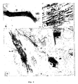
- Fig. 1 Expression of collagen IV and axonal sprouting after transection of the postcommissural fornix in untreated animals (b-d) and after injection of anti-Coll IV (e) or DPY (f) at two weeks postsurgery, a, Sagittal view of the adult rat brain showing the course of the fornix and the location of the transection site. Marked deposition of collagen IV in the lesion site (arrow) and proximal stump (P) of an untreated animal at low (b) and high magnification (c). Note, the fine structure and the spatial orientation of collagen IV deposits perpendicular to the trajectory of the tract, d, In untreated animals regrowing fornix axons stop sharply at the lesion site (arrow). Collagen IV deposition is markedly reduced in the lesion site after anti-Coil IV (e) or DPY injection (f). Scale bars, 100 ⁇ m.
- Fig. 2 Regeneration of transected fornix fibers across the lesion site in rats treated with anti-Coll IV (a, c, e) or DPY (b, d, f) at 6 weeks postsurgery. Sagittal serial sections reacted for NF-immunohistochemistry show that in both experimental groups fibers traverse the former lesion site (arrows) (c, d) and elongate within the distal stump (e, f) up to the mammillary body (MB). Scale bars, 100 ⁇ m.
- Fig. 3 Recovery of structural features of the regenerating fornix tract.
- a, b Anterograde tracing with biocytin of an anti-Coll IV treated animal at 6 weeks postsurgery reveals the large number of regenerating axons (a), their elongation within the former pathway (a) and their fine varicose morphology (b).
- c Large WGA-HRP-filled axon (arrowhead) in the mammillary body surrounded by compact myelin (arrows), d, e Electron micrographs of anterogradely WGA-HRP-labeled presynaptic terminals (arrowheads) in the mammillary body at 6 weeks after anti-Coll IV treatment. Scale bars, 100 ⁇ m (a), 50 ⁇ m (b), 0.1 ⁇ m (c), 0.5 ⁇ m (d) , 1 ⁇ m (e).
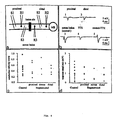
- Fig. 4 Electrophysiological properties of fornix fibers in unlesioned rats and lesioned/injected animals with regeneration.
- a Schematic illustration showing the location of the stimulating (S) and recording (R) electrode at various conditions
- b Characteristic recordings of extracellular action potentials in a sagittal slice prepared from an animal with regeneration. Recordings were obtained under conditions as illustrated in a.
- Application of Tetrodotoxin (TTX) blocks the stimulus-evoked response. The net action potential is shown in trace 5.
- TTX Tetrodotoxin
- lesion-induced BM deposition can be significantly reduced by local injection of anti-collagen IV antibodies or ⁇ , ⁇ dipyridyl, an inhibitor of collagen triple helix formation and synthesis. Reducing the collagen network allowed massive axon elongation across the lesion site.
- the left postcommissural fornix was stereotactically transected in adult Wistar rats Fig. 1a) and the postlesion deposition of BM was analyzed using antibodies against collagen IV (Coll IV) and laminin (LN), the major and unique components of BM (10,11).
- Coll IV collagen IV
- LN laminin
- BM the major and unique components of BM (10,11).
- Coll IV- and LN-rich BM Fig. 1b, c.
- These newly formed BM were either arranged in long continuous layers or associated with numerous blood vessels.
- the BM layers formed a parallel array aligned perpendicular to the course of the fiber tract (Fig. 1b, c).
- BM layers were deposited as hook-like turns extending along the longitudinal tract axis for about 200 ⁇ m into the fornix stumps (Fig. 1c).
- sprouting axons in the proximal stump reached the lesion site. They failed to cross or bypass it but stopped growing at the wound border at about 2 weeks after lesion (Fig. 1d).
- the spatio-temporal coincidence of BM formation with the abrupt axonal growth arrest at the tract-lesion border strongly suggests that the newly formed perpendicular layers of BM could be a physical impediment for regenerating axons.
- FIG. 1 Further preferred embodiments for restitution of functional circuitry after traumatic CNS lesion are the remyelination of regenerated fibers, the re-establishment of synaptic connections with the appropriate target and the restoration of normal conduction properties.
- Structural and functional properties of the regenerating axons were investigated using immunohistochemical, morphological and electrophysiological methods. Immunohistochemistry with an antibody against myelin basic protein demonstrated the remyelination of regenerated fornix axons along their entire length as early as 4 weeks after surgery (data not shown).
- the ultrastructural features of the labelled presynaptic profiles correspond to those described for the RA-type (round, asymmetric) of synaptic terminal, which is considered to be of subicular origin (8).
- the electrophysiological properties of regenerated fibers were studied using extracellular in vitro recording techniques applied to sagittal brain slices (400 ⁇ m) of 8 unlesioned rats and 4 treated animals showing regenerated fiber tracts.
- Electron microscopy For ultrastructural analysis vibratome sections of anti-Coll IV-treated animals were reacted for WGA-HRP, immersed for 12h in 1% osmium tetroxide and embedded in epon. Ultrathin sections were examined using a Hitachi H600 electron microscope.
- Sections were fixed with acetone (-20°C), preincubated in 3% H2O2 (v/v) in methanol to block endogeneous peroxidase, followed by PBS containing 3% (v/v) normal horse or normal goat serum to reduce unspecific staining and then incubated with one of the following primary antibodies: polyclonal anti-collagen IV (anti-Coll IV, Biogenex, 1:3), polyclonal anti-laminin (anti-LN, Biogenex, 1:5) or monoclonal cocktail against phosphorylated neurofilaments (anti-NF, Affinity, 1:800). Following, avidin-biotin-peroxidase complex staining (Vector Labs) was done using standard procedures.
- Tetrodotoxin (TTX, Sigma) was applied locally in a concentration of 10 ⁇ M (dissolved in ACSF) with a broken micropipette placed on the slice surface near the recording site. Injections of a small biocytin (Sigma) crystal into the fornix were performed with a miniature needle. After an incubation period of 8-10 h in the interface chamber, slices were fixed in 4 % paraformaldehyde, resectioned and reacted with ABC peroxidase reagent (Vector Labs)
Abstract
Description
- The present invention refers to the use of an inhibitor substance for the improvement of neuronal regeneration, and a medicament for the improvement of neuronal regeneration.
- Injury to adult mammalian CNS fiber tracts leads to the formation of a lesion scar consisting of a convoluted fringe of astroglial processes lined by a basal membrane (BM). This lesion scar is implicated as a major extrinsic constraint to effective axon regeneration in brain and spinal cord (1 - 4). While the dense astrocytic network is a permissive substrate for axon growth (5, 6), the presence of BM has been hypothesized as a crucial impediment for regeneration (7). However, experimental evidence was not shown. To the contrary, when the BM formed after a lesion of neuronal tissue was removed (24), no improved regeneration could be reproducibly monitored (25). Therefore, it is still of great importance to have a method for improving regeneration of injured neurons.
-
WO 93/19783 -
US-A-5,082,926 discloses the capability of competing peptide to promote nerve regeneration and indicates the clinical use in promoting regeneration of damaged nerves. It is speculated that the peptide disclosed is important in many diverse and clinically relevant processes such as cell attachment and migration in wound healing, tumor cell invasion and metastasis, diabetic microangiopathy, vascular hypertrophy due to hypertension and several kidney diseases such as diabetic nephropathy and nephrotic syndromes of variable etiology. -
US-A-5,493,008 discloses high affinity binding of nidogen to laminin to be mediated by an EGF-like repeat γ1III4 of the mouse laminin γ1 chain. -
WO-A-96/00582 - Surprisingly, improvement of regeneration of neuronal tissue after lesion is achieved by the use of an inhibitor substance of the present invention for the manufacturing of a medicament for the improvement of neuronal regeneration.
- According to the method of the invention improved regeneration of injured neuronal tissue is achieved by prevention or specific inhibition of basal membrane formation induced by a lesion of neuronal tissue by applying an inhibitor substance of the synthesis of basal membrane building elements, or the assembly of basal membrane building elements, or both the synthesis of basal membrane building elements and the assembly of basal membrane building elements to a body in need thereof wherein the inhibitor substance is selected from the group consisting of antibodies against collagen IV, laminin, entactin, accessory substances for proper function, or the assembly of the basal membrane; Fe-chelating agents; inhibitors of amino acids hydroxylases, such as prolyl-4-hydroxylase, lysine-hydroxylase; 2-oxoglutarate competitors.
- The basal membrane is a structure which is composed of different elements. Elements of the basal membrane are collagen IV, laminin, entactin (Nidogen) accessory substances. The assembly of the elements to the basal membrane is performed by enzymes which may be assisted by cofactors.
- Inhibitors of TGF-β are not involved with a specific prevention or specific inhibition of basal membrane formation induced by lesion of neuronal tissue. According to the present invention it is achieved in an advantageous manner that a specific interaction is provided by the use according to the invention.
- Preferably, the formation of the basal membrane is prevented or inhibited by applying a specific inhibitor substance of the synthesis of basal membrane building elements, or the assembly of basal membrane building elements, or both the synthesis of basal membrane building elements and the assembly of basal membrane building elements to a body in need thereof. The building elements of the basal membrane are in particular those which are involved with the formation of the basal membrane, for instance molecular structures building up the basal membrane, such as monomeric compounds, accessory substances, substances for the assembly of the components of the basal membrane and the like.
- In particular, the basal membrane building elements are selected from the group consisting of collagen IV, laminin, entactin, accessory substances for proper function, or the assembly of the basal membrane, or both the proper function and the assembly of the basal membrane.
- A specific inhibitor substance of the invention is capable of preventing or inhibiting the formation of the basal membrane and/or is specifically interfering with the assembly process of the basal membrane. The specific inhibitor substance of the invention is selected from the group consisting of antibodies against collagen IV, laminin, entactin, accessory substances for proper function, or the assembly of the basal membrane; Fe-chelating agents; inhibitors of amino acids hydroxylases, such as prolyl-4-hydroxylase, lysine-hydroxylase; 2-oxoglutarate competitors; which inhibitor substance is able to prevent or inhibit the expression of basal membrane building elements, and the like.
- According to the invention are preferred those inhibitor substances which are selected from the group consisting of N-oxaloglycine; Zn salts; pyridine derivatives, such as 5-arylcarbonyamino- or 5-arylcarbamoyl- derivatives, 2-carboxylate, 2,5 dicarboxylate, their ethyl esters or ethyl amides or -5-acyl sulfonamides, 2,4 dicarboxylate, their ethyl esters or ethylamides, or dimethoxyethylamides; 3,4 bipyridine, such as 5 amino-6-(1H)-one, 1,6-dihydro-2-methyl-6-oxo-5-carbonitril; 2,2'-bipyridine, such as 5,5'-dicarboxylic acid or its pharmaceutically acceptable salts, 4,4'-dicarboxylic acid ethyl ester or ethyl amide; 3,4'-dihydroxybenzoate, such as the diethyl ester; proline and its structural and functional analoges; β-aminopropionitrile; desferrioxamine; anthracyclines; 2,7,8-trihydroxy anthraquinones, fibrostatin-C; coumalic acid or its pharmaceutically acceptable salts; 5-oxaproline, β-lactam antibiotics.
- In a preferred embodiment of the present invention the specific inhibitor substance(s) are applied in combination with one or more substances being capable of stimulating neuronal growth or inducing the expression of growth promoting proteins. Such neuronal growth stimulating substances are neurotrophic growth factors of the neurotrophin family and other growth factor families such as fibroblast growth factors, insulin and insulin-like growth factors, as well as epidermal growth factor, ciliary neuronotrophic growth factor (CNTF), glial cell-derived growth factor (GDNF), cytokines, neurotrophic proteoglycans and glycosamino-glycans, neural cell adhesion molecules like L1 (NILE), growth-associated proteins like GAP43 and anti-apoptotic proteins like bcl-2.
- According to the invention it is preferred to locally apply the specific inhibitor substances in the neuronal tissue, intraventricularly, or systemically, in particular orally or intravenously.
- The concentration of the specific inhibitor substance varies in view of the chemical nature. For example, antisense inhibitor substances may have more specific effects so that lesser amounts can be applied.
- Typically, the specific inhibitor substance is applied in therapeutically effective amounts, such as 1 ng/kg to 1 mg/kg body weight, when low molecular compounds such as bipyridyl-derivatives are applied.
- The invention also provides a medicament for the improvement of neuronal regeneration comprising a therapeutically effective amount of a specific inhibitor substance which is capable of prevention or inhibition of basal membrane formation induced by a lesion of neuronal tissue. According to the invention the inhibitor substances are those as claimed in
claim 5. The medicament may further comprise carrier substances or adjuvants in order to facilitate an appropriate application. The medicament further comprises substances which are capable of stimulating neuronal growth as claimed inclaim 5. - Fig. 1:
Expression of collagen IV and axonal sprouting after transection of the postcommissural fornix in untreated animals (b-d) and after injection of anti-Coll IV (e) or DPY (f) at two weeks postsurgery, a, Sagittal view of the adult rat brain showing the course of the fornix and the location of the transection site. Marked deposition of collagen IV in the lesion site (arrow) and proximal stump (P) of an untreated animal at low (b) and high magnification (c). Note, the fine structure and the spatial orientation of collagen IV deposits perpendicular to the trajectory of the tract, d, In untreated animals regrowing fornix axons stop sharply at the lesion site (arrow). Collagen IV deposition is markedly reduced in the lesion site after anti-Coil IV (e) or DPY injection (f). Scale bars, 100 µm. - Fig. 2: Regeneration of transected fornix fibers across the lesion site in rats treated with anti-Coll IV (a, c, e) or DPY (b, d, f) at 6 weeks postsurgery. Sagittal serial sections reacted for NF-immunohistochemistry show that in both experimental groups fibers traverse the former lesion site (arrows) (c, d) and elongate within the distal stump (e, f) up to the mammillary body (MB). Scale bars, 100 µm.
- Fig. 3: Recovery of structural features of the regenerating fornix tract. a, b Anterograde tracing with biocytin of an anti-Coll IV treated animal at 6 weeks postsurgery reveals the large number of regenerating axons (a), their elongation within the former pathway (a) and their fine varicose morphology (b). c, Large WGA-HRP-filled axon (arrowhead) in the mammillary body surrounded by compact myelin (arrows), d, e Electron micrographs of anterogradely WGA-HRP-labeled presynaptic terminals (arrowheads) in the mammillary body at 6 weeks after anti-Coll IV treatment. Scale bars, 100 µm (a), 50 µm (b), 0.1 µm (c), 0.5 µm (d) , 1 µm (e).
- Fig. 4: Electrophysiological properties of fornix fibers in unlesioned rats and lesioned/injected animals with regeneration. a, Schematic illustration showing the location of the stimulating (S) and recording (R) electrode at various conditions, b, Characteristic recordings of extracellular action potentials in a sagittal slice prepared from an animal with regeneration. Recordings were obtained under conditions as illustrated in a. Application of Tetrodotoxin (TTX) blocks the stimulus-evoked response. The net action potential is shown in
trace 5. c and d, Distribution of conduction velocity and action potential response amplitude in unlesioned and lesioned/injected animals with regeneration. - The mechanically transected postcommissural fornix of the adult rat, a unidirectional and well-characterized fiber tract (8,9), was used to determine whether specific biochemical or immunochemical modulation of BM formation would provide a means to stimulate axon regeneration. Here we report that lesion-induced BM deposition can be significantly reduced by local injection of anti-collagen IV antibodies or α,α dipyridyl, an inhibitor of collagen triple helix formation and synthesis. Reducing the collagen network allowed massive axon elongation across the lesion site. The regenerating fornix fibers followed the original pathway, reinnervated their appropriate target, the mammillary body, were remyelinated and attained nearly normal conduction properties, on failure of adult mammalian CNS axons we examined its spatio-temporal distribution pattern after penetrant CNS lesion and determined whether its remodelling allows structural and functional regeneration of a transected CNS fiber tract.
- The left postcommissural fornix was stereotactically transected in adult Wistar rats Fig. 1a) and the postlesion deposition of BM was analyzed using antibodies against collagen IV (Coll IV) and laminin (LN), the major and unique components of BM (10,11). By the end of the second week after lesion the center of the wound was filled by Coll IV- and LN-rich BM (Fig. 1b, c). These newly formed BM were either arranged in long continuous layers or associated with numerous blood vessels. Within the center of the wound the BM layers formed a parallel array aligned perpendicular to the course of the fiber tract (Fig. 1b, c). In the vicinity of the transected stumps, however, BM layers were deposited as hook-like turns extending along the longitudinal tract axis for about 200 µm into the fornix stumps (Fig. 1c). In parallel with the deposition of the BM, sprouting axons in the proximal stump reached the lesion site. They failed to cross or bypass it but stopped growing at the wound border at about 2 weeks after lesion (Fig. 1d). The spatio-temporal coincidence of BM formation with the abrupt axonal growth arrest at the tract-lesion border strongly suggests that the newly formed perpendicular layers of BM could be a physical impediment for regenerating axons.
- In an effort to modulate postlesion BM deposition, either polyclonal antibodies against collagen IV (anti-Coll IV; n=14) or the iron chelator a, a'-dipyridyl (DPY; n=9) were injected locally into the lesion center immediately after transection. DPY is a competitive inhibitor of prolyl 4-hydroxylase (12) and has been shown to prevent collagen triple helix formation (12), which results in feedback inhibition of procollagen synthesis (13) and enhanced procollagen degradation (14). Control animals received a PBS injection (n=9) or were sham operated (n=3). Basal membrane formation was studied in response to antibody and drug treatment using immunohistochemical methods. Animals receiving a single injection of anti-Coll IV (80-160 ng) or DPY (1.6-16 µmol) showed a massive and specific reduction in Collimmunopositive laminae and blood vessels in the lesion center and the fornix stumps at all examined survival time points. At 2 weeks after lesion+injection only a very small number of Collimmunreactive structures perpendicular to the tract course had developed (Fig. 1e, f). Control animals, however, exhibited dense BM deposition as previously described for lesion only animals. The applied substances reduced the deposition of BM at the lesion site but did not affect the number or the distribution of vascular BM in the surrounding neuropil. Therefore, we conclude that the lesion-induced BM formation can be specifically reduced by immediate application of either anti-Coll IV antibodies or DPY.
- To determine whether reduction of BM deposition would permit regeneration of transected axons across the lesion site, we studied the elongation of fornix axons after anti-Coll IV or DPY treatment using immunocytochemical staining. While sprouting fornix fibers in control animals ceased growing at the proximal stump-lesion interface (Fig. 1d) large numbers of axons entered and traversed the lesion center between 2 and 4 weeks after lesion+injection in those animals receiving anti-Coll IV (n= 11) (Fig. 2 a, c, e) or DPY treatment (n= 6) (Fig. 2 b, d, f). Most regenerating axons formed a loop over the lesion site, entered the distal stump and continued in a parallel bundle of fine and beaded axons within their previous pathway (Fig. 3a, b). They reached their appropriate target, the mammillary body, at about 4-6 weeks postsurgery. Anterograde tracing with WGA-HRP into the subiculum, the origin of the fornix (not shown), or biocytin application into the proximal fornix stump (Fig. 3a) provided proof, that the vast majority of fibers emerge from the formerly transected fornix tract. All regenerating fornix axons remained within their original pathway and did not invade the surrounding neuropil. The present results demonstrate that the failure of postcommissural fornix regeneration in rat brain, in fact, depends upon the formation of an axon growth-inhibiting BM at the lesion site that is oriented perpendicular to the tract course. Reduction of BM deposition seems to be a prerequisite but also a sufficient condition for the transected axons to regenerate across the lesion site.
- Further preferred embodiments for restitution of functional circuitry after traumatic CNS lesion are the remyelination of regenerated fibers, the re-establishment of synaptic connections with the appropriate target and the restoration of normal conduction properties. Structural and functional properties of the regenerating axons were investigated using immunohistochemical, morphological and electrophysiological methods. Immunohistochemistry with an antibody against myelin basic protein demonstrated the remyelination of regenerated fornix axons along their entire length as early as 4 weeks after surgery (data not shown). This observation was confirmed by ultrastructural analysis of anterogradely WGA-HRP labeled axons in the distal stump which showed clear evidence of compact myelin sheath formation (Fig. 3c). In addition, ultrastructural studies provided evidence for the re-establishment of synaptic connections of regenerating axons within the mammillary body. Tracer reaction product was identified in presynaptic profiles with round vesicles that formed asymmetric synaptic junctions at unlabeled dendrites (Fig. 3d, e). The ultrastructural features of the labelled presynaptic profiles correspond to those described for the RA-type (round, asymmetric) of synaptic terminal, which is considered to be of subicular origin (8). The electrophysiological properties of regenerated fibers were studied using extracellular in vitro recording techniques applied to sagittal brain slices (400 µm) of 8 unlesioned rats and 4 treated animals showing regenerated fiber tracts. In unlesioned animals electrical stimulation of the fornix fibers elicited an extracellular action potential with an amplitude of 1.02 ± 0.14 mV and a conduction velocity of 0.48 ± 0.05 m/s (mean ± SEM, n=16, Fig. 4b-d). This axonal conduction velocity corresponds well to previously reported measurements (about 0.5 m/s for hippocampal Schaffer collaterals (15). Similar values for action potential amplitude and conduction velocity (1.12 ± 0.21 mV, 0.46 ± 0.1 m/s, n=5) were obtained in regenerating animals when the stimulating (S) and the recording (R) electrodes were positioned proximally to the lesion site (see S1 and R1 in Fig. 4a). In the latter animals, functionally intact fibers showing normal extracellular action potential amplitude and conduction velocity could also be demonstrated across (S3 and R3 in Fig. 4a; 0.8 ± 0.29 mV, 0.54 ± 0.14 m/s, n=3) and distal to the lesion site (S2 and R2 in Fig. 4a; 0.91 ± 0.24 mV, 0.43 ± 0.06 m/s, n=4) (Fig. 4c, d). In all animals, the stimulus-evoked extracellular responses were blocked by Tetrodotoxin, confirming their nature as Na+-dependent action potentials (Fig. 4b). From these data we conclude that the reorganization of the fornix tract is accompanied by structural and functional recovery of the regenerated axons.
- Our results demonstrate that structural and functional restoration of lesioned mature fornix pathway can be achieved by reduction of BM formation in the lesion site. Data described here underscore the importance of extrinsic determinants in axonal regeneration but also demonstrates that once the axons have crossed the lesion scar other potential extrinsic regeneration constraints, like CNS myelin and oligodendrocytes (9,16-18), dense astrogliosis (6) and sulfated proteoglycans (19,20), do not impede their progress. The results further indicate that similar to other CNS circuits (21,22), fornix axons have an innate potential for regeneration and self-organization. These results give rise to new and promising concepts for therapeutic strategies that might contribute to the reduction of neurological deficits after CNS lesions.
- The following examples are intended for further illustration of the invention but are not limiting.
- Surgery. The left postcommissural fornix of 42 Wistar rats (180-210g) was transected stereotactically at a distance of about 1 mm proximal to the target, the mammillary body, using a Scouten wire knife as described previously (9). The completeness of transection was confirmed by serial reconstruction of the lesion site for each of the animals. Immediately after transection animals received a topical application (1.6 µl) of either polyclonal antibodies against collagen IV (anti-Coll IV, Biogenex, 50-100 µg/ml, n=14) or the iron chelator a, a'-dipyridyl (DPY, 1-10 mM, n=9). Substances were pressure injected (injection time 10 min) directly into the lesion site via a micropipette coupled to a microsyringe. Controls received equal amounts of phosphate-buffered saline (n=9) or sham operation (n=3).
- Anterograde tracing was performed for analysis of fiber course, ultrastructural morphology and target reinnervation. After a survival time of 6 weeks, anti-Coll IV-treated animals (n=4) received two injections of a 2% (w/v) solution of wheat-germagglutinin-HRP (WGA-HRP) into the left subicular complex (dorsal and caudal pole). Rats were perfused 3 days later with 2% paraformaldehyde and 2% glutaraldehyde in 0.1M phosphate buffer. Vibratome sections were reacted for WGA-HRP using tetramethylbenzidine as substrate (23).
- Electron microscopy. For ultrastructural analysis vibratome sections of anti-Coll IV-treated animals were reacted for WGA-HRP, immersed for 12h in 1% osmium tetroxide and embedded in epon. Ultrathin sections were examined using a Hitachi H600 electron microscope.
- Immunohistochemical staining. After a survival time of 4 days (d), 6d, 2 weeks (w), 4w and 6w after surgery brains were removed, frozen in isopentan (-50/-60°C) and cut into serial sagittal 10 µm thick sections. Sections were fixed with acetone (-20°C), preincubated in 3% H2O2 (v/v) in methanol to block endogeneous peroxidase, followed by PBS containing 3% (v/v) normal horse or normal goat serum to reduce unspecific staining and then incubated with one of the following primary antibodies: polyclonal anti-collagen IV (anti-Coll IV, Biogenex, 1:3), polyclonal anti-laminin (anti-LN, Biogenex, 1:5) or monoclonal cocktail against phosphorylated neurofilaments (anti-NF, Affinity, 1:800). Following, avidin-biotin-peroxidase complex staining (Vector Labs) was done using standard procedures. For evaluation of remyelination brains were fixed with 4% paraformaldehyde, paraffinized, cut into 3-µm thick serial sagittal sections, deparaffinized and incubated as described above with a polyclonal anti-myelin basic protein (anti-MBP, Biogenex, 1:2) or anti-NF as primary antibodies. Specificity of the stainings was confirmed by omission of the primary antibody.
- Electrophysiology and biocytin injections. Sagittal slices of 400 µm thickness were cut on a vibratome and maintained at 34-35°C in an interface-type recording chamber. Artificial cerebrospinal fluid (ACSF) consisted of (in mM) 124 NaCl, 3 KCl, 1.25 NaH2PO4, 1.8 MgSO4, 1.6 CaCl2, 26 NaHCO3 and 10 glucose with a pH of 7.4 when saturated with 95% 02 - 5% CO2. Stimuli: 100 µs, 5-20 V were delivered via a bipolar tungsten electrode. Extracellular action potentials were registered with a recording electrode (3-5 MW) located in the middle of the postcommissural fornix. Tetrodotoxin (TTX, Sigma) was applied locally in a concentration of 10 µM (dissolved in ACSF) with a broken micropipette placed on the slice surface near the recording site. Injections of a small biocytin (Sigma) crystal into the fornix were performed with a miniature needle. After an incubation period of 8-10 h in the interface chamber, slices were fixed in 4 % paraformaldehyde, resectioned and reacted with ABC peroxidase reagent (Vector Labs)
- 1. Reier, P. J., Stensaas, L. J. & Guth, L. Spinal cord reconstruction (eds Kao, C.C., Bunge, R.P. & Reier, P.J.) 163-196 ( Raven Press, New York, 1983).
- 2. Clemente, C. D. Regeneration in the nervous system (ed Windle, W.F.) 147-161 ( Charles C. Thomas, Springfield, IL, 1955).
- 3. Schnell, L. & Schwab, M. E. .
- 4. Krüger, S., Sievers, J., Hansen, C., Sadler, M. & Berry, M. J. Comp. Neural 249, 103-116 (1986).
- 5. Berry, M., Carlile, J. & Hunter, A. J. Neurocytol 25, 147-170 (1996).
- 6. Stichel, C. C. & Müller, H. W. J. Neurocytol 23, 615-630 (1994).
- 7. Feringa, E. R., Kowalski, T. F. & Vahlsing, H. L. Ann. Neurol 8, 148-154 (1980).
- 8. Allen, G. V. & Hopkins, D. A. J. Comp. Neurol 275, 39-64 (1988).
- 9. Stichel, C. C., Wunderlich, G., Schwab, M. E. & Müller, H. W. Europ. J. Neurosci 7, 401-411 (1995).
- 10. Yurchenco, P. D. & Schittny, .
- 11. Timpl, R. & Brown, J. Curr. Opin. Cell. Biol 8, 618-624 (1996).
- 12. Kivirikko, K. L., Myllylä, R. & Pihlajaniemi, .
- 13. Ikeda, H., Wu, G. Y. & Wu, C. H. Hepatology 15, 282-287 (1992).
- 14. Bienkowski, R. S. J. Cell. Physiol 121, 152-158 (1984).
- 15. Andersen, P., Silfvenius, H., Sundberg, S., Sveen, O. & Wigström, H. Brain. Res 144, 11-18 (1978).
- 16. McKerracher, L. et al. Neuron 13, 805-811 (1994).
- 17. Mukhopadhyay, G., Doherty, P., Walsh, F. S., Crocker, P. R. & Filbin, M. T. Neuron 13, 757-767 (1994).
- 18. Schwab, M. E. & Caroni, P. J. Neurosci 8, 2381-2393 (1988).
- 19. Snow, A. D., Lemmon, V., Carrino, D. A., Caplan, A. I. & Silver, J. Exp. Neurol 109, 111-130 (1990).
- 20. Lips, K., Stichel, C. C. & Müller, H. W. J. Neurocytol 24, 449-464 (1995).
- 21. Iwashita, Y., Kawaguchi, S. & Murata, M. Nature 367, 167-170 (1994).
- 22. Kalderon, N. & Fuks, Z. Proc. Natl. Acad. Sci 93, 11179-11184 (1996).
- 23. Olucha, F., Martinez-Garcia, F. & Lopez-Garcia, C. J. Neurosci. Meth 13, 131-138 (1985).
- 24. Puchala, E. & Windle, W. F., Exp. Neurol. 55, 1 - 42 (1977).
- 25. Guth, L., Albuquerque, E. X ., Deshpande, S. S., Barret, C. P., Donati, E. J. & Warnick, J. E., J. Neurosurg. 52, 73 - 86 (1980).
Claims (6)
- Use of an inhibitor substance selected from the group consisting of antibodies against collagen IV, laminin, entactin, accessory substances for proper function, or the assembly of the basal membrane; Fe-chelating agents; inhibitors of amino acids hydroxylases, such as prolyl-4-hydroxylase, lysine-hydroxylase; 2-oxoglutarate competitors; which inhibitor substance is capable of prevention or specific inhibition of basal membrane formation induced by a lesion of neuronal tissue for the manufacturing of a medicament for the improvement of neuronal regeneration.
- Use of claim 1, wherein the inhibitor substance is a substance inhibiting the synthesis of basal membrane building elements, or the assembly of basal membrane building elements, or both the synthesis of basal membrane building elements and the assembly of basal membrane building elements.
- Use of claim 2, wherein the basal membrane building elements are selected from the group consisting of collagen IV, laminin, entactin, accessory substances for proper function, or the assembly of the basal membrane, or both the proper function and the assembly of the basal membrane.
- Use of claim 1, wherein the inhibitor substance is selected from the group consisting of N-oxaloglycine; Zn salts; pyridine derivatives, such as 5-arylcarbonyamino- or 5-arylcarbamoyl-derivatives, 2-carboxylate, 2,5-dicarboxylate, their ethyl esters or ethyl amides or -5-acyl sulfonamides, 2,4 dicarboxylate, their ethyl esters or ethylamides, or dimethoxyethylamides; 3,4'-bipyridine, such as 5 amino-6-(1H)-one, 1,6-dihydro-2-methyl-6-oxo-5-carbonltril; 2,2'-bipyridine, such as 5,5'-dicarboxylic acid or its pharmaceutically acceptable salts, 4,4'-dicarboxylic acid ethyl ester or ethyl amide; 3,4'-dihydroxybenzoate, such as the diethyl ester; proline and its structural and functional analoges; β-aminopropionitrile; desferrioxamine; anthracyclines; 2,7,8-trihydroxy anthraquinones, fibrostatin-C; coumalic acid or its pharmaceutically acceptable salts; 5-oxaproline,β-lactam antibiotics.
- A medicament for the improvement of neuronal regeneration comprising a therapeutically effective amount of an inhibitor substance which is capable of prevention or specific inhibition of basal membrane formation induced by a lesion of neuronal tissue comprising the inhibitor substance(s) selected from the group consisting of antibodies against collagen IV, laminin, entactin, accessory substances for proper function, or the assembly of the basal membrane; Fe-chelating agents; 2-oxoglutarate competitors; in combination with a substance being capable of stimulating neuronal growth or inducing the expression of growth promoting proteins such as fibroblast growth factors, neural cell adhesion molecules like L1 (NILE), growth-associated proteins like GAP43 and anti-apoptotic proteins like bcl-2.
- The medicament according to claim 5, wherein the inhibitor substance is applied in therapeutically effective amounts, such as 1 ng/kg to 1 mg/kg body weight, selected from the group consisting of pyridine derivatives, such as 5-aryl-carbonyamino- or 5-arylcarbamoyl-derivatives, 2-carboxylate, 2,5-dicarboxylate, their ethyl esters or ethyl amides or -5-acyl sulfonamides, 2,4 dicarboxylate, their ethyl esters or ethylamides, or dimethoxyethylamides; 3,4'-bipyridine, such as 5 amino-6-(1H)-one, 1,6-dihydro-2-methyl-6-oxo-5-carbonltril; 2,2'-bipyridine, such as 5,5'-dicarboxylic acid or its pharmaceutically acceptable salts, 4,4'-dicarboxylic acid ethyl ester or ethyl amide; 3,4'-dihydroxybenzoate, such as the diethyl ester; β-aminopropionitrile; desferrioxamine; anthracyclines; 2,7,8-trihydroxy anthraquinones, coumalic acid or its pharmaceutically acceptable salts; β-lactam antibiotics.
Priority Applications (1)
| Application Number | Priority Date | Filing Date | Title |
|---|---|---|---|
| EP98924307A EP0981549B9 (en) | 1997-05-14 | 1998-05-13 | Use of an inhibitor substance for the improvement of neuronal regeneration |
Applications Claiming Priority (4)
| Application Number | Priority Date | Filing Date | Title |
|---|---|---|---|
| EP97107846 | 1997-05-14 | ||
| EP97107846A EP0878480A1 (en) | 1997-05-14 | 1997-05-14 | A method for the improvement of neuronal regeneration |
| EP98924307A EP0981549B9 (en) | 1997-05-14 | 1998-05-13 | Use of an inhibitor substance for the improvement of neuronal regeneration |
| PCT/EP1998/002808 WO1998051708A1 (en) | 1997-05-14 | 1998-05-13 | A method for the improvement of neuronal regeneration |
Publications (4)
| Publication Number | Publication Date |
|---|---|
| EP0981549A1 EP0981549A1 (en) | 2000-03-01 |
| EP0981549B1 EP0981549B1 (en) | 2002-09-25 |
| EP0981549B2 true EP0981549B2 (en) | 2007-06-27 |
| EP0981549B9 EP0981549B9 (en) | 2008-02-27 |
Family
ID=8226794
Family Applications (2)
| Application Number | Title | Priority Date | Filing Date |
|---|---|---|---|
| EP97107846A Withdrawn EP0878480A1 (en) | 1997-05-14 | 1997-05-14 | A method for the improvement of neuronal regeneration |
| EP98924307A Expired - Lifetime EP0981549B9 (en) | 1997-05-14 | 1998-05-13 | Use of an inhibitor substance for the improvement of neuronal regeneration |
Family Applications Before (1)
| Application Number | Title | Priority Date | Filing Date |
|---|---|---|---|
| EP97107846A Withdrawn EP0878480A1 (en) | 1997-05-14 | 1997-05-14 | A method for the improvement of neuronal regeneration |
Country Status (8)
| Country | Link |
|---|---|
| US (3) | US20020115598A1 (en) |
| EP (2) | EP0878480A1 (en) |
| AT (1) | ATE224915T1 (en) |
| AU (1) | AU7654298A (en) |
| DE (1) | DE69808281T3 (en) |
| DK (1) | DK0981549T4 (en) |
| ES (1) | ES2180175T5 (en) |
| WO (1) | WO1998051708A1 (en) |
Families Citing this family (11)
| Publication number | Priority date | Publication date | Assignee | Title |
|---|---|---|---|---|
| EP0878480A1 (en) * | 1997-05-14 | 1998-11-18 | H.W. Prof. Dr. Müller | A method for the improvement of neuronal regeneration |
| US6200974B1 (en) | 1997-10-24 | 2001-03-13 | Zeneca Limited | Phenanthroline derivatives |
| CN102526044A (en) | 2001-12-06 | 2012-07-04 | 法布罗根股份有限公司 | Methods of increasing endogenous erythropoietin EPO |
| CN101664551B (en) * | 2001-12-06 | 2013-08-14 | 法布罗根股份有限公司 | Stabilization of hypoxia inducible factor (HIF) alpha |
| US8124582B2 (en) * | 2002-12-06 | 2012-02-28 | Fibrogen, Inc. | Treatment of diabetes |
| US7618940B2 (en) * | 2002-12-06 | 2009-11-17 | Fibrogen, Inc. | Fat regulation |
| DE10314082A1 (en) * | 2003-03-28 | 2004-10-21 | Mcs Micro Carrier Systems Gmbh | Biodegradable injectable implant |
| AU2006286445A1 (en) * | 2005-09-02 | 2007-03-08 | Neuraxo Biopharmaceuticals Gmbh | Pharmaceutical composition comprising an iron chelator |
| WO2007048846A1 (en) * | 2005-10-27 | 2007-05-03 | Neuraxo Biopharmaceuticals Gmbh | Use of iron-chelating compounds, cyclic adenosine monophosphate-increasing compounds or combinations thereof for treating axonal lesions in the cns |
| EP2000134A1 (en) * | 2007-06-06 | 2008-12-10 | Neuraxo Biopharmaceuticals GmbH | Use of a substance for the improvement of pns lesions |
| US9649381B2 (en) | 2013-11-06 | 2017-05-16 | Wayne State University | Transporter protein-coupled nanodevices for targeted drug delivery |
Citations (3)
| Publication number | Priority date | Publication date | Assignee | Title |
|---|---|---|---|---|
| US4214000A (en) † | 1978-10-30 | 1980-07-22 | Johnson & Johnson | Zinc salt of all-trans-retinoic acid for the treatment of acne |
| US5021404A (en) † | 1988-04-20 | 1991-06-04 | The Children's Medical Center Corporation | Angiostatic collagen modulators |
| JPH06103773A (en) † | 1992-09-16 | 1994-04-15 | Hitachi Ltd | Semiconductor memory device |
Family Cites Families (9)
| Publication number | Priority date | Publication date | Assignee | Title |
|---|---|---|---|---|
| DE3002989A1 (en) | 1980-01-29 | 1981-07-30 | Hoechst Ag, 6000 Frankfurt | HYDROXYPHENYL-THIAZOLE, -THIAZOLINE AND -THIAZOLIDINE-CARBONIC ACIDS, METHOD FOR THE PRODUCTION THEREOF AND THEIR USE FOR INFLUENCING COLLAGEN METABOLISM |
| CA1251729A (en) * | 1982-05-19 | 1989-03-28 | Steffen Gay | In vitro diagnostic methods using monoclonal antibodies against connective tissue proteins |
| DE3432094A1 (en) | 1984-08-31 | 1986-03-06 | Hoechst Ag, 6230 Frankfurt | ESTER OF PYRIDINE-2,4- AND -2,5-DICARBONIC ACID AS A MEDICINAL PRODUCT FOR INHIBITING PROLIN AND LYSINE HYDROXYLASE |
| AU3943793A (en) * | 1992-04-01 | 1993-11-08 | Whittier Institute For Diabetes And Endocrinology, The | Methods of inhibiting or enhancing scar formation in the CNS |
| AU5891394A (en) * | 1993-02-11 | 1994-08-29 | Erziehungsdirektion Of The Canton Zurich | A combination of neurotrophin and antibody directed toward myelin-associated neurite growth inhibitory protein promotes central nervous system regeneration |
| DE69429539T2 (en) * | 1993-10-15 | 2002-09-05 | Thomas Jefferson University Ph | INHIBITING THE EXTRACELLULAR MATRIX SYNTHESIS BY ANTISENSE COMPOSITIONS AGAINST C-MYC |
| WO1995013291A1 (en) * | 1993-11-08 | 1995-05-18 | New York University | Neuron-glia cell adhesion molecule, ng-cam, in treatment of nerve damage |
| US5567609A (en) * | 1994-06-30 | 1996-10-22 | University Of Kansas Medical Center | Use of isolated domains of type IV collagen to modify cell and tissue interactions |
| EP0878480A1 (en) * | 1997-05-14 | 1998-11-18 | H.W. Prof. Dr. Müller | A method for the improvement of neuronal regeneration |
-
1997
- 1997-05-14 EP EP97107846A patent/EP0878480A1/en not_active Withdrawn
-
1998
- 1998-03-13 US US09/423,622 patent/US20020115598A1/en not_active Abandoned
- 1998-05-13 AT AT98924307T patent/ATE224915T1/en not_active IP Right Cessation
- 1998-05-13 AU AU76542/98A patent/AU7654298A/en not_active Abandoned
- 1998-05-13 DE DE69808281T patent/DE69808281T3/en not_active Expired - Lifetime
- 1998-05-13 DK DK98924307T patent/DK0981549T4/en active
- 1998-05-13 EP EP98924307A patent/EP0981549B9/en not_active Expired - Lifetime
- 1998-05-13 WO PCT/EP1998/002808 patent/WO1998051708A1/en active IP Right Grant
- 1998-05-13 ES ES98924307T patent/ES2180175T5/en not_active Expired - Lifetime
-
2004
- 2004-05-04 US US10/837,762 patent/US7208153B2/en not_active Expired - Fee Related
-
2007
- 2007-03-16 US US11/723,191 patent/US20070231330A1/en not_active Abandoned
Patent Citations (3)
| Publication number | Priority date | Publication date | Assignee | Title |
|---|---|---|---|---|
| US4214000A (en) † | 1978-10-30 | 1980-07-22 | Johnson & Johnson | Zinc salt of all-trans-retinoic acid for the treatment of acne |
| US5021404A (en) † | 1988-04-20 | 1991-06-04 | The Children's Medical Center Corporation | Angiostatic collagen modulators |
| JPH06103773A (en) † | 1992-09-16 | 1994-04-15 | Hitachi Ltd | Semiconductor memory device |
Non-Patent Citations (1)
| Title |
|---|
| Feringa E R et al. (1980), Ann Neurol 8, 148-154 † |
Also Published As
| Publication number | Publication date |
|---|---|
| US20050118177A1 (en) | 2005-06-02 |
| EP0981549B9 (en) | 2008-02-27 |
| WO1998051708A1 (en) | 1998-11-19 |
| ES2180175T3 (en) | 2003-02-01 |
| EP0878480A1 (en) | 1998-11-18 |
| US7208153B2 (en) | 2007-04-24 |
| AU7654298A (en) | 1998-12-08 |
| ES2180175T5 (en) | 2008-02-01 |
| US20020115598A1 (en) | 2002-08-22 |
| EP0981549A1 (en) | 2000-03-01 |
| EP0981549B1 (en) | 2002-09-25 |
| ATE224915T1 (en) | 2002-10-15 |
| DE69808281T2 (en) | 2003-01-16 |
| DK0981549T3 (en) | 2003-02-10 |
| DE69808281T3 (en) | 2008-01-24 |
| DK0981549T4 (en) | 2007-10-29 |
| DE69808281D1 (en) | 2002-10-31 |
| US20070231330A1 (en) | 2007-10-04 |
Similar Documents
| Publication | Publication Date | Title |
|---|---|---|
| US20070231330A1 (en) | Method for the improvement of neuronal regeneration | |
| McKeon et al. | Reduction of neurite outgrowth in a model of glial scarring following CNS injury is correlated with the expression of inhibitory molecules on reactive astrocytes | |
| Weibel et al. | Regeneration of lesioned rat optic nerve fibers is improved after neutralization of myelin-associated neurite growth inhibitors | |
| Bartholdi et al. | Methylprednisolone inhibits early inflammatory processes but not ischemic cell death after experimental spinal cord lesion in the rat | |
| Bloch et al. | Nerve growth factor-and neurotrophin-3-releasing guidance channels promote regeneration of the transected rat dorsal root | |
| Agrawal et al. | Role of L-and N-type calcium channels in the pathophysiology of traumatic spinal cord white matter injury | |
| US8158578B2 (en) | Methods for treating neurological deficits | |
| Evans et al. | Kainic acid seizures and the reversibility of calcium loading in vulnerable neurons in the hippocampus | |
| Abrams et al. | Emerging strategies to promote improved functional outcome after peripheral nerve injury | |
| Lukasiuk et al. | Upregulation of cystatin C expression in the rat hippocampus during epileptogenesis in the amygdala stimulation model of temporal lobe epilepsy | |
| Urban et al. | Cell-type specific expression of constitutively-active Rheb promotes regeneration of bulbospinal respiratory axons following cervical SCI | |
| Rende et al. | Modulation of low‐affinity nerve growth factor receptor in injured adult rat spinal cord motoneurons | |
| Lehrmann et al. | Glial activation precedes seizures and hippocampal neurodegeneration in measles virus–infected mice | |
| Camborieux et al. | Changes in expression and localization of hemopexin and its transcripts in injured nervous system: a comparison of central and peripheral tissues | |
| US6969516B1 (en) | Immunological compositions and methods of use to transiently alter mammalian central nervous system myelin to promote neuronal regeneration | |
| Naffah-Mazzacoratti et al. | Growth-associated phosphoprotein expression is increased in the supragranular regions of the dentate gyrus following pilocarpine-induced seizures in rats | |
| US5387520A (en) | Treatment of tumor cells in vitro with neurotrophic factors and cell proliferation inhibitors | |
| Mori et al. | Detrimental effects of exogenous glutamate on spinal cord neurons during brief ischemia in vivo | |
| US20010007657A1 (en) | Compositions and methods for manipulating glial progenitor cells and treating neurological deficits | |
| WO2017042314A1 (en) | Methods and pharmaceutical composition for the treatment of post-traumatic epilepsies | |
| Sugiyama et al. | Acceleration by MS‐818 of early muscle regeneration and enhanced muscle recovery after surgical transection | |
| Xu et al. | Electroacupuncture at Governor Vessel improves neurobehavioral function via silencing complexin I | |
| Rupniak et al. | Neurotrophic factors in peripheral nerve injury and regeneration | |
| NZ537103A (en) | Metallothionein based neuronal therapeutic and therapeutic methods | |
| KLAPKA et al. | Glial Interfaces in the Nervous System 139 H. Aldskogius and J. Fraher (Eds.) IOS Press, 2002 |
Legal Events
| Date | Code | Title | Description |
|---|---|---|---|
| PUAI | Public reference made under article 153(3) epc to a published international application that has entered the european phase |
Free format text: ORIGINAL CODE: 0009012 |
|
| 17P | Request for examination filed |
Effective date: 19991214 |
|
| AK | Designated contracting states |
Kind code of ref document: A1 Designated state(s): AT BE CH DE DK ES FI FR GB IE IT LI NL SE |
|
| RAP1 | Party data changed (applicant data changed or rights of an application transferred) |
Owner name: MUELLER, H.W., PROF. DR. |
|
| RIN1 | Information on inventor provided before grant (corrected) |
Inventor name: STICHEL-GUNKEL, CHRISTINE, DR. Inventor name: MUELLER, H.W., PROF.DR. |
|
| 17Q | First examination report despatched |
Effective date: 20010913 |
|
| RAP1 | Party data changed (applicant data changed or rights of an application transferred) |
Owner name: NEURAXO-BIOTEC GMBH |
|
| GRAG | Despatch of communication of intention to grant |
Free format text: ORIGINAL CODE: EPIDOS AGRA |
|
| RTI1 | Title (correction) |
Free format text: USE OF AN INHIBITOR SUBSTANCE FOR THE IMPROVEMENT OF NEURONAL REGENERATION |
|
| GRAG | Despatch of communication of intention to grant |
Free format text: ORIGINAL CODE: EPIDOS AGRA |
|
| GRAH | Despatch of communication of intention to grant a patent |
Free format text: ORIGINAL CODE: EPIDOS IGRA |
|
| GRAG | Despatch of communication of intention to grant |
Free format text: ORIGINAL CODE: EPIDOS AGRA |
|
| GRAH | Despatch of communication of intention to grant a patent |
Free format text: ORIGINAL CODE: EPIDOS IGRA |
|
| GRAA | (expected) grant |
Free format text: ORIGINAL CODE: 0009210 |
|
| AK | Designated contracting states |
Kind code of ref document: B1 Designated state(s): AT BE CH DE DK ES FI FR GB IE IT LI NL SE |
|
| REF | Corresponds to: |
Ref document number: 224915 Country of ref document: AT Date of ref document: 20021015 Kind code of ref document: T |
|
| REG | Reference to a national code |
Ref country code: GB Ref legal event code: FG4D |
|
| REG | Reference to a national code |
Ref country code: CH Ref legal event code: EP |
|
| REG | Reference to a national code |
Ref country code: CH Ref legal event code: NV Representative=s name: SCHMAUDER & PARTNER AG PATENTANWALTSBUERO |
|
| REG | Reference to a national code |
Ref country code: IE Ref legal event code: FG4D |
|
| REF | Corresponds to: |
Ref document number: 69808281 Country of ref document: DE Date of ref document: 20021031 |
|
| REG | Reference to a national code |
Ref country code: ES Ref legal event code: FG2A Ref document number: 2180175 Country of ref document: ES Kind code of ref document: T3 |
|
| REG | Reference to a national code |
Ref country code: DK Ref legal event code: T3 |
|
| ET | Fr: translation filed | ||
| PLBI | Opposition filed |
Free format text: ORIGINAL CODE: 0009260 |
|
| PLBQ | Unpublished change to opponent data |
Free format text: ORIGINAL CODE: EPIDOS OPPO |
|
| PLAB | Opposition data, opponent's data or that of the opponent's representative modified |
Free format text: ORIGINAL CODE: 0009299OPPO |
|
| 26 | Opposition filed |
Opponent name: L'OREAL Effective date: 20030620 |
|
| R26 | Opposition filed (corrected) |
Opponent name: L'OREAL Effective date: 20030620 |
|
| NLR1 | Nl: opposition has been filed with the epo |
Opponent name: L'OREAL |
|
| PLAX | Notice of opposition and request to file observation + time limit sent |
Free format text: ORIGINAL CODE: EPIDOSNOBS2 |
|
| NLR1 | Nl: opposition has been filed with the epo |
Opponent name: L'OREAL |
|
| PLAX | Notice of opposition and request to file observation + time limit sent |
Free format text: ORIGINAL CODE: EPIDOSNOBS2 |
|
| PLBB | Reply of patent proprietor to notice(s) of opposition received |
Free format text: ORIGINAL CODE: EPIDOSNOBS3 |
|
| REG | Reference to a national code |
Ref country code: CH Ref legal event code: PK Ref country code: CH Ref legal event code: PFA Owner name: NEURAXO PHARMACEUTICALS GMBH Free format text: NEURAXO-BIOTEC GMBH#POSTFACH 250 318#40092 DUESSELDORF (DE) -TRANSFER TO- NEURAXO PHARMACEUTICALS GMBH#MAX-PLANCK-STRASSE 15A#40699 ERKRATH (DE) |
|
| RAP2 | Party data changed (patent owner data changed or rights of a patent transferred) |
Owner name: NEURAXO BIOPHARMACEUTICALS GMBH |
|
| NLT1 | Nl: modifications of names registered in virtue of documents presented to the patent office pursuant to art. 16 a, paragraph 1 |
Owner name: NEURAXO BIOPHARMACEUTICALS GMBH |
|
| NLT2 | Nl: modifications (of names), taken from the european patent patent bulletin |
Owner name: NEURAXO BIOPHARMACEUTICALS GMBH Effective date: 20060719 |
|
| PUAH | Patent maintained in amended form |
Free format text: ORIGINAL CODE: 0009272 |
|
| STAA | Information on the status of an ep patent application or granted ep patent |
Free format text: STATUS: PATENT MAINTAINED AS AMENDED |
|
| 27A | Patent maintained in amended form |
Effective date: 20070627 |
|
| AK | Designated contracting states |
Kind code of ref document: B2 Designated state(s): AT BE CH DE DK ES FI FR GB IE IT LI NL SE |
|
| REG | Reference to a national code |
Ref country code: SE Ref legal event code: RPEO |
|
| REG | Reference to a national code |
Ref country code: CH Ref legal event code: AEN Free format text: AUFRECHTERHALTUNG DES PATENTES IN GEAENDERTER FORM |
|
| NLR2 | Nl: decision of opposition |
Effective date: 20070627 |
|
| REG | Reference to a national code |
Ref country code: DK Ref legal event code: T4 |
|
| ET3 | Fr: translation filed ** decision concerning opposition | ||
| NLR3 | Nl: receipt of modified translations in the netherlands language after an opposition procedure | ||
| REG | Reference to a national code |
Ref country code: ES Ref legal event code: DC2A Date of ref document: 20070921 Kind code of ref document: T5 |
|
| REG | Reference to a national code |
Ref country code: CH Ref legal event code: PCAR Free format text: SCHMAUDER & PARTNER AG PATENT- UND MARKENANWAELTE VSP;ZWAENGIWEG 7;8038 ZUERICH (CH) |
|
| PGFP | Annual fee paid to national office [announced via postgrant information from national office to epo] |
Ref country code: IE Payment date: 20100520 Year of fee payment: 13 Ref country code: FR Payment date: 20100608 Year of fee payment: 13 Ref country code: FI Payment date: 20100520 Year of fee payment: 13 Ref country code: ES Payment date: 20100520 Year of fee payment: 13 Ref country code: DK Payment date: 20100525 Year of fee payment: 13 |
|
| PGFP | Annual fee paid to national office [announced via postgrant information from national office to epo] |
Ref country code: NL Payment date: 20100520 Year of fee payment: 13 Ref country code: IT Payment date: 20100528 Year of fee payment: 13 Ref country code: AT Payment date: 20100520 Year of fee payment: 13 |
|
| PGFP | Annual fee paid to national office [announced via postgrant information from national office to epo] |
Ref country code: CH Payment date: 20100525 Year of fee payment: 13 Ref country code: BE Payment date: 20100525 Year of fee payment: 13 |
|
| PGFP | Annual fee paid to national office [announced via postgrant information from national office to epo] |
Ref country code: SE Payment date: 20100521 Year of fee payment: 13 Ref country code: GB Payment date: 20100521 Year of fee payment: 13 Ref country code: DE Payment date: 20100728 Year of fee payment: 13 |
|
| BERE | Be: lapsed |
Owner name: *NEURAXO BIOPHARMACEUTICALS G.M.B.H. Effective date: 20110531 |
|
| REG | Reference to a national code |
Ref country code: DE Ref legal event code: R119 Ref document number: 69808281 Country of ref document: DE |
|
| REG | Reference to a national code |
Ref country code: DE Ref legal event code: R119 Ref document number: 69808281 Country of ref document: DE |
|
| REG | Reference to a national code |
Ref country code: NL Ref legal event code: V1 Effective date: 20111201 |
|
| REG | Reference to a national code |
Ref country code: CH Ref legal event code: PL |
|
| REG | Reference to a national code |
Ref country code: DK Ref legal event code: EBP |
|
| REG | Reference to a national code |
Ref country code: SE Ref legal event code: EUG |
|
| GBPC | Gb: european patent ceased through non-payment of renewal fee |
Effective date: 20110513 |
|
| PG25 | Lapsed in a contracting state [announced via postgrant information from national office to epo] |
Ref country code: NL Free format text: LAPSE BECAUSE OF NON-PAYMENT OF DUE FEES Effective date: 20111201 Ref country code: FI Free format text: LAPSE BECAUSE OF NON-PAYMENT OF DUE FEES Effective date: 20110513 Ref country code: CH Free format text: LAPSE BECAUSE OF NON-PAYMENT OF DUE FEES Effective date: 20110531 Ref country code: LI Free format text: LAPSE BECAUSE OF NON-PAYMENT OF DUE FEES Effective date: 20110531 |
|
| REG | Reference to a national code |
Ref country code: AT Ref legal event code: MM01 Ref document number: 224915 Country of ref document: AT Kind code of ref document: T Effective date: 20110513 |
|
| REG | Reference to a national code |
Ref country code: FR Ref legal event code: ST Effective date: 20120131 |
|
| PG25 | Lapsed in a contracting state [announced via postgrant information from national office to epo] |
Ref country code: AT Free format text: LAPSE BECAUSE OF NON-PAYMENT OF DUE FEES Effective date: 20110513 Ref country code: IT Free format text: LAPSE BECAUSE OF NON-PAYMENT OF DUE FEES Effective date: 20110513 |
|
| REG | Reference to a national code |
Ref country code: IE Ref legal event code: MM4A |
|
| PG25 | Lapsed in a contracting state [announced via postgrant information from national office to epo] |
Ref country code: BE Free format text: LAPSE BECAUSE OF NON-PAYMENT OF DUE FEES Effective date: 20110531 |
|
| PG25 | Lapsed in a contracting state [announced via postgrant information from national office to epo] |
Ref country code: IE Free format text: LAPSE BECAUSE OF NON-PAYMENT OF DUE FEES Effective date: 20110513 Ref country code: FR Free format text: LAPSE BECAUSE OF NON-PAYMENT OF DUE FEES Effective date: 20110531 |
|
| PG25 | Lapsed in a contracting state [announced via postgrant information from national office to epo] |
Ref country code: DK Free format text: LAPSE BECAUSE OF NON-PAYMENT OF DUE FEES Effective date: 20110531 Ref country code: GB Free format text: LAPSE BECAUSE OF NON-PAYMENT OF DUE FEES Effective date: 20110513 |
|
| REG | Reference to a national code |
Ref country code: ES Ref legal event code: FD2A Effective date: 20120717 |
|
| PG25 | Lapsed in a contracting state [announced via postgrant information from national office to epo] |
Ref country code: ES Free format text: LAPSE BECAUSE OF NON-PAYMENT OF DUE FEES Effective date: 20110514 |
|
| PG25 | Lapsed in a contracting state [announced via postgrant information from national office to epo] |
Ref country code: SE Free format text: LAPSE BECAUSE OF NON-PAYMENT OF DUE FEES Effective date: 20110514 |
|
| PG25 | Lapsed in a contracting state [announced via postgrant information from national office to epo] |
Ref country code: DE Free format text: LAPSE BECAUSE OF NON-PAYMENT OF DUE FEES Effective date: 20111130 |