EP0969279A2 - Fluorescence polarization method at multiple wavelengths - Google Patents
Fluorescence polarization method at multiple wavelengths Download PDFInfo
- Publication number
- EP0969279A2 EP0969279A2 EP99305267A EP99305267A EP0969279A2 EP 0969279 A2 EP0969279 A2 EP 0969279A2 EP 99305267 A EP99305267 A EP 99305267A EP 99305267 A EP99305267 A EP 99305267A EP 0969279 A2 EP0969279 A2 EP 0969279A2
- Authority
- EP
- European Patent Office
- Prior art keywords
- fluorescence polarization
- assay
- multiple wavelengths
- objects
- fluorescence
- Prior art date
- Legal status (The legal status is an assumption and is not a legal conclusion. Google has not performed a legal analysis and makes no representation as to the accuracy of the status listed.)
- Granted
Links
Images

Classifications
-
- G—PHYSICS
- G01—MEASURING; TESTING
- G01N—INVESTIGATING OR ANALYSING MATERIALS BY DETERMINING THEIR CHEMICAL OR PHYSICAL PROPERTIES
- G01N21/00—Investigating or analysing materials by the use of optical means, i.e. using sub-millimetre waves, infrared, visible or ultraviolet light
- G01N21/62—Systems in which the material investigated is excited whereby it emits light or causes a change in wavelength of the incident light
- G01N21/63—Systems in which the material investigated is excited whereby it emits light or causes a change in wavelength of the incident light optically excited
- G01N21/64—Fluorescence; Phosphorescence
- G01N21/6445—Measuring fluorescence polarisation
Definitions
- the present invention relates to a fluorescence polarization method at multiple wavelengths for analyzing two or more different assay-objects in a sample in one reaction system.
- the present invention is particularly useful in the fields of art of environmental assay, food control, and medical diagnosis.
- the fluorescence polarization method is known in the art as a method for assaying a substanoe in a sample.
- the method is based on the principle that when a fluorescent-labeled compound is excited by linearly polarized light, the fluorescence emitted from the compound has a degree of polarization which is in proportion to the molecular weight thereof.
- U.S. Patent No. 4,902,630 discloses an assay method in which a body fluid (particularly, a blood) containing CRP, an assay-object, is added to a mixed solution which contains: a "tracer” obtained by binding fluorescein, a fluorochrome, to C-reactive protein (CRP); and an antibody which is capable of specifically binding to CRP.
- CRP in the sample is assayed based on competition between the tracer and CRP for binding the antibody in the mixed solution.
- Japanese Laid-Open Publication No. 3-188374 discloses a method in which a fluorescent-labeled particle comprising an insoluble carrier particle carrying a fluorochrome (or a phosphorescence dye) and an antigen (or an antibody) is reacted with a sample solution containing an antibody (or an antigen).
- the antibody (or an antigen) in the sample is assayed based on the aggregation of the particles due to an antigen-antibody reaction.
- a fluorescence polarization method at multiple wavelengths for analyzing two or more different assay-objects in a sample.
- the method includes the steps of: (a) providing two or more different fluorescent-labeled substances, each being a substance which is capable of specifically binding to respective one of the assay-objects and is covalently bound to a fluorochrome, wherein the fluorochromes of the fluorescent-labeled substances are different from one another; (b) allowing the fluorescent-labeled substances to bind to the two or more different assay-objects, respectively; and (c) measuring a change in the degree of fluorescence polarization which has taken place in each of the fluorescent-labeled substances by its binding to one of the assay-objects.
- each of the two or more different assay-objects is independently a biological substance, a microorganism, a virus, a pharmaceutical, an environmental pollutant or an abused drug.
- the biological substance is a peptide, a protein, a lipid, a saccharide or a nucleic acid.
- the protein is an antibody, a hormone, an inflammation marker, a coagulation factor, an apolipoprotein, a high density lipoprotein (HDL), a low density lipoprotein (LDL), a glycosylated albumin, a glycosylated hemoglobin, a hemoglobin, or an enzyme.
- apolipoprotein a high density lipoprotein (HDL), a low density lipoprotein (LDL), a glycosylated albumin, a glycosylated hemoglobin, a hemoglobin, or an enzyme.
- the hormone is chorionic gonadotropin, thyroid-stimulating hormone, progesterone, follicular forming hormone, parathyroid-stimulating hormone, adrenocorticotropic hormone, or insulin.
- the inflammation marker is C-reactive protein (CRP), ⁇ 1-antitrypsin ( ⁇ 1-AT), ⁇ 1-antichymotrypsin ( ⁇ 1-X), ⁇ 1-acid glycoprotein ( ⁇ 1-AG), haptoglobin (Hp), ceruloplasmin (Cp), the 9th component of complement (C9), the 4th component of complement (C4), the 3rd component of complement (C3), complement factor B (B), fibrinogen (Fbg), serum amyloid A (SAA), C1 inhibitor (C1I), a sialoglycoprotein, an acid-soluble protein (ASP) or an immunosuppressive acidic protein (IAP).
- CRP C-reactive protein
- ⁇ 1-AT ⁇ 1-antitrypsin
- ⁇ 1-X ⁇ 1-acid glycoprotein
- Hp haptoglobin
- Cp ceruloplasmin
- C9 9th component of complement
- C4 the 4th component of complement
- C3 complement factor B
- the microorganism is staphylococcus, Sarcina, Spirillum, Streptococcus, coccobacillus, bacillus, Spirochaeta, tetracoccus, comma bacillus, or Actinomyces.
- the specifically-binding substance is a protein which is a antibody, an antigen, a receptor, or an inhibitor.
- the antibody is a set of polyclonal antibodies, a monoclonal antibody, a chimeric antibody, a Fab antibody or a (Fab)2 antibody.
- the fluorochrome has a functional group which is aapable of binding to a primary, secondary or tertiary amino group, a carboxyl group, a thiol group, a phenyl group, a phenol group or a hydroxyl group.
- a lifetime of fluorescence of the fluorochrome is in the range of about 0.1 nanoseconds to about 500 nanoseconds.
- the fluorochrome has a skeletal structure of fluorescein, dansyl, pyrene, rhodamine, dialkylaminonaphthalene, dialkylaminonaphthalenesulfonyl, cyanin, or indolenine.
- each of the fluorochromes is selected so that the fluorochrome is different from the other fluorochromes in terms of at least one of excitation wavelength, fluorescence wavelength, and lifetime of fluorescence.
- kits for use in a fluorescence polarization method at multiple wavelengths for analyzing two or more different assay-objects in a sample includes two or more different fluorescent-labeled substances, each being a substance which is capable of specifically binding to respective one of the assay-objects and is covalently bound to a fluorochrome, wherein the fluorochromes of the fluorescent-labeled substances are different from one another.
- a system for use in a fluorescence polarization method at multiple wavelengths for analyzing two or more different assay-objects in a sample includes: (a) two or more different fluorescent-labeled substances, each being a substance which is capable of specifically binding to respective one of the assay-objects and is covalently bound to a fluorochrome, wherein the fluorochromes of the fluorescent-labeled substances are different from one another: and (b) means for measuring the degree of fluorescence polarization.
- a change in molecular weight which has taken place in each fluorescent-labeled substance by its binding to an assay-object is measured as a change over time in the molecular orientation.
- the invention described herein makes possible the advantages of (1) providing a fluorescence polarization method at multiple wavelengths for analyzing two or more different assay-objects contained in a sample in one reaction system by measuring a change in the degree of fluorescence polarization for each of the assay-objects; (2) providing a kit for analyzing two or more different assay-objects contained in a sample by using a fluorescence polarization method at multiple wavelengths: and (3) providing a system for analyzing two or more different assay-objects contained in a sample by using a fluorescence polarization method at multiple wavelengths.
- Figure 1 is a graph showing the results of a simultaneous measurement of CRP and CG using a fluorescence polarization method at multiple wavelengths.
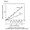
- Figure 2 is a graph showing the results of a simultaneous measurement of CRP and Amyloid A using a fluorescence polarization method at multiple wavelengths.
- the fluorescence polarization method at multiple wavelengths of the present invention it is possible to analyze, i.e., quantify or detect, two or more different assay-objects in a sample in one reaction system based on the above-described principle of the fluorescence polarization method.
- a fluorescent-labeled substance useful in the method of the present invention is provided.
- the specifically-binding substance may be any substance as long as it has a desired binding property with respect to the assay-object, and a functional group which allows for the binding to the fluorochrome.
- the specifically-binding substance is preferably a protein, and more preferably, a protein which is classified as any of antibodies, antigens, receptors or inhibitors.
- An antibody is particularly preferred for its broad spectrum of applications.
- the antibody type includes polyclonal antibodies, a monoclonal antibody, a chimeric antibody, a Fab antibody and a (Fab)2 antibody. Any type of antibody can be applied to the method of the present invention.
- An antigen is typically used in the case where the assay-object is an antibody.
- a receptor can be used in the case where the assay-object acts as a ligand for the receptor.
- An inhibitor can be used, for example, in the case where the assay-object is an enzyme.
- the functional group of the specifically-binding substance is typically a primary, secondary or tertiary amino group, a carboxyl group, a thiol group, a phenyl group, a phenol group or a hydroxyl group.
- a fluorochrome having an activated functional group e.g., a halogenated sulphonyl group, a succinimidized carboxyl group, or an isothiocyanated primary amino group, is desired.
- the number of fluorochrome molecules bound to one molecule of the object to be labeled i.e., the specifically-binding substance
- the specifically-binding substance can be varied arbitrarily. It is preferred in order to increase the detection sensitivity to bind two or more fluorochromes. However, when more fluorochromes than necessary are bound, it may adversely affect a property of the specifically-binding substance. For example, it may reduce the affinity, solubility, or the like, of the antibody. Therefore, the above-described binding number is preferably about 10 or less and, more preferably, about one.
- the present invention utilizes different fluorochromes for different assay-objects in order to analyze two or more different assay-objects in one reaction system.
- the combination of the fluorochromes is preferably selected so that any pair of fluorochromes are different from each other in terms of at least one of excitation wavelength, fluorescence wavelength, and lifetime of fluorescence.
- the selected fluorochromes are preferably different from one another in terms of either or both of excitation wavelength and fluorescence wavelength, particularly, excitation wavelength.
- the excitation wavelength, the fluorescence wavelength, the Stokes shift and the lifetime of the fluorescence are important.
- either or both of the excitation wavelength and the fluorescence wavelength exist in the visible light wavelength range, i.e., 300 nm to 700 nm.
- the difference in wavelength between the excitation wavelength and the fluorescence wavelength, i.e., the Stokes shift is at least 20 nm or more.
- the lifetime of the fluorescence (the fluorescence relaxation time) of the fluorochrome is typically selected from the range of about 0.1 nanoseconds to about 1,000 nanoseconds.
- the change in molecular weight of the fluorescent-labeled substance through the binding to the assay-object is taken into consideration. This is because the degree of polarization of fluorescence emitted from the fluorescent-labeled substance bound to the assay-object is in a proportional relationship with the size of the molecule.
- a fluorochrome having a lifetime of the fluorescence of about 1 to about 15 nanoseconds is preferred.
- examples of such a fluorochrome include dansyl derivatives and fluorescein.
- a fluorochrome having a lifetime of the fluorescence of about 10 nanoseconds to about 150 nanoseconds is preferred.
- Examples of such a fluorochrome include dansyl derivatives and pyrene derivatives.
- a fluorochrome having a lifetime of the fluorescence of about 100 nanoseconds to about 1,000 nanoseconds is preferred.
- Examples of such a fluorochrome include pyrene derivatives and metal complexes.
- fluorochrome examples include fluorochromes having a skeletal structure of rhodamine, pyrene, fluorescein, dialkylaminonaphthalene, dialkylaminonaphthalenesulfonyl, cyanin, or indolenine.
- a particularly preferred fluorochrome may be a fluorochrome having a skeletal structure of fluorescein, dansyl or pyrene.
- the reaction for forming the covalent bond between the specifically-binding substance and the fluorochrome can be carried out according to conditions well known to those skilled in the art.
- the specifically-binding substance has a primary, secondary or tertiary amino group, a carboxyl group, a thiol group, a phenyl group, a phenol group or a hydroxyl group
- the covalent bond can be formed by reacting the specifically-binding substance and the fluorochrome having an activated functional group normally at room temperature for several hours.
- the unreacted fluorochrome can be easily removed by an ordinary method, e.g., gel filtration or dialysis.
- the specifically-binding substance and the fluorochrome can be bound directly or can be bound indirectly via a bifunctional linker molecule, or the like.
- two or more different assay-objects in a sample can be analyzed as follows.
- the sample containing the two or more different assay-objects and the fluorescent-labeled substances are mixed with each other in a solution so as to measure the degree of fluorescence polarization of the fluorescent-labeled substance in the mixed solution. If necessary, the two or more different fluorescent-labeled substances are mixed in a solution, and the degree of fluorescence polarization of each of the fluorescent-labeled substances in the absence of the assay-object is also measured. Any polarization measurement apparatus can be used for measuring the degree of fluorescence polarization. The measurement is performed at a mild temperature (about 10 degrees centigrade (°C) to about 40°C) and, preferably, at a constant temperature.
- a mild temperature about 10 degrees centigrade (°C) to about 40°C
- the measurement of the degree of fluorescence polarization can be performed by measuring the degree after a predetermined time from the mixing of the assay-objects and the fluorescent-labeled substances, or by measuring a change in the degree of fluorescence polarization for a unit of time.
- a measurement at the time when the binding between each assay-object and the corresponding fluorescent-labeled substance has been completely finished more reproducible measurement values are obtained.
- a quicker measurement is possible.
- a standard curve is provided through a measurement of the degree of fluorescence polarization using a solution containing a known concentration of the assay-object so as to compare it with the measurement value for the sample.
- the sample intended to be used with the method of the present invention is a material including two or more assay-objects desired to be analyzed in any fields of art including environmental assay, food control, and medical diagnosis.
- Exemplary samples for environmental assay include materials collected from soil, a river, the air, or the like.
- Exemplary samples for food control include an extract from ground meat, and an extract from a chopping board.
- Exemplary samples for medical diagnosis include body fluids including a blood, a lymph, and a tissue fluid. The sample can be in any form so long as it can be used with the method of the present invention.
- the assay-objects of the method of the present invention include, but are not limited to, a biological substance, a microorganism, a virus, a pharmaceutical, an environmental pollutant and an abused drug.
- the biological substance refers to any organic or inorganic substance existing in the body of a human or other mammal.
- Typical examples of the biological substance include a peptide, a protein, a lipid, a saccharide and a nucleic acid.
- the microorganism includes a bacteria, a fungus and a protozoan.
- the virus includes a bacterial virus, a plant virus and an animal virus.
- the pharmaceutical includes any agent used for treating or diagnosing a human or other mammals. Examples of the pharmaceutical include, but are not limited to, digoxin and cyclopoietin.
- the environmental pollutant includes any substance causing environmental pollution which can be detected from soil, a river, the air, or the like.
- the environmental pollutant examples include, but are not limited to, environmental hormones such as dioxin.
- the abused drug refers to a drug, intake of which by a human is restricted by law or regulation, and which has been used in violation of the restriction. Examples of the abused drug include, but are not limited to, cocaine, methamphetamine, opium and morphine.
- the present invention also provides a kit suitable for use in the above-described method, comprising two or more fluorescent-labeled substances.
- the fluorescent-labeled substances may be contained in a sealed container individually or in combination.
- the fluorescent-labeled substance may be provided in various forms such as a dry form, a solution form wherein the substance is dissolved in a buffer solution, or the like.
- the kit may also include, as necessary, a standard solution containing a known concentration of an assay-object, a diluent, and an instruction manual.
- the present invention further provides a system for analyzing two or more different assay-objects, comprising the above-described fluorescent-labeled substances and a fluorescence polarization measurement apparatus.
- the system may include, as necessary, other substances and apparatuses such as an apparatus for pre-treatment of a sample, a computer for automated analysis of measured data, and the like.
- CG chorionic gonadotropin
- CRP C-reactive protein
- a set of dansyl-labeled anti-CG polyclonal antibodies was prepared as described below, using anti-CG polyclonal antibodies (obtained from Bio Reactive) and dansyl chloride (obtained from Wako Pure Chemical Industries, Ltd.).
- the mixed solution was reacted at about 4° C for about 24 hours while stirring.
- the reacted solution was subjected to Sephadex G-25 gel filtration column (Pharmacia) (size: about 10 ⁇ 60 mm, flow rate: about 2 ml/min). Unreacted dansyl chloride was removed, and fractions containing the dansyl-labeled anti-CG polyclonal antibodies were collected.
- the collected fractions were used to evaluate the labeling amount and the fluorescence property of the prepared dansyl-labeled anti-CG polyclonal antibodies.
- the labeling amount was measured using an ultraviolet/visible spectrometer (manufactured by Shimadzu Corp., UV-1600PC), confirming labeling of about 0.2 dansyl per one molecule of the anti-CG polyclonal antibodies.
- the fluorescence property was measured using a fluorescence spectrometer (manufactured by Shimadzu Corp., RF-5300PC), confirming that the fluorescence property of dansyl bound to the anti-CG polyclonal antibodies was such that the excitation wavelength was about 335 nm, the resulting fluorescence wavelength was about 520 nm, and the lifetime of the fluorescence was about 12 nanoseconds.
- a set of pyrene-labeled anti-CRP polyclonal antibodies was prepared as desoribed below, using an anti-CRP polyclonal antibodies (obtained from Bio Reactive) and succinimidylpyrenebutyrate (SPB) (obtained from Molecular Probes, Inc.).
- PBS phosphate-buffered saline
- SPB dimethyl sulfoxide
- the mixed solution was reacted at room temperature for about 4 hours while stirring.
- the reacted solution was subjected to Sephadex G-25 gel filtration column (Pharmacia) (size: about 10 ⁇ 60 mm, flow rate: about 2 ml/min). Unreacted SPB was removed, and fractions containing the pyrene-labeled anti-CRP polyclonal antibodies were collected.
- the collected fractions were used to evaluate the labeling amount and the fluorescence property of the prepared pyrene-labeled anti-CRP polyclonal antibodies.
- the labeling amount was measured using an ultraviolet/visible spectrometer (manufactured by Shimadzu Corp., UV-1600PC), confirming labeling of about 1.1 pyrenes per one molecule of the anti-CRP polyclonal antibodies.
- the fluorescence property was measured using a fluorescence spectrometer (manufactured by Shimadzu Corp., RF-5300PC), finding that the fluorescence property of pyrene bound to the anti-CRP polyclonal antibodies was such that the excitation wavelength was about 330 nm, the resulting fluorescence wavelengths were about 373 nm and about 397 nm, and the lifetime of the fluorescence was about 60 nanoseconds. Since the fluorescence intensity was greater at about 397 nm, as the measurement conditions to be used with the fluorescence polarization method, it was determined to utilize the excitation wavelength of about 330 nm and the fluorescence wavelength of about 397 nm.
- the amounts of CRP and CG in samples were determined by measuring the fluorescence polarity using the dansyl-labeled anti-CG polyclonal antibodies prepared in Example 1.1 and the pyrene-labeled anti-CRP polyclonal antibodies prepared in Example 1.2.
- a solution (about 350 ⁇ l) containing the pyrene-labeled anti-CRP polyclonal antibodies at about 400 ⁇ g/ml was mixed with another solution (about 350 ⁇ l) containing the dansyl-labeled anti-CG polyclonal antibodies at about 400 ⁇ g/ml.
- the mixed solution was placed into a cuvette (about 5 ⁇ 5 mm) so as to measure the degree of fluorescence polarization.
- the measurement conditions for the pyrene-labeled anti-CRP polyclonal antibodies were as follows: a measurement temperature of about 35° C, an excitation wavelength of about 330 nm, a fluorescence wavelength of about 397 nm and a G factor of about 0.942.
- the measurement conditions for the dansyl-labeled anti-CG polyclonal antibodies were as follows: a measurement temperature of about 35° C, an excitation wavelength of about 330 nm, a fluorescence wavelength of about 520 nm and a G factor of about 1.320.
- Solutions respectively containing 0, 0.1, 0.2, 0.3, 0.4, 0.5, 1, 4, 7, 10, 20, 30, and 50 mg/dl of CRP (obtained from O.E.M. Concepts, Inc.), and other solutions respectively containing 0, 50, 100, 200, 300, 500, 600, 800, and 1000 IU/l of CG (obtained from National Institute of Health Sciences, Japan) were prepared.
- the above-described mixed solution of the labeled antibodies (about 700 ⁇ l) was mixed with each concentration of the CRP solution (about 60 ⁇ l) and each concentration of the CG solution (about 60 ⁇ l). The mixture was stirred at about 35° C and for about 0.5 minute.
- CRP and CG can be respectively measured in a mixed solution containing CRP and CG.
- the change in the degree of fluorescence polarization for CRP was shown to be linear up to a concentration of about 30 mg/dl, whereas that for CG was shown to be linear up to a concentration of about 1000 IU/L.
- Amyloid A molecular weight of about 85,000
- C-reactive protein C-reactive protein
- a set of dansyl-labeled anti-Amyloid A polyclonal antibodies was prepared as described below, using anti-Amyloid A polyclonal antibodies (obtained from Bio Reactive) and dansyl chloride (obtained from Wako Pure Chemical Industries, Ltd.).
- the mixed solution was reacted at about 4° C for about 24 hours while stirring.
- the reacted solution was subjected to Sephadex G-25 gel filtration column (Pharmacia) (size: about 10 ⁇ 60 mm, flow rate: about 2 ml/min). Unreacted dansyl chloride was removed, and fractions containing the dansyl-labeled anti-Amyloid A polyclonal antibodies were collected.
- the collected fractions were used to evaluate the labeling amount and the fluorescence property of the prepared dansyl-labeled anti-Amyloid A polyclonal antibodies.
- the labeling amount was measured using an ultraviolet/visible spectrometer (manufactured by Shimadzu Corp., UV-1600PC), confirming labeling of about 0.26 dansyl per one molecule of the anti-Amyloid A polyclonal antibodies.
- the fluorescence property was measured using a fluorescence spectrometer (manufactured by Shimadzu Corp., RF-5300PC), confirming that the fluorescence property of dansyl bound to the a set of anti-Amyloid A polyclonal antibodies was such that the excitation wavelength was about 335 nm, the resulting fluorescence wavelength was about 520 nm, and the lifetime of the fluorescence was about 12 nanoseconds.
- the amounts of CRP and Amyloid A in samples were determined by measuring the fluorescence polarity using the set of dansyl-labeled anti-Amyloid A polyclonal antibodies prepared in Example 2.1 and the set of pyrene-labeled anti-CRP polyclonal antibodies prepared in Example 1.2.
- a solution (about 350 ⁇ l) containing the pyrene-labeled anti-CRP polyclonal antibodies at about 400 ⁇ g/ml was mixed with another solution (about 350 ⁇ l) containing the dansyl-labeled anti-Amyloid A polyclonal antibodies at about 400 ⁇ g/ml.
- the mixed solution was placed into a cuvette (about 5 ⁇ 5 mm) so as to measure the degree of fluorescence polarization.
- the measurement conditions for the pyrene-labeled anti-CRP polyclonal antibodies were as follows: a measurement temperature of about 35°C, an excitation wavelength of about 330 nm, a fluorescence wavelength of about 397 nm and a G factor of about 0.942.
- the measurement conditions for the dansyl-labeled anti-Amyloid A polyclonal antibodies were as follows: a measurement temperature of about 35° C, an excitation wavelength of about 330 nm, a fluorescence wavelength of about 520 nm and a G factor of about 1.320.
- Solutions respectively containing 0, 0.1, 0.2, 0.3, 0.4, 0.5, 1, 4, 7, 10, 20, 30, and 50 mg/dl of CRP (obtained from O.E.M. Concepts, Inc.), and other solutions respectively containing 0, 1, 2, 3, 5, 7, 8, 12, 15, and 20 mg/dl of Amyloid A (obtained from COSMO BIO CO., Ltd.) were prepared.
- the above-described mixed solution of the labeled antibodies (about 700 ⁇ l) was mixed with each concentration of the CRP solution (about 60 ⁇ l) and each concentration of the Amyloid A solution (about 60 ⁇ l). The mixture was stirred at about 35° C and for about 0.5 minute.
- CRP and Amyloid A can be respectively measured in a mixed solution containing CRP and Amyloid A.
- the change in the degree of fluorescence polarization for CRP showed to be linear up to a aoncentration of about 30 mg/dl, whereas that for Amyloid A showed to be linear up to a concentration of about 15 mg/dl.
- the method of the present invention provides a fluorescence polarization method at multiple wavelengths which allows for an easy, quick and high-accuracy analysis of two or more different assay-objects contained in a sample in one reaction system.
Abstract
Description
- The present invention relates to a fluorescence polarization method at multiple wavelengths for analyzing two or more different assay-objects in a sample in one reaction system. The present invention is particularly useful in the fields of art of environmental assay, food control, and medical diagnosis.
- The fluorescence polarization method is known in the art as a method for assaying a substanoe in a sample. The method is based on the principle that when a fluorescent-labeled compound is excited by linearly polarized light, the fluorescence emitted from the compound has a degree of polarization which is in proportion to the molecular weight thereof.
- As a fluorescence polarization method which has been developed, there is a fluorescence polarization immunossay based on an antigen-antibody reaction.
- For example, U.S. Patent No. 4,902,630 discloses an assay method in which a body fluid (particularly, a blood) containing CRP, an assay-object, is added to a mixed solution which contains: a "tracer" obtained by binding fluorescein, a fluorochrome, to C-reactive protein (CRP); and an antibody which is capable of specifically binding to CRP. CRP in the sample is assayed based on competition between the tracer and CRP for binding the antibody in the mixed solution.
- Japanese Laid-Open Publication No. 3-188374 discloses a method in which a fluorescent-labeled particle comprising an insoluble carrier particle carrying a fluorochrome (or a phosphorescence dye) and an antigen (or an antibody) is reacted with a sample solution containing an antibody (or an antigen). The antibody (or an antigen) in the sample is assayed based on the aggregation of the particles due to an antigen-antibody reaction.
- However, the conventional methods are designed to assay only one substance in a sample, and cannot assay two or more different substances in a sample in one reaction system. Accordingly, there has been a demand in the art for development of a method which is suitable for assaying two or more different substances in a sample in one reaction system.
- According to one aspect of this invention, there is provided a fluorescence polarization method at multiple wavelengths for analyzing two or more different assay-objects in a sample. The method includes the steps of: (a) providing two or more different fluorescent-labeled substances, each being a substance which is capable of specifically binding to respective one of the assay-objects and is covalently bound to a fluorochrome, wherein the fluorochromes of the fluorescent-labeled substances are different from one another; (b) allowing the fluorescent-labeled substances to bind to the two or more different assay-objects, respectively; and (c) measuring a change in the degree of fluorescence polarization which has taken place in each of the fluorescent-labeled substances by its binding to one of the assay-objects.
- In one embodiment of the invention, each of the two or more different assay-objects is independently a biological substance, a microorganism, a virus, a pharmaceutical, an environmental pollutant or an abused drug.
- In one embodiment of the invention, the biological substance is a peptide, a protein, a lipid, a saccharide or a nucleic acid.
- In one embodiment of the invention, the protein is an antibody, a hormone, an inflammation marker, a coagulation factor, an apolipoprotein, a high density lipoprotein (HDL), a low density lipoprotein (LDL), a glycosylated albumin, a glycosylated hemoglobin, a hemoglobin, or an enzyme.
- In one embodiment of the invention, the hormone is chorionic gonadotropin, thyroid-stimulating hormone, progesterone, follicular forming hormone, parathyroid-stimulating hormone, adrenocorticotropic hormone, or insulin.
- In one embodiment of the invention, the inflammation marker is C-reactive protein (CRP), α1-antitrypsin (α1-AT), α1-antichymotrypsin (α1-X), α1-acid glycoprotein (α1-AG), haptoglobin (Hp), ceruloplasmin (Cp), the 9th component of complement (C9), the 4th component of complement (C4), the 3rd component of complement (C3), complement factor B (B), fibrinogen (Fbg), serum amyloid A (SAA), C1 inhibitor (C1I), a sialoglycoprotein, an acid-soluble protein (ASP) or an immunosuppressive acidic protein (IAP).
- In one embodiment of the invention, the microorganism is staphylococcus, Sarcina, Spirillum, Streptococcus, coccobacillus, bacillus, Spirochaeta, tetracoccus, comma bacillus, or Actinomyces.
- In one embodiment of the invention, the specifically-binding substance is a protein which is a antibody, an antigen, a receptor, or an inhibitor.
- In one embodiment of the invention, the antibody is a set of polyclonal antibodies, a monoclonal antibody, a chimeric antibody, a Fab antibody or a (Fab)2 antibody.
- In one embodiment of the invention, the fluorochrome has a functional group which is aapable of binding to a primary, secondary or tertiary amino group, a carboxyl group, a thiol group, a phenyl group, a phenol group or a hydroxyl group.
- In one embodiment of the invention, a lifetime of fluorescence of the fluorochrome is in the range of about 0.1 nanoseconds to about 500 nanoseconds.
- In one embodiment of the invention, the fluorochrome has a skeletal structure of fluorescein, dansyl, pyrene, rhodamine, dialkylaminonaphthalene, dialkylaminonaphthalenesulfonyl, cyanin, or indolenine.
- In one embodiment of the invention, each of the fluorochromes is selected so that the fluorochrome is different from the other fluorochromes in terms of at least one of excitation wavelength, fluorescence wavelength, and lifetime of fluorescence.
- According to another aspect of this invention, there is provided a kit for use in a fluorescence polarization method at multiple wavelengths for analyzing two or more different assay-objects in a sample. The kit includes two or more different fluorescent-labeled substances, each being a substance which is capable of specifically binding to respective one of the assay-objects and is covalently bound to a fluorochrome, wherein the fluorochromes of the fluorescent-labeled substances are different from one another.
- According to still another aspect of this invention, there is provided a system for use in a fluorescence polarization method at multiple wavelengths for analyzing two or more different assay-objects in a sample. The system includes: (a) two or more different fluorescent-labeled substances, each being a substance which is capable of specifically binding to respective one of the assay-objects and is covalently bound to a fluorochrome, wherein the fluorochromes of the fluorescent-labeled substances are different from one another: and (b) means for measuring the degree of fluorescence polarization.
- In the assay system according to the method of the present invention, a change in molecular weight which has taken place in each fluorescent-labeled substance by its binding to an assay-object is measured as a change over time in the molecular orientation. Thus, it is possible to analyze, in a single process, two or more different assay-objects having different molecular weights, by appropriately selecting the types of fluorochromes used as labels in view of the change in molecular weight before and after the binding of fluorochromes.
- Thus, the invention described herein makes possible the advantages of (1) providing a fluorescence polarization method at multiple wavelengths for analyzing two or more different assay-objects contained in a sample in one reaction system by measuring a change in the degree of fluorescence polarization for each of the assay-objects; (2) providing a kit for analyzing two or more different assay-objects contained in a sample by using a fluorescence polarization method at multiple wavelengths: and (3) providing a system for analyzing two or more different assay-objects contained in a sample by using a fluorescence polarization method at multiple wavelengths.
- These and other advantages of the present invention will become apparent to those skilled in the art upon reading and understanding the following detailed description with reference to the accompanying figure.
- Figure 1 is a graph showing the results of a simultaneous measurement of CRP and CG using a fluorescence polarization method at multiple wavelengths.
- Figure 2 is a graph showing the results of a simultaneous measurement of CRP and Amyloid A using a fluorescence polarization method at multiple wavelengths.
- The present invention will be described in greater detail below.
- According to the fluorescence polarization method at multiple wavelengths of the present invention, it is possible to analyze, i.e., quantify or detect, two or more different assay-objects in a sample in one reaction system based on the above-described principle of the fluorescence polarization method.
- By covalently binding the substance capable of specifically binding to the assay-object (hereinafter, referred to as the "specifically-binding substance"), with a fluorochrome, a fluorescent-labeled substance useful in the method of the present invention is provided.
- The specifically-binding substance may be any substance as long as it has a desired binding property with respect to the assay-object, and a functional group which allows for the binding to the fluorochrome. The specifically-binding substance is preferably a protein, and more preferably, a protein which is classified as any of antibodies, antigens, receptors or inhibitors. An antibody is particularly preferred for its broad spectrum of applications. The antibody type includes polyclonal antibodies, a monoclonal antibody, a chimeric antibody, a Fab antibody and a (Fab)2 antibody. Any type of antibody can be applied to the method of the present invention. An antigen is typically used in the case where the assay-object is an antibody. A receptor can be used in the case where the assay-object acts as a ligand for the receptor. An inhibitor can be used, for example, in the case where the assay-object is an enzyme.
- For the fluorochrome, those having a functional group which can be covalently bound to a functional group of the specifically-binding substance are utilized. The functional group of the specifically-binding substance is typically a primary, secondary or tertiary amino group, a carboxyl group, a thiol group, a phenyl group, a phenol group or a hydroxyl group. Especially, in the case where a protein such as an antibody is used as the specifically-binding substance, in terms of the binding efficiency, a fluorochrome having an activated functional group, e.g., a halogenated sulphonyl group, a succinimidized carboxyl group, or an isothiocyanated primary amino group, is desired.
- The number of fluorochrome molecules bound to one molecule of the object to be labeled, i.e., the specifically-binding substance, can be varied arbitrarily. It is preferred in order to increase the detection sensitivity to bind two or more fluorochromes. However, when more fluorochromes than necessary are bound, it may adversely affect a property of the specifically-binding substance. For example, it may reduce the affinity, solubility, or the like, of the antibody. Therefore, the above-described binding number is preferably about 10 or less and, more preferably, about one.
- The present invention utilizes different fluorochromes for different assay-objects in order to analyze two or more different assay-objects in one reaction system. The combination of the fluorochromes is preferably selected so that any pair of fluorochromes are different from each other in terms of at least one of excitation wavelength, fluorescence wavelength, and lifetime of fluorescence. In order to simplify the assay procedure, the selected fluorochromes are preferably different from one another in terms of either or both of excitation wavelength and fluorescence wavelength, particularly, excitation wavelength.
- When selecting the skeletal structure of the fluorochrome to be used, the excitation wavelength, the fluorescence wavelength, the Stokes shift and the lifetime of the fluorescence are important. Preferably, either or both of the excitation wavelength and the fluorescence wavelength exist in the visible light wavelength range, i.e., 300 nm to 700 nm. Preferably, the difference in wavelength between the excitation wavelength and the fluorescence wavelength, i.e., the Stokes shift, is at least 20 nm or more. The lifetime of the fluorescence (the fluorescence relaxation time) of the fluorochrome is typically selected from the range of about 0.1 nanoseconds to about 1,000 nanoseconds. In selecting the lifetime of the fluorescence, the change in molecular weight of the fluorescent-labeled substance through the binding to the assay-object is taken into consideration. This is because the degree of polarization of fluorescence emitted from the fluorescent-labeled substance bound to the assay-object is in a proportional relationship with the size of the molecule.
- Specifically, when the change in the molecular weight is about 5,000 to about 50,000, i.e., when the molecular weight of the assay-object is several thousands to several ten thousands, a fluorochrome having a lifetime of the fluorescence of about 1 to about 15 nanoseconds is preferred. Examples of such a fluorochrome include dansyl derivatives and fluorescein. When the change in the molecular weight is about 50,000 to about 500,000, i.e., when the molecular weight of the assay-object is about several ten thousands to several hundred thousands, a fluorochrome having a lifetime of the fluorescence of about 10 nanoseconds to about 150 nanoseconds is preferred. Examples of such a fluorochrome include dansyl derivatives and pyrene derivatives. When the change in the molecular weight is about 500,000 to about 5,000,000, i.e., when the molecular weight of the assay-object is about several hundred thousands to several millions, a fluorochrome having a lifetime of the fluorescence of about 100 nanoseconds to about 1,000 nanoseconds is preferred. Examples of such a fluorochrome include pyrene derivatives and metal complexes.
- From the above-described points of view, preferred examples of fluorochrome include fluorochromes having a skeletal structure of rhodamine, pyrene, fluorescein, dialkylaminonaphthalene, dialkylaminonaphthalenesulfonyl, cyanin, or indolenine. A particularly preferred fluorochrome may be a fluorochrome having a skeletal structure of fluorescein, dansyl or pyrene.
- The reaction for forming the covalent bond between the specifically-binding substance and the fluorochrome can be carried out according to conditions well known to those skilled in the art. When the specifically-binding substance has a primary, secondary or tertiary amino group, a carboxyl group, a thiol group, a phenyl group, a phenol group or a hydroxyl group, the covalent bond can be formed by reacting the specifically-binding substance and the fluorochrome having an activated functional group normally at room temperature for several hours. After the completion of the reaction, the unreacted fluorochrome can be easily removed by an ordinary method, e.g., gel filtration or dialysis. The specifically-binding substance and the fluorochrome can be bound directly or can be bound indirectly via a bifunctional linker molecule, or the like.
- By using appropriately selected two or more of the above-described fluorescent-labeled substances, two or more different assay-objects in a sample can be analyzed as follows.
- The sample containing the two or more different assay-objects and the fluorescent-labeled substances are mixed with each other in a solution so as to measure the degree of fluorescence polarization of the fluorescent-labeled substance in the mixed solution. If necessary, the two or more different fluorescent-labeled substances are mixed in a solution, and the degree of fluorescence polarization of each of the fluorescent-labeled substances in the absence of the assay-object is also measured. Any polarization measurement apparatus can be used for measuring the degree of fluorescence polarization. The measurement is performed at a mild temperature (about 10 degrees centigrade (°C) to about 40°C) and, preferably, at a constant temperature.
- The measurement of the degree of fluorescence polarization can be performed by measuring the degree after a predetermined time from the mixing of the assay-objects and the fluorescent-labeled substances, or by measuring a change in the degree of fluorescence polarization for a unit of time. By taking a measurement at the time when the binding between each assay-object and the corresponding fluorescent-labeled substance has been completely finished, more reproducible measurement values are obtained. By measuring the ahange in the degree of fluorescence polarization for a unit of time while the binding reaction between each assay-object and the corresponding fluorescent-labeled substance is in progress, on the other hand, a quicker measurement is possible. For the purpose of quantifying the assay-objeot contained in the sample, a standard curve is provided through a measurement of the degree of fluorescence polarization using a solution containing a known concentration of the assay-object so as to compare it with the measurement value for the sample.
- The sample intended to be used with the method of the present invention is a material including two or more assay-objects desired to be analyzed in any fields of art including environmental assay, food control, and medical diagnosis. Exemplary samples for environmental assay include materials collected from soil, a river, the air, or the like. Exemplary samples for food control include an extract from ground meat, and an extract from a chopping board. Exemplary samples for medical diagnosis include body fluids including a blood, a lymph, and a tissue fluid. The sample can be in any form so long as it can be used with the method of the present invention.
- The assay-objects of the method of the present invention include, but are not limited to, a biological substance, a microorganism, a virus, a pharmaceutical, an environmental pollutant and an abused drug.
- The biological substance refers to any organic or inorganic substance existing in the body of a human or other mammal. Typical examples of the biological substance include a peptide, a protein, a lipid, a saccharide and a nucleic acid. The microorganism includes a bacteria, a fungus and a protozoan. The virus includes a bacterial virus, a plant virus and an animal virus. The pharmaceutical includes any agent used for treating or diagnosing a human or other mammals. Examples of the pharmaceutical include, but are not limited to, digoxin and cyclopoietin. The environmental pollutant includes any substance causing environmental pollution which can be detected from soil, a river, the air, or the like. Examples of the environmental pollutant include, but are not limited to, environmental hormones such as dioxin. The abused drug refers to a drug, intake of which by a human is restricted by law or regulation, and which has been used in violation of the restriction. Examples of the abused drug include, but are not limited to, cocaine, methamphetamine, opium and morphine.
- The present invention also provides a kit suitable for use in the above-described method, comprising two or more fluorescent-labeled substances. The fluorescent-labeled substances may be contained in a sealed container individually or in combination. The fluorescent-labeled substance may be provided in various forms such as a dry form, a solution form wherein the substance is dissolved in a buffer solution, or the like. The kit may also include, as necessary, a standard solution containing a known concentration of an assay-object, a diluent, and an instruction manual.
- The present invention further provides a system for analyzing two or more different assay-objects, comprising the above-described fluorescent-labeled substances and a fluorescence polarization measurement apparatus. The system may include, as necessary, other substances and apparatuses such as an apparatus for pre-treatment of a sample, a computer for automated analysis of measured data, and the like.
- The following examples of the invention are intended to illustrate, but not to limit, the present invention.
- Hereinbelow, the results of measurements in one reaction system for two different assay-objects: chorionic gonadotropin (CG; molecular weight of about 37,000) and C-reactive protein (CRP; molecular weight of about 120,000) according to the present invention will be described. F-4000 manufactured by Hitachi Ltd. was used as an apparatus for measuring the degree of fluorescence polarization.
- A set of dansyl-labeled anti-CG polyclonal antibodies was prepared as described below, using anti-CG polyclonal antibodies (obtained from Bio Reactive) and dansyl chloride (obtained from Wako Pure Chemical Industries, Ltd.).
- A solution (about 1000 µl) containing about 2.0 mg/ml of the anti-CG polyclonal antibodies in a phosphate-buffered saline (PBS) (pH: about 7.4) was mixed with a solution (about 20 µl) containing about 1.00 mg/ml of dansyl chloride (10-fold amount of antibodies) dissolved in acetone. The mixed solution was reacted at about 4° C for about 24 hours while stirring. The reacted solution was subjected to Sephadex G-25 gel filtration column (Pharmacia) (size: about 10×60 mm, flow rate: about 2 ml/min). Unreacted dansyl chloride was removed, and fractions containing the dansyl-labeled anti-CG polyclonal antibodies were collected.
- The collected fractions were used to evaluate the labeling amount and the fluorescence property of the prepared dansyl-labeled anti-CG polyclonal antibodies. The labeling amount was measured using an ultraviolet/visible spectrometer (manufactured by Shimadzu Corp., UV-1600PC), confirming labeling of about 0.2 dansyl per one molecule of the anti-CG polyclonal antibodies. The fluorescence property was measured using a fluorescence spectrometer (manufactured by Shimadzu Corp., RF-5300PC), confirming that the fluorescence property of dansyl bound to the anti-CG polyclonal antibodies was such that the excitation wavelength was about 335 nm, the resulting fluorescence wavelength was about 520 nm, and the lifetime of the fluorescence was about 12 nanoseconds.
- A set of pyrene-labeled anti-CRP polyclonal antibodies was prepared as desoribed below, using an anti-CRP polyclonal antibodies (obtained from Bio Reactive) and succinimidylpyrenebutyrate (SPB) (obtained from Molecular Probes, Inc.).
- A solution (about 1000 µl) containing about 2.0 mg/ml of the anti-CRP polyclonal antibodies in a phosphate-buffered saline (PBS) (pH: about 7.4) was mixed with a solution (about 20 µl) containing about 1.29 mg/ml of SPB (5-fold amount of antibodies) dissolved in dimethyl sulfoxide (DMSO). The mixed solution was reacted at room temperature for about 4 hours while stirring. The reacted solution was subjected to Sephadex G-25 gel filtration column (Pharmacia) (size: about 10×60 mm, flow rate: about 2 ml/min). Unreacted SPB was removed, and fractions containing the pyrene-labeled anti-CRP polyclonal antibodies were collected.
- The collected fractions were used to evaluate the labeling amount and the fluorescence property of the prepared pyrene-labeled anti-CRP polyclonal antibodies. The labeling amount was measured using an ultraviolet/visible spectrometer (manufactured by Shimadzu Corp., UV-1600PC), confirming labeling of about 1.1 pyrenes per one molecule of the anti-CRP polyclonal antibodies. The fluorescence property was measured using a fluorescence spectrometer (manufactured by Shimadzu Corp., RF-5300PC), finding that the fluorescence property of pyrene bound to the anti-CRP polyclonal antibodies was such that the excitation wavelength was about 330 nm, the resulting fluorescence wavelengths were about 373 nm and about 397 nm, and the lifetime of the fluorescence was about 60 nanoseconds. Since the fluorescence intensity was greater at about 397 nm, as the measurement conditions to be used with the fluorescence polarization method, it was determined to utilize the excitation wavelength of about 330 nm and the fluorescence wavelength of about 397 nm.
- The amounts of CRP and CG in samples were determined by measuring the fluorescence polarity using the dansyl-labeled anti-CG polyclonal antibodies prepared in Example 1.1 and the pyrene-labeled anti-CRP polyclonal antibodies prepared in Example 1.2.
- A solution (about 350 µl) containing the pyrene-labeled anti-CRP polyclonal antibodies at about 400 µg/ml was mixed with another solution (about 350 µl) containing the dansyl-labeled anti-CG polyclonal antibodies at about 400 µg/ml. The mixed solution was placed into a cuvette (about 5×5 mm) so as to measure the degree of fluorescence polarization. The measurement conditions for the pyrene-labeled anti-CRP polyclonal antibodies were as follows: a measurement temperature of about 35° C, an excitation wavelength of about 330 nm, a fluorescence wavelength of about 397 nm and a G factor of about 0.942. The measurement conditions for the dansyl-labeled anti-CG polyclonal antibodies were as follows: a measurement temperature of about 35° C, an excitation wavelength of about 330 nm, a fluorescence wavelength of about 520 nm and a G factor of about 1.320.
- Solutions respectively containing 0, 0.1, 0.2, 0.3, 0.4, 0.5, 1, 4, 7, 10, 20, 30, and 50 mg/dl of CRP (obtained from O.E.M. Concepts, Inc.), and other solutions respectively containing 0, 50, 100, 200, 300, 500, 600, 800, and 1000 IU/l of CG (obtained from National Institute of Health Sciences, Japan) were prepared. The above-described mixed solution of the labeled antibodies (about 700 µl) was mixed with each concentration of the CRP solution (about 60 µl) and each concentration of the CG solution (about 60 µl). The mixture was stirred at about 35° C and for about 0.5 minute. Then, the degree of fluorescence polarization for CRP and that for CG were measured under the above-described measurement conditions and for about 0.5 minutes, thereby observing changes thereof. The results are shown in Figure 1. The horizontal axis indicates the concentration of each assay-object as added before mixed with the mixed solution of the labeled antibodies.
- It was confirmed that CRP and CG can be respectively measured in a mixed solution containing CRP and CG. The change in the degree of fluorescence polarization for CRP was shown to be linear up to a concentration of about 30 mg/dl, whereas that for CG was shown to be linear up to a concentration of about 1000 IU/L.
- Hereinbelow, the results of measurements in one reaction system for two different assay-objects: Amyloid A ( molecular weight of about 85,000) and C-reactive protein (CRP; molecular weight of about 120,000) according to the present invention will be described. F-4000 manufactured by Hitachi Ltd. was used as an apparatus for measuring the degree of fluorescence polarization.
- A set of dansyl-labeled anti-Amyloid A polyclonal antibodies was prepared as described below, using anti-Amyloid A polyclonal antibodies (obtained from Bio Reactive) and dansyl chloride (obtained from Wako Pure Chemical Industries, Ltd.).
- A solution (about 1000 µl) containing about 2.0 mg/ml of the anti-Amyloid A polyclonal antibodies in a phosphate-buffered saline (PBS) (pH: about 7.4) was mixed with a solution (about 20 µl) containing about 1.00 mg/ml of dansyl chloride (10-fold amount of antibodies) dissolved in acetone. The mixed solution was reacted at about 4° C for about 24 hours while stirring. The reacted solution was subjected to Sephadex G-25 gel filtration column (Pharmacia) (size: about 10×60 mm, flow rate: about 2 ml/min). Unreacted dansyl chloride was removed, and fractions containing the dansyl-labeled anti-Amyloid A polyclonal antibodies were collected.
- The collected fractions were used to evaluate the labeling amount and the fluorescence property of the prepared dansyl-labeled anti-Amyloid A polyclonal antibodies. The labeling amount was measured using an ultraviolet/visible spectrometer (manufactured by Shimadzu Corp., UV-1600PC), confirming labeling of about 0.26 dansyl per one molecule of the anti-Amyloid A polyclonal antibodies. The fluorescence property was measured using a fluorescence spectrometer (manufactured by Shimadzu Corp., RF-5300PC), confirming that the fluorescence property of dansyl bound to the a set of anti-Amyloid A polyclonal antibodies was such that the excitation wavelength was about 335 nm, the resulting fluorescence wavelength was about 520 nm, and the lifetime of the fluorescence was about 12 nanoseconds.
- The amounts of CRP and Amyloid A in samples were determined by measuring the fluorescence polarity using the set of dansyl-labeled anti-Amyloid A polyclonal antibodies prepared in Example 2.1 and the set of pyrene-labeled anti-CRP polyclonal antibodies prepared in Example 1.2.
- A solution (about 350 µl) containing the pyrene-labeled anti-CRP polyclonal antibodies at about 400 µg/ml was mixed with another solution (about 350 µl) containing the dansyl-labeled anti-Amyloid A polyclonal antibodies at about 400 µg/ml. The mixed solution was placed into a cuvette (about 5×5 mm) so as to measure the degree of fluorescence polarization. The measurement conditions for the pyrene-labeled anti-CRP polyclonal antibodies were as follows: a measurement temperature of about 35°C, an excitation wavelength of about 330 nm, a fluorescence wavelength of about 397 nm and a G factor of about 0.942. The measurement conditions for the dansyl-labeled anti-Amyloid A polyclonal antibodies were as follows: a measurement temperature of about 35° C, an excitation wavelength of about 330 nm, a fluorescence wavelength of about 520 nm and a G factor of about 1.320.
- Solutions respectively containing 0, 0.1, 0.2, 0.3, 0.4, 0.5, 1, 4, 7, 10, 20, 30, and 50 mg/dl of CRP (obtained from O.E.M. Concepts, Inc.), and other solutions respectively containing 0, 1, 2, 3, 5, 7, 8, 12, 15, and 20 mg/dl of Amyloid A (obtained from COSMO BIO CO., Ltd.) were prepared. The above-described mixed solution of the labeled antibodies (about 700 µl) was mixed with each concentration of the CRP solution (about 60 µl) and each concentration of the Amyloid A solution (about 60 µl). The mixture was stirred at about 35° C and for about 0.5 minute. Then, the degree of fluorescence polarization for CRP and that for Amyloid A were measured under the above-described measurement conditions and for about 0.5 minutes, thereby observing changes thereof. The results are shown in Figure 2. The horizontal axis indicates the concentration of each assay-objects as added before mixed with the solution of the labeled antibodies.
- It was confirmed that CRP and Amyloid A can be respectively measured in a mixed solution containing CRP and Amyloid A. The change in the degree of fluorescence polarization for CRP showed to be linear up to a aoncentration of about 30 mg/dl, whereas that for Amyloid A showed to be linear up to a concentration of about 15 mg/dl.
- The method of the present invention provides a fluorescence polarization method at multiple wavelengths which allows for an easy, quick and high-accuracy analysis of two or more different assay-objects contained in a sample in one reaction system.
- Various other modifications will be apparent to and can be readily made by those skilled in the art without departing from the scope and spirit of this invention. Accordingly, it is not intended that the scope of the claims appended hereto be limited to the description as set forth herein, but rather that the claims be broadly construed.
Claims (15)
- A fluorescence polarization method at multiple wavelengths for analyzing two or more different assay-objects in a sample, the method comprising the steps of:(a) providing two or more different fluorescent-labeled substances, each being a substance which is capable of specifically binding to respective one of the assay-objects and is covalently bound to a fluorochrome, wherein the fluorochromes of the fluorescent-labeled substances are different from one another;(b) allowing the fluorescent-labeled substances to bind to the two or more different assay-objects, respectively: and(c) measuring a change in the degree of fluorescence polarization which has taken place in each of the fluorescent-labeled substances by its binding to one of the assay-objects.
- A fluorescence polarization method at multiple wavelengths according to claim 1, wherein each of the two or more different assay-objects is independently a biological substance, a microorganism, a virus, a pharmaceutical, an environmental pollutant or an abused drug.
- A fluorescence polarization method at multiple wavelengths according to claim 2, wherein the biological substance is a peptide, a protein, a lipid, a saccharide or a nucleic acid.
- A fluorescence polarization method at multiple wavelengths according to claim 3, wherein the protein is an antibody, a hormone, an inflammation marker, a coagulation factor, an apolipoprotein, a high density lipoprotein (HDL), a low density lipoprotein (LDL), a glycosylated albumin, a glycosylated hemoglobin, a hemoglobin, or an enzyme.
- A fluorescence polarization method at multiple wavelengths according to claim 4, wherein the hormone is chorionic gonadotropin, thyroid-stimulating hormone, progesterone, follicular forming hormone, parathyroid-stimulating hormone, adrenocorticotropic hormone, or insulin.
- A fluorescence polarization method at multiple wavelengths according to claim 4, wherein the inflammation marker is C-reactive protein (CRP), α1-antitrypain (α1-AT), α1-antichymotrypsin (α1-X), α1-acid glycoprotein (α1-AG), haptoglobin (Hp), ceruloplasmin (Cp), the 9th component of complement (C9), the 4th component of complement (C4), the 3rd component of complement (C3), complement factor B (B), fibrinogen (Fbg), serum amyloid A (SAA), C1 inhibitor (C1I), a sialoglycoprotein, an acid-soluble protein (ASP) or an immunosuppressive acidic protein (IAP).
- A fluorescence polarization method at multiple wavelengths according to claim 2, wherein the microorganism is staphylococcus, Sarcina, Spirillum, Streptococcus, coccobacillus, bacillus, Spirochaeta, tetracoccus, comma bacillus, or Actinomyces.
- A fluorescence polarization method at multiple wavelengths according to claim 1, wherein the specifically-binding substance is a protein which is a antibody, an antigen, a receptor, or an inhibitor.
- A fluorescence polarization method at multiple wavelengths according to claim 8, wherein the antibody is a set of polyclonal antibodies, a monoclonal antibody, a chimeric antibody, a Fab antibody or a (Fab)2 antibody.
- A fluorescence polarization method at multiple wavelengths according to claim 1, wherein the fluorochrome has a functional group which is capable of binding to a primary, secondary or tertiary amino group, a carboxyl group, a thiol group, a phenyl group, a phenol group or a hydroxyl group.
- A fluorescence polarization method at multiple wavelengths according to claim 1, wherein a lifetime of fluorescence of the fluorochrome is in the range of about 0.1 nanoseconds to about 500 nanoseconds.
- A fluorescence polarization method at multiple wavelengths according to claim 1, wherein the fluorochrome has a skeletal structure of fluorescein, dansyl, pyrene, rhodamine, dialkylaminonaphthalene, dialkylaminonaphthalenesulfonyl, cyanin, or indolenine.
- A fluorescenoe polarization method at multiple wavelengths according to claim 1, wherein each of the fluorochromes is selected so that the fluorochrome is different from the other fluorochromes in terms of at least one of excitation wavelength, fluorescence wavelength, and lifetime of fluorescence.
- A kit for use in a fluorescence polarization method at multiple wavelengths for analyzing two or more different assay-objects in a sample, the kit comprising two or more different fluorescent-labeled substances, each being a substance which is capable of specifically binding to respective one of the assay-objects and is covalently bound to a fluorochrome, wherein the fluorochromes of the fluorescent-labeled substances are different from one another.
- A system for use in a fluorescence polarization method at multiple wavelengths for analyzing two or more different assay-objects in a sample, the system comprising:(a) two or more different fluorescent-labeled substances, each being a substance which is aapable of specifically binding to respective one of the assay-objects and is covalently bound to a fluorochrome, wherein the fluorochromes of the fluorescent-labeled substances are different from one another; and(b) means for measuring the degree of fluorescence polarization.
Applications Claiming Priority (2)
| Application Number | Priority Date | Filing Date | Title |
|---|---|---|---|
| JP18951298A JP3437094B2 (en) | 1998-07-03 | 1998-07-03 | Multi-wavelength fluorescence polarization method |
| JP18951298 | 1998-07-03 |
Publications (3)
| Publication Number | Publication Date |
|---|---|
| EP0969279A2 true EP0969279A2 (en) | 2000-01-05 |
| EP0969279A3 EP0969279A3 (en) | 2000-05-24 |
| EP0969279B1 EP0969279B1 (en) | 2003-10-29 |
Family
ID=16242521
Family Applications (1)
| Application Number | Title | Priority Date | Filing Date |
|---|---|---|---|
| EP99305267A Expired - Lifetime EP0969279B1 (en) | 1998-07-03 | 1999-07-02 | Fluorescence polarization method at multiple wavelengths |
Country Status (4)
| Country | Link |
|---|---|
| US (1) | US6448018B1 (en) |
| EP (1) | EP0969279B1 (en) |
| JP (1) | JP3437094B2 (en) |
| DE (1) | DE69912361T2 (en) |
Cited By (8)
| Publication number | Priority date | Publication date | Assignee | Title |
|---|---|---|---|---|
| EP1269162A1 (en) * | 2000-02-25 | 2003-01-02 | Cambridge Research & Instrumentation, Inc. | Multiple label fluorescence polarization assay system and method |
| CN103140751A (en) * | 2010-09-30 | 2013-06-05 | 富士胶片株式会社 | Biomolecule detection device and biomolecule detection method |
| US9649061B2 (en) | 2015-03-10 | 2017-05-16 | Becton, Dickinson And Company | Biological fluid micro-sample management device |
| US9693723B2 (en) | 2014-10-14 | 2017-07-04 | Becton, Dickinson And Company | Blood sample management using open cell foam |
| CN109642905A (en) * | 2016-02-05 | 2019-04-16 | 丹纳基因有限公司 | Using the method for the antibody in conjunction with C1 inactivated factor, the antibody in conjunction with c reactive protein and/or the antibody diagnosis in conjunction with complement component C4 and treating cancer |
| US10578606B2 (en) | 2015-09-01 | 2020-03-03 | Becton, Dickinson And Company | Depth filtration device for separating specimen phases |
| CN111052997A (en) * | 2020-02-25 | 2020-04-24 | 青岛农业大学 | Preparation and application method of biological stimulin for improving strawberry continuous cropping obstacle resistance |
| US11298061B2 (en) | 2014-10-14 | 2022-04-12 | Becton, Dickinson And Company | Blood sample management using open cell foam |
Families Citing this family (21)
| Publication number | Priority date | Publication date | Assignee | Title |
|---|---|---|---|---|
| EP1279023B1 (en) * | 2000-03-20 | 2007-02-21 | Analytical Biological Services, Inc. | Method for detecting an analyte by fluorescence |
| JP2002272475A (en) * | 2001-03-22 | 2002-09-24 | Eiken Chem Co Ltd | Method for detecting nucleic acid amplification product by fluorescence polarization |
| WO2003081243A1 (en) * | 2002-03-27 | 2003-10-02 | Matsushita Electric Industrial Co., Ltd. | Fluorescent polarization method, kit used therefor and biosensor |
| KR101006793B1 (en) * | 2005-08-11 | 2011-01-10 | 에스알유 바이오시스템즈, 인코포레이티드 | Grating-based sensor combining label-free binding detection and fluorescence amplification and readout system for sensor |
| US7790406B2 (en) * | 2005-08-11 | 2010-09-07 | Sru Biosystems, Inc | Grating-based sensor combining label-free binding detection and fluorescence amplification and readout system for sensor |
| US9006283B2 (en) | 2007-07-12 | 2015-04-14 | Acumen Pharmaceuticals, Inc. | Methods of modifying amyloid β oligomers using non-peptidic compounds |
| US20110098309A1 (en) * | 2007-07-12 | 2011-04-28 | Acumen Pharmaceuticals, Inc. | Methods of inhibiting the formation of amyloid-beta diffusable ligands using acylhydrazide compounds |
| US8962677B2 (en) * | 2007-07-12 | 2015-02-24 | Acumen Pharmaceuticals, Inc. | Methods of restoring cognitive ability using non-peptidic compounds |
| CA2707309A1 (en) | 2007-12-18 | 2009-06-25 | Acumen Pharmaceuticals, Inc. | Novel addl receptor polypeptides, polynucleotides and host cells for recombinant production |
| EP2536335A1 (en) * | 2010-02-19 | 2012-12-26 | Glysure Ltd | Intravascular glucose sensor |
| AU2011217067A1 (en) * | 2010-02-19 | 2012-09-06 | Lightship Medical Limited | Subcutaneous glucose sensor |
| JP5734091B2 (en) * | 2010-06-04 | 2015-06-10 | 富士フイルム株式会社 | Biomolecule detection apparatus and biomolecule detection method |
| JP2012093338A (en) * | 2010-09-30 | 2012-05-17 | Fujifilm Corp | Biomolecule detection device and biomolecule detection method |
| JP5703126B2 (en) * | 2010-09-30 | 2015-04-15 | 富士フイルム株式会社 | Biomolecule detection apparatus and biomolecule detection method |
| DE102011108180B4 (en) * | 2011-07-20 | 2014-12-24 | Sensor Instruments Entwicklungs- Und Vertriebs Gmbh | Method and apparatus for identifying a photoluminescent material |
| EP2780705B1 (en) | 2011-11-16 | 2018-09-19 | Becton, Dickinson and Company | Methods and systems for detecting an analyte in a sample |
| BR112015010695B1 (en) | 2013-01-11 | 2023-03-07 | Becton, Dickinson And Company | MICROFLUID DEVICE AND METHOD FOR PERFORMING A LIQUID SAMPLE TEST, METHOD FOR FORMING A MICROFLUID DEVICE, SYSTEM AND KIT |
| ES2856191T3 (en) | 2013-11-06 | 2021-09-27 | Becton Dickinson Co | Microfluidic devices and methods of using them |
| BR112016010721B1 (en) | 2013-11-13 | 2021-06-01 | Becton, Dickinson And Company | METHOD AND SYSTEM OF ANALYSIS OF A SAMPLE FOR AN ANALYTE |
| US10379125B2 (en) | 2013-12-27 | 2019-08-13 | Becton, Dickinson And Company | System and method for dynamically calibrating and measuring analyte concentration in diabetes management monitors |
| US9995746B2 (en) | 2014-04-02 | 2018-06-12 | The United States Of America, As Represented By The Secretary Of The Army | Rapid dual direct fluorescent antibody assay for the identification of Bacillus antrhacis |
Citations (4)
| Publication number | Priority date | Publication date | Assignee | Title |
|---|---|---|---|---|
| US5166052A (en) * | 1986-05-27 | 1992-11-24 | Boris Cercek | Method for measuring polarization of bathochromically shifted fluorescence |
| US5210015A (en) * | 1990-08-06 | 1993-05-11 | Hoffman-La Roche Inc. | Homogeneous assay system using the nuclease activity of a nucleic acid polymerase |
| US5302349A (en) * | 1989-06-13 | 1994-04-12 | Diatron Corporation | Transient-state luminescence assay apparatus |
| US5824557A (en) * | 1996-04-02 | 1998-10-20 | Panvera Corporation | Method for detecting and quantitating nucleic acid impurities in biochemical preparations |
Family Cites Families (6)
| Publication number | Priority date | Publication date | Assignee | Title |
|---|---|---|---|---|
| US4902630A (en) | 1985-07-22 | 1990-02-20 | Abbott Laboratories | Fluorescence polarization immunoassy and reagents for measurement of c-reactive protein |
| US4923819A (en) * | 1987-03-27 | 1990-05-08 | Chimerix Corporation | Time-resolved fluorescence immunoassay |
| GB8902689D0 (en) * | 1989-02-07 | 1989-03-30 | Ici Plc | Assay method |
| US5132432A (en) * | 1989-09-22 | 1992-07-21 | Molecular Probes, Inc. | Chemically reactive pyrenyloxy sulfonic acid dyes |
| JP2893772B2 (en) | 1989-12-19 | 1999-05-24 | 三菱化学株式会社 | Immunoassay |
| US5994143A (en) * | 1996-02-01 | 1999-11-30 | Abbott Laboratories | Polymeric fluorophores enhanced by moieties providing a hydrophobic and conformationally restrictive microenvironment |
-
1998
- 1998-07-03 JP JP18951298A patent/JP3437094B2/en not_active Expired - Fee Related
-
1999
- 1999-07-02 EP EP99305267A patent/EP0969279B1/en not_active Expired - Lifetime
- 1999-07-02 DE DE69912361T patent/DE69912361T2/en not_active Expired - Fee Related
- 1999-07-02 US US09/346,817 patent/US6448018B1/en not_active Expired - Lifetime
Patent Citations (4)
| Publication number | Priority date | Publication date | Assignee | Title |
|---|---|---|---|---|
| US5166052A (en) * | 1986-05-27 | 1992-11-24 | Boris Cercek | Method for measuring polarization of bathochromically shifted fluorescence |
| US5302349A (en) * | 1989-06-13 | 1994-04-12 | Diatron Corporation | Transient-state luminescence assay apparatus |
| US5210015A (en) * | 1990-08-06 | 1993-05-11 | Hoffman-La Roche Inc. | Homogeneous assay system using the nuclease activity of a nucleic acid polymerase |
| US5824557A (en) * | 1996-04-02 | 1998-10-20 | Panvera Corporation | Method for detecting and quantitating nucleic acid impurities in biochemical preparations |
Cited By (18)
| Publication number | Priority date | Publication date | Assignee | Title |
|---|---|---|---|---|
| EP1269162A4 (en) * | 2000-02-25 | 2005-01-19 | Cambridge Res & Instrmnt Inc | Multiple label fluorescence polarization assay system and method |
| EP1269162A1 (en) * | 2000-02-25 | 2003-01-02 | Cambridge Research & Instrumentation, Inc. | Multiple label fluorescence polarization assay system and method |
| CN103140751A (en) * | 2010-09-30 | 2013-06-05 | 富士胶片株式会社 | Biomolecule detection device and biomolecule detection method |
| CN103140751B (en) * | 2010-09-30 | 2014-04-09 | 富士胶片株式会社 | Biomolecule detection device and biomolecule detection method |
| US10595762B2 (en) | 2014-10-14 | 2020-03-24 | Becton, Dickinson And Company | Blood sample management using open cell foam |
| US11134875B2 (en) | 2014-10-14 | 2021-10-05 | Becton, Dickinson And Company | Blood sample management using open cell foam |
| US9693723B2 (en) | 2014-10-14 | 2017-07-04 | Becton, Dickinson And Company | Blood sample management using open cell foam |
| US11298061B2 (en) | 2014-10-14 | 2022-04-12 | Becton, Dickinson And Company | Blood sample management using open cell foam |
| US10219731B2 (en) | 2014-10-14 | 2019-03-05 | Becton, Dickinson And Company | Blood sample management using open cell foam |
| US10888261B2 (en) | 2014-10-14 | 2021-01-12 | Becton, Dickinson And Company | Blood sample management using open cell foam |
| US9873117B2 (en) | 2015-03-10 | 2018-01-23 | Becton, Dickinson And Company | Biological fluid micro-sample management device |
| US9649061B2 (en) | 2015-03-10 | 2017-05-16 | Becton, Dickinson And Company | Biological fluid micro-sample management device |
| US10578606B2 (en) | 2015-09-01 | 2020-03-03 | Becton, Dickinson And Company | Depth filtration device for separating specimen phases |
| US11366095B2 (en) | 2015-09-01 | 2022-06-21 | Becton, Dickinson And Company | Depth filtration device for separating specimen phases |
| US11808757B2 (en) | 2015-09-01 | 2023-11-07 | Becton, Dickinson And Company | Depth filtration device for separating specimen phases |
| CN109642905A (en) * | 2016-02-05 | 2019-04-16 | 丹纳基因有限公司 | Using the method for the antibody in conjunction with C1 inactivated factor, the antibody in conjunction with c reactive protein and/or the antibody diagnosis in conjunction with complement component C4 and treating cancer |
| CN109642905B (en) * | 2016-02-05 | 2020-02-07 | 丹纳基因有限公司 | Methods of diagnosing and treating cancer using antibodies that bind to inactivated factor C1, antibodies that bind to C reactive protein, and/or antibodies that bind to complement component C4 |
| CN111052997A (en) * | 2020-02-25 | 2020-04-24 | 青岛农业大学 | Preparation and application method of biological stimulin for improving strawberry continuous cropping obstacle resistance |
Also Published As
| Publication number | Publication date |
|---|---|
| EP0969279B1 (en) | 2003-10-29 |
| JP3437094B2 (en) | 2003-08-18 |
| JP2000019172A (en) | 2000-01-21 |
| DE69912361D1 (en) | 2003-12-04 |
| US6448018B1 (en) | 2002-09-10 |
| EP0969279A3 (en) | 2000-05-24 |
| DE69912361T2 (en) | 2004-08-05 |
Similar Documents
| Publication | Publication Date | Title |
|---|---|---|
| EP0969279B1 (en) | Fluorescence polarization method at multiple wavelengths | |
| EP0957365B1 (en) | Fluorescence polarization method | |
| CN100420947C (en) | Method for quantitative determination of specific analyte with single trapping agent and reagent kit therefor | |
| JP4997222B2 (en) | Multifunctional and configurable assay | |
| JP4138250B2 (en) | How to operate the container | |
| KR0171392B1 (en) | Flow immunosensor method and apparatus | |
| US9274056B2 (en) | Use of non-chelated fluorochromes in rapid test systems | |
| CN105051535A (en) | Systems and methods for determining a chemical state | |
| EP0222341B1 (en) | A method for immunoassay and reagents therefor | |
| KR960029789A (en) | Kits for immunological analysis of in vivo substances and immunological analysis methods | |
| CN110426515A (en) | A kind of time-resolved fluoroimmunoassay chromatographic technique detects kit and its application of dirty underwater trace drugs | |
| US6468763B1 (en) | Optical detection of transmembrane potential changes | |
| Anderson et al. | Raptor: A portable, automated biosensor | |
| US5202269A (en) | Method for immunochemical determination of hapten | |
| WO2007016665A2 (en) | Single use fluorescent assays for determination of analytes | |
| US20040191793A1 (en) | Fluorescent polarization method, kit used therefor and biosensor | |
| Lakowicz et al. | Fluorescence lifetime energy-transfer immunoassay quantified by phase-modulation fluorometry | |
| RU2710262C1 (en) | Biological analysis method | |
| Shriver-Lake et al. | The array biosensor for counterterrorism | |
| US20220221465A1 (en) | Support for fluorescence polarization immunoassay, fluorescence polarization immunoassay kit and fluorescence polarization immunoassay | |
| US20050019844A1 (en) | Method of detecting malignant tumor and detection kit | |
| GB2312746A (en) | Immunoassay for an analyte in a water immiscible solvent | |
| EP0079962A1 (en) | Immunoprecipitation assay. | |
| JP2023176862A (en) | Concentration estimation kit and method for estimating concentration | |
| WO2003060519A1 (en) | Method of assaying agglutination reaction |
Legal Events
| Date | Code | Title | Description |
|---|---|---|---|
| PUAI | Public reference made under article 153(3) epc to a published international application that has entered the european phase |
Free format text: ORIGINAL CODE: 0009012 |
|
| AK | Designated contracting states |
Kind code of ref document: A2 Designated state(s): DE FR GB IT NL |
|
| AX | Request for extension of the european patent |
Free format text: AL;LT;LV;MK;RO;SI |
|
| PUAL | Search report despatched |
Free format text: ORIGINAL CODE: 0009013 |
|
| AK | Designated contracting states |
Kind code of ref document: A3 Designated state(s): AT BE CH CY DE DK ES FI FR GB GR IE IT LI LU MC NL PT SE |
|
| AX | Request for extension of the european patent |
Free format text: AL;LT;LV;MK;RO;SI |
|
| 17P | Request for examination filed |
Effective date: 20001027 |
|
| AKX | Designation fees paid |
Free format text: DE FR GB IT NL |
|
| GRAH | Despatch of communication of intention to grant a patent |
Free format text: ORIGINAL CODE: EPIDOS IGRA |
|
| GRAS | Grant fee paid |
Free format text: ORIGINAL CODE: EPIDOSNIGR3 |
|
| GRAA | (expected) grant |
Free format text: ORIGINAL CODE: 0009210 |
|
| AK | Designated contracting states |
Kind code of ref document: B1 Designated state(s): DE FR GB IT NL |
|
| PG25 | Lapsed in a contracting state [announced via postgrant information from national office to epo] |
Ref country code: NL Free format text: LAPSE BECAUSE OF FAILURE TO SUBMIT A TRANSLATION OF THE DESCRIPTION OR TO PAY THE FEE WITHIN THE PRESCRIBED TIME-LIMIT Effective date: 20031029 |
|
| REG | Reference to a national code |
Ref country code: GB Ref legal event code: FG4D |
|
| REF | Corresponds to: |
Ref document number: 69912361 Country of ref document: DE Date of ref document: 20031204 Kind code of ref document: P |
|
| NLV1 | Nl: lapsed or annulled due to failure to fulfill the requirements of art. 29p and 29m of the patents act | ||
| PG25 | Lapsed in a contracting state [announced via postgrant information from national office to epo] |
Ref country code: GB Free format text: LAPSE BECAUSE OF NON-PAYMENT OF DUE FEES Effective date: 20040702 |
|
| ET | Fr: translation filed | ||
| PLBE | No opposition filed within time limit |
Free format text: ORIGINAL CODE: 0009261 |
|
| STAA | Information on the status of an ep patent application or granted ep patent |
Free format text: STATUS: NO OPPOSITION FILED WITHIN TIME LIMIT |
|
| 26N | No opposition filed |
Effective date: 20040730 |
|
| GBPC | Gb: european patent ceased through non-payment of renewal fee |
Effective date: 20040702 |
|
| PGFP | Annual fee paid to national office [announced via postgrant information from national office to epo] |
Ref country code: DE Payment date: 20060629 Year of fee payment: 8 |
|
| PGFP | Annual fee paid to national office [announced via postgrant information from national office to epo] |
Ref country code: FR Payment date: 20060719 Year of fee payment: 8 |
|
| PGFP | Annual fee paid to national office [announced via postgrant information from national office to epo] |
Ref country code: IT Payment date: 20060731 Year of fee payment: 8 |
|
| PG25 | Lapsed in a contracting state [announced via postgrant information from national office to epo] |
Ref country code: DE Free format text: LAPSE BECAUSE OF NON-PAYMENT OF DUE FEES Effective date: 20080201 |
|
| REG | Reference to a national code |
Ref country code: FR Ref legal event code: ST Effective date: 20080331 |
|
| PG25 | Lapsed in a contracting state [announced via postgrant information from national office to epo] |
Ref country code: FR Free format text: LAPSE BECAUSE OF NON-PAYMENT OF DUE FEES Effective date: 20070731 |
|
| PG25 | Lapsed in a contracting state [announced via postgrant information from national office to epo] |
Ref country code: IT Free format text: LAPSE BECAUSE OF NON-PAYMENT OF DUE FEES Effective date: 20070702 |