EP0910300B1 - Site marking probe - Google Patents
Site marking probe Download PDFInfo
- Publication number
- EP0910300B1 EP0910300B1 EP97907631A EP97907631A EP0910300B1 EP 0910300 B1 EP0910300 B1 EP 0910300B1 EP 97907631 A EP97907631 A EP 97907631A EP 97907631 A EP97907631 A EP 97907631A EP 0910300 B1 EP0910300 B1 EP 0910300B1
- Authority
- EP
- European Patent Office
- Prior art keywords
- probe
- site
- patient
- instrument
- field
- Prior art date
- Legal status (The legal status is an assumption and is not a legal conclusion. Google has not performed a legal analysis and makes no representation as to the accuracy of the status listed.)
- Expired - Lifetime
Links
Images


Classifications
-
- A—HUMAN NECESSITIES
- A61—MEDICAL OR VETERINARY SCIENCE; HYGIENE
- A61B—DIAGNOSIS; SURGERY; IDENTIFICATION
- A61B8/00—Diagnosis using ultrasonic, sonic or infrasonic waves
- A61B8/08—Detecting organic movements or changes, e.g. tumours, cysts, swellings
- A61B8/0833—Detecting organic movements or changes, e.g. tumours, cysts, swellings involving detecting or locating foreign bodies or organic structures
- A61B8/0841—Detecting organic movements or changes, e.g. tumours, cysts, swellings involving detecting or locating foreign bodies or organic structures for locating instruments
-
- A—HUMAN NECESSITIES
- A61—MEDICAL OR VETERINARY SCIENCE; HYGIENE
- A61B—DIAGNOSIS; SURGERY; IDENTIFICATION
- A61B17/00—Surgical instruments, devices or methods, e.g. tourniquets
- A61B17/22—Implements for squeezing-off ulcers or the like on the inside of inner organs of the body; Implements for scraping-out cavities of body organs, e.g. bones; Calculus removers; Calculus smashing apparatus; Apparatus for removing obstructions in blood vessels, not otherwise provided for
- A61B17/22004—Implements for squeezing-off ulcers or the like on the inside of inner organs of the body; Implements for scraping-out cavities of body organs, e.g. bones; Calculus removers; Calculus smashing apparatus; Apparatus for removing obstructions in blood vessels, not otherwise provided for using mechanical vibrations, e.g. ultrasonic shock waves
- A61B17/22012—Implements for squeezing-off ulcers or the like on the inside of inner organs of the body; Implements for scraping-out cavities of body organs, e.g. bones; Calculus removers; Calculus smashing apparatus; Apparatus for removing obstructions in blood vessels, not otherwise provided for using mechanical vibrations, e.g. ultrasonic shock waves in direct contact with, or very close to, the obstruction or concrement
-
- A—HUMAN NECESSITIES
- A61—MEDICAL OR VETERINARY SCIENCE; HYGIENE
- A61B—DIAGNOSIS; SURGERY; IDENTIFICATION
- A61B17/00—Surgical instruments, devices or methods, e.g. tourniquets
- A61B17/34—Trocars; Puncturing needles
- A61B17/3403—Needle locating or guiding means
-
- A—HUMAN NECESSITIES
- A61—MEDICAL OR VETERINARY SCIENCE; HYGIENE
- A61B—DIAGNOSIS; SURGERY; IDENTIFICATION
- A61B18/00—Surgical instruments, devices or methods for transferring non-mechanical forms of energy to or from the body
- A61B18/18—Surgical instruments, devices or methods for transferring non-mechanical forms of energy to or from the body by applying electromagnetic radiation, e.g. microwaves
- A61B18/20—Surgical instruments, devices or methods for transferring non-mechanical forms of energy to or from the body by applying electromagnetic radiation, e.g. microwaves using laser
-
- A—HUMAN NECESSITIES
- A61—MEDICAL OR VETERINARY SCIENCE; HYGIENE
- A61B—DIAGNOSIS; SURGERY; IDENTIFICATION
- A61B34/00—Computer-aided surgery; Manipulators or robots specially adapted for use in surgery
- A61B34/20—Surgical navigation systems; Devices for tracking or guiding surgical instruments, e.g. for frameless stereotaxis
-
- A—HUMAN NECESSITIES
- A61—MEDICAL OR VETERINARY SCIENCE; HYGIENE
- A61B—DIAGNOSIS; SURGERY; IDENTIFICATION
- A61B5/00—Measuring for diagnostic purposes; Identification of persons
- A61B5/06—Devices, other than using radiation, for detecting or locating foreign bodies ; determining position of probes within or on the body of the patient
- A61B5/065—Determining position of the probe employing exclusively positioning means located on or in the probe, e.g. using position sensors arranged on the probe
-
- A—HUMAN NECESSITIES
- A61—MEDICAL OR VETERINARY SCIENCE; HYGIENE
- A61B—DIAGNOSIS; SURGERY; IDENTIFICATION
- A61B5/00—Measuring for diagnostic purposes; Identification of persons
- A61B5/24—Detecting, measuring or recording bioelectric or biomagnetic signals of the body or parts thereof
- A61B5/25—Bioelectric electrodes therefor
- A61B5/279—Bioelectric electrodes therefor specially adapted for particular uses
- A61B5/28—Bioelectric electrodes therefor specially adapted for particular uses for electrocardiography [ECG]
- A61B5/283—Invasive
- A61B5/287—Holders for multiple electrodes, e.g. electrode catheters for electrophysiological study [EPS]
-
- A—HUMAN NECESSITIES
- A61—MEDICAL OR VETERINARY SCIENCE; HYGIENE
- A61B—DIAGNOSIS; SURGERY; IDENTIFICATION
- A61B5/00—Measuring for diagnostic purposes; Identification of persons
- A61B5/68—Arrangements of detecting, measuring or recording means, e.g. sensors, in relation to patient
- A61B5/6846—Arrangements of detecting, measuring or recording means, e.g. sensors, in relation to patient specially adapted to be brought in contact with an internal body part, i.e. invasive
- A61B5/6847—Arrangements of detecting, measuring or recording means, e.g. sensors, in relation to patient specially adapted to be brought in contact with an internal body part, i.e. invasive mounted on an invasive device
- A61B5/6852—Catheters
- A61B5/6853—Catheters with a balloon
-
- A—HUMAN NECESSITIES
- A61—MEDICAL OR VETERINARY SCIENCE; HYGIENE
- A61B—DIAGNOSIS; SURGERY; IDENTIFICATION
- A61B5/00—Measuring for diagnostic purposes; Identification of persons
- A61B5/68—Arrangements of detecting, measuring or recording means, e.g. sensors, in relation to patient
- A61B5/6846—Arrangements of detecting, measuring or recording means, e.g. sensors, in relation to patient specially adapted to be brought in contact with an internal body part, i.e. invasive
- A61B5/6847—Arrangements of detecting, measuring or recording means, e.g. sensors, in relation to patient specially adapted to be brought in contact with an internal body part, i.e. invasive mounted on an invasive device
- A61B5/6852—Catheters
- A61B5/6856—Catheters with a distal loop
-
- A—HUMAN NECESSITIES
- A61—MEDICAL OR VETERINARY SCIENCE; HYGIENE
- A61B—DIAGNOSIS; SURGERY; IDENTIFICATION
- A61B5/00—Measuring for diagnostic purposes; Identification of persons
- A61B5/68—Arrangements of detecting, measuring or recording means, e.g. sensors, in relation to patient
- A61B5/6846—Arrangements of detecting, measuring or recording means, e.g. sensors, in relation to patient specially adapted to be brought in contact with an internal body part, i.e. invasive
- A61B5/6847—Arrangements of detecting, measuring or recording means, e.g. sensors, in relation to patient specially adapted to be brought in contact with an internal body part, i.e. invasive mounted on an invasive device
- A61B5/6852—Catheters
- A61B5/6858—Catheters with a distal basket, e.g. expandable basket
-
- A—HUMAN NECESSITIES
- A61—MEDICAL OR VETERINARY SCIENCE; HYGIENE
- A61B—DIAGNOSIS; SURGERY; IDENTIFICATION
- A61B8/00—Diagnosis using ultrasonic, sonic or infrasonic waves
- A61B8/08—Detecting organic movements or changes, e.g. tumours, cysts, swellings
- A61B8/0833—Detecting organic movements or changes, e.g. tumours, cysts, swellings involving detecting or locating foreign bodies or organic structures
-
- A—HUMAN NECESSITIES
- A61—MEDICAL OR VETERINARY SCIENCE; HYGIENE
- A61B—DIAGNOSIS; SURGERY; IDENTIFICATION
- A61B90/00—Instruments, implements or accessories specially adapted for surgery or diagnosis and not covered by any of the groups A61B1/00 - A61B50/00, e.g. for luxation treatment or for protecting wound edges
- A61B90/36—Image-producing devices or illumination devices not otherwise provided for
-
- A—HUMAN NECESSITIES
- A61—MEDICAL OR VETERINARY SCIENCE; HYGIENE
- A61F—FILTERS IMPLANTABLE INTO BLOOD VESSELS; PROSTHESES; DEVICES PROVIDING PATENCY TO, OR PREVENTING COLLAPSING OF, TUBULAR STRUCTURES OF THE BODY, e.g. STENTS; ORTHOPAEDIC, NURSING OR CONTRACEPTIVE DEVICES; FOMENTATION; TREATMENT OR PROTECTION OF EYES OR EARS; BANDAGES, DRESSINGS OR ABSORBENT PADS; FIRST-AID KITS
- A61F2/00—Filters implantable into blood vessels; Prostheses, i.e. artificial substitutes or replacements for parts of the body; Appliances for connecting them with the body; Devices providing patency to, or preventing collapsing of, tubular structures of the body, e.g. stents
- A61F2/95—Instruments specially adapted for placement or removal of stents or stent-grafts
- A61F2/954—Instruments specially adapted for placement or removal of stents or stent-grafts for placing stents or stent-grafts in a bifurcation
-
- A—HUMAN NECESSITIES
- A61—MEDICAL OR VETERINARY SCIENCE; HYGIENE
- A61M—DEVICES FOR INTRODUCING MEDIA INTO, OR ONTO, THE BODY; DEVICES FOR TRANSDUCING BODY MEDIA OR FOR TAKING MEDIA FROM THE BODY; DEVICES FOR PRODUCING OR ENDING SLEEP OR STUPOR
- A61M25/00—Catheters; Hollow probes
- A61M25/01—Introducing, guiding, advancing, emplacing or holding catheters
- A61M25/0105—Steering means as part of the catheter or advancing means; Markers for positioning
- A61M25/0127—Magnetic means; Magnetic markers
-
- A—HUMAN NECESSITIES
- A61—MEDICAL OR VETERINARY SCIENCE; HYGIENE
- A61M—DEVICES FOR INTRODUCING MEDIA INTO, OR ONTO, THE BODY; DEVICES FOR TRANSDUCING BODY MEDIA OR FOR TAKING MEDIA FROM THE BODY; DEVICES FOR PRODUCING OR ENDING SLEEP OR STUPOR
- A61M25/00—Catheters; Hollow probes
- A61M25/10—Balloon catheters
-
- A—HUMAN NECESSITIES
- A61—MEDICAL OR VETERINARY SCIENCE; HYGIENE
- A61N—ELECTROTHERAPY; MAGNETOTHERAPY; RADIATION THERAPY; ULTRASOUND THERAPY
- A61N7/00—Ultrasound therapy
- A61N7/02—Localised ultrasound hyperthermia
-
- A—HUMAN NECESSITIES
- A61—MEDICAL OR VETERINARY SCIENCE; HYGIENE
- A61B—DIAGNOSIS; SURGERY; IDENTIFICATION
- A61B10/00—Other methods or instruments for diagnosis, e.g. instruments for taking a cell sample, for biopsy, for vaccination diagnosis; Sex determination; Ovulation-period determination; Throat striking implements
- A61B10/02—Instruments for taking cell samples or for biopsy
-
- A—HUMAN NECESSITIES
- A61—MEDICAL OR VETERINARY SCIENCE; HYGIENE
- A61B—DIAGNOSIS; SURGERY; IDENTIFICATION
- A61B17/00—Surgical instruments, devices or methods, e.g. tourniquets
- A61B17/34—Trocars; Puncturing needles
- A61B17/3415—Trocars; Puncturing needles for introducing tubes or catheters, e.g. gastrostomy tubes, drain catheters
-
- A—HUMAN NECESSITIES
- A61—MEDICAL OR VETERINARY SCIENCE; HYGIENE
- A61B—DIAGNOSIS; SURGERY; IDENTIFICATION
- A61B17/00—Surgical instruments, devices or methods, e.g. tourniquets
- A61B17/00234—Surgical instruments, devices or methods, e.g. tourniquets for minimally invasive surgery
- A61B2017/00238—Type of minimally invasive operation
- A61B2017/00243—Type of minimally invasive operation cardiac
- A61B2017/00247—Making holes in the wall of the heart, e.g. laser Myocardial revascularization
-
- A—HUMAN NECESSITIES
- A61—MEDICAL OR VETERINARY SCIENCE; HYGIENE
- A61B—DIAGNOSIS; SURGERY; IDENTIFICATION
- A61B17/00—Surgical instruments, devices or methods, e.g. tourniquets
- A61B2017/00477—Coupling
- A61B2017/00482—Coupling with a code
-
- A—HUMAN NECESSITIES
- A61—MEDICAL OR VETERINARY SCIENCE; HYGIENE
- A61B—DIAGNOSIS; SURGERY; IDENTIFICATION
- A61B17/00—Surgical instruments, devices or methods, e.g. tourniquets
- A61B2017/00681—Aspects not otherwise provided for
- A61B2017/00725—Calibration or performance testing
-
- A—HUMAN NECESSITIES
- A61—MEDICAL OR VETERINARY SCIENCE; HYGIENE
- A61B—DIAGNOSIS; SURGERY; IDENTIFICATION
- A61B17/00—Surgical instruments, devices or methods, e.g. tourniquets
- A61B2017/00831—Material properties
- A61B2017/00876—Material properties magnetic
-
- A—HUMAN NECESSITIES
- A61—MEDICAL OR VETERINARY SCIENCE; HYGIENE
- A61B—DIAGNOSIS; SURGERY; IDENTIFICATION
- A61B17/00—Surgical instruments, devices or methods, e.g. tourniquets
- A61B17/22—Implements for squeezing-off ulcers or the like on the inside of inner organs of the body; Implements for scraping-out cavities of body organs, e.g. bones; Calculus removers; Calculus smashing apparatus; Apparatus for removing obstructions in blood vessels, not otherwise provided for
- A61B17/22004—Implements for squeezing-off ulcers or the like on the inside of inner organs of the body; Implements for scraping-out cavities of body organs, e.g. bones; Calculus removers; Calculus smashing apparatus; Apparatus for removing obstructions in blood vessels, not otherwise provided for using mechanical vibrations, e.g. ultrasonic shock waves
- A61B2017/22005—Effects, e.g. on tissue
- A61B2017/22007—Cavitation or pseudocavitation, i.e. creation of gas bubbles generating a secondary shock wave when collapsing
- A61B2017/22008—Cavitation or pseudocavitation, i.e. creation of gas bubbles generating a secondary shock wave when collapsing used or promoted
-
- A—HUMAN NECESSITIES
- A61—MEDICAL OR VETERINARY SCIENCE; HYGIENE
- A61B—DIAGNOSIS; SURGERY; IDENTIFICATION
- A61B18/00—Surgical instruments, devices or methods for transferring non-mechanical forms of energy to or from the body
- A61B2018/00315—Surgical instruments, devices or methods for transferring non-mechanical forms of energy to or from the body for treatment of particular body parts
- A61B2018/00345—Vascular system
- A61B2018/00351—Heart
- A61B2018/00392—Transmyocardial revascularisation
-
- A—HUMAN NECESSITIES
- A61—MEDICAL OR VETERINARY SCIENCE; HYGIENE
- A61B—DIAGNOSIS; SURGERY; IDENTIFICATION
- A61B34/00—Computer-aided surgery; Manipulators or robots specially adapted for use in surgery
- A61B34/10—Computer-aided planning, simulation or modelling of surgical operations
- A61B2034/107—Visualisation of planned trajectories or target regions
-
- A—HUMAN NECESSITIES
- A61—MEDICAL OR VETERINARY SCIENCE; HYGIENE
- A61B—DIAGNOSIS; SURGERY; IDENTIFICATION
- A61B34/00—Computer-aided surgery; Manipulators or robots specially adapted for use in surgery
- A61B34/20—Surgical navigation systems; Devices for tracking or guiding surgical instruments, e.g. for frameless stereotaxis
- A61B2034/2046—Tracking techniques
- A61B2034/2051—Electromagnetic tracking systems
-
- A—HUMAN NECESSITIES
- A61—MEDICAL OR VETERINARY SCIENCE; HYGIENE
- A61B—DIAGNOSIS; SURGERY; IDENTIFICATION
- A61B34/00—Computer-aided surgery; Manipulators or robots specially adapted for use in surgery
- A61B34/20—Surgical navigation systems; Devices for tracking or guiding surgical instruments, e.g. for frameless stereotaxis
- A61B2034/2072—Reference field transducer attached to an instrument or patient
-
- A—HUMAN NECESSITIES
- A61—MEDICAL OR VETERINARY SCIENCE; HYGIENE
- A61B—DIAGNOSIS; SURGERY; IDENTIFICATION
- A61B90/00—Instruments, implements or accessories specially adapted for surgery or diagnosis and not covered by any of the groups A61B1/00 - A61B50/00, e.g. for luxation treatment or for protecting wound edges
- A61B90/08—Accessories or related features not otherwise provided for
- A61B2090/0807—Indication means
-
- A—HUMAN NECESSITIES
- A61—MEDICAL OR VETERINARY SCIENCE; HYGIENE
- A61B—DIAGNOSIS; SURGERY; IDENTIFICATION
- A61B90/00—Instruments, implements or accessories specially adapted for surgery or diagnosis and not covered by any of the groups A61B1/00 - A61B50/00, e.g. for luxation treatment or for protecting wound edges
- A61B90/36—Image-producing devices or illumination devices not otherwise provided for
- A61B2090/363—Use of fiducial points
-
- A—HUMAN NECESSITIES
- A61—MEDICAL OR VETERINARY SCIENCE; HYGIENE
- A61B—DIAGNOSIS; SURGERY; IDENTIFICATION
- A61B90/00—Instruments, implements or accessories specially adapted for surgery or diagnosis and not covered by any of the groups A61B1/00 - A61B50/00, e.g. for luxation treatment or for protecting wound edges
- A61B90/36—Image-producing devices or illumination devices not otherwise provided for
- A61B90/37—Surgical systems with images on a monitor during operation
- A61B2090/378—Surgical systems with images on a monitor during operation using ultrasound
- A61B2090/3782—Surgical systems with images on a monitor during operation using ultrasound transmitter or receiver in catheter or minimal invasive instrument
- A61B2090/3784—Surgical systems with images on a monitor during operation using ultrasound transmitter or receiver in catheter or minimal invasive instrument both receiver and transmitter being in the instrument or receiver being also transmitter
-
- A—HUMAN NECESSITIES
- A61—MEDICAL OR VETERINARY SCIENCE; HYGIENE
- A61B—DIAGNOSIS; SURGERY; IDENTIFICATION
- A61B90/00—Instruments, implements or accessories specially adapted for surgery or diagnosis and not covered by any of the groups A61B1/00 - A61B50/00, e.g. for luxation treatment or for protecting wound edges
- A61B90/36—Image-producing devices or illumination devices not otherwise provided for
- A61B90/37—Surgical systems with images on a monitor during operation
- A61B2090/378—Surgical systems with images on a monitor during operation using ultrasound
- A61B2090/3782—Surgical systems with images on a monitor during operation using ultrasound transmitter or receiver in catheter or minimal invasive instrument
- A61B2090/3786—Surgical systems with images on a monitor during operation using ultrasound transmitter or receiver in catheter or minimal invasive instrument receiver only
-
- A—HUMAN NECESSITIES
- A61—MEDICAL OR VETERINARY SCIENCE; HYGIENE
- A61B—DIAGNOSIS; SURGERY; IDENTIFICATION
- A61B90/00—Instruments, implements or accessories specially adapted for surgery or diagnosis and not covered by any of the groups A61B1/00 - A61B50/00, e.g. for luxation treatment or for protecting wound edges
- A61B90/36—Image-producing devices or illumination devices not otherwise provided for
- A61B90/37—Surgical systems with images on a monitor during operation
- A61B2090/378—Surgical systems with images on a monitor during operation using ultrasound
- A61B2090/3782—Surgical systems with images on a monitor during operation using ultrasound transmitter or receiver in catheter or minimal invasive instrument
- A61B2090/3788—Surgical systems with images on a monitor during operation using ultrasound transmitter or receiver in catheter or minimal invasive instrument transmitter only
-
- A—HUMAN NECESSITIES
- A61—MEDICAL OR VETERINARY SCIENCE; HYGIENE
- A61B—DIAGNOSIS; SURGERY; IDENTIFICATION
- A61B90/00—Instruments, implements or accessories specially adapted for surgery or diagnosis and not covered by any of the groups A61B1/00 - A61B50/00, e.g. for luxation treatment or for protecting wound edges
- A61B90/39—Markers, e.g. radio-opaque or breast lesions markers
- A61B2090/3904—Markers, e.g. radio-opaque or breast lesions markers specially adapted for marking specified tissue
- A61B2090/3908—Soft tissue, e.g. breast tissue
-
- A—HUMAN NECESSITIES
- A61—MEDICAL OR VETERINARY SCIENCE; HYGIENE
- A61B—DIAGNOSIS; SURGERY; IDENTIFICATION
- A61B90/00—Instruments, implements or accessories specially adapted for surgery or diagnosis and not covered by any of the groups A61B1/00 - A61B50/00, e.g. for luxation treatment or for protecting wound edges
- A61B90/39—Markers, e.g. radio-opaque or breast lesions markers
- A61B2090/3925—Markers, e.g. radio-opaque or breast lesions markers ultrasonic
- A61B2090/3929—Active markers
-
- A—HUMAN NECESSITIES
- A61—MEDICAL OR VETERINARY SCIENCE; HYGIENE
- A61B—DIAGNOSIS; SURGERY; IDENTIFICATION
- A61B90/00—Instruments, implements or accessories specially adapted for surgery or diagnosis and not covered by any of the groups A61B1/00 - A61B50/00, e.g. for luxation treatment or for protecting wound edges
- A61B90/39—Markers, e.g. radio-opaque or breast lesions markers
- A61B2090/3954—Markers, e.g. radio-opaque or breast lesions markers magnetic, e.g. NMR or MRI
- A61B2090/3958—Markers, e.g. radio-opaque or breast lesions markers magnetic, e.g. NMR or MRI emitting a signal
-
- A—HUMAN NECESSITIES
- A61—MEDICAL OR VETERINARY SCIENCE; HYGIENE
- A61B—DIAGNOSIS; SURGERY; IDENTIFICATION
- A61B90/00—Instruments, implements or accessories specially adapted for surgery or diagnosis and not covered by any of the groups A61B1/00 - A61B50/00, e.g. for luxation treatment or for protecting wound edges
- A61B90/39—Markers, e.g. radio-opaque or breast lesions markers
- A61B2090/3983—Reference marker arrangements for use with image guided surgery
-
- A—HUMAN NECESSITIES
- A61—MEDICAL OR VETERINARY SCIENCE; HYGIENE
- A61B—DIAGNOSIS; SURGERY; IDENTIFICATION
- A61B2560/00—Constructional details of operational features of apparatus; Accessories for medical measuring apparatus
- A61B2560/02—Operational features
- A61B2560/0266—Operational features for monitoring or limiting apparatus function
- A61B2560/0276—Determining malfunction
-
- A—HUMAN NECESSITIES
- A61—MEDICAL OR VETERINARY SCIENCE; HYGIENE
- A61B—DIAGNOSIS; SURGERY; IDENTIFICATION
- A61B90/00—Instruments, implements or accessories specially adapted for surgery or diagnosis and not covered by any of the groups A61B1/00 - A61B50/00, e.g. for luxation treatment or for protecting wound edges
- A61B90/36—Image-producing devices or illumination devices not otherwise provided for
- A61B90/361—Image-producing devices, e.g. surgical cameras
-
- A—HUMAN NECESSITIES
- A61—MEDICAL OR VETERINARY SCIENCE; HYGIENE
- A61B—DIAGNOSIS; SURGERY; IDENTIFICATION
- A61B90/00—Instruments, implements or accessories specially adapted for surgery or diagnosis and not covered by any of the groups A61B1/00 - A61B50/00, e.g. for luxation treatment or for protecting wound edges
- A61B90/39—Markers, e.g. radio-opaque or breast lesions markers
-
- A—HUMAN NECESSITIES
- A61—MEDICAL OR VETERINARY SCIENCE; HYGIENE
- A61F—FILTERS IMPLANTABLE INTO BLOOD VESSELS; PROSTHESES; DEVICES PROVIDING PATENCY TO, OR PREVENTING COLLAPSING OF, TUBULAR STRUCTURES OF THE BODY, e.g. STENTS; ORTHOPAEDIC, NURSING OR CONTRACEPTIVE DEVICES; FOMENTATION; TREATMENT OR PROTECTION OF EYES OR EARS; BANDAGES, DRESSINGS OR ABSORBENT PADS; FIRST-AID KITS
- A61F2/00—Filters implantable into blood vessels; Prostheses, i.e. artificial substitutes or replacements for parts of the body; Appliances for connecting them with the body; Devices providing patency to, or preventing collapsing of, tubular structures of the body, e.g. stents
- A61F2/02—Prostheses implantable into the body
- A61F2/04—Hollow or tubular parts of organs, e.g. bladders, tracheae, bronchi or bile ducts
- A61F2/06—Blood vessels
- A61F2002/065—Y-shaped blood vessels
-
- A—HUMAN NECESSITIES
- A61—MEDICAL OR VETERINARY SCIENCE; HYGIENE
- A61M—DEVICES FOR INTRODUCING MEDIA INTO, OR ONTO, THE BODY; DEVICES FOR TRANSDUCING BODY MEDIA OR FOR TAKING MEDIA FROM THE BODY; DEVICES FOR PRODUCING OR ENDING SLEEP OR STUPOR
- A61M25/00—Catheters; Hollow probes
- A61M25/0021—Catheters; Hollow probes characterised by the form of the tubing
- A61M25/0023—Catheters; Hollow probes characterised by the form of the tubing by the form of the lumen, e.g. cross-section, variable diameter
- A61M2025/0025—Catheters; Hollow probes characterised by the form of the tubing by the form of the lumen, e.g. cross-section, variable diameter having a collapsible lumen
-
- A—HUMAN NECESSITIES
- A61—MEDICAL OR VETERINARY SCIENCE; HYGIENE
- A61M—DEVICES FOR INTRODUCING MEDIA INTO, OR ONTO, THE BODY; DEVICES FOR TRANSDUCING BODY MEDIA OR FOR TAKING MEDIA FROM THE BODY; DEVICES FOR PRODUCING OR ENDING SLEEP OR STUPOR
- A61M25/00—Catheters; Hollow probes
- A61M25/01—Introducing, guiding, advancing, emplacing or holding catheters
- A61M25/0105—Steering means as part of the catheter or advancing means; Markers for positioning
- A61M2025/0166—Sensors, electrodes or the like for guiding the catheter to a target zone, e.g. image guided or magnetically guided
-
- A—HUMAN NECESSITIES
- A61—MEDICAL OR VETERINARY SCIENCE; HYGIENE
- A61M—DEVICES FOR INTRODUCING MEDIA INTO, OR ONTO, THE BODY; DEVICES FOR TRANSDUCING BODY MEDIA OR FOR TAKING MEDIA FROM THE BODY; DEVICES FOR PRODUCING OR ENDING SLEEP OR STUPOR
- A61M25/00—Catheters; Hollow probes
- A61M25/10—Balloon catheters
- A61M2025/1043—Balloon catheters with special features or adapted for special applications
- A61M2025/1052—Balloon catheters with special features or adapted for special applications for temporarily occluding a vessel for isolating a sector
-
- A—HUMAN NECESSITIES
- A61—MEDICAL OR VETERINARY SCIENCE; HYGIENE
- A61M—DEVICES FOR INTRODUCING MEDIA INTO, OR ONTO, THE BODY; DEVICES FOR TRANSDUCING BODY MEDIA OR FOR TAKING MEDIA FROM THE BODY; DEVICES FOR PRODUCING OR ENDING SLEEP OR STUPOR
- A61M25/00—Catheters; Hollow probes
- A61M25/0021—Catheters; Hollow probes characterised by the form of the tubing
- A61M25/0023—Catheters; Hollow probes characterised by the form of the tubing by the form of the lumen, e.g. cross-section, variable diameter
-
- A—HUMAN NECESSITIES
- A61—MEDICAL OR VETERINARY SCIENCE; HYGIENE
- A61N—ELECTROTHERAPY; MAGNETOTHERAPY; RADIATION THERAPY; ULTRASOUND THERAPY
- A61N5/00—Radiation therapy
- A61N5/10—X-ray therapy; Gamma-ray therapy; Particle-irradiation therapy
- A61N5/1048—Monitoring, verifying, controlling systems and methods
- A61N5/1064—Monitoring, verifying, controlling systems and methods for adjusting radiation treatment in response to monitoring
Definitions
- the present invention relates to a site marking probe having a sensor for detecting the disposition of the probe or an antenna for transmitting a non-ionising field for, and to marking the position of the probe.
- endoscopes include an elongated body having a distal end and a proximal end.
- the distal end of the probe body can be inserted into the gastrointestinal tract through a body orifice.
- the endoscope may be equipped with optical devices such as cameras or fiber optics to permit observation of the tissues surrounding the distal end, and surgery may be performed by inserting and maneuvering surgical instruments through a channel in the endoscope body.
- probes commonly referred to as laparoscopes and orthoscopes are inserted into the body through small holes formed in surrounding tissues to reach the bodily structures to be treated or measured.
- Still other probes, commonly referred to as catheters can be advanced through the vascular system, as through a vein or artery, or through other bodily passages such as the urinary tract.
- the physician can guide the probe to the desired location within the body by feel or by continuously imaging the probe and the body, as by fluoroscopy, during the procedure.
- the physician can guide the probe based on visual observation of the tissues surrounding the distal tip of the probe.
- this option is available only for probes such as conventional endoscopes which are large enough to accommodate the optical elements.
- optical guidance normally is useful only where the distal tip of the probe is disposed within a cavernous organ; it is not normally useful in guiding the probe within solid or semisolid tissues.
- the position, orientation or both of the distal end of a probe can be determined by using one or more field transducers such as a Hall effect or magnetoresistive device, coil or other antenna carried on the probe, typically at or adjacent the distal end of the probe.
- One or more additional field transducers are disposed outside the body in an external frame of reference.
- the field transducers preferably are arranged to detect or transmit non-ionizing fields or field components such as a magnetic field, electromagnetic radiation or acoustical energy such as ultrasonic vibration.
- the field transducer of the probe allows determination of the position of the probe, such transducer is also referred to as a "position sensor".
- the frame of reference of the external field transducers can be registered with the frame of reference of imaging data such as magnetic resonance imaging data, computerized axial tomographic data, or conventional x-ray image data and hence the position and orientation data derived from the system can be displayed as a representation of the probe superimposed on an image of the patient's body.
- the physician can use this information to guide the probe to the desired location within the patient's body, and to monitor its orientation during treatment or measurement of the body structure.
- This arrangement greatly enhances the ability of the physician to navigate the distal end of the probe through bodily structures. It offers significant advantages over conventional methods of navigating probes by feel alone.
- the transducer-based system also avoids the difficulties associated with navigation of a probe by continuous imaging of the probe and patient during the procedure. For example, it avoids exposure to ionizing radiation inherent in fluoroscopic systems.
- the system can include one or more reference catheters and a mapping/ablation catheter.
- Each of these catheters has a field transducer as discussed above disposed adjacent the distal end of the catheter.
- the mapping/ablation catheter is provided with electrodes for detecting local electrical activity at the distal end of such catheter, and for applying radio frequency energy to ablate surrounding tissue.
- the reference catheters may be positioned with their distal tips at fixed locations within the heart, whereas the mapping/ablation catheter can be moved within the heart while measuring electrical activity. The tip positions of the reference catheter and of the mapping/ablation catheter are monitored in the frame of reference of external antennas.
- mapping/ablation catheter provides a map of the electrical activity of the heart.
- the reference catheter position information can be used to compensate for movement of the heart, and to register the mapping/ablation catheter position data with images such as fluoroscopic or MRI images.
- the map can be used to locate a site within the heart for treatment, and the position information provided by the position sensors can be used to maneuver the mapping/ablation catheter to the treatment site.
- EP-A-0 246 176 teaches a site marking probe according to the preamble of claim 1
- US-A-5 325 873 discloses a site marking probe according to the preamble of claim 2.
- transducer-based probe navigation and treatment systems would be desirable.
- the present invention addresses these needs.
- the present invention is a site marking probe as defined in claim 1 or claim 2.
- the site probe may be provided at a site within the body of a patient and an instrument probe can then be guided to the probe within the body of the patient.
- the probe may transmit one or more fields or detect such transmitted fields.
- the relative disposition of the site probe and the instrument probe is determined from the properties of the detected fields and the instrument probe can then be directed toward the site probe on the basis of the so determined relative disposition.
- disposition refers to the position of the probe, the orientation of the probe, or both.
- relative disposition refers to the direction from one probe to the other, the distance from one probe to the other, or both.
- the direction from one probe to the other, or the distance from one probe to the other can be determined.
- both direction and distance are determined so as to completely determine the relative positions of the two probes.
- the orientations of one or both probes may also be determined.
- magnetic, electromagnetic or acoustic fields may be transmitted between external field transducers and field transducers on the site and instrument probes, so that the position of each such probe is determined in a common frame of reference provided by the external transducers.
- the position of the instrument probe relative to the site probe can be determined by subtracting the position vectors of the two probes in the external antenna frame of reference.
- the site probe may be arranged to emit ultrasonic or electromagnetic radiation.
- the instrument probe may be guided to the site probe by moving it in the direction of increasing field amplitude.
- the instrument probe may be provided with a field transducer capable of detecting the amplitude of such radiation.
- the site probe may include a guidewire or other elongated member and the site probe may emit the field along the length of such elongated member.
- the site probe is placed at a site which requires treatment, testing or another medical procedure.
- the site probe may be placed in or near a lesion during an imaging procedure such as mammography or other x-ray procedures, magnetic resonance imaging or CAT scanning capable of imaging the lesion and the site probe.
- an imaging procedure such as mammography or other x-ray procedures, magnetic resonance imaging or CAT scanning capable of imaging the lesion and the site probe.
- the instrument probe can be guided accurately to the lesion even if the patient's tissues shift or deform.
- a device implanted within the patient for long-term treatment purposes may be provided with a field transducer so that the device can be located and removed.
- the heart pacemaker leads which are implanted within a patient, sometimes break, leaving the distal portion of the lead implanted in the patient.
- Each such lead may be provided with a field transducer before implantation, so that the detached lead portion can be located and removed in the same manner as the lesions discussed above.
- a passageway such as a fistula or shunt can be formed within a bodily structure by placing the site probe at an ending location corresponding to one end of the desired passageway; placing the instrument probe at a starting location corresponding to the opposite end of the desired passageway and then moving the instrument probe through the tissues to be penetrated while guiding the motion of the instrument probe toward the site probe using the detected relative positions of the two probes.
- the site probe serves as a target and the passageway is formed by moving the instrument probe through the tissues toward the target.
- the instrument probe is pointed toward the site probe and a substance or energy capable of destroying tissue is emitted from the instrument probe toward the site probe, thereby boring the hole from the starting location to the ending location.
- the probe can be used to make a intrahepatic portal-systemic shunt, i.e., a shunt from the hepatic vein to the portal vein.
- a shunt i.e., a shunt from the hepatic vein to the portal vein.
- Such shunts have been provided heretofore to relieve blockage of the portal vein arising from cirrhosis or other disease of the liver.
- Such a shunt serves to reduce the elevated venous pressure prevailing in this condition.
- An instrument probe such as a needle is guided from a starting location in the hepatic vein or the portal vein to an ending location in the opposite one of these veins, i.e., from the hepatic vein to the portal vein or from the portal vein to the hepatic vein.
- a site probe as discussed above is positioned at the ending location, i.e., in or near the portal vein or the hepatic vein and the needle or instrument probe is guided toward the ending location on the basis of the relative positions of the site probe and instrument probe.
- the instrument probe position can be determined in a frame of reference such as the frame of reference of external field transducers and the positional information can be registered with a previously acquired image of the liver, so that a representation of the needle can be superimposed on an image of the liver.
- Still further uses of the present invention include methods of treating tissues within the lungs by guiding an instrument probe such as a bronchoscope within the lung using non-ionizing fields transmitted to or from the instrument probe.
- Preferred methods utilize a site probe disposed in the lung tissue at or near the location to be treated, and provide guidance by monitoring the relative position of the instrument probe and site probe.
- Yet another aspect of the present invention includes the step of providing first and second probes, each probe having a field transducer mounted thereon, and determining the relative dispositions of the two probes. Methods preferably further include the steps of determining the relative dispositions of the two probes using non-ionizing fields transmitted to or from the position sensors on the probes.
- a site probe for marking a site within the body of a medical patient.
- a site probe according to the present invention preferably includes a field transducer in the form of a sensor adapted to detect a non-ionizing field such as a magnetic or electromagnetic field or an acoustic field and to provide one or more sensor signals representing one or more properties of the so detected fields.
- the site probe according to the present invention further includes an anchor adapted to fasten the field transducer to tissue within the body of the living patient and signal transmission means for conveying the sensor signals from within the body of the patient to outside of the body of the patient.
- a site probe for marking a site within the body of a patient includes a field transducer which incorporates an antenna or other transducer adapted to transmit a non-ionizing field in response to one or more drive signals, a site probe body housing the device, and an anchor adapted to fasten the site probe body to tissue within the body of the patient.
- a site probe according to this aspect of the present invention includes signal transmission means for transmitting the drive signals from outside the body of the patient to the device while the device is disposed within the body of the patient.
- the anchor may include a mechanical device such as a screw having threads adapted to engage tissue or a pincer incorporating a plurality of flexible or movable times adapted to engage tissue within the patient's body.
- the site probe as described above may be provided in an assembly together with an elongated probe such as a catheter having distal and proximal ends.
- the site probe may be releasably mounted to the elongated probe adjacent the distal end thereof so that the site probe can be advanced within the body of the patient while the proximal end of the elongated probe remains outside of the body, anchored to the patient tissue at a desired location within the body and left within the patient's body.
- the signal transmission means incorporated in the site probe may include one or more leads extending from the sensor towards the proximal end of the elongated probe, these leads being arranged so that the elongated probe can be withdrawn leaving the leads in position after the site probe has been fastened to the tissue by the anchor.
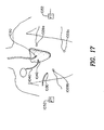
- FIG. 10 through 17 is a fragmentary, diagrammatic view depicting apparatus and portions of a patient during procedures in accordance with a further embodiment of the invention
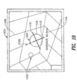
- Fig. 18 is a an elevational view depicting a computer screen display utilized in certain embodiments of the invention.
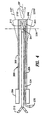
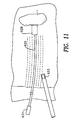
- a site marking probe assembly in accordance with one embodiment of the invention includes an elongated probe in the form of a tube or catheter 20 having a proximal end 22, a distal end 24 and an elongated bore 26 extending between such ends.
- a site probe body 28 incorporating a position sensor or field transducer 30 is physically connected to an anchor in the form of a set of hooks or grapples 32.
- Field transducer 30 is provided in the form of a sensor arranged to detect magnetic or electromagnetic fields.
- the sensor 30 may be a multiaxis, solid-state position sensor of the type disclosed in the aforementioned U.S. Patent 5,558,091.
- Such a sensor incorporates a plurality of transducers sensitive to magnetic field components in mutually orthogonal directions.
- Other suitable position sensors include coils as disclosed in the aforementioned U.S. Patent 5,391,199 and in PCT Application PCT/US95/01103, now published as PCT International Publication WO 96/05768.
- Position sensor or field transducer 30 is connected to leads 34 which extend through bore 26 to and beyond the proximal end 22 of tube 20. Connectors 35 are provided at the proximal ends of leads 34.
- a control rod 36 in the form of a flexible shaft extends axially within bore 26 from outside the proximal end 22 of the tube to the site probe body 28. Control rod 36 may be a braided cable or other flexible element capable of bending and deforming along with tube 20, but also capable of transmitting axial thrust.
- Elongated probe or tube 20 is constructed and arranged to reach within the body of the patient to the desired location.
- tube 20 may have the structure of a conventional catheter, bronchoscope, endoscope, laparoscope or the like.
- the size and shape of tube 20 will depend upon the region of the body to be treated.
- Tube 20 may be steerable or guidable, and may be provided with features as discussed below for selectively bending its distal end.
- Site probe body 28 is releasably engaged within bore 26 adjacent distal end 24 so that the body 28 and hence grapples 32 can be displaced out of the distal end 24 of the tube.
- Grapples 32 are spring-loaded so that they tend to expand to the positions indicated in broken lines at 32' in Fig. 1 when the device is displaced out of the distal end of the tube.
- the spring-loaded grapples 32 may provide frictional engagement with the wall of tube 20 while the device is disposed inside the tube and thus may serve to releasably retain the device in the tube.
- the assembly is advanced until distal end 24 is disposed adjacent a lesion L or other tissue of interest within the patient's body.
- body 28 and anchor or grapples 32 are advanced forwardly by control rod 36, thereby dislodging the device from within tube 20 and engaging grapples 32 with the tissue of lesion L at the distal end of the assembly as indicated at 32'.
- site probe body 28, and hence sensor or field transducer 30 are secured to the tissue of lesion L by the grapples.
- Tube 20 may be removed, leaving control rod 36 and leads 34 in place. During the removal process, the tube 20 slides in a proximal direction over control rod 36, leads 34 and connectors 35, so that the control rod and leads pass out of the distal end 24 of bore 26. To facilitate movement of the control rod and leads through the bore tube, the control rod and the leads may be formed integrally with one another. Also, although only tube leads are depicted in Fig. 1, it should be appreciated that the number of leads will depend upon the configuration of position sensor 30.
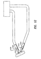
- FIG. 2 An assembly according to a further embodiment of the invention (Fig. 2) incorporates a tube 120, site probe body 128; sensor or field transducer 130; control rod 136 and leads 134 similar to the corresponding elements of the assembly in Fig. 1.
- this assembly includes an anchor in the form of a screw 132 in place of the grapples used in the embodiment of Fig 1.
- control rod 136 is used to turn active element 128 and hence screw 132 while forcing active element out of the interior of tube 120, thereby leaving the active element anchored to the tissue by the screw as indicated in broken lines at 132'.
- the screw or grapples can be replaced by a needle 140 affixed to the active element body.
- the needle is provided with a barb 142.
- the active element body and the anchor are forced distally out of the distal end 124 of the tube so that the anchor engages the tissue.
- Barb 142 holds the active element body and sensor in place.
- Assemblies according to this aspect of the invention may include many different forms of anchors, in addition to the barbs, screws and grapples discussed above.
- Various devices which can be implanted and mechanically engaged in the tissue of a patient are shown in U.S.
- the site probe may incorporate a site probe body in the form of a rigid needle housing the field transducer so that the needle, with the position sensor disposed adjacent the distal end of the needle, can be advanced into the body.
- a needle can be placed by itself, without any external covering, by advancing the needle into the patient's tissues.
- a rigid needle can be provided with anchors as discussed above to hold the needle in place after insertion in a method according to one embodiment of the invention.
- the site probe body can be integrated with the elongated flexible bodies discussed above.
- transducer 30 and grapples 32 (Fig. 1) could be mounted to elongated probe body 20, in which case the elongated probe body would be left in place.
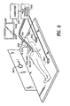
- Apparatus according to the present invention preferably also includes an instrument probe 200 (Fig. 4).
- the instrument probe may incorporate essentially any device which can be inserted or advanced into the body to perform a medical procedure, such as treatment, measurement or observation.
- treatment includes capturing samples of tissues or materials present within the body, and thus includes biopsies.
- instrument probe 200 includes a tubular body 202 having a handle portion 204 affixed to a proximal end of the body and having a distal portion 206 remote from handle 204.
- Body 202 has a bore 208 extending longitudinally from its proximal end to its distal end and open to the outside through handle 204.
- Body 202 may incorporate a flexible section adjacent the distal end, so that the distal end 206 can be bent or pivoted relative to the remainder of the body.
- the body of the instrument probe defines a long axis 237 at end 206, and axes 239 and 241 orthogonal to axis 241.
- Probe 200 may incorporate devices (not shown) for bending the distal end of body 202 relative to the remainder of the body and thereby steer the device as it is advanced into the patient's anatomy. Movement of end 206 around axis 237 is commonly referred to as "roll”, whereas movements of the end around axes 239 and 241, such as encountered during bending of the body or during tilting of the entire device, are referred to as "pitch” and "yaw” respectively.
- Surgical tool 210 may be any conventional surgical tool of the type commonly used in endoscopic, arthroscopic, laparoscopic surgical procedures, or a conventional biopsy sampling device. The tool is arranged so that it can be advanced to an operative position 210' outside of the distal end of body 202. Tool 210 is arranged so that it can be manipulated and controlled from the proximal end or handle 204 of the body.
- the tool is connected to a manipulating handle 212 by conventional control elements or linkages.
- Other expedients for manipulating and controlling a tool at the distal end of body 202 can be employed as, for example, electrical, electronic or optical control linkages.
- tool 210 can be mounted in fixed position on body 202 or formed integrally therewith as, for example, where body 202 is equipped with a cutting blade.
- body 202 may be a biopsy needle of generally conventional construction , or else may be a biopsy needle of the type described in the copending, International Application, entitled “Locatable Biopsy Needle”, naming Biosense, Inc. as an applicant and filed in the Israeli Receiving Office on even date herewith.
- the instrument probe may incorporate a conventional surgical instrument such as a scalpel, forceps, or other instrument having parts which can be advanced into the patient's body to perform a surgical or medical procedure at a place within the patient's body.
- Tool 210 may be a device for measuring or sensing phenomena within the body, such as a thermometer or electrode for measuring intrabody potentials; a device for imaging structures within the body, such as an optical or ultrasonic camera or other imaging device; a device for dispensing medications; a device for applying therapeutic radiation; or any other device which can be used to treat, measure or observe structures within the body of a living subject.
- a field transducer or position sensor 230 is mounted in instrument probe body 202 adjacent the distal end 206 thereof. Transducer 230 may be of the same types as discussed above with reference to the position sensors of the site probes. Transducer 230 is connected via leads 234 to the proximal end or handle 204 of the body. Leads 234 are provided with exposed terminals or connections 235 at the proximal end of the body. If the instrument probe incorporates a rigid or substantially rigid body, the field transducer 230 can be mounted at essentially any location on the body having a defined spatial relationship to the operative portions of the instrument, such that the disposition of the operative portions can be deduced from the disposition of the field transducer.
- the field transducer preferably is mounted adjacent any operative portions of the tool incorporated in the instrument probe, so that the disposition of the operative portions of the tool included in the instrument probe can be deduced from the disposition of the field transducer.
- the apparatus further includes a set of field transducers or antennas 300 mounted in a frame of reference external to the patient.
- field transducers 300 may be mounted to a patient-supporting bed.
- Antennas 300 are linked to a field transmitting and receiving device 302 and a computer 304, which in turn is linked to a displayed device such as a cathode ray tube 306.
- These elements are arranged to cooperate with the field transducers or position sensors on the site probe and on the instrument probe to determine the dispositions of the field transducers on the probes, and hence determine the dispositions of the site probe and the instrument probe in the frame of reference of the external field transducers or antennas.
- each such pair is disposed on the probe and the other element of each such pair is disposed at a known disposition in the external frame of reference.
- at least one element of each transmitter-receiver pair is disposed at a different position or orientation than the corresponding element of the other pairs.
- site probe body 28 is positioned and anchored on a lesion L within the patient's body during a conventional radiologic procedure.
- the site probe assembly is advanced to a lesion observed in such examination, and the site probe body 28 is anchored on the lesion using guidance provided by the imaging procedure.
- the anchoring element such as grapples 32 is actuated to anchor the site probe body in place on the lesion, and the tubular body 20 is withdrawn, leaving leads 34 protruding from the patient's body.
- the patient is positioned in the frame of reference of the external field transducers or antennas 300.
- Leads 34 are connected to the field transmitting and receiving unit 302, thereby connecting the field transducer or position sensor 30 on the site probe to the transmitting and receiving unit.
- the field transducer or position sensor 230 of the instrument probe (Fig. 4) is connected to the transmit/receive unit 302 through leads 234.
- the distal end 206 of the instrument probe is advanced into the patient towards the lesion, carrying the position sensor or field transducer 230 with it.
- the field transmitting and receiving unit 302 and computer 304 actuates external field transducers or antennas 300 and the field transducers or position sensors 30 and 230 of the probes to transmit and receive fields.
- the leads 34 and 234 from the probes will provide sensor signals representing the fields detected at the probes to the field transmit and receive unit.
- leads 34 and 234 can be used to send drive signals to field transducers or position sensors 30, 230 on the probes.
- the computer 304 deduces the disposition of the field transducers on the probes and thus deduces the disposition of the probes themselves in the frame of reference defined by the external field transducers.
- the computer deduces the position of probe 28 and causes the same to be displayed at a location 28' on display unit 306 and likewise deduces the position of the distal end 206 of the instrument probe and displays the same at a location 206" on display 306.
- Display of the two locations provides a visual indication of the distance and direction from the distal end 206 of instrument probe 200 to the site probe body 28. This guides the physician as he or she advances the instrument probe toward the site probe through the patient's tissues.
- the site probe is mounted in the patient's body at or adjacent to the lesion or other tissue to be treated, the actual position of the lesion or tissue will not affect operation of the system.
- the patient can move in the external frame of reference, and the lesion or tissue may move within the patient.
- the lung being treated may be deflated after placement of the site probe but before the other steps of the procedure. Nonetheless, the system will continue to display the correct position of the instrument probe distal end and the site probe, and will continue to provide proper guidance for navigating the instrument probe towards the lesion or other tissue to be treated.
- the terms “navigation” and "navigating” refer to the process of moving a probe within the body of a patient to a desired location.
- the physician may rely on additional information and cues such as his or her knowledge of the anatomy and feel of the instrument probe as the same is advanced through the patient's body. Where conventional visualization devices such as cameras or fiber optic devices are provided on the instrument probe, these may be used to provide additional guidance. Also, the system may augment the display 306 by providing a prominent indication of the direction from the instrument probe distal end to the site probe as, for example, a bold arrow 308 extending in that direction.
- the indicia representing the instrument probe and the site probe on the display may have different characteristics, such as different colors or shapes, so that the physician can readily distinguish them from one another.
- the display need not show any image of the patient's tissues. However, if previously acquired image data is readily available and can be readily registered with the probe position data, the previously acquired image data can be displayed in registration with the indicia representing the probes.
- display unit 306 may incorporate a pair of display screens 352 and 356 disposed in perpendicular planes. Each such display unit may display only components of position in directions parallel to the plane of that screen.
- screen 356 is disposed in a plane perpendicular to the long axis of the patient's body and thus presents an axially directed view showing the representation 206" of the instrument probe tip and the representation 28" of the site probe body in the correct relative positions.
- Screen 352 is disposed in a plane parallel to the longitudinal axis of the patient's body and presents a saggital view of the relative positions.
- Other convenient forms of representing three dimensional information can also be employed.
- the relative positions can be displayed in a stereoscopic imaging device such as a binocular imaging device of the type currently used in "virtual reality" computer graphics applications, in a holographic image or in any other form of three dimensional imaging device.
- a medical procedure as, for example, a procedure to remove the lesion in its entirety or to perform a biopsy on the lesion.
- site probe body 28 is removed. Where the tissue to which the site probe is anchored is cut from the patient during the procedure, the site probe can simply be pulled out of the patient by means of the control rod 36 (Fig. 1).
- the anchoring device used in the site probe may permit removal of the site probe from the tissue without removing the tissue.
- screw 132 (Fig. 2) can be released from the tissue by turning the associated control rod 136.
- a grapple arrangement may be provided with articulating elements which can be controlled by operation through the control rod, so that the grapple acts as a controllable pincer. In this event, the site probe can be released from the tissue by actuating the grapple to release the tissue.
- Computer 304 can calculate the relative positions of the instrument probe distal end 206 and site probe body 28 by subtracting the positions of the two probes. That is, the system may substract the coordinates of the site probe distal end in the external frame of reference defined by external field transducers 300 from the coordinates of the site probe body 28 in the same frame of reference to arrive at the components of the relative position vector from the instrument probe distal end to the site probe body.
- the relative positions can be provided as a human perceptible indication other than a visual display as, for example, a tactile display or one or more audible signals provided to the physician during navigation of the instrument probe.
- the calculated relative position can be displayed numerically as well as graphically as, for example, by displaying the individual components of the relative position vector. Also, where the instrument probe is manipulated by automated equipment, the relative position can be provided as vector coordinates or any other convenient form to the automated equipment so as to control the movement of the instrument probe toward the site probe automatically.
- a reference probe 328 having a reference probe field transducer or position sensor 330 thereon may be used in conjunction with site probe 28.
- the reference probe may be positioned in the patient's body adjacent the site probe during the procedure used to place the site probe.
- the position monitoring system detects the position of the reference probe in the same manner as it detects the positions of the site probe and instrument probe. Any substantial change in the relative positions of the site probe and reference probe indicates that one or the other of these probes has become dislodged from the tissue to which it is anchored.
- the system may be arranged to issue an automatic warning to the physician, such as a warning tone or visual indication upon occurrence of this condition.
- the relative disposition of the site probe and reference probe may be recorded during the placement step as, for example, from x-ray or other image data acquired during the placement procedure.
- This prerecorded disposition data can be compared to the relative disposition of the site probe and the reference probe acquired from the field transducers.
- any substantial change in the distance between the site probe and the reference probe indicates that one of the probes has become dislodged.
- a site probe 428 may include a field transducer in the form of an inductive antenna 430 linked to a capacitor 431 to form a resonant circuit.
- the external field transducers may apply drive signals in the form of alternating electromagnetic fields to the site probe at the resonant frequency of the circuit in the site probe.
- the site probe will then radiate electromagnetic fields at the same frequency.
- Such an arrangement can be used, for example, in a time-multiplex system. During some intervals, the external field transducers are actuated to drive the system.
- a site probe may incorporate a small biocompatible metal pellet 450 with a field transducer in the form of a magnetic metallic element such as 460 disposed therein.
- a field transducer in the form of a magnetic metallic element such as 460 disposed therein.
- a pellet will emit a field at the same frequency but out of phase with the external field, thereby altering the phase of the field in the vicinity of the pellet.
- the degree of phase alteration varies with distance from the pellet over a small region surrounding the pellet.
- the field transducer of the instrument probe may detect the alternating field, and the phase of the field may be used as an indication of distance from the site probe.
- Site probes of this nature may be used to mark multiple locations within the body. Such probes can be applied by injection using a syringe and can be fixed in place using a biocompatable adhesive. Site probes which are powered by radiated fields, such as those discussed above with reference to Figs. 9 and 9A, are particularly useful for long-term implantation. Provided that the site probe remains in place, the instrument probe can be navigated back to the same site even after a long time has elapsed, and even if the site has moved within the body due to growth, healing or other long-term processes.
- Still other arrangements can use a self-powered site probe, incorporating a storage battery or other source of energy and an internal field generating device such as an oscillator linked to an antenna.
- Battery-powered site field transducers are particularly useful for applications where the field transducer must be active only for a short time as, for example, where a site probe is used to mark a lesion which will be removed promptly after the field transducer is activated.
- a battery-powered field transducer may be installed in a long-term application and activated by an externally-applied signal or internal occurrence. More complex forms of such radiating devices can incorporate multiple field transducers or antennas in orthogonal directions for radiating multiple fields. These may be operated at different frequencies or according to a time division multiplexing scheme. Similar arrangements can be used for the instrument probe.
- field transducers may be arranged to radiate acoustic fields.
- Optical radiation such as visible or infrared light, may also be employed.
- Many tissues within the body are translucent at red and infrared wavelengths, so that the intensity of optical radiation emitted from a field transducer such as a light emitting diode can be used as an indication of distance from that transducer.
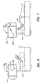
- the site probe body has a field transducer in the form of a permanent magnet 530 mounted therein.
- the instrument probe has a field transducer 531 which is adapted to measure constant magnetic field components in a plurality of orthogonal directions in a frame of reference fixed to the distal end 506 of the site probe body.
- field transducer or position sensor 531 may include a plurality of magnetoresistive, Hall effect or other similar solid state magnetic field transducers, each such transducer being sensitive to a field component in a given local direction relative to the instrument probe 506.
- the signals from the field transducer 531 represent the components of a vector 533 pointing from the distal end 506 of the instrument probe along the lines of magnetic flux impinging on the distal end.
- the distal end 506 is located in a region reasonably close to one of the poles of permanent magnet 530, the vector along the lines of flux points generally in the direction of site probe body 528.
- the display (not shown) may display this information as a direction vector.
- the physician must interpret this information as a direction relative to the end of the probe. For example, in using a steerable probe of the type discussed above, the physician can swing the distal end of the probe in various directions and find the direction in which the direction vector points straight ahead from the distal end of the instrument probe. The physician may then advance the instrument probe and repeat the process. In this way, the instrument probe "homes in" on the site probe.
- a site probe includes a probe body 628 with an elongated shaft 629.
- the field transducer in this site probe may be an ultrasonic transducer disposed in a handle 631 at the proximal end of shaft 629. Ultrasonic energy supplied by the field transducer is emitted from the site probe along essentially the entire length of the shaft, thus creating a field of ultrasonic energy surrounding the shaft. The field has progressively diminishing intensity in directions away from the shaft.
- the instrument probe has a field transducer or position sensor 633 in the form of a microphone or other transducer sensitive to ultrasonic energy.
- the monitoring system is arranged to provide a signal, such as an audible signal having intensity directly related to the intensity of ultrasonic energy impinging on the field transducer or microphone 633.
- a signal such as an audible signal having intensity directly related to the intensity of ultrasonic energy impinging on the field transducer or microphone 633.
- the system indicates the distance from the tip of the instrument probe to the shaft 629 of the site probe.
- the physician can use this information to guide the instrument probe into proximity with shaft 629 and to maintain the tip of the instrument probe in proximity with the shaft as the instrument probe is advanced towards the site probe body 629.
- the ultrasonic energy is emitted only from site probe body 628 itself, so that the ultrasonic field takes the form of a generally spherical field, with the intensity progressively diminishing in all directions away from the probe body.
- the sensed intensity represents distance from the site probe body 628.
- This information can be used to guide the instrument probe as, for example, by moving the instrument probe in various directions and detecting which direction of movement results in the greatest increase in intensity.
- Similar approaches can be used with magnetic and electromagnetic signals radiated from a site probe.
- the opposite approach in which an acoustic or electromagnetic field is radiated from the instrument probe and the intensity of the field impinging on the site probe is monitored, may also be employed.
- the signals from the field transducer or sensor on the probe which acts as the signal receiver indicate the distance between the probes and thus provide information concerning the disposition of the two probes relative to one another.
- information concerning the relative dispositions of a plurality of probes can be used to coordinate the action of the plural probes and to guide one or more of the probes, regardless of whether any of the probes is fixed to the patient's body.
- two instrumented catheters 690 and 694 are coordinated so that their distal ends are juxtaposed with one another.
- positional information derived by position sensors 630 and 632 adjacent the distal tips of catheter 690 and 694 is used by the physician to navigate both catheters into position adjacent a common treatment location 692.
- the position sensors 630 and 632 provide information concerning the orientations of both catheters relative to one another and relative to the common treatment location 692, so that the tips of both catheters can be aimed onto the common treatment location.
- the disposition-determining systems discussed above with reference to the instrument probes and site probes can be used to provide information concerning the relative dispositions of multiple probes either by measuring the disposition of each probe in an external frame of reference or by monitoring fields transmitted between field transducers on the multiple probes and determining relative dispositions directly from such monitored fields. It is useful to coordinate the actions of multiple catheters in catheter-based surgical procedures as, for example, where multiple different tools must be brought to a common location so that the common location can be treated by all of the tools.
- one or more of the catheters may carry devices for observing the treatment as, for example, optical or ultrasonic viewing equipment, whereas the other catheters may carry devices for manipulating, cutting or excavating tissues.
- the tissues can be cut or "excavated” by action of a laser beam directed out of a catheter tip or, alternatively, by applying small gas bubbles ("microbubbles") and causing rupture of the same by applying ultrasonic energy to the area infused with microbubbles.
- Coordinated catheters can be used in such surgical operations as, for example, by applying the laser light or microbubbles through one catheter and observing the process through another catheter.
- probes 702 and 704 lie on opposite sides of a bodily structure, but are aligned and aimed towards one another using positional information derived from position sensors or field transducers carried on the probes. Probes pointing towards one another can be used for a variety of purposes.
- probe 702 may transmit ultrasound to a detector on probe 704 and a detector on probe 704 may be actuated to generate an ultrasonic image of the tissues between the probes.
- probes which are initially disposed at a distance from one another can be aligned with one another and then one or both of the probes can be advanced towards the other probe, so that the probes are brought together in much the same manner as the site probe and instrument probe discussed above. In this arrangement, however, neither probe is fixed to the body tissue during the procedure.
- two or more probes can be coordinated by engaging both probes with related body structures or with spaced-apart locations in a body structure.
- a first probe is disposed within an intrabody lumen such as a vascular structure
- the second probe may be disposed in another part of the same lumen.
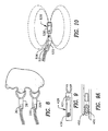
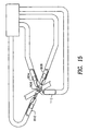
- a first probe in the form of a catheter 720 may be threaded through the vascular system and positioned in an artery within the brain upstream from an aneurysm in the brain.
- Catheter 720 may incorporate a first field transducer or position sensor 722 adjacent its distal end.
- the step of positioning catheter 720 may be conducted using conventional imaging techniques such as fluoroscopic guidance. Alternatively or additionally, the techniques disclosed in the '091 Patent may be used. Thus, the disposition of field transducer 722 may be detected by means of fields transmitted to or from one or more additional field transducers, the detected disposition is correlated to the frame of reference of previously-acquired imaging data, and a representation of the catheter tip is superposed on a display showing the image from the previously-acquired data.
- a first medical procedure is performed by inflating a balloon 726 adjacent the distal end of the catheter so as to block the blood supply to the artery at a first location upstream from the aneurysm A.
- a second probe in the form of a second catheter 726 having a field transducer 728 adjacent its distal end is advanced into the same artery through the surrounding brain tissue from outside of the artery at a second location downstream from the first location.
- the second probe is then used to place a stent or perform other treatment at the second location.
- information concerning the relative disposition of the first and second probes can be used to position the second probe at the desired location with respect to the first probe, and thereby position the second probe at the desired location with respect to the artery and aneurysm.
- the relative disposition information can be used in conjunction with other sources of disposition information.
- information concerning the disposition of second probe distal end or field transducer 728 relative to first probe distal end or field transducer 722 can be used in conjunction with a superposition scheme as described in the '091 patent, in which a representation of the second probe distal end is displayed in registration with a previously-acquired image.
- the superposition scheme can be used to guide the second probe around structures such as critical areas of the brain far from the first probe, whereas the relative position information can be used to bring the second probe to a precise placement.
- the relative disposition information can be combined with direct image guidance in similar manner. Use of relative disposition information in conjunction with other information can be adapted to procedures in other regions of the body.
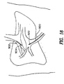
- FIG. 15 Other procedures wherein plural probes are placed on separated but functionally related sites include placement of two probes on separate points along a nerve for stimulation and measurement of nerve impulse travel, and placement of an infusion catheter along a blood vessel and a sampling catheter in the vascular bed served by that blood vessel. As depicted in Fig. 15, more than two probes can be coordinated in a similar fashion.
- catheter 804 is excavating tissue at a location 806, whereas catheter 808 is removing the debris from such location.
- Catheter 810 is viewing the tissue surrounding location 806 using an ultrasonic imaging system carried on the catheter, whereas catheter 112 is injecting microbubbles into the vascular bed at location 106 to enhance the contrast between various types of tissues at such location.
- each probe need only accommodate one or a few devices.
- the individual probes may be simpler and smaller in size than a composite probe incorporating all of the required devices.
- the preferred field transducers and probes utilized in accordance with the present invention desirably are small-diameter devices to facilitate insertion into the body.
- each field transducers desirably has a smallest dimension less than about 3mm, preferably less than 2mm, more preferably less than about 1mm; still more preferably less than about 0.2 mm and most preferably even smaller.
- the probe itself desirably has dimensions, at the field transducer, in the same ranges.
- liver bypass Patients who have advanced chirosis of the liver suffer, as a result of blockage of the portal vein, from elevated venous blood pressure, which may cause fatal GI bleeding.
- a shunt is created between the hepatic vein and the portal vein in the liver to bypass most of the liver.
- the venous blood pressure is reduced and GI bleeding eliminated.
- a catheter and guidewire are inserted through the jugular vein into the hepatic vein, and a needle is passed along the guidewire and used to probe for the portal vein.
- the needle is forcibly advanced through the liver tissue towards the portal vein. This entails considerable difficulty if the liver tissue is toughened or scarred as occurs in some diseases. Since the needle is hollow, when the other portal is found, blood flows through the needle.
- a catheter may replace the needle, so that the catheter extends between the veins.
- a stent such as an inflatable stent is guided along the needle or catheter to form a permanent passageway connecting the two veins.
- the opposite procedure wherein entry is made from the portal vein and the needle is passed through the liver tissue to the hepatic vein, can also be employed.
- This procedure is performed using a fluoroscope and is very lengthy, so the amount of radiation exposure of the patient and the surgeon is considerable.
- such a procedure can be greatly facilitated by using non-ionizing radiation transmitted to field transducer on a needle or other probe used to form the passageway, and determining the disposition of the probe during the procedure using such non-ionizing radiation.
- a needle 902 with a field transducer or position sensor 904 thereon is introduced into the hepatic vein.
- a site probe 920 having a position sensor or field transducer 922 thereon is disposed in the liver parenchyma adjacent to the portal vein. The needle is guided toward the portal vein by monitoring the relative dispositions of the needle and marker catheter in the manner discussed above while the needle is advanced through the liver tissue.
- the position of a needle with a position sensor thereon is monitored and the position information is registered with a previously-acquired image of the patient such as a CT image made using an intravenous contrast medium.
- a previously-acquired image of the patient such as a CT image made using an intravenous contrast medium.
- such registration can be achieved by means of fiducial markers imaged along with the patient.
- a representation of the needle tip is superposed on the displayed image, thereby allowing the physician to guide the needle from the hepatic vein toward the portal vein or vice-versa.
- passage of the needle can be facilitated by destroying the intervening tissue using a laser or by instilling microbubbles into the tissue ahead of the needle and applying focused ultrasound to destroy the tissue.
- the needle may be replaced by a flexible device such as a catheter.
- numerous site probes 1028a-1028c with field transducers thereon can be disposed at distant locations within the body of the patient to guide one or more instrument probes, also equipped with field transducers, 1030 to any of these locations.
- the numerous probes can also be employed in conjunction with additional field transducers disposed outside of the body, such as reference field transducers 1032 disposed in a fixed frame of reference.
- reference field transducers 1032 act as signal transmitters
- the field transducers on all probes 1028 and 1030 act as receivers.
- the field transducers act as transmitters.
- Various multiplexing and signal separation arrangements can be used to avoid interference between the field transducers associated with the multiple probes.
- field transducers associated with each of the site probes 1028 may be low-powered transmitting devices such as the re-radiating devices discussed above with reference to Figs 9 and 9A, and the field transducer of the instrument probes 1030 may be relatively insensitive receivers, or vice-versa, so that each instrument probe will interact with the closest site probe but will not interact with other site probes.
- Frequency-division multiplexing, code diversity multiplexing and time division multiplexing, as well as combinations of these multiplexing schemes may be employed.
- the various reference field transducers 1032 may transmit at different frequencies, whereas each of the field transducers 1028a, 1028b and 1028c of the site probes may transmit at other frequencies, these frequencies being different from one another.
- the various field transducers used in a single procedure may use different types of fields.
- the field transducers associated with some of the probes may be transmit or receive optical or acoustic fields, whereas others may transmit or receive magnetic or electromagnetic fields.
- the same field transducer of the instrument probe 1030 may be used both to establish disposition relative to site probes 1028 and to establish position in the frame of reference of reference transducers 1032 as, for example, for use in displaying position of the instrument probe superposed on an image of the patient.
- cardiac pacemaker lead 1040 is provided with a field transducer 1042 adjacent the distal tip of the lead.
- the cardiac pacemaker lead is implanted for long-term use in the conventional manner.
- transducer 1042 is arranged so that it can operate without a lead-supplied power source.
- transducer 1042 may be a permanent magnet or reradiating field transducer as discussed above.
- an instrument probe may be brought into proximity with the distal end and used to remove the distal end.
- the same techniques can be used to mark other implanted devices such as orthopedic implants, and tools which are not deliberately implanted but which may be accidentally lost during surgery.
- a preferred screen display for indicating the relative dispositions of probes is depicted in Fig. 18. This may be used in place of the arrow 306 discussed above with reference to Fig. 5.
- a display device such as a computer screen 1100 shows a representation 1102 of a site probe disposed within the patient and a representation 1104 of an instrument probe being guided toward the patient.
- both of these representations are displayed against a background of grid lines 1106 representing Cartesian (x-y-z) coordinates of a reference coordinate system such as the coordinate system of the external field transducers.
- the site probe representation 1102 may be in the form of a sphere or other arbitrary shape.
- Instrument probe representation 1104 includes a sighting aperture 1108 in the form of a circle or other closed or semi-closed geometric figure, as well as crossing lines 1110 and 1112 representing directions perpendicular to the a preselected axis of the instrument probe, preferably the long axis of the probe at the tip of the probe.
- the directions of lines 1110 and 1112 may correspond to the pitch and yaw axes 239 and 241 (Fig. 4) of the instrument probe. These directions are fixed with respect to the instrument probe distal tip, so that when the instrument probe rolls around its long axis, the directions of lines 1110 and 1112 move in the same manner.
- One end 1112a of line 1112 has a different appearance from the other lines so that the user can track the roll orientation of the instrument probe tip visually.
- Pitch and yaw movements of the instrument probe distal tip, around axes 239 and 241, are shown by movement of the coordinate system and site probe representation relative to the screen and relative to the instrument probe representation 1104.
- the view of grid lines 1106 representing the coordinate system, as well as the view of site probe representation 1102 which is displayed on the screen is a projection in a plane perpendicular to the long axis of the instrument probe at the tip of the probe.
- the physician can point the instrument probe long axis or roll axis 237 (Fig. 4) directly at the site probe. Advancement of the instrument probe in this condition will bring the instrument probe directly towards the site probe. The physician can use the displayed information to navigate the instrument probe to one side of the site probe.
- the instrument probe representation has a fixed size, whereas the site probe representation grows as the distance decreases and shrinks as the distance decreases.
- the site probe representation completely fills circle 1108 when the tip is at the site probe.
- Other representations of distance such as alphanumeric display 1114 and bar graph 1116 are also provided.
- the invention can be used in medical and related procedures.
Abstract
Description
- The present invention relates to a site marking probe having a sensor for detecting the disposition of the probe or an antenna for transmitting a non-ionising field for, and to marking the position of the probe.
- Conventional surgical procedures involve cutting through bodily structures to expose a lesion or organ within the body for treatment. Because these procedures create considerable trauma to the patient, physicians have developed minimally invasive procedures using probes inserted into the body through body orifices or through small holes to treat or measure structures within the body. For example, the devices commonly referred to as endoscopes include an elongated body having a distal end and a proximal end. The distal end of the probe body can be inserted into the gastrointestinal tract through a body orifice. The endoscope may be equipped with optical devices such as cameras or fiber optics to permit observation of the tissues surrounding the distal end, and surgery may be performed by inserting and maneuvering surgical instruments through a channel in the endoscope body. Other probes commonly referred to as laparoscopes and orthoscopes are inserted into the body through small holes formed in surrounding tissues to reach the bodily structures to be treated or measured. Still other probes, commonly referred to as catheters, can be advanced through the vascular system, as through a vein or artery, or through other bodily passages such as the urinary tract.
- The physician can guide the probe to the desired location within the body by feel or by continuously imaging the probe and the body, as by fluoroscopy, during the procedure. Where the probe includes optical elements, the physician can guide the probe based on visual observation of the tissues surrounding the distal tip of the probe. However, this option is available only for probes such as conventional endoscopes which are large enough to accommodate the optical elements. Moreover, optical guidance normally is useful only where the distal tip of the probe is disposed within a cavernous organ; it is not normally useful in guiding the probe within solid or semisolid tissues.
- As described, for example, in U.S. Patents 5,558,091, 5,391,199; 5,443,489; and in PCT International Publication WO 96/05768, the position, orientation or both of the distal end of a probe can be determined by using one or more field transducers such as a Hall effect or magnetoresistive device, coil or other antenna carried on the probe, typically at or adjacent the distal end of the probe. One or more additional field transducers are disposed outside the body in an external frame of reference. The field transducers preferably are arranged to detect or transmit non-ionizing fields or field components such as a magnetic field, electromagnetic radiation or acoustical energy such as ultrasonic vibration. By transmitting the field between the external field transducers and the field transducers on the probe, characteristics of field transmission between these devices can be determined. The position and/or orientation of the sensor in the external frame of reference can then be deduced from these transmission characteristics. Because the field transducer of the probe allows determination of the position of the probe, such transducer is also referred to as a "position sensor".
- As described, for example, in the aforementioned U.S. Patent 5,558,091, the frame of reference of the external field transducers can be registered with the frame of reference of imaging data such as magnetic resonance imaging data, computerized axial tomographic data, or conventional x-ray image data and hence the position and orientation data derived from the system can be displayed as a representation of the probe superimposed on an image of the patient's body. The physician can use this information to guide the probe to the desired location within the patient's body, and to monitor its orientation during treatment or measurement of the body structure. This arrangement greatly enhances the ability of the physician to navigate the distal end of the probe through bodily structures. It offers significant advantages over conventional methods of navigating probes by feel alone. For instance, because it does not require acquisition of an optical image of the surrounding tissues for navigation purposes, it can be used with probes which are too small to accommodate optical elements, and can be used for navigation of the probe within solid or semisolid tissues. The transducer-based system also avoids the difficulties associated with navigation of a probe by continuous imaging of the probe and patient during the procedure. For example, it avoids exposure to ionizing radiation inherent in fluoroscopic systems.
- As described in certain embodiments taught in U.S. Patent 5,391,199, the system can include one or more reference catheters and a mapping/ablation catheter. Each of these catheters has a field transducer as discussed above disposed adjacent the distal end of the catheter. The mapping/ablation catheter is provided with electrodes for detecting local electrical activity at the distal end of such catheter, and for applying radio frequency energy to ablate surrounding tissue. The reference catheters may be positioned with their distal tips at fixed locations within the heart, whereas the mapping/ablation catheter can be moved within the heart while measuring electrical activity. The tip positions of the reference catheter and of the mapping/ablation catheter are monitored in the frame of reference of external antennas. Electrical activity data and positional data provided by the mapping/ablation catheter provides a map of the electrical activity of the heart. The reference catheter position information can be used to compensate for movement of the heart, and to register the mapping/ablation catheter position data with images such as fluoroscopic or MRI images. In certain procedures taught in the '199 patent, the map can be used to locate a site within the heart for treatment, and the position information provided by the position sensors can be used to maneuver the mapping/ablation catheter to the treatment site.
- EP-A-0 246 176 teaches a site marking probe according to the preamble of
claim 1, and US-A-5 325 873 discloses a site marking probe according to the preamble of claim 2. - However, still further-improvements in transducer-based probe navigation and treatment systems would be desirable. In particular, it would be desirable to provide accurate guidance of a probe without reliance on registration between the sensor-based positional data and previously-acquired image data. This would be particularly desirable for placement and treatment of a probe in relatively soft, mobile tissues such as the breasts, lungs, liver, gastrointestinal tract and other internal organs. Moreover, it would be desirable to provide a system which provides probe guidance information to the physician in a form which can be readily assimilated and used by the physician. It would also be desirable to provide a system which facilitates the use of multiple probes in combination to treat, observe or measure a body structure.
- The present invention addresses these needs.
- The present invention is a site marking probe as defined in
claim 1 or claim 2. The site probe may be provided at a site within the body of a patient and an instrument probe can then be guided to the probe within the body of the patient. The probe may transmit one or more fields or detect such transmitted fields. The relative disposition of the site probe and the instrument probe is determined from the properties of the detected fields and the instrument probe can then be directed toward the site probe on the basis of the so determined relative disposition. As used in this disclosure with reference to a single probe, the term "disposition" refers to the position of the probe, the orientation of the probe, or both. As used in this disclosure with reference to any two probes, the term "relative disposition" refers to the direction from one probe to the other, the distance from one probe to the other, or both. Thus, in determining relative disposition, the direction from one probe to the other, or the distance from one probe to the other can be determined. Preferably, however, both direction and distance are determined so as to completely determine the relative positions of the two probes. The orientations of one or both probes may also be determined. - For example, magnetic, electromagnetic or acoustic fields may be transmitted between external field transducers and field transducers on the site and instrument probes, so that the position of each such probe is determined in a common frame of reference provided by the external transducers. The position of the instrument probe relative to the site probe can be determined by subtracting the position vectors of the two probes in the external antenna frame of reference.
- In yet another arrangement, the site probe may be arranged to emit ultrasonic or electromagnetic radiation. The instrument probe may be guided to the site probe by moving it in the direction of increasing field amplitude. The instrument probe may be provided with a field transducer capable of detecting the amplitude of such radiation. The site probe may include a guidewire or other elongated member and the site probe may emit the field along the length of such elongated member.
- In a particularly preferred arrangement, the site probe is placed at a site which requires treatment, testing or another medical procedure. For example, the site probe may be placed in or near a lesion during an imaging procedure such as mammography or other x-ray procedures, magnetic resonance imaging or CAT scanning capable of imaging the lesion and the site probe. As the site probe is anchored in the patient's tissue in or adjacent the lesion, the instrument probe can be guided accurately to the lesion even if the patient's tissues shift or deform.
- A device implanted within the patient for long-term treatment purposes may be provided with a field transducer so that the device can be located and removed. For example, the heart pacemaker leads, which are implanted within a patient, sometimes break, leaving the distal portion of the lead implanted in the patient. Each such lead may be provided with a field transducer before implantation, so that the detached lead portion can be located and removed in the same manner as the lesions discussed above.
- A passageway such as a fistula or shunt can be formed within a bodily structure by placing the site probe at an ending location corresponding to one end of the desired passageway; placing the instrument probe at a starting location corresponding to the opposite end of the desired passageway and then moving the instrument probe through the tissues to be penetrated while guiding the motion of the instrument probe toward the site probe using the detected relative positions of the two probes. In effect, the site probe serves as a target and the passageway is formed by moving the instrument probe through the tissues toward the target. In a variant of this approach, the instrument probe is pointed toward the site probe and a substance or energy capable of destroying tissue is emitted from the instrument probe toward the site probe, thereby boring the hole from the starting location to the ending location.
- The probe can be used to make a intrahepatic portal-systemic shunt, i.e., a shunt from the hepatic vein to the portal vein. Such shunts have been provided heretofore to relieve blockage of the portal vein arising from cirrhosis or other disease of the liver. Such a shunt serves to reduce the elevated venous pressure prevailing in this condition. An instrument probe such as a needle is guided from a starting location in the hepatic vein or the portal vein to an ending location in the opposite one of these veins, i.e., from the hepatic vein to the portal vein or from the portal vein to the hepatic vein. The guidance is performed using a field transducer on the instrument probe and using non-ionizing fields transmitted to or from such field transducer. In one arrangement, a site probe as discussed above is positioned at the ending location, i.e., in or near the portal vein or the hepatic vein and the needle or instrument probe is guided toward the ending location on the basis of the relative positions of the site probe and instrument probe. In another arrangement, the instrument probe position can be determined in a frame of reference such as the frame of reference of external field transducers and the positional information can be registered with a previously acquired image of the liver, so that a representation of the needle can be superimposed on an image of the liver. Still further uses of the present invention include methods of treating tissues within the lungs by guiding an instrument probe such as a bronchoscope within the lung using non-ionizing fields transmitted to or from the instrument probe. Preferred methods utilize a site probe disposed in the lung tissue at or near the location to be treated, and provide guidance by monitoring the relative position of the instrument probe and site probe.
- Yet another aspect of the present invention includes the step of providing first and second probes, each probe having a field transducer mounted thereon, and determining the relative dispositions of the two probes. Methods preferably further include the steps of determining the relative dispositions of the two probes using non-ionizing fields transmitted to or from the position sensors on the probes.
- In each of the aforesaid methods, additional probes having field transducers thereon may be employed as well.
- Thus, the present invention provides a site probe for marking a site within the body of a medical patient. A site probe according to the present invention preferably includes a field transducer in the form of a sensor adapted to detect a non-ionizing field such as a magnetic or electromagnetic field or an acoustic field and to provide one or more sensor signals representing one or more properties of the so detected fields. The site probe according to the present invention further includes an anchor adapted to fasten the field transducer to tissue within the body of the living patient and signal transmission means for conveying the sensor signals from within the body of the patient to outside of the body of the patient.
- Alternatively, a site probe for marking a site within the body of a patient includes a field transducer which incorporates an antenna or other transducer adapted to transmit a non-ionizing field in response to one or more drive signals, a site probe body housing the device, and an anchor adapted to fasten the site probe body to tissue within the body of the patient. A site probe according to this aspect of the present invention includes signal transmission means for transmitting the drive signals from outside the body of the patient to the device while the device is disposed within the body of the patient.
- The anchor may include a mechanical device such as a screw having threads adapted to engage tissue or a pincer incorporating a plurality of flexible or movable times adapted to engage tissue within the patient's body. The site probe as described above may be provided in an assembly together with an elongated probe such as a catheter having distal and proximal ends. The site probe may be releasably mounted to the elongated probe adjacent the distal end thereof so that the site probe can be advanced within the body of the patient while the proximal end of the elongated probe remains outside of the body, anchored to the patient tissue at a desired location within the body and left within the patient's body. The signal transmission means incorporated in the site probe may include one or more leads extending from the sensor towards the proximal end of the elongated probe, these leads being arranged so that the elongated probe can be withdrawn leaving the leads in position after the site probe has been fastened to the tissue by the anchor.
- Further aspects of the present invention provide apparatus for guiding an instrument to a site within a patient comprising a site probe as aforesaid and an instrument probe adapted for insertion within the body. The instrument probe has a field transducer mounted thereon adapted to send or receive fields of the type sent or received by the site probe. The apparatus may further include one or more external field transducers adapted for disposition outside of the patient's body and drive means for actuating the field transducers of the site probe, the instrument probe and, where provided, the external field transducers to transmit one or more non-ionizing fields therebetween and detect each so-transmitted field. As discussed above in connection with the method, the relative dispositions of the instrument probe and site probe may be determined from the detected fields.
- These and other objects, features and advantages of the present invention will be more readily apparent from the detailed description of the preferred embodiments, taken in conjunction with the accompanying claims.
-
- Fig. 1 is a diagrammatic view depicting a probe in accordance with one embodiment of the invention.
- Fig. 2 is a view similar to Fig. 1 but depicting a probe in accordance with a further embodiment of the invention.
- Fig. 3 is a fragmentary view depicting a probe in accordance with yet another embodiment of the invention.
- Fig. 4 is a view similar to Fig. 1 but depicting a probe in accordance with yet another embodiment of the invention.
- Fig. 5 is a diagrammatic perspective view depicting a patient and apparatus during performance of a process in accordance with one embodiment of the invention.
- Fig. 6 is a diagrammatic elevational view taken on an axial plane depicting a patient and apparatus during a further process in accordance with another embodiment of the invention.
- Fig. 7 is an elevational view of the same patient and apparatus of Fig. 6, but taken along a saggital plane.
- Fig. 8 is a fragmentary diagrammatic view depicting elements of apparatus in accordance with yet another embodiment of the invention.
- Fig. 9 is a fragmentary diagrammatic view depicting elements of apparatus in accordance with a further embodiment of the invention.
- Fig.9A is a fragmentary diagrammatic view depicting elements of apparatus in accordance with a another embodiment of the invention.
-
- Each one of Figs. 10 through 17 is a fragmentary, diagrammatic view depicting apparatus and portions of a patient during procedures in accordance with a further embodiment of the invention
- Fig. 18 is a an elevational view depicting a computer screen display utilized in certain embodiments of the invention.
- A site marking probe assembly in accordance with one embodiment of the invention includes an elongated probe in the form of a tube or
catheter 20 having aproximal end 22, adistal end 24 and anelongated bore 26 extending between such ends. Asite probe body 28 incorporating a position sensor orfield transducer 30 is physically connected to an anchor in the form of a set of hooks or grapples 32.Field transducer 30 is provided in the form of a sensor arranged to detect magnetic or electromagnetic fields. For example, thesensor 30 may be a multiaxis, solid-state position sensor of the type disclosed in the aforementioned U.S. Patent 5,558,091. Such a sensor incorporates a plurality of transducers sensitive to magnetic field components in mutually orthogonal directions. Other suitable position sensors include coils as disclosed in the aforementioned U.S. Patent 5,391,199 and in PCT Application PCT/US95/01103, now published as PCT International Publication WO 96/05768. - Such coils may be provided as a single coil or as a plurality of orthogonal coils capable of detecting field components in orthogonal directions. Position sensor or
field transducer 30 is connected to leads 34 which extend throughbore 26 to and beyond theproximal end 22 oftube 20.Connectors 35 are provided at the proximal ends of leads 34. Acontrol rod 36 in the form of a flexible shaft extends axially within bore 26 from outside theproximal end 22 of the tube to thesite probe body 28.Control rod 36 may be a braided cable or other flexible element capable of bending and deforming along withtube 20, but also capable of transmitting axial thrust. Elongated probe ortube 20 is constructed and arranged to reach within the body of the patient to the desired location. For example,tube 20 may have the structure of a conventional catheter, bronchoscope, endoscope, laparoscope or the like. The size and shape oftube 20 will depend upon the region of the body to be treated.Tube 20 may be steerable or guidable, and may be provided with features as discussed below for selectively bending its distal end. -
Site probe body 28 is releasably engaged withinbore 26 adjacentdistal end 24 so that thebody 28 and hence grapples 32 can be displaced out of thedistal end 24 of the tube. Grapples 32 are spring-loaded so that they tend to expand to the positions indicated in broken lines at 32' in Fig. 1 when the device is displaced out of the distal end of the tube. The spring-loaded grapples 32 may provide frictional engagement with the wall oftube 20 while the device is disposed inside the tube and thus may serve to releasably retain the device in the tube. As further discussed below, in use the assembly is advanced untildistal end 24 is disposed adjacent a lesion L or other tissue of interest within the patient's body. At that point,body 28 and anchor or grapples 32 are advanced forwardly bycontrol rod 36, thereby dislodging the device from withintube 20 and engaging grapples 32 with the tissue of lesion L at the distal end of the assembly as indicated at 32'. In this condition,site probe body 28, and hence sensor orfield transducer 30 are secured to the tissue of lesion L by the grapples. -
Tube 20 may be removed, leavingcontrol rod 36 and leads 34 in place. During the removal process, thetube 20 slides in a proximal direction overcontrol rod 36, leads 34 andconnectors 35, so that the control rod and leads pass out of thedistal end 24 ofbore 26. To facilitate movement of the control rod and leads through the bore tube, the control rod and the leads may be formed integrally with one another. Also, although only tube leads are depicted in Fig. 1, it should be appreciated that the number of leads will depend upon the configuration ofposition sensor 30. - An assembly according to a further embodiment of the invention (Fig. 2) incorporates a
tube 120,site probe body 128; sensor orfield transducer 130;control rod 136 and leads 134 similar to the corresponding elements of the assembly in Fig. 1. However, this assembly includes an anchor in the form of ascrew 132 in place of the grapples used in the embodiment of Fig 1. In use,control rod 136 is used to turnactive element 128 and hence screw 132 while forcing active element out of the interior oftube 120, thereby leaving the active element anchored to the tissue by the screw as indicated in broken lines at 132'. - As indicated in Fig. 3, the screw or grapples can be replaced by a
needle 140 affixed to the active element body. The needle is provided with abarb 142. Here again, the active element body and the anchor are forced distally out of the distal end 124 of the tube so that the anchor engages the tissue.Barb 142 holds the active element body and sensor in place. Assemblies according to this aspect of the invention may include many different forms of anchors, in addition to the barbs, screws and grapples discussed above. Various devices which can be implanted and mechanically engaged in the tissue of a patient are shown in U.S. Patents 5,217,484; 5,195,540; 4,592,356; 4,931,059; 5,234,426; 5,267,960; 5,409,004; 5,158,084; 5,059,197; 5,197,482; and 5,301,682. All of these patents teach mechanical devices for fastening a device to the tissue of a patient, for marking a lesion or for other purposes. Essentially any mechanical device which can be actuated to anchor the active device body to the patient's tissue can be employed as the anchoring device in structures as depicted in Figs. 1-3. Further, although it is preferred to insert the active device body through a tubular element as shown, the same is not essential. For example, the site probe may incorporate a site probe body in the form of a rigid needle housing the field transducer so that the needle, with the position sensor disposed adjacent the distal end of the needle, can be advanced into the body. Such a needle can be placed by itself, without any external covering, by advancing the needle into the patient's tissues. Such a rigid needle can be provided with anchors as discussed above to hold the needle in place after insertion in a method according to one embodiment of the invention. Similarly, the site probe body can be integrated with the elongated flexible bodies discussed above. For example,transducer 30 and grapples 32 (Fig. 1) could be mounted toelongated probe body 20, in which case the elongated probe body would be left in place. - Apparatus according to the present invention preferably also includes an instrument probe 200 (Fig. 4). The instrument probe may incorporate essentially any device which can be inserted or advanced into the body to perform a medical procedure, such as treatment, measurement or observation. As used herein, the term "treatment" includes capturing samples of tissues or materials present within the body, and thus includes biopsies. As illustrated,
instrument probe 200 includes atubular body 202 having ahandle portion 204 affixed to a proximal end of the body and having adistal portion 206 remote fromhandle 204.Body 202 has abore 208 extending longitudinally from its proximal end to its distal end and open to the outside throughhandle 204.Body 202 may incorporate a flexible section adjacent the distal end, so that thedistal end 206 can be bent or pivoted relative to the remainder of the body. The body of the instrument probe defines along axis 237 atend 206, and axes 239 and 241 orthogonal toaxis 241.Probe 200 may incorporate devices (not shown) for bending the distal end ofbody 202 relative to the remainder of the body and thereby steer the device as it is advanced into the patient's anatomy. Movement ofend 206 aroundaxis 237 is commonly referred to as "roll", whereas movements of the end around axes 239 and 241, such as encountered during bending of the body or during tilting of the entire device, are referred to as "pitch" and "yaw" respectively. These elements of the instrument probe may be substantially conventional, and may be of the types commonly utilized in steerable catheters, needles, endoscopes and other probes.Bore 208 is arranged to accommodate a conventional intrabody medical instrument such as scissors or forceps, or othersurgical tool 210 operable from the proximal end or handle of the device.Surgical tool 210 may be any conventional surgical tool of the type commonly used in endoscopic, arthroscopic, laparoscopic surgical procedures, or a conventional biopsy sampling device. The tool is arranged so that it can be advanced to anoperative position 210' outside of the distal end ofbody 202.Tool 210 is arranged so that it can be manipulated and controlled from the proximal end or handle 204 of the body. Thus, the tool is connected to a manipulatinghandle 212 by conventional control elements or linkages. Other expedients for manipulating and controlling a tool at the distal end ofbody 202 can be employed as, for example, electrical, electronic or optical control linkages. Alternatively,tool 210 can be mounted in fixed position onbody 202 or formed integrally therewith as, for example, wherebody 202 is equipped with a cutting blade. For example,body 202 may be a biopsy needle of generally conventional construction , or else may be a biopsy needle of the type described in the copending, International Application, entitled "Locatable Biopsy Needle", naming Biosense, Inc. as an applicant and filed in the Israeli Receiving Office on even date herewith. The instrument probe may incorporate a conventional surgical instrument such as a scalpel, forceps, or other instrument having parts which can be advanced into the patient's body to perform a surgical or medical procedure at a place within the patient's body.Tool 210 may be a device for measuring or sensing phenomena within the body, such as a thermometer or electrode for measuring intrabody potentials; a device for imaging structures within the body, such as an optical or ultrasonic camera or other imaging device; a device for dispensing medications; a device for applying therapeutic radiation; or any other device which can be used to treat, measure or observe structures within the body of a living subject. - A field transducer or position sensor 230 is mounted in
instrument probe body 202 adjacent thedistal end 206 thereof. Transducer 230 may be of the same types as discussed above with reference to the position sensors of the site probes. Transducer 230 is connected vialeads 234 to the proximal end or handle 204 of the body.Leads 234 are provided with exposed terminals orconnections 235 at the proximal end of the body. If the instrument probe incorporates a rigid or substantially rigid body, the field transducer 230 can be mounted at essentially any location on the body having a defined spatial relationship to the operative portions of the instrument, such that the disposition of the operative portions can be deduced from the disposition of the field transducer. However, where the instrument probe is flexible, the field transducer preferably is mounted adjacent any operative portions of the tool incorporated in the instrument probe, so that the disposition of the operative portions of the tool included in the instrument probe can be deduced from the disposition of the field transducer. - The apparatus further includes a set of field transducers or
antennas 300 mounted in a frame of reference external to the patient. For example,field transducers 300 may be mounted to a patient-supporting bed.Antennas 300 are linked to a field transmitting and receivingdevice 302 and acomputer 304, which in turn is linked to a displayed device such as acathode ray tube 306. These elements are arranged to cooperate with the field transducers or position sensors on the site probe and on the instrument probe to determine the dispositions of the field transducers on the probes, and hence determine the dispositions of the site probe and the instrument probe in the frame of reference of the external field transducers or antennas. These elements of the apparatus can be as described in the aforementioned '091 or '199 patents. Other devices for detecting disposition of probes equipped with position sensors by transmission of non-ionizing fields are known in the art. As is known in the art, electromagnetic or magnetic fields can be transmitted between an antenna or field transducer mounted in an external frame of reference and a position sensor or field transducer on a probe, and the disposition of the probe can be calculated from the characteristics of the fields detected by the transducer on the probe. Thus, the external field transducers or antennas and the position sensor or field transducer on the probe cooperatively define a plurality of transmitter-receiver pairs. Each such pair includes one transmitter and one receiver as elements of the pair. One element of each such pair is disposed on the probe and the other element of each such pair is disposed at a known disposition in the external frame of reference. Typically, at least one element of each transmitter-receiver pair is disposed at a different position or orientation than the corresponding element of the other pairs. By detecting the characteristics of field transmission between elements of the various pairs, the system can deduce information concerning the disposition of the probe in the external frame of reference. The disposition information can include the position of the probe, the orientation of the probe or both. - In a method according to one embodiment of the invention,
site probe body 28 is positioned and anchored on a lesion L within the patient's body during a conventional radiologic procedure. For example, while the patient is undergoing fluoroscopic, X-ray or other imaging examination of the lungs, the site probe assembly is advanced to a lesion observed in such examination, and thesite probe body 28 is anchored on the lesion using guidance provided by the imaging procedure. The anchoring element such as grapples 32 is actuated to anchor the site probe body in place on the lesion, and thetubular body 20 is withdrawn, leaving leads 34 protruding from the patient's body. - After the site probe has been placed, the patient is positioned in the frame of reference of the external field transducers or
antennas 300. Leads 34 are connected to the field transmitting and receivingunit 302, thereby connecting the field transducer orposition sensor 30 on the site probe to the transmitting and receiving unit. Similarly, the field transducer or position sensor 230 of the instrument probe (Fig. 4) is connected to the transmit/receiveunit 302 through leads 234. Thedistal end 206 of the instrument probe is advanced into the patient towards the lesion, carrying the position sensor or field transducer 230 with it. The field transmitting and receivingunit 302 andcomputer 304 actuates external field transducers orantennas 300 and the field transducers orposition sensors 30 and 230 of the probes to transmit and receive fields. Where the external devices orantennas 300 are used as field transmitters, theleads position sensors 30, 230 on the probes. In the conventional manner, thecomputer 304 deduces the disposition of the field transducers on the probes and thus deduces the disposition of the probes themselves in the frame of reference defined by the external field transducers. In the arrangement shown, the computer deduces the position ofprobe 28 and causes the same to be displayed at a location 28' ondisplay unit 306 and likewise deduces the position of thedistal end 206 of the instrument probe and displays the same at alocation 206" ondisplay 306. Display of the two locations provides a visual indication of the distance and direction from thedistal end 206 ofinstrument probe 200 to thesite probe body 28. This guides the physician as he or she advances the instrument probe toward the site probe through the patient's tissues. - Because the site probe is mounted in the patient's body at or adjacent to the lesion or other tissue to be treated, the actual position of the lesion or tissue will not affect operation of the system. Thus, the patient can move in the external frame of reference, and the lesion or tissue may move within the patient. For example, the lung being treated may be deflated after placement of the site probe but before the other steps of the procedure. Nonetheless, the system will continue to display the correct position of the instrument probe distal end and the site probe, and will continue to provide proper guidance for navigating the instrument probe towards the lesion or other tissue to be treated. As used in this disclosure, the terms "navigation" and "navigating" refer to the process of moving a probe within the body of a patient to a desired location. During the navigation procedure, the physician may rely on additional information and cues such as his or her knowledge of the anatomy and feel of the instrument probe as the same is advanced through the patient's body. Where conventional visualization devices such as cameras or fiber optic devices are provided on the instrument probe, these may be used to provide additional guidance. Also, the system may augment the
display 306 by providing a prominent indication of the direction from the instrument probe distal end to the site probe as, for example, a bold arrow 308 extending in that direction. The indicia representing the instrument probe and the site probe on the display may have different characteristics, such as different colors or shapes, so that the physician can readily distinguish them from one another. - The display need not show any image of the patient's tissues. However, if previously acquired image data is readily available and can be readily registered with the probe position data, the previously acquired image data can be displayed in registration with the indicia representing the probes.
- As shown in Figs. 6 and 7, the display of positions can be provided in multiple planes. Thus,
display unit 306 may incorporate a pair ofdisplay screens screen 356 is disposed in a plane perpendicular to the long axis of the patient's body and thus presents an axially directed view showing therepresentation 206" of the instrument probe tip and therepresentation 28" of the site probe body in the correct relative positions.Screen 352 is disposed in a plane parallel to the longitudinal axis of the patient's body and presents a saggital view of the relative positions. Other convenient forms of representing three dimensional information can also be employed. For example, the relative positions can be displayed in a stereoscopic imaging device such as a binocular imaging device of the type currently used in "virtual reality" computer graphics applications, in a holographic image or in any other form of three dimensional imaging device. Once the physician has moved the distal tip of the instrument probe to the location of interest, the physician can perform a medical procedure as, for example, a procedure to remove the lesion in its entirety or to perform a biopsy on the lesion. During or after the procedure,site probe body 28 is removed. Where the tissue to which the site probe is anchored is cut from the patient during the procedure, the site probe can simply be pulled out of the patient by means of the control rod 36 (Fig. 1). Alternatively, the anchoring device used in the site probe may permit removal of the site probe from the tissue without removing the tissue. For example, screw 132 (Fig. 2) can be released from the tissue by turning the associatedcontrol rod 136. A grapple arrangement may be provided with articulating elements which can be controlled by operation through the control rod, so that the grapple acts as a controllable pincer. In this event, the site probe can be released from the tissue by actuating the grapple to release the tissue. -
Computer 304 can calculate the relative positions of the instrument probedistal end 206 andsite probe body 28 by subtracting the positions of the two probes. That is, the system may substract the coordinates of the site probe distal end in the external frame of reference defined byexternal field transducers 300 from the coordinates of thesite probe body 28 in the same frame of reference to arrive at the components of the relative position vector from the instrument probe distal end to the site probe body. The relative positions can be provided as a human perceptible indication other than a visual display as, for example, a tactile display or one or more audible signals provided to the physician during navigation of the instrument probe. The calculated relative position can be displayed numerically as well as graphically as, for example, by displaying the individual components of the relative position vector. Also, where the instrument probe is manipulated by automated equipment, the relative position can be provided as vector coordinates or any other convenient form to the automated equipment so as to control the movement of the instrument probe toward the site probe automatically. - As shown in Fig. 8, a
reference probe 328 having a reference probe field transducer orposition sensor 330 thereon may be used in conjunction withsite probe 28. Thus, the reference probe may be positioned in the patient's body adjacent the site probe during the procedure used to place the site probe. The position monitoring system detects the position of the reference probe in the same manner as it detects the positions of the site probe and instrument probe. Any substantial change in the relative positions of the site probe and reference probe indicates that one or the other of these probes has become dislodged from the tissue to which it is anchored. The system may be arranged to issue an automatic warning to the physician, such as a warning tone or visual indication upon occurrence of this condition. Also, the relative disposition of the site probe and reference probe may be recorded during the placement step as, for example, from x-ray or other image data acquired during the placement procedure. This prerecorded disposition data can be compared to the relative disposition of the site probe and the reference probe acquired from the field transducers. Here again, any substantial change in the distance between the site probe and the reference probe indicates that one of the probes has become dislodged. - In the arrangements discussed above, the probes are connected by conductors to the external field transmitting or receiving device. However, such a hard-wired connection is not essential. For example, as shown in Fig. 9, a
site probe 428 may include a field transducer in the form of aninductive antenna 430 linked to acapacitor 431 to form a resonant circuit. The external field transducers may apply drive signals in the form of alternating electromagnetic fields to the site probe at the resonant frequency of the circuit in the site probe. The site probe will then radiate electromagnetic fields at the same frequency. Such an arrangement can be used, for example, in a time-multiplex system. During some intervals, the external field transducers are actuated to drive the system. During other intervals the external devices are used as receiving antennas. Alternatively, the reradiating circuit in the site probe may be arranged to receive signals at one frequency and to transmit reradiated signals at a different frequency. As shown in Fig. 9A, a site probe may incorporate a smallbiocompatible metal pellet 450 with a field transducer in the form of a magnetic metallic element such as 460 disposed therein. In the presence of an externally applied alternating magnetic field, such a pellet will emit a field at the same frequency but out of phase with the external field, thereby altering the phase of the field in the vicinity of the pellet. The degree of phase alteration varies with distance from the pellet over a small region surrounding the pellet. The field transducer of the instrument probe may detect the alternating field, and the phase of the field may be used as an indication of distance from the site probe. Site probes of this nature may be used to mark multiple locations within the body. Such probes can be applied by injection using a syringe and can be fixed in place using a biocompatable adhesive. Site probes which are powered by radiated fields, such as those discussed above with reference to Figs. 9 and 9A, are particularly useful for long-term implantation. Provided that the site probe remains in place, the instrument probe can be navigated back to the same site even after a long time has elapsed, and even if the site has moved within the body due to growth, healing or other long-term processes. Still other arrangements can use a self-powered site probe, incorporating a storage battery or other source of energy and an internal field generating device such as an oscillator linked to an antenna. Battery-powered site field transducers are particularly useful for applications where the field transducer must be active only for a short time as, for example, where a site probe is used to mark a lesion which will be removed promptly after the field transducer is activated. Alternatively, a battery-powered field transducer may be installed in a long-term application and activated by an externally-applied signal or internal occurrence. More complex forms of such radiating devices can incorporate multiple field transducers or antennas in orthogonal directions for radiating multiple fields. These may be operated at different frequencies or according to a time division multiplexing scheme. Similar arrangements can be used for the instrument probe. - Still other forms of field transducers may be arranged to radiate acoustic fields. Optical radiation, such as visible or infrared light, may also be employed. Many tissues within the body are translucent at red and infrared wavelengths, so that the intensity of optical radiation emitted from a field transducer such as a light emitting diode can be used as an indication of distance from that transducer.
- In a system according to yet another embodiment of the invention, the site probe body has a field transducer in the form of a
permanent magnet 530 mounted therein. The instrument probe has a field transducer 531 which is adapted to measure constant magnetic field components in a plurality of orthogonal directions in a frame of reference fixed to thedistal end 506 of the site probe body. For example, field transducer or position sensor 531 may include a plurality of magnetoresistive, Hall effect or other similar solid state magnetic field transducers, each such transducer being sensitive to a field component in a given local direction relative to theinstrument probe 506. The signals from the field transducer 531 represent the components of avector 533 pointing from thedistal end 506 of the instrument probe along the lines of magnetic flux impinging on the distal end. Provided that thedistal end 506 is located in a region reasonably close to one of the poles ofpermanent magnet 530, the vector along the lines of flux points generally in the direction ofsite probe body 528. The display (not shown) may display this information as a direction vector. The physician must interpret this information as a direction relative to the end of the probe. For example, in using a steerable probe of the type discussed above, the physician can swing the distal end of the probe in various directions and find the direction in which the direction vector points straight ahead from the distal end of the instrument probe. The physician may then advance the instrument probe and repeat the process. In this way, the instrument probe "homes in" on the site probe. - A site probe according to a further embodiment of the invention includes a
probe body 628 with anelongated shaft 629. The field transducer in this site probe may be an ultrasonic transducer disposed in ahandle 631 at the proximal end ofshaft 629. Ultrasonic energy supplied by the field transducer is emitted from the site probe along essentially the entire length of the shaft, thus creating a field of ultrasonic energy surrounding the shaft. The field has progressively diminishing intensity in directions away from the shaft. The instrument probe has a field transducer orposition sensor 633 in the form of a microphone or other transducer sensitive to ultrasonic energy. The monitoring system is arranged to provide a signal, such as an audible signal having intensity directly related to the intensity of ultrasonic energy impinging on the field transducer ormicrophone 633. In this way, the system indicates the distance from the tip of the instrument probe to theshaft 629 of the site probe. The physician can use this information to guide the instrument probe into proximity withshaft 629 and to maintain the tip of the instrument probe in proximity with the shaft as the instrument probe is advanced towards thesite probe body 629. In a variant of this approach, the ultrasonic energy is emitted only fromsite probe body 628 itself, so that the ultrasonic field takes the form of a generally spherical field, with the intensity progressively diminishing in all directions away from the probe body. In this case, the sensed intensity represents distance from thesite probe body 628. This information can be used to guide the instrument probe as, for example, by moving the instrument probe in various directions and detecting which direction of movement results in the greatest increase in intensity. Similar approaches can be used with magnetic and electromagnetic signals radiated from a site probe. The opposite approach, in which an acoustic or electromagnetic field is radiated from the instrument probe and the intensity of the field impinging on the site probe is monitored, may also be employed. In either approach, the signals from the field transducer or sensor on the probe which acts as the signal receiver indicate the distance between the probes and thus provide information concerning the disposition of the two probes relative to one another. - According to further aspects of the invention, information concerning the relative dispositions of a plurality of probes can be used to coordinate the action of the plural probes and to guide one or more of the probes, regardless of whether any of the probes is fixed to the patient's body. For example, in the embodiment depicted in Fig. 12 two instrumented
catheters position sensors catheter common treatment location 692. Moreover, theposition sensors common treatment location 692, so that the tips of both catheters can be aimed onto the common treatment location. The disposition-determining systems discussed above with reference to the instrument probes and site probes can be used to provide information concerning the relative dispositions of multiple probes either by measuring the disposition of each probe in an external frame of reference or by monitoring fields transmitted between field transducers on the multiple probes and determining relative dispositions directly from such monitored fields. It is useful to coordinate the actions of multiple catheters in catheter-based surgical procedures as, for example, where multiple different tools must be brought to a common location so that the common location can be treated by all of the tools. Also, one or more of the catheters may carry devices for observing the treatment as, for example, optical or ultrasonic viewing equipment, whereas the other catheters may carry devices for manipulating, cutting or excavating tissues. The tissues can be cut or "excavated" by action of a laser beam directed out of a catheter tip or, alternatively, by applying small gas bubbles ("microbubbles") and causing rupture of the same by applying ultrasonic energy to the area infused with microbubbles. Coordinated catheters can be used in such surgical operations as, for example, by applying the laser light or microbubbles through one catheter and observing the process through another catheter. - As shown in Fig. 13, plural probes can be coordinated with one another using information concerning their relative dispositions even without bringing the probes into close proximity to one another. Thus, in the embodiment of Fig. 13,
probes probe 704 and a detector onprobe 704 may be actuated to generate an ultrasonic image of the tissues between the probes. Alternatively, probes which are initially disposed at a distance from one another can be aligned with one another and then one or both of the probes can be advanced towards the other probe, so that the probes are brought together in much the same manner as the site probe and instrument probe discussed above. In this arrangement, however, neither probe is fixed to the body tissue during the procedure. - In procedures according to further embodiments of the invention, two or more probes can be coordinated by engaging both probes with related body structures or with spaced-apart locations in a body structure. For example, where a first probe is disposed within an intrabody lumen such as a vascular structure, the second probe may be disposed in another part of the same lumen. In a procedure according to a further embodiment of the invention using this approach, (Fig. 14), a first probe in the form of a
catheter 720 may be threaded through the vascular system and positioned in an artery within the brain upstream from an aneurysm in the brain.Catheter 720 may incorporate a first field transducer orposition sensor 722 adjacent its distal end. The step of positioningcatheter 720 may be conducted using conventional imaging techniques such as fluoroscopic guidance. Alternatively or additionally, the techniques disclosed in the '091 Patent may be used. Thus, the disposition offield transducer 722 may be detected by means of fields transmitted to or from one or more additional field transducers, the detected disposition is correlated to the frame of reference of previously-acquired imaging data, and a representation of the catheter tip is superposed on a display showing the image from the previously-acquired data. Aftercatheter 720 is placed, a first medical procedure is performed by inflating aballoon 726 adjacent the distal end of the catheter so as to block the blood supply to the artery at a first location upstream from the aneurysm A. A second probe in the form of asecond catheter 726 having afield transducer 728 adjacent its distal end is advanced into the same artery through the surrounding brain tissue from outside of the artery at a second location downstream from the first location. The second probe is then used to place a stent or perform other treatment at the second location. During placement of the second probe, information concerning the relative disposition of the first and second probes can be used to position the second probe at the desired location with respect to the first probe, and thereby position the second probe at the desired location with respect to the artery and aneurysm. - The relative disposition information can be used in conjunction with other sources of disposition information. For example, information concerning the disposition of second probe distal end or
field transducer 728 relative to first probe distal end orfield transducer 722 can be used in conjunction with a superposition scheme as described in the '091 patent, in which a representation of the second probe distal end is displayed in registration with a previously-acquired image. The superposition scheme can be used to guide the second probe around structures such as critical areas of the brain far from the first probe, whereas the relative position information can be used to bring the second probe to a precise placement. The relative disposition information can be combined with direct image guidance in similar manner. Use of relative disposition information in conjunction with other information can be adapted to procedures in other regions of the body. - Other procedures wherein plural probes are placed on separated but functionally related sites include placement of two probes on separate points along a nerve for stimulation and measurement of nerve impulse travel, and placement of an infusion catheter along a blood vessel and a sampling catheter in the vascular bed served by that blood vessel. As depicted in Fig. 15, more than two probes can be coordinated in a similar fashion. Thus, in the four-catheter arrangement of Fig. 15,
catheter 804 is excavating tissue at alocation 806, whereascatheter 808 is removing the debris from such location.Catheter 810 is viewing thetissue surrounding location 806 using an ultrasonic imaging system carried on the catheter, whereascatheter 112 is injecting microbubbles into the vascular bed at location 106 to enhance the contrast between various types of tissues at such location. - The use of multiple probes is advantageous in that each probe need only accommodate one or a few devices. The individual probes may be simpler and smaller in size than a composite probe incorporating all of the required devices. The preferred field transducers and probes utilized in accordance with the present invention desirably are small-diameter devices to facilitate insertion into the body. Thus, each field transducers desirably has a smallest dimension less than about 3mm, preferably less than 2mm, more preferably less than about 1mm; still more preferably less than about 0.2 mm and most preferably even smaller. The probe itself desirably has dimensions, at the field transducer, in the same ranges. Among the field transducers which can be employed are those disclosed in copending, commonly assigned United States Provisional Patent Application 012,242 Filed February 26, 1996 and in the PCT International Application entitled Catheter With Lumen, naming Biosense, Inc. as an applicant, filed of even date herewith in the United States Receiving Office and claiming priority of said '242 application.
- One medical procedure which can be performed using the techniques discussed above is a liver bypass. Patients who have advanced chirosis of the liver suffer, as a result of blockage of the portal vein, from elevated venous blood pressure, which may cause fatal GI bleeding. In the bypass procedure, a shunt is created between the hepatic vein and the portal vein in the liver to bypass most of the liver. Thus, the venous blood pressure is reduced and GI bleeding eliminated. As disclosed, for example, by Zemel et al., Technical Advances in Transjugular Intrahepatic Portosystemic Shunts, RadioGraphics, Vol. 12, No. 4, pp. 615-623 (1992), a catheter and guidewire are inserted through the jugular vein into the hepatic vein, and a needle is passed along the guidewire and used to probe for the portal vein. The needle is forcibly advanced through the liver tissue towards the portal vein. This entails considerable difficulty if the liver tissue is toughened or scarred as occurs in some diseases. Since the needle is hollow, when the other portal is found, blood flows through the needle. A catheter may replace the needle, so that the catheter extends between the veins. A stent such as an inflatable stent is guided along the needle or catheter to form a permanent passageway connecting the two veins. The opposite procedure, wherein entry is made from the portal vein and the needle is passed through the liver tissue to the hepatic vein, can also be employed. This procedure is performed using a fluoroscope and is very lengthy, so the amount of radiation exposure of the patient and the surgeon is considerable.
- According to further aspects of the present invention, such a procedure can be greatly facilitated by using non-ionizing radiation transmitted to field transducer on a needle or other probe used to form the passageway, and determining the disposition of the probe during the procedure using such non-ionizing radiation. As shown in Fig. 15, a
needle 902 with a field transducer orposition sensor 904 thereon is introduced into the hepatic vein. Asite probe 920 having a position sensor orfield transducer 922 thereon is disposed in the liver parenchyma adjacent to the portal vein. The needle is guided toward the portal vein by monitoring the relative dispositions of the needle and marker catheter in the manner discussed above while the needle is advanced through the liver tissue. Once the needle has penetrated to the portal vein, the remainder of the procedure is performed in the conventional manner. In a further embodiment of the invention, the position of a needle with a position sensor thereon is monitored and the position information is registered with a previously-acquired image of the patient such as a CT image made using an intravenous contrast medium. As described in the aforementioned U.S. Patent 5,558,091, such registration can be achieved by means of fiducial markers imaged along with the patient. As the needle is advanced through the liver tissue, a representation of the needle tip is superposed on the displayed image, thereby allowing the physician to guide the needle from the hepatic vein toward the portal vein or vice-versa. In any of the above-described procedures for tunneling through the liver tissue, passage of the needle can be facilitated by destroying the intervening tissue using a laser or by instilling microbubbles into the tissue ahead of the needle and applying focused ultrasound to destroy the tissue. Where these techniques are employed, the needle may be replaced by a flexible device such as a catheter. - As shown in Figure 17, numerous site probes 1028a-1028c with field transducers thereon can be disposed at distant locations within the body of the patient to guide one or more instrument probes, also equipped with field transducers, 1030 to any of these locations. The numerous probes can also be employed in conjunction with additional field transducers disposed outside of the body, such as
reference field transducers 1032 disposed in a fixed frame of reference. In one arrangement,reference field transducers 1032 act as signal transmitters, whereas the field transducers on allprobes 1028 and 1030 act as receivers. In other arrangements, the field transducers act as transmitters. Various multiplexing and signal separation arrangements can be used to avoid interference between the field transducers associated with the multiple probes. For example, field transducers associated with each of the site probes 1028 may be low-powered transmitting devices such as the re-radiating devices discussed above with reference to Figs 9 and 9A, and the field transducer of the instrument probes 1030 may be relatively insensitive receivers, or vice-versa, so that each instrument probe will interact with the closest site probe but will not interact with other site probes. Frequency-division multiplexing, code diversity multiplexing and time division multiplexing, as well as combinations of these multiplexing schemes may be employed. For example, the variousreference field transducers 1032 may transmit at different frequencies, whereas each of thefield transducers instrument probe 1030 may be used both to establish disposition relative to site probes 1028 and to establish position in the frame of reference ofreference transducers 1032 as, for example, for use in displaying position of the instrument probe superposed on an image of the patient. - As also illustrated in Fig. 17, cardiac pacemaker lead 1040 is provided with a
field transducer 1042 adjacent the distal tip of the lead. The cardiac pacemaker lead is implanted for long-term use in the conventional manner. Desirably,transducer 1042 is arranged so that it can operate without a lead-supplied power source. For example,transducer 1042 may be a permanent magnet or reradiating field transducer as discussed above. Thus, even if lead 1040 breaks, an instrument probe may be brought into proximity with the distal end and used to remove the distal end. The same techniques can be used to mark other implanted devices such as orthopedic implants, and tools which are not deliberately implanted but which may be accidentally lost during surgery. - A preferred screen display for indicating the relative dispositions of probes is depicted in Fig. 18. This may be used in place of the
arrow 306 discussed above with reference to Fig. 5. A display device such as acomputer screen 1100 shows arepresentation 1102 of a site probe disposed within the patient and arepresentation 1104 of an instrument probe being guided toward the patient. Optionally, both of these representations are displayed against a background ofgrid lines 1106 representing Cartesian (x-y-z) coordinates of a reference coordinate system such as the coordinate system of the external field transducers. Thus, thesite probe representation 1102 may be in the form of a sphere or other arbitrary shape.Instrument probe representation 1104 includes asighting aperture 1108 in the form of a circle or other closed or semi-closed geometric figure, as well as crossinglines lines yaw axes 239 and 241 (Fig. 4) of the instrument probe. These directions are fixed with respect to the instrument probe distal tip, so that when the instrument probe rolls around its long axis, the directions oflines line 1112 has a different appearance from the other lines so that the user can track the roll orientation of the instrument probe tip visually. Pitch and yaw movements of the instrument probe distal tip, around axes 239 and 241, (Fig. 4) are shown by movement of the coordinate system and site probe representation relative to the screen and relative to theinstrument probe representation 1104. Thus, the view ofgrid lines 1106 representing the coordinate system, as well as the view ofsite probe representation 1102 which is displayed on the screen is a projection in a plane perpendicular to the long axis of the instrument probe at the tip of the probe. By moving and/or bending the instrument probe so that the site probe representation is aligned with the instrument probe representation, as by centeringsphere 1102 incircle 1108 ofinstrument probe representation 1104, the physician can point the instrument probe long axis or roll axis 237 (Fig. 4) directly at the site probe. Advancement of the instrument probe in this condition will bring the instrument probe directly towards the site probe. The physician can use the displayed information to navigate the instrument probe to one side of the site probe. - Distance between the tip of the instrument probe and the site probe is represented by the size of the
site probe representation 1102 relative to theinstrument probe representation 1104. Preferably, the instrument probe representation has a fixed size, whereas the site probe representation grows as the distance decreases and shrinks as the distance decreases. In a particularly preferred arrangement, the site probe representation completely fillscircle 1108 when the tip is at the site probe. Other representations of distance, such asalphanumeric display 1114 and bar graph 1116 are also provided. As these and other variations and combinations of the features described above can be utilized without departing from the present invention, the foregoing description of the preferred embodiments should be taken by way of illustration rather than by way of limitation of the invention as defined by the claims. - The invention can be used in medical and related procedures.
Claims (10)
- A site marking probe (28) for marking a site (L) within the body of a medical patient comprising:(a) a site probe body (28) including a sensor (30) adapted to detect one or more non-ionising fields and provide one or more sensor signals representing one or more properties of the detected fields so that disposition of said site marking probe (28) within the body of the patient can be determined from said sensor signals;(b) signal transmission means (34) for transmitting said sensor signals from within the body of the patient to outside of the body of the patient; and characterised by(c) an anchor (32; 132; 142) adapted to fasten the site marking probe to tissue (L) within the body of the patient;
- A site marking probe (428) for marking a site (L) within the body of a medical patient comprising:(a) a site probe body (428) including an antenna (430) adapted to transmit one or more non-ionising fields in response to one or more drive signals so that disposition of said site marking probe (428) within the body of the patient can be determined by detection of said one or more non-ionising fields;(b) signal transmission means (300) for transmitting said drive signals from outside the body of the patient to said antenna (430) when the antenna (430) is disposed within the body of the patient; and characterised by(c) an anchor (32; 132; 142) adapted to fasten the site probe body (428) to tissue (L) within the body of the patient;
- A probe (128) as claimed in claim 1 or claim 2, wherein said anchor includes a screw having threads (132) adapted to engage tissue (L), said screw being physically connected to said site probe body (128).
- A probe (28) as claimed in claim 1 or claim 2, wherein said anchor includes a pincer (32) adapted to engage tissue (L), said pincer (32) being physically connected to said site probe body (28).
- A probe (28) as claimed in any one of claims 1 to 4, wherein said site probe body (28) has a smallest dimension less than about 3mm.
- A probe (28) as claimed in claim 1, wherein said sensor (30; 130) includes a plurality of magnetically-sensitive solid state transducers sensitive to field components in different directions.
- A probe (28; 428) as claimed in any one of claims 1 to 6, wherein said sensor (30) or antenna (430) includes a plurality of coils having axes oriented in different directions.
- An elongated assembly (20) including a probe (28; 128) as claimed in any one of claims 1 to 7, and having a distal end (24) and a proximal end (22), said site probe body (28; 128) and said anchor (32; 132; 142) being releasably mounted adjacent said distal end (24) of said elongated assembly (20) so that said site probe body (28; 128) and said anchor (32; 132; 142) can be advanced within the body of the patient while a proximal end (22) of the assembly (20) remains outside of the body.
- An assembly (20) as claimed in claim 8, wherein said signal transmission means includes one or more leads (34; 134) extending from said sensor (30; 130) towards the proximal end (22) of the assembly (20), said assembly (20) having an interior bore (26) extending between said proximal and distal ends (22, 24), said leads (34; 134) being disposed in said interior bore (26) so that said assembly (20) can be withdrawn leaving said leads (34; 134) in position after said sensor (30; 130) has been fastened to the tissue (L) by said anchor (32; 132; 142).
- An apparatus for guiding an instrument (200) to a site (L) within a patient comprising a site probe (28) as claimed in any one of claims 1 to 7; and an instrument probe (200) adapted for insertion within the body, said instrument probe (200) having a field transducer (230) adapted to send or receive magnetic or electromagnetic fields, a plurality of external magnetic field transducers (300) mounted at preselected locations relative to one another and defining an external reference frame, drive means (302) for actuating said external transducers (300), said site probe (28) and said instrument probe (200) to transmit one or more magnetic or electromagnetic fields therebetween and to detect each such transmitted field so that one or more properties of each such detected field depend upon the spatial disposition of one of said probes (28, 200) in said external reference frame.
Applications Claiming Priority (11)
| Application Number | Priority Date | Filing Date | Title |
|---|---|---|---|
| US1172196P | 1996-02-15 | 1996-02-15 | |
| US117212P | 1996-02-15 | ||
| US1227596P | 1996-02-26 | 1996-02-26 | |
| US12275P | 1996-02-26 | ||
| IL11913796A IL119137A0 (en) | 1996-02-15 | 1996-08-26 | Intrabody energy focusing |
| IL11913796 | 1996-08-26 | ||
| IL11926296A IL119262A0 (en) | 1996-02-15 | 1996-09-17 | Locatable biopsy needle |
| IL11926296 | 1996-09-17 | ||
| US3182496P | 1996-11-26 | 1996-11-26 | |
| US318242P | 1996-11-26 | ||
| PCT/US1997/002335 WO1997029709A1 (en) | 1996-02-15 | 1997-02-14 | Medical procedures and apparatus using intrabody probes |
Publications (3)
| Publication Number | Publication Date |
|---|---|
| EP0910300A1 EP0910300A1 (en) | 1999-04-28 |
| EP0910300A4 EP0910300A4 (en) | 1999-04-28 |
| EP0910300B1 true EP0910300B1 (en) | 2003-12-03 |
Family
ID=27547428
Family Applications (1)
| Application Number | Title | Priority Date | Filing Date |
|---|---|---|---|
| EP97907631A Expired - Lifetime EP0910300B1 (en) | 1996-02-15 | 1997-02-14 | Site marking probe |
Country Status (9)
| Country | Link |
|---|---|
| US (1) | US6332089B1 (en) |
| EP (1) | EP0910300B1 (en) |
| JP (2) | JP4166277B2 (en) |
| AU (1) | AU709081B2 (en) |
| CA (1) | CA2246287C (en) |
| DE (1) | DE69726576T2 (en) |
| ES (1) | ES2212079T3 (en) |
| IL (1) | IL125757A (en) |
| WO (1) | WO1997029709A1 (en) |
Families Citing this family (940)
| Publication number | Priority date | Publication date | Assignee | Title |
|---|---|---|---|---|
| FR2652928B1 (en) | 1989-10-05 | 1994-07-29 | Diadix Sa | INTERACTIVE LOCAL INTERVENTION SYSTEM WITHIN A AREA OF A NON-HOMOGENEOUS STRUCTURE. |
| WO1994004938A1 (en) | 1992-08-14 | 1994-03-03 | British Telecommunications Public Limited Company | Position location system |
| US5592939A (en) | 1995-06-14 | 1997-01-14 | Martinelli; Michael A. | Method and system for navigating a catheter probe |
| US9289618B1 (en) | 1996-01-08 | 2016-03-22 | Impulse Dynamics Nv | Electrical muscle controller |
| JP4175662B2 (en) | 1996-01-08 | 2008-11-05 | インパルス ダイナミクス エヌ.ヴイ. | Electric muscle control device |
| US9713723B2 (en) | 1996-01-11 | 2017-07-25 | Impulse Dynamics Nv | Signal delivery through the right ventricular septum |
| US6177792B1 (en) | 1996-03-26 | 2001-01-23 | Bisense, Inc. | Mutual induction correction for radiator coils of an objects tracking system |
| US6549800B1 (en) | 1996-04-25 | 2003-04-15 | Johns Hopkins Unversity School Of Medicine | Methods for in vivo magnetic resonance imaging |
| DE69737287T2 (en) | 1996-05-06 | 2007-11-15 | Biosense Webster, Inc., Diamond Bar | RADIATOR CALIBRATION |
| EP0923340B1 (en) * | 1997-01-03 | 2004-04-21 | Biosense, Inc. | Obstetrical instrument system |
| US6314310B1 (en) | 1997-02-14 | 2001-11-06 | Biosense, Inc. | X-ray guided surgical location system with extended mapping volume |
| CA2252825A1 (en) | 1997-02-25 | 1998-08-27 | Biosense, Inc. | Image-guided thoracic therapy and apparatus therefor |
| US6580938B1 (en) * | 1997-02-25 | 2003-06-17 | Biosense, Inc. | Image-guided thoracic therapy and apparatus therefor |
| US6096037A (en) | 1997-07-29 | 2000-08-01 | Medtronic, Inc. | Tissue sealing electrosurgery device and methods of sealing tissue |
| US6226548B1 (en) | 1997-09-24 | 2001-05-01 | Surgical Navigation Technologies, Inc. | Percutaneous registration apparatus and method for use in computer-assisted surgical navigation |
| US6147480A (en) * | 1997-10-23 | 2000-11-14 | Biosense, Inc. | Detection of metal disturbance |
| US6021343A (en) | 1997-11-20 | 2000-02-01 | Surgical Navigation Technologies | Image guided awl/tap/screwdriver |
| US6348058B1 (en) | 1997-12-12 | 2002-02-19 | Surgical Navigation Technologies, Inc. | Image guided spinal surgery guide, system, and method for use thereof |
| US6223066B1 (en) | 1998-01-21 | 2001-04-24 | Biosense, Inc. | Optical position sensors |
| AU735977B2 (en) * | 1998-02-25 | 2001-07-19 | Biosense, Inc. | Guided deployment of stents |
| EP2289423A1 (en) * | 1998-05-14 | 2011-03-02 | David N. Krag | System for bracketing tissue |
| JP4051764B2 (en) * | 1998-05-29 | 2008-02-27 | 株式会社島津製作所 | Surgical instrument position display device |
| US6477400B1 (en) | 1998-08-20 | 2002-11-05 | Sofamor Danek Holdings, Inc. | Fluoroscopic image guided orthopaedic surgery system with intraoperative registration |
| US6682554B2 (en) | 1998-09-05 | 2004-01-27 | Jomed Gmbh | Methods and apparatus for a stent having an expandable web structure |
| US20020019660A1 (en) * | 1998-09-05 | 2002-02-14 | Marc Gianotti | Methods and apparatus for a curved stent |
| US7815763B2 (en) | 2001-09-28 | 2010-10-19 | Abbott Laboratories Vascular Enterprises Limited | Porous membranes for medical implants and methods of manufacture |
| US6755856B2 (en) | 1998-09-05 | 2004-06-29 | Abbott Laboratories Vascular Enterprises Limited | Methods and apparatus for stenting comprising enhanced embolic protection, coupled with improved protection against restenosis and thrombus formation |
| US7887578B2 (en) | 1998-09-05 | 2011-02-15 | Abbott Laboratories Vascular Enterprises Limited | Stent having an expandable web structure |
| US20030074011A1 (en) * | 1998-09-24 | 2003-04-17 | Super Dimension Ltd. | System and method of recording and displaying in context of an image a location of at least one point-of-interest in a body during an intra-body medical procedure |
| IL126333A0 (en) * | 1998-09-24 | 1999-05-09 | Super Dimension Ltd | System and method of recording and displaying in context of an image a location of at least one point-of-interest in body during an intra-body medical procedure |
| US6373240B1 (en) | 1998-10-15 | 2002-04-16 | Biosense, Inc. | Metal immune system for tracking spatial coordinates of an object in the presence of a perturbed energy field |
| US6701176B1 (en) | 1998-11-04 | 2004-03-02 | Johns Hopkins University School Of Medicine | Magnetic-resonance-guided imaging, electrophysiology, and ablation |
| US7844319B2 (en) | 1998-11-04 | 2010-11-30 | Susil Robert C | Systems and methods for magnetic-resonance-guided interventional procedures |
| US8244370B2 (en) | 2001-04-13 | 2012-08-14 | Greatbatch Ltd. | Band stop filter employing a capacitor and an inductor tank circuit to enhance MRI compatibility of active medical devices |
| SE9804147D0 (en) * | 1998-12-01 | 1998-12-01 | Siemens Elema Ab | System for three-dimensional imaging of an internal organ or body structure |
| AU2388100A (en) | 1998-12-23 | 2000-07-12 | Peter D Jakab | Magnetic resonance scanner with electromagnetic position and orientation tracking device |
| US9101765B2 (en) | 1999-03-05 | 2015-08-11 | Metacure Limited | Non-immediate effects of therapy |
| US7174201B2 (en) | 1999-03-11 | 2007-02-06 | Biosense, Inc. | Position sensing system with integral location pad and position display |
| US7549960B2 (en) * | 1999-03-11 | 2009-06-23 | Biosense, Inc. | Implantable and insertable passive tags |
| US7575550B1 (en) * | 1999-03-11 | 2009-08-18 | Biosense, Inc. | Position sensing based on ultrasound emission |
| US7590441B2 (en) | 1999-03-11 | 2009-09-15 | Biosense, Inc. | Invasive medical device with position sensing and display |
| US6498477B1 (en) | 1999-03-19 | 2002-12-24 | Biosense, Inc. | Mutual crosstalk elimination in medical systems using radiator coils and magnetic fields |
| US6470207B1 (en) | 1999-03-23 | 2002-10-22 | Surgical Navigation Technologies, Inc. | Navigational guidance via computer-assisted fluoroscopic imaging |
| US7848788B2 (en) | 1999-04-15 | 2010-12-07 | The Johns Hopkins University | Magnetic resonance imaging probe |
| US6491699B1 (en) | 1999-04-20 | 2002-12-10 | Surgical Navigation Technologies, Inc. | Instrument guidance method and system for image guided surgery |
| AU2619301A (en) | 1999-10-25 | 2001-06-06 | Therus Corporation | Use of focused ultrasound for vascular sealing |
| US11331150B2 (en) | 1999-10-28 | 2022-05-17 | Medtronic Navigation, Inc. | Method and apparatus for surgical navigation |
| US6493573B1 (en) | 1999-10-28 | 2002-12-10 | Winchester Development Associates | Method and system for navigating a catheter probe in the presence of field-influencing objects |
| US7366562B2 (en) | 2003-10-17 | 2008-04-29 | Medtronic Navigation, Inc. | Method and apparatus for surgical navigation |
| US8644907B2 (en) | 1999-10-28 | 2014-02-04 | Medtronic Navigaton, Inc. | Method and apparatus for surgical navigation |
| US6499488B1 (en) * | 1999-10-28 | 2002-12-31 | Winchester Development Associates | Surgical sensor |
| US6474341B1 (en) | 1999-10-28 | 2002-11-05 | Surgical Navigation Technologies, Inc. | Surgical communication and power system |
| US6381485B1 (en) | 1999-10-28 | 2002-04-30 | Surgical Navigation Technologies, Inc. | Registration of human anatomy integrated for electromagnetic localization |
| US8239001B2 (en) | 2003-10-17 | 2012-08-07 | Medtronic Navigation, Inc. | Method and apparatus for surgical navigation |
| US6626855B1 (en) | 1999-11-26 | 2003-09-30 | Therus Corpoation | Controlled high efficiency lesion formation using high intensity ultrasound |
| WO2001056469A2 (en) | 2000-02-01 | 2001-08-09 | Surgi-Vision, Inc. | Magnetic resonance imaging transseptal needle antenna |
| WO2001064124A1 (en) | 2000-03-01 | 2001-09-07 | Surgical Navigation Technologies, Inc. | Multiple cannula image guided tool for image guided procedures |
| US8517923B2 (en) | 2000-04-03 | 2013-08-27 | Intuitive Surgical Operations, Inc. | Apparatus and methods for facilitating treatment of tissue via improved delivery of energy based and non-energy based modalities |
| US6468203B2 (en) | 2000-04-03 | 2002-10-22 | Neoguide Systems, Inc. | Steerable endoscope and improved method of insertion |
| US6610007B2 (en) | 2000-04-03 | 2003-08-26 | Neoguide Systems, Inc. | Steerable segmented endoscope and method of insertion |
| US6858005B2 (en) | 2000-04-03 | 2005-02-22 | Neo Guide Systems, Inc. | Tendon-driven endoscope and methods of insertion |
| US8888688B2 (en) | 2000-04-03 | 2014-11-18 | Intuitive Surgical Operations, Inc. | Connector device for a controllable instrument |
| US6535756B1 (en) | 2000-04-07 | 2003-03-18 | Surgical Navigation Technologies, Inc. | Trajectory storage apparatus and method for surgical navigation system |
| US20020107514A1 (en) * | 2000-04-27 | 2002-08-08 | Hooven Michael D. | Transmural ablation device with parallel jaws |
| EP1278471B1 (en) * | 2000-04-27 | 2005-06-15 | Medtronic, Inc. | Vibration sensitive ablation apparatus |
| US6546935B2 (en) * | 2000-04-27 | 2003-04-15 | Atricure, Inc. | Method for transmural ablation |
| DE10025285A1 (en) * | 2000-05-22 | 2001-12-06 | Siemens Ag | Fully automatic, robot-assisted camera guidance using position sensors for laparoscopic interventions |
| US7085400B1 (en) | 2000-06-14 | 2006-08-01 | Surgical Navigation Technologies, Inc. | System and method for image based sensor calibration |
| DE10033723C1 (en) * | 2000-07-12 | 2002-02-21 | Siemens Ag | Surgical instrument position and orientation visualization device for surgical operation has data representing instrument position and orientation projected onto surface of patient's body |
| US6484118B1 (en) * | 2000-07-20 | 2002-11-19 | Biosense, Inc. | Electromagnetic position single axis system |
| AU2001280040A1 (en) * | 2000-07-31 | 2002-02-13 | Galil Medical Ltd. | Planning and facilitation systems and methods for cryosurgery |
| US20040087877A1 (en) * | 2000-08-23 | 2004-05-06 | Besz William John | Catheter locator apparatus and method of use |
| DE60043788D1 (en) * | 2000-11-17 | 2010-03-18 | Calypso Medical Inc | SYSTEM FOR LOCALIZING AND DEFINING A TARGET POSITION IN A HUMAN BODY |
| US6820614B2 (en) * | 2000-12-02 | 2004-11-23 | The Bonutti 2003 Trust -A | Tracheal intubination |
| US20040138621A1 (en) | 2003-01-14 | 2004-07-15 | Jahns Scott E. | Devices and methods for interstitial injection of biologic agents into tissue |
| US7740623B2 (en) | 2001-01-13 | 2010-06-22 | Medtronic, Inc. | Devices and methods for interstitial injection of biologic agents into tissue |
| DE10105592A1 (en) | 2001-02-06 | 2002-08-08 | Achim Goepferich | Placeholder for drug release in the frontal sinus |
| US8214015B2 (en) | 2001-02-06 | 2012-07-03 | Medtronic Vascular, Inc. | In vivo localization and tracking of tissue penetrating catheters using magnetic resonance imaging |
| IL157120A0 (en) * | 2001-02-06 | 2004-02-08 | Transvascular Inc | Methods and apparatus for guided transluminal interventions using vessel wall penetrating catheters and other apparatus |
| US6767360B1 (en) * | 2001-02-08 | 2004-07-27 | Inflow Dynamics Inc. | Vascular stent with composite structure for magnetic reasonance imaging capabilities |
| JP2002253480A (en) * | 2001-03-02 | 2002-09-10 | Olympus Optical Co Ltd | Device for assisting medical treatment |
| US6785571B2 (en) * | 2001-03-30 | 2004-08-31 | Neil David Glossop | Device and method for registering a position sensor in an anatomical body |
| WO2002083016A1 (en) | 2001-04-13 | 2002-10-24 | Surgi-Vision, Inc. | Systems and methods for magnetic-resonance-guided interventional procedures |
| US9295828B2 (en) | 2001-04-13 | 2016-03-29 | Greatbatch Ltd. | Self-resonant inductor wound portion of an implantable lead for enhanced MRI compatibility of active implantable medical devices |
| US8989870B2 (en) | 2001-04-13 | 2015-03-24 | Greatbatch Ltd. | Tuned energy balanced system for minimizing heating and/or to provide EMI protection of implanted leads in a high power electromagnetic field environment |
| US8219208B2 (en) | 2001-04-13 | 2012-07-10 | Greatbatch Ltd. | Frequency selective passive component networks for active implantable medical devices utilizing an energy dissipating surface |
| US8977355B2 (en) | 2001-04-13 | 2015-03-10 | Greatbatch Ltd. | EMI filter employing a capacitor and an inductor tank circuit having optimum component values |
| US20070088416A1 (en) | 2001-04-13 | 2007-04-19 | Surgi-Vision, Inc. | Mri compatible medical leads |
| US8509913B2 (en) | 2001-04-13 | 2013-08-13 | Greatbatch Ltd. | Switched diverter circuits for minimizing heating of an implanted lead and/or providing EMI protection in a high power electromagnetic field environment |
| US8457760B2 (en) | 2001-04-13 | 2013-06-04 | Greatbatch Ltd. | Switched diverter circuits for minimizing heating of an implanted lead and/or providing EMI protection in a high power electromagnetic field environment |
| US8600519B2 (en) | 2001-04-13 | 2013-12-03 | Greatbatch Ltd. | Transient voltage/current protection system for electronic circuits associated with implanted leads |
| US20050125011A1 (en) * | 2001-04-24 | 2005-06-09 | Spence Paul A. | Tissue fastening systems and methods utilizing magnetic guidance |
| US7037334B1 (en) | 2001-04-24 | 2006-05-02 | Mitralign, Inc. | Method and apparatus for catheter-based annuloplasty using local plications |
| US20060069429A1 (en) * | 2001-04-24 | 2006-03-30 | Spence Paul A | Tissue fastening systems and methods utilizing magnetic guidance |
| US8202315B2 (en) | 2001-04-24 | 2012-06-19 | Mitralign, Inc. | Catheter-based annuloplasty using ventricularly positioned catheter |
| US20030069502A1 (en) * | 2001-05-29 | 2003-04-10 | Makin Inder Raj. S. | Ultrasound feedback in medically-treated patients |
| US6636757B1 (en) | 2001-06-04 | 2003-10-21 | Surgical Navigation Technologies, Inc. | Method and apparatus for electromagnetic navigation of a surgical probe near a metal object |
| US7207941B2 (en) * | 2001-06-05 | 2007-04-24 | Barnev Ltd. | Birth monitoring system |
| JP4842509B2 (en) * | 2001-06-19 | 2011-12-21 | ザ・トラステイーズ・オブ・ザ・ユニバーシテイ・オブ・ペンシルベニア | Optical guidance system for placement of invasive catheters |
| US7992573B2 (en) * | 2001-06-19 | 2011-08-09 | The Trustees Of The University Of Pennsylvania | Optically guided system for precise placement of a medical catheter in a patient |
| WO2006049787A2 (en) * | 2001-06-19 | 2006-05-11 | The Trustees Of The Univesity Of Pennsylvania | Optically guided system for precise placement of a medical catheter in a patient |
| US20030065373A1 (en) * | 2001-10-02 | 2003-04-03 | Lovett Eric G. | Medical device having rheometric materials and method therefor |
| US6895267B2 (en) | 2001-10-24 | 2005-05-17 | Scimed Life Systems, Inc. | Systems and methods for guiding and locating functional elements on medical devices positioned in a body |
| US20030093067A1 (en) * | 2001-11-09 | 2003-05-15 | Scimed Life Systems, Inc. | Systems and methods for guiding catheters using registered images |
| US8175680B2 (en) * | 2001-11-09 | 2012-05-08 | Boston Scientific Scimed, Inc. | Systems and methods for guiding catheters using registered images |
| IL162696A0 (en) | 2002-01-09 | 2005-11-20 | Neoguide Systems Inc | Apparatus and method for endoscopiccolectomy |
| WO2003105670A2 (en) | 2002-01-10 | 2003-12-24 | Guided Delivery Systems, Inc. | Devices and methods for heart valve repair |
| US7967816B2 (en) | 2002-01-25 | 2011-06-28 | Medtronic, Inc. | Fluid-assisted electrosurgical instrument with shapeable electrode |
| DE10203371A1 (en) * | 2002-01-29 | 2003-08-07 | Siemens Ag | Intravascular catheter with magnetic component in tip, allows magnetic field generated to be varied after introducing catheter into patient |
| US6947786B2 (en) | 2002-02-28 | 2005-09-20 | Surgical Navigation Technologies, Inc. | Method and apparatus for perspective inversion |
| US6990368B2 (en) | 2002-04-04 | 2006-01-24 | Surgical Navigation Technologies, Inc. | Method and apparatus for virtual digital subtraction angiography |
| US7998062B2 (en) | 2004-03-29 | 2011-08-16 | Superdimension, Ltd. | Endoscope structures and techniques for navigating to a target in branched structure |
| DE10219594A1 (en) * | 2002-05-02 | 2003-11-13 | Philips Intellectual Property | Transcutaneous catheter guidance method |
| US7134994B2 (en) | 2002-05-20 | 2006-11-14 | Volcano Corporation | Multipurpose host system for invasive cardiovascular diagnostic measurement acquisition and display |
| US6904307B2 (en) * | 2002-05-29 | 2005-06-07 | Surgi-Vision, Inc. | Magnetic resonance probes |
| JP4334839B2 (en) * | 2002-09-12 | 2009-09-30 | オリンパス株式会社 | Endoscope observation device |
| JP4147315B2 (en) * | 2002-09-13 | 2008-09-10 | Hoya株式会社 | Magnetic anchor remote guidance system |
| US8317816B2 (en) | 2002-09-30 | 2012-11-27 | Acclarent, Inc. | Balloon catheters and methods for treating paranasal sinuses |
| CN1705462A (en) | 2002-10-21 | 2005-12-07 | 米特拉利根公司 | Method and apparatus for performing catheter-based annuloplasty using local plications |
| US20050119735A1 (en) | 2002-10-21 | 2005-06-02 | Spence Paul A. | Tissue fastening systems and methods utilizing magnetic guidance |
| WO2004046754A2 (en) * | 2002-11-14 | 2004-06-03 | General Electric Medical Systems Global Technology Company, Llc | Interchangeable localizing devices for use with tracking systems |
| US7697972B2 (en) | 2002-11-19 | 2010-04-13 | Medtronic Navigation, Inc. | Navigation system for cardiac therapies |
| US7599730B2 (en) | 2002-11-19 | 2009-10-06 | Medtronic Navigation, Inc. | Navigation system for cardiac therapies |
| US20040097803A1 (en) * | 2002-11-20 | 2004-05-20 | Dorin Panescu | 3-D catheter localization using permanent magnets with asymmetrical properties about their longitudinal axis |
| US7945309B2 (en) | 2002-11-22 | 2011-05-17 | Biosense, Inc. | Dynamic metal immunity |
| US8388540B2 (en) * | 2002-12-13 | 2013-03-05 | Boston Scientific Scimed, Inc. | Method and apparatus for orienting a medical image |
| US7072703B2 (en) | 2002-12-31 | 2006-07-04 | Cardiac Pacemakers, Inc. | Medical device with force monitoring features and method therefor |
| US7660623B2 (en) | 2003-01-30 | 2010-02-09 | Medtronic Navigation, Inc. | Six degree of freedom alignment display for medical procedures |
| US7542791B2 (en) | 2003-01-30 | 2009-06-02 | Medtronic Navigation, Inc. | Method and apparatus for preplanning a surgical procedure |
| US8882657B2 (en) | 2003-03-07 | 2014-11-11 | Intuitive Surgical Operations, Inc. | Instrument having radio frequency identification systems and methods for use |
| US20040176683A1 (en) * | 2003-03-07 | 2004-09-09 | Katherine Whitin | Method and apparatus for tracking insertion depth |
| US11439815B2 (en) | 2003-03-10 | 2022-09-13 | Impulse Dynamics Nv | Protein activity modification |
| US7270634B2 (en) * | 2003-03-27 | 2007-09-18 | Koninklijke Philips Electronics N.V. | Guidance of invasive medical devices by high resolution three dimensional ultrasonic imaging |
| US7314448B2 (en) * | 2003-03-28 | 2008-01-01 | Scimed Life Systems, Inc. | Imaging transducer assembly |
| US6994094B2 (en) * | 2003-04-29 | 2006-02-07 | Biosense, Inc. | Method and device for transseptal facilitation based on injury patterns |
| US20040220461A1 (en) | 2003-04-29 | 2004-11-04 | Yitzhack Schwartz | Transseptal facilitation using sheath with electrode arrangement |
| US20040220471A1 (en) * | 2003-04-29 | 2004-11-04 | Yitzhack Schwartz | Method and device for transseptal facilitation using location system |
| JP4559215B2 (en) * | 2003-05-14 | 2010-10-06 | ボルケーノ・コーポレイション | A multi-purpose host system for capturing and displaying invasive cardiovascular diagnostic measurements |
| US7559931B2 (en) | 2003-06-09 | 2009-07-14 | OrthAlign, Inc. | Surgical orientation system and method |
| US7218970B2 (en) * | 2003-06-20 | 2007-05-15 | Cardiac Pacemakers, Inc. | System for medical lead tunneling |
| EP2335651A1 (en) * | 2003-07-11 | 2011-06-22 | DePuy Products, Inc. | In vivo joint space measurement device |
| US7347874B2 (en) * | 2003-07-11 | 2008-03-25 | Depuy Products, Inc. | In vivo joint implant cycle counter |
| US7470288B2 (en) * | 2003-07-11 | 2008-12-30 | Depuy Products, Inc. | Telemetric tibial tray |
| US7218232B2 (en) * | 2003-07-11 | 2007-05-15 | Depuy Products, Inc. | Orthopaedic components with data storage element |
| US20050261591A1 (en) * | 2003-07-21 | 2005-11-24 | The Johns Hopkins University | Image guided interventions with interstitial or transmission ultrasound |
| US8150495B2 (en) | 2003-08-11 | 2012-04-03 | Veran Medical Technologies, Inc. | Bodily sealants and methods and apparatus for image-guided delivery of same |
| US7398116B2 (en) | 2003-08-11 | 2008-07-08 | Veran Medical Technologies, Inc. | Methods, apparatuses, and systems useful in conducting image guided interventions |
| US7313430B2 (en) | 2003-08-28 | 2007-12-25 | Medtronic Navigation, Inc. | Method and apparatus for performing stereotactic surgery |
| WO2005024447A1 (en) * | 2003-09-09 | 2005-03-17 | Koninklijke Philips Electronics, N.V. | Catheter tip tracking for interventional procedures monitored by magnetic resonance imaging |
| EP2316328B1 (en) | 2003-09-15 | 2012-05-09 | Super Dimension Ltd. | Wrap-around holding device for use with bronchoscopes |
| WO2005025635A2 (en) | 2003-09-15 | 2005-03-24 | Super Dimension Ltd. | System of accessories for use with bronchoscopes |
| US7835778B2 (en) * | 2003-10-16 | 2010-11-16 | Medtronic Navigation, Inc. | Method and apparatus for surgical navigation of a multiple piece construct for implantation |
| US7840253B2 (en) | 2003-10-17 | 2010-11-23 | Medtronic Navigation, Inc. | Method and apparatus for surgical navigation |
| US8864822B2 (en) | 2003-12-23 | 2014-10-21 | Mitralign, Inc. | Devices and methods for introducing elements into tissue |
| US7431726B2 (en) * | 2003-12-23 | 2008-10-07 | Mitralign, Inc. | Tissue fastening systems and methods utilizing magnetic guidance |
| US7966058B2 (en) | 2003-12-31 | 2011-06-21 | General Electric Company | System and method for registering an image with a representation of a probe |
| US8764725B2 (en) | 2004-02-09 | 2014-07-01 | Covidien Lp | Directional anchoring mechanism, method and applications thereof |
| JP2005224528A (en) * | 2004-02-16 | 2005-08-25 | Olympus Corp | Endoscope |
| WO2005079492A2 (en) | 2004-02-17 | 2005-09-01 | Traxtal Technologies Inc. | Method and apparatus for registration, verification, and referencing of internal organs |
| US8046050B2 (en) * | 2004-03-05 | 2011-10-25 | Biosense Webster, Inc. | Position sensing system for orthopedic applications |
| US20050209524A1 (en) * | 2004-03-10 | 2005-09-22 | General Electric Company | System and method for receiving and storing information pertaining to a patient |
| US11779768B2 (en) | 2004-03-10 | 2023-10-10 | Impulse Dynamics Nv | Protein activity modification |
| WO2006119467A2 (en) | 2005-05-04 | 2006-11-09 | Impulse Dynamics Nv | Protein activity modification |
| US20050228251A1 (en) * | 2004-03-30 | 2005-10-13 | General Electric Company | System and method for displaying a three-dimensional image of an organ or structure inside the body |
| US20050228252A1 (en) * | 2004-04-02 | 2005-10-13 | General Electric Company | Electrophysiology system and method |
| US20080294022A1 (en) * | 2004-04-07 | 2008-11-27 | Barnev Ltd. | Birthing Medical Monitor |
| US8894614B2 (en) | 2004-04-21 | 2014-11-25 | Acclarent, Inc. | Devices, systems and methods useable for treating frontal sinusitis |
| US9101384B2 (en) | 2004-04-21 | 2015-08-11 | Acclarent, Inc. | Devices, systems and methods for diagnosing and treating sinusitis and other disorders of the ears, Nose and/or throat |
| US20070167682A1 (en) | 2004-04-21 | 2007-07-19 | Acclarent, Inc. | Endoscopic methods and devices for transnasal procedures |
| US8702626B1 (en) | 2004-04-21 | 2014-04-22 | Acclarent, Inc. | Guidewires for performing image guided procedures |
| US20070208252A1 (en) | 2004-04-21 | 2007-09-06 | Acclarent, Inc. | Systems and methods for performing image guided procedures within the ear, nose, throat and paranasal sinuses |
| US10188413B1 (en) | 2004-04-21 | 2019-01-29 | Acclarent, Inc. | Deflectable guide catheters and related methods |
| US7462175B2 (en) | 2004-04-21 | 2008-12-09 | Acclarent, Inc. | Devices, systems and methods for treating disorders of the ear, nose and throat |
| US20060004323A1 (en) | 2004-04-21 | 2006-01-05 | Exploramed Nc1, Inc. | Apparatus and methods for dilating and modifying ostia of paranasal sinuses and other intranasal or paranasal structures |
| US8747389B2 (en) | 2004-04-21 | 2014-06-10 | Acclarent, Inc. | Systems for treating disorders of the ear, nose and throat |
| US9554691B2 (en) | 2004-04-21 | 2017-01-31 | Acclarent, Inc. | Endoscopic methods and devices for transnasal procedures |
| US7361168B2 (en) | 2004-04-21 | 2008-04-22 | Acclarent, Inc. | Implantable device and methods for delivering drugs and other substances to treat sinusitis and other disorders |
| US7419497B2 (en) | 2004-04-21 | 2008-09-02 | Acclarent, Inc. | Methods for treating ethmoid disease |
| US8146400B2 (en) | 2004-04-21 | 2012-04-03 | Acclarent, Inc. | Endoscopic methods and devices for transnasal procedures |
| US9351750B2 (en) | 2004-04-21 | 2016-05-31 | Acclarent, Inc. | Devices and methods for treating maxillary sinus disease |
| US7803150B2 (en) | 2004-04-21 | 2010-09-28 | Acclarent, Inc. | Devices, systems and methods useable for treating sinusitis |
| US9089258B2 (en) | 2004-04-21 | 2015-07-28 | Acclarent, Inc. | Endoscopic methods and devices for transnasal procedures |
| US20190314620A1 (en) | 2004-04-21 | 2019-10-17 | Acclarent, Inc. | Apparatus and methods for dilating and modifying ostia of paranasal sinuses and other intranasal or paranasal structures |
| US8864787B2 (en) | 2004-04-21 | 2014-10-21 | Acclarent, Inc. | Ethmoidotomy system and implantable spacer devices having therapeutic substance delivery capability for treatment of paranasal sinusitis |
| US7410480B2 (en) | 2004-04-21 | 2008-08-12 | Acclarent, Inc. | Devices and methods for delivering therapeutic substances for the treatment of sinusitis and other disorders |
| US9399121B2 (en) | 2004-04-21 | 2016-07-26 | Acclarent, Inc. | Systems and methods for transnasal dilation of passageways in the ear, nose or throat |
| US7654997B2 (en) | 2004-04-21 | 2010-02-02 | Acclarent, Inc. | Devices, systems and methods for diagnosing and treating sinusitus and other disorders of the ears, nose and/or throat |
| US8932276B1 (en) | 2004-04-21 | 2015-01-13 | Acclarent, Inc. | Shapeable guide catheters and related methods |
| US8764729B2 (en) | 2004-04-21 | 2014-07-01 | Acclarent, Inc. | Frontal sinus spacer |
| US20060063973A1 (en) | 2004-04-21 | 2006-03-23 | Acclarent, Inc. | Methods and apparatus for treating disorders of the ear, nose and throat |
| US7559925B2 (en) | 2006-09-15 | 2009-07-14 | Acclarent Inc. | Methods and devices for facilitating visualization in a surgical environment |
| US7567834B2 (en) | 2004-05-03 | 2009-07-28 | Medtronic Navigation, Inc. | Method and apparatus for implantation between two vertebral bodies |
| EP1768568A4 (en) * | 2004-05-07 | 2009-03-18 | Univ Johns Hopkins | Image guided interventions with interstitial or transmission ultrasound |
| WO2005120376A2 (en) | 2004-06-02 | 2005-12-22 | Medtronic, Inc. | Ablation device with jaws |
| JP5030392B2 (en) * | 2004-06-14 | 2012-09-19 | オリンパス株式会社 | Medical device position detection system and medical device guidance system |
| US8239002B2 (en) | 2004-08-12 | 2012-08-07 | Novatek Medical Ltd. | Guiding a tool for medical treatment by detecting a source of radioactivity |
| US7952079B2 (en) | 2004-08-12 | 2011-05-31 | Navotek Medical Ltd. | Localization of a radioactive source |
| EP1805506A4 (en) | 2004-08-12 | 2010-06-02 | Navotek Medical Ltd | Localization of a radioactive source within a body of a subject |
| DE102004039202B3 (en) * | 2004-08-12 | 2006-01-19 | Erbe Elektromedizin Gmbh | Device for measuring a relative position of a surgical instrument and use thereof |
| US7555331B2 (en) * | 2004-08-26 | 2009-06-30 | Stereotaxis, Inc. | Method for surgical navigation utilizing scale-invariant registration between a navigation system and a localization system |
| US7618374B2 (en) * | 2004-09-27 | 2009-11-17 | Siemens Medical Solutions Usa, Inc. | Image plane sensing methods and systems for intra-patient probes |
| EP1804659A4 (en) * | 2004-10-19 | 2010-11-03 | Navotek Medical Ltd | Locating a catheter tip using a tracked guide |
| US20080039715A1 (en) * | 2004-11-04 | 2008-02-14 | Wilson David F | Three-dimensional optical guidance for catheter placement |
| CA2586560A1 (en) | 2004-11-05 | 2006-06-01 | The Government Of The United States Of America, As Represented By The Se Cretary, Department Of Health And Human Services | Access system |
| US7805269B2 (en) | 2004-11-12 | 2010-09-28 | Philips Electronics Ltd | Device and method for ensuring the accuracy of a tracking device in a volume |
| US7751868B2 (en) | 2004-11-12 | 2010-07-06 | Philips Electronics Ltd | Integrated skin-mounted multifunction device for use in image-guided surgery |
| US7655014B2 (en) | 2004-12-06 | 2010-02-02 | Cameron Health, Inc. | Apparatus and method for subcutaneous electrode insertion |
| EP1827571B1 (en) | 2004-12-09 | 2016-09-07 | Impulse Dynamics NV | Protein activity modification |
| CA2587986A1 (en) | 2005-01-18 | 2006-07-27 | Traxtal Inc. | Electromagnetically tracked k-wire device |
| EP1838378B1 (en) | 2005-01-18 | 2017-03-22 | Philips Electronics LTD | Apparatus for guiding an instrument to a target in the lung |
| JP4745833B2 (en) * | 2005-01-18 | 2011-08-10 | Hoya株式会社 | Electronic endoscope system |
| EP1838381B1 (en) * | 2005-01-18 | 2019-04-17 | Acclarent, Inc. | Systems for treating disorders of the ear, nose and throat |
| US20060241397A1 (en) | 2005-02-22 | 2006-10-26 | Assaf Govari | Reference pad for position sensing |
| JP2006230906A (en) * | 2005-02-28 | 2006-09-07 | Toshiba Corp | Medical diagnostic system and apparatus, and endoscope |
| EP1874215A2 (en) * | 2005-03-31 | 2008-01-09 | Cytyc Corporation | Internal biopsy marking |
| AU2006238292B2 (en) * | 2005-03-31 | 2010-04-15 | Olympus Medical Systems Corp. | Surgery assisting apparatus and treatment assisting apparatus |
| JP4766902B2 (en) * | 2005-03-31 | 2011-09-07 | オリンパスメディカルシステムズ株式会社 | Surgery support device |
| US20060270911A1 (en) | 2005-04-08 | 2006-11-30 | Voegele James W | Tissue retraction device |
| US8467854B2 (en) * | 2005-04-20 | 2013-06-18 | Scimed Life Systems, Inc. | Neurovascular intervention device |
| US20060241445A1 (en) * | 2005-04-26 | 2006-10-26 | Altmann Andres C | Three-dimensional cardial imaging using ultrasound contour reconstruction |
| US10143398B2 (en) * | 2005-04-26 | 2018-12-04 | Biosense Webster, Inc. | Registration of ultrasound data with pre-acquired image |
| US8870779B2 (en) * | 2005-04-26 | 2014-10-28 | Biosense Webster, Inc. | Display of two-dimensional ultrasound fan |
| US20060253024A1 (en) * | 2005-04-26 | 2006-11-09 | Altmann Andres C | Software product for three-dimensional cardiac imaging using ultrasound contour reconstruction |
| US7517318B2 (en) | 2005-04-26 | 2009-04-14 | Biosense Webster, Inc. | Registration of electro-anatomical map with pre-acquired image using ultrasound |
| US7706860B2 (en) | 2005-04-28 | 2010-04-27 | Boston Scientific Scimed, Inc. | Automated manipulation of imaging device field of view based on tracked medical device position |
| US8571635B2 (en) * | 2005-04-28 | 2013-10-29 | Boston Scientific Scimed, Inc. | Automated activation/deactivation of imaging device based on tracked medical device position |
| US8073528B2 (en) * | 2007-09-30 | 2011-12-06 | Intuitive Surgical Operations, Inc. | Tool tracking systems, methods and computer products for image guided surgery |
| US10555775B2 (en) * | 2005-05-16 | 2020-02-11 | Intuitive Surgical Operations, Inc. | Methods and system for performing 3-D tool tracking by fusion of sensor and/or camera derived data during minimally invasive robotic surgery |
| US9295529B2 (en) | 2005-05-16 | 2016-03-29 | Biosense Webster, Inc. | Position tracking using quasi-DC magnetic fields |
| US8951225B2 (en) | 2005-06-10 | 2015-02-10 | Acclarent, Inc. | Catheters with non-removable guide members useable for treatment of sinusitis |
| CA2612603C (en) | 2005-06-21 | 2015-05-19 | Traxtal Inc. | Device and method for a trackable ultrasound |
| WO2007002079A2 (en) | 2005-06-21 | 2007-01-04 | Traxtal Inc. | System, method and apparatus for navigated therapy and diagnosis |
| US8951285B2 (en) | 2005-07-05 | 2015-02-10 | Mitralign, Inc. | Tissue anchor, anchoring system and methods of using the same |
| US7324915B2 (en) * | 2005-07-14 | 2008-01-29 | Biosense Webster, Inc. | Data transmission to a position sensor |
| CN100445488C (en) * | 2005-08-01 | 2008-12-24 | 邱则有 | Hollow member for cast-in-situ concrete moulding |
| US20070055206A1 (en) * | 2005-08-10 | 2007-03-08 | Guided Delivery Systems, Inc. | Methods and devices for deployment of tissue anchors |
| CA2620196A1 (en) | 2005-08-24 | 2007-03-01 | Traxtal Inc. | System, method and devices for navigated flexible endoscopy |
| US8784336B2 (en) | 2005-08-24 | 2014-07-22 | C. R. Bard, Inc. | Stylet apparatuses and methods of manufacture |
| US20070049817A1 (en) * | 2005-08-30 | 2007-03-01 | Assaf Preiss | Segmentation and registration of multimodal images using physiological data |
| US20070073160A1 (en) | 2005-09-13 | 2007-03-29 | Children's Medical Center Corporation | Light-guided transluminal catheter |
| US20070066881A1 (en) | 2005-09-13 | 2007-03-22 | Edwards Jerome R | Apparatus and method for image guided accuracy verification |
| EP3492008B1 (en) | 2005-09-13 | 2021-06-02 | Veran Medical Technologies, Inc. | Apparatus and method for image guided accuracy verification |
| US8954134B2 (en) | 2005-09-13 | 2015-02-10 | Children's Medical Center Corporation | Light-guided transluminal catheter |
| US7835784B2 (en) | 2005-09-21 | 2010-11-16 | Medtronic Navigation, Inc. | Method and apparatus for positioning a reference frame |
| US8114113B2 (en) | 2005-09-23 | 2012-02-14 | Acclarent, Inc. | Multi-conduit balloon catheter |
| US7301332B2 (en) * | 2005-10-06 | 2007-11-27 | Biosense Webster, Inc. | Magnetic sensor assembly |
| US8167805B2 (en) | 2005-10-20 | 2012-05-01 | Kona Medical, Inc. | Systems and methods for ultrasound applicator station keeping |
| US7918793B2 (en) * | 2005-10-28 | 2011-04-05 | Biosense Webster, Inc. | Synchronization of ultrasound imaging data with electrical mapping |
| US20070106147A1 (en) * | 2005-11-01 | 2007-05-10 | Altmann Andres C | Controlling direction of ultrasound imaging catheter |
| EP1956962B1 (en) | 2005-11-22 | 2020-09-16 | Intuitive Surgical Operations, Inc. | System for determining the shape of a bendable instrument |
| JP2009517608A (en) | 2005-11-23 | 2009-04-30 | ネオガイド システムズ, インコーポレイテッド | Non-metallic multi-strand control cable for steerable devices |
| DE102005059271B4 (en) * | 2005-12-12 | 2019-02-21 | Siemens Healthcare Gmbh | catheter device |
| US7930065B2 (en) | 2005-12-30 | 2011-04-19 | Intuitive Surgical Operations, Inc. | Robotic surgery system including position sensors using fiber bragg gratings |
| US8862200B2 (en) | 2005-12-30 | 2014-10-14 | DePuy Synthes Products, LLC | Method for determining a position of a magnetic source |
| US7525309B2 (en) | 2005-12-30 | 2009-04-28 | Depuy Products, Inc. | Magnetic sensor array |
| US9962066B2 (en) | 2005-12-30 | 2018-05-08 | Intuitive Surgical Operations, Inc. | Methods and apparatus to shape flexible entry guides for minimally invasive surgery |
| US9629567B2 (en) * | 2006-01-12 | 2017-04-25 | Biosense Webster, Inc. | Mapping of complex fractionated atrial electrogram |
| US9168102B2 (en) | 2006-01-18 | 2015-10-27 | Medtronic Navigation, Inc. | Method and apparatus for providing a container to a sterile environment |
| US7918850B2 (en) * | 2006-02-17 | 2011-04-05 | Biosense Wabster, Inc. | Lesion assessment by pacing |
| US7996059B2 (en) * | 2006-03-08 | 2011-08-09 | Biosense Webster, Inc. | Esophagus imaging enhancement device |
| US7855723B2 (en) * | 2006-03-21 | 2010-12-21 | Biosense Webster, Inc. | Image registration using locally-weighted fitting |
| GB0605800D0 (en) * | 2006-03-23 | 2006-05-03 | Depuy Int Ltd | Probe assembly |
| US8357085B2 (en) | 2009-03-31 | 2013-01-22 | Ethicon Endo-Surgery, Inc. | Devices and methods for providing access into a body cavity |
| US8112292B2 (en) | 2006-04-21 | 2012-02-07 | Medtronic Navigation, Inc. | Method and apparatus for optimizing a therapy |
| US9364293B2 (en) | 2006-04-28 | 2016-06-14 | Biosense Webster, Inc. | Reduced field distortion in medical tools |
| US20090318804A1 (en) * | 2006-05-02 | 2009-12-24 | Galil Medical Ltd. | Cryotherapy Planning and Control System |
| US8075486B2 (en) | 2006-05-03 | 2011-12-13 | Biosense Webster, Inc. | Enhanced ultrasound image display |
| US20070265526A1 (en) * | 2006-05-11 | 2007-11-15 | Assaf Govari | Low-profile location pad |
| US20070265690A1 (en) * | 2006-05-12 | 2007-11-15 | Yoav Lichtenstein | Position tracking of passive resonance-based transponders |
| DE502006002064D1 (en) * | 2006-05-16 | 2008-12-24 | Brainlab Ag | Medical basin positioning and tracking device |
| US8190389B2 (en) | 2006-05-17 | 2012-05-29 | Acclarent, Inc. | Adapter for attaching electromagnetic image guidance components to a medical device |
| US8568299B2 (en) | 2006-05-19 | 2013-10-29 | Intuitive Surgical Operations, Inc. | Methods and apparatus for displaying three-dimensional orientation of a steerable distal tip of an endoscope |
| US8903505B2 (en) | 2006-06-08 | 2014-12-02 | Greatbatch Ltd. | Implantable lead bandstop filter employing an inductive coil with parasitic capacitance to enhance MRI compatibility of active medical devices |
| US9549663B2 (en) | 2006-06-13 | 2017-01-24 | Intuitive Surgical Operations, Inc. | Teleoperated surgical retractor system |
| DE102006029122A1 (en) * | 2006-06-22 | 2007-12-27 | Amedo Gmbh | System for determining the position of a medical instrument |
| US7688064B2 (en) * | 2006-07-11 | 2010-03-30 | Biosense Webster Inc. | Probe for assessment of metal distortion |
| US8718793B2 (en) | 2006-08-01 | 2014-05-06 | Cameron Health, Inc. | Electrode insertion tools, lead assemblies, kits and methods for placement of cardiac device electrodes |
| US8082020B2 (en) | 2006-08-07 | 2011-12-20 | Biosense Webster, Inc. | Distortion-immune position tracking using redundant magnetic field measurements |
| US8326402B2 (en) * | 2006-08-21 | 2012-12-04 | Biosense Webster, Inc. | Distortion-immune position tracking using frequency extrapolation |
| US20080125630A1 (en) * | 2006-09-11 | 2008-05-29 | Caylor Edward J | System and method for determining a location of an orthopaedic medical device |
| US9820688B2 (en) | 2006-09-15 | 2017-11-21 | Acclarent, Inc. | Sinus illumination lightwire device |
| US8660635B2 (en) | 2006-09-29 | 2014-02-25 | Medtronic, Inc. | Method and apparatus for optimizing a computer assisted surgical procedure |
| US20080146919A1 (en) * | 2006-09-29 | 2008-06-19 | Estelle Camus | Method for implanting a cardiac implant with real-time ultrasound imaging guidance |
| ATE497729T1 (en) | 2006-10-02 | 2011-02-15 | Hansen Medical Inc | SYSTEM FOR THREE-DIMENSIONAL ULTRASONIC IMAGING |
| US8019404B2 (en) * | 2006-10-06 | 2011-09-13 | The Cleveland Clinic Foundation | Apparatus and method for targeting a body tissue |
| US8694077B2 (en) | 2006-10-06 | 2014-04-08 | The Cleveland Clinic Foundation | Apparatus and method for targeting a body tissue |
| US7996060B2 (en) * | 2006-10-09 | 2011-08-09 | Biosense Webster, Inc. | Apparatus, method, and computer software product for registration of images of an organ using anatomical features outside the organ |
| US8273080B2 (en) | 2006-10-16 | 2012-09-25 | Syneron Medical Ltd. | Methods and devices for treating tissue |
| US7794407B2 (en) | 2006-10-23 | 2010-09-14 | Bard Access Systems, Inc. | Method of locating the tip of a central venous catheter |
| US8388546B2 (en) | 2006-10-23 | 2013-03-05 | Bard Access Systems, Inc. | Method of locating the tip of a central venous catheter |
| AU2007350982A1 (en) | 2006-11-10 | 2008-10-23 | Dorian Averbuch | Adaptive navigation technique for navigating a catheter through a body channel or cavity |
| US7848822B2 (en) * | 2006-11-14 | 2010-12-07 | Cardiac Pacemakers, Inc. | Cardiac force sensor and methods of use |
| US20080139915A1 (en) * | 2006-12-07 | 2008-06-12 | Medtronic Vascular, Inc. | Vascular Position Locating and/or Mapping Apparatus and Methods |
| US20080140180A1 (en) * | 2006-12-07 | 2008-06-12 | Medtronic Vascular, Inc. | Vascular Position Locating Apparatus and Method |
| US7831076B2 (en) * | 2006-12-08 | 2010-11-09 | Biosense Webster, Inc. | Coloring electroanatomical maps to indicate ultrasound data acquisition |
| US20080147173A1 (en) * | 2006-12-18 | 2008-06-19 | Medtronic Vascular, Inc. | Prosthesis Deployment Apparatus and Methods |
| US8068648B2 (en) | 2006-12-21 | 2011-11-29 | Depuy Products, Inc. | Method and system for registering a bone of a patient with a computer assisted orthopaedic surgery system |
| US8439687B1 (en) | 2006-12-29 | 2013-05-14 | Acclarent, Inc. | Apparatus and method for simulated insertion and positioning of guidewares and other interventional devices |
| US9585586B2 (en) | 2006-12-29 | 2017-03-07 | St. Jude Medical, Atrial Fibrillation Division, Inc. | Navigational reference dislodgement detection method and system |
| US9220439B2 (en) | 2006-12-29 | 2015-12-29 | St. Jude Medical, Atrial Fibrillation Division, Inc. | Navigational reference dislodgement detection method and system |
| US20080167639A1 (en) * | 2007-01-08 | 2008-07-10 | Superdimension Ltd. | Methods for localized intra-body treatment of tissue |
| US7907994B2 (en) | 2007-01-11 | 2011-03-15 | Biosense Webster, Inc. | Automated pace-mapping for identification of cardiac arrhythmic conductive pathways and foci |
| US8473030B2 (en) * | 2007-01-12 | 2013-06-25 | Medtronic Vascular, Inc. | Vessel position and configuration imaging apparatus and methods |
| US20080172119A1 (en) * | 2007-01-12 | 2008-07-17 | Medtronic Vascular, Inc. | Prosthesis Deployment Apparatus and Methods |
| US20080177258A1 (en) * | 2007-01-18 | 2008-07-24 | Assaf Govari | Catheter with microphone |
| WO2008091610A2 (en) | 2007-01-23 | 2008-07-31 | Cvdevices, Llc | Systems and methods for epicardial navigation |
| US7925068B2 (en) * | 2007-02-01 | 2011-04-12 | General Electric Company | Method and apparatus for forming a guide image for an ultrasound image scanner |
| US20080188921A1 (en) * | 2007-02-02 | 2008-08-07 | Medtronic Vascular, Inc. | Prosthesis Deployment Apparatus and Methods |
| US11660190B2 (en) | 2007-03-13 | 2023-05-30 | Edwards Lifesciences Corporation | Tissue anchors, systems and methods, and devices |
| US8911461B2 (en) | 2007-03-13 | 2014-12-16 | Mitralign, Inc. | Suture cutter and method of cutting suture |
| US8845723B2 (en) | 2007-03-13 | 2014-09-30 | Mitralign, Inc. | Systems and methods for introducing elements into tissue |
| US8118757B2 (en) | 2007-04-30 | 2012-02-21 | Acclarent, Inc. | Methods and devices for ostium measurement |
| US8485199B2 (en) | 2007-05-08 | 2013-07-16 | Acclarent, Inc. | Methods and devices for protecting nasal turbinate during surgery |
| US8128679B2 (en) | 2007-05-23 | 2012-03-06 | Abbott Laboratories Vascular Enterprises Limited | Flexible stent with torque-absorbing connectors |
| US8016874B2 (en) | 2007-05-23 | 2011-09-13 | Abbott Laboratories Vascular Enterprises Limited | Flexible stent with elevated scaffolding properties |
| US9173638B2 (en) * | 2007-06-04 | 2015-11-03 | Biosense Webster, Inc. | Cardiac mechanical assessment using ultrasound |
| US20090030307A1 (en) | 2007-06-04 | 2009-01-29 | Assaf Govari | Intracorporeal location system with movement compensation |
| US9301807B2 (en) | 2007-06-13 | 2016-04-05 | Intuitive Surgical Operations, Inc. | Surgical system counterbalance |
| US20100298858A1 (en) * | 2007-06-19 | 2010-11-25 | Dimmer Steven C | Methods and apparatus for external beam radiation treatments of resection cavities |
| US10292619B2 (en) * | 2007-07-09 | 2019-05-21 | Covidien Lp | Patient breathing modeling |
| US8057486B2 (en) | 2007-09-18 | 2011-11-15 | Bioness Inc. | Apparatus and method for inserting implants into the body |
| US8534293B2 (en) * | 2007-09-20 | 2013-09-17 | Medtronic, Inc | Apparatus for aligning needle with port of infusion device |
| US8573228B2 (en) | 2007-09-20 | 2013-11-05 | Medtronic, Inc. | Needle to port trajectory indicator |
| US8905920B2 (en) | 2007-09-27 | 2014-12-09 | Covidien Lp | Bronchoscope adapter and method |
| US8360977B2 (en) * | 2007-09-27 | 2013-01-29 | Baxter International Inc. | Continuity circuits for detecting access disconnection |
| US8357152B2 (en) | 2007-10-08 | 2013-01-22 | Biosense Webster (Israel), Ltd. | Catheter with pressure sensing |
| US8535308B2 (en) | 2007-10-08 | 2013-09-17 | Biosense Webster (Israel), Ltd. | High-sensitivity pressure-sensing probe |
| US9220398B2 (en) | 2007-10-11 | 2015-12-29 | Intuitive Surgical Operations, Inc. | System for managing Bowden cables in articulating instruments |
| US9521961B2 (en) | 2007-11-26 | 2016-12-20 | C. R. Bard, Inc. | Systems and methods for guiding a medical instrument |
| US8849382B2 (en) | 2007-11-26 | 2014-09-30 | C. R. Bard, Inc. | Apparatus and display methods relating to intravascular placement of a catheter |
| US9649048B2 (en) | 2007-11-26 | 2017-05-16 | C. R. Bard, Inc. | Systems and methods for breaching a sterile field for intravascular placement of a catheter |
| CN101925333B (en) | 2007-11-26 | 2014-02-12 | C·R·巴德股份有限公司 | Integrated system for intravascular placement of catheter |
| US10449330B2 (en) | 2007-11-26 | 2019-10-22 | C. R. Bard, Inc. | Magnetic element-equipped needle assemblies |
| US8781555B2 (en) | 2007-11-26 | 2014-07-15 | C. R. Bard, Inc. | System for placement of a catheter including a signal-generating stylet |
| US9636031B2 (en) | 2007-11-26 | 2017-05-02 | C.R. Bard, Inc. | Stylets for use with apparatus for intravascular placement of a catheter |
| US10751509B2 (en) | 2007-11-26 | 2020-08-25 | C. R. Bard, Inc. | Iconic representations for guidance of an indwelling medical device |
| US10524691B2 (en) | 2007-11-26 | 2020-01-07 | C. R. Bard, Inc. | Needle assembly including an aligned magnetic element |
| US10299753B2 (en) | 2007-11-29 | 2019-05-28 | Biosense Webster, Inc. | Flashlight view of an anatomical structure |
| US8359092B2 (en) * | 2007-11-29 | 2013-01-22 | Biosense Webster, Inc. | Determining locations of ganglia and plexi in the heart using complex fractionated atrial electrogram |
| US10492854B2 (en) | 2007-12-05 | 2019-12-03 | Biosense Webster, Inc. | Catheter-based acoustic radiation force impulse system |
| US20090156958A1 (en) * | 2007-12-12 | 2009-06-18 | Mehta Bankim H | Devices and methods for percutaneous energy delivery |
| US10206821B2 (en) | 2007-12-20 | 2019-02-19 | Acclarent, Inc. | Eustachian tube dilation balloon with ventilation path |
| US8337544B2 (en) | 2007-12-20 | 2012-12-25 | Abbott Laboratories Vascular Enterprises Limited | Endoprosthesis having flexible connectors |
| US8920488B2 (en) | 2007-12-20 | 2014-12-30 | Abbott Laboratories Vascular Enterprises Limited | Endoprosthesis having a stable architecture |
| US7850726B2 (en) | 2007-12-20 | 2010-12-14 | Abbott Laboratories Vascular Enterprises Limited | Endoprosthesis having struts linked by foot extensions |
| US20090177089A1 (en) | 2008-01-04 | 2009-07-09 | Assaf Govari | Three-dimensional image reconstruction using doppler ultrasound |
| US8082035B2 (en) * | 2008-01-10 | 2011-12-20 | Bioness Inc. | Methods and apparatus for implanting electronic implants within the body |
| US20090198124A1 (en) * | 2008-01-31 | 2009-08-06 | Ralf Adamus | Workflow to enhance a transjugular intrahepatic portosystemic shunt procedure |
| EP2249690B1 (en) | 2008-02-06 | 2021-09-29 | Intuitive Surgical Operations, Inc. | A segmented instrument having braking capabilities |
| US8478382B2 (en) | 2008-02-11 | 2013-07-02 | C. R. Bard, Inc. | Systems and methods for positioning a catheter |
| US8182418B2 (en) | 2008-02-25 | 2012-05-22 | Intuitive Surgical Operations, Inc. | Systems and methods for articulating an elongate body |
| US8926511B2 (en) * | 2008-02-29 | 2015-01-06 | Biosense Webster, Inc. | Location system with virtual touch screen |
| DE102008012342A1 (en) * | 2008-03-03 | 2009-09-10 | Siemens Aktiengesellschaft | medicine system |
| US8182432B2 (en) | 2008-03-10 | 2012-05-22 | Acclarent, Inc. | Corewire design and construction for medical devices |
| US9108066B2 (en) | 2008-03-20 | 2015-08-18 | Greatbatch Ltd. | Low impedance oxide resistant grounded capacitor for an AIMD |
| US10080889B2 (en) | 2009-03-19 | 2018-09-25 | Greatbatch Ltd. | Low inductance and low resistance hermetically sealed filtered feedthrough for an AIMD |
| US9575140B2 (en) | 2008-04-03 | 2017-02-21 | Covidien Lp | Magnetic interference detection system and method |
| US20090259284A1 (en) * | 2008-04-10 | 2009-10-15 | Medtronic Vascular, Inc. | Resonating Stent or Stent Element |
| US20090259296A1 (en) * | 2008-04-10 | 2009-10-15 | Medtronic Vascular, Inc. | Gate Cannulation Apparatus and Methods |
| US8218846B2 (en) * | 2008-05-15 | 2012-07-10 | Superdimension, Ltd. | Automatic pathway and waypoint generation and navigation method |
| EP2297673B1 (en) | 2008-06-03 | 2020-04-22 | Covidien LP | Feature-based registration method |
| US8218847B2 (en) | 2008-06-06 | 2012-07-10 | Superdimension, Ltd. | Hybrid registration method |
| US8437832B2 (en) | 2008-06-06 | 2013-05-07 | Biosense Webster, Inc. | Catheter with bendable tip |
| US7904143B2 (en) * | 2008-07-07 | 2011-03-08 | Biosense Webster, Inc. | Binary logistic mixed model for complex fractionated atrial electrogram procedures |
| US8932207B2 (en) | 2008-07-10 | 2015-01-13 | Covidien Lp | Integrated multi-functional endoscopic tool |
| EP2344078B1 (en) | 2008-07-24 | 2018-04-18 | OrthAlign, Inc. | Systems for joint replacement |
| US8979888B2 (en) | 2008-07-30 | 2015-03-17 | Acclarent, Inc. | Paranasal ostium finder devices and methods |
| US8494650B2 (en) * | 2008-08-07 | 2013-07-23 | Bioness, Inc. | Insertion tools and methods for an electrical stimulation implant |
| EP2156806A1 (en) | 2008-08-18 | 2010-02-24 | Navotek Medical Ltd. | Implantation device for soft tissue markers and other implants |
| US9901714B2 (en) | 2008-08-22 | 2018-02-27 | C. R. Bard, Inc. | Catheter assembly including ECG sensor and magnetic assemblies |
| US9101734B2 (en) | 2008-09-09 | 2015-08-11 | Biosense Webster, Inc. | Force-sensing catheter with bonded center strut |
| ES2750264T3 (en) | 2008-09-10 | 2020-03-25 | Orthalign Inc | Hip surgery systems |
| CN103623498B (en) | 2008-09-18 | 2015-12-30 | 阿克拉伦特公司 | Be used for the treatment of the method and apparatus of otorhinolaryngology disease |
| US8165658B2 (en) | 2008-09-26 | 2012-04-24 | Medtronic, Inc. | Method and apparatus for positioning a guide relative to a base |
| US8437833B2 (en) | 2008-10-07 | 2013-05-07 | Bard Access Systems, Inc. | Percutaneous magnetic gastrostomy |
| DE102008043452A1 (en) * | 2008-11-04 | 2010-05-06 | Biotronik Crm Patent Ag | Device for introducing medical implants |
| US8175681B2 (en) | 2008-12-16 | 2012-05-08 | Medtronic Navigation Inc. | Combination of electromagnetic and electropotential localization |
| US8447414B2 (en) | 2008-12-17 | 2013-05-21 | Greatbatch Ltd. | Switched safety protection circuit for an AIMD system during exposure to high power electromagnetic fields |
| US9326700B2 (en) * | 2008-12-23 | 2016-05-03 | Biosense Webster (Israel) Ltd. | Catheter display showing tip angle and pressure |
| US8600472B2 (en) | 2008-12-30 | 2013-12-03 | Biosense Webster (Israel), Ltd. | Dual-purpose lasso catheter with irrigation using circumferentially arranged ring bump electrodes |
| US8475450B2 (en) | 2008-12-30 | 2013-07-02 | Biosense Webster, Inc. | Dual-purpose lasso catheter with irrigation |
| US8241273B2 (en) | 2009-01-09 | 2012-08-14 | Ncontact Surgical, Inc. | Method and devices for coagulation of tissue |
| US20100198192A1 (en) | 2009-01-20 | 2010-08-05 | Eugene Serina | Anchor deployment devices and related methods |
| US20100191101A1 (en) * | 2009-01-23 | 2010-07-29 | Yoav Lichtenstein | Catheter with isolation between ultrasound transducer and position sensor |
| US8632468B2 (en) * | 2009-02-25 | 2014-01-21 | Koninklijke Philips N.V. | Method, system and devices for transjugular intrahepatic portosystemic shunt (TIPS) procedures |
| US9737334B2 (en) | 2009-03-06 | 2017-08-22 | Ethicon Llc | Methods and devices for accessing a body cavity |
| US8095224B2 (en) | 2009-03-19 | 2012-01-10 | Greatbatch Ltd. | EMI shielded conduit assembly for an active implantable medical device |
| US20100241155A1 (en) | 2009-03-20 | 2010-09-23 | Acclarent, Inc. | Guide system with suction |
| US20100249520A1 (en) | 2009-03-31 | 2010-09-30 | Shelton Iv Frederick E | Method Of Surgical Access |
| US7978742B1 (en) | 2010-03-24 | 2011-07-12 | Corning Incorporated | Methods for operating diode lasers |
| US8353824B2 (en) | 2009-03-31 | 2013-01-15 | Ethicon Endo-Surgery, Inc. | Access method with insert |
| US8435290B2 (en) | 2009-03-31 | 2013-05-07 | Acclarent, Inc. | System and method for treatment of non-ventilating middle ear by providing a gas pathway through the nasopharynx |
| US8548564B2 (en) * | 2009-04-03 | 2013-10-01 | Covidien Lp | Tracheal tube locating system and method |
| US20100256476A1 (en) * | 2009-04-06 | 2010-10-07 | Nellcor Puritan Bennett Llc | Tracheal tube locating system and method |
| US8457715B2 (en) * | 2009-04-08 | 2013-06-04 | Covidien Lp | System and method for determining placement of a tracheal tube |
| US8280489B2 (en) * | 2009-04-08 | 2012-10-02 | Nellcor Puritan Bennett Llc | Method and system for determining placement of a tracheal tube in a subject |
| US8611984B2 (en) | 2009-04-08 | 2013-12-17 | Covidien Lp | Locatable catheter |
| US20100261976A1 (en) * | 2009-04-14 | 2010-10-14 | Tyco Healthcare Group Lp | Apparatus and System for Performing Surgery |
| US8457716B2 (en) * | 2009-05-04 | 2013-06-04 | Covidien Lp | Time of flight based tracheal tube placement system and method |
| US20100292565A1 (en) * | 2009-05-18 | 2010-11-18 | Andreas Meyer | Medical imaging medical device navigation from at least two 2d projections from different angles |
| US20100305428A1 (en) * | 2009-05-29 | 2010-12-02 | Medtronic, Inc. | Ultrasonic guidance of subcutaneous tunneling |
| US9439735B2 (en) | 2009-06-08 | 2016-09-13 | MRI Interventions, Inc. | MRI-guided interventional systems that can track and generate dynamic visualizations of flexible intrabody devices in near real time |
| US9532724B2 (en) | 2009-06-12 | 2017-01-03 | Bard Access Systems, Inc. | Apparatus and method for catheter navigation using endovascular energy mapping |
| JP5795576B2 (en) | 2009-06-12 | 2015-10-14 | バード・アクセス・システムズ,インコーポレーテッド | Method of operating a computer-based medical device that uses an electrocardiogram (ECG) signal to position an intravascular device in or near the heart |
| US8396532B2 (en) | 2009-06-16 | 2013-03-12 | MRI Interventions, Inc. | MRI-guided devices and MRI-guided interventional systems that can track and generate dynamic visualizations of the devices in near real time |
| US8552915B2 (en) | 2009-06-19 | 2013-10-08 | Covidien Lp | Microwave ablation antenna radiation detector |
| US8323275B2 (en) * | 2009-06-19 | 2012-12-04 | Vivant Medical, Inc. | Laparoscopic port with microwave rectifier |
| US8606377B2 (en) * | 2009-07-23 | 2013-12-10 | Biosense Webster, Inc. | Preventing disruptive computer events during medical procedures |
| US10869771B2 (en) | 2009-07-24 | 2020-12-22 | OrthAlign, Inc. | Systems and methods for joint replacement |
| US8118815B2 (en) | 2009-07-24 | 2012-02-21 | OrthAlign, Inc. | Systems and methods for joint replacement |
| US8522787B2 (en) * | 2009-07-29 | 2013-09-03 | Covidien Lp | Ultrasound-based tracheal tube placement device and method |
| US20110028848A1 (en) * | 2009-07-31 | 2011-02-03 | Cem Shaquer | Methods and Apparatus for Detecting and Mapping Tissue Interfaces |
| WO2011019760A2 (en) | 2009-08-10 | 2011-02-17 | Romedex International Srl | Devices and methods for endovascular electrography |
| US8494613B2 (en) | 2009-08-31 | 2013-07-23 | Medtronic, Inc. | Combination localization system |
| US8494614B2 (en) | 2009-08-31 | 2013-07-23 | Regents Of The University Of Minnesota | Combination localization system |
| US8244329B2 (en) | 2009-09-29 | 2012-08-14 | Nellcor Puritan Bennett Llc | Multiple channel tracheal tube placement device and technique for using the same |
| US11103213B2 (en) | 2009-10-08 | 2021-08-31 | C. R. Bard, Inc. | Spacers for use with an ultrasound probe |
| US9474540B2 (en) | 2009-10-08 | 2016-10-25 | Ethicon-Endo-Surgery, Inc. | Laparoscopic device with compound angulation |
| US10639008B2 (en) | 2009-10-08 | 2020-05-05 | C. R. Bard, Inc. | Support and cover structures for an ultrasound probe head |
| US20110092880A1 (en) | 2009-10-12 | 2011-04-21 | Michael Gertner | Energetic modulation of nerves |
| US9174065B2 (en) | 2009-10-12 | 2015-11-03 | Kona Medical, Inc. | Energetic modulation of nerves |
| US8986211B2 (en) | 2009-10-12 | 2015-03-24 | Kona Medical, Inc. | Energetic modulation of nerves |
| US8295912B2 (en) | 2009-10-12 | 2012-10-23 | Kona Medical, Inc. | Method and system to inhibit a function of a nerve traveling with an artery |
| US8986231B2 (en) | 2009-10-12 | 2015-03-24 | Kona Medical, Inc. | Energetic modulation of nerves |
| US8517962B2 (en) | 2009-10-12 | 2013-08-27 | Kona Medical, Inc. | Energetic modulation of nerves |
| US20160059044A1 (en) | 2009-10-12 | 2016-03-03 | Kona Medical, Inc. | Energy delivery to intraparenchymal regions of the kidney to treat hypertension |
| US8469904B2 (en) | 2009-10-12 | 2013-06-25 | Kona Medical, Inc. | Energetic modulation of nerves |
| US20110118600A1 (en) | 2009-11-16 | 2011-05-19 | Michael Gertner | External Autonomic Modulation |
| US9119951B2 (en) | 2009-10-12 | 2015-09-01 | Kona Medical, Inc. | Energetic modulation of nerves |
| US20110106230A1 (en) * | 2009-11-04 | 2011-05-05 | Erhard Flach | Placement device for inserting medical implants such as electrode lines |
| US10688278B2 (en) | 2009-11-30 | 2020-06-23 | Biosense Webster (Israel), Ltd. | Catheter with pressure measuring tip |
| US10624553B2 (en) | 2009-12-08 | 2020-04-21 | Biosense Webster (Israel), Ltd. | Probe data mapping using contact information |
| US8920415B2 (en) | 2009-12-16 | 2014-12-30 | Biosense Webster (Israel) Ltd. | Catheter with helical electrode |
| US8374819B2 (en) | 2009-12-23 | 2013-02-12 | Biosense Webster (Israel), Ltd. | Actuator-based calibration system for a pressure-sensitive catheter |
| US8668686B2 (en) | 2009-12-23 | 2014-03-11 | Biosense Webster (Israel) Ltd. | Sensing contact of ablation catheter using differential temperature measurements |
| US9962217B2 (en) | 2009-12-23 | 2018-05-08 | Biosense Webster (Israel) Ltd. | Estimation and mapping of ablation volume |
| US8521462B2 (en) * | 2009-12-23 | 2013-08-27 | Biosense Webster (Israel), Ltd. | Calibration system for a pressure-sensitive catheter |
| US8926604B2 (en) * | 2009-12-23 | 2015-01-06 | Biosense Webster (Israel) Ltd. | Estimation and mapping of ablation volume |
| US8529476B2 (en) | 2009-12-28 | 2013-09-10 | Biosense Webster (Israel), Ltd. | Catheter with strain gauge sensor |
| US20110160740A1 (en) * | 2009-12-28 | 2011-06-30 | Acclarent, Inc. | Tissue Removal in The Paranasal Sinus and Nasal Cavity |
| US8608735B2 (en) * | 2009-12-30 | 2013-12-17 | Biosense Webster (Israel) Ltd. | Catheter with arcuate end section |
| US9675302B2 (en) * | 2009-12-31 | 2017-06-13 | Mediguide Ltd. | Prolapse detection and tool dislodgement detection |
| US8882763B2 (en) | 2010-01-12 | 2014-11-11 | Greatbatch Ltd. | Patient attached bonding strap for energy dissipation from a probe or a catheter during magnetic resonance imaging |
| US8374670B2 (en) | 2010-01-22 | 2013-02-12 | Biosense Webster, Inc. | Catheter having a force sensing distal tip |
| AU2011210257B2 (en) | 2010-02-01 | 2013-12-19 | Covidien Lp | Region-growing algorithm |
| US8934975B2 (en) | 2010-02-01 | 2015-01-13 | Metacure Limited | Gastrointestinal electrical therapy |
| EP2531098B1 (en) | 2010-02-02 | 2020-07-15 | C.R. Bard, Inc. | Apparatus and method for catheter navigation and tip location |
| WO2011100625A2 (en) * | 2010-02-12 | 2011-08-18 | Stefan Josef Matthias Kraemer | Apparatus and method for gastric bypass surgery |
| US9216257B2 (en) | 2010-03-25 | 2015-12-22 | Medtronic, Inc. | Method and apparatus for guiding an external needle to an implantable device |
| US8483802B2 (en) | 2010-03-25 | 2013-07-09 | Medtronic, Inc. | Method and apparatus for guiding an external needle to an implantable device |
| US9339601B2 (en) | 2010-03-25 | 2016-05-17 | Medtronic, Inc. | Method and apparatus for guiding an external needle to an implantable device |
| US8475407B2 (en) | 2010-03-25 | 2013-07-02 | Medtronic, Inc. | Method and apparatus for guiding an external needle to an implantable device |
| EP2566392A4 (en) | 2010-05-04 | 2015-07-15 | Pathfinder Therapeutics Inc | System and method for abdominal surface matching using pseudo-features |
| US9226760B2 (en) | 2010-05-07 | 2016-01-05 | Ethicon Endo-Surgery, Inc. | Laparoscopic devices with flexible actuation mechanisms |
| US8562592B2 (en) | 2010-05-07 | 2013-10-22 | Ethicon Endo-Surgery, Inc. | Compound angle laparoscopic methods and devices |
| WO2011150376A1 (en) | 2010-05-28 | 2011-12-01 | C.R. Bard, Inc. | Apparatus for use with needle insertion guidance system |
| MX2012013858A (en) | 2010-05-28 | 2013-04-08 | Bard Inc C R | Insertion guidance system for needles and medical components. |
| US8788045B2 (en) | 2010-06-08 | 2014-07-22 | Bluewind Medical Ltd. | Tibial nerve stimulation |
| US8798952B2 (en) | 2010-06-10 | 2014-08-05 | Biosense Webster (Israel) Ltd. | Weight-based calibration system for a pressure sensitive catheter |
| WO2011159834A1 (en) | 2010-06-15 | 2011-12-22 | Superdimension, Ltd. | Locatable expandable working channel and method |
| US8141558B2 (en) | 2010-06-16 | 2012-03-27 | Biosense Webster (Israel), Ltd. | Position dependent interference cancellation |
| US8923949B2 (en) * | 2010-06-23 | 2014-12-30 | Biosense Webster (Israel) Ltd. | Transesophageal echocardiography capsule |
| US8672837B2 (en) | 2010-06-24 | 2014-03-18 | Hansen Medical, Inc. | Methods and devices for controlling a shapeable medical device |
| US8226580B2 (en) | 2010-06-30 | 2012-07-24 | Biosense Webster (Israel), Ltd. | Pressure sensing for a multi-arm catheter |
| US9307927B2 (en) | 2010-08-05 | 2016-04-12 | Biosense Webster (Israel) Ltd. | Catheter entanglement indication |
| DE102010039304A1 (en) * | 2010-08-13 | 2012-02-16 | Siemens Aktiengesellschaft | Fastening device for a mitral valve and method |
| US8380276B2 (en) | 2010-08-16 | 2013-02-19 | Biosense Webster, Inc. | Catheter with thin film pressure sensing distal tip |
| MX338127B (en) | 2010-08-20 | 2016-04-04 | Bard Inc C R | Reconfirmation of ecg-assisted catheter tip placement. |
| US20130303887A1 (en) | 2010-08-20 | 2013-11-14 | Veran Medical Technologies, Inc. | Apparatus and method for four dimensional soft tissue navigation |
| US8425425B2 (en) * | 2010-09-20 | 2013-04-23 | M. Dexter Hagy | Virtual image formation method for an ultrasound device |
| US9155492B2 (en) | 2010-09-24 | 2015-10-13 | Acclarent, Inc. | Sinus illumination lightwire device |
| US8702592B2 (en) | 2010-09-30 | 2014-04-22 | David Allan Langlois | System and method for inhibiting injury to a patient during laparoscopic surgery |
| US8731859B2 (en) | 2010-10-07 | 2014-05-20 | Biosense Webster (Israel) Ltd. | Calibration system for a force-sensing catheter |
| US8406875B2 (en) | 2010-10-28 | 2013-03-26 | Biosense Webster (Israel), Ltd. | Routing of pacing signals |
| US9913693B2 (en) * | 2010-10-29 | 2018-03-13 | Medtronic, Inc. | Error correction techniques in surgical navigation |
| CN103189009B (en) | 2010-10-29 | 2016-09-07 | C·R·巴德股份有限公司 | The bio-impedance auxiliary of Medical Devices is placed |
| US8979772B2 (en) | 2010-11-03 | 2015-03-17 | Biosense Webster (Israel), Ltd. | Zero-drift detection and correction in contact force measurements |
| US9186504B2 (en) | 2010-11-15 | 2015-11-17 | Rainbow Medical Ltd | Sleep apnea treatment |
| US9457186B2 (en) | 2010-11-15 | 2016-10-04 | Bluewind Medical Ltd. | Bilateral feedback |
| US10016233B2 (en) | 2010-12-06 | 2018-07-10 | Biosense Webster (Israel) Ltd. | Treatment of atrial fibrillation using high-frequency pacing and ablation of renal nerves |
| US10307205B2 (en) | 2010-12-10 | 2019-06-04 | Biosense Webster (Israel) Ltd. | System and method for detection of metal disturbance based on orthogonal field components |
| US9044244B2 (en) | 2010-12-10 | 2015-06-02 | Biosense Webster (Israel), Ltd. | System and method for detection of metal disturbance based on mutual inductance measurement |
| US9211094B2 (en) | 2010-12-10 | 2015-12-15 | Biosense Webster (Israel), Ltd. | System and method for detection of metal disturbance based on contact force measurement |
| US9095262B2 (en) * | 2011-01-05 | 2015-08-04 | Mehdi Razavi | Guided ablation devices, systems, and methods |
| US10596369B2 (en) | 2011-03-01 | 2020-03-24 | Greatbatch Ltd. | Low equivalent series resistance RF filter for an active implantable medical device |
| US9931514B2 (en) | 2013-06-30 | 2018-04-03 | Greatbatch Ltd. | Low impedance oxide resistant grounded capacitor for an AIMD |
| US10350421B2 (en) | 2013-06-30 | 2019-07-16 | Greatbatch Ltd. | Metallurgically bonded gold pocket pad for grounding an EMI filter to a hermetic terminal for an active implantable medical device |
| US9427596B2 (en) | 2013-01-16 | 2016-08-30 | Greatbatch Ltd. | Low impedance oxide resistant grounded capacitor for an AIMD |
| US11198014B2 (en) | 2011-03-01 | 2021-12-14 | Greatbatch Ltd. | Hermetically sealed filtered feedthrough assembly having a capacitor with an oxide resistant electrical connection to an active implantable medical device housing |
| US10272252B2 (en) | 2016-11-08 | 2019-04-30 | Greatbatch Ltd. | Hermetic terminal for an AIMD having a composite brazed conductive lead |
| US8333103B2 (en) | 2011-03-30 | 2012-12-18 | Biosense Webster (Israel), Ltd. | Calibration of a force measuring system for large bend angles of a catheter |
| US8523787B2 (en) | 2011-06-03 | 2013-09-03 | Biosense Webster (Israel), Ltd. | Detection of tenting |
| US9220433B2 (en) | 2011-06-30 | 2015-12-29 | Biosense Webster (Israel), Ltd. | Catheter with variable arcuate distal section |
| KR20140051284A (en) | 2011-07-06 | 2014-04-30 | 씨. 알. 바드, 인크. | Needle length determination and calibration for insertion guidance system |
| US9977096B2 (en) | 2011-07-07 | 2018-05-22 | Biosense Webster (Israel) Ltd. | Connector with active shielding |
| US8847587B2 (en) | 2011-07-13 | 2014-09-30 | Biosense Webster (Israel) Ltd. | Field generator patch with distortion cancellation |
| US9662169B2 (en) | 2011-07-30 | 2017-05-30 | Biosense Webster (Israel) Ltd. | Catheter with flow balancing valve |
| USD724745S1 (en) | 2011-08-09 | 2015-03-17 | C. R. Bard, Inc. | Cap for an ultrasound probe |
| USD699359S1 (en) | 2011-08-09 | 2014-02-11 | C. R. Bard, Inc. | Ultrasound probe head |
| US9339348B2 (en) | 2011-08-20 | 2016-05-17 | Imperial Colege of Science, Technology and Medicine | Devices, systems, and methods for assessing a vessel |
| US10888232B2 (en) | 2011-08-20 | 2021-01-12 | Philips Image Guided Therapy Corporation | Devices, systems, and methods for assessing a vessel |
| WO2013036772A1 (en) * | 2011-09-08 | 2013-03-14 | Corpak Medsystems, Inc. | Apparatus and method used with guidance system for feeding and suctioning |
| US10791950B2 (en) | 2011-09-30 | 2020-10-06 | Biosense Webster (Israel) Ltd. | In-vivo calibration of contact force-sensing catheters using auto zero zones |
| US9387048B2 (en) | 2011-10-14 | 2016-07-12 | Intuitive Surgical Operations, Inc. | Catheter sensor systems |
| US20130303944A1 (en) | 2012-05-14 | 2013-11-14 | Intuitive Surgical Operations, Inc. | Off-axis electromagnetic sensor |
| US10238837B2 (en) | 2011-10-14 | 2019-03-26 | Intuitive Surgical Operations, Inc. | Catheters with control modes for interchangeable probes |
| US9452276B2 (en) | 2011-10-14 | 2016-09-27 | Intuitive Surgical Operations, Inc. | Catheter with removable vision probe |
| WO2013070775A1 (en) | 2011-11-07 | 2013-05-16 | C.R. Bard, Inc | Ruggedized ultrasound hydrogel insert |
| JP2015502790A (en) * | 2011-11-22 | 2015-01-29 | アセンション テクノロジー コーポレイションAscension Technology Corporation | Tracking guidewire |
| US8876726B2 (en) * | 2011-12-08 | 2014-11-04 | Biosense Webster (Israel) Ltd. | Prevention of incorrect catheter rotation |
| US9526856B2 (en) | 2011-12-15 | 2016-12-27 | The Board Of Trustees Of The Leland Stanford Junior University | Devices and methods for preventing tracheal aspiration |
| US9687289B2 (en) | 2012-01-04 | 2017-06-27 | Biosense Webster (Israel) Ltd. | Contact assessment based on phase measurement |
| US20150018728A1 (en) * | 2012-01-26 | 2015-01-15 | Bluewind Medical Ltd. | Wireless neurostimulators |
| US8808273B2 (en) | 2012-02-10 | 2014-08-19 | Biosense Webster (Israel) Ltd. | Electrophysiology catheter with mechanical use limiter |
| US9138165B2 (en) | 2012-02-22 | 2015-09-22 | Veran Medical Technologies, Inc. | Systems, methods and devices for forming respiratory-gated point cloud for four dimensional soft tissue navigation |
| US9216056B2 (en) | 2012-03-02 | 2015-12-22 | Biosense Webster (Israel) Ltd. | Catheter for treatment of atrial flutter having single action dual deflection mechanism |
| US11452464B2 (en) * | 2012-04-19 | 2022-09-27 | Koninklijke Philips N.V. | Guidance tools to manually steer endoscope using pre-operative and intra-operative 3D images |
| US11351369B2 (en) * | 2012-04-25 | 2022-06-07 | Medtronic Xomed, Inc. | Stimulation probe for robotic and laparoscopic surgery |
| US20130296729A1 (en) | 2012-05-04 | 2013-11-07 | Biosense Webster (Israel), Ltd. | Catheter having two-piece connector for a split handle assembly |
| US9439722B2 (en) | 2012-05-09 | 2016-09-13 | Biosense Webster (Israel) Ltd. | Ablation targeting nerves in or near the inferior vena cava and/or abdominal aorta for treatment of hypertension |
| US20130303886A1 (en) | 2012-05-09 | 2013-11-14 | Doron Moshe Ludwin | Locating a catheter sheath end point |
| US8750568B2 (en) | 2012-05-22 | 2014-06-10 | Covidien Lp | System and method for conformal ablation planning |
| US9498182B2 (en) | 2012-05-22 | 2016-11-22 | Covidien Lp | Systems and methods for planning and navigation |
| US9439627B2 (en) | 2012-05-22 | 2016-09-13 | Covidien Lp | Planning system and navigation system for an ablation procedure |
| US9439623B2 (en) | 2012-05-22 | 2016-09-13 | Covidien Lp | Surgical planning system and navigation system |
| US9439622B2 (en) | 2012-05-22 | 2016-09-13 | Covidien Lp | Surgical navigation system |
| CN104837413B (en) | 2012-06-15 | 2018-09-11 | C·R·巴德股份有限公司 | Detect the device and method of removable cap on ultrasonic detector |
| US9226710B2 (en) | 2012-06-25 | 2016-01-05 | Biosense Webster (Israel) Ltd. | Wireless catheter with base wireless transceiver |
| US9649160B2 (en) | 2012-08-14 | 2017-05-16 | OrthAlign, Inc. | Hip replacement navigation system and method |
| ES2691478T3 (en) | 2012-10-22 | 2018-11-27 | The Cleveland Clinic Foundation | Apparatus for acting on a body tissue |
| US20140142438A1 (en) | 2012-11-19 | 2014-05-22 | Biosense Webster (Israel), Ltd. | Using location and force measurements to estimate tissue thickness |
| WO2014087337A1 (en) | 2012-12-06 | 2014-06-12 | Bluewind Medical Ltd. | Delivery of implantable neurostimulators |
| US9204820B2 (en) | 2012-12-31 | 2015-12-08 | Biosense Webster (Israel) Ltd. | Catheter with combined position and pressure sensing structures |
| US9204841B2 (en) | 2012-12-31 | 2015-12-08 | Biosense Webster (Israel) Ltd. | Catheter with serially connected sensing structures and methods of calibration and detection |
| USRE46699E1 (en) | 2013-01-16 | 2018-02-06 | Greatbatch Ltd. | Low impedance oxide resistant grounded capacitor for an AIMD |
| US9295430B2 (en) | 2013-02-07 | 2016-03-29 | Biosense Webster (Israel), Ltd. | Operator controlled mixed modality feedback |
| US9057600B2 (en) | 2013-03-13 | 2015-06-16 | Hansen Medical, Inc. | Reducing incremental measurement sensor error |
| EP2967496B1 (en) * | 2013-03-15 | 2020-04-29 | Conavi Medical Inc. | Active localization and visualization of minimally invasive devices using ultrasound |
| US9433437B2 (en) | 2013-03-15 | 2016-09-06 | Acclarent, Inc. | Apparatus and method for treatment of ethmoid sinusitis |
| US9629595B2 (en) | 2013-03-15 | 2017-04-25 | Hansen Medical, Inc. | Systems and methods for localizing, tracking and/or controlling medical instruments |
| US9459770B2 (en) | 2013-03-15 | 2016-10-04 | Covidien Lp | Pathway planning system and method |
| US9014851B2 (en) | 2013-03-15 | 2015-04-21 | Hansen Medical, Inc. | Systems and methods for tracking robotically controlled medical instruments |
| US9639666B2 (en) | 2013-03-15 | 2017-05-02 | Covidien Lp | Pathway planning system and method |
| US9629684B2 (en) | 2013-03-15 | 2017-04-25 | Acclarent, Inc. | Apparatus and method for treatment of ethmoid sinusitis |
| US9271663B2 (en) | 2013-03-15 | 2016-03-01 | Hansen Medical, Inc. | Flexible instrument localization from both remote and elongation sensors |
| US9925009B2 (en) | 2013-03-15 | 2018-03-27 | Covidien Lp | Pathway planning system and method |
| US10575743B2 (en) | 2013-04-11 | 2020-03-03 | Biosense Webster (Israel) Ltd. | High electrode density basket catheter |
| US10602947B2 (en) | 2013-04-11 | 2020-03-31 | Biosense Webster (Israel), Ltd. | High density electrode structure |
| US11020016B2 (en) | 2013-05-30 | 2021-06-01 | Auris Health, Inc. | System and method for displaying anatomy and devices on a movable display |
| CN105407829A (en) * | 2013-06-12 | 2016-03-16 | 萨亚德·诺尔 | Positioning system for a medical device |
| JP6562919B2 (en) * | 2013-08-15 | 2019-08-21 | インテュイティブ サージカル オペレーションズ, インコーポレイテッド | System and method for medical treatment confirmation |
| EP3033132B1 (en) * | 2013-08-15 | 2021-01-06 | Intuitive Surgical Operations, Inc. | Graphical user interface for catheter positioning and insertion |
| US10070857B2 (en) | 2013-08-31 | 2018-09-11 | Mitralign, Inc. | Devices and methods for locating and implanting tissue anchors at mitral valve commissure |
| US20160192900A1 (en) * | 2013-09-03 | 2016-07-07 | The Johns Hopkins University | Device for utilizing transmission ultrasonography to enable ultrasound-guided placement of central venous catheters |
| US10687889B2 (en) | 2013-10-11 | 2020-06-23 | Biosense Webster (Israel) Ltd. | Patient-specific pre-shaped cardiac catheter |
| US9743991B2 (en) | 2013-10-21 | 2017-08-29 | Biosense Webster (Israel) Ltd. | Real-time estimation of tissue perforation risk during minimally invasive medical procedure |
| US9241656B2 (en) | 2013-10-25 | 2016-01-26 | Biosense Webster (Israel) Ltd. | Serially connected autonomous location pads |
| US9770194B2 (en) | 2013-11-05 | 2017-09-26 | Ciel Medical, Inc. | Devices and methods for airway measurement |
| US10105073B2 (en) | 2013-11-21 | 2018-10-23 | Biosense Webster (Israel) Ltd | Flexible multiple-arm diagnostic catheter |
| US9480416B2 (en) | 2014-01-17 | 2016-11-01 | Biosense Webster (Israel) Ltd. | Signal transmission using catheter braid wires |
| ES2811323T3 (en) | 2014-02-06 | 2021-03-11 | Bard Inc C R | Systems for the guidance and placement of an intravascular device |
| US9986949B2 (en) | 2014-03-05 | 2018-06-05 | Biosense Webster (Israel) Ltd. | Multi-arm catheter with signal transmission over braid wires |
| US9956035B2 (en) | 2014-03-27 | 2018-05-01 | Biosense Webster (Israel) Ltd. | Temperature measurement in catheter |
| US20150305650A1 (en) | 2014-04-23 | 2015-10-29 | Mark Hunter | Apparatuses and methods for endobronchial navigation to and confirmation of the location of a target tissue and percutaneous interception of the target tissue |
| US20150305612A1 (en) | 2014-04-23 | 2015-10-29 | Mark Hunter | Apparatuses and methods for registering a real-time image feed from an imaging device to a steerable catheter |
| US9675416B2 (en) | 2014-04-28 | 2017-06-13 | Biosense Webster (Israel) Ltd. | Prevention of steam pops during ablation |
| US9757182B2 (en) | 2014-06-02 | 2017-09-12 | Biosense Webster (Israel) Ltd. | Identification and visualization of gaps between cardiac ablation sites |
| US10952593B2 (en) | 2014-06-10 | 2021-03-23 | Covidien Lp | Bronchoscope adapter |
| CN106999248B (en) * | 2014-06-19 | 2021-04-06 | Kb医疗公司 | Systems and methods for performing minimally invasive surgery |
| US9848799B2 (en) | 2014-06-25 | 2017-12-26 | Biosense Webster (Israel) Ltd | Real-time generation of MRI slices |
| US9603668B2 (en) | 2014-07-02 | 2017-03-28 | Covidien Lp | Dynamic 3D lung map view for tool navigation inside the lung |
| CA2953146A1 (en) | 2014-07-02 | 2016-01-07 | Covidien Lp | System and method for segmentation of lung |
| EP3164073B1 (en) | 2014-07-02 | 2020-07-01 | Covidien LP | System and method for detecting trachea |
| US9770216B2 (en) | 2014-07-02 | 2017-09-26 | Covidien Lp | System and method for navigating within the lung |
| WO2016004310A2 (en) | 2014-07-02 | 2016-01-07 | Covidien Lp | Real-time automatic registration feedback |
| US9754367B2 (en) | 2014-07-02 | 2017-09-05 | Covidien Lp | Trachea marking |
| US20160000414A1 (en) | 2014-07-02 | 2016-01-07 | Covidien Lp | Methods for marking biopsy location |
| WO2016009350A1 (en) * | 2014-07-16 | 2016-01-21 | Koninklijke Philips N.V. | Intelligent real-time tool and anatomy visualization in 3d imaging workflows for interventional procedures |
| US10643371B2 (en) | 2014-08-11 | 2020-05-05 | Covidien Lp | Treatment procedure planning system and method |
| US9754372B2 (en) | 2014-08-15 | 2017-09-05 | Biosense Webster (Israel) Ltd. | Marking of fluoroscope field-of-view |
| WO2016042811A1 (en) * | 2014-09-19 | 2016-03-24 | オリンパス株式会社 | Medical observation system |
| US9721379B2 (en) | 2014-10-14 | 2017-08-01 | Biosense Webster (Israel) Ltd. | Real-time simulation of fluoroscopic images |
| US10231778B2 (en) | 2014-10-20 | 2019-03-19 | Biosense Webster (Israel) Ltd. | Methods for contemporaneous assessment of renal denervation |
| JP2017532146A (en) * | 2014-10-30 | 2017-11-02 | コーニンクレッカ フィリップス エヌ ヴェKoninklijke Philips N.V. | Ultrasonic visualization of curved structures |
| US10925579B2 (en) | 2014-11-05 | 2021-02-23 | Otsuka Medical Devices Co., Ltd. | Systems and methods for real-time tracking of a target tissue using imaging before and during therapy delivery |
| US10758302B2 (en) | 2014-11-11 | 2020-09-01 | Biosense Webster (Israel) Ltd. | Irrigated ablation catheter with sensor array |
| US9724154B2 (en) | 2014-11-24 | 2017-08-08 | Biosense Webster (Israel) Ltd. | Irrigated ablation catheter with multiple sensors |
| US10973584B2 (en) | 2015-01-19 | 2021-04-13 | Bard Access Systems, Inc. | Device and method for vascular access |
| US9764146B2 (en) | 2015-01-21 | 2017-09-19 | Bluewind Medical Ltd. | Extracorporeal implant controllers |
| US10004896B2 (en) | 2015-01-21 | 2018-06-26 | Bluewind Medical Ltd. | Anchors and implant devices |
| US9597521B2 (en) | 2015-01-21 | 2017-03-21 | Bluewind Medical Ltd. | Transmitting coils for neurostimulation |
| US10363149B2 (en) | 2015-02-20 | 2019-07-30 | OrthAlign, Inc. | Hip replacement navigation system and method |
| EP3265004B1 (en) | 2015-03-05 | 2023-06-28 | Ancora Heart, Inc. | Devices of visualizing and determining depth of penetration in cardiac tissue |
| US11367947B2 (en) * | 2015-03-16 | 2022-06-21 | St. Jude Medical International Holding S.á r.l. | Field concentrating antennas for magnetic position sensors |
| US10463425B2 (en) | 2015-05-04 | 2019-11-05 | Biosense Webster (Israel) Ltd. | RF ablation with acoustic feedback |
| US10426555B2 (en) | 2015-06-03 | 2019-10-01 | Covidien Lp | Medical instrument with sensor for use in a system and method for electromagnetic navigation |
| US9782589B2 (en) | 2015-06-10 | 2017-10-10 | Bluewind Medical Ltd. | Implantable electrostimulator for improving blood flow |
| WO2016210325A1 (en) | 2015-06-26 | 2016-12-29 | C.R. Bard, Inc. | Connector interface for ecg-based catheter positioning system |
| US11109774B2 (en) | 2015-07-06 | 2021-09-07 | Biosense Webster (Israel) Ltd. | Flat location pad using nonconcentric coils |
| AU2016210644A1 (en) | 2015-08-12 | 2017-03-02 | Biosense Webster (Israel) Ltd. | High electrode density basket catheter |
| CN108351301B (en) | 2015-09-02 | 2021-03-09 | 英思克斯公司 | System and method for color imaging |
| US10987045B2 (en) | 2015-09-14 | 2021-04-27 | Biosense Webster (Israel) Ltd. | Basket catheter with individual spine control |
| US10357173B2 (en) | 2015-09-14 | 2019-07-23 | Biosense Webster (Israel) Ltd. | Dual multiray electrode catheter |
| US20170071543A1 (en) | 2015-09-14 | 2017-03-16 | Biosense Webster (Israel) Ltd. | Convertible basket catheter |
| US10517668B2 (en) | 2015-09-14 | 2019-12-31 | Boisense Webster (Israel) Ltd. | Dual node multiray electrode catheter |
| US10524858B2 (en) | 2015-09-14 | 2020-01-07 | Biosense Webster (Israel) Ltd. | Dual node multiray electrode catheter |
| EP3349649B1 (en) | 2015-09-18 | 2022-03-09 | Auris Health, Inc. | Navigation of tubular networks |
| US10986990B2 (en) | 2015-09-24 | 2021-04-27 | Covidien Lp | Marker placement |
| US11007007B2 (en) | 2015-10-13 | 2021-05-18 | Biosense Webster (Israel) Ltd. | Self-centering multiray ablation catheter |
| US10687890B2 (en) | 2015-10-13 | 2020-06-23 | Biosense Webster (Israel) Ltd. | Lasso catheter with moveable ablation spine |
| US10709352B2 (en) | 2015-10-27 | 2020-07-14 | Covidien Lp | Method of using lung airway carina locations to improve ENB registration |
| US9962134B2 (en) | 2015-10-28 | 2018-05-08 | Medtronic Navigation, Inc. | Apparatus and method for maintaining image quality while minimizing X-ray dosage of a patient |
| EP3371572B1 (en) * | 2015-11-05 | 2021-05-05 | Inscopix, Inc. | System for optogenetic imaging |
| US10105540B2 (en) | 2015-11-09 | 2018-10-23 | Bluewind Medical Ltd. | Optimization of application of current |
| US9713707B2 (en) | 2015-11-12 | 2017-07-25 | Bluewind Medical Ltd. | Inhibition of implant migration |
| US10813689B2 (en) | 2015-11-25 | 2020-10-27 | Biosense Webster (Israel) Ltd. | Ablation catheter with radial force detection |
| US10143526B2 (en) | 2015-11-30 | 2018-12-04 | Auris Health, Inc. | Robot-assisted driving systems and methods |
| US10285752B2 (en) | 2015-12-07 | 2019-05-14 | Biosense Webster (Israel) Ltd. | Multilayer split ablation electrode |
| US10758304B2 (en) | 2015-12-07 | 2020-09-01 | Biosense Webster (Israel) Ltd. | Basket catheter with an improved seal |
| AU2016259372A1 (en) | 2015-12-09 | 2017-06-29 | Biosense Webster (Israel) Ltd. | Dual node multiray electrode catheter |
| AU2016259312A1 (en) | 2015-12-09 | 2017-06-29 | Biosense Webster (Israel) Ltd. | Dual node multiray electrode catheter |
| US10136945B2 (en) | 2015-12-09 | 2018-11-27 | Biosense Webster (Israel) Ltd. | Ablation catheter with light-based contact sensors |
| US10362952B2 (en) | 2015-12-10 | 2019-07-30 | Biosense Webster (Israel) Ltd. | Stabilized spine electrophysiologic catheter |
| US10362953B2 (en) | 2015-12-11 | 2019-07-30 | Biosense Webster (Israel) Ltd. | Electrode array catheter with interconnected framework |
| US10864040B2 (en) | 2015-12-29 | 2020-12-15 | Warsaw Orthopedic, Inc. | Multi-probe system using bipolar probes and methods of using the same |
| US20170189107A1 (en) * | 2016-01-03 | 2017-07-06 | Boston Scientific Scimed Inc. | Modular disc array for minimally invasive medical device |
| US11000207B2 (en) | 2016-01-29 | 2021-05-11 | C. R. Bard, Inc. | Multiple coil system for tracking a medical device |
| US10314505B2 (en) | 2016-03-15 | 2019-06-11 | Biosense Webster (Israel) Ltd. | Asymmetric basket catheter |
| US10285610B2 (en) | 2016-03-23 | 2019-05-14 | Biosense Webster (Israel) Ltd. | Dispersed irrigation configuration for catheter tip design |
| US10362991B2 (en) | 2016-04-04 | 2019-07-30 | Biosense Webster (Israel) Ltd. | Convertible basket catheter |
| US20170296262A1 (en) | 2016-04-13 | 2017-10-19 | Biosense Webster (Israel) Ltd. | Pulmonary-vein cork device with ablation guiding trench |
| US20170296251A1 (en) | 2016-04-13 | 2017-10-19 | Biosense Webster (Israel) Ltd. | Basket catheter with prestrained framework |
| US10537260B2 (en) | 2016-05-06 | 2020-01-21 | Biosense Webster (Israel) Ltd. | Varying diameter catheter distal end design for decreased distal hub size |
| US9974460B2 (en) | 2016-05-06 | 2018-05-22 | Biosense Webster (Israel) Ltd. | Basket-shaped catheter with improved distal hub |
| US10478254B2 (en) | 2016-05-16 | 2019-11-19 | Covidien Lp | System and method to access lung tissue |
| US10772566B2 (en) | 2016-05-17 | 2020-09-15 | Biosense Weber (Israel) Ltd. | Multi-electrode catheter spine and method of making the same |
| KR102387222B1 (en) | 2016-06-30 | 2022-04-18 | 인튜어티브 서지컬 오퍼레이션즈 인코포레이티드 | Graphical user interface for displaying guidance information during video-guided procedures |
| CN114027987A (en) | 2016-06-30 | 2022-02-11 | 直观外科手术操作公司 | Graphical user interface for displaying instructional information in multiple modes during an image guidance procedure |
| US10321913B2 (en) | 2016-08-04 | 2019-06-18 | Biosense Webster (Israel) Ltd. | Balloon positioning in a sinuplasty procedure |
| US20180085064A1 (en) | 2016-09-29 | 2018-03-29 | Biosense Webster (Israel) Ltd. | Basket catheter conforming to organ using strain-relief elements |
| US20180098816A1 (en) | 2016-10-06 | 2018-04-12 | Biosense Webster (Israel) Ltd. | Pre-Operative Registration of Anatomical Images with a Position-Tracking System Using Ultrasound |
| US10603472B2 (en) | 2016-10-25 | 2020-03-31 | Biosense Webster (Israel) Ltd. | Guidewires having improved mechanical strength and electromagnetic shielding |
| US10631935B2 (en) | 2016-10-25 | 2020-04-28 | Biosense Webster (Israel) Ltd. | Head registration using a personalized gripper |
| US10418705B2 (en) | 2016-10-28 | 2019-09-17 | Covidien Lp | Electromagnetic navigation antenna assembly and electromagnetic navigation system including the same |
| US10638952B2 (en) | 2016-10-28 | 2020-05-05 | Covidien Lp | Methods, systems, and computer-readable media for calibrating an electromagnetic navigation system |
| US10751126B2 (en) | 2016-10-28 | 2020-08-25 | Covidien Lp | System and method for generating a map for electromagnetic navigation |
| US10722311B2 (en) | 2016-10-28 | 2020-07-28 | Covidien Lp | System and method for identifying a location and/or an orientation of an electromagnetic sensor based on a map |
| US10792106B2 (en) | 2016-10-28 | 2020-10-06 | Covidien Lp | System for calibrating an electromagnetic navigation system |
| US10517505B2 (en) | 2016-10-28 | 2019-12-31 | Covidien Lp | Systems, methods, and computer-readable media for optimizing an electromagnetic navigation system |
| US10446931B2 (en) | 2016-10-28 | 2019-10-15 | Covidien Lp | Electromagnetic navigation antenna assembly and electromagnetic navigation system including the same |
| US10615500B2 (en) | 2016-10-28 | 2020-04-07 | Covidien Lp | System and method for designing electromagnetic navigation antenna assemblies |
| US10124178B2 (en) | 2016-11-23 | 2018-11-13 | Bluewind Medical Ltd. | Implant and delivery tool therefor |
| US10918306B2 (en) | 2016-12-13 | 2021-02-16 | Biosense Webster (Israel) Ltd. | Catheter splines with embedded circuit elements |
| US10327851B2 (en) | 2016-12-13 | 2019-06-25 | Biosense Webster (Israel) Ltd. | Method and apparatus for ablation planning and control |
| US10420612B2 (en) | 2016-12-22 | 2019-09-24 | Biosense Webster (Isreal) Ltd. | Interactive anatomical mapping and estimation of anatomical mapping quality |
| US10244926B2 (en) | 2016-12-28 | 2019-04-02 | Auris Health, Inc. | Detecting endolumenal buckling of flexible instruments |
| US20180184982A1 (en) | 2017-01-05 | 2018-07-05 | Biosense Webster (Israel) Ltd. | Hybrid balloon basket catheter |
| US10249415B2 (en) | 2017-01-06 | 2019-04-02 | Greatbatch Ltd. | Process for manufacturing a leadless feedthrough for an active implantable medical device |
| US20180192958A1 (en) | 2017-01-06 | 2018-07-12 | Biosense Webster (Israel) Ltd. | Multi-electrode assembly with controlled folding mechanism |
| US10932685B2 (en) | 2017-01-09 | 2021-03-02 | Biosense Webster (Israel) Ltd. | Catheter with supporting structure having variable dimensions |
| US11246534B2 (en) | 2017-01-23 | 2022-02-15 | Biosense Webster (Israel) Ltd. | Basket catheter made from flexible circuit board with mechanical strengthening |
| US20180228393A1 (en) | 2017-02-15 | 2018-08-16 | Biosense Webster (Israel) Ltd. | Electrophysiologic device construction |
| US11304642B2 (en) | 2017-02-15 | 2022-04-19 | Biosense Webster (Israel) Ltd. | Multi-axial position sensors printed on a folded flexible circuit board |
| US10758716B2 (en) | 2017-02-15 | 2020-09-01 | Biosense Webster (Israel) Ltd. | Planetary gear assembly for sputtering multiple balloon catheter distal ends |
| US20180228534A1 (en) | 2017-02-15 | 2018-08-16 | Biosense Webster (Israel) Ltd. | Interleaved ablation electrodes |
| US11116450B2 (en) | 2017-03-09 | 2021-09-14 | Biosense Webster (Israel) Ltd. | Electrode assembly having spines with controlled flexibility |
| EP3595554A4 (en) | 2017-03-14 | 2021-01-06 | OrthAlign, Inc. | Hip replacement navigation systems and methods |
| JP7344122B2 (en) | 2017-03-14 | 2023-09-13 | オースアライン・インコーポレイテッド | Systems and methods for measuring and balancing soft tissue |
| US10537350B2 (en) | 2017-03-28 | 2020-01-21 | Biosense Webster (Israel) Ltd. | Medical device having a reusable position sensor |
| CN108990412B (en) | 2017-03-31 | 2022-03-22 | 奥瑞斯健康公司 | Robot system for cavity network navigation compensating physiological noise |
| CN110621252B (en) | 2017-04-18 | 2024-03-15 | 直观外科手术操作公司 | Graphical user interface for monitoring image-guided procedures |
| US10643330B2 (en) | 2017-04-21 | 2020-05-05 | Biosense Webster (Israel) Ltd. | Registration of an anatomical image with a position-tracking coordinate system based on proximity to bone tissue |
| US10314658B2 (en) | 2017-04-21 | 2019-06-11 | Biosense Webster (Israel) Ltd. | Registration of an anatomical image with a position-tracking coordinate system based on visual proximity to bone tissue |
| US20180310987A1 (en) | 2017-04-27 | 2018-11-01 | Biosense Webster (Israel) Ltd. | Systems and processes for map-guided automatic cardiac ablation |
| US11612437B2 (en) | 2017-05-10 | 2023-03-28 | Biosense Webster (Israel) Ltd. | Location pad with improved immunity to interference |
| KR102385914B1 (en) * | 2017-05-12 | 2022-04-12 | 사이버 서저리, 에스. 엘. | A self-identifying surgical clamp, a reference element used with the clamp, and a kit including the clamps and the reference element |
| US10578737B2 (en) | 2017-05-19 | 2020-03-03 | Biosense Webster (Israel) Ltd. | Using proximal location sensors to improve accuracy and location immunity to interference |
| US10405776B2 (en) | 2017-06-13 | 2019-09-10 | Biosense Webster (Israel) Ltd. | Positioning tool for an orthopedic implant |
| US20180353764A1 (en) | 2017-06-13 | 2018-12-13 | Bluewind Medical Ltd. | Antenna configuration |
| US10390891B2 (en) | 2017-06-13 | 2019-08-27 | Biosense Webster (Israel) Ltd. | Hologram lens for positioning an orthopedic implant |
| US20180360342A1 (en) | 2017-06-16 | 2018-12-20 | Biosense Webster (Israel) Ltd. | Renal ablation and visualization system and method with composite anatomical display image |
| US10022192B1 (en) | 2017-06-23 | 2018-07-17 | Auris Health, Inc. | Automatically-initialized robotic systems for navigation of luminal networks |
| US10514719B2 (en) | 2017-06-27 | 2019-12-24 | Biosense Webster (Israel) Ltd. | System and method for synchronization among clocks in a wireless system |
| US11317966B2 (en) | 2017-07-19 | 2022-05-03 | Biosense Webster (Israel) Ltd. | Impedance-based position tracking performance using scattered interpolant |
| US10898272B2 (en) | 2017-08-08 | 2021-01-26 | Biosense Webster (Israel) Ltd. | Visualizing navigation of a medical device in a patient organ using a dummy device and a physical 3D model |
| US11523942B2 (en) | 2017-08-10 | 2022-12-13 | Biosense Webster (Israel) Ltd. | Medical suction tool for a Eustachian tube |
| US20190059818A1 (en) | 2017-08-29 | 2019-02-28 | Biosense Webster (Israel) Ltd. | Balloon advancement mechanism |
| US10506991B2 (en) | 2017-08-31 | 2019-12-17 | Biosense Webster (Israel) Ltd. | Displaying position and optical axis of an endoscope in an anatomical image |
| US11058493B2 (en) | 2017-10-13 | 2021-07-13 | Auris Health, Inc. | Robotic system configured for navigation path tracing |
| US10555778B2 (en) | 2017-10-13 | 2020-02-11 | Auris Health, Inc. | Image-based branch detection and mapping for navigation |
| US10874824B2 (en) | 2017-10-18 | 2020-12-29 | Biosense Webster (Israel) Ltd. | High-volume manufacturing of catheters comprising electrodes having low impedance at low frequency |
| US10893902B2 (en) | 2017-10-25 | 2021-01-19 | Biosense Webster (Israel) Ltd. | Integrated resistive filters in catheter distal end |
| US10874456B2 (en) | 2017-10-25 | 2020-12-29 | Biosense Webster (Israel) Ltd. | Integrated LC filters in catheter distal end |
| US11000206B2 (en) | 2017-10-26 | 2021-05-11 | Biosense Webster (Israel) Ltd. | Esophageal probe with transmitting coils |
| US11179203B2 (en) | 2017-10-26 | 2021-11-23 | Biosense Webster (Israel) Ltd. | Position-tracking-enabling connector for an ear-nose-throat (ENT) tool |
| US11219489B2 (en) | 2017-10-31 | 2022-01-11 | Covidien Lp | Devices and systems for providing sensors in parallel with medical tools |
| US10682496B2 (en) | 2017-11-16 | 2020-06-16 | Biosense Webster (Israel) Ltd. | Catheter handle |
| US20190159843A1 (en) | 2017-11-28 | 2019-05-30 | Biosense Webster (Israel) Ltd. | Low profile dual pad magnetic field location system with self tracking |
| US20190175262A1 (en) | 2017-12-11 | 2019-06-13 | Biosense Webster (Israel) Ltd. | Balloon catheter distal end comprising electrodes and thermocouples |
| US11135008B2 (en) | 2017-12-13 | 2021-10-05 | Biosense Webster (Israel) Ltd. | Graphical user interface (GUI) for displaying estimated cardiac catheter proximity to the esophagus |
| US10595938B2 (en) | 2017-12-13 | 2020-03-24 | Biosense Webster (Israel) Ltd. | Estimating cardiac catheter proximity to the esophagus |
| KR20200100613A (en) | 2017-12-14 | 2020-08-26 | 아우리스 헬스, 인코포레이티드 | System and method for estimating instrument position |
| CN110809453B (en) | 2017-12-18 | 2023-06-06 | 奥瑞斯健康公司 | Method and system for instrument tracking and navigation within a luminal network |
| US20190192280A1 (en) | 2017-12-21 | 2019-06-27 | Biosense Webster (Israel) Ltd. | System for Adjusting the Shape of a Breast Implant |
| US11058497B2 (en) | 2017-12-26 | 2021-07-13 | Biosense Webster (Israel) Ltd. | Use of augmented reality to assist navigation during medical procedures |
| US11116420B2 (en) | 2017-12-26 | 2021-09-14 | Biosense Webster (Israel) Ltd. | Monitoring distance to selected anatomical structures during a procedure |
| US10806365B2 (en) | 2017-12-26 | 2020-10-20 | Biosense Webster (Israel) Ltd. | Impedance-based position tracking performance using principal component analysis |
| US10918310B2 (en) | 2018-01-03 | 2021-02-16 | Biosense Webster (Israel) Ltd. | Fast anatomical mapping (FAM) using volume filling |
| US10952797B2 (en) | 2018-01-02 | 2021-03-23 | Biosense Webster (Israel) Ltd. | Tracking a rigid tool in a patient body |
| US11517715B2 (en) | 2018-01-02 | 2022-12-06 | Biosense Webster (Israel) Ltd. | Deflectable medical probe |
| US10876902B2 (en) | 2018-01-10 | 2020-12-29 | Biosense Webster (Israel) Ltd. | Position-controlled thermocouple |
| US11054315B2 (en) | 2018-01-10 | 2021-07-06 | Biosense Webster (Israel) Ltd. | Thermally isolated thermocouple |
| US10801899B2 (en) | 2018-01-10 | 2020-10-13 | Biosense Webster (Israel) Ltd. | Position-biasing thermocouple |
| US11389116B2 (en) | 2018-01-19 | 2022-07-19 | Biosense Webster (Israel) Ltd. | Apparatus and method for heartbeat classification based on time sequence and morphology of intracardiac and body surface electrocardiogram (ECG) signals |
| US11224392B2 (en) | 2018-02-01 | 2022-01-18 | Covidien Lp | Mapping disease spread |
| US11233369B2 (en) | 2018-03-06 | 2022-01-25 | Biosense Webster (Israel) Ltd. | Positioning cartridge for electrode |
| US10912945B2 (en) | 2018-03-22 | 2021-02-09 | Greatbatch Ltd. | Hermetic terminal for an active implantable medical device having a feedthrough capacitor partially overhanging a ferrule for high effective capacitance area |
| US10905888B2 (en) | 2018-03-22 | 2021-02-02 | Greatbatch Ltd. | Electrical connection for an AIMD EMI filter utilizing an anisotropic conductive layer |
| WO2019191144A1 (en) * | 2018-03-28 | 2019-10-03 | Auris Health, Inc. | Systems and methods for registration of location sensors |
| WO2019191143A1 (en) | 2018-03-28 | 2019-10-03 | Auris Health, Inc. | Systems and methods for displaying estimated location of instrument |
| US11219488B2 (en) | 2018-04-25 | 2022-01-11 | Biosense Webster (Israel) Ltd. | Determining catheter touch location using force-vector information |
| US10722141B2 (en) | 2018-04-30 | 2020-07-28 | Biosense Webster (Israel) Ltd. | Active voltage location (AVL) resolution |
| US11864825B2 (en) | 2018-05-02 | 2024-01-09 | Biosense Webster (Israel) Ltd. | Ablation catheter with selective radial energy delivery |
| US11806083B2 (en) | 2018-05-14 | 2023-11-07 | Biosense Webster (Israel) Ltd. | Correcting map shifting of a position tracking system including repositioning the imaging system and the patient in response to detecting magnetic interference |
| US10976148B2 (en) | 2018-05-15 | 2021-04-13 | Biosense Webster (Israel) Ltd. | Calibration jig for a catheter comprising a position sensor |
| US20190350489A1 (en) | 2018-05-21 | 2019-11-21 | Biosense Webster (Israel) Ltd. | Scaling impedance location measurements of a balloon catheter |
| US11877840B2 (en) | 2018-05-29 | 2024-01-23 | Biosense Webster (Israel) Ltd. | Catheter localization using current location combined with magnetic-field sensing |
| US11123135B2 (en) | 2018-05-30 | 2021-09-21 | Biosense Webster (Israel) Ltd. | Enhanced large-diameter balloon catheter |
| WO2019231895A1 (en) | 2018-05-30 | 2019-12-05 | Auris Health, Inc. | Systems and methods for location sensor-based branch prediction |
| US10898286B2 (en) | 2018-05-31 | 2021-01-26 | Auris Health, Inc. | Path-based navigation of tubular networks |
| WO2019231990A1 (en) | 2018-05-31 | 2019-12-05 | Auris Health, Inc. | Robotic systems and methods for navigation of luminal network that detect physiological noise |
| EP3801348A4 (en) | 2018-05-31 | 2022-07-06 | Auris Health, Inc. | Image-based airway analysis and mapping |
| US11218142B2 (en) | 2018-06-25 | 2022-01-04 | Biosense Webster (Israel) Ltd. | Signal quality in a multiplexing system by actively disconnecting unused connections |
| US10799147B2 (en) | 2018-06-26 | 2020-10-13 | Biosense Webster (Israel) Ltd. | Magnetic pickup cancellation by compensation leads |
| US11173285B2 (en) | 2018-06-28 | 2021-11-16 | Biosense Webster (Israel) Ltd. | Producing a guidewire comprising a position sensor |
| US10912484B2 (en) | 2018-07-09 | 2021-02-09 | Biosense Webster (Israel) Ltd. | Multiplexing of high count electrode catheter(s) |
| US20200038638A1 (en) | 2018-08-06 | 2020-02-06 | Biosense Webster (Israel) Ltd. | Balloon positioning using magnetic resonance imaging (mri) blood flow measurements |
| US20200046420A1 (en) | 2018-08-08 | 2020-02-13 | Biosense Webster (Israel) Ltd. | Contact force sensor comprising tuned amplifiers |
| US11399735B2 (en) | 2018-08-09 | 2022-08-02 | Biosense Webster (Israel) Ltd. | Nonlinear electric field location system |
| WO2020033947A1 (en) | 2018-08-10 | 2020-02-13 | Covidien Lp | Systems for ablation visualization |
| US11364368B2 (en) | 2018-08-14 | 2022-06-21 | Biosense Webster (Israel) Ltd. | Guidewire with an integrated flexible tube |
| US20200054282A1 (en) | 2018-08-14 | 2020-02-20 | Biosense Webster (Israel) Ltd. | Guidewire with an integrated optical fiber |
| US20200069218A1 (en) | 2018-09-04 | 2020-03-05 | Biosense Webster (Israel) Ltd. | Single axis sensor (sas) with hall sensor using external magnet |
| US10952637B2 (en) | 2018-09-25 | 2021-03-23 | Biosense Webster (Israel) Ltd. | Radiofrequency (RF) transmission system to find tissue proximity |
| US10992079B2 (en) | 2018-10-16 | 2021-04-27 | Bard Access Systems, Inc. | Safety-equipped connection systems and methods thereof for establishing electrical connections |
| US11567150B2 (en) | 2018-10-19 | 2023-01-31 | Transmural Systems Llc | MRI-compatible devices |
| US10973588B2 (en) | 2018-10-24 | 2021-04-13 | Biosense Webster (Israel) Ltd. | On-the-fly calibration for catheter location and orientation |
| US11246505B2 (en) | 2018-11-01 | 2022-02-15 | Biosense Webster (Israel) Ltd. | Using radiofrequency (RF) transmission system to find opening in tissue wall |
| US10928465B2 (en) * | 2018-11-02 | 2021-02-23 | Biosense Webster (Israel) Ltd. | Magnetic transmitters for a magnetic tracking system |
| US11751936B2 (en) | 2018-11-21 | 2023-09-12 | Biosense Webster (Israel) Ltd. | Configuring perimeter of balloon electrode as location sensor |
| US11324556B2 (en) | 2018-12-11 | 2022-05-10 | Biosense Webster (Israel) Ltd. | Combining catheter visualization from different coordinate frames |
| US10398351B1 (en) * | 2018-12-11 | 2019-09-03 | Respinor As | Systems and methods for motion compensation in ultrasonic respiration monitoring |
| US20200197097A1 (en) | 2018-12-20 | 2020-06-25 | Biosense Webster (Israel) Ltd. | Catheter representation using a dynamic spring model |
| US20200205887A1 (en) | 2018-12-27 | 2020-07-02 | Biosense Webster (Israel) Ltd. | Ablation Balloon Catheter Allowing Blood Flow |
| US11457995B2 (en) | 2018-12-27 | 2022-10-04 | Biosense Webster (Israel) Ltd. | Accurate balloon computation and visualization |
| US20200205889A1 (en) | 2018-12-28 | 2020-07-02 | Biosense Webster (Israel) Ltd. | Balloon Catheter with Distal End Having a Recessed Shape |
| US11207016B2 (en) | 2018-12-28 | 2021-12-28 | Biosense Webster (Israel) Ltd. | Mapping ECG signals using a multipole electrode assembly |
| US11672952B2 (en) | 2018-12-28 | 2023-06-13 | Biosense Webster (Israel) Ltd. | Finding elongation of expandable distal end of catheter |
| US20200206410A1 (en) | 2018-12-29 | 2020-07-02 | Biosense Webster (Israel) Ltd. | Irrigation Pump with Ablation and Non-Ablation Operational Modes |
| US11698059B2 (en) | 2018-12-29 | 2023-07-11 | Biosense Webster (Israel) Ltd. | Disposable dual-action reciprocating pump assembly |
| US11730882B2 (en) | 2018-12-29 | 2023-08-22 | Biosense Webster (Israel) Ltd. | Dual-action irrigation pump with variable speed to provide constant fluid flow |
| US11642172B2 (en) | 2019-03-05 | 2023-05-09 | Biosense Webster (Israel) Ltd. | Showing catheter in brain |
| US10736207B1 (en) | 2019-04-03 | 2020-08-04 | Biosense Webster (Israel) Ltd. | Canceling magnetic pickup using three-dimensional wiring assembly |
| US11172984B2 (en) | 2019-05-03 | 2021-11-16 | Biosense Webster (Israel) Ltd. | Device, system and method to ablate cardiac tissue |
| US10639106B1 (en) | 2019-05-17 | 2020-05-05 | Biosense Webster (Israel) Ltd. | Controlling appearance of displayed markers for improving catheter and tissue visibility |
| US11426126B2 (en) | 2019-05-23 | 2022-08-30 | Biosense Webster (Israel) Ltd. | Indicating electrode contact |
| US11213309B2 (en) | 2019-05-23 | 2022-01-04 | Biosense Webster (Israel) Ltd. | Medical probe having improved maneuverability |
| US20200375492A1 (en) | 2019-05-28 | 2020-12-03 | Biosense Webster (Israel) Ltd. | Brain signal tracking |
| US20200375461A1 (en) | 2019-05-28 | 2020-12-03 | Biosense Webster (Israel) Ltd. | Flexible brain probe over guidewire |
| US11510692B2 (en) | 2019-05-31 | 2022-11-29 | Biosense Webster (Israel) Ltd. | Ear-nose-throat (ENT) navigable shaver with ferromagnetic components |
| US20200397338A1 (en) | 2019-06-19 | 2020-12-24 | Biosense Webster (Israel) Ltd. | Multi-Arm Probe Rendering |
| EP3998969A4 (en) | 2019-07-15 | 2023-08-02 | Ancora Heart, Inc. | Devices and methods for tether cutting |
| US11896286B2 (en) | 2019-08-09 | 2024-02-13 | Biosense Webster (Israel) Ltd. | Magnetic and optical catheter alignment |
| US20210045805A1 (en) | 2019-08-15 | 2021-02-18 | Biosense Webster (Israel) Ltd. | Dynamic ablation and sensing according to contact of segmented electrodes |
| US11759150B2 (en) | 2019-08-27 | 2023-09-19 | Biosense Webster (Israel) Ltd. | Accurate basket catheter tracking |
| WO2021038469A1 (en) | 2019-08-30 | 2021-03-04 | Auris Health, Inc. | Systems and methods for weight-based registration of location sensors |
| US11147633B2 (en) | 2019-08-30 | 2021-10-19 | Auris Health, Inc. | Instrument image reliability systems and methods |
| US20210082157A1 (en) | 2019-09-12 | 2021-03-18 | Biosense Webster (Israel) Ltd. | Graphical user interface for an ablation system |
| US11918298B2 (en) | 2019-09-12 | 2024-03-05 | Biosense Webster (Israel) Ltd. | Very narrow probe with coil |
| US11344221B2 (en) | 2019-09-16 | 2022-05-31 | Biosense Webster (Israel) Ltd. | Flexible shielded position sensor |
| US11432754B2 (en) | 2019-09-24 | 2022-09-06 | Biosense Webster (Israel) Ltd. | Intracardiac electrocardiogram presentation |
| US20210093374A1 (en) | 2019-09-26 | 2021-04-01 | Biosense Webster (Israel) Ltd. | Wiring for Multi-Electrode Catheter |
| US11633228B2 (en) | 2019-10-04 | 2023-04-25 | Biosense Webster (Israel) Ltd. | Identifying pulmonary vein occlusion by dimension deformations of balloon catheter |
| US11633229B2 (en) | 2019-10-07 | 2023-04-25 | Biosense Webster (Israel) Ltd. | 3D electrical activity representation |
| US20210106382A1 (en) | 2019-10-10 | 2021-04-15 | Biosense Webster (Israel) Ltd. | Touch Indication of Balloon-Catheter Ablation Electrode via Balloon Surface Temperature Measurement |
| US20210137488A1 (en) * | 2019-11-12 | 2021-05-13 | Biosense Webster (Israel) Ltd. | Historical ultrasound data for display of live location data |
| US20210162210A1 (en) | 2019-12-03 | 2021-06-03 | Biosense Webster (Israel) Ltd. | Using reversible electroporation on cardiac tissue |
| US11931182B2 (en) | 2019-12-09 | 2024-03-19 | Biosense Webster (Israel) Ltd. | Catheter with plurality of sensing electrodes used as ablation electrode |
| US20210169368A1 (en) | 2019-12-09 | 2021-06-10 | Biosense Webster (Israel) Ltd. | Determining release of implant from sheath based on measuring impedance |
| US20210177355A1 (en) | 2019-12-11 | 2021-06-17 | Biosense Webster (Israel) Ltd. | Balloon Catheter with Position Sensors |
| IL279112A (en) | 2019-12-12 | 2021-06-30 | Biosense Webster Israel Ltd | Multi-dimensional acquisition of bipolar signals from a catheter |
| US11684302B2 (en) | 2019-12-13 | 2023-06-27 | Biosense Webster (Israel) Ltd. | Automated graphical presentation of electrophysiological parameters |
| US20210187241A1 (en) | 2019-12-20 | 2021-06-24 | Biosense Webster (Israel) Ltd. | Expandable Assembly Catheter |
| US11517218B2 (en) | 2019-12-20 | 2022-12-06 | Biosense Webster (Israel) Ltd. | Selective graphical presentation of electrophysiological parameters |
| US20210186305A1 (en) | 2019-12-23 | 2021-06-24 | Biosense Webster (Israel) Ltd. | Deflectable medical probe having improved resistance to forces applied in rotation |
| US11006902B1 (en) | 2019-12-23 | 2021-05-18 | Biosense Webster (Israel) Ltd. | GUI for selective operation of multi-electrode catheters |
| US20210186602A1 (en) | 2019-12-23 | 2021-06-24 | Biosense Webster (Israel) Ltd. | Respiration control during cardiac ablation |
| US11490850B2 (en) | 2019-12-23 | 2022-11-08 | Biosense Webster (Israel) Ltd. | Handling ectopic beats in electro-anatomical mapping of the heart |
| US11844603B2 (en) | 2019-12-24 | 2023-12-19 | Biosense Webster (Israel) Ltd. | Visualizing a treatment of breast cancer |
| US20210196319A1 (en) | 2019-12-29 | 2021-07-01 | Biosense Webster (Israel) Ltd | Trocar with internal camera providing tilted view angle |
| US11819242B2 (en) | 2019-12-29 | 2023-11-21 | Biosense Webster (Israel) Ltd. | Navigated trocar with internal camera |
| US11786271B2 (en) | 2019-12-29 | 2023-10-17 | Biosense Webster (Israel) Ltd. | Trocar with modular obturator head |
| US20210196230A1 (en) | 2019-12-29 | 2021-07-01 | Biosense Webster (Israel) Ltd. | Position registered sideview ultrasound (us) imager inserted into brain via trocar |
| US20210196315A1 (en) | 2019-12-29 | 2021-07-01 | Biosense Webster (Israel) Ltd. | Trocar with movable camera and built-in position sensor |
| US20210196370A1 (en) | 2019-12-30 | 2021-07-01 | Biosense Webster (Israel) Ltd. | Neurosurgery guidewire with integral connector for sensing and applying therapeutic electrical energy |
| US11541209B2 (en) | 2019-12-30 | 2023-01-03 | Biosense Webster (Israel) Ltd. | Preventing twisting of pull wires when deflecting an ear-nose-throat tool |
| US11553937B2 (en) | 2019-12-30 | 2023-01-17 | Biosense Webster (Israel) Ltd. | Deflection mechanism of an ear-nose-throat tool |
| US11712295B2 (en) | 2019-12-30 | 2023-08-01 | Biosense Webster (Israel) Ltd. | Multi-purpose sensing and radiofrequency (RF) ablation spiral electrode for catheter |
| US11589770B2 (en) | 2019-12-30 | 2023-02-28 | Biosense Webster (Israel) Ltd. | Location pad for neurosurgical procedures |
| US11737773B2 (en) | 2019-12-30 | 2023-08-29 | Biosense Webster (Israel) Ltd. | Non-circular working channel of an ear-nose-throat tool |
| EP4084721A4 (en) | 2019-12-31 | 2024-01-03 | Auris Health Inc | Anatomical feature identification and targeting |
| US11660147B2 (en) | 2019-12-31 | 2023-05-30 | Auris Health, Inc. | Alignment techniques for percutaneous access |
| US11602372B2 (en) | 2019-12-31 | 2023-03-14 | Auris Health, Inc. | Alignment interfaces for percutaneous access |
| US11723517B2 (en) | 2019-12-31 | 2023-08-15 | Biosense Webster (Israel) Ltd. | Wiring of trocar having movable camera and fixed position sensor |
| US11730414B2 (en) | 2020-01-21 | 2023-08-22 | Biosense Webster (Israel) Ltd. | Automatic pattern acquisition |
| US11707341B2 (en) | 2020-03-02 | 2023-07-25 | Biosense Webster (Israel) Ltd. | Jig for assembling a position sensor |
| US20210278936A1 (en) | 2020-03-09 | 2021-09-09 | Biosense Webster (Israel) Ltd. | Electrophysiological user interface |
| USD971227S1 (en) | 2020-03-12 | 2022-11-29 | Biosense Webster (Israel) Ltd. | Display screen or portion thereof with icon |
| US20210290094A1 (en) | 2020-03-23 | 2021-09-23 | Biosense Webster (Israel) Ltd. | Pacing induced electrical activation grading |
| US11628304B2 (en) | 2020-03-31 | 2023-04-18 | Biosense Webster (Israel) Ltd. | Detection and mapping of phrenic nerve by pacing |
| US11571260B2 (en) | 2020-03-31 | 2023-02-07 | Biosense Webster (Israel) Ltd. | Pre-operative registration of anatomical images with a position-tracking system using ultrasound measurement of skin tissue |
| US11832883B2 (en) | 2020-04-23 | 2023-12-05 | Johnson & Johnson Surgical Vision, Inc. | Using real-time images for augmented-reality visualization of an ophthalmology surgical tool |
| US20210330396A1 (en) | 2020-04-23 | 2021-10-28 | Johnson & Johnson Surgical Vision, Inc. | Location pad surrounding at least part of patient eye and having optical tracking elements |
| US11553961B2 (en) | 2020-04-30 | 2023-01-17 | Biosense Webster (Israel) Ltd. | Catheter with stretchable irrigation tube |
| IL282631A (en) | 2020-05-04 | 2021-12-01 | Biosense Webster Israel Ltd | Device, system and method to ablate cardiac tissue |
| US20210361352A1 (en) | 2020-05-19 | 2021-11-25 | Biosense Webster (Israel) Ltd. | Esophageal-tissue temperature monitoring |
| US20210369338A1 (en) | 2020-06-01 | 2021-12-02 | Biosense Webster (Israel) Ltd. | Application of irreversible electroporation (ire) ablation using catheter with electrode array |
| US20210378594A1 (en) | 2020-06-08 | 2021-12-09 | Biosense Webster (Israel) Ltd. | Features to assist in assembly and testing of devices |
| US11794004B2 (en) | 2020-06-10 | 2023-10-24 | Biosense Webster (Israel) Ltd. | Electroporation with cooling |
| US20210401491A1 (en) | 2020-06-29 | 2021-12-30 | Biosense Webster (Israel) Ltd. | Estimating progress of irreversible electroporation ablation based on amplitude of measured bipolar signals |
| US20220000543A1 (en) | 2020-07-06 | 2022-01-06 | Biosense Webster (Israel) Ltd. | Bipolar tissue ablation in accordance with a predefined periodic set of time slots |
| US20220008249A1 (en) | 2020-07-07 | 2022-01-13 | Johnson & Johnson Surgical Vision, Inc. | Ophthalmic curette |
| US20220008123A1 (en) | 2020-07-13 | 2022-01-13 | Biosense Webster (Israel) Ltd. | Sequential activation of electrode-pairs during irreversible electroporation (ire) |
| US20220031385A1 (en) | 2020-07-28 | 2022-02-03 | Biosense Webster (Israel) Ltd. | Automatically performing irreversible electroporation ablation during heart refractory period |
| US20220031386A1 (en) | 2020-07-28 | 2022-02-03 | Biosense Webster (Israel) Ltd. | Controlling irreversible electroporation ablation using a focal catheter having contact-force and temperature sensors |
| US11357594B2 (en) | 2020-08-07 | 2022-06-14 | Johnson & Johnson Surgical Vision, Inc. | Jig assembled on stereoscopic surgical microscope for applying augmented reality techniques to surgical procedures |
| US20220061730A1 (en) | 2020-08-27 | 2022-03-03 | Biosense Webster (Israel) Ltd. | Removing far-field from intracardiac signals |
| US20220061913A1 (en) | 2020-08-28 | 2022-03-03 | Biosense Webster (Israel) Ltd. | Proximal Electrode Cooling |
| US20220071695A1 (en) | 2020-09-10 | 2022-03-10 | Biosense Webster (Israel) Ltd. | Flex Circuit and Surface Mounted Electrode Catheter |
| US20220071693A1 (en) | 2020-09-10 | 2022-03-10 | Biosense Webster (Israel) Ltd. | Surface mounted electrode catheter |
| CN114246669A (en) | 2020-09-22 | 2022-03-29 | 伯恩森斯韦伯斯特(以色列)有限责任公司 | Basket catheter with insulated ablation and diagnostic electrodes |
| CN114246667A (en) | 2020-09-22 | 2022-03-29 | 伯恩森斯韦伯斯特(以色列)有限责任公司 | Basket catheter with insulated ablation electrode |
| US20220087736A1 (en) | 2020-09-23 | 2022-03-24 | Biosense Webster (Israel) Ltd. | Electrode shorting |
| US20220087737A1 (en) | 2020-09-24 | 2022-03-24 | Biosense Webster (Israel) Ltd. | Balloon catheter having a coil for sensing tissue temperature and position of the balloon |
| US20220096150A1 (en) | 2020-09-28 | 2022-03-31 | Biosense Webster (Israel) Ltd. | Displaying Indications of Mutual Distances Among Electrodes of a Flexible Ablation Catheter |
| US20220095942A1 (en) | 2020-09-29 | 2022-03-31 | Biosense Webster (Israel) Ltd. | Real time removal of ep parameter outliers from visual map |
| US11918281B2 (en) | 2020-10-07 | 2024-03-05 | Biosense Webster (Israel) Ltd. | Folding fan catheter with electrodes |
| US20220110675A1 (en) | 2020-10-12 | 2022-04-14 | Biosense Webster (Israel) Ltd. | Basket catheter with balloon |
| US20220110691A1 (en) | 2020-10-12 | 2022-04-14 | Johnson & Johnson Surgical Vision, Inc. | Virtual reality 3d eye-inspection by combining images from position-tracked optical visualization modalities |
| US20220122239A1 (en) | 2020-10-21 | 2022-04-21 | Johnson & Johnson Surgical Vision, Inc. | Visualizing an organ using multiple imaging modalities combined and displayed in virtual reality |
| US11904109B2 (en) | 2020-10-30 | 2024-02-20 | Biosense Webster (Israel) Ltd. | Catheter introducer |
| US20220133206A1 (en) | 2020-11-03 | 2022-05-05 | Biosense Webster (Israel) Ltd. | Recording apparatus noise reduction |
| US11694401B2 (en) | 2020-11-25 | 2023-07-04 | Biosense Webster (Israel) Ltd. | Reconstruction of registered geometry based on constant fluoroscopic snapshot |
| US20220160251A1 (en) | 2020-11-25 | 2022-05-26 | Biosense Webster (Israel) Ltd. | Acquisition guidance for electroanatomical mapping |
| US20220183761A1 (en) * | 2020-12-16 | 2022-06-16 | Biosense Webster (Israel) Ltd. | Regional resolution in fast anatomical mapping |
| US20220183748A1 (en) | 2020-12-16 | 2022-06-16 | Biosense Webster (Israel) Ltd. | Accurate tissue proximity |
| US20220193370A1 (en) | 2020-12-17 | 2022-06-23 | Biosense Webster (Israel) Ltd. | Accurate Measurement of Distal End Dimension |
| CA3202182A1 (en) * | 2020-12-18 | 2022-06-23 | Gt Metabolic Solutions, Inc. | Devices and methods for assisting magnetic compression anastomosis |
| US11864844B2 (en) | 2020-12-22 | 2024-01-09 | Biosense Webster (Israel) Ltd. | Distal end assembly guidance |
| US20220192748A1 (en) | 2020-12-22 | 2022-06-23 | Biosense Webster (Israel) Ltd. | Displaying annotations on design line formed on anatomical map |
| US20220192737A1 (en) | 2020-12-22 | 2022-06-23 | Biosense Webster (Israel) Ltd. | Improving lesion uniformity in bipolar cardiac ablation |
| US20220202468A1 (en) | 2020-12-28 | 2022-06-30 | Biosense Webster (Israel) Ltd. | Generic Box for Electrophysiology System Adapters |
| US20220202370A1 (en) * | 2020-12-29 | 2022-06-30 | Biosense Webster (Israel) Ltd. | Systems, methods, and processes for detecting electrode wire noise |
| US20220273362A1 (en) | 2021-02-26 | 2022-09-01 | Biosense Webster ( Israel) Ltd. | Focal Ablation Catheter Incorporating a Guidewire Inserted Through Irrigation Channel |
| US20220287764A1 (en) | 2021-03-10 | 2022-09-15 | Biosense Webster (Israel) Ltd. | Initiating ire generation with a ramp |
| US11915416B2 (en) | 2021-04-20 | 2024-02-27 | Biosense Webster (Israel) Ltd. | Multi-layered visualization of data points over heart map |
| US20220370128A1 (en) | 2021-05-18 | 2022-11-24 | Biosense Webster (Israel) Ltd. | Efficiency of ire ablation procedure by applying stress signal to target tissue |
| US20220370145A1 (en) | 2021-05-24 | 2022-11-24 | Biosense Webster (Israel) Ltd. | Gesture based selection of portion of catheter |
| US20220387100A1 (en) | 2021-06-07 | 2022-12-08 | Biosense Webster (Israel) Ltd. | Bipolar electrode pair selection |
| US20220387099A1 (en) | 2021-06-07 | 2022-12-08 | Biosense Webster (Israel) Ltd. | Automatic anatomical feature identification and map segmentation |
| US20220395214A1 (en) | 2021-06-09 | 2022-12-15 | Biosense Webster (Israel) Ltd. | Wave propagation control enhancement |
| US20220395321A1 (en) | 2021-06-10 | 2022-12-15 | Biosense Webster (Israel) Ltd. | Follow wave propagation |
| US20220395215A1 (en) | 2021-06-15 | 2022-12-15 | Biosense Webster (Israel) Ltd. | Visualization of electrical signals propagating over the surface of patient organ |
| US20220409167A1 (en) | 2021-06-24 | 2022-12-29 | Biosense Webster (Israel) Ltd. | Visualization of 4d ultrasound maps |
| US20220409172A1 (en) | 2021-06-24 | 2022-12-29 | Biosense Webster (Israel) Ltd. | Reconstructing a 4d shell of a volume of an organ using a 4d ultrasound catheter |
| US20220409180A1 (en) | 2021-06-24 | 2022-12-29 | Biosense Webster (Israel) Ltd. | Estimating strain on tissue using 4d ultrasound catheter |
| US11771339B2 (en) | 2021-06-29 | 2023-10-03 | Biosense Webster (Israel) Ltd. | Heterodyne catheter calibration system |
| US20230008606A1 (en) | 2021-07-06 | 2023-01-12 | Biosense Webster (Israel) Ltd. | Contact assessment for balloon catheter |
| US20230015298A1 (en) | 2021-07-13 | 2023-01-19 | Biosense Webster (Isreal) Ltd. | Ablation electrodes made from electrical traces of flexible printed circuit board |
| US20230028867A1 (en) | 2021-07-23 | 2023-01-26 | Biosense Webster (Israel) Ltd. | Accurate tissue proximity |
| US20230042941A1 (en) | 2021-08-06 | 2023-02-09 | Biosense Webster (Israel) Ltd. | Graphical user interface template for reducing setup time of electrophysiological procedures |
| US20230050590A1 (en) | 2021-08-12 | 2023-02-16 | Biosense Webster (Israel) Ltd. | Presenting quality measures of tissue ablation in a blood vessel using a two-dimensional map |
| US20230052130A1 (en) | 2021-08-12 | 2023-02-16 | Biosense Webster (Israel) Ltd. | Electro-anatomical mapping and annotation presented in electrophysiological procedures |
| US20230051977A1 (en) | 2021-08-12 | 2023-02-16 | Biosense Webster (Israel) Ltd. | Assessing lesions formed in an ablation procedure |
| US20230051310A1 (en) | 2021-08-16 | 2023-02-16 | Biosense Webster (Israel) Ltd. | Phrenic nerve warning |
| US20230053064A1 (en) | 2021-08-16 | 2023-02-16 | Biosense Webster (Israel) Ltd. | Catheter having electrodes with adjustable size |
| US20230056388A1 (en) | 2021-08-23 | 2023-02-23 | Biosense Webster (Israel) Ltd. | Identifying a vortex in an electro-anatomical map |
| US20230061165A1 (en) | 2021-08-31 | 2023-03-02 | Biosense Webster (Israel) Ltd. | Reducing perceived latency of catheters |
| US11400299B1 (en) | 2021-09-14 | 2022-08-02 | Rainbow Medical Ltd. | Flexible antenna for stimulator |
| US20230088042A1 (en) | 2021-09-20 | 2023-03-23 | Biosense Webster (Israel) Ltd. | Ablating a region of patient organ using selected ablation electrodes of an expandable catheter |
| US20230091996A1 (en) | 2021-09-23 | 2023-03-23 | Biosense Webster (Israel) Ltd. | Ultrasound imaging of cardiac anatomy using doppler analysis |
| US20230091133A1 (en) | 2021-09-23 | 2023-03-23 | Biosense Webster (Israel) Ltd. | Magnetic location sensor and ultrasound array on printed-circuit-board (pcb) of catheter and calibration thereof |
| US11903656B2 (en) | 2021-09-24 | 2024-02-20 | Biosense Webster (Israel) Ltd. | Automatic control and enhancement of 4D ultrasound images |
| US20230116781A1 (en) * | 2021-09-29 | 2023-04-13 | Cilag GmbH Intemational | Surgical devices, systems, and methods using multi-source imaging |
| EP4169463A3 (en) | 2021-09-30 | 2023-07-05 | Biosense Webster (Israel) Ltd | Devices for an expandable assembly catheter |
| US20230112597A1 (en) | 2021-10-11 | 2023-04-13 | Biosense Webster (Israel) Ltd. | Suppressing interference in electrocardiogram signals using a trained neural network |
| US20230120856A1 (en) | 2021-10-14 | 2023-04-20 | Biosense Webster (Israel) Ltd. | High frequency unipolar electroporation ablation |
| US20230146716A1 (en) | 2021-10-14 | 2023-05-11 | Biosense Webster (Israel) Ltd. | Digital twin of atria for atrial fibrillation patients |
| US20230117302A1 (en) | 2021-10-14 | 2023-04-20 | Johnson & Johnson Surgical Vision, Inc. | Robotic movement for vision care surgery mimicking probe navigated by magnetic tracking |
| US20230128764A1 (en) | 2021-10-25 | 2023-04-27 | Biosense Webster (Israel) Ltd. | Training system for a neural network to guide a robotic arm to operate a catheter |
| US20230147259A1 (en) | 2021-11-10 | 2023-05-11 | Biosense Webster (Israel) Ltd. | Estimating contact force applied between catheter and tissue using transmitter and receivers of the catheter |
| US20230157569A1 (en) | 2021-11-22 | 2023-05-25 | Biosense Webster (Israel) Ltd. | Mapping System with Real Time Electrogram Overlay |
| US20230157616A1 (en) | 2021-11-22 | 2023-05-25 | Biosense Webster (Israel) Ltd. | Transient Event Identification |
| WO2023094951A1 (en) | 2021-11-29 | 2023-06-01 | Johnson & Johnson Surgical Vision, Inc. | Ophthalmic curette |
| US20230172512A1 (en) | 2021-12-06 | 2023-06-08 | Biosense Webster (Israel) Ltd. | Catheter connection configuration system |
| WO2023105493A1 (en) | 2021-12-10 | 2023-06-15 | Biosense Webster (Israel) Ltd. | Cardiac vein ablation visualization system and catheter |
| WO2023111798A1 (en) | 2021-12-13 | 2023-06-22 | Biosense Webster (Israel) Ltd. | Intracardiac unipolar far field cancelation using multiple electrode catheters |
| US20230190366A1 (en) | 2021-12-17 | 2023-06-22 | Biosense Webster (Israel) Ltd. | High-frequency tissue ablation using coated electrodes |
| US20230190382A1 (en) | 2021-12-20 | 2023-06-22 | Biosense Webster (Israel) Ltd. | Directing an ultrasound probe using known positions of anatomical structures |
| US20230190233A1 (en) | 2021-12-20 | 2023-06-22 | Biosense Webster (Israel) Ltd. | Visualization of change in anatomical slope using 4d ultrasound catheter |
| US20230210588A1 (en) | 2021-12-30 | 2023-07-06 | Biosense Webster (Israel) Ltd. | Basket Catheter Having Ablation Electrodes and Electro-Anatomical Sensing Electrodes |
| US20230210437A1 (en) | 2021-12-30 | 2023-07-06 | Biosense Webster (Israel) Ltd. | Intuitive Mapping System |
| US20230210592A1 (en) | 2021-12-30 | 2023-07-06 | Biosense Webster (Israel) Ltd. | Dual balloons for pulmonary vein isolation |
| US20230210589A1 (en) | 2021-12-30 | 2023-07-06 | Biosense Webster (Israel) Ltd. | Basket Catheter Having Ablation Electrodes and Temperature Sensors |
| US20230211118A1 (en) | 2021-12-30 | 2023-07-06 | Biosense Webster (Israel) Ltd. | Soldering Leads to Pads in Producing Basket Catheter |
| US20230210574A1 (en) * | 2022-01-04 | 2023-07-06 | B2M Medical, Inc. | Method and System for Minimally Invasive Removal of Mesenteric Fat |
| US20230218272A1 (en) | 2022-01-10 | 2023-07-13 | Biosense Webster (Israel) Ltd. | Controlling and visualizing rotation and deflection of a 4d ultrasound catheter having multiple shafts |
| US20230263452A1 (en) | 2022-02-22 | 2023-08-24 | Biosense Webster (Israel) Ltd. | Automatic storage and display of ecg signals indicative of atrial fibrillation |
| US11900524B2 (en) | 2022-03-03 | 2024-02-13 | Biosense Webster (Israel) Ltd. | Constructing topography of lumen wall in 4D ultrasound image with virtual ellipsoid or polyhedron |
| WO2023166395A1 (en) | 2022-03-03 | 2023-09-07 | Biosense Webster (Israel) Ltd. | Displaying marks on walls of ear-nose-throat (ent) lumens for improving navigation of ent tools |
| US20230309853A1 (en) | 2022-03-31 | 2023-10-05 | Biosense Webster (Israel) Ltd. | Noise in electro-anatomic signals |
| US20230329678A1 (en) | 2022-04-14 | 2023-10-19 | Biosense Webster (Israel) Ltd. | Augmented ultrasonic images |
| US20230329617A1 (en) | 2022-04-15 | 2023-10-19 | Biosense Webster (Israel) Ltd. | Neural network intracardiac egm annotation |
| US20230329779A1 (en) | 2022-04-18 | 2023-10-19 | Biosense Webster (Israel) Ltd. | Switching unit for operating a multi-catheter system |
| US20230337960A1 (en) | 2022-04-20 | 2023-10-26 | Biosense Webster (Israel) Ltd. | Projecting activation wave velocity onto mapped cardiac chamber |
| US20230346465A1 (en) | 2022-04-28 | 2023-11-02 | Biosense Webster (Israel) Ltd. | Irrigation hub for an ablation catheter |
| US20230355159A1 (en) | 2022-05-04 | 2023-11-09 | Biosense Webster (Israel) Ltd. | Detecting potential slow-conduction cardiac tissue areas in stable arrhythmias |
| US20230372021A1 (en) | 2022-05-20 | 2023-11-23 | Biosense Webster (Israel) Ltd. | Displaying orthographic and endoscopic views of a plane selected in a three-dimensional anatomical image |
| US20230404676A1 (en) | 2022-05-20 | 2023-12-21 | Biosense Webster (Israel) Ltd. | Visualizing a quality index indicative of ablation stability at ablation site |
| US20230380890A1 (en) | 2022-05-26 | 2023-11-30 | Biosense Webster (Israel) Ltd. | Transseptal tissue puncture apparatuses, systems, and methods |
| US20230404644A1 (en) | 2022-06-16 | 2023-12-21 | Biosense Webster (Israel) Ltd. | High power multiplexer with low power components |
| US20230404677A1 (en) | 2022-06-20 | 2023-12-21 | Biosense Webster (Israel) Ltd. | Applying ablation signals to both sides of tissue |
| US20240000420A1 (en) | 2022-06-29 | 2024-01-04 | Biosense Webster (Israel) Ltd. | Systems and methods for cavity imaging in patient organ based on position of 4d ultrasound catheter |
| US20240016435A1 (en) | 2022-07-12 | 2024-01-18 | Biosense Webster (Israel) Ltd. | Location-based pattern matching of coronary sinus (cs) signals |
| US20240020926A1 (en) | 2022-07-14 | 2024-01-18 | Biosense Webster (Israel) Ltd. | Correcting a segmentation curve in an anatomical model |
| US20240050017A1 (en) | 2022-08-10 | 2024-02-15 | Biosense Webster (Israel) Ltd. | Visualizing and Clustering Multiple Electrodes of a High-Definition Catheter Projected on Tissue Surface |
| US20240058073A1 (en) | 2022-08-18 | 2024-02-22 | Biosense Webster (Israel) Ltd. | Multi-arm Catheter with Improved Magnetic Location Tracking |
| US20240074725A1 (en) | 2022-09-01 | 2024-03-07 | Biosense Webster (Israel) Ltd. | Safety alert based on 4d intracardiac echo (ice) catheter tracking |
| EP4338695A1 (en) | 2022-09-11 | 2024-03-20 | Biosense Webster (Israel) Ltd. | System for combined ablation modalities |
| US20240108402A1 (en) | 2022-10-03 | 2024-04-04 | Biosense Webster (Israel) Ltd. | Seamless switching between different modes of tissue ablation |
Family Cites Families (95)
| Publication number | Priority date | Publication date | Assignee | Title |
|---|---|---|---|---|
| US3644825A (en) | 1969-12-31 | 1972-02-22 | Texas Instruments Inc | Magnetic detection system for detecting movement of an object utilizing signals derived from two orthogonal pickup coils |
| US3868565A (en) | 1973-07-30 | 1975-02-25 | Jack Kuipers | Object tracking and orientation determination means, system and process |
| US4017858A (en) | 1973-07-30 | 1977-04-12 | Polhemus Navigation Sciences, Inc. | Apparatus for generating a nutating electromagnetic field |
| US4054881A (en) | 1976-04-26 | 1977-10-18 | The Austin Company | Remote object position locater |
| US4710708A (en) | 1981-04-27 | 1987-12-01 | Develco | Method and apparatus employing received independent magnetic field components of a transmitted alternating magnetic field for determining location |
| JPS59672A (en) | 1982-06-27 | 1984-01-05 | Tsutomu Jinno | Distance measuring sensor |
| US4613866A (en) | 1983-05-13 | 1986-09-23 | Mcdonnell Douglas Corporation | Three dimensional digitizer with electromagnetic coupling |
| US4642786A (en) | 1984-05-25 | 1987-02-10 | Position Orientation Systems, Ltd. | Method and apparatus for position and orientation measurement using a magnetic field and retransmission |
| US4570354A (en) | 1984-08-03 | 1986-02-18 | Humphrey Inc. | Radius of curvature transducer |
| US4592356A (en) | 1984-09-28 | 1986-06-03 | Pedro Gutierrez | Localizing device |
| US4651436A (en) | 1985-06-05 | 1987-03-24 | Gaal Peter S | Probe for measuring deviations from linearity |
| US4917095A (en) | 1985-11-18 | 1990-04-17 | Indianapolis Center For Advanced Research, Inc. | Ultrasound location and therapy method and apparatus for calculi in the body |
| EP0242522B1 (en) | 1986-02-27 | 1991-05-29 | Siemens Aktiengesellschaft | Device for measuring the position, the orientation and/or the positional and orientational change of a rigid object in space |
| IL78756A0 (en) * | 1986-05-12 | 1986-08-31 | Biodan Medical Systems Ltd | Catheter and probe |
| US4849692A (en) | 1986-10-09 | 1989-07-18 | Ascension Technology Corporation | Device for quantitatively measuring the relative position and orientation of two bodies in the presence of metals utilizing direct current magnetic fields |
| US4945305A (en) | 1986-10-09 | 1990-07-31 | Ascension Technology Corporation | Device for quantitatively measuring the relative position and orientation of two bodies in the presence of metals utilizing direct current magnetic fields |
| US4931059A (en) | 1986-11-24 | 1990-06-05 | Markham Charles W | Needle/stylet combination |
| US5588432A (en) | 1988-03-21 | 1996-12-31 | Boston Scientific Corporation | Catheters for imaging, sensing electrical potentials, and ablating tissue |
| US4869238A (en) | 1988-04-22 | 1989-09-26 | Opielab, Inc. | Endoscope for use with a disposable sheath |
| US5078144A (en) | 1988-08-19 | 1992-01-07 | Olympus Optical Co. Ltd. | System for applying ultrasonic waves and a treatment instrument to a body part |
| EP0357314B1 (en) | 1988-09-02 | 1993-09-22 | British Gas plc | Device for controlling the position of a self-propelled drilling tool |
| US4905698A (en) | 1988-09-13 | 1990-03-06 | Pharmacia Deltec Inc. | Method and apparatus for catheter location determination |
| US4921482A (en) | 1989-01-09 | 1990-05-01 | Hammerslag Julius G | Steerable angioplasty device |
| US5301682A (en) | 1989-02-03 | 1994-04-12 | Elie Debbas | Method for locating a breast mass |
| GB2230191B (en) | 1989-04-15 | 1992-04-22 | Robert Graham Urie | Lesion location device |
| DE3914619A1 (en) | 1989-05-03 | 1990-11-08 | Kontron Elektronik | DEVICE FOR TRANSOESOPHAGEAL ECHOCARDIOGRAPHY |
| CN1049287A (en) | 1989-05-24 | 1991-02-20 | 住友电气工业株式会社 | The treatment conduit |
| US5234426A (en) | 1989-06-15 | 1993-08-10 | Research Corporation Technologies, Inc. | Helical-tipped lesion localization needle device and method of using the same |
| US5197482A (en) | 1989-06-15 | 1993-03-30 | Research Corporation Technologies, Inc. | Helical-tipped lesion localization needle device and method of using the same |
| EP0419729A1 (en) | 1989-09-29 | 1991-04-03 | Siemens Aktiengesellschaft | Position finding of a catheter by means of non-ionising fields |
| US5158084A (en) | 1989-11-22 | 1992-10-27 | Board Of Regents, The University Of Texas System | Modified localization wire for excisional biopsy |
| US5195968A (en) | 1990-02-02 | 1993-03-23 | Ingemar Lundquist | Catheter steering mechanism |
| US5267960A (en) | 1990-03-19 | 1993-12-07 | Omnitron International Inc. | Tissue engaging catheter for a radioactive source wire |
| US5253647A (en) | 1990-04-13 | 1993-10-19 | Olympus Optical Co., Ltd. | Insertion position and orientation state pickup for endoscope |
| JP2750201B2 (en) | 1990-04-13 | 1998-05-13 | オリンパス光学工業株式会社 | Endoscope insertion state detection device |
| US5215680A (en) | 1990-07-10 | 1993-06-01 | Cavitation-Control Technology, Inc. | Method for the production of medical-grade lipid-coated microbubbles, paramagnetic labeling of such microbubbles and therapeutic uses of microbubbles |
| FR2665530B1 (en) | 1990-08-03 | 1994-04-08 | Sextant Avionique | MAGNETIC RADIATOR AND SENSOR FOR DETERMINING THE POSITION AND ORIENTATION OF A MOBILE. |
| GB9018660D0 (en) | 1990-08-24 | 1990-10-10 | Imperial College | Probe system |
| US5125926A (en) | 1990-09-24 | 1992-06-30 | Laser Engineering, Inc. | Heart-synchronized pulsed laser system |
| US5125924A (en) | 1990-09-24 | 1992-06-30 | Laser Engineering, Inc. | Heart-synchronized vacuum-assisted pulsed laser system and method |
| ATE405223T1 (en) | 1990-10-19 | 2008-09-15 | Univ St Louis | SYSTEM FOR LOCALIZING A SURGICAL PROBE RELATIVE TO THE HEAD |
| US5383923A (en) | 1990-10-20 | 1995-01-24 | Webster Laboratories, Inc. | Steerable catheter having puller wire with shape memory |
| US5389096A (en) | 1990-12-18 | 1995-02-14 | Advanced Cardiovascular Systems | System and method for percutaneous myocardial revascularization |
| US5380316A (en) | 1990-12-18 | 1995-01-10 | Advanced Cardiovascular Systems, Inc. | Method for intra-operative myocardial device revascularization |
| US5465717A (en) | 1991-02-15 | 1995-11-14 | Cardiac Pathways Corporation | Apparatus and Method for ventricular mapping and ablation |
| US5217484A (en) | 1991-06-07 | 1993-06-08 | Marks Michael P | Retractable-wire catheter device and method |
| US5195540A (en) | 1991-08-12 | 1993-03-23 | Samuel Shiber | Lesion marking process |
| US5265610A (en) | 1991-09-03 | 1993-11-30 | General Electric Company | Multi-planar X-ray fluoroscopy system using radiofrequency fields |
| EP0531081A1 (en) | 1991-09-03 | 1993-03-10 | General Electric Company | Tracking system to follow the position and orientation of a device with radiofrequency fields |
| US5211165A (en) | 1991-09-03 | 1993-05-18 | General Electric Company | Tracking system to follow the position and orientation of a device with radiofrequency field gradients |
| US5251635A (en) | 1991-09-03 | 1993-10-12 | General Electric Company | Stereoscopic X-ray fluoroscopy system using radiofrequency fields |
| US5255680A (en) | 1991-09-03 | 1993-10-26 | General Electric Company | Automatic gantry positioning for imaging systems |
| US5425367A (en) | 1991-09-04 | 1995-06-20 | Navion Biomedical Corporation | Catheter depth, position and orientation location system |
| US5383874A (en) | 1991-11-08 | 1995-01-24 | Ep Technologies, Inc. | Systems for identifying catheters and monitoring their use |
| US5437277A (en) | 1991-11-18 | 1995-08-01 | General Electric Company | Inductively coupled RF tracking system for use in invasive imaging of a living body |
| WO1995005773A1 (en) | 1992-02-11 | 1995-03-02 | Cardiac Pathways Corporation | Endocardial electrical mapping catheter |
| US5555883A (en) | 1992-02-24 | 1996-09-17 | Avitall; Boaz | Loop electrode array mapping and ablation catheter for cardiac chambers |
| WO1993020886A1 (en) | 1992-04-13 | 1993-10-28 | Ep Technologies, Inc. | Articulated systems for cardiac ablation |
| US5423807A (en) | 1992-04-16 | 1995-06-13 | Implemed, Inc. | Cryogenic mapping and ablation catheter |
| US5295484A (en) | 1992-05-19 | 1994-03-22 | Arizona Board Of Regents For And On Behalf Of The University Of Arizona | Apparatus and method for intra-cardiac ablation of arrhythmias |
| US5341807A (en) | 1992-06-30 | 1994-08-30 | American Cardiac Ablation Co., Inc. | Ablation catheter positioning system |
| US5325873A (en) * | 1992-07-23 | 1994-07-05 | Abbott Laboratories | Tube placement verifier system |
| WO1994004938A1 (en) | 1992-08-14 | 1994-03-03 | British Telecommunications Public Limited Company | Position location system |
| CA2678625A1 (en) | 1992-09-23 | 1994-03-31 | St. Jude Medical, Atrial Fibrillation Division, Inc. | Endocardial mapping system |
| US5375596A (en) | 1992-09-29 | 1994-12-27 | Hdc Corporation | Method and apparatus for determining the position of catheters, tubes, placement guidewires and implantable ports within biological tissue |
| US5471982A (en) | 1992-09-29 | 1995-12-05 | Ep Technologies, Inc. | Cardiac mapping and ablation systems |
| US5275166A (en) | 1992-11-16 | 1994-01-04 | Ethicon, Inc. | Method and apparatus for performing ultrasonic assisted surgical procedures |
| US5309913A (en) | 1992-11-30 | 1994-05-10 | The Cleveland Clinic Foundation | Frameless stereotaxy system |
| US5368564A (en) | 1992-12-23 | 1994-11-29 | Angeion Corporation | Steerable catheter |
| US5385146A (en) | 1993-01-08 | 1995-01-31 | Goldreyer; Bruce N. | Orthogonal sensing for use in clinical electrophysiology |
| WO1994015533A2 (en) | 1993-01-18 | 1994-07-21 | John Crowe | Endoscope forceps |
| US5373849A (en) | 1993-01-19 | 1994-12-20 | Cardiovascular Imaging Systems, Inc. | Forward viewing imaging catheter |
| US5423321A (en) * | 1993-02-11 | 1995-06-13 | Fontenot; Mark G. | Detection of anatomic passages using infrared emitting catheter |
| US5433198A (en) | 1993-03-11 | 1995-07-18 | Desai; Jawahar M. | Apparatus and method for cardiac ablation |
| JPH08509144A (en) | 1993-04-22 | 1996-10-01 | ピクシス,インコーポレイテッド | System to locate relative position of objects |
| US5403356A (en) | 1993-04-28 | 1995-04-04 | Medtronic, Inc. | Method and apparatus for prevention of atrial tachy arrhythmias |
| EP0652725A4 (en) | 1993-06-03 | 1995-07-05 | Xomed Treace Inc | Disposable endoscope sheath. |
| US5409004A (en) | 1993-06-11 | 1995-04-25 | Cook Incorporated | Localization device with radiopaque markings |
| US5391199A (en) | 1993-07-20 | 1995-02-21 | Biosense, Inc. | Apparatus and method for treating cardiac arrhythmias |
| US5385148A (en) | 1993-07-30 | 1995-01-31 | The Regents Of The University Of California | Cardiac imaging and ablation catheter |
| US5431168A (en) | 1993-08-23 | 1995-07-11 | Cordis-Webster, Inc. | Steerable open-lumen catheter |
| US5425382A (en) | 1993-09-14 | 1995-06-20 | University Of Washington | Apparatus and method for locating a medical tube in the body of a patient |
| US5409000A (en) | 1993-09-14 | 1995-04-25 | Cardiac Pathways Corporation | Endocardial mapping and ablation system utilizing separately controlled steerable ablation catheter with ultrasonic imaging capabilities and method |
| US5558091A (en) | 1993-10-06 | 1996-09-24 | Biosense, Inc. | Magnetic determination of position and orientation |
| WO1995010226A1 (en) | 1993-10-14 | 1995-04-20 | Ep Technologies, Inc. | Locating and ablating pathways in the heart |
| US5471988A (en) | 1993-12-24 | 1995-12-05 | Olympus Optical Co., Ltd. | Ultrasonic diagnosis and therapy system in which focusing point of therapeutic ultrasonic wave is locked at predetermined position within observation ultrasonic scanning range |
| US5404297A (en) | 1994-01-21 | 1995-04-04 | Puritan-Bennett Corporation | Aircraft reading light |
| US5487391A (en) | 1994-01-28 | 1996-01-30 | Ep Technologies, Inc. | Systems and methods for deriving and displaying the propagation velocities of electrical events in the heart |
| US5483951A (en) | 1994-02-25 | 1996-01-16 | Vision-Sciences, Inc. | Working channels for a disposable sheath for an endoscope |
| CA2197986C (en) | 1994-08-19 | 2008-03-18 | Shlomo Ben-Haim | Medical diagnosis, treatment and imaging systems |
| US5577502A (en) | 1995-04-03 | 1996-11-26 | General Electric Company | Imaging of interventional devices during medical procedures |
| US5558092A (en) | 1995-06-06 | 1996-09-24 | Imarx Pharmaceutical Corp. | Methods and apparatus for performing diagnostic and therapeutic ultrasound simultaneously |
| US5617857A (en) | 1995-06-06 | 1997-04-08 | Image Guided Technologies, Inc. | Imaging system having interactive medical instruments and methods |
| US5729129A (en) | 1995-06-07 | 1998-03-17 | Biosense, Inc. | Magnetic location system with feedback adjustment of magnetic field generator |
| US5715822A (en) | 1995-09-28 | 1998-02-10 | General Electric Company | Magnetic resonance devices suitable for both tracking and imaging |
-
1997
- 1997-02-14 EP EP97907631A patent/EP0910300B1/en not_active Expired - Lifetime
- 1997-02-14 IL IL12575797A patent/IL125757A/en not_active IP Right Cessation
- 1997-02-14 ES ES97907631T patent/ES2212079T3/en not_active Expired - Lifetime
- 1997-02-14 WO PCT/US1997/002335 patent/WO1997029709A1/en active IP Right Grant
- 1997-02-14 AU AU19589/97A patent/AU709081B2/en not_active Expired
- 1997-02-14 DE DE69726576T patent/DE69726576T2/en not_active Expired - Lifetime
- 1997-02-14 US US09/117,803 patent/US6332089B1/en not_active Expired - Lifetime
- 1997-02-14 JP JP52950897A patent/JP4166277B2/en not_active Expired - Lifetime
- 1997-02-14 CA CA002246287A patent/CA2246287C/en not_active Expired - Lifetime
-
2006
- 2006-09-07 JP JP2006243005A patent/JP4044590B2/en not_active Expired - Lifetime
Also Published As
| Publication number | Publication date |
|---|---|
| JP4166277B2 (en) | 2008-10-15 |
| WO1997029709A1 (en) | 1997-08-21 |
| IL125757A0 (en) | 1999-04-11 |
| DE69726576D1 (en) | 2004-01-15 |
| JP2001500749A (en) | 2001-01-23 |
| US6332089B1 (en) | 2001-12-18 |
| DE69726576T2 (en) | 2004-10-14 |
| AU1958997A (en) | 1997-09-02 |
| EP0910300A1 (en) | 1999-04-28 |
| CA2246287A1 (en) | 1997-08-21 |
| AU709081B2 (en) | 1999-08-19 |
| EP0910300A4 (en) | 1999-04-28 |
| CA2246287C (en) | 2006-10-24 |
| JP4044590B2 (en) | 2008-02-06 |
| ES2212079T3 (en) | 2004-07-16 |
| JP2007044537A (en) | 2007-02-22 |
| IL125757A (en) | 2003-09-17 |
Similar Documents
| Publication | Publication Date | Title |
|---|---|---|
| EP0910300B1 (en) | Site marking probe | |
| US11529070B2 (en) | System and methods for guiding a medical instrument | |
| US10327625B2 (en) | Apparatus and methods for facilitating treatment of tissue via improved delivery of energy based and non-energy based modalities | |
| US7966057B2 (en) | Methods and apparatus for guided transluminal interventions using vessel wall penetrating catheters and other apparatus | |
| EP1799103B1 (en) | Delivering a medical device to a selected position within a lumen | |
| US6580938B1 (en) | Image-guided thoracic therapy and apparatus therefor | |
| WO1997029682A1 (en) | Locatable biopsy needle | |
| JP2019501682A (en) | System and method for navigation of surgical instruments | |
| KR20040093452A (en) | Method and device for transseptal facilitation based on injury patterns | |
| CN111093536A (en) | Sensor-guided instrument with penetration feature | |
| AU741022B2 (en) | Image-guided thoracic therapy and apparatus therefor | |
| JP2003180680A (en) | Navigation system | |
| EP3539500B1 (en) | Navigation sleeve for medical instrument | |
| AU2002242144A1 (en) | Methods and apparatus for guided transluminal interventions using vessel wall penetrating catheters and other apparatus |
Legal Events
| Date | Code | Title | Description |
|---|---|---|---|
| PUAI | Public reference made under article 153(3) epc to a published international application that has entered the european phase |
Free format text: ORIGINAL CODE: 0009012 |
|
| 17P | Request for examination filed |
Effective date: 19980914 |
|
| A4 | Supplementary search report drawn up and despatched |
Effective date: 19990219 |
|
| AK | Designated contracting states |
Kind code of ref document: A4 Designated state(s): DE ES FR GB IT NL Kind code of ref document: A1 Designated state(s): DE ES FR GB IT NL |
|
| 17Q | First examination report despatched |
Effective date: 20010917 |
|
| RTI1 | Title (correction) |
Free format text: SITE MARKING PROBE |
|
| GRAH | Despatch of communication of intention to grant a patent |
Free format text: ORIGINAL CODE: EPIDOS IGRA |
|
| GRAS | Grant fee paid |
Free format text: ORIGINAL CODE: EPIDOSNIGR3 |
|
| GRAA | (expected) grant |
Free format text: ORIGINAL CODE: 0009210 |
|
| AK | Designated contracting states |
Kind code of ref document: B1 Designated state(s): DE ES FR GB IT NL |
|
| REG | Reference to a national code |
Ref country code: GB Ref legal event code: FG4D |
|
| REF | Corresponds to: |
Ref document number: 69726576 Country of ref document: DE Date of ref document: 20040115 Kind code of ref document: P |
|
| REG | Reference to a national code |
Ref country code: ES Ref legal event code: FG2A Ref document number: 2212079 Country of ref document: ES Kind code of ref document: T3 |
|
| ET | Fr: translation filed | ||
| PLBE | No opposition filed within time limit |
Free format text: ORIGINAL CODE: 0009261 |
|
| STAA | Information on the status of an ep patent application or granted ep patent |
Free format text: STATUS: NO OPPOSITION FILED WITHIN TIME LIMIT |
|
| RAP2 | Party data changed (patent owner data changed or rights of a patent transferred) |
Owner name: BIOSENSE WEBSTER, INC. |
|
| 26N | No opposition filed |
Effective date: 20040906 |
|
| NLT2 | Nl: modifications (of names), taken from the european patent patent bulletin |
Owner name: BIOSENSE WEBSTER, INC. |
|
| REG | Reference to a national code |
Ref country code: FR Ref legal event code: PLFP Year of fee payment: 20 |
|
| PGFP | Annual fee paid to national office [announced via postgrant information from national office to epo] |
Ref country code: IT Payment date: 20160222 Year of fee payment: 20 Ref country code: DE Payment date: 20160209 Year of fee payment: 20 Ref country code: ES Payment date: 20160113 Year of fee payment: 20 |
|
| PGFP | Annual fee paid to national office [announced via postgrant information from national office to epo] |
Ref country code: GB Payment date: 20160210 Year of fee payment: 20 Ref country code: NL Payment date: 20160210 Year of fee payment: 20 Ref country code: FR Payment date: 20160108 Year of fee payment: 20 |
|
| REG | Reference to a national code |
Ref country code: DE Ref legal event code: R071 Ref document number: 69726576 Country of ref document: DE |
|
| REG | Reference to a national code |
Ref country code: NL Ref legal event code: MK Effective date: 20170213 |
|
| REG | Reference to a national code |
Ref country code: GB Ref legal event code: PE20 Expiry date: 20170213 |
|
| REG | Reference to a national code |
Ref country code: ES Ref legal event code: FD2A Effective date: 20170526 |
|
| PG25 | Lapsed in a contracting state [announced via postgrant information from national office to epo] |
Ref country code: GB Free format text: LAPSE BECAUSE OF EXPIRATION OF PROTECTION Effective date: 20170213 |
|
| PG25 | Lapsed in a contracting state [announced via postgrant information from national office to epo] |
Ref country code: ES Free format text: LAPSE BECAUSE OF EXPIRATION OF PROTECTION Effective date: 20170215 |