EP0909816A1 - Anti-fas antibodies - Google Patents
Anti-fas antibodies Download PDFInfo
- Publication number
- EP0909816A1 EP0909816A1 EP98302575A EP98302575A EP0909816A1 EP 0909816 A1 EP0909816 A1 EP 0909816A1 EP 98302575 A EP98302575 A EP 98302575A EP 98302575 A EP98302575 A EP 98302575A EP 0909816 A1 EP0909816 A1 EP 0909816A1
- Authority
- EP
- European Patent Office
- Prior art keywords
- fas
- dna
- antibody
- amino acid
- seq
- Prior art date
- Legal status (The legal status is an assumption and is not a legal conclusion. Google has not performed a legal analysis and makes no representation as to the accuracy of the status listed.)
- Withdrawn
Links
Images


Classifications
-
- C—CHEMISTRY; METALLURGY
- C07—ORGANIC CHEMISTRY
- C07K—PEPTIDES
- C07K16/00—Immunoglobulins [IGs], e.g. monoclonal or polyclonal antibodies
- C07K16/18—Immunoglobulins [IGs], e.g. monoclonal or polyclonal antibodies against material from animals or humans
-
- C—CHEMISTRY; METALLURGY
- C07—ORGANIC CHEMISTRY
- C07K—PEPTIDES
- C07K14/00—Peptides having more than 20 amino acids; Gastrins; Somatostatins; Melanotropins; Derivatives thereof
- C07K14/435—Peptides having more than 20 amino acids; Gastrins; Somatostatins; Melanotropins; Derivatives thereof from animals; from humans
- C07K14/705—Receptors; Cell surface antigens; Cell surface determinants
- C07K14/70575—NGF/TNF-superfamily, e.g. CD70, CD95L, CD153, CD154
-
- A—HUMAN NECESSITIES
- A61—MEDICAL OR VETERINARY SCIENCE; HYGIENE
- A61K—PREPARATIONS FOR MEDICAL, DENTAL OR TOILETRY PURPOSES
- A61K39/00—Medicinal preparations containing antigens or antibodies
- A61K39/395—Antibodies; Immunoglobulins; Immune serum, e.g. antilymphocytic serum
- A61K39/39533—Antibodies; Immunoglobulins; Immune serum, e.g. antilymphocytic serum against materials from animals
- A61K39/3955—Antibodies; Immunoglobulins; Immune serum, e.g. antilymphocytic serum against materials from animals against proteinaceous materials, e.g. enzymes, hormones, lymphokines
-
- A—HUMAN NECESSITIES
- A61—MEDICAL OR VETERINARY SCIENCE; HYGIENE
- A61P—SPECIFIC THERAPEUTIC ACTIVITY OF CHEMICAL COMPOUNDS OR MEDICINAL PREPARATIONS
- A61P37/00—Drugs for immunological or allergic disorders
-
- C—CHEMISTRY; METALLURGY
- C07—ORGANIC CHEMISTRY
- C07K—PEPTIDES
- C07K16/00—Immunoglobulins [IGs], e.g. monoclonal or polyclonal antibodies
- C07K16/18—Immunoglobulins [IGs], e.g. monoclonal or polyclonal antibodies against material from animals or humans
- C07K16/28—Immunoglobulins [IGs], e.g. monoclonal or polyclonal antibodies against material from animals or humans against receptors, cell surface antigens or cell surface determinants
- C07K16/2878—Immunoglobulins [IGs], e.g. monoclonal or polyclonal antibodies against material from animals or humans against receptors, cell surface antigens or cell surface determinants against the NGF-receptor/TNF-receptor superfamily, e.g. CD27, CD30, CD40, CD95
-
- C—CHEMISTRY; METALLURGY
- C12—BIOCHEMISTRY; BEER; SPIRITS; WINE; VINEGAR; MICROBIOLOGY; ENZYMOLOGY; MUTATION OR GENETIC ENGINEERING
- C12N—MICROORGANISMS OR ENZYMES; COMPOSITIONS THEREOF; PROPAGATING, PRESERVING, OR MAINTAINING MICROORGANISMS; MUTATION OR GENETIC ENGINEERING; CULTURE MEDIA
- C12N15/00—Mutation or genetic engineering; DNA or RNA concerning genetic engineering, vectors, e.g. plasmids, or their isolation, preparation or purification; Use of hosts therefor
- C12N15/09—Recombinant DNA-technology
- C12N15/63—Introduction of foreign genetic material using vectors; Vectors; Use of hosts therefor; Regulation of expression
- C12N15/79—Vectors or expression systems specially adapted for eukaryotic hosts
- C12N15/85—Vectors or expression systems specially adapted for eukaryotic hosts for animal cells
-
- C—CHEMISTRY; METALLURGY
- C12—BIOCHEMISTRY; BEER; SPIRITS; WINE; VINEGAR; MICROBIOLOGY; ENZYMOLOGY; MUTATION OR GENETIC ENGINEERING
- C12N—MICROORGANISMS OR ENZYMES; COMPOSITIONS THEREOF; PROPAGATING, PRESERVING, OR MAINTAINING MICROORGANISMS; MUTATION OR GENETIC ENGINEERING; CULTURE MEDIA
- C12N5/00—Undifferentiated human, animal or plant cells, e.g. cell lines; Tissues; Cultivation or maintenance thereof; Culture media therefor
- C12N5/10—Cells modified by introduction of foreign genetic material
- C12N5/12—Fused cells, e.g. hybridomas
- C12N5/16—Animal cells
- C12N5/163—Animal cells one of the fusion partners being a B or a T lymphocyte
-
- A—HUMAN NECESSITIES
- A61—MEDICAL OR VETERINARY SCIENCE; HYGIENE
- A61K—PREPARATIONS FOR MEDICAL, DENTAL OR TOILETRY PURPOSES
- A61K38/00—Medicinal preparations containing peptides
-
- C—CHEMISTRY; METALLURGY
- C07—ORGANIC CHEMISTRY
- C07K—PEPTIDES
- C07K2317/00—Immunoglobulins specific features
- C07K2317/20—Immunoglobulins specific features characterized by taxonomic origin
- C07K2317/24—Immunoglobulins specific features characterized by taxonomic origin containing regions, domains or residues from different species, e.g. chimeric, humanized or veneered
-
- C—CHEMISTRY; METALLURGY
- C07—ORGANIC CHEMISTRY
- C07K—PEPTIDES
- C07K2317/00—Immunoglobulins specific features
- C07K2317/30—Immunoglobulins specific features characterized by aspects of specificity or valency
- C07K2317/34—Identification of a linear epitope shorter than 20 amino acid residues or of a conformational epitope defined by amino acid residues
-
- C—CHEMISTRY; METALLURGY
- C07—ORGANIC CHEMISTRY
- C07K—PEPTIDES
- C07K2319/00—Fusion polypeptide
Definitions
- the present invention relates to antibodies and fragments thereof, especially humanised antibodies, recognising the Fas antigen, to DNA encoding all or part of such an antibody, and to agents, comprising such antibodies, for the prophylaxis and/or treatment of conditions arising from abnormalities in the Fas/Fas ligand system.
- Apoptosis The physiological death of cells in a living organism in the natural course of events is known as apoptosis, and is distinguished from the pathological death of cells, i.e. necrosis [ c.f. Kerr et al., (1972), Br. J. Cancer, 26, 239 et seq.].
- Apoptosis is an example of programmed cell death, which is where certain cells are programmed, in advance, to die in a living organism in the natural course of events, such as when the cell in question has performed a pre-determined function.
- Apoptosis is characterised by such morphological changes as curved cell surface, condensed nuclear chromatin and fragmented chromosomal DNA, amongst others.
- Apoptosis plays a role in the differentiation of lymphocytes (T cells and B cells) by eliminating cells that recognise an autoantigen.
- T cells and B cells lymphocytes
- 95%, or even more, cells, such as those which react with autoantigens are eliminated in the thymus during the maturation of T lymphocytes [ c.f. Shigekazu Nagata, Tanpakushitsu Kakusan Koso, (1993), 38, 2208-2218].
- T lymphocytes c.f. Shigekazu Nagata, Tanpakushitsu Kakusan Koso, (1993), 38, 2208-2218].
- Fas [ c.f. Yonehara, S., et al., (1989), J. Exp. Med., 169, 1747-1756]; tumour necrosis factor receptor [ c.f. Loetscher, H., et al., (1990), Cell, 61 , 351-359]; CD40 [ c.f. Tsubata, T., et al., (1993), Nature, 364 , 645-648]; and perforin/granzyme A [ c.f. Jenne, D. E., et al., (1988), Immunol. Rev. 103, 53-71].
- Fas is a transmembrane protein, present on the cellular surface, and binding of its extracellular domain to a protein generally known as "Fas ligand", expressed on the surface of other cells, induces apoptosis in the cell expressing Fas.
- Abnormalities in the Fas/Fas ligand system result in various disorders, by failing to delete cells which could be detrimental to homeostasis, and which should have been eliminated by apoptosis, or, alternatively, by inducing apoptosis in cells not otherwise scheduled for elimination and which could be essential for maintaining homeostasis.
- Such disorders are those referred to herein as being conditions arising from abnormalities in the Fas/Fas ligand system.
- abnormal cells which express Fas but which, nevertheless, remain undeleted (abnormal cells), either attack normal tissues or cells, or else proliferate abnormally, thereby causing disorders in the tissues or cells which, in turn, lead to the respective disease symptoms.
- these disorders may arise from, or be exacerbated by, the expression of Fas on the abnormal cells, thereby stimulating apoptosis in normal tissues or cells.
- Specific examples of diseases attributable to abnormalities of the Fas/Fas ligand system are as follows.
- autoimmune diseases Hamoto disease, systemic lupus erythematosus, Sjögren syndrome, pernicious anaemia, Addison disease, insulin dependent diabetes mellitus, scleroderma, Goodpasture's syndrome, Crohn's disease, autoimmune haemolytic anaemia, sterility, myasthenia gravis, multiple sclerosis, Basedow's disease, thrombopenia purpura, rheumatoid arthritis
- Fas/Fas ligand system have been reported many times.
- lpr lymphoproliferation
- gld generalised lymphoproliferative disease
- lpr cg where the lpr gene complements the gld gene
- the MRL- lpr/lpr mouse a mouse model of spontaneous human systemic lupus erythematosus, shows a marked increase in the mass of its lymph nodes and produces autoantibodies causing nephritis owing to the formation of immune complexes. It is speculated that this mouse exhibits this pathology as a result of a mutation in the Fas gene, resulting in a lack of immunological tolerance to autoantigen by failure of Fas induced apoptosis in the peripheral system, as well as by the long-term accumulation of activated autoreactive T cells [ c.f. Strasser, A., Nature, 373 , 385 (1995)].
- rheumatoid arthritis has an autoimmune element, based on the fact that the vast majority of T cells invading affected regions of rheumatoid arthritis patients and causing tissue destruction express Fas [ c.f. Hoa, T. T. M., et al., (1996), J. Rheumatol., 23 , 1332-1337].
- the aim in preventing or treating this disease is to block apoptosis in the cells of the target organ and to decrease the numbers of cells attacking the target organ.
- Inflammatory cells involved in allergic diseases are normally activated and invade the lesions.
- the inflammatory cells accumulate locally in the lesion, and are able to continue to function long term, as their lives are extended by suppression of apoptosis.
- administration of an anti-Fas antibody, having apoptosis-inducing activity, via the air passage results in the disappearance of invasion of acidophiles under the mucosa normally seen after inhalation of the allergen [ c.f. Tsuyuki, S., et al., (1995), J. Clin. Invest., 96 , 2924]. Therefore, it is possible to alleviate the symptoms in allergic inflammation by inducing apoptosis in the inflammatory cells.
- Fas/Fas ligand system is not functioning properly in the foci of rheumatoid arthritis patients, and neither autoreactive T cells nor abnormally proliferating synovial cells are eliminated, despite both expressing Fas.
- foam cells which are macrophages gathering in the inner layer of the artery and incorporating lipids in early arteriosclerotic lesions, express Fas and are caused to undergo apoptosis with naturally occurring apoptosis-inducing anti-Fas antibodies [ c.f. Richardson, B. C., et al., (1994), Eur. J. Immunol., 24 , 2640].
- Arteriosclerotic lesions of are often associated with lymphocyte infiltration, suggesting a possibility that the Fas ligand of T cells, together with the Fas of macrophages, is responsible for controlling arteriosclerosis [ c.f. Kenji Harada (1997), Gendai Iryou, 29 , 109].
- the Fas/Fas ligand system is likely to be involved in the pathogeneses of autoimmune heart diseases, such as ischaemic heart disease, viral heart disease, dilated cardiomyopathy and chronic cardiomyopathy.
- Myocarditis is inflammation of the heart muscle considered to be caused mainly by viruses, such as coxsackie virus, and is typified by chest pain, arrhythmia, heart failure or shock, after cold-like symptoms.
- Cardiomyopathy is defined as "a disease of the cardiac muscle of unknown cause," although its cause is also considered likely to be as a result of viral infection.
- apoptotic cells as evidenced by condensation and/or fragmentation of the nuclei
- Increase in Fas expression is also observed in the mouse heart, which has led to speculation that the condition was as a result of apoptosis induced by Fas ligand derived from infiltrating inflammatory cells, predominantly lymphocytes [ c.f. Takehiko Yamada, et al. Gendai Iryou ,(1997), 29 , 119].
- Fas expression is remarkably elevated compared with that in normal individuals, suggesting the involvement of Fas in the decrease of haematopoietic stem cells in these patients [ c.f. Maciejewski, J. P., et al., (1995), Br. J. Haematol., 91 , 245].
- fulminant hepatitis In fulminant hepatitis, it is known that apoptosis is induced in many hepatocytes. Extensive hepatocyte death, similar to that observed in fulminant hepatitis, is observed upon intraperitoneal administration of the anti-Fas antibody Jo2 to mice. Thus, it is considered likely that the pathogenesis of fulminant hepatitis involves Fas-induced apoptosis of hepatocytes [ c.f. Kamogawa, Y., et al., (1996), Molecular Medicine, 33 , 1284; and Ogasawara, J., et al., (1993), Nature, 364, 806].
- Fas is expressed in the lesions of chronic persistent hepatitis C and chronic active hepatitis, both of which show dispersed staining of hepatocytes surrounded by infiltrating lymphocytes, which are apparently cytotoxic T cells [ c.f. Mita, E., et al., (1994), Biochem. Biophys. Res. Commun., 204 , 468].
- Cytotoxic T cells have the function of inducing apoptosis in infected cells via the Fas ligand expressed on their surface. They similarly induce apoptosis in the normal cells located near-by.
- Hepatocytes of patients with acute hepatic failure have increased amounts of Fas on the cell surface, and undergo apoptosis when exposed to apoptosis-inducing, anti-Fas antibodies. Further, in alcoholic hepatitis, Fas ligand is expressed on the hepatocytes themselves within the pseudoacinus [ c.f. Galle, P. R., et al., (1995), J. Exp. Med., 182 , 1223].
- Fas/Fas ligand system is abnormal.
- hepatic disorder is inhibited by the administration of a substance capable of inhibiting the binding of Fas ligand to Fas [ c.f. Kondo, T., et al., (1997), Nature Medicine, 3 , 409].
- HIV human immunodeficiency virus
- helper T cells die on contact with HIV. Since growth factor from helper T cells is essential for the suppression of apoptosis in cytotoxic T cells, depletion of helper T cells results in the apoptosis of cytotoxic T cells. It is also considered likely that apoptosis of immune cells in HIV-infected patients is due to abnormalities of the Fas/Fas ligand system, based on observations that expression of Fas in the peripheral blood lymphocytes of HIV-infected patients correlates well with the pathological progression of the disease [ c.f.
- Fas-positive peripheral blood lymphocytes from non-infected individuals do not readily undergo apoptosis by Fas stimulation, whereas peripheral blood lymphocytes from infected patients undergo Fas-induced apoptosis within a short period [ c.f. Owen-Schaub, L. B., et al., (1992), Cell Immunol., 140, 197].
- Rejection after organ transplantation shares certain similarities with autoimmune diseases, except that the transplanted organ is being attacked by cytotoxic T cells from a donor. Thus, alleviation of the symptoms can be expected if the functions of cytotoxic T cells can be suppressed.
- abnormal cells e.g., autoreactive T cells in autoimmune diseases, foam cells in arteriosclerosis, mesangial cells in acute glomerular nephritis, infected cells in viral infections, and synovial cells in rheumatoid arthritis
- abnormal cells e.g., autoreactive T cells in autoimmune diseases, foam cells in arteriosclerosis, mesangial cells in acute glomerular nephritis, infected cells in viral infections, and synovial cells in rheumatoid arthritis
- the problem lies in the fact that agents which are only capable of inducing Fas-mediated apoptosis, are highly likely to cause disorders in normal tissues, even though abnormal cells are eliminated.
- agents only capable of inhibiting Fas-mediated apoptosis cannot eliminate abnormal cells, even though they may be able to protect normal cells.
- the anti-mouse Fas monoclonal antibody Jo2 has apoptosis-inducing activity but causes fulminant hepatitis in mice [ c.f. Ogasawara, J., et al., (1993), Nature, 364 , 806-809].
- an anti-Fas antibody which can be used in the treatment and/or prophylaxis of any of the above diseases, but which is not associated with any undesirable side effects, is not known.
- Immunoglobulin G is composed of two light polypeptide chains (L chains), each having a molecular weight of about 23,000 kD, and two heavy polypeptide chains (H chains), each having a molecular weight of about 50,000 kD. Both H and L chains consist of a repeated region of conserved amino acids consisting of about 110 residues. This region is referred to herein as a "domain", and constitutes the basic three-dimensional structural unit of the of IgG. The H and L chains consist of four and two consecutive domains, respectively.
- the amino-terminal domain of both H and L chains is found to be more variable than the other domains. It is, therefore, referred to as the ⁇ variable' domain (V domain).
- V domains of H and L chains associate with each other by their complementary nature to form variable regions in the amino-termini of IgG molecules.
- the other domains associate to form constant regions.
- the constant region sequences are characteristic for a given species. For example, the constant regions of mouse IgG differ from those of human IgG, and a mouse IgG molecule is recognised as a foreign protein by the human immune system.
- HAMA human anti-mouse antibody
- CDR complementarity determining region
- Kabat and co-workers compared the primary sequences of a number of variable regions of H and L chains and identified putative CDR's or framework regions, based on sequence conservation (E. A. Kabat et al., Sequences of immunological interest, 5th edition, NIH Publication, No. 91-3242). Further, they classified the framework regions into several subgroups which share common amino acid sequences. They also identified framework regions that correspond between mouse and human sequences.
- the CDR-grafting technique should also involve the grafting of certain amino acid residues from the framework regions into the human antibody backbone [Queen et al., International Patent Publication No. WO90/07861].
- an antibody from a non-human mammal from which the CDR's are obtained for grafting is hereinafter termed a 'donor' molecule.
- a human antibody into which the CDR's are grafted is hereinafter termed an 'acceptor' molecule.
- the structures of the CDR region should ideally be conserved and the activity of the immunoglobulin molecule should be maintained. The following factors may, therefore, be relevant:
- Queen et. al [International Patent Publication No. WO90/07861] proposed a method for deciding whether an amino acid residue from the donor FR was to be grafted along with the CDR sequence. According to this method, an amino acid residue from a FR region is grafted onto the acceptor, together with the CDR sequence, if the residue meets at least one of the following criteria:
- the present invention provides a molecule having a binding region specific for a Fas epitope, the epitope being conserved between a primate and a non-primate animal.
- the present invention provides a molecule having a binding region specific for a common, mammalian, Fas epitope.
- the present invention further provides an antibody as produced by the hybridoma HFE7A having the accession number FERM BP-5828, as well as a molecule having at least six antibody CDR's, the antibody being specific for human Fas, wherein the CDR's have identity with the CDR's of the antibody as produced by the hybridoma HFE7A having the accession number FERM BP-5828.
- an anti-Fas antibody, or similar molecule, of the present invention can be evaluated in an animal model of a human Fas-related disease condition.
- a humanised anti-Fas antibody, or similar molecule, of the present invention is useful in the treatment and/or prophylaxis of conditions arising from abnormalities in the Fas/Fas ligand system.
- HFE7A antibody of the present invention It is a surprising advantage of the HFE7A antibody of the present invention that it is not only able to induce apoptosis in abnormal cells expressing Fas, but that it is also able to inhibit apoptosis in normal cells. This advantage generally extends to humanised anti-Fas antibodies of the present invention.
- Fas is a common molecule, but varies from species to species. Without being bound by theory, it is believed that there is at least one conserved region of Fas, which is common to all mammals, and which is necessary for the Fas apoptosis-inducing function.
- the molecules of the present invention recognise a conserved Fas epitope.
- the epitope in question need not necessarily be absolutely identical in the two molecules, provided that the epitope binding region of the molecule is able to recognise both. However, in general, the epitope will be exactly the same.
- Fas Many antibodies directed against Fas are known, including those capable of inducing apoptosis, but none has previously been obtained which bound any kind of consensus sequence.
- the antibodies of the present invention do bind a consensus sequence. Accordingly, as an extension of the theory, instead of merely acting to incapacitate or interfere by generalised binding to Fas, which can have dangerous and unpredictable effects, such as with Jo2 and fulminant hepatitis in mice (supra), the antibodies of the present invention actually act at the Fas active site, thereby mimicking a natural ligand, rather than merely non-specifically binding the Fas antigen.
- a normal laboratory mouse such as a BALB/c mouse
- cells producing antibodies which bind both human Fas and mouse Fas will be eliminated in the thymus, in the usual course of eliminating autoreactive antibodies.
- a mouse monoclonal antibody which is directed to an epitope conserved between human and mouse and which, accordingly, binds both human Fas and mouse Fas, it is necessary to use a mouse in which such elimination has been partially or completely disabled.
- Fas/Fas ligand system is involved in this elimination process of auto-reactive T cells in the thymus [ c.f. Shin Yonehara (1994) Nikkei Science Bessatsu, 110 , 66-77]. Therefore, by immunising a mouse having a mutation in the Fas/Fas ligand system (such a mouse is hereinafter referred to as a "Fas knock-out mouse" or "Fas/Fas ligand deficient mouse"), for example, one that is unable to express the gene coding for Fas, antibodies which bind mouse Fas as well as human Fas can be obtained.
- an antibody, or molecule, of the invention comprising administering an immunogenically effective amount of a substance comprising an immunogenic epitope of heterologous Fas to a non-human animal, which is at least partially deficient in the apoptotic elimination of autoreactive T cells, and selecting antibodies from the animal thereafter.
- the substance carrying the Fas epitope may be Fas itself, or may be another suitable substance, such as a fusion protein.
- the non-human animal is a mouse, although other rodent species, such as rabbits, and other mammals, in general, such as goats and macaques, may also be used, although such systems are not quite so well characterised as the mouse.
- rodent species such as rabbits
- other mammals in general, such as goats and macaques
- Fas used for administration be human, although, if desired, antibodies of the invention may be obtained for other mammals.
- the antibodies of the present invention have universal application.
- a hybridoma which produces a novel anti-Fas monoclonal antibody binding both human and mouse Fas.
- a Fas knock-out mouse was immunised with human Fas and then the spleen cells were fused with mouse myeloma cells, and monoclonal antibodies were then purified from the culture supernatant.
- the novel anti-Fas monoclonal antibody thus recovered has proved to be efficacious in alleviating the severity of the symptoms of autoimmune disease model mice. Moreover, it has been demonstrated that this anti-Fas monoclonal antibody does not induce hepatic disorders, which has previously been a problem.
- the respective genes for both chains of the new antibody were also cloned and sequenced, in order to obtain the amino acid sequences of the CDR's.
- Expression vectors comprising the respective genes for the heavy and light chains, were constructed in order to produce a recombinant anti-Fas antibody. This recombinant antibody, obtained in culture supernatant fluids of animal cells co-transfected with these vectors, was demonstrated to react with Fas.
- the anti-Fas antibody thus obtained, and its recombinant antibody clones, are able to protect the liver from Fas-induced fulminant hepatitis, and are also effective in the prevention and treatment of rheumatoid arthritis.
- the CDR amino acid sequences of the mouse-derived anti-Fas monoclonal antibody were grafted into a human antibody and a recombinant antibody which was not immunogenic to human subjects, but which still had Fas-binding activity, was successfully obtained.
- the present invention allows the construction of humanised antibodies which have a minimal risk of inducing a HAMA response, whilst still having an effective antibody effector function.
- the terms "human” and “humanised”, in relation to antibodies, relate to any antibody which is expected to elicit little, or no, immunogenic response in a human subject, the subject in question being an individual or a group.
- the epitope binding region of the present invention is a region of the molecule which corresponds to an epitope binding site of an antibody.
- the epitope binding region need not be derived directly from any particular antibody, or pair of antibodies, and may not resemble any particular epitope binding region. The only requirement is that the epitope binding region resemble the recognition site of an antibody insofar as it is able to bind an antigen, in this case, a Fas epitope. Even though the epitope binding region may be designed using the CDR's from a known antibody, if these are then grafted into a human antibody, the resulting epitope binding region may not necessarily resemble that from the known antibody, although a large degree of similarity is desirable, from the point of view of maintaining binding specificity.
- the CDR's from the non-human antibody be grafted into the human antibody.
- certain areas of the framework regions be incorporated into the acceptor antibody (also referred to as the human antibody, herein) in order to maintain the 3-dimensional structure of the non-human binding site.
- Such areas of the framework regions typically comprise individual amino acid residues selected for their importance (significant residues), in accordance with the guidelines below.
- those residues which are rare in human, but common in the relevant non-human antibody, and those residues having a high probability of interacting directly with the epitope or the recognition site are preferred to be grafted together with the CDR's.
- the non-human CDR replaces a relevant human CDR in its entirety, particularly where both are of the same length.
- CDR's from the non-human antibody should generally be used to replace the corresponding CDR's in the human antibody.
- a skeleton human light or heavy chain which only has positions for insertion of CDR's, rather than actually having CDR's, then similar considerations apply.
- the human heavy and light chains need not necessarily come from the same human antibody, nor even from the same class. What is important is that the sequence of the selected donor matches, as closely as possible, the sequence of the non-human antibody. The importance of matching the two chains (light/light or heavy/heavy) is that the resulting antibody should have a epitope binding region as closely resembling that of the original non-human antibody as possible, to ensure the best binding. Thus, the present invention also envisages the possibility of using matches which are not the closest possible, where there is a reasonable expectation that the resulting recombinant antibody will serve the required purpose.
- the molecules of the present invention are preferably antibodies, although this is not necessary, provided that the epitope binding region binds a Fas epitope.
- isolated and stabilised binding sites may be attached to an affinity purification column support, or an administration method may comprise an adjuvant carrier molecule, for example, to which are attached epitope binding regions of the invention.
- the molecules of the present invention will generally be termed antibodies herein, but such reference encompasses all molecules of the invention, unless otherwise indicated.
- the molecule of the invention is an antibody
- any appropriate antibody type may be emulated, or employed, such as IgG, IgA, IgE and IgM, with IgG being generally preferred.
- the antibody of the invention binds a peptide comprising the amino acid sequence of SEQ ID No. 1 of the Sequence Listing.
- the antibody is preferably IgG and, more preferably, comprises a light chain polypeptide protein selected individually from the amino acid sequence 1 to 218 of SEQ ID No. 50, the amino acid sequence 1 to 218 of SEQ ID No. 52, the amino acid sequence 1 to 218 of SEQ ID No. 54, the amino acid sequence 1 to 218 of SEQ ID No. 107, and the amino acid sequence 1 to 218 of SEQ ID No. 109 of the Sequence Listing, and wherein the heavy chain polypeptide protein preferably comprises the amino acid sequence 1 to 451 of SEQ ID No. 89 or the amino acid sequence 1 to 451 of SEQ ID No. 117 of the Sequence Listing.
- An antibody of the invention in a preferred embodiment, has a light chain and a heavy chain, the heavy chain having the following general formula (I): -FRH 1 -CDRH 1 -FRH 2 -CDRH 2 -FRH 3 -CDRH 3 -FRH 4 - wherein FRH 1 represents any amino acid sequence consisting of 18 to 30 amino acids, CDRH 1 represents the sequence as defined in SEQ ID No. 2 of the Sequence Listing, FRH 2 represents any amino acid sequence consisting of 14 amino acids, CDRH 2 represents the sequence as defined in SEQ ID No. 3 of the Sequence Listing, FRH 3 represents any amino acid sequence consisting of 32 amino acids, CDRH 3 represents the sequence as defined in SEQ ID No.
- FRH 4 represents any amino acid sequence consisting of 11 amino acids, and each amino acid binds another via a peptide bond, and the light chain having the following general formula (II) : -FRL 1 -CDRL 1 -FRL 2 -CDRL 2 -FRL 3 -CDRL 3 -FRL 4 - wherein FRL 1 represents any amino acid sequence consisting of 23 amino acids, CDRL 1 represents the sequence as defined in SEQ ID No. 5 of the Sequence Listing, FRL 2 represents any amino acid sequence consisting of 15 amino acids, CDRL 2 represents the sequence as defined in SEQ ID No.
- FRL 3 represents any amino acid sequence consisting of 32 amino acids
- CDRL 3 represents the sequence as defined in SEQ ID No. 7 of the Sequence Listing
- FRL 4 represents any amino acid sequence consisting of 10 amino acids, and each amino acid binds another via a peptide bond.
- the invention also provides DNA and RNA encoding any one of the light or heavy chain polypeptide proteins described above. More preferred is DNA comprising the nucleotide sequence 100 to 753 of SEQ ID No. 49, DNA comprising the nucleotide sequence 100 to 753 of SEQ ID No. 51, DNA comprising the nucleotide sequence 100 to 753 of SEQ ID No. 53 and/or DNA comprising the nucleotide sequence 84 to 2042 of SEQ ID No. 88 of the Sequence Listing.
- the invention further provides a recombinant DNA vector comprising DNA as defined above, as well as a host cell transformed with such a vector.
- the host is preferably transformed with a separate vector for each heavy and light chain encoded, so will usually contain two vectors, although the present invention also envisages a host transformed with only one expression vector encoding all sequences to be expressed.
- Such a host cell is preferably mammalian.
- E. coli pHSGMM6 SANK73697 (FERM BP-6071), E. coli pHSGHM17 SANK73597 (FERM BP-6072), E. coli pHSGHH7 SANK73497 (FERM BP-6073) , E. coli pHSHM2 SANK 70198 and E. coli pHSHH5 SANK 70398 (FERM BP-6272), E. coli pgHSL7A62 (FERM BP-6274) SANK73397 (FERM BP-6074) and E. coli pgHPDHV3 SANK 70298 (FERM BP-6273) are each preferred embodiments of the invention.
- the present invention also provides a method for producing a humanised anti-Fas antibody comprising culturing the above host cells, and then recovering the humanised anti-Fas antibody from the culture.
- autoimmune diseases systemic lupus erythematosus, Hashimoto disease, rheumatoid arthritis, graft versus host disease, Sjögren syndrome, pernicious anaemia, Addison's disease, scleroderma, Goodpasture syndrome, Crohn's disease, autoimmune haemolytic anaemia, sterility, myasthenia gravis, multiple sclerosis, Basedow disease, thrombopenia purpura, or insulin dependent diabetes mellitus).
- autoimmune diseases systemic lupus erythematosus, Hashimoto disease, rheumatoid arthritis, graft versus host disease, Sjögren syndrome, pernicious anaemia, Addison's disease, scleroderma, Goodpasture syndrome, Crohn's disease, autoimmune haemolytic anaemia, sterility, myasthenia gravis, multiple sclerosis, Basedow disease, thrombopenia purpura, or insulin dependent diabetes me
- hepatitis full hepatitis, chronic hepatitis, viral hepatitis (hepatitis C, hepatitis B, hepatitis D) or alcoholic hepatitis
- rejection after organ transplantation hepatitis (fulminant hepatitis, chronic hepatitis, viral hepatitis (hepatitis C, hepatitis B, hepatitis D) or alcoholic hepatitis).
- a molecule especially an antibody, particularly a humanised antibody, of the present invention, in the manufacture of a medicament for the treatment or prophylaxis of a condition as described herein.
- FRH 1 refers to the framework region located at the most N-terminal position in the variable region of an H chain subunit
- FRL 4 refers to the fourth framework region from the N-terminus of the variable region of an L chain subunit.
- CDRH 1 refers to the CDR present at the most N-terminal position in the variable region of an H chain subunit
- CDRL 3 refers to the third CDR from the N-terminus of the variable region of an L chain subunit.
- the FRs flank the CDR regions in any light or heavy chain.
- an anti-Fas monoclonal antibody suitable to prepare a humanised anti-Fas antibody according to the present invention, may be obtained by culturing a suitable hybridoma which, in turn, may be obtained by immunising a Fas knock-out mouse with human Fas and subsequently fusing the spleen cells from the mouse with mouse myeloma cells.
- a recombinant protein (hereinafter referred to as "recombinant human Fas"), effective as the Fas antigen, can be obtained by transfecting the monkey cell line COS-1 with the expression vector pME18S-mFas-AIC, which encodes a fusion protein comprising the extracellular domain of human Fas and the extracellular domain of the mouse interleukin-3 receptor [IL3R- c.f. Nishimura, Y., et al., (1995), J. Immunol., 154, 4395-4403], and collecting and partially purifying the expression product.
- IL3R- c.f. Nishimura, Y., et al., (1995), J. Immunol., 154, 4395-4403 mouse interleukin-3 receptor
- the plasmid phFas-AIC2 was constructed by inserting DNA encoding a human Fas and mouse IL3R fusion protein into pME18S, which is an expression vector for animal cells.
- pME18S an expression vector for animal cells.
- the materials used, such as the DNA encoding Fas, the vector and the host, are not restricted to those mentioned.
- the resulting human Fas and IL3R fusion protein, referred to herein as recombinant human Fas, collected from the culture supernatant of the transformed COS-1 cells may be partially purified by a suitable method, such as ion-exchange chromatography using a Resource Q column (tradename; Pharmacia).
- Fas obtained from the cell membranes of human cell lines can be used as the antigen.
- a peptide comprising a suitable portion of the amino acid sequence of human Fas such as that of SEQ ID No. 1 of the Sequence Listing, may be chemically synthesised by any suitable method and used as the antigen.
- An experimental animal is immunised with the immunogen produced in step a), suitably mixed with an adjuvant, such as Freund's complete, or incomplete, adjuvant and alum.
- an adjuvant such as Freund's complete, or incomplete, adjuvant and alum.
- a suitable experimental animal is a Fas knock-out mouse, which may be produced by the method of Senju et al. [Senju, S., et al., (1996), International Immunology, 8 , 423].
- Suitable administration routes to immunise the mouse include the subcutaneous, intraperitoneal, intravenous, intradermal and intramuscular injection routes, with subcutaneous and intraperitoneal injections being preferred.
- Immunisation can be by a single dose or, more preferably, by several repeated doses at appropriate intervals (preferably 1 to 5 weeks). Immunised mice are monitored for anti-Fas antibody activity in their sera, and an animal with a sufficiently high antibody titre is selected as the source of antibody producing cells. Selecting an animal with a high titre makes the subsequent process more efficient. Cells for the subsequent fusion are generally harvested from the animal 3 to 5 days after the final immunisation.
- Methods for assaying antibody titre include various well known techniques such as radioimmunoassay (RIA), solid-phase enzyme immunoassay (ELISA), fluorescent antibody assay and passive hemagglutination assay, with RIA and ELISA preferred for reasons of detection sensitivity, rapidity, accuracy and potential for automation.
- RIA radioimmunoassay
- ELISA solid-phase enzyme immunoassay
- fluorescent antibody assay fluorescent antibody assay
- passive hemagglutination assay with RIA and ELISA preferred for reasons of detection sensitivity, rapidity, accuracy and potential for automation.
- Determination of antibody titre may be performed, for example, by ELISA, as follows. First, purified or partially purified Fas is adsorbed onto the surface of a solid phase, such as a 96-well ELISA plate, followed by blocking any remaining surface, to which Fas has not bound, with a protein unrelated to the antigen, such as bovine serum albumin (BSA). After washing, the well surfaces are contacted with serially diluted samples of the antibody preparations to be tested (for example, mouse serum) to enable binding of the anti-Fas antibody in the samples to the antigen. An enzyme-labelled, anti-mouse antibody, as the secondary antibody, is added to bind the mouse antibody. After washing, the substrate for the enzyme is added, and anti-Fas binding activity can then be assayed by determining a suitable change, such as absorbance change due to colour development.
- a protein unrelated to the antigen such as bovine serum albumin
- cells from established mouse cell lines serve as the source of myeloma cells.
- Suitable cell lines include: 8-azaguanine resistant mouse (derived from BALB/c) myeloma strains, P3X63Ag8U.1 (P3-U1) [Yelton, D. E., et al., Current Topics in Microbiology and Immunology, 81 , 1-7, (1978)], P3/NSI/1-Ag4-1(NS-1) [Kohler, G., et al., European J.
- the cell line selected is serially transferred into an appropriate medium, such as 8-azaguanine medium [RPMI-1640 medium supplemented with glutamine, 2-mercaptoethanol, gentamicin, foetal calf serum (FCS), and 8-azaguanine], Iscove's Modified Dulbecco's Medium (IMDM) or Dulbecco's Modified Eagle Medium (DMEM).
- a normal medium such as ASF104 medium (Ajinomoto, K. K.) containing 10% w/v FCS, 3 to 4 days prior to fusion, in order to ensure that at least 2 x 10 7 cells are available on the day of fusion.
- the antibody producing cells used in fusion are plasma cells and their precursor cells, lymphocytes, which may be obtained from any suitable part of the animal. Typical areas are the spleen, lymph nodes, peripheral blood, or any appropriate combination thereof, spleen cells most commonly being used.
- tissue in which antibody producing cells are present such as the spleen
- tissue in which antibody producing cells are present such as the spleen
- the currently favoured technique for fusion of the spleen cells with the myeloma cells prepared in step c) employs polyethylene glycol, which has relatively low cytotoxicity and the fusion procedure using it is simple.
- An example of this technique is as follows.
- the spleen and myeloma cells are washed well with serum-free medium (such as RPMI 1640) or phosphate buffered saline (PBS), and then mixed, so that the number ratio of spleen cells to myeloma cells is approximately between 5 : 1 and 10 : 1, and then centrifuged. After the supernatant has been discarded and the pelleted cells sufficiently loosened, a suitable amount, generally 1 ml, of serum-free medium containing 50%(w/v) polyethylene glycol (m.w. 1,000 to 4,000) is added dropwise with mixing. Subsequently, 10 ml of serum-free medium is slowly added and then the mixture centrifuged.
- serum-free medium such as RPMI 1640
- PBS phosphate buffered saline
- HAT medium a solution of hypoxanthin, aminopterin and thymidine (these three compounds, together, are also known as "HAT") and mouse interleukin-2 (IL-2)].
- HAT hypoxanthin, aminopterin and thymidine
- IL-2 mouse interleukin-2
- any unfused myeloma cells and any myeloma-myeloma fusions are unable to survive in HAT medium.
- HGPRT hypoxanthin guanine phosphoribosyl transferase
- any unfused myeloma cells and any myeloma-myeloma fusions are unable to survive in HAT medium.
- fusions of antibody producing cells with each other, as well as hybridomas of antibody producing cells with myeloma cells can survive, the former only having a limited life. Accordingly, continued incubation in HAT medium results in selection of only the desired hybridomas.
- the resulting hybridomas are then grown up into colonies in HAT medium lacking aminopterin (HT medium). Thereafter, aliquots of the culture supernatant are removed to determine anti-Fas antibody titre by, for example, ELISA.
- ELISA anti-Fas antibody titre
- Hybridomas which have been shown to produce anti-Fas specific antibodies, using a method similar to that described in the step b) to determine antibody titre, are then transferred to another plate for cloning.
- Suitable cloning methods include: the limiting dilution method, in which hybridomas are diluted to contain 1 cell per well of a plate and then cultured; the soft agar method, in which colonies are recovered after culturing in soft agar medium; using a micromanipulator to separate a single cell for culture; and "sort-a-clone", in which single cells are separated by a cell sorter.
- Limiting dilution is generally the most simple and is commonly used.
- a suitable mouse Fas useful for this purpose is the fusion protein expressed by cultured animal cells transfected with the expression vector pME18S-mFas-AIC. This plasmid has DNA encoding a fusion protein comprising the extracellular domain of mouse Fas and the extracellular domain of the mouse IL3 receptor [ c.f. Nishimura, Y., et al. , (1995), J. Immunol., 154 , 4395-4403, incorporated herein by reference].
- Other sources of murine Fas include purified mouse Fas and cells which expressing mouse Fas on their surface.
- HFE7A The mouse-mouse hybridoma HFE7A was selected by the above methodology. Its specific preparation is described in the accompanying Examples.
- HFE7A is a cell line producing an anti-Fas monoclonal antibody suitable as the base in preparing a humanised anti-Fas antibody of the present invention, and was deposited with the Kogyo Gijutsuin Seimei-Kogaku Kogyo Gijutsu Kenkyujo on February 19, 1997, in accordance with the Budapest Treaty on the Deposition of Microorganisms, and was accorded the accession number FERM BP-5828. Accordingly, when preparing an antibody using the mouse-mouse hybridoma HFE7A, the preparation may be performed by following a procedure starting from step g) below, with steps a) to f), above, omitted.
- the hybridoma obtained by the preceding steps is then cultured in normal medium, rather than HT medium.
- Large-scale culture can be performed by roller bottle culture, using large culture bottles, or by spinner culture.
- the supernatant from the large-scale culture is then harvested and purified by a suitable method, such as gel filtration, which is well known to those skilled in the art, to obtain an anti-Fas monoclonal antibody.
- the hybridoma may also be grown intraperitoneally in a syngeneic mouse, such as a BALB/c mouse or a Nu/Nu mouse, to obtain ascitic fluid containing an anti-Fas monoclonal antibody in large quantities.
- monoclonal antibody purification kits for example, MAbTrap GII Kit; Pharmacia may conveniently be used to purify the harvested antibodies.
- Suitable identification methods include the Ouchterlony method, ELISA and RIA.
- the Ouchterlony method is simple, but requires concentration of the solutions used when the concentration of the monoclonal antibody is low.
- ELISA or RIA the culture supernatant can be reacted directly with an antigen adsorbed on a solid phase and with secondary antibodies having specificities for the various immunoglobulin isotypes and subclasses to identify the isotype and subclass of the monoclonal antibody.
- a commercial kit for identification such as a Mouse Typer Kit (tradename; BioRad).
- the partial structures may be prepared synthetically, such as by oligopeptide synthesis, or in vivo by using a suitable host, such as E. coli, which has been transformed by a suitable vector incorporating DNA encoding the desired fragments. Both methods are frequently used in combination for the identification of the epitope recognised by the epitope binding region.
- a series of polypeptides having appropriately reduced lengths, working from the C- or N-terminus of the antigen protein can be prepared by genetic engineering techniques well known to those skilled in the art. By establishing which fragments react with the antibody, an approximate idea of the epitopic site can be obtained.
- the epitope can be more closely identified by synthesising a variety of smaller oligopeptides corresponding to portions or mutants of the peptide, or peptides, recognised by the antibody. Oligopeptide synthesis is generally used for the preparation of these smaller fragments. Identification of the epitope may then be established by binding studies or by competitive inhibition studies with the recombinant human Fas fusion protein in ELISA, for example. Commercially available kits, such as the SPOTs Kit (Genosys Biotechnologies, Inc.) and a series of multipin peptide synthesis kits based on the multipin synthesis method (Chiron Corp.) may be conveniently used to obtain a large variety of oligopeptides.
- DNA encoding the heavy and light chains of the anti-Fas monoclonal antibody prepared above may be obtained by preparing mRNA from hybridoma cells producing the anti-Fas monoclonal antibody, converting the mRNA into cDNA by reverse transcription, and then isolating the DNA encoding the heavy and or light chains of the antibody, respectively. This DNA may then be used to generate the humanised anti-Fas antibody of the present invention.
- Extraction of mRNA can be performed by the guanidinium thiocyanate-hot phenol method or by the guanidinium thiocyanate-guanidinium HCl method, for example, but the guanidinium thiocyanate-caesium chloride method is preferred.
- Preparation of mRNA from cells is generally performed by first preparing total RNA and then purifying mRNA from the total RNA by using a poly(A) + RNA purification matrix, such as oligo(dT) cellulose and oligo(dT) latex beads.
- mRNA may be prepared directly from a cell lysate using such a matrix.
- Methods for preparing total RNA include: alkaline sucrose density gradient centrifugation [ c.f. Dougherty, W. G. and Hiebert, E., (1980), Virology, 101 , 466-474]; the guanidinium thiocyanate-phenol method; the guanidinium thiocyanate-trifluorocaesium method; and the phenol-SDS method.
- the currently preferred method uses guanidinium thiocyanate and caesium chloride [ c.f. Chirgwin, J. M., et al., (1979), Biochemistry, 18 , 5294-5299].
- the thus obtained poly(A) + RNA can be used as the template in a reverse transcriptase reaction to prepare single-strand cDNA [(ss) cDNA].
- the (ss) cDNA obtained by the use of reverse transcriptase, as described above, can then be converted to double stranded (ds) cDNA.
- Suitable methods for obtaining the ds cDNA include the S1 nuclease method [ c.f. Efstratiadis, A., et al. , (1976), Cell, 7, 279-288], the Gubler-Hoffman method [ c.f. Gubler, U. and Hoffman, B.
- the ds cDNA obtained above may then be integrated into a cloning vector and the resulting recombinant vector can then be used to transform a suitable micro-organism, such as E. coli.
- the transformant can be selected using a standard method, such as by selecting for tetracycline resistance or ampicillin resistance encoded by the recombinant vector. If E. coli is used, then transformation may be effected by the Hanahan method [ c.f. Hanahan, D., (1983), J. Mol. Biol., 166 , 557-580, incorporated herein by reference].
- the recombinant vector may be introduced into competent cells prepared by co-exposure to calcium chloride and either magnesium chloride or rubidium chloride.
- plasmid If a plasmid is used as a vector, then it is highly desirable that the plasmid harbours a drug-resistant gene, such as mentioned above, in order to facilitate selection. Brute force selection is possible, but not preferred. Although plasmids have been discussed, it will be appreciated that other cloning vehicles, such as lambda phages, may be used.
- transformants for those which carry cDNA encoding a subunit of an anti-human Fas antibody of interest various methods, such as those described below, can be used.
- these steps may be omitted.
- sense and antisense oligonucleotide primers corresponding to separate non-contiguous parts of the amino acid sequence can be synthesised. These primers can then be used in the polymerase chain reaction technique [ c.f. Saiki, R. K., et al. (1988), Science, 239 , 487-491] to amplify the desired DNA fragment coding for the mouse anti-human Fas monoclonal antibody subunit.
- the template DNA used in the PCR may be, for example, cDNA synthesised by reverse transcription from mRNA of the hybridoma producing the anti-human Fas monoclonal antibody HFE7A (FERM BP-5828).
- the DNA fragment thus synthesised may either be directly integrated into a plasmid vector, such as by using a commercial kit, or may be labelled with, for example, 32 P, 35 S or biotin, and then used as a probe for colony hybridisation or plaque hybridisation to obtain the desired clone.
- Harvesting of DNA encoding each subunit of anti-human Fas monoclonal antibody from the appropriate transformants obtained above may be performed by well known techniques, such as those described by Maniatis, T., et al. [in "Molecular Cloning A Laboratory Manual” Cold Spring Harbor Laboratory, NY, (1982) , incorporated herein by reference].
- the region of DNA coding for the desired subunit may be excised from plasmid DNA after separating the fraction corresponding to the vector DNA from a transformant which has been determined to possess the necessary plasmid.
- a short contiguous sequence which is also representative of the desired protein, may be used to construct an oligonucleotide probe.
- the probe encodes the amino acid sequence but, owing to the degeneracy of the genetic code, there may be a large number of probes that can be prepared.
- an amino acid sequence will normally be selected which can only be encoded by a limited number of oligonucleotides.
- the number of oligonucleotides which it is necessary to produce can be further reduced by the substitution of inosine where any of the four normal bases can be used.
- the probe is then suitably labelled, such as with 32 P, 35 S or biotin, and is then hybridised with denatured, transformed DNA from the transformant which has been immobilised on a nitrocellulose filter. Positive strains show up by detection of the label on the probe.
- DNA sequences may be sequenced by various well known methods in the art including, for example, the Maxam-Gilbert chemical modification technique [ c.f. Maxam, A. M. and Gilbert, W. (1980) in "Methods in Enzymology” 65 , 499-276] and the dideoxy chain termination method using M13 phage [ c.f. Messing, J. and Vieira, J. (1982), Gene, 19 , 269-276].
- a further method for sequencing DNA has gained wide acceptance, and involves the use of a fluorogenic dye in place of the conventional radioisotope in the dideoxy method. The whole process is computerised, including the reading of the nucleotide sequence after electrophoresis.
- Suitable machinery for the process is, for example, the Perkin-Elmer Sequence robot "CATALYST 800" and the Perkin-Elmer model 373A DNA Sequencer.
- This technique renders the determination of DNA nucleotide sequences both efficient and safe.
- determination of the DNA sequence can be performed efficiently and safely. Based on the data of the thus determined respective nucleotide sequences of the DNA of the present invention and the respective N-terminal amino acid sequences of the heavy and light chains, the entire amino acid sequences of the heavy and light chains of a monoclonal antibody of the present invention can be determined.
- the HFE7A monoclonal antibody of the present invention which is suitable to provide CDR's for grafting into a humanised antibody of the present invention, is an immunoglobulin G1 (IgG1) molecule and is, thus, a complex composed of ⁇ 1 heavy chain and ⁇ light chain subunits.
- Preferred methods for determining partial amino acid sequences of these respective subunits include, for example, isolating the respective subunits by a suitable technique, such as electrophoresis or column chromatography, and then analysing the N-terminal amino acid sequences of the respective subunits using, for example, an automated protein sequencer (for example, PPSQ-10, Shimadzu Seisakusyo, K. K.).
- the heavy and light chains of an immunoglobulin each consist of a variable region and a constant region, the variable region of each chain further consisting of three CDR's and four framework regions flanking the CDR's.
- the amino acid sequence of the constant region is constant within any given subclass, regardless of the antigen recognised.
- the amino acid sequence of the variable region at least for the CDR's, is specific for each antibody.
- amino acid sequences of the heavy and light chains of the anti-Fas monoclonal antibody HFE7A with those known amino acid sequence data, for example, the CDR's and the framework regions, as well as the location of the constant region, in each of the amino acid sequences determined above, can be established.
- FRH 1 The length of FRH 1 , i.e. , the most N-terminal framework region of heavy chains, has been occasionally found to be shorter than the normal length of 30 amino acids.
- the shortest known FRH 1 in mouse IgG1, of the same subtype as HFE7A is only 18 amino acids [ c.f. Kabat et al., ibid.).
- the length of that part of the overall molecule corresponding to FRH 1 may be of appropriate length, typically between 18 and 30 amino acids, but preferably about 30 amino acids, provided that the necessary Fas binding activity is not lost.
- the three-dimensional structure of the Fas binding region is mainly determined by the sequences in the variable regions, with support being provided by the constant regions.
- the framework regions provide structure to the CDR's which are chemically and structurally configured to interact with the antigen. Accordingly, an existing antibody, or a portion thereof, which recognises an antigen other than Fas can be selected and modified to recognise Fas by suitable alteration of the CDR's, in accordance with the guidelines above (see, for example, U.S. patent publication No. 5,331,573). In order to conserve as much binding activity as possible, it is generally preferred to select acceptor chains which have the greatest similarity to the donor chains. Such modified peptides thus modified are useful in the present invention, such as in prevention or treatment of diseases attributable to abnormalities of the Fas/Fas ligand system.
- Construction of a mutant wherein one or more amino acids in an amino acid sequence is deleted may be performed, for example, by cassette mutagenesis ( c.f. Toshimitsu Kishimoto, "Shin-Seikagaku Jikken Kouza 2 : Kakusan III Kumikae DNA Gijutsu,” 242-251).
- DNA sequences may be prepared by any appropriate method, and many are known.
- a suitable method, especially for shorter sequences, is chemical synthesis using a conventional method, such as the phosphite triester method [ c.f. Hunkapiller, M., et al., (1984), Nature, 310, 105-111].
- Selection of codons for any amino acid may be from any of the recognised codons corresponding to a desired amino acid, and such selection may be arbitrary, or by taking into account frequency of a given codon in a host, or because it is possible to create a restriction site by appropriate selection, without changing the amino acid sequence, for example.
- Partial modification of the nucleotide sequence can be accomplished by site specific mutagenesis utilising synthetic oligonucleotide primers coding for the desired modifications [ c.f. Mark, D. F., et al., (1984), Proc. Natl. Acad. Sci. USA, 81 , 5662-5666], by conventional techniques.
- Hybridisation of DNA with DNA encoding the heavy or light chain of an anti-Fas monoclonal antibody of the present invention can be determined, for example, by using an appropriate fragment of DNA of the invention labelled with ( ⁇ - 32 P)dCTP, for example, as a probe by a method such as the random primer method [ c.f. Feinberg, A. P. and Vogelstein, B. (1983), Anal. Biochem., 132, 6-13] or by the nick translation method [ c.f. Maniatis, T., et al., (1982), in "Molecular Cloning A Laboratory Manual” Cold Spring Harbor Laboratory, NY].
- a suitable technique is as follows.
- the potentially hybridising DNA is adsorbed onto a nitrocellulose or nylon membrane, for example, being subjected to alkaline treatment if necessary, and then being fixed by heating or UV irradiation.
- the membrane is next immersed in prehybridisation solution containing 6 ⁇ SSC (1 ⁇ SSC is an aqueous solution of 0.15 M NaCl and 0.015 M citric acid tri-sodium), 5% v/v Denhardt solution and 0.1% v/v sodium dodecyl sulphate (SDS), and incubated at 55°C for 4 hours or more.
- the probe previously prepared is dissolved in similar prehybridisation solution to a final specific activity of 1 ⁇ 10 6 cpm/ml, followed by incubation at 60°C overnight. Subsequently, the membrane is washed at room temperature by repeated washing with 6 ⁇ SSC for 5 minutes and further with 2 ⁇ SSC for 20 minutes, and is then subjected to autoradiography.
- DNA hybridisable with the DNA coding for the heavy or light chain of an anti-Fas monoclonal antibody which can serve as the basis for a humanised anti-Fas antibody of the present invention is isolatable from any cDNA library or genomic library [ c.f. Maniatis, T., et al., (1982), in "Molecular Cloning A Laboratory Manual” Cold Spring Harbor Laboratory, NY].
- Such DNA is comprised within the scope of the present invention, the essential features of the hybridisation being 6x SSC and 55°C, preferably 60°C and more preferably 70°C.
- DNA of the present invention thus obtained into an expression vector allows transformation of prokaryotic or eukaryotic host cells.
- Such expression vectors will typically contain suitable promoters, replication sites and sequences involved in gene expression, thereby allowing the DNA to be expressed in the host cell.
- Suitable prokaryotic host cells include, for example, E. coli (Escherichia coli) and Bacillus subtilis.
- these host cells may be transformed with a plasmid vector containing a replicon derived from a species compatible with the host, typically having an origin of replication and a promoter sequence, such as lac UV5.
- These vectors preferably have sequences capable of conferring a selection phenotype on the transformed cell.
- a suitable strain of E. coli is strain JM109 derived from E. coli K12.
- Suitable vectors include pBR322 and the pUC series plasmids.
- Suitable promoters include the lactose promoter (lac), the tryptophan lactose promoter (trc) , the tryptophan (trp) promoter, the lipoprotein (lpp) promoter, the lambda ( ⁇ ) PL promoter derived from bacteriophage ⁇ , and the polypeptide chain elongation factor Tu (tufB) promoter.
- lac lactose promoter
- trc tryptophan lactose promoter
- trp tryptophan
- lpp lipoprotein
- ⁇ lambda
- PL promoter derived from bacteriophage ⁇
- Tu polypeptide chain elongation factor Tu
- a suitable preferred strain of Bacillus subtilis is strain 207-25, and a preferred vector is pTUB228 [ c.f. Ohmura, K., et al., (1984), J. Biochem., 95 , 87-93].
- a suitable promoter is the regulatory sequence of the Bacillus subtilis a-amylase gene. If desired, the DNA sequence encoding the signal peptide sequence of ⁇ -amylase may be linked to the DNA of the present invention to enable extracellular secretion.
- Eukaryotic hosts include cell hosts from vertebrate and yeast species.
- An example of vertebrate cells used is the monkey COS-1 cell line [ c.f. Gluzman, Y., (1981), Cell, 23 , 175-182].
- Suitable yeast cell hosts include baker's yeast (Saccharomyces cerevisiae), methylotrophic yeast (Pichia pastoris) and fission yeast (Schizosaccharomyces pombe). It will be appreciated that other hosts may also be used as desired.
- suitable expression vectors for vertebrate cells comprise: a promoter, usually upstream of the gene to be expressed; an RNA splicing site; a polyadenylation site; and a transcription termination sequence, as well as any other functionalities required, such as an origin of replication.
- a suitable plasmid is pSV2dhfr containing the SV40 early promoter [ c.f. Subramani, S., et. al, (1981), Mol. Cell. Biol., 1, 854-884], but many others are known to those skilled in the art.
- Suitable eukaryotic micro-organisms are the yeasts, such as S. cerevisiae, and suitable expression vectors for yeasts include pAH301, pAH82 and YEp51.
- suitable vectors contain, for example, the promoter of the alcohol dehydrogenase gene [ c.f. Bennetzen, J. L. and Hall, B. D., (1982), J. Biol. Chem., 257 , 3018-3025] or of the carboxypeptidase Y GAL10 promoter [ c.f. Ichikawa, K., et. al, (1993), Biosci. Biotech. Biochem., 57 , 1686-1690].
- the DNA sequence encoding the signal peptide sequence of carboxypeptidase Y may be linked, for example, to the DNA to be expressed in order to enable extracellular secretion.
- vectors suitably comprise the SV40 replication origin, enabling autonomous replication, a transcription promoter, a transcription termination signal and an RNA splicing site.
- the expression vectors can be used to transform the cells by any suitable method, such as the DEAE-dextran method [ c.f. Luthman, H, and Magnusson, G. (1983), Nucleic Acids Res., 11 , 1295-1308], the phosphate calcium-DNA co-precipitation method [ c.f. Graham, F. L. and Van der Eb, A. J., (1973), Virology, 52 , 456-457] and the electric pulse electroporation method [c.f. Neumann, E., et. al., (1982), EMBO J., 1 , 841-845].
- COS cells are co-transfected with two separate expression vectors - one containing DNA encoding a protein comprising at least the variable region of the heavy chain of the HFE7A antibody, preferably as part of a whole humanised heavy chain, and one containing DNA encoding a protein comprising at least the variable region of the light chain of the HFE7A antibody, preferably as part of a whole humanised light chain, these vectors being expressed simultaneously to generate a humanised recombinant anti-human Fas antibody.
- Transformants of the present invention may be cultured using conventional methods, the desired proteins being expressed either intra- or extra- cellularly.
- Suitable culture media include various commonly used media, and will generally be selected according to the host chosen.
- suitable media for COS cells include RPMI-1640 and Dulbecco's Modified Eagle Minimum Essential medium (DMEM) which can be supplemented with, as desired, foetal bovine serum (FBS).
- DMEM Dulbecco's Modified Eagle Minimum Essential medium
- FBS foetal bovine serum
- the culture temperature may be any suitable temperature which does not markedly depress the protein synthesis capability of the cell, and is preferably in the range of 32 to 42°C, most preferably 37°C, especially for mammalian cells. If desired, culture may be effected in an atmosphere containing 1 to 10% (v/v) carbon dioxide.
- the transformant strains E. coli pME-H and E. coli pME-L, each transformed with a recombinant DNA vector for the expression in animal cells of DNA encoding the heavy and light chains, respectively, of an anti-Fas monoclonal antibody useful to prepare humanised anti-Fas antibodies of the present invention were deposited with the Kogyo Gijutsuin Seimei-Kogaku Kogyo Gijutsu Kenkyujo on March 12, 1997 in accordance with the Budapest Treaty, and the accession numbers FERM BP-5868 and BP-5867, respectively, were accorded them. Therefore, by transforming cultured animal cells such as COS-1 with the recombinant vectors isolated from the deposited strains and culturing the transformant cells, a recombinant anti-Fas antibody can be produced in culture.
- the protein expressed by the transformants of the present invention may be isolated and purified by various well known methods of separation according whether the protein is expressed intra- or extra- cellularly and depending on such considerations as the physical and chemical properties of the protein. Suitable specific methods of separation include: treatment with commonly used precipitating agents for protein; various methods of chromatography such as ultrafiltration, molecular sieve chromatography (gel filtration), adsorption chromatography, ion exchange chromatography, affinity chromatography and high performance liquid chromatography (HPLC); dialysis; and combinations thereof.
- various methods of separation include: treatment with commonly used precipitating agents for protein; various methods of chromatography such as ultrafiltration, molecular sieve chromatography (gel filtration), adsorption chromatography, ion exchange chromatography, affinity chromatography and high performance liquid chromatography (HPLC); dialysis; and combinations thereof.
- the desired protein can be readily obtained in high yields and high purity.
- variable regions In order to optimally humanise, in this instance, a mouse anti-Fas monoclonal antibody, it is preferred to graft the variable regions into a human antibody, at least so that the whole of each CDR is incorporated into the human antibody, and preferably also so that significant residues of the FR sequences are grafted into the human antibody in order to maintain as much of the structure of the binding site as possible. This may be accomplished by any one of the following three methods:
- amino acid sequence homology refers to the similarity of amino acid sequence between two different polypeptides or proteins.
- Amino acid sequence homology can be assessed by any one of a number of methods, commonly involving the computerised search of sequence databases. These methods are well known to the person skilled in the art. We prefer that the homology is assessed over the length of the framework regions.
- the amino acid sequences of the donor and the acceptor are aligned. If the aligned amino acid residues of the FRs differ at any position, it is necessary to decide which residue should be selected. The residue that is chosen should not interfere with, or only have a minimal effect upon, the three-dimensional structure of the CDRs derived from the donor.
- criterion (2) is omitted and two new criteria are introduced. Accordingly, in the present invention, an amino acid residue is grafted from a donor FR along with the CDR if the residue meets at least one of the following criteria:
- an amino acid is defined as "common” when it is found at that position in 90 % or more of the antibodies of the same subclass [Kabat et al., supra ].
- An amino acid is defined as "rare” when it is found in less than 10 % of antibodies of the same subclass.
- the positions of amino acids in the FR which frequently contact a CDR were identified, based upon X-ray crystallography data from the variable regions of various antibodies. These positions were determined irrespective of subgroups. For the light chains, these are positions 1, 2, 3, 4, 5, 23, 35, 36, 46, 48, 49, 58, 69, 71 and 88, and for the heavy chains positions 2, 4, 27, 28, 29, 30, 36, 38, 46, 47, 48, 49, 66, 67, 69, 71, 73, 78, 92, 93, 94 and 103.
- the above amino acid numbering is defined in accordance with Kabat et al., supra. This numbering system is followed hereinafter. When the same data are analysed by molecular modelling, the amino acid residues at these positions were shown to be in contact with the amino acid residues of CDRs in two thirds of the antibody variable regions that were examined.
- X-ray crystallography data from the variable regions of a number of antibodies indicates that the amino acid residues at positions 36, 38, 43, 44, 46, 49, 87 and 98 in light chains and those at positions 37, 39, 45, 47, 91, 103 and 104 in heavy chains are frequently involved in the contact between heavy and light chains. If any of these amino acids are predicted to be involved in light and heavy chain contact by molecular modelling, then grafting of the amino acid residue of the donor is given priority. In any other case, criterion (d) is not considered.
- DNA encoding the variable regions of the H and L chains of a humanised anti-human Fas antibody of the present invention may be prepared in a number of ways.
- polynucleotide fragments of between 60 and 70 nucleotides in length may be synthesised which represent partial nucleotide sequences of the desired DNA.
- the synthesis process is arranged such that the ends of fragments of the sense strand alternate with those of the antisense strand.
- the resulting polynucleotide fragments can be annealed to one another and ligated by DNA ligase. In this way the desired DNA fragment encoding the variable regions of the H and L chains of the humanised anti-human Fas antibody may be obtained.
- DNA coding for the entire variable region of the acceptor may be isolated from human lymphocytes.
- Site directed mutagenesis may be used to introduce restriction sites into the regions encoding the CDRs of the donor.
- the CDRs may then be excised from the acceptor using the relevant restriction enzyme.
- DNA encoding the CDRs of the donor can then be synthesised and ligated into the acceptor molecule, using DNA ligase.
- DNA encoding the variable regions of the heavy and light chains of a desired humanised anti-human Fas antibody is obtained by the technique of overlap extension PCR [Horton, et al., (1989), Gene, 77 , 61-68, incorporated herein by reference].
- Overlap extension PCR allows two DNA fragments, each coding for a desired amino acid sequence, to be joined.
- the two fragments are herein designated as (A) and (B).
- a sense primer (C) of 20 to 40 nucleotides which anneals with a 5'- region of (A) is synthesised, along with an antisense primer of 20 to 40 nucleotides (D), which anneals with a 3'- region of (B).
- D antisense primer of 20 to 40 nucleotides
- Two further primers are required.
- a chimaeric sense primer (E) which comprises 20 to 30 nucleotides from a 3'- region of (A) joined to 20 to 30 nucleotides from a 5'- region of (B).
- an antisense primer (F) is required, complementary to the sense primer.
- a PCR reaction may be carried out using primers (C) and (F), in combination with a DNA template containing fragment A.
- This allows a DNA product to be produced comprising 20 to 30 nucleotides of the 5'- region of (B) joined to the 3'-end of (A). This fragment is termed fragment (G).
- PCR may be carried out using primers (D) and (E), in combination with a DNA template containing fragment B.
- a DNA product comprising 20 to 30 nucleotides of the 3'- region of (A) joined to the 5'-end of (B). This fragment is termed fragment (H).
- the (G) and (H) fragments carry complementary sequences of 40 to 60 nucleotides in the 3'- region of (G) and 40 to 60 nucleotides in the 5'-region of (H), respectively.
- a PCR reaction may be carried out using a mixture of the (G) and (H) fragments as a template.
- the DNA becomes single stranded. Most of the DNA returns to the original form in the subsequent annealing step. However, a part of the DNA forms a heterologous DNA duplex, due to the annealing of (G) and (H) fragments in the region of sequence overlap.
- fragment (I) can be amplified using primer (C) and primer (D).
- fragments (A) and (B) may represent DNA encoding the CDR regions of the H and L chains of a mouse humanised anti-human Fas monoclonal antibody, DNA coding for the FR regions of human IgG or DNA coding for the secretion signal of human IgG.
- the codon or codons which correspond to a desired amino acid are known.
- any suitable codon may be selected.
- a codon can be selected based upon the codon usage of the host.
- Partial modification of a nucleotide sequence can be accomplished, for example by the standard technique of site directed mutagenesis, utilising synthetic oligonucleotide primers encoding the desired modifications [Mark, D. F., et al., (1984), Proc. Natl. Acad. Sci. USA, 81 , 5662-5666].
- DNA coding for the variable regions of the H and L chains of any desired humanised anti-human Fas antibody can be obtained.
- DNA of the present invention thus obtained into an expression vector allows transformation of prokaryotic or eukaryotic host cells.
- Such expression vectors will typically contain suitable promoters, replication sites and sequences involved in gene expression, allowing the DNA to be expressed in the host cell.
- the five transformant strains carrying DNA encoding the variable regions of the light chains of a humanised anti-Fas antibody of the present invention namely E. coli pHSGMM6 SANK 73697, E. coli pHSGHM17 SANK 73597, E. coli pHSGHH7 SANK 73497, E. coli pHSHM2 SANK 70198 and E. coli pHSHH5 SANK 70398, as well as two transformant strains carrying DNA encoding the variable region of the heavy chain of a humanised anti-Fas antibody of the present invention, namely E. coli pgHSL7A62 SANK 73397 and E.
- DNA coding for each subunit of the humanised anti-Fas antibody protein can be obtained, for example, by isolating a plasmid from these deposited strains, or by performing PCR using an extract of the deposited strains as the template.
- a high purity, recombinant, anti-Fas antibody can be readily produced in high yields by the methodology described above.
- ELISA may be performed in a manner similar to that described above for the evaluation of antibody titres in immunised mice.
- the HFE7A antibody, and humanised anti-Fas antibodies of the present invention has the various functional properties a) to f) below, each of which may be verified by, for example, a method described.
- Apoptosis-inducing activity in T cells expressing Fas may be assayed by removing the thymus from a mouse which has been given a humanised anti-Fas antibody of the present invention (also referred to hereinbelow as “the antibody”), disrupting the thymus and contacting the cells obtained with T cells and an antibody specific for mouse Fas, and measuring the proportion of the cells to which both antibodies bind by flow cytometry.
- a humanised anti-Fas antibody of the present invention also referred to hereinbelow as “the antibody”
- MRL gld/gld mice Amelioration of the autoimmune symptoms of MRL gld/gld mice.
- the antibody is intraperitoneally administered to a MRL gld/gld mouse. These mice carry a mutation in the gene coding for Fas ligand and exhibit symptoms resembling autoimmune diseases [ c.f. Shin Yonehara (1994), Nikkei Science Bessatsu, 110 , 66-77).
- the antibody is capable, in many instances, of preventing, or at least ameliorating, swelling of the limbs, which is one of the autoimmune disease-like symptoms.
- Peripheral blood is drawn from a BALB/c mouse which has been given the antibody and blood levels of the enzymes glutamic-oxaloacetic transaminase (GOT) and glutamic-pyruvic transaminase (GPT) are measured, using an automated analyser (for example, Model 7250; Hitachi Seisakusyo, K. K.) together with the reagent for the analyser (for example, transaminase-HRII; Wako Pure Chemical Industries, Ltd.). Failure to cause elevated blood GOT and GPT levels indicate that the antibody does not induce hepatic disorders upon administration in vivo.
- GOT glutamic-oxaloacetic transaminase
- GPT glutamic-pyruvic transaminase
- Therapeutic or prophylactic effect on fulminant hepatitis In an experimental system in which fulminant hepatitis is induced in mice by administering the anti-mouse Fas monoclonal antibody Jo2, the effects of administration of the above antibody simultaneously with Jo2 or after administration of Jo2 can be examined. Antibodies of the invention can prevent, to a large degree, all of the effects of Jo2 in mice, thereby demonstrating a protecting effect in the liver.
- Synovial cells obtained from an affected region of a patient with rheumatoid arthritis are cultured and the viability of the cells when the above antibody is contained in the culture medium is examined. Surprisingly, proliferation of the synovial cells is inhibited.
- antibodies of the present invention unlike previous, known, anti-Fas monoclonal antibodies, not only protect normal cells, but also kill abnormal cells. Accordingly, they are useful as prophylactic and therapeutic agents for diseases attributable to abnormalities of the Fas/Fas ligand system.
- the ability of the proteins of the present invention to induce apoptosis can be established, for example, by culturing cells such as the human lymphocyte cell line HPB-ALL [Morikawa, S., et al, (1978), Int. J. Cancer, 21 , 166-170] or Jurkat (American Type Culture No. TIB-1520) in medium in which the test sample has been or will be added.
- the survival rate may then be determined by, for example, an MTT assay [Green, L. M., et al., (1984), J. Immunological Methods, 70 , 257-268].
- Antibodies of the present invention can be used in various pharmaceutical preparations in respect of the various disease conditions connected with abnormalities of the Fas/Fas ligand system, such as those listed above.
- Such a prophylactic or therapeutic agent may be administered in any of a variety of forms. Suitable modes of administration include oral administration, such as by tablets, capsules, granules, powders and syrups, or parenteral administration, such as by any suitable form of injection, including intravenous, intramuscular and intradermal, as well as infusions and suppositories.
- parenteral administration such as by any suitable form of injection, including intravenous, intramuscular and intradermal, as well as infusions and suppositories.
- the present invention also provides methods and therapeutic compositions for treating the conditions referred to above.
- Such compositions typically comprise a therapeutically effective amount of the protein of the present invention in admixture with a pharmaceutically acceptable carrier therefor, and may be administered in any suitable manner, such as by parenteral, intravenous, subcutaneous or topical administration.
- the condition to be treated is local, then it is preferred to administer the protein as close as possible to the site.
- the protein may be administered at such locations.
- Systemically administered proteins of the present invention are particularly preferably administered in the form of a pyrogen-free, therapeutically, particularly parenterally, acceptable aqueous solution.
- the preparation of such pharmaceutically acceptable protein solutions with regard to aspects such as pH, isotonicity, stability and the like, is well within the skill of the person skilled in the art.
- the compositions of the present invention may comprise such further ingredients as may be deemed appropriate, such as cell growth retardants and other medicaments.
- the dosage will vary, depending on factors such as the condition, age and body weight of the patient, but usually the dosage for oral administration to an adult ranges between about 0.1 mg and 1,000 mg per day, which may be administered in a single dose or several divided doses.
- the dosage for parenteral administration typically ranges between 0.1 mg and 1,000 mg, which may be administered by a subcutaneous, intramuscular or intravenous injection (or injections).
- a suitable oral administration form of the humanised anti-Fas antibody of the present invention is as an ampoule of a sterile solution or suspension in water or a pharmaceutically acceptable solution.
- a sterile powder preferably, prepared by lyophilisation of the humanised anti-Fas antibody
- Figure 1 is a diagram depicting the construction of phFas-AIC2.
- Figure 2 is a diagram depicting the construction of pME-H and pME-L.
- Figure 3 is a figure showing the results of ELISA for the determination of the epitope recognised by the HFE7A antibody.
- Figure 4 is a figure showing the results of competitive assay for the determination of the epitope recognised by the HFE7A antibody.
- Figure 5 is a figure showing the results of toxicity testing of HFE7A.
- Figure 6 is a figure showing the results of testing with a fulminant hepatitis model.
- Figure 7 is a figure showing the results of testing of prevention of collagen-induced arthritis.
- Figure 8 is a summary of the first step PCR for the production of VHH-DNA.
- Figure 9 is a summary of the second step PCR for the production of VHH-DNA.
- Figure 10 is a summary of the third step PCR for the production of VHH-DNA.
- Figure 11 is a summary of the construction of the expression plasmid carrying VHH-DNA fragment.
- Figure 12 is a summary of the first step PCR for the production of VHM-DNA.
- Figure 13 is a summary of the second step PCR for the production of VHM-DNA.
- Figure 14 is a summary of the construction of the expression plasmid carrying VHM-DNA fragment.
- Figure 15 is a summary of the first step PCR for the production of VMM-DNA.
- Figure 16 is a summary of the second step PCR for the production of VMM-DNA.
- Figure 17 is a summary of the third step PCR for the production of VMM-DNA.
- Figure 18 is a summary of the construction of the expression plasmid carrying VMM-DNA fragment.
- Figure 19 shows the positions to which the light chain sequencing primers bind.
- Figure 20 is a summary of the first step PCR for the production of VD-DNA.
- Figure 21 is a summary of the second step PCR for the production of VD-DNA.
- Figure 22 is a summary of the third step PCR for the production of VD-DNA.
- Figure 23 is a summary of the construction of the expression plasmid carrying VD-DNA fragment.
- Figure 24 is a summary of the construction of the DNA (IG5'-DNA) fragment comprising CH1 region of human IgG1 and an intron.
- Figure 25 is a summary of the construction of the genomic DNA(IG3'-DNA)fragment comprising hinge region, CH2 region, CH3 region and introns of human IgG1.
- Figure 26 is a summary of the construction of the expression plasmid pEg7AH-H.
- Figure 27 shows the positions to which the heavy chain sequencing primers bind.
- Figure 28 is a graph depicting the binding activity of the humanised anti-Fas antibodies to the human Fas fusion protein.
- Figure 29 shows competitive inhibition of HFE7A and the humanised anti-Fas antibodies for the human Fas fusion protein.
- Figure 30 shows the cytotoxicity of the humanised HFE7A to WR19L12a.
- Figure 31 shows the outline of the first stage PCR for the preparation of LPDHH-DNA.
- Figure 32 shows the outline of the second stage PCR for the production of LPDHH-DNA.
- Figure 33 shows the outline of the third stage PCR for the production of LPDHH-DNA.
- Figure 34 shows the outline of the construction of an expression plasmid carrying the LPDHH-DNA fragment.
- Figure 35 shows the outline of the first stage PCR for the preparation of LPDHM-DNA.
- Figure 36 shows the outline of the second stage PCR for the production of PDHM-DNA.
- Figure 37 shows the outline of the third stage PCR for the preparation of LPDHM-DNA.
- Figure 38 shows the outline of the construction of a plasmid carrying the LPDHM-DNA fragment.
- Figure 39 shows the outline of the first stage PCR for the preparation of HPD1.2-DNA.
- Figure 40 shows the outline of the second stage PCR for the preparation of HPD1.2-DNA.
- Figure 41 shows the construction of a plasmid carrying the HPD1.2-DNA fragment.
- Figure 42 shows where primers for sequencing pEgPDHV3-21 bind.
- Figure 43 shows the construction of high-level expression vectors for the humanised light chains.
- Figure 44 shows the construction of high-level expression vectors for humanised heavy chains.
- Figure 45 shows the binding activity for the human Fas fusion protein for the supernatants of Example 12.
- Figure 46 shows the results of competitive inhibition of HFE7A antibody by the supernatants of Example 12.
- Figure 47 shows the results of inducing apoptosis in T cells by culture supernatant fluids of Example 12.
- Fas fusion protein a fusion protein comprising the extracellular domain of human Fas fused to the extracellular domain of the mouse interleukin 3 (IL3) receptor [ c.f. Gorman, D. M. et al., (1990), Proc. Natl. Acad. Sci. USA, 87 , 5459-5463].
- DNA encoding the human Fas fusion protein was prepared from this vector by PCR. The construction of the vector and preparation of DNA was as follows.
- the templates used for the PCR to construct the insert encoding the fusion protein were two plasmids.
- the first plasmid, pME18S-mFas-AIC [ c.f. Nishimura, Y. et al., (1995), J. Immunol. 154, 4395-4403]
- the second plasmid, pCEV4 [ c.f. Itoh, N., et al., (1991), Cell, 66 , 233-243], carried cDNA encoding human Fas.
- oligonucleotides in these Examples were synthesised using an automated DNA synthesiser (Model 380B; Perkin Elmer Japan, Applied Biosystems Division) following the instructions supplied with the manual [ c.f. Matteucci, M. D. and Caruthers, M. H., (1981), J. Am. Chem. Soc., 103, 3185-3191].
- each oligonucleotide (primer) was removed from the support, deprotected, and the resulting solution lyophilised to obtain a powder. This powder was then dissolved in distilled water and stored at -20°C until required.
- a DNA fragment, designated HFAS and encoding the extracellular domain of human Fas was prepared as follows. PCR was performed using the LA (Long and Accurate) PCR Kit (Takara Shuzo Co., Ltd., Japan). Composition of the PCR reaction solution: template pCEV4 DNA, 20 ng; primer N1, 0.5 ⁇ g; primer C3N, 0.5 ⁇ g; 10 ⁇ concentrated LA PCR buffer (provided with the kit), 25 ⁇ l; dNTP's (provided with the kit), 25 ⁇ l; and LA Tag polymerase (provided with the kit), 12.5 units.
- LA Long and Accurate
- dNTP's are provided as an equimolar mixture of dATP, dCTP, dGTP and dTTP.
- the PCR reaction was conducted as follows. The solution was first heated at 94°C for 2 minutes, after which a cycle of heating to 94°C for 1 minute, 55°C for 1 minute and 72°C for 2 minutes, was repeated 30 times. After completion of this procedure, the reaction solution was heated at 72°C for 10 minutes (in all PCR reactions described in the Reference Examples, the temperature was regulated using the GeneAmp PCR system 9600; Perkin Elmer, Japan).
- a DNA fragment, designated MAIC and encoding the extracellular domain of the mouse IL3 receptor, was prepared as follows. Composition of the PCR reaction solution: template pME18S-mFas-AIC DNA, 20 ng; primer N3N, 0.5 ⁇ g; primer CTN2, 0.5 ⁇ g; 10-fold concentrated LA PCR buffer, 25 ⁇ l; dNTP's, 25 ⁇ l; LA Tag polymerase, 12.5 units; and Sterile distilled water to a total volume of 250 ⁇ l. The PCR reaction was conducted as above.
- the Centricon-10 unit containing the eluate was discarded and centrifuged at 7,500 x g for about 1 hour to concentrate the DNA.
- the DNA was precipitated with ethanol and then dissolved in 20 ⁇ l of distilled water.
- the FASAIC DNA fragment encoding the human Fas fusion protein (human Fas/murine IL3 receptor) was prepared as follows. Composition of the PCR reaction solution: template DNA solution HFAS, 20 ⁇ l; template DNA solution MAIC, 20 ⁇ l; primer N1, 0.5 ⁇ g; primer CTN2, 0.5 ⁇ g; 10-fold concentrated LA PCR buffer, 25 ⁇ l; dNTP's, 25 ⁇ l; LA Tag polymerase, 12.5 units; and Sterile distilled water to a total volume of 250 ⁇ l. The PCR reaction was conducted as in c) above.
- the amplified FASAIC DNA fragment was first extracted with phenol, then precipitated with ethanol, after which it was electrophoresed on a 1% w/v polyacrylamide gel.
- the gel was stained with 1 ⁇ g/ml of ethidium bromide to show up DNA under UV light.
- the band determined to contain the desired DNA fragment was cut out using a razor blade and the DNA was electroeluted therefrom using an Amicon Centriruter equipped with a Centricon-10 device, as described above. After electroelution, the Centricon-10 unit containing the eluate was removed and centrifuged at 7,500 x g for about 1 hour to concentrate the DNA, and the DNA was then precipitated with ethanol and finally dissolved in 50 ⁇ l of distilled water.
- This plasmid was next further purified using a large scale plasmid preparation kit (MaxiPrep DNA purification system, Promega). 20 ⁇ g of purified plasmid DNA was precipitated with ethanol and the precipitate was dissolved in 20 ⁇ l of sterile Dulbecco's PBS(-) medium (hereinafter referred to as PBS; Nissui Pharmaceutical Co., Ltd.).
- PBS sterile Dulbecco's PBS(-) medium
- COS-1 cells (American Type Culture Collection No. CRL-1650) were grown to semi-confluence in a culture flask (culture area: 225 cm 2 ; Sumitomo Bakelite, K. K.) containing Dulbecco's modified Eagle medium (DMEM; Nissui Pharmaceutical Co., Ltd., Japan) supplemented with 10% v/v foetal calf serum (FCS; Gibco) at 37°C under an atmosphere of 5% v/v gaseous CO 2 .
- DMEM Dulbecco's modified Eagle medium
- FCS 10% v/v foetal calf serum
- the growth medium was then discarded, and 3 ml of an aqueous solution of 5 g/l trypsin and 2 g/l ethylenediaminetetraacetic acid (trypsin-EDTA solution; Sigma Chemicals, Co.) was added to the flask, which was then incubated at 37°C for 3 minutes to detach the cells from the flask.
- trypsin-EDTA solution 5 g/l trypsin and 2 g/l ethylenediaminetetraacetic acid
- the harvested cells were suspended in PBS, washed twice with PBS, and adjusted to 6 x 10 7 cells/ml with PBS. Twenty ⁇ l of the resulting cell suspension (1.2 x 10 6 cells) were mixed with 20 ⁇ l of the plasmid solution prepared above, and the mixture was introduced into a chamber with electrodes set 2 mm apart (Shimadzu Seisakusyo, K. K.). The chamber was next loaded into gene transfection apparatus (GTE-1; Shimadzu Seisakusyo, K. K.) and pulses of 600 V, duration 30 ⁇ sec, were applied twice, 1 second apart.
- GTE-1 Shimadzu Seisakusyo, K. K.
- the cell-DNA mixture in the chamber was then introduced into 10 ml of DMEM supplemented with 10% v/v FCS and incubated in a culture flask (culture area: 75 cm 2 ) under 7.5% v/v CO 2 at 37°C for 24 hours. After this time, the culture supernatant was discarded and the cells were washed with serum-free DMEM. Subsequently, 10 ml of serum-free DMEM were added to the washed cells and the mixture was further incubated under 7.5% v/v CO 2 at 37°C for 24 hours, after which time the supernatant was recovered.
- the recovered supernatant was dialyzed against 10 mM Tris-HCl (pH 8.0) in a dialysis tube (exclusion m.w. 12,000 - 14,000; Gibco BRL), and human Fas fusion protein was then further partially purified using FPLC apparatus by Pharmacia under the following conditions: Column: Resource Q column (trademark; ⁇ 6.4 x 30 mm; Pharmacia); Eluent: 10 mM Tris-HCl (pH 8.0); Flow rate: 5 ml/min; Elution: NaCl 0.1 M - 0.3 M, linear gradient in 30 minutes.
- the eluate was collected in fractions of 5 ml and these were assayed for Fas gene expression product by ELISA (Enzyme-Linked Immunosorbent Assay), as described below.
- ELISA Enzyme-Linked Immunosorbent Assay
- 100 ⁇ l of each fraction were separately placed into wells of a 96-well microplate (Costar) and incubated at 37°C for 1 hour. After this time, the solution in the wells was tipped off, and the plate was washed 3 times with 100 ⁇ l/well of PBS containing 0.1% v/v Tween 20 (PBS-Tween). After washing, PBS containing 2% w/v bovine serum albumin ("BSA”) was added in quantities of 100 ⁇ l/well, and the plate was then incubated at 37°C for 1 hour.
- PBS-Tween PBS containing 2% w/v bovine serum albumin
- a sample of 1 ml of the crude human Fas fusion protein solution obtained in Reference Example 1 above (total protein: 100 ⁇ g) was taken and, to this, were added 25 ⁇ l of 2N HCl, 250 ⁇ l of 9% w/v potash alum (final concentration: 1.1% w/v) and 25 ⁇ l of 2N NaOH.
- the resulting mixture was adjusted to a pH of between about 6.5 and 7.0 by the addition of about 120 ⁇ l of an aqueous solution of 10%(w/v) sodium hydrogencarbonate and left to stand at room temperature for about 30 minutes.
- mice After this time, 200 ⁇ l of killed Bordetella pertussis (Wako Pure Chemical Industries, Ltd.; 1.2 x 10 11 cells/ml) were added to the mixture in order to activate the T cells, and the mixture was administered intraperitoneally to a Fas knock-out mouse.
- the mouse used was prepared in accordance with the method described by Senju et al. [ c.f. Senju, S. et al., (1996), International Immunology, 8 , 423]. The mouse was given an intraperitoneal booster injection, after 2 weeks, of crude human Fas fusion protein only (20 ⁇ g protein / mouse).
- the spleen was removed form the mouse and put into 10 ml of serum-free RPMI 1640 medium (10.4 g/l RPMI1640 "Nussui” 1; Nissui Pharmaceutical Co., Ltd.) containing 20 mM HEPES buffer (pH 7.3), 350 mg/ml sodium hydrogencarbonate, 0.05 mM ⁇ -mercaptoethanol, 50 units/ml penicillin, 50 ⁇ g/ml streptomycin and 300 ⁇ g/ml L-glutamic acid, and disrupted by passing the organ through a mesh (Cell Strainer; Falcon) using a spatula. The resulting cell suspension was centrifuged to pelletise the spleen cells which were then washed twice with serum-free RPMI medium. The washed cells were then suspended in serum-free RPMI medium and counted.
- serum-free RPMI 1640 medium 10.4 g/l RPMI1640 "Nussui" 1; Nissui Pharmaceutical Co
- myeloma NS1 cells (American Type Culture Collection TIB-18) had been grown to a cell density not exceeding 1 x 10 8 cells/ml in ASF104 medium (Ajinomoto, K. K.) containing 10% v/v FCS (Gibco BRL) ("ASF medium with serum”) at 37°C under 5% v/v CO 2, and these were likewise disrupted, washed, suspended and counted.
- ASF104 medium Ajinomoto, K. K.
- FCS Gibco BRL
- HAT medium containing 10% v/v FCS was added, and the suspension was dispensed into 96-well cell culture microplates at 100 ⁇ l/well and incubated at 37°C under 5% v/v CO 2 . After 7 or 8 days, 100 ⁇ l/well of fresh HAT medium were used to replace medium in any wells exhibiting a yellowish hue. The fusion cells from these wells were screened by limiting dilution as described below.
- Thymuses from 4 to 10 week old female BALB/c mice were removed, disrupted on a mesh (Cell Strainer; Falcon) as described above, and the disrupted cells were washed twice with hypoxanthin thymidine medium ("HT medium"; Boehringer Manheim) containing 10% v/v FCS.
- HT medium hypoxanthin thymidine medium
- An amount of thymus cells corresponding to those from one mouse were suspended in 30 ml of HT medium containing 10% v/v FCS to produce a feeder cell suspension.
- the fusion cell preparation obtained above (2-2) was diluted with this feeder cell suspension 10- to 100-fold, and further diluted serially with feeder cell suspension to make suspensions having fusion cell densities of 5, 1 and 0.5 cells/ml.
- the thus prepared samples were dispensed into wells of 96-well cell culture microplates at 100 ⁇ l/well and incubated for 5 days at 37°C under 5% v/v CO 2 .
- WR19L12a cells [ c.f. Itoh, N. et al., (1991), Cell, 66, 233-243] were propagated by incubation in RPMI1640 medium containing 10% v/v FCS at 37°C under 5% v/v CO 2 .
- WR19L12a cells are derived from mouse T lymphoma WR19L cells (American Type Culture Collections TIB-52) and have been modified to express a gene encoding human Fas.
- the suspension of propagated WR19L12a cells was adjusted to a cell density of 1 x 10 7 cells/ml and aliquots of 50 ⁇ l/well were dispensed into the wells of a 96-well microplate, the wells having U-shaped bottoms (Nunc) and the plate was centrifuged (90 x g, 4°C, 10 minutes). The supernatant was discarded and 50 ⁇ l/well of culture supernatant obtained from the fusion cells cultured in 2-3 above were added to the wells, with mixing.
- the resulting mixtures were kept standing on ice for 1 hour and then centrifuged (90 x g, 4°C, 10 minutes), and the supernatant removed.
- the pellets were each washed twice with 100 ⁇ l/well of flow cytometry buffer [PBS containing 5% v/v FCS and 0.04%(w/v) sodium azide].
- a secondary antibody [50 ⁇ l of fluorescein-5-isothiocyanate (FITC) labeled goat anti-mouse IgG antibody IgG fraction (Organontechnika) diluted 500-fold] was added to the washed cells, and the mixture was kept standing on ice for 1 hour.
- FITC fluorescein-5-isothiocyanate
- the pellet was washed twice with 100 ⁇ l/well of flow cytometry buffer, and the cells were fixed by adding 50 ⁇ l of 3.7% v/v formaldehyde solution and standing on ice for 10 minutes. After centrifugation (90 x g, 4°C, 10 minutes) and removal of the supernatant, the pellets were again washed with 100 ⁇ l/well of flow cytometry buffer, and suspended in a further 100 ⁇ l/well of flow cytometry buffer to produce the flow cytometry samples.
- the intensity of FITC fluorescence of the cells in each sample was measured with a flow cytometer (Epics Elite; Coulter; excitation wave length: 488 nm; detection wave length: 530 nm) and fusion cells were selected from samples which had FITC fluorescence intensities clearly higher (FITC fluorescence intensities of about 100 to 1,000) than those for control WR19L12a cells to which no fusion cell supernatant had been added (FITC fluorescence intensity of about 0.3).
- HFE7A a mouse-mouse hybridoma, designated HFE7A and producing an antibody binding to L5178YA1 cells, but not L5178Y cells, was obtained.
- This hybridoma, HFE7A was deposited with Kogyo Gijutsuin Seimei Kogaku Kogyo Gijutsu Kenkyujo on February 20, 1997, in accordance with the Budapest Treaty for the Deposit of Microorganisms, and has been assigned accession No. FERM BP-5828.
- HFE7A The subclass of the antibody produced by the mouse-mouse hybridoma HFE7A (hereinafter referred to simply as "HFE7A") was demonstrated to be IgG1, ⁇ , after testing with a monoclonal antibody isotyping kit (Pierce).
- the mouse-mouse hybridoma HFE7A obtained in Reference Example 2 (FERM BP-5828) was grown to a cell density of 1 x 10 6 cells/ml by incubation in 1 l of ASF medium, containing 10% v/v FCS, at 37°C under 5% v/v CO 2 .
- the culture was then centrifuged (1,000 r.p.m., 2 minutes) and the supernatant discarded.
- the cell pellet was washed once with serum-free ASF medium, suspended in 1 l of serum-free ASF medium and incubated for 48 hours at 37°C under 5% v/v CO 2 . After this time, the culture was centrifuged (1,000 r.p.m.
- the eluate was collected in fractions of 5 ml and each fraction was assayed for anti-Fas antibody titre by ELISA using the human Fas fusion protein prepared above.
- the eluate was collected in tubes each containing 1.125 ml of 1 M Tris-HCl (pH 9.0) and centrifuged at 3,000 x g at 4°C for 2 hours in the top of a centrifuge tube-type ultrafiltration device (CentriPrep 10; Grace Japan, K. K.) immediately after completion of elution.
- the filtrate recovered in the bottom of the device was discarded, and 15 ml of PBS was added to the top and the preparation was once again centrifuged at 3,000 x g at 4°C for 2 hours. These same steps were repeated five times, in all.
- the 5th centrifugation was stopped when the volume of the solution remaining in the top reached 0.5 ml, and this was retained as the HFE7A sample.
- Cells of the mouse-mouse hybridoma HFE7A (FERM BP-5828), obtained in Reference Example 2, were grown to a cell density of 1 x 10 6 cells/ml in 1 1 of ASF medium supplemented with 10% v/v FCS at 37°C under 5% v/v CO 2 . These cells were harvested by centrifugation and lyzed in the presence of guanidinium thiocyanate solution [4 M guanidinium thiocyanate, 1% v/v Sarcosyl, 20 mM EDTA, 25 mM sodium citrate (pH 7.0), 100 mM 2-mercaptoethanol, 0.1% v/v Antifoam A] and the lysate was recovered.
- guanidinium thiocyanate solution [4 M guanidinium thiocyanate, 1% v/v Sarcosyl, 20 mM EDTA, 25 mM sodium citrate (pH 7.0), 100 mM 2-mercaptoethanol,
- the recovered cell lysate was sucked into and exhausted from a 10 ml-syringe equipped with a 21-gauge needle, several times.
- the cell lysate was layered over 3 ml of an aqueous solution of 5.7 M caesium chloride, 0.1 M EDTA solution (pH 7.5) in a polyallomer centrifuge tube for the bucket of a RPS-40T rotor (Hitachi Seisakusyo, K. K.).
- the lysate was then centrifuged at 30,000 r.p.m. at 20°C for 18 hours, and the resulting pellet was dissolved in 400 ⁇ l of distilled water and subjected to ethanol precipitation.
- the resulting precipitate was again dissolved in 400 ⁇ l of distilled water, mixed with an equal volume of a mixture of chloroform and 1-butanol (4:1, v/v), whereafter the aqueous layer was recovered after centrifugation at 5000 r.p.m. for 10 minutes.
- This aqueous layer was again precipitated with ethanol and the precipitate was dissolved in 600 ⁇ l of distilled water.
- the resulting solution was retained as the total RNA sample.
- Poly (A) + RNA was purified from 600 ⁇ g (dry weight) of the total RNA sample, obtained above, by oligo(dT) cellulose chromatography.
- RNA was dissolved in 200 ⁇ l of adsorption buffer [0.5 M NaCl, 20 mM Tris-HCl (pH 7.5), 1 mM EDTA, 0.1% v/v sodium dodecyl sulphate (SDS)], then heated at 65°C for 5 minutes, and then applied to a column of oligo(dT) cellulose (Type 7; Pharmacia) which had been loaded with adsorption buffer.
- Poly(A) + RNA was eluted and recovered from the column using elution buffer [10 mM Tris-HCl (pH 7.5), 1 mM EDTA, 0.05% v/v SDS]. A total of 100 ⁇ g of poly(A) + RNA fraction was obtained by this procedure.
- SDS-polyacrylamide gel electrophoresis (SDS-PAGE), using a gel concentration of 12% w/v, 100 V constant voltage, for 120 minutes. After electrophoresis the gel was immersed in transfer buffer [25 mM Tris-HCl (pH 9.5), 20% methanol, 0.02% v/v SDS] for 5 minutes.
- PVDF membrane polyvinylidene difluoride membrane
- KS-8451 blotting apparatus
- the PVDF membrane was washed with washing buffer [25 mM NaCl, 10 mM sodium borate buffer (pH 8.0)], then stained in a staining solution (50% v/v methanol, 20% v/v acetic acid and 0.05% w/v Coomassie Brilliant Blue) for 5 minutes to locate the protein bands.
- the PVDF membrane was then destained with 90% v/v aqueous methanol, and the bands corresponding to the heavy chain (the band with the lower mobility) and light chain (the band with the higher mobility) previously located on the PVDF membrane were excised and washed with deionised water.
- N-terminal amino acid sequences of the heavy and light chains could now be determined by the Edman automated method [ c.f. Edman, P., et al., (1967), Eur. J. Biochem., 1, 80] using a gas-phase protein sequencer (PPSQ-10; Shimadzu Seisakusyo, K. K.).
- the N-terminal amino acid sequence of the band corresponding to the heavy chain was determined to be: Gln-Xaa-Gln-Leu-Gln-Gln-Pro-Gly-Ala-Glu-Leu (SEQ ID No. 16 of the Sequence Listing); and the N-terminal amino acid sequence of the band corresponding to the light chain was determined to be: Asp-Ile-Val-Leu-Thr-Gln-Ser-Pro-Ala-Ser-Leu-Ala-Val-Ser-Leu-Gly-Gln-Arg-Ala-Thr-Ile-Ser (SEQ ID No. 17 of the Sequence Listing).
- RNA PCR Kit AMV Version 2 (Takara Shuzo Co., Ltd.).
- the oligonucleotide primer sets (5'-terminal and 3'-terminal primers), synthesised in (4-2) above, were used as primer pairs for the RT-PCR reaction for the heavy and light chains.
- Composition of the reaction solution poly(A) + RNA (heavy or light chain, as required), 1 ⁇ g; 3'-primer (H2 or L2), 0.3 ⁇ g; Tris-HCl (pH 8.3), 10 mM; potassium chloride, 50 mM; dNTP's, 1 mM; magnesium chloride, 5 mM; RNase inhibitor (provided with the kit), 0.5 unit; reverse transcriptase (provided with the kit), 0.25 unit; and redistilled water to a total volume of 20 ⁇ l.
- the reaction solution was incubated at 55°C for 30 minutes, 99°C for 5 minutes and then 5°C for 5 minutes.
- the thus treated RT solution was then used in the following PCR stage.
- composition of the PCR reaction solution reverse transcriptase reaction solution, 20 ⁇ l; 10-fold concentrated RNA PCR buffer (provided with the kit), 10 ⁇ l; magnesium chloride solution (provided with the kit), 10 ⁇ l; Tag polymerase (provided with the kit), 2.5 units; 5'-primer (Hl or L1), final concentration 0.2 ⁇ M; and sterile deionised water to a total volume of 100 ⁇ l.
- the PCR reaction solution was heated at 94°C for 2 minutes, then followed by a cycle of 94°C for 30 seconds, 60°C for 30 seconds and 72°C for 1.5 minutes, repeated 28 times.
- the amplified PCR reaction solutions could be used in the next step of cloning the amplified cDNA's using the TA Cloning kit (Invitrogen). This was performed as follows.
- the relevant PCR reaction solution together with 50 ng of pCRII vector (provided with the TA Cloning kit), was mixed in 1 ⁇ l of 10 ⁇ ligase reaction buffer [6 mM Tris-HCl (pH 7.5), 6 mM magnesium chloride , 5 mM sodium chloride, 7 mM ⁇ -mercaptoethanol, 0.1 mM ATP, 2 mM DTT, 1 mM spermidine, and 0.1 mg/ml bovine serum albumin], to which 4 units of T4 DNA ligase (1 ⁇ l) had been added. The total volume of the mixture was adjusted to 10 ⁇ l with sterile deionised water, and the resulting ligase solution was incubated at 14°C for 15 hours.
- 10 ⁇ ligase reaction buffer [6 mM Tris-HCl (pH 7.5), 6 mM magnesium chloride , 5 mM sodium chloride, 7 mM ⁇ -mercaptoethanol, 0.1 mM
- SOC medium 2% v/v tryptone, 0.5% w/v yeast extract, 0.05% w/v sodium chloride, 2.5 mM potassium chloride, 1 mM magnesium chloride, and 20 mM glucose
- the culture was spread on an L-broth agar plate [1% v/v tryptone, 0.5% w/v yeast extract, 0.5% w/v sodium chloride, 0.1% w/v glucose, and 0.6% w/v bacto-agar (Difco)], containing 100 ⁇ g/ml ampicillin, and incubated at 37°C, overnight.
- Single ampicillin resistant colonies appearing on the plate were selected and scraped off with a platinum transfer loop, and cultured in L-broth medium containing 100 ⁇ g/ml ampicillin at 37°C, overnight, with shaking at 200 r.p.m. After incubation, the cells were harvested by centrifugation, from which plasmid DNA was prepared by the alkali method.
- plasmids were designated as plasmid pCR-H (the plasmid carrying cDNA encoding the heavy chain of HFE7A) or pCR-L (the plasmid carrying cDNA encoding the light chain of HFE7A).
- nucleotide sequences of both of the cDNA's encoding the heavy chain of HFE7A (1.4 kbp) and the light chain of HFE7A (0.7 kbp) carried by the plasmids pCR-H and pCR-L, obtained in (4-3) above, were determined by the dideoxy method [ c.f. Sanger, F. S., et al., (1977), Proc. Natl. Acad. Sci. USA, 74 :5463-5467] using a gene sequence analyzer (Model 310 Genetic Analyzer; Perkin Elmer, Japan).
- the cDNA nucleotide sequences of the heavy and light chains of HFE7A are given as SEQ ID Nos. 8 and 10, respectively, in the Sequence Listing.
- the concomitant, complete amino acid sequences of the heavy and light chains of HFE7A, as coded by the cDNA's, are given as SEQ ID Nos. 9 and 11, respectively, of the Sequence Listing.
- the N-terminal amino acid sequence of HFE7A heavy chain established in (4-1) above (SEQ ID No. 16 of the Sequence Listing) matched perfectly with the sequence of amino acid Nos. 1 to 11 of SEQ ID No. 9, except for the one uncertain residue.
- the N-terminal amino acid sequence of the HFE7A light chain (SEQ ID No.
- amino acid sequences of the heavy and light chains were compared with the database of amino acid sequences of antibodies [Kabat E. A., et al., (1991), in "Sequences of Proteins of Immunological Interest Vol.II," U.S. Department of Health and Human Services], it was established that, for the heavy chain, amino acid Nos. 1 to 121 of SEQ ID NO. 9 constituted the variable region, while amino acid Nos. 122 to 445 constituted the constant region.
- amino acid Nos. 1 to 111 of SEQ ID NO. 11 constituted the variable region, while amino acid Nos. 112 to 218 constituted the constant region.
- Recombinant expression vectors for animal cells were constructed by inserting the cDNA's encoding the heavy and light chains of HFE7A (cloned in Reference Example 4) into the expression vector pMS18S [ c.f. Hara, T., et al. , (1992), EMBO J., 11 , 1875]. This was performed as follows.
- oligonucleotide primers were synthesised. These primers serve for the introduction of a recognition site for the restriction enzyme EcoRI, for a recognition site for the restriction enzyme XbaI, as well as a termination codon, at the 5'-end and at the 3'-end, respectively, of the heavy chain cDNA carried by plasmid pCR-H.
- Oligonucleotide primers were also synthesised. These primers serve for the introduction of a recognition site for the restriction enzyme EcoRI, for a recognition site for the restriction enzyme NotI, as well as for a termination codon, at the 5'-end and at the 3'-end, respectively, of the light chain cDNA carried by plasmid pCR-L.
- PCR was performed as follows. Composition of the reaction solution: template (pCR-H or pCR-L), 1 ⁇ g; 5'-primer (H3 or L3), 40 pmol; 3'-primer (H4 or L4), 40 pmol; Tris-HCl (pH 8.0), 20 mM; potassium chloride, 10 mM; ammonium sulphate, 6 mM; magnesium chloride, 2 mM; Triton X-100, 0.1%; bovine serum albumin, nuclease-free, 10 ⁇ g/ml; dNTP's, 0.25 mM; native Pfu DNA polymerase (Stratagene), 5 units; and sterile distilled water to a total volume of 100 ⁇ l.
- the resulting amplified DNA was digested with the restriction enzymes EcoRI and XbaI (for the heavy chain) or EcoRI and NotI (for the light chain), and then mixed with the animal cell expression plasmid pME18S [ c.f. Hara. T., et al., (1992), EMBO J., 11 , 1875] which had either been digested with the restriction enzymes EcoRI and XbaI (for the heavy chain) or EcoRI and NotI (for the light chain) and dephosphorylated using CIP [as described in Example 2 (2) 3) c) below].
- the resulting plasmids were designated pME-H (the expression plasmid vector carrying cDNA encoding the heavy chain of HFE7A) and pME-L (the expression plasmid vector carrying cDNA encoding the light chain of HFE7A).
- the transformant E. coli strains harboring these plasmids designated as E. coli pME-H and E. coli pME-L, were deposited with Kogyo Gijutsuin Seimei-kogaku Gijutsu Kenkyujo on March 12, 1997, in accordance with the Budapest Treaty for the Deposit of Microorganisms, and were accorded the accession numbers FERM BP-5868 and FERM BP-5867, respectively.
- COS-7 cells (American Type Culture Collection No. CRL-1651) were grown up to semi-confluence in a culture flask (culture area: 225 cm 2 ; Sumitomo Bakelite, K. K.) containing DMEM supplemented with 10% v/v FCS. Subsequently, the medium was discarded and 3 ml of trypsin-EDTA solution (Sigma Chemicals Co.) were added to the cells, followed by incubation at 37°C for 3 minutes. The cells detached by this process were harvested, washed twice with PBS and then adjusted to a cell density of 5 x 10 6 cells/ml with PBS.
- each of plasmids pME-H and pME-L prepared using a large-scale plasmid preparation kit (MaxiPrep DNA Purification System; Promega), were separately precipitated with ethanol and dissolved in 20 ⁇ l each of sterile PBS.
- 20 ⁇ g of each of the plasmids were used and dissolved together in 20 ⁇ l of sterile PBS.
- COS-7 cells were obtained which were transfected with either or both plasmids (as shown below), and the supernatant of each of the transformants was recovered:
- P1 to P15 are partial sequences of the amino acid sequence of Nos. 1 to 157 of the extracellular domain of human Fas, with between 9 and 11 amino acid residues overlapping one another.
- P16 is a negative control having no homology with human Fas.
- P1 to P16 were respectively dissolved completely in 48 ⁇ l dimethyl sulphoxide (DMSO) and each was then adjusted to adjusted to a final volume of 0.8 ml by the addition of 752 ⁇ l PBS containing 1 mM ⁇ -mercaptoethanol.
- DMSO dimethyl sulphoxide
- the above peptides each correspond to a portion of the extracellular domain of the human Fas molecule, but with a carboxyl group added to the C-terminus.
- Each peptide was diluted to 50 ⁇ g/ml with 0.05 M carbonate-bicarbonate buffer (pH 9.6), containing 10 mM 2-mercaptoethanol, and 50 ⁇ l of each were introduced into a well of a 96-well ELISA microplate (Nunc). The plate was kept standing at 4°C overnight to allow adsorption of the peptide to the well surface.
- P95, P100, P105 and P110 are each 10-residue partial sequences of the flanking region (corresponding to amino acids 95 to 128 of the extracellular domain of human Fas) of the amino acid sequence corresponding to P11 in the extracellular domain of human Fas, each having 5 overlapping amino acid residues with the next.
- P115 Pro-Asn-Phe-Phe-Cys-Asn-Ser-Thr-Val-Cys (P115: amino acid Nos. 1 to 10 of SEQ ID No. 45 of the Sequence Listing) has a 5-residue overlap with a 10-residue peptide P110, but was expected to have poor solubility, so 4 extra residues were added at the C-terminus of P115 to produce P115L.
- D355-364 was used as a negative control, this peptide having no homology with human Fas.
- Each of the above peptide solutions (corresponding to 200 ⁇ g peptide) were mixed with 0.25 ⁇ g of HFE7A in a microtube and adjusted to a total volume of 100 ⁇ l with PBS containing 1 mM 2-mercaptoethanol. The mixture was incubated at 37°C for 2 hours with stirring at 10 to 20 r.p.m., followed by the addition of FCS to a final concentration of 5%, thereby to yield the peptide-antibody mixture.
- WR19L12a cells were grown up by a method similar to that described in Reference Example 2. The cells were then recovered by centrifugation and adjusted to a cell density of 1 x 10 7 cells/ml with serum-free RPMI medium. The cell suspension was dispensed into a 96-well plate, with the wells having U-shaped bottoms, at 100 ⁇ l/well and centrifuged at 4°C, 1,000 r.p.m. for 3 minutes using a swing rotor for the microplates, and the supernatant was then discarded. Next, 100 ⁇ l of peptide-antibody mixture was added to each pellet and mixed by pipetting a few times, as described above.
- the plate was then allowed to stand at 4°C for 30 minutes, and was then centrifuged and the supernatant discarded.
- the pellet was washed 3 times with flow cytometry buffer, and then 50 ⁇ l FITC-labeled goat anti-mouse IgG antibody (Kappel), diluted 250-fold with flow cytometry buffer, was added per well, followed by light pipetting to mix the well contents.
- the plate was kept in the dark at 4°C for 30 minutes, then centrifuged and the supernatant discarded.
- the pellet was washed 3 times with flow cytometry buffer, which contained 10% v/v neutral buffered formaldehyde solution (Wako Pure Chemical Industries, Ltd.) for tissue fixation, this solution being 10-fold diluted with PBS and 50 ⁇ l/well was added and mixed with light pipetting.
- the plate was kept in the dark at 4°C for at least 12 hours to fix the cells.
- the cells were suspended in 100 ⁇ l/well of flow cytometry buffer and centrifuged, in order to remove the supernatant.
- the pellet was washed 3 times with flow cytometry buffer and suspended in 500 ⁇ l/well of flow cytometry buffer, and the resulting suspension was analyzed with a flow cytometer (Cytoace-150; Nippon Bunko, K. K. - excitation wave length: 488 nm; detection wave length: 530 nm) to calculate average intensities of FITC fluorescence per cell. Average intensities of FITC fluorescence for each sample were calculated by taking the value with no peptide-antibody mixture as 0% and the value of the sample containing D355-364 as 100%.
- P105 is able to strongly inhibit binding between HFE7A and WR19L12a cells, and that P100 and P110, the amino acid sequence of each of which overlaps 50% with P105, each inhibit binding between HFE7A and WR19L12a cells by about 50% and 60%, respectively. No inhibition was observed with either of P95 and P115L, which also have no overlapping segments shared with P105 ( Figure 4). From these results, it was established that P105 represents an amino acid sequence capable of inhibiting binding between HFE7A and human Fas and that, consequently, the epitope for HFE7A must be located within the amino acid sequence reproduced in P105. This epitopic amino acid sequence is a region which is conserved between human Fas and mouse Fas.
- peripheral blood samples were taken from a chimpanzee (Sanwa Kagaku Kenkyujo Kumamoto Primates Park, 40 ml), 20 ml from either a Japanese monkey (Macaca fuscata) or from a crab-eating monkey (Macaca irus) and 3 ml from a marmoset (of the genus Hapalidae).
- the blood samples had 1 ml of heparin (Novoheparin; Novo) added to them and the samples were then slowly layered over an equal volume of Ficol Paque solution [(Pharmacia) specific gravity: 1.077 for all except the crab-eating monkey, which had a specific gravity of 1.072] and centrifuged at 1,700 r.p.m. for 30 minutes in order to obtain a fraction of peripheral blood mononuclear cells. This mononuclear cell fraction was washed twice with Hanks' balanced salt solution and then suspended in RPMI 1640 medium with 10% v/v FCS to a cell density of 1 x 10 6 cells/ml.
- Phytohaemagglutinin-P (Sigma Chemicals, Co.) was added to the resulting suspension to a final concentration of 5 ⁇ g/ml and the sample incubated at 37°C under 5% v/v CO 2 for 24 hours. After this time, the cells were recovered by centrifugation, washed and resuspended in RPMI 1640 medium containing 10% v/v FCS. Then, to activate the recovered cells, interleukin-2 (Amersham) was added to the suspension to a final concentration of 10 units/ml, and this was incubated at 37°C under 5% v/v CO 2 for 72 hours.
- interleukin-2 Amersham
- An amount of the activated preparation calculated to contain 1 x 10 6 activated lymphocyte cells was placed in a test tube and either suspended in 50 ⁇ l of 20 ⁇ g/ml HFE7A in PBS or 50 ⁇ l of PBS alone. The resulting suspension was allowed to stand on ice for 1 hour, after which the cells were washed 3 times with aliquots of 500 ⁇ l of PBS and then suspended in 50 ⁇ l of 20 ⁇ g/ml FITC-labeled anti-mouse IgG antibody (Bioresource) in PBS. This suspension was then placed on ice for 30 minutes, and washed 3 times with aliquots of 500 ⁇ l of PBS. Using the cells suspended in 500 ⁇ l of PBS as controls, the fluorescence intensities were measured, using a flow cytometer (Cytoace; Nippon Bunko, K. K.).
- HFE7A is capable of binding a wide range of primate Fas including humans against which HFE7A was originally prepared.
- Either 500 ⁇ l of PBS, alone, or 0.05 or 0.1 mg of HFE7A monoclonal antibody (in 500 ⁇ l of PBS) was administered intraperitoneally to the members of groups of three 6-week old female C3H/HeJ mice (from Japan Clea).
- the mice were anesthetised with ether, 42 hours post administration, and their thymuses removed. These thymuses were washed with RPMI medium containing 10% v/v FCS, and subsequently disrupted, using a spatula on a mesh (Cell Strainer; Falcon). The disrupted cells (which had passed through the mesh) were washed twice with RPMI 1640 medium containing 10% v/v FCS.
- washing more than once is referred to in any of the Examples herein, it will be understood that the medium with which the washing is performed is replaced with fresh such medium for each wash, unless otherwise required.
- the washed cells obtained above were counted and adjusted to 1 x 10 6 cells in 50 ⁇ l of RPMI 1640 medium containing 10% v/v FCS. Each of the resulting suspensions was dispensed into a well of a 96-well microplate, the wells having U-shaped bottoms (Nunc) and the plate was then centrifuged (90 x g, 4°C, 10 minutes).
- the plate was shaken to mix the contents of the wells and then kept on ice for 1 hour before centrifuging (90 x g, 4°C, 10 minutes). After discarding the supernatant and washing the wells twice with 100 ⁇ l/well of flow cytometry buffer, cells were fixed by adding 50 ⁇ l/well of 3.7% v/v formaldehyde solution and were then stood on ice for 10 minutes. After further centrifugation (90 x g, 4°C, 10 minutes) to remove the supernatant, the cell pellets were again washed with 100 ⁇ l/well of flow cytometry buffer and suspended in 100 ⁇ l/well of flow cytometry buffer. Using the thus obtained cell suspensions from each well as samples, the fluorescence of samples of 1 x 10 4 cells was measured, using a flow cytometer (Epics Elite; Coulter) under the following conditions:
- Fluorescence distributions of FITC and PE for the cell populations of each sample could then be prepared.
- the proportion of the number of cells that were positive for Fas and CD90 hereinafter referred to as "Fas + CD90 + ” relative to the total cell number was calculated.
- the proportion of the number of cells that were positive for CD4 and CD8 hereinafter referred to as "CD4 + CDB + ” or those that were positive for CD4 but negative for CD8 (hereinafter referred to as "CD4 + CD8 - ") relative to the total cell number was calculated.
- the anti-Fas monoclonal antibody HFE7A had apoptosis-inducing activity, in vivo, on Fas-expressing T cells.
- mice carry a mutant of the Fas ligand gene and serve as an animal model of systemic lupus erythaematosus-like autoimmune diseases.
- 18-week old MRL gld/gld mice (from Japan SLC, K. K.), were treated intraperitoneally with a single dose of either 0.2 or 0.5 mg of HFE7A monoclonal antibody prepared in Reference Example 3 (in 500 ⁇ l of PBS) or with 500 ⁇ l of PBS alone.
- Each test mouse was monitored for swelling of the ankles as a symptom of autoimmune disease.
- the degree of swelling was evaluated and recorded over time for each group [ c.f. Shin Yonehara, (1994), Nikkei Science Bessatsu, 110, 66-77].
- the degree of swelling of the ankles was observed to markedly decrease with administration of HFE7A.
- the thymuses were removed from the test mice and the proportions of T cells which expressed Fas in the thymuses were determined by the method described in Reference Example 8 above. The results showed that the number of Fas-expressing T cells in the thymuses were significantly reduced after the administration of HFE7A, in accordance with the results of Reference Example 8.
- mice were intraperitoneally administered a single dose of one of the following:
- Jo2 is a known anti-mouse Fas antibody which has apoptosis-inducing activity.
- Blood was taken from the posterior aorta of the mice at 8 hours, 24 hours or 72 hours post administration. Blood was taken at 3 hours post administration for the Jo2-treated mice, while they were still alive. All blood was taken under light ether anesthetization.
- the blood levels of glutamic-oxaloacetic transaminase (GOT) and glutamic-pyruvic transaminase (GPT) were measured for each blood sample, using an automated analyzer (Model 7250; Hitachi Seisakusyo, K.
- mice Female, 6 week old BALB/c mice (three mice per group; from Japan SLC) received intraperitoneal administration of an antibody preparation as follows:
- HFE7A has preventive and therapeutic effects on various diseases involving disorders of normal tissues which are mediated by the Fas/Fas ligand system, both in the liver and in other organs.
- mice obtained from the mating of a female BALB/c mouse and a male DBA/1J mouse (CD1F1 mice, 6 weeks old, female, from Japan Charles River, K. K.) were tamed for 1 week. After this time, the mice were treated with collagen to induce arthritis.
- the method was based on one described in the literature [ c.f. Phadke, K., (1985), Immunopharmacol., 10 , 51-60].
- a 0.3% w/v solution of bovine collagen type II (Collagen Gijutsu Kensyukai, supplied in a 50 mM acetic acid solution) was diluted to 0.2% (2 mg/ml) with further 50 mM acetic acid and then emulsified with an equal volume of Freund's complete adjuvant (Difco).
- This emulsion was then administered, in an amount of 100 ⁇ l (corresponding to 100 ⁇ g bovine collagen type II), intradermally in the proximal portion of the tail, which was held in a fixing device for intravenous injection, using a 1 ml plastic syringe equipped with a tuberculin needle.
- An identical booster dose was administered under similar conditions, 1 week after the initial challenge.
- Score 0 no symptom
- 1 swelling and reddening of only one of the small joints, e.g., of a toe
- 2 swelling and reddening of 2 or more small joints, or of one relatively large joint, such as an ankle
- 3 swelling and reddening of a limb in its entirety.
- the maximum score for one animal is when all 4 limbs swell, and is 12. An animal scoring at least 1 for all four limbs was designated as an "affected mouse.” The results are shown in Figure 7.
- mice were affected by the 7th week after the initial challenge, whereas in the group to which HFE7A was administered, a half of the mice showed no reddening of any joints at all up to the 8th week ( Figure 7A).
- HFE7A-treated group had a lower average score compared with the control group ( Figure 7B).
- HFE7A The effects of HFE7A on the viability of synovial cells from patients with rheumatoid arthritis were evaluated.
- the method was as described below, using the reducing power of the mitochondria as the index.
- Synovial tissue obtained from an affected region of a patient with rheumatoid arthritis was cut into small pieces, with scissors, in Dulbecco's modified Eagle medium (Gibco) supplemented with 10% v/v FCS (Summit). The fat was removed and collagenase (Sigma Chemical Co.) was then added to a final concentration of 5 ⁇ g/ml and the mixture was incubated at 37°C for 90 minutes under 5% v/v CO 2 . The resulting incubated cells then served as the synovial cells for the remainder of the Experiment.
- Dulbecco's modified Eagle medium Gibco
- collagenase Sigma Chemical Co.
- synovial cells were separated into single cells by treatment with a 0.05% w/v aqueous trypsin solution at 37°C for 2 minutes, then suspended in Dulbecco's modified Eagle medium containing 10% v/v FCS to a cell density of 1 x 10 5 /ml. This cell suspension was then dispensed into wells of a 96-well plate at 2 x 10 4 cells /200 ⁇ l per well, and incubated at 37°C under 5% v/v CO 2 for 6 days. The culture supernatant was discarded and the cells were washed 3 times with Hank's buffer (Gibco).
- Hank's buffer Hank's buffer
- Viability (%) 100 x (a-b) / (c-b), wherein "a” is the absorbance of a test well, “b” is the absorbance of a well with no cells, and “c” is the absorbance of a well with no antibody added.
- HFE7A inhibited the survival of synovial cells from patients with rheumatism in a dose-dependent manner.
- HFE7A concentration (ng/ml) average viability (%) 0 100 10 91 100 77 1000 42
- variable regions of human immunoglobulin registered in the Protein Data Bank (hereinafter referred to as the "PDB"; Chemistry Department, Building 555, Brookhaven National Laboratory, P.O. Box 5000, Upton, NY 11973-5000, USA), for which X-ray crystallography had been performed, were compared with the framework regions of HFE7A determined above.
- 1GGI and 2HFL were selected as having the highest homologies of the three-dimensional structures of the framework regions for the light and heavy chains, respectively.
- Three-dimensional structures of the framework regions were generated by combining the properties of 1GGI and 2HFL and by calculating the properties of the regions of HFE7A, as described below, to obtain the "framework model".
- CDRL 2 , CDRL 3 and CDRH 1 all belong to canonical class 1, while CDRL 1 , CDRH 2 and CDRH 3 do not currently appear to belong to any specific canonical class.
- the CDR loops of CDRL 2 , CDRL 3 and CDRH 1 were ascribed the conformations inherent to their respective canonical classes, and then integrated into the framework model.
- CDRL 1 was assigned the conformation of cluster 15B, in accordance with the classification of Thornton et al. [ c.f. J. Mol. Biol., 263 , 800-815, (1996)].
- CDRH 2 and CDRH 3 conformations of sequences with high homologies were selected from the PDB and then these were combined with the results of energy calculations. The conformations of the CDR loops with the highest probabilities were then taken and integrated into the framework model.
- the subgroups of the light and heavy chains of HFE7A shared identities of 79% with the subgroup ⁇ IV and also 79% with the subgroup I, respectively, by comparison with the consensus sequences of the respective subgroups of human antibodies. However, there was no human antibody having a combination of a ⁇ IV light chain and a sub-group I heavy chain. Thus, 8E10'CL, which has a light chain of subgroup ⁇ III and a heavy chain of subgroup I, having 72% and 77% sequence identities with the light and heavy chains of HFE7A, respectively, was selected as the single human antibody which had light and heavy chains which both had an identity of greater than 70% with the light and heavy chains of HFE7A.
- cDNA encoding a full-length human immunoglobulin light chain was prepared by PCR, inserted into a plasmid and cloned into E. coli.
- the HL-DNA fragment encoding a full-length human immunoglobulin light chain was prepared under the following conditions: Composition of the PCR reaction solution: human lymphocyte cDNA library (Life Technologies), 25 ng; oligonucleotide primer HVKII5-4, 50 pmol; oligonucleotide primer HKCL3-3, 50 pmol; 25 mM dNTP cocktail, 10 ⁇ l; 100 mM Tris-HCl buffer (pH 8.5), 10 ⁇ l; 1 M potassium chloride [KCl], 5 ⁇ l; 25 mM magnesium chloride [MgCl 2 ], 10 ⁇ l; Tag DNA polymerase (Perkin Elmer Japan), 1 unit; Redistilled water to a total volume of 100 ⁇ l.
- human lymphocyte cDNA library Life Technologies
- oligonucleotide primer HVKII5-4 50 pmol
- oligonucleotide primer HKCL3-3 50 pmol
- the PCR reaction was conducted as follows. The solution was first heated at 94°C for 2 minutes, after which a cycle of heating to 94°C for 1 minute, 55°C for 1 minute and 72°C for 2 minutes, was repeated 30 times. After completion of this procedure, the reaction solution was heated at 72°C for 10 minutes.
- HL-DNA (human light chain DNA) fragment was inserted into plasmid pCR3DNA using a eukaryote TA Cloning Kit (Invitrogen), following the manufacturer's protocol, and introduced into competent E. coli TOP10F' contained in the kit, and following the instructions in the kit. Plasmid pHL15-27 carrying the HL-DNA fragment, i.e., cDNA for a human immunoglobulin light chain, was thereby obtained.
- region II the 80th amino acid (histidine), the 81st amino acid (proline), the 82nd amino acid (valine), the 84th amino acid (glutamic acid), the 85th amino acid (glutamic acid), the 87th amino acid (alanine) and the 89th amino acid (threonine) (hereinafter referred to as "region II") with serine, arginine, leucine, proline, alanine, phenylalanine and valine, respectively, as these are also conserved in the human light chain ( ⁇ chain).
- Expression plasmids respectively carrying these 3 types of humanised light chain amino acid sequences from the anti-human Fas antibody HFE7A, were constructed as follows.
- DNA sequences each of which comprised one of the HH, HM or MM sequences described above, together with the constant region of the human immunoglobulin light chain ( ⁇ chain): DNA (SEQ ID No. 49 of the Sequence Listing) encoding the HH type polypeptide chain (SEQ ID No. 50 of the Sequence Listing); DNA (SEQ ID No. 51 of the Sequence Listing) encoding the HM type polypeptide chain (SEQ ID No. 52 of the Sequence Listing); and DNA (SEQ ID No. 53 of the Sequence Listing) encoding the MM type polypeptide chain (SEQ ID No. 54 of the Sequence Listing).
- VHH-DNA fragment (SEQ ID No. 49 of the Sequence Listing) encoding the amino acid sequence of SEQ ID No. 50 of the Sequence Listing was prepared by performing 3-stage PCR, and then inserted into a plasmid vector and cloned into E. coli.
- the L7A1-DNA fragment encoding a secretion signal sequence and a portion of the FRL 1 region altered to contain a Hind III restriction enzyme cleavage site at the 5'-end, was prepared as follows. Composition of the PCR reaction solution: plasmid pME-L DNA, 200 ng; oligonucleotide primer 7AL1P, 80 pmol; oligonucleotide primer 7AL1N, 80 pmol; dNTP cocktail, 20 ⁇ l; 10 ⁇ Pfu buffer, 20 ⁇ l; Pfu DNA polymerase (Stratagene), 10 units; and redistilled water to a final volume of 200 ⁇ l.
- the PCR reaction was conducted as follows. The solution was first heated at 94°C for 2 minutes, after which a cycle of heating to 94°C for 1 minute, 55°C for 1 minute and 72°C for 2 minutes, was repeated 30 times. After completion of this procedure, the reaction solution was heated at 72°C for 10 minutes.
- the L7A2-DNA fragment encoding a portion of the FRL 1 , CDRL 1 , FRL 2 and a portion of the CDRL 2 region, was prepared as follows. Composition of the PCR reaction solution: plasmid pME-L DNA, 200 ng; oligonucleotide primer 7AL2P, 80 pmol; oligonucleotide primer M7AL2N, 80 pmol; dNTP cocktail, 20 ⁇ l; 10 ⁇ Pfu buffer, 20 ⁇ l; Pfu DNA polymerase, 10 units; and redistilled water to a final volume of 200 ⁇ l.
- the PCR reaction was conducted as follows. The solution was first heated at 94°C for 2 minutes, after which a cycle of heating to 94°C for 1 minute, 55°C for 1 minute and 72°C for 2 minutes, was repeated 30 times. After completion of this procedure, the reaction solution was heated at 72°C for 10 minutes.
- the L7A3-DNA fragment, encoding the CDRL 2 and a portion of the FRL 3 was prepared as follows. Composition of the PCR reaction solution: plasmid pME-L DNA, 200 ng; oligonucleotide primer 7AL3PA, 80 pmol; oligonucleotide primer 7AL3N, 80 pmol; dNTP cocktail, 20 ⁇ l; 10 ⁇ Pfu buffer, 20 ⁇ l; Pfu DNA polymerase, 10 units; and redistilled water to a final volume of 200 ⁇ l.
- the PCR reaction was conducted as follows. The solution was first heated at 94°C for 2 minutes, after which a cycle of heating to 94°C for 1 minute, 55°C for 1 minute and 72°C for 2 minutes, was repeated 30 times. After completion of this procedure, the reaction solution was heated at 72°C for 10 minutes.
- the L7A4-DNA fragment, encoding a portion of the FRL 3 , CDRL 3 , FRL 4 and a portion of the constant region was prepared as follows. Composition of the PCR reaction solution: plasmid pME-L DNA, 200 ng; oligonucleotide primer 7AL4P, 80 pmol; oligonucleotide primer 7AL4N, 80 pmol; dNTP cocktail, 20 ⁇ l; 10 ⁇ Pfu buffer, 20 ⁇ l; Pfu DNA polymerase, 10 units; and redistilled water to a final volume of 200 ⁇ l.
- the PCR reaction was conducted as follows. The solution was first heated at 94°C for 2 minutes, after which a cycle of heating to 94°C for 1 minute, 55°C for 1 minute and 72°C for 2 minutes, was repeated 30 times. After completion of this procedure, the reaction solution was heated at 72°C for 10 minutes.
- the L7A5-DNA fragment encoding a portion of the FRL 4 and the constant region altered to have an EcoRI restriction enzyme cleavage site at the 3'-end, was prepared as follows. Composition of the PCR reaction solution: plasmid pHL15-27 DNA, 200 ng; oligonucleotide primer 7ALCP, 80 pmol; oligonucleotide primer 7ALCN, 80 pmol; dNTP cocktail, 20 ⁇ l; 10 ⁇ Pfu buffer, 20 ⁇ l; Pfu DNA polymerase, 10 units; and redistilled water to a final volume of 200 ⁇ l.
- the PCR reaction was conducted as follows. The solution was first heated at 94°C for 2 minutes, after which a cycle of heating to 94°C for 1 minute, 55°C for 1 minute and 72°C for 2 minutes, was repeated 30 times. After completion of this procedure, the reaction solution was heated at 72°C for 10 minutes.
- Ethanol precipitation was then performed on the recovered aqueous layer.
- ethanol precipitation consists of adding, with mixing, a one tenth volume of 3M sodium acetate (pH 5.2) and 2.5 volumes of 100% ethanol to the solution to be treated, and freezing the mixture using dry ice. The resulting mixture is then centrifuged at 10,000 x g to recover DNA as a precipitate.
- the resulting DNA precipitate was vacuum-dried, dissolved in a minimum of redistilled water, and separated by 5% w/v polyacrylamide gel electrophoresis. After electrophoresis, the gel was stained with a 1 ⁇ g/ml aqueous solution of ethidium bromide to allow detection of DNA under UV light.
- the DNA bands corresponding to L7A1-DNA, L7A2-DNA, L7A3-DNA, L7A4-DNA and L7A5-DNA were cut out using a razor blade and eluted from the gel using Centriruter and Centricon-10, as described above. The eluted DNA was then concentrated by centrifugation at 7,500 x g, followed by ethanol precipitation, and finally dissolved in 50 ⁇ l of distilled water.
- L7A1.2-DNA in which the L7A1-DNA and L7A2-DNA fragments, described above, were fused, was prepared as follows. Composition of the PCR reaction solution: L7A1-DNA solution prepared in the first stage PCR, 10 ⁇ l; L7A2-DNA solution prepared in the first stage PCR, 10 ⁇ l; oligonucleotide primer 7AL1P, 80 pmol; oligonucleotide primer 7AL2N, 80 pmol; dNTP cocktail, 20 ⁇ l; 10 ⁇ Pfu buffer, 20 ⁇ l; Pfu DNA polymerase, 10 units; and redistilled water to a final volume of 200 ⁇ l.
- the PCR reaction was conducted as follows. The solution was first heated at 94°C for 2 minutes, after which a cycle of heating to 94°C for 1 minute, 55°C for 1 minute and 72°C for 2 minutes, was repeated 30 times. After completion of this procedure, the reaction solution was heated at 72°C for 10 minutes.
- L7A4.5-DNA in which the L7A4-DNA and L7A5-DNA fragments described above were fused, was prepared as follows. Composition of the reaction solution: L7A4-DNA solution prepared in the first stage PCR, 10 ⁇ l; L7A5-DNA solution prepared in the first stage PCR, 10 ⁇ l; oligonucleotide primer 7AL4P, 80 pmol; oligonucleotide primer 7ALCN, 80 pmol; dNTP cocktail, 20 ⁇ l; 10 ⁇ Pfu buffer, 20 ⁇ l; Pfu DNA polymerase, 10 units; and redistilled water to a final volume of 200 ⁇ l.
- the PCR reaction was conducted as follows. The solution was first heated at 94°C for 2 minutes, after which a cycle of heating to 94°C for 1 minute, 55°C for 1 minute and 72°C for 2 minutes, was repeated 30 times. After completion of this procedure, the reaction solution was heated at 72°C for 10 minutes.
- the VHH-DNA fragment in which the above described L7A1.2-DNA and L7A4.5-DNA fragments and L7A3-DNA were fused was prepared as follows. Composition of the PCR reaction solution: L7A1.2-DNA solution prepared in the second stage PCR, 10 ⁇ l; L7A4.5-DNA solution prepared in the second stage PCR, 10 ⁇ l; L7A3-DNA solution prepared in the first stage PCR, 10 ⁇ l; oligonucleotide primer 7AL1P, 80 pmol; oligonucleotide primer 7ALCN, 80 pmol; dNTP cocktail, 20 ⁇ l; 10 ⁇ Pfu buffer, 20 ⁇ l; Pfu DNA polymerase, 10 units; and redistilled water to a final volume of 200 ⁇ l.
- the PCR reaction was conducted as follows. The solution was first heated at 94°C for 2 minutes, after which a cycle of heating to 94°C for 1 minute, 55°C for 1 minute and 72°C for 2 minutes, was repeated 30 times. After completion of this procedure, the reaction solution was heated at 72°C for 10 minutes.
- the amplified PCR VHH-DNA fragment was subjected first to phenol extraction and then to ethanol precipitation, before separation on a 5% w/v polyacrylamide electrophoresis gel. After electrophoresis, the gel was stained with 1 ⁇ g/ml of ethidium bromide and the VHH-DNA band detected under UV light was cut out using a razor blade and eluted from the gel using a Centriruter and Centricon-10, as described above. The eluted DNA was then concentrated by centrifugation at 7,500 x g, followed by ethanol precipitation, and finally dissolved in 50 ⁇ l of distilled water.
- VHH-DNA fragment obtained above was further purified by phenol extraction followed by ethanol precipitation, and it was then digested with the restriction enzymes Hind III and EcoRI.
- cloning plasmid pHSG399 DNA (Takara Shuzo Co., Ltd.) was digested with the restriction enzymes Hind III and EcoRI, and then dephosphorylated with alkaline phosphatase (derived from calf intestine; hereinafter abbreviated as CIP).
- CIP alkaline phosphatase
- the ligated DNA was recovered by ethanol precipitation, dissolved in 5 ⁇ l of redistilled water, and then mixed with E. coli JM109 Electro-Cell (Takara Shuzo Co., Ltd.). The mixture was transferred to a Gene Pulser/ E. coli Pulser Cuvette, 0.1 cm (BioRad) and the ligated mix was then used to transform the E. coli JM 109 using Gene Pulser II (BioRad) by the manufacturer's protocol (the series of steps in this paragraph is referred to herein as "transformation").
- the cells were plated onto LB agar medium [Bacto-tryptone (Difco) 10 g, Bacto-yeast extract (Difco) 5 g, NaCl 10 g, Bacto-agar (Difco) 15g; dissolved in distilled water, q.s.
- the resulting, extracted plasmid DNA was digested with the restriction enzymes Hind III and EcoRI, and a clone carrying the VHH-DNA fragment was then selected by 1% w/v agarose gel electrophoresis.
- Plasmid pHSGHH7 carrying a fusion fragment of the variable region of the humanised HH type HFE7A light chain and DNA encoding the constant region of human immunoglobulin ⁇ chain was obtained accordingly.
- the transformant E. coli pHSGHH7 SANK 73497 harboring plasmid pHSGHH7 was deposited with the Kogyo Gijutsuin Seimei-Kogaku Kogyo Gijutsu Kenkyujo on August 22, 1997, in accordance with the Budapest Treaty, and was accorded the accession number FERM BP-6073.
- pEE.12.1 DNA an expression vector for mammalian cells, was digested with the restriction enzymes Hind III and EcoRI, and then dephosphorylated using CIP.
- the resulting digested, dephosphorylated plasmid DNA 100 ng was ligated with 10 ⁇ g of the pHSGHH7 DNA fragment which had also been digested with Hind III and EcoRI, using a DNA Ligation Kit Version 2.0 (Takara Shuzo Co., Ltd.).
- the ligation mix was then used to transform E. coli JM109 (as described above), which was then plated on LB agar plates containing 50 ⁇ g/ml ampicillin.
- the transformants obtained by this method were cultured in 2 ml of liquid LB medium containing 50 ⁇ g/ml ampicillin at 37°C overnight, and plasmid DNA was subsequently extracted from the resulting culture by the alkaline-SDS method.
- the extracted plasmid DNA was digested with Hind III and EcoRI, and subjected to 1% w/v agarose gel electrophoresis to confirm the presence or absence of the insert of interest.
- the fusion fragment was found to be located downstream of the cytomegalovirus (CMV) promoter in the correct orientation.
- CMV cytomegalovirus
- VHM-DNA fragment of SEQ ID No. 51 of the Sequence Listing encoding the amino acid sequence of SEQ ID No. 52 of the Sequence Listing was produced by performing a 3-stage PCR, inserted into a plasmid vector and then cloned into E. coli.
- the PCR reaction was conducted as follows. The solution was first heated at 94°C for 2 minutes, after which a cycle of heating to 94°C for 1 minute, 55°C for 1 minute and 72°C for 2 minutes, was repeated 30 times. After completion of this procedure, the reaction solution was heated at 72°C for 10 minutes.
- the PCR products were subjected first to phenol extraction and then to ethanol precipitation, and then separated by 5% w/v polyacrylamide gel electrophoresis. After electrophoresis, the gel was stained with 1 ⁇ g/ml of ethidium bromide and the DNA band detected under UV light, corresponding to ML7A3-DNA, was cut out using a razor blade and eluted from the gel using a Centriruter and Centricon-10, as described above. The eluted DNA was then concentrated by centrifugation at 7,500 x g followed by ethanol precipitation, and then dissolved in 50 ⁇ l of distilled water.
- a VHM-DNA fusion fragment comprising the L7A1.2-DNA, the ML7A3-DNA and the L7A5-DNA fragment above was prepared as follows. Composition of the PCR reaction solution: L7A1.2-DNA solution prepared in the second stage PCR, 10 ⁇ l; ML7A3-DNA solution prepared in the first stage PCR, 10 ⁇ l; L7A5-DNA solution prepared in the first stage PCR, 10 ⁇ l; oligonucleotide primer 7AL1P, 80 pmol; oligonucleotide primer 7ALCN, 80 pmol; dNTP cocktail, 20 ⁇ l; 10 ⁇ Pfu buffer, 20 ⁇ l; Pfu DNA polymerase, 10 units; and redistilled water to a final volume of 200 ⁇ l.
- the PCR reaction was conducted as follows. The solution was first heated at 94°C for 2 minutes, after which a cycle of heating to 94°C for 1 minute, 55°C for 1 minute and 72°C for 2 minutes, was repeated 30 times. After completion of this procedure, the reaction solution was heated at 72°C for 10 minutes.
- the resulting, amplified VHM-DNA fragment was subjected first to phenol extraction and then to ethanol precipitation, and then separated by 5% w/v polyacrylamide gel electrophoresis. After electrophoresis, the gel was stained with 1 ⁇ g/ml of ethidium bromide and the VHM-DNA band thus detected was cut out using a razor blade and eluted from the gel using a Centriruter and Centricon-10, as described above. The eluted DNA was concentrated by centrifugation at 7,500 x g, followed by ethanol precipitation, and dissolved in 50 ⁇ l of distilled water.
- VHM-DNA obtained above was further purified by phenol extraction followed by ethanol precipitation, and then digested with the restriction enzymes Hind III and EcoRI.
- the white transformants obtained were cultured in 2 ml liquid LB medium containing 50 ⁇ g/ml chloramphenicol at 37°C overnight, and plasmid DNA was extracted from the resulting culture by the alkaline-SDS method.
- the extracted plasmid DNA was then digested with Hind III and EcoRI, and a clone carrying VHM-DNA fragment was selected using 1% w/v agarose gel electrophoresis.
- plasmid pHSGHM17 carrying a fusion fragment of the variable region of the humanised HM type HFE7A light chain and DNA encoding the constant region of human Ig ⁇ chain, was obtained.
- the transformant E. coli pHSGHM17 SANK 73597 harboring plasmid pHSGHM17 was deposited with the Kogyo Gijutsuin Seimei-Kogaku Kogyo Gijutsu Kenkyujo on August 22, 1997, in accordance with the Budapest Treaty, and was accorded the accession number FERM BP-6072.
- an expression vector plasmid p7AL-HM was constructed that carried the DNA of SEQ ID No. 51 of the Sequence Listing, encoding the humanised HM type HFE7A light chain polypeptide of SEQ ID No. 52 of the Sequence Listing.
- pEE.12.1 DNA an expression vector for mammalian cells, was digested with the restriction enzymes Hind III and EcoRI, and then dephosphorylated using CIP.
- the resulting digested, dephosphorylated plasmid DNA 100 ng was ligated with 10 ⁇ g of the pHSGHM17-DNA fragment which had also been digested with Hind III and EcoRI, using a DNA Ligation Kit Version 2.0 (Takara Shuzo Co., Ltd.).
- the ligation mix was then used to transform E. coli JM109 (as described above), which was then plated on LB agar plates containing 50 ⁇ g/ml ampicillin.
- the transformants obtained by this method were cultured in 2 ml of liquid LB medium containing 50 ⁇ g/ml ampicillin at 37°C overnight, and plasmid DNA was subsequently extracted from the resulting culture by the alkaline-SDS method.
- the extracted plasmid DNA was digested with Hind III and EcoRI, and subjected to 1% w/v agarose gel electrophoresis to confirm the presence or absence of the insert of interest.
- the fusion fragment was found to be located downstream of the cytomegalovirus (CMV) promoter in the correct orientation.
- CMV cytomegalovirus
- VMM-DNA fragment of SEQ ID No. 53 of the Sequence Listing encoding the amino acid sequence of SEQ ID No. 54 of the Sequence Listing was produced by performing 3-stage PCR, inserted into a plasmid vector, and then cloned into E. coli.
- the ML7A2M-DNA fragment encoding a portion of FRL 1 , CDRL 1 , FRL 2 , CDRL 2 and a portion of FRL 3 , was prepared as follows. Composition of the PCR reaction solution: plasmid pME-L DNA, 200 ng; oligonucleotide primer 7AL2P, 80 pmol; oligonucleotide primer M7AL2N, 80 pmol; dNTP cocktail, 20 ⁇ l; 10 ⁇ Pfu buffer, 20 ⁇ l; Pfu DNA polymerase, 10 units; and redistilled water to a final volume of 200 ⁇ l.
- the PCR reaction was conducted as follows. The solution was first heated at 94°C for 2 minutes, after which a cycle of heating to 94°C for 1 minute, 55°C for 1 minute and 72°C for 2 minutes, was repeated 30 times. After completion of this procedure, the reaction solution was heated at 72°C for 10 minutes.
- the ML7A3M-DNA fragment encoding a portion of FRL 3 , CDRL 3 , FRL 4 and a portion of the constant region, was prepared as follows. Composition of the PCR reaction solution: plasmid pME-L DNA, 200 ng; oligonucleotide primer M7AL3PA, 80 pmol; oligonucleotide primer 7AL4NA, 80 pmol; dNTP cocktail, 20 ⁇ l; 10 ⁇ Pfu buffer, 20 ⁇ l; Pfu DNA polymerase, 10 units; and redistilled water to a final volume of 200 ⁇ l.
- the PCR reaction was conducted as follows. The solution was first heated at 94°C for 2 minutes, after which a cycle of heating to 94°C for 1 minute, 55°C for 1 minute and 72°C for 2 minutes, was repeated 30 times. After completion of this procedure, the reaction solution was heated at 72°C for 10 minutes.
- the PCR products were subjected first to phenol extraction and then to ethanol precipitation, and then separated by 5% w/v polyacrylamide gel electrophoresis. After electrophoresis, the gel was stained with 1 ⁇ g/ml of ethidium bromide and the DNA bands corresponding to ML7A2M-DNA and ML7A3M-DNA, as detected by UV light, were cut out using a razor blade and eluted from the gel using a Centriruter and Centricon-10, as described above. The eluted DNA's were concentrated by centrifugation at 7,500 x g, followed by ethanol precipitation, and dissolved in 50 ⁇ l of distilled water.
- the ML7A1.2-DNA fragment comprising a fusion of the above ML7A1-DNA and ML7A2M-DNA fragmentsm, was prepared as follows. Composition of the PCR reaction solution: L7A1-DNA solution prepared in the first stage PCR, 10 ⁇ l; ML7A2M-DNA solution prepared in the first stage PCR, 10 ⁇ l; oligonucleotide primer 7AL1P, 80 pmol; oligonucleotide primer 7AL2N, 80 pmol; dNTP cocktail, 20 ⁇ l; 10 ⁇ Pfu buffer, 20 ⁇ l; Pfu DNA polymerase, 10 units; and redistilled water to a final volume of 200 ⁇ l.
- the PCR reaction was conducted as follows. The solution was first heated at 94°C for 2 minutes, after which a cycle of heating to 94°C for 1 minute, 55°C for 1 minute and 72°C for 2 minutes, was repeated 30 times. After completion of this procedure, the reaction solution was heated at 72°C for 10 minutes.
- the resulting, amplified ML7A1.2-DNA fragment was subjected first to phenol extraction and then to ethanol precipitation, and separated by 5% w/v polyacrylamide gel electrophoresis. After electrophoresis, the gel was stained with 1 ⁇ g/ml of ethidium bromide and the fusion-DNA band thus detected was cut out using a razor blade and eluted from the gel using a Centriruter and Centricon-10, as described above. The eluted DNA was concentrated by centrifugation at 7,500 x g, followed by ethanol precipitation, and dissolved in 50 ⁇ l of distilled water.
- the VMM-DNA fragment comprising a fusion of the above ML7A1.2-DNA, ML7A3M-DNA and the L7A5-DNA fragment, was prepared as follows. Composition of the PCR reaction solution: ML7A1.2-DNA solution prepared in the second stage PCR, 10 ⁇ l; ML7A3M-DNA solution prepared in the first stage PCR, 10 ⁇ l; L7A5-DNA solution prepared in the first stage PCR, 10 ⁇ l; oligonucleotide primer 7AL1P, 80 pmol; oligonucleotide primer 7ALCN, 80 pmol; dNTP cocktail, 20 ⁇ l; 10 ⁇ Pfu buffer, 20 ⁇ l; Pfu DNA polymerase, 10 units; and redistilled water to a final volume of 200 ⁇ l.
- the PCR reaction was conducted as follows. The solution was first heated at 94°C for 2 minutes, after which a cycle of heating to 94°C for 1 minute, 55°C for 1 minute and 72°C for 2 minutes, was repeated 30 times. After completion of this procedure, the reaction solution was heated at 72°C for 10 minutes.
- the resulting, amplified VMM-DNA fragment was subjected first to phenol extraction and then to ethanol precipitation, and separated by 5% w/v polyacrylamide gel electrophoresis. After electrophoresis, the gel was stained with 1 ⁇ g/ml of ethidium bromide and the VMM-DNA band thus detected was cut out using a razor blade and eluted from the gel using a Centriruter and Centricon-10, as described above. The eluted DNA was concentrated by centrifugation at 7,500 x g, followed by ethanol precipitation, and dissolved in 50 ⁇ l of distilled water.
- VMM-DNA obtained above was further purified by phenol extraction followed by ethanol precipitation, and then digested with the restriction enzymes Hind III and EcoRI.
- the white transformants obtained were cultured in 2 ml liquid LB medium containing 50 ⁇ g/ml chloramphenicol at 37°C overnight, and plasmid DNA was extracted from the resulting culture by the alkaline-SDS method.
- the extracted plasmid DNA was then digested with Hind III and EcoRI, and a clone carrying VMM-DNA fragment was selected using 1% w/v agarose gel electrophoresis.
- plasmid pHSGMM6 carrying a fusion fragment of the variable region of the MM type HFE7A light chain and DNA encoding the constant region of human immunoglobulin ⁇ chain was obtained.
- the transformant E. coli pHSGMM6 SANK 73697 harboring plasmid pHSGMM6 was deposited with the Kogyo Gijutsuin Seimei-Kogaku Kogyo Gijutsu Kenkyujo on August 22, 1997, in accordance with the Budapest Treaty, and was accorded the accession number FERM BP-6071.
- the expression vector plasmid p7AL-MM was constructed using the above described plasmid pHSGMM6, and carries the DNA of SEQ ID No. 53 of the Sequence Listing encoding the MM type HFE7A light chain polypeptide ofSEQ ID No. 54 of the Sequence Listing.
- pEE.12.1 DNA an expression vector for mammalian cells, was digested with the restriction enzymes Hind III and EcoRI, and then dephosphorylated using CIP.
- the resulting digested, dephosphorylated plasmid DNA 100 ng was ligated with 10 ⁇ g of the pHSGMM6-DNA fragment which had also been digested with Hind III and EcoRI, using a DNA Ligation Kit Version 2.0 (Takara Shuzo Co., Ltd.).
- the ligation mix was then used to transform E. coli JM109 (as described above), which was then plated on LB agar plates containing 50 ⁇ g/ml ampicillin.
- the transformants obtained by this method were cultured in 2 ml of liquid LB medium containing 50 ⁇ g/ml ampicillin at 37°C overnight, and plasmid DNA was subsequently extracted from the resulting culture by the alkaline-SDS method.
- the extracted plasmid DNA was digested with Hind III and EcoRI, and subjected to 1% w/v agarose gel electrophoresis to confirm the presence or absence of the insert of interest.
- the fusion fragment was found to be located downstream of the cytomegalovirus (CMV) promoter in the correct orientation.
- CMV cytomegalovirus
- oligonucleotide primers prepared for nucleotide sequencing were as follows:
- 3 ⁇ g of purified plasmid DNA were dissolved in 13 ⁇ l of redistilled water, followed by the addition of 2 ⁇ l each of 2 mM EDTA and 2 N NaOH, and the mixture was then allowed to stand at room temperature for 5 minutes.
- 4 ⁇ l of 10 M ammonium acetate solution and 100 ⁇ l of 100% ethanol were added and mixed in, and the mixture was placed on dry ice for 10 minutes.
- the mixture was centrifuged at 15,000 x g, and the pellet obtained was washed with 80% v/v aqueous ethanol and then vacuum-dried.
- the resulting, dried DNA was dissolved in 7 ⁇ l of redistilled water and used for nucleotide sequencing.
- the nucleotide sequencing reaction was performed using a 7-Deaza-Sequenase, Version 2.0 Kit (for dCTP; Amersham).
- reaction product was then subjected to gel electrophoresis on a 5% w/v polyacrylamide gel containing 8 M urea in 1 ⁇ TBE buffer (100 mM Tris, 100 mM boric acid, 1 mM EDTA, pH8.3). After drying, the sequences on the gel were read by autoradiography. As used herein, all nucleotide sequencing was performed as above, unless otherwise specified.
- VD-DNA fragment (SEQ ID No. 74 of the Sequence Listing) encoding the amino acid sequence of SEQ ID No. 75 of the Sequence Listing, was prepared by performing 3-stage PCR, then inserted into a plasmid and cloned into E. coli.
- the H7A1-DNA fragment encoding a secretion signal sequence and an N-terminal portion of FRH 1 and having a Hind III restriction enzyme cleavage site added at the 5'-end, was prepared as follows. Composition of the PCR reaction solution: plasmid pME-H DNA, 200 ng; oligonucleotide primer 7AH1P, 80 pmol; oligonucleotide primer 7AH1NNEW, 80 pmol; dNTP cocktail, 20 ⁇ l; 10 ⁇ Pfu buffer, 20 ⁇ l; Pfu DNA polymerase, 10 units; and redistilled water to a final volume of 200 ⁇ l.
- the PCR reaction was conducted as follows. The solution was first heated at 94°C for 2 minutes, after which a cycle of heating to 94°C for 1 minute, 55°C for 1 minute and 72°C for 2 minutes, was repeated 30 times. After completion of this procedure, the reaction solution was heated at 72°C for 10 minutes.
- the H7A2-DNA fragment encoding a portion of FRH 1 , CDRH 1 , and a portion of FRH 2 , was prepared as follows. Composition of the PCR reaction solution: plasmid pME-H DNA, 200 ng; oligonucleotide primer 7AH2N, 80 pmol; oligonucleotide primer 7AH2PNEW, 80 pmol; dNTP cocktail, 20 ⁇ l; 10 ⁇ Pfu buffer, 20 ⁇ l; Pfu DNA polymerase, 10 units; and redistilled water to a final volume of 200 ⁇ l.
- the PCR reaction was conducted as follows. The solution was first heated at 94°C for 2 minutes, after which a cycle of heating to 94°C for 1 minute, 55°C for 1 minute and 72°C for 2 minutes, was repeated 30 times. After completion of this procedure, the reaction solution was heated at 72°C for 10 minutes.
- the H7A3-DNA fragment encoding a portion of FRH 2 , CDRH 2 and a portion of FRH 3 , was prepared as follows. Composition of the PCR reaction solution: plasmid pME-H DNA, 200 ng; oligonucleotide primer 7AH3P, 80 pmol; oligonucleotide primer 7AH3N, 80 pmol; dNTP cocktail, 20 ⁇ l; 10 ⁇ Pfu buffer, 20 ⁇ l; Pfu DNA polymerase, 10 units; and redistilled water to a final volume of 200 ⁇ l.
- the PCR reaction was conducted as follows. The solution was first heated at 94°C for 2 minutes, after which a cycle of heating to 94°C for 1 minute, 55°C for 1 minute and 72°C for 2 minutes, was repeated 30 times. After completion of this procedure, the reaction solution was heated at 72°C for 10 minutes.
- the H7A4-DNA fragment encoding a portion of FRH 3 , CDRH 3 , FRH 4 and the 5 N-terminal amino acid residues of the CH1 region, was prepared as follows. Composition of the PCR reaction solution: plasmid pME-H DNA, 200 ng; oligonucleotide primer 7AH4P, 80 pmol; oligonucleotide primer 7AH4N, 80 pmol; dNTP cocktail, 20 ⁇ l; 10 ⁇ Pfu buffer, 20 ⁇ l; Pfu DNA polymerase, 10 units; and redistilled water to a final volume of 200 ⁇ l.
- the PCR reaction was conducted as follows. The solution was first heated at 94°C for 2 minutes, after which a cycle of heating to 94°C for 1 minute, 55°C for 1 minute and 72°C for 2 minutes, was repeated 30 times. After completion of this procedure, the reaction solution was heated at 72°C for 10 minutes.
- the respective PCR products were first subjected to phenol extraction and then to ethanol precipitation, and then separated by 5% w/v polyacrylamide gel electrophoresis. After electrophoresis, the gel was stained with 1 ⁇ g/ml of ethidium bromide and the DNA bands corresponding to H7A1-DNA, H7A2-DNA, H7A3-DNA and H7A4-DNA, detected under UV light, were cut out using a razor blade and eluted from the gel using a Centriruter and Centricon-10, as described above. The eluted DNA was concentrated by centrifugation at 7,500 x g, followed by ethanol precipitation, and dissolved in 50 ⁇ l of distilled water.
- the H7A1.2-DNA fragment in which the above described H7A1-DNA and H7A2-DNA fragments were fused, was prepared as follows. Composition of the PCR reaction solution: H7A1-DNA solution prepared in the first stage PCR, 10 ⁇ l; H7A2-DNA solution prepared in the first stage PCR, 10 ⁇ l; oligonucleotide primer 7AH1P, 80 pmol; oligonucleotide primer 7AH2N, 80 pmol; dNTP cocktail, 20 ⁇ l; 10 ⁇ Pfu buffer, 20 ⁇ l; Pfu DNA polymerase, 10 units; and redistilled water to a final volume of 200 ⁇ l.
- the PCR reaction was conducted as follows. The solution was first heated at 94°C for 2 minutes, after which a cycle of heating to 94°C for 1 minute, 55°C for 1 minute and 72°C for 2 minutes, was repeated 30 times. After completion of this procedure, the reaction solution was heated at 72°C for 10 minutes.
- the H7A3.4-DNA fragment in which the above described H7A3-DNA and H7A4-DNA fragments were fused, was prepared as follows. Composition of the PCR reaction solution: H7A3-DNA solution prepared in the first stage PCR, 10 ⁇ l; H7A4-DNA solution prepared in the first stage PCR, 10 ⁇ l; oligonucleotide primer 7AH3P, 80 pmol; oligonucleotide primer 7AH4N, 80 pmol; dNTP cocktail, 20 ⁇ l; 10 ⁇ Pfu buffer, 20 ⁇ l; Pfu DNA polymerase, 10 units; and redistilled water to a final volume of 200 ⁇ l.
- the PCR reaction was conducted as follows. The solution was first heated at 94°C for 2 minutes, after which a cycle of heating to 94°C for 1 minute, 55°C for 1 minute and 72°C for 2 minutes, was repeated 30 times. After completion of this procedure, the reaction solution was heated at 72°C for 10 minutes.
- the resulting, amplified H7A1.2-DNA and H7A3.4-DNA fragments were subjected first to phenol extraction and then to ethanol precipitation, and then separated by 5% w/v polyacrylamide gel electrophoresis. After electrophoresis, the gels were stained with 1 ⁇ g/ml of ethidium bromide and the relevant bands thus detected were cut out using a razor blade and eluted from the gel using a Centriruter and Centricon-10, as described above. The eluted DNA was concentrated by centrifugation at 7,500 x g, followed by ethanol precipitation, and dissolved in 50 ⁇ l of distilled water.
- the VD-DNA fragment in which above described H7A1.2-DNA and H7A3.4-DNA fragments were fused, was prepared as follows. Composition of the PCR reaction solution: H7A1.2-DNA solution prepared in the second stage PCR, 10 ⁇ l; H7A3.4-DNA solution prepared in the second stage PCR, 10 ⁇ l; oligonucleotide primer 7AH1P, 80 pmol; oligonucleotide primer 7AH4N, 80 pmol; dNTP cocktail, 20 ⁇ l; 10 ⁇ Pfu buffer, 20 ⁇ l; Pfu DNA polymerase, 10 units; and redistilled water to a final volume of 200 ⁇ l.
- the PCR reaction was conducted as follows. The solution was first heated at 94°C for 2 minutes, after which a cycle of heating to 94°C for 1 minute, 55°C for 1 minute and 72°C for 2 minutes, was repeated 30 times. After completion of this procedure, the reaction solution was heated at 72°C for 10 minutes.
- the resulting, amplified VD-DNA fragment was subjected first to phenol extraction and then to ethanol precipitation, and then separated by 5% w/v polyacrylamide gel electrophoresis. After electrophoresis, the gel was stained with 1 ⁇ g/ml of ethidium bromide and the VD-DNA band thus detected was cut out using a razor blade and eluted from the gel using a Centriruter and Centricon-10, as described above. The eluted DNA was concentrated by centrifugation at 7,500 x g, followed by ethanol precipitation, and dissolved in 50 ⁇ l of distilled water.
- VD-DNA fragment obtained above was further purified by phenol extraction followed by ethanol precipitation, and then digested with the restriction enzymes Hind III and ApaI.
- plasmid vector pBLUESCRIPT-II SK+ DNA (Stratagene) was digested with Hind III and Apa I, and then dephosphorylated using CIP.
- the resulting ligation mix was then used to transform E. coli JM109, which was then plated on LB agar plates containing final concentrations of 1 mM IPTG, 0.1% w/v X-Gal and 50 ⁇ g/ml ampicillin.
- any resulting white transformants were cultured in 2 ml liquid LB medium containing 50 ⁇ g/ml ampicillin at 37°C overnight, and plasmid DNA was then extracted from the culture by the alkaline-SDS method.
- the resulting plasmid was digested with Hind III and Apa I and subjected to agarose gel electrophoresis to confirm the presence or absence of the insert of interest. Accordingly, the plasmid pBL7A27 with a VD-DNA insert was obtained.
- a 5'-terminal human IgG1 genomic DNA fragment was synthesised by PCR.
- the following 2 oligonucleotide primers were prepared:
- Genomic DNA comprising the CHl region of human IgGl together with an intron following a Hind III cleavage sequence, was separated and amplified by PCR using human genomic DNA as the template, and then inserted into the plasmid pHSG399 (Takara Shuzo Co., Ltd.) and cloned into E. coli.
- the preparation of this DNA (hereinafter referred to as "IG5'-DNA”) is outlined in Figure 24.
- An IG5'-DNA fragment was prepared as follows. Composition of the PCR reaction solution: human genomic DNA (Clonetech), 2 ⁇ g; oligonucleotide primer 5'Hind, 80 pmol; oligonucleotide primer IGGCPSTN, 80 pmol; dNTP cocktail, 20 ⁇ l; 10 ⁇ Pfu buffer, 20 ⁇ l; Pfu DNA polymerase, 10 units; and redistilled water to a final volume of 200 ⁇ l.
- the PCR reaction was conducted as follows. The solution was first heated at 94°C for 2 minutes, after which a cycle of heating to 94°C for 1 minute, 55°C for 1 minute and 72°C for 2 minutes, was repeated 30 times. After completion of this procedure, the reaction solution was heated at 72°C for 10 minutes.
- the resulting, amplified IG5'-DNA fragment was subjected first to phenol extraction and then to ethanol precipitation, and separated by 5% w/v polyacrylamide gel electrophoresis. After electrophoresis, the gel was stained with 1 ⁇ g/ml of ethidium bromide and the IG5'-DNA band thus detected was cut out using a razor blade and eluted from the gel using a Centriruter and Centricon-10, as described above. The eluted DNA was concentrated by centrifugation at 7,500 x g, followed by ethanol precipitation, and dissolved in 50 ⁇ l of distilled water.
- the IG5'-DNA fragment thus obtained was further purified by phenol extraction and then ethanol precipitation, and was then digested with the restriction enzymes Hind III and Pst I.
- plasmid pHSG399 DNA (Takara Shuzo Co., Ltd.) was digested with the restriction enzymes Hind III and Pst I, and then dephosphorylated using CIP.
- coli JM109 which was then plated onto LB agar medium containing final concentrations of 1 mM IPTG, 0.1% w/v X-Gal and 50 ⁇ g/ml chloramphenicol. Any white transformants obtained were cultured in 2 ml liquid LB medium containing 50 ⁇ g/ml chloramphenicol at 37°C overnight, and plasmid DNA was extracted from the resulting culture by the alkaline-SDS method. The extracted plasmid DNA was then digested with Hind III and Pst I, and subjected to 1% w/v agarose gel electrophoresis to confirm the presence or absence of the insert of interest. Accordingly, the plasmid pIG5'03, containing a IG5'-DNA fragment insert, was obtained.
- DNA comprising the sequence: intron from human IgG1; hinge region; intron from human IgGl; CH2 region; intron from human IgGl; CH3 region; and an EcoRI cleavage sequence, was separated and amplified by PCR using human genomic DNA as the template, and then inserted into the plasmid pHSG399 (Takara Shuzo Co., Ltd.) and cloned into E. coli.
- the preparation of the above DNA (hereinafter referred to as "IG3'-DNA”) is outlined in Figure 25.
- IG3'-DNA was prepared as follows. Composition of the PCR reaction solution: human genomic DNA (Clonetech), 2 ⁇ g; oligonucleotide primer IGGCPSTP, 80 pmol; oligonucleotide primer Eco3', 80 pmol; dNTP cocktail, 20 ⁇ l; 10 ⁇ Pfu buffer, 20 ⁇ l; Pfu DNA polymerase, 10 units; and redistilled water to a final volume of 200 ⁇ l.
- the PCR reaction was conducted as follows. The solution was first heated at 94°C for 2 minutes, after which a cycle of heating to 94°C for 1 minute, 55°C for 1 minute and 72°C for 2 minutes, was repeated 30 times. After completion of this procedure, the reaction solution was heated at 72°C for 10 minutes.
- the resulting, amplified IG3'-DNA fragment was subjected first to phenol extraction and then to ethanol precipitation, and then separated by 5% w/v polyacrylamide gel electrophoresis. After electrophoresis, the gel was stained with 1 ⁇ g/ml of ethidium bromide and the IG3'-DNA band thus detected was cut out using a razor blade and eluted from the gel using a Centriruter and Centricon-10, as described above. The eluted DNA was concentrated by centrifugation at 7,500 x g, followed by ethanol precipitation, and dissolved in 50 ⁇ l of distilled water.
- the IG3'-DNA fragment thus obtained was further purified by phenol extraction and then ethanol precipitation, and was then digested with the restriction enzymes EcoRI and Pst I.
- plasmid pHSG399 DNA (Takara Shuzo Co., Ltd.) was digested with EcoRI and Pst I, and then dephosphorylated using CIP. The resulting dephosphorylated plasmid DNA was then ligated with 100 ng of the IG3'-DNA fragment, which had also been digested with EcoRI and Pst I, using a DNA Ligation Kit Version 2.0 (Takara Shuzo Co., Ltd.). The ligation mix was then used to transform E. coli JM109, which was plated onto LB agar medium containing final concentrations of 1 mM IPTG, 0.1% w/v X-Gal and 50 ⁇ g/ml chloramphenicol. Any white colonies were selected and the plasmid pIG3'08, containing an IG3'-DNA insert, was obtained.
- the expression plasmid vector pEg7AH-H carrying the DNA of SEQ ID No. 88 of the Sequence Listing and encoding the humanised HFE7A heavy chain polypeptide of SEQ ID No. 89 of the Sequence Listing, was constructed using the above described plasmids pBL7A27, pIG5'03 and pIG3'08. The procedure is outlined in Figure 26.
- plasmid pIG5'03 DNA comprising the CH1 region of human IgG1 heavy chain and an intron
- pBL7A27 DNA was also digested with the restriction enzymes ApaI and Kpn I, and then dephosphorylated using CIP.
- the resulting dephosphorylated pBL7A27 DNA (100 ng) was ligated with 10 ⁇ g of the digested and dephosphorylated pIG5'03 DNA, using a DNA Ligation Kit Version 2.0 (Takara Shuzo Co., Ltd.). The ligation mix was then used to transform E.
- coli JM109 which was plated on LB medium containing 50 ⁇ g/ml ampicillin. Resulting transformants were cultured in 2 ml liquid LB medium containing 50 ⁇ g/ml ampicillin at 37°C overnight, and plasmid DNA was extracted from the culture by the alkaline-SDS method. The plasmid was digested with Apa I and Kpn I, or with Hind III and Pst I, to confirm the presence or absence of the insert of interest by 1% w/v agarose gel electrophoresis. Thus, the plasmid pBL7AF184, containing a VD-DNA fragment of humanised HFE7A connected with the IGS'-DNA fragment, was obtained.
- Any resulting transformants were cultured in 2 ml liquid LB medium containing 50 ⁇ g/ml chloramphenicol at 37°C overnight, and plasmid DNA was extracted from the culture by the alkaline-SDS method.
- the plasmid was digested with Hind III and Pst I, or with Hind III and EcoRI, to confirm the presence or absence of the insert of interest by 1% w/v agarose gel electrophoresis.
- Plasmid pgHSL7A62 containing a VD-DNA fragment of humanised HFE7A connected to a genomic DNA fragment encoding human IgGl constant region, was obtained.
- the transformant E. coli pgHSL7A62 SANK 73397 harboring plasmid pgHSL7A62 was deposited with the Kogyo Gijutsuin Seimei-Kogaku Kogyo Gijutsu Kenkyujo on August 22, 1997, in accordance with the Budapest Treaty, and was accorded the accession number FERM BP-6074.
- Any resulting transformants were cultured in 2 ml liquid LB medium containing 50 ⁇ g/ml ampicillin at 37°C overnight, and plasmid DNA was extracted from the culture by the alkaline-SDS method.
- the plasmid was digested with Hind III and EcoRI, to confirm the presence or absence of the insert of interest by 1% w/v agarose gel electrophoresis.
- the resulting plasmid, pEg7AH-H contained a fusion fragment comprising a VD-DNA fragment of humanised HFE7A and a genomic DNA fragment encoding human IgG1 constant region in connection and inserted downstream of the CMV promoter in the correct orientation.
- the DNA insert of the pEg7AH-H had the expected nucleotide sequence
- the DNA insert was analyzed to determine the nucleotide sequence.
- the following primers were synthesised:
- COS-1 cells (derived from a monkey kidney) were transfected with the expression plasmids for the humanised HFE7A heavy chain and with the expression plasmids for each of the humanised HFE7A light chains obtained above. Transfection was performed by electroporation using the gene transfection apparatus GTE-1 (Shimadzu Seisakusyo, K. K.) equipped with an FCT-13 chamber having electrodes separated by 2 mm (Shimadzu Seisakusyo, K. K.).
- COS-1 cells (American Type Culture Collection No. CRL-1650) were grown to semi-confluence in a culture flask (culture area: 225 cm 2 ; Sumitomo Bakelite) containing Minimal Essential a medium [" ⁇ (+)MEM”; Gibco BRL] supplemented with 10% v/v FCS (Moregate). Subsequently, the medium was discarded and the COS-1 cells were detached from the flask by treatment with 3 ml of trypsin-EDTA solution (Sigma Chemicals Co.) at 37°C for 3 minutes. The detached cells were then harvested by centrifugation at 800 r.p.m.
- phosphate buffer 0.02% w/v potassium chloride [KCl], 0.02% w/v potassium dihydrogenphoshate [KH 2 PO 4 ], 0.8% w/v sodium chloride [NaCl], 1.15% w/v disodium hydrogenphosphate [Na 2 HPO 4 ] ; hereinafter referred to as "PBS(-) buffer”; Nissui Pharmaceutical Co., Ltd.).
- PBS(-) buffer Nissui Pharmaceutical Co., Ltd.
- COS-1 cells were variously transfected with each of the following plasmid or plasmid combinations:
- Verification and quantitative assay of the expression of humanised antibodies as expression products in the culture supernatant fluids prepared in Example 4 were performed by ELISA, using an anti-human IgG antibody.
- Goat anti-human IgG Fc specific polyclonal antibody (Kappel) was dissolved to a final concentration of 1 ⁇ g/ml in adsorption buffer (0.05 M sodium hydrogencarbonate, 0.02% w/v sodium azide, pH 9.6) and 100 ⁇ l aliquots were added to each well of a 96-well plate (MaxiSorb, Nunc), and the plate was incubated at 37°C for 2 hours to encourage adsorption of the antibody. Next, each well was washed with 350 ⁇ l of PBS-T [PBS(-) containing 0.05% w/v Tween-20 (BioRad)] four times. After washing, culture supernatant diluted with ⁇ (+)MEM containing 10% v/v FCS was added to the wells, and the plate was further incubated at 37°C for 2 hours.
- adsorption buffer 0.05 M sodium hydrogencarbonate, 0.02% w/v sodium azide, pH 9.6
- human plasma IgG subclass 1 (IgGl; Biopure AG) diluted with ⁇ (+)MEM containing 10% v/v FCS to certain desired concentrations was used to provide concentration reference samples of the humanised HFE7A antibodies contained in the culture supernatant fluids.
- each supernatants of transformants (B), (C) and (D) was determined to express human antibody, as detected by anti-human IgG antibody.
- the negative control, (A) showed no expression of human antibody.
- the assay for Fas-binding activity in the cell culture supernatants prepared in Example 4 was performed by ELISA as follows.
- PBS-T containing 5% v/v BSA (bovine serum albumin; Wako Pure Chemical Industries, Ltd.) was added to the wells at 50 ⁇ l per well and the plate was incubated at 37°C for 1 hour to block the remainder of the surface of each well. The wells were then again washed four times with PBS-T.
- BSA bovine serum albumin
- Example 4 The culture supernatants obtained in Example 4 were adjusted to have a final concentration of the product of interest of 100 ng/ml in ⁇ (+)MEM containing 10% v/v FCS. Concentrations were estimated by the method described in Example 5. Each of the resulting 100 ng/ml solutions was then used to produce serial dilutions by serial 2-fold dilution with ⁇ (+)MEM containing 10% v/v FCS. Next, 50 ⁇ l of each of the resulting serial dilutions of each expression product was added to a well prepared as above, and the plate was incubated at 37°C for 2 hours to allow reaction.
- HFE7A purified from mouse hybridoma HFE7A was used as a control (IgGl), and was detected using alkaline phosphatase-labeled goat anti-mouse IgG + IgA + IgM (Gibco BRL), diluted 5,000-fold with PBS-T, in place of the alkaline phosphatase-labeled goat anti-human IgG Fc specific polyclonal antibody.
- the humanised anti-Fas antibodies of the Examples should inhibit the binding of HFE7A to Fas, as the antibodies of the Examples were derived from HFE7A. Therefore, the ability of the expression products obtained in Example 4 to competitively inhibit the binding of HFE7A to the human Fas fusion protein was measured.
- One mg of the purified monoclonal antibody HFE7A obtained in Reference Example 3 was labeled using a commercially available alkaline phosphatase labeling kit (Immuno-Link AP and APL Labeling Kit; Genosis), using the protocol supplied with the kit.
- the resulting, labeled antibody is also referred to herein as "AP-HFE7A”.
- luminescence detection Luminescent Solid Assay Plate, high binding property; Costar
- each well was washed 4 times with 350 ⁇ l of PBS-T, and then 100 ⁇ l PBS-T containing 5% v/v BSA was added to each well and the plate was incubated at 37°C for 1 hour to block the remainder of the surface of each well. The wells were then again washed four times with PBS-T.
- Example 4 The culture supernatants obtained in Example 4 were adjusted to final concentrations of antibody of 1 ⁇ g/ml in ⁇ (+)MEM containing 10% v/v FCS by the method of Example 5. Each of the resulting solutions of the expression products was used to produce serial dilutions by serial 2-fold dilution with ⁇ (+)MEM containing 10% v/v FCS.
- AP-HFE7A was diluted to 50 ng/ml with ⁇ (+)MEM containing 10% v/v FCS, and 25 ⁇ l of the resulting solution was mixed with an equal volume of each of the prepared serial dilutions.
- CDP-star buffer (9.58 ml diethanol amine, 0.2 g magnesium chloride, 0.25 g sodium azide, pH8.5) was dispensed into each well and the plate was allowed to stand at room temperature for 10 minutes. After this time, the CDP-star buffer was discarded and CDP-star substrate [1.2 ml sapphire II (Tropix), 200 ⁇ l CDP-star (Tropix), q.s. to 12 ml with CDP-star buffer] was added at 50 ⁇ l per well, and the plate was then allowed to stand at room temperature for a further 40 minutes.
- CDP-star buffer 9.58 ml diethanol amine, 0.2 g magnesium chloride, 0.25 g sodium azide, pH8.5
- WR19L12a cells ( c.f. Itoh, N. et al., ibid. ) were used to examine the apoptosis-inducing activity of the COS-1 cell culture supernatant of Example 4.
- WR19L12a cells were cultured in RPMI1640 medium with 10% v/v FCS (Gibco BRL) at 37°C for 3 days under 5% v/v CO 2 , and 50 ⁇ l (1 x 10 5 cells) of the resulting culture were then dispensed into each well of a 96-well microplate (Sumitomo Bakelite).
- the culture supernatants obtained in Example 4 were adjusted to a final concentration of antibody of 100 ng/ml in RPMI1640 medium containing 10% v/v FCS. Concentrations were estimated by the method of Example 5.
- Each of the adjusted solutions of the expression products was used to produce serial dilutions by serial 2-fold dilution with RPMI1640 containing 10% v/v FCS.
- each of the resulting dilutions of each expression product solution was added to individual wells, at 50 ⁇ l per well, and the plate was incubated at 37°C for 1 hour. After this time, the cells in each well were washed four times with RPMI1640 containing 10% v/v FCS and then the washed cells were suspended in 75 ⁇ l per well of RPMI1640 containing 10% v/v FCS.
- the plate was allowed to stand at 37°C for 12 hours, and then 50 ⁇ l of 25 ⁇ M PMS (phenazine methosulphate; Sigma Chemical Co.), containing 1 mg/ml XTT [2,3-bis(2-methoxy-4-nitro-5-sulphophenyl) -2 H -tetrazolium-5-carboxyanilide inner salt; Sigma Chemical Co.] to final concentrations of 250 ⁇ g/ml for XTT and 5 ⁇ M for PMS, were added to each well. The plate was then incubated for 3 hours at 37°C, and the absorbance at 450 nm of each well was measured, to calculate cell viability, using the reducing power of the mitochondria as the index.
- PMS phenazine methosulphate
- XTT 2,3-bis(2-methoxy-4-nitro-5-sulphophenyl) -2 H -tetrazolium-5-carboxyanilide inner salt
- the plate was then incubated for 3 hours at
- Viability (%) 100 x (a-b) / (c-b) wherein "a” is the absorbance of a test well, “b” is the absorbance of a well with no cells, and “c” is the absorbance of a well with no antibody added.
- the 1st amino acid (aspartic acid), the 85th amino acid (alanine) and the 107th amino acid (arginine) from the N-terminus of the amino acid sequence of the HH type light chain were replaced with glutamic acid, glutamic acid and lysine, respectively. These replacement residues are conserved in the human light chain ( ⁇ chain).
- the resulting sequence was designated as "PDHH type.”
- PDHH type For the HM light chain sequence, the 1st amino acid (aspartic acid) and the 107th amino acid (arginine) from the N-terminus of the amino acid sequence were replaced with the conserved glutamic acid and lysine residues, respectively.
- the resulting sequence was designated as "PDHM type.”
- Expression plasmids separately carrying these 2 types of humanised light chain amino acid sequences were constructed as follows.
- DNA (SEQ ID No. 106 of the Sequence Listing) encoding the PDHH type polypeptide chain (SEQ ID No. 107 of the Sequence Listing) and DNA (SEQ ID No. 108 of the Sequence Listing) encoding the PDHM type polypeptide chain (SEQ ID No. 109 of the Sequence Listing) were prepared by PCR. Each of these sequences is a fusion of one the humanised versions of the variable region of the HFE7A light chain with the constant region of the human Ig light chain ( ⁇ chain).
- LPDHH-DNA (light chain constant region fused with PDHH DNA), as defined in SEQ ID No. 106 of the Sequence Listing, and encoding the amino acid sequence of SEQ ID No. 107 of the Sequence Listing, was prepared by performing 3-stage PCR, inserted into a plasmid vector, and cloned into E. coli.
- the LPD1-DNA fragment encoding a secretion signal sequence and a portion of FRL 1 , but having an added HindIII restriction enzyme cleavage site at the 5'-end, was prepared as follows. Composition of the PCR reaction solution: plasmid pHSGHH7 DNA, 200 ng; oligonucleotide primer 7AL1P, 80 pmol; oligonucleotide primer LPD1N, 80 pmol; dNTP cocktail, 20 ⁇ l; 10 ⁇ Pfu buffer, 20 ⁇ l; Pfu DNA polymerase (Stratagene), 10 units; and redistilled water to a final volume of 200 ⁇ l.
- the PCR reaction was conducted as follows. The solution was first heated at 94°C for 2 minutes, after which a cycle of heating to 94°C for 1 minute, 55°C for 1 minute and 72°C for 2 minutes, was repeated 30 times. After completion of this procedure, the reaction solution was heated at 72°C for 10 minutes.
- the LPDHH1-DNA fragment encoding a portion of FRL 1 , CDRL 1 , FRL 2 , CDRL 2 and a portion of the FRL 3 region, was prepared as follows. Composition of the PCR reaction solution: plasmid pHSGHH7 DNA, 200 ng; oligonucleotide primer LPD1P, 80 pmol; oligonucleotide primer LPD2N, 80 pmol; dNTP cocktail, 20 ⁇ l; 10 ⁇ Pfu buffer, 20 ⁇ l; Pfu DNA polymerase, 10 units; and redistilled water to a final volume of 200 ⁇ l.
- the PCR reaction was conducted as follows. The solution was first heated at 94°C for 2 minutes, after which a cycle of heating to 94°C for 1 minute, 55°C for 1 minute and 72°C for 2 minutes, was repeated 30 times. After completion of this procedure, the reaction solution was heated at 72°C for 10 minutes.
- the LPDHH2-DNA fragment encoding a portion of FRL 3 , CDRL 3 and FRL 4 , was prepared as follows. Composition of the PCR reaction solution: plasmid pHSGHH7 DNA, 200 ng; oligonucleotide primer LPD2P, 80 pmol; oligonucleotide primer LPD3N, 80 pmol; dNTP cocktail, 20 ⁇ l; 10 ⁇ Pfu buffer, 20 ⁇ l; Pfu DNA polymerase, 10 units; and redistilled water to a final volume of 200 ⁇ l.
- the PCR reaction was conducted as follows. The solution was first heated at 94°C for 2 minutes, after which a cycle of heating to 94°C for 1 minute, 55°C for 1 minute and 72°C for 2 minutes, was repeated 30 times. After completion of this procedure, the reaction solution was heated at 72°C for 10 minutes.
- the LPDC-DNA fragment encoding a portion of FRL 4 and the HFE7A light chain constant region, but having an EcoRI restriction enzyme cleavage site added at the 3'-end, was prepared as follows. Composition of the PCR reaction solution: plasmid pHSGHH7 DNA, 200 ng; oligonucleotide primer LPD3P, 80 pmol; oligonucleotide primer 7ALCN, 80 pmol; dNTP cocktail, 20 ⁇ l; 10 ⁇ Pfu buffer, 20 ⁇ l; Pfu DNA polymerase, 10 units; and redistilled water to a final volume of 200 ⁇ l.
- the PCR reaction was conducted as follows. The solution was first heated at 94°C for 2 minutes, after which a cycle of heating to 94°C for 1 minute, 55°C for 1 minute and 72°C for 2 minutes, was repeated 30 times. After completion of this procedure, the reaction solution was heated at 72°C for 10 minutes.
- the amplified DNA fragments were subjected first to phenol extraction and then to ethanol precipitation, and then separated by 5% w/v polyacrylamide gel electrophoresis. After electrophoresis, the gel was stained with 1 ⁇ g/ml of ethidium bromide, and the DNA bands thus detected, under UV light, were cut out with a razor blade and eluted from the gel using a Centriruter and a Centricon-10, as described above. The eluted DNA was concentrated by centrifugation at 7,500 x g, followed by ethanol precipitation, and was finally dissolved in 50 ⁇ l of distilled water.
- LPDHH1.2-DNA in which the above described LPD1-DNA, LPDHH1-DNA and LPDHH2-DNA fragments are fused, was prepared as follows. Composition of the PCR reaction solution: LPD1-DNA solution (from the first stage PCR), 10 ⁇ l; LPDHH1-DNA solution (from the first stage PCR), 10 ⁇ l; LPDHH2-DNA solution (from the first stage PCR), 10 ⁇ l; oligonucleotide primer 7AL1P, 80 pmol; oligonucleotide primer LPD3N, 80 pmol; dNTP cocktail, 20 ⁇ l; 10 ⁇ Pfu buffer, 20 ⁇ l; Pfu DNA polymerase, 10 units; and redistilled water to a final volume of 200 ⁇ l.
- the PCR reaction was conducted as follows. The solution was first heated at 94°C for 2 minutes, after which a cycle of heating to 94°C for 1 minute, 55°C for 1 minute and 72°C for 2 minutes, was repeated 30 times. After completion of this procedure, the reaction solution was heated at 72°C for 10 minutes.
- the amplified LPDHH1.2 fragment was subjected first to phenol extraction and then to ethanol precipitation, and then separated by 5% w/v polyacrylamide gel electrophoresis. After electrophoresis, the gel was stained with 1 ⁇ g/ml of ethidium bromide, and the fusion DNA band thus detected, under UV light, was cut out with a razor blade and eluted from the gel using a Centriruter and a Centricon-10, as described above. The eluted DNA was concentrated by centrifugation at 7,500 x g, followed by ethanol precipitation, and was finally dissolved in 50 ⁇ l of distilled water.
- the LPDHH-DNA fragment in which the above described LPDHH1.2-DNA and LPDC-DNA fragments were fused, was prepared as follows. Composition of the PCR reaction solution: LPDHH1.2-DNA solution (from second stage PCR), 10 ⁇ l; LPDC-DNA solution (from first stage PCR), 10 ⁇ l; oligonucleotide primer 7AL1P, 80 pmol; oligonucleotide primer 7ALCN, 80 pmol; dNTP cocktail, 20 ⁇ l; 10 ⁇ Pfu buffer, 20 ⁇ l; Pfu DNA polymerase, 10 units; and redistilled water to a final volume of 200 ⁇ l.
- the PCR reaction was conducted as follows. The solution was first heated at 94°C for 2 minutes, after which a cycle of heating to 94°C for 1 minute, 55°C for 1 minute and 72°C for 2 minutes, was repeated 30 times. After completion of this procedure, the reaction solution was heated at 72°C for 10 minutes.
- the amplified LPDHH-DNA fragment was subjected first to phenol extraction and then to ethanol precipitation, and then separated by 5% w/v polyacrylamide gel electrophoresis. After electrophoresis, the gel was stained with 1 ⁇ g/ml of ethidium bromide, and the DNA band thus detected, under UV light, was cut out with a razor blade and eluted from the gel using a Centriruter and a Centricon-10, as described above. The eluted DNA was concentrated by centrifugation at 7,500 x g, followed by ethanol precipitation, and was finally dissolved in 50 ⁇ l of distilled water.
- the LPDHH-DNA fragment, obtained above, was further purified by phenol extraction followed by ethanol precipitation, and then digested with the restriction enzymes HindIII and EcoRI.
- Any white transformants obtained were cultured in 2 ml liquid LB medium containing 50 ⁇ g/ml chloramphenicol at 37°C overnight, and plasmid DNA was extracted from the resulting culture by the alkaline-SDS method.
- the extracted plasmid DNA was digested with HindIII and EcoRI, and a clone carrying the LPDHH-DNA fragment was then selected by 1% w/v agarose gel electrophoresis.
- plasmid pHSHH5 carrying a fusion insert comprising the variable region of the humanised LPDHH DNA and DNA encoding the constant region of human immunoglobulin K chain, was isolated.
- pEE.12.1 DNA an expression vector for mammalian cells, was digested with the restriction enzymes HindIII and EcoRI, and then dephosphorylated using CIP.
- the resulting digested, dephosphorylated plasmid DNA 100 ng was ligated with 10 ⁇ g of the pHSHH5 DNA fragment which had also been digested with Hind III and EcoRI, using a DNA Ligation Kit Version 2.0 (Takara Shuzo Co., Ltd.).
- the ligation mix was then used to transform E. coli JM109 (as described above), which was then plated on LB agar plates containing 50 ⁇ g/ml ampicillin.
- the transformants obtained by this method were cultured in 2 ml of liquid LB medium containing 50 ⁇ g/ml ampicillin at 37°C overnight, and plasmid DNA was subsequently extracted from the resulting culture by the alkaline-SDS method.
- the extracted plasmid DNA was digested with HindIII and EcoRI, and subjected to 1% w/v agarose gel electrophoresis to confirm the presence or absence of the insert of interest.
- the fusion fragment was found to be located downstream of the cytomegalovirus (CMV) promoter in the correct orientation.
- CMV cytomegalovirus
- the LPDHM-DNA fragment (SEQ ID No. 108 of the Sequence Listing, encoding the amino acid sequence of SEQ ID No. 109 thereof) was produced by performing 3-stage PCR, the fragment then being inserted into a plasmid vector and cloned into E. coli.
- the LPDHM1-DNA fragment encoding a portion of CDRL 1 , FRL 2 , CDRL 2 , FRL 3 , CDRL3 and FRL 4 , was prepared as follows. Composition of the PCR reaction solution: plasmid pHSGHM17 DNA, 200 ng; oligonucleotide primer LPD1P, 80 pmol; oligonucleotide primer LPD3N, 80 pmol; dNTP cocktail, 20 ⁇ l; 10 ⁇ Pfu buffer, 20 ⁇ l; Pfu DNA polymerase, 10 units; and redistilled water to a final volume of 200 ⁇ l.
- the PCR reaction was conducted as follows. The solution was first heated at 94°C for 2 minutes, after which a cycle of heating to 94°C for 1 minute, 55°C for 1 minute and 72°C for 2 minutes, was repeated 30 times. After completion of this procedure, the reaction solution was heated at 72°C for 10 minutes.
- the amplified LPD1, LPDHM1 and LPDC-DNA fragments were subjected first to phenol extraction and then to ethanol precipitation, and then separated by 5% w/v polyacrylamide gel electrophoresis. After electrophoresis, the gel was stained with 1 ⁇ g/ml of ethidium bromide, and the fusion DNA bands thus detected, under UV light, were cut out with a razor blade and eluted from the gel using a Centriruter and a Centricon-10, as described above. The eluted DNA was concentrated by centrifugation at 7,500 x g, followed by ethanol precipitation, and was finally dissolved in 50 ⁇ l of distilled water.
- LPDHM1.2-DNA in which the above LPD1-DNA and LPDHM1-DNA fragments were fused, was prepared as follows. Composition of the PCR reaction solution: LPD1-DNA solution (from the first stage PCR), 10 ⁇ l; LPDHM1-DNA solution (from the first stage PCR), 10 ⁇ l; oligonucleotide primer 7AL1P, 80 pmol; oligonucleotide primer LPD3N, 80 pmol; dNTP cocktail, 20 ⁇ l; 10 ⁇ Pfu buffer, 20 ⁇ l; Pfu DNA polymerase, 10 units; and redistilled water to a final volume of 200 ⁇ l.
- the PCR reaction was conducted as follows. The solution was first heated at 94°C for 2 minutes, after which a cycle of heating to 94°C for 1 minute, 55°C for 1 minute and 72°C for 2 minutes, was repeated 30 times. After completion of this procedure, the reaction solution was heated at 72°C for 10 minutes.
- the amplified LPDHM1.2-DNA fragment was subjected first to phenol extraction and then to ethanol precipitation, and then separated by 5% w/v polyacrylamide gel electrophoresis. After electrophoresis, the gel was stained with 1 ⁇ g/ml of ethidium bromide, and the DNA band thus detected, under UV light, was cut out with a razor blade and eluted from the gel using a Centriruter and a Centricon-10, as described above. The eluted DNA was concentrated by centrifugation at 7,500 x g, followed by ethanol precipitation, and was finally dissolved in 50 ⁇ l of distilled water.
- the LPDHM-DNA fragment comprising a fusion of the LPDHM1.2-DNA and LPDC-DNA fragments above, was prepared as follows. Composition of the PCR reaction solution: LPDHM1.2-DNA solution (from the second stage PCR), 10 ⁇ l; LPDC-DNA solution (from the first stage PCR), 10 ⁇ l; oligonucleotide primer 7AL1P, 80 pmol; oligonucleotide primer 7ALCN, 80 pmol; dNTP cocktail, 20 ⁇ l; 10 ⁇ Pfu buffer, 20 ⁇ l; Pfu DNA polymerase, 10 units; and redistilled water to a final volume of 200 ⁇ l.
- the PCR reaction was conducted as follows. The solution was first heated at 94°C for 2 minutes, after which a cycle of heating to 94°C for 1 minute, 55°C for 1 minute and 72°C for 2 minutes, was repeated 30 times. After completion of this procedure, the reaction solution was heated at 72°C for 10 minutes.
- the amplified LPDHM-DNA fragment was subjected first to phenol extraction and then to ethanol precipitation, and then separated by 5% w/v polyacrylamide gel electrophoresis. After electrophoresis, the gel was stained with 1 ⁇ g/ml of ethidium bromide, and the DNA band thus detected, under UV light, was cut out with a razor blade and eluted from the gel using a Centriruter and a Centricon-10, as described above. The eluted DNA was concentrated by centrifugation at 7,500 x g, followed by ethanol precipitation, and was finally dissolved in 50 ⁇ l of distilled water.
- the LPDHM-DNA obtained above was further purified by phenol extraction followed by ethanol precipitation, and then digested with the restriction enzymes HindIII and EcoRI.
- Any white transformants obtained were cultured in 2 ml liquid LB medium containing 50 ⁇ g/ml chloramphenicol at 37°C overnight, and plasmid DNA was extracted from the resulting culture by the alkaline-SDS method.
- the extracted plasmid DNA was digested with HindIII and EcoRI, and a clone carrying the LPDHM-DNA fragment was then selected by 1% w/v agarose gel electrophoresis.
- plasmid pHSHM2 carrying a fusion insert comprising the variable region of the humanised PDMM type HFE7A light chain and DNA encoding the constant region of the human Ig ⁇ chain, was obtained.
- the transformant E. coli pHSHM2 SANK 70198, harboring plasmid pHSHM2 was deposited with the Kogyo Gijutsuin Seimei-Kogaku Kogyo Gijutsu Kenkyujo on February 26, 1998, in accordance with the Budapest Treaty on the Deposit of Microorganisms, and was accorded the accession number FERM BP-6272.
- the expression vector plasmid pLPDHM32 was constructed, carrying the DNA of SEQ ID No. 108 of the Sequence Listing encoding the humanised PDHM type HFE7A light chain polypeptide of SEQ ID No. 109, using plasmid pHSHM2 obtained above.
- pEE.12.1 DNA an expression vector for mammalian cells, was digested with the restriction enzymes Hind III and EcoRI, and then dephosphorylated using CIP.
- the resulting digested, dephosphorylated plasmid DNA 100 ng was ligated with 10 ⁇ g of the pHSHM2 DNA fragment which had also been digested with Hind III and EcoRI, using a DNA Ligation Kit Version 2.0 (Takara Shuzo Co., Ltd.).
- the ligation mix was then used to transform E. coli JM109 (as described above), which was then plated on LB agar plates containing 50 ⁇ g/ml ampicillin.
- the transformants obtained by this method were cultured in 2 ml of liquid LB medium containing 50 ⁇ g/ml ampicillin at 37°C overnight, and plasmid DNA was subsequently extracted from the resulting culture by the alkaline-SDS method.
- the extracted plasmid DNA was digested with HindIII and EcoRI, and subjected to 1% w/v agarose gel electrophoresis to confirm the presence or absence of the insert of interest.
- the fusion fragment was found to be located downstream of the cytomegalovirus (CMV) promoter in the correct orientation.
- CMV cytomegalovirus
- the DNA inserts of plasmids pLPDHH75 and pLPDHM32 have the desired nucleotide sequences.
- the oligonucleotide primers used for nucleotide sequencing were SP1 (SEQ ID No. 68), SP2 (SEQ ID No. 69), SP3 (SEQ ID No. 70), SP4 (SEQ ID No. 71), SP5 (SEQ ID No. 72) and SP6 (SEQ ID No. 73).
- each primer binds The positions to which each primer binds are shown in Figure 19.
- the determination of the nucleotide sequence was performed by the dideoxynucleotide chain termination method using, as the templates, the plasmid containing the sequence to be confirmed, the plasmid having been purified by the alkaline-SDS method and the caesium chloride method.
- pLPDHH75 was confirmed to have the nucleotide sequence of SEQ ID No. 106 of the Sequence Listing, encoding the polypeptide of SEQ ID No. 107, and that pLPDHM32 had the nucleotide sequence of SEQ ID No. 108 of the Sequence Listing, encoding the polypeptide of SEQ ID No. 109.
- Expression plasmids carrying the HV type humanised heavy chain amino acid sequences of the anti-human Fas antibody HFE7A were constructed as follows.
- HPD1.2-DNA fragment encoding amino acid No's -19-+84 of SEQ ID No. 117 of the Sequence Listing, was prepared by performing 2-stage PCR, inserted into a plasmid and then cloned into E. coli.
- the HPD1-DNA fragment encoding a secretion signal sequence and FRH 1 , CDRH 1 and a portion of FRH 2 with an added HindIII restriction enzyme cleavage site added at the 5'-end, was prepared as follows. Composition of the PCR reaction solution: plasmid pgHSL7A62 DNA, 200 ng; oligonucleotide primer 7AH1P, 80 pmol; oligonucleotide primer HPD1N, 80 pmol; dNTP cocktail, 20 ⁇ l; 10 ⁇ Pfu buffer, 20 ⁇ l; Pfu DNA polymerase, 10 units; and redistilled water to a final volume of 200 ⁇ l.
- the PCR reaction was conducted as follows. The solution was first heated at 94°C for 2 minutes, after which a cycle of heating to 94°C for 1 minute, 55°C for 1 minute and 72°C for 2 minutes, was repeated 30 times. After completion of this procedure, the reaction solution was heated at 72°C for 10 minutes.
- the HPD2-DNA fragment encoding a portion of FRH 2 , CDRH 3 , and a portion of FRH 3 , was prepared as follows. Composition of the PCR reaction solution: plasmid pgHSL7A62 DNA, 200 ng; oligonucleotide primer HPD1P, 80 pmol; oligonucleotide primer HPD2N, 80 pmol; dNTP cocktail, 20 ⁇ l; 10 ⁇ Pfu buffer, 20 ⁇ l; Pfu DNA polymerase, 10 units; and redistilled water to a final volume of 200 ⁇ l.
- the PCR reaction was conducted as follows. The solution was first heated at 94°C for 2 minutes, after which a cycle of heating to 94°C for 1 minute, 55°C for 1 minute and 72°C for 2 minutes, was repeated 30 times. After completion of this procedure, the reaction solution was heated at 72°C for 10 minutes.
- the amplified HPD1 and HPD2 DNA fragments were subjected first to phenol extraction and then to ethanol precipitation, and then separated by 5% w/v polyacrylamide gel electrophoresis. After electrophoresis, the gel was stained with 1 ⁇ g/ml of ethidium bromide, and the DNA bands thus detected, under UV light, were cut out with a razor blade and eluted from the gel using a Centriruter and a Centricon-10, as described above. The eluted DNA was concentrated by centrifugation at 7,500 x g, followed by ethanol precipitation, and was finally dissolved in 50 ⁇ l of distilled water.
- HPD1.2-DNA fragment in which above described HPD1-DNA and HPD2-DNA fragments are fused, was prepared as follows. Composition of the PCR reaction solution: HPD1-DNA solution (from the first stage PCR), 10 ⁇ l; HPD2-DNA solution (from the first stage PCR), 10 ⁇ l; oligonucleotide primer 7AH1P, 80 pmol; oligonucleotide primer HPD2N, 80 pmol; dNTP cocktail, 20 ⁇ l; 10 ⁇ Pfu buffer, 20 ⁇ l; Pfu DNA polymerase, 10 units; and redistilled water to a final volume of 200 ⁇ l.
- the PCR reaction was conducted as follows. The solution was first heated at 94°C for 2 minutes, after which a cycle of heating to 94°C for 1 minute, 55°C for 1 minute and 72°C for 2 minutes, was repeated 30 times. After completion of this procedure, the reaction solution was heated at 72°C for 10 minutes.
- the amplified HPD1.2 DNA fragment was subjected first to phenol extraction and then to ethanol precipitation, and then separated by 5% w/v polyacrylamide gel electrophoresis. After electrophoresis, the gel was stained with 1 ⁇ g/ml of ethidium bromide, and the DNA band thus detected, under UV light, was cut out with a razor blade and eluted from the gel using a Centriruter and a Centricon-10, as described above. The eluted DNA was concentrated by centrifugation at 7,500 x g, followed by ethanol precipitation, and was finally dissolved in 50 ⁇ l of distilled water.
- HPD1.2-DNA fragment obtained above was further purified by phenol extraction followed by ethanol precipitation, and then digested with the restriction enzymes HindIII and SacI.
- the extracted plasmid was then digested with HindIII and SacI, in order to confirm the presence or absence of the insert of interest by 1% w/v agarose gel electrophoresis.
- the plasmid pgHPDHV3 carrying a fusion insert comprising the DNA fragment encoding the variable region of the humanised HV type HFE7A heavy chain and a genomic DNA fragment encoding the constant region of human IgG1 heavy chain, was obtained.
- the transformant E E.
- any resulting transformants were cultured in 2 ml liquid LB medium containing 50 ⁇ g/ml ampicillin at 37°C overnight, and plasmid DNA was extracted from the culture by the alkaline-SDS method.
- the plasmid was digested with HindIII and EcoRI, to confirm the presence or absence of the insert of interest by 1% w/v agarose gel electrophoresis.
- the plasmid pEgPDHV3-21 containing a fusion insert comprising the DNA fragment encoding the variable region of the humanised HV type HFE7A heavy chain and a genomic DNA fragment encoding the constant region of human IgGl heavy chain downstream of CMV promoter, and in the correct orientation, was obtained.
- the DNA insert of the plasmid pEgPDHV3-21 had the desired nucleotide sequence
- the DNA insert was analyzed to determine the nucleotide sequence.
- the primers PEEF SEQ ID No. 104
- HPD1P SEQ ID No. 1118
- HPD1N SEQ ID No. 119
- HPD2N SEQ ID No. 120
- pEgPDHV3-21 had the nucleotide sequence of SEQ ID No. 116 of the Sequence Listing, encoding the polypeptide of SEQ ID No. 117.
- High-level expression vectors optimised for COS-1 cells, were constructed, using the above described vectors p7AL-HH, p7AL-HM, p7AL-MM, pLPDHH75, pLPDHM32, pEg7AH-H and pEgPDHV3-21.
- the SR ⁇ promoter DNA fragment for insertion into the expression vectors for humanised light chains, was synthesised using PCR.
- the SR ⁇ promoter DNA fragment was inserted into the vectors p7AL-HH, p7AL-HM, p7AL-MM, pLPDHH75 or pLPDHM32 and then cloned into E. coli.
- LSR ⁇ -DNA fragment comprising the SR ⁇ promoter with a MulI restriction enzyme cleavage site added at the 5'-end and a HindIII restriction enzyme cleavage site added at the 3'-end, was prepared as follows.
- Composition of the PCR reaction solution plasmid pME18S DNA, 200 ng; oligonucleotide primer MLUA, 80 pmol; oligonucleotide primer HINDB, 80 pmol; dNTP cocktail, 20 ⁇ l; 10 ⁇ Pfu buffer, 20 ⁇ l; Pfu DNA polymerase, 10 units; and redistilled water to a final volume of 200 ⁇ l.
- the PCR reaction was conducted as follows. The solution was first heated at 94°C for 2 minutes, after which a cycle of heating to 94°C for 1 minute, 55°C for 1 minute and 72°C for 2 minutes, was repeated 30 times. After completion of this procedure, the reaction solution was heated at 72°C for 10 minutes.
- the amplified LSR ⁇ -DNA fragment was subjected first to phenol extraction and then to ethanol precipitation, and then separated by 5% w/v polyacrylamide gel electrophoresis. After electrophoresis, the gel was stained with 1 ⁇ g/ml of ethidium bromide, and the DNA band thus detected, under UV light, was cut out with a razor blade and eluted from the gel using a Centriruter and a Centricon-10, as described above. The eluted DNA was concentrated by centrifugation at 7,500 x g, followed by ethanol precipitation, and was finally dissolved in 50 ⁇ l of distilled water.
- plasmid p7AL-HH, p7AL-HM, p7AL-MM, pLPDHH75 or pLPDHM32 DNA was digested with the restriction enzymes MulI and HindIII, and then dephosphorylated with CIP.
- the resulting dephosphorylated plasmid DNA (100 ng) was ligated with 10 ⁇ l of the solution containing the LSR ⁇ -DNA fragment, which had previously been digested with MulI and HindIII, using a DNA Ligation Kit Version 2.0 (Takara Shuzo, Co. Ltd.).
- E. coli JM109 was then transformed with the ligation mix and plated onto LB agar medium containing 50 ⁇ g/ml ampicillin.
- the transformants obtained were cultured in 2 ml liquid LB medium containing 50 ⁇ g/ml ampicillin at 37°C overnight, and plasmid DNA was extracted from the resulting culture by the alkaline-SDS method.
- the extracted plasmid DNA was digested with MulI and HindIII, and a clone carrying LSR ⁇ -DNA fragment was selected by 1% w/v agarose gel electrophoresis.
- the high-level expression vector plasmids pSRHH (HH type), pSRHM (HM type), pSRMM (MM type), pSRPDHH (PDHH type) and pSRPDHM (PDHM type), were obtained.
- the SR ⁇ promoter DNA fragment for insertion into the expression vectors for humanised heavy chains, was synthesised using PCR.
- the SR ⁇ promoter DNA fragment was synthesised using PCR, inserted into the above described vector, pEg7AH-H or pEgPDHV3-21, and then cloned into E. coli.
- the HSR ⁇ -DNA fragment comprising the SR ⁇ promoter with a NotI restriction enzyme cleavage site added at the 5'-end and a HindIII restriction enzyme cleavage site added at the 3'-end, was prepared as follows. Composition of the PCR reaction solution: plasmid pME18S DNA, 200 ng; oligonucleotide primer NOTA, 80 pmol; oligonucleotide primer HINDB, 80 pmol; dNTP cocktail, 20 ⁇ l; 10 ⁇ Pfu buffer, 20 ⁇ l; Pfu DNA polymerase, 10 units; and redistilled water to a final volume of 200 ⁇ l.
- the PCR reaction was conducted as follows. The solution was first heated at 94°C for 2 minutes, after which a cycle of heating to 94°C for 1 minute, 55°C for 1 minute and 72°C for 2 minutes, was repeated 30 times. After completion of this procedure, the reaction solution was heated at 72°C for 10 minutes.
- the amplified HSR ⁇ -DNA fragment was subjected first to phenol extraction and then to ethanol precipitation, and then separated by 5% w/v polyacrylamide gel electrophoresis. After electrophoresis, the gel was stained with 1 ⁇ g/ml of ethidium bromide, and the DNA band thus detected, under UV light, was cut out with a razor blade and eluted from the gel using a Centriruter and a Centricon-10, as described above. The eluted DNA was concentrated by centrifugation at 7,500 x g, followed by ethanol precipitation, and was finally dissolved in 50 ⁇ l of distilled water.
- plasmid pEg7AH-H or pEgPDHV3-21 DNA was then digested with the restriction enzymes NotI and HindIII, and then dephosphorylated with CIP.
- the resulting dephosphorylated plasmid DNA (100 ng) was ligated with 10 ⁇ l of the solution containing the HSR ⁇ -DNA fragment, which had previously also been digested with NotI and HindIII, using a DNA Ligation Kit Version 2.0 (Takara Shuzo, Co. Ltd.).
- coli JM109 was transformed with the ligation mix and plated onto LB agar medium containing final concentrations of 1 mM IPTG, 0.1% w/v X-Gal and 50 ⁇ g/ml ampicillin.
- the white transformants obtained were cultured in 2 ml liquid LB medium containing 50 ⁇ g/ml ampicillin at 37°C overnight, and plasmid DNA was extracted from the resulting culture by the alkaline-SDS method.
- the extracted plasmid DNA was digested with NotI and HindIII, and a clone carrying then HSR ⁇ -DNA fragment was selected by 1% w/v agarose gel electrophoresis.
- the high-level expression vector plasmids pSRg7AH and pSRgPDH were obtained.
- COS-1 cells were grown to semi-confluence in a culture flask (culture area: 225 cm 2 ), containing ⁇ (+)MEM supplemented with 10% v/v FCS. Next, the medium was discarded and the COS-1 cells were detached from the flask by treatment with 3 ml of trypsin-EDTA solution (Sigma Chemicals Co.) at 37°C for 3 minutes. The detached cells were then harvested by centrifugation at 800 r.p.m. for 2 minutes and then washed twice with PBS(-) buffer. The washed COS-1 cells were then adjusted to 1 x 10 8 cells/ml with PBS(-) buffer to produce a COS-1 cell suspension.
- trypsin-EDTA solution Sigma Chemicals Co.
- This chamber was then loaded into gene transfection apparatus GTE-1 (Shimadzu Seisakusyo, K. K.) and two pulses, each of 600 V, 50 ⁇ F, were applied with a one second interval to transform the COS-1 cells with the plasmid DNA of interest.
- GTE-1 Shiadzu Seisakusyo, K. K.
- the cell-DNA mixture in the chamber was suspended in 5 ml of ⁇ (+)MEM, supplemented with 10% v/v FCS, in a culture flask (culture area 25 cm 2 ; Sumitomo Bakelite) and incubated under 5% v/v CO 2 at 37°C for 72 hours. After this time, the culture supernatant was taken and analyzed for the expression products.
- the assay for Fas-binding activity in the cell culture supernatant fluids prepared in Example 12 was performed by ELISA as follows.
- PBS-T containing 5% v/v BSA (bovine serum albumin; Wako Pure Chemical Industries, Ltd.) was added to the wells at 50 ⁇ l per well and the plate was incubated at 37°C for 1 hour to block the remainder of the surface of each well. The wells were then again washed four times with PBS-T.
- BSA bovine serum albumin
- Example 4 The culture supernatants obtained in Example 4 were adjusted to have a final concentration of the product of interest of 100 ng/ml in ⁇ (+)MEM containing 10% v/v FCS. Concentrations were estimated by the method described in Example 5. Each of the resulting 100 ng/ml solutions was then used to produce serial dilutions by serial 2-fold dilution with ⁇ (+)MEM containing 10% v/v FCS. Next, 50 ⁇ l of each of the resulting serial dilutions of each expression product was added to a well prepared as above, and the plate was incubated at 37°C for 2 hours to allow reaction.
- HFE7A purified from mouse hybridoma HFE7A was used as a control (IgGl), and was detected using alkaline phosphatase-labeled goat anti-mouse IgG + IgA + IgM (Gibco BRL), diluted 5,000-fold with PBS-T, in place of the alkaline phosphatase-labeled goat anti-human IgG Fc specific polyclonal antibody.
- the humanised anti-Fas antibodies obtained in Example 12 should inhibit the binding of HFE7A to Fas, as the antibodies of this Example were derived from HFE7A. Therefore, the ability of the expression products obtained in Example 12 to competitively inhibit the binding of HFE7A to the human Fas fusion protein was measured.
- luminescence detection Luminescent Solid Assay Plate, high binding property; Costar
- each well was washed 4 times with 350 ⁇ l of PBS-T, and then 100 ⁇ l PBS-T containing 5% v/v BSA was added to each well and the plate was incubated at 37°C for 1 hour to block the remainder of the surface of each well. The wells were then again washed four times with PBS-T.
- Example 12 The culture supernatants obtained in Example 12 were adjusted to final concentrations of antibody of 1 ⁇ g/ml in ⁇ (+)MEM containing 10% v/v FCS by the method of Example 5. Each of the resulting solutions of the expression products was used to produce serial dilutions by serial 2-fold dilution with ⁇ (+)MEM containing 10% v/v FCS.
- AP-HFE7A was diluted to 50 ng/ml with ⁇ (+)MEM containing 10% v/v FCS, and 25 ⁇ l of the resulting solution was mixed with an equal volume of each of the prepared serial dilutions.
- CDP-star buffer (9.58 ml diethanol amine, 0.2 g magnesium chloride, 0.25 g sodium azide, pH8.5) was dispensed into each well and the plate was allowed to stand at room temperature for 10 minutes. After this time, the CDP-star buffer was discarded and CDP-star substrate [1.2 ml sapphire II (Tropix), 200 ⁇ l CDP-star (Tropix), q.s. to 12 ml with CDP-star buffer] was added at 50 ⁇ l per well, and the plate was then allowed to stand at room temperature for a further 40 minutes.
- CDP-star buffer 9.58 ml diethanol amine, 0.2 g magnesium chloride, 0.25 g sodium azide, pH8.5
- Example 12 The apoptosis-inducing activity of the expression products in the culture supernatant fluids obtained in Example 12 was examined in a manner similar to that described in Example 8.
- WR19L12a cells were cultured in RPMI1640 medium with 10% v/v FCS (Gibco BRL) at 37°C for 3 days under 5% v/v CO 2 , and 50 ⁇ l (1 x 10 5 cells) of the resulting culture were then dispensed into each well of a 96-well microplate (Sumitomo Bakelite).
- the culture supernatants obtained in Example 12 were adjusted to a final concentration of antibody of 100 ng/ml in RPMI1640 medium containing 10% v/v FCS. Concentrations were estimated by the method of Example 5.
- Each of the adjusted solutions of the expression products was used to produce serial dilutions by serial 2-fold dilution with RPMI1640 containing 10% v/v FCS.
- each of the resulting dilutions of each expression product solution was added to individual wells, at 50 ⁇ l per well, and the plate was incubated at 37°C for 1 hour. After this time, the cells in each well were washed once with RPMI1640 containing 10% v/v FCS and then the washed cells were suspended in 100 ⁇ l per well of 0.5 ⁇ g/ml goat anti-human IgG Fc specific polyclonal antibody (Kappel) in RPMI1640 containing 10% v/v FCS.
- Kappel goat anti-human IgG Fc specific polyclonal antibody
- the plate was allowed to stand at 37°C for 12 hours, and then 50 ⁇ l of 25 ⁇ M PMS (phenazine methosulphate; Sigma Chemical Co.), containing 1 mg/ml XTT [2,3-bis(2-methoxy-4-nitro-5-sulphophenyl)-2 H -tetrazolium-5-carboxyanilide inner salt; Sigma Chemical Co.] to final concentrations of 333 ⁇ g/ml for XTT and 8.3 ⁇ M for PMS, were added to each well. The plate was then incubated for 3 hours at 37°C, and the absorbance at 450 nm of each well was measured, to calculate cell viability, using the reducing power of the mitochondria as the index.
- PMS phenazine methosulphate
- XTT 2,3-bis(2-methoxy-4-nitro-5-sulphophenyl)-2 H -tetrazolium-5-carboxyanilide inner salt
- the plate was then incubated for 3 hours at
- Viability (%) 100 x (a-b) / (c-b) wherein "a” is the absorbance of a test well, “b” is the absorbance of a well with no cells, and “c” is the absorbance of a well with no antibody added.
Abstract
Description
- The present invention relates to antibodies and fragments thereof, especially humanised antibodies, recognising the Fas antigen, to DNA encoding all or part of such an antibody, and to agents, comprising such antibodies, for the prophylaxis and/or treatment of conditions arising from abnormalities in the Fas/Fas ligand system.
- The physiological death of cells in a living organism in the natural course of events is known as apoptosis, and is distinguished from the pathological death of cells, i.e. necrosis [c.f. Kerr et al., (1972), Br. J. Cancer, 26, 239 et seq.]. Apoptosis is an example of programmed cell death, which is where certain cells are programmed, in advance, to die in a living organism in the natural course of events, such as when the cell in question has performed a pre-determined function. Apoptosis is characterised by such morphological changes as curved cell surface, condensed nuclear chromatin and fragmented chromosomal DNA, amongst others.
- Apoptosis plays a role in the differentiation of lymphocytes (T cells and B cells) by eliminating cells that recognise an autoantigen. In this respect, it has been demonstrated that 95%, or even more, cells, such as those which react with autoantigens, are eliminated in the thymus during the maturation of T lymphocytes [c.f. Shigekazu Nagata, Tanpakushitsu Kakusan Koso, (1993), 38, 2208-2218]. When such cells are not eliminated by apoptosis, then it is believed that this is a cause of autoimmune disease, due to the presence of mature, auto-reactive lymphocytes in the system [c.f. Nakayama et al., (1995), Mebio, 12 (10), 79-86].
- Various molecules have been identified as being involved in apoptosis, including: Fas [c.f. Yonehara, S., et al., (1989), J. Exp. Med., 169, 1747-1756]; tumour necrosis factor receptor [c.f. Loetscher, H., et al., (1990), Cell, 61, 351-359]; CD40 [c.f. Tsubata, T., et al., (1993), Nature, 364, 645-648]; and perforin/granzyme A [c.f. Jenne, D. E., et al., (1988), Immunol. Rev. 103, 53-71].
- Fas is a transmembrane protein, present on the cellular surface, and binding of its extracellular domain to a protein generally known as "Fas ligand", expressed on the surface of other cells, induces apoptosis in the cell expressing Fas. Abnormalities in the Fas/Fas ligand system result in various disorders, by failing to delete cells which could be detrimental to homeostasis, and which should have been eliminated by apoptosis, or, alternatively, by inducing apoptosis in cells not otherwise scheduled for elimination and which could be essential for maintaining homeostasis. Such disorders are those referred to herein as being conditions arising from abnormalities in the Fas/Fas ligand system.
- In the development, or progression, of diseases arising from abnormalities of the Fas/Fas ligand, it is often the case that abnormal cells, which express Fas but which, nevertheless, remain undeleted (abnormal cells), either attack normal tissues or cells, or else proliferate abnormally, thereby causing disorders in the tissues or cells which, in turn, lead to the respective disease symptoms. In some cases, these disorders may arise from, or be exacerbated by, the expression of Fas on the abnormal cells, thereby stimulating apoptosis in normal tissues or cells. Specific examples of diseases attributable to abnormalities of the Fas/Fas ligand system are as follows.
- Links between various human autoimmune diseases (Hashimoto disease, systemic lupus erythematosus, Sjögren syndrome, pernicious anaemia, Addison disease, insulin dependent diabetes mellitus, scleroderma, Goodpasture's syndrome, Crohn's disease, autoimmune haemolytic anaemia, sterility, myasthenia gravis, multiple sclerosis, Basedow's disease, thrombopenia purpura, rheumatoid arthritis) and abnormalities in the Fas/Fas ligand system have been reported many times.
- In the mouse, various genetic abnormalities of the Fas/Fas ligand system are known, including the lpr (lymphoproliferation), gld (generalised lymphoproliferative disease), and lprcg (where the lpr gene complements the gld gene) mutations. Mice having such genetic abnormalities all exhibit various autoimmune symptoms, accompanied by characteristic systemic swelling of the lymph nodes.
- The MRL-lpr/lpr mouse, a mouse model of spontaneous human systemic lupus erythematosus, shows a marked increase in the mass of its lymph nodes and produces autoantibodies causing nephritis owing to the formation of immune complexes. It is speculated that this mouse exhibits this pathology as a result of a mutation in the Fas gene, resulting in a lack of immunological tolerance to autoantigen by failure of Fas induced apoptosis in the peripheral system, as well as by the long-term accumulation of activated autoreactive T cells [c.f. Strasser, A., Nature, 373, 385 (1995)].
- In the human, several cases have been reported, including two paediatric cases involving swelling of the lymph nodes, hyper γ-globulinaemia and marked increase in CD4-CD8- T cells [c.f. Sneller, MC., et al., (1992), J. Clin. Invest., 90, 334]. These cases were reported to be based on abnormalities in the Fas gene [c.f. Fisher, G. H., et al., (1995), Cell, 81, 935; and Rieux-Laucat, F., et al., (1995), Science, 268, 1347], and designated autoimmune lymphoproliferative syndrome (ALPS). Based on these findings, it is considered that the apoptosis-inducing system, mediated by Fas, is involved to a large extent in the establishment and maintenance of self-tolerance, not only in the mouse but also in the human, and disorders of this system induce various autoimmune diseases.
- It is also known that rheumatoid arthritis has an autoimmune element, based on the fact that the vast majority of T cells invading affected regions of rheumatoid arthritis patients and causing tissue destruction express Fas [c.f. Hoa, T. T. M., et al., (1996), J. Rheumatol., 23, 1332-1337].
- Many cases of insulin dependent diabetes mellitus result from a critical shortage of insulin secretion, owing to destruction of pancreatic beta cells by autoreactive T cells. Thus, elimination of autoreactive T cells is important in the radical treatment of certain forms of insulin dependent diabetes mellitus.
- In graft versus host disease, such as occurs after a bone marrow transplant, expression of Fas increases in the affected organ, and there is a direct correlation between the degree of increase in Fas expression and damage to the target organ [c.f. Chu, J. L., et al., (1995), J. Exp. Med., 181, 393]. Therefore, the aim in preventing or treating this disease, is to block apoptosis in the cells of the target organ and to decrease the numbers of cells attacking the target organ.
- Inflammatory cells involved in allergic diseases are normally activated and invade the lesions. The inflammatory cells accumulate locally in the lesion, and are able to continue to function long term, as their lives are extended by suppression of apoptosis. In an experimental model in which acidophilic inflammation of the air passage is induced in mice, it has been demonstrated that administration of an anti-Fas antibody, having apoptosis-inducing activity, via the air passage, results in the disappearance of invasion of acidophiles under the mucosa normally seen after inhalation of the allergen [c.f. Tsuyuki, S., et al., (1995), J. Clin. Invest., 96, 2924]. Therefore, it is possible to alleviate the symptoms in allergic inflammation by inducing apoptosis in the inflammatory cells.
- Apart from the autoimmune aspect of rheumatoid arthritis described above, abnormally proliferating synovial cells in the lesions are known to express Fas [c.f. Hoa, T. T. M., et al., (1996), J. Rheumatol., 23, 1332]. Apoptosis can be induced by stimulating the synovial cells from such lesions with anti-Fas antibody having apoptosis-inducing activity [c.f. Nakajima, T., et al., (1995), Arthr. Rheum., 38, 485]. In other words, the Fas/Fas ligand system is not functioning properly in the foci of rheumatoid arthritis patients, and neither autoreactive T cells nor abnormally proliferating synovial cells are eliminated, despite both expressing Fas.
- Although the final diagnosis of cell deaths at the centre of arteriosclerosis lesions is necrosis, involvement of apoptosis in the progression and degeneration processes has been reported [c.f. Kenji Harada (1997), Gendai Iryou, 29, 109]. Electron microscopy of the lesions of arteriosclerosis shows apoptosis of smooth muscle cells, characterised by nucleic condensation [c.f. Isner, J. M., et al., (1995), Circulation, 91, 2703]. Further it has been reported that foam cells, which are macrophages gathering in the inner layer of the artery and incorporating lipids in early arteriosclerotic lesions, express Fas and are caused to undergo apoptosis with naturally occurring apoptosis-inducing anti-Fas antibodies [c.f. Richardson, B. C., et al., (1994), Eur. J. Immunol., 24, 2640]. Arteriosclerotic lesions of are often associated with lymphocyte infiltration, suggesting a possibility that the Fas ligand of T cells, together with the Fas of macrophages, is responsible for controlling arteriosclerosis [c.f. Kenji Harada (1997), Gendai Iryou, 29, 109].
- The Fas/Fas ligand system is likely to be involved in the pathogeneses of autoimmune heart diseases, such as ischaemic heart disease, viral heart disease, dilated cardiomyopathy and chronic cardiomyopathy. Myocarditis is inflammation of the heart muscle considered to be caused mainly by viruses, such as coxsackie virus, and is typified by chest pain, arrhythmia, heart failure or shock, after cold-like symptoms.
- Cardiomyopathy is defined as "a disease of the cardiac muscle of unknown cause," although its cause is also considered likely to be as a result of viral infection. In studies of mouse myocarditis models, with heart failure, apoptotic cells (as evidenced by condensation and/or fragmentation of the nuclei), are observed in the mouse heart after viral inoculation. Increase in Fas expression is also observed in the mouse heart, which has led to speculation that the condition was as a result of apoptosis induced by Fas ligand derived from infiltrating inflammatory cells, predominantly lymphocytes [c.f. Takehiko Yamada, et al. Gendai Iryou ,(1997), 29, 119]. It is known that apoptosis is induced in cultured rat cardiac muscle cells by ischaemia, concurrently with an increase in mRNA coding for Fas in the cells [c.f. Tanaka M., et al., (1994), Circ. Res. 75, 426] .
- In many chronic renal diseases, reconstitution of the tissue within the glomeruli results in the accumulation of extracellular substrates within the glomeruli, thereby promoting sclerosis of the glomeruli, leading to the pathological loss of filtering function and, ultimately, to chronic renal failure. In a model of progressive glomerulosclerosis, the sclerotic regions exhibited typical apoptotic appearances, at electron microscopic levels, and an increase in apoptosis in glomeruli is observed, consistent with a decrease in the number of glomerular cells associated with the progression of sclerosis [c.f. Sugiyama H., et al., (1996), Kidney Int., 49, 103]. In acute glomerular nephritis, it is known that the disease is alleviated by the apoptotic reduction in numbers of abnormally proliferated mesangial cells [c.f. Shimizu A., et al., (1995), Kidney Int., 47, 114 and Baker A. J., et al., (1994), J. Clin. Invest., 94, 2105]. In diseases such as purpura nephritis and lupus nephritis, a marked increase in cells expressing Fas in glomeruli has been reported [c.f. Takemura T., et al., (1995), Kidney Int., 48, 1886].
- On the surface of haematopoietic precursor cells of patients with hypoplastic anaemia, Fas expression is remarkably elevated compared with that in normal individuals, suggesting the involvement of Fas in the decrease of haematopoietic stem cells in these patients [c.f. Maciejewski, J. P., et al., (1995), Br. J. Haematol., 91, 245].
- In fulminant hepatitis, it is known that apoptosis is induced in many hepatocytes. Extensive hepatocyte death, similar to that observed in fulminant hepatitis, is observed upon intraperitoneal administration of the anti-Fas antibody Jo2 to mice. Thus, it is considered likely that the pathogenesis of fulminant hepatitis involves Fas-induced apoptosis of hepatocytes [c.f. Kamogawa, Y., et al., (1996), Molecular Medicine, 33, 1284; and Ogasawara, J., et al., (1993), Nature, 364, 806]. In immunohistochemical studies, enhanced Fas expression was observed in the cytoplasm of hepatocytes in the regions showing high levels of hepatocyte necrosis, such as within lesions of chronic hepatitis and on the cell membrane of hepatocytes of lesions of hepatic diseases, such as fatty liver [c.f. Hiramatsu, N., et al., (1994), Hepatology, 19, 1354 and Takatani, M., et al., (1996), International Hepatology Commun., 4, 334].
- In addition, Fas is expressed in the lesions of chronic persistent hepatitis C and chronic active hepatitis, both of which show dispersed staining of hepatocytes surrounded by infiltrating lymphocytes, which are apparently cytotoxic T cells [c.f. Mita, E., et al., (1994), Biochem. Biophys. Res. Commun., 204, 468]. Cytotoxic T cells have the function of inducing apoptosis in infected cells via the Fas ligand expressed on their surface. They similarly induce apoptosis in the normal cells located near-by. This affect on local, normal cells, called the bystander disorder, results from the fact that many cells in the body express Fas after either their own infection or after infection of neighbouring cells. In chronic hepatitis cases, where hepatitis C virus-derived RNA is substantially reduced after the administration of interferon, Fas expression in the hepatic tissue decreases markedly.
- Hepatocytes of patients with acute hepatic failure have increased amounts of Fas on the cell surface, and undergo apoptosis when exposed to apoptosis-inducing, anti-Fas antibodies. Further, in alcoholic hepatitis, Fas ligand is expressed on the hepatocytes themselves within the pseudoacinus [c.f. Galle, P. R., et al., (1995), J. Exp. Med., 182, 1223]. In studies involving in situ hybridisation of Fas ligand gene expression, expression was found, both in hepatic infiltrating lymphocytes, in cases of acute hepatic failure, and also in the hepatocytes themselves in the pseudoacinus in cases of alcoholic cirrhosis (as above). Thus, it is speculated that apoptosis is induced by different mechanisms in viral cirrhosis and alcoholic cirrhosis, but in both cases the Fas/Fas ligand system is abnormal. In a mouse model of hepatitis, it is known that hepatic disorder is inhibited by the administration of a substance capable of inhibiting the binding of Fas ligand to Fas [c.f. Kondo, T., et al., (1997), Nature Medicine, 3, 409].
- Immunodeficiency in patients infected with the human immunodeficiency virus (HIV) results, at least partially, from the apoptotic cell deaths of numerous immune cells not infected with HIV. Helper T cells die on contact with HIV. Since growth factor from helper T cells is essential for the suppression of apoptosis in cytotoxic T cells, depletion of helper T cells results in the apoptosis of cytotoxic T cells. It is also considered likely that apoptosis of immune cells in HIV-infected patients is due to abnormalities of the Fas/Fas ligand system, based on observations that expression of Fas in the peripheral blood lymphocytes of HIV-infected patients correlates well with the pathological progression of the disease [c.f. Dhein, J., et al., (1995), Behring Inst. Mitt., 96, 13 and McCloskey, T. W., et al., (1995), Cytometry, 22, 111]. Fas-positive peripheral blood lymphocytes from non-infected individuals do not readily undergo apoptosis by Fas stimulation, whereas peripheral blood lymphocytes from infected patients undergo Fas-induced apoptosis within a short period [c.f. Owen-Schaub, L. B., et al., (1992), Cell Immunol., 140, 197].
- Rejection after organ transplantation shares certain similarities with autoimmune diseases, except that the transplanted organ is being attacked by cytotoxic T cells from a donor. Thus, alleviation of the symptoms can be expected if the functions of cytotoxic T cells can be suppressed.
- For the diseases listed above, effective means for their treatment is by the elimination of abnormal cells (e.g., autoreactive T cells in autoimmune diseases, foam cells in arteriosclerosis, mesangial cells in acute glomerular nephritis, infected cells in viral infections, and synovial cells in rheumatoid arthritis) and/or by the protection of normal tissues or cells.
- The problem lies in the fact that agents which are only capable of inducing Fas-mediated apoptosis, are highly likely to cause disorders in normal tissues, even though abnormal cells are eliminated. On the other hand, agents only capable of inhibiting Fas-mediated apoptosis cannot eliminate abnormal cells, even though they may be able to protect normal cells. For example, the anti-mouse Fas monoclonal antibody Jo2 has apoptosis-inducing activity but causes fulminant hepatitis in mice [c.f. Ogasawara, J., et al., (1993), Nature, 364, 806-809].
- To date, an anti-Fas antibody which can be used in the treatment and/or prophylaxis of any of the above diseases, but which is not associated with any undesirable side effects, is not known.
- Immunoglobulin G (IgG) is composed of two light polypeptide chains (L chains), each having a molecular weight of about 23,000 kD, and two heavy polypeptide chains (H chains), each having a molecular weight of about 50,000 kD. Both H and L chains consist of a repeated region of conserved amino acids consisting of about 110 residues. This region is referred to herein as a "domain", and constitutes the basic three-dimensional structural unit of the of IgG. The H and L chains consist of four and two consecutive domains, respectively.
- When antibody amino acid sequences are compared, the amino-terminal domain of both H and L chains is found to be more variable than the other domains. It is, therefore, referred to as the `variable' domain (V domain). The V domains of H and L chains associate with each other by their complementary nature to form variable regions in the amino-termini of IgG molecules. The other domains associate to form constant regions. The constant region sequences are characteristic for a given species. For example, the constant regions of mouse IgG differ from those of human IgG, and a mouse IgG molecule is recognised as a foreign protein by the human immune system. Administration of a mouse IgG molecule into a human subject results in the production of a human anti-mouse antibody (hereinafter referred to as "HAMA") response [Schroff et al., (1985), Cancer Res., 45, 879-885]. Accordingly, a mouse antibody cannot be repeatedly administered to a human subject. For effective administration, the antibody must be modified to avoid inducing the HAMA response, while maintaining the antibody specificity.
- Data from X-ray crystallography analysis indicates that the immunoglobulin fold generally forms a long cylindrical structure comprising two layers of antiparallel β-sheets, each consisting of three or four β-chains. In a variable region, three loops from each of the V domains of H and L chains cluster together to form an antigen-binding site. Each of these loops is termed a complementarity determining region (CDR). The CDR's have the highest variability in amino acid sequence. The portions of the variable region that are not part of a CDR are called "framework regions" ("FR" regions) and generally play a role in maintaining the structure of CDR's.
- Kabat and co-workers compared the primary sequences of a number of variable regions of H and L chains and identified putative CDR's or framework regions, based on sequence conservation (E. A. Kabat et al., Sequences of immunological interest, 5th edition, NIH Publication, No. 91-3242). Further, they classified the framework regions into several subgroups which share common amino acid sequences. They also identified framework regions that correspond between mouse and human sequences.
- Studies on the structural characteristics of IgG molecules have led to the development of methods for preparing humanised antibodies, which do not provoke a HAMA response, as described below.
- Initial suggestions were directed towards the preparation of a chimaeric antibody, by joining the variable region of a mouse antibody to the constant regions of human origin [Morrison, S. L., et al., (1984), Proc. Natl. Acad. Sci. USA, 81, p6851-6855]. Such a chimaeric antibody, however, still contains many non-human amino acid residues, and thus can cause a HAMA response, especially when administered for a prolonged period [Begent et al., (1990), Br. J. Cancer, 62, p487 et seq.].
- The grafting of CDR segments alone into a human antibody was then proposed, in order to further reduce the number of non-human amino acid sequences causing the HAMA response [Jones, P. T. et al., (1986), Nature, 321, 522-525]. However, the grafting of the CDR portions alone was generally found to be insufficient to maintain the activity of the immunoglobulin against an antigen.
- Based on data from X-ray crystallography, Chothia and co-workers [Chothia et al., (1987), J. Mol. Biol., 196, 901-917] determined that:
- 1) A CDR has a region involved in antigen binding and a region involved in maintaining the structure of the CDR itself. Possible three-dimensional structures for CDR's can be classified into several classes with characteristic patterns (canonical structures); and
- 2) The classes of canonical structures are determined not only by the CDR sequences but also by the nature of amino acids in specific positions in the framework regions.
-
- As a result, it has been suggested that the CDR-grafting technique should also involve the grafting of certain amino acid residues from the framework regions into the human antibody backbone [Queen et al., International Patent Publication No. WO90/07861].
- In the context of the above, an antibody from a non-human mammal from which the CDR's are obtained for grafting is hereinafter termed a 'donor' molecule. A human antibody into which the CDR's are grafted is hereinafter termed an 'acceptor' molecule.
- In performing CDR-grafting, the structures of the CDR region should ideally be conserved and the activity of the immunoglobulin molecule should be maintained. The following factors may, therefore, be relevant:
- 1) the subgroup of the acceptor; and
- 2) the nature of the amino acid residues that are transferred from the framework regions of the donor.
-
- Queen et. al [International Patent Publication No. WO90/07861] proposed a method for deciding whether an amino acid residue from the donor FR was to be grafted along with the CDR sequence. According to this method, an amino acid residue from a FR region is grafted onto the acceptor, together with the CDR sequence, if the residue meets at least one of the following criteria:
- 1) The amino acid in the human framework region of the acceptor is rarely found at that position in the acceptor, whereas the corresponding amino acid in the donor is commonly found at that position in the acceptor
- 2) the amino acid is closely located to one of the CDR's; and
- 3) the amino acid has a side-chain atom within approximately 3 Å of a CDR, as judged by a three-dimensional model of the immunoglobulin, and is potentially able to interact with an antigen or a CDR of a humanised antibody.
-
- However, no-one has successfully obtained a humanised, anti-Fas, IgG type antibody which has apoptosis-inducing activity.
- Thus, in a first aspect, the present invention provides a molecule having a binding region specific for a Fas epitope, the epitope being conserved between a primate and a non-primate animal.
- In a second aspect, the present invention provides a molecule having a binding region specific for a common, mammalian, Fas epitope.
- The present invention further provides an antibody as produced by the hybridoma HFE7A having the accession number FERM BP-5828, as well as a molecule having at least six antibody CDR's, the antibody being specific for human Fas, wherein the CDR's have identity with the CDR's of the antibody as produced by the hybridoma HFE7A having the accession number FERM BP-5828.
- It is an advantage that an anti-Fas antibody, or similar molecule, of the present invention, can be evaluated in an animal model of a human Fas-related disease condition.
- It is also an advantage that a humanised anti-Fas antibody, or similar molecule, of the present invention, is useful in the treatment and/or prophylaxis of conditions arising from abnormalities in the Fas/Fas ligand system.
- It is a surprising advantage of the HFE7A antibody of the present invention that it is not only able to induce apoptosis in abnormal cells expressing Fas, but that it is also able to inhibit apoptosis in normal cells. This advantage generally extends to humanised anti-Fas antibodies of the present invention.
- No known monoclonal antibody which binds human Fas and which has apoptosis-inducing activity is capable of binding mouse Fas. Monoclonal antibodies that bind mouse Fas are known, but none of them binds human Fas. Thus, no known anti-Fas antibody can be evaluated in disease model mice. By contrast, HFE7A antibodies are able to be evaluated in disease model mice, thereby both providing means for ensuring pharmaceutical efficacy and also establishing a model for the investigation of the role of Fas, in general.
- It is believed that the advantages of the antibodies of the present invention arise from their ability to recognise a conserved epitope on the Fas antigen. Fas is a common molecule, but varies from species to species. Without being bound by theory, it is believed that there is at least one conserved region of Fas, which is common to all mammals, and which is necessary for the Fas apoptosis-inducing function. The molecules of the present invention recognise a conserved Fas epitope. In this respect, when comparing murine and human Fas, for example, the epitope in question need not necessarily be absolutely identical in the two molecules, provided that the epitope binding region of the molecule is able to recognise both. However, in general, the epitope will be exactly the same.
- Many antibodies directed against Fas are known, including those capable of inducing apoptosis, but none has previously been obtained which bound any kind of consensus sequence. The antibodies of the present invention, by way of contrast, do bind a consensus sequence. Accordingly, as an extension of the theory, instead of merely acting to incapacitate or interfere by generalised binding to Fas, which can have dangerous and unpredictable effects, such as with Jo2 and fulminant hepatitis in mice (supra), the antibodies of the present invention actually act at the Fas active site, thereby mimicking a natural ligand, rather than merely non-specifically binding the Fas antigen.
- If a normal laboratory mouse, such as a BALB/c mouse, is immunised with human Fas, cells producing antibodies which bind both human Fas and mouse Fas will be eliminated in the thymus, in the usual course of eliminating autoreactive antibodies. Thus, in order to obtain a mouse monoclonal antibody which is directed to an epitope conserved between human and mouse and which, accordingly, binds both human Fas and mouse Fas, it is necessary to use a mouse in which such elimination has been partially or completely disabled.
- It has been speculated that the Fas/Fas ligand system is involved in this elimination process of auto-reactive T cells in the thymus [c.f. Shin Yonehara (1994) Nikkei Science Bessatsu, 110, 66-77]. Therefore, by immunising a mouse having a mutation in the Fas/Fas ligand system (such a mouse is hereinafter referred to as a "Fas knock-out mouse" or "Fas/Fas ligand deficient mouse"), for example, one that is unable to express the gene coding for Fas, antibodies which bind mouse Fas as well as human Fas can be obtained.
- Thus, there is further provided a method for obtaining an antibody, or molecule, of the invention, comprising administering an immunogenically effective amount of a substance comprising an immunogenic epitope of heterologous Fas to a non-human animal, which is at least partially deficient in the apoptotic elimination of autoreactive T cells, and selecting antibodies from the animal thereafter.
- The substance carrying the Fas epitope may be Fas itself, or may be another suitable substance, such as a fusion protein.
- Selection of appropriate antibodies is within the skill of those in the art, and is exemplified below. In particular, it is preferred to use the immunised animal of the method of the invention to obtain at least one monoclonal antibody, which is readily obtainable using methods well known in the art.
- It will also be appreciated that, for ease of manipulation, it is preferred that the non-human animal is a mouse, although other rodent species, such as rabbits, and other mammals, in general, such as goats and macaques, may also be used, although such systems are not quite so well characterised as the mouse. It will also be appreciated that it is preferred that the Fas used for administration be human, although, if desired, antibodies of the invention may be obtained for other mammals. However, it is generally envisaged that, owing to the sharing of a common epitope, the antibodies of the present invention have universal application.
- Using the above method, a hybridoma was prepared which produces a novel anti-Fas monoclonal antibody binding both human and mouse Fas. A Fas knock-out mouse was immunised with human Fas and then the spleen cells were fused with mouse myeloma cells, and monoclonal antibodies were then purified from the culture supernatant.
- The novel anti-Fas monoclonal antibody thus recovered has proved to be efficacious in alleviating the severity of the symptoms of autoimmune disease model mice. Moreover, it has been demonstrated that this anti-Fas monoclonal antibody does not induce hepatic disorders, which has previously been a problem.
- The respective genes for both chains of the new antibody were also cloned and sequenced, in order to obtain the amino acid sequences of the CDR's. Expression vectors, comprising the respective genes for the heavy and light chains, were constructed in order to produce a recombinant anti-Fas antibody. This recombinant antibody, obtained in culture supernatant fluids of animal cells co-transfected with these vectors, was demonstrated to react with Fas.
- The anti-Fas antibody thus obtained, and its recombinant antibody clones, are able to protect the liver from Fas-induced fulminant hepatitis, and are also effective in the prevention and treatment of rheumatoid arthritis.
- Accordingly, it has now been demonstrated that it is readily possible to provide antibodies which are able both to induce apoptosis via Fas in abnormal cells and to inhibit Fas-induced apoptosis in normal cells, and are, therefore, effective in the prevention, treatment and/or prophylaxis of diseases attributable to abnormalities of the Fas/Fas ligand system.
- The CDR amino acid sequences of the mouse-derived anti-Fas monoclonal antibody were grafted into a human antibody and a recombinant antibody which was not immunogenic to human subjects, but which still had Fas-binding activity, was successfully obtained.
- The present invention allows the construction of humanised antibodies which have a minimal risk of inducing a HAMA response, whilst still having an effective antibody effector function.
- As used herein, the terms "human" and "humanised", in relation to antibodies, relate to any antibody which is expected to elicit little, or no, immunogenic response in a human subject, the subject in question being an individual or a group.
- It will be appreciated that, in general, it is preferred that all of the CDR's from a given antibody be grafted into an acceptor antibody, in order to preserve the binding region for the Fas epitope, or epitope binding region, as it is generally referred to herein. However, there may be occasions when it is appropriate or desirable for less than the total amount of CDR's to be grafted into the donor, and these are envisaged by the present invention. It will also be understood that grafting generally entails the replacement, residue for residue, of one amino acid or region, for another. However, occasionally, especially with the transfer of a region, one or more residues may be added or omitted, as desired, and that such deletions and insertions, as well as appropriate replacements and invertions, are within the skill of those in the art.
- The epitope binding region of the present invention is a region of the molecule which corresponds to an epitope binding site of an antibody. The epitope binding region need not be derived directly from any particular antibody, or pair of antibodies, and may not resemble any particular epitope binding region. The only requirement is that the epitope binding region resemble the recognition site of an antibody insofar as it is able to bind an antigen, in this case, a Fas epitope. Even though the epitope binding region may be designed using the CDR's from a known antibody, if these are then grafted into a human antibody, the resulting epitope binding region may not necessarily resemble that from the known antibody, although a large degree of similarity is desirable, from the point of view of maintaining binding specificity.
- We particularly prefer that all of the CDR's from the non-human antibody be grafted into the human antibody. Further, we prefer that certain areas of the framework regions be incorporated into the acceptor antibody (also referred to as the human antibody, herein) in order to maintain the 3-dimensional structure of the non-human binding site. Such areas of the framework regions typically comprise individual amino acid residues selected for their importance (significant residues), in accordance with the guidelines below. In particular, those residues which are rare in human, but common in the relevant non-human antibody, and those residues having a high probability of interacting directly with the epitope or the recognition site, are preferred to be grafted together with the CDR's.
- When grafting the CDR's into the human antibody, it will normally be the case that the non-human CDR replaces a relevant human CDR in its entirety, particularly where both are of the same length. However, it may also be the case that only a part of a human CDR is replaced, or only a part of the non-human CDR is grafted, the two usually going hand-in-hand.
- It will also be appreciated that the CDR's from the non-human antibody should generally be used to replace the corresponding CDR's in the human antibody. In the situation where a skeleton human light or heavy chain is used, which only has positions for insertion of CDR's, rather than actually having CDR's, then similar considerations apply.
- It will also be understood that the human heavy and light chains need not necessarily come from the same human antibody, nor even from the same class. What is important is that the sequence of the selected donor matches, as closely as possible, the sequence of the non-human antibody. The importance of matching the two chains (light/light or heavy/heavy) is that the resulting antibody should have a epitope binding region as closely resembling that of the original non-human antibody as possible, to ensure the best binding. Thus, the present invention also envisages the possibility of using matches which are not the closest possible, where there is a reasonable expectation that the resulting recombinant antibody will serve the required purpose.
- The molecules of the present invention are preferably antibodies, although this is not necessary, provided that the epitope binding region binds a Fas epitope. Thus, isolated and stabilised binding sites, for example, may be attached to an affinity purification column support, or an administration method may comprise an adjuvant carrier molecule, for example, to which are attached epitope binding regions of the invention. For ease of reference, the molecules of the present invention will generally be termed antibodies herein, but such reference encompasses all molecules of the invention, unless otherwise indicated.
- Where the molecule of the invention is an antibody, it will be appreciated that any appropriate antibody type may be emulated, or employed, such as IgG, IgA, IgE and IgM, with IgG being generally preferred.
- Where molecules and antibodies are discussed herein, it will also be understood that similar considerations apply, mutatis mutandis, to any nucleic acid sequences encoding them, as appropriate.
- Certain preferred embodiments of the present invention are as follows.
- It is preferred that the antibody of the invention binds a peptide comprising the amino acid sequence of SEQ ID No. 1 of the Sequence Listing.
- The antibody is preferably IgG and, more preferably, comprises a light chain polypeptide protein selected individually from the
amino acid sequence 1 to 218 of SEQ ID No. 50, theamino acid sequence 1 to 218 of SEQ ID No. 52, theamino acid sequence 1 to 218 of SEQ ID No. 54, theamino acid sequence 1 to 218 of SEQ ID No. 107, and theamino acid sequence 1 to 218 of SEQ ID No. 109 of the Sequence Listing, and wherein the heavy chain polypeptide protein preferably comprises theamino acid sequence 1 to 451 of SEQ ID No. 89 or theamino acid sequence 1 to 451 of SEQ ID No. 117 of the Sequence Listing. - An antibody of the invention, in a preferred embodiment, has a light chain and a heavy chain, the heavy chain having the following general formula (I):
- The invention also provides DNA and RNA encoding any one of the light or heavy chain polypeptide proteins described above. More preferred is DNA comprising the
nucleotide sequence 100 to 753 of SEQ ID No. 49, DNA comprising thenucleotide sequence 100 to 753 of SEQ ID No. 51, DNA comprising thenucleotide sequence 100 to 753 of SEQ ID No. 53 and/or DNA comprising the nucleotide sequence 84 to 2042 of SEQ ID No. 88 of the Sequence Listing. - The invention further provides a recombinant DNA vector comprising DNA as defined above, as well as a host cell transformed with such a vector. The host is preferably transformed with a separate vector for each heavy and light chain encoded, so will usually contain two vectors, although the present invention also envisages a host transformed with only one expression vector encoding all sequences to be expressed. Such a host cell is preferably mammalian.
- E. coli pHSGMM6 SANK73697 (FERM BP-6071), E. coli pHSGHM17 SANK73597 (FERM BP-6072), E. coli pHSGHH7 SANK73497 (FERM BP-6073) , E. coli pHSHM2 SANK 70198 and E. coli pHSHH5 SANK 70398 (FERM BP-6272), E. coli pgHSL7A62 (FERM BP-6274) SANK73397 (FERM BP-6074) and E. coli pgHPDHV3 SANK 70298 (FERM BP-6273) are each preferred embodiments of the invention.
- The present invention also provides a method for producing a humanised anti-Fas antibody comprising culturing the above host cells, and then recovering the humanised anti-Fas antibody from the culture.
- Further provided is an agent for the prophylaxis or treatment of diseases attributable to abnormalities of the Fas/Fas ligand system comprising as an active ingredient the antibody of the present invention, especially where the diseases are as defined above. Targeted diseases are autoimmune diseases (systemic lupus erythematosus, Hashimoto disease, rheumatoid arthritis, graft versus host disease, Sjögren syndrome, pernicious anaemia, Addison's disease, scleroderma, Goodpasture syndrome, Crohn's disease, autoimmune haemolytic anaemia, sterility, myasthenia gravis, multiple sclerosis, Basedow disease, thrombopenia purpura, or insulin dependent diabetes mellitus). Separate preparations are also envisaged for: allergy; rheumatoid arthritis; arteriosclerosis; myocarditis or cardiomyopathy; glomerular nephritis; hypoplastic anaemia; hepatitis (fulminant hepatitis, chronic hepatitis, viral hepatitis (hepatitis C, hepatitis B, hepatitis D) or alcoholic hepatitis); and rejection after organ transplantation.
- There is further provided use of a molecule, especially an antibody, particularly a humanised antibody, of the present invention, in the manufacture of a medicament for the treatment or prophylaxis of a condition as described herein.
- The FR's are present in the variable region of an H or L chain subunit of an immunoglobulin molecule. For instance, FRH1 refers to the framework region located at the most N-terminal position in the variable region of an H chain subunit, and FRL4 refers to the fourth framework region from the N-terminus of the variable region of an L chain subunit. Similarly, CDRH1, for example, refers to the CDR present at the most N-terminal position in the variable region of an H chain subunit, and CDRL3 refers to the third CDR from the N-terminus of the variable region of an L chain subunit. The FRs flank the CDR regions in any light or heavy chain.
- In one embodiment, an anti-Fas monoclonal antibody, suitable to prepare a humanised anti-Fas antibody according to the present invention, may be obtained by culturing a suitable hybridoma which, in turn, may be obtained by immunising a Fas knock-out mouse with human Fas and subsequently fusing the spleen cells from the mouse with mouse myeloma cells.
- Preparation of a monoclonal antibody typically involves the following steps:
- a) purification of a biomacromolecule for use as the immunising antigen;
- b) preparation of antibody producing cells, after first immunising an appropriate animal using injections of the antigen, bleeding the animal and assaying the antibody titre, in order to determine when to remove the spleen;
- c) preparation of myeloma cells;
- d) fusing the antibody producing cells and myeloma cells;
- e) selecting a hybridoma producing an antibody of interest;
- f) preparing a single cell clone (cloning);
- g) optionally, culturing the hybridoma cells, or growing animals into which the hybridoma cells have been transplanted, for large scale preparation of the monoclonal antibody; and h) testing the biological activities and the specificity, or assaying marker agent properties, of the monoclonal antibody thus prepared.
-
- The general procedure followed for the preparation of an anti-Fas monoclonal antibody is herein below described in more detail, in line with the above described steps. However, it will be appreciated that the method described below only represents one way of preparing a suitable antibody, and other procedures may be followed, as desired, such as for instance, using cells other antibody producing cells than spleen cells and other cell lines than myeloma.
- A recombinant protein (hereinafter referred to as "recombinant human Fas"), effective as the Fas antigen, can be obtained by transfecting the monkey cell line COS-1 with the expression vector pME18S-mFas-AIC, which encodes a fusion protein comprising the extracellular domain of human Fas and the extracellular domain of the mouse interleukin-3 receptor [IL3R-c.f. Nishimura, Y., et al., (1995), J. Immunol., 154, 4395-4403], and collecting and partially purifying the expression product. The plasmid phFas-AIC2 was constructed by inserting DNA encoding a human Fas and mouse IL3R fusion protein into pME18S, which is an expression vector for animal cells. As noted above, the materials used, such as the DNA encoding Fas, the vector and the host, are not restricted to those mentioned.
- The resulting human Fas and IL3R fusion protein, referred to herein as recombinant human Fas, collected from the culture supernatant of the transformed COS-1 cells may be partially purified by a suitable method, such as ion-exchange chromatography using a Resource Q column (tradename; Pharmacia).
- As a suitable alternative, purified Fas obtained from the cell membranes of human cell lines can be used as the antigen. Further, since the primary structure of Fas is known [c.f. Itoh, N., et al., (1991), Cell, 66, 233-243], a peptide comprising a suitable portion of the amino acid sequence of human Fas, such as that of SEQ ID No. 1 of the Sequence Listing, may be chemically synthesised by any suitable method and used as the antigen.
- An experimental animal is immunised with the immunogen produced in step a), suitably mixed with an adjuvant, such as Freund's complete, or incomplete, adjuvant and alum. In the present instance, a suitable experimental animal is a Fas knock-out mouse, which may be produced by the method of Senju et al. [Senju, S., et al., (1996), International Immunology, 8, 423].
- Suitable administration routes to immunise the mouse include the subcutaneous, intraperitoneal, intravenous, intradermal and intramuscular injection routes, with subcutaneous and intraperitoneal injections being preferred.
- Immunisation can be by a single dose or, more preferably, by several repeated doses at appropriate intervals (preferably 1 to 5 weeks). Immunised mice are monitored for anti-Fas antibody activity in their sera, and an animal with a sufficiently high antibody titre is selected as the source of antibody producing cells. Selecting an animal with a high titre makes the subsequent process more efficient. Cells for the subsequent fusion are generally harvested from the
animal 3 to 5 days after the final immunisation. - Methods for assaying antibody titre include various well known techniques such as radioimmunoassay (RIA), solid-phase enzyme immunoassay (ELISA), fluorescent antibody assay and passive hemagglutination assay, with RIA and ELISA preferred for reasons of detection sensitivity, rapidity, accuracy and potential for automation.
- Determination of antibody titre may be performed, for example, by ELISA, as follows. First, purified or partially purified Fas is adsorbed onto the surface of a solid phase, such as a 96-well ELISA plate, followed by blocking any remaining surface, to which Fas has not bound, with a protein unrelated to the antigen, such as bovine serum albumin (BSA). After washing, the well surfaces are contacted with serially diluted samples of the antibody preparations to be tested (for example, mouse serum) to enable binding of the anti-Fas antibody in the samples to the antigen. An enzyme-labelled, anti-mouse antibody, as the secondary antibody, is added to bind the mouse antibody. After washing, the substrate for the enzyme is added, and anti-Fas binding activity can then be assayed by determining a suitable change, such as absorbance change due to colour development.
- In general, cells from established mouse cell lines serve as the source of myeloma cells. Suitable cell lines include: 8-azaguanine resistant mouse (derived from BALB/c) myeloma strains, P3X63Ag8U.1 (P3-U1) [Yelton, D. E., et al., Current Topics in Microbiology and Immunology, 81, 1-7, (1978)], P3/NSI/1-Ag4-1(NS-1) [Kohler, G., et al., European J. Immunology, 6, 511-519 (1976)], Sp2/O-Ag14(SP-2) [Shulman, M., et al., Nature, 276, 269-270 (1978)], P3X63Ag8.653 (653) [Kearney, J. F., et al., J. Immunology, 123, 1548-1550 (1979)] and P3X63Ag8 (X63) [Horibata, K. and Harris, A. W., Nature, 256, 495-497 (1975)]. The cell line selected is serially transferred into an appropriate medium, such as 8-azaguanine medium [RPMI-1640 medium supplemented with glutamine, 2-mercaptoethanol, gentamicin, foetal calf serum (FCS), and 8-azaguanine], Iscove's Modified Dulbecco's Medium (IMDM) or Dulbecco's Modified Eagle Medium (DMEM). The cells are then transferred to a normal medium, such as ASF104 medium (Ajinomoto, K. K.) containing 10% w/v FCS, 3 to 4 days prior to fusion, in order to ensure that at least 2 x 10⁷ cells are available on the day of fusion.
- The antibody producing cells used in fusion are plasma cells and their precursor cells, lymphocytes, which may be obtained from any suitable part of the animal. Typical areas are the spleen, lymph nodes, peripheral blood, or any appropriate combination thereof, spleen cells most commonly being used.
- After the last booster injection, tissue in which antibody producing cells are present, such as the spleen, is removed from a mouse having the predetermined antibody titre to prepare antibody producing cells. The currently favoured technique for fusion of the spleen cells with the myeloma cells prepared in step c), employs polyethylene glycol, which has relatively low cytotoxicity and the fusion procedure using it is simple. An example of this technique is as follows.
- The spleen and myeloma cells are washed well with serum-free medium (such as RPMI 1640) or phosphate buffered saline (PBS), and then mixed, so that the number ratio of spleen cells to myeloma cells is approximately between 5 : 1 and 10 : 1, and then centrifuged. After the supernatant has been discarded and the pelleted cells sufficiently loosened, a suitable amount, generally 1 ml, of serum-free medium containing 50%(w/v) polyethylene glycol (m.w. 1,000 to 4,000) is added dropwise with mixing. Subsequently, 10 ml of serum-free medium is slowly added and then the mixture centrifuged. The supernatant is discarded again, and the pelleted cells are suspended in an appropriate amount of HAT medium [a solution of hypoxanthin, aminopterin and thymidine (these three compounds, together, are also known as "HAT") and mouse interleukin-2 (IL-2)]. The suspension is then dispensed into the wells of culture plates (also referred herein simply as "plates") and incubated in the presence of 5% v/v CO2 at 37°C for about 2 weeks, with the supplementary addition of HAT medium as appropriate.
- When the myeloma strain used is resistant to 8-azaguanine, i.e., it is deficient in the hypoxanthin guanine phosphoribosyl transferase (HGPRT) enzyme, any unfused myeloma cells and any myeloma-myeloma fusions are unable to survive in HAT medium. On the other hand, fusions of antibody producing cells with each other, as well as hybridomas of antibody producing cells with myeloma cells can survive, the former only having a limited life. Accordingly, continued incubation in HAT medium results in selection of only the desired hybridomas.
- The resulting hybridomas are then grown up into colonies in HAT medium lacking aminopterin (HT medium). Thereafter, aliquots of the culture supernatant are removed to determine anti-Fas antibody titre by, for example, ELISA. When the above recombinant human Fas fusion protein is used as the ELISA antigen, it is also necessary to eliminate clones producing an antibody which specifically binds the extracellular domain of the mouse IL3 receptor. The presence or absence of such a clone may be verified, for example, by ELISA using mouse IL3 receptor, or its extracellular domain, as the antigen.
- Although the above selection procedure is exemplified using an 8-azaguanine resistant cell line is used, it will be appreciated that other cell lines may be used with appropriate selection markers and with appropriate modifications to the media used.
- Hybridomas which have been shown to produce anti-Fas specific antibodies, using a method similar to that described in the step b) to determine antibody titre, are then transferred to another plate for cloning. Suitable cloning methods include: the limiting dilution method, in which hybridomas are diluted to contain 1 cell per well of a plate and then cultured; the soft agar method, in which colonies are recovered after culturing in soft agar medium; using a micromanipulator to separate a single cell for culture; and "sort-a-clone", in which single cells are separated by a cell sorter. Limiting dilution is generally the most simple and is commonly used.
- Whichever cloning procedure is selected is repeated 2 to 4 times for each well demonstrating an antibody titre, and clones having stable antibody titres are selected as anti-Fas monoclonal antibody producing hybridomas. Hybridomas producing an anti mouse Fas antibody are selected by a similar method to obtain an anti-Fas monoclonal antibody producing cell line. A suitable mouse Fas useful for this purpose, for example, is the fusion protein expressed by cultured animal cells transfected with the expression vector pME18S-mFas-AIC. This plasmid has DNA encoding a fusion protein comprising the extracellular domain of mouse Fas and the extracellular domain of the mouse IL3 receptor [c.f. Nishimura, Y., et al., (1995), J. Immunol., 154, 4395-4403, incorporated herein by reference]. Other sources of murine Fas include purified mouse Fas and cells which expressing mouse Fas on their surface.
- The mouse-mouse hybridoma HFE7A was selected by the above methodology. Its specific preparation is described in the accompanying Examples. HFE7A is a cell line producing an anti-Fas monoclonal antibody suitable as the base in preparing a humanised anti-Fas antibody of the present invention, and was deposited with the Kogyo Gijutsuin Seimei-Kogaku Kogyo Gijutsu Kenkyujo on February 19, 1997, in accordance with the Budapest Treaty on the Deposition of Microorganisms, and was accorded the accession number FERM BP-5828. Accordingly, when preparing an antibody using the mouse-mouse hybridoma HFE7A, the preparation may be performed by following a procedure starting from step g) below, with steps a) to f), above, omitted.
- The hybridoma obtained by the preceding steps is then cultured in normal medium, rather than HT medium. Large-scale culture can be performed by roller bottle culture, using large culture bottles, or by spinner culture. The supernatant from the large-scale culture is then harvested and purified by a suitable method, such as gel filtration, which is well known to those skilled in the art, to obtain an anti-Fas monoclonal antibody. The hybridoma may also be grown intraperitoneally in a syngeneic mouse, such as a BALB/c mouse or a Nu/Nu mouse, to obtain ascitic fluid containing an anti-Fas monoclonal antibody in large quantities. Commercially available monoclonal antibody purification kits (for example, MAbTrap GII Kit; Pharmacia) may conveniently be used to purify the harvested antibodies.
- Monoclonal antibodies prepared as above, and which have been selected for specificity for human and mouse Fas, have a high specificity to human and mouse Fas.
- Determination of the isotype and the subclass of the monoclonal antibody thus obtained may be performed as follows. Suitable identification methods include the Ouchterlony method, ELISA and RIA. The Ouchterlony method is simple, but requires concentration of the solutions used when the concentration of the monoclonal antibody is low. By contrast, when ELISA or RIA is used, the culture supernatant can be reacted directly with an antigen adsorbed on a solid phase and with secondary antibodies having specificities for the various immunoglobulin isotypes and subclasses to identify the isotype and subclass of the monoclonal antibody. However, in general, it is preferred to use a commercial kit for identification, such as a Mouse Typer Kit (tradename; BioRad).
- Quantification of protein may be performed by the Folin-Lowry method, for example, or by calculation based on the absorbance at 280 nm [1.4 (OD280) =
Immunoglobulin 1 mg/ml]. - Identification of the Fas epitope that the monoclonal antibody recognises may be performed as follows. First, various partial Fas structures are prepared. The partial structures may be prepared synthetically, such as by oligopeptide synthesis, or in vivo by using a suitable host, such as E. coli, which has been transformed by a suitable vector incorporating DNA encoding the desired fragments. Both methods are frequently used in combination for the identification of the epitope recognised by the epitope binding region. For example, a series of polypeptides having appropriately reduced lengths, working from the C- or N-terminus of the antigen protein, can be prepared by genetic engineering techniques well known to those skilled in the art. By establishing which fragments react with the antibody, an approximate idea of the epitopic site can be obtained.
- The epitope can be more closely identified by synthesising a variety of smaller oligopeptides corresponding to portions or mutants of the peptide, or peptides, recognised by the antibody. Oligopeptide synthesis is generally used for the preparation of these smaller fragments. Identification of the epitope may then be established by binding studies or by competitive inhibition studies with the recombinant human Fas fusion protein in ELISA, for example. Commercially available kits, such as the SPOTs Kit (Genosys Biotechnologies, Inc.) and a series of multipin peptide synthesis kits based on the multipin synthesis method (Chiron Corp.) may be conveniently used to obtain a large variety of oligopeptides.
- DNA encoding the heavy and light chains of the anti-Fas monoclonal antibody prepared above may be obtained by preparing mRNA from hybridoma cells producing the anti-Fas monoclonal antibody, converting the mRNA into cDNA by reverse transcription, and then isolating the DNA encoding the heavy and or light chains of the antibody, respectively. This DNA may then be used to generate the humanised anti-Fas antibody of the present invention.
- Extraction of mRNA can be performed by the guanidinium thiocyanate-hot phenol method or by the guanidinium thiocyanate-guanidinium HCl method, for example, but the guanidinium thiocyanate-caesium chloride method is preferred. Preparation of mRNA from cells is generally performed by first preparing total RNA and then purifying mRNA from the total RNA by using a poly(A)+ RNA purification matrix, such as oligo(dT) cellulose and oligo(dT) latex beads. Alternatively, mRNA may be prepared directly from a cell lysate using such a matrix. Methods for preparing total RNA include: alkaline sucrose density gradient centrifugation [c.f. Dougherty, W. G. and Hiebert, E., (1980), Virology, 101, 466-474]; the guanidinium thiocyanate-phenol method; the guanidinium thiocyanate-trifluorocaesium method; and the phenol-SDS method. The currently preferred method uses guanidinium thiocyanate and caesium chloride [c.f. Chirgwin, J. M., et al., (1979), Biochemistry, 18, 5294-5299].
- The thus obtained poly(A)+ RNA can be used as the template in a reverse transcriptase reaction to prepare single-strand cDNA [(ss) cDNA]. The (ss) cDNA obtained by the use of reverse transcriptase, as described above, can then be converted to double stranded (ds) cDNA. Suitable methods for obtaining the ds cDNA include the S1 nuclease method [c.f. Efstratiadis, A., et al., (1976), Cell, 7, 279-288], the Gubler-Hoffman method [c.f. Gubler, U. and Hoffman, B. J., (1983), Gene, 25, 263-269] and the Okayama-Berg method [c.f. Okayama, H. and Berg, P., (1982), Mol. Cell. Biol., 2, 161-170]. However, the currently preferred method involves the polymerase chain reaction [PCR-c.f. Saiki, R. K., et al., (1988), Science, 239, 487-491, incorporated herein by reference] using single-strand cDNA as the template. Thus the preferred procedure is labelled "RT-PCR", as it involves reverse transcription and PCR.
- The ds cDNA obtained above may then be integrated into a cloning vector and the resulting recombinant vector can then be used to transform a suitable micro-organism, such as E. coli. The transformant can be selected using a standard method, such as by selecting for tetracycline resistance or ampicillin resistance encoded by the recombinant vector. If E. coli is used, then transformation may be effected by the Hanahan method [c.f. Hanahan, D., (1983), J. Mol. Biol., 166, 557-580, incorporated herein by reference]. Alternatively, the recombinant vector may be introduced into competent cells prepared by co-exposure to calcium chloride and either magnesium chloride or rubidium chloride. If a plasmid is used as a vector, then it is highly desirable that the plasmid harbours a drug-resistant gene, such as mentioned above, in order to facilitate selection. Brute force selection is possible, but not preferred. Although plasmids have been discussed, it will be appreciated that other cloning vehicles, such as lambda phages, may be used.
- To select transformants for those which carry cDNA encoding a subunit of an anti-human Fas antibody of interest, various methods, such as those described below, can be used. When the cDNA of interest is specifically amplified by RT-PCR, these steps may be omitted.
- If all or part of the amino acid sequence of the desired protein has been elucidated, then sense and antisense oligonucleotide primers corresponding to separate non-contiguous parts of the amino acid sequence can be synthesised. These primers can then be used in the polymerase chain reaction technique [c.f. Saiki, R. K., et al. (1988), Science, 239, 487-491] to amplify the desired DNA fragment coding for the mouse anti-human Fas monoclonal antibody subunit. The template DNA used in the PCR may be, for example, cDNA synthesised by reverse transcription from mRNA of the hybridoma producing the anti-human Fas monoclonal antibody HFE7A (FERM BP-5828).
- The DNA fragment thus synthesised may either be directly integrated into a plasmid vector, such as by using a commercial kit, or may be labelled with, for example, 32P, 35S or biotin, and then used as a probe for colony hybridisation or plaque hybridisation to obtain the desired clone.
- Harvesting of DNA encoding each subunit of anti-human Fas monoclonal antibody from the appropriate transformants obtained above may be performed by well known techniques, such as those described by Maniatis, T., et al. [in "Molecular Cloning A Laboratory Manual" Cold Spring Harbor Laboratory, NY, (1982) , incorporated herein by reference]. For example, the region of DNA coding for the desired subunit may be excised from plasmid DNA after separating the fraction corresponding to the vector DNA from a transformant which has been determined to possess the necessary plasmid.
- If all or part of the amino acid sequence of the desired protein has been elucidated, then a short contiguous sequence, which is also representative of the desired protein, may be used to construct an oligonucleotide probe. The probe encodes the amino acid sequence but, owing to the degeneracy of the genetic code, there may be a large number of probes that can be prepared. Thus, an amino acid sequence will normally be selected which can only be encoded by a limited number of oligonucleotides. The number of oligonucleotides which it is necessary to produce can be further reduced by the substitution of inosine where any of the four normal bases can be used. The probe is then suitably labelled, such as with 32P, 35S or biotin, and is then hybridised with denatured, transformed DNA from the transformant which has been immobilised on a nitrocellulose filter. Positive strains show up by detection of the label on the probe.
- Wherever appropriate, DNA sequences may be sequenced by various well known methods in the art including, for example, the Maxam-Gilbert chemical modification technique [c.f. Maxam, A. M. and Gilbert, W. (1980) in "Methods in Enzymology" 65, 499-276] and the dideoxy chain termination method using M13 phage [c.f. Messing, J. and Vieira, J. (1982), Gene, 19, 269-276]. In recent years, a further method for sequencing DNA has gained wide acceptance, and involves the use of a fluorogenic dye in place of the conventional radioisotope in the dideoxy method. The whole process is computerised, including the reading of the nucleotide sequence after electrophoresis. Suitable machinery for the process is, for example, the Perkin-Elmer Sequence robot "
CATALYST 800" and the Perkin-Elmer model 373A DNA Sequencer. The use of this technique renders the determination of DNA nucleotide sequences both efficient and safe. - By using techniques such as those described above, determination of the DNA sequence can be performed efficiently and safely. Based on the data of the thus determined respective nucleotide sequences of the DNA of the present invention and the respective N-terminal amino acid sequences of the heavy and light chains, the entire amino acid sequences of the heavy and light chains of a monoclonal antibody of the present invention can be determined.
- For example, the HFE7A monoclonal antibody of the present invention, which is suitable to provide CDR's for grafting into a humanised antibody of the present invention, is an immunoglobulin G1 (IgG1) molecule and is, thus, a complex composed of γ1 heavy chain and κ light chain subunits. Preferred methods for determining partial amino acid sequences of these respective subunits include, for example, isolating the respective subunits by a suitable technique, such as electrophoresis or column chromatography, and then analysing the N-terminal amino acid sequences of the respective subunits using, for example, an automated protein sequencer (for example, PPSQ-10, Shimadzu Seisakusyo, K. K.).
- The heavy and light chains of an immunoglobulin each consist of a variable region and a constant region, the variable region of each chain further consisting of three CDR's and four framework regions flanking the CDR's.
- The amino acid sequence of the constant region is constant within any given subclass, regardless of the antigen recognised. On the other hand, the amino acid sequence of the variable region, at least for the CDR's, is specific for each antibody. However, it has been established by comparison studies, using data on amino acid sequences of numerous antibodies, that that both the locations of CDR's and the lengths of framework sequences are roughly similar among antibody subunits belonging to the same subgroup [c.f. Kabat, E. A., et al., (1991), in "Sequences of Proteins of Immunological Interest Vol. II," U.S. Department of Health and Human Services, incorporated herein by reference]. Therefore, by comparing the amino acid sequences of the heavy and light chains of the anti-Fas monoclonal antibody HFE7A with those known amino acid sequence data, for example, the CDR's and the framework regions, as well as the location of the constant region, in each of the amino acid sequences determined above, can be established.
- The length of FRH1, i.e., the most N-terminal framework region of heavy chains, has been occasionally found to be shorter than the normal length of 30 amino acids. For example, the shortest known FRH1 in mouse IgG1, of the same subtype as HFE7A, is only 18 amino acids [c.f. Kabat et al., ibid.). Accordingly, in the antibody of the present invention, it will be appreciated that the length of that part of the overall molecule corresponding to FRH1 may be of appropriate length, typically between 18 and 30 amino acids, but preferably about 30 amino acids, provided that the necessary Fas binding activity is not lost.
- The three-dimensional structure of the Fas binding region is mainly determined by the sequences in the variable regions, with support being provided by the constant regions. The framework regions provide structure to the CDR's which are chemically and structurally configured to interact with the antigen. Accordingly, an existing antibody, or a portion thereof, which recognises an antigen other than Fas can be selected and modified to recognise Fas by suitable alteration of the CDR's, in accordance with the guidelines above (see, for example, U.S. patent publication No. 5,331,573). In order to conserve as much binding activity as possible, it is generally preferred to select acceptor chains which have the greatest similarity to the donor chains. Such modified peptides thus modified are useful in the present invention, such as in prevention or treatment of diseases attributable to abnormalities of the Fas/Fas ligand system.
- Construction of a mutant wherein one or more amino acids in an amino acid sequence is deleted may be performed, for example, by cassette mutagenesis (c.f. Toshimitsu Kishimoto, "Shin-Seikagaku Jikken Kouza 2: Kakusan III Kumikae DNA Gijutsu," 242-251).
- DNA sequences may be prepared by any appropriate method, and many are known. A suitable method, especially for shorter sequences, is chemical synthesis using a conventional method, such as the phosphite triester method [c.f. Hunkapiller, M., et al., (1984), Nature, 310, 105-111]. Selection of codons for any amino acid may be from any of the recognised codons corresponding to a desired amino acid, and such selection may be arbitrary, or by taking into account frequency of a given codon in a host, or because it is possible to create a restriction site by appropriate selection, without changing the amino acid sequence, for example. Partial modification of the nucleotide sequence can be accomplished by site specific mutagenesis utilising synthetic oligonucleotide primers coding for the desired modifications [c.f. Mark, D. F., et al., (1984), Proc. Natl. Acad. Sci. USA, 81, 5662-5666], by conventional techniques.
- Hybridisation of DNA with DNA encoding the heavy or light chain of an anti-Fas monoclonal antibody of the present invention can be determined, for example, by using an appropriate fragment of DNA of the invention labelled with (α-32P)dCTP, for example, as a probe by a method such as the random primer method [c.f. Feinberg, A. P. and Vogelstein, B. (1983), Anal. Biochem., 132, 6-13] or by the nick translation method [c.f. Maniatis, T., et al., (1982), in "Molecular Cloning A Laboratory Manual" Cold Spring Harbor Laboratory, NY]. A suitable technique is as follows.
- First, the potentially hybridising DNA is adsorbed onto a nitrocellulose or nylon membrane, for example, being subjected to alkaline treatment if necessary, and then being fixed by heating or UV irradiation. In a preferred method, the membrane is next immersed in prehybridisation solution containing 6 × SSC (1 × SSC is an aqueous solution of 0.15 M NaCl and 0.015 M citric acid tri-sodium), 5% v/v Denhardt solution and 0.1% v/v sodium dodecyl sulphate (SDS), and incubated at 55°C for 4 hours or more. Then, the probe previously prepared is dissolved in similar prehybridisation solution to a final specific activity of 1 × 10⁶ cpm/ml, followed by incubation at 60°C overnight. Subsequently, the membrane is washed at room temperature by repeated washing with 6 × SSC for 5 minutes and further with 2 × SSC for 20 minutes, and is then subjected to autoradiography.
- By using such a method, DNA hybridisable with the DNA coding for the heavy or light chain of an anti-Fas monoclonal antibody which can serve as the basis for a humanised anti-Fas antibody of the present invention is isolatable from any cDNA library or genomic library [c.f. Maniatis, T., et al., (1982), in "Molecular Cloning A Laboratory Manual" Cold Spring Harbor Laboratory, NY]. Such DNA is comprised within the scope of the present invention, the essential features of the hybridisation being 6x SSC and 55°C, preferably 60°C and more preferably 70°C.
- Integration of DNA of the present invention thus obtained into an expression vector allows transformation of prokaryotic or eukaryotic host cells. Such expression vectors will typically contain suitable promoters, replication sites and sequences involved in gene expression, thereby allowing the DNA to be expressed in the host cell.
- Suitable prokaryotic host cells include, for example, E. coli (Escherichia coli) and Bacillus subtilis. In order to express the gene of interest in such host cells, these host cells may be transformed with a plasmid vector containing a replicon derived from a species compatible with the host, typically having an origin of replication and a promoter sequence, such as lac UV5. These vectors preferably have sequences capable of conferring a selection phenotype on the transformed cell.
- A suitable strain of E. coli is strain JM109 derived from E. coli K12. Suitable vectors include pBR322 and the pUC series plasmids. Suitable promoters include the lactose promoter (lac), the tryptophan lactose promoter (trc) , the tryptophan (trp) promoter, the lipoprotein (lpp) promoter, the lambda (λ) PL promoter derived from bacteriophage λ, and the polypeptide chain elongation factor Tu (tufB) promoter. In general, it will be appreciated that the present invention is not limited to the use of such hosts, vectors, promoters, etc., as exemplified herein and that any suitable systems may be used, as desired.
- A suitable preferred strain of Bacillus subtilis is strain 207-25, and a preferred vector is pTUB228 [c.f. Ohmura, K., et al., (1984), J. Biochem., 95, 87-93]. A suitable promoter is the regulatory sequence of the Bacillus subtilis a-amylase gene. If desired, the DNA sequence encoding the signal peptide sequence of α-amylase may be linked to the DNA of the present invention to enable extracellular secretion.
- Eukaryotic hosts include cell hosts from vertebrate and yeast species. An example of vertebrate cells used is the monkey COS-1 cell line [c.f. Gluzman, Y., (1981), Cell, 23, 175-182]. Suitable yeast cell hosts include baker's yeast (Saccharomyces cerevisiae), methylotrophic yeast (Pichia pastoris) and fission yeast (Schizosaccharomyces pombe). It will be appreciated that other hosts may also be used as desired.
- In general, the requirements for suitable expression vectors for vertebrate cells are that they comprise: a promoter, usually upstream of the gene to be expressed; an RNA splicing site; a polyadenylation site; and a transcription termination sequence, as well as any other functionalities required, such as an origin of replication. A suitable plasmid is pSV2dhfr containing the SV40 early promoter [c.f. Subramani, S., et. al, (1981), Mol. Cell. Biol., 1, 854-884], but many others are known to those skilled in the art.
- Suitable eukaryotic micro-organisms are the yeasts, such as S. cerevisiae, and suitable expression vectors for yeasts include pAH301, pAH82 and YEp51. Suitable vectors contain, for example, the promoter of the alcohol dehydrogenase gene [c.f. Bennetzen, J. L. and Hall, B. D., (1982), J. Biol. Chem., 257, 3018-3025] or of the carboxypeptidase Y GAL10 promoter [c.f. Ichikawa, K., et. al, (1993), Biosci. Biotech. Biochem., 57, 1686-1690]. If desired, the DNA sequence encoding the signal peptide sequence of carboxypeptidase Y may be linked, for example, to the DNA to be expressed in order to enable extracellular secretion.
- When COS cells are used as hosts, vectors suitably comprise the SV40 replication origin, enabling autonomous replication, a transcription promoter, a transcription termination signal and an RNA splicing site. The expression vectors can be used to transform the cells by any suitable method, such as the DEAE-dextran method [c.f. Luthman, H, and Magnusson, G. (1983), Nucleic Acids Res., 11, 1295-1308], the phosphate calcium-DNA co-precipitation method [c.f. Graham, F. L. and Van der Eb, A. J., (1973), Virology, 52, 456-457] and the electric pulse electroporation method [c.f. Neumann, E., et. al., (1982), EMBO J., 1, 841-845].
- In a preferred embodiment, COS cells are co-transfected with two separate expression vectors - one containing DNA encoding a protein comprising at least the variable region of the heavy chain of the HFE7A antibody, preferably as part of a whole humanised heavy chain, and one containing DNA encoding a protein comprising at least the variable region of the light chain of the HFE7A antibody, preferably as part of a whole humanised light chain, these vectors being expressed simultaneously to generate a humanised recombinant anti-human Fas antibody.
- Transformants of the present invention may be cultured using conventional methods, the desired proteins being expressed either intra- or extra- cellularly. Suitable culture media include various commonly used media, and will generally be selected according to the host chosen. For example, suitable media for COS cells include RPMI-1640 and Dulbecco's Modified Eagle Minimum Essential medium (DMEM) which can be supplemented with, as desired, foetal bovine serum (FBS).
- The culture temperature may be any suitable temperature which does not markedly depress the protein synthesis capability of the cell, and is preferably in the range of 32 to 42°C, most preferably 37°C, especially for mammalian cells. If desired, culture may be effected in an atmosphere containing 1 to 10% (v/v) carbon dioxide.
- The transformant strains E. coli pME-H and E. coli pME-L, each transformed with a recombinant DNA vector for the expression in animal cells of DNA encoding the heavy and light chains, respectively, of an anti-Fas monoclonal antibody useful to prepare humanised anti-Fas antibodies of the present invention, were deposited with the Kogyo Gijutsuin Seimei-Kogaku Kogyo Gijutsu Kenkyujo on March 12, 1997 in accordance with the Budapest Treaty, and the accession numbers FERM BP-5868 and BP-5867, respectively, were accorded them. Therefore, by transforming cultured animal cells such as COS-1 with the recombinant vectors isolated from the deposited strains and culturing the transformant cells, a recombinant anti-Fas antibody can be produced in culture.
- The protein expressed by the transformants of the present invention may be isolated and purified by various well known methods of separation according whether the protein is expressed intra- or extra- cellularly and depending on such considerations as the physical and chemical properties of the protein. Suitable specific methods of separation include: treatment with commonly used precipitating agents for protein; various methods of chromatography such as ultrafiltration, molecular sieve chromatography (gel filtration), adsorption chromatography, ion exchange chromatography, affinity chromatography and high performance liquid chromatography (HPLC); dialysis; and combinations thereof.
- By the use of such methods as described above, the desired protein can be readily obtained in high yields and high purity.
- In order to optimally humanise, in this instance, a mouse anti-Fas monoclonal antibody, it is preferred to graft the variable regions into a human antibody, at least so that the whole of each CDR is incorporated into the human antibody, and preferably also so that significant residues of the FR sequences are grafted into the human antibody in order to maintain as much of the structure of the binding site as possible. This may be accomplished by any one of the following three methods:
- 1) using heavy and light chains from the same known human antibody; or
- 2) using heavy and light chains derived from different human antibodies, which have high sequence homology to, or share consensus sequences with, the chains of the donor, while at the same time maintaining the combination of the subgroups of the acceptor chains; or
- 3) selecting the FR's of heavy and light chains that have the highest homologies with the FR's of the donor from a library of the primary sequences of human antibodies, regardless of the combination of the subgroups.
-
- Such a selection method based upon sequence homology alone, with no other constraints, makes it possible for the donor and the acceptor to share at least 70% amino acid identity in the FR portions. By adopting this approach, it is possible to reduce the number of amino acids grafted from the donor, with respect to known methods, and thus to minimise induction of the HAMA response.
- The term 'amino acid sequence homology', as used herein, refers to the similarity of amino acid sequence between two different polypeptides or proteins. Amino acid sequence homology can be assessed by any one of a number of methods, commonly involving the computerised search of sequence databases. These methods are well known to the person skilled in the art. We prefer that the homology is assessed over the length of the framework regions.
- It will be appreciated that the role of amino acid residues that occur rarely in the donor subgroup cannot be fully defined, since techniques for predicting the three-dimensional structure of an antibody molecule from its primary sequence (hereinafter referred to as "molecular modelling") have limited accuracy. Known methods, such as the method of Queen and co-workers (Queen et al., supra), do not indicate whether the amino acid residue from the donor or from the acceptor should be selected in such a position. The selection of an acceptor molecule based upon sequence homology alone can significantly reduce the need to make this type of selection.
- In addition, we have discovered a further refinement to this method by the provision of an additional selection procedure, designed to identify amino acids from the donor FR's which are important in the maintenance of the structure and function of the donor CDR regions.
- Once the human acceptor molecule has been selected for a given chain, then selection of the amino acid residues to be grafted from a FR of a donor is carried out as follows.
- The amino acid sequences of the donor and the acceptor are aligned. If the aligned amino acid residues of the FRs differ at any position, it is necessary to decide which residue should be selected. The residue that is chosen should not interfere with, or only have a minimal effect upon, the three-dimensional structure of the CDRs derived from the donor.
- Queen et al. [International Patent Publication No. WO90/07861, incorporated herein by reference] proposed a method for deciding whether an amino acid residue from the donor FR was to be grafted along with the CDR sequence. According to this method, an amino acid residue from a FR region is grafted onto the acceptor, together with the CDR sequence, if the residue meets at least one of the following criteria:
- 1) The amino acid in the human framework region of the acceptor is rarely found at that position in the acceptor, whereas the corresponding amino acid in the donor is commonly found at that position in the acceptor;
- 2) the amino acid is closely located to one of the CDRs; and
- 3) the amino acid has a side-chain atom within approximately 3 Å of a CDR, as judged by a three-dimensional model of the immunoglobulin, and is potentially able to interact with an antigen or a CDR of a humanised antibody.
-
- A residue identified by criterion (2), above, often displays the characteristics of criterion (3). Thus, in the present invention, criterion (2) is omitted and two new criteria are introduced. Accordingly, in the present invention, an amino acid residue is grafted from a donor FR along with the CDR if the residue meets at least one of the following criteria:
- a) the amino acid in the human framework region of the acceptor is rarely found at that position in the acceptor, whereas the corresponding amino acid in the donor is commonly found at that position in the acceptor;
- b) the amino acid has a side-chain atom within approximately 3 Å of a CDR, as judged by a three-dimensional model of the immunoglobulin, and is potentially able to interact with an antigen or a CDR of a humanised antibody;
- c) the amino acid is found in a position which is involved in determining the structure of the canonical class of the CDR;
- d) the position of the amino acid is found at the contact surface of the heavy and light chains.
-
- With respect to criterion (a), an amino acid is defined as "common" when it is found at that position in 90 % or more of the antibodies of the same subclass [Kabat et al., supra]. An amino acid is defined as "rare" when it is found in less than 10 % of antibodies of the same subclass.
- With respect to criterion (c), the position of a canonical class determinant residues an be determined unambiguously according to the information provided by Chothia and co-workers [Chothia et al., supra].
- With respect to criteria (b) and (d), it is necessary to carry out molecular modelling of the variable regions of the antibody in advance. While any commercially available software for molecular modelling can be used, we prefer that the AbM software is used [Oxford Molecular Limited, Inc.].
- Predictions made by molecular modelling have limited accuracy. Therefore, in the present invention, the structure prediction obtained by molecular modelling was assessed by comparing it with X-ray crystallography data from the variable regions of various antibodies.
- When using a structural model generated by molecular modelling (such as AbM software), two atoms are presumed to be in contact with each other by Van der Waal's forces when the distance between the two atoms is less than the sum of their Van der Waal's radii plus 0.5 Å. A hydrogen bond is presumed to be present when the distance between polar atoms, such as an amide nitrogen and a carbonyl oxygen of the main and side chains, is shorter than 2.9 Å (the average length for a hydrogen bond) plus 0.5 Å. Furthermore, when the distance between the two oppositely charged atoms is shorter than 2.85 Å plus 0.5 Å, they are presumed to form an ion pair.
- The positions of amino acids in the FR which frequently contact a CDR were identified, based upon X-ray crystallography data from the variable regions of various antibodies. These positions were determined irrespective of subgroups. For the light chains, these are
positions heavy chains positions - These findings were used to define criterion (b) above. Specifically, if an amino acid position in an FR is predicted both to contact a CDR by molecular modelling and is frequently found experimentally to contact a CDR by X-ray crystallographic analysis, then the grafting of the amino acid residue of the donor is made a priority. In any other case, criterion (b) is not considered.
- Similarly, with respect to criterion (d), X-ray crystallography data from the variable regions of a number of antibodies indicates that the amino acid residues at positions 36, 38, 43, 44, 46, 49, 87 and 98 in light chains and those at positions 37, 39, 45, 47, 91, 103 and 104 in heavy chains are frequently involved in the contact between heavy and light chains. If any of these amino acids are predicted to be involved in light and heavy chain contact by molecular modelling, then grafting of the amino acid residue of the donor is given priority. In any other case, criterion (d) is not considered.
- DNA encoding the variable regions of the H and L chains of a humanised anti-human Fas antibody of the present invention may be prepared in a number of ways.
- In one method, polynucleotide fragments of between 60 and 70 nucleotides in length may be synthesised which represent partial nucleotide sequences of the desired DNA. The synthesis process is arranged such that the ends of fragments of the sense strand alternate with those of the antisense strand. The resulting polynucleotide fragments can be annealed to one another and ligated by DNA ligase. In this way the desired DNA fragment encoding the variable regions of the H and L chains of the humanised anti-human Fas antibody may be obtained.
- Alternatively, DNA coding for the entire variable region of the acceptor may be isolated from human lymphocytes. Site directed mutagenesis, for example, may be used to introduce restriction sites into the regions encoding the CDRs of the donor. The CDRs may then be excised from the acceptor using the relevant restriction enzyme. DNA encoding the CDRs of the donor can then be synthesised and ligated into the acceptor molecule, using DNA ligase.
- We prefer that DNA encoding the variable regions of the heavy and light chains of a desired humanised anti-human Fas antibody is obtained by the technique of overlap extension PCR [Horton, et al., (1989), Gene, 77, 61-68, incorporated herein by reference].
- Overlap extension PCR allows two DNA fragments, each coding for a desired amino acid sequence, to be joined. For the sake of example, the two fragments are herein designated as (A) and (B). A sense primer (C) of 20 to 40 nucleotides which anneals with a 5'- region of (A) is synthesised, along with an antisense primer of 20 to 40 nucleotides (D), which anneals with a 3'- region of (B). Two further primers are required. First, a chimaeric sense primer (E), which comprises 20 to 30 nucleotides from a 3'- region of (A) joined to 20 to 30 nucleotides from a 5'- region of (B). Secondly, an antisense primer (F) is required, complementary to the sense primer.
- A PCR reaction may be carried out using primers (C) and (F), in combination with a DNA template containing fragment A. This allows a DNA product to be produced comprising 20 to 30 nucleotides of the 5'- region of (B) joined to the 3'-end of (A). This fragment is termed fragment (G).
- Similarly, PCR may be carried out using primers (D) and (E), in combination with a DNA template containing fragment B. This allows a DNA product to be produced comprising 20 to 30 nucleotides of the 3'- region of (A) joined to the 5'-end of (B). This fragment is termed fragment (H).
- The (G) and (H) fragments carry complementary sequences of 40 to 60 nucleotides in the 3'- region of (G) and 40 to 60 nucleotides in the 5'-region of (H), respectively. A PCR reaction may be carried out using a mixture of the (G) and (H) fragments as a template. In the first denaturation step, the DNA becomes single stranded. Most of the DNA returns to the original form in the subsequent annealing step. However, a part of the DNA forms a heterologous DNA duplex, due to the annealing of (G) and (H) fragments in the region of sequence overlap. In the subsequent extension step, the protruding single-stranded portions are repaired to result in chimaeric DNA which represents a ligation of (A) and (B). This DNA fragment is hereinafter referred to as (I). Fragment (I) can be amplified using primer (C) and primer (D).
- In embodiments of the present invention, fragments (A) and (B) may represent DNA encoding the CDR regions of the H and L chains of a mouse humanised anti-human Fas monoclonal antibody, DNA coding for the FR regions of human IgG or DNA coding for the secretion signal of human IgG.
- The codon or codons which correspond to a desired amino acid are known. When designing a DNA sequence from which to produce a protein, any suitable codon may be selected. For example, a codon can be selected based upon the codon usage of the host. Partial modification of a nucleotide sequence can be accomplished, for example by the standard technique of site directed mutagenesis, utilising synthetic oligonucleotide primers encoding the desired modifications [Mark, D. F., et al., (1984), Proc. Natl. Acad. Sci. USA, 81, 5662-5666]. By using selected primers to introduce a specific point mutation or mutations, DNA coding for the variable regions of the H and L chains of any desired humanised anti-human Fas antibody can be obtained.
- Integration of DNA of the present invention thus obtained into an expression vector allows transformation of prokaryotic or eukaryotic host cells. Such expression vectors will typically contain suitable promoters, replication sites and sequences involved in gene expression, allowing the DNA to be expressed in the host cell.
- The five transformant strains carrying DNA encoding the variable regions of the light chains of a humanised anti-Fas antibody of the present invention, namely E. coli pHSGMM6 SANK 73697, E. coli pHSGHM17 SANK 73597, E. coli pHSGHH7 SANK 73497, E. coli pHSHM2 SANK 70198 and E. coli pHSHH5 SANK 70398, as well as two transformant strains carrying DNA encoding the variable region of the heavy chain of a humanised anti-Fas antibody of the present invention, namely E. coli pgHSL7A62 SANK 73397 and E. coli pgHPDHV3 SANK 70298 were deposited with the Kogyo Gijutsuin Seimei-Kogaku Kogyo Gijutsu Kenkyujo on August 22, 1997, in accordance with the Budapest Treaty, and the accession numbers FERM BP-6071, FERM BP-6072, FERM BP-6073, FERM-6272 and FERM-6274 (light chains), and FERM BP-6074 and FERM BP-6273 (heavy chains), respectively, were accorded them. Therefore, DNA coding for each subunit of the humanised anti-Fas antibody protein can be obtained, for example, by isolating a plasmid from these deposited strains, or by performing PCR using an extract of the deposited strains as the template.
- A high purity, recombinant, anti-Fas antibody can be readily produced in high yields by the methodology described above.
- In order to check that a recombinant anti-Fas antibody, prepared as above, specifically binds Fas, ELISA may be performed in a manner similar to that described above for the evaluation of antibody titres in immunised mice.
- The HFE7A antibody, and humanised anti-Fas antibodies of the present invention, has the various functional properties a) to f) below, each of which may be verified by, for example, a method described.
- Apoptosis-inducing activity in T cells expressing Fas may be assayed by removing the thymus from a mouse which has been given a humanised anti-Fas antibody of the present invention (also referred to hereinbelow as "the antibody"), disrupting the thymus and contacting the cells obtained with T cells and an antibody specific for mouse Fas, and measuring the proportion of the cells to which both antibodies bind by flow cytometry.
- Amelioration of the autoimmune symptoms of MRL gld/gld mice. The antibody is intraperitoneally administered to a MRL gld/gld mouse. These mice carry a mutation in the gene coding for Fas ligand and exhibit symptoms resembling autoimmune diseases [c.f. Shin Yonehara (1994), Nikkei Science Bessatsu, 110, 66-77). The antibody is capable, in many instances, of preventing, or at least ameliorating, swelling of the limbs, which is one of the autoimmune disease-like symptoms.
- Peripheral blood is drawn from a BALB/c mouse which has been given the antibody and blood levels of the enzymes glutamic-oxaloacetic transaminase (GOT) and glutamic-pyruvic transaminase (GPT) are measured, using an automated analyser (for example, Model 7250; Hitachi Seisakusyo, K. K.) together with the reagent for the analyser (for example, transaminase-HRII; Wako Pure Chemical Industries, Ltd.). Failure to cause elevated blood GOT and GPT levels indicate that the antibody does not induce hepatic disorders upon administration in vivo.
- Therapeutic or prophylactic effect on fulminant hepatitis. In an experimental system in which fulminant hepatitis is induced in mice by administering the anti-mouse Fas monoclonal antibody Jo2, the effects of administration of the above antibody simultaneously with Jo2 or after administration of Jo2 can be examined. Antibodies of the invention can prevent, to a large degree, all of the effects of Jo2 in mice, thereby demonstrating a protecting effect in the liver.
- Preventative effect on the onset of collagen-induced arthritis. The effects of administration of the antibody on a rheumatoid arthritis model elicited by administering to a mouse an emulsion comprising collagen and Freund's complete adjuvant are examined. The antibody has prophylactic properties.
- Synovial cells obtained from an affected region of a patient with rheumatoid arthritis are cultured and the viability of the cells when the above antibody is contained in the culture medium is examined. Surprisingly, proliferation of the synovial cells is inhibited.
- Thus, antibodies of the present invention, unlike previous, known, anti-Fas monoclonal antibodies, not only protect normal cells, but also kill abnormal cells. Accordingly, they are useful as prophylactic and therapeutic agents for diseases attributable to abnormalities of the Fas/Fas ligand system.
- The ability of the proteins of the present invention to induce apoptosis can be established, for example, by culturing cells such as the human lymphocyte cell line HPB-ALL [Morikawa, S., et al, (1978), Int. J. Cancer, 21, 166-170] or Jurkat (American Type Culture No. TIB-1520) in medium in which the test sample has been or will be added. The survival rate may then be determined by, for example, an MTT assay [Green, L. M., et al., (1984), J. Immunological Methods, 70, 257-268].
- Antibodies of the present invention can be used in various pharmaceutical preparations in respect of the various disease conditions connected with abnormalities of the Fas/Fas ligand system, such as those listed above.
- Such a prophylactic or therapeutic agent may be administered in any of a variety of forms. Suitable modes of administration include oral administration, such as by tablets, capsules, granules, powders and syrups, or parenteral administration, such as by any suitable form of injection, including intravenous, intramuscular and intradermal, as well as infusions and suppositories. Thus, the present invention also provides methods and therapeutic compositions for treating the conditions referred to above. Such compositions typically comprise a therapeutically effective amount of the protein of the present invention in admixture with a pharmaceutically acceptable carrier therefor, and may be administered in any suitable manner, such as by parenteral, intravenous, subcutaneous or topical administration.
- In particular, where the condition to be treated is local, then it is preferred to administer the protein as close as possible to the site. For example, serious rheumatic pain may be experienced in major joints, and the protein may be administered at such locations.
- Systemically administered proteins of the present invention are particularly preferably administered in the form of a pyrogen-free, therapeutically, particularly parenterally, acceptable aqueous solution. The preparation of such pharmaceutically acceptable protein solutions with regard to aspects such as pH, isotonicity, stability and the like, is well within the skill of the person skilled in the art. In addition, the compositions of the present invention may comprise such further ingredients as may be deemed appropriate, such as cell growth retardants and other medicaments.
- It will be appreciated that the dosage will vary, depending on factors such as the condition, age and body weight of the patient, but usually the dosage for oral administration to an adult ranges between about 0.1 mg and 1,000 mg per day, which may be administered in a single dose or several divided doses. The dosage for parenteral administration typically ranges between 0.1 mg and 1,000 mg, which may be administered by a subcutaneous, intramuscular or intravenous injection (or injections).
- A suitable oral administration form of the humanised anti-Fas antibody of the present invention is as an ampoule of a sterile solution or suspension in water or a pharmaceutically acceptable solution. Alternatively, a sterile powder (preferably, prepared by lyophilisation of the humanised anti-Fas antibody) may be filled into an ampoule, which may then be diluted with a pharmaceutically acceptable solution for use.
- Owing to the fact that the antibodies of the present invention used in human treatment have been humanised, toxicity is very low.
- In the following Examples, there is made reference to the accompanying Figures, a summary of which is as follows:
- Figure 1 is a diagram depicting the construction of phFas-AIC2.
- Figure 2 is a diagram depicting the construction of pME-H and pME-L.
- Figure 3 is a figure showing the results of ELISA for the determination of the epitope recognised by the HFE7A antibody.
- Figure 4 is a figure showing the results of competitive assay for the determination of the epitope recognised by the HFE7A antibody.
- Figure 5 is a figure showing the results of toxicity testing of HFE7A.
- Figure 6 is a figure showing the results of testing with a fulminant hepatitis model.
- Figure 7 is a figure showing the results of testing of prevention of collagen-induced arthritis.
- Figure 8 is a summary of the first step PCR for the production of VHH-DNA.
- Figure 9 is a summary of the second step PCR for the production of VHH-DNA.
- Figure 10 is a summary of the third step PCR for the production of VHH-DNA.
- Figure 11 is a summary of the construction of the expression plasmid carrying VHH-DNA fragment.
- Figure 12 is a summary of the first step PCR for the production of VHM-DNA.
- Figure 13 is a summary of the second step PCR for the production of VHM-DNA.
- Figure 14 is a summary of the construction of the expression plasmid carrying VHM-DNA fragment.
- Figure 15 is a summary of the first step PCR for the production of VMM-DNA.
- Figure 16 is a summary of the second step PCR for the production of VMM-DNA.
- Figure 17 is a summary of the third step PCR for the production of VMM-DNA.
- Figure 18 is a summary of the construction of the expression plasmid carrying VMM-DNA fragment.
- Figure 19 shows the positions to which the light chain sequencing primers bind.
- Figure 20 is a summary of the first step PCR for the production of VD-DNA.
- Figure 21 is a summary of the second step PCR for the production of VD-DNA.
- Figure 22 is a summary of the third step PCR for the production of VD-DNA.
- Figure 23 is a summary of the construction of the expression plasmid carrying VD-DNA fragment.
- Figure 24 is a summary of the construction of the DNA (IG5'-DNA) fragment comprising CH1 region of human IgG1 and an intron.
- Figure 25 is a summary of the construction of the genomic DNA(IG3'-DNA)fragment comprising hinge region, CH2 region, CH3 region and introns of human IgG1.
- Figure 26 is a summary of the construction of the expression plasmid pEg7AH-H.
- Figure 27 shows the positions to which the heavy chain sequencing primers bind.
- Figure 28 is a graph depicting the binding activity of the humanised anti-Fas antibodies to the human Fas fusion protein.
- Figure 29 shows competitive inhibition of HFE7A and the humanised anti-Fas antibodies for the human Fas fusion protein.
- Figure 30 shows the cytotoxicity of the humanised HFE7A to WR19L12a.
- Figure 31 shows the outline of the first stage PCR for the preparation of LPDHH-DNA.
- Figure 32 shows the outline of the second stage PCR for the production of LPDHH-DNA.
- Figure 33 shows the outline of the third stage PCR for the production of LPDHH-DNA.
- Figure 34 shows the outline of the construction of an expression plasmid carrying the LPDHH-DNA fragment.
- Figure 35 shows the outline of the first stage PCR for the preparation of LPDHM-DNA.
- Figure 36 shows the outline of the second stage PCR for the production of PDHM-DNA.
- Figure 37 shows the outline of the third stage PCR for the preparation of LPDHM-DNA.
- Figure 38 shows the outline of the construction of a plasmid carrying the LPDHM-DNA fragment.
- Figure 39 shows the outline of the first stage PCR for the preparation of HPD1.2-DNA.
- Figure 40 shows the outline of the second stage PCR for the preparation of HPD1.2-DNA.
- Figure 41 shows the construction of a plasmid carrying the HPD1.2-DNA fragment.
- Figure 42 shows where primers for sequencing pEgPDHV3-21 bind.
- Figure 43 shows the construction of high-level expression vectors for the humanised light chains.
- Figure 44 shows the construction of high-level expression vectors for humanised heavy chains.
- Figure 45 shows the binding activity for the human Fas fusion protein for the supernatants of Example 12.
- Figure 46 shows the results of competitive inhibition of HFE7A antibody by the supernatants of Example 12.
- Figure 47 shows the results of inducing apoptosis in T cells by culture supernatant fluids of Example 12.
- The invention will now be illustrated in more detail with reference to the following Examples, the Examples being illustrative of, but not binding upon, the present invention. The Examples represent specific embodiments of the present invention.
- Any methods, preparations, solutions and such like which are not specifically defined may be found in 'Molecular cloning - A laboratory Handbook' (supra, incorporated herein by reference). All solutions are aqueous and made up in sterile deionised water, unless otherwise specified.
- In order to obtain a soluble version of human Fas lacking the transmembrane domain, an expression vector was constructed. This vector was designed to encode a fusion protein (the "Fas fusion protein") comprising the extracellular domain of human Fas fused to the extracellular domain of the mouse interleukin 3 (IL3) receptor [c.f. Gorman, D. M. et al., (1990), Proc. Natl. Acad. Sci. USA, 87, 5459-5463]. DNA encoding the human Fas fusion protein was prepared from this vector by PCR. The construction of the vector and preparation of DNA was as follows.
- The templates used for the PCR to construct the insert encoding the fusion protein were two plasmids. The first plasmid, pME18S-mFas-AIC [c.f. Nishimura, Y. et al., (1995), J. Immunol. 154, 4395-4403], was a DNA expression plasmid vector encoding a fusion protein, comprising the extracellular domain of mouse Fas and the extracellular domain of the mouse IL3 receptor. The second plasmid, pCEV4 [c.f. Itoh, N., et al., (1991), Cell, 66, 233-243], carried cDNA encoding human Fas.
- The following oligonucleotide primers were synthesised:

- Unless otherwise specified, all oligonucleotides in these Examples were synthesised using an automated DNA synthesiser (Model 380B; Perkin Elmer Japan, Applied Biosystems Division) following the instructions supplied with the manual [c.f. Matteucci, M. D. and Caruthers, M. H., (1981), J. Am. Chem. Soc., 103, 3185-3191]. After synthesis, each oligonucleotide (primer) was removed from the support, deprotected, and the resulting solution lyophilised to obtain a powder. This powder was then dissolved in distilled water and stored at -20°C until required.
- i) A DNA fragment, designated HFAS and encoding the extracellular domain of human Fas, was prepared as follows. PCR was performed using the LA (Long and Accurate) PCR Kit (Takara Shuzo Co., Ltd., Japan).
Composition of the PCR reaction solution: template pCEV4 DNA, 20 ng; primer N1, 0.5 µg; primer C3N, 0.5 µg; 10× concentrated LA PCR buffer (provided with the kit), 25 µl; dNTP's (provided with the kit), 25 µl; and LA Tag polymerase (provided with the kit), 12.5 units. - Sterile distilled water was added to the solution to a total volume of 250 µl. Unless otherwise specified, dNTP's are provided as an equimolar mixture of dATP, dCTP, dGTP and dTTP.
- The PCR reaction was conducted as follows. The solution was first heated at 94°C for 2 minutes, after which a cycle of heating to 94°C for 1 minute, 55°C for 1 minute and 72°C for 2 minutes, was repeated 30 times. After completion of this procedure, the reaction solution was heated at 72°C for 10 minutes (in all PCR reactions described in the Reference Examples, the temperature was regulated using the GeneAmp PCR system 9600; Perkin Elmer, Japan).
- ii) A DNA fragment, designated MAIC and encoding the extracellular domain of the mouse IL3 receptor, was prepared as follows.
Composition of the PCR reaction solution: template pME18S-mFas-AIC DNA, 20 ng; primer N3N, 0.5 µg; primer CTN2, 0.5 µg; 10-fold concentrated LA PCR buffer, 25 µl; dNTP's, 25 µl; LA Tag polymerase, 12.5 units; and Sterile distilled water to a total volume of 250 µl. - The amplified HFAS and MAIC DNA fragments, thus obtained, were separately first subjected to phenol extraction, then to ethanol precipitation [these two processes are defined in Example 2 (2) 3) a) below], after which the purified fragments were electrophoresed on a 5% w/v polyacrylamide gel. The gel was stained with 1 µg/ml of ethidium bromide to show up DNA under UV light. The bands determined to contain the desired DNA fragments were cut out using a razor blade and the DNA was electroeluted therefrom using an Amicon Centriruter equipped with the centrifuge tube-type ultrafiltration device Centricon-10 (Amicon). After electroelution, the Centricon-10 unit containing the eluate was discarded and centrifuged at 7,500 x g for about 1 hour to concentrate the DNA. The DNA was precipitated with ethanol and then dissolved in 20 µl of distilled water.
- The FASAIC DNA fragment encoding the human Fas fusion protein (human Fas/murine IL3 receptor) was prepared as follows.
Composition of the PCR reaction solution: template DNA solution HFAS, 20 µl; template DNA solution MAIC, 20 µl; primer N1, 0.5 µg; primer CTN2, 0.5 µg; 10-fold concentrated LA PCR buffer, 25 µl; dNTP's, 25 µl; LA Tag polymerase, 12.5 units; and Sterile distilled water to a total volume of 250 µl. - The amplified FASAIC DNA fragment, thus obtained, was first extracted with phenol, then precipitated with ethanol, after which it was electrophoresed on a 1% w/v polyacrylamide gel. The gel was stained with 1 µg/ml of ethidium bromide to show up DNA under UV light. The band determined to contain the desired DNA fragment was cut out using a razor blade and the DNA was electroeluted therefrom using an Amicon Centriruter equipped with a Centricon-10 device, as described above. After electroelution, the Centricon-10 unit containing the eluate was removed and centrifuged at 7,500 x g for about 1 hour to concentrate the DNA, and the DNA was then precipitated with ethanol and finally dissolved in 50 µl of distilled water.
- The whole of the FASAIC DNA, obtained in d) above, was digested with the restriction enzymes EcoRI and XbaI, then extracted with a phenol/chloroform mixture (50% v/v phenol saturated with water, 48% v/v chloroform, 2% v/v isoamyl alcohol), then precipitated with ethanol. The resulting precipitate was suspended in 2 µl of sterile deionised water.
- Two microgrammes of plasmid pME18S-mFas-AIC were digested with the restriction enzymes EcoRI and XbaI and dephosphorylated [the dephosphorylation process is defined in Example 2 (2) 3) a) below]. The resulting DNA fragment was then ligated with the restriction-digested FASAIC DNA obtained above using a ligation kit (Takara Shuzo Co., Ltd.). The ligation product was then used in the transformation of E. coli strain DH5α (Gibco BRL) as described by Hanahan [Hanahan, D., (1983), J. Mol. Biol., 166, 557-580]. Plasmid was then obtained from the transformed E. coli by the alkaline-SDS method [c.f. Maniatis, T., et al., (1989), in Molecular Cloning: A Laboratory Manual (2nd Edition), Cold Spring Harbor Laboratory, NY]. The plasmid thus obtained was designated phFas-AIC2.
- This plasmid was next further purified using a large scale plasmid preparation kit (MaxiPrep DNA purification system, Promega). 20 µg of purified plasmid DNA was precipitated with ethanol and the precipitate was dissolved in 20 µl of sterile Dulbecco's PBS(-) medium (hereinafter referred to as PBS; Nissui Pharmaceutical Co., Ltd.).
- COS-1 cells (American Type Culture Collection No. CRL-1650) were grown to semi-confluence in a culture flask (culture area: 225 cm2; Sumitomo Bakelite, K. K.) containing Dulbecco's modified Eagle medium (DMEM; Nissui Pharmaceutical Co., Ltd., Japan) supplemented with 10% v/v foetal calf serum (FCS; Gibco) at 37°C under an atmosphere of 5% v/v gaseous CO2. The growth medium was then discarded, and 3 ml of an aqueous solution of 5 g/l trypsin and 2 g/l ethylenediaminetetraacetic acid (trypsin-EDTA solution; Sigma Chemicals, Co.) was added to the flask, which was then incubated at 37°C for 3 minutes to detach the cells from the flask.
- The harvested cells were suspended in PBS, washed twice with PBS, and adjusted to 6 x 10⁷ cells/ml with PBS. Twenty µl of the resulting cell suspension (1.2 x 10⁶ cells) were mixed with 20 µl of the plasmid solution prepared above, and the mixture was introduced into a chamber with electrodes set 2 mm apart (Shimadzu Seisakusyo, K. K.). The chamber was next loaded into gene transfection apparatus (GTE-1; Shimadzu Seisakusyo, K. K.) and pulses of 600 V,
duration 30 µsec, were applied twice, 1 second apart. The cell-DNA mixture in the chamber was then introduced into 10 ml of DMEM supplemented with 10% v/v FCS and incubated in a culture flask (culture area: 75 cm2) under 7.5% v/v CO2 at 37°C for 24 hours. After this time, the culture supernatant was discarded and the cells were washed with serum-free DMEM. Subsequently, 10 ml of serum-free DMEM were added to the washed cells and the mixture was further incubated under 7.5% v/v CO2 at 37°C for 24 hours, after which time the supernatant was recovered. - The recovered supernatant was dialyzed against 10 mM Tris-HCl (pH 8.0) in a dialysis tube (exclusion m.w. 12,000 - 14,000; Gibco BRL), and human Fas fusion protein was then further partially purified using FPLC apparatus by Pharmacia under the following conditions:
Column: Resource Q column (trademark; 6.4 x 30 mm; Pharmacia); Eluent: 10 mM Tris-HCl (pH 8.0); Flow rate: 5 ml/min; Elution: NaCl 0.1 M - 0.3 M, linear gradient in 30 minutes. - The eluate was collected in fractions of 5 ml and these were assayed for Fas gene expression product by ELISA (Enzyme-Linked Immunosorbent Assay), as described below. First, 100 µl of each fraction were separately placed into wells of a 96-well microplate (Costar) and incubated at 37°C for 1 hour. After this time, the solution in the wells was tipped off, and the plate was washed 3 times with 100 µl/well of PBS containing 0.1% v/v Tween 20 (PBS-Tween). After washing, PBS containing 2% w/v bovine serum albumin ("BSA") was added in quantities of 100 µl/well, and the plate was then incubated at 37°C for 1 hour.
- After this time, the wells were washed a further 3 times with 100 µl/well of PBS-Tween, after which 100 µl/well of a solution of anti-mouse IL-3 receptor β subunit monoclonal antibody HC (1 mg/ml; Igaku Seibutsugaku Kenkyujo, K. K.) diluted 100-fold with PBS-Tween was added to each well, and the plate was once again incubated at 37°C for 1 hour. The wells were then washed 3 times with 100 µl/well of PBS-Tween, and then 100 µl/well of horse radish peroxidase-labeled anti-mouse immunoglobulin antibody (Amersham) diluted 2000-fold with PBS-Tween was added to each well, and the plate was incubated at 37°C for another 1 hour, after which each well was again washed 3 times with 100 µl PBS-Tween. Horse radish peroxidase substrate (BioRad) was then added in a quantity of 100 µl/well and left for 5 minutes. After this time, the absorbance at 415 nm was measured with a microplate reader (Model 450; BioRad). The 19th to 23rd fractions, inclusive, which had high absorbance values at this wavelength, were collected to prepare the crude human Fas fusion protein sample.
- A sample of 1 ml of the crude human Fas fusion protein solution obtained in Reference Example 1 above (total protein: 100 µg) was taken and, to this, were added 25 µl of 2N HCl, 250 µl of 9% w/v potash alum (final concentration: 1.1% w/v) and 25 µl of 2N NaOH. The resulting mixture was adjusted to a pH of between about 6.5 and 7.0 by the addition of about 120 µl of an aqueous solution of 10%(w/v) sodium hydrogencarbonate and left to stand at room temperature for about 30 minutes. After this time, 200 µl of killed Bordetella pertussis (Wako Pure Chemical Industries, Ltd.; 1.2 x 10¹¹ cells/ml) were added to the mixture in order to activate the T cells, and the mixture was administered intraperitoneally to a Fas knock-out mouse. The mouse used was prepared in accordance with the method described by Senju et al. [c.f. Senju, S. et al., (1996), International Immunology, 8, 423]. The mouse was given an intraperitoneal booster injection, after 2 weeks, of crude human Fas fusion protein only (20 µg protein / mouse).
- On the third day after the booster injection, the spleen was removed form the mouse and put into 10 ml of serum-free RPMI 1640 medium (10.4 g/l RPMI1640 "Nussui" 1; Nissui Pharmaceutical Co., Ltd.) containing 20 mM HEPES buffer (pH 7.3), 350 mg/ml sodium hydrogencarbonate, 0.05 mM β-mercaptoethanol, 50 units/ml penicillin, 50 µg/ml streptomycin and 300 µg/ml L-glutamic acid, and disrupted by passing the organ through a mesh (Cell Strainer; Falcon) using a spatula. The resulting cell suspension was centrifuged to pelletise the spleen cells which were then washed twice with serum-free RPMI medium. The washed cells were then suspended in serum-free RPMI medium and counted.
- In the meantime, myeloma NS1 cells (American Type Culture Collection TIB-18) had been grown to a cell density not exceeding 1 x 10⁸ cells/ml in ASF104 medium (Ajinomoto, K. K.) containing 10% v/v FCS (Gibco BRL) ("ASF medium with serum") at 37°C under 5% v/v CO2, and these were likewise disrupted, washed, suspended and counted.
- An amount of the NS1 cell suspension calculated to contain 3 x 10⁷ cells was mixed with an amount of the spleen cell suspension calculated to contain 3 x 10⁸ cells. The resulting mix was centrifuged and the supernatant discarded. The following steps of cell fusion were performed whilst, all the time, keeping the plastic tube containing the pellet at 37°C in a beaker of warm water.
- One ml of 50%(w/v) polyethylene glycol 1500 (Boehringer Manheim) was then slowly added to the tube, all the while stirring the pellet using the tip of a pipette. Subsequently, 1 ml of serum-free RPMI medium, prewarmed to 37°C, was slowly added in 2 portions, followed by the addition of a further 7 ml of serum-free RPMI medium. The resulting mix was then centrifuged, the supernatant was discarded and 10 ml of hypoxanthin aminopterin thymidine medium ("HAT medium"; Boehringer Manheim) containing 10% v/v FCS were added while stirring gently with the tip of a pipette. A further 20 ml of HAT medium containing 10% v/v FCS was added, and the suspension was dispensed into 96-well cell culture microplates at 100 µl/well and incubated at 37°C under 5% v/v CO2. After 7 or 8 days, 100 µl/well of fresh HAT medium were used to replace medium in any wells exhibiting a yellowish hue. The fusion cells from these wells were screened by limiting dilution as described below.
- Thymuses from 4 to 10 week old female BALB/c mice (from Japan SLC, Inc.) were removed, disrupted on a mesh (Cell Strainer; Falcon) as described above, and the disrupted cells were washed twice with hypoxanthin thymidine medium ("HT medium"; Boehringer Manheim) containing 10% v/v FCS. An amount of thymus cells corresponding to those from one mouse were suspended in 30 ml of HT medium containing 10% v/v FCS to produce a feeder cell suspension. The fusion cell preparation obtained above (2-2) was diluted with this feeder cell suspension 10- to 100-fold, and further diluted serially with feeder cell suspension to make suspensions having fusion cell densities of 5, 1 and 0.5 cells/ml. The thus prepared samples were dispensed into wells of 96-well cell culture microplates at 100 µl/well and incubated for 5 days at 37°C under 5% v/v CO2.
- WR19L12a cells [c.f. Itoh, N. et al., (1991), Cell, 66, 233-243] were propagated by incubation in RPMI1640 medium containing 10% v/v FCS at 37°C under 5% v/v CO2. WR19L12a cells are derived from mouse T lymphoma WR19L cells (American Type Culture Collections TIB-52) and have been modified to express a gene encoding human Fas. The suspension of propagated WR19L12a cells was adjusted to a cell density of 1 x 10⁷ cells/ml and aliquots of 50 µl/well were dispensed into the wells of a 96-well microplate, the wells having U-shaped bottoms (Nunc) and the plate was centrifuged (90 x g, 4°C, 10 minutes). The supernatant was discarded and 50 µl/well of culture supernatant obtained from the fusion cells cultured in 2-3 above were added to the wells, with mixing.
- The resulting mixtures were kept standing on ice for 1 hour and then centrifuged (90 x g, 4°C, 10 minutes), and the supernatant removed. The pellets were each washed twice with 100 µl/well of flow cytometry buffer [PBS containing 5% v/v FCS and 0.04%(w/v) sodium azide]. A secondary antibody [50 µl of fluorescein-5-isothiocyanate (FITC) labeled goat anti-mouse IgG antibody IgG fraction (Organon Technika) diluted 500-fold] was added to the washed cells, and the mixture was kept standing on ice for 1 hour. After further centrifugation (90 x g, 4°C, 10 minutes), and removal of the supernatant, the pellet was washed twice with 100 µl/well of flow cytometry buffer, and the cells were fixed by adding 50 µl of 3.7% v/v formaldehyde solution and standing on ice for 10 minutes. After centrifugation (90 x g, 4°C, 10 minutes) and removal of the supernatant, the pellets were again washed with 100 µl/well of flow cytometry buffer, and suspended in a further 100 µl/well of flow cytometry buffer to produce the flow cytometry samples.
- The intensity of FITC fluorescence of the cells in each sample was measured with a flow cytometer (Epics Elite; Coulter; excitation wave length: 488 nm; detection wave length: 530 nm) and fusion cells were selected from samples which had FITC fluorescence intensities clearly higher (FITC fluorescence intensities of about 100 to 1,000) than those for control WR19L12a cells to which no fusion cell supernatant had been added (FITC fluorescence intensity of about 0.3).
- The steps described in (2-3) and (2-4) above were repeated 5 times for the cells selected in (2-4), thereby enabling the selection of several hybridoma clones which each produced a single antibody binding WR19L12a but not binding WR19L. Binding of these antibodies to mouse Fas was examined by using an assay similar to the one described in (2-4), but using L5178YA1 cells. The L5178YA1 cell line expresses murine Fas. L5178YA1 is a cell line produced by transfecting L5178Y cells with a mouse Fas expression vector. L5178Y cells (American Type Culture Collection No. CRL-1722) express almost no Fas.
- As a result of this selection procedure, a mouse-mouse hybridoma, designated HFE7A and producing an antibody binding to L5178YA1 cells, but not L5178Y cells, was obtained. This hybridoma, HFE7A, was deposited with Kogyo Gijutsuin Seimei Kogaku Kogyo Gijutsu Kenkyujo on February 20, 1997, in accordance with the Budapest Treaty for the Deposit of Microorganisms, and has been assigned accession No. FERM BP-5828.
- The subclass of the antibody produced by the mouse-mouse hybridoma HFE7A (hereinafter referred to simply as "HFE7A") was demonstrated to be IgG1, κ, after testing with a monoclonal antibody isotyping kit (Pierce).
- The mouse-mouse hybridoma HFE7A obtained in Reference Example 2 (FERM BP-5828) was grown to a cell density of 1 x 10⁶ cells/ml by incubation in 1 l of ASF medium, containing 10% v/v FCS, at 37°C under 5% v/v CO2. The culture was then centrifuged (1,000 r.p.m., 2 minutes) and the supernatant discarded. The cell pellet was washed once with serum-free ASF medium, suspended in 1 l of serum-free ASF medium and incubated for 48 hours at 37°C under 5% v/v CO2. After this time, the culture was centrifuged (1,000 r.p.m. for 2 minutes) to recover the supernatant. This supernatant was then placed in a dialysis tube (exclusion m.w.: 12,000 - 14,000; Gibco BRL), and dialyzed against 10 volumes of 10 mM sodium phosphate buffer (pH 8.0). Partial purification of IgG from the inner solution was achieved using a high performance liquid chromatography apparatus (FPLC system; Pharmacia) under the following conditions:
- column: DEAE-Sepharose CL-6B column (
column size 10 ml; Pharmacia); - eluent: 10 mM sodium phosphate buffer (pH 8.0);
- flow rate: 1 ml/min;
- elution: linear gradient of 1 M NaCl (0 to 50%, 180 min).
-
- The eluate was collected in fractions of 5 ml and each fraction was assayed for anti-Fas antibody titre by ELISA using the human Fas fusion protein prepared above.
- First, 100 µl/well of the crude human Fas fusion protein solution prepared in Reference Example 1 was introduced into the wells of a 96-well ELISA microplate. After incubation at 37°C for 1 hour, the solution was discarded and the wells were each washed 3 times with 100 µl/well of PBS-Tween. Then, 100 µl/well of PBS containing 2% BSA was added and incubated at 37°C for 1 hour. After this time, the cells were washed 3 times with 100 µl/well of PBS-Tween, and then 100 µl samples of the fractions to be assayed were added to the wells, and the plate incubated at 37°C for 1 hour. Next, after washing each of the
wells 3 times with 100 µl/well of PBS-Tween, 100 µl/well of horse radish peroxidase labeled anti-mouse immunoglobulin antibody (Amersham), diluted 2000-fold with PBS-Tween, were added and allowed to react at 37°C for 1 hour. After this time, each well was washed 3 times with 100 µl/well PBS-Tween. Horse radish peroxidase substrate (BioRad) was added in a quantity of 100 µl/well and left for 5 minutes before reading the absorbance of each well at 415 nm with a microplate reader. - The 21st to 30th fractions, inclusive, which had high absorbance values, were pooled and applied to two antibody affinity purification columns (HighTrap Protein G column,
column volume 5 ml; Pharmacia). After washing the columns with equilibrium buffer [20 mM sodium phosphate buffer (pH 7.0), 25 ml/column], antibody was eluted with 15 ml per column of elution buffer [0.1 M glycine-HCl (pH 2.7)]. The eluate was collected in tubes each containing 1.125 ml of 1 M Tris-HCl (pH 9.0) and centrifuged at 3,000 x g at 4°C for 2 hours in the top of a centrifuge tube-type ultrafiltration device (CentriPrep 10; Grace Japan, K. K.) immediately after completion of elution. The filtrate recovered in the bottom of the device was discarded, and 15 ml of PBS was added to the top and the preparation was once again centrifuged at 3,000 x g at 4°C for 2 hours. These same steps were repeated five times, in all. The 5th centrifugation was stopped when the volume of the solution remaining in the top reached 0.5 ml, and this was retained as the HFE7A sample. - Cells of the mouse-mouse hybridoma HFE7A (FERM BP-5828), obtained in Reference Example 2, were grown to a cell density of 1 x 10⁶ cells/ml in 1 1 of ASF medium supplemented with 10% v/v FCS at 37°C under 5% v/v CO2. These cells were harvested by centrifugation and lyzed in the presence of guanidinium thiocyanate solution [4 M guanidinium thiocyanate, 1% v/v Sarcosyl, 20 mM EDTA, 25 mM sodium citrate (pH 7.0), 100 mM 2-mercaptoethanol, 0.1% v/v Antifoam A] and the lysate was recovered. Isolation of poly(A)+ RNA was performed essentially as described in "Molecular Cloning A Laboratory Manual" [c.f. Maniatis, T., et al., (1982), pp. 196-198]. More specifically, the procedure was as follows.
- The recovered cell lysate was sucked into and exhausted from a 10 ml-syringe equipped with a 21-gauge needle, several times. The cell lysate was layered over 3 ml of an aqueous solution of 5.7 M caesium chloride, 0.1 M EDTA solution (pH 7.5) in a polyallomer centrifuge tube for the bucket of a RPS-40T rotor (Hitachi Seisakusyo, K. K.). The lysate was then centrifuged at 30,000 r.p.m. at 20°C for 18 hours, and the resulting pellet was dissolved in 400 µl of distilled water and subjected to ethanol precipitation. The resulting precipitate was again dissolved in 400 µl of distilled water, mixed with an equal volume of a mixture of chloroform and 1-butanol (4:1, v/v), whereafter the aqueous layer was recovered after centrifugation at 5000 r.p.m. for 10 minutes. This aqueous layer was again precipitated with ethanol and the precipitate was dissolved in 600 µl of distilled water. The resulting solution was retained as the total RNA sample.
- Poly (A)+ RNA was purified from 600 µg (dry weight) of the total RNA sample, obtained above, by oligo(dT) cellulose chromatography.
- More specifically, the total RNA was dissolved in 200 µl of adsorption buffer [0.5 M NaCl, 20 mM Tris-HCl (pH 7.5), 1 mM EDTA, 0.1% v/v sodium dodecyl sulphate (SDS)], then heated at 65°C for 5 minutes, and then applied to a column of oligo(dT) cellulose (
Type 7; Pharmacia) which had been loaded with adsorption buffer. Poly(A)+ RNA was eluted and recovered from the column using elution buffer [10 mM Tris-HCl (pH 7.5), 1 mM EDTA, 0.05% v/v SDS]. A total of 100 µg of poly(A)+ RNA fraction was obtained by this procedure. - Ten microlitres of the solution containing the anti-human Fas antibody HFE7A, obtained in Reference Example 3, was subjected to SDS-polyacrylamide gel electrophoresis ("SDS-PAGE"), using a gel concentration of 12% w/v, 100 V constant voltage, for 120 minutes. After electrophoresis the gel was immersed in transfer buffer [25 mM Tris-HCl (pH 9.5), 20% methanol, 0.02% v/v SDS] for 5 minutes. After this time, the protein content of the gel was transferred to a polyvinylidene difluoride membrane ("PVDF membrane"; pore size 0.45 µm; Millipore, Japan), presoaked in transfer buffer, using a blotting apparatus (KS-8451; Marysol) under conditions of 10 V constant voltage, 4°C, for 14 hours.
- After this time, the PVDF membrane was washed with washing buffer [25 mM NaCl, 10 mM sodium borate buffer (pH 8.0)], then stained in a staining solution (50% v/v methanol, 20% v/v acetic acid and 0.05% w/v Coomassie Brilliant Blue) for 5 minutes to locate the protein bands. The PVDF membrane was then destained with 90% v/v aqueous methanol, and the bands corresponding to the heavy chain (the band with the lower mobility) and light chain (the band with the higher mobility) previously located on the PVDF membrane were excised and washed with deionised water.
- The N-terminal amino acid sequences of the heavy and light chains could now be determined by the Edman automated method [c.f. Edman, P., et al., (1967), Eur. J. Biochem., 1, 80] using a gas-phase protein sequencer (PPSQ-10; Shimadzu Seisakusyo, K. K.).
- The N-terminal amino acid sequence of the band corresponding to the heavy chain was determined to be: Gln-Xaa-Gln-Leu-Gln-Gln-Pro-Gly-Ala-Glu-Leu (SEQ ID No. 16 of the Sequence Listing);
and the N-terminal amino acid sequence of the band corresponding to the light chain was determined to be:
Asp-Ile-Val-Leu-Thr-Gln-Ser-Pro-Ala-Ser-Leu-Ala-Val-Ser-Leu-Gly-Gln-Arg-Ala-Thr-Ile-Ser (SEQ ID No. 17 of the Sequence Listing). - Comparison of these amino acid sequences with the database of amino acid sequences of antibodies produced by Kabat et al. [c.f. Kabat E. A., et al., (1991), in "Sequences of Proteins of Immunological Interest Vol.II," U.S. Department of Health and Human Services] revealed that the heavy chain (γ1 chain) and the light chain (κ chain) of HFE7A belonged to
subtypes 2b and 3, respectively. Based on these findings, oligonucleotide primers were synthesised which would be expected to hybridise with portions of the 5'-untranslated regions and the very ends of the 3'-translated regions of the genes belonging to these mouse subtypes [c.f. Kabat et al., ibid.; Matti Kartinen et al., (1988), 25, 859-865; and Heinrich, G., et al., (1984), J. Exp. Med., 159, 417-435]:
- cDNA encoding the heavy and light chains of the mouse anti-human Fas monoclonal antibody HFE7A was cloned by a combination of reverse transcription and PCR ("RT-PCR"). Amplification was performed on the poly(A)+ RNA fraction obtained from HFE7A-producing hybridoma cells as described in (4-1) above. The RT-PCR reaction was performed using RNA PCR Kit (AMV) Version 2 (Takara Shuzo Co., Ltd.).
- The oligonucleotide primer sets (5'-terminal and 3'-terminal primers), synthesised in (4-2) above, were used as primer pairs for the RT-PCR reaction for the heavy and light chains.
Composition of the reaction solution: poly(A)+ RNA (heavy or light chain, as required), 1 µg; 3'-primer (H2 or L2), 0.3 µg; Tris-HCl (pH 8.3), 10 mM; potassium chloride, 50 mM; dNTP's, 1 mM; magnesium chloride, 5 mM; RNase inhibitor (provided with the kit), 0.5 unit; reverse transcriptase (provided with the kit), 0.25 unit; and redistilled water to a total volume of 20 µl. - The reaction solution was incubated at 55°C for 30 minutes, 99°C for 5 minutes and then 5°C for 5 minutes. The thus treated RT solution was then used in the following PCR stage.
-
Composition of the PCR reaction solution: reverse transcriptase reaction solution, 20 µl; 10-fold concentrated RNA PCR buffer (provided with the kit), 10 µl; magnesium chloride solution (provided with the kit), 10 µl; Tag polymerase (provided with the kit), 2.5 units; 5'-primer (Hl or L1), final concentration 0.2 µM; and sterile deionised water to a total volume of 100 µl. - The PCR reaction solution was heated at 94°C for 2 minutes, then followed by a cycle of 94°C for 30 seconds, 60°C for 30 seconds and 72°C for 1.5 minutes, repeated 28 times.
- After the PCR reaction, aliquots of the reaction solutions were electrophoresed on 1.5% w/v agarose gels. Bands of about 1.4 kbp and about 0.7 kbp were found to have been amplified in the reaction solutions, using the primers for the heavy chain and those for the light chain, respectively. This confirmed that cDNA's encoding heavy and light chains had been amplified, as intended. Accordingly, the amplified PCR reaction solutions could be used in the next step of cloning the amplified cDNA's using the TA Cloning kit (Invitrogen). This was performed as follows.
- The relevant PCR reaction solution, together with 50 ng of pCRII vector (provided with the TA Cloning kit), was mixed in 1 µl of 10× ligase reaction buffer [6 mM Tris-HCl (pH 7.5), 6 mM magnesium chloride , 5 mM sodium chloride, 7 mM β-mercaptoethanol, 0.1 mM ATP, 2 mM DTT, 1 mM spermidine, and 0.1 mg/ml bovine serum albumin], to which 4 units of T4 DNA ligase (1 µl) had been added. The total volume of the mixture was adjusted to 10 µl with sterile deionised water, and the resulting ligase solution was incubated at 14°C for 15 hours.
- After this time, 2 µl of the ligase reaction solution was added to 50 µl of competent E. coli strain TOP10F' (provided with the TA Cloning kit and brought to competence in accordance with the kit's instruction manual) to which 2 µl of 0.5 M β-mercaptoethanol had been added, and the resulting mixture was kept on ice for 30 minutes, then at 42°C for 30 seconds, and again on ice for 5 minutes. Next, 500 µl of SOC medium (2% v/v tryptone, 0.5% w/v yeast extract, 0.05% w/v sodium chloride, 2.5 mM potassium chloride, 1 mM magnesium chloride, and 20 mM glucose) was added to the culture, and the mixture was incubated for 1 hour at 37°C with shaking.
- After this time, the culture was spread on an L-broth agar plate [1% v/v tryptone, 0.5% w/v yeast extract, 0.5% w/v sodium chloride, 0.1% w/v glucose, and 0.6% w/v bacto-agar (Difco)], containing 100 µg/ml ampicillin, and incubated at 37°C, overnight. Single ampicillin resistant colonies appearing on the plate were selected and scraped off with a platinum transfer loop, and cultured in L-broth medium containing 100 µg/ml ampicillin at 37°C, overnight, with shaking at 200 r.p.m. After incubation, the cells were harvested by centrifugation, from which plasmid DNA was prepared by the alkali method. The thus obtained plasmids were designated as plasmid pCR-H (the plasmid carrying cDNA encoding the heavy chain of HFE7A) or pCR-L (the plasmid carrying cDNA encoding the light chain of HFE7A).
- The nucleotide sequences of both of the cDNA's encoding the heavy chain of HFE7A (1.4 kbp) and the light chain of HFE7A (0.7 kbp) carried by the plasmids pCR-H and pCR-L, obtained in (4-3) above, were determined by the dideoxy method [c.f. Sanger, F. S., et al., (1977), Proc. Natl. Acad. Sci. USA, 74:5463-5467] using a gene sequence analyzer (Model 310 Genetic Analyzer; Perkin Elmer, Japan).
- The cDNA nucleotide sequences of the heavy and light chains of HFE7A, thus determined, are given as SEQ ID Nos. 8 and 10, respectively, in the Sequence Listing. The concomitant, complete amino acid sequences of the heavy and light chains of HFE7A, as coded by the cDNA's, are given as SEQ ID Nos. 9 and 11, respectively, of the Sequence Listing. The N-terminal amino acid sequence of HFE7A heavy chain established in (4-1) above (SEQ ID No. 16 of the Sequence Listing) matched perfectly with the sequence of amino acid Nos. 1 to 11 of SEQ ID No. 9, except for the one uncertain residue. The N-terminal amino acid sequence of the HFE7A light chain (SEQ ID No. 17 of the Sequence Listing) matched exactly the sequence of amino acid Nos. 1 to 22 of SEQ ID No. 11. Thus, the N-termini of the mature heavy and light chain proteins of HFE7A were demonstrated to be amino acids Nos. 1 to 11 and Nos. 1 to 22 in SEQ ID Nos. 9 and 11, respectively.
- Furthermore, when the amino acid sequences of the heavy and light chains were compared with the database of amino acid sequences of antibodies [Kabat E. A., et al., (1991), in "Sequences of Proteins of Immunological Interest Vol.II," U.S. Department of Health and Human Services], it was established that, for the heavy chain, amino acid Nos. 1 to 121 of SEQ ID NO. 9 constituted the variable region, while amino acid Nos. 122 to 445 constituted the constant region. For the light chain, amino acid Nos. 1 to 111 of SEQ ID NO. 11 constituted the variable region, while amino acid Nos. 112 to 218 constituted the constant region.
- The locations and sequences of the CDR's in the amino acid sequences of the variable regions of the heavy and light chains of HFE7A, as determined above, were also elucidated by comparing the homologies with the same database of amino acid sequences of antibodies [c.f. Kabat E. A., et al., (1991), ibid.]. From this publication, it can be established the lengths of the framework regions in the variable regions are substantially the same, and that the amino acid sequences share common characteristics, among different antibodies of the same subtype. CDR's are unique sequences located between the framework regions. Therefore, by comparing the amino acid sequences of the heavy and light chains of HFE7A with those of the same subtypes in Kabat's work, it was possible to identify the CDR's of HFE7A.
- Accordingly, it was established that, in the heavy chain of HFE7A (SEQ ID No. 9 in the Sequence Listing), amino acid Nos. 31 to 35 form CDRH1, amino acid Nos. 50 to 66 form CDRH2 and amino acid Nos. 99 to 110 form CDRH3. The CDR's in the light chain of HFE7A (SEQ ID No. 11 in the Sequence Listing) were identified as amino acid Nos. 24 to 38 (CDRL1), amino acid Nos. 54 to 60 (CDRL2), and amino acid Nos. 93 to 101 (CDRL3).
- Recombinant expression vectors for animal cells were constructed by inserting the cDNA's encoding the heavy and light chains of HFE7A (cloned in Reference Example 4) into the expression vector pMS18S [c.f. Hara, T., et al., (1992), EMBO J., 11, 1875]. This was performed as follows.
- First, oligonucleotide primers:were synthesised. These primers serve for the introduction of a recognition site for the restriction enzyme EcoRI, for a recognition site for the restriction enzyme XbaI, as well as a termination codon, at the 5'-end and at the 3'-end, respectively, of the heavy chain cDNA carried by plasmid pCR-H.

- Oligonucleotide primers:were also synthesised. These primers serve for the introduction of a recognition site for the restriction enzyme EcoRI, for a recognition site for the restriction enzyme NotI, as well as for a termination codon, at the 5'-end and at the 3'-end, respectively, of the light chain cDNA carried by plasmid pCR-L.
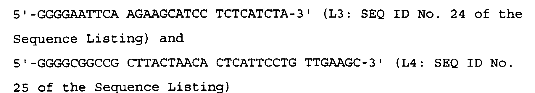
- Using these respective primers for the heavy and light chains, PCR was performed as follows.
Composition of the reaction solution: template (pCR-H or pCR-L), 1 µg; 5'-primer (H3 or L3), 40 pmol; 3'-primer (H4 or L4), 40 pmol; Tris-HCl (pH 8.0), 20 mM; potassium chloride, 10 mM; ammonium sulphate, 6 mM; magnesium chloride, 2 mM; Triton X-100, 0.1%; bovine serum albumin, nuclease-free, 10 µg/ml; dNTP's, 0.25 mM; native Pfu DNA polymerase (Stratagene), 5 units; and sterile distilled water to a total volume of 100 µl. - Initial heating of the reaction solution was at 94°C for 2 minutes, after which a thermal cycle of 94°C for 30 seconds, 60°C for 30 seconds and 75°C for 1.5 minutes was repeated 28 times.
- The resulting amplified DNA was digested with the restriction enzymes EcoRI and XbaI (for the heavy chain) or EcoRI and NotI (for the light chain), and then mixed with the animal cell expression plasmid pME18S [c.f. Hara. T., et al., (1992), EMBO J., 11, 1875] which had either been digested with the restriction enzymes EcoRI and XbaI (for the heavy chain) or EcoRI and NotI (for the light chain) and dephosphorylated using CIP [as described in Example 2 (2) 3) c) below]. One microlitre of 4 units of T4 DNA ligase were added to 8 µl of the resulting mixture, and 1 µl of 10× ligase reaction buffer [6 mM Tris-HCl (pH 7.5), 6 mM magnesium chloride, 5 mM sodium chloride, 7 mM β-mercaptoethanol, 0.1 mM ATP, 2 mM DTT, 1 mM spermidine, and 0.1 mg/ml bovine serum albumin] was then also added to the mixture, which was then incubated at 14°C for 15 hours.
- After this time, 2 µl of the incubated ligase reaction solution was mixed with 50 µl of competent E. coli strain JM109 at a cell density of 1-2
x 109 cells/ml (Takara Shuzo Co., Ltd.), and the mixture was kept on ice for 30 minutes, then at 42°C for 30 seconds, and again on ice for 5 minutes. Then, 500 µl of SOC medium (2% v/v tryptone, 0.5% w/v yeast extract, 0.05% w/v sodium chloride, 2.5 mM w/v potassium chloride, 1 mM magnesium chloride, and 20 mM glucose) was added to the mixture, which was incubated for a further hour, with shaking. Transformant strains were then isolated, and plasmid DNA was prepared from the strains, following the methods described in Reference Example 4 (4-3). - The resulting plasmids were designated pME-H (the expression plasmid vector carrying cDNA encoding the heavy chain of HFE7A) and pME-L (the expression plasmid vector carrying cDNA encoding the light chain of HFE7A). The transformant E. coli strains harboring these plasmids, designated as E. coli pME-H and E. coli pME-L, were deposited with Kogyo Gijutsuin Seimei-kogaku Gijutsu Kenkyujo on March 12, 1997, in accordance with the Budapest Treaty for the Deposit of Microorganisms, and were accorded the accession numbers FERM BP-5868 and FERM BP-5867, respectively.
- Transfection of COS-7 cells with the expression plasmids pME-H and pME-L obtained in (5-1) above was performed by electroporation using a gene transfection apparatus (ECM600; BTX).
- COS-7 cells (American Type Culture Collection No. CRL-1651) were grown up to semi-confluence in a culture flask (culture area: 225 cm2; Sumitomo Bakelite, K. K.) containing DMEM supplemented with 10% v/v FCS. Subsequently, the medium was discarded and 3 ml of trypsin-EDTA solution (Sigma Chemicals Co.) were added to the cells, followed by incubation at 37°C for 3 minutes. The cells detached by this process were harvested, washed twice with PBS and then adjusted to a cell density of 5 x 10⁶ cells/ml with PBS.
- Meanwhile, 20 µg each of plasmids pME-H and pME-L, prepared using a large-scale plasmid preparation kit (MaxiPrep DNA Purification System; Promega), were separately precipitated with ethanol and dissolved in 20 µl each of sterile PBS. Where COS-7 cells were cotransfected with both plasmids, 20 µg of each of the plasmids were used and dissolved together in 20 µl of sterile PBS.
- Twenty µl of the cell suspension prepared above (1.2 x 10⁶ cells) and 20 µl of the relevant plasmid solution were mixed and transferred to a chamber with electrodes set at a distance apart of 2 mm (BTX). The chamber was then loaded in the gene transfection apparatus and given a single pulse of 10 msec at 150 V to provide a total charge of 900 µF. The cell-DNA mixture in the chamber was added to 40 ml of DMEM supplemented with 10% v/v FCS and incubated in plastic cell culture dishes under 5% v/v CO2 at 37°C for 24 hours. After this time, the culture supernatant was discarded and the cells were washed with serum-free DMEM medium. After that, 40 ml of serum-free DMEM was added to each of the plastic dishes and the supernatant recovered after the cells had been cultured under 5% v/v CO2 at 37°C for a further 72 hours.
- Using the above method, COS-7 cells were obtained which were transfected with either or both plasmids (as shown below), and the supernatant of each of the transformants was recovered:
- (A): pME-H only;
- (B): pME-L only; and
- (C): cotransfection of pME-H and pME-L.
-
- Expression of anti-Fas antibody in the culture supernatants obtained in (5-2) above was determined by ELISA, in a manner similar to that described in Reference Example 3, and using the human Fas fusion protein as the antigen. It was established that the production of an antibody reacting with the human Fas antigen fusion protein in the culture supernatant only happened when pME-H and pME-L were both used to cotransfect COS-7 cells [5-2 (C)].
- The following peptides were synthesised by Fmoc solid phase synthesis (c.f. Carpino, L. A. and Han, G. Y., (1970), J. Am. Chem. Soc., 92, 5748-5749) using an automated peptide synthesiser (Model 430A; Perkin Elmer, Japan, Applied Biosystems Division):

- P1 to P15 are partial sequences of the amino acid sequence of Nos. 1 to 157 of the extracellular domain of human Fas, with between 9 and 11 amino acid residues overlapping one another. P16 is a negative control having no homology with human Fas.
- P1 to P16 were respectively dissolved completely in 48 µl dimethyl sulphoxide (DMSO) and each was then adjusted to adjusted to a final volume of 0.8 ml by the addition of 752 µl PBS containing 1 mM β-mercaptoethanol.
- The above peptides each correspond to a portion of the extracellular domain of the human Fas molecule, but with a carboxyl group added to the C-terminus. Each peptide was diluted to 50 µg/ml with 0.05 M carbonate-bicarbonate buffer (pH 9.6), containing 10 mM 2-mercaptoethanol, and 50 µl of each were introduced into a well of a 96-well ELISA microplate (Nunc). The plate was kept standing at 4°C overnight to allow adsorption of the peptide to the well surface.
- After this time, the solution in the wells was discarded and each well was washed 4 times with PBS-Tween. Then, 100 µl of PBS containing 1%(w/v) bovine serum albumin (A3803; Sigma Chemicals Co.) was added to each well and the plate was incubated at 37°C for 1 hour. The wells were then washed a further 4 times with PBS-Tween, and then 50 µl of HFE7A or CH11 adjusted to 5 µg/ml in PBS was added to each well. The plate was then incubated at 37°C for 1 hour, and the wells were again washed 4 times with PBS-Tween. After washing, 50 µl of horse radish peroxidase labeled goat anti-mouse immunoglobulin antibody (Amersham), diluted 1000-fold with PBS, was added per well, and the plate was again incubated at 37°C for 1 hour, after which the wells were washed 4 times with PBS-Tween. Horse radish peroxidase substrate (BioRad) was then added in an amount of 100 µl/well and the plate was allowed to stand at room temperature for 15 minutes before reading the absorbance of each well at 415 nm using a microplate reader (Corona). As a positive control, the human Fas fusion protein prepared in Reference Example 1 was used in place of the synthetic peptides.
- Using the above methodology, it was established that only the wells with adsorbed P11 showed high absorbance values, demonstrating that HFE7A specifically binds an amino acid sequence contained in P11 (Figure 3).
- The following peptides were synthesised:
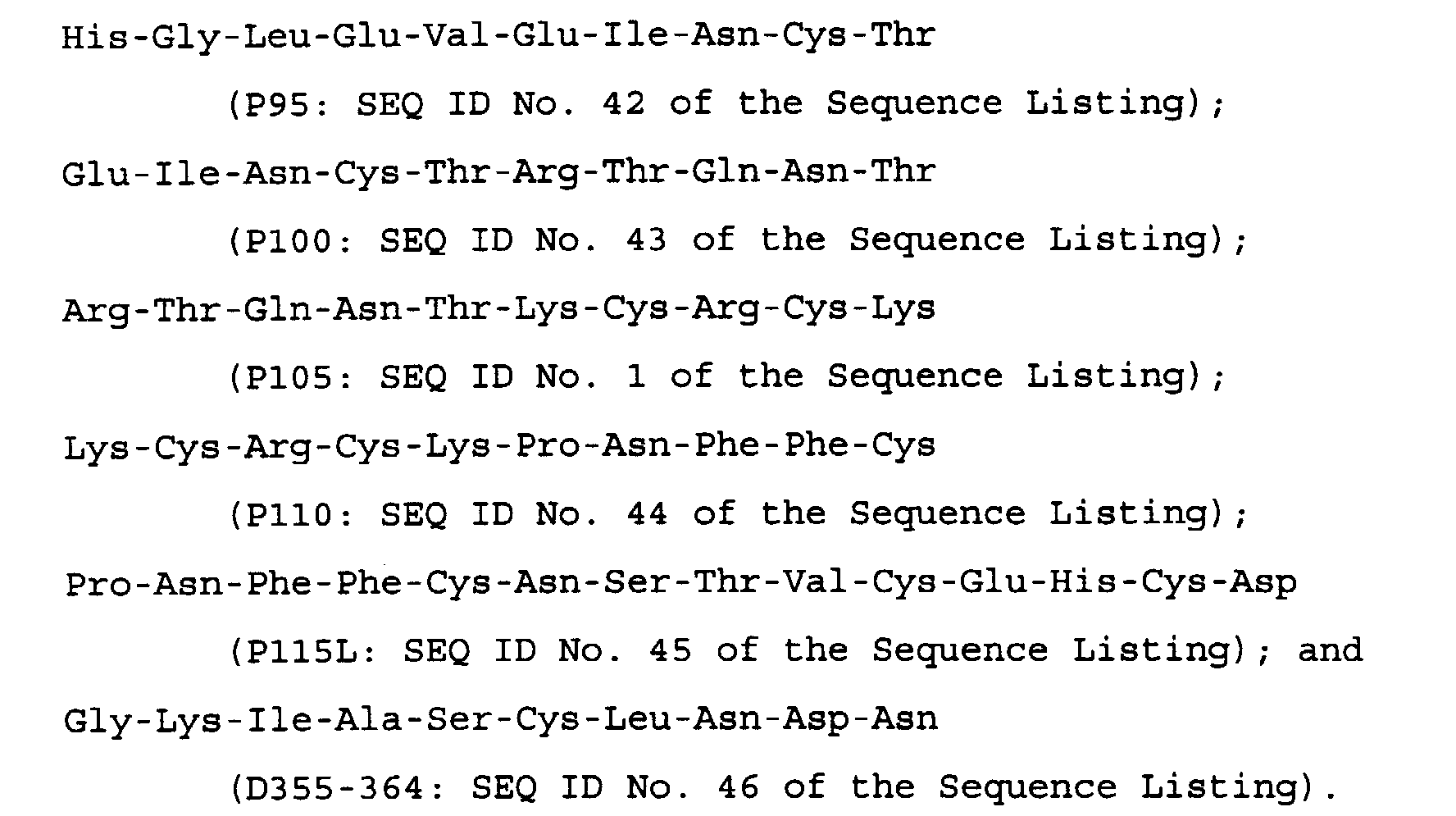
- P95, P100, P105 and P110 are each 10-residue partial sequences of the flanking region (corresponding to amino acids 95 to 128 of the extracellular domain of human Fas) of the amino acid sequence corresponding to P11 in the extracellular domain of human Fas, each having 5 overlapping amino acid residues with the next.
- Intended Peptide P115, Pro-Asn-Phe-Phe-Cys-Asn-Ser-Thr-Val-Cys (P115: amino acid Nos. 1 to 10 of SEQ ID No. 45 of the Sequence Listing) has a 5-residue overlap with a 10-residue peptide P110, but was expected to have poor solubility, so 4 extra residues were added at the C-terminus of P115 to produce P115L.
- D355-364 was used as a negative control, this peptide having no homology with human Fas.
- Each peptide, except P115L, was dissolved completely in 16 µl DMSO each was then adjusted to a final volume of 0.8 ml by the addition of 784 µl PBS containing 1 mM 2-mercaptoethanol. P115L was dissolved completely in 48 µl DMSO and was then adjusted to a final volume of 0.8 ml by the addition of PBS containing 1 mM 2-mercaptoethanol.
- Each of the above peptide solutions (corresponding to 200 µg peptide) were mixed with 0.25 µg of HFE7A in a microtube and adjusted to a total volume of 100 µl with PBS containing 1 mM 2-mercaptoethanol. The mixture was incubated at 37°C for 2 hours with stirring at 10 to 20 r.p.m., followed by the addition of FCS to a final concentration of 5%, thereby to yield the peptide-antibody mixture.
- WR19L12a cells were grown up by a method similar to that described in Reference Example 2. The cells were then recovered by centrifugation and adjusted to a cell density of 1 x 10⁷ cells/ml with serum-free RPMI medium. The cell suspension was dispensed into a 96-well plate, with the wells having U-shaped bottoms, at 100 µl/well and centrifuged at 4°C, 1,000 r.p.m. for 3 minutes using a swing rotor for the microplates, and the supernatant was then discarded. Next, 100 µl of peptide-antibody mixture was added to each pellet and mixed by pipetting a few times, as described above. The plate was then allowed to stand at 4°C for 30 minutes, and was then centrifuged and the supernatant discarded. The pellet was washed 3 times with flow cytometry buffer, and then 50 µl FITC-labeled goat anti-mouse IgG antibody (Kappel), diluted 250-fold with flow cytometry buffer, was added per well, followed by light pipetting to mix the well contents.
- The plate was kept in the dark at 4°C for 30 minutes, then centrifuged and the supernatant discarded. The pellet was washed 3 times with flow cytometry buffer, which contained 10% v/v neutral buffered formaldehyde solution (Wako Pure Chemical Industries, Ltd.) for tissue fixation, this solution being 10-fold diluted with PBS and 50 µl/well was added and mixed with light pipetting. Next, the plate was kept in the dark at 4°C for at least 12 hours to fix the cells.
- After this time, the cells were suspended in 100 µl/well of flow cytometry buffer and centrifuged, in order to remove the supernatant. The pellet was washed 3 times with flow cytometry buffer and suspended in 500 µl/well of flow cytometry buffer, and the resulting suspension was analyzed with a flow cytometer (Cytoace-150; Nippon Bunko, K. K. - excitation wave length: 488 nm; detection wave length: 530 nm) to calculate average intensities of FITC fluorescence per cell. Average intensities of FITC fluorescence for each sample were calculated by taking the value with no peptide-antibody mixture as 0% and the value of the sample containing D355-364 as 100%.
- By the above procedure, it was established that P105 is able to strongly inhibit binding between HFE7A and WR19L12a cells, and that P100 and P110, the amino acid sequence of each of which overlaps 50% with P105, each inhibit binding between HFE7A and WR19L12a cells by about 50% and 60%, respectively. No inhibition was observed with either of P95 and P115L, which also have no overlapping segments shared with P105 (Figure 4). From these results, it was established that P105 represents an amino acid sequence capable of inhibiting binding between HFE7A and human Fas and that, consequently, the epitope for HFE7A must be located within the amino acid sequence reproduced in P105. This epitopic amino acid sequence is a region which is conserved between human Fas and mouse Fas.
- The following test was performed, in order to establish whether HFE7A was able to bind Fas antigen from various primate species.
- First, peripheral blood samples were taken from a chimpanzee (Sanwa Kagaku Kenkyujo Kumamoto Primates Park, 40 ml), 20 ml from either a Japanese monkey (Macaca fuscata) or from a crab-eating monkey (Macaca irus) and 3 ml from a marmoset (of the genus Hapalidae). The blood samples had 1 ml of heparin (Novoheparin; Novo) added to them and the samples were then slowly layered over an equal volume of Ficol Paque solution [(Pharmacia) specific gravity: 1.077 for all except the crab-eating monkey, which had a specific gravity of 1.072] and centrifuged at 1,700 r.p.m. for 30 minutes in order to obtain a fraction of peripheral blood mononuclear cells. This mononuclear cell fraction was washed twice with Hanks' balanced salt solution and then suspended in RPMI 1640 medium with 10% v/v FCS to a cell density of 1 x 10⁶ cells/ml. Phytohaemagglutinin-P (Sigma Chemicals, Co.) was added to the resulting suspension to a final concentration of 5 µg/ml and the sample incubated at 37°C under 5% v/v CO2 for 24 hours. After this time, the cells were recovered by centrifugation, washed and resuspended in RPMI 1640 medium containing 10% v/v FCS. Then, to activate the recovered cells, interleukin-2 (Amersham) was added to the suspension to a final concentration of 10 units/ml, and this was incubated at 37°C under 5% v/v CO2 for 72 hours.
- An amount of the activated preparation calculated to contain 1 x 10⁶ activated lymphocyte cells was placed in a test tube and either suspended in 50 µl of 20 µg/ml HFE7A in PBS or 50 µl of PBS alone. The resulting suspension was allowed to stand on ice for 1 hour, after which the cells were washed 3 times with aliquots of 500 µl of PBS and then suspended in 50 µl of 20 µg/ml FITC-labeled anti-mouse IgG antibody (Bioresource) in PBS. This suspension was then placed on ice for 30 minutes, and washed 3 times with aliquots of 500 µl of PBS. Using the cells suspended in 500 µl of PBS as controls, the fluorescence intensities were measured, using a flow cytometer (Cytoace; Nippon Bunko, K. K.).
- Distributions of cell numbers by fluorescence intensity were obtained and the proportions of the numbers of the stained cells to those of total cells were calculated. As a result, in the samples without HFE7A, the stained cells constituted less than 3% for all species. However, in the samples treated with HFE7A, at least 17% of the cells were stained, the maximum being 82%. Accordingly, HFE7A is capable of binding a wide range of primate Fas including humans against which HFE7A was originally prepared.
- Either 500 µl of PBS, alone, or 0.05 or 0.1 mg of HFE7A monoclonal antibody (in 500 µl of PBS) was administered intraperitoneally to the members of groups of three 6-week old female C3H/HeJ mice (from Japan Clea). The mice were anesthetised with ether, 42 hours post administration, and their thymuses removed. These thymuses were washed with RPMI medium containing 10% v/v FCS, and subsequently disrupted, using a spatula on a mesh (Cell Strainer; Falcon). The disrupted cells (which had passed through the mesh) were washed twice with RPMI 1640 medium containing 10% v/v FCS.
- Where washing more than once is referred to in any of the Examples herein, it will be understood that the medium with which the washing is performed is replaced with fresh such medium for each wash, unless otherwise required.
- The washed cells obtained above were counted and adjusted to 1 x 10⁶ cells in 50 µl of RPMI 1640 medium containing 10% v/v FCS. Each of the resulting suspensions was dispensed into a well of a 96-well microplate, the wells having U-shaped bottoms (Nunc) and the plate was then centrifuged (90 x g, 4°C, 10 minutes).
- The supernatants were discarded and then one of the following two fluorescence-labeled antibody solutions in PBS, (a) or (b), was added to each well:
- (a) 10 µl of 0.5 mg/ml of FITC-labeled anti-mouse CD95 (Fas) antibody (Jo2; PharMingen), and 10 µl of 0.5 mg/ml of phycoerythrin (PE) labeled anti-mouse CD90 antibody (Thy-1.2; Cedarlane; CD90 being a cell surface antigen expressed only on T cells);
- (b) 10 µl of 0.5 mg/ml of FITC-labeled anti-mouse CD4 antibody (L3T4; PharMingen), and 10 µl of 0.2 mg/ml of PE-labeled anti-mouse CD8 antibody (Ly-2; PharMingen).
-
- After addition of the antibody mixtures, the plate was shaken to mix the contents of the wells and then kept on ice for 1 hour before centrifuging (90 x g, 4°C, 10 minutes). After discarding the supernatant and washing the wells twice with 100 µl/well of flow cytometry buffer, cells were fixed by adding 50 µl/well of 3.7% v/v formaldehyde solution and were then stood on ice for 10 minutes. After further centrifugation (90 x g, 4°C, 10 minutes) to remove the supernatant, the cell pellets were again washed with 100 µl/well of flow cytometry buffer and suspended in 100 µl/well of flow cytometry buffer. Using the thus obtained cell suspensions from each well as samples, the fluorescence of samples of 1 x 10⁴ cells was measured, using a flow cytometer (Epics Elite; Coulter) under the following conditions:
- excitation wave length: 488 nm;
- detection wave length: 530 nm (FITC) or 600 nm (PE).
-
- Fluorescence distributions of FITC and PE for the cell populations of each sample could then be prepared. For the samples to which antibody mixture (a) was added, the proportion of the number of cells that were positive for Fas and CD90 (hereinafter referred to as "Fas+CD90+") relative to the total cell number was calculated. Similarly, for the samples to which antibody mixture (b) was added, the proportion of the number of cells that were positive for CD4 and CD8 (hereinafter referred to as "CD4+CDB+") or those that were positive for CD4 but negative for CD8 (hereinafter referred to as "CD4+CD8-") relative to the total cell number was calculated.
- The results are shown as percentages in Table 1, below.
Cell Fas+ CD90+ CD4+ CD8+ CD4+ CD8- PBS only 76.2 62.6 11.7 HFE7A 0.05 mg 2.3 1.9 1.2 HFE7A 0.1 mg 1.7 2.8 0.7 - Compared with the group to which PBS only was administered, the proportions of T cells expressing Fas (Fas+CD90+) in the thymus cells of mice from the groups to which HFE7A was administered were remarkably reduced at both doses. Further, the CD4+CD8+ and CD4+CD8- cell populations, known for substantial Fas expression, were also markedly reduced in number after HFE7A administration, compared with the PBS only group.
- Accordingly, it was deduced that the anti-Fas monoclonal antibody HFE7A had apoptosis-inducing activity, in vivo, on Fas-expressing T cells.
- The effects of administration of anti-Fas monoclonal antibody HFE7A on autoimmune disease symptoms were examined using MRL gld/gld mice. These mice carry a mutant of the Fas ligand gene and serve as an animal model of systemic lupus erythaematosus-like autoimmune diseases.
- 18-week old MRL gld/gld mice (from Japan SLC, K. K.), were treated intraperitoneally with a single dose of either 0.2 or 0.5 mg of HFE7A monoclonal antibody prepared in Reference Example 3 (in 500 µl of PBS) or with 500 µl of PBS alone.
- Each test mouse was monitored for swelling of the ankles as a symptom of autoimmune disease. The degree of swelling was evaluated and recorded over time for each group [c.f. Shin Yonehara, (1994), Nikkei Science Bessatsu, 110, 66-77]. The degree of swelling of the ankles was observed to markedly decrease with administration of HFE7A.
- The thymuses were removed from the test mice and the proportions of T cells which expressed Fas in the thymuses were determined by the method described in Reference Example 8 above. The results showed that the number of Fas-expressing T cells in the thymuses were significantly reduced after the administration of HFE7A, in accordance with the results of Reference Example 8.
- BALB/c mice were intraperitoneally administered a single dose of one of the following:
- i) 0.2 mg HFE7A in 500 µl of PBS;
- ii) 0.5 mg HFE7A in 500 µl of PBS;
- iii) 0.1 mg Jo2 (PharMingen) in 500 µl of PBS; and
- iv) 500 µl of PBS alone.
-
- Of the above, Jo2 is a known anti-mouse Fas antibody which has apoptosis-inducing activity. Blood was taken from the posterior aorta of the mice at 8 hours, 24 hours or 72 hours post administration. Blood was taken at 3 hours post administration for the Jo2-treated mice, while they were still alive. All blood was taken under light ether anesthetization. The blood levels of glutamic-oxaloacetic transaminase (GOT) and glutamic-pyruvic transaminase (GPT) were measured for each blood sample, using an automated analyzer (Model 7250; Hitachi Seisakusyo, K. K.) together with the appropriate reagent for the analyzer (Transaminase-HRII; Wako Pure Chemical Industries, Ltd.). As a result, the Jo2-treated group showed rapid elevation of GOT and GPT values after 3 hours, whereas the corresponding values for the groups treated with HFE7A showed little change, as with the group treated with PBS only (Figure 5). From these results, it was could be established that HFE7A did not induce acute hepatic disorders.
- It is known that, upon intraperitoneal administration of the anti-mouse Fas antibody Jo2, a mouse develops fulminant hepatitis and dies within several hours [c.f. Ogasawara, J., et al., (1993), Nature, 364, 806]. Accordingly, in order to evaluate the effects of HFE7A on hepatic disorders induced by Jo2, the viability of mice was tested by administering HFE7A simultaneously with, or subsequently to, Jo2 administration.
- Female, 6 week old BALB/c mice (three mice per group; from Japan SLC) received intraperitoneal administration of an antibody preparation as follows:
- (A) 0.1 mg of Jo2 in 0.5 ml PBS;
- (B) 0.01 mg of Jo2 in 0.5 ml PBS;
- (C) 0.1 mg of Jo2 and 0.5 mg of HFE7A together in 0.5 ml PBS (simultaneous administration);
- (D) 0.1 mg of Jo2 and 0.05 mg of HFE7A in 0.5 ml PBS (simultaneous administration); and
- (E) 0.01 mg of Jo2 in 0.2 ml PBS, followed by 0.1 mg of HFE7A in 0.2 ml PBS after 20 minutes; and the mice were then observed over time. The results are shown in Figure 6.
-
- When Jo2 alone was administered, all mice died within 9 hours, regardless of whether they were administered with 0.1 mg or with 0.01 mg Jo2 /mouse, i.e., mice of groups (A) and (B) above all died within 9 hours of administration. In contrast, when HFE7A was administered simultaneously with Jo2 (both 0.5 mg/mouse and 0.05 mg/mouse), i.e., groups (C) and (D) above, the mice showed no disorders even for several weeks post administration, demonstrating that HFE7A administration can block the development of fulminant hepatitis. Moreover, mice remained normal, with no apparent symptoms developing, even when HFE7A was administered 20 minutes after Jo2 administration.
- Thus, HFE7A has preventive and therapeutic effects on various diseases involving disorders of normal tissues which are mediated by the Fas/Fas ligand system, both in the liver and in other organs.
- F1 mice obtained from the mating of a female BALB/c mouse and a male DBA/1J mouse (CD1F1 mice, 6 weeks old, female, from Japan Charles River, K. K.) were tamed for 1 week. After this time, the mice were treated with collagen to induce arthritis.
- In more detail, the method was based on one described in the literature [c.f. Phadke, K., (1985), Immunopharmacol., 10, 51-60]. In this method, a 0.3% w/v solution of bovine collagen type II (Collagen Gijutsu Kensyukai, supplied in a 50 mM acetic acid solution) was diluted to 0.2% (2 mg/ml) with further 50 mM acetic acid and then emulsified with an equal volume of Freund's complete adjuvant (Difco). This emulsion was then administered, in an amount of 100 µl (corresponding to 100 µg bovine collagen type II), intradermally in the proximal portion of the tail, which was held in a fixing device for intravenous injection, using a 1 ml plastic syringe equipped with a tuberculin needle. An identical booster dose was administered under similar conditions, 1 week after the initial challenge.
- At the same time as the booster injection, an injection of 100 µg of either HFE7A or control mouse IgG in 0.5 ml PBS was administered intraperitoneally (6 mice per group). Starting five weeks after the initial challenge, swelling of the limbs was monitored visually. The degree of swelling of the joints of the limbs was scored based on the method of Wood, F. D., et al. [Int. Arch. Allergy Appl. Immunol., (1969), 35, 456-467]. Accordingly, the following criteria were used in the calculation of the scores for each of the limbs:
Score 0: no symptom; 1: swelling and reddening of only one of the small joints, e.g., of a toe; 2: swelling and reddening of 2 or more small joints, or of one relatively large joint, such as an ankle; and 3: swelling and reddening of a limb in its entirety. - Accordingly, the maximum score for one animal is when all 4 limbs swell, and is 12. An animal scoring at least 1 for all four limbs was designated as an "affected mouse." The results are shown in Figure 7.
- In the control group to which non-specific mouse IgG was given, all mice were affected by the 7th week after the initial challenge, whereas in the group to which HFE7A was administered, a half of the mice showed no reddening of any joints at all up to the 8th week (Figure 7A). In addition, the HFE7A-treated group had a lower average score compared with the control group (Figure 7B).
- The effects of HFE7A on the viability of synovial cells from patients with rheumatoid arthritis were evaluated. The method was as described below, using the reducing power of the mitochondria as the index.
- Synovial tissue obtained from an affected region of a patient with rheumatoid arthritis was cut into small pieces, with scissors, in Dulbecco's modified Eagle medium (Gibco) supplemented with 10% v/v FCS (Summit). The fat was removed and collagenase (Sigma Chemical Co.) was then added to a final concentration of 5 µg/ml and the mixture was incubated at 37°C for 90 minutes under 5% v/v CO2. The resulting incubated cells then served as the synovial cells for the remainder of the Experiment.
- The thus obtained synovial cells were separated into single cells by treatment with a 0.05% w/v aqueous trypsin solution at 37°C for 2 minutes, then suspended in Dulbecco's modified Eagle medium containing 10% v/v FCS to a cell density of 1 x 10⁵/ml. This cell suspension was then dispensed into wells of a 96-well plate at 2 x 10⁴ cells /200 µl per well, and incubated at 37°C under 5% v/v CO2 for 6 days. The culture supernatant was discarded and the cells were washed 3 times with Hank's buffer (Gibco). After washing, 200 µl of Dulbecco's modified Eagle medium containing 10% v/v FCS and between 10 and 1,000 ng/ml of HFE7A (serial 10-fold dilutions) were added to each well and the plate further incubated at 37°C under 5% v/v CO2 for 20 hours. Next, 50 µl of an aqueous solution of 1 mg/ml XTT (2,3-bis [2-methoxy-4-nitro-5-sulphophenyl]-2H-tetrazolium-5-carboxanilide inner salt; Sigma Chemical Co.) and 25 µM PMS (phenazine methosulphate; Sigma Chemical Co.) was added to each well (final concentrations: 250 µg/ml XTT and 5 µM PMS). After a further 4 hours of incubation at 37°C under 5% v/v CO2, the absorbance of each well was read at 450 nm.
- The viability of cells in each well was calculated according to the following formula:
- The results are shown in Table 2. HFE7A inhibited the survival of synovial cells from patients with rheumatism in a dose-dependent manner.
HFE7A concentration (ng/ml) average viability (%) 0 100 10 91 100 77 1000 42 - Molecular modeling of the variable regions of HFE7A was performed by the method generally known as homology modeling [c.f. Methods in Enzymology, 203, 121-153, (1991)].
- The primary sequences of variable regions of human immunoglobulin registered in the Protein Data Bank (hereinafter referred to as the "PDB"; Chemistry Department, Building 555, Brookhaven National Laboratory, P.O.
Box 5000, Upton, NY 11973-5000, USA), for which X-ray crystallography had been performed, were compared with the framework regions of HFE7A determined above. As a result, 1GGI and 2HFL were selected as having the highest homologies of the three-dimensional structures of the framework regions for the light and heavy chains, respectively. Three-dimensional structures of the framework regions were generated by combining the properties of 1GGI and 2HFL and by calculating the properties of the regions of HFE7A, as described below, to obtain the "framework model". - Using the classification described by Chothia et al., the CDR's of HFE7A could be classified as follows: CDRL2, CDRL3 and CDRH1 all belong to
canonical class 1, while CDRL1, CDRH2 and CDRH3 do not currently appear to belong to any specific canonical class. The CDR loops of CDRL2, CDRL3 and CDRH1 were ascribed the conformations inherent to their respective canonical classes, and then integrated into the framework model. CDRL1 was assigned the conformation of cluster 15B, in accordance with the classification of Thornton et al. [c.f. J. Mol. Biol., 263, 800-815, (1996)]. For CDRH2 and CDRH3, conformations of sequences with high homologies were selected from the PDB and then these were combined with the results of energy calculations. The conformations of the CDR loops with the highest probabilities were then taken and integrated into the framework model. - Finally, energy calculations were carried out to eliminate undesirable contact between inappropriate atoms, in terms of energy, in order to obtain an overall molecular model of HFE7A. The above procedure was performed using the commercially available common molecular modeling system, AbM (Oxford Molecular Limited, Inc.), although any other appropriate system could have been used.
- For the molecular model obtained, the accuracy of the structure was further evaluated using the software, PROCHECK [J. Appl. Cryst., (1993), 26, 283-291], and the degree of surface exposure of each residue was calculated to determine which surface atoms and groups interacted.
- The subgroups of the light and heavy chains of HFE7A shared identities of 79% with the subgroup κIV and also 79% with the subgroup I, respectively, by comparison with the consensus sequences of the respective subgroups of human antibodies. However, there was no human antibody having a combination of a κIV light chain and a sub-group I heavy chain. Thus, 8E10'CL, which has a light chain of subgroup κIII and a heavy chain of subgroup I, having 72% and 77% sequence identities with the light and heavy chains of HFE7A, respectively, was selected as the single human antibody which had light and heavy chains which both had an identity of greater than 70% with the light and heavy chains of HFE7A.
- Using the software, Cameleon (Oxford Molecular Limited, Inc.), the amino acid sequence of each of the light and heavy chains of HFE7A was aligned with that of the relevant chain of 8E10'CL, and humanised sequences of the variable regions were made as described in the following Examples in accordance with the general guidelines set out in a) to e) above. Plasmids were constructed which could serve as recombinant vectors comprising DNA nucleotide sequences encoding humanised anti-human Fas antibodies.
- Prior to humanisation of the light chain amino acid sequence of the mouse anti-human Fas antibody HFE7A, cDNA cloning of a human immunoglobulin light chain comprising the constant region was first performed.
- Separation of cDNA encoding a human light chain was carried out by PCR. For the PCR, the following two primers were synthesised:

- cDNA encoding a full-length human immunoglobulin light chain was prepared by PCR, inserted into a plasmid and cloned into E. coli.
- The HL-DNA fragment encoding a full-length human immunoglobulin light chain was prepared under the following conditions:
Composition of the PCR reaction solution: human lymphocyte cDNA library (Life Technologies), 25 ng; oligonucleotide primer HVKII5-4, 50 pmol; oligonucleotide primer HKCL3-3, 50 pmol; 25 mM dNTP cocktail, 10 µl; 100 mM Tris-HCl buffer (pH 8.5), 10 µl; 1 M potassium chloride [KCl], 5 µl; 25 mM magnesium chloride [MgCl2], 10 µl; Tag DNA polymerase (Perkin Elmer Japan), 1 unit; Redistilled water to a total volume of 100 µl. - The PCR reaction was conducted as follows. The solution was first heated at 94°C for 2 minutes, after which a cycle of heating to 94°C for 1 minute, 55°C for 1 minute and 72°C for 2 minutes, was repeated 30 times. After completion of this procedure, the reaction solution was heated at 72°C for 10 minutes.
- The thus prepared HL-DNA (human light chain DNA) fragment was inserted into plasmid pCR3DNA using a eukaryote TA Cloning Kit (Invitrogen), following the manufacturer's protocol, and introduced into competent E. coli TOP10F' contained in the kit, and following the instructions in the kit. Plasmid pHL15-27 carrying the HL-DNA fragment, i.e., cDNA for a human immunoglobulin light chain, was thereby obtained.
- Humanisation of the amino acid sequence of the light chain of the mouse anti-human Fas antibody HFE7A entailed replacing the 47th amino acid (proline) and the 49th amino acid (lysine) from the N-terminus of the amino acid sequence of the light chain (hereinafter referred to as "region I") with alanine and arginine, respectively. Alanine (47) and arginine (49) are conserved in the human light chain (κ chain). Further humanisation was also performed, and entailed replacing the 80th amino acid (histidine), the 81st amino acid (proline), the 82nd amino acid (valine), the 84th amino acid (glutamic acid), the 85th amino acid (glutamic acid), the 87th amino acid (alanine) and the 89th amino acid (threonine) (hereinafter referred to as "region II") with serine, arginine, leucine, proline, alanine, phenylalanine and valine, respectively, as these are also conserved in the human light chain (κ chain).
- Where both regions I and II were humanised, the sequence was designated as "HH type."
- Where only region I was humanised, the sequence was designated as "HM type."
- Where neither region was humanised, the sequence was designated as "MM type."
- Expression plasmids, respectively carrying these 3 types of humanised light chain amino acid sequences from the anti-human Fas antibody HFE7A, were constructed as follows.
- PCR was used to construct the following DNA sequences, each of which comprised one of the HH, HM or MM sequences described above, together with the constant region of the human immunoglobulin light chain (κ chain):
DNA (SEQ ID No. 49 of the Sequence Listing) encoding the HH type polypeptide chain (SEQ ID No. 50 of the Sequence Listing); DNA (SEQ ID No. 51 of the Sequence Listing) encoding the HM type polypeptide chain (SEQ ID No. 52 of the Sequence Listing); and DNA (SEQ ID No. 53 of the Sequence Listing) encoding the MM type polypeptide chain (SEQ ID No. 54 of the Sequence Listing). - The following 13 oligonucleotide PCR primers were synthesised:

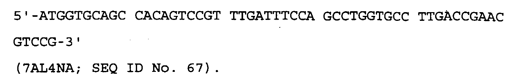
- The VHH-DNA fragment (SEQ ID No. 49 of the Sequence Listing) encoding the amino acid sequence of SEQ ID No. 50 of the Sequence Listing was prepared by performing 3-stage PCR, and then inserted into a plasmid vector and cloned into E. coli.
- The outline of the first stage PCR for the preparation of VHH-DNA is shown in Figure 8.
- The L7A1-DNA fragment, encoding a secretion signal sequence and a portion of the FRL1 region altered to contain a Hind III restriction enzyme cleavage site at the 5'-end, was prepared as follows.
Composition of the PCR reaction solution: plasmid pME-L DNA, 200 ng; oligonucleotide primer 7AL1P, 80 pmol; oligonucleotide primer 7AL1N, 80 pmol; dNTP cocktail, 20 µl; 10×Pfu buffer, 20 µl; Pfu DNA polymerase (Stratagene), 10 units; and redistilled water to a final volume of 200 µl. - The PCR reaction was conducted as follows. The solution was first heated at 94°C for 2 minutes, after which a cycle of heating to 94°C for 1 minute, 55°C for 1 minute and 72°C for 2 minutes, was repeated 30 times. After completion of this procedure, the reaction solution was heated at 72°C for 10 minutes.
- The L7A2-DNA fragment, encoding a portion of the FRL1, CDRL1, FRL2 and a portion of the CDRL2 region, was prepared as follows.
Composition of the PCR reaction solution: plasmid pME-L DNA, 200 ng; oligonucleotide primer 7AL2P, 80 pmol; oligonucleotide primer M7AL2N, 80 pmol; dNTP cocktail, 20 µl; 10×Pfu buffer, 20 µl; Pfu DNA polymerase, 10 units; and redistilled water to a final volume of 200 µl. - The PCR reaction was conducted as follows. The solution was first heated at 94°C for 2 minutes, after which a cycle of heating to 94°C for 1 minute, 55°C for 1 minute and 72°C for 2 minutes, was repeated 30 times. After completion of this procedure, the reaction solution was heated at 72°C for 10 minutes.
- The L7A3-DNA fragment, encoding the CDRL2 and a portion of the FRL3 was prepared as follows.
Composition of the PCR reaction solution: plasmid pME-L DNA, 200 ng; oligonucleotide primer 7AL3PA, 80 pmol; oligonucleotide primer 7AL3N, 80 pmol; dNTP cocktail, 20 µl; 10×Pfu buffer, 20 µl; Pfu DNA polymerase, 10 units; and redistilled water to a final volume of 200 µl. - The PCR reaction was conducted as follows. The solution was first heated at 94°C for 2 minutes, after which a cycle of heating to 94°C for 1 minute, 55°C for 1 minute and 72°C for 2 minutes, was repeated 30 times. After completion of this procedure, the reaction solution was heated at 72°C for 10 minutes.
- The L7A4-DNA fragment, encoding a portion of the FRL3, CDRL3, FRL4 and a portion of the constant region was prepared as follows.
Composition of the PCR reaction solution: plasmid pME-L DNA, 200 ng; oligonucleotide primer 7AL4P, 80 pmol; oligonucleotide primer 7AL4N, 80 pmol; dNTP cocktail, 20 µl; 10×Pfu buffer, 20 µl; Pfu DNA polymerase, 10 units; and redistilled water to a final volume of 200 µl. - The PCR reaction was conducted as follows. The solution was first heated at 94°C for 2 minutes, after which a cycle of heating to 94°C for 1 minute, 55°C for 1 minute and 72°C for 2 minutes, was repeated 30 times. After completion of this procedure, the reaction solution was heated at 72°C for 10 minutes.
- The L7A5-DNA fragment, encoding a portion of the FRL4 and the constant region altered to have an EcoRI restriction enzyme cleavage site at the 3'-end, was prepared as follows.
Composition of the PCR reaction solution: plasmid pHL15-27 DNA, 200 ng; oligonucleotide primer 7ALCP, 80 pmol; oligonucleotide primer 7ALCN, 80 pmol; dNTP cocktail, 20 µl; 10×Pfu buffer, 20 µl; Pfu DNA polymerase, 10 units; and redistilled water to a final volume of 200 µl. - The PCR reaction was conducted as follows. The solution was first heated at 94°C for 2 minutes, after which a cycle of heating to 94°C for 1 minute, 55°C for 1 minute and 72°C for 2 minutes, was repeated 30 times. After completion of this procedure, the reaction solution was heated at 72°C for 10 minutes.
- An equal volume of phenol-chloroform (50% v/v phenol saturated with water, 48% v/v chloroform, 2% v/v isoamyl alcohol) was added to 200 µl of each of the PCR products, and vigorously mixed for 1 minute. After this time, the mixture was centrifuged at 10,000 x g, and the aqueous layer was recovered and mixed with an equal volume of chloroform-isoamyl alcohol (96% v/v chloroform and 4% v/v isoamyl alcohol), which was again vigorously mixed for 1 minute. The resulting mixture was centrifuged at 10,000 x g and the aqueous layer was recovered (the series of steps recited in this paragraph is referred to, herein, as "phenol extraction").
- Ethanol precipitation was then performed on the recovered aqueous layer. As used and referred to herein, "ethanol precipitation" consists of adding, with mixing, a one tenth volume of 3M sodium acetate (pH 5.2) and 2.5 volumes of 100% ethanol to the solution to be treated, and freezing the mixture using dry ice. The resulting mixture is then centrifuged at 10,000 x g to recover DNA as a precipitate.
- After phenol extraction and ethanol precipitation, the resulting DNA precipitate was vacuum-dried, dissolved in a minimum of redistilled water, and separated by 5% w/v polyacrylamide gel electrophoresis. After electrophoresis, the gel was stained with a 1 µg/ml aqueous solution of ethidium bromide to allow detection of DNA under UV light. The DNA bands corresponding to L7A1-DNA, L7A2-DNA, L7A3-DNA, L7A4-DNA and L7A5-DNA were cut out using a razor blade and eluted from the gel using Centriruter and Centricon-10, as described above. The eluted DNA was then concentrated by centrifugation at 7,500 x g, followed by ethanol precipitation, and finally dissolved in 50 µl of distilled water.
- The outline of the second stage PCR for the production of VHH-DNA is shown in Figure 9.
- L7A1.2-DNA, in which the L7A1-DNA and L7A2-DNA fragments, described above, were fused, was prepared as follows.
Composition of the PCR reaction solution: L7A1-DNA solution prepared in the first stage PCR, 10 µl; L7A2-DNA solution prepared in the first stage PCR, 10 µl; oligonucleotide primer 7AL1P, 80 pmol; oligonucleotide primer 7AL2N, 80 pmol; dNTP cocktail, 20 µl; 10×Pfu buffer, 20 µl; Pfu DNA polymerase, 10 units; and redistilled water to a final volume of 200 µl. - The PCR reaction was conducted as follows. The solution was first heated at 94°C for 2 minutes, after which a cycle of heating to 94°C for 1 minute, 55°C for 1 minute and 72°C for 2 minutes, was repeated 30 times. After completion of this procedure, the reaction solution was heated at 72°C for 10 minutes.
- L7A4.5-DNA, in which the L7A4-DNA and L7A5-DNA fragments described above were fused, was prepared as follows.
Composition of the reaction solution: L7A4-DNA solution prepared in the first stage PCR, 10 µl; L7A5-DNA solution prepared in the first stage PCR, 10 µl; oligonucleotide primer 7AL4P, 80 pmol; oligonucleotide primer 7ALCN, 80 pmol; dNTP cocktail, 20 µl; 10×Pfu buffer, 20 µl; Pfu DNA polymerase, 10 units; and redistilled water to a final volume of 200 µl. - The PCR reaction was conducted as follows. The solution was first heated at 94°C for 2 minutes, after which a cycle of heating to 94°C for 1 minute, 55°C for 1 minute and 72°C for 2 minutes, was repeated 30 times. After completion of this procedure, the reaction solution was heated at 72°C for 10 minutes.
- First, phenol extraction and then ethanol precipitation were performed on the amplified PCR L7A1.2-DNA and L7A4.5-DNA fragments, and these fragments were then separated by 5% w/v polyacrylamide gel electrophoresis. After electrophoresis, the gel was stained with 1 µg/ml of ethidium bromide, and the bands detected under UV light were cut out using a razor blade and eluted from the gel using a Centriruter and Centricon-10, as described above. The eluted DNA was concentrated first by centrifugation at 7,500 x g, then by ethanol precipitation, and then dissolved in 50 µl of distilled water.
- The outline of the third stage PCR for the production of VHH-DNA is shown in Figure 10.
- The VHH-DNA fragment in which the above described L7A1.2-DNA and L7A4.5-DNA fragments and L7A3-DNA were fused was prepared as follows.
Composition of the PCR reaction solution: L7A1.2-DNA solution prepared in the second stage PCR, 10 µl; L7A4.5-DNA solution prepared in the second stage PCR, 10 µl; L7A3-DNA solution prepared in the first stage PCR, 10 µl; oligonucleotide primer 7AL1P, 80 pmol; oligonucleotide primer 7ALCN, 80 pmol; dNTP cocktail, 20 µl; 10×Pfu buffer, 20 µl; Pfu DNA polymerase, 10 units; and redistilled water to a final volume of 200 µl. - The PCR reaction was conducted as follows. The solution was first heated at 94°C for 2 minutes, after which a cycle of heating to 94°C for 1 minute, 55°C for 1 minute and 72°C for 2 minutes, was repeated 30 times. After completion of this procedure, the reaction solution was heated at 72°C for 10 minutes.
- The amplified PCR VHH-DNA fragment was subjected first to phenol extraction and then to ethanol precipitation, before separation on a 5% w/v polyacrylamide electrophoresis gel. After electrophoresis, the gel was stained with 1 µg/ml of ethidium bromide and the VHH-DNA band detected under UV light was cut out using a razor blade and eluted from the gel using a Centriruter and Centricon-10, as described above. The eluted DNA was then concentrated by centrifugation at 7,500 x g, followed by ethanol precipitation, and finally dissolved in 50 µl of distilled water.
- The construction of an expression plasmid carrying VHH-DNA fragment is outlined in Figure 11.
- The VHH-DNA fragment obtained above was further purified by phenol extraction followed by ethanol precipitation, and it was then digested with the restriction enzymes Hind III and EcoRI.
- One µg of cloning plasmid pHSG399 DNA (Takara Shuzo Co., Ltd.) was digested with the restriction enzymes Hind III and EcoRI, and then dephosphorylated with alkaline phosphatase (derived from calf intestine; hereinafter abbreviated as CIP). The resulting, dephosphorylated plasmid pHSG399 DNA and the digested VHH-DNA fragment were ligated using a DNA Ligation Kit Version 2.0 (Takara Shuzo Co., Ltd.) using the manufacturer's protocol.
- The ligated DNA was recovered by ethanol precipitation, dissolved in 5 µl of redistilled water, and then mixed with E. coli JM109 Electro-Cell (Takara Shuzo Co., Ltd.). The mixture was transferred to a Gene Pulser/E. coli Pulser Cuvette, 0.1 cm (BioRad) and the ligated mix was then used to transform the E. coli JM 109 using Gene Pulser II (BioRad) by the manufacturer's protocol (the series of steps in this paragraph is referred to herein as "transformation").
- After transformation, the cells were plated onto LB agar medium [Bacto-tryptone (Difco) 10 g, Bacto-yeast extract (Difco) 5 g, NaCl 10 g, Bacto-agar (Difco) 15g; dissolved in distilled water, q.s. to 11] containing final concentrations of 1 mM IPTG (isopropylthio-β-D-galactoside; Takara Shuzo Co., Ltd.), 0.1% w/v X-Gal (5-bromo-4-chloro-3-indolyl-β-D-galactoside; Takara Shuzo Co., Ltd.) and 50 µg/ml chloramphenicol, and the plates were incubated at 37°C overnight to obtain E. coli transformants.
- Any white transformants obtained were cultured in 2 ml of liquid LB medium at 37°C overnight, and plasmid DNA was extracted from the resulting culture by the alkaline-SDS method [Sambrook, J., et al., (1989), in "Molecular Cloning: A Laboratory Manual (2nd Edition)", Cold Spring Harbor Laboratory Press].
- The resulting, extracted plasmid DNA was digested with the restriction enzymes Hind III and EcoRI, and a clone carrying the VHH-DNA fragment was then selected by 1% w/v agarose gel electrophoresis.
- Plasmid pHSGHH7 carrying a fusion fragment of the variable region of the humanised HH type HFE7A light chain and DNA encoding the constant region of human immunoglobulin κ chain was obtained accordingly. The transformant E. coli pHSGHH7 SANK 73497 harboring plasmid pHSGHH7 was deposited with the Kogyo Gijutsuin Seimei-Kogaku Kogyo Gijutsu Kenkyujo on August 22, 1997, in accordance with the Budapest Treaty, and was accorded the accession number FERM BP-6073.
- Using above described plasmid pHSGHH7, it was then possible to construct the expression vector plasmid p7AL-HH, carrying the DNA of SEQ ID No. 49 of the Sequence Listing and which encodes the humanised HH type HFE7A light chain polypeptide of SEQ ID No. 50 of the Sequence Listing.
- One µg of pEE.12.1 DNA (Lonza), an expression vector for mammalian cells, was digested with the restriction enzymes Hind III and EcoRI, and then dephosphorylated using CIP. The resulting digested, dephosphorylated plasmid DNA (100 ng) was ligated with 10 µg of the pHSGHH7 DNA fragment which had also been digested with Hind III and EcoRI, using a DNA Ligation Kit Version 2.0 (Takara Shuzo Co., Ltd.). The ligation mix was then used to transform E. coli JM109 (as described above), which was then plated on LB agar plates containing 50 µg/ml ampicillin.
- The transformants obtained by this method were cultured in 2 ml of liquid LB medium containing 50 µg/ml ampicillin at 37°C overnight, and plasmid DNA was subsequently extracted from the resulting culture by the alkaline-SDS method.
- The extracted plasmid DNA was digested with Hind III and EcoRI, and subjected to 1% w/v agarose gel electrophoresis to confirm the presence or absence of the insert of interest. This enabled the isolation of the plasmid p7AL-HH, which contains a fusion fragment having the variable region of the humanised HH type HFE7A light chain together with DNA encoding the constant region of the human immunoglobulin κ chain. The fusion fragment was found to be located downstream of the cytomegalovirus (CMV) promoter in the correct orientation.
- The VHM-DNA fragment of SEQ ID No. 51 of the Sequence Listing encoding the amino acid sequence of SEQ ID No. 52 of the Sequence Listing was produced by performing a 3-stage PCR, inserted into a plasmid vector and then cloned into E. coli.
- The outline of the first stage PCR for the preparation of the VHM-DNA fragment is shown in Figure 12.
- The L7A1-DNA fragment, encoding a secretion signal sequence and a portion of FRL1 having a Hind III restriction enzyme cleavage site added at the 5'-end, the L7A2-DNA fragment, encoding a portion of FRL1, CDRL1, FRL2 and a portion of CDRL2, and the L7A5-DNA fragment, encoding a portion of FRL4 and the constant region having an EcoRI site added at the 3'-end, were used in this process, and were those obtained in the preparation of the VHH-DNA fragment in 2) above.
- An ML7A3-DNA fragment, encoding CDRL2, FRL3, CDRL3, FRL4 and a portion of the constant region, was prepared as follows.
Composition of the PCR reaction solution: plasmid pME-L DNA, 200 ng; oligonucleotide primer 7AL3PA, 80 pmol; oligonucleotide primer 7AL4NA, 80 pmol; dNTP cocktail, 20 µl; 10×Pfu buffer, 20 µl; Pfu DNA polymerase, 10 units; and redistilled water to a final volume of 200 µl. - The PCR reaction was conducted as follows. The solution was first heated at 94°C for 2 minutes, after which a cycle of heating to 94°C for 1 minute, 55°C for 1 minute and 72°C for 2 minutes, was repeated 30 times. After completion of this procedure, the reaction solution was heated at 72°C for 10 minutes.
- The PCR products were subjected first to phenol extraction and then to ethanol precipitation, and then separated by 5% w/v polyacrylamide gel electrophoresis. After electrophoresis, the gel was stained with 1 µg/ml of ethidium bromide and the DNA band detected under UV light, corresponding to ML7A3-DNA, was cut out using a razor blade and eluted from the gel using a Centriruter and Centricon-10, as described above. The eluted DNA was then concentrated by centrifugation at 7,500 x g followed by ethanol precipitation, and then dissolved in 50 µl of distilled water.
- The outline of the second stage PCR for the preparation of VHM-DNA is shown in Figure 13.
- A VHM-DNA fusion fragment comprising the L7A1.2-DNA, the ML7A3-DNA and the L7A5-DNA fragment above was prepared as follows.
Composition of the PCR reaction solution: L7A1.2-DNA solution prepared in the second stage PCR, 10 µl; ML7A3-DNA solution prepared in the first stage PCR, 10 µl; L7A5-DNA solution prepared in the first stage PCR, 10 µl; oligonucleotide primer 7AL1P, 80 pmol; oligonucleotide primer 7ALCN, 80 pmol; dNTP cocktail, 20 µl; 10×Pfu buffer, 20 µl; Pfu DNA polymerase, 10 units; and redistilled water to a final volume of 200 µl. - The PCR reaction was conducted as follows. The solution was first heated at 94°C for 2 minutes, after which a cycle of heating to 94°C for 1 minute, 55°C for 1 minute and 72°C for 2 minutes, was repeated 30 times. After completion of this procedure, the reaction solution was heated at 72°C for 10 minutes.
- The resulting, amplified VHM-DNA fragment was subjected first to phenol extraction and then to ethanol precipitation, and then separated by 5% w/v polyacrylamide gel electrophoresis. After electrophoresis, the gel was stained with 1 µg/ml of ethidium bromide and the VHM-DNA band thus detected was cut out using a razor blade and eluted from the gel using a Centriruter and Centricon-10, as described above. The eluted DNA was concentrated by centrifugation at 7,500 x g, followed by ethanol precipitation, and dissolved in 50 µl of distilled water.
- The construction of an expression plasmid carrying VHM-DNA fragment is outlined in Figure 14.
- The VHM-DNA obtained above was further purified by phenol extraction followed by ethanol precipitation, and then digested with the restriction enzymes Hind III and EcoRI.
- One µg of the cloning plasmid pHSG399 DNA (Takara Shuzo Co., Ltd.) was digested with the restriction enzymes Hind III and EcoRI, and then dephosphorylated using CIP. The resulting dephosphorylated pHSG399 DNA was then ligated with VHM-DNA, which had also been digested with Hind III and EcoRI, using a DNA Ligation Kit Version 2.0 (Takara Shuzo Co., Ltd.). E. coli JM109 was then transformed with the ligated DNA and spread onto LB agar medium containing final concentrations of 1 mM IPTG, 0.1% w/v X-Gal and 50 µg/ml chloramphenicol. The white transformants obtained were cultured in 2 ml liquid LB medium containing 50 µg/ml chloramphenicol at 37°C overnight, and plasmid DNA was extracted from the resulting culture by the alkaline-SDS method. The extracted plasmid DNA was then digested with Hind III and EcoRI, and a clone carrying VHM-DNA fragment was selected using 1% w/v agarose gel electrophoresis.
- Accordingly, plasmid pHSGHM17, carrying a fusion fragment of the variable region of the humanised HM type HFE7A light chain and DNA encoding the constant region of human Igκ chain, was obtained. The transformant E. coli pHSGHM17 SANK 73597 harboring plasmid pHSGHM17 was deposited with the Kogyo Gijutsuin Seimei-Kogaku Kogyo Gijutsu Kenkyujo on August 22, 1997, in accordance with the Budapest Treaty, and was accorded the accession number FERM BP-6072.
- Using the above described plasmid pHSGHM17, an expression vector plasmid p7AL-HM was constructed that carried the DNA of SEQ ID No. 51 of the Sequence Listing, encoding the humanised HM type HFE7A light chain polypeptide of SEQ ID No. 52 of the Sequence Listing.
- One µg of pEE.12.1 DNA (Lonza), an expression vector for mammalian cells, was digested with the restriction enzymes Hind III and EcoRI, and then dephosphorylated using CIP. The resulting digested, dephosphorylated plasmid DNA (100 ng) was ligated with 10 µg of the pHSGHM17-DNA fragment which had also been digested with Hind III and EcoRI, using a DNA Ligation Kit Version 2.0 (Takara Shuzo Co., Ltd.). The ligation mix was then used to transform E. coli JM109 (as described above), which was then plated on LB agar plates containing 50 µg/ml ampicillin.
- The transformants obtained by this method were cultured in 2 ml of liquid LB medium containing 50 µg/ml ampicillin at 37°C overnight, and plasmid DNA was subsequently extracted from the resulting culture by the alkaline-SDS method.
- The extracted plasmid DNA was digested with Hind III and EcoRI, and subjected to 1% w/v agarose gel electrophoresis to confirm the presence or absence of the insert of interest. This enabled the isolation of the plasmid p7AL-HM, which contains a fusion fragment having the variable region of the humanised HM type HFE7A light chain together with DNA encoding the constant region of the human immunoglobulin κ chain. The fusion fragment was found to be located downstream of the cytomegalovirus (CMV) promoter in the correct orientation.
- The VMM-DNA fragment of SEQ ID No. 53 of the Sequence Listing encoding the amino acid sequence of SEQ ID No. 54 of the Sequence Listing was produced by performing 3-stage PCR, inserted into a plasmid vector, and then cloned into E. coli.
- The outline of the first stage PCR for the preparation of VMM-DNA is shown in Figure 15.
- The L7A1-DNA fragment, encoding a secretion signal sequence and a portion of FRL1 and having a HindIII restriction enzyme cleavage site added at the 5'-end, and the L7A5-DNA fragment encoding a portion of FRL4 and the constant region having an EcoRI restriction site added at the 3'-end, were as obtained in the preparation of the VHH-DNA fragment in (2) above. These fragments were used in the first stage PCR construction of VMM-DNA.
- The ML7A2M-DNA fragment, encoding a portion of FRL1, CDRL1, FRL2, CDRL2 and a portion of FRL3, was prepared as follows.
Composition of the PCR reaction solution: plasmid pME-L DNA, 200 ng; oligonucleotide primer 7AL2P, 80 pmol; oligonucleotide primer M7AL2N, 80 pmol; dNTP cocktail, 20 µl; 10×Pfu buffer, 20 µl; Pfu DNA polymerase, 10 units; and redistilled water to a final volume of 200 µl. - The PCR reaction was conducted as follows. The solution was first heated at 94°C for 2 minutes, after which a cycle of heating to 94°C for 1 minute, 55°C for 1 minute and 72°C for 2 minutes, was repeated 30 times. After completion of this procedure, the reaction solution was heated at 72°C for 10 minutes.
- The ML7A3M-DNA fragment, encoding a portion of FRL3, CDRL3, FRL4 and a portion of the constant region, was prepared as follows.
Composition of the PCR reaction solution: plasmid pME-L DNA, 200 ng; oligonucleotide primer M7AL3PA, 80 pmol; oligonucleotide primer 7AL4NA, 80 pmol; dNTP cocktail, 20 µl; 10×Pfu buffer, 20 µl; Pfu DNA polymerase, 10 units; and redistilled water to a final volume of 200 µl. - The PCR reaction was conducted as follows. The solution was first heated at 94°C for 2 minutes, after which a cycle of heating to 94°C for 1 minute, 55°C for 1 minute and 72°C for 2 minutes, was repeated 30 times. After completion of this procedure, the reaction solution was heated at 72°C for 10 minutes.
- The PCR products were subjected first to phenol extraction and then to ethanol precipitation, and then separated by 5% w/v polyacrylamide gel electrophoresis. After electrophoresis, the gel was stained with 1 µg/ml of ethidium bromide and the DNA bands corresponding to ML7A2M-DNA and ML7A3M-DNA, as detected by UV light, were cut out using a razor blade and eluted from the gel using a Centriruter and Centricon-10, as described above. The eluted DNA's were concentrated by centrifugation at 7,500 x g, followed by ethanol precipitation, and dissolved in 50 µl of distilled water.
- The outline of the second stage PCR for the preparation of the VMM-DNA is shown in Figure 16.
- The ML7A1.2-DNA fragment, comprising a fusion of the above ML7A1-DNA and ML7A2M-DNA fragmentsm, was prepared as follows.
Composition of the PCR reaction solution: L7A1-DNA solution prepared in the first stage PCR, 10 µl; ML7A2M-DNA solution prepared in the first stage PCR, 10 µl; oligonucleotide primer 7AL1P, 80 pmol; oligonucleotide primer 7AL2N, 80 pmol; dNTP cocktail, 20 µl; 10×Pfu buffer, 20 µl; Pfu DNA polymerase, 10 units; and redistilled water to a final volume of 200 µl. - The PCR reaction was conducted as follows. The solution was first heated at 94°C for 2 minutes, after which a cycle of heating to 94°C for 1 minute, 55°C for 1 minute and 72°C for 2 minutes, was repeated 30 times. After completion of this procedure, the reaction solution was heated at 72°C for 10 minutes.
- The resulting, amplified ML7A1.2-DNA fragment was subjected first to phenol extraction and then to ethanol precipitation, and separated by 5% w/v polyacrylamide gel electrophoresis. After electrophoresis, the gel was stained with 1 µg/ml of ethidium bromide and the fusion-DNA band thus detected was cut out using a razor blade and eluted from the gel using a Centriruter and Centricon-10, as described above. The eluted DNA was concentrated by centrifugation at 7,500 x g, followed by ethanol precipitation, and dissolved in 50 µl of distilled water.
- The outline of the third stage PCR for the preparation of the VMM-DNA is shown in Figure 17.
- The VMM-DNA fragment, comprising a fusion of the above ML7A1.2-DNA, ML7A3M-DNA and the L7A5-DNA fragment, was prepared as follows.
Composition of the PCR reaction solution: ML7A1.2-DNA solution prepared in the second stage PCR, 10 µl; ML7A3M-DNA solution prepared in the first stage PCR, 10 µl; L7A5-DNA solution prepared in the first stage PCR, 10 µl; oligonucleotide primer 7AL1P, 80 pmol; oligonucleotide primer 7ALCN, 80 pmol; dNTP cocktail, 20 µl; 10×Pfu buffer, 20 µl; Pfu DNA polymerase, 10 units; and redistilled water to a final volume of 200 µl. - The PCR reaction was conducted as follows. The solution was first heated at 94°C for 2 minutes, after which a cycle of heating to 94°C for 1 minute, 55°C for 1 minute and 72°C for 2 minutes, was repeated 30 times. After completion of this procedure, the reaction solution was heated at 72°C for 10 minutes.
- The resulting, amplified VMM-DNA fragment was subjected first to phenol extraction and then to ethanol precipitation, and separated by 5% w/v polyacrylamide gel electrophoresis. After electrophoresis, the gel was stained with 1 µg/ml of ethidium bromide and the VMM-DNA band thus detected was cut out using a razor blade and eluted from the gel using a Centriruter and Centricon-10, as described above. The eluted DNA was concentrated by centrifugation at 7,500 x g, followed by ethanol precipitation, and dissolved in 50 µl of distilled water.
- The construction of a plasmid carrying the VMM-DNA fragment is outlined in Figure 18.
- The VMM-DNA obtained above was further purified by phenol extraction followed by ethanol precipitation, and then digested with the restriction enzymes Hind III and EcoRI.
- One µg of the cloning plasmid pHSG399 DNA (Takara Shuzo Co., Ltd.) was digested with the restriction enzymes Hind III and EcoRI, and then dephosphorylated using CIP. The resulting dephosphorylated pHSG399 DNA was then ligated with VMM-DNA, which had also been digested with Hind III and EcoRI, using a DNA Ligation Kit Version 2.0 (Takara Shuzo Co., Ltd.). E. coli JM109 was then transformed with the ligated DNA and spread onto LB agar medium containing final concentrations of 1 mM IPTG, 0.1% w/v X-Gal and 50 µg/ml chloramphenicol. The white transformants obtained were cultured in 2 ml liquid LB medium containing 50 µg/ml chloramphenicol at 37°C overnight, and plasmid DNA was extracted from the resulting culture by the alkaline-SDS method. The extracted plasmid DNA was then digested with Hind III and EcoRI, and a clone carrying VMM-DNA fragment was selected using 1% w/v agarose gel electrophoresis.
- Accordingly, plasmid pHSGMM6, carrying a fusion fragment of the variable region of the MM type HFE7A light chain and DNA encoding the constant region of human immunoglobulin κ chain was obtained. The transformant E. coli pHSGMM6 SANK 73697 harboring plasmid pHSGMM6 was deposited with the Kogyo Gijutsuin Seimei-Kogaku Kogyo Gijutsu Kenkyujo on August 22, 1997, in accordance with the Budapest Treaty, and was accorded the accession number FERM BP-6071.
- The expression vector plasmid p7AL-MM was constructed using the above described plasmid pHSGMM6, and carries the DNA of SEQ ID No. 53 of the Sequence Listing encoding the MM type HFE7A light chain polypeptide ofSEQ ID No. 54 of the Sequence Listing.
- One µg of pEE.12.1 DNA (Lonza), an expression vector for mammalian cells, was digested with the restriction enzymes Hind III and EcoRI, and then dephosphorylated using CIP. The resulting digested, dephosphorylated plasmid DNA (100 ng) was ligated with 10 µg of the pHSGMM6-DNA fragment which had also been digested with Hind III and EcoRI, using a DNA Ligation Kit Version 2.0 (Takara Shuzo Co., Ltd.). The ligation mix was then used to transform E. coli JM109 (as described above), which was then plated on LB agar plates containing 50 µg/ml ampicillin.
- The transformants obtained by this method were cultured in 2 ml of liquid LB medium containing 50 µg/ml ampicillin at 37°C overnight, and plasmid DNA was subsequently extracted from the resulting culture by the alkaline-SDS method.
- The extracted plasmid DNA was digested with Hind III and EcoRI, and subjected to 1% w/v agarose gel electrophoresis to confirm the presence or absence of the insert of interest. This enabled the isolation of the plasmid p7AL-MM, which contains a fusion fragment having the variable region of the MM type HFE7A light chain together with DNA encoding the constant region of the human immunoglobulin κ chain. The fusion fragment was found to be located downstream of the cytomegalovirus (CMV) promoter in the correct orientation.
- To verify that the DNA inserts of plasmids p7AL-HH, p7AL-HM and p7AL-MM have the desired nucleotide sequences, their DNA inserts were analyzed to determine the nucleotide sequences. The oligonucleotide primers prepared for nucleotide sequencing were as follows:

- The positions to which each primer binds are shown in Figure 19. The determination of the nucleotide sequences was performed by the dideoxynucleotide chain termination method [Sanger, F. S. et al., (1977), Proc. Natl. Acad. Sci. USA, 74, 5463]. The templates used were the respective plasmid DNA's purified by the alkaline-SDS method and by the caesium chloride method [c.f. Sambrook, J. et al. (1989), in "Molecular Cloning: A Laboratory Manual, Second Edition" Cold Spring Harbor Laboratory Press, for both methods].
- More specifically, 3 µg of purified plasmid DNA were dissolved in 13 µl of redistilled water, followed by the addition of 2 µl each of 2 mM EDTA and 2 N NaOH, and the mixture was then allowed to stand at room temperature for 5 minutes. Next, 4 µl of 10 M ammonium acetate solution and 100 µl of 100% ethanol were added and mixed in, and the mixture was placed on dry ice for 10 minutes. After this time, the mixture was centrifuged at 15,000 x g, and the pellet obtained was washed with 80% v/v aqueous ethanol and then vacuum-dried. The resulting, dried DNA was dissolved in 7 µl of redistilled water and used for nucleotide sequencing.
- The nucleotide sequencing reaction was performed using a 7-Deaza-Sequenase, Version 2.0 Kit (for dCTP; Amersham). A mixture of 7 µl of the above described plasmid solution, 1 pmol of a primer, which had been synthesised in advance, and 1 µl of reaction buffer (provided with the kit) was made up, and this mixture was then incubated at 65°C for 2 minutes. Subsequently, the DNA was annealed with the primer by gradually cooling to room temperature, followed by labeling with [α-32P]dCTP (Amersham). The reaction product was then subjected to gel electrophoresis on a 5% w/v polyacrylamide gel containing 8 M urea in 1× TBE buffer (100 mM Tris, 100 mM boric acid, 1 mM EDTA, pH8.3). After drying, the sequences on the gel were read by autoradiography. As used herein, all nucleotide sequencing was performed as above, unless otherwise specified.
- As a result, it was established that the DNA inserts of plasmids p7AL-HH, p7AL-HM and p7AL-MM had the expected nucleotide sequences, that is:
- SEQ ID No. 49 encoding the polypeptide sequence of SEQ ID No. 50; SEQ ID No. 51 encoding the polypeptide sequence of SEQ ID No. 52; and
- SEQ ID No. 53 encoding the polypeptide sequence of SEQ ID No. 54; respectively, of the Sequence Listing.
-
- The synthesis of DNA (SEQ ID No. 74 of the Sequence Listing) encoding a polypeptide chain comprising the variable region of humanised anti-Fas antibody HFE7A heavy chain and the 5 amino acid residues at the N-terminus of the IgG-CHl region (SEQ ID No. 75 of the Sequence Listing) was performed using a combination of PCR.
- The following 8 PCR primers were synthesised as described above:
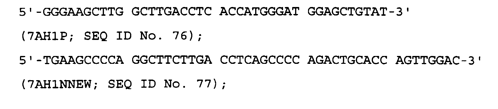

- The VD-DNA fragment (SEQ ID No. 74 of the Sequence Listing) encoding the amino acid sequence of SEQ ID No. 75 of the Sequence Listing, was prepared by performing 3-stage PCR, then inserted into a plasmid and cloned into E. coli.
- The outline of the first stage PCR for the preparation of VD-DNA is shown in Figure 20.
- The H7A1-DNA fragment, encoding a secretion signal sequence and an N-terminal portion of FRH1 and having a Hind III restriction enzyme cleavage site added at the 5'-end, was prepared as follows.
Composition of the PCR reaction solution: plasmid pME-H DNA, 200 ng; oligonucleotide primer 7AH1P, 80 pmol; oligonucleotide primer 7AH1NNEW, 80 pmol; dNTP cocktail, 20 µl; 10×Pfu buffer, 20 µl; Pfu DNA polymerase, 10 units; and redistilled water to a final volume of 200 µl. - The PCR reaction was conducted as follows. The solution was first heated at 94°C for 2 minutes, after which a cycle of heating to 94°C for 1 minute, 55°C for 1 minute and 72°C for 2 minutes, was repeated 30 times. After completion of this procedure, the reaction solution was heated at 72°C for 10 minutes.
- The H7A2-DNA fragment, encoding a portion of FRH1, CDRH1, and a portion of FRH2, was prepared as follows.
Composition of the PCR reaction solution: plasmid pME-H DNA, 200 ng; oligonucleotide primer 7AH2N, 80 pmol; oligonucleotide primer 7AH2PNEW, 80 pmol; dNTP cocktail, 20 µl; 10×Pfu buffer, 20 µl; Pfu DNA polymerase, 10 units; and redistilled water to a final volume of 200 µl. - The PCR reaction was conducted as follows. The solution was first heated at 94°C for 2 minutes, after which a cycle of heating to 94°C for 1 minute, 55°C for 1 minute and 72°C for 2 minutes, was repeated 30 times. After completion of this procedure, the reaction solution was heated at 72°C for 10 minutes.
- The H7A3-DNA fragment, encoding a portion of FRH2, CDRH2 and a portion of FRH3, was prepared as follows.
Composition of the PCR reaction solution: plasmid pME-H DNA, 200 ng; oligonucleotide primer 7AH3P, 80 pmol; oligonucleotide primer 7AH3N, 80 pmol; dNTP cocktail, 20 µl; 10×Pfu buffer, 20 µl; Pfu DNA polymerase, 10 units; and redistilled water to a final volume of 200 µl. - The PCR reaction was conducted as follows. The solution was first heated at 94°C for 2 minutes, after which a cycle of heating to 94°C for 1 minute, 55°C for 1 minute and 72°C for 2 minutes, was repeated 30 times. After completion of this procedure, the reaction solution was heated at 72°C for 10 minutes.
- The H7A4-DNA fragment, encoding a portion of FRH3, CDRH3, FRH4 and the 5 N-terminal amino acid residues of the CH1 region, was prepared as follows.
Composition of the PCR reaction solution: plasmid pME-H DNA, 200 ng; oligonucleotide primer 7AH4P, 80 pmol; oligonucleotide primer 7AH4N, 80 pmol; dNTP cocktail, 20 µl; 10×Pfu buffer, 20 µl; Pfu DNA polymerase, 10 units; and redistilled water to a final volume of 200 µl. - The PCR reaction was conducted as follows. The solution was first heated at 94°C for 2 minutes, after which a cycle of heating to 94°C for 1 minute, 55°C for 1 minute and 72°C for 2 minutes, was repeated 30 times. After completion of this procedure, the reaction solution was heated at 72°C for 10 minutes.
- The respective PCR products were first subjected to phenol extraction and then to ethanol precipitation, and then separated by 5% w/v polyacrylamide gel electrophoresis. After electrophoresis, the gel was stained with 1 µg/ml of ethidium bromide and the DNA bands corresponding to H7A1-DNA, H7A2-DNA, H7A3-DNA and H7A4-DNA, detected under UV light, were cut out using a razor blade and eluted from the gel using a Centriruter and Centricon-10, as described above. The eluted DNA was concentrated by centrifugation at 7,500 x g, followed by ethanol precipitation, and dissolved in 50 µl of distilled water.
- The outline of the second stage PCR for the preparation of VD-DNA is shown in Figure 21.
- The H7A1.2-DNA fragment, in which the above described H7A1-DNA and H7A2-DNA fragments were fused, was prepared as follows.
Composition of the PCR reaction solution: H7A1-DNA solution prepared in the first stage PCR, 10 µl; H7A2-DNA solution prepared in the first stage PCR, 10 µl; oligonucleotide primer 7AH1P, 80 pmol; oligonucleotide primer 7AH2N, 80 pmol; dNTP cocktail, 20 µl; 10×Pfu buffer, 20 µl; Pfu DNA polymerase, 10 units; and redistilled water to a final volume of 200 µl. - The PCR reaction was conducted as follows. The solution was first heated at 94°C for 2 minutes, after which a cycle of heating to 94°C for 1 minute, 55°C for 1 minute and 72°C for 2 minutes, was repeated 30 times. After completion of this procedure, the reaction solution was heated at 72°C for 10 minutes.
- The H7A3.4-DNA fragment, in which the above described H7A3-DNA and H7A4-DNA fragments were fused, was prepared as follows.
Composition of the PCR reaction solution: H7A3-DNA solution prepared in the first stage PCR, 10 µl; H7A4-DNA solution prepared in the first stage PCR, 10 µl; oligonucleotide primer 7AH3P, 80 pmol; oligonucleotide primer 7AH4N, 80 pmol; dNTP cocktail, 20 µl; 10×Pfu buffer, 20 µl; Pfu DNA polymerase, 10 units; and redistilled water to a final volume of 200 µl. - The PCR reaction was conducted as follows. The solution was first heated at 94°C for 2 minutes, after which a cycle of heating to 94°C for 1 minute, 55°C for 1 minute and 72°C for 2 minutes, was repeated 30 times. After completion of this procedure, the reaction solution was heated at 72°C for 10 minutes.
- The resulting, amplified H7A1.2-DNA and H7A3.4-DNA fragments were subjected first to phenol extraction and then to ethanol precipitation, and then separated by 5% w/v polyacrylamide gel electrophoresis. After electrophoresis, the gels were stained with 1 µg/ml of ethidium bromide and the relevant bands thus detected were cut out using a razor blade and eluted from the gel using a Centriruter and Centricon-10, as described above. The eluted DNA was concentrated by centrifugation at 7,500 x g, followed by ethanol precipitation, and dissolved in 50 µl of distilled water.
- The outline of the third stage PCR for the preparation of VD-DNA is shown in Figure 22.
- The VD-DNA fragment, in which above described H7A1.2-DNA and H7A3.4-DNA fragments were fused, was prepared as follows.
Composition of the PCR reaction solution: H7A1.2-DNA solution prepared in the second stage PCR, 10 µl; H7A3.4-DNA solution prepared in the second stage PCR, 10 µl; oligonucleotide primer 7AH1P, 80 pmol; oligonucleotide primer 7AH4N, 80 pmol; dNTP cocktail, 20 µl; 10×Pfu buffer, 20 µl; Pfu DNA polymerase, 10 units; and redistilled water to a final volume of 200 µl. - The PCR reaction was conducted as follows. The solution was first heated at 94°C for 2 minutes, after which a cycle of heating to 94°C for 1 minute, 55°C for 1 minute and 72°C for 2 minutes, was repeated 30 times. After completion of this procedure, the reaction solution was heated at 72°C for 10 minutes.
- The resulting, amplified VD-DNA fragment was subjected first to phenol extraction and then to ethanol precipitation, and then separated by 5% w/v polyacrylamide gel electrophoresis. After electrophoresis, the gel was stained with 1 µg/ml of ethidium bromide and the VD-DNA band thus detected was cut out using a razor blade and eluted from the gel using a Centriruter and Centricon-10, as described above. The eluted DNA was concentrated by centrifugation at 7,500 x g, followed by ethanol precipitation, and dissolved in 50 µl of distilled water.
- The construction of a plasmid carrying the VD-DNA fragment is outlined in Figure 23.
- The VD-DNA fragment obtained above was further purified by phenol extraction followed by ethanol precipitation, and then digested with the restriction enzymes Hind III and ApaI.
- One µg of the plasmid vector pBLUESCRIPT-II SK+ DNA (Stratagene) was digested with Hind III and Apa I, and then dephosphorylated using CIP. The resulting, dephosphorylated plasmid DNA and 100 ng of the VD-DNA fragment, which had also been digested with Hind III and Apa I, were ligated using a DNA Ligation Kit Version 2.0 (Takara Shuzo Co., Ltd.). The resulting ligation mix was then used to transform E. coli JM109, which was then plated on LB agar plates containing final concentrations of 1 mM IPTG, 0.1% w/v X-Gal and 50 µg/ml ampicillin. Any resulting white transformants were cultured in 2 ml liquid LB medium containing 50 µg/ml ampicillin at 37°C overnight, and plasmid DNA was then extracted from the culture by the alkaline-SDS method. The resulting plasmid was digested with Hind III and Apa I and subjected to agarose gel electrophoresis to confirm the presence or absence of the insert of interest. Accordingly, the plasmid pBL7A27 with a VD-DNA insert was obtained.
- A 5'-terminal human IgG1 genomic DNA fragment was synthesised by PCR. For this, the following 2 oligonucleotide primers were prepared:

- Genomic DNA, comprising the CHl region of human IgGl together with an intron following a Hind III cleavage sequence, was separated and amplified by PCR using human genomic DNA as the template, and then inserted into the plasmid pHSG399 (Takara Shuzo Co., Ltd.) and cloned into E. coli. The preparation of this DNA (hereinafter referred to as "IG5'-DNA") is outlined in Figure 24.
- An IG5'-DNA fragment was prepared as follows.
Composition of the PCR reaction solution: human genomic DNA (Clonetech), 2 µg; oligonucleotide primer 5'Hind, 80 pmol; oligonucleotide primer IGGCPSTN, 80 pmol; dNTP cocktail, 20 µl; 10×Pfu buffer, 20 µl; Pfu DNA polymerase, 10 units; and redistilled water to a final volume of 200 µl. - The PCR reaction was conducted as follows. The solution was first heated at 94°C for 2 minutes, after which a cycle of heating to 94°C for 1 minute, 55°C for 1 minute and 72°C for 2 minutes, was repeated 30 times. After completion of this procedure, the reaction solution was heated at 72°C for 10 minutes.
- The resulting, amplified IG5'-DNA fragment was subjected first to phenol extraction and then to ethanol precipitation, and separated by 5% w/v polyacrylamide gel electrophoresis. After electrophoresis, the gel was stained with 1 µg/ml of ethidium bromide and the IG5'-DNA band thus detected was cut out using a razor blade and eluted from the gel using a Centriruter and Centricon-10, as described above. The eluted DNA was concentrated by centrifugation at 7,500 x g, followed by ethanol precipitation, and dissolved in 50 µl of distilled water.
- The IG5'-DNA fragment thus obtained was further purified by phenol extraction and then ethanol precipitation, and was then digested with the restriction enzymes Hind III and Pst I.
- One µg of plasmid pHSG399 DNA (Takara Shuzo Co., Ltd.) was digested with the restriction enzymes Hind III and Pst I, and then dephosphorylated using CIP. The resulting dephosphorylated plasmid DNA and 100 ng of the IG5'-DNA fragment, which had also been digested with Hind III and Pst I, were ligated using a DNA Ligation Kit Version 2.0 (Takara Shuzo Co., Ltd.). The ligation mix was then used to transform E. coli JM109, which was then plated onto LB agar medium containing final concentrations of 1 mM IPTG, 0.1% w/v X-Gal and 50 µg/ml chloramphenicol. Any white transformants obtained were cultured in 2 ml liquid LB medium containing 50 µg/ml chloramphenicol at 37°C overnight, and plasmid DNA was extracted from the resulting culture by the alkaline-SDS method. The extracted plasmid DNA was then digested with Hind III and Pst I, and subjected to 1% w/v agarose gel electrophoresis to confirm the presence or absence of the insert of interest. Accordingly, the plasmid pIG5'03, containing a IG5'-DNA fragment insert, was obtained.
- A 3'-terminal human IgGl genomic DNA fragment was synthesised by PCR. For this, the following 2 primers were prepared:

- DNA comprising the sequence: intron from human IgG1; hinge region; intron from human IgGl; CH2 region; intron from human IgGl; CH3 region; and an EcoRI cleavage sequence, was separated and amplified by PCR using human genomic DNA as the template, and then inserted into the plasmid pHSG399 (Takara Shuzo Co., Ltd.) and cloned into E. coli. The preparation of the above DNA (hereinafter referred to as "IG3'-DNA") is outlined in Figure 25.
- IG3'-DNA was prepared as follows.
Composition of the PCR reaction solution: human genomic DNA (Clonetech), 2 µg; oligonucleotide primer IGGCPSTP, 80 pmol; oligonucleotide primer Eco3', 80 pmol; dNTP cocktail, 20 µl; 10×Pfu buffer, 20 µl; Pfu DNA polymerase, 10 units; and redistilled water to a final volume of 200 µl. - The PCR reaction was conducted as follows. The solution was first heated at 94°C for 2 minutes, after which a cycle of heating to 94°C for 1 minute, 55°C for 1 minute and 72°C for 2 minutes, was repeated 30 times. After completion of this procedure, the reaction solution was heated at 72°C for 10 minutes.
- The resulting, amplified IG3'-DNA fragment was subjected first to phenol extraction and then to ethanol precipitation, and then separated by 5% w/v polyacrylamide gel electrophoresis. After electrophoresis, the gel was stained with 1 µg/ml of ethidium bromide and the IG3'-DNA band thus detected was cut out using a razor blade and eluted from the gel using a Centriruter and Centricon-10, as described above. The eluted DNA was concentrated by centrifugation at 7,500 x g, followed by ethanol precipitation, and dissolved in 50 µl of distilled water.
- The IG3'-DNA fragment thus obtained was further purified by phenol extraction and then ethanol precipitation, and was then digested with the restriction enzymes EcoRI and Pst I.
- One µg of plasmid pHSG399 DNA (Takara Shuzo Co., Ltd.) was digested with EcoRI and Pst I, and then dephosphorylated using CIP. The resulting dephosphorylated plasmid DNA was then ligated with 100 ng of the IG3'-DNA fragment, which had also been digested with EcoRI and Pst I, using a DNA Ligation Kit Version 2.0 (Takara Shuzo Co., Ltd.). The ligation mix was then used to transform E. coli JM109, which was plated onto LB agar medium containing final concentrations of 1 mM IPTG, 0.1% w/v X-Gal and 50 µg/ml chloramphenicol. Any white colonies were selected and the plasmid pIG3'08, containing an IG3'-DNA insert, was obtained.
- The expression plasmid vector pEg7AH-H, carrying the DNA of SEQ ID No. 88 of the Sequence Listing and encoding the humanised HFE7A heavy chain polypeptide of SEQ ID No. 89 of the Sequence Listing, was constructed using the above described plasmids pBL7A27, pIG5'03 and pIG3'08. The procedure is outlined in Figure 26.
- Ten µg of plasmid pIG5'03 DNA, comprising the CH1 region of human IgG1 heavy chain and an intron, was digested with the restriction enzymes ApaI and Kpn I. In addition, 1 µg of pBL7A27 DNA above was also digested with the restriction enzymes Apa I and Kpn I, and then dephosphorylated using CIP. The resulting dephosphorylated pBL7A27 DNA (100 ng) was ligated with 10 µg of the digested and dephosphorylated pIG5'03 DNA, using a DNA Ligation Kit Version 2.0 (Takara Shuzo Co., Ltd.). The ligation mix was then used to transform E. coli JM109, which was plated on LB medium containing 50 µg/ml ampicillin. Resulting transformants were cultured in 2 ml liquid LB medium containing 50 µg/ml ampicillin at 37°C overnight, and plasmid DNA was extracted from the culture by the alkaline-SDS method. The plasmid was digested with Apa I and Kpn I, or with Hind III and Pst I, to confirm the presence or absence of the insert of interest by 1% w/v agarose gel electrophoresis. Thus, the plasmid pBL7AF184, containing a VD-DNA fragment of humanised HFE7A connected with the IGS'-DNA fragment, was obtained.
- Next, 10 µg of the thus obtained plasmid pBL7AF184 was digested with the restriction enzymes Hind III and Pst I, and 1 µg of the plasmid pIG3'08 DNA above was likewise digested with Hind III and Pst I, and dephosphorylated using CIP. The resulting dephosphorylated pIG3'08 DNA (100 ng) was ligated with 10 µg of the digested pBL7AF184 DNA, using a DNA Ligation Kit Version 2.0 (Takara Shuzo Co., Ltd.). E. coli JM109 was transformed with the ligation mix and plated onto LB medium containing 50 µg/ml ampicillin. Any resulting transformants were cultured in 2 ml liquid LB medium containing 50 µg/ml chloramphenicol at 37°C overnight, and plasmid DNA was extracted from the culture by the alkaline-SDS method. The plasmid was digested with Hind III and Pst I, or with Hind III and EcoRI, to confirm the presence or absence of the insert of interest by 1% w/v agarose gel electrophoresis.
- Plasmid pgHSL7A62, containing a VD-DNA fragment of humanised HFE7A connected to a genomic DNA fragment encoding human IgGl constant region, was obtained. The transformant E. coli pgHSL7A62 SANK 73397 harboring plasmid pgHSL7A62 was deposited with the Kogyo Gijutsuin Seimei-Kogaku Kogyo Gijutsu Kenkyujo on August 22, 1997, in accordance with the Budapest Treaty, and was accorded the accession number FERM BP-6074.
- Ten microgrammes of the thus obtained plasmid pgHSL7A62 DNA were digested with the restriction enzymes Hind III and EcoRI and, likewise, 1 µg of the expression plasmid pEE.6.1 DNA was digested with Hind III and EcoRI, and dephosphorylated using CIP. The resulting dephosphorylated pEE.6.1 DNA (100 ng) was ligated with 10 µg of the digested pgHSL7A62 DNA, using a DNA Ligation Kit Version 2.0 (Takara Shuzo Co., Ltd.). E. coli JM109 was transformed with the ligation mix and plated onto LB medium containing 50 µg/ml ampicillin. Any resulting transformants were cultured in 2 ml liquid LB medium containing 50 µg/ml ampicillin at 37°C overnight, and plasmid DNA was extracted from the culture by the alkaline-SDS method. The plasmid was digested with Hind III and EcoRI, to confirm the presence or absence of the insert of interest by 1% w/v agarose gel electrophoresis.
- The resulting plasmid, pEg7AH-H, contained a fusion fragment comprising a VD-DNA fragment of humanised HFE7A and a genomic DNA fragment encoding human IgG1 constant region in connection and inserted downstream of the CMV promoter in the correct orientation.
- To verify that the DNA insert of the pEg7AH-H had the expected nucleotide sequence, the DNA insert was analyzed to determine the nucleotide sequence. For this, the following primers were synthesised:
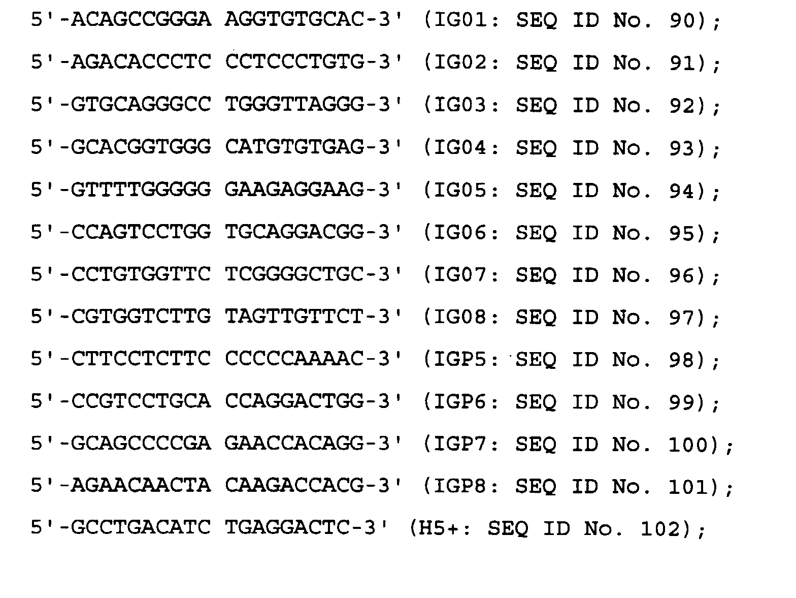

- The positions to which each primer binds are shown in Figure 27. The determination of the nucleotide sequence was performed by the dideoxynucleotide chain termination method (ibid.) using, as templates, the respective plasmids purified by the alkaline-SDS method and the caesium chloride method (ibid.). It was confirmed that pEg7AH-H had the nucleotide sequence of SEQ ID No. 88 of the Sequence Listing, encoding the polypeptide of SEQ ID No. 89 of the Sequence Listing.
- COS-1 cells (derived from a monkey kidney) were transfected with the expression plasmids for the humanised HFE7A heavy chain and with the expression plasmids for each of the humanised HFE7A light chains obtained above. Transfection was performed by electroporation using the gene transfection apparatus GTE-1 (Shimadzu Seisakusyo, K. K.) equipped with an FCT-13 chamber having electrodes separated by 2 mm (Shimadzu Seisakusyo, K. K.).
- COS-1 cells (American Type Culture Collection No. CRL-1650) were grown to semi-confluence in a culture flask (culture area: 225 cm2; Sumitomo Bakelite) containing Minimal Essential a medium ["α(+)MEM"; Gibco BRL] supplemented with 10% v/v FCS (Moregate). Subsequently, the medium was discarded and the COS-1 cells were detached from the flask by treatment with 3 ml of trypsin-EDTA solution (Sigma Chemicals Co.) at 37°C for 3 minutes. The detached cells were then harvested by centrifugation at 800 r.p.m. for 2 minutes, discarding the supernatant and washing twice with phosphate buffer (0.02% w/v potassium chloride [KCl], 0.02% w/v potassium dihydrogenphoshate [KH2PO4], 0.8% w/v sodium chloride [NaCl], 1.15% w/v disodium hydrogenphosphate [Na2HPO4] ; hereinafter referred to as "PBS(-) buffer"; Nissui Pharmaceutical Co., Ltd.). The washed COS-1 cells were then suspended to a cell density of 1 x 10⁸ cells/ml in PBS(-) buffer.
- In parallel, 4 µg of humanised HFE7A heavy chain expression plasmid DNA were mixed with 4 µg of humanised HFE7A light chain expression plasmid DNA, each purified by the alkaline-SDS method and caesium chloride density gradient centrifugation. The resulting mixture was subjected to ethanol precipitation and then suspended in 20 µl of PBS(-) buffer. These mixing, precipitation and resuspension steps were all performed in the same tube. The whole of the resulting plasmid suspension (20 µl) was mixed with 20 µl of the previously prepared COS-1 cell suspension (2 x 10⁶ cells) and the mixture was transferred to an FCT-13 electroporation chamber (Shimadzu Seisakusyo, K. K.) having electrodes set 2 mm apart, which was then loaded into gene transfection apparatus GTE-1 (Shimadzu Seisakusyo, K. K.). Pulses of 600 V, each of 50 µF were applied twice with a 1 second interval, in order transform the COS-1 cells with the plasmid DNA. After electroporation, the cell-DNA mixture in the chamber was suspended in 20 µl of α(+)MEM supplemented with 10% v/v FCS and transferred to a culture flask (culture area 75 cm2; Sumitomo Bakelite). After incubating under 5% v/v CO2 at 37°C for 72 hours, the culture supernatant was recovered, and analysis was performed on the supernatants to determine what expression products were present.
- Using the above method, but modified as appropriate, COS-1 cells were variously transfected with each of the following plasmid or plasmid combinations:
- (A): no plasmid DNA
- (B): cotransfection with pEg7AH-H and p7AL-MM
- (C): cotransfection with pEg7AH-H and p7AL-HM
- (D): cotransfection with pEg7AH-H and p7AL-HH
-
- Verification and quantitative assay of the expression of humanised antibodies as expression products in the culture supernatant fluids prepared in Example 4 were performed by ELISA, using an anti-human IgG antibody.
- Goat anti-human IgG Fc specific polyclonal antibody (Kappel) was dissolved to a final concentration of 1 µg/ml in adsorption buffer (0.05 M sodium hydrogencarbonate, 0.02% w/v sodium azide, pH 9.6) and 100 µl aliquots were added to each well of a 96-well plate (MaxiSorb, Nunc), and the plate was incubated at 37°C for 2 hours to encourage adsorption of the antibody. Next, each well was washed with 350 µl of PBS-T [PBS(-) containing 0.05% w/v Tween-20 (BioRad)] four times. After washing, culture supernatant diluted with α(+)MEM containing 10% v/v FCS was added to the wells, and the plate was further incubated at 37°C for 2 hours.
- After this time, the wells were again washed four times with PBS-T, and then 100 µl of alkaline phosphatase-labeled goat anti-human IgG Fc specific polyclonal antibody (Caltag Lab.) diluted 5,000-fold with PBS-T were added to each well and the plate was incubated at 37°C for 2 hours. Each well was then again washed four times with PBS-T, and 100 µl of a substrate solution of 1 mg/ml p-nitrophenyl phosphate, prepared in 10% v/v diethanol amine (pH 9.8), was added to each well. After a subsequent incubation at 37°C for 0.5 to 1 hour, absorbance at 405 nm was measured.
- In the present experiments, human plasma IgG subclass 1 (IgGl; Biopure AG) diluted with α(+)MEM containing 10% v/v FCS to certain desired concentrations was used to provide concentration reference samples of the humanised HFE7A antibodies contained in the culture supernatant fluids.
- As expected, each supernatants of transformants (B), (C) and (D) was determined to express human antibody, as detected by anti-human IgG antibody. The negative control, (A), showed no expression of human antibody.
- The assay for Fas-binding activity in the cell culture supernatants prepared in Example 4 was performed by ELISA as follows.
- Culture supernatant from COS-1 cells expressing the human Fas fusion protein, as obtained in Reference Example 1 above, diluted 5-fold with adsorption buffer, was dispensed into wells of a 96-well plate (MaxiSorb; Nunc) at 50 µl per well and the plate was incubated at 4°C overnight to allow adsorption of the human Fas fusion protein to the surface of the wells. Next, each of the wells was washed 4 times with 350 µl of PBS-T. After washing, PBS-T containing 5% v/v BSA (bovine serum albumin; Wako Pure Chemical Industries, Ltd.) was added to the wells at 50 µl per well and the plate was incubated at 37°C for 1 hour to block the remainder of the surface of each well. The wells were then again washed four times with PBS-T.
- The culture supernatants obtained in Example 4 were adjusted to have a final concentration of the product of interest of 100 ng/ml in α(+)MEM containing 10% v/v FCS. Concentrations were estimated by the method described in Example 5. Each of the resulting 100 ng/ml solutions was then used to produce serial dilutions by serial 2-fold dilution with α(+)MEM containing 10% v/v FCS. Next, 50 µl of each of the resulting serial dilutions of each expression product was added to a well prepared as above, and the plate was incubated at 37°C for 2 hours to allow reaction.
- After this time, the wells were again washed four times with PBS-T, and then 50 µl of alkaline phosphatase-labeled goat anti-human IgG Fc specific polyclonal antibody (Caltag Lab.), diluted 10,000-fold with PBS-T, were dispensed into each well and reaction was allowed to proceed at 37°C for 2 hours.
- HFE7A purified from mouse hybridoma HFE7A was used as a control (IgGl), and was detected using alkaline phosphatase-labeled goat anti-mouse IgG + IgA + IgM (Gibco BRL), diluted 5,000-fold with PBS-T, in place of the alkaline phosphatase-labeled goat anti-human IgG Fc specific polyclonal antibody.
- The wells were again washed four times with PBS-T, and then 50 µl of substrate solution [1 mg/ml p-nitrophenyl phosphate in 10% v/v diethanol amine (pH9.8)] was dispensed into each well and the plate was incubated at 37°C for 0.5 to 1 hour. Binding activity of the expression product contained in each culture supernatant fluid with the human Fas fusion protein was evaluated by reading the absorbance of each well at 405 nm.
- As expected, binding activity to the human Fas fusion protein was demonstrated for supernatants (B), (C) and (D) above (Figure 28).
- The humanised anti-Fas antibodies of the Examples should inhibit the binding of HFE7A to Fas, as the antibodies of the Examples were derived from HFE7A. Therefore, the ability of the expression products obtained in Example 4 to competitively inhibit the binding of HFE7A to the human Fas fusion protein was measured.
- One mg of the purified monoclonal antibody HFE7A obtained in Reference Example 3 was labeled using a commercially available alkaline phosphatase labeling kit (Immuno-Link AP and APL Labeling Kit; Genosis), using the protocol supplied with the kit. The resulting, labeled antibody is also referred to herein as "AP-HFE7A".
- The COS-1 cell culture supernatant containing the human Fas fusion protein, as obtained in Reference Example 1, was diluted 5-fold with adsorption buffer, and dispensed into the wells of a 96-well plate for luminescence detection (Luminescent Solid Assay Plate, high binding property; Costar) at 50 µl per well. The plate was then incubated at 4°C overnight to allow adsorption of the human Fas fusion protein to the surface of the wells.
- After this time, each well was washed 4 times with 350 µl of PBS-T, and then 100 µl PBS-T containing 5% v/v BSA was added to each well and the plate was incubated at 37°C for 1 hour to block the remainder of the surface of each well. The wells were then again washed four times with PBS-T.
- The culture supernatants obtained in Example 4 were adjusted to final concentrations of antibody of 1 µg/ml in α(+)MEM containing 10% v/v FCS by the method of Example 5. Each of the resulting solutions of the expression products was used to produce serial dilutions by serial 2-fold dilution with α(+)MEM containing 10% v/v FCS. AP-HFE7A was diluted to 50 ng/ml with α(+)MEM containing 10% v/v FCS, and 25 µl of the resulting solution was mixed with an equal volume of each of the prepared serial dilutions.
- Each of the wells was again washed four times with PBS-T, and then 50 µl of each of the resulting antibody mixtures were added to individual wells, and the plate was allowed to stand at at room temperature overnight. Subsequently, after washing each well with PBS-T again four times, 100 µl of CDP-star buffer (9.58 ml diethanol amine, 0.2 g magnesium chloride, 0.25 g sodium azide, pH8.5) was dispensed into each well and the plate was allowed to stand at room temperature for 10 minutes. After this time, the CDP-star buffer was discarded and CDP-star substrate [1.2 ml sapphire II (Tropix), 200 µl CDP-star (Tropix), q.s. to 12 ml with CDP-star buffer] was added at 50 µl per well, and the plate was then allowed to stand at room temperature for a further 40 minutes.
- Competitive inhibition of the expression products of Example 4 of the binding of HFE7A to the human Fas fusion protein was evaluated by measuring the intensity of the luminescence with Luminoscan (Titertech).
- As a result, it was verified that the expression products prepared in Example 4 specifically inhibited the binding of HFE7A to the human Fas fusion protein (Figure 29).
- WR19L12a cells (c.f. Itoh, N. et al., ibid.) were used to examine the apoptosis-inducing activity of the COS-1 cell culture supernatant of Example 4.
- WR19L12a cells were cultured in RPMI1640 medium with 10% v/v FCS (Gibco BRL) at 37°C for 3 days under 5% v/v CO2, and 50 µl (1 x 10⁵ cells) of the resulting culture were then dispensed into each well of a 96-well microplate (Sumitomo Bakelite). The culture supernatants obtained in Example 4 were adjusted to a final concentration of antibody of 100 ng/ml in RPMI1640 medium containing 10% v/v FCS. Concentrations were estimated by the method of Example 5. Each of the adjusted solutions of the expression products was used to produce serial dilutions by serial 2-fold dilution with RPMI1640 containing 10% v/v FCS. Each of the resulting dilutions of each expression product solution was added to individual wells, at 50 µl per well, and the plate was incubated at 37°C for 1 hour. After this time, the cells in each well were washed four times with RPMI1640 containing 10% v/v FCS and then the washed cells were suspended in 75 µl per well of RPMI1640 containing 10% v/v FCS.
- Subsequently, 75 µl of 1.25 µg/ml goat anti-human IgG Fc specific polyclonal antibody (Kappel) in RPMI 1640 medium containing 10% v/v FCS was added to each well, as secondary antibody. The plate was allowed to stand at 37°C for 12 hours, and then 50 µl of 25 µM PMS (phenazine methosulphate; Sigma Chemical Co.), containing 1 mg/ml XTT [2,3-bis(2-methoxy-4-nitro-5-sulphophenyl) -2H-tetrazolium-5-carboxyanilide inner salt; Sigma Chemical Co.] to final concentrations of 250 µg/ml for XTT and 5 µM for PMS, were added to each well. The plate was then incubated for 3 hours at 37°C, and the absorbance at 450 nm of each well was measured, to calculate cell viability, using the reducing power of the mitochondria as the index.
- The viability of the cells in each well was calculated according to the following formula:
- As expected, each of the expression products (B), (C) and (D) of Example 4, were demonstrated to induce apoptosis in T cells expressing the human Fas antigen (Figure 30).
- In order to humanise the amino acid sequence of the light chain of the mouse anti-human Fas antibody HFE7A, the 1st amino acid (aspartic acid), the 85th amino acid (alanine) and the 107th amino acid (arginine) from the N-terminus of the amino acid sequence of the HH type light chain were replaced with glutamic acid, glutamic acid and lysine, respectively. These replacement residues are conserved in the human light chain (κ chain). The resulting sequence was designated as "PDHH type." For the HM light chain sequence, the 1st amino acid (aspartic acid) and the 107th amino acid (arginine) from the N-terminus of the amino acid sequence were replaced with the conserved glutamic acid and lysine residues, respectively. The resulting sequence was designated as "PDHM type."
- Expression plasmids separately carrying these 2 types of humanised light chain amino acid sequences (PDHH and PFHM) were constructed as follows.
- DNA (SEQ ID No. 106 of the Sequence Listing) encoding the PDHH type polypeptide chain (SEQ ID No. 107 of the Sequence Listing) and DNA (SEQ ID No. 108 of the Sequence Listing) encoding the PDHM type polypeptide chain (SEQ ID No. 109 of the Sequence Listing) were prepared by PCR. Each of these sequences is a fusion of one the humanised versions of the variable region of the HFE7A light chain with the constant region of the human Ig light chain (κ chain).
- 7AL1P (SEQ ID No. 55) and 7ALCN (SEQ ID No. 64) had already been synthesised [Example 2 (2) 2) above], and the following oligonucleotide primers were also synthesised for PCR:
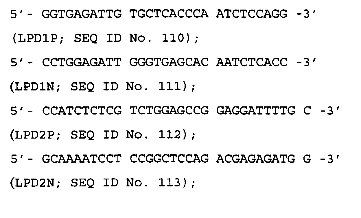

- LPDHH-DNA (light chain constant region fused with PDHH DNA), as defined in SEQ ID No. 106 of the Sequence Listing, and encoding the amino acid sequence of SEQ ID No. 107 of the Sequence Listing, was prepared by performing 3-stage PCR, inserted into a plasmid vector, and cloned into E. coli.
- The outline of the first stage PCR for the preparation of LPDHH-DNA is shown in Figure 31.
- The LPD1-DNA fragment, encoding a secretion signal sequence and a portion of FRL1, but having an added HindIII restriction enzyme cleavage site at the 5'-end, was prepared as follows.
Composition of the PCR reaction solution: plasmid pHSGHH7 DNA, 200 ng; oligonucleotide primer 7AL1P, 80 pmol; oligonucleotide primer LPD1N, 80 pmol; dNTP cocktail, 20 µl; 10× Pfu buffer, 20 µl; Pfu DNA polymerase (Stratagene), 10 units; and redistilled water to a final volume of 200 µl. - The PCR reaction was conducted as follows. The solution was first heated at 94°C for 2 minutes, after which a cycle of heating to 94°C for 1 minute, 55°C for 1 minute and 72°C for 2 minutes, was repeated 30 times. After completion of this procedure, the reaction solution was heated at 72°C for 10 minutes.
- The LPDHH1-DNA fragment, encoding a portion of FRL1, CDRL1, FRL2, CDRL2 and a portion of the FRL3 region, was prepared as follows.
Composition of the PCR reaction solution: plasmid pHSGHH7 DNA, 200 ng; oligonucleotide primer LPD1P, 80 pmol; oligonucleotide primer LPD2N, 80 pmol; dNTP cocktail, 20 µl; 10× Pfu buffer, 20 µl; Pfu DNA polymerase, 10 units; and redistilled water to a final volume of 200 µl. - The PCR reaction was conducted as follows. The solution was first heated at 94°C for 2 minutes, after which a cycle of heating to 94°C for 1 minute, 55°C for 1 minute and 72°C for 2 minutes, was repeated 30 times. After completion of this procedure, the reaction solution was heated at 72°C for 10 minutes.
- The LPDHH2-DNA fragment, encoding a portion of FRL3, CDRL3 and FRL4, was prepared as follows.
Composition of the PCR reaction solution: plasmid pHSGHH7 DNA, 200 ng; oligonucleotide primer LPD2P, 80 pmol; oligonucleotide primer LPD3N, 80 pmol; dNTP cocktail, 20 µl; 10× Pfu buffer, 20 µl; Pfu DNA polymerase, 10 units; and redistilled water to a final volume of 200 µl. - The PCR reaction was conducted as follows. The solution was first heated at 94°C for 2 minutes, after which a cycle of heating to 94°C for 1 minute, 55°C for 1 minute and 72°C for 2 minutes, was repeated 30 times. After completion of this procedure, the reaction solution was heated at 72°C for 10 minutes.
- The LPDC-DNA fragment, encoding a portion of FRL4 and the HFE7A light chain constant region, but having an EcoRI restriction enzyme cleavage site added at the 3'-end, was prepared as follows.
Composition of the PCR reaction solution: plasmid pHSGHH7 DNA, 200 ng; oligonucleotide primer LPD3P, 80 pmol; oligonucleotide primer 7ALCN, 80 pmol; dNTP cocktail, 20 µl; 10× Pfu buffer, 20 µl; Pfu DNA polymerase, 10 units; and redistilled water to a final volume of 200 µl. - The PCR reaction was conducted as follows. The solution was first heated at 94°C for 2 minutes, after which a cycle of heating to 94°C for 1 minute, 55°C for 1 minute and 72°C for 2 minutes, was repeated 30 times. After completion of this procedure, the reaction solution was heated at 72°C for 10 minutes.
- After PCR, the amplified DNA fragments were subjected first to phenol extraction and then to ethanol precipitation, and then separated by 5% w/v polyacrylamide gel electrophoresis. After electrophoresis, the gel was stained with 1 µg/ml of ethidium bromide, and the DNA bands thus detected, under UV light, were cut out with a razor blade and eluted from the gel using a Centriruter and a Centricon-10, as described above. The eluted DNA was concentrated by centrifugation at 7,500 x g, followed by ethanol precipitation, and was finally dissolved in 50 µl of distilled water.
- The outline of the second stage PCR for the production of LPDHH-DNA is shown in Figure 32.
- LPDHH1.2-DNA, in which the above described LPD1-DNA, LPDHH1-DNA and LPDHH2-DNA fragments are fused, was prepared as follows.
Composition of the PCR reaction solution: LPD1-DNA solution (from the first stage PCR), 10 µl; LPDHH1-DNA solution (from the first stage PCR), 10 µl; LPDHH2-DNA solution (from the first stage PCR), 10 µl; oligonucleotide primer 7AL1P, 80 pmol; oligonucleotide primer LPD3N, 80 pmol; dNTP cocktail, 20 µl; 10× Pfu buffer, 20 µl; Pfu DNA polymerase, 10 units; and redistilled water to a final volume of 200 µl. - The PCR reaction was conducted as follows. The solution was first heated at 94°C for 2 minutes, after which a cycle of heating to 94°C for 1 minute, 55°C for 1 minute and 72°C for 2 minutes, was repeated 30 times. After completion of this procedure, the reaction solution was heated at 72°C for 10 minutes.
- After PCR, the amplified LPDHH1.2 fragment was subjected first to phenol extraction and then to ethanol precipitation, and then separated by 5% w/v polyacrylamide gel electrophoresis. After electrophoresis, the gel was stained with 1 µg/ml of ethidium bromide, and the fusion DNA band thus detected, under UV light, was cut out with a razor blade and eluted from the gel using a Centriruter and a Centricon-10, as described above. The eluted DNA was concentrated by centrifugation at 7,500 x g, followed by ethanol precipitation, and was finally dissolved in 50 µl of distilled water.
- The outline of the third stage PCR for the production of LPDHH-DNA is shown in Figure 33.
- The LPDHH-DNA fragment, in which the above described LPDHH1.2-DNA and LPDC-DNA fragments were fused, was prepared as follows.
Composition of the PCR reaction solution: LPDHH1.2-DNA solution (from second stage PCR), 10 µl; LPDC-DNA solution (from first stage PCR), 10 µl; oligonucleotide primer 7AL1P, 80 pmol; oligonucleotide primer 7ALCN, 80 pmol; dNTP cocktail, 20 µl; 10× Pfu buffer, 20 µl; Pfu DNA polymerase, 10 units; and redistilled water to a final volume of 200 µl. - The PCR reaction was conducted as follows. The solution was first heated at 94°C for 2 minutes, after which a cycle of heating to 94°C for 1 minute, 55°C for 1 minute and 72°C for 2 minutes, was repeated 30 times. After completion of this procedure, the reaction solution was heated at 72°C for 10 minutes.
- After PCR, the amplified LPDHH-DNA fragment was subjected first to phenol extraction and then to ethanol precipitation, and then separated by 5% w/v polyacrylamide gel electrophoresis. After electrophoresis, the gel was stained with 1 µg/ml of ethidium bromide, and the DNA band thus detected, under UV light, was cut out with a razor blade and eluted from the gel using a Centriruter and a Centricon-10, as described above. The eluted DNA was concentrated by centrifugation at 7,500 x g, followed by ethanol precipitation, and was finally dissolved in 50 µl of distilled water.
- Construction of an expression plasmid carrying the LPDHH-DNA fragment is outlined in Figure 34.
- The LPDHH-DNA fragment, obtained above, was further purified by phenol extraction followed by ethanol precipitation, and then digested with the restriction enzymes HindIII and EcoRI.
- One µg of the cloning plasmid pHSG399 DNA was digested with the restriction enzymes HindIII and EcoRI, and then dephosphorylated with CIP. The resulting dephosphorylated pHSG399 DNA was then ligated with the LPDHH-DNA fragment, which had previously also been digested with HindIII and EcoRI, using a DNA Ligation Kit Version 2.0 (Takara Shuzo, Co. Ltd.). E. coli JM109 was then transformed with the ligation mix and plated onto LB agar medium containing final concentrations of 1 mM IPTG, 0.1% w/v X-Gal and 50 µg/ml chloramphenicol. Any white transformants obtained were cultured in 2 ml liquid LB medium containing 50 µg/ml chloramphenicol at 37°C overnight, and plasmid DNA was extracted from the resulting culture by the alkaline-SDS method. The extracted plasmid DNA was digested with HindIII and EcoRI, and a clone carrying the LPDHH-DNA fragment was then selected by 1% w/v agarose gel electrophoresis.
- Accordingly, plasmid pHSHH5, carrying a fusion insert comprising the variable region of the humanised LPDHH DNA and DNA encoding the constant region of human immunoglobulin K chain, was isolated. The transformant E. coli pHSHH5 SANK 70398, harboring plasmid pHSHH5, was deposited with the Kogyo Gijutsuin Seimei-Kogaku Kogyo Gijutsu Kenkyujo on February 26, 1998, in accordance with the Budapest Treaty on the Deposit of Microorganisms, and was accorded the accession number FERM BP-6274.
- The expression vector plasmid pLPDHH75 carrying the DNA fragment of SEQ ID No. 106 of the Sequence Listing, encoding the humanised PDHH type HFE7A light chain polypeptide of SEQ ID No. 107 of the Sequence Listing, was then prepared using the plasmid pHSHH5.
- One µg of pEE.12.1 DNA (Lonza), an expression vector for mammalian cells, was digested with the restriction enzymes HindIII and EcoRI, and then dephosphorylated using CIP. The resulting digested, dephosphorylated plasmid DNA (100 ng) was ligated with 10 µg of the pHSHH5 DNA fragment which had also been digested with Hind III and EcoRI, using a DNA Ligation Kit Version 2.0 (Takara Shuzo Co., Ltd.). The ligation mix was then used to transform E. coli JM109 (as described above), which was then plated on LB agar plates containing 50 µg/ml ampicillin.
- The transformants obtained by this method were cultured in 2 ml of liquid LB medium containing 50 µg/ml ampicillin at 37°C overnight, and plasmid DNA was subsequently extracted from the resulting culture by the alkaline-SDS method.
- The extracted plasmid DNA was digested with HindIII and EcoRI, and subjected to 1% w/v agarose gel electrophoresis to confirm the presence or absence of the insert of interest. This enabled the isolation of the plasmid pLPDHH75, which contains a fusion fragment having the variable region of the humanised PDHH type HFE7A light chain together with DNA encoding the constant region of the human immunoglobulin κ chain. The fusion fragment was found to be located downstream of the cytomegalovirus (CMV) promoter in the correct orientation.
- The LPDHM-DNA fragment (SEQ ID No. 108 of the Sequence Listing, encoding the amino acid sequence of SEQ ID No. 109 thereof) was produced by performing 3-stage PCR, the fragment then being inserted into a plasmid vector and cloned into E. coli.
- The outline of the first stage PCR for the preparation of LPDHM-DNA is shown in Figure 35.
- The LPD1-DNA fragment, encoding a secretion signal sequence and a portion of FRL1 having a HindIII restriction enzyme cleavage site added at the 5'-end, and the LPDC-DNA fragment, encoding a portion of FRL4 and the constant region having an EcoRI restriction enzyme cleavage site added at the 3'-end, were those obtained in the preparation of the LPDHH-DNA fragment [see "2) Construction of plasmid pLPDHH75 (expression plasmid for humanised PDHH type HFE7A light chain)" and "a) First Stage PCR"].
- The LPDHM1-DNA fragment, encoding a portion of CDRL1, FRL2, CDRL2, FRL3, CDRL3 and FRL4, was prepared as follows.
Composition of the PCR reaction solution: plasmid pHSGHM17 DNA, 200 ng; oligonucleotide primer LPD1P, 80 pmol; oligonucleotide primer LPD3N, 80 pmol; dNTP cocktail, 20 µl; 10× Pfu buffer, 20 µl; Pfu DNA polymerase, 10 units; and redistilled water to a final volume of 200 µl. - The PCR reaction was conducted as follows. The solution was first heated at 94°C for 2 minutes, after which a cycle of heating to 94°C for 1 minute, 55°C for 1 minute and 72°C for 2 minutes, was repeated 30 times. After completion of this procedure, the reaction solution was heated at 72°C for 10 minutes.
- After PCR, the amplified LPD1, LPDHM1 and LPDC-DNA fragments were subjected first to phenol extraction and then to ethanol precipitation, and then separated by 5% w/v polyacrylamide gel electrophoresis. After electrophoresis, the gel was stained with 1 µg/ml of ethidium bromide, and the fusion DNA bands thus detected, under UV light, were cut out with a razor blade and eluted from the gel using a Centriruter and a Centricon-10, as described above. The eluted DNA was concentrated by centrifugation at 7,500 x g, followed by ethanol precipitation, and was finally dissolved in 50 µl of distilled water.
- The outline of the second stage PCR for the production of PDHM-DNA is shown in Figure 36.
- LPDHM1.2-DNA, in which the above LPD1-DNA and LPDHM1-DNA fragments were fused, was prepared as follows.
Composition of the PCR reaction solution: LPD1-DNA solution (from the first stage PCR), 10 µl; LPDHM1-DNA solution (from the first stage PCR), 10 µl; oligonucleotide primer 7AL1P, 80 pmol; oligonucleotide primer LPD3N, 80 pmol; dNTP cocktail, 20 µl; 10× Pfu buffer, 20 µl; Pfu DNA polymerase, 10 units; and redistilled water to a final volume of 200 µl. - The PCR reaction was conducted as follows. The solution was first heated at 94°C for 2 minutes, after which a cycle of heating to 94°C for 1 minute, 55°C for 1 minute and 72°C for 2 minutes, was repeated 30 times. After completion of this procedure, the reaction solution was heated at 72°C for 10 minutes.
- After PCR, the amplified LPDHM1.2-DNA fragment was subjected first to phenol extraction and then to ethanol precipitation, and then separated by 5% w/v polyacrylamide gel electrophoresis. After electrophoresis, the gel was stained with 1 µg/ml of ethidium bromide, and the DNA band thus detected, under UV light, was cut out with a razor blade and eluted from the gel using a Centriruter and a Centricon-10, as described above. The eluted DNA was concentrated by centrifugation at 7,500 x g, followed by ethanol precipitation, and was finally dissolved in 50 µl of distilled water.
- The outline of the third stage PCR for the preparation of LPDHM-DNA is shown in Figure 37.
- The LPDHM-DNA fragment, comprising a fusion of the LPDHM1.2-DNA and LPDC-DNA fragments above, was prepared as follows.
Composition of the PCR reaction solution: LPDHM1.2-DNA solution (from the second stage PCR), 10 µl; LPDC-DNA solution (from the first stage PCR), 10 µl; oligonucleotide primer 7AL1P, 80 pmol; oligonucleotide primer 7ALCN, 80 pmol; dNTP cocktail, 20 µl; 10× Pfu buffer, 20 µl; Pfu DNA polymerase, 10 units; and redistilled water to a final volume of 200 µl. - The PCR reaction was conducted as follows. The solution was first heated at 94°C for 2 minutes, after which a cycle of heating to 94°C for 1 minute, 55°C for 1 minute and 72°C for 2 minutes, was repeated 30 times. After completion of this procedure, the reaction solution was heated at 72°C for 10 minutes.
- After PCR, the amplified LPDHM-DNA fragment was subjected first to phenol extraction and then to ethanol precipitation, and then separated by 5% w/v polyacrylamide gel electrophoresis. After electrophoresis, the gel was stained with 1 µg/ml of ethidium bromide, and the DNA band thus detected, under UV light, was cut out with a razor blade and eluted from the gel using a Centriruter and a Centricon-10, as described above. The eluted DNA was concentrated by centrifugation at 7,500 x g, followed by ethanol precipitation, and was finally dissolved in 50 µl of distilled water.
- The construction of a plasmid carrying the LPDHM-DNA fragment is outlined in Figure 38.
- The LPDHM-DNA obtained above was further purified by phenol extraction followed by ethanol precipitation, and then digested with the restriction enzymes HindIII and EcoRI.
- One µg of the cloning plasmid pHSG399 DNA was digested with the restriction enzymes HindIII and EcoRI, and then dephosphorylated with CIP. The resulting dephosphorylated pHSG399 DNA and was then ligated with the LPDHM-DNA fragment, which had previously also been digested with HindIII and EcoRI, using a DNA Ligation Kit Version 2.0 (Takara Shuzo, Co. Ltd.). E. coli JM109 was then transformed with the ligation mix and plated onto LB agar medium containing final concentrations of 1 mM IPTG, 0.1% w/v X-Gal and 50 µg/ml chloramphenicol. Any white transformants obtained were cultured in 2 ml liquid LB medium containing 50 µg/ml chloramphenicol at 37°C overnight, and plasmid DNA was extracted from the resulting culture by the alkaline-SDS method. The extracted plasmid DNA was digested with HindIII and EcoRI, and a clone carrying the LPDHM-DNA fragment was then selected by 1% w/v agarose gel electrophoresis.
- As a result of the above procedure, plasmid pHSHM2 carrying a fusion insert comprising the variable region of the humanised PDMM type HFE7A light chain and DNA encoding the constant region of the human Ig κ chain, was obtained. The transformant E. coli pHSHM2 SANK 70198, harboring plasmid pHSHM2, was deposited with the Kogyo Gijutsuin Seimei-Kogaku Kogyo Gijutsu Kenkyujo on February 26, 1998, in accordance with the Budapest Treaty on the Deposit of Microorganisms, and was accorded the accession number FERM BP-6272.
- The expression vector plasmid pLPDHM32 was constructed, carrying the DNA of SEQ ID No. 108 of the Sequence Listing encoding the humanised PDHM type HFE7A light chain polypeptide of SEQ ID No. 109, using plasmid pHSHM2 obtained above.
- One µg of pEE.12.1 DNA (Lonza), an expression vector for mammalian cells, was digested with the restriction enzymes Hind III and EcoRI, and then dephosphorylated using CIP. The resulting digested, dephosphorylated plasmid DNA (100 ng) was ligated with 10 µg of the pHSHM2 DNA fragment which had also been digested with Hind III and EcoRI, using a DNA Ligation Kit Version 2.0 (Takara Shuzo Co., Ltd.). The ligation mix was then used to transform E. coli JM109 (as described above), which was then plated on LB agar plates containing 50 µg/ml ampicillin.
- The transformants obtained by this method were cultured in 2 ml of liquid LB medium containing 50 µg/ml ampicillin at 37°C overnight, and plasmid DNA was subsequently extracted from the resulting culture by the alkaline-SDS method.
- The extracted plasmid DNA was digested with HindIII and EcoRI, and subjected to 1% w/v agarose gel electrophoresis to confirm the presence or absence of the insert of interest. This enabled the isolation of the plasmid pLPDHM32, which contains a fusion fragment having the variable region of the humanised PDHM type HFE7A light chain together with DNA encoding the constant region of the human immunoglobulin κ chain. The fusion fragment was found to be located downstream of the cytomegalovirus (CMV) promoter in the correct orientation.
- To verify that the DNA inserts of plasmids pLPDHH75 and pLPDHM32 have the desired nucleotide sequences, the DNA inserts were analyzed to determine their nucleotide sequences. The oligonucleotide primers used for nucleotide sequencing were SP1 (SEQ ID No. 68), SP2 (SEQ ID No. 69), SP3 (SEQ ID No. 70), SP4 (SEQ ID No. 71), SP5 (SEQ ID No. 72) and SP6 (SEQ ID No. 73).
- The positions to which each primer binds are shown in Figure 19. The determination of the nucleotide sequence was performed by the dideoxynucleotide chain termination method using, as the templates, the plasmid containing the sequence to be confirmed, the plasmid having been purified by the alkaline-SDS method and the caesium chloride method. As expected pLPDHH75 was confirmed to have the nucleotide sequence of SEQ ID No. 106 of the Sequence Listing, encoding the polypeptide of SEQ ID No. 107, and that pLPDHM32 had the nucleotide sequence of SEQ ID No. 108 of the Sequence Listing, encoding the polypeptide of SEQ ID No. 109.
- In further humanising the amino acid sequence of SEQ ID No. 75 of the Sequence Listing (the humanised heavy chain of the mouse anti-human Fas antibody HFE7A), the 44th amino acid (arginine) and the 76th amino acid (alanine) in the amino acid sequence of SEQ ID No. 75 were replaced with glycine and threonine, respectively, these residues being conserved in the human heavy chain. The resulting sequence was designated as the "HV type."
- Expression plasmids carrying the HV type humanised heavy chain amino acid sequences of the anti-human Fas antibody HFE7A were constructed as follows.
- The synthesis of DNA (SEQ ID No. 116 of the Sequence Listing) encoding the humanised anti-Fas antibody HFE7A heavy chain (SEQ ID No. 117 of the Sequence Listing) was performed by using a combination of PCR steps.
- In addition to 7AH1P (SEQ ID No: 76 above), the following 3 primers were synthesised for PCR:

- The HPD1.2-DNA fragment, encoding amino acid No's -19-+84 of SEQ ID No. 117 of the Sequence Listing, was prepared by performing 2-stage PCR, inserted into a plasmid and then cloned into E. coli.
- The outline of the first stage PCR for the preparation of HPD1.2-DNA is shown in Figure 39.
- The HPD1-DNA fragment, encoding a secretion signal sequence and FRH1, CDRH1 and a portion of FRH2 with an added HindIII restriction enzyme cleavage site added at the 5'-end, was prepared as follows.
Composition of the PCR reaction solution: plasmid pgHSL7A62 DNA, 200 ng; oligonucleotide primer 7AH1P, 80 pmol; oligonucleotide primer HPD1N, 80 pmol; dNTP cocktail, 20 µl; 10× Pfu buffer, 20 µl; Pfu DNA polymerase, 10 units; and redistilled water to a final volume of 200 µl. - The PCR reaction was conducted as follows. The solution was first heated at 94°C for 2 minutes, after which a cycle of heating to 94°C for 1 minute, 55°C for 1 minute and 72°C for 2 minutes, was repeated 30 times. After completion of this procedure, the reaction solution was heated at 72°C for 10 minutes.
- The HPD2-DNA fragment, encoding a portion of FRH2, CDRH3, and a portion of FRH3, was prepared as follows.
Composition of the PCR reaction solution: plasmid pgHSL7A62 DNA, 200 ng; oligonucleotide primer HPD1P, 80 pmol; oligonucleotide primer HPD2N, 80 pmol; dNTP cocktail, 20 µl; 10× Pfu buffer, 20 µl; Pfu DNA polymerase, 10 units; and redistilled water to a final volume of 200 µl. - The PCR reaction was conducted as follows. The solution was first heated at 94°C for 2 minutes, after which a cycle of heating to 94°C for 1 minute, 55°C for 1 minute and 72°C for 2 minutes, was repeated 30 times. After completion of this procedure, the reaction solution was heated at 72°C for 10 minutes.
- After PCR, the amplified HPD1 and HPD2 DNA fragments were subjected first to phenol extraction and then to ethanol precipitation, and then separated by 5% w/v polyacrylamide gel electrophoresis. After electrophoresis, the gel was stained with 1 µg/ml of ethidium bromide, and the DNA bands thus detected, under UV light, were cut out with a razor blade and eluted from the gel using a Centriruter and a Centricon-10, as described above. The eluted DNA was concentrated by centrifugation at 7,500 x g, followed by ethanol precipitation, and was finally dissolved in 50 µl of distilled water.
- The outline of the second stage PCR for the preparation of HPD1.2-DNA is shown in Figure 40.
- The HPD1.2-DNA fragment, in which above described HPD1-DNA and HPD2-DNA fragments are fused, was prepared as follows.
Composition of the PCR reaction solution: HPD1-DNA solution (from the first stage PCR), 10 µl; HPD2-DNA solution (from the first stage PCR), 10 µl; oligonucleotide primer 7AH1P, 80 pmol; oligonucleotide primer HPD2N, 80 pmol; dNTP cocktail, 20 µl; 10× Pfu buffer, 20 µl; Pfu DNA polymerase, 10 units; and redistilled water to a final volume of 200 µl. - The PCR reaction was conducted as follows. The solution was first heated at 94°C for 2 minutes, after which a cycle of heating to 94°C for 1 minute, 55°C for 1 minute and 72°C for 2 minutes, was repeated 30 times. After completion of this procedure, the reaction solution was heated at 72°C for 10 minutes.
- After PCR, the amplified HPD1.2 DNA fragment was subjected first to phenol extraction and then to ethanol precipitation, and then separated by 5% w/v polyacrylamide gel electrophoresis. After electrophoresis, the gel was stained with 1 µg/ml of ethidium bromide, and the DNA band thus detected, under UV light, was cut out with a razor blade and eluted from the gel using a Centriruter and a Centricon-10, as described above. The eluted DNA was concentrated by centrifugation at 7,500 x g, followed by ethanol precipitation, and was finally dissolved in 50 µl of distilled water.
- The construction of a plasmid carrying HPD1.2-DNA fragment is outlined in Figure 41.
- The HPD1.2-DNA fragment obtained above was further purified by phenol extraction followed by ethanol precipitation, and then digested with the restriction enzymes HindIII and SacI.
- Next, 10 µg of the plasmid pgHSL7A62 DNA was digested with the restriction enzymes HindIII and SacI and dephosphorylated with CIP. The resulting dephosphorylated pgHSL7A62 DNA (100 ng), was ligated with 10 µg of HPD1.2-DNA, which had previously been digested with HindIII and SacI, using a DNA Ligation Kit Version 2.0 (Takara Shuzo, Co. Ltd.) and the ligation mix was cloned into E. coli JM109. Any resulting transformants were cultured in 2 ml liquid LB medium, containing 50 µg/ml chloramphenicol at 37°C overnight, and plasmid DNA was extracted from the culture by the alkaline-SDS method.
- The extracted plasmid was then digested with HindIII and SacI, in order to confirm the presence or absence of the insert of interest by 1% w/v agarose gel electrophoresis. Thus, the plasmid pgHPDHV3, carrying a fusion insert comprising the DNA fragment encoding the variable region of the humanised HV type HFE7A heavy chain and a genomic DNA fragment encoding the constant region of human IgG1 heavy chain, was obtained. The transformant E. coli pgHPDHV3 SANK 70298, harboring plasmid pgHPDHV3, was deposited with the Kogyo Gijutsuin Seimei-Kogaku Kogyo Gijutsu Kenkyujo on February 26, 1998, in accordance with the Budapest Treaty on the Deposit of Microorganisms, and was accorded the accession number FERM BP-6273.
- Ten microgrammes of the thus obtained plasmid pgHPDHV3 DNA was digested with the restriction enzymes HindIII and EcoRI. In parallel, 1 µg of the mammalian expression plasmid pEE.6.1 DNA was digested with HindIII and EcoRI, and then dephosphorylated with CIP. The resulting dephosphorylated pEE.6.1 DNA (100 ng) was ligated with 10 µg of digested pgHPDHV3 DNA using a ligation kit Version 2.0 (Takara Shuzo, Co. Ltd.), and cloned into E. coli JM109. Any resulting transformants were cultured in 2 ml liquid LB medium containing 50 µg/ml ampicillin at 37°C overnight, and plasmid DNA was extracted from the culture by the alkaline-SDS method. The plasmid was digested with HindIII and EcoRI, to confirm the presence or absence of the insert of interest by 1% w/v agarose gel electrophoresis. Thus, the plasmid pEgPDHV3-21, containing a fusion insert comprising the DNA fragment encoding the variable region of the humanised HV type HFE7A heavy chain and a genomic DNA fragment encoding the constant region of human IgGl heavy chain downstream of CMV promoter, and in the correct orientation, was obtained.
- To verify that the DNA insert of the plasmid pEgPDHV3-21 had the desired nucleotide sequence, the DNA insert was analyzed to determine the nucleotide sequence. For this sequencing, the primers PEEF (SEQ ID No. 104), HPD1P (SEQ ID No. 1118), HPD1N (SEQ ID No. 119) and HPD2N (SEQ ID No. 120) were used.
- The positions, to which the primers bind, are shown in Figure 42. Determination of nucleotide sequences was performed by the dideoxynucleotide chain termination method using, as templates, the plasmids, purified by the alkaline-SDS method and the caesium chloride method, containing the sequences for confirmation.
- As expected, it was verified that pEgPDHV3-21 had the nucleotide sequence of SEQ ID No. 116 of the Sequence Listing, encoding the polypeptide of SEQ ID No. 117.
- High-level expression vectors, optimised for COS-1 cells, were constructed, using the above described vectors p7AL-HH, p7AL-HM, p7AL-MM, pLPDHH75, pLPDHM32, pEg7AH-H and pEgPDHV3-21.
- The construction of high-level expression vectors for the humanised light chains is outlined in Figure 43.
- The SR α promoter DNA fragment, for insertion into the expression vectors for humanised light chains, was synthesised using PCR.
- The following 2 oligonucleotide primers were synthesised for PCR:

- After synthesis, the SR α promoter DNA fragment was inserted into the vectors p7AL-HH, p7AL-HM, p7AL-MM, pLPDHH75 or pLPDHM32 and then cloned into E. coli.
- An LSR α-DNA fragment, comprising the SR α promoter with a MulI restriction enzyme cleavage site added at the 5'-end and a HindIII restriction enzyme cleavage site added at the 3'-end, was prepared as follows.
Composition of the PCR reaction solution: plasmid pME18S DNA, 200 ng; oligonucleotide primer MLUA, 80 pmol; oligonucleotide primer HINDB, 80 pmol; dNTP cocktail, 20 µl; 10× Pfu buffer, 20 µl; Pfu DNA polymerase, 10 units; and redistilled water to a final volume of 200 µl. - The PCR reaction was conducted as follows. The solution was first heated at 94°C for 2 minutes, after which a cycle of heating to 94°C for 1 minute, 55°C for 1 minute and 72°C for 2 minutes, was repeated 30 times. After completion of this procedure, the reaction solution was heated at 72°C for 10 minutes.
- After PCR, the amplified LSR α-DNA fragment was subjected first to phenol extraction and then to ethanol precipitation, and then separated by 5% w/v polyacrylamide gel electrophoresis. After electrophoresis, the gel was stained with 1 µg/ml of ethidium bromide, and the DNA band thus detected, under UV light, was cut out with a razor blade and eluted from the gel using a Centriruter and a Centricon-10, as described above. The eluted DNA was concentrated by centrifugation at 7,500 x g, followed by ethanol precipitation, and was finally dissolved in 50 µl of distilled water.
- One µg of plasmid p7AL-HH, p7AL-HM, p7AL-MM, pLPDHH75 or pLPDHM32 DNA was digested with the restriction enzymes MulI and HindIII, and then dephosphorylated with CIP. The resulting dephosphorylated plasmid DNA (100 ng) was ligated with 10 µl of the solution containing the LSR α-DNA fragment, which had previously been digested with MulI and HindIII, using a DNA Ligation Kit Version 2.0 (Takara Shuzo, Co. Ltd.). E. coli JM109 was then transformed with the ligation mix and plated onto LB agar medium containing 50 µg/ml ampicillin. The transformants obtained were cultured in 2 ml liquid LB medium containing 50 µg/ml ampicillin at 37°C overnight, and plasmid DNA was extracted from the resulting culture by the alkaline-SDS method. The extracted plasmid DNA was digested with MulI and HindIII, and a clone carrying LSR α-DNA fragment was selected by 1% w/v agarose gel electrophoresis.
- Following the above procedure, the high-level expression vector plasmids pSRHH (HH type), pSRHM (HM type), pSRMM (MM type), pSRPDHH (PDHH type) and pSRPDHM (PDHM type), were obtained.
- The construction of high-level expression vectors for humanised heavy chains is outlined in Figure 44.
- The SR α promoter DNA fragment, for insertion into the expression vectors for humanised heavy chains, was synthesised using PCR.
- In addition to HINDB (SEQ ID No 122), the following oligonucleotide primer was synthesised for PCR:
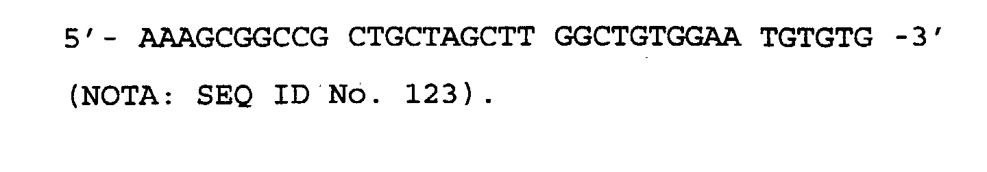
- The SR α promoter DNA fragment was synthesised using PCR, inserted into the above described vector, pEg7AH-H or pEgPDHV3-21, and then cloned into E. coli.
- The HSR α-DNA fragment, comprising the SR α promoter with a NotI restriction enzyme cleavage site added at the 5'-end and a HindIII restriction enzyme cleavage site added at the 3'-end, was prepared as follows.
Composition of the PCR reaction solution: plasmid pME18S DNA, 200 ng; oligonucleotide primer NOTA, 80 pmol; oligonucleotide primer HINDB, 80 pmol; dNTP cocktail, 20 µl; 10× Pfu buffer, 20 µl; Pfu DNA polymerase, 10 units; and redistilled water to a final volume of 200 µl. - The PCR reaction was conducted as follows. The solution was first heated at 94°C for 2 minutes, after which a cycle of heating to 94°C for 1 minute, 55°C for 1 minute and 72°C for 2 minutes, was repeated 30 times. After completion of this procedure, the reaction solution was heated at 72°C for 10 minutes.
- After PCR, the amplified HSR α-DNA fragment was subjected first to phenol extraction and then to ethanol precipitation, and then separated by 5% w/v polyacrylamide gel electrophoresis. After electrophoresis, the gel was stained with 1 µg/ml of ethidium bromide, and the DNA band thus detected, under UV light, was cut out with a razor blade and eluted from the gel using a Centriruter and a Centricon-10, as described above. The eluted DNA was concentrated by centrifugation at 7,500 x g, followed by ethanol precipitation, and was finally dissolved in 50 µl of distilled water.
- One µg of plasmid pEg7AH-H or pEgPDHV3-21 DNA was then digested with the restriction enzymes NotI and HindIII, and then dephosphorylated with CIP. The resulting dephosphorylated plasmid DNA (100 ng) was ligated with 10 µl of the solution containing the HSR α-DNA fragment, which had previously also been digested with NotI and HindIII, using a DNA Ligation Kit Version 2.0 (Takara Shuzo, Co. Ltd.). E. coli JM109 was transformed with the ligation mix and plated onto LB agar medium containing final concentrations of 1 mM IPTG, 0.1% w/v X-Gal and 50 µg/ml ampicillin. The white transformants obtained were cultured in 2 ml liquid LB medium containing 50 µg/ml ampicillin at 37°C overnight, and plasmid DNA was extracted from the resulting culture by the alkaline-SDS method. The extracted plasmid DNA was digested with NotI and HindIII, and a clone carrying then HSR α-DNA fragment was selected by 1% w/v agarose gel electrophoresis.
- By following the above procedure, the high-level expression vector plasmids pSRg7AH and pSRgPDH (HV type) were obtained.
- Transfection of COS-1 cells with the high-level expression plasmids, for each of the humanised HFE7A heavy chains and for each of the humanised HFE7A light chains obtained above, was conducted by electroporation in a manner similar to that described in Example 4.
- COS-1 cells were grown to semi-confluence in a culture flask (culture area: 225 cm2), containing α(+)MEM supplemented with 10% v/v FCS. Next, the medium was discarded and the COS-1 cells were detached from the flask by treatment with 3 ml of trypsin-EDTA solution (Sigma Chemicals Co.) at 37°C for 3 minutes. The detached cells were then harvested by centrifugation at 800 r.p.m. for 2 minutes and then washed twice with PBS(-) buffer. The washed COS-1 cells were then adjusted to 1 x 10⁸ cells/ml with PBS(-) buffer to produce a COS-1 cell suspension.
- In parallel, 4 µg of humanised HFE7A heavy chain expression plasmid DNA and 4 µg of humanised HFE7A light chain expression plasmid DNA, prepared by the alkaline-SDS method and caesium chloride density gradient centrifugation, were mixed and then precipitated with ethanol, before being suspended in 20 µl of PBS(-) buffer, the whole operation being performed in the same tube. The whole of the resulting plasmid suspension (20 µl) was mixed with 20 µl of the previously prepared COS-1 cell suspension (2 x 10⁶ cells) and the mixture was transferred to a FCT-13 (Shimadzu Seisakusyo, K. K.) chamber having electrodes set 2 mm apart. This chamber was then loaded into gene transfection apparatus GTE-1 (Shimadzu Seisakusyo, K. K.) and two pulses, each of 600 V, 50 µF, were applied with a one second interval to transform the COS-1 cells with the plasmid DNA of interest.
- After electroporation, the cell-DNA mixture in the chamber was suspended in 5 ml of α(+)MEM, supplemented with 10% v/v FCS, in a culture flask (culture area 25 cm2; Sumitomo Bakelite) and incubated under 5% v/v CO2 at 37°C for 72 hours. After this time, the culture supernatant was taken and analyzed for the expression products.
- By following the above method, COS-1 cells transfected with each of the following plasmid combinations were obtained:
- (A) no plasmid DNA;
- (B) cotransfection of pSRgPDH and pSRPDHH;
- (C) cotransfection of pSRgPDH and pSRPDHM;
- (D) cotransfection of pSRg7AH and pSRHH;
- (E) cotransfection of pSRg7AH and pSRHM; and
- (F) cotransfection of pSRg7AH and pSRMM.
-
- The assay for Fas-binding activity in the cell culture supernatant fluids prepared in Example 12 was performed by ELISA as follows.
- Culture supernatant from COS-1 cells expressing the human Fas fusion protein, as obtained in Reference Example 1 above, diluted 5-fold with adsorption buffer, was dispensed into wells of a 96-well plate (MaxiSorb; Nunc) at 50 µl per well and the plate was incubated at 4°C overnight to allow adsorption of the human Fas fusion protein to the surface of the wells. Next, each of the wells was washed 4 times with 350 µl of PBS-T. After washing, PBS-T containing 5% v/v BSA (bovine serum albumin; Wako Pure Chemical Industries, Ltd.) was added to the wells at 50 µl per well and the plate was incubated at 37°C for 1 hour to block the remainder of the surface of each well. The wells were then again washed four times with PBS-T.
- The culture supernatants obtained in Example 4 were adjusted to have a final concentration of the product of interest of 100 ng/ml in α(+)MEM containing 10% v/v FCS. Concentrations were estimated by the method described in Example 5. Each of the resulting 100 ng/ml solutions was then used to produce serial dilutions by serial 2-fold dilution with α(+)MEM containing 10% v/v FCS. Next, 50 µl of each of the resulting serial dilutions of each expression product was added to a well prepared as above, and the plate was incubated at 37°C for 2 hours to allow reaction.
- After this time, the wells were again washed four times with PBS-T, and then 50 µl of alkaline phosphatase-labeled goat anti-human IgG Fc specific polyclonal antibody (Caltag Lab.), diluted 10,000-fold with PBS-T, were dispensed into each well and reaction was allowed to proceed at 37°C for 2 hours.
- HFE7A purified from mouse hybridoma HFE7A was used as a control (IgGl), and was detected using alkaline phosphatase-labeled goat anti-mouse IgG + IgA + IgM (Gibco BRL), diluted 5,000-fold with PBS-T, in place of the alkaline phosphatase-labeled goat anti-human IgG Fc specific polyclonal antibody.
- The wells were again washed four times with PBS-T, and then 50 µl of substrate solution [1 mg/ml p-nitrophenyl phosphate in 10% v/v diethanol amine (pH 9.8)] was dispensed into each well and the plate was incubated at 37°C for 0.5 to 1 hour. Binding activity of the expression product contained in each culture supernatant fluid with the human Fas fusion protein was evaluated by reading the absorbance of each well at 405 nm.
- As expected, binding activity for the human Fas fusion protein was demonstrated for the supernatants of categories (B), (C), (D), (E) and (F) above of Example 12, and is shown Figure 45.
- The humanised anti-Fas antibodies obtained in Example 12 should inhibit the binding of HFE7A to Fas, as the antibodies of this Example were derived from HFE7A. Therefore, the ability of the expression products obtained in Example 12 to competitively inhibit the binding of HFE7A to the human Fas fusion protein was measured.
- The COS-1 cell culture supernatant containing the human Fas fusion protein, as obtained in Reference Example 1, was diluted 5-fold with adsorption buffer, and dispensed into the wells of a 96-well plate for luminescence detection (Luminescent Solid Assay Plate, high binding property; Costar) at 50 µl per well. The plate was then incubated at 4°C overnight to allow adsorption of the human Fas fusion protein to the surface of the wells.
- After this time, each well was washed 4 times with 350 µl of PBS-T, and then 100 µl PBS-T containing 5% v/v BSA was added to each well and the plate was incubated at 37°C for 1 hour to block the remainder of the surface of each well. The wells were then again washed four times with PBS-T.
- The culture supernatants obtained in Example 12 were adjusted to final concentrations of antibody of 1 µg/ml in α(+)MEM containing 10% v/v FCS by the method of Example 5. Each of the resulting solutions of the expression products was used to produce serial dilutions by serial 2-fold dilution with α(+)MEM containing 10% v/v FCS. AP-HFE7A was diluted to 50 ng/ml with α(+)MEM containing 10% v/v FCS, and 25 µl of the resulting solution was mixed with an equal volume of each of the prepared serial dilutions.
- Each of the wells was again washed four times with PBS-T, and then 50 µl of each of the resulting antibody mixtures were added to individual wells, and the plate was allowed to stand at room temperature overnight. Subsequently, after washing each well with PBS-T again four times, 100 µl of CDP-star buffer (9.58 ml diethanol amine, 0.2 g magnesium chloride, 0.25 g sodium azide, pH8.5) was dispensed into each well and the plate was allowed to stand at room temperature for 10 minutes. After this time, the CDP-star buffer was discarded and CDP-star substrate [1.2 ml sapphire II (Tropix), 200 µl CDP-star (Tropix), q.s. to 12 ml with CDP-star buffer] was added at 50 µl per well, and the plate was then allowed to stand at room temperature for a further 40 minutes.
- Competitive inhibition of the expression products of Example 12 of the binding of HFE7A to the human Fas fusion protein was evaluated by measuring the intensity of the luminescence with Luminoscan (Titertech).
- As expected, it was verified that each of the expression products of supernatants (B), (C), (D), (E) and (F) obtained in Example 12 above specifically inhibited the binding of HFE7A antibody to the human Fas fusion protein. The results are shown in Figure 46.
- The apoptosis-inducing activity of the expression products in the culture supernatant fluids obtained in Example 12 was examined in a manner similar to that described in Example 8.
- WR19L12a cells were cultured in RPMI1640 medium with 10% v/v FCS (Gibco BRL) at 37°C for 3 days under 5% v/v CO2, and 50 µl (1 x 10⁵ cells) of the resulting culture were then dispensed into each well of a 96-well microplate (Sumitomo Bakelite). The culture supernatants obtained in Example 12 were adjusted to a final concentration of antibody of 100 ng/ml in RPMI1640 medium containing 10% v/v FCS. Concentrations were estimated by the method of Example 5. Each of the adjusted solutions of the expression products was used to produce serial dilutions by serial 2-fold dilution with RPMI1640 containing 10% v/v FCS. Each of the resulting dilutions of each expression product solution was added to individual wells, at 50 µl per well, and the plate was incubated at 37°C for 1 hour. After this time, the cells in each well were washed once with RPMI1640 containing 10% v/v FCS and then the washed cells were suspended in 100 µl per well of 0.5 µg/ml goat anti-human IgG Fc specific polyclonal antibody (Kappel) in RPMI1640 containing 10% v/v FCS.
- The plate was allowed to stand at 37°C for 12 hours, and then 50 µl of 25 µM PMS (phenazine methosulphate; Sigma Chemical Co.), containing 1 mg/ml XTT [2,3-bis(2-methoxy-4-nitro-5-sulphophenyl)-2H-tetrazolium-5-carboxyanilide inner salt; Sigma Chemical Co.] to final concentrations of 333 µg/ml for XTT and 8.3 µM for PMS, were added to each well. The plate was then incubated for 3 hours at 37°C, and the absorbance at 450 nm of each well was measured, to calculate cell viability, using the reducing power of the mitochondria as the index.
- The viability of the cells in each well was calculated according to the following formula:
- As expected, each of the expression products of the culture supernatant fluids (B), (C), (D), (E) and (F) obtained in Example 12 above were demonstrated to induce apoptosis in T cells of this lymphoma cell line expressing human Fas antigen (Figure 47).
-


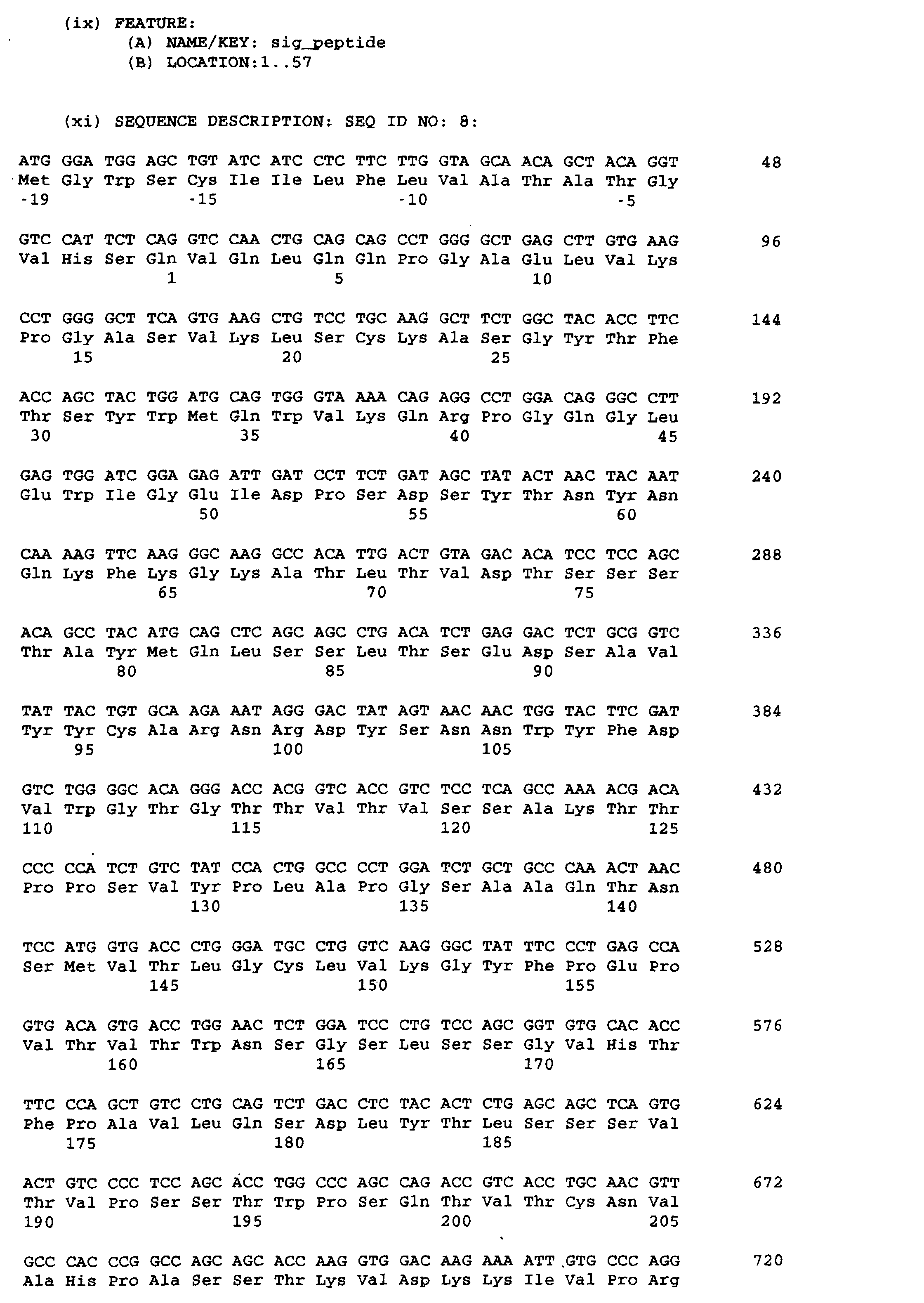


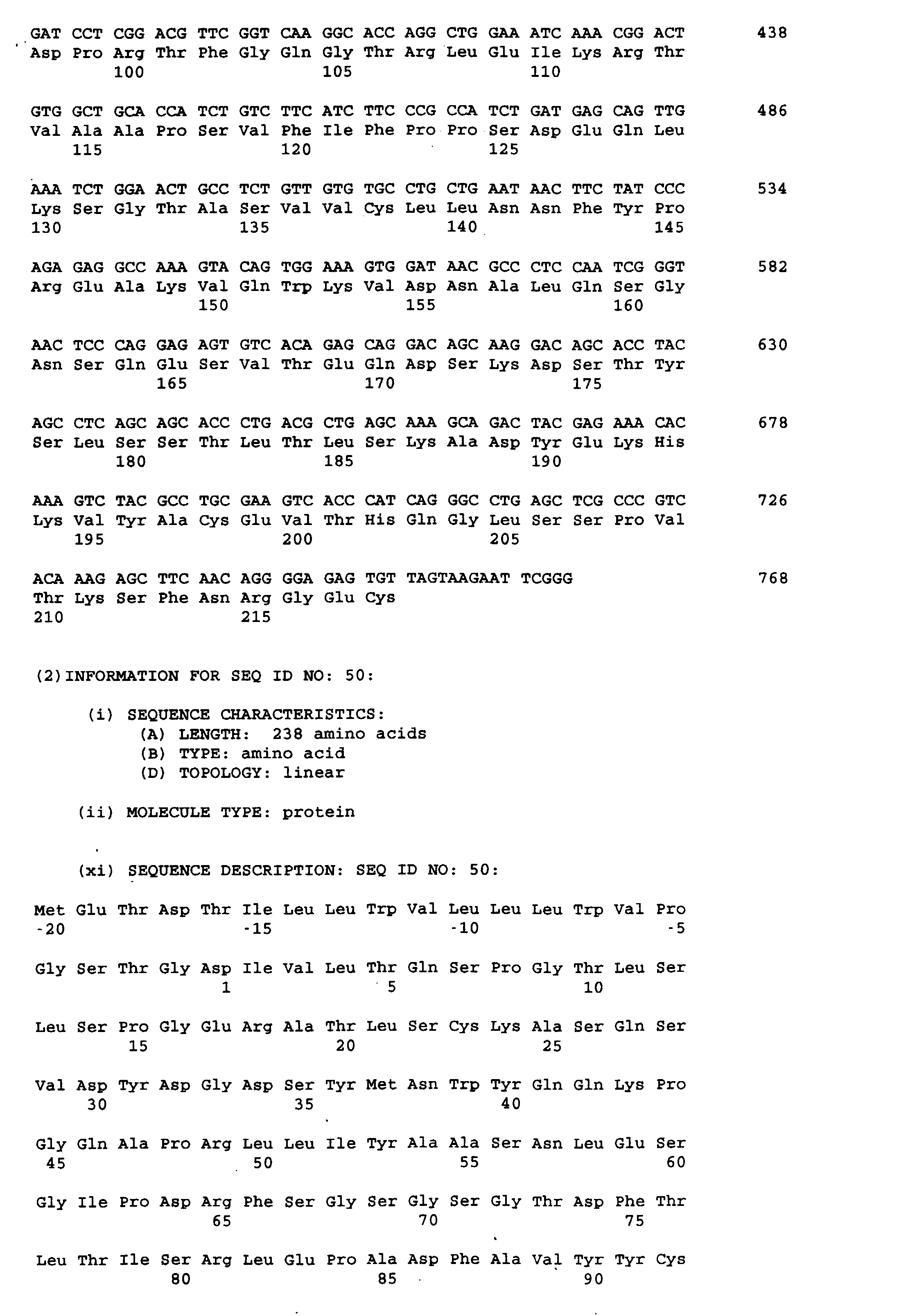
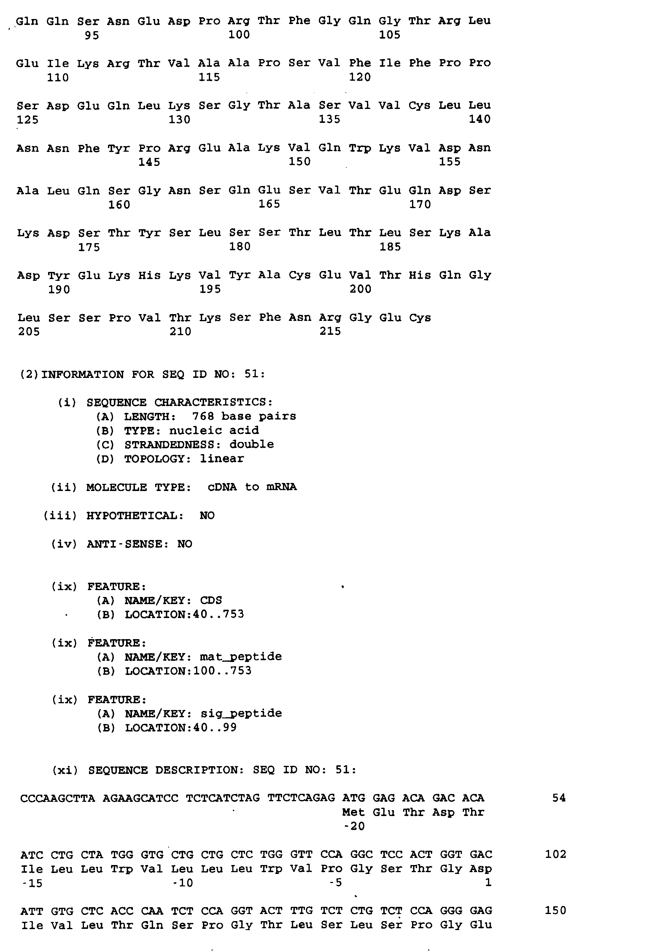


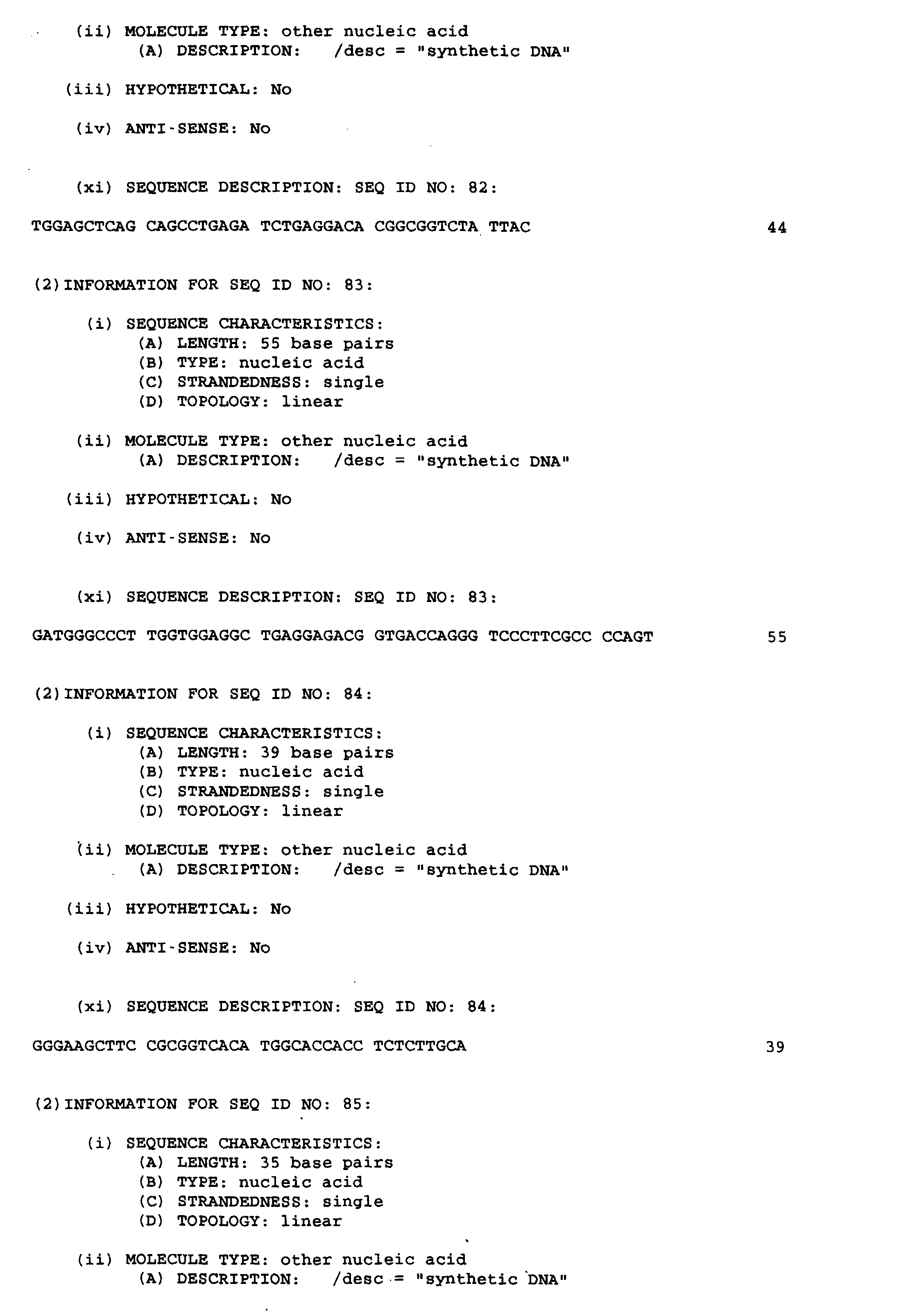


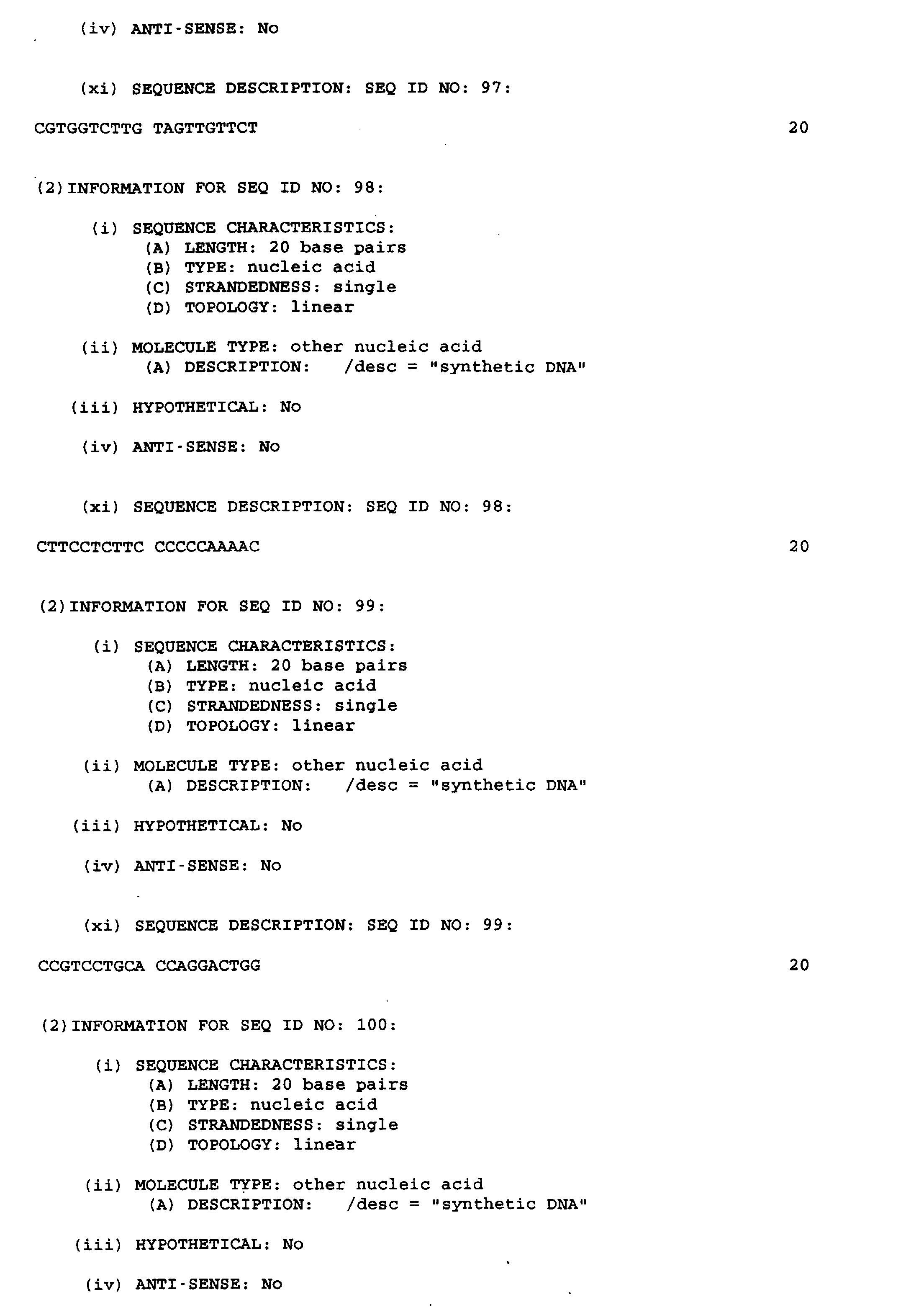

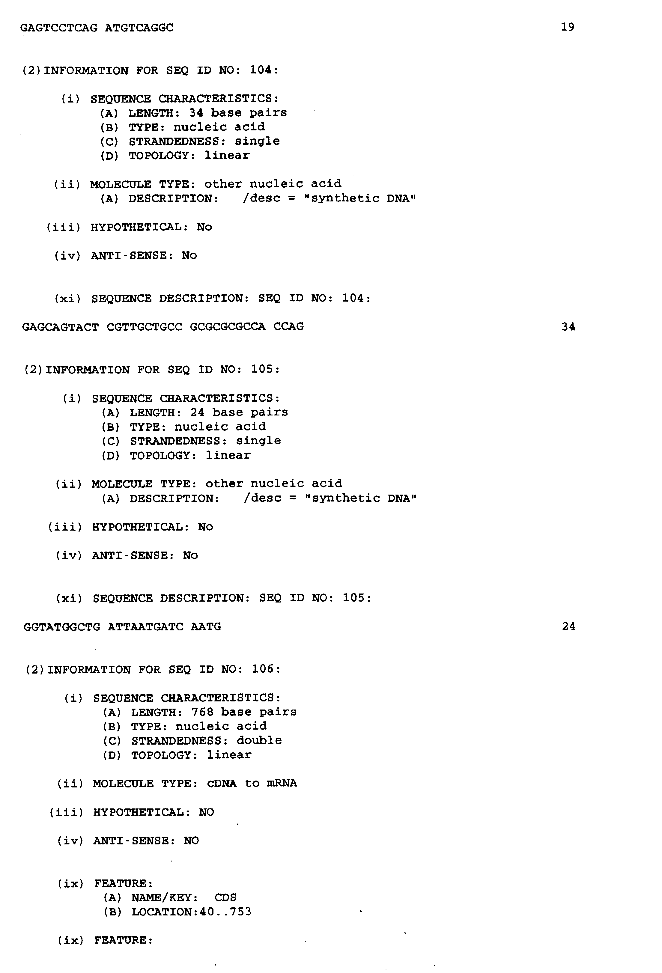


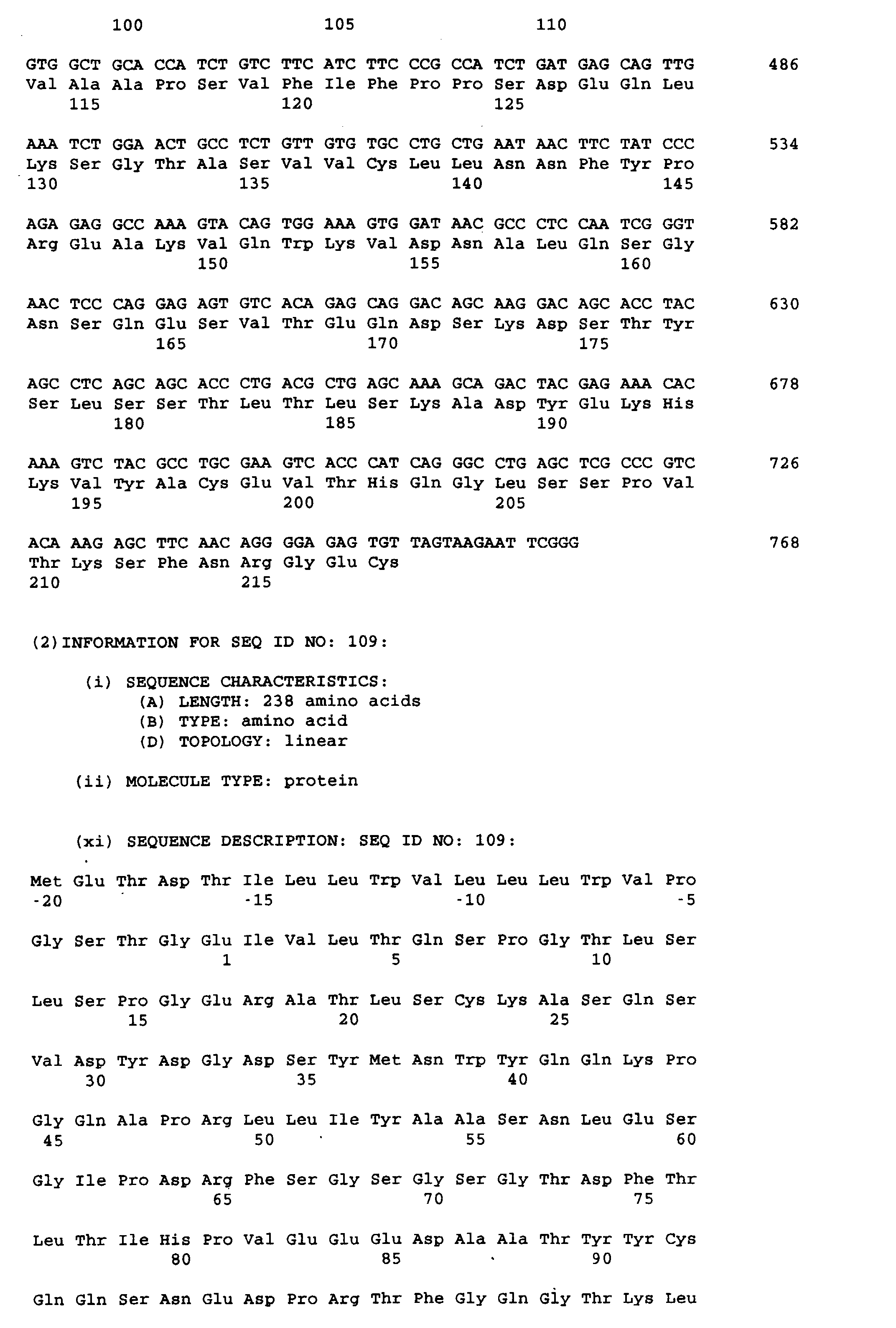


Claims (56)
- A molecule having an antigen binding region specific for an epitope of the Fas antigen, the epitope being conserved between a primate and a non-primate animal.
- A molecule according to claim 1, wherein the primate is human.
- A molecule according to claim 1 or 2, wherein the non-primate is a rodent.
- A molecule according to claim 3, wherein the rodent is a mouse.
- A molecule having an antigen binding region specific for a conserved, mammalian, epitope of the Fas antigen.
- An antibody as produced by the hybridoma HFE7A having the accession number FERM BP-5828.
- A molecule having at least six antibody CDR's, the antibody being specific for human Fas, wherein the CDR's have identity with the CDR's of the antibody as produced by the hybridoma HFE7A having the accession number FERM BP-5828.
- A molecule having an antigen binding region, the binding region having specificity for the antigenic determinant recognised by the antibody as produced by the hybridoma HFE7A having the accession number FERM BP-5828.
- A molecule comprising a light polypeptide chain and a heavy polypeptide chain, the heavy chain having the following general formula (I) :
the light chain having the following general formula (II) : - A molecule according to any preceding claim, which has a property selected from:inducing apoptosis in T cells expressing Fas;ameliorating autoimmune symptoms in MRL gld/gld mice;does not induce hepatic disorders;a therapeutic or prophylactic effect on fulminant hepatitis;a preventative effect on the onset of collagen-induced arthritis;inducing apoptosis in synovial cells from a rheumatoid arthritis patient.
- A molecule according to claim 10, which has all of the said properties.
- A molecule according to any preceding claim, which is humanised.
- A molecule according to any preceding claim, which is an antibody.
- An antibody according to claim 13, which is of the immunoglobulin G type.
- A molecule according to any preceding claim, which is able to induce apoptosis in abnormal cells expressing Fas, and which is able to inhibit apoptosis in normal cells.
- Use of a molecule according to any preceding claim to evaluate therapies for conditions in humans affected by the Fas/Fas ligand interaction, the evaluation being in animals which are models for such conditions.
- An humanised molecule having an antigen binding region specific for an epitope of the Fas antigen which is conserved between a primate and a non-primate animal, the molecule being obtainable by grafting the respective CDR's from an antibody specific for the epitope of the Fas antigen into each of at least one human light chain, or fragment thereof, and at least one human heavy chain, or fragment thereof.
- A molecule according to claim 17, wherein the human light and heavy chains are selected on the basis of closest similarity between the variable regions comprised therein and the variable regions of the antibody.
- A molecule according to claim 17 or 18, wherein significant amino acids, from the framework regions from the antibody from which the CDR's are obtainable, are also grafted into the heavy and light chains, in order to maintain structure in the epitope recognition site.
- A molecule according to any preceding claim, that binds a peptide comprising the amino acid sequence of SEQ ID No. 1 of the Sequence Listing.
- A molecule according to any preceding claim, that comprises a light chain polypeptide protein which has the amino acid sequence 1 to 218 of SEQ ID No. 50, the amino acid sequence 1 to 218 of SEQ ID No. 52, the amino acid sequence 1 to 218 of SEQ ID No. 54, the amino acid sequence 1 to 218 of SEQ ID No. 107 or the amino acid sequence 1 to 218 of SEQ ID No. 109 of the Sequence Listing.
- A molecule according to any preceding claim, that comprises a heavy chain polypeptide protein which has the amino acid sequence 1 to 451 of SEQ ID No. 89 or the amino acid sequence 1 to 451 of SEQ ID No. 117 of the Sequence Listing.
- A molecule according to any preceding claim that comprises a light chain polypeptide protein having the amino acid sequence 1 to 218 of SEQ ID No. 50 of the Sequence Listing and a heavy chain polypeptide protein having the amino acid sequence 1 to 451 of SEQ ID No. 89 of the Sequence Listing.
- A molecule according to any preceding claim that comprises a light chain polypeptide protein having the amino acid sequence 1 to 218 of SEQ ID No. 107 of the Sequence Listing and a heavy chain polypeptide protein having the amino acid sequence 1 to 451 of SEQ ID No. 117 of the Sequence Listing.
- DNA encoding any single polypeptide portion of a molecule according to any preceding claim.
- DNA comprising nucleotide sequence 100 to 753 of SEQ ID No. 49, nucleotide sequence 100 to 753 of SEQ ID No. 51, nucleotide sequence 100 to 753 of SEQ ID No. 53, nucleotide sequence 100 to 753 of SEQ ID No. 106 or nucleotide sequence 100 to 753 of SEQ ID No. 108.
- DNA comprising nucleotide sequence 84 to 2042 of SEQ ID No. 88 or nucleotide sequence 84 to 2042 of SEQ ID No. 116 of the Sequence Listing.
- A recombinant DNA vector comprising DNA according to any of claims 25 to 27.
- A host cell transformed with a recombinant DNA vector according to claim 28.
- A host cell transformed with at least one recombinant DNA vector according to claim 28, comprising DNA encoding a light chain polypeptide protein and DNA encoding a heavy chain polypeptide.
- A host cell according to claim 29 or 30, which is mammalian.
- E. coli pHSGMM6 5ANK73697 (FERM BP-6071).
- E. coli pHSGHM17 SANK73597 (FERM BP-6072).
- E. coli pHSGHH7 SANK73497 (FERM BP-6073).
- E. coli pHSHM2 SANK 70198 (FERM BP-6272).
- E. coli pHSHH5 SANK 70398 (FERM BP-6274).
- E. coli SANK73397 (FERM BP-6074).
- E. coli pgHPDHV3 SANK 70298 (FERM BP-6273).
- A method for producing an anti-Fas antibody comprising culturing a host cell according to any of claims 29 to 31, and then recovering the anti-Fas antibody from the culture.
- An agent for the treatment or prophylaxis of conditions attributable to abnormalities of the Fas/Fas ligand system comprising, as an active ingredient, a molecule according to any of claims 1 to 24.
- An agent for the treatment or prophylaxis of conditions attributable to abnormalities of the Fas/Fas ligand system comprising, as an active ingredient, a molecule according to any of claims 1 to 24, wherein the condition is selected from autoimmune diseases, allergy, atopy, arteriosclerosis, myocarditis, cardiomyopathy, glomerular nephritis, hypoplastic anaemia, hepatitis, acquired immunodeficiency syndrome and rejection after organ transplantation.
- An agent for the treatment or prophylaxis of conditions attributable to abnormalities of the Fas/Fas ligand system comprising, as an active ingredient, a molecule according to any of claims 1 to 24, wherein the condition is an autoimmune disease selected from systemic lupus erythematosus, Hashimoto's disease, rheumatoid arthritis, graft versus host disease, Sjögren syndrome, pernicious anaemia, Addison's disease, scleroderma, Goodpasture syndrome, Crohn's disease, autoimmune haemolytic anaemia, sterility, myasthenia gravis, multiple sclerosis, Basedow's disease, thrombopenia purpura and insulin dependent diabetes mellitus.
- An agent for the treatment or prophylaxis of conditions attributable to abnormalities of the Fas/Fas ligand system comprising, as an active ingredient, a molecule according to any of claims 1 to 24, wherein the condition is an allergy.
- An agent for the treatment or prophylaxis of conditions attributable to abnormalities of the Fas/Fas ligand system comprising, as an active ingredient, a molecule according to any of claims 1 to 24, wherein the condition is rheumatoid arthritis.
- An agent for the treatment or prophylaxis of conditions attributable to abnormalities of the Fas/Fas ligand system comprising, as an active ingredient, a molecule according to any of claims 1 to 24, wherein the condition is arteriosclerosis.
- An agent for the treatment or prophylaxis of conditions attributable to abnormalities of the Fas/Fas ligand system comprising, as an active ingredient, a molecule according to any of claims 1 to 24, wherein the condition is selected from myocarditis and cardiomyopathy.
- An agent for the treatment or prophylaxis of conditions attributable to abnormalities of the Fas/Fas ligand system comprising, as an active ingredient, a molecule according to any of claims 1 to 24, wherein the condition is glomerular nephritis.
- An agent for the treatment or prophylaxis of conditions attributable to abnormalities of the Fas/Fas ligand system comprising, as an active ingredient, a molecule according to any of claims 1 to 24, wherein the condition is hypoplastic anaemia.
- An agent for the treatment or prophylaxis of conditions attributable to abnormalities of the Fas/Fas ligand system comprising, as an active ingredient, a molecule according to any of claims 1 to 24, wherein the condition is hepatitis.
- An agent for the treatment or prophylaxis of conditions attributable to abnormalities of the Fas/Fas ligand system comprising, as an active ingredient, a molecule according to any of claims 1 to 24, wherein the condition is selected from fulminant hepatitis, chronic hepatitis, viral hepatitis, hepatitis C, hepatitis B, hepatitis D and alcoholic hepatitis.
- An agent for the treatment or prophylaxis of conditions attributable to abnormalities of the Fas/Fas ligand system comprising, as an active ingredient, a molecule according to any of claims 1 to 24, wherein the condition is rejection after organ transplantation.
- An agent for the treatment or prophylaxis of conditions attributable to abnormalities of the Fas/Fas ligand system comprising, as an active ingredient, a molecule according to any of claims 1 to 24, wherein the condition is acquired immune deficiency syndrome.
- Use of a molecule according to any of claims 1 to 24 in the manufacture of a medicament for the treatment or prophylaxis of a condition as defined in any of claims 40 to 52.
- The hybridoma HFE7A having the accession number FERM BP-5828.
- A method for producing an antibody having an antigen binding region specific for an epitope of the Fas antigen, the method comprising culturing the hybridoma HFE7A having the accession number FERM BP-5828, and harvesting expressed antibody.
- A method for producing an antibody, the method comprising culturing the hybridoma HFE7A having the accession number FERM BP-5828, and harvesting expressed antibody.
Applications Claiming Priority (6)
| Application Number | Priority Date | Filing Date | Title |
|---|---|---|---|
| JP82953/97 | 1997-04-01 | ||
| JP8295397 | 1997-04-01 | ||
| JP16908897 | 1997-06-25 | ||
| JP169088/97 | 1997-06-25 | ||
| JP27606497 | 1997-10-08 | ||
| JP276064/97 | 1997-10-08 |
Publications (1)
| Publication Number | Publication Date |
|---|---|
| EP0909816A1 true EP0909816A1 (en) | 1999-04-21 |
Family
ID=27304060
Family Applications (1)
| Application Number | Title | Priority Date | Filing Date |
|---|---|---|---|
| EP98302575A Withdrawn EP0909816A1 (en) | 1997-04-01 | 1998-04-01 | Anti-fas antibodies |
Country Status (17)
| Country | Link |
|---|---|
| EP (1) | EP0909816A1 (en) |
| KR (1) | KR19980080993A (en) |
| CN (1) | CN1207395A (en) |
| AR (1) | AR010139A1 (en) |
| AU (1) | AU734758B2 (en) |
| BR (1) | BR9803296A (en) |
| CA (1) | CA2233783A1 (en) |
| CZ (1) | CZ294340B6 (en) |
| HU (1) | HUP9800744A3 (en) |
| ID (1) | ID20117A (en) |
| IL (1) | IL123888A0 (en) |
| NO (1) | NO981454L (en) |
| NZ (1) | NZ330095A (en) |
| PL (1) | PL325667A1 (en) |
| RU (1) | RU2212412C2 (en) |
| TR (1) | TR199800604A3 (en) |
| TW (1) | TW526205B (en) |
Cited By (9)
| Publication number | Priority date | Publication date | Assignee | Title |
|---|---|---|---|---|
| WO1999065935A2 (en) * | 1998-06-18 | 1999-12-23 | Karolinska Innovations Ab | Fas peptides and antibodies for modulating apoptosis |
| EP0990663A2 (en) * | 1998-09-30 | 2000-04-05 | Sankyo Company Limited | Anti-Fas Antibodies |
| WO2000071160A1 (en) * | 1999-05-24 | 2000-11-30 | Sankyo Company, Limited | MEDICINAL COMPOSITIONS CONTAINING ANTI-Fas ANTIBODY |
| WO2003022299A1 (en) * | 2001-08-01 | 2003-03-20 | Genset S.A. | Genobix agonists and antagonists for use in the treatment of metabolic disorders |
| WO2003037913A2 (en) | 2001-11-01 | 2003-05-08 | Uab Research Foundation | An antibody selective for a tumor necrosis factor-related apoptosis-inducing ligand receptor and uses thereof |
| US6972323B1 (en) | 1997-04-01 | 2005-12-06 | Sankyo Company, Limited | Anti-Fas antibodies |
| EP2065401A2 (en) | 2000-05-02 | 2009-06-03 | UAB Research Foundation | An antibody selective for a tumor necrosis factor-related apoptosis-inducing ligand receptor and uses thereof |
| US8298769B2 (en) | 2001-08-23 | 2012-10-30 | Rsr Limited | Epitope regions of a thyrotrophin (TSH) receptor, uses thereof and antibodies thereto |
| US9079956B2 (en) | 2004-01-07 | 2015-07-14 | Novartis Vaccines And Diagnostics Inc. | M-CSF specific monoclonal antibody and uses thereof |
Families Citing this family (2)
| Publication number | Priority date | Publication date | Assignee | Title |
|---|---|---|---|---|
| DK1818343T3 (en) * | 1998-01-23 | 2012-07-16 | Hoffmann La Roche | Antibodies to human IL-12 |
| EP2322220B1 (en) * | 2008-08-01 | 2014-01-01 | Axis, Inc. | Therapeutic agent or preventive agent for osteoarthritis |
Citations (4)
| Publication number | Priority date | Publication date | Assignee | Title |
|---|---|---|---|---|
| WO1995010540A1 (en) * | 1993-10-14 | 1995-04-20 | Immunex Corporation | Fas antagonists and uses thereof |
| WO1995018819A1 (en) * | 1994-01-07 | 1995-07-13 | Immunex Corporation | Ligand that binds fas antigen |
| WO1996040041A2 (en) * | 1995-06-07 | 1996-12-19 | Chiron Corporation | Antibodies to fas antigen capable of inhibiting apoptosis |
| WO1997018307A1 (en) * | 1995-11-16 | 1997-05-22 | Novartis Ag | Fas ligand fusion protein |
-
1998
- 1998-03-30 IL IL12388898A patent/IL123888A0/en unknown
- 1998-03-30 AU AU59701/98A patent/AU734758B2/en not_active Ceased
- 1998-03-31 RU RU98105631/13A patent/RU2212412C2/en not_active IP Right Cessation
- 1998-03-31 NO NO981454A patent/NO981454L/en not_active Application Discontinuation
- 1998-03-31 ID IDP980478A patent/ID20117A/en unknown
- 1998-03-31 NZ NZ330095A patent/NZ330095A/en unknown
- 1998-03-31 TW TW087104806A patent/TW526205B/en not_active IP Right Cessation
- 1998-03-31 CA CA002233783A patent/CA2233783A1/en not_active Abandoned
- 1998-04-01 KR KR1019980011477A patent/KR19980080993A/en not_active Application Discontinuation
- 1998-04-01 EP EP98302575A patent/EP0909816A1/en not_active Withdrawn
- 1998-04-01 CZ CZ1998985A patent/CZ294340B6/en not_active IP Right Cessation
- 1998-04-01 CN CN98108265A patent/CN1207395A/en active Pending
- 1998-04-01 PL PL98325667A patent/PL325667A1/en unknown
- 1998-04-01 BR BR9803296-8A patent/BR9803296A/en not_active IP Right Cessation
- 1998-04-01 AR ARP980101481A patent/AR010139A1/en unknown
- 1998-04-01 HU HU9800744A patent/HUP9800744A3/en unknown
- 1998-04-01 TR TR1998/00604A patent/TR199800604A3/en unknown
Patent Citations (4)
| Publication number | Priority date | Publication date | Assignee | Title |
|---|---|---|---|---|
| WO1995010540A1 (en) * | 1993-10-14 | 1995-04-20 | Immunex Corporation | Fas antagonists and uses thereof |
| WO1995018819A1 (en) * | 1994-01-07 | 1995-07-13 | Immunex Corporation | Ligand that binds fas antigen |
| WO1996040041A2 (en) * | 1995-06-07 | 1996-12-19 | Chiron Corporation | Antibodies to fas antigen capable of inhibiting apoptosis |
| WO1997018307A1 (en) * | 1995-11-16 | 1997-05-22 | Novartis Ag | Fas ligand fusion protein |
Non-Patent Citations (4)
| Title |
|---|
| C. MANGURIAN ET AL.: "Expression of a Fas-like proapoptotic molecule on the lymphocytes of Xenopus laevis.", IMMUNOLOGY LETTERS, vol. 64, no. 1, 1998, Amsterdam, NL, pages 31 - 38, XP002089806 * |
| M. ALDERSON ET AL.: "Regulation of apoptosis and T cell activation by Fas-specific mAb.", INTERNATIONAL IMMUNOLOGY, vol. 6, no. 11, November 1994 (1994-11-01), Oxford, GB, pages 1799 - 1806, XP000616652 * |
| M. CIFONE ET AL.: "Apoptotic signaling through CD95 (Fas/Apo-1) activates an acidic sphingomyelinase.", THE JOURNAL OF EXPERIMENTAL MEDICINE, vol. 177, October 1993 (1993-10-01), New York, NY, USA, pages 1547 - 1552, XP000566306 * |
| Y. NISHIMURA ET AL.: "In vivo analysis of Fas antigen-mediated apoptosis: effects of agonistic anti-mouse Fas mAb on thymus, spleen and liver.", INTERNATIONAL IMMUNOLOGY, vol. 9, no. 2, February 1997 (1997-02-01), Oxford, GB, pages 307 - 316, XP002089805 * |
Cited By (21)
| Publication number | Priority date | Publication date | Assignee | Title |
|---|---|---|---|---|
| US6972323B1 (en) | 1997-04-01 | 2005-12-06 | Sankyo Company, Limited | Anti-Fas antibodies |
| US6846637B1 (en) | 1998-06-18 | 2005-01-25 | Imed Ab | Fas peptides and antibodies for modulating apoptosis |
| WO1999065935A2 (en) * | 1998-06-18 | 1999-12-23 | Karolinska Innovations Ab | Fas peptides and antibodies for modulating apoptosis |
| WO1999065935A3 (en) * | 1998-06-18 | 2003-04-17 | Karolinska Innovations Ab | Fas peptides and antibodies for modulating apoptosis |
| EP0990663A2 (en) * | 1998-09-30 | 2000-04-05 | Sankyo Company Limited | Anti-Fas Antibodies |
| EP0990663A3 (en) * | 1998-09-30 | 2002-04-17 | Sankyo Company Limited | Anti-Fas Antibodies |
| WO2000071160A1 (en) * | 1999-05-24 | 2000-11-30 | Sankyo Company, Limited | MEDICINAL COMPOSITIONS CONTAINING ANTI-Fas ANTIBODY |
| US6746673B2 (en) | 1999-05-24 | 2004-06-08 | Sankyo Company, Limited | Pharmaceutical compositions containing anti-Fas antibody |
| EP2065401A2 (en) | 2000-05-02 | 2009-06-03 | UAB Research Foundation | An antibody selective for a tumor necrosis factor-related apoptosis-inducing ligand receptor and uses thereof |
| EP2065400A2 (en) | 2000-05-02 | 2009-06-03 | UAB Research Foundation | An antibody selective for a tumor necrosis factor-related apoptosis-inducing ligand receptor and uses thereof |
| WO2003022299A1 (en) * | 2001-08-01 | 2003-03-20 | Genset S.A. | Genobix agonists and antagonists for use in the treatment of metabolic disorders |
| US8298769B2 (en) | 2001-08-23 | 2012-10-30 | Rsr Limited | Epitope regions of a thyrotrophin (TSH) receptor, uses thereof and antibodies thereto |
| US8298771B2 (en) | 2001-08-23 | 2012-10-30 | Rsr Limited | Epitope regions of a thyrotrophin (TSH) receptor, uses thereof and antibodies thereto |
| US8309693B2 (en) | 2001-08-23 | 2012-11-13 | Rsr Limited | Epitope regions of a thyrotrophin (TSH) receptor, uses thereof and antibodies thereto |
| US9751940B2 (en) | 2001-08-23 | 2017-09-05 | Rsr Limited | Epitope regions of a thyrotrophin (TSH) receptor, uses thereof and antibodies thereto |
| WO2003037913A2 (en) | 2001-11-01 | 2003-05-08 | Uab Research Foundation | An antibody selective for a tumor necrosis factor-related apoptosis-inducing ligand receptor and uses thereof |
| EP2287285A2 (en) | 2001-11-01 | 2011-02-23 | Uab Research Foundation | An antibody selective for a tumor necrosis factor-related apoptosis-inducing ligand receptor and uses thereof |
| US9079956B2 (en) | 2004-01-07 | 2015-07-14 | Novartis Vaccines And Diagnostics Inc. | M-CSF specific monoclonal antibody and uses thereof |
| US9522186B2 (en) | 2004-01-07 | 2016-12-20 | Novartis Vaccines And Diagnostics Inc. | M-CSF specific monoclonal antibody and uses therof |
| US9926371B2 (en) | 2004-01-07 | 2018-03-27 | Novartis Vaccines And Diagnostics, Inc. | M-CSF specific monoclonal antibody and uses thereof |
| US11034759B2 (en) | 2004-01-07 | 2021-06-15 | Novartis Vaccines And Diagnostics, Inc. | Humanized M-CSF specific monoclonal antibodies, kits, and uses thereof |
Also Published As
| Publication number | Publication date |
|---|---|
| AU734758B2 (en) | 2001-06-21 |
| IL123888A0 (en) | 1998-10-30 |
| TR199800604A2 (en) | 1999-10-21 |
| TW526205B (en) | 2003-04-01 |
| HUP9800744A3 (en) | 2001-04-28 |
| CA2233783A1 (en) | 1998-10-01 |
| NZ330095A (en) | 1998-12-23 |
| RU2212412C2 (en) | 2003-09-20 |
| HUP9800744A2 (en) | 1999-02-01 |
| PL325667A1 (en) | 1998-10-12 |
| TR199800604A3 (en) | 1999-10-21 |
| CZ294340B6 (en) | 2004-12-15 |
| KR19980080993A (en) | 1998-11-25 |
| NO981454D0 (en) | 1998-03-31 |
| NO981454L (en) | 1998-10-02 |
| HU9800744D0 (en) | 1998-05-28 |
| AR010139A1 (en) | 2000-05-17 |
| ID20117A (en) | 1998-10-01 |
| BR9803296A (en) | 2000-01-11 |
| AU5970198A (en) | 1998-10-08 |
| CZ98598A3 (en) | 1998-10-14 |
| CN1207395A (en) | 1999-02-10 |
Similar Documents
| Publication | Publication Date | Title |
|---|---|---|
| US6972323B1 (en) | Anti-Fas antibodies | |
| JP4156238B2 (en) | Antibodies selective for tumor necrosis factor-related apoptosis-inducing ligand receptors and uses thereof | |
| JP4371814B2 (en) | Antibodies selective for tumor necrosis factor-related apoptosis-inducing ligand receptor and use thereof | |
| EP0990663A2 (en) | Anti-Fas Antibodies | |
| US6746673B2 (en) | Pharmaceutical compositions containing anti-Fas antibody | |
| AU734758B2 (en) | Anti-fas antibodies | |
| AU736287B2 (en) | Humanized anti-human Fas antibody | |
| KR100277769B1 (en) | Anti-Pass Recombinant Antibodies and DieAs | |
| JP3444585B2 (en) | Anti-Fas antibody | |
| JP2000169393A (en) | MEDICAMENT INCLUDING ANTI-Fas ANTIBODY | |
| MXPA98002536A (en) | Antibodies anti- | |
| JP2000166574A (en) | HUMANIZED ANTI-Fas ANTIBODY | |
| EP1421118A1 (en) | A method for the treatment or prevention of bone erosion | |
| JP2004000228A (en) | ANTI-Fas ANTIBODY | |
| JP2000166573A (en) | HUMANIZED ANTI-Fas ANTIBODY | |
| JP2001342149A (en) | MEDICINE CONTAINING HUMANIZED ANTI-Fas ANTIBODY | |
| CZ9903452A3 (en) | Humanized molecule of the type of antibody against FAS, molecule of the antibody type with selected one or plurality of heavy and light chains, prophylactic and therapeutic preparation, DNA encoding antibody, recombinant DNA vector, transformed host cell, process for preparing antibody against FAS, transformation strain Escherichia coli, polypeptide, encoding DNA, recombinant DNA vector and transformed host cell thereof | |
| JP2001342148A (en) | MEDICINE CONTAINING HUMANIZED ANTI-Fas ANTIBODY | |
| AU2002328084A1 (en) | A method for the treatment or prevention of bone erosion | |
| JP2003146903A (en) | Therapeutic or prophylactic agent composition for osteoclasia | |
| JP2001039892A (en) | MEDICINAL COMPOSITION CONTAINING ANTI-Fas ANTIBODY |
Legal Events
| Date | Code | Title | Description |
|---|---|---|---|
| PUAI | Public reference made under article 153(3) epc to a published international application that has entered the european phase |
Free format text: ORIGINAL CODE: 0009012 |
|
| AK | Designated contracting states |
Kind code of ref document: A1 Designated state(s): AT BE CH DE DK ES FI FR GB GR IE IT LI LU MC NL PT SE |
|
| AX | Request for extension of the european patent |
Free format text: AL;LT;LV;MK;RO;SI |
|
| RIN1 | Information on inventor provided before grant (corrected) |
Inventor name: SHIRAISHI, AKIO, SANKYO COMPANY LIMITED Inventor name: YOSHIDA, HIROKO, SANKYO COMPANY LIMITED Inventor name: TAKAHASHI, TOHRU, SANKYO COMPANY LIMITED Inventor name: HARUYAMA, HIDEYUKI, SANKYO COMPANY LIMITED Inventor name: OHTSUKI, MASAHIKO, SANKYO COMPANY LIMITED Inventor name: OHSUMI, JUN, SANKYO COMPANY LIMITED Inventor name: ICHIKAWA, KIMIHISA, SANKYO COMPANY LIMITED Inventor name: SERIZAWA, NOBUFUSA, SANKYO COMPANY LIMITED |
|
| 17P | Request for examination filed |
Effective date: 19991020 |
|
| AKX | Designation fees paid |
Free format text: AT BE CH DE DK ES FI FR GB GR IE IT LI LU MC NL PT SE |
|
| 17Q | First examination report despatched |
Effective date: 20020905 |
|
| GRAP | Despatch of communication of intention to grant a patent |
Free format text: ORIGINAL CODE: EPIDOSNIGR1 |
|
| STAA | Information on the status of an ep patent application or granted ep patent |
Free format text: STATUS: THE APPLICATION IS DEEMED TO BE WITHDRAWN |
|
| 18D | Application deemed to be withdrawn |
Effective date: 20041204 |
|
| REG | Reference to a national code |
Ref country code: HK Ref legal event code: WD Ref document number: 1018909 Country of ref document: HK |