EP0805987B1 - Mri-system and catheter for interventional procedures - Google Patents
Mri-system and catheter for interventional procedures Download PDFInfo
- Publication number
- EP0805987B1 EP0805987B1 EP96935271A EP96935271A EP0805987B1 EP 0805987 B1 EP0805987 B1 EP 0805987B1 EP 96935271 A EP96935271 A EP 96935271A EP 96935271 A EP96935271 A EP 96935271A EP 0805987 B1 EP0805987 B1 EP 0805987B1
- Authority
- EP
- European Patent Office
- Prior art keywords
- catheter
- magnetic field
- generating
- conductors
- conductor loop
- Prior art date
- Legal status (The legal status is an assumption and is not a legal conclusion. Google has not performed a legal analysis and makes no representation as to the accuracy of the status listed.)
- Expired - Lifetime
Links
Images


Classifications
-
- G—PHYSICS
- G01—MEASURING; TESTING
- G01R—MEASURING ELECTRIC VARIABLES; MEASURING MAGNETIC VARIABLES
- G01R33/00—Arrangements or instruments for measuring magnetic variables
- G01R33/20—Arrangements or instruments for measuring magnetic variables involving magnetic resonance
- G01R33/28—Details of apparatus provided for in groups G01R33/44 - G01R33/64
- G01R33/285—Invasive instruments, e.g. catheters or biopsy needles, specially adapted for tracking, guiding or visualization by NMR
-
- A—HUMAN NECESSITIES
- A61—MEDICAL OR VETERINARY SCIENCE; HYGIENE
- A61M—DEVICES FOR INTRODUCING MEDIA INTO, OR ONTO, THE BODY; DEVICES FOR TRANSDUCING BODY MEDIA OR FOR TAKING MEDIA FROM THE BODY; DEVICES FOR PRODUCING OR ENDING SLEEP OR STUPOR
- A61M25/00—Catheters; Hollow probes
- A61M25/0067—Catheters; Hollow probes characterised by the distal end, e.g. tips
- A61M25/0082—Catheter tip comprising a tool
Definitions
- the invention relates to an MR system for interventional procedures, comprising an MR device which is arranged to acquire images of a part of an object and comprises:
- An MR system of this kind is known from United States Patent No. 4,572,198.
- the catheter is positioned, in cooperation with the MR device, so as to subject the object to an interventional procedure for which the catheter has been designed. This is, for example balloon angioplasty of a patient.
- the image processing unit determines the position of the catheter tip in the object on the basis of two successive MR images of the patient. The additional magnetic field is then switched off during the generating of the MR signals for the reconstruction of a first image.whereas it is switched on during the generating of the MR signals for the reconstruction of a next MR image.
- the additional magnetic field generated by a coil provided in the catheter tip disturbs the magnetic fields generated by the MR device, so that a difference arises between the two MR images.
- the processing unit determines the position of the catheter tip on the basis of the difference between the two MR images. Subsequently, via a cursor the position of the catheter tip is superposed on the MR image of the body so as to be displayed on a monitor.
- a catheter is to be understood to mean also other instruments whose positioning is of importance, for example instruments such as a guide wire and biopsy needles.
- the strength of the additional magnetic field can be adjusted by adjustment of a current through the conductor loop. A desired contrast can thus be adjusted or the contrast can be modulated.
- the advantage thereof consists in that the same contrast of the catheter can be adjusted for different imaging techniques with different sensitivities to disturbances caused by the additional magnetic field or by different positions of the catheter relative to the static magnetic field.
- the strength of the additional magnetic field is also determined by the distance between the two conductors of the conductor loop.
- a difference with respect to the catheter known from the cited US Patent No. 4,572,198 consists in that in the known catheter disturbing effects on the magnetic field of the MR device are counteracted by using paired conductors extending along the catheter to the coil in the catheter tip, so that only a disturbance of the magnetic field in the vicinity of the catheter tip is imaged in the MR image.
- a catheter which can also be imaged in its entirety in an MR image is known from U.S. Patent No. 4,989,608.
- a permanent magnetic field is generated by ferromagnetic particles distributed over the length of the catheter.
- the strength of the disturbing magnetic field cannot be adjusted so that the contrast with which the catheter is imaged in an MR image is dependent only on the properties of the tissue in the vicinity of the catheter, on the position of the catheter relative to the steady magnetic field, and on the imaging pulse sequences used.
- the sensitivity is too high for some imaging pulse sequences whereas for other imaging sequences it is too low for visualisation of the catheter in an MR image.
- a special embodiment of an MR system in accordance with the invention is characterized in that the conductor loop is connected to a variable voltage power supply.
- the current in the conductor loop can be simply adjusted, thus enabling adjustment bf the contrast of the catheter in an image.
- an MR system in accordance with the invention is characterized in that the catheter is provided with a coil for generating a second additional magnetic field which deviates from the first additional magnetic field.
- the second additional magnetic field can generate a second disturbance in the magnetic field in the vicinity of the location of the coil, so that in an MR image this location can be imaged with a contrast which deviates from that of the remainder of the catheter.
- Another embodiment of an MR-system according to the invention is characterized in that the distance between the conductors along the catheter is different for some parts of the catheter. By varying the distance and there by the strength of the disturbance along the catheter a kind of pattern, can be visualized in an MR-image, so that the catheter is better recognizable.
- Another embodiment according to the invention is characterized in that the conductors of the conductor loop being curled in a helix configuration being formed by a uni-directional winding of the respective conductors along the length of the catheter. As a result the induced RF currents in the conductor loop are reduced, which RF currents are induced during generation of RF signals in the MR system.
- Another embodiment according to the invention is characterized in that the conductors of the conductor loop being curled in an anti-parallel helix configuration being formed by a bi-directional winding of the respective conductors along the length of the catheter.
- a further embodiment of an MR system in accordance with the invention is characterized in that the MR device is arranged to execute the following steps:
- This step enables accurate determination of the position of the catheter from the difference between two MR images.
- the difference between the MR images is caused by the fact that the additional magnetic field is switched off during the generating of the MR signals for a first image whereas it is switched on during the generating of the MR signals for a second image.
- a further embodiment of an MR system in accordance with the invention is characterized in that it is also arranged to derive images from MR signals by means of a so-called keyhole technique.
- This step offers a reduction of the exposure time of two temporally successive images.
- This technique is known inter alia from EP-A 543468.
- the keyhole technique according to the cited Patent Application utilizes the MR signal set associated with the complete k-space so as to acquire a first image. Subsequently, only a part of the k-space is used to generate new MR signals which are subsequently substituted in the positions of the MR signals of the previously obtained MR signal set which are associated with the relevant part of the k-space. From this updated MR signal set a temporally subsequent image is determined.
- This method is capable of forming quasi real time images in the described manner, so that the catheter can be tracked during displacement within the object.
- a further embodiment of an MR system in accordance with the invention is characterized in that the control unit is arranged to adapt a position of a region to be imaged, in which the catheter is present, to the catheter position determined.
- the region to be imaged follows the catheter in the object.
- the invention also relates to a catheter as defined in claim 10.
- a further embodiment of the catheter in accordance with the invention is characterized in that the conductors of the conductor loop being curled in a helix configuration, said helix configuration being formed by a uni-directional winding of the respective conductors along the length of the catheter.
- a further embodiment of the catheter in accordance with the invention is characterized in that the conductors of the conductor loop being curled in an anti-parallel helix configuration, said anti-parallel helix configuration being formed by a bi-directional winding of the respective conductors along the length of the catheter.
- a further embodiment of the catheter in accordance with the invention is characterized in that the conductor loop is connectable to a variable-voltage power supply.
- a further embodiment of the catheter in accordance with the invention is characterized in that the catheter comprises a coil for generating a second additional magnetic field which deviates from the first additional magnetic field.
- Fig. 1 shows a magnetic resonance system 1 comprising an MR device 1 and a catheter 15.
- the MR device 1 comprises a first magnet system 2 for generating a static magnetic field, a second magnetic system 3 for generating gradient fields, and power supply units 4 for the first magnet system 2 and the second magnet system 3.
- the z-direction of the coordinate system shown corresponds to the direction of the steady magnetic field in the magnet system 2.
- An RF transmitter coil 5 serves to generate RF magnetic fields and is connected to an RF source and modulator 6.
- a receiver coil 8 serves to receive the MR signal generated by the RF field in the object 7 to be examined, for example a patient.
- the receiver coil 8 and RF transmitter coil 5 may be one and the same coil.
- the magnet system 2 encloses an examination space which is large enough to accommodate the patient to be examined.
- the RF transmitter coil 5 is arranged around a part of the patient within the examination space.
- the RF transmitter coil 5 is connected to a signal amplification and demodulation unit 10 via a transmitter/receiver circuit 9.
- the phase and amplitude derived therefrom are further processed.
- the image reconstruction unit 11 processes the signals presented so as to form an image. This image is displayed, via an image processing unit 12, for example on a monitor 14.
- the control unit 13 controls a modulator 6 for the RF transmitter, the power supply units 4 for the magnetic gradient fields, and the image processing unit 12.
- Fig. 1 also shows a catheter 15 in accordance with the invention which comprises a conductor loop 16 connected to a catheter power supply unit 17.
- Fig. 2 shows the catheter 15 with the conductor loop 16 in accordance with the invention.
- the catheter 15 is made of an electrically suitably insulating material having a low magnetic susceptibility.
- the catheter 15 has a fixed diameter which is, for example between 0.3 mm and 3 mm and also has a fixed length which may be, for example 110 cm or 150 cm.
- the catheter 15 has a customary shape and construction and comprises a distal end 23 to be introduced into, for example a blood vessel of the patient.
- the catheter 15 also comprises a customary duct 24 and proximal end 25 where through, for example, instruments can be introduced into the patient or where through, for example, thinner catheters or guide wires for controlling the catheter 15 can be inserted.
- contrast media or active substances can also be administered via the catheter 15.
- the conductor loop 16 is provided directly underneath the surface and adjacent the duct 24 in the catheter 15.
- the conductor loop 16 consists of a nonmagnetic conductive materials for example copper wire, having a diameter of, for example 0.1 mm.
- the conductor loop 16 is provided underneath the surface along substantially the entire length of the catheter 15.
- the conductor loop 16 is connected to a voltage source 19 via a first variable resistor 18 and a first switch 22.
- the additional magnetic field is adjusted by adjustment of the current through the conductor loop 16 by means of the variable resistor 18.
- the strength of the additional magnetic field is determined by the distance between the first conductor 20 and the second conductor 21 of the conductor loop 16.
- the catheter comprises a coil 31 in addition to the conductor loop 16.
- the contrast of an image of the coil can be adjusted separately from the contrast of the catheter when the current is applied to the coil via separate conductors.
- a catheter of this kind will be described in detail with reference to Fig. 3.
- Fig. 3 shows a coil 31 which is provided underneath the surface of the catheter 15, for example near the distal end 23.
- the coil 31 may be constructed as, for example a saddle-coil provided near the exterior of the catheter.
- the coil 31 is connected to the voltage source 19 via separate conductors 27, 28 and a second variable resistor 29 and a second switch 30.
- the second additional magnetic field is adjusted by adjustment of the current through the coil 31 by means of the second variable resistor 29.
- Another possibility consists in connecting the coil 31 in series or in parallel with the conductor loop 16, the number of turns and the diameter of the turns and the shape of the coil then determining the difference in contrast with respect to the remainder of the catheter; however, in that case separate control of the coil contrast is not possible relative to the contrast of the remainder of the catheter.
- several other coils can be provided in different locations on the catheter, each of said other coils generating an additional magnetic field so that the various locations can be imaged in an MR image.
- the position of the catheter 15 can be determined in various ways.
- a first way is, for example by determining the position of the catheter 15 with the conductor loop 16 from an MR image.
- a second way is by determining a position of a location of the catheter in which the coil 31 is provided, for example the distal end 23, from successive 1D projection signals.
- a current is adjusted through the conductor loop 16 during the generating of MR signals for the reconstruction of an MR image.
- the contrast with which the catheter is imaged with respect to the surrounding tissue can be adjusted, for example by increasing the value of the current through the conductor loop. If the disturbance is not visible, an image of the catheter 15 can be derived from two successive images by the image processing unit 12.
- the additional magnetic field along the conductor loop 16 in the catheter is absent, whereas it is present during the generating of the MR signals for determining a next image.
- An image and a position of the catheter 15 are derived from the differences between the two MR images by the image processing unit 12. Subsequently, the image of the catheter can be reproduced in the first MR image on the monitor 14.
- the additional magnetic field of the conductor loop 16 can be activated by means of a control signal from the control unit 13 which is connected to the catheter control unit 17 for this purpose.
- the 15 positioning accuracy can be further improved when during the generating of the MR signals for a first image a disturbance is caused by the additional magnetic field under the influence of a current in a first direction and during the generating of the MR signals for a next image a disturbance is caused by the additional magnetic field under the influence of a current in a direction opposing said first direction.
- the catheter position determined can be used to adjust the region of a next MR image to be imaged. To this end, the position is applied to the control unit 13.
- the MR image acquisition time can be reduced by utilizing, for example a so-called keyhole method.
- a keyhole method is known from the patent application EP-A 0 543468. According to the keyhole method disclosed in the patent application an MR signal set associated with a complete k-space is used so as to obtain a first reference image. In order to obtain a next image, for only a part of the k-space new MR signals are generated. The MR signals received are subsequently substituted in the positions of the MR signals associated with this part of the k-space previously obtained and stored in a memory of the image reconstruction unit 11. The image reconstruction unit 11 subsequently determines the next image from the updated MR signal set.
- control unit 13 In order to determine the position of the distal end 23 of the catheter 15 from successive 1D projection signals, the control unit 13 successively generates control signals for generating successive series of three 1D projection signals for the three orthogonal main axes, for example the x-axis, the y-axis and the z-axis, the current through the coil 31 being switched off during the generating of a first series of 1D projection signals whereas it is the current through the coil 31 is switched on during the generating of a second series of 1D projection signals.
- Proton density profiles are obtained from the 1D projection 5 signals by way of successive 1D Fourier transformations.
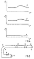
- Fig. 4 shows an example of two temporally successive proton density profiles 40 and 41 along the same main axis.
- the first proton density profile 40 does not include a disturbance whereas the second proton density profile 41 includes the disturbance.
- the position 42 of a deviation along, for example the first main axis is determined from the difference between the proton density profiles 40 and 41.
- the position of the deviation on the other two main axes is determined in a similar manner, the position of the distal end 23 of the catheter 15 in the body 7 thus being determined.
- the 1D Fourier transformations and the determination of the differences between the proton density profiles are executed by the image reconstruction unit 11.
- the position of the distal end 23 of the catheter 15 thus determined can be visualized on the monitor 14 by means of a cursor; moreover, it can also be used for continuously tracking the catheter 15 in an MR image.
- the position of the catheter 15 is applied to the control unit 13.
- the control unit 13 subsequently adapts the position of the region of the body to be imaged so that, for example the distal end 23 of the catheter 15 remains visible in the image.
- By varying the distance between the conductors in some parts of the catheter the magnetic field can be disturbed in a predeterminal pattern. This pattern enables a better recognizability of the catheter in an MR-image.
- a catheter with different distances between some parts of the conducting is shown in Fig. 5.
- FIG. 5 shows the catheter 50 with a conductor loop 51 in accordance with the invention. Further the distance between a first conductor 54 and a second conductor 55 of a first part 52 and the distance between the first conductor 54 and a second conductor 55 of a second part 53 are equal, but different from the distance of the conductors outside the two parts 52, 53. Therefore the extra magnetic field nearby the first part 52 and the second part 53 will be different from the extra magnetic field nearby the remainder of the catheter 50. Consequently a pattern around the catheter will be visible in an MR-image. Further a variable resistor 6 and a powersupply 57 are connected with the catheter 50. With the variable resistor 56 the current through the conductor loop 51 could be controlled and consequently the contrast of the catheter in the MR-image. To reduce HF disturbances both conductors could also be curled around the long axis of the catheter.
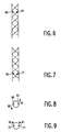
- a catheter with a conductor loop 17 comprising conductors curled in a helix configuration.
- Catheters with these helix configuration conductor loops are explained with reference to Fig. 6, 7, 8 and 9.
- Fig. 6 shows a catheter 16 with conductor loop 17 having a parallel helix configuration, in which both conductors 60, 61 of the conductor loop are parallel to each other.
- Fig. 7 shows a catheter 15 with the conductor loop 17 in an anti-parallel helix configuration in which the conductors 70, 71 cross each other.
- An advantage of the anti-parallel helix configuration is that the torsion on the catheter due to electromagnetic forces on the conductors 70, 71 is reduced with respect the torsion on a catheter due to the electromagnetic forces on the conductors 60, 61 in a parallel helix configuration. These electromagnetic forces are generated when a current is directed through the conductor loop in the static magnetic field.
- Fig. 8 shows the forces on the conductors 60, 61 of the conductor loop 16 in a parallel helix configuration.
- the arrows 80, 81 indicate the directions of the generated electromagnetic forces when a direct current is generated in the conductor loop. The directions of the forces are pointed to opposite directions and introduce a torsion on the catheter.
- Fig. 9 shows the directions of the forces on the conductors 70, 71 of a conductor loop curled in the anti-parallel helix.
- the arrows 90, 91 indicate the directions of the forces when a direct current is directed through the conductor loop. Because the directions of the forces 37, 38 on the conductors 70, 71 are pointed to the same directions the torsion on the catheter 15 is substantially reduced.
Abstract
Description
characterized in that the distance between the conductors along the catheter is different for some parts of the catheter. By varying the distance and there by the strength of the disturbance along the catheter a kind of pattern, can be visualized in an MR-image, so that the catheter is better recognizable.
Claims (14)
- An MR system for interventional procedures, comprising an MR device 1 which is arranged to acquire images of a part of an object (7) and comprises:characterized in that the means for generating the additional magnetic field comprise an electrical conductor loop with two non-magnetic, spaced apart conductors, said loop extending along substantially the length of the catheter, such that the catheter can bea. a magnet (2) for generating a steady magnetic field,b. means (6) for generating temporary magnetic gradient fields, andc. a catheter (15) which comprises means for generating an additional magnetic field,
imaged in an MR image along said spaced apart conductors. - An MR system as claimed in Claim 1, characterized in that the conductor loop is connected to a variable-voltage power supply.
- An MR system as claimed in Claim 1 or 2, characterized in that the catheter is provided with a coil for generating a second additional magnetic field which deviates from the first additional magnetic field.
- An MR system as claimed in Claim 1, 2 or 3 characterized in that the distance between the conductors along the catheter is different for some parts of the catheter.
- An MR system as claimed in Claim 1,2,3, or 4, characterized in that the conductors of the conductor loop are curled in a helix configuration, said helix configuration being formed by a uni-directional winding of the respective conductors along the length of the catheter.
- An MR system as claimed in Claim 1,2,3 or 4, characterized in that the conductors of the conductor loop are curled in an anti-parallel helix configuration, said anti-parallel helix configuration being formed by a bi-directional winding of the respective conductors along the length of the catheter.
- An MR system as claimed in Claim 1, 2, 3, 4, 5 or 6, characterized in that the MR device is arranged to execute the following steps:a) generating MR signals in order to determine temporally successive first and second MR images,b) generating the first and/or the second additional magnetic field during the generating of MR signals for determining the second MR image,c) receiving the MR signals generated,d) processing the MR signals received so as to form the first and the second MR image,e) determining a position of the catheter within the object on the basis of the first and the second MR image.
- An MR system as claimed in Claim 7, characterized in that the MR device is also arranged to derive images from MR signals by means of a so called keyhole technique.
- An MR system as claimed in Claim 7, or 8, characterized in that a control unit (13) is arranged to adapt a position of a region to be imaged, in which the catheter (15) is present, to the catheter position determined.
- A catheter for use in cooperation with an MR device, said catheter comprising means for generating an additional magnetic field, characterized in that the means for generating the additional magnetic field comprise an electrical conductor loop with two non-magnetic, spaced apart conductors, said loop extending along substantially the length of the catheter, such that the catheter can be imaged in an MR image along said spaced apart conductors.
- A catheter as claimed in Claim 10, characterized in that the conductor loop is connectable to a variable voltage power supply.
- A catheter as claimed in Claim 10 or 11, characterized in that the conductors of the conductor loop are curled in a helix configuration, said helix configuration being formed by a uni-directional winding of the respective conductors along the length of the catheter.
- A catheter as claimed in Claim 10 or 11, characterized in that the conductors of the conductor loop are curled in an anti-parallel helix configuration, said anti-parallel helix configuration being formed by a bi-directional winding of respective conductors along the length of the catheter.
- A catheter as claimed in Claim 10, 11 or 12, characterized in that it comprises a coil for generating a second additional magnetic field which deviates from the first additional magnetic field.
Priority Applications (1)
| Application Number | Priority Date | Filing Date | Title |
|---|---|---|---|
| EP96935271A EP0805987B1 (en) | 1995-11-24 | 1996-11-15 | Mri-system and catheter for interventional procedures |
Applications Claiming Priority (4)
| Application Number | Priority Date | Filing Date | Title |
|---|---|---|---|
| EP95203229 | 1995-11-24 | ||
| EP95203229 | 1995-11-24 | ||
| EP96935271A EP0805987B1 (en) | 1995-11-24 | 1996-11-15 | Mri-system and catheter for interventional procedures |
| PCT/IB1996/001238 WO1997019362A1 (en) | 1995-11-24 | 1996-11-15 | Mri-system and catheter for interventional procedures |
Publications (2)
| Publication Number | Publication Date |
|---|---|
| EP0805987A1 EP0805987A1 (en) | 1997-11-12 |
| EP0805987B1 true EP0805987B1 (en) | 2004-12-15 |
Family
ID=8220860
Family Applications (1)
| Application Number | Title | Priority Date | Filing Date |
|---|---|---|---|
| EP96935271A Expired - Lifetime EP0805987B1 (en) | 1995-11-24 | 1996-11-15 | Mri-system and catheter for interventional procedures |
Country Status (5)
| Country | Link |
|---|---|
| US (1) | US5868674A (en) |
| EP (1) | EP0805987B1 (en) |
| JP (1) | JPH10513098A (en) |
| DE (1) | DE69634035T2 (en) |
| WO (1) | WO1997019362A1 (en) |
Families Citing this family (171)
| Publication number | Priority date | Publication date | Assignee | Title |
|---|---|---|---|---|
| FR2652928B1 (en) | 1989-10-05 | 1994-07-29 | Diadix Sa | INTERACTIVE LOCAL INTERVENTION SYSTEM WITHIN A AREA OF A NON-HOMOGENEOUS STRUCTURE. |
| CA2142338C (en) * | 1992-08-14 | 1999-11-30 | John Stuart Bladen | Position location system |
| US5592939A (en) | 1995-06-14 | 1997-01-14 | Martinelli; Michael A. | Method and system for navigating a catheter probe |
| US7236816B2 (en) * | 1996-04-25 | 2007-06-26 | Johns Hopkins University | Biopsy and sampling needle antennas for magnetic resonance imaging-guided biopsies |
| US6263229B1 (en) | 1998-11-13 | 2001-07-17 | Johns Hopkins University School Of Medicine | Miniature magnetic resonance catheter coils and related methods |
| US6898454B2 (en) * | 1996-04-25 | 2005-05-24 | The Johns Hopkins University | Systems and methods for evaluating the urethra and the periurethral tissues |
| US6675033B1 (en) | 1999-04-15 | 2004-01-06 | Johns Hopkins University School Of Medicine | Magnetic resonance imaging guidewire probe |
| US6549800B1 (en) | 1996-04-25 | 2003-04-15 | Johns Hopkins Unversity School Of Medicine | Methods for in vivo magnetic resonance imaging |
| WO1998020358A1 (en) * | 1996-11-04 | 1998-05-14 | Philips Electronics N.V. | Mr system and invasive device for interventional procedures |
| NL1005946C2 (en) * | 1997-05-01 | 1998-11-03 | Cordis Europ | Catheter |
| US6061587A (en) * | 1997-05-15 | 2000-05-09 | Regents Of The University Of Minnesota | Method and apparatus for use with MR imaging |
| US6026316A (en) * | 1997-05-15 | 2000-02-15 | Regents Of The University Of Minnesota | Method and apparatus for use with MR imaging |
| US7048716B1 (en) | 1997-05-15 | 2006-05-23 | Stanford University | MR-compatible devices |
| US7505807B1 (en) * | 1997-05-15 | 2009-03-17 | Regents Of The University Of Minnesota | Magnetic resonance apparatus for use with active electrode and drug deliver catheter |
| US6272370B1 (en) | 1998-08-07 | 2001-08-07 | The Regents Of University Of Minnesota | MR-visible medical device for neurological interventions using nonlinear magnetic stereotaxis and a method imaging |
| DE19727081A1 (en) * | 1997-06-25 | 1999-01-07 | Siemens Ag | Object localisation method in MR examination |
| US6226548B1 (en) | 1997-09-24 | 2001-05-01 | Surgical Navigation Technologies, Inc. | Percutaneous registration apparatus and method for use in computer-assisted surgical navigation |
| US6021343A (en) | 1997-11-20 | 2000-02-01 | Surgical Navigation Technologies | Image guided awl/tap/screwdriver |
| US6348058B1 (en) * | 1997-12-12 | 2002-02-19 | Surgical Navigation Technologies, Inc. | Image guided spinal surgery guide, system, and method for use thereof |
| US6129667A (en) * | 1998-02-02 | 2000-10-10 | General Electric Company | Luminal diagnostics employing spectral analysis |
| US6463317B1 (en) | 1998-05-19 | 2002-10-08 | Regents Of The University Of Minnesota | Device and method for the endovascular treatment of aneurysms |
| EP2279692A3 (en) * | 1998-08-02 | 2011-02-23 | Super Dimension Ltd. | Intrabody navigation system for medical applications |
| US6477400B1 (en) | 1998-08-20 | 2002-11-05 | Sofamor Danek Holdings, Inc. | Fluoroscopic image guided orthopaedic surgery system with intraoperative registration |
| JP4319270B2 (en) * | 1998-08-25 | 2009-08-26 | 武蔵エンジニアリング株式会社 | Object position detection method and position detection apparatus |
| US6701176B1 (en) * | 1998-11-04 | 2004-03-02 | Johns Hopkins University School Of Medicine | Magnetic-resonance-guided imaging, electrophysiology, and ablation |
| US7844319B2 (en) * | 1998-11-04 | 2010-11-30 | Susil Robert C | Systems and methods for magnetic-resonance-guided interventional procedures |
| US8244370B2 (en) * | 2001-04-13 | 2012-08-14 | Greatbatch Ltd. | Band stop filter employing a capacitor and an inductor tank circuit to enhance MRI compatibility of active medical devices |
| WO2000033099A1 (en) * | 1998-12-03 | 2000-06-08 | Koninklijke Philips Electronics N.V. | Interventional instrument with adjustable visibility in mri images |
| US6470207B1 (en) * | 1999-03-23 | 2002-10-22 | Surgical Navigation Technologies, Inc. | Navigational guidance via computer-assisted fluoroscopic imaging |
| US7848788B2 (en) | 1999-04-15 | 2010-12-07 | The Johns Hopkins University | Magnetic resonance imaging probe |
| US6491699B1 (en) * | 1999-04-20 | 2002-12-10 | Surgical Navigation Technologies, Inc. | Instrument guidance method and system for image guided surgery |
| US7366562B2 (en) | 2003-10-17 | 2008-04-29 | Medtronic Navigation, Inc. | Method and apparatus for surgical navigation |
| AU1240801A (en) | 1999-10-28 | 2001-05-08 | Enterprise Medical Technology, Inc. | Coil structures and methods for generating magnetic fields |
| US6474341B1 (en) * | 1999-10-28 | 2002-11-05 | Surgical Navigation Technologies, Inc. | Surgical communication and power system |
| US6381485B1 (en) * | 1999-10-28 | 2002-04-30 | Surgical Navigation Technologies, Inc. | Registration of human anatomy integrated for electromagnetic localization |
| US6747539B1 (en) | 1999-10-28 | 2004-06-08 | Michael A. Martinelli | Patient-shielding and coil system |
| US11331150B2 (en) | 1999-10-28 | 2022-05-17 | Medtronic Navigation, Inc. | Method and apparatus for surgical navigation |
| US8644907B2 (en) * | 1999-10-28 | 2014-02-04 | Medtronic Navigaton, Inc. | Method and apparatus for surgical navigation |
| US6493573B1 (en) | 1999-10-28 | 2002-12-10 | Winchester Development Associates | Method and system for navigating a catheter probe in the presence of field-influencing objects |
| US6499488B1 (en) | 1999-10-28 | 2002-12-31 | Winchester Development Associates | Surgical sensor |
| US8239001B2 (en) * | 2003-10-17 | 2012-08-07 | Medtronic Navigation, Inc. | Method and apparatus for surgical navigation |
| US7167615B1 (en) | 1999-11-05 | 2007-01-23 | Board Of Regents, The University Of Texas System | Resonant waveguide-grating filters and sensors and methods for making and using same |
| DE19962848C2 (en) * | 1999-12-24 | 2003-03-27 | Forschungszentrum Juelich Gmbh | Echo planar imaging |
| ATE484757T1 (en) | 2000-02-01 | 2010-10-15 | Surgivision Inc | TRANSSEPTAL NEEDLE ANTENNA FOR AN MR IMAGING DEVICE |
| WO2001064124A1 (en) * | 2000-03-01 | 2001-09-07 | Surgical Navigation Technologies, Inc. | Multiple cannula image guided tool for image guided procedures |
| CN1273082C (en) * | 2000-03-03 | 2006-09-06 | 卡迪亚克M.R.I.公司 | Magnetic resonance specimen analysis apparatus |
| EP1269206A2 (en) | 2000-03-24 | 2003-01-02 | Surgi-Vision | Apparatus, systems and methods for in vivo magnetic resonance imaging |
| US6535756B1 (en) * | 2000-04-07 | 2003-03-18 | Surgical Navigation Technologies, Inc. | Trajectory storage apparatus and method for surgical navigation system |
| US6626902B1 (en) | 2000-04-12 | 2003-09-30 | University Of Virginia Patent Foundation | Multi-probe system |
| JP2004511271A (en) * | 2000-05-12 | 2004-04-15 | シー・アール・バード・インコーポレーテッド | MRI ablation catheter |
| US7085400B1 (en) * | 2000-06-14 | 2006-08-01 | Surgical Navigation Technologies, Inc. | System and method for image based sensor calibration |
| US20020055678A1 (en) * | 2000-07-13 | 2002-05-09 | Scott Greig C. | Electrode probe coil for MRI |
| AU8370301A (en) * | 2000-08-23 | 2002-03-04 | Micronix Pty Ltd | Catheter locator apparatus and method of use |
| US6714809B2 (en) * | 2000-11-20 | 2004-03-30 | Surgi-Vision, Inc. | Connector and guidewire connectable thereto |
| US20020103430A1 (en) | 2001-01-29 | 2002-08-01 | Hastings Roger N. | Catheter navigation within an MR imaging device |
| US8600519B2 (en) * | 2001-04-13 | 2013-12-03 | Greatbatch Ltd. | Transient voltage/current protection system for electronic circuits associated with implanted leads |
| US8219208B2 (en) * | 2001-04-13 | 2012-07-10 | Greatbatch Ltd. | Frequency selective passive component networks for active implantable medical devices utilizing an energy dissipating surface |
| US8509913B2 (en) * | 2001-04-13 | 2013-08-13 | Greatbatch Ltd. | Switched diverter circuits for minimizing heating of an implanted lead and/or providing EMI protection in a high power electromagnetic field environment |
| US9295828B2 (en) | 2001-04-13 | 2016-03-29 | Greatbatch Ltd. | Self-resonant inductor wound portion of an implantable lead for enhanced MRI compatibility of active implantable medical devices |
| US8977355B2 (en) * | 2001-04-13 | 2015-03-10 | Greatbatch Ltd. | EMI filter employing a capacitor and an inductor tank circuit having optimum component values |
| US20070088416A1 (en) * | 2001-04-13 | 2007-04-19 | Surgi-Vision, Inc. | Mri compatible medical leads |
| US8457760B2 (en) | 2001-04-13 | 2013-06-04 | Greatbatch Ltd. | Switched diverter circuits for minimizing heating of an implanted lead and/or providing EMI protection in a high power electromagnetic field environment |
| WO2002083016A1 (en) | 2001-04-13 | 2002-10-24 | Surgi-Vision, Inc. | Systems and methods for magnetic-resonance-guided interventional procedures |
| US8989870B2 (en) * | 2001-04-13 | 2015-03-24 | Greatbatch Ltd. | Tuned energy balanced system for minimizing heating and/or to provide EMI protection of implanted leads in a high power electromagnetic field environment |
| GB2378760A (en) * | 2001-04-20 | 2003-02-19 | Marconi Medical Systems Uk Ltd | Surgical Probe |
| US20030208252A1 (en) * | 2001-05-14 | 2003-11-06 | O' Boyle Gary S. | Mri ablation catheter |
| US6636757B1 (en) * | 2001-06-04 | 2003-10-21 | Surgical Navigation Technologies, Inc. | Method and apparatus for electromagnetic navigation of a surgical probe near a metal object |
| US6496714B1 (en) | 2001-07-20 | 2002-12-17 | Koninklijke Philips Electronics N.V. | RF-safe invasive device |
| DE10137170B4 (en) * | 2001-07-31 | 2005-04-28 | Siemens Ag | Method for triggering respiration in an imaging process |
| US6619838B2 (en) | 2001-08-22 | 2003-09-16 | Scimed Life Systems, Inc. | Two-piece sensor assembly |
| US7587234B2 (en) * | 2001-11-02 | 2009-09-08 | Abbott Cardiovascular Systems Inc. | Method and apparatus for computer modified magnetic resonance imaging |
| US7194297B2 (en) * | 2001-11-13 | 2007-03-20 | Boston Scientific Scimed, Inc. | Impedance-matching apparatus and construction for intravascular device |
| US20030114747A1 (en) * | 2001-12-14 | 2003-06-19 | Smith Scott R. | Recanalization of occluded vessel using magnetic resonance guidance |
| WO2003066124A2 (en) * | 2002-02-01 | 2003-08-14 | The Cleveland Clinic Foundation | Apparatus for facilitating delivery of at least one device to a target site in a body |
| US6947786B2 (en) * | 2002-02-28 | 2005-09-20 | Surgical Navigation Technologies, Inc. | Method and apparatus for perspective inversion |
| US6990368B2 (en) * | 2002-04-04 | 2006-01-24 | Surgical Navigation Technologies, Inc. | Method and apparatus for virtual digital subtraction angiography |
| US7998062B2 (en) | 2004-03-29 | 2011-08-16 | Superdimension, Ltd. | Endoscope structures and techniques for navigating to a target in branched structure |
| CA2487140C (en) | 2002-05-29 | 2011-09-20 | Surgi-Vision, Inc. | Magnetic resonance probes |
| US7248914B2 (en) * | 2002-06-28 | 2007-07-24 | Stereotaxis, Inc. | Method of navigating medical devices in the presence of radiopaque material |
| US6892090B2 (en) * | 2002-08-19 | 2005-05-10 | Surgical Navigation Technologies, Inc. | Method and apparatus for virtual endoscopy |
| DE10240960A1 (en) * | 2002-09-05 | 2004-03-18 | Philips Intellectual Property & Standards Gmbh | Catheters, especially for use in MR imaging |
| US7697972B2 (en) | 2002-11-19 | 2010-04-13 | Medtronic Navigation, Inc. | Navigation system for cardiac therapies |
| US7599730B2 (en) | 2002-11-19 | 2009-10-06 | Medtronic Navigation, Inc. | Navigation system for cardiac therapies |
| ATE365334T1 (en) * | 2003-01-10 | 2007-07-15 | Deutsches Krebsforsch | DEVICE FOR DETERMINING THE LOCATION AND ORIENTATION OF AN INVASIVE DEVICE |
| US7660623B2 (en) | 2003-01-30 | 2010-02-09 | Medtronic Navigation, Inc. | Six degree of freedom alignment display for medical procedures |
| US7542791B2 (en) * | 2003-01-30 | 2009-06-02 | Medtronic Navigation, Inc. | Method and apparatus for preplanning a surgical procedure |
| US7792568B2 (en) * | 2003-03-17 | 2010-09-07 | Boston Scientific Scimed, Inc. | MRI-visible medical devices |
| JP2006524082A (en) | 2003-04-23 | 2006-10-26 | コーニンクレッカ フィリップス エレクトロニクス エヌ ヴィ | Magnetic resonance position detection method |
| US7570791B2 (en) * | 2003-04-25 | 2009-08-04 | Medtronic Navigation, Inc. | Method and apparatus for performing 2D to 3D registration |
| US7313430B2 (en) * | 2003-08-28 | 2007-12-25 | Medtronic Navigation, Inc. | Method and apparatus for performing stereotactic surgery |
| EP2316328B1 (en) | 2003-09-15 | 2012-05-09 | Super Dimension Ltd. | Wrap-around holding device for use with bronchoscopes |
| ATE556643T1 (en) | 2003-09-15 | 2012-05-15 | Super Dimension Ltd | COVERING DEVICE FOR FIXING BRONCHOSCOPES |
| US20050085895A1 (en) * | 2003-10-15 | 2005-04-21 | Scimed Life Systems, Inc. | RF-based markers for MRI visualization of medical devices |
| US7835778B2 (en) | 2003-10-16 | 2010-11-16 | Medtronic Navigation, Inc. | Method and apparatus for surgical navigation of a multiple piece construct for implantation |
| US7840253B2 (en) * | 2003-10-17 | 2010-11-23 | Medtronic Navigation, Inc. | Method and apparatus for surgical navigation |
| US8764725B2 (en) * | 2004-02-09 | 2014-07-01 | Covidien Lp | Directional anchoring mechanism, method and applications thereof |
| CA2505464C (en) * | 2004-04-28 | 2013-12-10 | Sunnybrook And Women's College Health Sciences Centre | Catheter tracking with phase information |
| US7567834B2 (en) * | 2004-05-03 | 2009-07-28 | Medtronic Navigation, Inc. | Method and apparatus for implantation between two vertebral bodies |
| US20050251031A1 (en) * | 2004-05-06 | 2005-11-10 | Scimed Life Systems, Inc. | Apparatus and construction for intravascular device |
| US7496397B2 (en) * | 2004-05-06 | 2009-02-24 | Boston Scientific Scimed, Inc. | Intravascular antenna |
| US20050283226A1 (en) * | 2004-06-18 | 2005-12-22 | Scimed Life Systems, Inc. | Medical devices |
| US7636595B2 (en) * | 2004-10-28 | 2009-12-22 | Medtronic Navigation, Inc. | Method and apparatus for calibrating non-linear instruments |
| US20060122488A1 (en) * | 2004-11-18 | 2006-06-08 | Abdol-Mohammad Kajbafzadeh | Urodynamic diagnostic method and system |
| US7976518B2 (en) | 2005-01-13 | 2011-07-12 | Corpak Medsystems, Inc. | Tubing assembly and signal generator placement control device and method for use with catheter guidance systems |
| US20070066880A1 (en) * | 2005-09-09 | 2007-03-22 | Warren Lee | Image-based probe guidance system |
| US7835784B2 (en) | 2005-09-21 | 2010-11-16 | Medtronic Navigation, Inc. | Method and apparatus for positioning a reference frame |
| EP1785739A1 (en) | 2005-11-14 | 2007-05-16 | DKFZ Deutsches Krebsforschungszentrum | An elongate, segmented, RF safe device for use with an MRI machine |
| US20070156042A1 (en) * | 2005-12-30 | 2007-07-05 | Orhan Unal | Medical device system and method for tracking and visualizing a medical device system under MR guidance |
| US8457712B2 (en) | 2005-12-30 | 2013-06-04 | Wisconsin Alumni Research Foundation | Multi-mode medical device system and methods of manufacturing and using same |
| US9168102B2 (en) | 2006-01-18 | 2015-10-27 | Medtronic Navigation, Inc. | Method and apparatus for providing a container to a sterile environment |
| US8112292B2 (en) * | 2006-04-21 | 2012-02-07 | Medtronic Navigation, Inc. | Method and apparatus for optimizing a therapy |
| US8903505B2 (en) | 2006-06-08 | 2014-12-02 | Greatbatch Ltd. | Implantable lead bandstop filter employing an inductive coil with parasitic capacitance to enhance MRI compatibility of active medical devices |
| US7622920B2 (en) * | 2006-07-06 | 2009-11-24 | Kabushiki Kaisha Toshiba | Magnetic resonance imaging apparatus capable of automatically determining RF coil positions |
| US8197494B2 (en) | 2006-09-08 | 2012-06-12 | Corpak Medsystems, Inc. | Medical device position guidance system with wireless connectivity between a noninvasive device and an invasive device |
| US8660635B2 (en) * | 2006-09-29 | 2014-02-25 | Medtronic, Inc. | Method and apparatus for optimizing a computer assisted surgical procedure |
| US8532742B2 (en) | 2006-11-15 | 2013-09-10 | Wisconsin Alumni Research Foundation | System and method for simultaneous 3DPR device tracking and imaging under MR-guidance for therapeutic endovascular interventions |
| EP2120705A1 (en) * | 2006-12-21 | 2009-11-25 | Koninklijke Philips Electronics N.V. | Wireless interventional device and a system for wireless energy transmission |
| US20080275395A1 (en) * | 2006-12-22 | 2008-11-06 | Innerspace Medical, Inc. | MRI-Compatible Temperature-Sensing Catheter |
| US20080208039A1 (en) | 2007-02-28 | 2008-08-28 | Wisconsin Alumni Research Foundation | System and method of performing therapeutic endovascular interventions |
| US8905920B2 (en) | 2007-09-27 | 2014-12-09 | Covidien Lp | Bronchoscope adapter and method |
| US8175679B2 (en) * | 2007-12-26 | 2012-05-08 | St. Jude Medical, Atrial Fibrillation Division, Inc. | Catheter electrode that can simultaneously emit electrical energy and facilitate visualization by magnetic resonance imaging |
| US9675410B2 (en) | 2007-12-28 | 2017-06-13 | St. Jude Medical, Atrial Fibrillation Division, Inc. | Flexible polymer electrode for MRI-guided positioning and radio frequency ablation |
| IL196660A (en) | 2008-01-23 | 2014-09-30 | Mediguide Ltd | Sensor mounted flexible guidewire |
| US9095685B2 (en) | 2008-01-23 | 2015-08-04 | Mediguide Ltd. | Sensor mounted flexible guidewire |
| US9108066B2 (en) | 2008-03-20 | 2015-08-18 | Greatbatch Ltd. | Low impedance oxide resistant grounded capacitor for an AIMD |
| US10080889B2 (en) | 2009-03-19 | 2018-09-25 | Greatbatch Ltd. | Low inductance and low resistance hermetically sealed filtered feedthrough for an AIMD |
| WO2009122273A2 (en) | 2008-04-03 | 2009-10-08 | Superdimension, Ltd. | Magnetic interference detection system and method |
| EP2297673B1 (en) | 2008-06-03 | 2020-04-22 | Covidien LP | Feature-based registration method |
| US8218847B2 (en) | 2008-06-06 | 2012-07-10 | Superdimension, Ltd. | Hybrid registration method |
| DE102008030942A1 (en) | 2008-07-02 | 2010-01-07 | Christoph Miethke Gmbh & Co Kg | Cerebrospinal fluid drainage |
| US8932207B2 (en) * | 2008-07-10 | 2015-01-13 | Covidien Lp | Integrated multi-functional endoscopic tool |
| US8165658B2 (en) | 2008-09-26 | 2012-04-24 | Medtronic, Inc. | Method and apparatus for positioning a guide relative to a base |
| US8175681B2 (en) * | 2008-12-16 | 2012-05-08 | Medtronic Navigation Inc. | Combination of electromagnetic and electropotential localization |
| US8447414B2 (en) * | 2008-12-17 | 2013-05-21 | Greatbatch Ltd. | Switched safety protection circuit for an AIMD system during exposure to high power electromagnetic fields |
| WO2010076681A1 (en) * | 2008-12-31 | 2010-07-08 | Koninklijke Philips Electronics N.V. | General inductive handpiece for active devices |
| US8095224B2 (en) * | 2009-03-19 | 2012-01-10 | Greatbatch Ltd. | EMI shielded conduit assembly for an active implantable medical device |
| US8611984B2 (en) | 2009-04-08 | 2013-12-17 | Covidien Lp | Locatable catheter |
| JP5859431B2 (en) * | 2009-06-08 | 2016-02-10 | エムアールアイ・インターヴェンションズ,インコーポレイテッド | MRI guided intervention system capable of tracking flexible internal devices and generating dynamic visualization in near real time |
| US8369930B2 (en) | 2009-06-16 | 2013-02-05 | MRI Interventions, Inc. | MRI-guided devices and MRI-guided interventional systems that can track and generate dynamic visualizations of the devices in near real time |
| US8494614B2 (en) * | 2009-08-31 | 2013-07-23 | Regents Of The University Of Minnesota | Combination localization system |
| US8494613B2 (en) * | 2009-08-31 | 2013-07-23 | Medtronic, Inc. | Combination localization system |
| US8882763B2 (en) | 2010-01-12 | 2014-11-11 | Greatbatch Ltd. | Patient attached bonding strap for energy dissipation from a probe or a catheter during magnetic resonance imaging |
| CN102237842A (en) * | 2010-04-23 | 2011-11-09 | 鸿富锦精密工业(深圳)有限公司 | Magnetic field generation and control circuit |
| WO2011159834A1 (en) | 2010-06-15 | 2011-12-22 | Superdimension, Ltd. | Locatable expandable working channel and method |
| US10272252B2 (en) | 2016-11-08 | 2019-04-30 | Greatbatch Ltd. | Hermetic terminal for an AIMD having a composite brazed conductive lead |
| US9931514B2 (en) | 2013-06-30 | 2018-04-03 | Greatbatch Ltd. | Low impedance oxide resistant grounded capacitor for an AIMD |
| US10596369B2 (en) | 2011-03-01 | 2020-03-24 | Greatbatch Ltd. | Low equivalent series resistance RF filter for an active implantable medical device |
| US9427596B2 (en) | 2013-01-16 | 2016-08-30 | Greatbatch Ltd. | Low impedance oxide resistant grounded capacitor for an AIMD |
| US10350421B2 (en) | 2013-06-30 | 2019-07-16 | Greatbatch Ltd. | Metallurgically bonded gold pocket pad for grounding an EMI filter to a hermetic terminal for an active implantable medical device |
| US11198014B2 (en) | 2011-03-01 | 2021-12-14 | Greatbatch Ltd. | Hermetically sealed filtered feedthrough assembly having a capacitor with an oxide resistant electrical connection to an active implantable medical device housing |
| WO2013036772A1 (en) | 2011-09-08 | 2013-03-14 | Corpak Medsystems, Inc. | Apparatus and method used with guidance system for feeding and suctioning |
| DE102012023124B4 (en) * | 2012-11-27 | 2015-11-26 | Hansjörg Graf | Method and device for visualizing interventional instruments in magnetic resonance imaging via sequence-triggered energization with evaluation of the magnitude and phase images |
| USRE46699E1 (en) | 2013-01-16 | 2018-02-06 | Greatbatch Ltd. | Low impedance oxide resistant grounded capacitor for an AIMD |
| US10952593B2 (en) | 2014-06-10 | 2021-03-23 | Covidien Lp | Bronchoscope adapter |
| US10426555B2 (en) | 2015-06-03 | 2019-10-01 | Covidien Lp | Medical instrument with sensor for use in a system and method for electromagnetic navigation |
| EP3349650A4 (en) | 2015-09-16 | 2019-04-24 | THE UNITED STATES OF AMERICA, as represented by the Secretary, DEPARTMENT OF HEALTH AND HUMAN SERVICES | Segmented mri catheters and other interventional devices |
| DE102015012201A1 (en) | 2015-09-18 | 2017-03-23 | Hansjörg Graf | Method and device for visualizing interventional instruments in magnetic resonance imaging (MRI) via discrete phase coding artifacts generated by means of sequence-triggered energization |
| US9962134B2 (en) | 2015-10-28 | 2018-05-08 | Medtronic Navigation, Inc. | Apparatus and method for maintaining image quality while minimizing X-ray dosage of a patient |
| US10478254B2 (en) | 2016-05-16 | 2019-11-19 | Covidien Lp | System and method to access lung tissue |
| US10751126B2 (en) | 2016-10-28 | 2020-08-25 | Covidien Lp | System and method for generating a map for electromagnetic navigation |
| US10446931B2 (en) | 2016-10-28 | 2019-10-15 | Covidien Lp | Electromagnetic navigation antenna assembly and electromagnetic navigation system including the same |
| US10418705B2 (en) | 2016-10-28 | 2019-09-17 | Covidien Lp | Electromagnetic navigation antenna assembly and electromagnetic navigation system including the same |
| US10638952B2 (en) | 2016-10-28 | 2020-05-05 | Covidien Lp | Methods, systems, and computer-readable media for calibrating an electromagnetic navigation system |
| US10517505B2 (en) | 2016-10-28 | 2019-12-31 | Covidien Lp | Systems, methods, and computer-readable media for optimizing an electromagnetic navigation system |
| US10792106B2 (en) | 2016-10-28 | 2020-10-06 | Covidien Lp | System for calibrating an electromagnetic navigation system |
| US10615500B2 (en) | 2016-10-28 | 2020-04-07 | Covidien Lp | System and method for designing electromagnetic navigation antenna assemblies |
| US10722311B2 (en) | 2016-10-28 | 2020-07-28 | Covidien Lp | System and method for identifying a location and/or an orientation of an electromagnetic sensor based on a map |
| US10249415B2 (en) | 2017-01-06 | 2019-04-02 | Greatbatch Ltd. | Process for manufacturing a leadless feedthrough for an active implantable medical device |
| US11219489B2 (en) | 2017-10-31 | 2022-01-11 | Covidien Lp | Devices and systems for providing sensors in parallel with medical tools |
| US10912945B2 (en) | 2018-03-22 | 2021-02-09 | Greatbatch Ltd. | Hermetic terminal for an active implantable medical device having a feedthrough capacitor partially overhanging a ferrule for high effective capacitance area |
| US10905888B2 (en) | 2018-03-22 | 2021-02-02 | Greatbatch Ltd. | Electrical connection for an AIMD EMI filter utilizing an anisotropic conductive layer |
Citations (1)
| Publication number | Priority date | Publication date | Assignee | Title |
|---|---|---|---|---|
| US4572198A (en) * | 1984-06-18 | 1986-02-25 | Varian Associates, Inc. | Catheter for use with NMR imaging systems |
Family Cites Families (10)
| Publication number | Priority date | Publication date | Assignee | Title |
|---|---|---|---|---|
| GB9018660D0 (en) * | 1990-08-24 | 1990-10-10 | Imperial College | Probe system |
| US5437277A (en) * | 1991-11-18 | 1995-08-01 | General Electric Company | Inductively coupled RF tracking system for use in invasive imaging of a living body |
| US5445150A (en) * | 1991-11-18 | 1995-08-29 | General Electric Company | Invasive system employing a radiofrequency tracking system |
| US5271400A (en) * | 1992-04-01 | 1993-12-21 | General Electric Company | Tracking system to monitor the position and orientation of a device using magnetic resonance detection of a sample contained within the device |
| US5318025A (en) * | 1992-04-01 | 1994-06-07 | General Electric Company | Tracking system to monitor the position and orientation of a device using multiplexed magnetic resonance detection |
| US5647361A (en) * | 1992-09-28 | 1997-07-15 | Fonar Corporation | Magnetic resonance imaging method and apparatus for guiding invasive therapy |
| US5417708A (en) * | 1994-03-09 | 1995-05-23 | Cook Incorporated | Intravascular treatment system and percutaneous release mechanism therefor |
| ES2114626T3 (en) * | 1994-03-18 | 1998-06-01 | Schneider Europ Ag | MAGNETIC RESONANCE VISUALIZATION SYSTEM TO LOCATE A MEDICAL INSTRUMENT. |
| US5447156A (en) * | 1994-04-04 | 1995-09-05 | General Electric Company | Magnetic resonance (MR) active invasive devices for the generation of selective MR angiograms |
| DE19507617A1 (en) * | 1995-03-04 | 1996-09-05 | Philips Patentverwaltung | MR method and MR device for performing the method |
-
1996
- 1996-11-15 JP JP9519551A patent/JPH10513098A/en active Pending
- 1996-11-15 DE DE69634035T patent/DE69634035T2/en not_active Expired - Fee Related
- 1996-11-15 EP EP96935271A patent/EP0805987B1/en not_active Expired - Lifetime
- 1996-11-15 WO PCT/IB1996/001238 patent/WO1997019362A1/en active IP Right Grant
- 1996-11-22 US US08/754,358 patent/US5868674A/en not_active Expired - Fee Related
Patent Citations (1)
| Publication number | Priority date | Publication date | Assignee | Title |
|---|---|---|---|---|
| US4572198A (en) * | 1984-06-18 | 1986-02-25 | Varian Associates, Inc. | Catheter for use with NMR imaging systems |
Also Published As
| Publication number | Publication date |
|---|---|
| EP0805987A1 (en) | 1997-11-12 |
| DE69634035T2 (en) | 2005-12-08 |
| JPH10513098A (en) | 1998-12-15 |
| WO1997019362A1 (en) | 1997-05-29 |
| DE69634035D1 (en) | 2005-01-20 |
| US5868674A (en) | 1999-02-09 |
Similar Documents
| Publication | Publication Date | Title |
|---|---|---|
| EP0805987B1 (en) | Mri-system and catheter for interventional procedures | |
| US5916162A (en) | Invasive device for use in a magnetic resonance imaging apparatus | |
| US6171240B1 (en) | MRI RF catheter coil | |
| US6741882B2 (en) | MR device and MR method for localizing and/or visualizing a medical instrument provided with a passive magnet device | |
| US6993373B2 (en) | Invasive device provided with a segmented electrical connection conductor | |
| US7650178B2 (en) | Magnetic field sensor-based navigation system to track MR image-guided interventional procedures | |
| US6975896B2 (en) | Fiducial markers for MRI | |
| Zhang et al. | Active MR guidance of interventional devices with target‐navigation | |
| US5819737A (en) | Magnetic resonance methods and apparatus | |
| JP5710970B2 (en) | Magnetic resonance apparatus and method | |
| JPH09187435A (en) | Magnetic resonance system and magnetic resonance imaging/tracking system | |
| JP2000504976A (en) | MR system and invasive device for interventional treatment | |
| WO2003005902A1 (en) | Endoscopic image pickup method and magnetic resonance imaging device using the same | |
| JP2005508718A (en) | Ceramic reinforcement for MRI equipment | |
| EP2549284A1 (en) | Position marker for use in an MRI apparatus | |
| US7835780B1 (en) | MR invasive device and method for active MR guidance of invasive devices with target navigation | |
| US6430429B1 (en) | Magnetic resonance imaging system with an interventional instrument | |
| US20220409115A1 (en) | Apparatus and method for magnetoencephalography with electropermanent magnet array | |
| EP3430416A1 (en) | Mr-visible marker for an mri apparatus and an mr guided radiation therapy system | |
| JP3213819B2 (en) | High frequency receiving coil for magnetic resonance imaging equipment | |
| JP2811352B2 (en) | High frequency coil for magnetic resonance imaging equipment | |
| JP2767311B2 (en) | High frequency coil for magnetic resonance imaging equipment | |
| JP3496891B2 (en) | Nuclear magnetic resonance imaging equipment | |
| JPH05161624A (en) | High frequency reception coil for magnetic resonance imaging device |
Legal Events
| Date | Code | Title | Description |
|---|---|---|---|
| PUAI | Public reference made under article 153(3) epc to a published international application that has entered the european phase |
Free format text: ORIGINAL CODE: 0009012 |
|
| AK | Designated contracting states |
Kind code of ref document: A1 Designated state(s): DE FR GB NL |
|
| 17P | Request for examination filed |
Effective date: 19971201 |
|
| 17Q | First examination report despatched |
Effective date: 20030605 |
|
| GRAP | Despatch of communication of intention to grant a patent |
Free format text: ORIGINAL CODE: EPIDOSNIGR1 |
|
| GRAS | Grant fee paid |
Free format text: ORIGINAL CODE: EPIDOSNIGR3 |
|
| GRAA | (expected) grant |
Free format text: ORIGINAL CODE: 0009210 |
|
| AK | Designated contracting states |
Kind code of ref document: B1 Designated state(s): DE FR GB NL |
|
| PG25 | Lapsed in a contracting state [announced via postgrant information from national office to epo] |
Ref country code: NL Free format text: LAPSE BECAUSE OF FAILURE TO SUBMIT A TRANSLATION OF THE DESCRIPTION OR TO PAY THE FEE WITHIN THE PRESCRIBED TIME-LIMIT Effective date: 20041215 |
|
| REG | Reference to a national code |
Ref country code: GB Ref legal event code: FG4D |
|
| REG | Reference to a national code |
Ref country code: GB Ref legal event code: 746 Effective date: 20041222 |
|
| REF | Corresponds to: |
Ref document number: 69634035 Country of ref document: DE Date of ref document: 20050120 Kind code of ref document: P |
|
| NLV1 | Nl: lapsed or annulled due to failure to fulfill the requirements of art. 29p and 29m of the patents act | ||
| PLBE | No opposition filed within time limit |
Free format text: ORIGINAL CODE: 0009261 |
|
| STAA | Information on the status of an ep patent application or granted ep patent |
Free format text: STATUS: NO OPPOSITION FILED WITHIN TIME LIMIT |
|
| PGFP | Annual fee paid to national office [announced via postgrant information from national office to epo] |
Ref country code: GB Payment date: 20051129 Year of fee payment: 10 Ref country code: FR Payment date: 20051129 Year of fee payment: 10 |
|
| 26N | No opposition filed |
Effective date: 20050916 |
|
| ET | Fr: translation filed | ||
| REG | Reference to a national code |
Ref country code: FR Ref legal event code: D6 |
|
| PGFP | Annual fee paid to national office [announced via postgrant information from national office to epo] |
Ref country code: DE Payment date: 20060117 Year of fee payment: 10 |
|
| PG25 | Lapsed in a contracting state [announced via postgrant information from national office to epo] |
Ref country code: DE Free format text: LAPSE BECAUSE OF NON-PAYMENT OF DUE FEES Effective date: 20070601 |
|
| GBPC | Gb: european patent ceased through non-payment of renewal fee |
Effective date: 20061115 |
|
| REG | Reference to a national code |
Ref country code: FR Ref legal event code: ST Effective date: 20070731 |
|
| PG25 | Lapsed in a contracting state [announced via postgrant information from national office to epo] |
Ref country code: GB Free format text: LAPSE BECAUSE OF NON-PAYMENT OF DUE FEES Effective date: 20061115 |
|
| PG25 | Lapsed in a contracting state [announced via postgrant information from national office to epo] |
Ref country code: FR Free format text: LAPSE BECAUSE OF NON-PAYMENT OF DUE FEES Effective date: 20061130 |