EP0804731B2 - Method and apparatus for fabricating microarrays of biological samples - Google Patents
Method and apparatus for fabricating microarrays of biological samples Download PDFInfo
- Publication number
- EP0804731B2 EP0804731B2 EP95923921A EP95923921A EP0804731B2 EP 0804731 B2 EP0804731 B2 EP 0804731B2 EP 95923921 A EP95923921 A EP 95923921A EP 95923921 A EP95923921 A EP 95923921A EP 0804731 B2 EP0804731 B2 EP 0804731B2
- Authority
- EP
- European Patent Office
- Prior art keywords
- microarray
- reagent
- analyte
- array
- substrate
- Prior art date
- Legal status (The legal status is an assumption and is not a legal conclusion. Google has not performed a legal analysis and makes no representation as to the accuracy of the status listed.)
- Expired - Lifetime
Links
Images


Classifications
-
- B—PERFORMING OPERATIONS; TRANSPORTING
- B01—PHYSICAL OR CHEMICAL PROCESSES OR APPARATUS IN GENERAL
- B01L—CHEMICAL OR PHYSICAL LABORATORY APPARATUS FOR GENERAL USE
- B01L3/00—Containers or dishes for laboratory use, e.g. laboratory glassware; Droppers
- B01L3/02—Burettes; Pipettes
- B01L3/0241—Drop counters; Drop formers
- B01L3/0244—Drop counters; Drop formers using pins
-
- B—PERFORMING OPERATIONS; TRANSPORTING
- B01—PHYSICAL OR CHEMICAL PROCESSES OR APPARATUS IN GENERAL
- B01J—CHEMICAL OR PHYSICAL PROCESSES, e.g. CATALYSIS OR COLLOID CHEMISTRY; THEIR RELEVANT APPARATUS
- B01J19/00—Chemical, physical or physico-chemical processes in general; Their relevant apparatus
- B01J19/0046—Sequential or parallel reactions, e.g. for the synthesis of polypeptides or polynucleotides; Apparatus and devices for combinatorial chemistry or for making molecular arrays
-
- B—PERFORMING OPERATIONS; TRANSPORTING
- B01—PHYSICAL OR CHEMICAL PROCESSES OR APPARATUS IN GENERAL
- B01J—CHEMICAL OR PHYSICAL PROCESSES, e.g. CATALYSIS OR COLLOID CHEMISTRY; THEIR RELEVANT APPARATUS
- B01J4/00—Feed or outlet devices; Feed or outlet control devices
- B01J4/02—Feed or outlet devices; Feed or outlet control devices for feeding measured, i.e. prescribed quantities of reagents
-
- B—PERFORMING OPERATIONS; TRANSPORTING
- B01—PHYSICAL OR CHEMICAL PROCESSES OR APPARATUS IN GENERAL
- B01L—CHEMICAL OR PHYSICAL LABORATORY APPARATUS FOR GENERAL USE
- B01L3/00—Containers or dishes for laboratory use, e.g. laboratory glassware; Droppers
- B01L3/02—Burettes; Pipettes
- B01L3/0241—Drop counters; Drop formers
- B01L3/0244—Drop counters; Drop formers using pins
- B01L3/0248—Prongs, quill pen type dispenser
-
- B—PERFORMING OPERATIONS; TRANSPORTING
- B01—PHYSICAL OR CHEMICAL PROCESSES OR APPARATUS IN GENERAL
- B01L—CHEMICAL OR PHYSICAL LABORATORY APPARATUS FOR GENERAL USE
- B01L3/00—Containers or dishes for laboratory use, e.g. laboratory glassware; Droppers
- B01L3/02—Burettes; Pipettes
- B01L3/0241—Drop counters; Drop formers
- B01L3/0268—Drop counters; Drop formers using pulse dispensing or spraying, eg. inkjet type, piezo actuated ejection of droplets from capillaries
-
- B—PERFORMING OPERATIONS; TRANSPORTING
- B01—PHYSICAL OR CHEMICAL PROCESSES OR APPARATUS IN GENERAL
- B01L—CHEMICAL OR PHYSICAL LABORATORY APPARATUS FOR GENERAL USE
- B01L3/00—Containers or dishes for laboratory use, e.g. laboratory glassware; Droppers
- B01L3/50—Containers for the purpose of retaining a material to be analysed, e.g. test tubes
- B01L3/508—Containers for the purpose of retaining a material to be analysed, e.g. test tubes rigid containers not provided for above
- B01L3/5088—Containers for the purpose of retaining a material to be analysed, e.g. test tubes rigid containers not provided for above confining liquids at a location by surface tension, e.g. virtual wells on plates, wires
-
- C—CHEMISTRY; METALLURGY
- C12—BIOCHEMISTRY; BEER; SPIRITS; WINE; VINEGAR; MICROBIOLOGY; ENZYMOLOGY; MUTATION OR GENETIC ENGINEERING
- C12Q—MEASURING OR TESTING PROCESSES INVOLVING ENZYMES, NUCLEIC ACIDS OR MICROORGANISMS; COMPOSITIONS OR TEST PAPERS THEREFOR; PROCESSES OF PREPARING SUCH COMPOSITIONS; CONDITION-RESPONSIVE CONTROL IN MICROBIOLOGICAL OR ENZYMOLOGICAL PROCESSES
- C12Q1/00—Measuring or testing processes involving enzymes, nucleic acids or microorganisms; Compositions therefor; Processes of preparing such compositions
- C12Q1/68—Measuring or testing processes involving enzymes, nucleic acids or microorganisms; Compositions therefor; Processes of preparing such compositions involving nucleic acids
- C12Q1/6809—Methods for determination or identification of nucleic acids involving differential detection
-
- C—CHEMISTRY; METALLURGY
- C12—BIOCHEMISTRY; BEER; SPIRITS; WINE; VINEGAR; MICROBIOLOGY; ENZYMOLOGY; MUTATION OR GENETIC ENGINEERING
- C12Q—MEASURING OR TESTING PROCESSES INVOLVING ENZYMES, NUCLEIC ACIDS OR MICROORGANISMS; COMPOSITIONS OR TEST PAPERS THEREFOR; PROCESSES OF PREPARING SUCH COMPOSITIONS; CONDITION-RESPONSIVE CONTROL IN MICROBIOLOGICAL OR ENZYMOLOGICAL PROCESSES
- C12Q1/00—Measuring or testing processes involving enzymes, nucleic acids or microorganisms; Compositions therefor; Processes of preparing such compositions
- C12Q1/68—Measuring or testing processes involving enzymes, nucleic acids or microorganisms; Compositions therefor; Processes of preparing such compositions involving nucleic acids
- C12Q1/6813—Hybridisation assays
- C12Q1/6834—Enzymatic or biochemical coupling of nucleic acids to a solid phase
- C12Q1/6837—Enzymatic or biochemical coupling of nucleic acids to a solid phase using probe arrays or probe chips
-
- G—PHYSICS
- G01—MEASURING; TESTING
- G01N—INVESTIGATING OR ANALYSING MATERIALS BY DETERMINING THEIR CHEMICAL OR PHYSICAL PROPERTIES
- G01N33/00—Investigating or analysing materials by specific methods not covered by groups G01N1/00 - G01N31/00
- G01N33/48—Biological material, e.g. blood, urine; Haemocytometers
- G01N33/50—Chemical analysis of biological material, e.g. blood, urine; Testing involving biospecific ligand binding methods; Immunological testing
- G01N33/53—Immunoassay; Biospecific binding assay; Materials therefor
- G01N33/543—Immunoassay; Biospecific binding assay; Materials therefor with an insoluble carrier for immobilising immunochemicals
- G01N33/54366—Apparatus specially adapted for solid-phase testing
-
- B—PERFORMING OPERATIONS; TRANSPORTING
- B01—PHYSICAL OR CHEMICAL PROCESSES OR APPARATUS IN GENERAL
- B01J—CHEMICAL OR PHYSICAL PROCESSES, e.g. CATALYSIS OR COLLOID CHEMISTRY; THEIR RELEVANT APPARATUS
- B01J2219/00—Chemical, physical or physico-chemical processes in general; Their relevant apparatus
- B01J2219/00274—Sequential or parallel reactions; Apparatus and devices for combinatorial chemistry or for making arrays; Chemical library technology
- B01J2219/00277—Apparatus
- B01J2219/00279—Features relating to reactor vessels
- B01J2219/00306—Reactor vessels in a multiple arrangement
- B01J2219/00313—Reactor vessels in a multiple arrangement the reactor vessels being formed by arrays of wells in blocks
- B01J2219/00315—Microtiter plates
- B01J2219/00317—Microwell devices, i.e. having large numbers of wells
-
- B—PERFORMING OPERATIONS; TRANSPORTING
- B01—PHYSICAL OR CHEMICAL PROCESSES OR APPARATUS IN GENERAL
- B01J—CHEMICAL OR PHYSICAL PROCESSES, e.g. CATALYSIS OR COLLOID CHEMISTRY; THEIR RELEVANT APPARATUS
- B01J2219/00—Chemical, physical or physico-chemical processes in general; Their relevant apparatus
- B01J2219/00274—Sequential or parallel reactions; Apparatus and devices for combinatorial chemistry or for making arrays; Chemical library technology
- B01J2219/00277—Apparatus
- B01J2219/00351—Means for dispensing and evacuation of reagents
- B01J2219/00364—Pipettes
- B01J2219/00367—Pipettes capillary
-
- B—PERFORMING OPERATIONS; TRANSPORTING
- B01—PHYSICAL OR CHEMICAL PROCESSES OR APPARATUS IN GENERAL
- B01J—CHEMICAL OR PHYSICAL PROCESSES, e.g. CATALYSIS OR COLLOID CHEMISTRY; THEIR RELEVANT APPARATUS
- B01J2219/00—Chemical, physical or physico-chemical processes in general; Their relevant apparatus
- B01J2219/00274—Sequential or parallel reactions; Apparatus and devices for combinatorial chemistry or for making arrays; Chemical library technology
- B01J2219/00277—Apparatus
- B01J2219/00351—Means for dispensing and evacuation of reagents
- B01J2219/00378—Piezo-electric or ink jet dispensers
-
- B—PERFORMING OPERATIONS; TRANSPORTING
- B01—PHYSICAL OR CHEMICAL PROCESSES OR APPARATUS IN GENERAL
- B01J—CHEMICAL OR PHYSICAL PROCESSES, e.g. CATALYSIS OR COLLOID CHEMISTRY; THEIR RELEVANT APPARATUS
- B01J2219/00—Chemical, physical or physico-chemical processes in general; Their relevant apparatus
- B01J2219/00274—Sequential or parallel reactions; Apparatus and devices for combinatorial chemistry or for making arrays; Chemical library technology
- B01J2219/00277—Apparatus
- B01J2219/00457—Dispensing or evacuation of the solid phase support
- B01J2219/00475—Sheets
-
- B—PERFORMING OPERATIONS; TRANSPORTING
- B01—PHYSICAL OR CHEMICAL PROCESSES OR APPARATUS IN GENERAL
- B01J—CHEMICAL OR PHYSICAL PROCESSES, e.g. CATALYSIS OR COLLOID CHEMISTRY; THEIR RELEVANT APPARATUS
- B01J2219/00—Chemical, physical or physico-chemical processes in general; Their relevant apparatus
- B01J2219/00274—Sequential or parallel reactions; Apparatus and devices for combinatorial chemistry or for making arrays; Chemical library technology
- B01J2219/00277—Apparatus
- B01J2219/00497—Features relating to the solid phase supports
- B01J2219/00527—Sheets
-
- B—PERFORMING OPERATIONS; TRANSPORTING
- B01—PHYSICAL OR CHEMICAL PROCESSES OR APPARATUS IN GENERAL
- B01J—CHEMICAL OR PHYSICAL PROCESSES, e.g. CATALYSIS OR COLLOID CHEMISTRY; THEIR RELEVANT APPARATUS
- B01J2219/00—Chemical, physical or physico-chemical processes in general; Their relevant apparatus
- B01J2219/00274—Sequential or parallel reactions; Apparatus and devices for combinatorial chemistry or for making arrays; Chemical library technology
- B01J2219/00583—Features relative to the processes being carried out
- B01J2219/00585—Parallel processes
-
- B—PERFORMING OPERATIONS; TRANSPORTING
- B01—PHYSICAL OR CHEMICAL PROCESSES OR APPARATUS IN GENERAL
- B01J—CHEMICAL OR PHYSICAL PROCESSES, e.g. CATALYSIS OR COLLOID CHEMISTRY; THEIR RELEVANT APPARATUS
- B01J2219/00—Chemical, physical or physico-chemical processes in general; Their relevant apparatus
- B01J2219/00274—Sequential or parallel reactions; Apparatus and devices for combinatorial chemistry or for making arrays; Chemical library technology
- B01J2219/00583—Features relative to the processes being carried out
- B01J2219/0059—Sequential processes
-
- B—PERFORMING OPERATIONS; TRANSPORTING
- B01—PHYSICAL OR CHEMICAL PROCESSES OR APPARATUS IN GENERAL
- B01J—CHEMICAL OR PHYSICAL PROCESSES, e.g. CATALYSIS OR COLLOID CHEMISTRY; THEIR RELEVANT APPARATUS
- B01J2219/00—Chemical, physical or physico-chemical processes in general; Their relevant apparatus
- B01J2219/00274—Sequential or parallel reactions; Apparatus and devices for combinatorial chemistry or for making arrays; Chemical library technology
- B01J2219/00583—Features relative to the processes being carried out
- B01J2219/00596—Solid-phase processes
-
- B—PERFORMING OPERATIONS; TRANSPORTING
- B01—PHYSICAL OR CHEMICAL PROCESSES OR APPARATUS IN GENERAL
- B01J—CHEMICAL OR PHYSICAL PROCESSES, e.g. CATALYSIS OR COLLOID CHEMISTRY; THEIR RELEVANT APPARATUS
- B01J2219/00—Chemical, physical or physico-chemical processes in general; Their relevant apparatus
- B01J2219/00274—Sequential or parallel reactions; Apparatus and devices for combinatorial chemistry or for making arrays; Chemical library technology
- B01J2219/00583—Features relative to the processes being carried out
- B01J2219/00603—Making arrays on substantially continuous surfaces
- B01J2219/00605—Making arrays on substantially continuous surfaces the compounds being directly bound or immobilised to solid supports
- B01J2219/0061—The surface being organic
-
- B—PERFORMING OPERATIONS; TRANSPORTING
- B01—PHYSICAL OR CHEMICAL PROCESSES OR APPARATUS IN GENERAL
- B01J—CHEMICAL OR PHYSICAL PROCESSES, e.g. CATALYSIS OR COLLOID CHEMISTRY; THEIR RELEVANT APPARATUS
- B01J2219/00—Chemical, physical or physico-chemical processes in general; Their relevant apparatus
- B01J2219/00274—Sequential or parallel reactions; Apparatus and devices for combinatorial chemistry or for making arrays; Chemical library technology
- B01J2219/00583—Features relative to the processes being carried out
- B01J2219/00603—Making arrays on substantially continuous surfaces
- B01J2219/00605—Making arrays on substantially continuous surfaces the compounds being directly bound or immobilised to solid supports
- B01J2219/00612—Making arrays on substantially continuous surfaces the compounds being directly bound or immobilised to solid supports the surface being inorganic
-
- B—PERFORMING OPERATIONS; TRANSPORTING
- B01—PHYSICAL OR CHEMICAL PROCESSES OR APPARATUS IN GENERAL
- B01J—CHEMICAL OR PHYSICAL PROCESSES, e.g. CATALYSIS OR COLLOID CHEMISTRY; THEIR RELEVANT APPARATUS
- B01J2219/00—Chemical, physical or physico-chemical processes in general; Their relevant apparatus
- B01J2219/00274—Sequential or parallel reactions; Apparatus and devices for combinatorial chemistry or for making arrays; Chemical library technology
- B01J2219/00583—Features relative to the processes being carried out
- B01J2219/00603—Making arrays on substantially continuous surfaces
- B01J2219/00605—Making arrays on substantially continuous surfaces the compounds being directly bound or immobilised to solid supports
- B01J2219/00614—Delimitation of the attachment areas
- B01J2219/00617—Delimitation of the attachment areas by chemical means
- B01J2219/00619—Delimitation of the attachment areas by chemical means using hydrophilic or hydrophobic regions
-
- B—PERFORMING OPERATIONS; TRANSPORTING
- B01—PHYSICAL OR CHEMICAL PROCESSES OR APPARATUS IN GENERAL
- B01J—CHEMICAL OR PHYSICAL PROCESSES, e.g. CATALYSIS OR COLLOID CHEMISTRY; THEIR RELEVANT APPARATUS
- B01J2219/00—Chemical, physical or physico-chemical processes in general; Their relevant apparatus
- B01J2219/00274—Sequential or parallel reactions; Apparatus and devices for combinatorial chemistry or for making arrays; Chemical library technology
- B01J2219/00583—Features relative to the processes being carried out
- B01J2219/00603—Making arrays on substantially continuous surfaces
- B01J2219/00605—Making arrays on substantially continuous surfaces the compounds being directly bound or immobilised to solid supports
- B01J2219/00614—Delimitation of the attachment areas
- B01J2219/00621—Delimitation of the attachment areas by physical means, e.g. trenches, raised areas
-
- B—PERFORMING OPERATIONS; TRANSPORTING
- B01—PHYSICAL OR CHEMICAL PROCESSES OR APPARATUS IN GENERAL
- B01J—CHEMICAL OR PHYSICAL PROCESSES, e.g. CATALYSIS OR COLLOID CHEMISTRY; THEIR RELEVANT APPARATUS
- B01J2219/00—Chemical, physical or physico-chemical processes in general; Their relevant apparatus
- B01J2219/00274—Sequential or parallel reactions; Apparatus and devices for combinatorial chemistry or for making arrays; Chemical library technology
- B01J2219/00583—Features relative to the processes being carried out
- B01J2219/00603—Making arrays on substantially continuous surfaces
- B01J2219/00605—Making arrays on substantially continuous surfaces the compounds being directly bound or immobilised to solid supports
- B01J2219/00623—Immobilisation or binding
- B01J2219/00626—Covalent
-
- B—PERFORMING OPERATIONS; TRANSPORTING
- B01—PHYSICAL OR CHEMICAL PROCESSES OR APPARATUS IN GENERAL
- B01J—CHEMICAL OR PHYSICAL PROCESSES, e.g. CATALYSIS OR COLLOID CHEMISTRY; THEIR RELEVANT APPARATUS
- B01J2219/00—Chemical, physical or physico-chemical processes in general; Their relevant apparatus
- B01J2219/00274—Sequential or parallel reactions; Apparatus and devices for combinatorial chemistry or for making arrays; Chemical library technology
- B01J2219/00583—Features relative to the processes being carried out
- B01J2219/00603—Making arrays on substantially continuous surfaces
- B01J2219/00605—Making arrays on substantially continuous surfaces the compounds being directly bound or immobilised to solid supports
- B01J2219/00623—Immobilisation or binding
- B01J2219/0063—Other, e.g. van der Waals forces, hydrogen bonding
-
- B—PERFORMING OPERATIONS; TRANSPORTING
- B01—PHYSICAL OR CHEMICAL PROCESSES OR APPARATUS IN GENERAL
- B01J—CHEMICAL OR PHYSICAL PROCESSES, e.g. CATALYSIS OR COLLOID CHEMISTRY; THEIR RELEVANT APPARATUS
- B01J2219/00—Chemical, physical or physico-chemical processes in general; Their relevant apparatus
- B01J2219/00274—Sequential or parallel reactions; Apparatus and devices for combinatorial chemistry or for making arrays; Chemical library technology
- B01J2219/00583—Features relative to the processes being carried out
- B01J2219/00603—Making arrays on substantially continuous surfaces
- B01J2219/00605—Making arrays on substantially continuous surfaces the compounds being directly bound or immobilised to solid supports
- B01J2219/00632—Introduction of reactive groups to the surface
- B01J2219/00637—Introduction of reactive groups to the surface by coating it with another layer
-
- B—PERFORMING OPERATIONS; TRANSPORTING
- B01—PHYSICAL OR CHEMICAL PROCESSES OR APPARATUS IN GENERAL
- B01J—CHEMICAL OR PHYSICAL PROCESSES, e.g. CATALYSIS OR COLLOID CHEMISTRY; THEIR RELEVANT APPARATUS
- B01J2219/00—Chemical, physical or physico-chemical processes in general; Their relevant apparatus
- B01J2219/00274—Sequential or parallel reactions; Apparatus and devices for combinatorial chemistry or for making arrays; Chemical library technology
- B01J2219/00583—Features relative to the processes being carried out
- B01J2219/00603—Making arrays on substantially continuous surfaces
- B01J2219/00639—Making arrays on substantially continuous surfaces the compounds being trapped in or bound to a porous medium
- B01J2219/00641—Making arrays on substantially continuous surfaces the compounds being trapped in or bound to a porous medium the porous medium being continuous, e.g. porous oxide substrates
-
- B—PERFORMING OPERATIONS; TRANSPORTING
- B01—PHYSICAL OR CHEMICAL PROCESSES OR APPARATUS IN GENERAL
- B01J—CHEMICAL OR PHYSICAL PROCESSES, e.g. CATALYSIS OR COLLOID CHEMISTRY; THEIR RELEVANT APPARATUS
- B01J2219/00—Chemical, physical or physico-chemical processes in general; Their relevant apparatus
- B01J2219/00274—Sequential or parallel reactions; Apparatus and devices for combinatorial chemistry or for making arrays; Chemical library technology
- B01J2219/00583—Features relative to the processes being carried out
- B01J2219/00603—Making arrays on substantially continuous surfaces
- B01J2219/00659—Two-dimensional arrays
-
- B—PERFORMING OPERATIONS; TRANSPORTING
- B01—PHYSICAL OR CHEMICAL PROCESSES OR APPARATUS IN GENERAL
- B01J—CHEMICAL OR PHYSICAL PROCESSES, e.g. CATALYSIS OR COLLOID CHEMISTRY; THEIR RELEVANT APPARATUS
- B01J2219/00—Chemical, physical or physico-chemical processes in general; Their relevant apparatus
- B01J2219/00274—Sequential or parallel reactions; Apparatus and devices for combinatorial chemistry or for making arrays; Chemical library technology
- B01J2219/0068—Means for controlling the apparatus of the process
- B01J2219/00686—Automatic
- B01J2219/00689—Automatic using computers
-
- B—PERFORMING OPERATIONS; TRANSPORTING
- B01—PHYSICAL OR CHEMICAL PROCESSES OR APPARATUS IN GENERAL
- B01J—CHEMICAL OR PHYSICAL PROCESSES, e.g. CATALYSIS OR COLLOID CHEMISTRY; THEIR RELEVANT APPARATUS
- B01J2219/00—Chemical, physical or physico-chemical processes in general; Their relevant apparatus
- B01J2219/00274—Sequential or parallel reactions; Apparatus and devices for combinatorial chemistry or for making arrays; Chemical library technology
- B01J2219/00718—Type of compounds synthesised
- B01J2219/0072—Organic compounds
- B01J2219/00722—Nucleotides
-
- B—PERFORMING OPERATIONS; TRANSPORTING
- B01—PHYSICAL OR CHEMICAL PROCESSES OR APPARATUS IN GENERAL
- B01J—CHEMICAL OR PHYSICAL PROCESSES, e.g. CATALYSIS OR COLLOID CHEMISTRY; THEIR RELEVANT APPARATUS
- B01J2219/00—Chemical, physical or physico-chemical processes in general; Their relevant apparatus
- B01J2219/00274—Sequential or parallel reactions; Apparatus and devices for combinatorial chemistry or for making arrays; Chemical library technology
- B01J2219/00718—Type of compounds synthesised
- B01J2219/0072—Organic compounds
- B01J2219/00725—Peptides
-
- C—CHEMISTRY; METALLURGY
- C40—COMBINATORIAL TECHNOLOGY
- C40B—COMBINATORIAL CHEMISTRY; LIBRARIES, e.g. CHEMICAL LIBRARIES
- C40B40/00—Libraries per se, e.g. arrays, mixtures
- C40B40/04—Libraries containing only organic compounds
- C40B40/06—Libraries containing nucleotides or polynucleotides, or derivatives thereof
-
- C—CHEMISTRY; METALLURGY
- C40—COMBINATORIAL TECHNOLOGY
- C40B—COMBINATORIAL CHEMISTRY; LIBRARIES, e.g. CHEMICAL LIBRARIES
- C40B40/00—Libraries per se, e.g. arrays, mixtures
- C40B40/04—Libraries containing only organic compounds
- C40B40/10—Libraries containing peptides or polypeptides, or derivatives thereof
-
- C—CHEMISTRY; METALLURGY
- C40—COMBINATORIAL TECHNOLOGY
- C40B—COMBINATORIAL CHEMISTRY; LIBRARIES, e.g. CHEMICAL LIBRARIES
- C40B60/00—Apparatus specially adapted for use in combinatorial chemistry or with libraries
- C40B60/14—Apparatus specially adapted for use in combinatorial chemistry or with libraries for creating libraries
-
- G—PHYSICS
- G01—MEASURING; TESTING
- G01N—INVESTIGATING OR ANALYSING MATERIALS BY DETERMINING THEIR CHEMICAL OR PHYSICAL PROPERTIES
- G01N1/00—Sampling; Preparing specimens for investigation
- G01N1/28—Preparing specimens for investigation including physical details of (bio-)chemical methods covered elsewhere, e.g. G01N33/50, C12Q
- G01N1/2813—Producing thin layers of samples on a substrate, e.g. smearing, spinning-on
- G01N2001/282—Producing thin layers of samples on a substrate, e.g. smearing, spinning-on with mapping; Identification of areas; Spatial correlated pattern
-
- G—PHYSICS
- G01—MEASURING; TESTING
- G01N—INVESTIGATING OR ANALYSING MATERIALS BY DETERMINING THEIR CHEMICAL OR PHYSICAL PROPERTIES
- G01N35/00—Automatic analysis not limited to methods or materials provided for in any single one of groups G01N1/00 - G01N33/00; Handling materials therefor
- G01N2035/00178—Special arrangements of analysers
- G01N2035/00237—Handling microquantities of analyte, e.g. microvalves, capillary networks
-
- G—PHYSICS
- G01—MEASURING; TESTING
- G01N—INVESTIGATING OR ANALYSING MATERIALS BY DETERMINING THEIR CHEMICAL OR PHYSICAL PROPERTIES
- G01N35/00—Automatic analysis not limited to methods or materials provided for in any single one of groups G01N1/00 - G01N33/00; Handling materials therefor
- G01N35/10—Devices for transferring samples or any liquids to, in, or from, the analysis apparatus, e.g. suction devices, injection devices
- G01N2035/1027—General features of the devices
- G01N2035/1034—Transferring microquantities of liquid
- G01N2035/1037—Using surface tension, e.g. pins or wires
-
- Y—GENERAL TAGGING OF NEW TECHNOLOGICAL DEVELOPMENTS; GENERAL TAGGING OF CROSS-SECTIONAL TECHNOLOGIES SPANNING OVER SEVERAL SECTIONS OF THE IPC; TECHNICAL SUBJECTS COVERED BY FORMER USPC CROSS-REFERENCE ART COLLECTIONS [XRACs] AND DIGESTS
- Y10—TECHNICAL SUBJECTS COVERED BY FORMER USPC
- Y10T—TECHNICAL SUBJECTS COVERED BY FORMER US CLASSIFICATION
- Y10T436/00—Chemistry: analytical and immunological testing
- Y10T436/11—Automated chemical analysis
- Y10T436/115831—Condition or time responsive
- Y10T436/116664—Condition or time responsive with automated titrator
Definitions
- This invention relates to a method and apparatus for fabricating microarrays of biological samples for large scale screening assays, such as arrays of DNA samples to be used in DNA hybridization assays for genetic research and diagnostic applications.
- a variety of methods are currently available for making arrays of biological macromolecules, such as arrays of nucleic acid molecules or proteins.
- One method for making ordered arrays of DNA on a porous membrane is a "dot blot" approach.
- a vacuum manifold transfers a plurality, e.g., 96, aqueous samples of DNA from 3 millimeter diameter wells to a porous membrane.
- a common variant of this procedure is a "slot-blot" method in which the wells have highly-elongated oval shapes.
- the DNA is immobilized on the porous membrane by baking the membrane or exposing it to UV radiation. This is a manual procedure practical for making one array at a time and usually limited to 96 samples per array. "Dot-blot" procedures are therefore inadequate for applications in which many thousand samples must be determined.
- a more efficient technique employed for making ordered arrays of genomic fragments uses an array of pins dipped into the wells, e.g., the 96 wells of a microtitre plate, for transferring an array of samples to a substrate, such as a porous membrane.
- One array includes pins that are designed to spot a membrane in a staggered fashion, for creating an array of 9216 spots in a 22 ⁇ 22 cm area (Lehrach, et al ., 1990).
- a limitation with this approach is that the volume of DNA spotted in each pixel of each array is highly variable. In addition, the number of arrays that can be made with each dipping is usually quite small.
- Khrapko, et al. (1991) describes a method of making an oligonucleotide matrix by spotting DNA onto a thin layer of polyacrylamide. The spotting is done manually with a micropipette.
- None of the methods or devices described in the prior art are designed for mass fabrication of microarrays characterized by (i) a large number of microsized assay regions separated by a distance of 50-200 microns or less, and (ii) a well-defined amount, typically in the picomole range, of analyte associated with each region of the array.
- Abouzied, et al. (1994) describes a method of printing horizontal lines of antibodies on a nitrocellulose membrane and separating regions of the membrane with vertical stripes of a hydrophobic material. Each vertical stripe is then reacted with a different antigen and the reaction between the immobilized antibody and an antigen is detected using a standard ELISA colorimetric technique.
- Abouzied's technique makes it possible to screen many one-dimensional arrays simultaneously on a single sheet of nitrocellulose.
- Abouzied makes the nitrocellulose somewhat hydrophobic using a line drawn with PAP Pen (Research Products International).
- PAP Pen Research Products International
- Abouzied does not describe a technology that is capable of completely sealing the pores of the nitrocellulose. The pores of the nitrocellulose are still physically open and so the assay reagents can leak through the hydrophobic barrier during extended high temperature incubations or in the presence of detergents which makes the Abouzied technique unacceptable for DNA hybridization assays.
- QA Life Sciences (San Diego CA) makes such a membrane with a grid pattern printed on it.
- this membrane has the same disadvantage as the Abouzied technique since reagents can still flow between the gridded arrays making them unusable for separate DNA hybridization assays.
- Pall Corporation make a 96-well plate with a porous filter heat sealed to the bottom of the plate. These plates are capable of containing different reagents in each well without cross-contamination. However, each well is intended to hold only one target element whereas the invention described here makes a microarray of many biomolecules in each subdivided region of the solid support. Furthermore, the 96 well plates are at least 1 cm thick and prevent the use of the device for many colorimetric, fluorescent and radioactive detection formats which require that the membrane lie flat against the detection surface. The invention described here requires no further processing after the assay step since the barriers elements are shallow and do not interfere with the detection step thereby greatly increasing convenience.
- Hyseq Corporation has described a method of making an "array of arrays" on a non-porous solid support for use with their sequencing by hybridization technique.
- the method described by Hyseq involves modifying the chemistry of the solid support material to form a hydrophobic grid pattern where each subdivided region contains a microarray of biomolecules.
- Hyseq's flat hydrophobic pattern does not make use of physical blocking as an additional means of preventing cross contamination.
- the invention includes, in one aspect, a method of forming a microarray of analyte-assay regions on a solid support, where each region in the array has a known amount of a selected, analyte-specific reagent.
- the method involves first loading a solution of a selected analyte-specific reagent in a reagent-dispensing device having an elongate capillary channel (i) formed by spaced-apart, coextensive elongate members, (ii) adapted to hold a quantity of the reagent solution and (iii) having a tip region at which aqueous solution in the channel forms a meniscus.
- the channel is preferably formed by a pair of spaced-apart tapered elements.
- the tip of the dispensing device is tapped against a solid support at a defined position on the support surface with an impulse effective to break the meniscus in the capillary channel deposit a selected volume of solution on the surface, preferably a selected volume in the range 0.01 to 100 nl. The two steps are repeated until the desired array is formed.
- the method may be practiced in forming a plurality of such arrays, where the solution-depositing step is applied to a selected position on each of a plurality of solid supports at each repeat cycle.
- the dispensing device may be loaded with a new solution, by the steps of (i) dipping the capillary channel of the device in a wash solution, (ii) removing wash solution drawn into the capillary channel, and (iii) dipping the capillary channel into the new reagent solution.
- an automated apparatus for forming a microarray of analyte-assay regions on a plurality of solid supports, where each region in the array has a known amount of a selected, analyte-specific reagent.
- the apparatus has a holder for holding, at known positions, a plurality of planar supports, and a reagent dispensing device of the type described above the capillary channel being open-sided.
- the apparatus further includes positioning structure for positioning the dispensing device at a selected array position with respect to a support in said holder, and dispensing structure for moving the dispensing device into tapping engagement against a support with a selected impulse effective to deposit a selected volume on the support, e.g., a selected volume in the volume range 0.01 to 100 nl.
- the positioning and dispensing structures are controlled by a control unit in the apparatus.
- the unit operates to (i) place the dispensing device at a loading station, (ii) move the capillary channel in the device into a selected reagent at the loading station, to load the dispensing device with the reagent, and (iii) dispense the reagent at a defined array position on each of the supports on said holder.
- the unit may further operate, at the end of a dispensing cycle, to wash the dispensing device by (i) placing the dispensing device at a washing station, (ii) moving the capillary channel in the device into a wash fluid, to load the dispensing device with the fluid, and (iii) remove the wash fluid prior to loading the dispensing device with a fresh selected reagent.
- the dispensing device in the apparatus may be one of a plurality of such devices which are carried on the arm for dispensing different analyte assay reagents at selected spaced array positions.
- the invention in another aspect, includes a substrate with a surface having a microarray of at least 10 3 distinct polynucleotide biopolymers in a surface area of less than about 1 cm 2 .
- Each distinct biopolymer (i) is disposed at a separate, defined position in said array, (ii) has a length of at least 50 subunits, and (iii) is present in a defined amount between about 0.1 femtomoles and 100 nanomoles.
- the surface is glass slide surface coated with a polycationic polymer, such as polylysine, and the biopolymers are polynucleotides with a length of at least 50 subunits.
- this substrate has a water-impermeable backing, a water-permeable film formed on the backing, and a grid formed on the film.
- the grid is composed of Intersecting water-impervious grid elements extending from said backing to positions raised above the surface of said film, and partitions the film into a plurality of water-impervious cells.
- a biopolymer array is formed within each well.
- This substrate is provided for use in detecting binding of labeled polynucleotides to one or more of a plurality of different-sequence, immobilized polynucleotides.
- This substrate includes, in one aspect, a glass support, a coating of a polycationic polymer, such as polylysine, on said surface of the support, and an array of distinct polynucleotides electrostatically bound non-covalently to said coating, where each distinct biopolymer is disposed at a separate, defined position in a surface array of polynucleotides.
- this substrate includes a water-impermeable backing, a water-permeable film formed on the backing, and a grid formed on the film, where the grid is composed of intersecting water-impervious grid elements extending from the backing to positions raised above the surface of the film, forming a plurality of cells.
- a biopolymer array is formed within each cell.
- Ligand refers to one member of a ligand/anti-ligand binding pair.
- the ligand may be, for example, one of the nucleic acid strands in a complementary, hybridized nucleic acid duplex binding pair; an effector molecule in an effector/receptor binding pair; or an antigen in an antigen/antibody or antigen/antibody fragment binding pair.
- Antiligand refers to the opposite member of a ligand/anti-ligand binding pair.
- the antiligand may be the other of the nucleic acid strands in a complementary, hybridized nucleic acid duplex binding pair; the receptor molecule in an effector/receptor binding pair; or an antibody or antibody fragment molecule in antigen/antibody or antigen/antibody fragment binding pair, respectively.
- analyte or “analyte molecule” refers to a molecule, typically a macromolecule, such as a polynucleotide or polypeptide, whose presence, amount, and/or identity are to be determined.
- the analyte is one member of a ligand/anti-ligand binding pair.
- Analyte-specific assay reagent refers to a molecule effective to bind specifically to an analyte molecule.
- the reagent is the opposite member of a ligand/anti-ligand binding pair.
- An "array of regions on a solid support” is a linear or two-dimensional array of preferably discrete regions, each having a finite area, formed on the surface of a solid support.
- a "microarray” is an array of regions having a density of discrete regions of at least about 100/cm 2 , and preferably at least about 1000/cm 2 .
- the regions in a microarray have typical dimensions, e.g., diameters, in the range of between about 10-250 ⁇ m, and are separated from other regions in the array by about the same distance.
- a support surface is "hydrophobic" if a aqueous-medium droplet applied to the surface does not spread out substantially beyond the area size of the applied droplet. That is, the surface acts to prevent spreading of the droplet applied to the surface by hydrophobic interaction with the droplet.
- a "meniscus” means a concave or convex surface that forms on the bottom of a liquid in a channel as a result of the surface tension of the liquid.
- Distinct biopolymers as applied to the biopolymers forming a microarray, means an array member which is distinct from other array members on the basis of a different biopolymer sequence, and/or different concentrations of the same or distinct biopolymers, and/or different mixtures of distinct or different-concentration biopolymers.
- an array of "distinct polynucleotides” means an array containing, as its members, (i) distinct polynucleotides, which may have a defined amount of each member, (ii) different, graded concentrations of given-sequence polynucleotides, and/or (iii) different-composition mixtures of two or more distinct polynucleotides.
- Cell type means a cell from a given source, e.g., a tissue, or organ, or a cell in a given state of differentiation, or a cell associated with a given pathology or genetic makeup.
- This section describes a method of forming a microarray of analyte-assay regions on a solid support or substrate, where each region in the array has a known amount of a selected, analyte-specific reagent.
- Fig. 1 illustrates, in a partially schematic view, a reagent-dispensing device 10 useful in practicing the method.
- the device generally includes a reagent dispenser 12 having an elongate open capillary channel 14 adapted to hold a quantity of the reagent solution, such as indicated at 16, as will be described below.
- the capillary channel is formed by a pair of spaced-apart, coextensive, elongate members 12a, 12b which are tapered toward one another and converge at a tip or tip region 18 at the lower end of the channel.
- the open channel is formed by at least two elongate, spaced-apart members adapted to hold a quantity of reagent solutions and having a tip region at which aqueous solution in the channel forms a meniscus, such as the concave meniscus illustrated at 20 in Fig. 2A .
- aqueous solution in the channel forms a meniscus, such as the concave meniscus illustrated at 20 in Fig. 2A .
- the dispenser device also includes structure for moving the dispenser rapidly toward and away from a support surface, for effecting deposition of a known amount of solution in the dispenser on a support, as will be described below with reference to Figs. 2A-2C .
- this structure includes a solenoid 22 which is activatable to draw a solenoid piston 24 rapidly downwardly, then release the piston, e.g., under spring bias, to a normal, raised position, as shown.
- the dispenser is carried on the piston by a connecting member 26, as shown.
- the just-described moving structure is also referred to herein as dispensing means for moving the dispenser into engagement with a solid support, for dispensing a known volume of fluid on the support.
- the dispensing device just described is carried on an arm 28 that may be moved either linearly or in an x-y plane to position the dispenser at a selected deposition position, as will be described.
- Figs. 2A-2C illustrate the method of depositing a known amount of reagent solution in the just-described dispenser on the surface of a solid support, such as the support indicated at 30.
- the support is a polymer, glass, or other solid-material support having a surface indicated at 31.
- the surface is a relatively hydrophilic, i.e. , wettable surface, such as a surface having native, bound or covalently attached charged groups.
- a relatively hydrophilic surface i.e. , wettable surface, such as a surface having native, bound or covalently attached charged groups.
- a glass surface having an absorbed layer of a polycationic polymer, such as poly-1-lysine.
- the surface has or is formed to have a relatively hydrophobic character, i.e. , one that causes aqueous medium deposited on the surface to bead.
- a relatively hydrophobic character i.e. , one that causes aqueous medium deposited on the surface to bead.
- hydrophobic polymers such as polystyrene, polypropylene, or polyethylene have desired hydrophobic properties, as do glass and a variety of lubricant or other hydrophobic films that may be applied to the support surface.
- the dispenser is loaded with a selected analyte-specific reagent solution, such as by dipping the dispenser tip, after washing, into a solution of the reagent, and allowing filling by capillary flow into the dispenser channel.
- a selected analyte-specific reagent solution such as by dipping the dispenser tip, after washing, into a solution of the reagent, and allowing filling by capillary flow into the dispenser channel.
- the dispenser is now moved to a selected position with respect to a support surface, placing the dispenser tip directly above the support-surface position at which the reagent is to be deposited. This movement takes place with the dispenser tip in its raised position, as seen in Fig. 2A , where the tip is typically at least several 1-5 mm above the surface of the substrate.
- solenoid 22 is now activated to cause the dispenser tip to move rapidly toward and away from the substrate surface, making momentary contact with the surface, in effect, tapping the tip of the dispenser against the support surface.
- the tapping movement of the tip against the surface acts to break the liquid meniscus in the tip channel, bringing the liquid in the tip into contact with the support surface. This, in turn, produces a flowing of the liquid into the capillary space between the tip and the surface, acting to draw liquid out of the dispenser channel, as seen in Fig. 2B .
- Fig. 2C shows flow of fluid from the tip onto the support surface, which in this case is a hydrophobic surface.
- the figure illustrates that liquid continues to flow from the dispenser onto the support surface until it forms a liquid bead 32.
- a given bead size i.e ., volume
- the tendency of liquid to flow onto the surface will be balanced by the hydrophobic surface interaction of the bead with the support surface, which acts to limit the total bead area on the surface, and by the surface tension of the droplet, which tends toward a given bead curvature.
- a given bead volume will have formed, and continued contact of the dispenser tip with the bead, as the dispenser tip is being withdrawn, will have little or no effect on bead volume.
- the liquid will have less of a tendency to bead, and the dispensed volume will be more sensitive to the total dwell time of the dispenser tip in the immediate vicinity of the support surface, e.g., the positions illustrated in Figs. 2B and 2C .
- the desired deposition volume, i.e ., bead volume, formed by this method is preferably in the range 2 pl (picoliters) to 2 nl (nanoliters), although volumes as high as 100 nl or more may be dispensed. It will be appreciated that the selected dispensed volume will depend on (i) the "footprint" of the dispenser tip, i.e. , the size of the area spanned by the tip, (ii) the hydrophobicity of the support surface, and (iii) the time of contact with and rate of withdrawal of the tip from the support surface. In addition, bead size may be reduced by increasing the viscosity of the medium, effectively reducing the flow time of liquid from the dispenser onto the support surface. The drop size may be further constrained by depositing the drop in a hydrophilic region surrounded by a hydrophobic grid pattern on the support surface.
- the dispenser tip is tapped rapidly against the support surface, with a total residence time in contact with the support of less than about 1 msec, and a rate of upward travel from the surface of about 10 cm/sec.
- the volume of the bead formed in relation to dispenser tip width (d) is given in Table 1 below. As seen, the volume of the bead ranges between 2 pl to 2 nl as the width size is increased from about 20 to 200 ⁇ m. Table 1 d volume (n1) 20 ⁇ m 2 ⁇ 10 -3 50 ⁇ m 3.1 x 10 -2 100 ⁇ m 2.5 ⁇ 10-1 200 ⁇ m 2
- bead volume can be reduced in a controlled fashion by increasing surface hydrophobicity, reducing time of contact of the tip with the surface, increasing rate of movement of the tip away from the surface, and/or increasing the viscosity of the medium. Once these parameters are fixed, a selected deposition volume in the desired pl to nl range can be achieved in a repeatable fashion.
- the tip After depositing a bead at one selected location on a support, the tip is typically moved to a corresponding position on a second support, a droplet is deposited at that position, and this process is repeated until a liquid droplet of the reagent has been deposited at a selected position on each of a plurality of supports.
- the tip is then washed to remove the reagent liquid, filled with another reagent liquid and this reagent is now deposited at each another array position on each of the supports.
- the tip is washed and refilled by the steps of (i) dipping the capillary channel of the device in a wash solution, (ii) removing wash solution drawn into the capillary channel, and (iii) dipping the capillary channel into the new reagent solution.
- the tweezers-like, open-capillary dispenser tip provides the advantages that (i) the open channel of the tip facilitates rapid, efficient washing and drying before reloading the tip with a new reagent, (ii) passive capillary action can load the sample directly from a standard microwell plate while retaining sufficient sample in the open capillary reservoir for the printing of numerous arrays, (iii) open capillaries are less prone to clogging than closed capillaries, and (iv) open capillaries do not require a perfectly faced bottom surface for fluid delivery.
- FIG. 3 A portion of a microarray 36 formed on the surface 38 of a solid support 40 in accordance with the method just described is shown in Fig. 3 .
- the array is formed of a plurality of analyte-specific reagent regions, such as regions 42, where each region may include a different analyte-specific reagent.
- the diameter of each region is preferably between about 20-200 ⁇ m.
- the spacing between each region and its closest (non-diagonal) neighbor, measured from center-to-center (indicated at 44), is preferably in the range of about 20-400 ⁇ m.
- an array having a center-to-center spacing of about 250 ⁇ m contains about 40 regions/cm or 1,600 regions/cm 2 .
- the support is treated to evaporate the liquid of the droplet forming each region, to leave a desired array of dried, relatively flat regions. This drying may be done by heating or under vacuum.
- the invention includes an automated apparatus for forming an array of analyte-assay regions on a solid support, where each region in the array has a known amount of a selected, analyte-specific reagent.
- a dispenser device 72 in the apparatus has the basic construction described above with respect to Fig. 1 , and includes a dispenser 74 having an open-capillary channel terminating at a tip, substantially as shown in Figs. 1 and 2A-2C .
- the dispenser is mounted in the device for movement toward and away from a dispensing position at which the tip of the dispenser taps a support surface, to dispense a selected volume of reagent solution, as described above.
- This movement is effected by a solenoid 76 as described above.
- Solenoid 76 is under the control of a control unit 77 whose operation will be described below.
- the solenoid is also referred to herein as dispensing means for moving the device into tapping engagement with a support, when the device is positioned at a defined array position with respect to that support.
- the dispenser device is carried on an arm 74 which is threadedly mounted on a worm screw 80 driven (rotated) in a desired direction by a stepper motor 82 also under the control of unit 77.
- a stepper motor 82 At its left end in the figure screw 80 is carried in a sleeve 84 for rotation about the screw axis.
- the screw At its other end, the screw is mounted to the drive shaft of the stepper motor, which in turn is carried on a sleeve 86.
- the dispenser device, worm screw, the two sleeves mounting the worm screw, and the stepper motor used in moving the device in the "x" (horizontal) direction in the figure form what is referred to here collectively as a displacement assembly 86.
- the displacement assembly is constructed to produce precise, micro-range movement in the direction of the screw, i.e ., along an x axis in the figure.
- the assembly functions to move the dispenser in x-axis increments having a selected distance in the range 5-25 ⁇ m.
- the dispenser unit may be moved in precise x-axis increments of several microns or more, for positioning the dispenser at associated positions on adjacent supports, as will be described below.
- the displacement assembly is mounted for movement in the "y" (vertical) axis of the figure, for positioning the dispenser at a selected y axis position.
- the structure mounting the assembly includes a fixed rod 88 mounted rigidly between a pair of frame bars 90, 92, and a worm screw 94 mounted for rotation between a pair of frame bars 96, 98.
- the worm screw is driven (rotated) by a stepper motor 100 which operates under the control of unit 77.
- the motor is mounted on bar 96, as shown.
- the structure just described including worm screw 94 and motor 100, is constructed to produce precise, micro-range movement in the direction of the screw, i.e ., along an y axis in the figure.
- the structure functions in one mode to move the dispenser in y-axis increments having a selected distance in the range 5-250 ⁇ m, and in a second mode, to move the dispenser in precise y-axis increments of several microns ( ⁇ m) or more, for positioning the dispenser at associated positions on adjacent supports.
- the displacement assembly and structure for moving this assembly in the y axis are referred to herein collectively as positioning means for positioning the dispensing device at a selected array position with respect to a support.
- a holder 102 in the apparatus functions to hold a plurality of supports, such as supports 104 on which the microarrays of reagent regions are to be formed by the apparatus.
- the holder provides a number of recessed slots, such as slot 106, which receive the supports, and position them at precise selected positions with respect to the frame bars on which the dispenser moving means is mounted.
- control unit in the device functions to actuate the two stepper motors and dispenser solenoid in a sequence designed for automated operation of the apparatus in forming a selected microarray of reagent regions on each of a plurality of supports.
- the control unit is constructed, according to conventional microprocessor control principles, to provide appropriate signals to each of the solenoid and each of the stepper motors, in a given timed sequence and for appropriate signalling time.
- the construction of the unit, and the settings that are selected by the user to achieve a desired array pattern, will be understood from the following description of a typical apparatus operation.
- one or more supports are placed in one or more slots in the holder.
- the dispenser is then moved to a position directly above a well (not shown) containing a solution of the first reagent to be dispensed on the support(s).
- the dispenser solenoid is actuated now to lower the dispenser tip into this well, causing the capillary channel in the dispenser to fill.
- Motors 82, 100 are now actuated to position the dispenser at a selected array position at the first of the supports. Solenoid actuation of the dispenser is then effective to dispense a selected-volume droplet of that reagent at this location. As noted above, this operation is effective to dispense a selected volume preferably between 2 pl and 2 nl of the reagent solution.
- the dispenser is now moved to the corresponding position at an adjacent support and a similar volume of the solution is dispensed at this position. The process is repeated until the reagent has been dispensed at this preselected corresponding position on each of the supports.
- the dispenser may be moved to different array positions at each support, before moving the dispenser to a new support, or solution can be dispensed at individual positions on each support, at one selected position, then the cycle repeated for each new array position.
- the dispenser is positioned over a wash solution (not shown), and the dispenser tip is dipped in and out of this solution until the reagent solution has been substantially washed from the tip.
- Solution can be removed from the tip, after each dipping, by vacuum, compressed air spray, sponge, or the like.
- the dispenser tip is now dipped in a second reagent well, and the filled tip is moved to a second selected array position in the first support.
- the process of dispensing reagent at each of the corresponding second-array positions is then carried as above. This process is repeated until an entire microarray of reagent solutions on each of the supports has been formed.
- Subsection A describes a multi-cell substrate, each cell of which contains a microarray, and preferably an identical microarray, of distinct biopolymers, such as distinct polynucleotides, formed on a porous surface.
- Subsection B describes a microarray of distinct polynucleotides bound on a glass slide coated with a polycationic polymer.
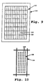
- Fig. 9 illustrates, in plan view, a substrate 110 constructed according to the invention.
- the substrate has an 8 ⁇ 12 rectangular array 112 of cells, such as cells 114, 116, formed on the substrate surface.
- each cell such as cell 114, in turn supports a microarray 118 of distinct biopolymers, such as polypeptides or polynucleotides at known, addressable regions of the microarray.
- Two such regions forming the microarray are indicated at 120, and correspond to regions, such as regions 42, forming the microarray of distinct biopolymers shown in Fig. 3 .
- the 96-cell array shown in Fig. 9 has typically array dimensions between about 12 and 244 mm in width and 8 and 400 mm in length, with the cells in the array having width and length dimension of 1/12 and 1/8 the array width and length dimensions, respectively, i . e. , between about 1 and 20 in width and 1 and 50 mm in length.
- the construction of substrate is shown cross-sectionally in Fig. 11 , which is an enlarged sectional view taken along view line 124 in Fig. 9 .
- the substrate includes a water-impermeable backing 126, such as a glass slide or rigid polymer sheet. Formed on the surface of the backing is a water-permeable film 128.
- the film is formed of a porous membrane material, such as nitrocellulose membrane, or a porous web material, such as a nylon, polypropylene, or PVDF porous polymer material.
- the thickness of the film is preferably between about 10 and 1000 ⁇ m.
- the film may be applied to the backing by spraying or coating uncured material on the backing, or by applying a preformed membrane to the backing.
- the backing and film may be obtained as a preformed unit from commercial source, e.g., a plastic-backed nitrocellulose film available from Schleicher and Schuell Corporation.
- the film-covered surface in the substrate is partitioned into a desired array of cells by water-impermeable grid lines, such as lines 130, 132, which have infiltrated the film down to the level of the backing, and extend above the surface of the film as shown, typically a distance of 100 to 2000 ⁇ m above the film surface.
- the grid lines are formed on the substrate by laying down an uncured or otherwise flowable resin or elastomer solution in an array grid, allowing the material to infiltrate the porous film down to the backing, then curing or otherwise hardening the grid lines to form the cell-array substrate.
- One preferred material for the grid is a flowable silicone available from Loctite Corporation.
- the barrier material can be extruded through a narrow syringe (e. g., 22 gauge) using air pressure or mechanical pressure. The syringe is moved relative to the solid support to print the barrier elements as a grid pattern. The extruded bead of silicone wicks into the pores of the solid support and cures to form a shallow waterproof barrier separating the regions of the solid support.
- the barrier element can be a wax-based material or a thermoset material such as epoxy.
- the barrier material can also be a UV-curing polymer which is exposed to UV light after being printed onto the solid support.
- the barrier material may also be applied to the solid support using printing techniques such as silk-screen printing.
- the barrier material may also be a heat-seal stamping of the porous solid support which seals its pores and forms a water-impervious barrier element.
- the barrier material may also be a shallow grid which is laminated or otherwise adhered to the solid support.
- the solid support can be virtually any porous membrane with or without a non-porous backing.
- Such membranes are readily available from numerous vendors and are made from nylon, PVDF, polysulfone and the like.
- the barrier element may also be used to adhere the porous membrane to a non-porous backing in addition to functioning as a barrier to prevent cross contamination of the assay reagents.
- the solid support can be of a non-porous material.
- the barrier can be printed either before or after the microarray of biomolecules is printed on the solid support.
- the cells formed by the grid lines and the underlying backing are water-impermeable, having side barriers projecting above the porous film in the cells.
- defined-volume samples can be placed in each well without risk of cross-contamination with sample material in adjacent cells.
- defined volumes samples such as sample 134, are shown in the cells.
- each well contains a microarray of distinct biopolymers.
- the microarrays in the well are identical arrays of distinct biopolymers, e.g., different sequence polynucleotides.
- Such arrays can be formed in accordance with the methods described in Section II, by depositing a first selected polynucleotide at the same selected microarray position in each of the cells, then depositing a second polynucleotide at a different microarray position in each well, and so on until a complete, identical microarray is formed in each cell.
- each microarray contains about 10 3 distinct polynucleotide or polypeptide biopolymers per surface area of less than about 1 cm 2 .
- the biopolymers in each microarray region are present in a defined amount between about 0.1 ferntomoles and 100 nanomoles. The ability to form high-density arrays of biopolymers, where each region is formed of a well-defined amount of deposited material, can be achieved in accordance with the microarray-forming method described in Section II.
- the biopolymers are polynucleotides having lengths of at least about 50 bp, i.e. , substantially longer than oligonucleotides which can be formed in high-density arrays by schemes involving parallel, step-wise polymer synthesis on the array surface.
- a small volume of the labeled DNA probe mixture in a standard hybridization solution is loaded onto each cell.
- the solution will spread to cover the entire microarray and stop at the barrier elements.
- the solid support is then incubated in a humid chamber at the appropriate temperature as required by the assay.
- Each assay may be conducted in an "open-face" format where no further sealing step is required, since the hybridization solution will be kept properly hydrated by the water vapor in the humid chamber.
- the entire solid support containing the numerous microarrays is rinsed quickly enough to dilute the assay reagents so that no significant cross contamination occurs.
- the entire solid support is then reacted with detection reagents if needed and analyzed using standard colorimetric, radioactive or fluorescent detection means. All processing and detection steps are performed simultaneously to all of the microarrays on the solid support ensuring uniform assay conditions for all of the microarrays on the solid support.
- Fig. 5 shows a substrate 136 formed according to another aspect of the invention, and intended for use in detecting binding of labeled polynucleotides to one or more of a plurality distinct polynucleotides.
- the substrate includes a glass substrate 138 having formed on its surface, a coating of a polycationic polymer, preferably a cationic polypeptide, such as polylysine or polyarginine.
- a polycationic polymer preferably a cationic polypeptide, such as polylysine or polyarginine.
- a microarray 140 of distinct polynucleotides, each localized at known selected array regions, such as regions 142.
- the slide is coated by placing a uniform-thickness film of a polycationic polymer, e.g. , poly-1-lysine, on the surface of a slide and drying the film to form a dried coating.
- a polycationic polymer e.g. , poly-1-lysine
- the amount of polycationic polymer added is sufficient to form at least a monolayer of polymers on the glass surface.
- the polymer film is bound to surface via electrostatic binding between negative silyi-OH groups on the surface and charged amine groups in the polymers.
- Poly-1-lysine coated glass slides may be obtained commercially, e.g. , from Sigma Chemical Co. (St. Louis, MO).
- microarray defined volumes of distinct polynucleotides are deposited on the polymer-coated slide, as described in Section II. According to an important feature of the substrate, the deposited polynucleotides remain bound to the coated slide surface non-covalently when an aqueous DNA sample is applied to the substrate under conditions which allow hybridization of reporter-labeled polynucleotides in the sample to complementary-sequence (single-stranded) polynucleotides in the substrate array. The method is illustrated in Examples 1 and 2.
- a substrate of the type just described, but having an array of same-sequence polynucleotides was mixed with fluorescent-labeled complementary DNA under hybridization conditions. After washing to remove non-hybridized material, the substrate was examined by low-power fluorescence microscopy. The array can be visualized by the relatively uniform labeling pattern of the array regions.
- each microarray contains at least 10 3 distinct polynucleotide or polypeptide biopolymers per surface area of less than about 1 cm 2 .
- the microarray contains 400 regions in an area of about 16 mm 2 , or 2.5 ⁇ 10 3 regions/cm 2 .
- the biopolymers in each microarray region are present in a defined amount between about 0.1 femtomoles and 100 nanomoles in the case of polynucleotides.
- the polynucleotides have lengths of at least about 50 bp, i.e. , substantially longer than oligonucleotides which can be formed in high-density arrays by various in situ synthesis schemes.
- Microarrays of immobilized nucleic acid sequences prepared in accordance with the invention can be used for large scale hybridization assays in numerous genetic applications, including genetic and physical mapping of genomes, monitoring of gene expression, DNA sequencing, genetic diagnosis, genotyping of organisms, and distribution of DNA reagents to researchers.
- a gene or a cloned DNA fragment is hybridized to an ordered array of DNA fragments, and the identity of the DNA elements applied to the array is unambiguously established by the pixel or pattern of pixels of the array that are detected.
- One application of such arrays for creating a genetic map is described by Nelson, et al. (1993).
- arrays of immobilized cloned DNA fragments are hybridized with other cloned DNA fragments to establish whether the cloned fragments in the probe mixture overlap and are therefore contiguous to the immobilized clones on the array.
- Lehrach, et al . describe such a process.
- the arrays of immobilized DNA fragments may also be used for genetic diagnostics.
- an array containing multiple forms of a mutated gene or genes can be probed with a labeled mixture of a patient's DNA which will preferentially interact with only one of the immobilized versions of the gene.
- Arrays of immobilized DNA fragments can also be used in DNA probe diagnostics.
- identity of a pathogenic microorganism can be established unambiguously by hybridizing a sample of the unknown pathogen's DNA to an array containing many types of known pathogenic DNA.
- a similar technique can also be used for unambiguous genotyping of any organism.
- Other molecules of genetic interest, such as cDNA's and RNA's can be immobilized on the array or alternately used as the labeled probe mixture that is applied to the array.
- an array of cDNA clones representing genes is hybridized with total cDNA from an organism to monitor gene expression for research or diagnostic purposes. Labeling total cDNA from a normal cell with one color fluorophore and total cDNA from a diseased cell with another color fluorophore and simultaneously hybridizing the two cDNA samples to the same array of cDNA clones allows for differential gene expression to be measured as the ratio of the two fluorophore intensities.
- This two-color experiment can be used to monitor gene expression in different tissue types, disease states, response to drugs, or response to environmental factors. An example of this approach is illustrated in Example 2, described with respect to Fig. 8 .
- Such a procedure could be used to simultaneously screen many patients against all known mutations in a disease gene.
- This invention could be used in the form of, for example, 96 identical 0.9 cm ⁇ 2.2 cm microarrays fabricated on a single 12 cm ⁇ 18 cm sheet of plastic-backed nitrocellulose where each microarray could contain, for example, 100 DNA fragments representing all known mutations of a given gene.
- the region of interest from each of the DNA samples from 96 patients could be amplified, labeled, and hybridized to the 96 individual arrays with each assay performed in 100 microliters of hybridization solution.
- the approximately 1 thick silicone rubber barrier elements between individual arrays prevent cross contamination of the patient samples by sealing the pores of the nitrocellulose and by acting as a physical barrier between each microarray.
- the solid support containing all 96 microarrays assayed with the 96 patient samples is incubated, rinsed, detected and analyzed as a single sheet of material using standard radioactive, fluorescent, or colorimetric detection means (Maniatas, et al ., 1989). Previously, such a procedure would involve the handling, processing and tracking of 96 separate membranes in 96 separate sealed chambers. By processing all 96 arrays as a single sheet of material, significant time and cost savings are possible.
- the assay format can be reversed where the patient or organism's DNA is immobilized as the array elements and each array is hybridized with a different mutated allele or genetic marker.
- the gridded solid support can also be used for parallel non-DNA ELISA assays. Furthermore, the invention allows for the use of all standard detection methods without the need to remove the shallow barrier elements to carry out the detection step.
- arrays of whole cells, peptides, enzymes, antibodies, antigens, receptors, ligands, phospholipids, polymers, drug cogener preparations or chemical substances can be fabricated by the means described in this invention for large scale screening assays in medical diagnostics, drug discovery, molecular biology, immunology and toxicology.
- the multi-cell substrate aspect of the invention allows for the rapid and convenient screening of many DNA probes against many ordered arrays of DNA fragments. This eliminates the need to handle and detect many individual arrays for performing mass screenings for genetic research and diagnostic applications. Numerous microarrays can be fabricated on the same solid support and each microarray reacted with a different DNA probe while the solid support is processed as a single sheet of material.
- Genomic-Complexity Hybridization to Micro DNA Arrays Representing the Yeast Saccharomyces cerevisiae Genome with Two-Color Fluorescent Detection
- the array elements were randomly amplified PCR (Bohlander, et al ., 1992) products using physically mapped lambda clones of S. cerevisiae genomic DNA templates (Riles, et al ., 1993).
- the PCR was performed directly on the lambda phage lysates resulting in an amplification of both the 35 kb lambda vector and the 5-15 kb yeast insert sequences in the form of a uniform distribution of PCR product between 250-1500 base pairs in length.
- the PCR product was purified using Sephadex G50 gel filtration (Pharmacia, Piscataway, NJ) and concentrated by evaporation to dryness at room temperature overnight.
- lambda clones was rehydrated in 15 ⁇ l of 3 ⁇ SSC in preparation for spotting onto the glass.
- micro arrays were fabricated on microscope slides which were coated with a layer of poly-1-lysine (Sigma).
- the automated apparatus described in Section IV loaded 1 ⁇ l of the concentrated lambda clone PCR product in 3 ⁇ SSC directly from 96 well storage plates into the open capillary printing element and deposited -5 nl of sample per slide at 380 micron spacing between spots, on each of 40 slides. The process was repeated for all 864 samples and 8 control spots.
- the slides were rehydrated in a humid chamber for 2 hours, baked in a dry 80°C vacuum oven for 2 hours, rinsed to remove unabsorbed DNA and then treated with succinic anhydride to reduce non-specific adsorption of the labeled hybridization probe to the poly-1-lysine coated glass surface.
- the immobilized DNA on the array was denatured in distilled water at 90°C for 2 minutes.
- the 16 chromosomes of Saccharomyces cerevisiae were separated in a CHEF agarose gel apparatus (Biorad, Richmond, CA). The six largest chromosomes were isolated in one gel slice and the smallest 10 chromosomes in a second gel slice. The DNA was recovered using a gel extraction kit (Qiagen, Chatsworth, CA). The two chromosome pools were randomly amplified in a manner similar to that used for the target lambda clones.
- each of the amplified chromosome pools was separately random-primer labeled using Klenow polymerase (Amersham, Arlington Heights, IL) with a lissamine conjugated nucleotide analog (Dupont NEN, Boston, MA) for the pool containing the six largest chromosomes, and with a fluorescein conjugated nucleotide analog (BMB) for the pool containing smallest ten chromosomes.
- Klenow polymerase Amersham, Arlington Heights, IL
- BMB fluorescein conjugated nucleotide analog
- a custom built laser fluorescent scanner was used to detect the two-color hybridization signals from the 1.8 ⁇ 1.8 cm array at 20 micron resolution.
- the scanned image was gridded and analyzed using custom image analysis software. After correcting for optical crosstalk between the fluorophores due to their overlapping emission spectra, the red and green hybridization values for each clone on the array were correlated to the known physical map position of the clone resulting in a computer-generated color karyotype of the yeast genome.
- Figure 6 shows the hybridization pattern of the two chromosome pools.
- a red signal indicates that the lambda clone on the array surface contains a cloned genomic DNA segment from one of the largest six yeast chromosomes.
- a green signal indicates that the lambda clone insert comes from one of the smallest ten yeast chromosomes.
- Orange signals indicate repetitive sequences which cross hydridized to both chromosome pools. Control spots on the array confirm that the hybridization is specific and reproducible.
- the physical map locations of the genomic DNA fragments contained in each of the clones used as array elements have been previously determined by Olson and co-workers (Riles, et al . ) allowing for the automatic generation of the color karyotype shown in Figure 7 .
- the color of a chromosomal section on the karyotype corresponds to the color of the array element containing the clone from that section.
- the black regions of the karyotype represent false negative dark spots on the array (10%) or regions of the genome not covered by the Olson clone library (90%). Note that the largest six chromosomes are mainly red while the smallest ten chromosomes are mainly green matching the original CHEF gel isolation of the hybridization probe. Areas of the red chromosomes containing green spots and vice-versa are probably due to spurious sample tracking errors in the formation of the original library and in the amplification and spotting procedures.
- yeast genome arrays have also been probed with individual clones or pools of clones that are fluorescently labeled for physical mapping purposes.
- the hybridization signals of these clones to the array were translated into a position on the physical map of yeast.
- Total poly-A mRNA from wild type Arabidopsis was isolated using standard methods (Maniatis, et al., 1989) and reverse transcribed into total cDNA, using fluorescein nucleotide analog to label the cDNA product (green fluorescence).
- a similar procedure was performed with the transgenic line of Arabidopsis where the transcription factor HAT4 was inserted into the genome using standard gene transfer protocols.
- cDNA copies of mRNA from the transgenic plant are labeled with a lissamine nucleotide analog (red fluorescence).
- Two micrograms of the cDNA products from each type of plant were pooled together and hybridized to the cDNA clone array in a 10 microliter hybridization reaction in a manner similar to Example 1. Rinsing and detection of hybridization was also performed in a manner similar to Example 1.
- Fig. 8 show the resulting hybridization pattern of the array.
- An advantage of the microarray hybridization format for gene expression studies is the high partial concentration of each cDNA species achievable in the 10 microliter hybridization reaction. This high partial concentration allows for detection of rare transcripts without the need for PCR amplification of the hybridization probe which may bias the true genetic representation of each discrete cDNA species.
- Gene expression studies such as these can be used for genomics research to discover which genes are expressed in which cell types, disease states, development states or environmental conditions. Gene expression studies can also be used for diagnosis of disease by empirically correlating gene expression patterns to disease states.
- a sheet of plastic-backed nitrocellulose was gridded with barrier elements made from silicone rubber according to the description in Section IV-A.
- the sheet was soaked in 10 ⁇ SSC and allowed to dry.
- 192 M13 clones each with a different yeast insert were arrayed 400 microns apart in four quadrants of the solid support using the automated device described in Section III.
- the bottom left quadrant served as a negative control for hybridization while each of the other three quadrants was hybridized simultaneously with a different oligonucleotide using the open-face hybridization technology described in Section IV-A.
- the first two and last four elements of each array are positive controls for the colorimetric detection step.
- the oligonucleotides were labeled with fluorescein which was detected using an anti-fluorescein antibody conjugated to alkaline phosphatase that precipitated an NBT/BCIP dye on the solid support (Amersham). Perfect matches between the labeled oligos and the M13 clones resulted in dark spots visible to the naked eye and detected using an optical scanner (HP ScanJet II) attached to a personal computer.
- the hybridization patterns are different in every quadrant indicating that each oligo found several unique M13 clones from among the 192 with a perfect sequence match. Note that the open capillary printing tip leaves detectable dimples on the nitrocellulose which can be used to automatically align and analyze the images.
Abstract
Description
- This invention relates to a method and apparatus for fabricating microarrays of biological samples for large scale screening assays, such as arrays of DNA samples to be used in DNA hybridization assays for genetic research and diagnostic applications.
- Abouzied, et al., Journal of AOAC International 77 (2) :495-500 (1994).
- Bohlander, et al., Genomics 13:1322-1324 (1992).
- Drmanac, et al., Science 260:1649-1652 (1993).
- Fodor, et al., Science 251:767-773 (1991).
- Khrapko, et al., DNA Sequence 1:375-388 (1991).
- Kuriyama, et al., AN ISFET BIOSENSOR, APPLIED BIOSENSORS (Donald Wise, Ed.), Butterworths, pp. 93-114 (1989).
- Lehrach, et al., HYBRIDIZATION FINGERPRINTING IN GENOME MAPPING AND SEQUENCING, GENOME ANALYSIS, VOL 1 (Davies and Tilgham, Eds.), Cold Spring Harbor Press, pp. 39-81 (1990).
- Maniatis, et al., MOLECULAR CLONING, A LABORATORY MANUAL, Cold Spring Harbor Press (1989).
- Nelson, et al., Nature Genetics 4:11-18 (1993).
- Pirrung, et al., U.S. Patent No. 5,143,854 (1992).
- Riles, et al., Genetics 134:81-150 (1993).
- Schena, M. et al., Proc. Nat. Acad. Sci. USA 89:3894-3898 (1992).
- Southern, et al., Genomics 13:1008-1017 (1992).
- A variety of methods are currently available for making arrays of biological macromolecules, such as arrays of nucleic acid molecules or proteins. One method for making ordered arrays of DNA on a porous membrane is a "dot blot" approach. In this method, a vacuum manifold transfers a plurality, e.g., 96, aqueous samples of DNA from 3 millimeter diameter wells to a porous membrane. A common variant of this procedure is a "slot-blot" method in which the wells have highly-elongated oval shapes.
- The DNA is immobilized on the porous membrane by baking the membrane or exposing it to UV radiation. This is a manual procedure practical for making one array at a time and usually limited to 96 samples per array. "Dot-blot" procedures are therefore inadequate for applications in which many thousand samples must be determined.
- A more efficient technique employed for making ordered arrays of genomic fragments uses an array of pins dipped into the wells, e.g., the 96 wells of a microtitre plate, for transferring an array of samples to a substrate, such as a porous membrane. One array includes pins that are designed to spot a membrane in a staggered fashion, for creating an array of 9216 spots in a 22 × 22 cm area (Lehrach, et al., 1990). A limitation with this approach is that the volume of DNA spotted in each pixel of each array is highly variable. In addition, the number of arrays that can be made with each dipping is usually quite small.
- An alternate method of creating ordered arrays of nucleic acid sequences is described by Pirrung, et al. (1992), and also by Fodor, et al. (1991). The method involves synthesizing different nucleic acid sequences at different discrete regions of a support. This method employs elaborate synthetic schemes, and is generally limited to relatively short nucleic acid sample, e.g., less than 20 bases. A related method has been described by Southern, et al. (1992).
- Khrapko, et al. (1991) describes a method of making an oligonucleotide matrix by spotting DNA onto a thin layer of polyacrylamide. The spotting is done manually with a micropipette.
- None of the methods or devices described in the prior art are designed for mass fabrication of microarrays characterized by (i) a large number of microsized assay regions separated by a distance of 50-200 microns or less, and (ii) a well-defined amount, typically in the picomole range, of analyte associated with each region of the array.
- Furthermore, current technology is directed at performing such assays one at a time to a single array of DNA molecules. For example, the most common method for performing DNA hybridizations to arrays spotted onto porous membrane involves sealing the membrane in a plastic bag (Maniatis, et al., 1989) or a rotating glass cylinder (Robbins Scientific) with the labeled hybridization probe inside the sealed chamber. For arrays made on non-porous surfaces, such as a microscope slide, each array is incubated with the labeled hybridization probe sealed under a coverslip. These techniques require a separate sealed chamber for each array which makes the screening and handling of many such arrays inconvenient and time intensive.
- Abouzied, et al. (1994) describes a method of printing horizontal lines of antibodies on a nitrocellulose membrane and separating regions of the membrane with vertical stripes of a hydrophobic material. Each vertical stripe is then reacted with a different antigen and the reaction between the immobilized antibody and an antigen is detected using a standard ELISA colorimetric technique. Abouzied's technique makes it possible to screen many one-dimensional arrays simultaneously on a single sheet of nitrocellulose. Abouzied makes the nitrocellulose somewhat hydrophobic using a line drawn with PAP Pen (Research Products International). However Abouzied does not describe a technology that is capable of completely sealing the pores of the nitrocellulose. The pores of the nitrocellulose are still physically open and so the assay reagents can leak through the hydrophobic barrier during extended high temperature incubations or in the presence of detergents which makes the Abouzied technique unacceptable for DNA hybridization assays.
- Porous membranes with printed patterns of hydrophilic/hydrophobic regions exist for applications such as ordered arrays of bacteria colonies. QA Life Sciences (San Diego CA) makes such a membrane with a grid pattern printed on it. However, this membrane has the same disadvantage as the Abouzied technique since reagents can still flow between the gridded arrays making them unusable for separate DNA hybridization assays.
- Pall Corporation make a 96-well plate with a porous filter heat sealed to the bottom of the plate. These plates are capable of containing different reagents in each well without cross-contamination. However, each well is intended to hold only one target element whereas the invention described here makes a microarray of many biomolecules in each subdivided region of the solid support. Furthermore, the 96 well plates are at least 1 cm thick and prevent the use of the device for many colorimetric, fluorescent and radioactive detection formats which require that the membrane lie flat against the detection surface. The invention described here requires no further processing after the assay step since the barriers elements are shallow and do not interfere with the detection step thereby greatly increasing convenience.
- Hyseq Corporation has described a method of making an "array of arrays" on a non-porous solid support for use with their sequencing by hybridization technique. The method described by Hyseq involves modifying the chemistry of the solid support material to form a hydrophobic grid pattern where each subdivided region contains a microarray of biomolecules. Hyseq's flat hydrophobic pattern does not make use of physical blocking as an additional means of preventing cross contamination.
- The invention includes, in one aspect, a method of forming a microarray of analyte-assay regions on a solid support, where each region in the array has a known amount of a selected, analyte-specific reagent. The method involves first loading a solution of a selected analyte-specific reagent in a reagent-dispensing device having an elongate capillary channel (i) formed by spaced-apart, coextensive elongate members, (ii) adapted to hold a quantity of the reagent solution and (iii) having a tip region at which aqueous solution in the channel forms a meniscus. The channel is preferably formed by a pair of spaced-apart tapered elements.
- The tip of the dispensing device is tapped against a solid support at a defined position on the support surface with an impulse effective to break the meniscus in the capillary channel deposit a selected volume of solution on the surface, preferably a selected volume in the range 0.01 to 100 nl. The two steps are repeated until the desired array is formed.
- The method may be practiced in forming a plurality of such arrays, where the solution-depositing step is applied to a selected position on each of a plurality of solid supports at each repeat cycle.
- The dispensing device may be loaded with a new solution, by the steps of (i) dipping the capillary channel of the device in a wash solution, (ii) removing wash solution drawn into the capillary channel, and (iii) dipping the capillary channel into the new reagent solution.
- Also included in the invention is an automated apparatus for forming a microarray of analyte-assay regions on a plurality of solid supports, where each region in the array has a known amount of a selected, analyte-specific reagent. The apparatus has a holder for holding, at known positions, a plurality of planar supports, and a reagent dispensing device of the type described above the capillary channel being open-sided.
- The apparatus further includes positioning structure for positioning the dispensing device at a selected array position with respect to a support in said holder, and dispensing structure for moving the dispensing device into tapping engagement against a support with a selected impulse effective to deposit a selected volume on the support, e.g., a selected volume in the volume range 0.01 to 100 nl.
- The positioning and dispensing structures are controlled by a control unit in the apparatus. The unit operates to (i) place the dispensing device at a loading station, (ii) move the capillary channel in the device into a selected reagent at the loading station, to load the dispensing device with the reagent, and (iii) dispense the reagent at a defined array position on each of the supports on said holder. The unit may further operate, at the end of a dispensing cycle, to wash the dispensing device by (i) placing the dispensing device at a washing station, (ii) moving the capillary channel in the device into a wash fluid, to load the dispensing device with the fluid, and (iii) remove the wash fluid prior to loading the dispensing device with a fresh selected reagent.
- The dispensing device in the apparatus may be one of a plurality of such devices which are carried on the arm for dispensing different analyte assay reagents at selected spaced array positions.
- In another aspect, the invention includes a substrate with a surface having a microarray of at least 103 distinct polynucleotide biopolymers in a surface area of less than about 1 cm2. Each distinct biopolymer (i) is disposed at a separate, defined position in said array, (ii) has a length of at least 50 subunits, and (iii) is present in a defined amount between about 0.1 femtomoles and 100 nanomoles.
- In one embodiment, the surface is glass slide surface coated with a polycationic polymer, such as polylysine, and the biopolymers are polynucleotides with a length of at least 50 subunits. In another embodiment, this substrate has a water-impermeable backing, a water-permeable film formed on the backing, and a grid formed on the film. The grid is composed of Intersecting water-impervious grid elements extending from said backing to positions raised above the surface of said film, and partitions the film into a plurality of water-impervious cells. A biopolymer array is formed within each well.
- This substrate is provided for use in detecting binding of labeled polynucleotides to one or more of a plurality of different-sequence, immobilized polynucleotides. This substrate includes, in one aspect, a glass support, a coating of a polycationic polymer, such as polylysine, on said surface of the support, and an array of distinct polynucleotides electrostatically bound non-covalently to said coating, where each distinct biopolymer is disposed at a separate, defined position in a surface array of polynucleotides.
- In another aspect, this substrate includes a water-impermeable backing, a water-permeable film formed on the backing, and a grid formed on the film, where the grid is composed of intersecting water-impervious grid elements extending from the backing to positions raised above the surface of the film, forming a plurality of cells. A biopolymer array is formed within each cell. The objects of the invention are achieved by the features of the claims. Moreover, the objects and features of the invention will become more fully apparent when the following detailed description of the invention is read In conjunction with the accompanying figures.
-
-
Fig. 1 is a side view of a reagent-dispensing device having a open-capillary dispensing head constructed for use in one embodiment of the invention: -
Figs. 2A-2C illustrate steps in the delivery of a fixed-volume bead on a hydrophobic surface employing the dispensing head fromFig. 1 , in accordance with one embodiment of the method of the invention; -
Fig. 3 shows a portion of a two-dimensional array of analyte-assay regions constructed according to the method of the invention; -
Fig. 4 is a planar view showing components of an automated apparatus for forming arrays in accordance with the invention; -
Fig. 5 shows a fluorescent image of an actual 20 × 20 array of 400 fluorescently-labeled DNA samples immobilized on a poly-1-lysine coated slide, where the total area covered by the 400 element array is 16 square millimeters; -
Fig. 6 is a fluorescent image of a 1.8 cm × 1.8 cm microarray containing lambda clones with yeast inserts, the fluorescent signal arising from the hybridization to the array with approximately half the yeast genome labeled with a green fluorophore and the other half with a red fluorophore; -
Fig. 7 shows the translation of the hybridization Image ofFig. 6 into a karyotype of the yeast genome, where the elements of Fig.-6 microarray contain yeast DNA sequences that have been previously physically mapped in the yeast genome; -
Fig. 8 show a fluorescent image of a 0.5 cm × 0.5 cm microarray of 24 cDNA clones, where the microarray was hybridized simultaneously with total cD-NA from wild type Arabidopsis plant labeled with a green fluorophore and total cDNA from a transgenic Arabidopsis plant labeled with a red fluorophore, and the arrow points to the cDNA clone representing the gene introduced into the transgenic Arabidopsis plant; -
Fig. 9 shows a plan view of substrate having an array of cells formed by barrier elements in the form of a grid; -
Fig. 10 shows an enlarged plan view of one of the cells in the substrate inFig. 9 , showing an array of polynucleotide regions in the cell; -
Fig. 11 is an enlarged sectional view of the substrate inFig. 9 , taken along a section line in that figure; andFig. 12 is a scanned image of a 3 cm × 3 cm nitrocellulose solid support containing four identical arrays of M13 clones in each of four quadrants, where each quadrant was hybridized simultaneously to a different oligonucleotide using an open face hybridization method. - Unless indicated otherwise, the terms defined below have the following meanings:
- "Ligand" refers to one member of a ligand/anti-ligand binding pair. The ligand may be, for example, one of the nucleic acid strands in a complementary, hybridized nucleic acid duplex binding pair; an effector molecule in an effector/receptor binding pair; or an antigen in an antigen/antibody or antigen/antibody fragment binding pair.
- "Antiligand" refers to the opposite member of a ligand/anti-ligand binding pair. The antiligand may be the other of the nucleic acid strands in a complementary, hybridized nucleic acid duplex binding pair; the receptor molecule in an effector/receptor binding pair; or an antibody or antibody fragment molecule in antigen/antibody or antigen/antibody fragment binding pair, respectively.
- "Analyte" or "analyte molecule" refers to a molecule, typically a macromolecule, such as a polynucleotide or polypeptide, whose presence, amount, and/or identity are to be determined. The analyte is one member of a ligand/anti-ligand binding pair.
- "Analyte-specific assay reagent" refers to a molecule effective to bind specifically to an analyte molecule. The reagent is the opposite member of a ligand/anti-ligand binding pair.
- An "array of regions on a solid support" is a linear or two-dimensional array of preferably discrete regions, each having a finite area, formed on the surface of a solid support.
- A "microarray" is an array of regions having a density of discrete regions of at least about 100/cm2, and preferably at least about 1000/cm2. The regions in a microarray have typical dimensions, e.g., diameters, in the range of between about 10-250 µm, and are separated from other regions in the array by about the same distance.
- A support surface is "hydrophobic" if a aqueous-medium droplet applied to the surface does not spread out substantially beyond the area size of the applied droplet. That is, the surface acts to prevent spreading of the droplet applied to the surface by hydrophobic interaction with the droplet.
- A "meniscus" means a concave or convex surface that forms on the bottom of a liquid in a channel as a result of the surface tension of the liquid.
- "Distinct biopolymers", as applied to the biopolymers forming a microarray, means an array member which is distinct from other array members on the basis of a different biopolymer sequence, and/or different concentrations of the same or distinct biopolymers, and/or different mixtures of distinct or different-concentration biopolymers. Thus an array of "distinct polynucleotides" means an array containing, as its members, (i) distinct polynucleotides, which may have a defined amount of each member, (ii) different, graded concentrations of given-sequence polynucleotides, and/or (iii) different-composition mixtures of two or more distinct polynucleotides.
- "Cell type" means a cell from a given source, e.g., a tissue, or organ, or a cell in a given state of differentiation, or a cell associated with a given pathology or genetic makeup.
- This section describes a method of forming a microarray of analyte-assay regions on a solid support or substrate, where each region in the array has a known amount of a selected, analyte-specific reagent.
-
Fig. 1 illustrates, in a partially schematic view, a reagent-dispensingdevice 10 useful in practicing the method. The device generally includes areagent dispenser 12 having an elongate opencapillary channel 14 adapted to hold a quantity of the reagent solution, such as indicated at 16, as will be described below. The capillary channel is formed by a pair of spaced-apart, coextensive,elongate members tip region 18 at the lower end of the channel. More generally, the open channel is formed by at least two elongate, spaced-apart members adapted to hold a quantity of reagent solutions and having a tip region at which aqueous solution in the channel forms a meniscus, such as the concave meniscus illustrated at 20 inFig. 2A . The advantages of the open channel construction of the dispenser are discussed below. - With continued reference to
Fig. 1 , the dispenser device also includes structure for moving the dispenser rapidly toward and away from a support surface, for effecting deposition of a known amount of solution in the dispenser on a support, as will be described below with reference toFigs. 2A-2C . In the embodiment shown, this structure includes asolenoid 22 which is activatable to draw asolenoid piston 24 rapidly downwardly, then release the piston, e.g., under spring bias, to a normal, raised position, as shown. The dispenser is carried on the piston by a connectingmember 26, as shown. The just-described moving structure is also referred to herein as dispensing means for moving the dispenser into engagement with a solid support, for dispensing a known volume of fluid on the support. - The dispensing device just described is carried on an
arm 28 that may be moved either linearly or in an x-y plane to position the dispenser at a selected deposition position, as will be described. -
Figs. 2A-2C illustrate the method of depositing a known amount of reagent solution in the just-described dispenser on the surface of a solid support, such as the support indicated at 30. The support is a polymer, glass, or other solid-material support having a surface indicated at 31. - In one general embodiment, the surface is a relatively hydrophilic, i.e., wettable surface, such as a surface having native, bound or covalently attached charged groups. One such surface described below is a glass surface having an absorbed layer of a polycationic polymer, such as poly-1-lysine.
- In another embodiment, the surface has or is formed to have a relatively hydrophobic character, i.e., one that causes aqueous medium deposited on the surface to bead. A variety of known hydrophobic polymers, such as polystyrene, polypropylene, or polyethylene have desired hydrophobic properties, as do glass and a variety of lubricant or other hydrophobic films that may be applied to the support surface.
- Initially, the dispenser is loaded with a selected analyte-specific reagent solution, such as by dipping the dispenser tip, after washing, into a solution of the reagent, and allowing filling by capillary flow into the dispenser channel. The dispenser is now moved to a selected position with respect to a support surface, placing the dispenser tip directly above the support-surface position at which the reagent is to be deposited. This movement takes place with the dispenser tip in its raised position, as seen in
Fig. 2A , where the tip is typically at least several 1-5 mm above the surface of the substrate. - With the dispenser so positioned,
solenoid 22 is now activated to cause the dispenser tip to move rapidly toward and away from the substrate surface, making momentary contact with the surface, in effect, tapping the tip of the dispenser against the support surface. The tapping movement of the tip against the surface acts to break the liquid meniscus in the tip channel, bringing the liquid in the tip into contact with the support surface. This, in turn, produces a flowing of the liquid into the capillary space between the tip and the surface, acting to draw liquid out of the dispenser channel, as seen inFig. 2B . -
Fig. 2C shows flow of fluid from the tip onto the support surface, which in this case is a hydrophobic surface. The figure illustrates that liquid continues to flow from the dispenser onto the support surface until it forms aliquid bead 32. At a given bead size, i.e., volume, the tendency of liquid to flow onto the surface will be balanced by the hydrophobic surface interaction of the bead with the support surface, which acts to limit the total bead area on the surface, and by the surface tension of the droplet, which tends toward a given bead curvature. At this point, a given bead volume will have formed, and continued contact of the dispenser tip with the bead, as the dispenser tip is being withdrawn, will have little or no effect on bead volume. - For liquid-dispensing on a more hydrophilic surface, the liquid will have less of a tendency to bead, and the dispensed volume will be more sensitive to the total dwell time of the dispenser tip in the immediate vicinity of the support surface, e.g., the positions illustrated in
Figs. 2B and 2C . - The desired deposition volume, i.e., bead volume, formed by this method is preferably in the range 2 pl (picoliters) to 2 nl (nanoliters), although volumes as high as 100 nl or more may be dispensed. It will be appreciated that the selected dispensed volume will depend on (i) the "footprint" of the dispenser tip, i.e., the size of the area spanned by the tip, (ii) the hydrophobicity of the support surface, and (iii) the time of contact with and rate of withdrawal of the tip from the support surface. In addition, bead size may be reduced by increasing the viscosity of the medium, effectively reducing the flow time of liquid from the dispenser onto the support surface. The drop size may be further constrained by depositing the drop in a hydrophilic region surrounded by a hydrophobic grid pattern on the support surface.
- In a typical embodiment, the dispenser tip is tapped rapidly against the support surface, with a total residence time in contact with the support of less than about 1 msec, and a rate of upward travel from the surface of about 10 cm/sec.
- Assuming that the bead that forms on contact with the surface is a hemispherical bead, with a diameter approximately equal to the width of the dispenser tip, as shown in
Fig. 2C , the volume of the bead formed in relation to dispenser tip width (d) is given in Table 1 below. As seen, the volume of the bead ranges between 2 pl to 2 nl as the width size is increased from about 20 to 200 µm.Table 1 d volume (n1) 20 µm 2 × 10⁻³ 50 µm 3.1 x 10⁻² 100 µm 2.5 × 10⁻¹ 200 µm 2 - At a given tip size, bead volume can be reduced in a controlled fashion by increasing surface hydrophobicity, reducing time of contact of the tip with the surface, increasing rate of movement of the tip away from the surface, and/or increasing the viscosity of the medium. Once these parameters are fixed, a selected deposition volume in the desired pl to nl range can be achieved in a repeatable fashion.
- After depositing a bead at one selected location on a support, the tip is typically moved to a corresponding position on a second support, a droplet is deposited at that position, and this process is repeated until a liquid droplet of the reagent has been deposited at a selected position on each of a plurality of supports.
- The tip is then washed to remove the reagent liquid, filled with another reagent liquid and this reagent is now deposited at each another array position on each of the supports. In one embodiment, the tip is washed and refilled by the steps of (i) dipping the capillary channel of the device in a wash solution, (ii) removing wash solution drawn into the capillary channel, and (iii) dipping the capillary channel into the new reagent solution.
- From the foregoing, it will be appreciated that the tweezers-like, open-capillary dispenser tip provides the advantages that (i) the open channel of the tip facilitates rapid, efficient washing and drying before reloading the tip with a new reagent, (ii) passive capillary action can load the sample directly from a standard microwell plate while retaining sufficient sample in the open capillary reservoir for the printing of numerous arrays, (iii) open capillaries are less prone to clogging than closed capillaries, and (iv) open capillaries do not require a perfectly faced bottom surface for fluid delivery.
- A portion of a
microarray 36 formed on thesurface 38 of asolid support 40 in accordance with the method just described is shown inFig. 3 . The array is formed of a plurality of analyte-specific reagent regions, such as regions 42, where each region may include a different analyte-specific reagent. As indicated above, the diameter of each region is preferably between about 20-200 µm. The spacing between each region and its closest (non-diagonal) neighbor, measured from center-to-center (indicated at 44), is preferably in the range of about 20-400 µm. Thus, for example, an array having a center-to-center spacing of about 250 µm contains about 40 regions/cm or 1,600 regions/cm2. After formation of the array, the support is treated to evaporate the liquid of the droplet forming each region, to leave a desired array of dried, relatively flat regions. This drying may be done by heating or under vacuum. - In some cases, it is desired to first rehydrate the droplets containing the analyte reagents to allow for more time for adsorption to the solid support. It is also possible to spot out the analyte reagents in a humid environment so that droplets do not dry until the arraying operation is complete.
- In another aspect, the invention includes an automated apparatus for forming an array of analyte-assay regions on a solid support, where each region in the array has a known amount of a selected, analyte-specific reagent.
- The apparatus is shown in planar, and partially schematic view in
Fig. 4 . Adispenser device 72 in the apparatus has the basic construction described above with respect toFig. 1 , and includes adispenser 74 having an open-capillary channel terminating at a tip, substantially as shown inFigs. 1 and 2A-2C . - The dispenser is mounted in the device for movement toward and away from a dispensing position at which the tip of the dispenser taps a support surface, to dispense a selected volume of reagent solution, as described above. This movement is effected by a
solenoid 76 as described above.Solenoid 76 is under the control of acontrol unit 77 whose operation will be described below. The solenoid is also referred to herein as dispensing means for moving the device into tapping engagement with a support, when the device is positioned at a defined array position with respect to that support. - The dispenser device is carried on an
arm 74 which is threadedly mounted on aworm screw 80 driven (rotated) in a desired direction by astepper motor 82 also under the control ofunit 77. At its left end in thefigure screw 80 is carried in asleeve 84 for rotation about the screw axis. At its other end, the screw is mounted to the drive shaft of the stepper motor, which in turn is carried on asleeve 86. The dispenser device, worm screw, the two sleeves mounting the worm screw, and the stepper motor used in moving the device in the "x" (horizontal) direction in the figure form what is referred to here collectively as adisplacement assembly 86. - The displacement assembly is constructed to produce precise, micro-range movement in the direction of the screw, i.e., along an x axis in the figure. In one mode, the assembly functions to move the dispenser in x-axis increments having a selected distance in the range 5-25 µm. In another mode, the dispenser unit may be moved in precise x-axis increments of several microns or more, for positioning the dispenser at associated positions on adjacent supports, as will be described below.
- The displacement assembly, in turn, is mounted for movement in the "y" (vertical) axis of the figure, for positioning the dispenser at a selected y axis position. The structure mounting the assembly includes a fixed
rod 88 mounted rigidly between a pair of frame bars 90, 92, and aworm screw 94 mounted for rotation between a pair of frame bars 96, 98. The worm screw is driven (rotated) by astepper motor 100 which operates under the control ofunit 77. The motor is mounted onbar 96, as shown. - The structure just described, including
worm screw 94 andmotor 100, is constructed to produce precise, micro-range movement in the direction of the screw, i.e., along an y axis in the figure. As above, the structure functions in one mode to move the dispenser in y-axis increments having a selected distance in the range 5-250 µm, and in a second mode, to move the dispenser in precise y-axis increments of several microns (µm) or more, for positioning the dispenser at associated positions on adjacent supports. - The displacement assembly and structure for moving this assembly in the y axis are referred to herein collectively as positioning means for positioning the dispensing device at a selected array position with respect to a support.
- A
holder 102 in the apparatus functions to hold a plurality of supports, such assupports 104 on which the microarrays of reagent regions are to be formed by the apparatus. The holder provides a number of recessed slots, such asslot 106, which receive the supports, and position them at precise selected positions with respect to the frame bars on which the dispenser moving means is mounted. - As noted above, the control unit in the device functions to actuate the two stepper motors and dispenser solenoid in a sequence designed for automated operation of the apparatus in forming a selected microarray of reagent regions on each of a plurality of supports.
- The control unit is constructed, according to conventional microprocessor control principles, to provide appropriate signals to each of the solenoid and each of the stepper motors, in a given timed sequence and for appropriate signalling time. The construction of the unit, and the settings that are selected by the user to achieve a desired array pattern, will be understood from the following description of a typical apparatus operation.
- Initially, one or more supports are placed in one or more slots in the holder. The dispenser is then moved to a position directly above a well (not shown) containing a solution of the first reagent to be dispensed on the support(s). The dispenser solenoid is actuated now to lower the dispenser tip into this well, causing the capillary channel in the dispenser to fill.
Motors - The dispenser is now moved to the corresponding position at an adjacent support and a similar volume of the solution is dispensed at this position. The process is repeated until the reagent has been dispensed at this preselected corresponding position on each of the supports.
- Where it is desired to dispense a single reagent at more than two array positions on a support, the dispenser may be moved to different array positions at each support, before moving the dispenser to a new support, or solution can be dispensed at individual positions on each support, at one selected position, then the cycle repeated for each new array position.
- To dispense the next reagent, the dispenser is positioned over a wash solution (not shown), and the dispenser tip is dipped in and out of this solution until the reagent solution has been substantially washed from the tip. Solution can be removed from the tip, after each dipping, by vacuum, compressed air spray, sponge, or the like.
- The dispenser tip is now dipped in a second reagent well, and the filled tip is moved to a second selected array position in the first support. The process of dispensing reagent at each of the corresponding second-array positions is then carried as above. This process is repeated until an entire microarray of reagent solutions on each of the supports has been formed.
- This section describes embodiments of a substrate having a microarray of biological polymers carried on the substrate surface. Subsection A describes a multi-cell substrate, each cell of which contains a microarray, and preferably an identical microarray, of distinct biopolymers, such as distinct polynucleotides, formed on a porous surface. Subsection B describes a microarray of distinct polynucleotides bound on a glass slide coated with a polycationic polymer.
-
Fig. 9 illustrates, in plan view, asubstrate 110 constructed according to the invention. The substrate has an 8 × 12rectangular array 112 of cells, such ascells Fig. 10 , each cell, such ascell 114, in turn supports amicroarray 118 of distinct biopolymers, such as polypeptides or polynucleotides at known, addressable regions of the microarray. Two such regions forming the microarray are indicated at 120, and correspond to regions, such as regions 42, forming the microarray of distinct biopolymers shown inFig. 3 . - The 96-cell array shown in
Fig. 9 has typically array dimensions between about 12 and 244 mm in width and 8 and 400 mm in length, with the cells in the array having width and length dimension of 1/12 and 1/8 the array width and length dimensions, respectively, i. e., between about 1 and 20 in width and 1 and 50 mm in length. - The construction of substrate is shown cross-sectionally in
Fig. 11 , which is an enlarged sectional view taken along view line 124 inFig. 9 . The substrate includes a water-impermeable backing 126, such as a glass slide or rigid polymer sheet. Formed on the surface of the backing is a water-permeable film 128. The film is formed of a porous membrane material, such as nitrocellulose membrane, or a porous web material, such as a nylon, polypropylene, or PVDF porous polymer material. The thickness of the film is preferably between about 10 and 1000 µm. The film may be applied to the backing by spraying or coating uncured material on the backing, or by applying a preformed membrane to the backing. The backing and film may be obtained as a preformed unit from commercial source, e.g., a plastic-backed nitrocellulose film available from Schleicher and Schuell Corporation. - With continued reference to
Fig. 11 , the film-covered surface in the substrate is partitioned into a desired array of cells by water-impermeable grid lines, such aslines - The grid lines are formed on the substrate by laying down an uncured or otherwise flowable resin or elastomer solution in an array grid, allowing the material to infiltrate the porous film down to the backing, then curing or otherwise hardening the grid lines to form the cell-array substrate.
- One preferred material for the grid is a flowable silicone available from Loctite Corporation. The barrier material can be extruded through a narrow syringe (e. g., 22 gauge) using air pressure or mechanical pressure. The syringe is moved relative to the solid support to print the barrier elements as a grid pattern. The extruded bead of silicone wicks into the pores of the solid support and cures to form a shallow waterproof barrier separating the regions of the solid support.
- In alternative embodiments, the barrier element can be a wax-based material or a thermoset material such as epoxy. The barrier material can also be a UV-curing polymer which is exposed to UV light after being printed onto the solid support. The barrier material may also be applied to the solid support using printing techniques such as silk-screen printing. The barrier material may also be a heat-seal stamping of the porous solid support which seals its pores and forms a water-impervious barrier element. The barrier material may also be a shallow grid which is laminated or otherwise adhered to the solid support.
- In addition to plastic-backed nitrocellulose, the solid support can be virtually any porous membrane with or without a non-porous backing. Such membranes are readily available from numerous vendors and are made from nylon, PVDF, polysulfone and the like. In an alternative embodiment, the barrier element may also be used to adhere the porous membrane to a non-porous backing in addition to functioning as a barrier to prevent cross contamination of the assay reagents.
- In an alternative embodiment, the solid support can be of a non-porous material. The barrier can be printed either before or after the microarray of biomolecules is printed on the solid support.
- As can be appreciated, the cells formed by the grid lines and the underlying backing are water-impermeable, having side barriers projecting above the porous film in the cells. Thus, defined-volume samples can be placed in each well without risk of cross-contamination with sample material in adjacent cells. In
Fig. 11 , defined volumes samples, such as sample 134, are shown in the cells. - As noted above, each well contains a microarray of distinct biopolymers. In one general embodiment, the microarrays in the well are identical arrays of distinct biopolymers, e.g., different sequence polynucleotides. Such arrays can be formed in accordance with the methods described in Section II, by depositing a first selected polynucleotide at the same selected microarray position in each of the cells, then depositing a second polynucleotide at a different microarray position in each well, and so on until a complete, identical microarray is formed in each cell.
- In a preferred embodiment, each microarray contains about 103 distinct polynucleotide or polypeptide biopolymers per surface area of less than about 1 cm2. Also in a preferred embodiment, the biopolymers in each microarray region are present in a defined amount between about 0.1 ferntomoles and 100 nanomoles. The ability to form high-density arrays of biopolymers, where each region is formed of a well-defined amount of deposited material, can be achieved in accordance with the microarray-forming method described in Section II.
- Also in a preferred embodiment, the biopolymers are polynucleotides having lengths of at least about 50 bp, i.e., substantially longer than oligonucleotides which can be formed in high-density arrays by schemes involving parallel, step-wise polymer synthesis on the array surface.
- In the case of a polynucleotide array, in an assay procedure, a small volume of the labeled DNA probe mixture in a standard hybridization solution is loaded onto each cell. The solution will spread to cover the entire microarray and stop at the barrier elements. The solid support is then incubated in a humid chamber at the appropriate temperature as required by the assay.
- Each assay may be conducted in an "open-face" format where no further sealing step is required, since the hybridization solution will be kept properly hydrated by the water vapor in the humid chamber. At the conclusion of the incubation step, the entire solid support containing the numerous microarrays is rinsed quickly enough to dilute the assay reagents so that no significant cross contamination occurs. The entire solid support is then reacted with detection reagents if needed and analyzed using standard colorimetric, radioactive or fluorescent detection means. All processing and detection steps are performed simultaneously to all of the microarrays on the solid support ensuring uniform assay conditions for all of the microarrays on the solid support.
-
Fig. 5 shows a substrate 136 formed according to another aspect of the invention, and intended for use in detecting binding of labeled polynucleotides to one or more of a plurality distinct polynucleotides. The substrate includes a glass substrate 138 having formed on its surface, a coating of a polycationic polymer, preferably a cationic polypeptide, such as polylysine or polyarginine. Formed on the polycationic coating is a microarray 140 of distinct polynucleotides, each localized at known selected array regions, such as regions 142. - The slide is coated by placing a uniform-thickness film of a polycationic polymer, e.g., poly-1-lysine, on the surface of a slide and drying the film to form a dried coating. The amount of polycationic polymer added is sufficient to form at least a monolayer of polymers on the glass surface. The polymer film is bound to surface via electrostatic binding between negative silyi-OH groups on the surface and charged amine groups in the polymers. Poly-1-lysine coated glass slides may be obtained commercially, e.g., from Sigma Chemical Co. (St. Louis, MO).
- To form the microarray, defined volumes of distinct polynucleotides are deposited on the polymer-coated slide, as described in Section II. According to an important feature of the substrate, the deposited polynucleotides remain bound to the coated slide surface non-covalently when an aqueous DNA sample is applied to the substrate under conditions which allow hybridization of reporter-labeled polynucleotides in the sample to complementary-sequence (single-stranded) polynucleotides in the substrate array. The method is illustrated in Examples 1 and 2.
- To illustrate this feature, a substrate of the type just described, but having an array of same-sequence polynucleotides, was mixed with fluorescent-labeled complementary DNA under hybridization conditions. After washing to remove non-hybridized material, the substrate was examined by low-power fluorescence microscopy. The array can be visualized by the relatively uniform labeling pattern of the array regions.
- In a preferred embodiment, each microarray contains at least 103 distinct polynucleotide or polypeptide biopolymers per surface area of less than about 1 cm2. In the embodiment shown in
Fig. 5 , the microarray contains 400 regions in an area of about 16 mm2, or 2.5 × 10³ regions/cm2. Also in a preferred embodiment, the biopolymers in each microarray region are present in a defined amount between about 0.1 femtomoles and 100 nanomoles in the case of polynucleotides. As above, the ability to form high-density arrays of this type, where each region is formed of a well-defined amount of deposited material, can be achieved in accordance with the microarray-forming method described in Section II. - The polynucleotides have lengths of at least about 50 bp, i.e., substantially longer than oligonucleotides which can be formed in high-density arrays by various in situ synthesis schemes.
- Microarrays of immobilized nucleic acid sequences prepared in accordance with the invention can be used for large scale hybridization assays in numerous genetic applications, including genetic and physical mapping of genomes, monitoring of gene expression, DNA sequencing, genetic diagnosis, genotyping of organisms, and distribution of DNA reagents to researchers.
- For gene mapping, a gene or a cloned DNA fragment is hybridized to an ordered array of DNA fragments, and the identity of the DNA elements applied to the array is unambiguously established by the pixel or pattern of pixels of the array that are detected. One application of such arrays for creating a genetic map is described by Nelson, et al. (1993). In constructing physical maps of the genome, arrays of immobilized cloned DNA fragments are hybridized with other cloned DNA fragments to establish whether the cloned fragments in the probe mixture overlap and are therefore contiguous to the immobilized clones on the array. For example, Lehrach, et al., describe such a process.
- The arrays of immobilized DNA fragments may also be used for genetic diagnostics. To illustrate, an array containing multiple forms of a mutated gene or genes can be probed with a labeled mixture of a patient's DNA which will preferentially interact with only one of the immobilized versions of the gene.
- The detection of this interaction can lead to a medical diagnosis. Arrays of immobilized DNA fragments can also be used in DNA probe diagnostics. For example, the identity of a pathogenic microorganism can be established unambiguously by hybridizing a sample of the unknown pathogen's DNA to an array containing many types of known pathogenic DNA. A similar technique can also be used for unambiguous genotyping of any organism. Other molecules of genetic interest, such as cDNA's and RNA's can be immobilized on the array or alternately used as the labeled probe mixture that is applied to the array.
- In one application, an array of cDNA clones representing genes is hybridized with total cDNA from an organism to monitor gene expression for research or diagnostic purposes. Labeling total cDNA from a normal cell with one color fluorophore and total cDNA from a diseased cell with another color fluorophore and simultaneously hybridizing the two cDNA samples to the same array of cDNA clones allows for differential gene expression to be measured as the ratio of the two fluorophore intensities. This two-color experiment can be used to monitor gene expression in different tissue types, disease states, response to drugs, or response to environmental factors. An example of this approach is illustrated in Example 2, described with respect to
Fig. 8 . - By way of example and without implying a limitation of scope, such a procedure could be used to simultaneously screen many patients against all known mutations in a disease gene. This invention could be used in the form of, for example, 96 identical 0.9 cm × 2.2 cm microarrays fabricated on a single 12 cm × 18 cm sheet of plastic-backed nitrocellulose where each microarray could contain, for example, 100 DNA fragments representing all known mutations of a given gene. The region of interest from each of the DNA samples from 96 patients could be amplified, labeled, and hybridized to the 96 individual arrays with each assay performed in 100 microliters of hybridization solution. The approximately 1 thick silicone rubber barrier elements between individual arrays prevent cross contamination of the patient samples by sealing the pores of the nitrocellulose and by acting as a physical barrier between each microarray. The solid support containing all 96 microarrays assayed with the 96 patient samples is incubated, rinsed, detected and analyzed as a single sheet of material using standard radioactive, fluorescent, or colorimetric detection means (Maniatas, et al., 1989). Previously, such a procedure would involve the handling, processing and tracking of 96 separate membranes in 96 separate sealed chambers. By processing all 96 arrays as a single sheet of material, significant time and cost savings are possible.
- The assay format can be reversed where the patient or organism's DNA is immobilized as the array elements and each array is hybridized with a different mutated allele or genetic marker. The gridded solid support can also be used for parallel non-DNA ELISA assays. Furthermore, the invention allows for the use of all standard detection methods without the need to remove the shallow barrier elements to carry out the detection step.
- In addition to the genetic applications listed above, arrays of whole cells, peptides, enzymes, antibodies, antigens, receptors, ligands, phospholipids, polymers, drug cogener preparations or chemical substances can be fabricated by the means described in this invention for large scale screening assays in medical diagnostics, drug discovery, molecular biology, immunology and toxicology.
- The multi-cell substrate aspect of the invention allows for the rapid and convenient screening of many DNA probes against many ordered arrays of DNA fragments. This eliminates the need to handle and detect many individual arrays for performing mass screenings for genetic research and diagnostic applications. Numerous microarrays can be fabricated on the same solid support and each microarray reacted with a different DNA probe while the solid support is processed as a single sheet of material.
- The following examples illustrate, but in no way are intended to limit, the present invention.
- The array elements were randomly amplified PCR (Bohlander, et al., 1992) products using physically mapped lambda clones of S. cerevisiae genomic DNA templates (Riles, et al., 1993). The PCR was performed directly on the lambda phage lysates resulting in an amplification of both the 35 kb lambda vector and the 5-15 kb yeast insert sequences in the form of a uniform distribution of PCR product between 250-1500 base pairs in length. The PCR product was purified using Sephadex G50 gel filtration (Pharmacia, Piscataway, NJ) and concentrated by evaporation to dryness at room temperature overnight. Each of the 864 amplified
- lambda clones was rehydrated in 15 µl of 3 × SSC in preparation for spotting onto the glass.
- The micro arrays were fabricated on microscope slides which were coated with a layer of poly-1-lysine (Sigma). The automated apparatus described in Section IV loaded 1 µl of the concentrated lambda clone PCR product in 3 × SSC directly from 96 well storage plates into the open capillary printing element and deposited -5 nl of sample per slide at 380 micron spacing between spots, on each of 40 slides. The process was repeated for all 864 samples and 8 control spots. After the spotting operation was complete, the slides were rehydrated in a humid chamber for 2 hours, baked in a dry 80°C vacuum oven for 2 hours, rinsed to remove unabsorbed DNA and then treated with succinic anhydride to reduce non-specific adsorption of the labeled hybridization probe to the poly-1-lysine coated glass surface. Immediately prior to use, the immobilized DNA on the array was denatured in distilled water at 90°C for 2 minutes.
- For the pooled chromosome experiment, the 16 chromosomes of Saccharomyces cerevisiae were separated in a CHEF agarose gel apparatus (Biorad, Richmond, CA). The six largest chromosomes were isolated in one gel slice and the smallest 10 chromosomes in a second gel slice. The DNA was recovered using a gel extraction kit (Qiagen, Chatsworth, CA). The two chromosome pools were randomly amplified in a manner similar to that used for the target lambda clones. Following amplification, 5 micrograms of each of the amplified chromosome pools were separately random-primer labeled using Klenow polymerase (Amersham, Arlington Heights, IL) with a lissamine conjugated nucleotide analog (Dupont NEN, Boston, MA) for the pool containing the six largest chromosomes, and with a fluorescein conjugated nucleotide analog (BMB) for the pool containing smallest ten chromosomes. The two pools were mixed and concentrated using an ultrafiltration device (Amicon, Danvers, MA).
- Five micrograms of the hybridization probe consisting of both chromosome pools in 7.5 µl of TE was denatured in a boiling water bath and then snap cooled on ice. 2.5 µl of concentrated hybridization solution (5 × SSC and 0.1% SDS) was added and all 10 µl transferred to the array surface, covered with a cover slip, placed in a custom-built single-slide humidity chamber and incubated at 60°C for 12 hours. The slides were then rinsed at room temperature in 0.1 × SSC and 0.1%SDS for 5 minutes, cover slipped and scanned.
- A custom built laser fluorescent scanner was used to detect the two-color hybridization signals from the 1.8 × 1.8 cm array at 20 micron resolution. The scanned image was gridded and analyzed using custom image analysis software. After correcting for optical crosstalk between the fluorophores due to their overlapping emission spectra, the red and green hybridization values for each clone on the array were correlated to the known physical map position of the clone resulting in a computer-generated color karyotype of the yeast genome.
-
Figure 6 shows the hybridization pattern of the two chromosome pools. A red signal indicates that the lambda clone on the array surface contains a cloned genomic DNA segment from one of the largest six yeast chromosomes. A green signal indicates that the lambda clone insert comes from one of the smallest ten yeast chromosomes. Orange signals indicate repetitive sequences which cross hydridized to both chromosome pools. Control spots on the array confirm that the hybridization is specific and reproducible. - The physical map locations of the genomic DNA fragments contained in each of the clones used as array elements have been previously determined by Olson and co-workers (Riles, et al.) allowing for the automatic generation of the color karyotype shown in
Figure 7 . The color of a chromosomal section on the karyotype corresponds to the color of the array element containing the clone from that section. The black regions of the karyotype represent false negative dark spots on the array (10%) or regions of the genome not covered by the Olson clone library (90%). Note that the largest six chromosomes are mainly red while the smallest ten chromosomes are mainly green matching the original CHEF gel isolation of the hybridization probe. Areas of the red chromosomes containing green spots and vice-versa are probably due to spurious sample tracking errors in the formation of the original library and in the amplification and spotting procedures. - The yeast genome arrays have also been probed with individual clones or pools of clones that are fluorescently labeled for physical mapping purposes. The hybridization signals of these clones to the array were translated into a position on the physical map of yeast.
- 24 clones containing cDNA inserts from the plant Arabidopsis were amplified using PCR. Salt was added to the purified PCR products to a final concentration of 3 × SSC. The cDNA clones were spotted on poly-1-lysine coated microscope slides in a manner similar to Example 1. Among the cDNA clones was a clone representing a
transcription factor HAT 4, which had previously been used to create a transgenic line of the plant Arabidopsis, in which this gene is present at ten times the level found in wild-type Arabidopsis (Schena, et al., 1992). - Total poly-A mRNA from wild type Arabidopsis was isolated using standard methods (Maniatis, et al., 1989) and reverse transcribed into total cDNA, using fluorescein nucleotide analog to label the cDNA product (green fluorescence). A similar procedure was performed with the transgenic line of Arabidopsis where the transcription factor HAT4 was inserted into the genome using standard gene transfer protocols. cDNA copies of mRNA from the transgenic plant are labeled with a lissamine nucleotide analog (red fluorescence). Two micrograms of the cDNA products from each type of plant were pooled together and hybridized to the cDNA clone array in a 10 microliter hybridization reaction in a manner similar to Example 1. Rinsing and detection of hybridization was also performed in a manner similar to Example 1.
Fig. 8 show the resulting hybridization pattern of the array. - Genes equally expressed in wild type and the transgenic Arabidopsis appeared yellow due to equal contributions of the green and red fluorescence to the final signal. The dots are different intensities of yellow indicating various levels of gene expression. The cDNA clone representing the transcription factor HAT4, expressed in the transgenic line of Arabidopsis but not detectably expressed in wild type Arabidopsis, appears as a red dot (with the arrow pointing to it), indicating the preferential expression of the transcription factor in the red-labeled transgenic Arabidopsis and the relative lack of expression of the transcription factor in the green-labeled wild type Arabidopsis.
- An advantage of the microarray hybridization format for gene expression studies is the high partial concentration of each cDNA species achievable in the 10 microliter hybridization reaction. This high partial concentration allows for detection of rare transcripts without the need for PCR amplification of the hybridization probe which may bias the true genetic representation of each discrete cDNA species.
- Gene expression studies such as these can be used for genomics research to discover which genes are expressed in which cell types, disease states, development states or environmental conditions. Gene expression studies can also be used for diagnosis of disease by empirically correlating gene expression patterns to disease states.
- A sheet of plastic-backed nitrocellulose was gridded with barrier elements made from silicone rubber according to the description in Section IV-A. The sheet was soaked in 10 × SSC and allowed to dry. As shown in
Fig. 12 , 192 M13 clones each with a different yeast insert were arrayed 400 microns apart in four quadrants of the solid support using the automated device described in Section III. The bottom left quadrant served as a negative control for hybridization while each of the other three quadrants was hybridized simultaneously with a different oligonucleotide using the open-face hybridization technology described in Section IV-A. The first two and last four elements of each array are positive controls for the colorimetric detection step. - The oligonucleotides were labeled with fluorescein which was detected using an anti-fluorescein antibody conjugated to alkaline phosphatase that precipitated an NBT/BCIP dye on the solid support (Amersham). Perfect matches between the labeled oligos and the M13 clones resulted in dark spots visible to the naked eye and detected using an optical scanner (HP ScanJet II) attached to a personal computer. The hybridization patterns are different in every quadrant indicating that each oligo found several unique M13 clones from among the 192 with a perfect sequence match. Note that the open capillary printing tip leaves detectable dimples on the nitrocellulose which can be used to automatically align and analyze the images.
Claims (14)
- A method of forming a microarray of analyte-specific assay regions on one or a plurality of solid support(s), comprising:(a) loading a solution of an analyte-specific assay reagent in a reagent-dispensing device having an elongate capillary channel (i) formed by spaced-apart, coextensive elongate members, (ii) adapted to hold a selected quantity of the reagent solution, and (iii) having a tip region at which the reagent solution in the channel forms a meniscus,(b) tapping the tip of the dispensing device against the solid support at a defined position on the solid support, with an impulse effective to break the meniscus in the capillary channel and deposit a selected volume of the reagent solution on the solid support(s), and(c) repeating steps (a) and (b) with different analyte-specific assay reagents, deposited at different defined positions on the solid support(s), until the microarray(s) is formed.
- The method according to claim 1, wherein the microarray has discrete analyte-specific assay regions and each discrete region in the microarray has a selected, analyte-specific assay reagent.
- The method according to claim 2, wherein the selected volume is between 0.002 and 2 nl.
- The method of claim 1, 2 or 3, wherein the analyte-specific assay reagents are nucleic acid strands and wherein the microarray has at least about 1000 discrete regions per cm2 of solid support.
- The method of claim 4, wherein the nucleic acid strands are distinct polynucleotides and wherein each distinct polynucleotide is located at a separate region of the microarray.
- The method of any one of claims 1 to 5, comprising, after performing steps (a) and (b), the step of reloading the reagent-dispensing device with a new analyte-specific assay reagent solution by the steps of (i) dipping the capillary channel of the device in a wash solution, (ii) removing the wash solution drawn into the capillary channel, and (iii) dipping the capillary channel into the new reagent solution.
- The method of claim 5 or 6, comprising the step of immobilizing the distinct polynucleotides at the separate regions of the microarray.
- An apparatus useful for forming a microarray of analyte-assay regions on a plurality of solid supports, where each region has a selected, analyte-specific reagent comprising:(a) a holder for holding, at known positions, a plurality of planar supports,(b) a reagent dispensing device having an elongate open capillary channel formed by spaced-apart, coextensive elongate members, adapted to hold a quantity of the reagent solution and having a tip region at which the reagent solution in the channel forms a meniscus,(c) positioning means for positioning the dispensing device at a selected array position with respect to a support in said holder,(d) dispensing means for moving the device into tapping engagement against a support with a selected impulse, when the dispensing device is positioned at a defined microarray position with respect to that support, with an impulse effective to break the meniscus of liquid in the capillary channel and deposit a selected volume of solution on the surface, and(e) control means for controlling and positioning the dispensing means.
- The apparatus of claim 8, wherein the reagent dispensing device deposits between about 0.002 and 100 nl of the analyte-specific assay reagent at each region of the microarray.
- A substrate with a surface comprising a microarray of distinct polynucleotides, wherein (i) the microarray has at least about 1000 discrete regions of polynucleotides per cm2 of substrate surface, (ii) each distinct polynucleotide is located at a separate region of the microarray, and (iii) each distinct polynucleotide is at least 50 subunits in length.
- A substrate with a surface comprising a microarray of distinct polynucleotides that are at least 50 subunits in length, obtainable by the method of any one of claims 1 to 7, wherein the microarray has at least about 1000 discrete regions of distinct polynucleotides per cm2 of substrate surface.
- The substrate of claim 10 or 11, wherein the substrate surface is hydrophobic.
- The substrate of claim 10 or 11, wherein the substrate is glass.
- The substrate of any one of claims 10 to 13, wherein the polynucleotides are mRNA-derived sequences, genomic DNA sequences or fragments thereof.
Priority Applications (2)
| Application Number | Priority Date | Filing Date | Title |
|---|---|---|---|
| EP98123001A EP0913485A1 (en) | 1994-06-17 | 1995-06-16 | Method and apparatus for fabricating microarrays of biological samples |
| DE69509925T DE69509925T3 (en) | 1994-06-17 | 1995-06-16 | METHOD AND DEVICE FOR PREPARING MICROMATRICES FOR BIOLOGICAL SAMPLES |
Applications Claiming Priority (5)
| Application Number | Priority Date | Filing Date | Title |
|---|---|---|---|
| US26138894A | 1994-06-17 | 1994-06-17 | |
| US261388 | 1994-06-17 | ||
| US477809 | 1995-06-07 | ||
| US08/477,809 US5807522A (en) | 1994-06-17 | 1995-06-07 | Methods for fabricating microarrays of biological samples |
| PCT/US1995/007659 WO1995035505A1 (en) | 1994-06-17 | 1995-06-16 | Method and apparatus for fabricating microarrays of biological samples |
Related Child Applications (2)
| Application Number | Title | Priority Date | Filing Date |
|---|---|---|---|
| EP98123001A Division EP0913485A1 (en) | 1994-06-17 | 1995-06-16 | Method and apparatus for fabricating microarrays of biological samples |
| EP98123001.4 Division-Into | 1998-12-04 |
Publications (4)
| Publication Number | Publication Date |
|---|---|
| EP0804731A1 EP0804731A1 (en) | 1997-11-05 |
| EP0804731A4 EP0804731A4 (en) | 1997-11-26 |
| EP0804731B1 EP0804731B1 (en) | 1999-05-26 |
| EP0804731B2 true EP0804731B2 (en) | 2012-05-09 |
Family
ID=26948575
Family Applications (2)
| Application Number | Title | Priority Date | Filing Date |
|---|---|---|---|
| EP95923921A Expired - Lifetime EP0804731B2 (en) | 1994-06-17 | 1995-06-16 | Method and apparatus for fabricating microarrays of biological samples |
| EP98123001A Withdrawn EP0913485A1 (en) | 1994-06-17 | 1995-06-16 | Method and apparatus for fabricating microarrays of biological samples |
Family Applications After (1)
| Application Number | Title | Priority Date | Filing Date |
|---|---|---|---|
| EP98123001A Withdrawn EP0913485A1 (en) | 1994-06-17 | 1995-06-16 | Method and apparatus for fabricating microarrays of biological samples |
Country Status (11)
| Country | Link |
|---|---|
| US (2) | US5807522A (en) |
| EP (2) | EP0804731B2 (en) |
| JP (2) | JP3272365B2 (en) |
| AT (1) | ATE180570T1 (en) |
| AU (1) | AU709276B2 (en) |
| CA (2) | CA2192095C (en) |
| DE (1) | DE69509925T3 (en) |
| DK (1) | DK0804731T4 (en) |
| ES (1) | ES2134481T5 (en) |
| GR (1) | GR3030430T3 (en) |
| WO (1) | WO1995035505A1 (en) |
Families Citing this family (1376)
| Publication number | Priority date | Publication date | Assignee | Title |
|---|---|---|---|---|
| US6054270A (en) * | 1988-05-03 | 2000-04-25 | Oxford Gene Technology Limited | Analying polynucleotide sequences |
| US5547839A (en) * | 1989-06-07 | 1996-08-20 | Affymax Technologies N.V. | Sequencing of surface immobilized polymers utilizing microflourescence detection |
| US6406844B1 (en) | 1989-06-07 | 2002-06-18 | Affymetrix, Inc. | Very large scale immobilized polymer synthesis |
| US6040138A (en) | 1995-09-15 | 2000-03-21 | Affymetrix, Inc. | Expression monitoring by hybridization to high density oligonucleotide arrays |
| US5143854A (en) | 1989-06-07 | 1992-09-01 | Affymax Technologies N.V. | Large scale photolithographic solid phase synthesis of polypeptides and receptor binding screening thereof |
| US5744101A (en) | 1989-06-07 | 1998-04-28 | Affymax Technologies N.V. | Photolabile nucleoside protecting groups |
| US6346413B1 (en) | 1989-06-07 | 2002-02-12 | Affymetrix, Inc. | Polymer arrays |
| US6309822B1 (en) * | 1989-06-07 | 2001-10-30 | Affymetrix, Inc. | Method for comparing copy number of nucleic acid sequences |
| US6551784B2 (en) | 1989-06-07 | 2003-04-22 | Affymetrix Inc | Method of comparing nucleic acid sequences |
| US6379895B1 (en) | 1989-06-07 | 2002-04-30 | Affymetrix, Inc. | Photolithographic and other means for manufacturing arrays |
| US5800992A (en) | 1989-06-07 | 1998-09-01 | Fodor; Stephen P.A. | Method of detecting nucleic acids |
| US6506558B1 (en) | 1990-03-07 | 2003-01-14 | Affymetrix Inc. | Very large scale immobilized polymer synthesis |
| DE69132843T2 (en) | 1990-12-06 | 2002-09-12 | Affymetrix Inc N D Ges D Staat | Identification of nucleic acids in samples |
| US5677195A (en) * | 1991-11-22 | 1997-10-14 | Affymax Technologies N.V. | Combinatorial strategies for polymer synthesis |
| US6943034B1 (en) | 1991-11-22 | 2005-09-13 | Affymetrix, Inc. | Combinatorial strategies for polymer synthesis |
| US6468740B1 (en) | 1992-11-05 | 2002-10-22 | Affymetrix, Inc. | Cyclic and substituted immobilized molecular synthesis |
| US5795714A (en) * | 1992-11-06 | 1998-08-18 | Trustees Of Boston University | Method for replicating an array of nucleic acid probes |
| US5605798A (en) | 1993-01-07 | 1997-02-25 | Sequenom, Inc. | DNA diagnostic based on mass spectrometry |
| DE4344726C2 (en) * | 1993-12-27 | 1997-09-25 | Deutsches Krebsforsch | Method for the detection of unbalanced genetic material of a species or for the detection of gene expression in cells of a species |
| US6287850B1 (en) * | 1995-06-07 | 2001-09-11 | Affymetrix, Inc. | Bioarray chip reaction apparatus and its manufacture |
| US5807522A (en) * | 1994-06-17 | 1998-09-15 | The Board Of Trustees Of The Leland Stanford Junior University | Methods for fabricating microarrays of biological samples |
| US7615368B1 (en) | 1994-06-17 | 2009-11-10 | The Board Of Trustees Of The Leland Stanford Junior University | Microarrays of polypeptides |
| US7323298B1 (en) * | 1994-06-17 | 2008-01-29 | The Board Of Trustees Of The Leland Stanford Junior University | Microarray for determining the relative abundances of polynuceotide sequences |
| GB9418981D0 (en) * | 1994-09-21 | 1994-11-09 | Univ Glasgow | Apparatus and method for carrying out analysis of samples |
| USRE43097E1 (en) | 1994-10-13 | 2012-01-10 | Illumina, Inc. | Massively parallel signature sequencing by ligation of encoded adaptors |
| US6121048A (en) * | 1994-10-18 | 2000-09-19 | Zaffaroni; Alejandro C. | Method of conducting a plurality of reactions |
| US5795716A (en) | 1994-10-21 | 1998-08-18 | Chee; Mark S. | Computer-aided visualization and analysis system for sequence evaluation |
| US8236493B2 (en) | 1994-10-21 | 2012-08-07 | Affymetrix, Inc. | Methods of enzymatic discrimination enhancement and surface-bound double-stranded DNA |
| SE9404166D0 (en) * | 1994-11-30 | 1994-11-30 | Pharmacia Biotech Ab | Multifunctional surfaces |
| US5830645A (en) | 1994-12-09 | 1998-11-03 | The Regents Of The University Of California | Comparative fluorescence hybridization to nucleic acid arrays |
| US6428955B1 (en) | 1995-03-17 | 2002-08-06 | Sequenom, Inc. | DNA diagnostics based on mass spectrometry |
| US7803529B1 (en) | 1995-04-11 | 2010-09-28 | Sequenom, Inc. | Solid phase sequencing of biopolymers |
| US5830655A (en) | 1995-05-22 | 1998-11-03 | Sri International | Oligonucleotide sizing using cleavable primers |
| US5690894A (en) * | 1995-05-23 | 1997-11-25 | The Regents Of The University Of California | High density array fabrication and readout method for a fiber optic biosensor |
| US6720149B1 (en) * | 1995-06-07 | 2004-04-13 | Affymetrix, Inc. | Methods for concurrently processing multiple biological chip assays |
| US6660233B1 (en) * | 1996-01-16 | 2003-12-09 | Beckman Coulter, Inc. | Analytical biochemistry system with robotically carried bioarray |
| US20010018514A1 (en) | 1998-07-31 | 2001-08-30 | Mcgall Glenn H. | Nucleic acid labeling compounds |
| US7423143B2 (en) | 1996-01-23 | 2008-09-09 | Affymetrix. Inc. | Nucleic acid labeling compounds |
| US6864059B2 (en) | 1996-01-23 | 2005-03-08 | Affymetrix, Inc. | Biotin containing C-glycoside nucleic acid labeling compounds |
| US7291463B2 (en) | 1996-01-23 | 2007-11-06 | Affymetrix, Inc. | Nucleic acid labeling compounds |
| US7282327B2 (en) | 1996-01-23 | 2007-10-16 | Affymetrix, Inc. | Nucleic acid labeling compounds |
| EP0880598A4 (en) | 1996-01-23 | 2005-02-23 | Affymetrix Inc | Nucleic acid analysis techniques |
| US6965020B2 (en) | 1996-01-23 | 2005-11-15 | Affymetrix, Inc. | Nucleic acid labeling compounds |
| US5837196A (en) * | 1996-01-26 | 1998-11-17 | The Regents Of The University Of California | High density array fabrication and readout method for a fiber optic biosensor |
| AU2069597A (en) | 1996-03-04 | 1997-09-22 | Genetrace Systems, Inc. | Methods of screening nucleic acids using mass spectrometry |
| US6881571B1 (en) * | 1998-03-11 | 2005-04-19 | Exonhit Therapeutics S.A. | Qualitative differential screening |
| US6361937B1 (en) | 1996-03-19 | 2002-03-26 | Affymetrix, Incorporated | Computer-aided nucleic acid sequencing |
| US7244622B2 (en) * | 1996-04-03 | 2007-07-17 | Applera Corporation | Device and method for multiple analyte detection |
| US6586193B2 (en) * | 1996-04-25 | 2003-07-01 | Genicon Sciences Corporation | Analyte assay using particulate labels |
| US5958342A (en) * | 1996-05-17 | 1999-09-28 | Incyte Pharmaceuticals, Inc. | Jet droplet device |
| US5770151A (en) * | 1996-06-05 | 1998-06-23 | Molecular Dynamics, Inc. | High-speed liquid deposition device for biological molecule array formation |
| DE19628928A1 (en) | 1996-07-18 | 1998-01-22 | Basf Ag | Solid supports for analytical measurement processes, a process for their production and their use |
| US6582921B2 (en) * | 1996-07-29 | 2003-06-24 | Nanosphere, Inc. | Nanoparticles having oligonucleotides attached thereto and uses thereof |
| US6329140B1 (en) * | 1996-09-19 | 2001-12-11 | Affymetrix, Inc. | Identification of molecular sequence signatures and methods involving the same |
| US5965363A (en) | 1996-09-19 | 1999-10-12 | Genetrace Systems Inc. | Methods of preparing nucleic acids for mass spectrometric analysis |
| US6391550B1 (en) * | 1996-09-19 | 2002-05-21 | Affymetrix, Inc. | Identification of molecular sequence signatures and methods involving the same |
| US5777324A (en) * | 1996-09-19 | 1998-07-07 | Sequenom, Inc. | Method and apparatus for maldi analysis |
| US6124092A (en) | 1996-10-04 | 2000-09-26 | The Perkin-Elmer Corporation | Multiplex polynucleotide capture methods and compositions |
| GB9622665D0 (en) * | 1996-10-31 | 1997-01-08 | Zeneca Ltd | Methods |
| US7285422B1 (en) | 1997-01-23 | 2007-10-23 | Sequenom, Inc. | Systems and methods for preparing and analyzing low volume analyte array elements |
| US6024925A (en) * | 1997-01-23 | 2000-02-15 | Sequenom, Inc. | Systems and methods for preparing low volume analyte array elements |
| AU745624B2 (en) * | 1996-11-06 | 2002-03-28 | Sequenom, Inc. | High density immobilization of nucleic acids |
| AU761161B2 (en) * | 1996-11-06 | 2003-05-29 | Sequenom, Inc. | High density immobilization of nucleic acids |
| CA2268740C (en) | 1996-11-06 | 2010-07-20 | Sequenom, Inc. | High density immobilization of nucleic acids |
| US6699719B2 (en) * | 1996-11-29 | 2004-03-02 | Proteomic Systems, Inc. | Biosensor arrays and methods |
| US6503452B1 (en) * | 1996-11-29 | 2003-01-07 | The Board Of Trustees Of The Leland Stanford Junior University | Biosensor arrays and methods |
| US6054325A (en) * | 1996-12-02 | 2000-04-25 | Glaxo Wellcom Inc. | Method and apparatus for transferring and combining distinct chemical compositions with reagents |
| US6083761A (en) * | 1996-12-02 | 2000-07-04 | Glaxo Wellcome Inc. | Method and apparatus for transferring and combining reagents |
| CA2274587A1 (en) | 1996-12-10 | 1998-06-18 | Genetrace Systems Inc. | Releasable nonvolatile mass-label molecules |
| US6060240A (en) * | 1996-12-13 | 2000-05-09 | Arcaris, Inc. | Methods for measuring relative amounts of nucleic acids in a complex mixture and retrieval of specific sequences therefrom |
| JP4663824B2 (en) * | 1996-12-31 | 2011-04-06 | ハイ スループット ジェノミクス インコーポレイテッド | Multiplexed molecular analyzer and method |
| US6303301B1 (en) * | 1997-01-13 | 2001-10-16 | Affymetrix, Inc. | Expression monitoring for gene function identification |
| US6309824B1 (en) * | 1997-01-16 | 2001-10-30 | Hyseq, Inc. | Methods for analyzing a target nucleic acid using immobilized heterogeneous mixtures of oligonucleotide probes |
| DE19707204A1 (en) * | 1997-02-24 | 1998-08-27 | Boehringer Mannheim Gmbh | System for the production of multiple diagnostic test elements |
| US6553317B1 (en) * | 1997-03-05 | 2003-04-22 | Incyte Pharmaceuticals, Inc. | Relational database and system for storing information relating to biomolecular sequences and reagents |
| US6419883B1 (en) | 1998-01-16 | 2002-07-16 | University Of Washington | Chemical synthesis using solvent microdroplets |
| US6028189A (en) * | 1997-03-20 | 2000-02-22 | University Of Washington | Solvent for oligonucleotide synthesis and methods of use |
| CA2284211A1 (en) | 1997-03-20 | 1998-09-24 | University Of Washington | Solvent for biopolymer synthesis, solvent microdroplets and methods of use |
| AU736321B2 (en) | 1997-05-23 | 2001-07-26 | Lynx Therapeutics, Inc. | System and apparatus for sequential processing of analytes |
| PT921857E (en) * | 1997-06-02 | 2007-09-28 | Aurora Discovery Inc | Low background multi-well plates for fluorescence measurements of biological and biochemical samples |
| US6664044B1 (en) * | 1997-06-19 | 2003-12-16 | Toyota Jidosha Kabushiki Kaisha | Method for conducting PCR protected from evaporation |
| NZ516848A (en) | 1997-06-20 | 2004-03-26 | Ciphergen Biosystems Inc | Retentate chromatography apparatus with applications in biology and medicine |
| WO1999000520A1 (en) * | 1997-06-30 | 1999-01-07 | The Government Of The United States Of America, Reresented By The Secretary Of The Department Of Health And Human Services | Spectral cloning-a new technical approach to the cloning and characterization of every chromosomal aberration in cancer samples |
| WO1999005321A1 (en) | 1997-07-22 | 1999-02-04 | Rapigene, Inc. | Amplification and other enzymatic reactions performed on nucleic acid arrays |
| EP1000176A1 (en) | 1997-07-22 | 2000-05-17 | Rapigene, Inc. | Computer method and system for correlating sequencing data by ms |
| CA2297681A1 (en) * | 1997-07-22 | 1999-02-04 | Kristen Moynihan | Apparatus and methods for arraying solution onto a solid support |
| JP4313861B2 (en) | 1997-08-01 | 2009-08-12 | キヤノン株式会社 | Manufacturing method of probe array |
| EP0895083B1 (en) * | 1997-08-01 | 2009-09-23 | Canon Kabushiki Kaisha | Reaction site array, preparation of it, reaction process using it and quantitative determination method of substance in sample solution using it |
| US6432639B1 (en) | 1997-09-10 | 2002-08-13 | Dna Sciences Laboratories, Inc. | Isolated CYP3A4 nucleic acid molecules and detection methods |
| US7745142B2 (en) | 1997-09-15 | 2010-06-29 | Molecular Devices Corporation | Molecular modification assays |
| US6013486A (en) * | 1997-09-17 | 2000-01-11 | Yale University | Method for selection of insertion mutants |
| US7214298B2 (en) * | 1997-09-23 | 2007-05-08 | California Institute Of Technology | Microfabricated cell sorter |
| DE19742246A1 (en) * | 1997-09-25 | 1999-04-01 | Basf Ag | Analytical measurement method and its use |
| US6762019B2 (en) * | 1997-09-30 | 2004-07-13 | Surmodics, Inc. | Epoxide polymer surfaces |
| US6465178B2 (en) * | 1997-09-30 | 2002-10-15 | Surmodics, Inc. | Target molecule attachment to surfaces |
| US7157234B2 (en) | 1997-10-24 | 2007-01-02 | Beckman Coulter, Inc. | Detection of very low quantities of analyte bound to a solid phase |
| US7351578B2 (en) | 1999-12-10 | 2008-04-01 | Invitrogen Corp. | Use of multiple recombination sites with unique specificity in recombinational cloning |
| JP4564165B2 (en) * | 1997-10-31 | 2010-10-20 | アプライド バイオシステムズ, エルエルシー | Method and apparatus for making array (s) |
| US7321023B2 (en) * | 1997-11-07 | 2008-01-22 | Incyte Corporation | SP16 protein |
| US5922617A (en) * | 1997-11-12 | 1999-07-13 | Functional Genetics, Inc. | Rapid screening assay methods and devices |
| US6177558B1 (en) * | 1997-11-13 | 2001-01-23 | Protogene Laboratories, Inc. | Method and composition for chemical synthesis using high boiling point organic solvents to control evaporation |
| US6101946A (en) * | 1997-11-21 | 2000-08-15 | Telechem International Inc. | Microarray printing device including printing pins with flat tips and exterior channel and method of manufacture |
| US6268131B1 (en) | 1997-12-15 | 2001-07-31 | Sequenom, Inc. | Mass spectrometric methods for sequencing nucleic acids |
| US5957167A (en) * | 1997-12-18 | 1999-09-28 | Pharmacopeia, Inc. | Article for dispensing small volumes of liquid |
| US6232066B1 (en) * | 1997-12-19 | 2001-05-15 | Neogen, Inc. | High throughput assay system |
| US20030039967A1 (en) * | 1997-12-19 | 2003-02-27 | Kris Richard M. | High throughput assay system using mass spectrometry |
| US20100105572A1 (en) * | 1997-12-19 | 2010-04-29 | Kris Richard M | High throughput assay system |
| US20030096232A1 (en) * | 1997-12-19 | 2003-05-22 | Kris Richard M. | High throughput assay system |
| US6087102A (en) * | 1998-01-07 | 2000-07-11 | Clontech Laboratories, Inc. | Polymeric arrays and methods for their use in binding assays |
| US7470547B2 (en) * | 2003-07-31 | 2008-12-30 | Biodot, Inc. | Methods and systems for dispensing sub-microfluidic drops |
| US6265163B1 (en) | 1998-01-09 | 2001-07-24 | Lynx Therapeutics, Inc. | Solid phase selection of differentially expressed genes |
| US6063339A (en) * | 1998-01-09 | 2000-05-16 | Cartesian Technologies, Inc. | Method and apparatus for high-speed dot array dispensing |
| US20020159919A1 (en) * | 1998-01-09 | 2002-10-31 | Carl Churchill | Method and apparatus for high-speed microfluidic dispensing using text file control |
| US6893877B2 (en) | 1998-01-12 | 2005-05-17 | Massachusetts Institute Of Technology | Methods for screening substances in a microwell array |
| ATE477850T1 (en) * | 1998-01-12 | 2010-09-15 | Massachusetts Inst Technology | DEVICE FOR PERFORMING MICROTESTS |
| US6722395B2 (en) | 1998-01-13 | 2004-04-20 | James W. Overbeck | Depositing fluid specimens on substrates, resulting ordered arrays, techniques for analysis of deposited arrays |
| US6407858B1 (en) | 1998-05-14 | 2002-06-18 | Genetic Microsystems, Inc | Focusing of microscopes and reading of microarrays |
| US6428752B1 (en) | 1998-05-14 | 2002-08-06 | Affymetrix, Inc. | Cleaning deposit devices that form microarrays and the like |
| US6269846B1 (en) | 1998-01-13 | 2001-08-07 | Genetic Microsystems, Inc. | Depositing fluid specimens on substrates, resulting ordered arrays, techniques for deposition of arrays |
| AU756357B2 (en) * | 1998-01-26 | 2003-01-09 | Genzyme Corporation | Methods for identifying therapeutic targets |
| US6297010B1 (en) | 1998-01-30 | 2001-10-02 | Genzyme Corporation | Method for detecting and identifying mutations |
| AU2583899A (en) | 1998-02-04 | 1999-08-23 | Invitrogen Corporation | Microarrays and uses therefor |
| US6537505B1 (en) | 1998-02-20 | 2003-03-25 | Bio Dot, Inc. | Reagent dispensing valve |
| GB9803684D0 (en) * | 1998-02-24 | 1998-04-15 | Genevac Ltd | Method and apparatus for controlling temperature during evaporation of samples |
| US20020090639A1 (en) * | 1998-02-26 | 2002-07-11 | Mcginnis Ralph Evan | Two dimensional linkage study methods and related inventions |
| US6610526B2 (en) * | 1998-03-13 | 2003-08-26 | Inctye Corporation | Human E1-like protein |
| FR2776389B1 (en) * | 1998-03-20 | 2000-06-16 | Fondation Jean Dausset Ceph | AUTOMATIC DEVICE FOR PRODUCING SAMPLES FOR THE IMPLEMENTATION OF CHEMICAL OR BIOLOGICAL REACTIONS IN A LIQUID MEDIUM |
| US7095032B2 (en) * | 1998-03-20 | 2006-08-22 | Montagu Jean I | Focusing of microscopes and reading of microarrays |
| US6218114B1 (en) * | 1998-03-27 | 2001-04-17 | Academia Sinica | Methods for detecting differentially expressed genes |
| US6077673A (en) * | 1998-03-31 | 2000-06-20 | Clontech Laboratories, Inc. | Mouse arrays and kits comprising the same |
| DE69835342T2 (en) | 1998-04-27 | 2007-08-23 | Corning Inc. | Method for storing biological samples with the aid of a redrawn capillary store |
| US6884626B1 (en) | 1998-04-27 | 2005-04-26 | Corning Incorporated | Redrawn capillary imaging reservoir |
| US6350618B1 (en) | 1998-04-27 | 2002-02-26 | Corning Incorporated | Redrawn capillary imaging reservoir |
| US6048695A (en) * | 1998-05-04 | 2000-04-11 | Baylor College Of Medicine | Chemically modified nucleic acids and methods for coupling nucleic acids to solid support |
| US6723564B2 (en) | 1998-05-07 | 2004-04-20 | Sequenom, Inc. | IR MALDI mass spectrometry of nucleic acids using liquid matrices |
| DE19823660C1 (en) * | 1998-05-20 | 1999-10-07 | Nanomont Ges Fuer Nanotechnolo | Process and assembly to apply and fix biochemically-active micro- and nano- micro-spheres on a substrate, especially useful in biochemical analysis such as DNA, viral and genetic testing |
| US6268210B1 (en) * | 1998-05-27 | 2001-07-31 | Hyseq, Inc. | Sandwich arrays of biological compounds |
| EP2045334A1 (en) | 1998-06-24 | 2009-04-08 | Illumina, Inc. | Decoding of array sensors with microspheres |
| AU4834999A (en) | 1998-06-26 | 2000-01-17 | Incyte Pharmaceuticals, Inc. | Human signal peptide-containing proteins |
| US6762061B1 (en) | 1998-07-03 | 2004-07-13 | Corning Incorporated | Redrawn capillary imaging reservoir |
| US6551557B1 (en) * | 1998-07-07 | 2003-04-22 | Cartesian Technologies, Inc. | Tip design and random access array for microfluidic transfer |
| CN1315913A (en) * | 1998-07-07 | 2001-10-03 | 笛卡尔技术公司 | Tip design and random access array for microfluidic transfer |
| US6406921B1 (en) * | 1998-07-14 | 2002-06-18 | Zyomyx, Incorporated | Protein arrays for high-throughput screening |
| US6576478B1 (en) | 1998-07-14 | 2003-06-10 | Zyomyx, Inc. | Microdevices for high-throughput screening of biomolecules |
| US6897073B2 (en) | 1998-07-14 | 2005-05-24 | Zyomyx, Inc. | Non-specific binding resistant protein arrays and methods for making the same |
| US6682942B1 (en) | 1998-07-14 | 2004-01-27 | Zyomyx, Inc. | Microdevices for screening biomolecules |
| AU2003262452B2 (en) * | 1998-07-14 | 2007-02-08 | Zyomyx, Inc. | Arrays of proteins and methods of use thereof I |
| US6291190B1 (en) | 1998-08-25 | 2001-09-18 | The Board Of Trustees Of The Leland Stanford Junior University | Molecular differences between species of the M. tuberculosis complex |
| US6929912B2 (en) | 1998-08-31 | 2005-08-16 | Genaissance Pharmaceuticals, Inc. | Methods for evaluating the ability to metabolize pharmaceuticals |
| US6461812B2 (en) | 1998-09-09 | 2002-10-08 | Agilent Technologies, Inc. | Method and multiple reservoir apparatus for fabrication of biomolecular arrays |
| US6309891B1 (en) * | 1998-09-09 | 2001-10-30 | Incyte Genomics, Inc. | Capillary printing systems |
| US20020012913A1 (en) * | 1998-09-15 | 2002-01-31 | Kevin L. Gunderson | Nucleic acid analysis using complete n-mer arrays |
| US6475440B1 (en) | 1998-09-16 | 2002-11-05 | Clontech Laboratories, Inc. | Applicator for use in deposition of fluid samples onto a substrate surface |
| US6703228B1 (en) * | 1998-09-25 | 2004-03-09 | Massachusetts Institute Of Technology | Methods and products related to genotyping and DNA analysis |
| US6277628B1 (en) | 1998-10-02 | 2001-08-21 | Incyte Genomics, Inc. | Linear microarrays |
| US6545264B1 (en) | 1998-10-30 | 2003-04-08 | Affymetrix, Inc. | Systems and methods for high performance scanning |
| US6689323B2 (en) | 1998-10-30 | 2004-02-10 | Agilent Technologies | Method and apparatus for liquid transfer |
| US7014815B1 (en) | 1998-10-30 | 2006-03-21 | Burstein Technologies, Inc. | Trackable optical discs with concurrently readable nonoperational features |
| IL143027A0 (en) | 1998-11-16 | 2002-04-21 | Genway Biotech Inc | Generation of antibodies using polynucleotide vaccination in avian species |
| US6309828B1 (en) | 1998-11-18 | 2001-10-30 | Agilent Technologies, Inc. | Method and apparatus for fabricating replicate arrays of nucleic acid molecules |
| US6184347B1 (en) * | 1998-11-19 | 2001-02-06 | Agilent Technologies Inc. | Minimization of blooming in high-density arrays by using reactive wash reagents |
| US6441152B1 (en) * | 1998-12-08 | 2002-08-27 | Boston Probes, Inc. | Methods, kits and compositions for the identification of nucleic acids electrostatically bound to matrices |
| US6881570B1 (en) * | 1998-12-15 | 2005-04-19 | Fuji Photo Film Co., Ltd. | Test piece and quantitative method and apparatus for an organism-oriented substance |
| US6429027B1 (en) | 1998-12-28 | 2002-08-06 | Illumina, Inc. | Composite arrays utilizing microspheres |
| US6351712B1 (en) * | 1998-12-28 | 2002-02-26 | Rosetta Inpharmatics, Inc. | Statistical combining of cell expression profiles |
| US7510841B2 (en) | 1998-12-28 | 2009-03-31 | Illumina, Inc. | Methods of making and using composite arrays for the detection of a plurality of target analytes |
| US6087112A (en) | 1998-12-30 | 2000-07-11 | Oligos Etc. Inc. | Arrays with modified oligonucleotide and polynucleotide compositions |
| US20030180789A1 (en) * | 1998-12-30 | 2003-09-25 | Dale Roderic M.K. | Arrays with modified oligonucleotide and polynucleotide compositions |
| WO2000040590A2 (en) | 1999-01-05 | 2000-07-13 | Bio Merieux | Functionalised polynucleotide compound, optionally marked and method for detecting a target nucleic acid |
| US7595189B2 (en) * | 1999-01-08 | 2009-09-29 | Applied Biosystems, Llc | Integrated optics fiber array |
| ATE343425T1 (en) | 1999-01-08 | 2006-11-15 | Applera Corp | FIBER MATRIX FOR BRINGING CHEMICAL SUBSTANCES TOGETHER, AND METHOD FOR THE PRODUCTION AND USE THEREOF |
| US20050026209A1 (en) * | 1999-01-08 | 2005-02-03 | Vann Charles S. | Optical fiber bundle for detecting binding of chemical species |
| US6720139B1 (en) | 1999-01-27 | 2004-04-13 | Elitra Pharmaceuticals, Inc. | Genes identified as required for proliferation in Escherichia coli |
| US6251601B1 (en) * | 1999-02-02 | 2001-06-26 | Vysis, Inc. | Simultaneous measurement of gene expression and genomic abnormalities using nucleic acid microarrays |
| US6429302B1 (en) | 1999-02-02 | 2002-08-06 | Chiron Corporation | Polynucleotides related to pancreatic disease |
| US9534254B1 (en) | 1999-02-02 | 2017-01-03 | Abbott Molecular Inc. | Patient stratification for cancer therapy based on genomic DNA microarray analysis |
| US6232129B1 (en) | 1999-02-03 | 2001-05-15 | Peter Wiktor | Piezoelectric pipetting device |
| DE60000836T2 (en) * | 1999-02-08 | 2003-09-04 | Fuji Photo Film Co Ltd | METHOD FOR PRODUCING A DNA CHIP |
| US7101510B2 (en) | 1999-02-16 | 2006-09-05 | Applera Corporation | Matrix storage and dispensing system |
| US6432719B1 (en) | 1999-02-16 | 2002-08-13 | Pe Corporation (Ny) | Matrix storage and dispensing system |
| US7659386B2 (en) | 2000-08-03 | 2010-02-09 | Ceres, Inc. | Nucleic acid sequences encoding transcription factor proteins |
| US7365183B2 (en) | 2001-01-03 | 2008-04-29 | Ceres, Inc. | Sequence-determined DNA fragments encoding SRF-type transcription factor proteins |
| US8299231B2 (en) | 1999-03-09 | 2012-10-30 | Ceres, Inc. | Nucleic acid sequences encoding AN1-like zinc finger proteins |
| EP1586645A3 (en) | 1999-02-25 | 2006-02-22 | Ceres Incorporated | Sequence-determined DNA fragments and corresponding polypeptides encoded thereby |
| US8710201B2 (en) | 1999-12-08 | 2014-04-29 | Ceres, Inc. | Nucleic acid sequences encoding strictosidine synthase proteins |
| US8710204B2 (en) | 1999-02-25 | 2014-04-29 | Ceres, Inc. | Nucleic acid sequences encoding secE/sec61-gamma subunits of protein translocation complexes |
| US6627157B1 (en) | 1999-03-04 | 2003-09-30 | Ut-Battelle, Llc | Dual manifold system and method for fluid transfer |
| US9000140B2 (en) | 1999-03-05 | 2015-04-07 | Ceres, Inc. | Sequence-determined DNA fragments encoding AN1-like zinc finger proteins |
| FR2790766B1 (en) * | 1999-03-08 | 2002-12-20 | Commissariat Energie Atomique | METHOD FOR PRODUCING A CHEMICAL OR BIOLOGICAL MOLECULE SEQUENCE MATRIX FOR A CHEMICAL OR BIOLOGICAL ANALYSIS DEVICE |
| US6296702B1 (en) * | 1999-03-15 | 2001-10-02 | Pe Corporation (Ny) | Apparatus and method for spotting a substrate |
| WO2000056456A1 (en) * | 1999-03-19 | 2000-09-28 | Genencor International, Inc. | Multi-through hole testing plate for high throughput screening |
| AU3915400A (en) * | 1999-03-22 | 2000-10-09 | Novo Nordisk A/S | Methods for monitoring multiple gene expression |
| WO2000056934A1 (en) * | 1999-03-24 | 2000-09-28 | Packard Bioscience Company | Continuous porous matrix arrays |
| US6347259B1 (en) | 1999-04-01 | 2002-02-12 | Virtek Vision International Inc. | High precision positioning device and method of operating same |
| US6235479B1 (en) | 1999-04-13 | 2001-05-22 | Bio Merieux, Inc. | Methods and devices for performing analysis of a nucleic acid sample |
| JP2002542487A (en) * | 1999-04-15 | 2002-12-10 | ザ、ボード、オブ、トラスティーズ、オブ、ザ、リーランド、スタンフォード、ジュニア、ユニバーシティ | Polypeptide microarray |
| JP2002541840A (en) * | 1999-04-15 | 2002-12-10 | ケンブリッジ・ユニヴァーシティ・テクニカル・サーヴィシズ・リミテッド | Novel family of beta-subunit proteins from voltage-gated sodium channels, nucleic acids encoding them, and therapeutic or diagnostic uses thereof |
| US6245297B1 (en) | 1999-04-16 | 2001-06-12 | Pe Corporation (Ny) | Apparatus and method for transferring small volumes of substances |
| US20060275782A1 (en) | 1999-04-20 | 2006-12-07 | Illumina, Inc. | Detection of nucleic acid reactions on bead arrays |
| US20030207295A1 (en) * | 1999-04-20 | 2003-11-06 | Kevin Gunderson | Detection of nucleic acid reactions on bead arrays |
| CA2368247A1 (en) | 1999-04-27 | 2000-11-09 | Ciphergen Biosystems, Inc. | Probes for a gas phase ion spectrometer |
| US20040197817A1 (en) * | 1999-04-30 | 2004-10-07 | Caren Michael P. | Polynucleotide array fabrication |
| JP2001021558A (en) * | 1999-04-30 | 2001-01-26 | Agilent Technol Inc | Preparation of polynucleotide array |
| US7276336B1 (en) | 1999-07-22 | 2007-10-02 | Agilent Technologies, Inc. | Methods of fabricating an addressable array of biopolymer probes |
| US6395559B1 (en) * | 1999-05-04 | 2002-05-28 | Orchid Biosciences, Inc. | Multiple fluid sample processor with single well addressability |
| US20020182749A1 (en) * | 1999-05-11 | 2002-12-05 | Aclara Biosciences, Inc. | Sample evaporative control |
| US7932213B2 (en) * | 1999-05-11 | 2011-04-26 | President And Fellows Of Harvard College | Small molecule printing |
| US6824987B1 (en) * | 1999-05-11 | 2004-11-30 | President And Fellows Of Harvard College | Small molecule printing |
| US6852493B2 (en) * | 1999-05-14 | 2005-02-08 | Iris Biotechnologies, Inc. | Magnetic field enhanced hybridization of target molecules to immobilized probes |
| JP3957118B2 (en) * | 1999-05-18 | 2007-08-15 | 富士フイルム株式会社 | Test piece and image information reading device from the test piece |
| DE60000583T3 (en) * | 1999-05-19 | 2009-04-30 | Eppendorf Array Technologies | METHOD FOR IDENTIFYING AND / OR QUANTIFYING A TARGET LINK |
| US20030096321A1 (en) * | 1999-05-19 | 2003-05-22 | Jose Remacle | Method for the identification and/or the quantification of a target compound obtained from a biological sample upon chips |
| US6365410B1 (en) * | 1999-05-19 | 2002-04-02 | Genencor International, Inc. | Directed evolution of microorganisms |
| AU776129B2 (en) * | 1999-05-21 | 2004-08-26 | Invitrogen Corporation | Compositions and methods for labeling of nucleic acid molecules |
| US6573369B2 (en) * | 1999-05-21 | 2003-06-03 | Bioforce Nanosciences, Inc. | Method and apparatus for solid state molecular analysis |
| US20030186311A1 (en) * | 1999-05-21 | 2003-10-02 | Bioforce Nanosciences, Inc. | Parallel analysis of molecular interactions |
| US20030073250A1 (en) * | 1999-05-21 | 2003-04-17 | Eric Henderson | Method and apparatus for solid state molecular analysis |
| US20020042081A1 (en) * | 2000-10-10 | 2002-04-11 | Eric Henderson | Evaluating binding affinities by force stratification and force panning |
| AU754172B2 (en) | 1999-05-27 | 2002-11-07 | Applera Corporation | Apparatus and method for the precise location of reaction plates |
| US6225109B1 (en) * | 1999-05-27 | 2001-05-01 | Orchid Biosciences, Inc. | Genetic analysis device |
| WO2000075377A2 (en) * | 1999-06-03 | 2000-12-14 | Jacques Schrenzel | Non-cognate hybridization system (nchs) |
| US6716579B1 (en) | 1999-06-11 | 2004-04-06 | Narayan Baidya | Gene specific arrays, preparation and use |
| WO2000079006A1 (en) * | 1999-06-17 | 2000-12-28 | Fred Hutchinson Cancer Research Center | Oligonucleotide arrays for high resolution hla typing |
| US7420049B2 (en) | 1999-06-18 | 2008-09-02 | Ceres, Inc. | Sequence-determined DNA fragments encoding AP2 domain proteins |
| US7399850B2 (en) | 1999-06-18 | 2008-07-15 | Ceres, Inc. | Sequence-determined DNA fragments encoding AP2 domain proteins |
| US7485715B2 (en) | 1999-06-18 | 2009-02-03 | Ceres, Inc. | Sequence-determined DNA encoding AP2 domain polypeptides |
| US6296673B1 (en) * | 1999-06-18 | 2001-10-02 | The Regents Of The University Of California | Methods and apparatus for performing array microcrystallizations |
| US6630006B2 (en) * | 1999-06-18 | 2003-10-07 | The Regents Of The University Of California | Method for screening microcrystallizations for crystal formation |
| US6656682B1 (en) | 1999-06-21 | 2003-12-02 | Nisshinbo Industries, Inc. | Nucleic acid-immobilized substrate |
| US6399394B1 (en) | 1999-06-30 | 2002-06-04 | Agilent Technologies, Inc. | Testing multiple fluid samples with multiple biopolymer arrays |
| AU5785400A (en) | 1999-07-02 | 2001-01-22 | Symyx Technologies, Inc. | Polymer brushes for immobilizing molecules to a surface or substrate, where the polymers have water-soluble or water-dispersible segments and probes bonded thereto |
| GB9916406D0 (en) * | 1999-07-13 | 1999-09-15 | Biorobotics Ltd | Liquid transfer pin |
| US6346423B1 (en) | 1999-07-16 | 2002-02-12 | Agilent Technologies, Inc. | Methods and compositions for producing biopolymeric arrays |
| US7371516B1 (en) * | 1999-07-16 | 2008-05-13 | Rosetta Inpharmatics Llc | Methods for determining the specificity and sensitivity of oligonucleo tides for hybridization |
| US7209836B1 (en) * | 1999-07-16 | 2007-04-24 | Perkinelmer Las, Inc. | Method and system for automatically creating crosstalk-corrected data of a microarray |
| US6306599B1 (en) | 1999-07-16 | 2001-10-23 | Agilent Technologies Inc. | Biopolymer arrays and their fabrication |
| DE19933838A1 (en) * | 1999-07-20 | 2001-02-01 | Max Planck Gesellschaft | Needle and liquid transfer method and method of making the needle |
| GB2348002B (en) * | 1999-07-20 | 2001-02-07 | Genpak Ltd | Microarrayer |
| US7479555B2 (en) | 1999-07-21 | 2009-01-20 | Ceres, Inc. | Polynucleotides having a nucleotide sequence that encodes a polypeptide having MOV34 family activity |
| US20030170908A1 (en) * | 2000-07-28 | 2003-09-11 | Bright Frank V. | Method for making microsensor arrays for detecting analytes |
| US20090011945A1 (en) * | 1999-07-28 | 2009-01-08 | Bright Frank V | Method For Making Microsensor Arrays For Detecting Analytes |
| US6470277B1 (en) * | 1999-07-30 | 2002-10-22 | Agy Therapeutics, Inc. | Techniques for facilitating identification of candidate genes |
| KR100320752B1 (en) * | 1999-08-06 | 2002-01-17 | 박한오 | automated microarray of samples |
| WO2001010903A2 (en) * | 1999-08-09 | 2001-02-15 | Incyte Genomics, Inc. | Proteases and protease inhibitors |
| EP1618948A3 (en) * | 1999-08-09 | 2007-02-14 | Thk Co. Ltd. | Micro array manufacturing apparatus |
| US6498032B1 (en) * | 1999-08-11 | 2002-12-24 | Corning Incorporated | Use of ceramic labels for identifying biomolecule immobilizing subtrates |
| CA2391758C (en) * | 1999-08-13 | 2010-02-16 | Cartesian Technologies, Inc. | Apparatus for liquid sample handling |
| EP1208238B1 (en) * | 1999-08-27 | 2008-11-12 | Matrix Technologies Corporation | Methods of immobilizing ligands on solid supports |
| US6994972B2 (en) * | 1999-09-02 | 2006-02-07 | Corning Incorporated | Porous substrates for DNA arrays |
| EP1212461A4 (en) * | 1999-09-02 | 2006-10-18 | Corning Inc | Porous substrates for dna arrays |
| US6528319B1 (en) | 1999-09-02 | 2003-03-04 | Amersham Biosciences Corp | Method for anchoring oligonucleotides to a substrate |
| US6750023B2 (en) * | 1999-09-02 | 2004-06-15 | Corning Incorporated | Porous inorganic substrate for high-density arrays |
| WO2001018042A2 (en) * | 1999-09-10 | 2001-03-15 | Incyte Genomics, Inc. | Apoptosis proteins |
| FR2798673B1 (en) * | 1999-09-16 | 2004-05-28 | Exonhit Therapeutics Sa | METHODS AND COMPOSITIONS FOR DETECTION OF PATHOLOGICAL EVENTS |
| DE60031997D1 (en) * | 1999-09-17 | 2007-01-04 | Whitehead Biomedical Inst | UMKEHRTRANSFEKTIONSVERFAHREN |
| EP2343128A1 (en) * | 1999-09-17 | 2011-07-13 | BioArray Solutions Ltd. | Substrate and chip for conducting bioassays |
| US20020006664A1 (en) * | 1999-09-17 | 2002-01-17 | Sabatini David M. | Arrayed transfection method and uses related thereto |
| US7078167B2 (en) * | 1999-09-17 | 2006-07-18 | Agilent Technologies, Inc. | Arrays having background features and methods for using the same |
| US7122303B2 (en) * | 1999-09-17 | 2006-10-17 | Agilent Technologies, Inc. | Arrays comprising background features that provide for a measure of a non-specific binding and methods for using the same |
| US6623936B1 (en) | 1999-09-24 | 2003-09-23 | Imgenex | Compositions and methods for improved detection and classification of neoplasms |
| DE19946525A1 (en) * | 1999-09-28 | 2001-05-03 | Fraunhofer Ges Forschung | Device for taking up and dispensing the smallest amounts of liquid |
| US6844151B1 (en) * | 1999-09-29 | 2005-01-18 | Oligos Etc. Inc. | Methods for production of arrays with modified oligonucleotide and polynucleotide compositions |
| US6558623B1 (en) * | 2000-07-06 | 2003-05-06 | Robodesign International, Inc. | Microarray dispensing with real-time verification and inspection |
| US7668658B2 (en) * | 1999-10-13 | 2010-02-23 | Sequenom, Inc. | Methods for generating databases and databases for identifying polymorphic genetic markers |
| US6607885B1 (en) | 1999-10-15 | 2003-08-19 | E. I. Du Pont De Nemours And Company | Method for high-density microarray medicated gene expression profiling |
| US6709816B1 (en) * | 1999-10-18 | 2004-03-23 | Affymetrix, Inc. | Identification of alleles |
| US6171797B1 (en) | 1999-10-20 | 2001-01-09 | Agilent Technologies Inc. | Methods of making polymeric arrays |
| US20030036070A1 (en) * | 1999-10-21 | 2003-02-20 | Shukti Chakravarti | Gene expression profiling of inflammatory bowel disease |
| JP2001186881A (en) * | 1999-10-22 | 2001-07-10 | Ngk Insulators Ltd | Method for producing dna chip |
| JP2001186880A (en) * | 1999-10-22 | 2001-07-10 | Ngk Insulators Ltd | Method for producing dna chip |
| US6656432B1 (en) * | 1999-10-22 | 2003-12-02 | Ngk Insulators, Ltd. | Micropipette and dividedly injectable apparatus |
| US6958225B2 (en) | 1999-10-27 | 2005-10-25 | Affymetrix, Inc. | Complexity management of genomic DNA |
| US6689319B1 (en) | 1999-10-29 | 2004-02-10 | Agilent Technologies, Ind. | Apparatus for deposition and inspection of chemical and biological fluids |
| CA2389769A1 (en) * | 1999-11-02 | 2001-05-10 | Celine Hu | Molecular microarrays and methods for production and use thereof |
| US6362004B1 (en) * | 1999-11-09 | 2002-03-26 | Packard Biochip Technologies, Llc | Apparatus and method for using fiducial marks on a microarray substrate |
| AU2041901A (en) | 1999-11-09 | 2001-06-06 | Elitra Pharmaceuticals, Inc. | Genes essential for microbial proliferation and antisense thereto |
| US7420046B2 (en) | 1999-11-10 | 2008-09-02 | Ceres, Inc. | Sequence-determined DNA fragments encoding RNA polymerase proteins |
| US7041445B2 (en) | 1999-11-15 | 2006-05-09 | Clontech Laboratories, Inc. | Long oligonucleotide arrays |
| US7109024B2 (en) * | 1999-11-15 | 2006-09-19 | Dr. Chip Biotechnology Inc. | Biomolecule-bound substrates |
| GB2356063B (en) | 1999-11-22 | 2001-10-24 | Genpak Ltd | Adhesive label with grid for microscope slide |
| US6446642B1 (en) | 1999-11-22 | 2002-09-10 | Agilent Technologies, Inc. | Method and apparatus to clean an inkjet reagent deposition device |
| US6423535B1 (en) | 1999-11-24 | 2002-07-23 | Incyte Genomics, Inc. | Normalization control for hybridization reactions |
| AU4507201A (en) * | 1999-11-24 | 2001-06-18 | Incyte Genomics, Inc. | Normalization controls and duplex probes for hybridization reactions |
| JP2001149060A (en) | 1999-11-29 | 2001-06-05 | Nisshinbo Ind Inc | Nucleic acid-immobilized board |
| KR100794975B1 (en) * | 1999-12-01 | 2008-01-16 | 더 트러스티즈 오브 프린스턴 유니버시티 | Complexes of form l2mx as phosphorescent dopants for organic leds |
| EP1238095A4 (en) * | 1999-12-16 | 2004-07-28 | Univ Louisville Res Found | Compositions and methods for caspase-induced apoptosis |
| US6902891B2 (en) | 1999-12-17 | 2005-06-07 | Bio Merieux | Process for labeling a nucleic acid |
| US6489114B2 (en) | 1999-12-17 | 2002-12-03 | Bio Merieux | Process for labeling a ribonucleic acid, and labeled RNA fragments which are obtained thereby |
| US20020022718A1 (en) * | 1999-12-23 | 2002-02-21 | Forsyth R. Allyn | Genes identified as required for proliferation of E. coli |
| CN1500150A (en) | 1999-12-29 | 2004-05-26 | ��Ĭ���� | Methods for amplifying and detecting multiple polynucleotides on solid phase support |
| WO2001049414A2 (en) * | 2000-01-06 | 2001-07-12 | Caliper Technologies Corp. | Ultra high throughput sampling and analysis systems and methods |
| US20050089923A9 (en) * | 2000-01-07 | 2005-04-28 | Levinson Douglas A. | Method and system for planning, performing, and assessing high-throughput screening of multicomponent chemical compositions and solid forms of compounds |
| US20070021929A1 (en) * | 2000-01-07 | 2007-01-25 | Transform Pharmaceuticals, Inc. | Computing methods for control of high-throughput experimental processing, digital analysis, and re-arraying comparative samples in computer-designed arrays |
| US20050118637A9 (en) * | 2000-01-07 | 2005-06-02 | Levinson Douglas A. | Method and system for planning, performing, and assessing high-throughput screening of multicomponent chemical compositions and solid forms of compounds |
| US20020048610A1 (en) * | 2000-01-07 | 2002-04-25 | Cima Michael J. | High-throughput formation, identification, and analysis of diverse solid-forms |
| US7108970B2 (en) * | 2000-01-07 | 2006-09-19 | Transform Pharmaceuticals, Inc. | Rapid identification of conditions, compounds, or compositions that inhibit, prevent, induce, modify, or reverse transitions of physical state |
| US20040252299A9 (en) * | 2000-01-07 | 2004-12-16 | Lemmo Anthony V. | Apparatus and method for high-throughput preparation and spectroscopic classification and characterization of compositions |
| US20050095696A9 (en) * | 2000-01-07 | 2005-05-05 | Lemmo Anthony V. | Apparatus and method for high-throughput preparation and characterization of compositions |
| US20070020662A1 (en) * | 2000-01-07 | 2007-01-25 | Transform Pharmaceuticals, Inc. | Computerized control of high-throughput experimental processing and digital analysis of comparative samples for a compound of interest |
| AU6769700A (en) * | 2000-01-11 | 2001-07-24 | Nanogen, Inc. | Biomolecules having multiple attachment moieties for binding to a substrate surface |
| US6635423B2 (en) | 2000-01-14 | 2003-10-21 | Integriderm, Inc. | Informative nucleic acid arrays and methods for making same |
| US7429466B2 (en) * | 2000-01-24 | 2008-09-30 | Hypromatrix, Inc | Methods and arrays for detecting biological molecules |
| US8143195B2 (en) * | 2000-01-24 | 2012-03-27 | Yingjian Wang | Arrays for bringing two or more reagents in contact with one or more biological targets and methods for making and using the arrays |
| US20030097222A1 (en) * | 2000-01-25 | 2003-05-22 | Craford David M. | Method, system, and computer software for providing a genomic web portal |
| US6587579B1 (en) | 2000-01-26 | 2003-07-01 | Agilent Technologies Inc. | Feature quality in array fabrication |
| US7355026B2 (en) | 2000-01-27 | 2008-04-08 | Ceres, Inc. | Sequence-determined DNA fragments encoding SRF-type transcription factor proteins |
| EP1257664A4 (en) | 2000-01-28 | 2006-04-05 | Althea Technologies Inc | Methods for analysis of gene expression |
| US6399396B1 (en) | 2000-01-28 | 2002-06-04 | Agilent Technologies, Inc. | Compressed loading apparatus and method for liquid transfer |
| EP1259643B1 (en) * | 2000-02-07 | 2008-10-15 | Illumina, Inc. | Nucleic acid detection methods using universal priming |
| US8076063B2 (en) | 2000-02-07 | 2011-12-13 | Illumina, Inc. | Multiplexed methylation detection methods |
| US7582420B2 (en) | 2001-07-12 | 2009-09-01 | Illumina, Inc. | Multiplex nucleic acid reactions |
| US7955794B2 (en) | 2000-09-21 | 2011-06-07 | Illumina, Inc. | Multiplex nucleic acid reactions |
| US6770441B2 (en) * | 2000-02-10 | 2004-08-03 | Illumina, Inc. | Array compositions and methods of making same |
| US20020039728A1 (en) * | 2000-02-10 | 2002-04-04 | Robert Kain | Alternative substrates and formats for bead-based array of arrays |
| US6783985B1 (en) | 2000-02-18 | 2004-08-31 | Elitra Pharmaceuticals Inc. | Gene disruption methodologies for drug target discovery |
| US20020151040A1 (en) | 2000-02-18 | 2002-10-17 | Matthew O' Keefe | Apparatus and methods for parallel processing of microvolume liquid reactions |
| CA2400644C (en) * | 2000-02-18 | 2009-07-14 | Board Of Trustees Of The Leland Stanford Junior University | Apparatus and methods for parallel processing of micro-volume liquid reactions |
| US7160511B2 (en) | 2000-02-18 | 2007-01-09 | Olympus Corporation | Liquid pipetting apparatus and micro array manufacturing apparatus |
| CN1404415A (en) * | 2000-02-22 | 2003-03-19 | 基因谱公司 | Microarray fabrication techniques and apparatus |
| US20040014102A1 (en) * | 2000-02-22 | 2004-01-22 | Shiping Chen | High density parallel printing of microarrays |
| CA2399189A1 (en) * | 2000-02-22 | 2001-08-30 | Genospectra, Inc. | Microarray fabrication techniques and apparatus |
| WO2001062962A2 (en) * | 2000-02-28 | 2001-08-30 | Variom Biotechnology Ag | Component for interaction analysis comprising probe molecule species which create co-operation effects |
| EP1299713A1 (en) * | 2000-02-28 | 2003-04-09 | Variom Biotechnology AG | Component comprising a plurality of fiber elements and sample molecules that are immobilized on said fiber elements |
| AU3605901A (en) * | 2000-03-07 | 2001-09-17 | Chiba Prefecture | Nucleic acid sequences showing enhanced expression in benign neuroblastoma compared with acritical human neuroblastoma |
| US6897015B2 (en) * | 2000-03-07 | 2005-05-24 | Bioforce Nanosciences, Inc. | Device and method of use for detection and characterization of pathogens and biological materials |
| ITCA20000004A1 (en) * | 2000-03-07 | 2001-09-07 | Mario Raimondo Caria | METHOD AND SYSTEM FOR SIMULTANEOUS AND MULTIPLE DETECTION WITH QUANTIFICATION OF THE HYBRIDIZATION OF MOLECULAR COMPOUNDS SUCH AS CORE ACIDS |
| US6537433B1 (en) | 2000-03-10 | 2003-03-25 | Applera Corporation | Methods and apparatus for the location and concentration of polar analytes using an alternating electric field |
| US6447723B1 (en) | 2000-03-13 | 2002-09-10 | Packard Instrument Company, Inc. | Microarray spotting instruments incorporating sensors and methods of using sensors for improving performance of microarray spotting instruments |
| US20060252920A1 (en) | 2001-08-10 | 2006-11-09 | Nickolai Alexandrov | Sequence-determined DNA fragments encoding cyclopropyl isomerase proteins |
| US6756232B1 (en) * | 2000-03-20 | 2004-06-29 | Perkinelmer Las, Inc. | Method and apparatus for producing compact microarrays |
| US6878554B1 (en) | 2000-03-20 | 2005-04-12 | Perkinelmer Las, Inc. | Method and apparatus for automatic pin detection in microarray spotting instruments |
| US6376191B1 (en) | 2000-03-22 | 2002-04-23 | Mergen, Ltd. | Microarray-based analysis of polynucleotide sequence variations |
| US6413722B1 (en) | 2000-03-22 | 2002-07-02 | Incyte Genomics, Inc. | Polymer coated surfaces for microarray applications |
| JP4152054B2 (en) * | 2000-03-23 | 2008-09-17 | 富士フイルム株式会社 | Image reading device |
| US7205104B2 (en) * | 2000-03-24 | 2007-04-17 | Eppendorf Array Technologies Sa (Eat) | Identification of biological (micro) organisms by detection of their homologous nucleotide sequences on arrays |
| US20080085515A1 (en) * | 2000-03-24 | 2008-04-10 | Eppendorf Array Technologies Sa (Eat) | Identification of multiple biological (micro) organisms by detection of their nucleotide sequences on arrays |
| US7202026B2 (en) * | 2000-03-24 | 2007-04-10 | Eppendorf Array Technologies Sa (Eat) | Identification of a large number of biological (micro)organisms groups at different levels by their detection on a same array |
| US7875442B2 (en) * | 2000-03-24 | 2011-01-25 | Eppendorf Array Technologies | Identification and quantification of a plurality of biological (micro)organisms or their components |
| US7829313B2 (en) * | 2000-03-24 | 2010-11-09 | Eppendorf Array Technologies | Identification and quantification of a plurality of biological (micro)organisms or their components |
| KR100340126B1 (en) * | 2000-03-24 | 2002-06-10 | 박한오 | Automated Apparatus for Microarraying Biological Samples |
| IL151865A0 (en) * | 2000-03-31 | 2003-04-10 | Genentech Inc | Compositions and methods for detecting and quantifying gene expression |
| US6800455B2 (en) * | 2000-03-31 | 2004-10-05 | Scios Inc. | Secreted factors |
| US20050003364A1 (en) * | 2000-03-31 | 2005-01-06 | Stanton Lawrence W. | Secreted factors |
| US7157227B2 (en) * | 2000-03-31 | 2007-01-02 | University Of Louisville Research Foundation | Microarrays to screen regulatory genes |
| JP4425420B2 (en) * | 2000-04-03 | 2010-03-03 | 独立行政法人理化学研究所 | Microarray production equipment |
| DE10017105A1 (en) * | 2000-04-06 | 2001-10-11 | Basf Ag | Method and device for producing biopolymer fields |
| US6759235B2 (en) * | 2000-04-06 | 2004-07-06 | Quantum Dot Corporation | Two-dimensional spectral imaging system |
| GB0008563D0 (en) * | 2000-04-07 | 2000-05-24 | Cambridge Discovery Chemistry | Investigating different physical and/or chemical forms of materials |
| US7691991B2 (en) | 2000-04-17 | 2010-04-06 | Ceres, Inc. | Sequence-determined DNA fragments encoding cytochrome P450 proteins |
| DE10020704B4 (en) * | 2000-04-27 | 2006-09-28 | Bioref Gmbh | Biochip for archiving and laboratory medical analysis of biological sample material, process for its production and its use in diagnostic procedures |
| ATE418733T1 (en) * | 2000-05-04 | 2009-01-15 | Blood Res Center | COLLOID COMPOSITIONS FOR SOLID PHASE BIOMOLECULAR ANALYTICAL SYSTEMS |
| US8399383B2 (en) | 2000-05-04 | 2013-03-19 | Yale University | Protein chips for high throughput screening of protein activity |
| US6709840B1 (en) | 2000-05-11 | 2004-03-23 | The Board Of Trustees Of The Leland Stanford Junior University | Anergy associated genes |
| US6902882B2 (en) | 2000-05-23 | 2005-06-07 | Kerong Gu | Methods of monitoring production of gene products and uses thereof |
| CA2410950A1 (en) * | 2000-05-30 | 2001-12-06 | Hans-Michael Wenz | Methods for detecting target nucleic acids using coupled ligation and amplification |
| WO2005000087A2 (en) * | 2003-06-03 | 2005-01-06 | Chiron Corporation | Gene products differentially expressed in cancerous colon cells and their methods of use ii |
| US7700359B2 (en) * | 2000-06-02 | 2010-04-20 | Novartis Vaccines And Diagnostics, Inc. | Gene products differentially expressed in cancerous cells |
| US7521245B1 (en) | 2000-06-05 | 2009-04-21 | Perkinelmer Las, Inc. | Method for washing and drying pins in microarray spotting instruments |
| US6455007B1 (en) * | 2000-06-13 | 2002-09-24 | Symyx Technologies, Inc. | Apparatus and method for testing compositions in contact with a porous medium |
| ATE423221T1 (en) | 2000-06-13 | 2009-03-15 | Univ Boston | USE OF MASS-MATCHED NUCLEOTIDES IN THE ANALYSIS OF OLIGONUCLEOTIDE MIXTURES AND IN HIGH-MULTIPLEX NUCLEIC ACID SEQUENCING |
| EP1164201A1 (en) * | 2000-06-14 | 2001-12-19 | Facultés Universitaires Notre-Dame de la Paix | Reverse detection for identification and/or quantification of nucleotide target sequences on biochips |
| EP1370684B1 (en) | 2000-06-15 | 2008-05-28 | Novartis Vaccines and Diagnostics, Inc. | Polynucleotides related to colon cancer |
| EP2093293A3 (en) | 2000-06-16 | 2010-07-07 | Incyte Corporation | G-Protein coupled receptors |
| AU2001269906A1 (en) * | 2000-06-19 | 2002-01-02 | Zyomyx, Inc. | Methods for immobilizing polypeptides |
| WO2002000343A2 (en) | 2000-06-27 | 2002-01-03 | Fluidigm Corporation | A microfluidic design automation method and system |
| US7000622B2 (en) * | 2002-09-30 | 2006-02-21 | Lam Research Corporation | Methods and systems for processing a bevel edge of a substrate using a dynamic liquid meniscus |
| US7584761B1 (en) | 2000-06-30 | 2009-09-08 | Lam Research Corporation | Wafer edge surface treatment with liquid meniscus |
| US7234477B2 (en) * | 2000-06-30 | 2007-06-26 | Lam Research Corporation | Method and apparatus for drying semiconductor wafer surfaces using a plurality of inlets and outlets held in close proximity to the wafer surfaces |
| US20030219816A1 (en) * | 2001-07-02 | 2003-11-27 | Keith Solomon | Composite microarray slides |
| US7025933B2 (en) * | 2000-07-06 | 2006-04-11 | Robodesign International, Inc. | Microarray dispensing with real-time verification and inspection |
| US6913879B1 (en) | 2000-07-10 | 2005-07-05 | Telechem International Inc. | Microarray method of genotyping multiple samples at multiple LOCI |
| US6599693B1 (en) * | 2000-07-31 | 2003-07-29 | Agilent Technologies Inc. | Array fabrication |
| US7205400B2 (en) * | 2000-07-31 | 2007-04-17 | Agilent Technologies, Inc. | Array fabrication |
| US6613893B1 (en) | 2000-07-31 | 2003-09-02 | Agilent Technologies Inc. | Array fabrication |
| US6890760B1 (en) * | 2000-07-31 | 2005-05-10 | Agilent Technologies, Inc. | Array fabrication |
| AU2001277245A1 (en) * | 2000-08-03 | 2002-02-18 | Massachusetts Institute Of Technology | Microarrays of functional biomolecules, and uses therefor |
| US20020102617A1 (en) * | 2000-08-03 | 2002-08-01 | Macbeath Gavin | Protein microarrays |
| US6984522B2 (en) | 2000-08-03 | 2006-01-10 | Regents Of The University Of Michigan | Isolation and use of solid tumor stem cells |
| US7390893B2 (en) | 2000-08-07 | 2008-06-24 | Ceres, Inc. | Sequence-determined DNA fragments encoding peptide transport proteins |
| US9085771B2 (en) | 2001-01-03 | 2015-07-21 | Ceres, Inc. | Sequence-determined DNA fragments with regulatory functions |
| US10106586B2 (en) | 2000-08-07 | 2018-10-23 | Ceres, Inc. | Sequence-determined DNA fragments encoding peptide transport proteins |
| JP3398366B2 (en) * | 2000-08-09 | 2003-04-21 | 富士写真フイルム株式会社 | Method for producing microarray for DNA analysis |
| US7129229B2 (en) * | 2000-08-11 | 2006-10-31 | Nanogen Recognomics Gmbh | Hydrazide building blocks and hydrazide modified biomolecules |
| CN1261745C (en) * | 2000-08-15 | 2006-06-28 | 拜澳富斯毫微科学有限公司 | Nanoscale molecular matrix arrayer |
| US7198924B2 (en) | 2000-12-11 | 2007-04-03 | Invitrogen Corporation | Methods and compositions for synthesis of nucleic acid molecules using multiple recognition sites |
| US6965704B2 (en) * | 2000-08-22 | 2005-11-15 | Affymetrix, Inc. | System, method, and computer software product for grid alignment of multiple scanned images |
| US7062092B2 (en) | 2000-08-22 | 2006-06-13 | Affymetrix, Inc. | System, method, and computer software product for gain adjustment in biological microarray scanner |
| US20040161789A1 (en) * | 2000-08-30 | 2004-08-19 | Tanner Cameron W. | Porous inorganic substrate for high-density arrays |
| US7608441B2 (en) | 2000-08-31 | 2009-10-27 | Ceres, Inc. | Sequence-determined DNA fragments encoding sterol desaturase proteins |
| US20030190612A1 (en) * | 2000-08-31 | 2003-10-09 | Nobuko Yamamoto | Detecting method and detection substrate for use therein |
| US9024004B2 (en) | 2000-08-31 | 2015-05-05 | Ceres, Inc. | Sequence-determined DNA fragments encoding acetohydroxyacid synthase proteins |
| US20040185464A1 (en) * | 2000-09-15 | 2004-09-23 | Kris Richard M. | High throughput assay system |
| AU2001292959A1 (en) * | 2000-09-22 | 2002-04-02 | Clontech Laboratories, Inc. | Highly sensitive proteomic analysis methods and kits and systems for practicing the same |
| JP4990476B2 (en) * | 2000-09-25 | 2012-08-01 | ピコリター インコーポレイテッド | Focused acoustic energy in the preparation and screening of combinatorial libraries |
| US6642061B2 (en) | 2000-09-25 | 2003-11-04 | Picoliter Inc. | Use of immiscible fluids in droplet ejection through application of focused acoustic energy |
| US6548308B2 (en) | 2000-09-25 | 2003-04-15 | Picoliter Inc. | Focused acoustic energy method and device for generating droplets of immiscible fluids |
| US20020061258A1 (en) * | 2000-09-25 | 2002-05-23 | Mutz Mitchell W. | Focused acoustic energy in the preparation and screening of combinatorial libraries |
| AU2002224336B2 (en) * | 2000-09-25 | 2007-01-04 | Picoliter Inc. | Acoustic ejection of fluids from a plurality of reservoirs |
| DE60126110T2 (en) * | 2000-09-25 | 2007-07-05 | Picoliter, Inc., Sunnyvale | FOCUSED SOIL ENERGY METHOD AND APPARATUS FOR PRODUCING DROPS OF TWO MIXABLE LIQUIDS |
| US7900505B2 (en) | 2000-09-25 | 2011-03-08 | Labcyte Inc. | Acoustic assessment of fluids in a plurality of reservoirs |
| US20020037359A1 (en) | 2000-09-25 | 2002-03-28 | Mutz Mitchell W. | Focused acoustic energy in the preparation of peptide arrays |
| US20060141507A1 (en) * | 2000-09-27 | 2006-06-29 | Kronick Mel N | Manufacture and use of non-standard size microarray slides |
| US20030082604A1 (en) * | 2000-09-27 | 2003-05-01 | Swanson Melvin J. | High density arrays |
| US6658324B2 (en) | 2000-09-29 | 2003-12-02 | Gpc Biotech Ag | Pick and place robot system |
| US8168773B2 (en) | 2000-10-06 | 2012-05-01 | Novozymes, Inc. | Methods for monitoring multiple gene expression |
| EP2072526B1 (en) | 2000-10-06 | 2012-05-30 | Novozymes, Inc. | Methods for disrupting a gene involved in PGA synthesis in Bacillus licheniformis |
| EP1336097A4 (en) | 2000-10-13 | 2006-02-01 | Fluidigm Corp | Microfluidic device based sample injection system for analytical devices |
| US6849409B2 (en) * | 2000-10-16 | 2005-02-01 | Axxima Pharmaceuticals Ag | Cellular kinases involved in Cytomegalovirus infection and their inhibition |
| US7604971B2 (en) | 2000-10-19 | 2009-10-20 | Ceres, Inc. | Sequence-determined DNA fragments encoding UBIE/COQ5 methyltransferase family proteins |
| WO2002034944A1 (en) * | 2000-10-23 | 2002-05-02 | Dia Chip Limited | High precision and intellectual biochip arrayer having function of respotting |
| US20020061538A1 (en) * | 2000-10-26 | 2002-05-23 | Rajan Kumar | Fluidic arrays |
| KR100649342B1 (en) | 2000-10-30 | 2006-11-27 | 시쿼넘, 인코포레이티드 | Method and apparatus for delivery of submicroliter volumes onto a substrate |
| US20020051995A1 (en) * | 2000-10-30 | 2002-05-02 | Rajan Kumar | Stacked arrays |
| US7608704B2 (en) | 2000-11-08 | 2009-10-27 | Incyte Corporation | Secreted proteins |
| US20020150887A1 (en) * | 2000-11-09 | 2002-10-17 | National Institute Of Advanced Industrial Science And Technology | Methods and nucleic acid probes for molecular genetic analysis of polluted environments and environmental samples |
| WO2002040634A2 (en) * | 2000-11-14 | 2002-05-23 | Genetag Technology, Inc. | Expression miniarrays and uses thereof |
| US20020177138A1 (en) * | 2000-11-15 | 2002-11-28 | The United States Of America , Represented By The Secretary, Department Of Health And Human Services | Methods for the indentification of textual and physical structured query fragments for the analysis of textual and biopolymer information |
| AU2002241602A1 (en) * | 2000-11-16 | 2002-06-11 | Burstein Technologies, Inc. | Methods and apparatus for detecting and quantifying lymphocytes with optical biodiscs |
| WO2002040716A2 (en) * | 2000-11-16 | 2002-05-23 | Cemines, Llc | Profiling tumor specific markers for the diagnosis and treatment of neoplastic disease |
| US20050202470A1 (en) * | 2000-11-16 | 2005-09-15 | Caliper Life Sciences, Inc. | Binding assays using molecular melt curves |
| US7026131B2 (en) * | 2000-11-17 | 2006-04-11 | Nagaoka & Co., Ltd. | Methods and apparatus for blood typing with optical bio-discs |
| US7087203B2 (en) * | 2000-11-17 | 2006-08-08 | Nagaoka & Co., Ltd. | Methods and apparatus for blood typing with optical bio-disc |
| US6740530B1 (en) * | 2000-11-22 | 2004-05-25 | Xerox Corporation | Testing method and configurations for multi-ejector system |
| JP2002153272A (en) * | 2000-11-24 | 2002-05-28 | Inst Of Physical & Chemical Res | Biomolecule microarray |
| EP1397679B1 (en) | 2000-11-27 | 2010-01-20 | Intelligent Medical Devices LLC | Clinically intelligent diagnostic devices and methods |
| US6905816B2 (en) | 2000-11-27 | 2005-06-14 | Intelligent Medical Devices, Inc. | Clinically intelligent diagnostic devices and methods |
| CA2430379A1 (en) | 2000-12-07 | 2002-06-13 | Chiron Corporation | Endogenous retroviruses up-regulated in prostate cancer |
| AU2002227255A1 (en) * | 2000-12-08 | 2002-06-18 | Ardais Corporation | Container for cryopreserved material |
| US20040262318A1 (en) * | 2000-12-08 | 2004-12-30 | Ardais Corporation | Container, method and system for cryptopreserved material |
| CA2431170C (en) * | 2000-12-12 | 2011-07-26 | Aisin Seiki Kabushiki Kaisha | Method of analyzing expression of gene |
| JP3974528B2 (en) * | 2000-12-12 | 2007-09-12 | オウトジエノミクス・インコーポレーテツド | Improved biochip |
| US6448387B1 (en) * | 2000-12-18 | 2002-09-10 | Monsanto Technology, Llc | Polymeric arrays adapted for high expressing polynucleotides |
| US20020155473A1 (en) * | 2000-12-22 | 2002-10-24 | Zaoyuan Peng | Methods for identifying G-protein coupled receptors associated with diseases |
| US7645441B2 (en) | 2000-12-22 | 2010-01-12 | Sagres Discovery Inc. | Compositions and methods in cancer associated with altered expression of PRLR |
| US20020094304A1 (en) * | 2000-12-22 | 2002-07-18 | Tom Yang | High speed liquid deposition apparatus for microarray fabrication |
| US7820447B2 (en) | 2000-12-22 | 2010-10-26 | Sagres Discovery Inc. | Compositions and methods for cancer |
| US7892730B2 (en) | 2000-12-22 | 2011-02-22 | Sagres Discovery, Inc. | Compositions and methods for cancer |
| US7700274B2 (en) | 2000-12-22 | 2010-04-20 | Sagres Discovery, Inc. | Compositions and methods in cancer associated with altered expression of KCNJ9 |
| US20030232334A1 (en) | 2000-12-22 | 2003-12-18 | Morris David W. | Novel compositions and methods for cancer |
| US20030180953A1 (en) * | 2000-12-29 | 2003-09-25 | Elitra Pharmaceuticals, Inc. | Gene disruption methodologies for drug target discovery |
| US7368555B2 (en) | 2001-01-03 | 2008-05-06 | Ceres, Inc. | Sequence-determined DNA fragments encoding EF-hand calcium-binding proteins |
| US7385046B2 (en) | 2001-01-03 | 2008-06-10 | Ceres, Inc. | Sequence-determined DNA fragments encoding ethylene responsive element binding proteins |
| JP3605039B2 (en) * | 2001-01-09 | 2004-12-22 | 株式会社日立製作所 | Nucleic acid microarray, method for producing the same, and method for detecting nucleic acid using the same |
| US7205161B2 (en) * | 2001-01-10 | 2007-04-17 | Symyx Technologies, Inc. | Polymer brushes for immobilizing molecules to a surface or substrate having improved stability |
| FR2819524B1 (en) * | 2001-01-18 | 2003-07-04 | Antonios Vekris | DIFFERENTIAL COMPETITION HYBRIDIZATION |
| JP2005500008A (en) * | 2001-01-19 | 2005-01-06 | インサイト・ゲノミックス・インコーポレイテッド | Receptors and membrane-bound proteins |
| WO2002062993A1 (en) * | 2001-02-06 | 2002-08-15 | Takara Bio Inc. | Amplified nucleic acids and immobilized products thereof |
| US7087389B2 (en) * | 2001-02-09 | 2006-08-08 | Council Of Scientific & Industrial Research | Highly cost-effective analytical device for performing immunoassays with ultra high sensitivity |
| WO2002064842A2 (en) | 2001-02-13 | 2002-08-22 | Children`S Hospital Research Foundation | Quantitative epstein barr virus pcr rapid assay |
| IL157361A0 (en) * | 2001-02-16 | 2004-02-19 | Pepscan Systems Bv | Pixel arrays |
| WO2002068932A2 (en) | 2001-02-23 | 2002-09-06 | Genicon Sciences Corporation | Methods for providing extended dynamic range in analyte assays |
| US20050196785A1 (en) * | 2001-03-05 | 2005-09-08 | California Institute Of Technology | Combinational array for nucleic acid analysis |
| JP2002262876A (en) * | 2001-03-07 | 2002-09-17 | Ngk Insulators Ltd | Method for sensitively hybridizing nucleic acid, and method for analyzing gene using the method |
| EP1385829A4 (en) | 2001-03-12 | 2005-10-19 | Affymetrix Inc | Nucleic acid labeling compounds |
| ATE536185T1 (en) | 2001-03-26 | 2011-12-15 | Dana Farber Cancer Inst Inc | METHOD FOR ATTENUATE REACTIONS TO SKIN IRRITANTS |
| US6605438B1 (en) * | 2001-03-26 | 2003-08-12 | Louis Dischler | Lyophilizing of liquid drops having arbitrary edges and application thereof to microarrays of biological reagents |
| JP3937742B2 (en) | 2001-03-28 | 2007-06-27 | キヤノン株式会社 | Probe carrier and method for producing the same |
| JP2002286735A (en) | 2001-03-28 | 2002-10-03 | Canon Inc | Liquid discharge device for manufacturing probe carrier, device for manufacturing probe carrier, and method for manufacturing probe carrier |
| US20020142341A1 (en) * | 2001-03-28 | 2002-10-03 | Makoto Kameyama | Method and apparatus for producing probe carrier |
| US6869551B2 (en) * | 2001-03-30 | 2005-03-22 | Picoliter Inc. | Precipitation of solid particles from droplets formed using focused acoustic energy |
| US7604976B2 (en) | 2001-04-02 | 2009-10-20 | Ceres, Inc. | Sequence-determined DNA fragments encoding glutamine amidotransferase proteins |
| US6960437B2 (en) | 2001-04-06 | 2005-11-01 | California Institute Of Technology | Nucleic acid amplification utilizing microfluidic devices |
| US20020150944A1 (en) * | 2001-04-06 | 2002-10-17 | Fuji Photo Film Co., Ltd. | Biochemical analysis unit and method for exposing stimulable phosphor sheet using the same |
| US20060129329A1 (en) * | 2001-04-09 | 2006-06-15 | Kobylecki Ryszard J | Investigating different physical and/or chemical forms of materials |
| AU2002256168B2 (en) | 2001-04-10 | 2007-09-20 | The Board Of Trustees Of The Leland Stanford Junior University | Therapeutic and diagnostic uses of antibody specificity profiles |
| WO2003087827A2 (en) | 2001-04-11 | 2003-10-23 | Burstein Technologies, Inc. | Multi-parameter assays including analysis discs and methods relating thereto |
| US20020155587A1 (en) | 2001-04-20 | 2002-10-24 | Sequenom, Inc. | System and method for testing a biological sample |
| KR100777362B1 (en) * | 2001-05-08 | 2007-11-19 | (주)바이오니아 | Spotting Pin for microarraying of biological preparation |
| US20030119013A1 (en) * | 2001-04-23 | 2003-06-26 | Bo Jiang | Identification of essential genes of Aspergillus fumigatus and methods of use |
| US6852524B2 (en) | 2001-04-27 | 2005-02-08 | Canon Kabushiki Kaisha | Probe carrier, probe fixing carrier and method of manufacturing the same |
| US6943036B2 (en) * | 2001-04-30 | 2005-09-13 | Agilent Technologies, Inc. | Error detection in chemical array fabrication |
| AU2002340641A1 (en) * | 2001-05-03 | 2002-11-18 | Sigma-Genosys, Ltd. | Methods for assembling protein microarrays |
| US7338805B2 (en) | 2001-05-04 | 2008-03-04 | Bio Merieux | Labeling reagents, methods for synthesizing such reagents and methods for detecting biological molecules |
| KR100419002B1 (en) * | 2001-05-11 | 2004-02-14 | (주)로봇앤드디자인 | An alignment apparatus for glass chip |
| US6558907B2 (en) | 2001-05-16 | 2003-05-06 | Corning Incorporated | Methods and compositions for arraying nucleic acids onto a solid support |
| DE60202240T2 (en) | 2001-05-21 | 2005-12-15 | Fuji Photo Film Co., Ltd., Minami-Ashigara | Apparatus for carrying out biochemical analyzes and process for their preparation |
| US20040175710A1 (en) * | 2001-05-22 | 2004-09-09 | Haushalter Robert C. | Method for in situ, on-chip chemical synthesis |
| AU2002312070A1 (en) * | 2001-05-29 | 2002-12-09 | Genomic Solutions, Inc. | Method and apparatus for facilitating the creation and analysis of microarrays |
| WO2002099386A2 (en) * | 2001-06-07 | 2002-12-12 | Proligo Llc | Microcalorimetric detection of analytes and binding events |
| US6905827B2 (en) | 2001-06-08 | 2005-06-14 | Expression Diagnostics, Inc. | Methods and compositions for diagnosing or monitoring auto immune and chronic inflammatory diseases |
| US7235358B2 (en) | 2001-06-08 | 2007-06-26 | Expression Diagnostics, Inc. | Methods and compositions for diagnosing and monitoring transplant rejection |
| US7026121B1 (en) | 2001-06-08 | 2006-04-11 | Expression Diagnostics, Inc. | Methods and compositions for diagnosing and monitoring transplant rejection |
| US20020192657A1 (en) * | 2001-06-19 | 2002-12-19 | Erwin Robert L. | Bio-tolerant substrata having analyte binding microarray |
| US7041257B2 (en) * | 2001-09-25 | 2006-05-09 | Cytonome, Inc. | Microfabricated two-pin liquid sample dispensing system |
| ATE371018T1 (en) | 2001-06-22 | 2007-09-15 | Stemcells Inc | LE CELLS (LIVER ENGRAFTING CELLS), ASSAY AND USES THEREOF |
| US7402286B2 (en) * | 2001-06-27 | 2008-07-22 | The Regents Of The University Of California | Capillary pins for high-efficiency microarray printing device |
| US6855538B2 (en) * | 2001-06-27 | 2005-02-15 | The Regents Of The University Of California | High-efficiency microarray printing device |
| US7297480B2 (en) * | 2001-06-28 | 2007-11-20 | Dermtech International | Method for detection of melanoma |
| US20030003493A1 (en) * | 2001-07-02 | 2003-01-02 | Fuji Photo Film Co., Ltd. | Biochemical analysis unit and biochemical analysis kit |
| US6485918B1 (en) | 2001-07-02 | 2002-11-26 | Packard Bioscience Corporation | Method and apparatus for incubation of a liquid reagent and target spots on a microarray substrate |
| US6989267B2 (en) * | 2001-07-02 | 2006-01-24 | Agilent Technologies, Inc. | Methods of making microarrays with substrate surfaces having covalently bound polyelectrolyte films |
| US6753145B2 (en) * | 2001-07-05 | 2004-06-22 | Agilent Technologies, Inc. | Buffer composition and method for hybridization of microarrays on adsorbed polymer siliceous surfaces |
| ATE424468T1 (en) * | 2001-07-11 | 2009-03-15 | Baylor College Medicine | METHOD AND DEVICES BASED ON A NEW FORM OF A NUCLEIC ACID DUPLEX ON A SURFACE |
| US10272409B2 (en) | 2001-07-11 | 2019-04-30 | Michael E. Hogan | Methods and devices based upon a novel form of nucleic acid duplex on a surface |
| US20030013208A1 (en) * | 2001-07-13 | 2003-01-16 | Milagen, Inc. | Information enhanced antibody arrays |
| US20030134273A1 (en) * | 2001-07-17 | 2003-07-17 | Eric Henderson | Combined molecular binding detection through force microscopy and mass spectrometry |
| US7402282B2 (en) | 2001-07-20 | 2008-07-22 | Ortho-Clinical Diagnostics, Inc. | Auxiliary sample supply for a clinical analyzer |
| US20040006033A1 (en) * | 2001-08-06 | 2004-01-08 | Zhu York Yuan-Yuan | Methods for identifying low-abundance polynucleotides and related compositions |
| US6787312B2 (en) * | 2001-08-09 | 2004-09-07 | Corning Incorporated | Treatment of substrates for immobilizing biomolecules |
| WO2003013718A1 (en) * | 2001-08-10 | 2003-02-20 | Oxford Glycosciences (Uk) Ltd | Liquid delivery apparatus and method |
| US20050019223A1 (en) * | 2001-08-10 | 2005-01-27 | Platt Albert Edward | Liquid delivery apparatus and method |
| US20030040129A1 (en) * | 2001-08-20 | 2003-02-27 | Shah Haresh P. | Binding assays using magnetically immobilized arrays |
| US20050014147A1 (en) * | 2001-08-21 | 2005-01-20 | Hessner Martin J | Method and apparatus for three label microarrays |
| US6969489B2 (en) | 2001-08-24 | 2005-11-29 | Cytoplex Biosciences | Micro array for high throughout screening |
| JP2003135063A (en) * | 2001-08-24 | 2003-05-13 | Chiba Prefecture | New gene nedl-1 |
| US7014744B2 (en) | 2001-08-24 | 2006-03-21 | Applera Corporation | Method of purification and concentration using AC fields with a transfer tip |
| JP4723133B2 (en) * | 2001-08-24 | 2011-07-13 | 千葉県 | Novel human BMCC1 gene |
| US20030087309A1 (en) * | 2001-08-27 | 2003-05-08 | Shiping Chen | Desktop drug screening system |
| US20030129665A1 (en) * | 2001-08-30 | 2003-07-10 | Selvan Gowri Pyapali | Methods for qualitative and quantitative analysis of cells and related optical bio-disc systems |
| US20050123913A1 (en) * | 2001-08-30 | 2005-06-09 | Emory University | Human mitochondrial dna polymorphisms, haplogroups, associations with physiological conditions, and genotyping arrays |
| CA2356540A1 (en) * | 2001-08-30 | 2003-02-28 | Emory University | Expressed dna sequences involved in mitochondrial functions |
| US20040151627A1 (en) * | 2001-08-30 | 2004-08-05 | Nelson Mary Anne | Methods for covalently attaching nucleic acids to substrates |
| WO2003020898A2 (en) * | 2001-08-30 | 2003-03-13 | Spectral Genomics, Inc. | Arrays comprising pre-labeled biological molecules and methods for making and using these arrays |
| US20050147984A1 (en) * | 2001-08-31 | 2005-07-07 | Clondiag Chip Technologies Gmbh | Interaction detection on several probe arrays |
| US20030143637A1 (en) * | 2001-08-31 | 2003-07-31 | Selvan Gowri Pyapali | Capture layer assemblies for cellular assays including related optical analysis discs and methods |
| US6955788B2 (en) * | 2001-09-07 | 2005-10-18 | Affymetrix, Inc. | Apparatus and method for aligning microarray printing head |
| AU2002335715A1 (en) * | 2001-09-07 | 2003-03-24 | Burstein Technologies, Inc. | Optical bio-disc systems for nuclear morphology based identification |
| US20030106492A1 (en) * | 2001-09-07 | 2003-06-12 | Douglas Levinson | Apparatus and method for high-throughput preparation, visualization and screening of compositions |
| EP2405019A1 (en) * | 2001-09-10 | 2012-01-11 | Meso Scale Technologies LLC | Methods, reagents, kits and apparatus for protein function analysis |
| US6974671B1 (en) | 2001-09-12 | 2005-12-13 | Salk Institute For Biological Studies | Methods for indentifying compounds that modulate gluconeogenesis through the binding of CREB to the PGC-1 promoter |
| AU2002326939A1 (en) | 2001-09-18 | 2003-04-01 | Fibrogen, Inc. | Methods of assaying connective tissue growth factor |
| US6670129B2 (en) | 2001-09-24 | 2003-12-30 | Corning Incorporated | Cell transfection apparatus and methods for making and using the cell transfection apparatus |
| US7153699B2 (en) * | 2001-12-21 | 2006-12-26 | Cytonome, Inc. | Microfabricated two-pin system for biomolecule crystallization |
| US20070054408A1 (en) * | 2001-09-25 | 2007-03-08 | Cytonome, Inc. | Microfabricated two-pin system for biomolecule crystallization |
| KR100443641B1 (en) * | 2001-09-25 | 2004-08-09 | 김흥기 | DNA stealth pin manufacture method |
| US7042488B2 (en) | 2001-09-27 | 2006-05-09 | Fujinon Corporation | Electronic endoscope for highlighting blood vessel |
| WO2003031044A2 (en) * | 2001-10-05 | 2003-04-17 | Vanderbilt University | Capillary tube printing tips for microarray printing |
| US7439346B2 (en) | 2001-10-12 | 2008-10-21 | Perkinelmer Las Inc. | Nucleic acids arrays and methods of use therefor |
| CA2463725A1 (en) | 2001-10-12 | 2003-11-06 | Spectral Genomics, Inc. | Compilations of nucleic acids and arrays and methods of using them |
| US20040029129A1 (en) * | 2001-10-25 | 2004-02-12 | Liangsu Wang | Identification of essential genes in microorganisms |
| US8440093B1 (en) | 2001-10-26 | 2013-05-14 | Fuidigm Corporation | Methods and devices for electronic and magnetic sensing of the contents of microfluidic flow channels |
| US20030082560A1 (en) * | 2001-10-29 | 2003-05-01 | Yingjian Wang | Method of making interactive protein arrays |
| US20030082294A1 (en) * | 2001-10-31 | 2003-05-01 | Laurakay Bruhn | Surface treatment method for bio-arrays |
| US6866825B2 (en) | 2001-11-05 | 2005-03-15 | Industrial Technology Research Institute | Micro-dispenser for biochemical analysis |
| WO2003046508A2 (en) * | 2001-11-09 | 2003-06-05 | Biomicroarrays, Inc. | High surface area substrates for microarrays and methods to make same |
| ATE466020T1 (en) * | 2001-11-09 | 2010-05-15 | Univ Georgetown | NEW ISOFORM OF VASCULAR DOTHELIAL CELL GROWTH INHIBITOR (VEGI) |
| WO2003042697A1 (en) * | 2001-11-14 | 2003-05-22 | Genospectra, Inc. | Biochemical analysis system with combinatorial chemistry applications |
| WO2003044484A2 (en) * | 2001-11-16 | 2003-05-30 | Sloan-Kettering Institute For Cancer Research | High-density cell microarrays for parallel functional determinations |
| EP1448992A4 (en) * | 2001-11-20 | 2005-11-02 | Burstein Technologies Inc | Optical bio-discs and fluidic circuits for analysis of cells and methods relating thereto |
| JP2005509870A (en) * | 2001-11-20 | 2005-04-14 | ジェネンテック・インコーポレーテッド | Cell and tissue arrays and microarrays and methods of use |
| US7691333B2 (en) | 2001-11-30 | 2010-04-06 | Fluidigm Corporation | Microfluidic device and methods of using same |
| US7118910B2 (en) | 2001-11-30 | 2006-10-10 | Fluidigm Corporation | Microfluidic device and methods of using same |
| AU2002365759A1 (en) * | 2001-12-03 | 2003-06-17 | Invitrogen Corporation | Identification of rearrangements in nucleic acid molecules |
| EP2295060B1 (en) | 2001-12-06 | 2018-10-31 | Fibrogen, Inc. | Treatment or prevention of ischemic or hypoxic conditions |
| CA2916093C (en) | 2001-12-06 | 2020-11-03 | Fibrogen, Inc. | Use of hif prolyl hydroxylase inhibitors for treating neurological disorders |
| CA2469049A1 (en) * | 2001-12-07 | 2003-06-19 | Chiron Corporation | Endogenous retrovirus polypeptides linked to oncogenic transformation |
| JP2005519881A (en) | 2001-12-11 | 2005-07-07 | ファイブローゲン、インコーポレーテッド | How to suppress eye processes |
| US20030170694A1 (en) * | 2001-12-21 | 2003-09-11 | Daniel Wall | Stabilized nucleic acids in gene and drug discovery and methods of use |
| US7258839B2 (en) * | 2001-12-21 | 2007-08-21 | Cytonome, Inc. | Temperature controlled microfabricated two-pin liquid sample dispensing system |
| US6846454B2 (en) * | 2001-12-24 | 2005-01-25 | Agilent Technologies, Inc. | Fluid exit in reaction chambers |
| US20030124542A1 (en) * | 2001-12-28 | 2003-07-03 | Spectral Genomics, Inc. | Methods for mapping the chromosomal loci of genes expressed by a cell |
| DE60326931D1 (en) * | 2002-01-08 | 2009-05-14 | Novartis Vaccines & Diagnostic | DIFFERENTIALLY EXPRESSED GENEVA PRODUCTS AND THEIR USE PROCEDURES IN CANNULAR MAMMA CELLS |
| FR2834521B1 (en) * | 2002-01-10 | 2004-12-17 | Bio Merieux | METHOD FOR DETECTION AND / OR IDENTIFICATION OF THE ORIGINAL ANIMAL SPECIES OF THE ANIMAL MATERIAL CONTAINED IN A SAMPLE |
| US20030148391A1 (en) * | 2002-01-24 | 2003-08-07 | Salafsky Joshua S. | Method using a nonlinear optical technique for detection of interactions involving a conformational change |
| US20030143329A1 (en) * | 2002-01-30 | 2003-07-31 | Shchegrova Svetlana V. | Error correction in array fabrication |
| KR100969209B1 (en) * | 2002-01-31 | 2010-07-09 | 후지필름 가부시키가이샤 | Producing method of unit for biochemical analysis |
| DE60300424T2 (en) | 2002-01-31 | 2006-03-09 | Fuji Photo Film Co., Ltd., Minami-Ashigara | Apparatus for carrying out biochemical analyzes and process for their preparation |
| AU2003222225A1 (en) * | 2002-02-20 | 2003-09-09 | Incyte Corporation | Receptors and membrane-associated proteins |
| AU2003212471A1 (en) * | 2002-02-27 | 2003-09-09 | Miragene, Inc. | Improved substrate chemistry for protein immobilization on a rigid support |
| US20030161761A1 (en) * | 2002-02-28 | 2003-08-28 | Williams Roger O. | Apparatus and method for composing high density materials onto target substrates by a rapid sequence |
| DE10229253A1 (en) * | 2002-06-28 | 2004-01-29 | Axaron Bioscience Ag | Application of materials to substrates especially to form organic displays or arrays of spots of different composition (e.g. DNA and nucleotides) is effected using a capillary tube |
| AU2003218727A1 (en) * | 2002-03-11 | 2003-09-22 | Axaron Bioscience Ag | Method for applying a substance to a substrate |
| US20060099142A1 (en) * | 2002-03-14 | 2006-05-11 | Delaney Allen D | Cancer associated araf1 protein kinase and its uses |
| US20040241723A1 (en) * | 2002-03-18 | 2004-12-02 | Marquess Foley Leigh Shaw | Systems and methods for improving protein and milk production of dairy herds |
| AU2003218350A1 (en) | 2002-03-21 | 2003-10-08 | Sagres Discovery, Inc. | Novel compositions and methods in cancer |
| US6919531B2 (en) * | 2002-03-25 | 2005-07-19 | Agilent Technologies, Inc. | Methods for producing glass substrates for use in biopolymeric microarrays |
| US6916621B2 (en) * | 2002-03-27 | 2005-07-12 | Spectral Genomics, Inc. | Methods for array-based comparitive binding assays |
| JP2005521425A (en) | 2002-04-01 | 2005-07-21 | フルイディグム コーポレイション | Microfluidic particle analysis system |
| US20040029151A1 (en) * | 2002-04-09 | 2004-02-12 | Affymetrix, Inc. | Molecular genetic profiling of gleason grades 3 and 4/5 prostate cancer |
| US7687256B2 (en) * | 2002-04-11 | 2010-03-30 | Spire Corporation | Surface activated biochip |
| CN102174546A (en) | 2002-04-19 | 2011-09-07 | 维莱尼姆公司 | Phospholipases, nucleic acids encoding them and methods for making and using them |
| US7226771B2 (en) | 2002-04-19 | 2007-06-05 | Diversa Corporation | Phospholipases, nucleic acids encoding them and methods for making and using them |
| KR100456213B1 (en) * | 2002-05-02 | 2004-11-09 | 주식회사 가이아모 | Micro-printing pen for microarray device |
| US20040072274A1 (en) * | 2002-05-09 | 2004-04-15 | Lebrun Stewart J. | System and method for visualization and digital analysis of protein and other macromolecule microarrays |
| US6841379B2 (en) | 2002-05-15 | 2005-01-11 | Beckman Coulter, Inc. | Conductive microplate |
| JP3732457B2 (en) * | 2002-05-20 | 2006-01-05 | 日立ソフトウエアエンジニアリング株式会社 | Spot pin |
| TWI221523B (en) * | 2002-05-21 | 2004-10-01 | Sony Corp | Bioassay method, bioassay device, and bioassay substrate |
| US6980677B2 (en) * | 2002-05-24 | 2005-12-27 | Niles Scientific, Inc. | Method, system, and computer code for finding spots defined in biological microarrays |
| US20060088447A1 (en) * | 2002-05-28 | 2006-04-27 | Pss Bio Instruments | Specimen distributing device, coating part manufacturing method, specimen distributing method and substrate activating device |
| AU2003238178A1 (en) | 2002-05-29 | 2003-12-12 | Develogen Aktiengesellschaft Fur Entwicklungsbiologische Forschung | Pancreas-specific proteins |
| US20050239193A1 (en) * | 2002-05-30 | 2005-10-27 | Bioforce Nanosciences, Inc. | Device and method of use for detection and characterization of microorganisms and microparticles |
| US7537936B2 (en) * | 2002-05-31 | 2009-05-26 | Agilent Technologies, Inc. | Method of testing multiple fluid samples with multiple biopolymer arrays |
| US20040031167A1 (en) * | 2002-06-13 | 2004-02-19 | Stein Nathan D. | Single wafer method and apparatus for drying semiconductor substrates using an inert gas air-knife |
| ATE545651T1 (en) * | 2002-06-13 | 2012-03-15 | Novartis Vaccines & Diagnostic | VECTORS FOR EXPRESSING HML-2 POLYPEPTIDES |
| US20030232379A1 (en) * | 2002-06-14 | 2003-12-18 | Amorese Douglas A. | Methods of performing array based assays and compositions for practicing the same |
| US7919308B2 (en) * | 2002-06-14 | 2011-04-05 | Agilent Technologies, Inc. | Form in place gaskets for assays |
| US9068173B2 (en) | 2002-06-17 | 2015-06-30 | Ceres, Inc. | Sequence-determined DNA fragments encoding trehalose-6P phosphatase proteins |
| US20030231986A1 (en) * | 2002-06-18 | 2003-12-18 | Eastman Kodak Company | Micro-array identification means |
| USH2223H1 (en) | 2002-07-11 | 2008-09-02 | The United States Of America As Represented By The Secretary Of The Navy | Patterned, micrometer-sized antibody features |
| DE10329928A1 (en) * | 2002-07-12 | 2004-02-05 | Benq Corp., Kweishan | Process for the preparation of biochips and biochip |
| JP2004045346A (en) * | 2002-07-16 | 2004-02-12 | Nisshinbo Ind Inc | Microarray preparing method |
| AU2003261168A1 (en) * | 2002-07-19 | 2004-02-09 | Althea Technologies, Inc. | Strategies for gene expression analysis |
| US20040018497A1 (en) * | 2002-07-26 | 2004-01-29 | Warden Craig H | Human obesity LIPIN3 polynucleotide and polypeptide sequences and methods of use thereof |
| US7105347B2 (en) * | 2002-07-30 | 2006-09-12 | Corning Incorporated | Method and device for protein delivery into cells |
| AU2003265431A1 (en) * | 2002-08-13 | 2004-02-25 | Incyte Corporation | Cell adhesion and extracellular matrix proteins |
| AU2003255253A1 (en) * | 2002-08-13 | 2004-02-25 | Discovery Partners International | Spotting pattern for placement of compounds in an array |
| AU2003259041A1 (en) * | 2002-08-15 | 2004-03-03 | Proteoplex, Inc. | Methods and apparatus for preparing and assaying biological samples to determine protein concentration |
| US7384742B2 (en) * | 2002-08-16 | 2008-06-10 | Decision Biomarkers, Inc. | Substrates for isolating reacting and microscopically analyzing materials |
| US20030036085A1 (en) * | 2002-08-19 | 2003-02-20 | Salinaro Richard F | Membranes |
| US8277753B2 (en) * | 2002-08-23 | 2012-10-02 | Life Technologies Corporation | Microfluidic transfer pin |
| EP1548105A4 (en) * | 2002-08-30 | 2006-03-01 | Japan Science & Tech Agency | Method of targeted gene disruption, genome of hyperthermostable bacterium and genome chip using the same |
| US20070219353A1 (en) * | 2002-09-03 | 2007-09-20 | Incyte Corporation | Immune Response Associated Proteins |
| US7595883B1 (en) | 2002-09-16 | 2009-09-29 | The Board Of Trustees Of The Leland Stanford Junior University | Biological analysis arrangement and approach therefor |
| US20040054160A1 (en) * | 2002-09-16 | 2004-03-18 | Santona Pal | Nucleic-acid ink compositions for arraying onto a solid support |
| CA2503905A1 (en) * | 2002-09-16 | 2004-03-25 | Plexxikon, Inc. | Crystal structure of pim-1 kinase |
| US7329545B2 (en) | 2002-09-24 | 2008-02-12 | Duke University | Methods for sampling a liquid flow |
| US6989234B2 (en) * | 2002-09-24 | 2006-01-24 | Duke University | Method and apparatus for non-contact electrostatic actuation of droplets |
| US6911132B2 (en) | 2002-09-24 | 2005-06-28 | Duke University | Apparatus for manipulating droplets by electrowetting-based techniques |
| US7143785B2 (en) | 2002-09-25 | 2006-12-05 | California Institute Of Technology | Microfluidic large scale integration |
| US7997288B2 (en) * | 2002-09-30 | 2011-08-16 | Lam Research Corporation | Single phase proximity head having a controlled meniscus for treating a substrate |
| US7367345B1 (en) | 2002-09-30 | 2008-05-06 | Lam Research Corporation | Apparatus and method for providing a confined liquid for immersion lithography |
| US7614411B2 (en) * | 2002-09-30 | 2009-11-10 | Lam Research Corporation | Controls of ambient environment during wafer drying using proximity head |
| US6988326B2 (en) * | 2002-09-30 | 2006-01-24 | Lam Research Corporation | Phobic barrier meniscus separation and containment |
| US7293571B2 (en) * | 2002-09-30 | 2007-11-13 | Lam Research Corporation | Substrate proximity processing housing and insert for generating a fluid meniscus |
| US7383843B2 (en) * | 2002-09-30 | 2008-06-10 | Lam Research Corporation | Method and apparatus for processing wafer surfaces using thin, high velocity fluid layer |
| US7632376B1 (en) | 2002-09-30 | 2009-12-15 | Lam Research Corporation | Method and apparatus for atomic layer deposition (ALD) in a proximity system |
| US7513262B2 (en) * | 2002-09-30 | 2009-04-07 | Lam Research Corporation | Substrate meniscus interface and methods for operation |
| US7389783B2 (en) * | 2002-09-30 | 2008-06-24 | Lam Research Corporation | Proximity meniscus manifold |
| US8236382B2 (en) * | 2002-09-30 | 2012-08-07 | Lam Research Corporation | Proximity substrate preparation sequence, and method, apparatus, and system for implementing the same |
| US6988327B2 (en) * | 2002-09-30 | 2006-01-24 | Lam Research Corporation | Methods and systems for processing a substrate using a dynamic liquid meniscus |
| US7045018B2 (en) * | 2002-09-30 | 2006-05-16 | Lam Research Corporation | Substrate brush scrubbing and proximity cleaning-drying sequence using compatible chemistries, and method, apparatus, and system for implementing the same |
| US7153400B2 (en) * | 2002-09-30 | 2006-12-26 | Lam Research Corporation | Apparatus and method for depositing and planarizing thin films of semiconductor wafers |
| US6954993B1 (en) | 2002-09-30 | 2005-10-18 | Lam Research Corporation | Concentric proximity processing head |
| US7093375B2 (en) * | 2002-09-30 | 2006-08-22 | Lam Research Corporation | Apparatus and method for utilizing a meniscus in substrate processing |
| US7069937B2 (en) * | 2002-09-30 | 2006-07-04 | Lam Research Corporation | Vertical proximity processor |
| US7240679B2 (en) * | 2002-09-30 | 2007-07-10 | Lam Research Corporation | System for substrate processing with meniscus, vacuum, IPA vapor, drying manifold |
| CA2500783C (en) * | 2002-10-01 | 2012-07-17 | Nimblegen Systems, Inc. | Microarrays having multiple oligonucleotides in single array features |
| EP1546412B1 (en) | 2002-10-02 | 2014-05-21 | California Institute Of Technology | Microfluidic nucleic acid analysis |
| US20040067492A1 (en) * | 2002-10-04 | 2004-04-08 | Allan Peng | Reverse transcription on microarrays |
| AU2003279829A1 (en) * | 2002-10-04 | 2004-05-04 | Incyte Corp | Protein modification and maintenance molecules |
| US20040089750A1 (en) * | 2002-10-23 | 2004-05-13 | Muniswamappa Anjanappa | Micro-array fluid dispensing apparatus and method |
| US20040235005A1 (en) * | 2002-10-23 | 2004-11-25 | Ernest Friedlander | Methods and composition for detecting targets |
| US20040082058A1 (en) * | 2002-10-29 | 2004-04-29 | Arthur Schleifer | Array hybridization apparatus and method for making uniform sample volumes |
| US20040086868A1 (en) * | 2002-10-30 | 2004-05-06 | Parker Russell A. | Enclosed arrays and their reading |
| US20030153013A1 (en) * | 2002-11-07 | 2003-08-14 | Ruo-Pan Huang | Antibody-based protein array system |
| AU2002952696A0 (en) | 2002-11-14 | 2002-11-28 | Genomics Research Partners Pty Ltd | Status determination |
| AU2003295692A1 (en) * | 2002-11-15 | 2004-06-15 | Sangamo Biosciences, Inc. | Methods and compositions for analysis of regulatory sequences |
| WO2004048550A2 (en) * | 2002-11-26 | 2004-06-10 | Incyte Corporation | Immune response associated proteins |
| AU2003298733B2 (en) | 2002-11-27 | 2009-06-18 | Agena Bioscience, Inc. | Fragmentation-based methods and systems for sequence variation detection and discovery |
| US20040109045A1 (en) * | 2002-12-06 | 2004-06-10 | Eastman Kodak Company | Print head for micro-deposition of bio-molecules |
| ATE545428T1 (en) | 2002-12-16 | 2012-03-15 | Develogen Ag | PIK4CB IS INVOLVED IN THE REGULATION OF ENERGY METABOLISM |
| US7247337B1 (en) | 2002-12-16 | 2007-07-24 | Agilent Technologies, Inc. | Method and apparatus for microarray fabrication |
| US20040121339A1 (en) * | 2002-12-19 | 2004-06-24 | Jizhong Zhou | Special film-coated substrate for bio-microarray fabrication and use thereof |
| EP2474631B1 (en) | 2002-12-20 | 2014-02-12 | Celera Corporation | Genetic polymorphisms associated with myocardial infarction, methods of detection and uses thereof |
| WO2004074818A2 (en) | 2002-12-20 | 2004-09-02 | Biotrove, Inc. | Assay apparatus and method using microfluidic arrays |
| US20040120859A1 (en) * | 2002-12-20 | 2004-06-24 | Kocher Thomas E. | Biomolecular micro-deposition system |
| US7414117B2 (en) | 2002-12-26 | 2008-08-19 | Ngk Insulators, Ltd. | Nucleotide derivative and DNA microarray |
| US7323555B2 (en) | 2002-12-26 | 2008-01-29 | Isao Saito | Nucleotide derivative and DNA microarray |
| US20030166263A1 (en) * | 2002-12-30 | 2003-09-04 | Haushalter Robert C. | Microfabricated spotting apparatus for producing low cost microarrays |
| JP2006512583A (en) * | 2003-01-02 | 2006-04-13 | バイオフォース ナノサイエンシズ インコーポレイテッド | Method and apparatus for molecular analysis in small sample volumes |
| US7029855B1 (en) * | 2003-01-28 | 2006-04-18 | Proteomyx Inc. | Radioactive multiplexing analytical methods for biomarkers discovery |
| US7691580B2 (en) * | 2003-01-29 | 2010-04-06 | Corning Incorporated | Reverse protein delivery into cells on coded microparticles |
| US20040152081A1 (en) * | 2003-01-31 | 2004-08-05 | Leproust Eric M. | Viscosity control during polynucleotide synthesis |
| US20040151635A1 (en) * | 2003-01-31 | 2004-08-05 | Leproust Eric M. | Array fabrication using deposited drop splat size |
| US20040152083A1 (en) * | 2003-01-31 | 2004-08-05 | Leproust Eric M. | Multiple arrays with surface energy transition to maintain separation of samples on the arrays |
| US20050048573A1 (en) * | 2003-02-03 | 2005-03-03 | Plexxikon, Inc. | PDE5A crystal structure and uses |
| EP2058408A3 (en) | 2003-02-14 | 2009-09-09 | Sagres Discovery, Inc. | Therapeutic GPCR targets in cancer |
| US7767387B2 (en) | 2003-06-13 | 2010-08-03 | Sagres Discovery, Inc. | Therapeutic targets in cancer |
| US6861251B2 (en) * | 2003-02-24 | 2005-03-01 | Pritest, Inc. | Translucent solid matrix assay device for microarray analysis |
| JP2007524374A (en) * | 2003-02-28 | 2007-08-30 | プレキシコン,インコーポレーテッド | PYK2 crystal structure and use |
| US20040233250A1 (en) * | 2003-03-05 | 2004-11-25 | Haushalter Robert C. | Microcontact printhead device |
| EP3023498B1 (en) | 2003-03-06 | 2018-11-28 | BASF Enzymes LLC | Amylases, nucleic acids encoding them and methods for making and using them |
| DK2853593T3 (en) | 2003-03-07 | 2018-01-08 | Dsm Ip Assets Bv | Hydrolases, nucleic acids encoding them, and processes for their preparation and use |
| EP1606419A1 (en) | 2003-03-18 | 2005-12-21 | Quantum Genetics Ireland Limited | Systems and methods for improving protein and milk production of dairy herds |
| DE10312628A1 (en) * | 2003-03-21 | 2004-10-07 | Friz Biochem Gmbh | Method and device for wetting a substrate with a liquid |
| WO2004087323A1 (en) * | 2003-03-28 | 2004-10-14 | Mergen Ltd. | Multi-array systems and methods of use thereof |
| CA2521171C (en) | 2003-04-03 | 2013-05-28 | Fluidigm Corp. | Microfluidic devices and methods of using same |
| US8828663B2 (en) | 2005-03-18 | 2014-09-09 | Fluidigm Corporation | Thermal reaction device and method for using the same |
| US20050145496A1 (en) | 2003-04-03 | 2005-07-07 | Federico Goodsaid | Thermal reaction device and method for using the same |
| US7604965B2 (en) | 2003-04-03 | 2009-10-20 | Fluidigm Corporation | Thermal reaction device and method for using the same |
| US7476363B2 (en) | 2003-04-03 | 2009-01-13 | Fluidigm Corporation | Microfluidic devices and methods of using same |
| DK1613733T3 (en) | 2003-04-04 | 2015-08-31 | Basf Enzymes Llc | PEKTATLYASER, nucleic acids encoding them, and methods for making and using the |
| US7025935B2 (en) * | 2003-04-11 | 2006-04-11 | Illumina, Inc. | Apparatus and methods for reformatting liquid samples |
| US8652774B2 (en) * | 2003-04-16 | 2014-02-18 | Affymetrix, Inc. | Automated method of manufacturing polyer arrays |
| US7892745B2 (en) | 2003-04-24 | 2011-02-22 | Xdx, Inc. | Methods and compositions for diagnosing and monitoring transplant rejection |
| MXPA05011404A (en) * | 2003-04-25 | 2006-05-31 | Johnson & Johnson | Preservation of rna in a biological sample. |
| US20040214310A1 (en) * | 2003-04-25 | 2004-10-28 | Parker Russell A. | Apparatus and method for array alignment |
| US7829682B1 (en) | 2003-04-30 | 2010-11-09 | Incyte Corporation | Human β-adrenergic receptor kinase nucleic acid molecule |
| US7709610B2 (en) | 2003-05-08 | 2010-05-04 | Facet Biotech Corporation | Therapeutic use of anti-CS1 antibodies |
| US20050025763A1 (en) | 2003-05-08 | 2005-02-03 | Protein Design Laboratories, Inc. | Therapeutic use of anti-CS1 antibodies |
| US20040223890A1 (en) * | 2003-05-09 | 2004-11-11 | Summers Douglas G. | Clamshell slide holder |
| EP1623020A4 (en) * | 2003-05-09 | 2007-05-09 | Sigma Aldrich Co | Genomic and proteomic approaches for the development of cell culture medium |
| US8150626B2 (en) * | 2003-05-15 | 2012-04-03 | Illumina, Inc. | Methods and compositions for diagnosing lung cancer with specific DNA methylation patterns |
| US8150627B2 (en) | 2003-05-15 | 2012-04-03 | Illumina, Inc. | Methods and compositions for diagnosing lung cancer with specific DNA methylation patterns |
| ES2727785T3 (en) | 2003-05-19 | 2019-10-18 | Toray Industries | Support having a selective binding substance attached to it |
| US20040241659A1 (en) * | 2003-05-30 | 2004-12-02 | Applera Corporation | Apparatus and method for hybridization and SPR detection |
| FR2855832B1 (en) * | 2003-06-03 | 2007-09-14 | Biomerieux Sa | DIAGNOSTIC AND / OR PROGNOSTIC METHOD OF SEPTIC SYNDROME |
| US20050239089A1 (en) * | 2003-06-06 | 2005-10-27 | Johnson Martin D | Mobility cassettes |
| US20040248323A1 (en) | 2003-06-09 | 2004-12-09 | Protometrix, Inc. | Methods for conducting assays for enzyme activity on protein microarrays |
| DE60319596T2 (en) * | 2003-06-17 | 2009-04-02 | Moussa Hoummady | DEVICE FOR REMOVING AND SEPARATING DROPS OF AT LEAST ONE LIQUID, METHOD FOR USING THE DEVICE AND SERVO SYSTEM FOR THE PROCESS |
| US20040259100A1 (en) | 2003-06-20 | 2004-12-23 | Illumina, Inc. | Methods and compositions for whole genome amplification and genotyping |
| US20040259261A1 (en) * | 2003-06-20 | 2004-12-23 | Phalanx Biotech Group, Inc. | Method for manufacturing a microarray and verifying the same |
| EP1637881B1 (en) | 2003-06-20 | 2012-05-02 | Universal Bio Research Co., Ltd. | Sample arraying/assembling device, its method, and apparatus using sample assembly |
| US7675000B2 (en) * | 2003-06-24 | 2010-03-09 | Lam Research Corporation | System method and apparatus for dry-in, dry-out, low defect laser dicing using proximity technology |
| BRPI0412279A (en) | 2003-07-02 | 2006-09-19 | Diversa Corp | glucanases, nucleic acids encoding the same and methods for preparing and applying them |
| WO2005003769A1 (en) * | 2003-07-04 | 2005-01-13 | Kubota Corporation | Bio-chip |
| US20050079548A1 (en) * | 2003-07-07 | 2005-04-14 | Plexxikon, Inc. | Ligand development using PDE4B crystal structures |
| FR2857375B1 (en) * | 2003-07-10 | 2007-11-09 | Biomerieux Sa | PROCESS FOR THE DETECTION AND / OR IDENTIFICATION OF STAPHYLOCOCCUS GENE BACTERIA |
| JP2005030869A (en) * | 2003-07-10 | 2005-02-03 | Fuji Photo Film Co Ltd | Spot deposition method of probe solution |
| DK1644406T3 (en) | 2003-07-11 | 2012-11-19 | Develogen Ag | Use of DG153 secreted protein products to prevent and treat pancreatic and / or obesity and / or metabolic syndrome |
| WO2005007691A2 (en) | 2003-07-14 | 2005-01-27 | International Livestock Research Institute | East coast fever vaccine based on ctl-specific schizont antigens |
| US20050048571A1 (en) * | 2003-07-29 | 2005-03-03 | Danielson Paul S. | Porous glass substrates with reduced auto-fluorescence |
| US20050026304A1 (en) * | 2003-07-30 | 2005-02-03 | Laurakay Bruhn | Microarray based affinity purification and analysis device coupled with secondary analysis technologies |
| EP2107129B1 (en) | 2003-07-31 | 2012-08-22 | Sequenom, Inc. | Methods for high level multiplexed polymerase chain reactions and homogeneous mass extension reactions for genotyping of polymorhisms |
| US20050031488A1 (en) * | 2003-08-06 | 2005-02-10 | Hui Ge | Sample array test slide with suspended membrane |
| AU2004263896A1 (en) | 2003-08-08 | 2005-02-17 | Genenews Inc. | Osteoarthritis biomarkers and uses thereof |
| US7413712B2 (en) | 2003-08-11 | 2008-08-19 | California Institute Of Technology | Microfluidic rotary flow reactor matrix |
| EP1668113B1 (en) | 2003-08-11 | 2013-06-19 | Verenium Corporation | Laccases, nucleic acids encoding them and methods for making and using them |
| WO2005017807A2 (en) * | 2003-08-13 | 2005-02-24 | Iconix Pharmaceuticals, Inc. | Apparatus and method for classifying multi-dimensional biological data |
| JP2007504445A (en) * | 2003-08-26 | 2007-03-01 | ブルーシフト・バイオテクノロジーズ・インコーポレーテッド | Time-dependent fluorescence measurement |
| JP2005077284A (en) * | 2003-09-01 | 2005-03-24 | Seiko Epson Corp | Manufacturing device and method for particle array, and method of detecting target substance |
| US9394565B2 (en) | 2003-09-05 | 2016-07-19 | Agena Bioscience, Inc. | Allele-specific sequence variation analysis |
| US7824856B2 (en) * | 2003-09-10 | 2010-11-02 | Althea Technologies, Inc. | Expression profiling using microarrays |
| US20050164300A1 (en) * | 2003-09-15 | 2005-07-28 | Plexxikon, Inc. | Molecular scaffolds for kinase ligand development |
| US20050059083A1 (en) * | 2003-09-15 | 2005-03-17 | Becton Dickinson And Company | High throughput method to identify ligands for cell attachment |
| US20050060100A1 (en) * | 2003-09-15 | 2005-03-17 | Becton Dickinson And Company | Computer software and algorithms for systems biologically linked to cellular phenotype |
| US20050282177A1 (en) * | 2003-09-22 | 2005-12-22 | Aichi Prefecture | Types of lymphoma and method for prognosis thereof |
| US7601532B2 (en) * | 2003-09-25 | 2009-10-13 | Hisamitsu Pharmaceutical Co., Inc. | Microarray for predicting the prognosis of neuroblastoma and method for predicting the prognosis of neuroblastoma |
| FR2860518B1 (en) * | 2003-10-01 | 2006-02-17 | Biomerieux Sa | METHOD FOR THE DIAGNOSIS / PROGNOSIS OF NEUROBLASTOMA |
| JP2005114427A (en) * | 2003-10-03 | 2005-04-28 | Sony Corp | Method for manufacturing substrate for bioassay by superposing two substrates one upon another and substrate for bioassay |
| EP1689882B1 (en) * | 2003-10-07 | 2014-11-26 | TRAN, Nathaniel Tue | Radioactive multiplexing analytical methods |
| EP2322278B1 (en) * | 2003-10-24 | 2017-01-04 | Aushon Biosystems, Inc. | Apparatus and Method for Dispensing Fluid, Semi-Solid and Solid Samples |
| US20070212689A1 (en) * | 2003-10-30 | 2007-09-13 | Bianchi Diana W | Prenatal Diagnosis Using Cell-Free Fetal DNA in Amniotic Fluid |
| WO2005049848A2 (en) * | 2003-11-12 | 2005-06-02 | Geneohm Sciences, Inc. | Nucleic acid hybridization methods |
| FR2862239B1 (en) * | 2003-11-14 | 2007-11-23 | Commissariat Energie Atomique | DEVICE FOR RECEIVING A SAMPLE OF FLUID, AND ITS APPLICATIONS |
| US7879543B2 (en) | 2003-11-17 | 2011-02-01 | Luminex Molecular Diagnostics, Inc. | Method of detecting mutations associated with thrombosis |
| US20070107073A1 (en) | 2003-11-19 | 2007-05-10 | Develogen Aktiengesellschaft | Use of secreted protein products for preventing and treating pancreatic diseases and/or obesity and/or metabolic syndrome |
| CA2921196C (en) | 2003-11-26 | 2019-03-05 | Celera Corporation | Single nucleotide polymorphisms associated with cardiovascular disorders and statin response, methods of detection and uses thereof |
| JP2007512838A (en) | 2003-12-01 | 2007-05-24 | インヴィトロジェン コーポレーション | Nucleic acid molecules containing recombination sites and methods of use thereof |
| JP2005173484A (en) * | 2003-12-15 | 2005-06-30 | Canon Inc | Image forming apparatus and process cartridge |
| US20070066641A1 (en) * | 2003-12-19 | 2007-03-22 | Prabha Ibrahim | Compounds and methods for development of RET modulators |
| SI1696920T1 (en) | 2003-12-19 | 2015-02-27 | Plexxikon Inc. | Compounds and methods for development of ret modulators |
| EP1711618B1 (en) | 2003-12-23 | 2010-08-04 | Mount Sinai Hospital Corporation | Methods for detecting markers associated with endometrial disease or phase |
| EP2216640A1 (en) | 2004-01-09 | 2010-08-11 | Novozymes, Inc. | Bacillus licheniformis chromosome |
| EP1711517A4 (en) | 2004-01-21 | 2008-02-13 | Univ Utah Res Found | MUTANT SODIUM CHANNEL NAv1.7 AND METHODS RELATED THERETO |
| WO2005070020A2 (en) | 2004-01-23 | 2005-08-04 | The Regents Of The University Of Colorado | Gefitinib sensitivity-related gene expression and products and methods related thereto |
| US20050264805A1 (en) * | 2004-02-09 | 2005-12-01 | Blueshift Biotechnologies, Inc. | Methods and apparatus for scanning small sample volumes |
| WO2005112544A2 (en) | 2004-02-19 | 2005-12-01 | The Governors Of The University Of Alberta | Leptin promoter polymorphisms and uses thereof |
| US20070248579A1 (en) | 2004-02-20 | 2007-10-25 | Develogen Akteiengesellschaft | Use of Secreted Protein Products for Preventing and Treating Pancreatic Diseases and/or Obesity and/or Metabolic Syndrome |
| US20050196761A1 (en) * | 2004-03-08 | 2005-09-08 | Thompson Allen C. | Array hybridization apparatus and method |
| AU2005222618A1 (en) | 2004-03-12 | 2005-09-29 | Biotrove, Inc. | Nanoliter array loading |
| KR100590550B1 (en) * | 2004-03-12 | 2006-06-19 | 삼성전자주식회사 | A microarray having probe polynucleotide spots binding to a same target polynucleotide fragment maximally apart therebetween and a method of producing the same |
| WO2005090972A1 (en) * | 2004-03-18 | 2005-09-29 | Nissui Pharmaceutical Co., Ltd. | Biological substance analyzing kit, analyzer and analyzing method |
| ES2382475T3 (en) * | 2004-03-23 | 2012-06-08 | Toray Industries, Inc. | Solution stirring procedure |
| FR2868071B1 (en) * | 2004-03-26 | 2006-06-09 | Biomerieux Sa | MARKING REAGENTS, METHODS FOR SYNTHESIZING SUCH REAGENTS AND METHODS FOR DETECTING BIOLOGICAL MOLECULES |
| US7608394B2 (en) | 2004-03-26 | 2009-10-27 | Sequenom, Inc. | Methods and compositions for phenotype identification based on nucleic acid methylation |
| US20050214930A1 (en) * | 2004-03-26 | 2005-09-29 | Bynum Magdalena A | Apparatus and method for preventing cross contamination when dissassembling an array |
| US9249456B2 (en) | 2004-03-26 | 2016-02-02 | Agena Bioscience, Inc. | Base specific cleavage of methylation-specific amplification products in combination with mass analysis |
| JP2005283306A (en) * | 2004-03-29 | 2005-10-13 | Lintec Corp | Member for manufacturing probe array, and manufacturing method for probe array |
| US8062471B2 (en) * | 2004-03-31 | 2011-11-22 | Lam Research Corporation | Proximity head heating method and apparatus |
| AU2005228446B2 (en) | 2004-03-31 | 2007-02-22 | Dana-Farber Cancer Institute, Inc. | Method to determine responsiveness of cancer to epidermal growth factor receptor targeting treatments |
| US7183057B2 (en) * | 2004-03-31 | 2007-02-27 | Dermtech International | Tape stripping methods for analysis of skin disease and pathological skin state |
| US20050227239A1 (en) * | 2004-04-08 | 2005-10-13 | Joyce Timothy H | Microarray based affinity purification and analysis device coupled with solid state nanopore electrodes |
| US7384779B2 (en) * | 2004-04-12 | 2008-06-10 | Corning Incorporated | Porous substrate plates and the use thereof |
| EP1742658B1 (en) | 2004-04-23 | 2014-12-24 | Zoetis P&U LLC | Cellular permissivity factor for viruses, and uses thereof |
| US20070021918A1 (en) * | 2004-04-26 | 2007-01-25 | Georges Natsoulis | Universal gene chip for high throughput chemogenomic analysis |
| US7690325B2 (en) * | 2004-04-30 | 2010-04-06 | Bioforce Nanosciences, Inc. | Method and apparatus for depositing material onto a surface |
| DE102004021904B4 (en) * | 2004-05-04 | 2011-08-18 | Carl Zeiss Microlmaging GmbH, 07745 | Method and device for generating an analysis arrangement with discrete, separate measurement ranges for biological, biochemical or chemical analysis |
| EP1742627A4 (en) | 2004-05-06 | 2009-08-26 | Plexxikon Inc | Pde4b inhibitors and uses therefor |
| EP2423326B1 (en) | 2004-05-07 | 2014-01-08 | Celera Corporation | Genetic polymorphism associated with liver fibrosis methods of detection and uses thereof |
| US8911942B2 (en) * | 2004-05-20 | 2014-12-16 | Quest Diagnostics Investments Incorporated | Single label comparative hybridization |
| EP1751309B1 (en) | 2004-05-27 | 2015-07-22 | The Regents of The University of Colorado | Methods for prediction of clinical outcome to epidermal growth factor receptor inhibitors by cancer patients |
| EP2290071B1 (en) | 2004-05-28 | 2014-12-31 | Asuragen, Inc. | Methods and compositions involving microRNA |
| US7338763B2 (en) * | 2004-06-02 | 2008-03-04 | Eppendorf Array Technologies S.A. | Method and kit for the detection and/or quantification of homologous nucleotide sequences on arrays |
| WO2005118810A1 (en) | 2004-06-03 | 2005-12-15 | Athlomics Pty Ltd | Agents and methods for diagnosing stress |
| WO2005124650A2 (en) * | 2004-06-10 | 2005-12-29 | Iconix Pharmaceuticals, Inc. | Sufficient and necessary reagent sets for chemogenomic analysis |
| EA013993B1 (en) | 2004-06-16 | 2010-08-30 | Верениум Корпорейшн | Compositions and methods for enzymatic de-colorization of chlorophyll |
| US7498342B2 (en) | 2004-06-17 | 2009-03-03 | Plexxikon, Inc. | Compounds modulating c-kit activity |
| WO2006009797A1 (en) * | 2004-06-17 | 2006-01-26 | Plexxikon, Inc. | Azaindoles modulating c-kit activity and uses therefor |
| DE102004031177A1 (en) * | 2004-06-29 | 2006-01-19 | Henkel Kgaa | New odoriferous gene products from Bacillus licheniformis and improved biotechnological production processes based on them |
| US7702466B1 (en) | 2004-06-29 | 2010-04-20 | Illumina, Inc. | Systems and methods for selection of nucleic acid sequence probes |
| WO2006002526A1 (en) | 2004-06-30 | 2006-01-12 | Tm Bioscience Pgx, Inc. | Method of detecting mutations in the gene encoding cytochrome p450-2d6 |
| DE102004044179B4 (en) * | 2004-06-30 | 2010-04-22 | Osram Opto Semiconductors Gmbh | Method for mounting semiconductor chips |
| US20060003345A1 (en) | 2004-06-30 | 2006-01-05 | Pfizer Inc | RNA bioassay |
| JP2008505321A (en) * | 2004-07-02 | 2008-02-21 | ブルーシフト・バイオテクノロジーズ・インコーポレーテッド | Search for phosphor microenvironment |
| CN100496749C (en) * | 2004-07-06 | 2009-06-10 | 犹他大学研究基金会 | Spotting device and method for high concentration spot deposition on microarrays and other mini devices |
| US7588892B2 (en) * | 2004-07-19 | 2009-09-15 | Entelos, Inc. | Reagent sets and gene signatures for renal tubule injury |
| US20060024677A1 (en) | 2004-07-20 | 2006-02-02 | Morris David W | Novel therapeutic targets in cancer |
| EP1782321A4 (en) | 2004-07-23 | 2009-11-04 | Univ North Carolina | Methods and materials for determining pain sensitivity and predicting and treating related disorders |
| US20060105453A1 (en) | 2004-09-09 | 2006-05-18 | Brenan Colin J | Coating process for microfluidic sample arrays |
| NZ578983A (en) | 2004-08-06 | 2011-03-31 | Genentech Inc | Assays and methods to detect biomarkers predictive of sensitivity to Apo2L/TRAIL |
| CN101061238B (en) | 2004-08-06 | 2013-11-20 | 健泰科生物技术公司 | Assays and methods using biomarkers |
| EP1623996A1 (en) * | 2004-08-06 | 2006-02-08 | Deutsches Krebsforschungszentrum Stiftung des öffentlichen Rechts | Improved method of selecting a desired protein from a library |
| AU2005279795A1 (en) * | 2004-09-03 | 2006-03-09 | Plexxikon, Inc. | Bicyclic heteroaryl PDE4B inhibitors |
| WO2006029184A2 (en) | 2004-09-08 | 2006-03-16 | Expression Diagnostics, Inc. | Genes useful for diagnosing and monitoring inflammation related disorders |
| JP2006078356A (en) * | 2004-09-10 | 2006-03-23 | Yokogawa Electric Corp | Biochip producing device |
| US20060059585A1 (en) | 2004-09-14 | 2006-03-16 | Boris Jankowski | Modulating plant sugar levels |
| US20060108287A1 (en) * | 2004-09-21 | 2006-05-25 | Arnold Todd E | Discrete zoned microporous nylon coated glass platform for use in microwell plates and methods of making and using same |
| US20060063274A1 (en) * | 2004-09-23 | 2006-03-23 | Schremp Donald J | Methods for manufacturing and using chemical array calibration devices |
| WO2006037130A2 (en) * | 2004-09-27 | 2006-04-06 | Viagen, Inc. | Method of targeted and comprehensive sequencing using high-density oligonucleotide array |
| FR2876116B1 (en) * | 2004-10-01 | 2007-04-06 | Biomerieux Sa | METHOD FOR GENERATING TRANSCRIPTS |
| US20060078896A1 (en) * | 2004-10-12 | 2006-04-13 | Peck Bill J | Method, system, and computer program product to assess properties of a chemical array |
| FR2876705B1 (en) * | 2004-10-19 | 2008-12-12 | Biomerieux Sa | METHOD FOR THE DIAGNOSIS OF ASPIRIN INTOLERANCE |
| US20090118132A1 (en) * | 2004-11-04 | 2009-05-07 | Roche Molecular Systems, Inc. | Classification of Acute Myeloid Leukemia |
| EP2281887A1 (en) | 2004-11-12 | 2011-02-09 | Asuragen, Inc. | Methods and compositions involving miRNA and miRNA inhibitor molecules |
| KR100624458B1 (en) * | 2005-01-17 | 2006-09-19 | 삼성전자주식회사 | Handheld centrifuge |
| EP1846760B1 (en) * | 2005-01-27 | 2010-12-15 | Quest Diagnostics Investments Incorporated | Rapid comparative genome hybridization |
| PL1859330T3 (en) | 2005-01-28 | 2013-01-31 | Univ Duke | Apparatuses and methods for manipulating droplets on a printed circuit board |
| FR2881437B1 (en) | 2005-01-31 | 2010-11-19 | Biomerieux Sa | METHOD FOR THE DIAGNOSIS / PROGNOSIS OF A SEPTIC SYNDROME |
| CA2599589A1 (en) | 2005-02-07 | 2006-08-17 | Genenews,Inc. | Mild osteoarthritis biomarkers and uses thereof |
| US20060199207A1 (en) * | 2005-02-24 | 2006-09-07 | Matysiak Stefan M | Self-assembly of molecules using combinatorial hybridization |
| US20080279727A1 (en) * | 2005-03-01 | 2008-11-13 | Haushalter Robert C | Polymeric Fluid Transfer and Printing Devices |
| CA2614769A1 (en) | 2005-03-10 | 2006-09-21 | Verenium Corporation | Lyase enzymes, nucleic acids encoding them and methods for making and using them |
| JP5590697B2 (en) | 2005-03-11 | 2014-09-17 | セレラ コーポレーション | Genetic polymorphism associated with coronary heart disease, detection method and use thereof |
| CA2611859C (en) | 2005-03-15 | 2015-03-31 | Verenium Corporation | Cellulases, nucleic acids encoding them and methods for making and using them |
| WO2006102497A2 (en) * | 2005-03-22 | 2006-09-28 | The Board Of Trustees Of The Leland Stanford Junior University | Methods and compositions for diagnosis, monitoring and development of therapeutics for treatment of atherosclerotic disease |
| US20080020941A1 (en) | 2005-03-24 | 2008-01-24 | Jimpei Tabata | Biomolecule-immobilized plate and method for fabricating biomolecule-immobilized plate |
| JP4921458B2 (en) * | 2005-04-04 | 2012-04-25 | ブルーシフト・バイオテクノロジーズ・インコーポレーテッド | Screening method using polarization anisotropy in FRET emission |
| WO2006108183A2 (en) | 2005-04-05 | 2006-10-12 | Corning Incorporated | Label free biosensors and cells |
| US20060246576A1 (en) | 2005-04-06 | 2006-11-02 | Affymetrix, Inc. | Fluidic system and method for processing biological microarrays in personal instrumentation |
| EP1865981A2 (en) | 2005-04-07 | 2007-12-19 | Chiron Corporation | Cacna1e in cancer diagnosis, detection and treatment |
| WO2006110585A2 (en) | 2005-04-07 | 2006-10-19 | Novartis Vaccines And Diagnostics Inc. | Cancer-related genes (prlr) |
| PT1877559E (en) | 2005-04-18 | 2011-01-19 | Mitomics Inc | Mitochondrial mutations and rearrangements as a diagnostic tool for the detection of sun exposure, prostate cancer and other cancers |
| KR20080027775A (en) * | 2005-05-17 | 2008-03-28 | 플렉시콘, 인코퍼레이티드 | Pyrrole(2,3-b)pyridine derivatives protein kinase inhibitors |
| US8263414B2 (en) * | 2005-05-23 | 2012-09-11 | Siemens Healthcare Diagnostics Inc. | Dispensing of a diagnostic liquid onto a diagnostic reagent |
| EP3492602A1 (en) | 2005-06-15 | 2019-06-05 | Complete Genomics, Inc. | Single molecule arrays for genetic and chemical analysis |
| US20060286548A1 (en) * | 2005-06-16 | 2006-12-21 | Gregory Liposky | Method of making recombinant human antibodies for use in biosensor technology |
| US7863288B2 (en) * | 2005-06-22 | 2011-01-04 | Plexxikon, Inc. | Compounds and methods for kinase modulation, and indications therefor |
| US20060292559A1 (en) * | 2005-06-23 | 2006-12-28 | Beckman Coulter, Inc. | Cell-based microarrays, and methods for their preparation and use |
| EP2476761A3 (en) | 2005-07-07 | 2012-10-17 | Athlomics Pty Ltd | Polynucleotide marker genes and their expression, for diagnosis of endotoxemia |
| CN1908189A (en) * | 2005-08-02 | 2007-02-07 | 博奥生物有限公司 | Method of external assistant identifying intestinal-type gastric cancer and differentiation degree thereof and special reagent case |
| US20070092890A1 (en) | 2005-08-05 | 2007-04-26 | Genentech, Inc. | Methods and compositions for detecting autoimmune disorders |
| EP2287615A1 (en) | 2005-08-16 | 2011-02-23 | Genentech, Inc. | Apoptosis sensitivity to APO2L/TRAIL by testing for GALNAC-T14 expression in cells/tissues |
| EP1945761B1 (en) * | 2005-08-19 | 2011-09-28 | Commonwealth Scientific and Industrial Research Organisation | Arachnocampa luciferases |
| US20070047388A1 (en) * | 2005-08-25 | 2007-03-01 | Rockwell Scientific Licensing, Llc | Fluidic mixing structure, method for fabricating same, and mixing method |
| US7799530B2 (en) | 2005-09-23 | 2010-09-21 | Celera Corporation | Genetic polymorphisms associated with cardiovascular disorders and drug response, methods of detection and uses thereof |
| US8383059B2 (en) | 2005-09-30 | 2013-02-26 | University Of Utah Research Foundation | Microfluidic interface for highly parallel addressing of sensing arrays |
| WO2007047408A2 (en) * | 2005-10-12 | 2007-04-26 | Pathologica, Llc. | Promac signature application |
| US7749704B2 (en) | 2005-11-01 | 2010-07-06 | Mayo Foundation For Medical Education And Research | Promoter polymorphisms of the BLyS gene and use in diagnostic methods |
| US20070105125A1 (en) * | 2005-11-08 | 2007-05-10 | Tsai George P | Chemical array housing having a gas delivery element and methods of using the same |
| US7329860B2 (en) | 2005-11-23 | 2008-02-12 | Illumina, Inc. | Confocal imaging methods and apparatus |
| US8076074B2 (en) | 2005-11-29 | 2011-12-13 | Quest Diagnostics Investments Incorporated | Balanced translocation in comparative hybridization |
| CA2631675A1 (en) * | 2005-11-30 | 2007-06-21 | The University Of North Carolina At Chapel Hill | Identification of genetic polymorphic variants associated with somatosensory disorders and methods of using the same |
| US7875274B2 (en) | 2005-12-16 | 2011-01-25 | The Board Of Trustees Of The Leland Stanford Junior University | Protein modulators of resistance to alkylating agents |
| US20070161031A1 (en) * | 2005-12-16 | 2007-07-12 | The Board Of Trustees Of The Leland Stanford Junior University | Functional arrays for high throughput characterization of gene expression regulatory elements |
| EP1976567B1 (en) | 2005-12-28 | 2020-05-13 | The Scripps Research Institute | Natural antisense and non-coding rna transcripts as drug targets |
| US20070198653A1 (en) * | 2005-12-30 | 2007-08-23 | Kurt Jarnagin | Systems and methods for remote computer-based analysis of user-provided chemogenomic data |
| JP2009522576A (en) * | 2006-01-03 | 2009-06-11 | プレジデント アンド フェローズ オブ ハーバード カレッジ | Small molecule printing |
| CN101426536A (en) | 2006-01-20 | 2009-05-06 | 妇女儿童健康研究院公司 | Method of treatment, prophylaxis and diagnosis of pathologies of the bone |
| US20070172840A1 (en) * | 2006-01-26 | 2007-07-26 | Lin Liu | Parallel microarray hybridization |
| JPWO2007086515A1 (en) | 2006-01-27 | 2009-06-25 | 武田薬品工業株式会社 | Gene expression analysis tool |
| US20090324574A1 (en) | 2006-02-02 | 2009-12-31 | Verenium Corporation | Esterases and Related Nucleic Acids and Methods |
| MY160772A (en) | 2006-02-10 | 2017-03-15 | Verenium Corp | Cellulolytic enzymes, nucleic acids encoding them and methods for making and using them |
| PL1989302T3 (en) | 2006-02-14 | 2019-03-29 | Bp Corp North America Inc | Xylanases, nucleic acids encoding them and methods for making and using them |
| US8460878B2 (en) * | 2006-02-21 | 2013-06-11 | The Trustees Of Tufts College | Methods and arrays for detecting cells and cellular components in small defined volumes |
| US11237171B2 (en) | 2006-02-21 | 2022-02-01 | Trustees Of Tufts College | Methods and arrays for target analyte detection and determination of target analyte concentration in solution |
| EP3153580A3 (en) | 2006-03-07 | 2017-04-19 | BASF Enzymes LLC | Aldolases, nucleic acids encoding them and methods for making and using them |
| EP2112227B1 (en) | 2006-03-07 | 2013-05-15 | Cargill, Incorporated | Aldolases, nucleic acids encoding them and methods for making and using them |
| US7914988B1 (en) | 2006-03-31 | 2011-03-29 | Illumina, Inc. | Gene expression profiles to predict relapse of prostate cancer |
| US20070232556A1 (en) * | 2006-03-31 | 2007-10-04 | Montine Thomas J | Methods and compositions for the treatment of neurological diseases and disorders |
| ES2503621T3 (en) | 2006-04-24 | 2014-10-07 | Genentech, Inc. | Methods to detect autoimmune disorders |
| EP3026123A1 (en) | 2006-04-27 | 2016-06-01 | Klaritos, Inc. | Method and kit for predicting antibody therapy |
| AU2007252306B2 (en) | 2006-05-22 | 2013-10-17 | Clinical Genomics Pty Ltd | Detection method |
| EP2489745B1 (en) | 2006-06-05 | 2017-01-11 | California Institute Of Technology | Real time micro arrays |
| US11001881B2 (en) | 2006-08-24 | 2021-05-11 | California Institute Of Technology | Methods for detecting analytes |
| US8637436B2 (en) | 2006-08-24 | 2014-01-28 | California Institute Of Technology | Integrated semiconductor bioarray |
| CN101460619B (en) | 2006-06-06 | 2012-07-25 | 松下电器产业株式会社 | Method of modifying nucleotide chain |
| US7928366B2 (en) * | 2006-10-06 | 2011-04-19 | Lam Research Corporation | Methods of and apparatus for accessing a process chamber using a dual zone gas injector with improved optical access |
| US8663577B2 (en) | 2006-07-12 | 2014-03-04 | National Institute Of Advanced Industrial Science And Technology | Variable pitch array spotter |
| AU2007273055B2 (en) | 2006-07-14 | 2014-05-01 | The Regents Of The University Of California | Cancer biomarkers and methods of use thereof |
| US8048626B2 (en) * | 2006-07-28 | 2011-11-01 | California Institute Of Technology | Multiplex Q-PCR arrays |
| US11525156B2 (en) | 2006-07-28 | 2022-12-13 | California Institute Of Technology | Multiplex Q-PCR arrays |
| AU2007356171B8 (en) | 2006-08-04 | 2014-01-16 | Bp Corporation North America Inc. | Glucanases, nucleic acids encoding them, and methods for making and using them |
| TWI316873B (en) * | 2006-08-08 | 2009-11-11 | Ind Tech Res Inst | Apparatus of manufacturing biochip and manufacture method of biochip tip array |
| AU2007284651B2 (en) | 2006-08-09 | 2014-03-20 | Institute For Systems Biology | Organ-specific proteins and methods of their use |
| WO2008021431A2 (en) | 2006-08-14 | 2008-02-21 | Xdx, Inc. | Methods and compositions for diagnosing and monitoring the status of transplant rejection and immune disorders |
| US11560588B2 (en) | 2006-08-24 | 2023-01-24 | California Institute Of Technology | Multiplex Q-PCR arrays |
| US8813764B2 (en) | 2009-05-29 | 2014-08-26 | Lam Research Corporation | Method and apparatus for physical confinement of a liquid meniscus over a semiconductor wafer |
| US20100021885A1 (en) * | 2006-09-18 | 2010-01-28 | Mark Fielden | Reagent sets and gene signatures for non-genotoxic hepatocarcinogenicity |
| AU2007299828C1 (en) | 2006-09-19 | 2014-07-17 | Interpace Diagnostics, Llc | MicroRNAs differentially expressed in pancreatic diseases and uses thereof |
| JP2010510964A (en) | 2006-09-19 | 2010-04-08 | アシュラジェン インコーポレイテッド | MiR-15, miR-26, miR-31, miR-145, miR-147, miR-188, miR-215, miR-216, miR-331, mmu-miR-292 as targets for therapeutic intervention Genes and pathways regulated by 3p |
| JP4692454B2 (en) * | 2006-09-20 | 2011-06-01 | パナソニック株式会社 | Application member and application method |
| MX2009003032A (en) | 2006-09-21 | 2009-11-18 | Verenium Corp | Phytases, nucleic acids encoding them and methods for making and using them. |
| WO2011008990A1 (en) | 2009-07-15 | 2011-01-20 | Prometheus Laboratories Inc. | Drug selection for gastric cancer therapy using antibody-based arrays |
| CN101558154B (en) | 2006-09-21 | 2016-02-24 | 帝斯曼知识产权资产管理有限公司 | Phospholipid hydrolase, encode their nucleic acid and methods for making and using same thereof |
| US20080076139A1 (en) | 2006-09-21 | 2008-03-27 | Sharat Singh | Methods and compositions for detecting the activation states of multiple signal transducers in rare circulating cells |
| WO2008034914A2 (en) | 2006-09-22 | 2008-03-27 | Clondiag Gmbh | A dispenser device for and a method of dispensing a substance onto a substrate |
| US10753927B2 (en) | 2006-09-22 | 2020-08-25 | ALERE TECHNOLOGIES GmbH | Methods for detecting an analyte |
| FR2906537A1 (en) | 2006-09-28 | 2008-04-04 | Biomerieux Sa | METHOD FOR IN VITRO DIAGNOSIS OF BRONCHO-PULMONARY CANCER BY DETECTION OF ALTERNATIVE MAJORITY TRANSCRIPTS OF KLK8 GENE ENCODING KALLICREIN 8 AND USE THEREOF FOR THE PROGNOSIS OF SURVIVAL |
| CA2915679C (en) | 2006-10-20 | 2017-12-12 | Celera Corporation | Genetic polymorphisms associated with venous thrombosis, methods of detection and uses thereof |
| EP1914317A1 (en) * | 2006-10-21 | 2008-04-23 | Biolytix AG | A method for qualitative and quantitative detection of short nucleic acid sequences of about 8 to 50 nucleotides in length |
| US8618248B2 (en) | 2006-10-31 | 2013-12-31 | President And Fellows Of Harvard College | Phosphopeptide compositions and anti-phosphopeptide antibody compositions and methods of detecting phosphorylated peptides |
| WO2008140484A2 (en) | 2006-11-09 | 2008-11-20 | Xdx, Inc. | Methods for diagnosing and monitoring the status of systemic lupus erythematosus |
| US8305579B2 (en) | 2006-11-16 | 2012-11-06 | Thomas Pirrie Treynor | Sequential analysis of biological samples |
| US7629125B2 (en) | 2006-11-16 | 2009-12-08 | General Electric Company | Sequential analysis of biological samples |
| US7741045B2 (en) * | 2006-11-16 | 2010-06-22 | General Electric Company | Sequential analysis of biological samples |
| US9201063B2 (en) | 2006-11-16 | 2015-12-01 | General Electric Company | Sequential analysis of biological samples |
| WO2008063888A2 (en) | 2006-11-22 | 2008-05-29 | Plexxikon, Inc. | Compounds modulating c-fms and/or c-kit activity and uses therefor |
| WO2008066931A2 (en) | 2006-11-29 | 2008-06-05 | Novozymes, Inc. | Bacillus licheniformis chromosome |
| JP4307478B2 (en) * | 2006-12-05 | 2009-08-05 | キヤノン株式会社 | Cartridge for biochemical reaction |
| WO2008079909A1 (en) * | 2006-12-21 | 2008-07-03 | Plexxikon, Inc. | Pyrrolo [2,3-b] pyridines as kinase modulators |
| PE20081581A1 (en) * | 2006-12-21 | 2008-11-12 | Plexxikon Inc | PIRROLO [2,3-b] PYRIDINES COMPOUNDS AS KINASE MODULATORS |
| PL2479267T3 (en) | 2006-12-21 | 2017-06-30 | Basf Enzymes Llc | Amylases and glucoamylases, nucleic acids encoding them and methods for making and using them |
| WO2008080015A2 (en) | 2006-12-21 | 2008-07-03 | Plexxikon, Inc. | Compounds and methods for kinase modulation, and indications therefor |
| US8146902B2 (en) * | 2006-12-21 | 2012-04-03 | Lam Research Corporation | Hybrid composite wafer carrier for wet clean equipment |
| WO2008089449A2 (en) | 2007-01-19 | 2008-07-24 | Biodot, Inc. | Systems and methods for high speed array printing and hybridization |
| EP2106439B1 (en) | 2007-01-24 | 2014-11-12 | The Regents of the University of Michigan | Compositions and methods for treating and diagnosing pancreatic cancer |
| DK2115477T3 (en) * | 2007-01-25 | 2015-08-10 | Hoffmann La Roche | USE OF IGFBP-7 IN THE EVIDENCE OF HEART FAILURE |
| BRPI0807132A2 (en) | 2007-01-30 | 2018-12-04 | Syngenta Participations Ag | enzymes for the treatment of lignocellulosics, nucleic acids encoding them, and methods and use thereof |
| US7695915B2 (en) | 2007-01-31 | 2010-04-13 | Celera Corporation | Molecular prognostic signature for predicting breast cancer distant metastasis, and uses thereof |
| US20080220983A1 (en) * | 2007-03-08 | 2008-09-11 | Switchgear Genomics A California Corporation | Functional arrays for high throughput characterization of regulatory elements in untranslated regions of genes |
| JP5081930B2 (en) * | 2007-03-08 | 2012-11-28 | エフ.ホフマン−ラ ロシュ アーゲー | Use of SLIM-1 in the assessment of heart failure |
| US20080243865A1 (en) * | 2007-03-28 | 2008-10-02 | Oracle International Corporation | Maintaining global state of distributed transaction managed by an external transaction manager for clustered database systems |
| US7975708B2 (en) * | 2007-03-30 | 2011-07-12 | Lam Research Corporation | Proximity head with angled vacuum conduit system, apparatus and method |
| US8464736B1 (en) | 2007-03-30 | 2013-06-18 | Lam Research Corporation | Reclaim chemistry |
| CA2684801C (en) | 2007-04-04 | 2017-10-10 | The Regents Of The University Of California | Compositions, devices, systems, and methods for using a nanopore |
| EP2851091B1 (en) | 2007-04-13 | 2017-12-27 | Dana-Farber Cancer Institute, Inc. | Methods for treating cancer resistant to ERBB therapeutics |
| WO2008127707A1 (en) * | 2007-04-13 | 2008-10-23 | Dana Farber Cancer Institute, Inc. | Receptor tyrosine kinase profiling |
| CN103555735B (en) | 2007-04-27 | 2016-03-09 | 加利福尼亚大学董事会 | Plant CO 2sensor, encode their nucleic acid and manufacture and their method of use |
| AU2008247649A1 (en) | 2007-05-01 | 2008-11-13 | University Of Miami | Transcriptomic biomarkers for individual risk assessment in new onset heart failure |
| EP2152911A4 (en) * | 2007-05-04 | 2010-05-19 | Dermtech Int | Diagnosis of melanoma by nucleic acid analysis |
| AU2008260029B2 (en) | 2007-05-31 | 2015-02-12 | Yale University | A genetic lesion associated with cancer |
| WO2008144827A1 (en) | 2007-05-31 | 2008-12-04 | The University Of Queensland | Diagnostic markers for ankylosing spondylitis and uses thereof |
| PE20090321A1 (en) | 2007-06-04 | 2009-04-20 | Genentech Inc | ANTI-NOTCH1 NRR ANTIBODIES, METHOD OF PREPARATION AND PHARMACEUTICAL COMPOSITION |
| FR2917090B1 (en) | 2007-06-11 | 2012-06-15 | Biomerieux Sa | MARKING REAGENTS HAVING DIAZO AND NITRO FUNCTIONS, METHODS FOR SYNTHESIZING SUCH REAGENTS AND METHODS FOR DETECTING BIOLOGICAL MOLECULES |
| CN103254308B (en) | 2007-06-15 | 2015-01-21 | 厦门大学 | Monoclonal antibody of haemagglutinin protein of H5 subtype of avian influenza virus, or binding activity segment thereof and application of monoclonal antibody or binding activity segment |
| US8141566B2 (en) * | 2007-06-19 | 2012-03-27 | Lam Research Corporation | System, method and apparatus for maintaining separation of liquids in a controlled meniscus |
| PL3009148T3 (en) | 2007-07-02 | 2019-03-29 | Oncomed Pharmaceuticals, Inc. | Compositions and methods for treating and diagnosing cancer |
| EP2666873B1 (en) | 2007-07-12 | 2016-03-09 | The Brigham and Women's Hospital, Inc. | Compositions and methods for diagnosing and assessing inflammatory myopathies |
| AU2008276063B2 (en) | 2007-07-17 | 2013-11-28 | Plexxikon Inc. | Compounds and methods for kinase modulation, and indications therefor |
| EP2676678A1 (en) | 2007-07-17 | 2013-12-25 | The General Hospital Corporation | Methods to identify and enrich populations of ovarian cancer stem cells and somatic stem cells and uses thereof |
| JP5156829B2 (en) | 2007-07-25 | 2013-03-06 | ユニバーシティー オブ ルイヴィル リサーチ ファウンデーション,インコーポレーテッド | Exosome-related microRNAs as diagnostic markers |
| US7507539B2 (en) * | 2007-07-30 | 2009-03-24 | Quest Diagnostics Investments Incorporated | Substractive single label comparative hybridization |
| WO2009021233A2 (en) * | 2007-08-09 | 2009-02-12 | Advanced Liquid Logic, Inc. | Pcb droplet actuator fabrication |
| CA2734029C (en) | 2007-08-30 | 2016-03-29 | The Trustees Of Tufts College | Methods for determining the concentration of an analyte in solution |
| US8053198B2 (en) * | 2007-09-14 | 2011-11-08 | Meso Scale Technologies, Llc | Diagnostic methods |
| US9388457B2 (en) | 2007-09-14 | 2016-07-12 | Affymetrix, Inc. | Locus specific amplification using array probes |
| WO2009039122A2 (en) | 2007-09-17 | 2009-03-26 | Sequenom, Inc. | Integrated robotic sample transfer device |
| NZ601191A (en) | 2007-10-03 | 2014-01-31 | Verenium Corp | Xylanases, nucleic acids encoding them and methods for making and using them |
| JP2011501674A (en) | 2007-10-23 | 2011-01-13 | クリニカル・ジェノミックス・プロプライエタリー・リミテッド | Diagnosis of type 2 neoplasia (NEOPLASMS-II) |
| WO2009052571A1 (en) | 2007-10-23 | 2009-04-30 | Clinical Genomics Pty. Ltd. | A method of diagnosing neoplasms |
| US8039212B2 (en) | 2007-11-05 | 2011-10-18 | Celera Corporation | Genetic polymorphisms associated with liver fibrosis, methods of detection and uses thereof |
| US8486634B2 (en) | 2007-11-06 | 2013-07-16 | Ambergen, Inc. | Amplifying bisulfite-treated template |
| US20090286286A1 (en) * | 2007-11-06 | 2009-11-19 | Ambergen , Inc. | Methods for controlling amplification |
| US8932879B2 (en) * | 2007-11-06 | 2015-01-13 | Ambergen, Inc. | Methods and compounds for phototransfer |
| US20090264298A1 (en) * | 2007-11-06 | 2009-10-22 | Ambergen, Inc. | Methods for enriching subpopulations |
| US8481263B2 (en) * | 2007-11-06 | 2013-07-09 | Ambergen | Bead-ligand-nascent protein complexes |
| US8906700B2 (en) | 2007-11-06 | 2014-12-09 | Ambergen, Inc. | Methods and compositions for phototransfer |
| US20090270278A1 (en) * | 2007-11-06 | 2009-10-29 | Ambergen, Inc. | Methods and compounds for making arrays |
| AU2008323701B2 (en) | 2007-11-07 | 2015-03-26 | Genentech, Inc | Methods and compositions for assessing responsiveness of B-cell lymphoma to treatment with anti-CD40 antibodies |
| US20100075374A1 (en) * | 2007-11-08 | 2010-03-25 | Ambergen, Inc. | Methods for capturing nascent proteins |
| EP2728017B1 (en) | 2007-11-19 | 2016-08-24 | Celera Corporation | Lung cancer markers and uses thereof |
| US20090131263A1 (en) * | 2007-11-19 | 2009-05-21 | Longying Dong | Normalization methods for G-protein coupled receptor membrane array |
| WO2009073511A2 (en) | 2007-11-30 | 2009-06-11 | Mayo Foundation For Medical Education And Research | Polymorphisms of the blys gene and use in diagnostic methods |
| US8440425B2 (en) * | 2007-12-18 | 2013-05-14 | Abbott Laboratories | Method of preparing a biological specimen slide |
| US20110003708A1 (en) * | 2007-12-27 | 2011-01-06 | Compugen Ltd. | Biomarkers for the prediction of renal injury |
| EP2865750A3 (en) | 2008-01-03 | 2015-08-05 | BASF Enzymes LLC | Transferases and oxidoreductases, nucleic acids encoding them and methods for making and using them |
| EP3574909A1 (en) | 2008-01-30 | 2019-12-04 | Imbed Biosciences, Inc. | Methods and compositions for wound healing |
| CA2618163A1 (en) | 2008-02-07 | 2009-08-07 | K. W. Michael Siu | Head and neck cancer biomarkers |
| US20090221620A1 (en) | 2008-02-20 | 2009-09-03 | Celera Corporation | Gentic polymorphisms associated with stroke, methods of detection and uses thereof |
| CA2716826C (en) | 2008-02-25 | 2017-05-09 | Prometheus Laboratories Inc. | Drug selection for breast cancer therapy using antibody-based arrays |
| US9074244B2 (en) | 2008-03-11 | 2015-07-07 | Affymetrix, Inc. | Array-based translocation and rearrangement assays |
| US20110172113A1 (en) * | 2008-03-28 | 2011-07-14 | Mitomics Inc. | Aberrant mitochondrial dna, associated fusion transcripts and hybridization probes therefor |
| DE102008018982A1 (en) * | 2008-04-14 | 2009-11-05 | Merz, Hartmut, Prof. Dr. med. | Automatic device for carrying out detection reactions and method for dispensing reagents on microscope slides |
| US20090260458A1 (en) * | 2008-04-17 | 2009-10-22 | Victor Joseph | High throughput dispenser |
| JP2012515931A (en) | 2008-04-25 | 2012-07-12 | ウィンケルマン、ジェイムズ | System and method for determining total blood count and white blood cell percentage |
| US9602777B2 (en) | 2008-04-25 | 2017-03-21 | Roche Diagnostics Hematology, Inc. | Systems and methods for analyzing body fluids |
| WO2009132815A1 (en) * | 2008-04-30 | 2009-11-05 | Roche Diagnostics Gmbh | Use of sfrp-3 in the assessment of heart failure |
| WO2009137369A1 (en) * | 2008-05-03 | 2009-11-12 | Tufts Medical Center, Inc. | Neonatal salivary genomics |
| US8258111B2 (en) | 2008-05-08 | 2012-09-04 | The Johns Hopkins University | Compositions and methods related to miRNA modulation of neovascularization or angiogenesis |
| CA2724322C (en) | 2008-05-14 | 2019-07-16 | Dermtech International | Diagnosis of melanoma and solar lentigo by nucleic acid analysis |
| EP3093351B1 (en) | 2008-07-09 | 2018-04-18 | Celera Corporation | Genetic polymorphisms associated with cardiovascular diseases, methods of detection and uses thereof |
| FR2934595B1 (en) * | 2008-07-29 | 2013-04-05 | Biomerieux Sa | MARKING REAGENTS HAVING A PYRIDINE CORE HAVING DIAZOMETHYL FUNCTION, METHODS FOR SYNTHESIZING SUCH REAGENTS AND METHODS FOR DETECTING BIOLOGICAL MOLECULES |
| US9182406B2 (en) * | 2008-08-04 | 2015-11-10 | Biodesy, Inc. | Nonlinear optical detection of molecules comprising an unnatural amino acid possessing a hyperpolarizability |
| AU2009279682B2 (en) | 2008-08-04 | 2015-01-22 | University Of Miami | STING (stimulator of interferon genes), a regulator of innate immune responses |
| WO2010025341A2 (en) * | 2008-08-28 | 2010-03-04 | Dermtech International | Determining age ranges of skin samples |
| US8198062B2 (en) | 2008-08-29 | 2012-06-12 | Dsm Ip Assets B.V. | Hydrolases, nucleic acids encoding them and methods for making and using them |
| US8153391B2 (en) | 2008-08-29 | 2012-04-10 | Bunge Oils, Inc. | Hydrolases, nucleic acids encoding them and methods for making and using them |
| US8357503B2 (en) | 2008-08-29 | 2013-01-22 | Bunge Oils, Inc. | Hydrolases, nucleic acids encoding them and methods for making and using them |
| WO2010034010A2 (en) | 2008-09-22 | 2010-03-25 | Wasatch Microfluidics | Microassay with internal referencing |
| US20100075439A1 (en) * | 2008-09-23 | 2010-03-25 | Quanterix Corporation | Ultra-sensitive detection of molecules by capture-and-release using reducing agents followed by quantification |
| US8222047B2 (en) | 2008-09-23 | 2012-07-17 | Quanterix Corporation | Ultra-sensitive detection of molecules on single molecule arrays |
| US20100075862A1 (en) * | 2008-09-23 | 2010-03-25 | Quanterix Corporation | High sensitivity determination of the concentration of analyte molecules or particles in a fluid sample |
| CA2738417A1 (en) * | 2008-10-17 | 2010-04-22 | F. Hoffmann-La Roche Ag | Use of biglycan in the assessment of heart failure |
| CA2742324A1 (en) | 2008-10-30 | 2010-06-03 | Caris Life Sciences Luxembourg Holdings, S.A.R.L. | Methods for assessing rna patterns |
| US8539905B2 (en) * | 2008-11-07 | 2013-09-24 | The Research Foundation For The State University Of New York | Polymeric micro-cantilevers for ultra-low volume fluid and living cell deposition |
| KR101588547B1 (en) | 2008-11-11 | 2016-01-28 | 더 리젠츠 오브 더 유니버시티 오브 미시간 | Anti-cxcr1 compositions and methods |
| GB0820822D0 (en) | 2008-11-13 | 2008-12-24 | Inst Catala D Oncologia | Novel product and processes |
| RU2746478C2 (en) | 2008-12-04 | 2021-04-14 | КьюРНА, Инк. | Treatment of tumors of diseases related to the genom-suppressor by therapy of natural transcript inhibition in anti-significant orientation regarding this gene |
| JP5971948B2 (en) | 2008-12-04 | 2016-08-17 | クルナ・インコーポレーテッド | Treatment of vascular endothelial growth factor (VEGF) -related diseases by suppression of natural antisense transcripts against VEGF |
| KR101829469B1 (en) | 2008-12-04 | 2018-03-30 | 큐알엔에이, 인크. | Treatment of erythropoietin (epo) related diseases by inhibition of natural antisense transcript to epo |
| WO2010075249A2 (en) | 2008-12-22 | 2010-07-01 | Genentech, Inc. | A method for treating rheumatoid arthritis with b-cell antagonists |
| CA2746120A1 (en) | 2008-12-23 | 2010-07-01 | Genentech, Inc. | Methods and compositions for diagnostic use in cancer patients |
| KR101682735B1 (en) | 2009-02-12 | 2016-12-06 | 큐알엔에이, 인크. | Treatment of brain derived neurotrophic factor (bdnf) related diseases by inhibition of natural antisense transcript to bdnf |
| GB0903168D0 (en) | 2009-02-25 | 2009-04-08 | Fusion Antibodies Ltd | Diagnostic method and kit |
| MX338098B (en) | 2009-03-09 | 2016-04-01 | Bioatla Llc | Mirac proteins. |
| ES2656290T3 (en) | 2009-03-16 | 2018-02-26 | Curna, Inc. | Treatment of diseases related to nuclear factor (derived from erythroid 2) similar to 2 (NRF2) by inhibition of natural antisense transcript to NRF2 |
| EP2408920B1 (en) | 2009-03-17 | 2017-03-08 | CuRNA, Inc. | Treatment of delta-like 1 homolog (dlk1) related diseases by inhibition of natural antisense transcript to dlk1 |
| JP5734947B2 (en) | 2009-03-27 | 2015-06-17 | ミトミクス インコーポレイテッドGenesis Genomics Inc. | Abnormal mitochondrial DNA, related fusion transcripts and translation products, and hybridization probes thereof |
| MX2011008303A (en) | 2009-04-03 | 2011-11-29 | Plexxikon Inc | Propane- i-sulfonic acid {3- [5- (4 -chloro-phenyl) -1h-pyrrolo [2, 3-b] pyridine-3-carbonyl] -2, 4-difluoro-pheny l } -amide compositions and uses thereof. |
| WO2010118243A2 (en) | 2009-04-08 | 2010-10-14 | Genentech, Inc. | Use of il-27 antagonists to treat lupus |
| MX2011010938A (en) | 2009-04-18 | 2012-01-12 | Genentech Inc | Methods for assessing responsiveness of b-cell lymphoma to treatment with anti-cd40 antibodies. |
| US9085798B2 (en) | 2009-04-30 | 2015-07-21 | Prognosys Biosciences, Inc. | Nucleic acid constructs and methods of use |
| US20120046197A1 (en) | 2009-05-01 | 2012-02-23 | Meso Scale Technologies, Llc | Biomarkers of therapeutic responsiveness |
| WO2010129746A2 (en) | 2009-05-06 | 2010-11-11 | Curna, Inc. | Treatment of tristetraproline (ttp) related diseases by inhibition of natural antisense transcript to ttp |
| JP5883782B2 (en) | 2009-05-06 | 2016-03-15 | クルナ・インコーポレーテッド | Treatment of lipid transport metabolism gene-related diseases by suppression of natural antisense transcripts on lipid transport metabolism genes |
| KR101742334B1 (en) | 2009-05-08 | 2017-06-01 | 큐알엔에이, 인크. | Treatment of dystrophin family related diseases by inhibition of natural antisense transcript to dmd family |
| WO2010132208A2 (en) | 2009-05-13 | 2010-11-18 | Meso Scale Technologies, Llc | Diagnostic methods for liver disorders |
| DK2432881T3 (en) | 2009-05-18 | 2018-02-26 | Curna Inc | TREATMENT OF REPROGRAMMING FACTOR-RELATED DISEASES BY INHIBITING NATURAL ANTISENSE TRANSCRIPTS TO A REPROGRAMMING FACTOR |
| JP2012527247A (en) | 2009-05-21 | 2012-11-08 | ヴェレニウム コーポレイション | Phytase, nucleic acid encoding phytase and method for producing and using the same |
| KR101703695B1 (en) | 2009-05-22 | 2017-02-08 | 큐알엔에이, 인크. | Treatment of transcription factor e3 (tfe3) and insulin receptor substrate 2 (irs2) related diseases by inhibition of natural antisense transcript to tfe3 |
| CN103221541B (en) | 2009-05-28 | 2017-03-01 | 库尔纳公司 | Antiviral gene relevant disease is treated by the natural antisense transcript suppressing antiviral gene |
| US20100304996A1 (en) | 2009-05-29 | 2010-12-02 | Regents Of The University Of California | B cell signature associated with tolerance in transplant recipients |
| WO2010141546A1 (en) | 2009-06-02 | 2010-12-09 | University Of Miami | Diagnostic transcriptomic biomakers in inflammatory cardiomyopathies |
| PT2264183T (en) | 2009-06-09 | 2017-03-14 | Gendiag Exe Sl | Risk markers for cardiovascular disease |
| JP6128846B2 (en) | 2009-06-16 | 2017-05-17 | クルナ・インコーポレーテッド | Treatment of PON1 gene-related diseases by suppression of natural antisense transcripts against paraoxonase (PON1) |
| WO2010148050A2 (en) | 2009-06-16 | 2010-12-23 | Curna, Inc. | Treatment of collagen gene related diseases by inhibition of natural antisense transcript to a collagen gene |
| WO2010148115A1 (en) * | 2009-06-18 | 2010-12-23 | The Penn State Research Foundation | Methods, systems and kits for detecting protein-nucleic acid interactions |
| US8859515B2 (en) | 2009-06-24 | 2014-10-14 | Curna, Inc. | Treatment of tumor necrosis factor receptor 2 (TNFR2) related diseases by inhibition of natural antisense transcript to TNFR2 |
| CA2766210A1 (en) | 2009-06-25 | 2010-12-29 | Yale University | Single nucleotide polymorphisms in brca1 and cancer risk |
| EP2446037B1 (en) | 2009-06-26 | 2016-04-20 | CuRNA, Inc. | Treatment of down syndrome gene related diseases by inhibition of natural antisense transcript to a down syndrome gene |
| US20110076271A1 (en) | 2009-07-13 | 2011-03-31 | Genentech, Inc. | Diagnostic methods and compositions for treatment of cancer |
| US20120252869A1 (en) | 2009-07-24 | 2012-10-04 | Opko Curna, Llc | Treatment of sirtuin (sirt) related diseases by inhibition of natural antisense transcript to a sirtuin (sirt) |
| WO2011012268A1 (en) | 2009-07-27 | 2011-02-03 | Roche Diagnostics Gmbh | Use of mimecan in the assessment of heart failure |
| US8329724B2 (en) | 2009-08-03 | 2012-12-11 | Hoffmann-La Roche Inc. | Process for the manufacture of pharmaceutically active compounds |
| CA2769665A1 (en) | 2009-08-05 | 2011-02-10 | Opko Curna, Llc | Treatment of insulin gene (ins) related diseases by inhibition of natural antisense transcript to an insulin gene (ins) |
| DE10806564T1 (en) | 2009-08-07 | 2013-05-08 | Nec Soft, Ltd. | NUCLEIC ACID ELEMENT FOR USE IN ANALYSIS AND ANALYSIS METHOD, ANALYSIS AGENT AND ANALYZING INSTRUMENT THEREWITH |
| CN102625841A (en) | 2009-08-11 | 2012-08-01 | 欧科库尔纳有限责任公司 | Treatment of Adiponectin (ADIPOQ) related diseases by inhibition of natural antisense transcript to an Adiponectin (ADIPOQ) |
| CA2770321A1 (en) | 2009-08-14 | 2011-02-17 | Genentech, Inc. | Biological markers for monitoring patient response to vegf antagonists |
| US8791087B2 (en) | 2009-08-21 | 2014-07-29 | Curna, Inc. | Treatment of ‘C terminus of HSP70-interacting protein’ (CHIP)related diseases by inhibition of natural antisense transcript to CHIP |
| CN102482671B (en) | 2009-08-25 | 2017-12-01 | 库尔纳公司 | IQGAP relevant diseases are treated by suppressing the natural antisense transcript of ' gtpase activating protein containing IQ die bodys ' (IQGAP) |
| US8815779B2 (en) | 2009-09-16 | 2014-08-26 | SwitchGear Genomics, Inc. | Transcription biomarkers of biological responses and methods |
| MX2012002909A (en) | 2009-09-17 | 2012-04-19 | Hoffmann La Roche | Methods and compositions for diagnostics use in cancer patients. |
| CN102844443B (en) | 2009-09-21 | 2014-08-06 | 保罗·沃尔菲什 | Methods and compositions for the diagnosis and treatment of thyroid cancer |
| CA2774116A1 (en) | 2009-09-25 | 2011-03-31 | Signature Genomics Laboratories Llc | Multiplex (+/-) stranded arrays and assays for detecting chromosomal abnormalities associated with cancer and other diseases |
| ES2664591T3 (en) | 2009-09-25 | 2018-04-20 | Curna, Inc. | Treatment of phylagrin-related diseases (flg) by modulating the expression and activity of the FLG gene |
| US9927435B2 (en) | 2009-10-15 | 2018-03-27 | Robert Bosch Gmbh | Multisite biosensor and associated method |
| UA111708C2 (en) | 2009-10-16 | 2016-06-10 | Бандж Ойлз, Інк. | METHOD OF OIL REFINING |
| UA109884C2 (en) | 2009-10-16 | 2015-10-26 | A POLYPEPTIDE THAT HAS THE ACTIVITY OF THE PHOSPHATIDYLINOSYTOL-SPECIFIC PHOSPHOLIPASE C, NUCLEIC ACID, AND METHOD OF METHOD | |
| US20120322677A1 (en) | 2009-10-19 | 2012-12-20 | Stichting Het Nederlands Kanker Instituut | Predicting benefit of anti-cancer therapy via array comparative genomic hybridization |
| WO2011049964A1 (en) | 2009-10-19 | 2011-04-28 | Lab Scientific Group | Rna detection and quantitation |
| US20120316080A1 (en) | 2009-10-19 | 2012-12-13 | Stichting Het Nederlands Kanker Instiuut | Differentiation between brca2-associated tumours and sporadic tumours via array comparative genomic hybridization |
| WO2011048499A1 (en) | 2009-10-19 | 2011-04-28 | Stichting Het Nederlands Kanker Instituut | Predicting response to anti-cancer therapy via array comparative genomic hybridization |
| JP5819307B2 (en) | 2009-10-20 | 2015-11-24 | ネステク ソシエテ アノニム | Proximity-mediated assay to detect oncogenic fusion proteins |
| US9677125B2 (en) * | 2009-10-21 | 2017-06-13 | General Electric Company | Detection of plurality of targets in biological samples |
| EP2496086B1 (en) | 2009-11-06 | 2017-05-31 | Plexxikon Inc. | Compounds and methods for kinase modulation, and indications therefor |
| US20120316187A1 (en) | 2009-11-13 | 2012-12-13 | Pangaea Biotech, S.L. | Molecular biomarkers for predicting response to tyrosine kinase inhibitors in lung cancer |
| WO2011084455A2 (en) | 2009-12-16 | 2011-07-14 | Opko Curna, Llc. | Treatment of membrane bound transcription factor peptidase, site 1 (mbtps1) related diseases by inhibition of natural antisense transcript to mbtps1 |
| RU2619185C2 (en) | 2009-12-23 | 2017-05-12 | Курна, Инк. | Treatment of diseases associated with uncoupling proteins 2 (ucp2), by inhibiting of natural antisense transcript to ucp2 |
| HUE027713T2 (en) | 2009-12-23 | 2016-10-28 | Hoffmann La Roche | Anti-bv8 antibodies and uses thereof |
| KR101891352B1 (en) | 2009-12-23 | 2018-08-24 | 큐알엔에이, 인크. | Treatment of hepatocyte growth factor (hgf) related diseases by inhibition of natural antisense transcript to hgf |
| EP2519633B1 (en) | 2009-12-29 | 2017-10-25 | CuRNA, Inc. | Treatment of nuclear respiratory factor 1 (nrf1) related diseases by inhibition of natural antisense transcript to nrf1 |
| JP5982288B2 (en) | 2009-12-29 | 2016-08-31 | カッパーアールエヌエー,インコーポレイテッド | Treatment of tumor protein 63-related diseases by inhibition of natural antisense transcripts against tumor protein 63 (p63) |
| US20130195884A1 (en) | 2009-12-31 | 2013-08-01 | Deutsches Krebsforschungszentrum | Novel modulators of trail signalling |
| US20120289583A1 (en) | 2009-12-31 | 2012-11-15 | Curna, Inc. | Treatment of insulin receptor substrate 2 (irs2) related diseases by inhibition of natural antisense transcript to irs2 and transcription factor e3 (tfe3) |
| NO2521784T3 (en) | 2010-01-04 | 2018-05-05 | ||
| EP2521785B1 (en) | 2010-01-06 | 2022-03-09 | CuRNA, Inc. | Inhibition of natural antisense transcript to a pancreatic developmental gene for use in a treatment of pancreatic developmental gene related diseases |
| ES2664866T3 (en) | 2010-01-11 | 2018-04-23 | Curna, Inc. | Treatment of diseases related to sex hormone binding globulin (shbg) by inhibition of the natural antisense transcript to shbg |
| KR20120115390A (en) | 2010-01-12 | 2012-10-17 | 네스텍 소시에테아노님 | Methods for predicting response of triple-negative breast cancer to therapy |
| US11275084B2 (en) | 2010-01-15 | 2022-03-15 | Stanford University | Successive sampling device and associated method |
| JP5691178B2 (en) * | 2010-01-22 | 2015-04-01 | 凸版印刷株式会社 | Dispensing method and dispensing apparatus |
| CA2786568A1 (en) | 2010-01-25 | 2011-07-28 | Curna, Inc. | Treatment of rnase h1 related diseases by inhibition of natural antisense transcript to rnase h1 |
| US8648016B2 (en) | 2010-02-08 | 2014-02-11 | Robert Bosch Gmbh | Array with extended dynamic range and associated method |
| US8324914B2 (en) | 2010-02-08 | 2012-12-04 | Genia Technologies, Inc. | Systems and methods for characterizing a molecule |
| JP5976548B2 (en) | 2010-02-22 | 2016-08-23 | カッパーアールエヌエー,インコーポレイテッド | Treatment of pyrroline-5-carboxylate reductase 1 (PYCR1) related diseases by inhibition of natural antisense transcripts against PYCR1 |
| US9110025B2 (en) | 2010-03-01 | 2015-08-18 | Quanterix Corporation | Methods and systems for extending dynamic range in assays for the detection of molecules or particles |
| US8236574B2 (en) | 2010-03-01 | 2012-08-07 | Quanterix Corporation | Ultra-sensitive detection of molecules or particles using beads or other capture objects |
| US8415171B2 (en) | 2010-03-01 | 2013-04-09 | Quanterix Corporation | Methods and systems for extending dynamic range in assays for the detection of molecules or particles |
| US9678068B2 (en) | 2010-03-01 | 2017-06-13 | Quanterix Corporation | Ultra-sensitive detection of molecules using dual detection methods |
| EP2542678B1 (en) | 2010-03-04 | 2017-04-12 | InteRNA Technologies B.V. | A MiRNA MOLECULE DEFINED BY ITS SOURCE AND ITS THERAPEUTIC USES IN CANCER ASSOCIATED WITH EMT |
| WO2011123745A2 (en) | 2010-04-02 | 2011-10-06 | Opko Curna Llc | Treatment of colony-stimulating factor 3 (csf3) related diseases by inhibition of natural antisense transcript to csf3 |
| US10787701B2 (en) | 2010-04-05 | 2020-09-29 | Prognosys Biosciences, Inc. | Spatially encoded biological assays |
| US9371598B2 (en) | 2010-04-05 | 2016-06-21 | Prognosys Biosciences, Inc. | Spatially encoded biological assays |
| US20190300945A1 (en) | 2010-04-05 | 2019-10-03 | Prognosys Biosciences, Inc. | Spatially Encoded Biological Assays |
| WO2011127337A2 (en) | 2010-04-09 | 2011-10-13 | Opko Curna Llc | Treatment of fibroblast growth factor 21 (fgf21) related diseases by inhibition of natural antisense transcript to fgf21 |
| WO2011128096A1 (en) | 2010-04-16 | 2011-10-20 | Roche Diagnostics Gmbh | Polymorphism markers for predicting response to interleukin-6 receptor-inhibiting monoclonal antibody drug treatment |
| US20110269735A1 (en) | 2010-04-19 | 2011-11-03 | Celera Corporation | Genetic polymorphisms associated with statin response and cardiovascular diseases, methods of detection and uses thereof |
| US10295539B2 (en) | 2010-04-19 | 2019-05-21 | Meso Scale Technologies, Llc. | Serology assays |
| EP3508854A1 (en) | 2010-04-27 | 2019-07-10 | The Regents of The University of California | Cancer biomarkers and methods of use thereof |
| WO2011139387A1 (en) | 2010-05-03 | 2011-11-10 | Opko Curna, Llc | Treatment of sirtuin (sirt) related diseases by inhibition of natural antisense transcript to a sirtuin (sirt) |
| EP2567235B1 (en) | 2010-05-04 | 2017-08-30 | Paul Walfish | Method for the diagnosis of epithelial cancers by the detection of epicd polypeptide |
| US9188585B2 (en) | 2010-05-13 | 2015-11-17 | Robert Bosch Gmbh | Device and method for indirect modulation of detection environment |
| TWI586356B (en) | 2010-05-14 | 2017-06-11 | 可娜公司 | Treatment of par4 related diseases by inhibition of natural antisense transcript to par4 |
| RU2620978C2 (en) | 2010-05-26 | 2017-05-30 | Курна, Инк. | Treatment of diseases associated with methionine sulfoxide reductase a (msra), by msra natural antisense transcript inhibition |
| RU2585229C2 (en) | 2010-05-26 | 2016-05-27 | Курна, Инк. | Treatment of diseases associated with atonal homolog 1 (aton1) by inhibiting natural antisense transcript of gene aton1 |
| WO2011153224A2 (en) | 2010-06-02 | 2011-12-08 | Genentech, Inc. | Diagnostic methods and compositions for treatment of cancer |
| ES2647612T3 (en) | 2010-06-04 | 2017-12-22 | Chronix Biomedical | Biomarkers of circulating nucleic acids associated with prostate cancer |
| US9422598B2 (en) | 2010-06-04 | 2016-08-23 | Biomerieux | Method and kit for the prognosis of colorectal cancer |
| CA2801107A1 (en) | 2010-06-07 | 2011-12-15 | F. Hoffman-La Roche Ag | Gene expression markers for predicting response to interleukin-6 receptor-inhibiting monoclonal antibody drug treatment |
| EP2582398B1 (en) | 2010-06-16 | 2016-04-13 | Dynavax Technologies Corporation | Methods of treatment using tlr7 and/or tlr9 inhibitors |
| WO2011159942A1 (en) | 2010-06-18 | 2011-12-22 | Illumina, Inc. | Conformational probes and methods for sequencing nucleic acids |
| EP2400304A1 (en) | 2010-06-22 | 2011-12-28 | Centro de Investigación Cooperativa En Biomateriales ( CIC biomaGUNE) | Method for the characterization of intermolecular interactions |
| ES2863526T3 (en) | 2010-06-23 | 2021-10-11 | Curna Inc | Treatment of voltage-controlled sodium channel alpha subunit (SCNA) -related diseases by inhibiting natural antisense transcription to SCNA |
| WO2012005572A1 (en) | 2010-07-06 | 2012-01-12 | Interna Technologies Bv | Mirna and its diagnostic and therapeutic uses in diseases or conditions associated with melanoma, or in diseases or conditions associated with activated braf pathway |
| RU2611190C2 (en) | 2010-07-14 | 2017-02-21 | Курна, Инк. | Treatment of diseases related with gene dlg by inhibition of natural antisense transcript of dlg gene |
| CA2807209C (en) | 2010-08-16 | 2019-09-17 | Mount Sinai Hospital | Markers of the male urogenital tract |
| CN103459611B (en) | 2010-09-17 | 2016-11-02 | 哈佛大学校长及研究员协会 | The functional genomics research that effectiveness and the safety of pluripotent stem cell are characterized |
| EP2619326A2 (en) | 2010-09-20 | 2013-07-31 | Stichting Het Nederlands Kanker Instituut | Methods for predicting response to anti-cancer therapy in cancer patients |
| US9110079B2 (en) | 2010-09-29 | 2015-08-18 | Biomerieux | Method and kit for establishing an in vitro prognosis on a patient exhibiting SIRS |
| CA2811787A1 (en) | 2010-10-06 | 2012-04-19 | Bp Corporation North America Inc. | Variant cbh i polypeptides |
| ES2640755T3 (en) | 2010-10-06 | 2017-11-06 | Curna, Inc. | Treatment of diseases related to Sialidase 4 (neu4) by inhibition of the natural antisense transcript to the neu4 gene |
| WO2012054723A2 (en) | 2010-10-22 | 2012-04-26 | Opko Curna Llc | Treatment of alpha-l-iduronidase (idua) related diseases by inhibition of natural antisense transcript to idua |
| US20120108651A1 (en) | 2010-11-02 | 2012-05-03 | Leiden University Medical Center (LUMC) Acting on Behalf of Academic Hospital Leiden (AZL) | Genetic polymorphisms associated with venous thrombosis and statin response, methods of detection and uses thereof |
| BR112013012265A2 (en) | 2010-11-17 | 2016-08-02 | Asuragen Inc | mirnas as biomarkers to distinguish benign from malignant thyroid neoplasms |
| US10000752B2 (en) | 2010-11-18 | 2018-06-19 | Curna, Inc. | Antagonat compositions and methods of use |
| CA2818824A1 (en) | 2010-11-23 | 2012-05-31 | Joseph Collard | Treatment of nanog related diseases by inhibition of natural antisense transcript to nanog |
| US20120302456A1 (en) * | 2010-11-23 | 2012-11-29 | President And Fellows Of Harvard College | Vertical flow-through devices for multiplexed elisa driven by wicking |
| CN103328653A (en) | 2010-11-24 | 2013-09-25 | 霍夫曼-拉罗奇有限公司 | Methods for detecting low grade inflammation |
| SG10201510086VA (en) | 2010-12-23 | 2016-01-28 | Nestec Sa | Drug selection for malignant cancer therapy using antibody-based arrays |
| CN103270417A (en) | 2010-12-24 | 2013-08-28 | Nec软件有限公司 | Analytical device and analytical method |
| EP2474617A1 (en) | 2011-01-11 | 2012-07-11 | InteRNA Technologies BV | Mir for treating neo-angiogenesis |
| US9068566B2 (en) | 2011-01-21 | 2015-06-30 | Biodot, Inc. | Piezoelectric dispenser with a longitudinal transducer and replaceable capillary tube |
| US9952237B2 (en) | 2011-01-28 | 2018-04-24 | Quanterix Corporation | Systems, devices, and methods for ultra-sensitive detection of molecules or particles |
| JP5804395B2 (en) | 2011-02-03 | 2015-11-04 | ポップ テスト オンコロジー エルエルシー | Systems and methods for diagnosis and treatment |
| CA2826123C (en) | 2011-02-07 | 2016-08-09 | Plexxikon Inc. | Compounds and methods for kinase modulation, and indications therefor |
| US8873815B2 (en) | 2011-02-08 | 2014-10-28 | Dacadoo Ag | System and apparatus for the remote analysis of chemical compound microarrays |
| TWI558702B (en) | 2011-02-21 | 2016-11-21 | 普雷辛肯公司 | Solid forms of a pharmaceutically active substance |
| EP2492688A1 (en) | 2011-02-23 | 2012-08-29 | Pangaea Biotech, S.A. | Molecular biomarkers for predicting response to antitumor treatment in lung cancer |
| WO2012158238A2 (en) | 2011-02-28 | 2012-11-22 | University Of Iowa Research Foundation | Anti-müllerian hormone changes in pregnancy and prediction ofadverse pregnancy outcomes and gender |
| EP2684049A1 (en) | 2011-03-11 | 2014-01-15 | Roche Diagniostics GmbH | Seprase as marker for chronic obstructive pulmonary disease (copd) |
| ES2583688T3 (en) | 2011-03-11 | 2016-09-21 | F. Hoffmann-La Roche Ag | FEN1 as a marker of chronic obstructive pulmonary disease (COPD) |
| CA2826811A1 (en) | 2011-03-11 | 2012-09-20 | F. Hoffmann-La Roche Ag | Apex1 as marker for chronic obstructive pulmonary disease (copd) |
| JP5976693B2 (en) | 2011-03-11 | 2016-08-24 | エフ.ホフマン−ラ ロシュ アーゲーF. Hoffmann−La Roche Aktiengesellschaft | ARMET as a marker for chronic obstructive pulmonary disease (COPD) |
| EP2684054B1 (en) | 2011-03-11 | 2016-05-04 | Roche Diagniostics GmbH | Nnmt as marker for chronic obstructive pulmonary disease (copd) |
| CA2826820A1 (en) | 2011-03-11 | 2012-09-20 | F. Hoffmann-La Roche Ag | Asc as marker for chronic obstructive pulmonary disease (copd) |
| CA2831136A1 (en) | 2011-03-21 | 2012-09-27 | Biodesy, Llc | Classification of kinase inhibitors using nonlinear optical techniques |
| EP2689252A1 (en) | 2011-03-25 | 2014-01-29 | Roche Diagnostics GmbH | MEASUREMENT OF C-TERMINAL proSP-B |
| WO2012129758A1 (en) | 2011-03-25 | 2012-10-04 | Biomerieux | Method and kit for determining in vitro probability for individual to suffer from colorectal cancer |
| EP2694709B1 (en) | 2011-04-08 | 2016-09-14 | Prognosys Biosciences, Inc. | Peptide constructs and assay systems |
| US20140302532A1 (en) | 2011-04-12 | 2014-10-09 | Quanterix Corporation | Methods of determining a treatment protocol for and/or a prognosis of a patient's recovery from a brain injury |
| GB201106254D0 (en) | 2011-04-13 | 2011-05-25 | Frisen Jonas | Method and product |
| CN111789956B (en) | 2011-04-27 | 2023-02-17 | 耶鲁大学 | Drug therapy, screening technology and kit for inhibiting side effects caused by chemotherapy |
| BR112013029163A2 (en) | 2011-05-17 | 2017-01-31 | Plexxikon Inc | kinase modulation and indications thereof |
| EP2718465B1 (en) | 2011-06-09 | 2022-04-13 | Illumina, Inc. | Method of making an analyte array |
| ES2653247T3 (en) | 2011-06-09 | 2018-02-06 | Curna, Inc. | Treatment of frataxin-related diseases (FXN) by inhibiting the natural antisense transcript to the FXN gene |
| JP2014518069A (en) | 2011-06-17 | 2014-07-28 | デイナ ファーバー キャンサー インスティチュート,インコーポレイテッド | Mutation signatures to predict survival in subjects with myelodysplastic syndrome |
| BR112013033443A2 (en) | 2011-07-04 | 2017-01-31 | Nec Software Ltd | method for assessing the redox activity of the nucleic acid molecule and the redox activity nucleic acid molecule |
| JP6186575B2 (en) | 2011-09-02 | 2017-08-30 | ダイアテック ホールディングス, インコーポレイテッドDiaTech Holdings, Inc. | Signal pathway protein profiling to determine therapeutic efficacy |
| JP6125505B2 (en) | 2011-09-06 | 2017-05-10 | カッパーアールエヌエー,インコーポレイテッド | Treatment of voltage-gated sodium channel alpha subunit related diseases with small molecules |
| US9644241B2 (en) | 2011-09-13 | 2017-05-09 | Interpace Diagnostics, Llc | Methods and compositions involving miR-135B for distinguishing pancreatic cancer from benign pancreatic disease |
| KR20140094511A (en) | 2011-09-22 | 2014-07-30 | 메드벳 싸이언스 피티와이. 리미티드 | Screening method |
| LT2768607T (en) | 2011-09-26 | 2021-12-27 | Thermo Fisher Scientific Geneart Gmbh | Multiwell plate for high efficiency, small volume nucleic acid synthesis |
| DE102011083555B4 (en) | 2011-09-27 | 2013-10-10 | Aspre Ag | Analysis method and analyzer |
| WO2013049455A1 (en) | 2011-09-29 | 2013-04-04 | William Blakely | Biodosimetry panels and methods |
| US20140303008A1 (en) | 2011-10-21 | 2014-10-09 | Chronix Biomedical | Colorectal cancer associated circulating nucleic acid biomarkers |
| WO2013063519A1 (en) | 2011-10-26 | 2013-05-02 | Asuragen, Inc. | Methods and compositions involving mirna expression levels for distinguishing pancreatic cysts |
| WO2013063544A1 (en) | 2011-10-27 | 2013-05-02 | Asuragen, Inc. | Mirnas as diagnostic biomarkers to distinguish benign from malignant thyroid tumors |
| US8778849B2 (en) | 2011-10-28 | 2014-07-15 | Illumina, Inc. | Microarray fabrication system and method |
| WO2013067448A2 (en) | 2011-11-03 | 2013-05-10 | Vca Antech Inc. | Compositions and methods to detect various infectious organisms |
| CA2856107C (en) | 2011-11-18 | 2022-10-18 | Memorial Sloan-Kettering Cancer Center | 2-hydroxyglutarate as a biomarker for chronic hypoxia |
| WO2013077864A1 (en) | 2011-11-22 | 2013-05-30 | Robert Bosch Gmbh | Method for self-referenced confidence test |
| US8748097B1 (en) | 2011-12-02 | 2014-06-10 | President And Fellows Of Harvard College | Identification of agents for treating calcium disorders and uses thereof |
| WO2013087789A1 (en) | 2011-12-13 | 2013-06-20 | Glykos Finland Ltd. | Antibody isoform arrays and methods thereof |
| CA2859021A1 (en) | 2011-12-19 | 2013-06-27 | Valley Health System | Methods and kits for detecting subjects at risk of having cancer |
| EP2795500B1 (en) | 2011-12-20 | 2020-04-15 | Siemens Healthcare Diagnostics Inc. | Coated substrates for high energy capture phase binding and methods of production and use thereof |
| US20130164217A1 (en) | 2011-12-21 | 2013-06-27 | Meso Scale Technologies, Llc | Method of diagnosing, preventing and/or treating dementia & related disorders |
| WO2013096661A1 (en) | 2011-12-22 | 2013-06-27 | Illumina, Inc. | Methylation biomarkers for ovarian cancer |
| WO2013101771A2 (en) | 2011-12-30 | 2013-07-04 | Genentech, Inc. | Compositions and method for treating autoimmune diseases |
| EP2802876A1 (en) | 2012-01-12 | 2014-11-19 | Robert Bosch GmbH | Device and method for indirect electronic condition control in biomolecule detection platforms |
| EP2802346B1 (en) | 2012-01-13 | 2018-09-12 | F.Hoffmann-La Roche Ag | Biological markers for identifying patients for treatment with vegf antagonists |
| JP6253596B2 (en) | 2012-02-16 | 2017-12-27 | ザ ペン ステイト リサーチ ファンデーション | Acyl coenzyme A: a method for identifying an inhibitor of expression, function or activity of lysocardiolipin acyltransferase 1 (ALCAT1) |
| EP2827882B1 (en) | 2012-02-21 | 2020-04-08 | Cytonics Corporation | Systems, compositions, and methods for transplantation |
| JP6206181B2 (en) | 2012-02-27 | 2017-10-04 | 東レ株式会社 | Nucleic acid detection method |
| CA2867262C (en) | 2012-03-15 | 2021-03-16 | Curna, Inc. | Treatment of brain derived neurotrophic factor (bdnf) related diseases by inhibition of natural antisense transcript to bdnf |
| JP5999786B2 (en) | 2012-03-23 | 2016-09-28 | Necソリューションイノベータ株式会社 | ATP or AMP analysis device and analysis method |
| JP6183917B2 (en) | 2012-03-23 | 2017-08-23 | Necソリューションイノベータ株式会社 | Streptavidin analysis device and analysis method |
| BR112014024219A8 (en) | 2012-03-30 | 2017-07-25 | Genentech Inc | METHODS FOR DETERMINING, OPTIMIZING THERAPEUTIC EFFECTIVENESS, MONITORING, THERAPY SELECTION AND DISORDER DIAGNOSIS AND KIT |
| EP2834371B1 (en) | 2012-04-05 | 2019-01-09 | The Regents of The University of California | Gene expression panel for breast cancer prognosis |
| US9150570B2 (en) | 2012-05-31 | 2015-10-06 | Plexxikon Inc. | Synthesis of heterocyclic compounds |
| WO2013184645A2 (en) | 2012-06-04 | 2013-12-12 | The Scripps Research Institute | Novel phenyl glyoxal probes |
| US9012022B2 (en) | 2012-06-08 | 2015-04-21 | Illumina, Inc. | Polymer coatings |
| EP3339861B8 (en) | 2012-06-15 | 2023-11-01 | Genesis Theranostix Korlatolt Felelossegu Tarsasag | Biomarker test for prediction or early detection of preeclampsia |
| US10670610B2 (en) | 2012-06-15 | 2020-06-02 | Wayne State University | Biomarker test for prediction or early detection of preeclampsia and/or HELLP syndrome |
| CN104271771A (en) | 2012-06-20 | 2015-01-07 | 东丽株式会社 | Method for detecting nucleic acid and nucleic acid detection kit |
| WO2014007623A1 (en) | 2012-07-03 | 2014-01-09 | Interna Technologies B.V. | Diagnostic portfolio and its uses |
| JP2015524397A (en) | 2012-07-20 | 2015-08-24 | ラトローブ ユニヴァーシティ | Methods of diagnosis and treatment |
| US20160169875A1 (en) | 2012-07-27 | 2016-06-16 | Nec Solution Innovators, Ltd. | Nucleic acid sensor for melamine analysis, device for melamine analysis, and method for melamine analysis |
| AU2013302861A1 (en) | 2012-08-13 | 2015-03-05 | The Rockefeller University | Treatment and diagnosis of melanoma |
| CN104428424A (en) | 2012-08-31 | 2015-03-18 | 东丽株式会社 | Method for detecting target nucleic acid |
| US10300450B2 (en) | 2012-09-14 | 2019-05-28 | Carterra, Inc. | Method and device for depositing a substance on a submerged surface |
| WO2014055117A1 (en) | 2012-10-04 | 2014-04-10 | Asuragen, Inc. | Diagnostic mirnas for differential diagnosis of incidental pancreatic cystic lesions |
| DE102012020496A1 (en) | 2012-10-18 | 2014-04-24 | Charité - Universitätsmedizin Berlin | Invitro diagnosis and/or early detection of neurofibromatosis type 1 and/or neurofibromatosis type 1-associated tumor from sample of a patient for treating the patient with the tumor, comprises detecting protein or its fragment in sample |
| JP6109951B2 (en) | 2012-10-31 | 2017-04-05 | ザ ロックフェラー ユニバーシティー | Treatment and diagnosis of colon cancer |
| JP6334550B2 (en) | 2012-11-06 | 2018-05-30 | インベッド バイオサイエンシズ,インコーポレイテッド | Methods and compositions for wound healing |
| BR112015012295A8 (en) | 2012-11-28 | 2023-03-14 | Merck Sharp & Dohme | USE OF A WEE1 INHIBITOR, E, KIT TO IDENTIFY A PATIENT WITH CANCER |
| US9932626B2 (en) | 2013-01-15 | 2018-04-03 | Quanterix Corporation | Detection of DNA or RNA using single molecule arrays and other techniques |
| EP2759602A1 (en) | 2013-01-25 | 2014-07-30 | Charité - Universitätsmedizin Berlin | Non-invasive prenatal genetic diagnostic methods |
| US20140309263A1 (en) | 2013-02-01 | 2014-10-16 | Meso Scale Technologies, Llc | Biomarkers of therapeutic responsiveness |
| US20140221368A1 (en) | 2013-02-01 | 2014-08-07 | Meso Scale Technologies, Llc | Biomarkers of therapeutic responsiveness |
| RU2015140573A (en) | 2013-02-25 | 2017-03-30 | Дженентек, Инк. | METHODS AND COMPOSITIONS FOR DETECTION AND TREATMENT OF DRUG-RESISTANT MUTANT RESISTANT TO MEDICINES |
| US9267171B2 (en) | 2013-02-28 | 2016-02-23 | New York University | DNA photolithography with cinnamate crosslinkers |
| US9797888B2 (en) | 2013-03-08 | 2017-10-24 | Nec Solution Innovators, Ltd. | Nucleic acid element candidate molecule and screening method for screening for nucleic acid element for target analysis using the same |
| US10563240B2 (en) | 2013-03-14 | 2020-02-18 | Life Technologies Corporation | High efficiency, small volume nucleic acid synthesis |
| EP3404116B1 (en) | 2013-03-15 | 2022-10-19 | The University of Chicago | Methods and compositions related to t-cell activity |
| DK2970356T3 (en) | 2013-03-15 | 2018-08-27 | Illumina Cambridge Ltd | Modified nucleosides or nucleotides |
| EP2971132B1 (en) | 2013-03-15 | 2020-05-06 | Baylor Research Institute | Tissue and blood-based mirna biomarkers for the diagnosis, prognosis and metastasis-predictive potential in colorectal cancer |
| EP2971149B1 (en) | 2013-03-15 | 2018-05-09 | Baylor Research Institute | Ulcerative colitis (uc)-associated colorectal neoplasia markers |
| US20140274749A1 (en) | 2013-03-15 | 2014-09-18 | Affymetrix, Inc. | Systems and Methods for SNP Characterization and Identifying off Target Variants |
| CA2908391A1 (en) | 2013-05-01 | 2014-11-06 | Five Prime Therapeutics, Inc. | Methods of treating cancer |
| US10072300B2 (en) | 2013-05-21 | 2018-09-11 | Biomerieux | Kit for the prognosis of colorectal cancer |
| RU2015154985A (en) | 2013-05-23 | 2017-06-29 | Файв Прайм Терапьютикс, Инк. | CANCER TREATMENT METHODS |
| US11085067B2 (en) | 2013-06-10 | 2021-08-10 | President And Fellows Of Harvard College | Early developmental genomic assay for characterizing pluripotent stem cell utility and safety |
| US10208290B2 (en) | 2013-06-21 | 2019-02-19 | The Johns Hopkins University | Virion display array for profiling functions and interactions of human membrane proteins |
| EP3013983B1 (en) | 2013-06-25 | 2023-02-15 | Prognosys Biosciences, Inc. | Spatially encoded biological assays using a microfluidic device |
| PL3017065T3 (en) | 2013-07-01 | 2019-03-29 | Illumina, Inc. | Catalyst-free surface functionalization and polymer grafting |
| JP5503062B1 (en) | 2013-07-23 | 2014-05-28 | Necソフト株式会社 | Fluorescence sensor for target analysis, target analysis kit, and target analysis method using the same |
| US20150038365A1 (en) | 2013-08-01 | 2015-02-05 | Meso Scale Technologies, Llc | Lung cancer biomarkers |
| US11136625B2 (en) | 2013-08-28 | 2021-10-05 | Crown Bioscience, Inc. (Taicang) | Gene expression signatures predictive of subject response to a multi-kinase inhibitor and methods of using the same |
| WO2015031654A2 (en) | 2013-08-28 | 2015-03-05 | Cytonics Corporation | Systems, compositions, and methods for transplantation and treating conditions |
| US10456470B2 (en) | 2013-08-30 | 2019-10-29 | Genentech, Inc. | Diagnostic methods and compositions for treatment of glioblastoma |
| EP3685884A1 (en) | 2013-09-19 | 2020-07-29 | The Board of Trustees of the Leland Stanford Junior University | Methods and compositions for producing hepatocyte-like cells |
| RU2016119425A (en) | 2013-10-23 | 2017-11-28 | Дженентек, Инк. | METHODS FOR DIAGNOSIS AND TREATMENT OF EOSINOPHILIC DISEASES |
| EP3084523B1 (en) | 2013-12-19 | 2019-07-03 | Illumina, Inc. | Substrates comprising nano-patterning surfaces and methods of preparing thereof |
| JP6666852B2 (en) | 2014-01-16 | 2020-03-18 | イルミナ インコーポレイテッド | Gene expression panel for prognosis of prostate cancer recurrence |
| US10850278B2 (en) | 2014-01-29 | 2020-12-01 | Arizona Board Of Regents On Behalf Of Arizona State University | Microreactor array platform |
| ES2838804T3 (en) | 2014-01-31 | 2021-07-02 | Univ Temple | BAG3 as a target for the treatment of heart failure |
| US10261084B1 (en) | 2014-02-19 | 2019-04-16 | Stc.Unm | Activated GTPase-based assays and kits for the diagnosis of sepsis and other infections |
| CA2940653A1 (en) | 2014-02-27 | 2015-09-03 | Vijay Kuchroo | T cell balance gene expression, compositions of matters and methods of use thereof |
| WO2015161148A1 (en) * | 2014-04-18 | 2015-10-22 | Ibis Biosciences, Inc. | Uniform deposition of material |
| ES2913205T3 (en) | 2014-05-13 | 2022-06-01 | Bioatla Inc | Conditionally active biological proteins |
| EP3149210B1 (en) | 2014-06-02 | 2020-08-05 | Valley Health System | Method for lung cancer diagnosis |
| WO2016011052A1 (en) | 2014-07-14 | 2016-01-21 | Genentech, Inc. | Diagnostic methods and compositions for treatment of glioblastoma |
| GB201414098D0 (en) | 2014-08-08 | 2014-09-24 | Illumina Cambridge Ltd | Modified nucleotide linkers |
| EP3183577B1 (en) | 2014-08-21 | 2020-08-19 | Illumina Cambridge Limited | Reversible surface functionalization |
| US11111288B2 (en) | 2014-08-28 | 2021-09-07 | Bioatla, Inc. | Conditionally active chimeric antigen receptors for modified t-cells |
| WO2016033331A1 (en) | 2014-08-28 | 2016-03-03 | Bioatla, Llc | Conditionally active chimeric antigen receptors for modified t-cells |
| CN106795548A (en) | 2014-09-03 | 2017-05-31 | 生物蛋白有限公司 | Found and Production conditions active biological protein in identical eukaryotic produces host |
| FI3198035T3 (en) | 2014-09-26 | 2023-01-31 | Methods for predicting drug responsiveness | |
| PT3212684T (en) | 2014-10-31 | 2020-02-03 | Illumina Cambridge Ltd | Novel polymers and dna copolymer coatings |
| JP7231326B2 (en) | 2014-11-10 | 2023-03-01 | ジェネンテック, インコーポレイテッド | Therapeutic and diagnostic methods for IL-33-mediated disorders |
| US10683552B2 (en) | 2014-11-25 | 2020-06-16 | Presidents And Fellows Of Harvard College | Clonal haematopoiesis |
| US11168369B2 (en) | 2014-11-25 | 2021-11-09 | The Brigham And Women's Hospital, Inc. | Method of identifying and treating a person having a predisposition to or afflicted with a cardiometabolic disease |
| US11543411B2 (en) | 2014-12-05 | 2023-01-03 | Prelude Corporation | DCIS recurrence and invasive breast cancer |
| JP6759229B2 (en) | 2014-12-08 | 2020-09-23 | バーグ エルエルシー | Use of markers containing filamin A in the diagnosis and treatment of prostate cancer |
| LT3557262T (en) | 2014-12-09 | 2022-11-10 | Life Technologies Corporation | High efficiency, small volume nucleic acid synthesis |
| WO2016106286A1 (en) | 2014-12-23 | 2016-06-30 | Biodesy, Inc. | Attachment of proteins to interfaces for use in nonlinear optical detection |
| JP6900314B2 (en) | 2014-12-24 | 2021-07-07 | ジェネンテック, インコーポレイテッド | Treatment, diagnosis, and prognosis of bladder cancer |
| EP3056903A1 (en) | 2015-02-13 | 2016-08-17 | F. Hoffmann-La Roche AG | Method of assessing rheumatoid arthritis by measuring anti-CCP and anti-Casp8 |
| EP3056904A1 (en) | 2015-02-13 | 2016-08-17 | F. Hoffmann-La Roche AG | Method of assessing rheumatoid arthritis by measuring anti-CCP and anti-MCM3 |
| US11434264B2 (en) | 2015-02-13 | 2022-09-06 | Quanterix Corporation | Immunoassays for differential detection of clostridium difficile |
| JP2018506720A (en) | 2015-02-13 | 2018-03-08 | エフ.ホフマン−ラ ロシュ アーゲーF. Hoffmann−La Roche Aktiengesellschaft | Method for assessing rheumatoid arthritis by measuring anti-CCP and anti-PIK3CD |
| KR20210096326A (en) | 2015-02-24 | 2021-08-04 | 바이오아트라, 인코퍼레이티드 | Conditionally active biological proteins |
| HUE063406T2 (en) | 2015-02-24 | 2024-01-28 | Hope City | Chemically encoded spatially addressed library screening platforms |
| EP3262193A2 (en) | 2015-02-26 | 2018-01-03 | The Broad Institute Inc. | T cell balance gene expression, compositions of matters and methods of use thereof |
| CN104826673B (en) * | 2015-03-16 | 2017-06-23 | 中国科学院微生物研究所 | Write two dimension Microfluidic droplet array device, purposes and its application method |
| US9708647B2 (en) | 2015-03-23 | 2017-07-18 | Insilixa, Inc. | Multiplexed analysis of nucleic acid hybridization thermodynamics using integrated arrays |
| WO2016164641A1 (en) | 2015-04-08 | 2016-10-13 | Plexxikon Inc. | Compounds and methods for kinase modulation, and indications therefor |
| FI3901281T3 (en) | 2015-04-10 | 2023-01-31 | Spatially distinguished, multiplex nucleic acid analysis of biological specimens | |
| AU2016257772B2 (en) | 2015-05-01 | 2022-06-02 | Griffith University | Diagnostic methods |
| WO2016196065A1 (en) | 2015-05-29 | 2016-12-08 | Genentech, Inc. | Methods and compositions for assessing responsiveness of cancers to bet inhibitors |
| US10196701B2 (en) | 2015-06-01 | 2019-02-05 | The Penn State Research Foundation | Hepatitis B virus capsid assembly |
| EP3325648B1 (en) | 2015-07-17 | 2023-03-29 | Illumina, Inc. | Polymer sheets for sequencing applications |
| WO2017019804A2 (en) | 2015-07-28 | 2017-02-02 | Plexxikon Inc. | Compounds and methods for kinase modulation, and indications therefor |
| EP3347714B1 (en) | 2015-09-10 | 2021-03-10 | InSilixa Inc. | Methods for multiplex quantitative nucleic acid amplification |
| US9499861B1 (en) | 2015-09-10 | 2016-11-22 | Insilixa, Inc. | Methods and systems for multiplex quantitative nucleic acid amplification |
| EP3141613A1 (en) | 2015-09-11 | 2017-03-15 | Nederlandse Organisatie voor toegepast- natuurwetenschappelijk onderzoek TNO | Detection of quality of surface water |
| US11740248B2 (en) | 2015-09-23 | 2023-08-29 | Oklahoma Medical Research Foundation | Biomarkers for assessing subjects with multiple sclerosis |
| CA2998208A1 (en) | 2015-10-22 | 2017-04-27 | Jounce Therapeutics, Inc. | Gene signatures for determining icos expression |
| AU2016367147B2 (en) | 2015-12-07 | 2021-04-08 | Plexxikon Inc. | Compounds and methods for kinase modulation, and indications therefor |
| WO2017155858A1 (en) | 2016-03-07 | 2017-09-14 | Insilixa, Inc. | Nucleic acid sequence identification using solid-phase cyclic single base extension |
| US11285478B2 (en) | 2016-04-04 | 2022-03-29 | Combinati Incorporated | Microfluidic siphoning array for nucleic acid quantification |
| US9845499B2 (en) * | 2016-04-04 | 2017-12-19 | Combinati Incorporated | Microfluidic siphoning array for nucleic acid quantification |
| MX2018013306A (en) | 2016-05-13 | 2019-01-30 | Bioatla Llc | Anti-ror2 antibodies, antibody fragments, their immunoconjugates and uses thereof. |
| AU2017267653B2 (en) | 2016-05-18 | 2021-05-13 | Illumina, Inc. | Self assembled patterning using patterned Hydrophobic surfaces |
| EP3512964B1 (en) | 2016-09-14 | 2021-12-15 | The U.S.A. as represented by the Secretary, Department of Health and Human Services | Methods of diagnosing and treating cd55 deficiency, hyperactivation of complement, angiopathic thrombosis and protein losing enteropathy (chaple), a newly identified orphan disease |
| TW201815766A (en) | 2016-09-22 | 2018-05-01 | 美商普雷辛肯公司 | Compounds and methods for IDO and TDO modulation, and indications therefor |
| EP3535297B1 (en) | 2016-11-02 | 2022-08-10 | Debiopharm International, S.A. | Methods for improving anti-cd37 immunoconjugate therapy |
| CN110177621B (en) | 2016-11-17 | 2022-07-08 | 康比纳提公司 | Methods and systems for nucleic acid analysis and quantification |
| US10703757B2 (en) | 2016-12-23 | 2020-07-07 | Plexxikon Inc. | Compounds and methods for CDK8 modulation and indications therefor |
| EP3596117A4 (en) | 2017-03-17 | 2021-01-13 | The Johns Hopkins University | Targeted epigenetic therapy against distal regulatory element of tgfb2 expression |
| CN110520543A (en) | 2017-03-29 | 2019-11-29 | 中美冠科生物技术(太仓)有限公司 | Determine gastric cancer to the system and method for Cetuximab sensibility |
| US20200024351A1 (en) | 2017-04-03 | 2020-01-23 | Jounce Therapeutics, Inc. | Compositions and Methods for the Treatment of Cancer |
| EP3615694B1 (en) | 2017-04-25 | 2022-03-30 | The Brigham and Women's Hospital, Inc. | Il-8, il-6, il-1b and tet2 and dnmt3a in atherosclerosis |
| CA3063283A1 (en) | 2017-05-17 | 2018-11-22 | Microbio Pty Ltd | Methods and agents for identifying or classifying microbes based on polymorphisms within ribosomal rna genes |
| JP2020524062A (en) * | 2017-05-19 | 2020-08-13 | シャロン、ティドハー | Devices and methods for collecting gastrointestinal samples |
| WO2018226846A1 (en) | 2017-06-07 | 2018-12-13 | Plexxikon Inc. | Compounds and methods for kinase modulation |
| AU2017424318B2 (en) * | 2017-07-21 | 2023-06-08 | Germitec | Bioassay carrier and preparation thereof |
| EP3675880B1 (en) | 2017-09-01 | 2023-06-07 | The Johns Hopkins University | Targeted epigenetic therapy for inherited aortic aneurysm condition |
| US20200271643A1 (en) | 2017-09-20 | 2020-08-27 | Quanterix Corporation | Methods and systems for the detection of analyte molecules |
| US11497762B2 (en) | 2017-11-03 | 2022-11-15 | Interna Technologies B.V. | MiRNA molecule, equivalent, antagomir, or source thereof for treating and/or diagnosing a condition and/or a disease associated with neuronal deficiency or for neuronal (re)generation |
| AU2018373028A1 (en) | 2017-11-21 | 2020-04-30 | Inspirna, Inc. | Polymorphs and uses thereof |
| WO2019151972A1 (en) * | 2018-01-30 | 2019-08-08 | Hewlett-Packard Development Company, L.P. | Fluid ejections in nanowells |
| KR20220098056A (en) | 2018-02-09 | 2022-07-08 | 제넨테크, 인크. | Therapeutic and diagnostic methods for mast cell-mediated inflammatory diseases |
| CA3093455A1 (en) | 2018-03-08 | 2019-09-12 | Meso Scale Technologies, Llc. | Genetic marker and/or biomarkers for traumatic brain injury, and ultrasensitive assays for biomarkers of traumatic brain injury |
| WO2020047005A1 (en) | 2018-08-28 | 2020-03-05 | 10X Genomics, Inc. | Resolving spatial arrays |
| US20230323447A1 (en) | 2018-08-28 | 2023-10-12 | 10X Genomics, Inc. | Method for transposase-mediated spatial tagging and analyzing genomic dna in a biological sample |
| US11519033B2 (en) | 2018-08-28 | 2022-12-06 | 10X Genomics, Inc. | Method for transposase-mediated spatial tagging and analyzing genomic DNA in a biological sample |
| AU2019339508A1 (en) | 2018-09-14 | 2021-04-15 | Prelude Corporation | Method of selection for treatment of subjects at risk of invasive breast cancer |
| EP3859009A4 (en) | 2018-09-25 | 2022-08-10 | Toray Industries, Inc. | Method for evaluating quality of dephosphorylation reagent and method for detecting target nucleic acid |
| EP3858988A4 (en) | 2018-09-25 | 2022-08-03 | Toray Industries, Inc. | Alkaline phosphatase composition, method for producing dephosphorylated nucleic acid and method for producing labeled nucleic acid |
| EP3858989A4 (en) | 2018-09-25 | 2022-08-03 | Toray Industries, Inc. | Alkaline phosphatase composition and production methods for dephosphorylated nucleic acid and labelled nucleic acid |
| US11884980B2 (en) | 2018-11-28 | 2024-01-30 | Bioscreening & Diagnostics Llc | Method for detection of traumatic brain injury |
| JP2022512328A (en) | 2018-12-07 | 2022-02-03 | エレメント バイオサイエンシーズ,インク. | Flow cell device and its use |
| EP3894590A2 (en) | 2018-12-10 | 2021-10-20 | 10X Genomics, Inc. | Methods of using master / copy arrays for spatial detection |
| US11293061B2 (en) | 2018-12-26 | 2022-04-05 | Illumina Cambridge Limited | Sequencing methods using nucleotides with 3′ AOM blocking group |
| US11926867B2 (en) | 2019-01-06 | 2024-03-12 | 10X Genomics, Inc. | Generating capture probes for spatial analysis |
| US11649485B2 (en) | 2019-01-06 | 2023-05-16 | 10X Genomics, Inc. | Generating capture probes for spatial analysis |
| CN114174531A (en) | 2019-02-28 | 2022-03-11 | 10X基因组学有限公司 | Profiling of biological analytes with spatially barcoded oligonucleotide arrays |
| EP3937780A4 (en) | 2019-03-14 | 2022-12-07 | InSilixa, Inc. | Methods and systems for time-gated fluorescent-based detection |
| CN114127309A (en) | 2019-03-15 | 2022-03-01 | 10X基因组学有限公司 | Method for single cell sequencing using spatial arrays |
| WO2020198071A1 (en) | 2019-03-22 | 2020-10-01 | 10X Genomics, Inc. | Three-dimensional spatial analysis |
| JP2022524641A (en) | 2019-03-26 | 2022-05-09 | ダームテック,インク. | New gene classifiers and their use in skin cancer |
| US11421271B2 (en) | 2019-03-28 | 2022-08-23 | Illumina Cambridge Limited | Methods and compositions for nucleic acid sequencing using photoswitchable labels |
| US20220162198A1 (en) | 2019-04-12 | 2022-05-26 | The Regents Of The University Of California | Compositions and methods for increasing muscle mass and oxidative metabolism |
| KR20220015394A (en) | 2019-04-30 | 2022-02-08 | 라리마 테라퓨틱스, 인코포레이티드 | Frataxin sensitivity markers for determining the efficacy of prataxin replacement therapy |
| EP3976820A1 (en) | 2019-05-30 | 2022-04-06 | 10X Genomics, Inc. | Methods of detecting spatial heterogeneity of a biological sample |
| CN114761992B (en) | 2019-10-01 | 2023-08-08 | 10X基因组学有限公司 | System and method for identifying morphological patterns in tissue samples |
| CA3160211A1 (en) | 2019-11-05 | 2021-05-14 | Meso Scale Technologies, Llc. | Methods and kits for quantitating radiation exposure |
| EP4055185A1 (en) | 2019-11-08 | 2022-09-14 | 10X Genomics, Inc. | Spatially-tagged analyte capture agents for analyte multiplexing |
| WO2021092433A2 (en) | 2019-11-08 | 2021-05-14 | 10X Genomics, Inc. | Enhancing specificity of analyte binding |
| EP4058598A1 (en) | 2019-11-13 | 2022-09-21 | 10X Genomics, Inc. | Generating capture probes for spatial analysis |
| CN115004260A (en) | 2019-11-18 | 2022-09-02 | 10X基因组学有限公司 | System and method for tissue classification |
| CN117078725A (en) | 2019-11-21 | 2023-11-17 | 10X基因组学有限公司 | Spatial analysis of analytes |
| US11501440B2 (en) | 2019-11-22 | 2022-11-15 | 10X Genomics, Inc. | Systems and methods for spatial analysis of analytes using fiducial alignment |
| US20230021837A1 (en) | 2019-11-26 | 2023-01-26 | Meso Scale Technologies, Llc. | Methods and kits for detecting autoimmune diseases |
| CN114728875A (en) | 2019-12-13 | 2022-07-08 | 因思博纳公司 | Metal salts and their use |
| CN114885610A (en) | 2019-12-23 | 2022-08-09 | 10X基因组学有限公司 | Methods for spatial analysis using RNA templated ligation |
| CA3164510A1 (en) | 2020-01-13 | 2021-07-22 | Yasmin RAMSAY | Methods for the treatment of cancer |
| US11732299B2 (en) | 2020-01-21 | 2023-08-22 | 10X Genomics, Inc. | Spatial assays with perturbed cells |
| US11702693B2 (en) | 2020-01-21 | 2023-07-18 | 10X Genomics, Inc. | Methods for printing cells and generating arrays of barcoded cells |
| US11821035B1 (en) | 2020-01-29 | 2023-11-21 | 10X Genomics, Inc. | Compositions and methods of making gene expression libraries |
| US11898205B2 (en) | 2020-02-03 | 2024-02-13 | 10X Genomics, Inc. | Increasing capture efficiency of spatial assays |
| US11732300B2 (en) | 2020-02-05 | 2023-08-22 | 10X Genomics, Inc. | Increasing efficiency of spatial analysis in a biological sample |
| US11835462B2 (en) | 2020-02-11 | 2023-12-05 | 10X Genomics, Inc. | Methods and compositions for partitioning a biological sample |
| AU2021225020A1 (en) | 2020-02-21 | 2022-08-18 | 10X Genomics, Inc. | Methods and compositions for integrated in situ spatial assay |
| US11891654B2 (en) | 2020-02-24 | 2024-02-06 | 10X Genomics, Inc. | Methods of making gene expression libraries |
| US11926863B1 (en) | 2020-02-27 | 2024-03-12 | 10X Genomics, Inc. | Solid state single cell method for analyzing fixed biological cells |
| US11768175B1 (en) | 2020-03-04 | 2023-09-26 | 10X Genomics, Inc. | Electrophoretic methods for spatial analysis |
| EP4135897A2 (en) | 2020-04-15 | 2023-02-22 | Quanterix Corporation | Methods and systems related to highly sensitive, digital assays by improved delivery of capture objects |
| EP4242325A3 (en) | 2020-04-22 | 2023-10-04 | 10X Genomics, Inc. | Methods for spatial analysis using targeted rna depletion |
| EP4143576A1 (en) | 2020-05-01 | 2023-03-08 | Meso Scale Technologies, LLC | Viral serology assays |
| WO2021237087A1 (en) | 2020-05-22 | 2021-11-25 | 10X Genomics, Inc. | Spatial analysis to detect sequence variants |
| EP4153775A1 (en) | 2020-05-22 | 2023-03-29 | 10X Genomics, Inc. | Simultaneous spatio-temporal measurement of gene expression and cellular activity |
| WO2021242834A1 (en) | 2020-05-26 | 2021-12-02 | 10X Genomics, Inc. | Method for resetting an array |
| CN116249785A (en) | 2020-06-02 | 2023-06-09 | 10X基因组学有限公司 | Space transcriptomics for antigen-receptor |
| AU2021283174A1 (en) | 2020-06-02 | 2023-01-05 | 10X Genomics, Inc. | Nucleic acid library methods |
| EP4162074A1 (en) | 2020-06-08 | 2023-04-12 | 10X Genomics, Inc. | Methods of determining a surgical margin and methods of use thereof |
| EP4165207A1 (en) | 2020-06-10 | 2023-04-19 | 10X Genomics, Inc. | Methods for determining a location of an analyte in a biological sample |
| JP2023531009A (en) | 2020-06-22 | 2023-07-20 | イルミナ ケンブリッジ リミテッド | Nucleosides and nucleotides with 3' acetal blocking groups |
| EP4172362A1 (en) | 2020-06-25 | 2023-05-03 | 10X Genomics, Inc. | Spatial analysis of dna methylation |
| US11761038B1 (en) | 2020-07-06 | 2023-09-19 | 10X Genomics, Inc. | Methods for identifying a location of an RNA in a biological sample |
| US20220033900A1 (en) | 2020-07-28 | 2022-02-03 | Illumina Cambridge Limited | Substituted coumarin dyes and uses as fluorescent labels |
| WO2022047359A1 (en) | 2020-08-31 | 2022-03-03 | Berg Llc | Protein biomarkers for pancreatic cancer |
| US11926822B1 (en) | 2020-09-23 | 2024-03-12 | 10X Genomics, Inc. | Three-dimensional spatial analysis |
| US11827935B1 (en) | 2020-11-19 | 2023-11-28 | 10X Genomics, Inc. | Methods for spatial analysis using rolling circle amplification and detection probes |
| US20220195196A1 (en) | 2020-12-17 | 2022-06-23 | Illumina Cambridge Limited | Alkylpyridinium coumarin dyes and uses in sequencing applications |
| US20220195517A1 (en) | 2020-12-17 | 2022-06-23 | Illumina Cambridge Limited | Long stokes shift chromenoquinoline dyes and uses in sequencing applications |
| US20220195516A1 (en) | 2020-12-17 | 2022-06-23 | Illumina Cambridge Limited | Methods, systems and compositions for nucleic acid sequencing |
| WO2022140028A1 (en) | 2020-12-21 | 2022-06-30 | 10X Genomics, Inc. | Methods, compositions, and systems for capturing probes and/or barcodes |
| US20220195518A1 (en) | 2020-12-22 | 2022-06-23 | Illumina Cambridge Limited | Methods and compositions for nucleic acid sequencing |
| US20220228201A1 (en) | 2020-12-30 | 2022-07-21 | 10X Genomics, Inc. | Molecular arrays and methods for generating and using the arrays |
| WO2022147140A1 (en) | 2020-12-30 | 2022-07-07 | 10X Genomics, Inc. | Molecular array generation using photoresist |
| US20220314187A1 (en) | 2020-12-30 | 2022-10-06 | 10X Genomics, Inc. | Methods and compositions for light-controlled surface patterning using a polymer |
| AU2022238446A1 (en) | 2021-03-18 | 2023-09-07 | 10X Genomics, Inc. | Multiplex capture of gene and protein expression from a biological sample |
| WO2022216841A1 (en) | 2021-04-06 | 2022-10-13 | Berg Llc | Protein markers for estrogen receptor (er)-positive luminal a(la)-like and luminal b1 (lb1)-like breast cancer |
| CA3214833A1 (en) | 2021-04-06 | 2022-10-13 | Bpgbio, Inc. | Protein markers for the prognosis of breast cancer progression |
| WO2022216846A1 (en) | 2021-04-06 | 2022-10-13 | Berg Llc | Protein markers for estrogen receptor (er)-positive-like and estrogen receptor (er)-negative-like breast cancer |
| AU2022269804A1 (en) | 2021-05-05 | 2023-11-02 | Illumina Cambridge Limited | Fluorescent dyes containing bis-boron fused heterocycles and uses in sequencing |
| EP4341434A1 (en) | 2021-05-20 | 2024-03-27 | Illumina, Inc. | Compositions and methods for sequencing by synthesis |
| EP4341692A1 (en) | 2021-05-21 | 2024-03-27 | Meso Scale Technologies, LLC | Viral strain serology assays |
| WO2022265994A1 (en) | 2021-06-15 | 2022-12-22 | Illumina, Inc. | Hydrogel-free surface functionalization for sequencing |
| US20230116852A1 (en) | 2021-07-23 | 2023-04-13 | Illumina, Inc. | Methods for preparing substrate surface for dna sequencing |
| WO2023034489A1 (en) | 2021-09-01 | 2023-03-09 | 10X Genomics, Inc. | Methods, compositions, and kits for blocking a capture probe on a spatial array |
| WO2023069927A1 (en) | 2021-10-20 | 2023-04-27 | Illumina, Inc. | Methods for capturing library dna for sequencing |
| US11820140B2 (en) | 2022-03-21 | 2023-11-21 | Funai Electric Co., Ltd | Dispense modes for multi-mode capable device |
| WO2023183764A1 (en) | 2022-03-22 | 2023-09-28 | Illumina Cambridge Limited | Substrate with orthogonally functional nanodomains |
| WO2023186815A1 (en) | 2022-03-28 | 2023-10-05 | Illumina Cambridge Limited | Labeled avidin and methods for sequencing |
| WO2023186819A1 (en) | 2022-03-29 | 2023-10-05 | Illumina Cambridge Limited | Chromenoquinoline dyes and uses in sequencing |
| WO2023196937A1 (en) | 2022-04-06 | 2023-10-12 | Larimar Therapeutics, Inc. | Frataxin-sensitive markers for monitoring frataxin replacement therapy |
| WO2023194369A1 (en) | 2022-04-08 | 2023-10-12 | Fundació Institut Mar D'investigacions Mèdiques (Imim) | Genetic markers for severe covid-19 |
| US20230383342A1 (en) | 2022-05-31 | 2023-11-30 | Illumina Cambridge Limited | Compositions and methods for nucleic acid sequencing |
| WO2023240201A1 (en) | 2022-06-08 | 2023-12-14 | Larimar Therapeutics, Inc. | Frataxin-sensitive markers for monitoring progression and treatment of leigh syndrome |
| WO2023245184A1 (en) | 2022-06-17 | 2023-12-21 | Meso Scale Technologies, Llc. | Viral strain serology assays |
| US20230416279A1 (en) | 2022-06-28 | 2023-12-28 | Illumina Cambridge Limited | Fluorescent dyes containing fused tetracyclic bis-boron heterocycle and uses in sequencing |
| WO2024006830A1 (en) | 2022-06-29 | 2024-01-04 | 10X Genomics, Inc. | Methods and compositions for patterned molecular array generation by directed bead delivery |
| US20240060127A1 (en) | 2022-06-29 | 2024-02-22 | 10X Genomics, Inc. | Methods and systems for light-controlled surface patterning using photomasks |
| US20240076722A1 (en) | 2022-06-29 | 2024-03-07 | 10X Genomics, Inc. | Compositions and methods for oligonucleotide inversion on arrays |
| WO2024006832A1 (en) | 2022-06-29 | 2024-01-04 | 10X Genomics, Inc. | Click chemistry-based dna photo-ligation for manufacturing of high-resolution dna arrays |
| WO2024006799A1 (en) | 2022-06-29 | 2024-01-04 | 10X Genomics, Inc. | Covalent attachment of splint oligonucleotides for molecular array generation using ligation |
| US20240026444A1 (en) | 2022-06-29 | 2024-01-25 | 10X Genomics, Inc. | Compositions and methods for generating molecular arrays using oligonucleotide printing and photolithography |
| US20240076656A1 (en) | 2022-06-29 | 2024-03-07 | 10X Genomics, Inc. | High definition molecular array feature generation using photoresist |
| WO2024006814A1 (en) | 2022-06-29 | 2024-01-04 | 10X Genomics, Inc. | Method of generating arrays using microfluidics and photolithography |
| WO2024006797A1 (en) | 2022-06-29 | 2024-01-04 | 10X Genomics, Inc. | Methods and compositions for refining feature boundaries in molecular arrays |
| WO2024028794A1 (en) | 2022-08-02 | 2024-02-08 | Temple Therapeutics BV | Methods for treating endometrial and ovarian hyperproliferative disorders |
| WO2024039516A1 (en) | 2022-08-19 | 2024-02-22 | Illumina, Inc. | Third dna base pair site-specific dna detection |
| WO2024064904A1 (en) | 2022-09-23 | 2024-03-28 | Meso Scale Technologies, Llc. | Orthopoxvirus serology assays |
Family Cites Families (69)
| Publication number | Priority date | Publication date | Assignee | Title |
|---|---|---|---|---|
| US3730844A (en) * | 1971-08-27 | 1973-05-01 | Purdue Research Foundation | Polynucleotide analysis |
| FR2353856A1 (en) * | 1976-06-02 | 1977-12-30 | Chateau Guy | TAPE INTENDED TO BE USED AS A SUPPORT FOR A REACTION FOR EXAMPLE CHEMICAL OR BIOCHEMICAL, AND ANALYSIS PROCESS IMPLEMENTING IT |
| US4711955A (en) * | 1981-04-17 | 1987-12-08 | Yale University | Modified nucleotides and methods of preparing and using same |
| FI63596C (en) * | 1981-10-16 | 1983-07-11 | Orion Yhtymae Oy | MICROBIA DIAGNOSIS FOERFARANDE SOM GRUNDAR SIG PAO SKIKTSHYBRIDISERING AV NUCLEINSYROR OCH VID FOERFARANDET ANVAENDA KOMBINATIONER AV REAGENSER |
| US4556643A (en) * | 1982-07-26 | 1985-12-03 | Agracetus | Assay method and probe for polynucleotide sequences |
| US4591570A (en) * | 1983-02-02 | 1986-05-27 | Centocor, Inc. | Matrix of antibody-coated spots for determination of antigens |
| US4677054A (en) * | 1983-08-08 | 1987-06-30 | Sloan-Kettering Institute For Cancer Research | Method for simple analysis of relative nucleic acid levels in multiple small samples by cytoplasmic dot hybridization |
| FI71768C (en) * | 1984-02-17 | 1987-02-09 | Orion Yhtymae Oy | Enhanced nucleic acid reagents and process for their preparation. |
| GB8405437D0 (en) * | 1984-03-01 | 1984-04-04 | Amersham Int Plc | Detecting polynucleotide sequences |
| US4670380A (en) * | 1984-05-23 | 1987-06-02 | Molecular Diagnostics, Inc. | Assays utilizing labeled nucleic acid probes |
| US4755458A (en) * | 1984-08-30 | 1988-07-05 | Enzo Biochem, Inc. | Composition and method for the detection of the presence of a polynucleotide sequence of interest |
| US5064754A (en) * | 1984-12-14 | 1991-11-12 | Mills Randell L | Genomic sequencing method |
| US4767700A (en) * | 1985-02-15 | 1988-08-30 | Beckman Research Institute Of The City Of Hope | Detection of particular nucleotide sequences |
| US4683202A (en) * | 1985-03-28 | 1987-07-28 | Cetus Corporation | Process for amplifying nucleic acid sequences |
| US4683195A (en) * | 1986-01-30 | 1987-07-28 | Cetus Corporation | Process for amplifying, detecting, and/or-cloning nucleic acid sequences |
| US4868104A (en) * | 1985-09-06 | 1989-09-19 | Syntex (U.S.A.) Inc. | Homogeneous assay for specific polynucleotides |
| US4868105A (en) * | 1985-12-11 | 1989-09-19 | Chiron Corporation | Solution phase nucleic acid sandwich assay |
| US5348855A (en) * | 1986-03-05 | 1994-09-20 | Miles Inc. | Assay for nucleic acid sequences in an unpurified sample |
| US4981783A (en) * | 1986-04-16 | 1991-01-01 | Montefiore Medical Center | Method for detecting pathological conditions |
| US5202231A (en) * | 1987-04-01 | 1993-04-13 | Drmanac Radoje T | Method of sequencing of genomes by hybridization of oligonucleotide probes |
| US5100777A (en) * | 1987-04-27 | 1992-03-31 | Tanox Biosystems, Inc. | Antibody matrix device and method for evaluating immune status |
| SE458968B (en) * | 1987-06-16 | 1989-05-22 | Wallac Oy | BIOSPECIFIC ANALYTICAL PROCEDURE FOR MULTIPLE ANALYTICS WHICH DO NOT INCLUDE PARTICULAR COATING AND LABELING WITH FLUORESCING LABEL SUBSTANCES |
| US4921805A (en) * | 1987-07-29 | 1990-05-01 | Life Technologies, Inc. | Nucleic acid capture method |
| GB8810400D0 (en) * | 1988-05-03 | 1988-06-08 | Southern E | Analysing polynucleotide sequences |
| US5013669A (en) * | 1988-06-01 | 1991-05-07 | Smithkline Diagnostics, Inc. | Mass producible biologically active solid phase devices |
| US5204268A (en) * | 1988-06-02 | 1993-04-20 | Fuji Photo Film Co., Ltd. | Method and apparatus for applying liquid samples |
| US5188963A (en) * | 1989-11-17 | 1993-02-23 | Gene Tec Corporation | Device for processing biological specimens for analysis of nucleic acids |
| US5185243A (en) * | 1988-08-25 | 1993-02-09 | Syntex (U.S.A.) Inc. | Method for detection of specific nucleic acid sequences |
| GB8822228D0 (en) * | 1988-09-21 | 1988-10-26 | Southern E M | Support-bound oligonucleotides |
| US5389512A (en) * | 1988-10-07 | 1995-02-14 | Hoffman-La Roche Inc. | Method for determining the relative amount of a viral nucleic acid segment in a sample by the polymerase chain reaction |
| US5200051A (en) * | 1988-11-14 | 1993-04-06 | I-Stat Corporation | Wholly microfabricated biosensors and process for the manufacture and use thereof |
| US5200312A (en) * | 1989-01-30 | 1993-04-06 | The United States Of America As Represented By The Secretary Of The Navy | Membrane based dot immunoassay and method of use |
| US5143854A (en) * | 1989-06-07 | 1992-09-01 | Affymax Technologies N.V. | Large scale photolithographic solid phase synthesis of polypeptides and receptor binding screening thereof |
| US5800992A (en) * | 1989-06-07 | 1998-09-01 | Fodor; Stephen P.A. | Method of detecting nucleic acids |
| US6040138A (en) * | 1995-09-15 | 2000-03-21 | Affymetrix, Inc. | Expression monitoring by hybridization to high density oligonucleotide arrays |
| US5744101A (en) * | 1989-06-07 | 1998-04-28 | Affymax Technologies N.V. | Photolabile nucleoside protecting groups |
| US5242974A (en) * | 1991-11-22 | 1993-09-07 | Affymax Technologies N.V. | Polymer reversal on solid surfaces |
| US5252743A (en) * | 1989-11-13 | 1993-10-12 | Affymax Technologies N.V. | Spatially-addressable immobilization of anti-ligands on surfaces |
| US5091652A (en) * | 1990-01-12 | 1992-02-25 | The Regents Of The University Of California | Laser excited confocal microscope fluorescence scanner and method |
| ATE167523T1 (en) * | 1990-05-11 | 1998-07-15 | Microprobe Corp | IMMERSIBLE TEST STRIPS FOR NUCLEIC ACID HYBRIDIZATION ASSAY AND METHOD FOR COVALENT IMMOBILIZATION OF OLIGONUCLEOTIDES |
| US5252296A (en) * | 1990-05-15 | 1993-10-12 | Chiron Corporation | Method and apparatus for biopolymer synthesis |
| DE4024545A1 (en) * | 1990-08-02 | 1992-02-06 | Boehringer Mannheim Gmbh | Metered delivery of biochemical analytical soln., esp. reagent |
| EP0557456A4 (en) * | 1990-11-14 | 1995-11-15 | Siska Diagnostics Inc | Non-isotopic detection of nucleic acids using a polystyrene support-based sandwich hybridization assay and compositions useful therefor |
| DE69132843T2 (en) * | 1990-12-06 | 2002-09-12 | Affymetrix Inc N D Ges D Staat | Identification of nucleic acids in samples |
| WO1992022680A1 (en) * | 1991-06-17 | 1992-12-23 | Stauffer John E | Improved process for removal of sulfur dioxide from waste gases |
| US5556748A (en) * | 1991-07-30 | 1996-09-17 | Xenopore Corporation | Methods of sandwich hybridization for the quantitative analysis of oligonucleotides |
| US5474796A (en) * | 1991-09-04 | 1995-12-12 | Protogene Laboratories, Inc. | Method and apparatus for conducting an array of chemical reactions on a support surface |
| US5605662A (en) * | 1993-11-01 | 1997-02-25 | Nanogen, Inc. | Active programmable electronic devices for molecular biological analysis and diagnostics |
| US5326691A (en) * | 1991-11-21 | 1994-07-05 | John Hozier | Micro-libraries and methods of making and manipulating them methods for generating and analyzing micro-libraries |
| US5412087A (en) * | 1992-04-24 | 1995-05-02 | Affymax Technologies N.V. | Spatially-addressable immobilization of oligonucleotides and other biological polymers on surfaces |
| US5677195A (en) * | 1991-11-22 | 1997-10-14 | Affymax Technologies N.V. | Combinatorial strategies for polymer synthesis |
| JPH05236997A (en) * | 1992-02-28 | 1993-09-17 | Hitachi Ltd | Chip for catching polynucleotide |
| EP1134293A3 (en) * | 1992-03-04 | 2004-01-07 | The Regents of The University of California | Comparative genomic hybridization (CGH) |
| EP0636186B1 (en) * | 1992-04-03 | 1998-11-25 | The Perkin-Elmer Corporation | Probe composition and method |
| JP3311752B2 (en) * | 1992-07-02 | 2002-08-05 | ソイニ,エルッキ | Biospecific multivariable test method |
| AU7212494A (en) * | 1993-06-25 | 1995-01-17 | Affymax Technologies N.V. | Hybridization and sequencing of nucleic acids |
| US5512430A (en) * | 1993-07-13 | 1996-04-30 | Hri Research, Inc. | Diagnostic array for virus infection |
| US5472842A (en) * | 1993-10-06 | 1995-12-05 | The Regents Of The University Of California | Detection of amplified or deleted chromosomal regions |
| DE69433180T2 (en) * | 1993-10-26 | 2004-06-24 | Affymetrix, Inc., Santa Clara | FIELDS OF NUCLEIC ACID PROBE ON ORGANIC CHIPS |
| US5610287A (en) * | 1993-12-06 | 1997-03-11 | Molecular Tool, Inc. | Method for immobilizing nucleic acid molecules |
| US5578832A (en) * | 1994-09-02 | 1996-11-26 | Affymetrix, Inc. | Method and apparatus for imaging a sample on a device |
| EP1813684A3 (en) * | 1994-02-14 | 2009-11-18 | Smithkline Beecham Corporation | Differently expressed genes in healthy and diseased subjects |
| US6015880A (en) * | 1994-03-16 | 2000-01-18 | California Institute Of Technology | Method and substrate for performing multiple sequential reactions on a matrix |
| US5807522A (en) * | 1994-06-17 | 1998-09-15 | The Board Of Trustees Of The Leland Stanford Junior University | Methods for fabricating microarrays of biological samples |
| US5795716A (en) * | 1994-10-21 | 1998-08-18 | Chee; Mark S. | Computer-aided visualization and analysis system for sequence evaluation |
| US6974666B1 (en) * | 1994-10-21 | 2005-12-13 | Appymetric, Inc. | Methods of enzymatic discrimination enhancement and surface-bound double-stranded DNA |
| US5556752A (en) * | 1994-10-24 | 1996-09-17 | Affymetrix, Inc. | Surface-bound, unimolecular, double-stranded DNA |
| US5830645A (en) * | 1994-12-09 | 1998-11-03 | The Regents Of The University Of California | Comparative fluorescence hybridization to nucleic acid arrays |
| US5545531A (en) * | 1995-06-07 | 1996-08-13 | Affymax Technologies N.V. | Methods for making a device for concurrently processing multiple biological chip assays |
-
1995
- 1995-06-07 US US08/477,809 patent/US5807522A/en not_active Expired - Lifetime
- 1995-06-16 WO PCT/US1995/007659 patent/WO1995035505A1/en active IP Right Grant
- 1995-06-16 EP EP95923921A patent/EP0804731B2/en not_active Expired - Lifetime
- 1995-06-16 AU AU28629/95A patent/AU709276B2/en not_active Ceased
- 1995-06-16 AT AT95923921T patent/ATE180570T1/en active
- 1995-06-16 JP JP50249896A patent/JP3272365B2/en not_active Expired - Fee Related
- 1995-06-16 EP EP98123001A patent/EP0913485A1/en not_active Withdrawn
- 1995-06-16 CA CA002192095A patent/CA2192095C/en not_active Expired - Fee Related
- 1995-06-16 DK DK95923921.1T patent/DK0804731T4/en active
- 1995-06-16 DE DE69509925T patent/DE69509925T3/en not_active Expired - Lifetime
- 1995-06-16 ES ES95923921T patent/ES2134481T5/en not_active Expired - Lifetime
-
1996
- 1996-12-04 CA CA002192005A patent/CA2192005C/en not_active Expired - Fee Related
-
1997
- 1997-12-30 US US09/001,027 patent/US6110426A/en not_active Expired - Lifetime
-
1999
- 1999-06-09 GR GR990401515T patent/GR3030430T3/en unknown
-
2001
- 2001-04-27 JP JP2001132630A patent/JP2002243736A/en active Pending
Also Published As
| Publication number | Publication date |
|---|---|
| JP3272365B2 (en) | 2002-04-08 |
| DK0804731T3 (en) | 1999-11-08 |
| US5807522A (en) | 1998-09-15 |
| EP0804731A4 (en) | 1997-11-26 |
| GR3030430T3 (en) | 1999-09-30 |
| CA2192005C (en) | 2005-10-25 |
| WO1995035505A1 (en) | 1995-12-28 |
| ATE180570T1 (en) | 1999-06-15 |
| US6110426A (en) | 2000-08-29 |
| DK0804731T4 (en) | 2012-07-09 |
| CA2192005A1 (en) | 1998-06-04 |
| ES2134481T3 (en) | 1999-10-01 |
| JP2002243736A (en) | 2002-08-28 |
| CA2192095C (en) | 1999-08-31 |
| EP0804731B1 (en) | 1999-05-26 |
| DE69509925T3 (en) | 2012-07-19 |
| DE69509925D1 (en) | 1999-07-01 |
| AU2862995A (en) | 1996-01-15 |
| JPH10503841A (en) | 1998-04-07 |
| EP0804731A1 (en) | 1997-11-05 |
| ES2134481T5 (en) | 2012-06-25 |
| EP0913485A1 (en) | 1999-05-06 |
| CA2192095A1 (en) | 1995-12-28 |
| DE69509925T2 (en) | 1999-12-09 |
| AU709276B2 (en) | 1999-08-26 |
Similar Documents
| Publication | Publication Date | Title |
|---|---|---|
| EP0804731B2 (en) | Method and apparatus for fabricating microarrays of biological samples | |
| EP1322413B1 (en) | Method and apparatus for processing biomolecule arrays | |
| US6309828B1 (en) | Method and apparatus for fabricating replicate arrays of nucleic acid molecules | |
| US6013446A (en) | Methods and systems for biological reagent placement | |
| US7625697B2 (en) | Methods for constructing subarrays and subarrays made thereby | |
| US8129196B2 (en) | Parallel loading of arrays | |
| AU2001239760B2 (en) | Array of individual arrays as substrate for bead-based simultaneous processing of samples and manufacturing method therefor | |
| EP1246698B1 (en) | Ultra high throughput sampling and analysis systems and methods | |
| AU701032B2 (en) | Method and apparatus for performing multiple sequential reactions on a matrix | |
| US20010051714A1 (en) | Linear probe carrier | |
| AU2001239760A1 (en) | Array of individual arrays as substrate for bead-based simultaneous processing of samples and manufacturing method therefor | |
| Rose | Microfluidic technologies and instrumentation for printing DNA microarrays | |
| US20040043494A1 (en) | Apparatus for studying arrays | |
| US7378236B1 (en) | Method for analyzing gene expression patterns | |
| US20040171017A1 (en) | Method to distribute liquids containing molecules in solution and to deposit said molecules on solid supports, and relative device | |
| US20040120859A1 (en) | Biomolecular micro-deposition system | |
| US20060270059A1 (en) | Modular printing of biopolymer arrays | |
| US20040109045A1 (en) | Print head for micro-deposition of bio-molecules |
Legal Events
| Date | Code | Title | Description |
|---|---|---|---|
| PUAI | Public reference made under article 153(3) epc to a published international application that has entered the european phase |
Free format text: ORIGINAL CODE: 0009012 |
|
| 17P | Request for examination filed |
Effective date: 19970117 |
|
| AK | Designated contracting states |
Kind code of ref document: A1 Designated state(s): AT BE CH DE DK ES FR GB GR IE IT LI LU MC NL PT SE |
|
| A4 | Supplementary search report drawn up and despatched |
Effective date: 19971014 |
|
| AK | Designated contracting states |
Kind code of ref document: A4 Designated state(s): AT BE CH DE DK ES FR GB GR IE IT LI LU MC NL PT SE |
|
| 17Q | First examination report despatched |
Effective date: 19980326 |
|
| GRAG | Despatch of communication of intention to grant |
Free format text: ORIGINAL CODE: EPIDOS AGRA |
|
| GRAG | Despatch of communication of intention to grant |
Free format text: ORIGINAL CODE: EPIDOS AGRA |
|
| GRAH | Despatch of communication of intention to grant a patent |
Free format text: ORIGINAL CODE: EPIDOS IGRA |
|
| GRAG | Despatch of communication of intention to grant |
Free format text: ORIGINAL CODE: EPIDOS AGRA |
|
| GRAH | Despatch of communication of intention to grant a patent |
Free format text: ORIGINAL CODE: EPIDOS IGRA |
|
| GRAA | (expected) grant |
Free format text: ORIGINAL CODE: 0009210 |
|
| AK | Designated contracting states |
Kind code of ref document: B1 Designated state(s): AT BE CH DE DK ES FR GB GR IE IT LI LU MC NL PT SE |
|
| REF | Corresponds to: |
Ref document number: 180570 Country of ref document: AT Date of ref document: 19990615 Kind code of ref document: T |
|
| REG | Reference to a national code |
Ref country code: CH Ref legal event code: EP |
|
| REF | Corresponds to: |
Ref document number: 69509925 Country of ref document: DE Date of ref document: 19990701 |
|
| ET | Fr: translation filed | ||
| REG | Reference to a national code |
Ref country code: IE Ref legal event code: FG4D |
|
| ITF | It: translation for a ep patent filed |
Owner name: MODIANO & ASSOCIATI S.R.L. |
|
| REG | Reference to a national code |
Ref country code: CH Ref legal event code: NV Representative=s name: E. BLUM & CO. PATENTANWAELTE |
|
| REG | Reference to a national code |
Ref country code: ES Ref legal event code: FG2A Ref document number: 2134481 Country of ref document: ES Kind code of ref document: T3 |
|
| REG | Reference to a national code |
Ref country code: PT Ref legal event code: SC4A Free format text: AVAILABILITY OF NATIONAL TRANSLATION Effective date: 19990721 |
|
| REG | Reference to a national code |
Ref country code: DK Ref legal event code: T3 |
|
| PLBQ | Unpublished change to opponent data |
Free format text: ORIGINAL CODE: EPIDOS OPPO |
|
| PLBI | Opposition filed |
Free format text: ORIGINAL CODE: 0009260 |
|
| PLBQ | Unpublished change to opponent data |
Free format text: ORIGINAL CODE: EPIDOS OPPO |
|
| PLBI | Opposition filed |
Free format text: ORIGINAL CODE: 0009260 |
|
| 26 | Opposition filed |
Opponent name: GARCHING INNOVATION GMBH Effective date: 20000225 |
|
| 26 | Opposition filed |
Opponent name: GLAXO GROUP LIMITED Effective date: 20000228 Opponent name: SHEARD, ANDREW GREGORY, KILBURN & STRODE Effective date: 20000228 Opponent name: AFFYMETRIX, INC. Effective date: 20000228 Opponent name: VYSIS CORP. Effective date: 20000225 Opponent name: GARCHING INNOVATION GMBH Effective date: 20000225 |
|
| REG | Reference to a national code |
Ref country code: IE Ref legal event code: MM4A |
|
| PLBF | Reply of patent proprietor to notice(s) of opposition |
Free format text: ORIGINAL CODE: EPIDOS OBSO |
|
| NLR1 | Nl: opposition has been filed with the epo |
Opponent name: GLAXO GROUP LIMITED Opponent name: SHEARD, ANDREW GREGORY, KILBURN & STRODE Opponent name: AFFYMETRIX, INC. Opponent name: VYSIS CORP. Opponent name: GARCHING INNOVATION GMBH |
|
| PLBQ | Unpublished change to opponent data |
Free format text: ORIGINAL CODE: EPIDOS OPPO |
|
| PLAB | Opposition data, opponent's data or that of the opponent's representative modified |
Free format text: ORIGINAL CODE: 0009299OPPO |
|
| R26 | Opposition filed (corrected) |
Opponent name: GARCHING INNOVATION GMBH * 20000225 VYSIS CORP. * Effective date: 20000225 |
|
| PLBF | Reply of patent proprietor to notice(s) of opposition |
Free format text: ORIGINAL CODE: EPIDOS OBSO |
|
| REG | Reference to a national code |
Ref country code: IE Ref legal event code: MM4A Free format text: ERRATA: PUBLISHED IN JOURNAL NO. 1889 DATED 3RD MAY, 2000, PAGE NO. 383, UNDER PATENTS LAPSED DUE TO NON PAYMENT FOR RENEWAL FEES. PATENT LAPSED IN ERROR - HAS NOW BEEN REINSTATED. |
|
| NLR1 | Nl: opposition has been filed with the epo |
Opponent name: GLAXO GROUP LIMITED Opponent name: SHEARD, ANDREW GREGORY, KILBURN & STRODE Opponent name: AFFYMETRIX, INC. Opponent name: VYSIS CORP. Opponent name: GARCHING INNOVATION GMBH |
|
| PLBF | Reply of patent proprietor to notice(s) of opposition |
Free format text: ORIGINAL CODE: EPIDOS OBSO |
|
| PLBF | Reply of patent proprietor to notice(s) of opposition |
Free format text: ORIGINAL CODE: EPIDOS OBSO |
|
| PLBF | Reply of patent proprietor to notice(s) of opposition |
Free format text: ORIGINAL CODE: EPIDOS OBSO |
|
| PLBF | Reply of patent proprietor to notice(s) of opposition |
Free format text: ORIGINAL CODE: EPIDOS OBSO |
|
| PLBQ | Unpublished change to opponent data |
Free format text: ORIGINAL CODE: EPIDOS OPPO |
|
| PLAB | Opposition data, opponent's data or that of the opponent's representative modified |
Free format text: ORIGINAL CODE: 0009299OPPO |
|
| PLBF | Reply of patent proprietor to notice(s) of opposition |
Free format text: ORIGINAL CODE: EPIDOS OBSO |
|
| R26 | Opposition filed (corrected) |
Opponent name: GARCHING INNOVATION GMBH * 20000225 VYSIS CORP. * Effective date: 20000225 |
|
| NLR1 | Nl: opposition has been filed with the epo |
Opponent name: GLAXO GROUP LIMITED Opponent name: SHEARD, ANDREW GREGORY, KILBURN & STRODE Opponent name: AFFYMETRIX, INC. Opponent name: VYSIS CORP. Opponent name: GARCHING INNOVATION GMBH |
|
| REG | Reference to a national code |
Ref country code: GB Ref legal event code: IF02 |
|
| PLBF | Reply of patent proprietor to notice(s) of opposition |
Free format text: ORIGINAL CODE: EPIDOS OBSO |
|
| PLBP | Opposition withdrawn |
Free format text: ORIGINAL CODE: 0009264 |
|
| PLAB | Opposition data, opponent's data or that of the opponent's representative modified |
Free format text: ORIGINAL CODE: 0009299OPPO |
|
| PLBQ | Unpublished change to opponent data |
Free format text: ORIGINAL CODE: EPIDOS OPPO |
|
| R26 | Opposition filed (corrected) |
Opponent name: GLAXO GROUP LIMITED Effective date: 20000228 Opponent name: SHEARD, ANDREW GREGORY, KILBURN & STRODE Effective date: 20000228 Opponent name: VYSIS CORP. Effective date: 20000225 Opponent name: GARCHING INNOVATION GMBH Effective date: 20000225 |
|
| NLR1 | Nl: opposition has been filed with the epo |
Opponent name: GLAXO GROUP LIMITED Opponent name: SHEARD, ANDREW GREGORY, KILBURN & STRODE Opponent name: VYSIS CORP. Opponent name: GARCHING INNOVATION GMBH |
|
| PLBP | Opposition withdrawn |
Free format text: ORIGINAL CODE: 0009264 |
|
| PLAB | Opposition data, opponent's data or that of the opponent's representative modified |
Free format text: ORIGINAL CODE: 0009299OPPO |
|
| APBP | Date of receipt of notice of appeal recorded |
Free format text: ORIGINAL CODE: EPIDOSNNOA2O |
|
| PLAB | Opposition data, opponent's data or that of the opponent's representative modified |
Free format text: ORIGINAL CODE: 0009299OPPO |
|
| R26 | Opposition filed (corrected) |
Opponent name: GLAXO GROUP LIMITED Effective date: 20000228 Opponent name: SHEARD, ANDREW GREGORY Effective date: 20000228 Opponent name: ABBOTT MOLECULAR INC. Effective date: 20000225 |
|
| APBQ | Date of receipt of statement of grounds of appeal recorded |
Free format text: ORIGINAL CODE: EPIDOSNNOA3O |
|
| APAH | Appeal reference modified |
Free format text: ORIGINAL CODE: EPIDOSCREFNO |
|
| NLR1 | Nl: opposition has been filed with the epo |
Opponent name: GLAXO GROUP LIMITED Opponent name: SHEARD, ANDREW GREGORY Opponent name: ABBOTT MOLECULAR INC. |
|
| PLBP | Opposition withdrawn |
Free format text: ORIGINAL CODE: 0009264 |
|
| REG | Reference to a national code |
Ref country code: CH Ref legal event code: PFA Owner name: THE BOARD OF TRUSTEES OF THE LELAND STANFORD JUNI Free format text: THE BOARD OF TRUSTEES OF THE LELAND STANFORD JUNIOR UNIVERSITY# #STANFORD, CALIFORNIA 94305 (US) -TRANSFER TO- THE BOARD OF TRUSTEES OF THE LELAND STANFORD JUNIOR UNIVERSITY# #STANFORD, CALIFORNIA 94305 (US) |
|
| REG | Reference to a national code |
Ref country code: DK Ref legal event code: EBP |
|
| PLAB | Opposition data, opponent's data or that of the opponent's representative modified |
Free format text: ORIGINAL CODE: 0009299OPPO |
|
| REG | Reference to a national code |
Ref country code: DK Ref legal event code: EGE |
|
| NLR1 | Nl: opposition has been filed with the epo |
Opponent name: SHEARD, ANDREW GREGORY Opponent name: ABBOTT MOLECULAR INC. |
|
| APBU | Appeal procedure closed |
Free format text: ORIGINAL CODE: EPIDOSNNOA9O |
|
| PLAY | Examination report in opposition despatched + time limit |
Free format text: ORIGINAL CODE: EPIDOSNORE2 |
|
| PLBP | Opposition withdrawn |
Free format text: ORIGINAL CODE: 0009264 |
|
| PLAB | Opposition data, opponent's data or that of the opponent's representative modified |
Free format text: ORIGINAL CODE: 0009299OPPO |
|
| PLAH | Information related to despatch of examination report in opposition + time limit modified |
Free format text: ORIGINAL CODE: EPIDOSCORE2 |
|
| R26 | Opposition filed (corrected) |
Opponent name: ABBOTT MOLECULAR INC. Effective date: 20000225 |
|
| PLBC | Reply to examination report in opposition received |
Free format text: ORIGINAL CODE: EPIDOSNORE3 |
|
| PUAH | Patent maintained in amended form |
Free format text: ORIGINAL CODE: 0009272 |
|
| STAA | Information on the status of an ep patent application or granted ep patent |
Free format text: STATUS: PATENT MAINTAINED AS AMENDED |
|
| 27A | Patent maintained in amended form |
Effective date: 20120509 |
|
| AK | Designated contracting states |
Kind code of ref document: B2 Designated state(s): AT BE CH DE DK ES FR GB GR IE IT LI LU MC NL PT SE |
|
| REG | Reference to a national code |
Ref country code: DE Ref legal event code: R102 Ref document number: 69509925 Country of ref document: DE |
|
| REG | Reference to a national code |
Ref country code: CH Ref legal event code: AEN Free format text: AUFRECHTERHALTUNG DES PATENTES IN GEAENDERTER FORM |
|
| REG | Reference to a national code |
Ref country code: ES Ref legal event code: DC2A Ref document number: 2134481 Country of ref document: ES Kind code of ref document: T5 Effective date: 20120625 |
|
| REG | Reference to a national code |
Ref country code: DE Ref legal event code: R102 Ref document number: 69509925 Country of ref document: DE Effective date: 20120509 |
|
| REG | Reference to a national code |
Ref country code: DK Ref legal event code: T4 |
|
| REG | Reference to a national code |
Ref country code: NL Ref legal event code: T3 |
|
| REG | Reference to a national code |
Ref country code: SE Ref legal event code: RPEO |
|
| REG | Reference to a national code |
Ref country code: GR Ref legal event code: EP Ref document number: 20120401523 Country of ref document: GR Effective date: 20120903 |
|
| PGFP | Annual fee paid to national office [announced via postgrant information from national office to epo] |
Ref country code: ES Payment date: 20120626 Year of fee payment: 18 |
|
| PGFP | Annual fee paid to national office [announced via postgrant information from national office to epo] |
Ref country code: AT Payment date: 20120601 Year of fee payment: 18 |
|
| PGFP | Annual fee paid to national office [announced via postgrant information from national office to epo] |
Ref country code: DE Payment date: 20130627 Year of fee payment: 19 Ref country code: GB Payment date: 20130627 Year of fee payment: 19 Ref country code: CH Payment date: 20130627 Year of fee payment: 19 Ref country code: SE Payment date: 20130627 Year of fee payment: 19 Ref country code: IE Payment date: 20130625 Year of fee payment: 19 Ref country code: DK Payment date: 20130625 Year of fee payment: 19 Ref country code: MC Payment date: 20130603 Year of fee payment: 19 |
|
| PGFP | Annual fee paid to national office [announced via postgrant information from national office to epo] |
Ref country code: IT Payment date: 20130625 Year of fee payment: 19 Ref country code: FR Payment date: 20130702 Year of fee payment: 19 Ref country code: PT Payment date: 20130607 Year of fee payment: 19 Ref country code: GR Payment date: 20130627 Year of fee payment: 19 Ref country code: LU Payment date: 20130702 Year of fee payment: 19 |
|
| PGFP | Annual fee paid to national office [announced via postgrant information from national office to epo] |
Ref country code: BE Payment date: 20130627 Year of fee payment: 19 |
|
| PGFP | Annual fee paid to national office [announced via postgrant information from national office to epo] |
Ref country code: NL Payment date: 20130626 Year of fee payment: 19 |
|
| REG | Reference to a national code |
Ref country code: PT Ref legal event code: MM4A Free format text: LAPSE DUE TO NON-PAYMENT OF FEES Effective date: 20141216 |
|
| REG | Reference to a national code |
Ref country code: DE Ref legal event code: R119 Ref document number: 69509925 Country of ref document: DE |
|
| REG | Reference to a national code |
Ref country code: DK Ref legal event code: EBP Effective date: 20140630 Ref country code: DK Ref legal event code: EBP Effective date: 20090106 |
|
| REG | Reference to a national code |
Ref country code: NL Ref legal event code: V1 Effective date: 20150101 |
|
| PG25 | Lapsed in a contracting state [announced via postgrant information from national office to epo] |
Ref country code: SE Free format text: LAPSE BECAUSE OF NON-PAYMENT OF DUE FEES Effective date: 20140617 Ref country code: MC Free format text: LAPSE BECAUSE OF NON-PAYMENT OF DUE FEES Effective date: 20140630 Ref country code: LU Free format text: LAPSE BECAUSE OF NON-PAYMENT OF DUE FEES Effective date: 20140616 Ref country code: PT Free format text: LAPSE BECAUSE OF NON-PAYMENT OF DUE FEES Effective date: 20141216 |
|
| REG | Reference to a national code |
Ref country code: CH Ref legal event code: PL |
|
| REG | Reference to a national code |
Ref country code: SE Ref legal event code: EUG |
|
| REG | Reference to a national code |
Ref country code: AT Ref legal event code: MM01 Ref document number: 180570 Country of ref document: AT Kind code of ref document: T Effective date: 20140616 |
|
| GBPC | Gb: european patent ceased through non-payment of renewal fee |
Effective date: 20140616 |
|
| REG | Reference to a national code |
Ref country code: GR Ref legal event code: ML Ref document number: 20120401523 Country of ref document: GR Effective date: 20150105 |
|
| REG | Reference to a national code |
Ref country code: IE Ref legal event code: MM4A |
|
| REG | Reference to a national code |
Ref country code: DE Ref legal event code: R119 Ref document number: 69509925 Country of ref document: DE Effective date: 20150101 |
|
| REG | Reference to a national code |
Ref country code: FR Ref legal event code: ST Effective date: 20150227 |
|
| PG25 | Lapsed in a contracting state [announced via postgrant information from national office to epo] |
Ref country code: NL Free format text: LAPSE BECAUSE OF NON-PAYMENT OF DUE FEES Effective date: 20150101 |
|
| PG25 | Lapsed in a contracting state [announced via postgrant information from national office to epo] |
Ref country code: CH Free format text: LAPSE BECAUSE OF NON-PAYMENT OF DUE FEES Effective date: 20140630 Ref country code: DE Free format text: LAPSE BECAUSE OF NON-PAYMENT OF DUE FEES Effective date: 20150101 Ref country code: IT Free format text: LAPSE BECAUSE OF NON-PAYMENT OF DUE FEES Effective date: 20140616 Ref country code: LI Free format text: LAPSE BECAUSE OF NON-PAYMENT OF DUE FEES Effective date: 20140630 Ref country code: IE Free format text: LAPSE BECAUSE OF NON-PAYMENT OF DUE FEES Effective date: 20140616 |
|
| PG25 | Lapsed in a contracting state [announced via postgrant information from national office to epo] |
Ref country code: FR Free format text: LAPSE BECAUSE OF NON-PAYMENT OF DUE FEES Effective date: 20140630 Ref country code: GR Free format text: LAPSE BECAUSE OF NON-PAYMENT OF DUE FEES Effective date: 20150105 Ref country code: GB Free format text: LAPSE BECAUSE OF NON-PAYMENT OF DUE FEES Effective date: 20140616 Ref country code: AT Free format text: LAPSE BECAUSE OF NON-PAYMENT OF DUE FEES Effective date: 20140616 |
|
| REG | Reference to a national code |
Ref country code: PT Ref legal event code: MM4A Free format text: MAXIMUM VALIDITY LIMIT REACHED Effective date: 20150616 |
|
| REG | Reference to a national code |
Ref country code: ES Ref legal event code: FD2A Effective date: 20150729 |
|
| PG25 | Lapsed in a contracting state [announced via postgrant information from national office to epo] |
Ref country code: DK Free format text: LAPSE BECAUSE OF NON-PAYMENT OF DUE FEES Effective date: 20140630 Ref country code: PT Free format text: LAPSE BECAUSE OF EXPIRATION OF PROTECTION Effective date: 20150623 |
|
| PG25 | Lapsed in a contracting state [announced via postgrant information from national office to epo] |
Ref country code: ES Free format text: LAPSE BECAUSE OF NON-PAYMENT OF DUE FEES Effective date: 20140617 |
|
| PG25 | Lapsed in a contracting state [announced via postgrant information from national office to epo] |
Ref country code: PT Free format text: LAPSE BECAUSE OF EXPIRATION OF PROTECTION Effective date: 20140616 |
|
| PG25 | Lapsed in a contracting state [announced via postgrant information from national office to epo] |
Ref country code: BE Free format text: LAPSE BECAUSE OF NON-PAYMENT OF DUE FEES Effective date: 20140630 |