EP0728446A1 - Stereotaxy systems - Google Patents
Stereotaxy systems Download PDFInfo
- Publication number
- EP0728446A1 EP0728446A1 EP95309062A EP95309062A EP0728446A1 EP 0728446 A1 EP0728446 A1 EP 0728446A1 EP 95309062 A EP95309062 A EP 95309062A EP 95309062 A EP95309062 A EP 95309062A EP 0728446 A1 EP0728446 A1 EP 0728446A1
- Authority
- EP
- European Patent Office
- Prior art keywords
- soft portion
- guide
- subject
- trajectory
- coordinate system
- Prior art date
- Legal status (The legal status is an assumption and is not a legal conclusion. Google has not performed a legal analysis and makes no representation as to the accuracy of the status listed.)
- Granted
Links
Images

Classifications
-
- A—HUMAN NECESSITIES
- A61—MEDICAL OR VETERINARY SCIENCE; HYGIENE
- A61B—DIAGNOSIS; SURGERY; IDENTIFICATION
- A61B6/00—Apparatus for radiation diagnosis, e.g. combined with radiation therapy equipment
- A61B6/04—Positioning of patients; Tiltable beds or the like
- A61B6/0407—Supports, e.g. tables or beds, for the body or parts of the body
- A61B6/0421—Supports, e.g. tables or beds, for the body or parts of the body with immobilising means
-
- A—HUMAN NECESSITIES
- A61—MEDICAL OR VETERINARY SCIENCE; HYGIENE
- A61B—DIAGNOSIS; SURGERY; IDENTIFICATION
- A61B34/00—Computer-aided surgery; Manipulators or robots specially adapted for use in surgery
- A61B34/20—Surgical navigation systems; Devices for tracking or guiding surgical instruments, e.g. for frameless stereotaxis
-
- A—HUMAN NECESSITIES
- A61—MEDICAL OR VETERINARY SCIENCE; HYGIENE
- A61B—DIAGNOSIS; SURGERY; IDENTIFICATION
- A61B90/00—Instruments, implements or accessories specially adapted for surgery or diagnosis and not covered by any of the groups A61B1/00 - A61B50/00, e.g. for luxation treatment or for protecting wound edges
- A61B90/10—Instruments, implements or accessories specially adapted for surgery or diagnosis and not covered by any of the groups A61B1/00 - A61B50/00, e.g. for luxation treatment or for protecting wound edges for stereotaxic surgery, e.g. frame-based stereotaxis
- A61B90/14—Fixators for body parts, e.g. skull clamps; Constructional details of fixators, e.g. pins
- A61B90/18—Retaining sheets, e.g. immobilising masks made from a thermoplastic material
-
- A—HUMAN NECESSITIES
- A61—MEDICAL OR VETERINARY SCIENCE; HYGIENE
- A61B—DIAGNOSIS; SURGERY; IDENTIFICATION
- A61B34/00—Computer-aided surgery; Manipulators or robots specially adapted for use in surgery
- A61B34/10—Computer-aided planning, simulation or modelling of surgical operations
- A61B2034/107—Visualisation of planned trajectories or target regions
-
- A—HUMAN NECESSITIES
- A61—MEDICAL OR VETERINARY SCIENCE; HYGIENE
- A61B—DIAGNOSIS; SURGERY; IDENTIFICATION
- A61B34/00—Computer-aided surgery; Manipulators or robots specially adapted for use in surgery
- A61B34/20—Surgical navigation systems; Devices for tracking or guiding surgical instruments, e.g. for frameless stereotaxis
- A61B2034/2068—Surgical navigation systems; Devices for tracking or guiding surgical instruments, e.g. for frameless stereotaxis using pointers, e.g. pointers having reference marks for determining coordinates of body points
-
- A—HUMAN NECESSITIES
- A61—MEDICAL OR VETERINARY SCIENCE; HYGIENE
- A61B—DIAGNOSIS; SURGERY; IDENTIFICATION
- A61B34/00—Computer-aided surgery; Manipulators or robots specially adapted for use in surgery
- A61B34/20—Surgical navigation systems; Devices for tracking or guiding surgical instruments, e.g. for frameless stereotaxis
- A61B2034/2072—Reference field transducer attached to an instrument or patient
-
- A—HUMAN NECESSITIES
- A61—MEDICAL OR VETERINARY SCIENCE; HYGIENE
- A61B—DIAGNOSIS; SURGERY; IDENTIFICATION
- A61B90/00—Instruments, implements or accessories specially adapted for surgery or diagnosis and not covered by any of the groups A61B1/00 - A61B50/00, e.g. for luxation treatment or for protecting wound edges
- A61B90/36—Image-producing devices or illumination devices not otherwise provided for
- A61B90/37—Surgical systems with images on a monitor during operation
- A61B2090/374—NMR or MRI
-
- A—HUMAN NECESSITIES
- A61—MEDICAL OR VETERINARY SCIENCE; HYGIENE
- A61B—DIAGNOSIS; SURGERY; IDENTIFICATION
- A61B90/00—Instruments, implements or accessories specially adapted for surgery or diagnosis and not covered by any of the groups A61B1/00 - A61B50/00, e.g. for luxation treatment or for protecting wound edges
- A61B90/39—Markers, e.g. radio-opaque or breast lesions markers
- A61B2090/3954—Markers, e.g. radio-opaque or breast lesions markers magnetic, e.g. NMR or MRI
Definitions
- This invention relates to stereotaxy systems and methods. It finds particular application with breast surgery and will be described with particular reference thereto. It should be appreciated, however, that the present invention will also find application in conjunction with stereotactic procedures on other portions of the anatomy and on subjects other than a human or animal body.
- Three-dimensional diagnostic image data of selected regions of a human patient are commonly available with magnetic resonance imagers, CT scanners, and other medical diagnostic equipment. These imaging modalities provide structural detail with a resolution of a millimeter or better. Various stereotaxy procedures have been developed which require extreme accuracy. One of the difficulties which has arisen is accurately determining an accurate spatial correlation between the medical diagnostic image and the patient.
- Stereotaxy has found particular application in neurosurgical procedures.
- Neurosurgery includes numerous procedures which require extreme accuracy, such as guided-needle biopsies, shunt placements, craniotomies for lesion or tumor resection, and the like.
- a mechanical ring or frame was attached to the patient's skull before images were taken, from which the name "framed" stereotaxy has evolved.
- This frame was typically attached to the patient using various mounting hardware methods that include sharp points or pins that pierce the skin and locate into the skull.
- the frame carried a "localizer” that includes various adjustable, but lockable hardware elements, angle indicators, and the like which enabled a surgical instrument or a guide for the surgical instrument to be mounted in a selected position relative to the patient. Due to the presence of the localizer during the diagnostic imaging, the localizer was visible in the resultant images. Hence, the relationships between the diagnostic image data, the frame, the localizer, and the patient were precisely known.
- One such restraint included a base plate or support for the back of the head made of perspex acrylic.
- a mask of open mesh was stretched over the patient's face and rigidly affixed to the base.
- the mask was configured of a thermoplastic material which could be softened in hot water, stretched or molded into conformity with the patient's face, and connected to the base plate. When the plastic mesh material was cooled it set and the patient's head was rigidly positioned.
- the prior art neurosurgery frames or other restraints relied on the patient's internal skeleton, such as the skull, as an integral part of its position maintenance system.
- Breast tissue lacking an internal skeleton has typically been handled differently.
- the tissue has commonly been clamped between a pair of plates causing significant patient discomfort.
- a stereotactic frame was associated with the plates and the radiologist inserted a probe carrying a barbed wire to the lesion or tumor to be resectioned or biopsied. Because the wire was barbed, its end would hold its position even after the plates were removed.
- the surgeon in a separate procedure with the plates removed, followed the wire to the lesion or tumor.
- An additional problem with this technique is that the surgeons often found the wire did not define the best approach to the tumor or lesion.
- the plates constrained the radiologist's approaches.
- the extreme distortion caused by the plates interfered with selection of the best approach.
- x-ray mammography One problem with x-ray mammography is that it has difficulty distinguishing among soft tissue types. There is a generally poor contrast between "dense" breast tissue found in about 8% of women and lesions or tumors. Scars from prior treatment are also difficult to distinguish from tumors or lesions.
- Magnetic resonance is ideally suited for examining soft tissue and distinguishing among various types and conditions.
- the clamping plates again have the above-described problems, whether used for radiology or magnetic resonance imaging.
- a stereotaxy system comprising: an imaging system in which at least a soft portion of a subject that is supported on a support is positioned for generation of a volumetric electronic image representation using the imaging system; markers which are imageable by the imaging system disposed in a fixed position relative to the soft portion of the subject such that the imaging system images the markers along with the soft portion of the subject in the volumetric electronic image representation; an image processor which converts selected portions of the volumetric image representation to a human-readable display of the soft portion of the subject and the markers; a coordinate system transform for establishing a transform relationship between a coordinate system of the human-readable display of the volumetric image representation and a coordinate system of the soft portion; and a guide for defining a trajectory along which an instrument may be inserted into the soft portion; characterized by: a non-ferrous exoskeleton material which has a stretchable state in which it is stretched to conform to the soft portion, the exoskeleton material being fixed relative to said support such that the soft portion is
- the exoskeleton material suitably includes a thermoplastic mesh that is thermally softenable to enable it to be conformed to the soft portion and sets to a relatively rigid state after conforming to the soft portion.
- a stereotaxy method comprising: positioning at least a soft portion of a subject on a support in a non-invasive imaging system; affixing markers in association with the soft portion; generating a volumetric electronic image representation of the soft portion and the markers; converting selected portions of the volumetric electronic image representation to a human-readable display of the soft portion and the markers; positioning a guide for defining a trajectory along which an instrument is to be inserted into the soft portion of the subject and; inserting an instrument along the trajectory; the method being characterized by: applying and conforming a non-ferrous material which has a stretchable conforming state to the soft portion of the subject; and fixing the non-ferrous material relative to the support such that the soft portion is constrained against movement.
- the exoskeleton material is suitably a thermoplastic mesh and prior to the applying and conforming step the thermoplastic mesh is heat softened and then cooled below human body temperature; and after the applying and conforming step the thermoplastic material is caused to set to a stiff, relatively rigid state.
- the non-invasive imaging system is a magnetic resonance imaging system
- the guide and the instrument are non-ferrous, and insertion of the instrument is performed in the magnetic resonance imaging system.
- the soft portion is a human breast.
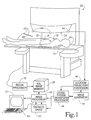
- the patient in use of the system for carrying out a stereotactic procedure on a soft tissue portion of a patient, the patient is received on a patient support 10.
- a rectangular sheet of an exoskeleton material 12 is temporarily softened to become flexible and stretchable.
- the exoskeleton material 12 is a mesh of a thermoplastic material which softens at temperatures of about 70° C.
- the material 12 is held or immersed in hot water until soft.
- the material 12 is then removed from hot water and allowed to cool to about body temperature.
- the material 12 is then stretched over and molded to the soft tissue region and affixed by clamps or other affixing means 14 to sides of the patient support.
- a base 16 is provided between the patient support and the patient to which the exoskeleton material 12 is affixed (see Figure 2). Thereafter, the material 12 is allowed or caused to set to a relatively rigid state.
- the material sets as it cools. However, setting may be accelerated by chilling the material, such as with cold water.
- Other exoskeleton materials are also contemplated.
- a plaster impregnated gauze type material may be wet, molded into place, affixed to the base or table, and allowed to cure or set.
- the exoskeleton material can be a mesh of a very stiff but pliable elastic mesh which is sufficiently stiff to hold the soft tissue substantially fixed relative to itself. Depending on the region of the anatomy, the mesh may be interconnected with more rigid plastic or other non-ferrous inserts. The inserts are preferably apertured to permit easy surgical access therethrough.
- a plurality of fiducials or other markers 18 are affixed to the exoskeleton material 12.
- the fiducials 18 carry a material which is readily detected by a magnetic resonance imaging apparatus.
- the fiducials 18 are hollow spheres that are filled with a material that is magnetic resonance susceptible and x-ray susceptible such that the patient can be examined by either or both x-ray or magnetic resonance techniques.
- a plurality of fiducials 18 are positioned to define appropriate reference points around the region of interest. Typically, a minimum of three fiducials 18 are used.
- the exoskeleton material 12 may contain magnetic resonance visible markers at relatively regular intervals. In another embodiment, the exoskeleton material 12 may be itself magnetic resonance or CT imageable.
- the patient support 10, patient, and exoskeleton material 12 are moved into a magnetic resonance imaging system 20.
- the patient support 10 is mounted on tracks or guides for movement between poles 24 of the magnetic resonance imaging system.
- a magnetic resonance examination is conducted to generate magnetic resonance data which is reconstructed by a reconstruction processor 26 to form a volumetric image representation which is stored in an image memory 28.
- An image processor 30 selectively accesses the volumetric image memory 28 under operator control to withdraw axial, sagittal, coronal, oblique, or other slice images, as well as volume renderings, and other conventionally displayed magnetic resonance and diagnostic images.
- the image processor 30 is connected with a monitor 32, such as a video monitor, LCD monitor, or gas plasma monitor.
- the patient is prepared for a surgical procedure.
- the surgical procedure may be performed right in the magnetic resonance examination apparatus, adjacent the magnetic resonance apparatus, or at a remote location. Because the exoskeleton material maintains the soft tissue in a fixed position even after the patient has been moved, a high degree of freedom in moving the patient to other venues is accorded.
- the surgical procedure is conducted at the magnetic resonance examination site.
- the coordinate system of the electronic image representation and the coordinate system of the patient are correlated.
- a wand 40 has a plurality of emitters 42 mounted thereon. Suitable emitters include light emitting diodes, lasers, ultrasonic transducers, radio transmitters, and the like.
- a plurality of receivers 44 are disposed adjacent the patient.
- the wand 40 is positioned on each of a plurality of the fiducials 18, preferably at least three, and the emitters 42 are caused to emit.
- the receivers 44 receive the emitted signals and through conventional triangulation, velocity measurement, and analogous techniques, a processor 46 determines the location of the tip of the wand 40, and hence of each fiducial 18 in the receiver or patient coordinate system. More specifically, the processor 46 determines the location of each emitter 42. From the emitter locations and any offset of the emitters 42 from the central axis, a trajectory along a central axis of the wand 40, is determined. From the known dimensions of the wand 40, the location of the tip is readily determined. Alternately, an articulated, mechanical arm digitizer can be used. A coordinate system transform means 48 determines a relationship between the coordinate system of the patient and the image data.
- the location of the tumors, lesions, or other structures to be biopsied or resectioned are determined in the electronic image space. That is, the radiologist views the images on the monitor 32 and determines the location of tumors, lesions or the like. The surgeon also views the images on the monitor 32 and plans a surgical approach or trajectory.
- a stereotaxy guide such as a needle guide 50, is mounted to the patient support, e.g., the base 16, along a generally proposed trajectory. In one embodiment, the guide has emitters 52 which are actuated. The receivers 44 receive the emitted signals from the guide emitters 52.
- An image generator 54 causes the image processor 30 to generate an image on the monitor 32 depicting a straight line trajectory from the guide 50 into the patient and a cross-hair referencing a preselected reference point on the guide 50. More specifically, the trajectory is conveyed through the transform means 48 to the image processor 30 such that the trajectory is displayed on the monitor 32 along with the cross-hair indicative of the reference point.
- the guide 50 is non-ferrous and has magnetic resonance image portion markers, e.g., at 52.
- the guide 50 is positioned in the magnetic resonance imager 20 with the patient and an image generated.
- the markers at 52 appear in the displayed human-readable image.
- the image generated derives the trajectory and the reference point from the images of the markers at 52.
- the surgeon views a multiplicity of slices and images to determine whether the trajectory intersects the lesion, tumor, or other tissue of interest and whether the trajectory passes through the appropriate tissue.
- the trajectory is selected to avoid major blood vessels or arteries, damaged tissue, bones, and the like.
- the guide 50 is repositioned one or more times and the process repeated until a satisfactory trajectory is determined.
- the guide 50 is removably supported on a support 56 that includes lockable gimbals, hinges, or the like 58 for selectively positioning the guide 50.
- the surgeon moves a cursor control, such as a mouse 60, to move a cursor display on the monitor 32.
- a gauging device 62 calculates a distance between the cursor controlled by the cursor control and the cross-hairs or other indicator of the reference point on the guide.
- the operator selects a biopsy needle 64, or other appropriate surgical implement.
- a stop 66 is set in accordance with the distance between the cursor and the cross-hairs, i.e., the distance to the tumor or the like.
- the surgeon then inserts the biopsy needle or other surgical instrument to the appropriate depth, performs the biopsy, resection, or the like, and withdraws the needle. After the operation, the incision is closed according to conventional medical practice and the exoskeleton restraint is removed.
- the surgical procedure is performed at or in the magnetic resonance imager.
- the patient and patient support can be moved to a remote site.
- a set of receivers 44 are disposed at the remote site for relating the coordinate system of the patient and guide at the remote site to the coordinate system of the images.
Abstract
Description
- This invention relates to stereotaxy systems and methods. It finds particular application with breast surgery and will be described with particular reference thereto. It should be appreciated, however, that the present invention will also find application in conjunction with stereotactic procedures on other portions of the anatomy and on subjects other than a human or animal body.
- Three-dimensional diagnostic image data of selected regions of a human patient are commonly available with magnetic resonance imagers, CT scanners, and other medical diagnostic equipment. These imaging modalities provide structural detail with a resolution of a millimeter or better. Various stereotaxy procedures have been developed which require extreme accuracy. One of the difficulties which has arisen is accurately determining an accurate spatial correlation between the medical diagnostic image and the patient.
- Stereotaxy has found particular application in neurosurgical procedures. Neurosurgery includes numerous procedures which require extreme accuracy, such as guided-needle biopsies, shunt placements, craniotomies for lesion or tumor resection, and the like. A mechanical ring or frame was attached to the patient's skull before images were taken, from which the name "framed" stereotaxy has evolved. This frame was typically attached to the patient using various mounting hardware methods that include sharp points or pins that pierce the skin and locate into the skull. The frame carried a "localizer" that includes various adjustable, but lockable hardware elements, angle indicators, and the like which enabled a surgical instrument or a guide for the surgical instrument to be mounted in a selected position relative to the patient. Due to the presence of the localizer during the diagnostic imaging, the localizer was visible in the resultant images. Hence, the relationships between the diagnostic image data, the frame, the localizer, and the patient were precisely known.
- Rather than using a mechanical frame which pierced the skin, other constructions which hold the head immovably in the registered position have also been proposed. One such restraint included a base plate or support for the back of the head made of perspex acrylic. A mask of open mesh was stretched over the patient's face and rigidly affixed to the base. For accurate conformity with individual patients, the mask was configured of a thermoplastic material which could be softened in hot water, stretched or molded into conformity with the patient's face, and connected to the base plate. When the plastic mesh material was cooled it set and the patient's head was rigidly positioned.
- The prior art neurosurgery frames or other restraints relied on the patient's internal skeleton, such as the skull, as an integral part of its position maintenance system. Breast tissue, lacking an internal skeleton has typically been handled differently. For x-ray mammographic examinations, the tissue has commonly been clamped between a pair of plates causing significant patient discomfort. In one technique for biopsies or resections, a stereotactic frame was associated with the plates and the radiologist inserted a probe carrying a barbed wire to the lesion or tumor to be resectioned or biopsied. Because the wire was barbed, its end would hold its position even after the plates were removed. The surgeon, in a separate procedure with the plates removed, followed the wire to the lesion or tumor. An additional problem with this technique is that the surgeons often found the wire did not define the best approach to the tumor or lesion. Generally, the plates constrained the radiologist's approaches. Moreover, the extreme distortion caused by the plates interfered with selection of the best approach.
- One problem with x-ray mammography is that it has difficulty distinguishing among soft tissue types. There is a generally poor contrast between "dense" breast tissue found in about 8% of women and lesions or tumors. Scars from prior treatment are also difficult to distinguish from tumors or lesions.
- Magnetic resonance is ideally suited for examining soft tissue and distinguishing among various types and conditions. However, in order to maintain an accurate registration between the soft tissue and resultant images, it has again been proposed to use non-ferrous radiographic style clamping plates. The clamping plates again have the above-described problems, whether used for radiology or magnetic resonance imaging.
- It is an object of the present invention to provide stereotaxy systems and methods which overcome the above problems.
- In accordance with one aspect of the invention there is provided a stereotaxy system comprising: an imaging system in which at least a soft portion of a subject that is supported on a support is positioned for generation of a volumetric electronic image representation using the imaging system; markers which are imageable by the imaging system disposed in a fixed position relative to the soft portion of the subject such that the imaging system images the markers along with the soft portion of the subject in the volumetric electronic image representation; an image processor which converts selected portions of the volumetric image representation to a human-readable display of the soft portion of the subject and the markers; a coordinate system transform for establishing a transform relationship between a coordinate system of the human-readable display of the volumetric image representation and a coordinate system of the soft portion; and a guide for defining a trajectory along which an instrument may be inserted into the soft portion; characterized by: a non-ferrous exoskeleton material which has a stretchable state in which it is stretched to conform to the soft portion, the exoskeleton material being fixed relative to said support such that the soft portion is constrained against movement.
- The exoskeleton material suitably includes a thermoplastic mesh that is thermally softenable to enable it to be conformed to the soft portion and sets to a relatively rigid state after conforming to the soft portion.
- In accordance with a second aspect of the invention there is provided a stereotaxy method comprising: positioning at least a soft portion of a subject on a support in a non-invasive imaging system; affixing markers in association with the soft portion; generating a volumetric electronic image representation of the soft portion and the markers; converting selected portions of the volumetric electronic image representation to a human-readable display of the soft portion and the markers; positioning a guide for defining a trajectory along which an instrument is to be inserted into the soft portion of the subject and; inserting an instrument along the trajectory; the method being characterized by: applying and conforming a non-ferrous material which has a stretchable conforming state to the soft portion of the subject; and fixing the non-ferrous material relative to the support such that the soft portion is constrained against movement.
- In a method according to the invention the exoskeleton material is suitably a thermoplastic mesh and prior to the applying and conforming step the thermoplastic mesh is heat softened and then cooled below human body temperature; and after the applying and conforming step the thermoplastic material is caused to set to a stiff, relatively rigid state.
- In one particular method according to the invention the non-invasive imaging system is a magnetic resonance imaging system, the guide and the instrument are non-ferrous, and insertion of the instrument is performed in the magnetic resonance imaging system.
- In one particular application of the invention the soft portion is a human breast.
- One stereotaxy system and method in accordance with the invention will now be described, by way of example, with reference to the accompanying drawings in which:-
- Figure 1 is a diagrammatic illustration of the stereotaxy system;
- Figure 1A is a detailed view of a sheet of exoskeleton material used in the system of Figure 1; and,
- Figure 2 is a detailed illustration of a patient restraint and an exemplary surgical tool of the system of Figure 1.
- Referring to the drawings, in use of the system for carrying out a stereotactic procedure on a soft tissue portion of a patient, the patient is received on a
patient support 10. A rectangular sheet of anexoskeleton material 12 is temporarily softened to become flexible and stretchable. In a preferred embodiment, theexoskeleton material 12 is a mesh of a thermoplastic material which softens at temperatures of about 70° C. Thematerial 12 is held or immersed in hot water until soft. Thematerial 12 is then removed from hot water and allowed to cool to about body temperature. Thematerial 12 is then stretched over and molded to the soft tissue region and affixed by clamps or other affixing means 14 to sides of the patient support. Optionally, abase 16 is provided between the patient support and the patient to which theexoskeleton material 12 is affixed (see Figure 2). Thereafter, thematerial 12 is allowed or caused to set to a relatively rigid state. With the thermoplastic material of the preferred embodiment, the material sets as it cools. However, setting may be accelerated by chilling the material, such as with cold water. Other exoskeleton materials are also contemplated. For example, a plaster impregnated gauze type material may be wet, molded into place, affixed to the base or table, and allowed to cure or set. As another alternative, the exoskeleton material can be a mesh of a very stiff but pliable elastic mesh which is sufficiently stiff to hold the soft tissue substantially fixed relative to itself. Depending on the region of the anatomy, the mesh may be interconnected with more rigid plastic or other non-ferrous inserts. The inserts are preferably apertured to permit easy surgical access therethrough. - A plurality of fiducials or
other markers 18 are affixed to theexoskeleton material 12. Thefiducials 18 carry a material which is readily detected by a magnetic resonance imaging apparatus. In one embodiment, thefiducials 18 are hollow spheres that are filled with a material that is magnetic resonance susceptible and x-ray susceptible such that the patient can be examined by either or both x-ray or magnetic resonance techniques. A plurality offiducials 18 are positioned to define appropriate reference points around the region of interest. Typically, a minimum of threefiducials 18 are used. Alternately, theexoskeleton material 12 may contain magnetic resonance visible markers at relatively regular intervals. In another embodiment, theexoskeleton material 12 may be itself magnetic resonance or CT imageable. - Once the
fiducials 18 are secured, such as with an adhesive, thepatient support 10, patient, andexoskeleton material 12 are moved into a magnetic resonance imaging system 20. In the illustrated embodiment, thepatient support 10 is mounted on tracks or guides for movement betweenpoles 24 of the magnetic resonance imaging system. A magnetic resonance examination is conducted to generate magnetic resonance data which is reconstructed by areconstruction processor 26 to form a volumetric image representation which is stored in animage memory 28. Animage processor 30 selectively accesses thevolumetric image memory 28 under operator control to withdraw axial, sagittal, coronal, oblique, or other slice images, as well as volume renderings, and other conventionally displayed magnetic resonance and diagnostic images. Theimage processor 30 is connected with amonitor 32, such as a video monitor, LCD monitor, or gas plasma monitor. - Once the magnetic resonance examination has been completed and the volume image representation generated, the patient is prepared for a surgical procedure. The surgical procedure may be performed right in the magnetic resonance examination apparatus, adjacent the magnetic resonance apparatus, or at a remote location. Because the exoskeleton material maintains the soft tissue in a fixed position even after the patient has been moved, a high degree of freedom in moving the patient to other venues is accorded.
- In the illustrated embodiment, the surgical procedure is conducted at the magnetic resonance examination site. First, the coordinate system of the electronic image representation and the coordinate system of the patient are correlated. More specifically, a
wand 40 has a plurality ofemitters 42 mounted thereon. Suitable emitters include light emitting diodes, lasers, ultrasonic transducers, radio transmitters, and the like. A plurality ofreceivers 44 are disposed adjacent the patient. Thewand 40 is positioned on each of a plurality of thefiducials 18, preferably at least three, and theemitters 42 are caused to emit. Thereceivers 44 receive the emitted signals and through conventional triangulation, velocity measurement, and analogous techniques, aprocessor 46 determines the location of the tip of thewand 40, and hence of each fiducial 18 in the receiver or patient coordinate system. More specifically, theprocessor 46 determines the location of eachemitter 42. From the emitter locations and any offset of theemitters 42 from the central axis, a trajectory along a central axis of thewand 40, is determined. From the known dimensions of thewand 40, the location of the tip is readily determined. Alternately, an articulated, mechanical arm digitizer can be used. A coordinate system transform means 48 determines a relationship between the coordinate system of the patient and the image data. - With continuing reference to FIGURE 1 and further reference to FIGURE 2, the location of the tumors, lesions, or other structures to be biopsied or resectioned are determined in the electronic image space. That is, the radiologist views the images on the
monitor 32 and determines the location of tumors, lesions or the like. The surgeon also views the images on themonitor 32 and plans a surgical approach or trajectory. A stereotaxy guide, such as aneedle guide 50, is mounted to the patient support, e.g., thebase 16, along a generally proposed trajectory. In one embodiment, the guide hasemitters 52 which are actuated. Thereceivers 44 receive the emitted signals from theguide emitters 52. Animage generator 54 causes theimage processor 30 to generate an image on themonitor 32 depicting a straight line trajectory from theguide 50 into the patient and a cross-hair referencing a preselected reference point on theguide 50. More specifically, the trajectory is conveyed through the transform means 48 to theimage processor 30 such that the trajectory is displayed on themonitor 32 along with the cross-hair indicative of the reference point. - In another embodiment, the
guide 50 is non-ferrous and has magnetic resonance image portion markers, e.g., at 52. Theguide 50 is positioned in the magnetic resonance imager 20 with the patient and an image generated. The markers at 52 appear in the displayed human-readable image. In this embodiment, the image generated derives the trajectory and the reference point from the images of the markers at 52. - The surgeon then views a multiplicity of slices and images to determine whether the trajectory intersects the lesion, tumor, or other tissue of interest and whether the trajectory passes through the appropriate tissue. For example, the trajectory is selected to avoid major blood vessels or arteries, damaged tissue, bones, and the like. Commonly, the
guide 50 is repositioned one or more times and the process repeated until a satisfactory trajectory is determined. Preferably, theguide 50 is removably supported on a support 56 that includes lockable gimbals, hinges, or the like 58 for selectively positioning theguide 50. - Once a satisfactory trajectory is determined, the surgeon moves a cursor control, such as a
mouse 60, to move a cursor display on themonitor 32. A gaugingdevice 62 calculates a distance between the cursor controlled by the cursor control and the cross-hairs or other indicator of the reference point on the guide. The operator then selects abiopsy needle 64, or other appropriate surgical implement. Astop 66 is set in accordance with the distance between the cursor and the cross-hairs, i.e., the distance to the tumor or the like. The surgeon then inserts the biopsy needle or other surgical instrument to the appropriate depth, performs the biopsy, resection, or the like, and withdraws the needle. After the operation, the incision is closed according to conventional medical practice and the exoskeleton restraint is removed. - With non-ferrous guides and surgical instruments, the surgical procedure is performed at or in the magnetic resonance imager. Alternately, the patient and patient support can be moved to a remote site. A set of
receivers 44 are disposed at the remote site for relating the coordinate system of the patient and guide at the remote site to the coordinate system of the images.
Claims (10)
- A stereotaxy system comprising: an imaging system (20) in which at least a soft portion of a subject that is supported on a support (10) is positioned for generation of a volumetric electronic image representation using the imaging system (20); markers (18) which are imageable by the imaging system disposed in a fixed position relative to the soft portion of the subject such that the imaging system (20) images the markers (18) along with the soft portion of the subject in the volumetric electronic image representation; an image processor (30) which converts selected portions of the volumetric image representation to a human-readable display (32) of the soft portion of the subject and the markers (18); a coordinate system transform (48) for establishing a transform relationship between a coordinate system of the human-readable display of the volumetric image representation and a coordinate system of the soft portion; and a guide (50) for defining a trajectory along which an instrument (64) may be inserted into the soft portion; characterized by: a non-ferrous exoskeleton material (12) which has a stretchable state in which it is stretched to conform to the soft portion, the exoskeleton material (12) being fixed relative to said support (10) such that the soft portion is constrained against movement.
- A system according to claim 1 including an adjustable mechanical mounting (56) for selectively mounting the guide (50) in a selected orientation relative to the subject; emitters (52) disposed on the guide (50) for sending emitting signals; receivers (44) for receiving the signals from the emitters (52); and a location processor (46) connected with the receivers (44) for determining the location of at least one characteristic point on the guide (50) and a trajectory defined by the guide (50), the location processor (46) being connected with the coordinate system transform means (48) for transforming the characteristic point on the guide (50) and the trajectory into the image coordinate system of the human-readable display and being connected with the image processor (30) for generating a human-readable display of the trajectory and the characteristic point; and an instrument (64) received in the guide (50).
- A system as set forth in claim 1 or claim 2 wherein the exoskeleton material (12) includes a thermoplastic mesh that is thermally softenable to enable it to be conformed to the soft portion and sets to a relatively rigid state after conforming to the soft portion.
- A stereotaxy method comprising positioning at least a soft portion of a subject on a support (10) in a non-invasive imaging system (20); affixing markers (18) in association with the soft portion; generating a volumetric electronic image representation of the soft portion and the markers (18); converting selected portions of the volumetric electronic image representation to a human-readable display of the soft portion and the markers (18); positioning a guide (50) for defining a trajectory along which an instrument is to be inserted into the soft portion of the subject; and inserting an instrument (64) along the trajectory; the method being characterized by: applying and conforming a non-ferrous material (12) which has a stretchable conforming state to the soft portion of the subject; and fixing the non-ferrous material (12) relative to the support (10) such that the soft portion is constrained against movement.
- A method as set forth in claim 4 wherein after applying and conforming the non-ferrous material (12) to the soft portion of the subject, the non-ferrous material (12) is set into a rigid exoskeleton.
- A method as set forth in claim 5 in which the exoskeleton material (12) is a thermoplastic mesh and prior to the applying and conforming step the thermoplastic mesh is heat softened and then cooled below human body temperature; and after the applying and conforming step the thermoplastic material is caused to set to a stiff, relatively rigid state.
- A method as set forth in claim 5 or claim 6 wherein the non-invasive imaging system (20) is a magnetic resonance imaging system, the guide (50) and the instrument (64) are non-ferrous, and insertion of the instrument (64) is performed in the magnetic resonance imaging system (20).
- A stereotaxy method as set forth in claim 7 wherein at least portions of the guide (50) are imageable in the magnetic resonance imaging system (20) and the guide (50) is imaged along with the soft portion of the subject to establish a relationship between a coordinate system of the human-readable display and a coordinate system of the soft portion of the subject and the guide (50).
- A method as set forth in any one of claims 5 to 8 wherein the subject is moved to a remote site before inserting the instrument.
- A stereotaxy method as set forth in any one of claims 5 to 9 wherein the soft portion is a human breast.
Applications Claiming Priority (2)
| Application Number | Priority Date | Filing Date | Title |
|---|---|---|---|
| US08/378,511 US5682890A (en) | 1995-01-26 | 1995-01-26 | Magnetic resonance stereotactic surgery with exoskeleton tissue stabilization |
| US378511 | 1995-01-26 |
Publications (2)
| Publication Number | Publication Date |
|---|---|
| EP0728446A1 true EP0728446A1 (en) | 1996-08-28 |
| EP0728446B1 EP0728446B1 (en) | 2000-03-01 |
Family
ID=23493401
Family Applications (1)
| Application Number | Title | Priority Date | Filing Date |
|---|---|---|---|
| EP95309062A Expired - Lifetime EP0728446B1 (en) | 1995-01-26 | 1995-12-13 | Stereotaxy systems |
Country Status (4)
| Country | Link |
|---|---|
| US (1) | US5682890A (en) |
| EP (1) | EP0728446B1 (en) |
| JP (1) | JPH08238257A (en) |
| DE (1) | DE69515288T2 (en) |
Cited By (20)
| Publication number | Priority date | Publication date | Assignee | Title |
|---|---|---|---|---|
| WO1997002942A1 (en) * | 1995-07-07 | 1997-01-30 | Alk Associates | A thermoplastic sheet with edge parts |
| WO1998040026A1 (en) * | 1997-03-11 | 1998-09-17 | Sonometrics Corporation | System for carrying out surgery, biopsy and ablation of a tumor or other physical anomaly |
| WO1999003398A1 (en) * | 1997-07-19 | 1999-01-28 | Mueller Christian | Device and method for fixing, compressing or shaping (parts) of the body |
| GB2329473A (en) * | 1997-07-30 | 1999-03-24 | Bruker Medizintech | Interventional device positioning system for NMR tomograph |
| EP0908146A2 (en) * | 1997-10-06 | 1999-04-14 | General Electric Company | Real-time image-guided placement of anchor devices |
| EP0915675A2 (en) * | 1997-02-14 | 1999-05-19 | Biosense, Inc. | X-ray guided surgical location system with extended mapping volume |
| EP0922438A1 (en) * | 1997-11-28 | 1999-06-16 | Picker International, Inc. | Image guided interventional procedures |
| WO2000049958A1 (en) * | 1999-02-22 | 2000-08-31 | Vtarget Ltd. | A method and system for guiding a diagnostic or therapeutic instrument towards a target region inside the patient's body |
| EP0927542A3 (en) * | 1998-01-05 | 2000-09-13 | Galil Medical Ltd | System and method for MRI-guided cryosurgery |
| EP0919203A3 (en) * | 1997-11-28 | 2000-11-29 | Picker International, Inc. | Frameless stereotactic surgical apparatus |
| US6314310B1 (en) | 1997-02-14 | 2001-11-06 | Biosense, Inc. | X-ray guided surgical location system with extended mapping volume |
| US6368331B1 (en) | 1999-02-22 | 2002-04-09 | Vtarget Ltd. | Method and system for guiding a diagnostic or therapeutic instrument towards a target region inside the patient's body |
| WO2002034153A1 (en) * | 2000-10-23 | 2002-05-02 | Deutsches Krebsforschungszentrum Stiffung D. Öffentl. Rechts | Method and device for navigation during medical interventions or for localising a non-osseous structure |
| EP1319368A2 (en) * | 2001-12-12 | 2003-06-18 | TecMedic GmbH | Method for determining the orientation and relative position of a medical instrument |
| WO2003061477A1 (en) * | 2002-01-23 | 2003-07-31 | Med-Tec Iowa, Inc. | Hermoplastic patient restraint member for radiation therapy |
| EP2198801A1 (en) * | 2008-12-19 | 2010-06-23 | BrainLAB AG | Marker application device for applying a marker device |
| US8974460B2 (en) | 2008-10-22 | 2015-03-10 | Blue Ortho | Device for controlled adjustment of a surgical positioning unit |
| US9168106B2 (en) | 2009-05-05 | 2015-10-27 | Blue Ortho | Device and method for instrument adjustment in computer assisted surgery |
| US9220509B2 (en) | 2009-06-30 | 2015-12-29 | Blue Ortho | Adjustable guide in computer assisted orthopaedic surgery |
| US9655628B2 (en) | 2009-05-06 | 2017-05-23 | Blue Ortho | Reduced invasivity fixation system for trackers in computer assisted surgery |
Families Citing this family (184)
| Publication number | Priority date | Publication date | Assignee | Title |
|---|---|---|---|---|
| US6331180B1 (en) | 1988-05-03 | 2001-12-18 | Sherwood Services Ag | Target-centered stereotaxtic surgical arc system with reorientatable arc axis |
| FR2652928B1 (en) | 1989-10-05 | 1994-07-29 | Diadix Sa | INTERACTIVE LOCAL INTERVENTION SYSTEM WITHIN A AREA OF A NON-HOMOGENEOUS STRUCTURE. |
| US6006126A (en) | 1991-01-28 | 1999-12-21 | Cosman; Eric R. | System and method for stereotactic registration of image scan data |
| US6405072B1 (en) * | 1991-01-28 | 2002-06-11 | Sherwood Services Ag | Apparatus and method for determining a location of an anatomical target with reference to a medical apparatus |
| US5603318A (en) | 1992-04-21 | 1997-02-18 | University Of Utah Research Foundation | Apparatus and method for photogrammetric surgical localization |
| US5913820A (en) | 1992-08-14 | 1999-06-22 | British Telecommunications Public Limited Company | Position location system |
| EP0951874A3 (en) | 1994-09-15 | 2000-06-14 | Visualization Technology, Inc. | Position tracking and imaging system for use in medical applications using a reference unit secured to a patients head |
| US6351659B1 (en) * | 1995-09-28 | 2002-02-26 | Brainlab Med. Computersysteme Gmbh | Neuro-navigation system |
| US5800353A (en) * | 1996-02-12 | 1998-09-01 | Mclaurin, Jr.; Robert L. | Automatic image registration of magnetic resonance imaging scans for localization, 3-dimensional treatment planning, and radiation treatment of abnormal lesions |
| US6684098B2 (en) * | 1996-08-16 | 2004-01-27 | Brigham And Women's Hospital, Inc. | Versatile stereotactic device and methods of use |
| US7302288B1 (en) * | 1996-11-25 | 2007-11-27 | Z-Kat, Inc. | Tool position indicator |
| US6026315A (en) * | 1997-03-27 | 2000-02-15 | Siemens Aktiengesellschaft | Method and apparatus for calibrating a navigation system in relation to image data of a magnetic resonance apparatus |
| US5928342A (en) * | 1997-07-02 | 1999-07-27 | Creative Technology Ltd. | Audio effects processor integrated on a single chip with a multiport memory onto which multiple asynchronous digital sound samples can be concurrently loaded |
| US6226548B1 (en) | 1997-09-24 | 2001-05-01 | Surgical Navigation Technologies, Inc. | Percutaneous registration apparatus and method for use in computer-assisted surgical navigation |
| US5951475A (en) * | 1997-09-25 | 1999-09-14 | International Business Machines Corporation | Methods and apparatus for registering CT-scan data to multiple fluoroscopic images |
| US6021343A (en) | 1997-11-20 | 2000-02-01 | Surgical Navigation Technologies | Image guided awl/tap/screwdriver |
| US5967982A (en) * | 1997-12-09 | 1999-10-19 | The Cleveland Clinic Foundation | Non-invasive spine and bone registration for frameless stereotaxy |
| US6348058B1 (en) | 1997-12-12 | 2002-02-19 | Surgical Navigation Technologies, Inc. | Image guided spinal surgery guide, system, and method for use thereof |
| US6119032A (en) * | 1997-12-31 | 2000-09-12 | U.S. Philips Corporation | Method and system for positioning an invasive device by magnetic resonance (MR) imaging of an MR visible device |
| US6122539A (en) * | 1997-12-31 | 2000-09-19 | General Electric Company | Method for verifying accuracy during intra-operative MR imaging |
| US6175760B1 (en) * | 1998-02-17 | 2001-01-16 | University Of Iowa Research Foundation | Lesion localizer for nuclear medicine |
| JPH11267133A (en) | 1998-03-25 | 1999-10-05 | Olympus Optical Co Ltd | Therapeutic apparatus |
| US6118845A (en) | 1998-06-29 | 2000-09-12 | Surgical Navigation Technologies, Inc. | System and methods for the reduction and elimination of image artifacts in the calibration of X-ray imagers |
| US6459927B1 (en) * | 1999-07-06 | 2002-10-01 | Neutar, Llc | Customizable fixture for patient positioning |
| US6477400B1 (en) | 1998-08-20 | 2002-11-05 | Sofamor Danek Holdings, Inc. | Fluoroscopic image guided orthopaedic surgery system with intraoperative registration |
| DE19848765C2 (en) | 1998-10-22 | 2000-12-21 | Brainlab Med Computersyst Gmbh | Position verification in camera images |
| US6826423B1 (en) | 1999-01-04 | 2004-11-30 | Midco-Medical Instrumentation And Diagnostics Corporation | Whole body stereotactic localization and immobilization system |
| US6775404B1 (en) * | 1999-03-18 | 2004-08-10 | University Of Washington | Apparatus and method for interactive 3D registration of ultrasound and magnetic resonance images based on a magnetic position sensor |
| US6470207B1 (en) | 1999-03-23 | 2002-10-22 | Surgical Navigation Technologies, Inc. | Navigational guidance via computer-assisted fluoroscopic imaging |
| US6491699B1 (en) | 1999-04-20 | 2002-12-10 | Surgical Navigation Technologies, Inc. | Instrument guidance method and system for image guided surgery |
| DE19917867B4 (en) | 1999-04-20 | 2005-04-21 | Brainlab Ag | Method and device for image support in the treatment of treatment objectives with integration of X-ray detection and navigation system |
| US7853311B1 (en) * | 1999-04-23 | 2010-12-14 | 3M Innovative Properties Company | Surgical targeting system |
| JP2001000430A (en) * | 1999-06-24 | 2001-01-09 | Alcare Co Ltd | Marker for image photographing |
| US6499488B1 (en) | 1999-10-28 | 2002-12-31 | Winchester Development Associates | Surgical sensor |
| US6379302B1 (en) | 1999-10-28 | 2002-04-30 | Surgical Navigation Technologies Inc. | Navigation information overlay onto ultrasound imagery |
| US6474341B1 (en) | 1999-10-28 | 2002-11-05 | Surgical Navigation Technologies, Inc. | Surgical communication and power system |
| US8239001B2 (en) | 2003-10-17 | 2012-08-07 | Medtronic Navigation, Inc. | Method and apparatus for surgical navigation |
| US6493573B1 (en) | 1999-10-28 | 2002-12-10 | Winchester Development Associates | Method and system for navigating a catheter probe in the presence of field-influencing objects |
| US11331150B2 (en) | 1999-10-28 | 2022-05-17 | Medtronic Navigation, Inc. | Method and apparatus for surgical navigation |
| US6235038B1 (en) | 1999-10-28 | 2001-05-22 | Medtronic Surgical Navigation Technologies | System for translation of electromagnetic and optical localization systems |
| US6381485B1 (en) | 1999-10-28 | 2002-04-30 | Surgical Navigation Technologies, Inc. | Registration of human anatomy integrated for electromagnetic localization |
| US8644907B2 (en) | 1999-10-28 | 2014-02-04 | Medtronic Navigaton, Inc. | Method and apparatus for surgical navigation |
| US7366562B2 (en) | 2003-10-17 | 2008-04-29 | Medtronic Navigation, Inc. | Method and apparatus for surgical navigation |
| US7024237B1 (en) * | 1999-10-29 | 2006-04-04 | University Of Florida Research Foundation, Inc. | Mask system and method for stereotactic radiotherapy and image guided procedures |
| US6671538B1 (en) * | 1999-11-26 | 2003-12-30 | Koninklijke Philips Electronics, N.V. | Interface system for use with imaging devices to facilitate visualization of image-guided interventional procedure planning |
| US6725080B2 (en) | 2000-03-01 | 2004-04-20 | Surgical Navigation Technologies, Inc. | Multiple cannula image guided tool for image guided procedures |
| US6712856B1 (en) | 2000-03-17 | 2004-03-30 | Kinamed, Inc. | Custom replacement device for resurfacing a femur and method of making the same |
| US6490473B1 (en) * | 2000-04-07 | 2002-12-03 | Coin Medical Technologies, Ltd. | System and method of interactive positioning |
| US6535756B1 (en) | 2000-04-07 | 2003-03-18 | Surgical Navigation Technologies, Inc. | Trajectory storage apparatus and method for surgical navigation system |
| US6889073B2 (en) * | 2000-05-08 | 2005-05-03 | David A. Lampman | Breast biopsy and therapy system for magnetic resonance imagers |
| US7085400B1 (en) | 2000-06-14 | 2006-08-01 | Surgical Navigation Technologies, Inc. | System and method for image based sensor calibration |
| AU6779901A (en) * | 2000-06-27 | 2002-01-08 | Insightec-Image Guided Treatment Ltd. | Registration of target object images to stored image data |
| US6493574B1 (en) * | 2000-09-28 | 2002-12-10 | Koninklijke Philips Electronics, N.V. | Calibration phantom and recognition algorithm for automatic coordinate transformation in diagnostic imaging |
| US6636757B1 (en) | 2001-06-04 | 2003-10-21 | Surgical Navigation Technologies, Inc. | Method and apparatus for electromagnetic navigation of a surgical probe near a metal object |
| US6947786B2 (en) | 2002-02-28 | 2005-09-20 | Surgical Navigation Technologies, Inc. | Method and apparatus for perspective inversion |
| US6990368B2 (en) | 2002-04-04 | 2006-01-24 | Surgical Navigation Technologies, Inc. | Method and apparatus for virtual digital subtraction angiography |
| US7998062B2 (en) | 2004-03-29 | 2011-08-16 | Superdimension, Ltd. | Endoscope structures and techniques for navigating to a target in branched structure |
| JP2005525868A (en) * | 2002-05-21 | 2005-09-02 | プルス エンドプロシェティク アーゲー | Device for determining geometric parameters for functional determination of spinal joints |
| CA2633137C (en) | 2002-08-13 | 2012-10-23 | The Governors Of The University Of Calgary | Microsurgical robot system |
| US7697972B2 (en) | 2002-11-19 | 2010-04-13 | Medtronic Navigation, Inc. | Navigation system for cardiac therapies |
| US7599730B2 (en) | 2002-11-19 | 2009-10-06 | Medtronic Navigation, Inc. | Navigation system for cardiac therapies |
| US7226456B2 (en) * | 2002-12-31 | 2007-06-05 | Depuy Acromed, Inc. | Trackable medical tool for use in image guided surgery |
| US7542791B2 (en) | 2003-01-30 | 2009-06-02 | Medtronic Navigation, Inc. | Method and apparatus for preplanning a surgical procedure |
| US7660623B2 (en) | 2003-01-30 | 2010-02-09 | Medtronic Navigation, Inc. | Six degree of freedom alignment display for medical procedures |
| US20040152955A1 (en) * | 2003-02-04 | 2004-08-05 | Mcginley Shawn E. | Guidance system for rotary surgical instrument |
| US20040171930A1 (en) * | 2003-02-04 | 2004-09-02 | Zimmer Technology, Inc. | Guidance system for rotary surgical instrument |
| US7458977B2 (en) | 2003-02-04 | 2008-12-02 | Zimmer Technology, Inc. | Surgical navigation instrument useful in marking anatomical structures |
| US6932823B2 (en) * | 2003-06-24 | 2005-08-23 | Zimmer Technology, Inc. | Detachable support arm for surgical navigation system reference array |
| US7313430B2 (en) | 2003-08-28 | 2007-12-25 | Medtronic Navigation, Inc. | Method and apparatus for performing stereotactic surgery |
| ES2432616T3 (en) | 2003-09-15 | 2013-12-04 | Covidien Lp | Accessory system for use with bronchoscopes |
| EP2316328B1 (en) | 2003-09-15 | 2012-05-09 | Super Dimension Ltd. | Wrap-around holding device for use with bronchoscopes |
| DE10343826B4 (en) * | 2003-09-22 | 2006-02-09 | Plus-Endoprothetik Ag | Bone-proof locator and navigation system |
| US7970452B2 (en) * | 2003-09-30 | 2011-06-28 | Hologic, Inc. | Open architecture imaging apparatus and coil system for magnetic resonance imaging |
| US7908690B2 (en) * | 2003-09-30 | 2011-03-22 | Sentinelle Medical, Inc. | Supine patient support for medical imaging |
| US7379769B2 (en) * | 2003-09-30 | 2008-05-27 | Sunnybrook Health Sciences Center | Hybrid imaging method to monitor medical device delivery and patient support for use in the method |
| US20080077005A1 (en) * | 2004-08-12 | 2008-03-27 | Piron Cameron A | System and Method for Multimodality Breast Imaging |
| US7835778B2 (en) | 2003-10-16 | 2010-11-16 | Medtronic Navigation, Inc. | Method and apparatus for surgical navigation of a multiple piece construct for implantation |
| US7840253B2 (en) | 2003-10-17 | 2010-11-23 | Medtronic Navigation, Inc. | Method and apparatus for surgical navigation |
| US8764725B2 (en) | 2004-02-09 | 2014-07-01 | Covidien Lp | Directional anchoring mechanism, method and applications thereof |
| US7641660B2 (en) | 2004-03-08 | 2010-01-05 | Biomet Manufacturing Corporation | Method, apparatus, and system for image guided bone cutting |
| US7567834B2 (en) | 2004-05-03 | 2009-07-28 | Medtronic Navigation, Inc. | Method and apparatus for implantation between two vertebral bodies |
| DE102004048066A1 (en) * | 2004-09-30 | 2006-04-20 | Tecmedic Gmbh | Device for geometric calibration of different measurement devices, especially when using image generating operating, therapeutic or diagnostic methods, balances measurement devices using known relative position, orientation of two markings |
| US7623250B2 (en) * | 2005-02-04 | 2009-11-24 | Stryker Leibinger Gmbh & Co. Kg. | Enhanced shape characterization device and method |
| US7831293B2 (en) * | 2005-05-10 | 2010-11-09 | Advanced Clinical Solutions, Inc. | Method of defining a biological target for treatment |
| US7840256B2 (en) * | 2005-06-27 | 2010-11-23 | Biomet Manufacturing Corporation | Image guided tracking array and method |
| CN101247847B (en) | 2005-07-11 | 2013-01-09 | 导管机器人技术公司 | Remotely controlled catheter insertion system |
| US20070050030A1 (en) * | 2005-08-23 | 2007-03-01 | Kim Richard C | Expandable implant device with interchangeable spacer |
| US8784336B2 (en) | 2005-08-24 | 2014-07-22 | C. R. Bard, Inc. | Stylet apparatuses and methods of manufacture |
| DE102005044033B4 (en) * | 2005-09-14 | 2010-11-18 | Cas Innovations Gmbh & Co. Kg | Positioning system for percutaneous interventions |
| US7643862B2 (en) | 2005-09-15 | 2010-01-05 | Biomet Manufacturing Corporation | Virtual mouse for use in surgical navigation |
| US7835784B2 (en) | 2005-09-21 | 2010-11-16 | Medtronic Navigation, Inc. | Method and apparatus for positioning a reference frame |
| US9168102B2 (en) | 2006-01-18 | 2015-10-27 | Medtronic Navigation, Inc. | Method and apparatus for providing a container to a sterile environment |
| US20070239153A1 (en) * | 2006-02-22 | 2007-10-11 | Hodorek Robert A | Computer assisted surgery system using alternative energy technology |
| US8165659B2 (en) | 2006-03-22 | 2012-04-24 | Garrett Sheffer | Modeling method and apparatus for use in surgical navigation |
| US8112292B2 (en) | 2006-04-21 | 2012-02-07 | Medtronic Navigation, Inc. | Method and apparatus for optimizing a therapy |
| US20090080602A1 (en) * | 2006-08-03 | 2009-03-26 | Kenneth Brooks | Dedicated breast radiation imaging/therapy system |
| US8660635B2 (en) | 2006-09-29 | 2014-02-25 | Medtronic, Inc. | Method and apparatus for optimizing a computer assisted surgical procedure |
| US8388546B2 (en) | 2006-10-23 | 2013-03-05 | Bard Access Systems, Inc. | Method of locating the tip of a central venous catheter |
| US7794407B2 (en) | 2006-10-23 | 2010-09-14 | Bard Access Systems, Inc. | Method of locating the tip of a central venous catheter |
| US20090012509A1 (en) * | 2007-04-24 | 2009-01-08 | Medtronic, Inc. | Navigated Soft Tissue Penetrating Laser System |
| US8108025B2 (en) * | 2007-04-24 | 2012-01-31 | Medtronic, Inc. | Flexible array for use in navigated surgery |
| US8734466B2 (en) * | 2007-04-25 | 2014-05-27 | Medtronic, Inc. | Method and apparatus for controlled insertion and withdrawal of electrodes |
| US8311611B2 (en) * | 2007-04-24 | 2012-11-13 | Medtronic, Inc. | Method for performing multiple registrations in a navigated procedure |
| US8301226B2 (en) | 2007-04-24 | 2012-10-30 | Medtronic, Inc. | Method and apparatus for performing a navigated procedure |
| ATE476934T1 (en) * | 2007-04-24 | 2010-08-15 | Medtronic Inc | FLEXIBLE ARRANGEMENT FOR USE IN NAVIGATED SURGERY |
| US9289270B2 (en) * | 2007-04-24 | 2016-03-22 | Medtronic, Inc. | Method and apparatus for performing a navigated procedure |
| US8989842B2 (en) | 2007-05-16 | 2015-03-24 | General Electric Company | System and method to register a tracking system with intracardiac echocardiography (ICE) imaging system |
| US8527032B2 (en) | 2007-05-16 | 2013-09-03 | General Electric Company | Imaging system and method of delivery of an instrument to an imaged subject |
| US8428690B2 (en) | 2007-05-16 | 2013-04-23 | General Electric Company | Intracardiac echocardiography image reconstruction in combination with position tracking system |
| US8364242B2 (en) * | 2007-05-17 | 2013-01-29 | General Electric Company | System and method of combining ultrasound image acquisition with fluoroscopic image acquisition |
| US8934961B2 (en) | 2007-05-18 | 2015-01-13 | Biomet Manufacturing, Llc | Trackable diagnostic scope apparatus and methods of use |
| US20080319491A1 (en) | 2007-06-19 | 2008-12-25 | Ryan Schoenefeld | Patient-matched surgical component and methods of use |
| US8905920B2 (en) | 2007-09-27 | 2014-12-09 | Covidien Lp | Bronchoscope adapter and method |
| US8290569B2 (en) | 2007-11-23 | 2012-10-16 | Hologic, Inc. | Open architecture tabletop patient support and coil system |
| US9521961B2 (en) | 2007-11-26 | 2016-12-20 | C. R. Bard, Inc. | Systems and methods for guiding a medical instrument |
| US10524691B2 (en) | 2007-11-26 | 2020-01-07 | C. R. Bard, Inc. | Needle assembly including an aligned magnetic element |
| US9649048B2 (en) | 2007-11-26 | 2017-05-16 | C. R. Bard, Inc. | Systems and methods for breaching a sterile field for intravascular placement of a catheter |
| US10751509B2 (en) | 2007-11-26 | 2020-08-25 | C. R. Bard, Inc. | Iconic representations for guidance of an indwelling medical device |
| ES2832713T3 (en) | 2007-11-26 | 2021-06-11 | Bard Inc C R | Integrated system for intravascular catheter placement |
| US10449330B2 (en) | 2007-11-26 | 2019-10-22 | C. R. Bard, Inc. | Magnetic element-equipped needle assemblies |
| US8849382B2 (en) | 2007-11-26 | 2014-09-30 | C. R. Bard, Inc. | Apparatus and display methods relating to intravascular placement of a catheter |
| US8781555B2 (en) | 2007-11-26 | 2014-07-15 | C. R. Bard, Inc. | System for placement of a catheter including a signal-generating stylet |
| US8986246B2 (en) * | 2008-01-16 | 2015-03-24 | Catheter Robotics Inc. | Remotely controlled catheter insertion system |
| EP2231253B1 (en) | 2008-01-16 | 2019-06-12 | Catheter Precision, Inc. | Remotely controlled catheter insertion system |
| US8571637B2 (en) | 2008-01-21 | 2013-10-29 | Biomet Manufacturing, Llc | Patella tracking method and apparatus for use in surgical navigation |
| WO2009122273A2 (en) | 2008-04-03 | 2009-10-08 | Superdimension, Ltd. | Magnetic interference detection system and method |
| US8473032B2 (en) | 2008-06-03 | 2013-06-25 | Superdimension, Ltd. | Feature-based registration method |
| US8218847B2 (en) | 2008-06-06 | 2012-07-10 | Superdimension, Ltd. | Hybrid registration method |
| US8932207B2 (en) | 2008-07-10 | 2015-01-13 | Covidien Lp | Integrated multi-functional endoscopic tool |
| US9901714B2 (en) | 2008-08-22 | 2018-02-27 | C. R. Bard, Inc. | Catheter assembly including ECG sensor and magnetic assemblies |
| US8165658B2 (en) | 2008-09-26 | 2012-04-24 | Medtronic, Inc. | Method and apparatus for positioning a guide relative to a base |
| US8437833B2 (en) | 2008-10-07 | 2013-05-07 | Bard Access Systems, Inc. | Percutaneous magnetic gastrostomy |
| US8175681B2 (en) | 2008-12-16 | 2012-05-08 | Medtronic Navigation Inc. | Combination of electromagnetic and electropotential localization |
| US8611984B2 (en) | 2009-04-08 | 2013-12-17 | Covidien Lp | Locatable catheter |
| CN102802514B (en) | 2009-06-12 | 2015-12-02 | 巴德阿克塞斯系统股份有限公司 | Catheter tip positioning equipment |
| US9532724B2 (en) | 2009-06-12 | 2017-01-03 | Bard Access Systems, Inc. | Apparatus and method for catheter navigation using endovascular energy mapping |
| US9445734B2 (en) | 2009-06-12 | 2016-09-20 | Bard Access Systems, Inc. | Devices and methods for endovascular electrography |
| EP2445413B1 (en) * | 2009-06-23 | 2020-02-12 | Invivo Corporation | Variable angle guide holder for a biopsy guide plug |
| US8494613B2 (en) | 2009-08-31 | 2013-07-23 | Medtronic, Inc. | Combination localization system |
| US8494614B2 (en) | 2009-08-31 | 2013-07-23 | Regents Of The University Of Minnesota | Combination localization system |
| GB0915948D0 (en) | 2009-09-11 | 2009-10-28 | Materialise Nv | Surgical, therapeutic or diagnostic tool |
| EP2480134B1 (en) | 2009-09-25 | 2014-12-03 | Diethard Bergers | Equipment for use in mammary biopsy |
| AU2010300677B2 (en) | 2009-09-29 | 2014-09-04 | C.R. Bard, Inc. | Stylets for use with apparatus for intravascular placement of a catheter |
| US9019262B2 (en) * | 2009-11-27 | 2015-04-28 | Hologic, Inc. | Systems and methods for tracking positions between imaging modalities and transforming a displayed three-dimensional image corresponding to a position and orientation of a probe |
| CN102821679B (en) | 2010-02-02 | 2016-04-27 | C·R·巴德股份有限公司 | For the apparatus and method that catheter navigation and end are located |
| EP4122385A1 (en) | 2010-05-28 | 2023-01-25 | C. R. Bard, Inc. | Insertion guidance system for needles and medical components |
| EP2912999B1 (en) | 2010-05-28 | 2022-06-29 | C. R. Bard, Inc. | Apparatus for use with needle insertion guidance system |
| WO2011159834A1 (en) | 2010-06-15 | 2011-12-22 | Superdimension, Ltd. | Locatable expandable working channel and method |
| KR101856267B1 (en) | 2010-08-20 | 2018-05-09 | 씨. 알. 바드, 인크. | Reconfirmation of ecg-assisted catheter tip placement |
| WO2012058461A1 (en) | 2010-10-29 | 2012-05-03 | C.R.Bard, Inc. | Bioimpedance-assisted placement of a medical device |
| US9913596B2 (en) | 2010-11-25 | 2018-03-13 | Invivo Corporation | Systems and methods for MRI guided trans-orifice and transperineal intervention apparatus with adjustable biopsy needle insertion |
| JP5959150B2 (en) * | 2011-01-12 | 2016-08-02 | オリンパス株式会社 | Endoscope system |
| JP5752945B2 (en) * | 2011-01-24 | 2015-07-22 | オリンパス株式会社 | Endoscope system |
| JP6121406B2 (en) | 2011-06-16 | 2017-04-26 | スミス アンド ネフュー インコーポレイテッド | Surgical alignment using criteria |
| EP2729073A4 (en) | 2011-07-06 | 2015-03-11 | Bard Inc C R | Needle length determination and calibration for insertion guidance system |
| US20150025548A1 (en) | 2012-03-08 | 2015-01-22 | Neutar, Llc | Patient and Procedure Customized Fixation and Targeting Devices for Stereotactic Frames |
| US9076246B2 (en) | 2012-08-09 | 2015-07-07 | Hologic, Inc. | System and method of overlaying images of different modalities |
| US9854991B2 (en) | 2013-03-15 | 2018-01-02 | Medtronic Navigation, Inc. | Integrated navigation array |
| EP2973217B1 (en) | 2013-03-15 | 2023-10-25 | Hologic, Inc. | System and method for reviewing and analyzing cytological specimens |
| US9724493B2 (en) | 2013-08-27 | 2017-08-08 | Catheter Precision, Inc. | Components and methods for balancing a catheter controller system with a counterweight |
| US9993614B2 (en) | 2013-08-27 | 2018-06-12 | Catheter Precision, Inc. | Components for multiple axis control of a catheter in a catheter positioning system |
| US9999751B2 (en) | 2013-09-06 | 2018-06-19 | Catheter Precision, Inc. | Adjustable nose cone for a catheter positioning system |
| US9750577B2 (en) | 2013-09-06 | 2017-09-05 | Catheter Precision, Inc. | Single hand operated remote controller for remote catheter positioning system |
| US9700698B2 (en) | 2013-09-27 | 2017-07-11 | Catheter Precision, Inc. | Components and methods for a catheter positioning system with a spreader and track |
| US9795764B2 (en) | 2013-09-27 | 2017-10-24 | Catheter Precision, Inc. | Remote catheter positioning system with hoop drive assembly |
| EP3073910B1 (en) | 2014-02-06 | 2020-07-15 | C.R. Bard, Inc. | Systems for guidance and placement of an intravascular device |
| US10952593B2 (en) | 2014-06-10 | 2021-03-23 | Covidien Lp | Bronchoscope adapter |
| US10973584B2 (en) | 2015-01-19 | 2021-04-13 | Bard Access Systems, Inc. | Device and method for vascular access |
| US10426555B2 (en) | 2015-06-03 | 2019-10-01 | Covidien Lp | Medical instrument with sensor for use in a system and method for electromagnetic navigation |
| US10349890B2 (en) | 2015-06-26 | 2019-07-16 | C. R. Bard, Inc. | Connector interface for ECG-based catheter positioning system |
| US9962134B2 (en) | 2015-10-28 | 2018-05-08 | Medtronic Navigation, Inc. | Apparatus and method for maintaining image quality while minimizing X-ray dosage of a patient |
| US11000207B2 (en) | 2016-01-29 | 2021-05-11 | C. R. Bard, Inc. | Multiple coil system for tracking a medical device |
| US10478254B2 (en) | 2016-05-16 | 2019-11-19 | Covidien Lp | System and method to access lung tissue |
| KR102488295B1 (en) | 2016-05-23 | 2023-01-16 | 마코 서지컬 코포레이션 | Systems and methods for identifying and tracking physical objects during robotic surgical procedures |
| US10792106B2 (en) | 2016-10-28 | 2020-10-06 | Covidien Lp | System for calibrating an electromagnetic navigation system |
| US10751126B2 (en) | 2016-10-28 | 2020-08-25 | Covidien Lp | System and method for generating a map for electromagnetic navigation |
| US10446931B2 (en) | 2016-10-28 | 2019-10-15 | Covidien Lp | Electromagnetic navigation antenna assembly and electromagnetic navigation system including the same |
| US10638952B2 (en) | 2016-10-28 | 2020-05-05 | Covidien Lp | Methods, systems, and computer-readable media for calibrating an electromagnetic navigation system |
| US10615500B2 (en) | 2016-10-28 | 2020-04-07 | Covidien Lp | System and method for designing electromagnetic navigation antenna assemblies |
| US10418705B2 (en) | 2016-10-28 | 2019-09-17 | Covidien Lp | Electromagnetic navigation antenna assembly and electromagnetic navigation system including the same |
| US10722311B2 (en) | 2016-10-28 | 2020-07-28 | Covidien Lp | System and method for identifying a location and/or an orientation of an electromagnetic sensor based on a map |
| US10517505B2 (en) | 2016-10-28 | 2019-12-31 | Covidien Lp | Systems, methods, and computer-readable media for optimizing an electromagnetic navigation system |
| US11219489B2 (en) | 2017-10-31 | 2022-01-11 | Covidien Lp | Devices and systems for providing sensors in parallel with medical tools |
| EP3852622A1 (en) | 2018-10-16 | 2021-07-28 | Bard Access Systems, Inc. | Safety-equipped connection systems and methods thereof for establishing electrical connections |
Citations (4)
| Publication number | Priority date | Publication date | Assignee | Title |
|---|---|---|---|---|
| EP0032751A2 (en) * | 1980-01-21 | 1981-07-29 | Wolfgang Dr. Igl | Mechanical accessory for ultrasonic mammography equipment |
| WO1991004711A1 (en) * | 1989-10-05 | 1991-04-18 | Diadix S.A. | Local intervention interactive system inside a region of a non homogeneous structure |
| US5260985A (en) * | 1992-08-14 | 1993-11-09 | Mosby Richard A | Conforming localization/biopsy grid and control apparatus |
| US5370117A (en) * | 1994-01-03 | 1994-12-06 | Mclaurin, Jr.; Robert L. | Immobilization system for repeated use in imaging and treating of brain tumors |
Family Cites Families (7)
| Publication number | Priority date | Publication date | Assignee | Title |
|---|---|---|---|---|
| US4793355A (en) * | 1987-04-17 | 1988-12-27 | Biomagnetic Technologies, Inc. | Apparatus for process for making biomagnetic measurements |
| US4733661A (en) * | 1987-04-27 | 1988-03-29 | Palestrant Aubrey M | Guidance device for C.T. guided drainage and biopsy procedures |
| US4896673A (en) * | 1988-07-15 | 1990-01-30 | Medstone International, Inc. | Method and apparatus for stone localization using ultrasound imaging |
| US5099846A (en) * | 1988-12-23 | 1992-03-31 | Hardy Tyrone L | Method and apparatus for video presentation from a variety of scanner imaging sources |
| DE69026196T2 (en) * | 1989-11-08 | 1996-09-05 | George S Allen | Mechanical arm for an interactive, image-controlled, surgical system |
| US5309913A (en) * | 1992-11-30 | 1994-05-10 | The Cleveland Clinic Foundation | Frameless stereotaxy system |
| US5517990A (en) * | 1992-11-30 | 1996-05-21 | The Cleveland Clinic Foundation | Stereotaxy wand and tool guide |
-
1995
- 1995-01-26 US US08/378,511 patent/US5682890A/en not_active Expired - Lifetime
- 1995-12-13 EP EP95309062A patent/EP0728446B1/en not_active Expired - Lifetime
- 1995-12-13 DE DE69515288T patent/DE69515288T2/en not_active Expired - Fee Related
-
1996
- 1996-01-17 JP JP8023317A patent/JPH08238257A/en active Pending
Patent Citations (4)
| Publication number | Priority date | Publication date | Assignee | Title |
|---|---|---|---|---|
| EP0032751A2 (en) * | 1980-01-21 | 1981-07-29 | Wolfgang Dr. Igl | Mechanical accessory for ultrasonic mammography equipment |
| WO1991004711A1 (en) * | 1989-10-05 | 1991-04-18 | Diadix S.A. | Local intervention interactive system inside a region of a non homogeneous structure |
| US5260985A (en) * | 1992-08-14 | 1993-11-09 | Mosby Richard A | Conforming localization/biopsy grid and control apparatus |
| US5370117A (en) * | 1994-01-03 | 1994-12-06 | Mclaurin, Jr.; Robert L. | Immobilization system for repeated use in imaging and treating of brain tumors |
Cited By (32)
| Publication number | Priority date | Publication date | Assignee | Title |
|---|---|---|---|---|
| US5868673A (en) * | 1995-03-28 | 1999-02-09 | Sonometrics Corporation | System for carrying out surgery, biopsy and ablation of a tumor or other physical anomaly |
| WO1997002942A1 (en) * | 1995-07-07 | 1997-01-30 | Alk Associates | A thermoplastic sheet with edge parts |
| EP0915675A2 (en) * | 1997-02-14 | 1999-05-19 | Biosense, Inc. | X-ray guided surgical location system with extended mapping volume |
| US6314310B1 (en) | 1997-02-14 | 2001-11-06 | Biosense, Inc. | X-ray guided surgical location system with extended mapping volume |
| EP0915675A4 (en) * | 1997-02-14 | 2001-04-25 | Biosense Inc | X-ray guided surgical location system with extended mapping volume |
| WO1998040026A1 (en) * | 1997-03-11 | 1998-09-17 | Sonometrics Corporation | System for carrying out surgery, biopsy and ablation of a tumor or other physical anomaly |
| EP1306060A1 (en) | 1997-03-11 | 2003-05-02 | Sonometrics Corporation | System for carrying out surgery biopsy and ablation of a tumor or other physical anomaly |
| WO1999003398A1 (en) * | 1997-07-19 | 1999-01-28 | Mueller Christian | Device and method for fixing, compressing or shaping (parts) of the body |
| US6530941B1 (en) * | 1997-07-19 | 2003-03-11 | Mueller Christian | Device and method for fixing, compressing or shaping (parts) of the body |
| EP1609420A1 (en) * | 1997-07-19 | 2005-12-28 | Christian Müller | Device and method for fixing, compressing or shaping (parts) of the body |
| GB2329473A (en) * | 1997-07-30 | 1999-03-24 | Bruker Medizintech | Interventional device positioning system for NMR tomograph |
| GB2329473B (en) * | 1997-07-30 | 2002-07-17 | Bruker Medizintech | Positioning system for an mr tomograph |
| US6185445B1 (en) | 1997-07-30 | 2001-02-06 | Bruker Medizintechnik Gmbh | MR tomograph comprising a positioning system for the exact determination of the position of a manually guided manipulator |
| EP0908146A3 (en) * | 1997-10-06 | 1999-10-06 | General Electric Company | Real-time image-guided placement of anchor devices |
| EP0908146A2 (en) * | 1997-10-06 | 1999-04-14 | General Electric Company | Real-time image-guided placement of anchor devices |
| EP0919203A3 (en) * | 1997-11-28 | 2000-11-29 | Picker International, Inc. | Frameless stereotactic surgical apparatus |
| US6064904A (en) * | 1997-11-28 | 2000-05-16 | Picker International, Inc. | Frameless stereotactic CT scanner with virtual needle display for planning image guided interventional procedures |
| EP0922438A1 (en) * | 1997-11-28 | 1999-06-16 | Picker International, Inc. | Image guided interventional procedures |
| EP0927542A3 (en) * | 1998-01-05 | 2000-09-13 | Galil Medical Ltd | System and method for MRI-guided cryosurgery |
| US6368331B1 (en) | 1999-02-22 | 2002-04-09 | Vtarget Ltd. | Method and system for guiding a diagnostic or therapeutic instrument towards a target region inside the patient's body |
| JP2002541885A (en) * | 1999-02-22 | 2002-12-10 | ヴイ−ターゲット リミテッド | Method and system for guiding a diagnostic or therapeutic instrument towards a target area inside a patient's body |
| AU766301B2 (en) * | 1999-02-22 | 2003-10-16 | Spectrum Dynamics Medical Limited | A method and system for guiding a diagnostic or therapeutic instrument towards a target region inside the patient's body |
| WO2000049958A1 (en) * | 1999-02-22 | 2000-08-31 | Vtarget Ltd. | A method and system for guiding a diagnostic or therapeutic instrument towards a target region inside the patient's body |
| WO2002034153A1 (en) * | 2000-10-23 | 2002-05-02 | Deutsches Krebsforschungszentrum Stiffung D. Öffentl. Rechts | Method and device for navigation during medical interventions or for localising a non-osseous structure |
| EP1319368A2 (en) * | 2001-12-12 | 2003-06-18 | TecMedic GmbH | Method for determining the orientation and relative position of a medical instrument |
| EP1319368A3 (en) * | 2001-12-12 | 2003-09-10 | TecMedic GmbH | Method for determining the orientation and relative position of a medical instrument |
| WO2003061477A1 (en) * | 2002-01-23 | 2003-07-31 | Med-Tec Iowa, Inc. | Hermoplastic patient restraint member for radiation therapy |
| US8974460B2 (en) | 2008-10-22 | 2015-03-10 | Blue Ortho | Device for controlled adjustment of a surgical positioning unit |
| EP2198801A1 (en) * | 2008-12-19 | 2010-06-23 | BrainLAB AG | Marker application device for applying a marker device |
| US9168106B2 (en) | 2009-05-05 | 2015-10-27 | Blue Ortho | Device and method for instrument adjustment in computer assisted surgery |
| US9655628B2 (en) | 2009-05-06 | 2017-05-23 | Blue Ortho | Reduced invasivity fixation system for trackers in computer assisted surgery |
| US9220509B2 (en) | 2009-06-30 | 2015-12-29 | Blue Ortho | Adjustable guide in computer assisted orthopaedic surgery |
Also Published As
| Publication number | Publication date |
|---|---|
| JPH08238257A (en) | 1996-09-17 |
| EP0728446B1 (en) | 2000-03-01 |
| US5682890A (en) | 1997-11-04 |
| DE69515288T2 (en) | 2000-06-21 |
| DE69515288D1 (en) | 2000-04-06 |
Similar Documents
| Publication | Publication Date | Title |
|---|---|---|
| EP0728446B1 (en) | Stereotaxy systems | |
| US7937132B2 (en) | Hybrid imaging method to monitor medical device delivery and patient support for use in method | |
| US6122341A (en) | System for determining target positions in the body observed in CT image data | |
| US6731966B1 (en) | Systems and methods for targeting a lesion | |
| US6374135B1 (en) | System for indicating the position of a surgical probe within a head on an image of the head | |
| EP0600610B1 (en) | A position determining system and method | |
| US5483961A (en) | Magnetic field digitizer for stereotactic surgery | |
| US5776064A (en) | Frameless stereotaxy system for indicating the position and axis of a surgical probe | |
| EP0931516B1 (en) | Surgical probe locating system for head use | |
| US5787886A (en) | Magnetic field digitizer for stereotatic surgery | |
| US20100286509A1 (en) | Methods and apparatus for performing procedures on target locations in the body | |
| JP2000512189A (en) | System for surgery, biopsy and resection of tumors or other physical abnormalities | |
| US6270506B1 (en) | Medical targeting apparatus | |
| Nagel et al. | A navigation system for minimally invasive CT-guided interventions | |
| EP1180963A1 (en) | Systems and methods for targeting a breast lesion | |
| Rousu et al. | Computer-Assisted Image-Guided Surgery Using the Regulus [TM] Navigator | |
| CN116035832A (en) | Apparatus and method for registering live and scanned images |
Legal Events
| Date | Code | Title | Description |
|---|---|---|---|
| PUAI | Public reference made under article 153(3) epc to a published international application that has entered the european phase |
Free format text: ORIGINAL CODE: 0009012 |
|
| AK | Designated contracting states |
Kind code of ref document: A1 Designated state(s): DE FR GB NL |
|
| 17P | Request for examination filed |
Effective date: 19970218 |
|
| 17Q | First examination report despatched |
Effective date: 19971107 |
|
| GRAG | Despatch of communication of intention to grant |
Free format text: ORIGINAL CODE: EPIDOS AGRA |
|
| GRAG | Despatch of communication of intention to grant |
Free format text: ORIGINAL CODE: EPIDOS AGRA |
|
| GRAH | Despatch of communication of intention to grant a patent |
Free format text: ORIGINAL CODE: EPIDOS IGRA |
|
| GRAH | Despatch of communication of intention to grant a patent |
Free format text: ORIGINAL CODE: EPIDOS IGRA |
|
| GRAA | (expected) grant |
Free format text: ORIGINAL CODE: 0009210 |
|
| AK | Designated contracting states |
Kind code of ref document: B1 Designated state(s): DE FR GB NL |
|
| REF | Corresponds to: |
Ref document number: 69515288 Country of ref document: DE Date of ref document: 20000406 |
|
| ET | Fr: translation filed | ||
| PLBE | No opposition filed within time limit |
Free format text: ORIGINAL CODE: 0009261 |
|
| STAA | Information on the status of an ep patent application or granted ep patent |
Free format text: STATUS: NO OPPOSITION FILED WITHIN TIME LIMIT |
|
| 26N | No opposition filed | ||
| REG | Reference to a national code |
Ref country code: GB Ref legal event code: IF02 |
|
| PGFP | Annual fee paid to national office [announced via postgrant information from national office to epo] |
Ref country code: NL Payment date: 20040127 Year of fee payment: 10 |
|
| PG25 | Lapsed in a contracting state [announced via postgrant information from national office to epo] |
Ref country code: NL Free format text: LAPSE BECAUSE OF NON-PAYMENT OF DUE FEES Effective date: 20050701 |
|
| REG | Reference to a national code |
Ref country code: GB Ref legal event code: 746 Effective date: 20050628 |
|
| NLV4 | Nl: lapsed or anulled due to non-payment of the annual fee |
Effective date: 20050701 |
|
| PGFP | Annual fee paid to national office [announced via postgrant information from national office to epo] |
Ref country code: DE Payment date: 20080131 Year of fee payment: 13 |
|
| PGFP | Annual fee paid to national office [announced via postgrant information from national office to epo] |
Ref country code: GB Payment date: 20081231 Year of fee payment: 14 |
|
| PG25 | Lapsed in a contracting state [announced via postgrant information from national office to epo] |
Ref country code: DE Free format text: LAPSE BECAUSE OF NON-PAYMENT OF DUE FEES Effective date: 20090701 |
|
| PGFP | Annual fee paid to national office [announced via postgrant information from national office to epo] |
Ref country code: FR Payment date: 20081219 Year of fee payment: 14 |
|
| GBPC | Gb: european patent ceased through non-payment of renewal fee |
Effective date: 20091213 |
|
| REG | Reference to a national code |
Ref country code: FR Ref legal event code: ST Effective date: 20100831 |
|
| PG25 | Lapsed in a contracting state [announced via postgrant information from national office to epo] |
Ref country code: FR Free format text: LAPSE BECAUSE OF NON-PAYMENT OF DUE FEES Effective date: 20091231 |
|
| PG25 | Lapsed in a contracting state [announced via postgrant information from national office to epo] |
Ref country code: GB Free format text: LAPSE BECAUSE OF NON-PAYMENT OF DUE FEES Effective date: 20091213 |