EP0714665A2 - Osteogenic devices - Google Patents
Osteogenic devices Download PDFInfo
- Publication number
- EP0714665A2 EP0714665A2 EP95201872A EP95201872A EP0714665A2 EP 0714665 A2 EP0714665 A2 EP 0714665A2 EP 95201872 A EP95201872 A EP 95201872A EP 95201872 A EP95201872 A EP 95201872A EP 0714665 A2 EP0714665 A2 EP 0714665A2
- Authority
- EP
- European Patent Office
- Prior art keywords
- bone
- protein
- osteogenic
- mammal
- matrix
- Prior art date
- Legal status (The legal status is an assumption and is not a legal conclusion. Google has not performed a legal analysis and makes no representation as to the accuracy of the status listed.)
- Granted
Links
Images


Classifications
-
- A—HUMAN NECESSITIES
- A61—MEDICAL OR VETERINARY SCIENCE; HYGIENE
- A61K—PREPARATIONS FOR MEDICAL, DENTAL OR TOILETRY PURPOSES
- A61K9/00—Medicinal preparations characterised by special physical form
- A61K9/0012—Galenical forms characterised by the site of application
- A61K9/0019—Injectable compositions; Intramuscular, intravenous, arterial, subcutaneous administration; Compositions to be administered through the skin in an invasive manner
- A61K9/0024—Solid, semi-solid or solidifying implants, which are implanted or injected in body tissue
-
- A—HUMAN NECESSITIES
- A61—MEDICAL OR VETERINARY SCIENCE; HYGIENE
- A61L—METHODS OR APPARATUS FOR STERILISING MATERIALS OR OBJECTS IN GENERAL; DISINFECTION, STERILISATION OR DEODORISATION OF AIR; CHEMICAL ASPECTS OF BANDAGES, DRESSINGS, ABSORBENT PADS OR SURGICAL ARTICLES; MATERIALS FOR BANDAGES, DRESSINGS, ABSORBENT PADS OR SURGICAL ARTICLES
- A61L27/00—Materials for grafts or prostheses or for coating grafts or prostheses
- A61L27/14—Macromolecular materials
- A61L27/22—Polypeptides or derivatives thereof, e.g. degradation products
- A61L27/227—Other specific proteins or polypeptides not covered by A61L27/222, A61L27/225 or A61L27/24
-
- A—HUMAN NECESSITIES
- A61—MEDICAL OR VETERINARY SCIENCE; HYGIENE
- A61L—METHODS OR APPARATUS FOR STERILISING MATERIALS OR OBJECTS IN GENERAL; DISINFECTION, STERILISATION OR DEODORISATION OF AIR; CHEMICAL ASPECTS OF BANDAGES, DRESSINGS, ABSORBENT PADS OR SURGICAL ARTICLES; MATERIALS FOR BANDAGES, DRESSINGS, ABSORBENT PADS OR SURGICAL ARTICLES
- A61L27/00—Materials for grafts or prostheses or for coating grafts or prostheses
- A61L27/14—Macromolecular materials
- A61L27/22—Polypeptides or derivatives thereof, e.g. degradation products
- A61L27/24—Collagen
-
- A—HUMAN NECESSITIES
- A61—MEDICAL OR VETERINARY SCIENCE; HYGIENE
- A61L—METHODS OR APPARATUS FOR STERILISING MATERIALS OR OBJECTS IN GENERAL; DISINFECTION, STERILISATION OR DEODORISATION OF AIR; CHEMICAL ASPECTS OF BANDAGES, DRESSINGS, ABSORBENT PADS OR SURGICAL ARTICLES; MATERIALS FOR BANDAGES, DRESSINGS, ABSORBENT PADS OR SURGICAL ARTICLES
- A61L27/00—Materials for grafts or prostheses or for coating grafts or prostheses
- A61L27/28—Materials for coating prostheses
- A61L27/34—Macromolecular materials
-
- A—HUMAN NECESSITIES
- A61—MEDICAL OR VETERINARY SCIENCE; HYGIENE
- A61L—METHODS OR APPARATUS FOR STERILISING MATERIALS OR OBJECTS IN GENERAL; DISINFECTION, STERILISATION OR DEODORISATION OF AIR; CHEMICAL ASPECTS OF BANDAGES, DRESSINGS, ABSORBENT PADS OR SURGICAL ARTICLES; MATERIALS FOR BANDAGES, DRESSINGS, ABSORBENT PADS OR SURGICAL ARTICLES
- A61L27/00—Materials for grafts or prostheses or for coating grafts or prostheses
- A61L27/36—Materials for grafts or prostheses or for coating grafts or prostheses containing ingredients of undetermined constitution or reaction products thereof, e.g. transplant tissue, natural bone, extracellular matrix
- A61L27/3604—Materials for grafts or prostheses or for coating grafts or prostheses containing ingredients of undetermined constitution or reaction products thereof, e.g. transplant tissue, natural bone, extracellular matrix characterised by the human or animal origin of the biological material, e.g. hair, fascia, fish scales, silk, shellac, pericardium, pleura, renal tissue, amniotic membrane, parenchymal tissue, fetal tissue, muscle tissue, fat tissue, enamel
- A61L27/3608—Bone, e.g. demineralised bone matrix [DBM], bone powder
-
- A—HUMAN NECESSITIES
- A61—MEDICAL OR VETERINARY SCIENCE; HYGIENE
- A61L—METHODS OR APPARATUS FOR STERILISING MATERIALS OR OBJECTS IN GENERAL; DISINFECTION, STERILISATION OR DEODORISATION OF AIR; CHEMICAL ASPECTS OF BANDAGES, DRESSINGS, ABSORBENT PADS OR SURGICAL ARTICLES; MATERIALS FOR BANDAGES, DRESSINGS, ABSORBENT PADS OR SURGICAL ARTICLES
- A61L27/00—Materials for grafts or prostheses or for coating grafts or prostheses
- A61L27/36—Materials for grafts or prostheses or for coating grafts or prostheses containing ingredients of undetermined constitution or reaction products thereof, e.g. transplant tissue, natural bone, extracellular matrix
- A61L27/3641—Materials for grafts or prostheses or for coating grafts or prostheses containing ingredients of undetermined constitution or reaction products thereof, e.g. transplant tissue, natural bone, extracellular matrix characterised by the site of application in the body
- A61L27/3645—Connective tissue
- A61L27/365—Bones
-
- A—HUMAN NECESSITIES
- A61—MEDICAL OR VETERINARY SCIENCE; HYGIENE
- A61L—METHODS OR APPARATUS FOR STERILISING MATERIALS OR OBJECTS IN GENERAL; DISINFECTION, STERILISATION OR DEODORISATION OF AIR; CHEMICAL ASPECTS OF BANDAGES, DRESSINGS, ABSORBENT PADS OR SURGICAL ARTICLES; MATERIALS FOR BANDAGES, DRESSINGS, ABSORBENT PADS OR SURGICAL ARTICLES
- A61L27/00—Materials for grafts or prostheses or for coating grafts or prostheses
- A61L27/36—Materials for grafts or prostheses or for coating grafts or prostheses containing ingredients of undetermined constitution or reaction products thereof, e.g. transplant tissue, natural bone, extracellular matrix
- A61L27/3641—Materials for grafts or prostheses or for coating grafts or prostheses containing ingredients of undetermined constitution or reaction products thereof, e.g. transplant tissue, natural bone, extracellular matrix characterised by the site of application in the body
- A61L27/3645—Connective tissue
- A61L27/3654—Cartilage, e.g. meniscus
-
- A—HUMAN NECESSITIES
- A61—MEDICAL OR VETERINARY SCIENCE; HYGIENE
- A61L—METHODS OR APPARATUS FOR STERILISING MATERIALS OR OBJECTS IN GENERAL; DISINFECTION, STERILISATION OR DEODORISATION OF AIR; CHEMICAL ASPECTS OF BANDAGES, DRESSINGS, ABSORBENT PADS OR SURGICAL ARTICLES; MATERIALS FOR BANDAGES, DRESSINGS, ABSORBENT PADS OR SURGICAL ARTICLES
- A61L27/00—Materials for grafts or prostheses or for coating grafts or prostheses
- A61L27/50—Materials characterised by their function or physical properties, e.g. injectable or lubricating compositions, shape-memory materials, surface modified materials
- A61L27/56—Porous materials, e.g. foams or sponges
-
- A—HUMAN NECESSITIES
- A61—MEDICAL OR VETERINARY SCIENCE; HYGIENE
- A61P—SPECIFIC THERAPEUTIC ACTIVITY OF CHEMICAL COMPOUNDS OR MEDICINAL PREPARATIONS
- A61P1/00—Drugs for disorders of the alimentary tract or the digestive system
- A61P1/02—Stomatological preparations, e.g. drugs for caries, aphtae, periodontitis
-
- A—HUMAN NECESSITIES
- A61—MEDICAL OR VETERINARY SCIENCE; HYGIENE
- A61P—SPECIFIC THERAPEUTIC ACTIVITY OF CHEMICAL COMPOUNDS OR MEDICINAL PREPARATIONS
- A61P43/00—Drugs for specific purposes, not provided for in groups A61P1/00-A61P41/00
-
- C—CHEMISTRY; METALLURGY
- C07—ORGANIC CHEMISTRY
- C07K—PEPTIDES
- C07K14/00—Peptides having more than 20 amino acids; Gastrins; Somatostatins; Melanotropins; Derivatives thereof
- C07K14/435—Peptides having more than 20 amino acids; Gastrins; Somatostatins; Melanotropins; Derivatives thereof from animals; from humans
- C07K14/475—Growth factors; Growth regulators
- C07K14/51—Bone morphogenetic factor; Osteogenins; Osteogenic factor; Bone-inducing factor
-
- A—HUMAN NECESSITIES
- A61—MEDICAL OR VETERINARY SCIENCE; HYGIENE
- A61C—DENTISTRY; APPARATUS OR METHODS FOR ORAL OR DENTAL HYGIENE
- A61C8/00—Means to be fixed to the jaw-bone for consolidating natural teeth or for fixing dental prostheses thereon; Dental implants; Implanting tools
- A61C8/0003—Not used, see subgroups
- A61C8/0004—Consolidating natural teeth
- A61C8/0006—Periodontal tissue or bone regeneration
-
- A—HUMAN NECESSITIES
- A61—MEDICAL OR VETERINARY SCIENCE; HYGIENE
- A61F—FILTERS IMPLANTABLE INTO BLOOD VESSELS; PROSTHESES; DEVICES PROVIDING PATENCY TO, OR PREVENTING COLLAPSING OF, TUBULAR STRUCTURES OF THE BODY, e.g. STENTS; ORTHOPAEDIC, NURSING OR CONTRACEPTIVE DEVICES; FOMENTATION; TREATMENT OR PROTECTION OF EYES OR EARS; BANDAGES, DRESSINGS OR ABSORBENT PADS; FIRST-AID KITS
- A61F2310/00—Prostheses classified in A61F2/28 or A61F2/30 - A61F2/44 being constructed from or coated with a particular material
- A61F2310/00005—The prosthesis being constructed from a particular material
- A61F2310/00365—Proteins; Polypeptides; Degradation products thereof
-
- A—HUMAN NECESSITIES
- A61—MEDICAL OR VETERINARY SCIENCE; HYGIENE
- A61K—PREPARATIONS FOR MEDICAL, DENTAL OR TOILETRY PURPOSES
- A61K38/00—Medicinal preparations containing peptides
-
- A—HUMAN NECESSITIES
- A61—MEDICAL OR VETERINARY SCIENCE; HYGIENE
- A61L—METHODS OR APPARATUS FOR STERILISING MATERIALS OR OBJECTS IN GENERAL; DISINFECTION, STERILISATION OR DEODORISATION OF AIR; CHEMICAL ASPECTS OF BANDAGES, DRESSINGS, ABSORBENT PADS OR SURGICAL ARTICLES; MATERIALS FOR BANDAGES, DRESSINGS, ABSORBENT PADS OR SURGICAL ARTICLES
- A61L2430/00—Materials or treatment for tissue regeneration
- A61L2430/02—Materials or treatment for tissue regeneration for reconstruction of bones; weight-bearing implants
Definitions
- This invention relates to osteogenic devices, to genes encoding proteins which can induce osteogenesis in mammals and methods for their production using recombinant DNA techniques, to a method of reproducibly purifying osteogenic protein from mammalian bone, and to bone and cartilage repair procedures using the osteogenic device.
- Mammalian bone tissue is known to contain one or more proteinaceous materials, presumably active during growth and natural bone healing, which can induce a developmental cascade of cellular events resulting in endochondral bone formation.
- This active factor (or factors) has variously been referred to in the literature as bone morphogenetic or morphogenic protein, bone inductive protein, osteogenic protein, osteogenin, or osteoinductive protein.
- the developmental cascade of bone differentiation consists of recruitment of mesenchymal cells, proliferation of progenitor cells, calcification of cartilage, vascular invasion, bone formation, remodeling, and finally marrow differentiation (Reddi (1981) Collagen Rel. Res. 1 :209-226).
- This putative bone inductive protein has been shown to have a molecular mass of less than 50 kilodaltons (kD).
- kD kilodaltons
- Several species of mammals produce closely related protein as demonstrated by cross species implant experiments (Sampath and Reddi (1983) Proc. Natl. Acad. Sci. USA 80 :6591-6595).
- Urist et al. (Proc. Natl. Acad. Sci. USA (1984) 81 :371-375) disclose a bovine bone morphogenetic protein extract having the properties of an acidic polypeptide and a molecular weight of approximately 18 kD. The authors reported that the protein was present in a fraction separated by hydroxyapatite chromatography, and that it induced bone formation in mouse hindquarter muscle and bone regeneration in trephine defects in rat and dog skulls. Their method of obtaining the extract from bone results in ill-defined and impure preparations.
- Wozney et al. discloses the isolation of full-length cDNA's encoding the human equivalents of three polypeptides originally purified from bovine bone. The authors report that each of the three recombinantly expressed human proteins are independently or in combination capable of inducing cartilage formation. No evidence of bone formation is reported.
- It is an object of this invention to provide osteogenic devices comprising matrices containing dispersed osteogenic protein capable of bone induction in allogenic and xenogenic implants. Another object is to provide a reproducible method of isolating osteogenic protein from mammalian bone tissue. Another object is to characterize the protein responsible for osteogenesis. Another object is to provide natural and recombinant osteogenic proteins capable of inducing endochondral bone formation in mammals, including humans. Yet another object is to provide genes encoding osteogenic proteins and methods for their production using recombinant DNA techniques. Another object is to provide methods for inducing cartilage formation.
- This invention involves osteogenic devices which, when implanted in a mammalian body, can induce at the locus of the implant the full developmental cascade of endochondral bone formation and bone marrow differentiation. Suitably modified as disclosed herein, the devices also may be used to induce cartilage formation.
- the devices comprise a carrier material, referred to herein as a matrix, having the characteristics disclosed below, containing dispersed osteogenic protein either in its native form as purified from natural sources or produced using recombinant DNA techniques.
- the protein has a half-maximum bone forming activity of about 0.8 to 1.0 ng per mg of implant.
- the protein is believed to be a dimer. It appears not to be active when reduced.
- Various combinations of species of the proteins may exist as heterodimers or homodimers.
- the invention provides native forms of osteogenic protein, extracted from bone or produced using recombinant DNA techniques.
- the substantially pure osteogenic protein may include forms having varying glycosylation patterns, varying N-termini, a family of related proteins having regions of amino acid sequence homology, and active truncated or mutated forms of native protein, no matter how derived.
- the naturally sourced osteogenic protein in its native form is glycosylated and has an apparent molecular weight of about 30 kD as determined by SDS-PAGE. When reduced, the 30 kD protein gives rise to two glycosylated polypeptide chains having apparent molecular weights of about 16 kD and 18 kD. In the reduced state, the 30 kD protein has no detectable osteogenic activity.
- the deglycosylated protein which has osteogenic activity, has an apparent molecular weight of about 27 kD. When reduced, the 27 kD protein gives rise to the two deglycosylated polypeptides have molecular weights of about 14 kD to 16 kD.
- Consensus DNA sequences designed as disclosed herein based on partial sequence data and observed homologies with regulatory proteins disclosed in the literature are useful as probes for extracting genes encoding osteogenic protein from genomic and cDNA libraries.
- One of the consensus sequences has been used to isolate a heretofore unidentified genomic DNA sequence, portions of which when ligated encode a protein having a region capable of inducing endochondral bone formation.
- This protein designated OP1
- Fig. 1A discloses the genomic DNA sequence of OP1.
- the probes have also retrieved the DNA sequences identified in PCT/087/01537, referenced above, designated therein as BMPII(b) and BMPIII.
- BMPII(b) and BMPIII DNA sequences identified in PCT/087/01537, referenced above, designated therein as BMPII(b) and BMPIII.
- the inventors herein have discovered that certain subparts of these genomic DNAs, and BMPIIa, from the same publication, when properly assembled, encode proteins (CBMPIIa, CBMPIIb, and CBMPIII) which have true osteogenic activity, i.e., induce the full cascade of events when properly implanted in a mammal leading to endochondral bone formation.
- the substantially pure osteogenic proteins are useful in clinical applications in conjunction with a suitable delivery or support system (matrix).
- the matrix is made up of particles or porous materials.
- the pores must be of a dimension to permit progenitor cell migration and subsequent differentiation and proliferation.
- the particle size should be within the range of 70 - 850 ⁇ m, preferably 70 - 420 ⁇ m.
- preferred carriers include particulate, demineralized, guanidine extracted, species-specific (allogenic) bone, and particulate, deglycosylated (or HF treated), protein extracted, demineralized, xenogenic bone.
- xenogenic bone powder matrices also may be treated with proteases such as trypsin.
- Other useful matrix materials comprise collagen, homopolymers and copolymers of glycolic acid and lactic acid, hydroxyapatite, tricalcium phosphate and other calcium phosphates.
- the osteogenic proteins and implantable osteogenic devices enabled and disclosed herein will permit the physician to obtain optimal predictable bone formation to correct, for example, acquired and congenital craniofacial and other skeletal or dental anomalies (Glowacki et al. (1981) Lancet 1 :959-963).
- the devices may be used to induce local endochondral bone formation in non-union fractures as demonstrated in animal tests, and in other clinical applications including periodontal applications where bone formation is required.
- Another potential clinical application is in cartilage repair, for example, in the treatment of osteoarthritis.
- BOP bovine osteogenic protein
- Useful cells harboring the mRNAs include, for example, osteoblasts from bone or osteosarcoma, hypertrophic chondrocytes, and stem cells.
- the mRNAs can be used to produce cDNA libraries.
- relevant DNAs encoding osteogenic protein may be retrieved from cloned genomic DNA libraries from various mammalian species.
- DNAs encoding totally novel, non-native protein constructs which individually, and combined are capable of producing true endochondral bone. They also permit expression of the natural material, truncated forms, muteins, analogs, fusion proteins, and various other variants and constructs, from cDNAs retrieved from natural sources or synthesized using the techniques disclosed herein using automated, commercially available equipment.
- the DNAs may be expressed using well established recombinant DNA technologies in procaryotic or eucaryotic host cells, and may be oxidized and refolded in vitro if necessary for biological activity.
- Bovine diaphyseal bones (age 1-10 days) are obtained from a local slaughterhouse and used fresh. The bones are stripped of muscle and fat, cleaned of periosteum, demarrowed by pressure with cold water, dipped in cold absolute ethanol, and stored at -20°C. They are then dried and fragmented by crushing and pulverized in a large mill. Care is taken to prevent heating by using liquid nitrogen.
- the pulverized bone is milled to a particle size between 70-420 ⁇ m and is defatted by two washes of approximately two hours duration with three volumes of chloroform and methanol (3:1).
- the particulate bone is then washed with one volume of absolute ethanol and dried over one volume of anhydrous ether.
- the defatted bone powder (the alternative method is to obtain Bovine Cortical Bone Powder (75-425 ⁇ m) from American Biomaterials) is then demineralized with 10 volumes of 0.5 N HCl at 4°C for 40 min., four times. Finally, neutralizing washes are done on the demineralized bone powder with a large volume of water.
- Demineralized bone matrix thus prepared is dissociatively extracted with 5 volumes of 4 M guanidine-HCl (Gu-HCl), 50mM Tris-HCl, pH 7.0, containing protease inhibitors (5 mM benzamidine, 44 mM 6-aminohexanoic acid, 4.3 mM N-ethylmaleimide, 0.44 mM phenylmethylsulfonyfluoride) for 16 hr. at 4°C.
- the suspension is filtered.
- the supernatant is collected and concentrated to one volume using an ultrafiltration hollow fiber membrane (Amicon, YM-10).
- the concentrate is centrifuged (8,000 x g for 10 min.
- the ethanol precipitated, lyophilized, extracted crude protein is dissolved in 25 volumes of 6 M urea, 50 mM Tris-HCl, pH 7.0 (Buffer A) containing 0.15 M NaCl, and clarified by centrifugation at 8,000 x g for 10 min.
- the heparin-Sepharose is column-equilibrated with Buffer A.
- the protein is loaded onto the column and after washing with three column volume of initial buffer (Buffer A containing 0.15 M NaCl), protein is eluted with Buffer A containing 0.5 M NaCl.
- the absorption of the eluate is monitored continuously at 280 nm.
- the pool of protein eluted by 0.5 M NaCl (approximately 1 column volumes) is collected and stored at 4°C.
- the volume of protein eluted by Buffer A containing 0.5 M NaCl from the heparin-Sepharose is applied directly to a column of hydroxyapaptite-ultrogel (HAP-ultrogel) (LKB Instruments), equilibrated with Buffer A containing 0.5 M NaCl.
- the HAP-ultrogel is treated with Buffer A containing 500 mM Na phosphate prior to equilibration.
- the unadsorbed protein is collected as an unbound fraction, and the column is washed with three column volumes of Buffer A containing 0.5 M NaCl.
- the column is subsequently eluted with Buffer A containing 100 mM Na Phosphate (FIGURE 2B).
- the eluted component can induce endochondral bone as measured by alkaline phosphatase activity and histology.
- the biologically active protein is bound to HAP in the presence of 6 M urea and 0.5 M NaCl, it is likely that the protein has an affinity for bone mineral and may be displaced only by phosphate ions.
- Sephacryl S-300 HR High Resolution, 5 cm x 100 cm column
- 4 M guanidine-HCl 50 mM Tris-HCl, pH 7.0.
- the bound protein fraction from HA-ultrogel is concentrated and exhanged from urea to 4 M guanidine-HCl, 50 mM Tris-HCl, pH 7.0 via an Amicon ultrafiltration YM-10 membrane.
- the solution is then filtered with Schleicher and Schuell CENTREX disposable microfilters.
- a sample aliquot of approximately 15 ml containing approximately 400 mg of protein is loaded onto the column and then eluted with 4 M guanidine-HCl, 50 mM Tris-HCl, pH 7.0, with a flow rate of 3 ml/min; 12 ml fractions are collected over 8 hours and the concentration of protein is measured at A280nm (FIGURE 2C).
- An aliquot of the individual fractions is bioassayed for bone formation. Those fractions which have shown bone formation and migrate with an apparent molecular weight of less than 35 kD are pooled and concentrated via an Amicon ultrafiltration system with YM-10 membrane.
- the pooled osteo-inductive fractions obtained from gel exclusion chromatography are dialysed extensively against distilled water (dH20 and then against 6 M urea, 50 mM Tris-HCl, pH 7.0 (Buffer A) containing 0.1 M NaCl. The dialysate is then cleared through centrifugation. The sample is applied to the heparin-sepharose column (equilibrated with the same buffer). After washing with three column volumes of initial buffer, the column is developed sequentially with Buffer B containing 0.15 M NaCl, and 0.5 M NaCl (FIGURE 2D). The protein eluted by 0.5 M NaCl is collected and dialyzed extensively against distilled water. It is then dialyzed against 30% acetonitrile, 0.1% TFA at 4°C.
- the protein is further purified by C-18 Vydac silica-based HPLC column chromatography (particle size 5 ⁇ m; pore size 300 A).
- the osteoinductive fraction obtained from heparin-sepharose-II chromatograph is loaded onto the column, and washed in 0.1% TFA, 10% acetonitrile for five min.
- the bound proteins are eluted with a linear gradient of 10-30% acetonitrile over 15 min., 30-50% acetonitrile over 60 min, and 50-70% acetonitrile over 10 min at 22°C with a flow rate of 1.5 ml/min and 1.4 ml samples are collected in polycarbonate tubes.
- Protein is monitored by absorbance at A214 nm. Column fractions are tested for the presence of osteoinductive activity, and concanavalin A-blottable proteins. These fractions are then pooled, and characterized biochemically for the presence of 30 kD protein by autoradiography, concanavalin A blotting, and Coomassie blue dye staining. They are then assayed for in vivo osteogenic activity. Biological activity is not found in the absence of 30 kD protein.
- the glycosylated or deglycosylated protein is eluted from SDS gels (0.5 mm thickness) for further characterization.
- 125I-labelled 30 kD protein is routinely added to each preparation to monitor yields.
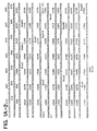
- TABLE 1 shows the various elution buffers that have been tested and the yields of 125I-labelled protein.
- TABLE 2 lists the steps used to isolate the 30 kD or deglycosylated 27 kD gel-bound protein.
- the standard protocol uses diffusion elution using 4M guanidine-HCl containing 0.5% Triton x 100 in Tris-HCl buffer or in Tris-HCl buffer containing 0.1% SDS to achieve greater than 95% elution of the protein from the 27 or 30 kD region of the gel for demonstration of osteogenic activity in vivo as described in later section.
- the overall yield of labelled 30 kD protein from the gel elution protocol is 50 - 60% of the loaded sample. Most of the loss occurs in the electrophoresis step, due to protein aggregation and/or smearing.
- the yield is 0.5 to 1.0 ⁇ g substantially pure osteogenic protein per kg of bone.
- FIGURE 3 summarizes the procedures involved in the preparation of the subunits.
- Approximately 10 ⁇ g of gel eluted 30 kD protein (FIGURE 3) is carboxymethylated and electrophoresed on an SDS-gel.
- the sample contains 125I-label to trace yields and to use as an indicator for slicing the 16 kD and 18 kD regions from the gel.
- FIGURE 15 shows a Coomassie blue stained gel of gel-purified 16 kD and 18 kD proteins.
- a sample containing the 30 kD protein is solubilized using 0.1% SDS, 50 mM Tris-HCl, and is applied to a column of concanavalin A (Con A)-Sepharose equilibrated with the same buffer.
- the bound material is eluted in SDS Tris-HCl buffer containing 0.5 M alpha-methyl mannoside.
- Con A-bound materials when implanted, result in extensive bone formation. Further characterization of the bound materials show a Con A-blottable 30 kD protein. Accordingly, the 30 kD glycosylated protein is responsible for the bone forming activity.
- TSK-3000/2000 gel permeation chromatography in guanidine-HCl alternately is used to achieve separation of the high specific activity fraction obtained from C-18 chromatography (FIGURE 9).
- the results demonstrate that the peak of bone inducing activity elutes in fractions containing substantially pure 30 kD protein by Coomassie blue staining. When this fraction is iodinated and subjected to autoradiography, a strong band at 30 kD accounts for 90% of the iodinated proteins. The fraction induces bone formation in vivo at a dose of 50 to 100 ng per implant.
- the 30 kD protein has been chemically deglycosylated using HF (see below). After analyzing an aliquot of the reaction product by Con A blot to confirm the absence of carbohydrate, the material is assayed for its activity in vivo .
- the bioassay is positive (i.e., the deglycosylated protein produces a bone formation response as determined by histological examination shown in FIGURE 17C), demonstrating that exposure to HF did not destroy the biological function of the protein, and thus that the OP does not require carboyhdrate for biological activity.
- the specific activity of the deglycosylated protein is approximately the same as that of the native glycosylated protein.
- the bone inducing activity is determined biochemically by the specific activity of alkaline phosphatase and calcium content of the day 12 implant.
- An increase in the specific activity of alkaline phosphatase indicates the onset of bone formation.
- Calcium content is proportional to the amount of bone formed in the implant.
- the bone formation is therefore calculated by determining calcium content of the implant on day 12 in rats and expressed as bone forming units, which represent the amount that exhibits half maximal bone inducing activity compared to rat demineralized bone matrix. Bone induction exhibited by intact demineralized rat bone matrix is considered to be the maximal bone-differentiation activity for comparison.
- a standard curve is developed employing known amounts of a standard protein, bovine serum albumin.
- the protein at varying concentration 50-300 ng
- the protein at varying concentration is loaded on a 15% SDS gel, electrophoresed, stained in comassie and destained.
- the gel is scanned at predetermined settings using a gel scanner at 580 nm.
- the area covered by the protein band is calculated and a standard curve against concentrations of protein is constructed.
- a sample with an unknown protein concentration is electrophoresed with BSA as a standard.
- the lane containing the unknown sample is scanned, and the concentration of protein is determined from the area under the curve.
- C-18 highly purified active fraction is subjected to SDS gel and sliced according to molecular weights described in FIGURE 14. Proteins are eluted from the slices in 4 M guanidine-HCl containing 0.5% Triton X-100, desalted, concentrated and assayed for endochondral bone forming activity as determined by calcium content.
- the C-18 highly active fractions and gel eluted substantially pure 30 kD osteogenic protein are implanted in varying concentrations in order to determine the half maximal bone inducing activity.
- FIGURE 14 shows that the bone inducing activity is due to proteins eluted in the 28-34 kD region.
- the recovery of activity after the gel elution step is determined by calcium content.
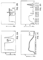
- FIGURES 19A and 19B represent the bone inducing activity for the various concentrations of 30 kD protein before and after gel elution as estimated by calcium content.
- the data suggest that the half maximal activity for 30 kD protein before gel elution is 69 ng per 25 mg implant and is 21 ng per 25 mg implant after elution.
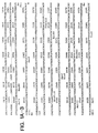
- TABLE 4 describes the yield, total specific activity, and fold purification of osteogenic protein at each step during purification.
- heparin sepharose I fraction Approximately 500 ug of heparin sepharose I fraction, 130-150 ug of the HA ultrogel fraction, 10-12 ug of the gel filtration fraction, 4-5 ug of the heparin sepharose II fraction, 0.4-0.5 ug of the C-18 highly purified fraction, and 20-25 ng of the gel eluted, substantially purified fraction is needed per 25 mg of implant for unequivocal bone formation for half maximal activity.
- 0.8-1.0 ng purified osteogenic protein per mg. of implant is required to exhibit half maximal bone differentiation activity in vivo .
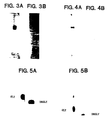
- Electrophoresis of the proteins after the final purification step on non-reducing SDS polyacrylamide gels reveals a diffuse band at about 30 kD as detected by both Coomassie blue staining (FIGURE 3A) and autoradiography.
- FIGURE 3B shows an SDS gel of BOP in the presence of dithiothreitol.
- 30 kD BOP yields two species which are stained with Coomassic blue dye: a 16 kD species and an 18 kD species. Reduction causes loss of biological activity.
- the two reduced BOP species have been analyzed to determine if they are structurally related. Comparison of the amino acid composition and peptide mapping of the two species (as disclosed below) shows little differences, indicating that the native protein may comprise two chains having significant homology.
- the 30 kD protein has been tested for the presence of carbohydrate by Con A blotting after SDS-PAGE and transfer to nitrocellulose paper.
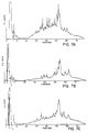
- the results demonstrate that the 30 kD protein has a high affinity for Con A, indicating that the protein is glycosylated (FIGURE 4A).
- the Con A blots provide evidence for a substructure in the 30 kD region of the gel, suggesting heterogeneity due to varying degrees of glycosylation.
- Con A blots show evidence for two major components at 16 kD and 18 kD.
- the 30 kD protein is treated with N-glycanase, a deglycosylating enzyme with a broad specificity.
- Samples of the 125I-labelled 30 kD protein are incubated with the enzyme in the presence of SDS for 24 hours at 37°C.
- the treated samples appear as a prominent species at about 27 kD (FIGURE 5A).
- the 27 kD species is reduced to species having a molecular weight of about 14 kD - 16 kD (FIGURE 5B).
- HF hydrogen fluoride
- the samples are dissolved in 100 ⁇ l of 50% acetonitrile/0.1% TFA and aliquoted for SDS gel analysis, Con A binding, and biological assay. Aliquots are dried and dissolved in either SDS gel sample buffer in preparation for SDS gel analysis and Con A blotting or 4 M guanidine-HCl, 50 mM Tris-HCl, pH 7.0 for biological assay.
- Cleavage reactions with CNBr are analyzed using Con A binding for detection of fragments associated with carbohydrate.
- Cleavage reactions are conducted using trifluoroacetic acid (TFA) in the presence and absence of CNBr. Reactions are conducted at 37°C for 18 hours, and the samples are vacuum dried. The samples are washed with water, dissolved in SDS gel sample buffer with reducing agent, boiled and applied to an SDS gel. After electrophoresis, the protein is transferred to Immobilon membrane and visualized by Con A binding.
- CNBr cleaves the majority of 16 kD and 18 kD species to one product, a species about 14 kD.
- reactions using 10% TFA a 14 kD species is observed both with and without CNBr.
- proteolytic enzymes are used in these experiments to examine the digestion products of the 30 kD protein: 1) V-8 protease; 2) Endo Lys C protease; 3) pepsin; and 4) trypsin. Except for pepsin, the digestion buffer for the enzymes is 0.1 M ammonium bicarbonate, pH 8.3. The pepsin reactions are done in 0.1% TFA. The digestion volume is 100 ⁇ l and the ratio of enzyme to substrate is 1:10. 125I-labelled 30 kD osteogenic protein is added for detection. After incubation at 37°C for 16 hr., digestion mixtures are dried down and taken up in gel sample buffer containing dithiothreitol for SDS-PAGE.
- FIGURE 6 shows an autoradiograph of an SDS gel of the digestion products. The results show that under these conditions, only trypsin digests the reduced 16 kD/18 kD species completely and yields a major species at around 12 kD. Pepsin digestion yields better defined, lower molecular weight species. However, the 16 kD/18 kD fragments were not digested completely. The V-8 digest shows limited digestion with one dominant species at 16 kD.
- the protein is cleaved with trypsin or Endoproteinase Asp-N (EndoAsp-N).
- the tryptic digest of reduced and carboxymethylated 30 kD protein (approximately 10 ⁇ g) is fractionated by reverse-phase HPLC using a C-8 narrowbore column (13 cm x 2.1 mm ID) with a TFA/acetonitrile gradient and a flow rate of 150 ⁇ l/min.
- the gradient employs (A) 0.06% TFA in water and (B) 0.04% TFA in water and acetonitrile (1:4; v:v). The procedure was 10% B for five min., followed by a linear gradient for 70 min.
- FIGUREs 7B and 7C The HPLC profiles of the similarly digested 16 kD and 18 kD subunits are shown in FIGUREs 7B and 7C, respectively. These peptide maps are similar suggesting that the subunits are identical or are closely related.
- the 16 kD and 18 kD subunits are digested with EndoAsp-N proteinase.
- the protein is treated with 0.5 ⁇ g EndoAsp-N in 50 mM sodium phosphate buffer, pH 7.8 at 36°C for 20 hr.
- the conditions for fractionation are the same as those described previously for the 30 kD, 16 kD, and 18 KD digests.
- the profiles obtained are shown in FIGUREs 16A and 16B.
- Samples of oxidized (30 kD) and reduced (16 kD and 18 kD) BOP are electrophoresed on a gel and transferred to Immobilon for hydrolysis and amino acid analysis using conventional, commercially available reagents to derivatize samples and HPLC using the PicO Tag (Millipore) system.
- the composition data generated by amino acid analyses of 30 kD BOP is reproducible, with some variation in the number of residues for a few amino acids, especially cysteine and isoleucine.
- Human bone is obtained from the Bone Bank, (Massachusetts General Hospital, Boston, MA), and is milled, defatted, demarrowed and demineralized by the procedure disclosed above. 320 g of mineralized bone matrix yields 70 - 80 g of demineralized bone matrix. Dissociative extraction and ethanol precipitation of the matrix gives 12.5 g of guanidine-HCl extract.
- ethanol precipitate 0.5 g
- 4 M guanidine-HCl Approximately 70-80 g of ethanol precipitate per run is used.
- In vivo bone inducing activity is localized in the fractions containing proteins in the 30 kD range. They are pooled and equilibrated in 6 M urea, 0.5 M NaCl buffer, and applied directly onto a HAP column; the bound protein is eluted stepwise by using the same buffer containing 100 mM and 500 mM phosphate (FIGURE 10B).
- Bioassay of HAP bound and unbound fractions demonstrates that only the fraction eluted by 100 mM phosphate has bone inducing activity in vivo .
- the biologically active fraction obtained from HAP chromatography is subjected to heparin-Sepharose affinity chromatography in buffer containing low salt; the bound proteins are eluted by 0.5 M NaCl (FIGURE 10C).
- Assaying the heparin-Sepharose fractions shows that the bound fraction eluted by 0.5 M NaCl have bone-inducing activity.
- the active fraction is then subjected to C-18 reverse phase chromatography. (FIGURE 10D).
- the active fraction can then be subjected to SDS-PAGE as noted above to yield a band at about 30 kD comprising substantially pure human osteogenic protein.
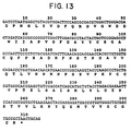
- a synthetic consensus gene shown in FIGURE 13 was designed as a hybridization probe based on amino acid predictions from homology with the TGF-beta gene family and using human codon bias as found in human TGF-beta.
- the designed concensus sequence was then constructed using known techniques involving assembly of oligonucleotides manufactured in a DNA synthesizer.
- Tryptic peptides derived from BOP and sequenced by Edman degradation provided amino acid sequences that showed strong homology with the Drosophila DPP protein sequence (as inferred from the gene), the Xenopus VGl protein, and somewhat less homology to inhibin and TGF-beta, as demonstrated below in TABLE 6.
- the amino acid sequence of an osteogenic protein (from which the nucleic acid sequence can be determined), the following points were considered: (1) the amino acid sequence determined by Edman degradation of osteogenic protein tryptic fragments is ranked highest as long as it has a strong signal and shows homology or conservative changes when aligned with the other members of the gene family; (2) where the sequence matches for all four proteins, it is used in the synthetic gene sequence; (3) matching amino acids in DPP and Vgl are used; (4) If Vgl or DPP diverged but either one were matched by inhibin or by TGF-beta, this matched amino acid is chosen; (5) where all sequences diverged, the DPP sequence is initially chosen, with a later plan of creating the Vgl sequence by mutagenesis kept as a possibility.
- the consensus sequence is designed to preserve the disulfide crosslinking and the apparent structural homology.
- probes may be constructed using conventional techniques comprising a group of sequences of nucleotides which encode any portion of the amino acid sequence of the osteogenic protein produced in accordance with the foregoing isolation procedure. Use of such pools of probes also will enable isolation of a DNA encoding the intact protein.
- a human genomic library (Maniatis-library) carried in lambda phage (Charon 4A) was screened using the COP0 consensus gene as probe. The initial screening was of 500,000 plaques (10 plates of 50,000 each). Areas giving hybridization signal were punched out from the plates, phage particles were eluted and plated again at a density of 2000-3000 plaques per plate. A second hybridization yielded plaques which were plated once more, this time at a density of ca 100 plaques per plate allowing isolation of pure clones.
- the probe is a 300 base pair BamHI-PstI fragment restricted from an amplification plasmid which was labeled using alpha 32 dCTP according to the random priming method of Feinberg and Vogelstein (1984) Anal. Biochem. 137 : 266-267. Prehybridization was done for 1 hr in 5x SSPE, 10x Denhardt's mix, 0.5% SDS at 50°C. Hybridization was overnight in the same solution as above plus probe. The washing of nitrocellulose membranes was done, once cold for 5 min. in 1x SSPE with 0.1% SDS and twice at 50°C for 2 x 30 min. in the same solution. Using this procedure, twenty-four positive clones were found. Two contained a gene never before reported designated OP1, osteogenic protein-1 described below. Two others yielded the genes corresponding to BMP-2b, one yielded BMP-3 (see PCT US 87/01537).
- cysteine patterns were used and then the adjacent amino acids were compared. Consensus splice signals were found where amino acid homologies ended, designating exon intron boundaries. Three exons were combined to obtain a functional TGF-beta-like domain containing seven cysteines. Two introns were deleted by looping out via primers bridging the exons using the single stranded mutagenesis method of Kunkel. Also, upstream of the first cysteine, an EcoRI site and an asp-pro junction for acid cleavage were introduced, and at the 3' end a PstI site was added by the same technique. Further sequence information (penultimate exon) was obtained by sequencing the entire insert.
- the sequencing was done by generating a set of unidirectionally deleted clones (Ozkaynak, E., and Putney, S. (1987) Biotechniques, 5 :770-773).
- the obtained sequence covers about 80% of the TGF-beta-like region of OP1 and is set forth in FIGURE 1A.
- the complete sequence of the TGF-beta like region was obtained by first subcloning all EcoRI generated fragments of lambda clone #13 DNA and sequencing a 4 kb fragment that includes the first portion of the TGF-beta like region (third exon counting from end) as well as sequences characterized earlier.
- the gene on an EcoRI to PstI fragment was inserted into an E.
- the OP1 gene encodes amino acids corresponding substantially to a peptide found in sequences of naturally sourced material.
- the amino acid sequence of what is believed to be its active region is set forth below: A longer active sequence is:
- the amino acid sequence of what is believed to be the active regions encoded by the other three native genes retrieved using the consensus probe are:
- RNA encoding the intact protein is shown by the following.
- Cells known to express the protein e.g., osteoblasts or osteosarcoma
- An oligo-dT column can be used to isolate mRNA.
- This mRNA can be size fractionated by, for example, gel electrophoresis.
- the fraction which includes the mRNA of interest may be determined by inducing transient expression in a suitable host cell and testing for the presence of osteogenic protein using, for example, antibody raised against peptides derived from the tryptic fragments of osteogenic protein in an immunoassay.
- the mRNA fraction is then reverse transcribed to single stranded cDNA using reverse transcriptase; a second complementary DNA strand can then be synthesized using the cDNA as a template.
- the double-standard DNA is then ligated into vectors which are used to transfect bacteria to produce a cDNA library.
- radiolabelled consensus sequence, portions thereof, and/or synthetic deoxy oligonucleotides complementary to codons for the known amino acid sequences in the osteogenic protein may be used to identify which of the DNAs in the cDNA library encode the full length osteogenic protein by standard DNA-DNA hybridization techniques.
- the cDNA may then be integrated in an expression vector and transfected into an appropriate host cell for protein expression.
- the host may be a prokaryotic or eucaryotic cell since the former's inability to glycosylate osteogenic protein will not effect the protein's enzymatic activity.
- Useful host cells include Saccharomyces , E. coli , and various mammalian cell cultures.
- the vector may additionally encode various signal sequences for protein secretion and/or may encode osteogenic protein as a fusion protein. After being translated, protein may be purified from the cells or recovered from the culture medium.
- Natural gene sequences and cDNAs retrieved as described above may be used for expression.
- the genes above may also be produced by assembly of chemically synthesized oligonucleotides. 15-100mer oligonucleotides may be synthesized on a Biosearch DNA Model 8600 Synthesizer, and purified by polyacrylamide gel electrophoresis (PAGE) in Tris-Borate-EDTA buffer (TBE). The DNA is then electroeluted from the gel. Overlapping oligomers may be phosphorylated by T4 polynucleotide kinase and ligated into larger blocks which may also be purifed by PAGE.
- the genes can be expressed in appropriate prokaryotic hosts such as various strains of E. coli , and also in bacillus, yeasts, and various animal cells such as CHO, myeloma, etc. Generally, expression may be achieved using many cell types and expression systems well known to those skilled in the art. For example, if the gene is to be expressed in E. coli , an expression vector based on pBR322 and containing a synthetic trp promoter operator and the modified trp LE leader may be used. The vector can be opened at the EcoRI and PSTI restriction sites, and, for example, an OP gene fragment can be inserted between these two sites. The OP protein is joined to the leader protein via a hinge region having the sequence Asp-Pro. This hinge permits chemical cleavage of the fusion protein with dilute acid at the Asp-Pro site.
- E. coli cells containing the fusion proteins are lysed.
- the fusion proteins are purified by differential solubilization. Cleavage is conducted with dilute acid, and the resulting cleavage products are passed through a Sephacryl-200HR or SP Trisacyl column to separate the cleaved proteins. The reduced OP fractions are then subjected to HPLC on a semi-prep C-18 column.
- Conditions for refolding of OP were at pH 8.0 using 50 mM Tris-HCl and 6M Gu-HCl. Samples were refolded for 18 hours at 4°C.
- Refolding may not be required if the proteins are expressed in animal cells.
- the carrier described in the bioassay section, infra may be replaced by either a biodegradable-synthetic or synthetic-inorganic matrix (e.g., HAP, collagen, tricalcium phosphate, or polylactic acid, polyglycolic acid and various copolymers thereof). Also xenogeneic bone may be used if pretreated as described below.
- a biodegradable-synthetic or synthetic-inorganic matrix e.g., HAP, collagen, tricalcium phosphate, or polylactic acid, polyglycolic acid and various copolymers thereof.
- xenogeneic bone may be used if pretreated as described below.
- the sequential cellular reactions at the interface of the bone matrix/OP implants are complex.
- the multistep cascade includes: binding of fibrin and fibronectin to implanted matrix, chemotaxis of cells, proliferation of fibroblasts, differentiation into chondroblasts, cartilage formation, vascular invasion, bone formation, remodeling, and bone marrow differentiation.
- a successful carrier for osteogenic protein must perform several important functions. It must bind osteogenic protein and act as a slow release delivery system, accommodate each step of the cellular response during bone development, and protect the osteogenic protein from nonspecific proteolysis.
- selected materials must be biocompatible in vivo and biodegradable; the carrier must act as a temporary scaffold until replaced completely by new bone. Biocompatibility requires that the matrix not induce significant inflammation when implanted and not be rejected by the host animal. Biodegradability requires that the matrix be slowly absorbed by the body of the host during development of new bone or cartilage.
- Polylactic acid (PLA), polyglycolic acid (PGA), and various combinations have different dissolution rates in vivo . In bones, the dissolution rates can vary according to whether the implant is placed in cortical or trabecular bone.
- Matrix geometry, particle size, the presence of surface charge, and porosity or the presence of interstices among the particles of a size sufficient to permit cell infiltration, are all important to successful matrix performance. It is preferred to shape the matrix to the desired form of the new bone and to have dimensions which span non-union defects. Rat studies show that the new bone is formed essentially having the dimensions of the device implanted.
- the matrix may comprise a shape-retaining solid made of loosely adhered particulate material, e.g., with collagen. It may also comprise a molded, porous solid, or simply an aggregation of close-packed particles held in place by surrounding tissue. Masticated muscle or other tissue may also be used. Large allogeneic bone implants can act as a carrier for the matrix if their marrow cavities are cleaned and packed with particles and the dispersed osteogenic protein.
- Demineralized bone matrix is prepared from the dehydrated diaphyseal shafts of rat femur and tibia as described herein to produce a bone particle size which pass through a 420 ⁇ sieve.
- the bone particles are subjected to dissociative extraction with 4 M guanidine-HCl.
- Such treatment results in a complete loss of the inherent ability of the bone matrix to induce endochondral bone differentiation.
- the remaining insoluble material is used to fabricate the matrix.
- the material is mostly collagenous in nature, and upon implantation, does not induce cartilage and bone. All new preparations are tested for mineral content and false positives before use.
- the total loss of biological activity of bone matrix is restored when an active osteoinductive protein fraction or a pure protein is reconstituted with the biologically inactive insoluble collagenous matrix.
- the osteoinductive protein can be obtained from any vertebrate, e.g., bovine, porcine, monkey, or human, or produced using recombinant DNA techniques.
- osteogenic protein When osteogenic protein is reconstituted with collagenous bone matrix from other species and implanted in rat, no bone is formed. This suggests that while the osteogenic protein is xenogenic (not species specific), the matrix is species specific and cannot be implanted cross species perhaps due to intrinsic immunogenic or inhibitory components. Thus, heretofore, for bone-based matrices, in order for the osteogenic protein to exhibit its full bone inducing activity, a species specific collagenous bone matrix was required.
- Type I collagen The major component of all bone matrices is Type I collagen.
- extracted bone includes non-collagenous proteins which may account for 5% of its mass.
- non-collagenous components of bone matrix are glycoproteins. Although the biological significance of the glycoproteins in bone formation is not known, they may present themselves as potent antigens by virtue of their carbohydrate content and may constitute immunogenic and/or inhibitory components that are present in xenogenic matrix.
- a collagenous bone matrix may be used as a carrier to effect bone inducing activity in xenogenic implants, if one first removes the immonogenic and inhibitory components from the matrix.
- the matrix is deglycosglated chemically using, for example, hydrogen fluoride to achieve this purpose.
- Bovine bone residue prepared as described above is sieved, and particles of the 74-420 ⁇ M are collected.
- the sample is dried in vacuo over P205, transferred to the reaction vessel and anhydrous hydrogen fluoride (HF) (10-20 ml/g of matrix) is then distilled onto the sample at -70°C.
- HF hydrous hydrogen fluoride
- the vessel is allowed to warm to 0°C and the reaction mixture is stirred at this temperature for 120 min. After evaporation of the HF in vacuo , the residue is dried thoroughly in vacuo over KOH pellets to remove any remaining traces of acid.
- Extent of deglycosylation can be determined from carbohydrate analysis of matrix samples taken before and after treatment with HF, after washing the samples appropriately to remove non-covalently bound carbohydrates.
- the deglycosylated bone matrix is next treated as set forth below:
- Fabrication of osteogenic devices using any of the matrices set forth above with any of the osteogenic proteins described above may be performed as follows.
- osteogenic protein in an acetonitrile trifluroacetic acid (ACN/TFA) solution was added to the carrier. Samples were vigorously vortexed many times and then lyophilized.
- the protein is mixed with the matrix, vortexed many times, and then lyophilized.
- the lyophilized material may be used "as is" for implants.
- Substantially pure BOP, BOP-rich extracts comprising protein having the properties set forth above, and several of the recombinant proteins have been incorporated in matrices to produce osteogenic devices, and assayed in rat for endochondral bone.
- Studies in rats show the osteogenic effect to be dependent on the dose of osteogenic protein dispersed in the osteogenic device. No activity is observed if the matrix is implanted alone.
- the following sets forth guidelines for how the osteogenic devices disclosed herein might be assayed for determining active fractions of osteogenic protein when employing the isolation procedure of the invention, and evaluating protein constructs and matrices for biological activity.
- This assay consists of implanting the test samples in subcutaneous sites in allogeneic recipient rats under ether anesthesia. Male Long-Evans rats, aged 28-32 days, were used. A vertical incision (1 cm) is made under sterile conditions in the skin over the thoraic region, and a pocket is prepared by blunt dissection. Approximately 25 mg of the test sample is implanted deep into the pocket and the incision is closed with a metallic skin clip. The day of implantation is designated as day of the experiment. Implants were removed on day 12. The heterotropic site allows for the study of bone induction without the possible ambiguities resulting from the use of orthotopic sites.
- the implant model in rats exhibits a controlled progression through the stages of matrix induced endochondral bone development including: (1) transient infiltration by polymorphonuclear leukocytes on day one; (2) mesenchymal cell migration and proliferation on days two and three; (3) chondrocyte appearance on days five and six; (4) cartilage matrix formation on day seven; (5) cartiliage calcification on day eight; (6) vascular invasion, appearance of osteoblasts, and formation of new bone on days nine and ten; (7) appearance of osteoblastic and bone remodeling and dissolution of the implanted matrix on days twelve to eighteen; and (8) hematopoietic bone marrow differentiation in the ossicle on day twenty-one.
- the results show that the shape of the new bone conforms to the shape of the implanted matrix.
- Implants are fixed in Bouins Solution, embedded in parafilm, cut into 6-8 mm sections. Staining with toluidine blue or hemotoxylin/eosin demonstrates clearly the ultimate development of endochondrial bone. Twelve day implants are usually sufficient to determine whether the implants show bone inducing activity.
- Alkaline phosphatase activity may be used as a marker for osteogenesis.
- the enzyme activity may be determined spectrophotometrically after homogenization of the implant. The activity peaks at 9-10 days in vivo and thereafter slowly declines. Implants showing no bone development by histology should have little or no alkaline phosphatase activity under these assay conditions.
- the assay is useful for quantitation and obtaining an estimate of bone formation very quickly after the implants are removed from the rat. Alternatively the amount of bone formation can be determined by measuring the calcium content of the implant.
- Implants containing osteogenic protein at several levels of purity have been tested to determine the most effective dose/purity level, in order to seek a formulation which could be produced on a commercial scale.
- the results are measured by specific acivity of alkaline phosphatase, calcium content, and histological examination.
- the specific activity of alkaline phosphatase is elevated during onset of bone formation and then declines.
- calcium content is directly proportional to the total amount of bone that is formed.
- the osteogenic activity due to osteogenic protein is represented by "bone forming units".
- one bone forming unit represents the amount of protein that is needed for half maximal bone forming activity as compared to rat demineralized bone matrix as control and determined by calcium content of the implant on day 12.
- Dose curves are constructed for bone inducing activity in vivo at each step of the purification scheme by assaying various concentrations of protein.
- FIGURE 11 shows representative dose curves in rats as determined by alkaline phosphatase. Similar results are obtained when represented as bone forming units. Approximately 10-12 ⁇ g of the TSK-fraction, 3-4 ⁇ g of heparin-Sepharose-II fraction, 0.4-0.5 ⁇ g of the C-18 column purified fraction, and 20-25 ng of gel eluted highly purified 30 kD protein is needed for unequivocal bone formation (half maximum activity). 20-25 ng of the substantially pure protein per 25 mg of implant is normally sufficient to produce endochondral bone. Thus, 1-2 ng osteogenic protein per mg of implant is a reasonable dosage, although higher dosages may be used. (See section IB5 on specific activity of osteogenic protein.)
- OP1 expressed as set forth above when assayed for activity histologically, induced cartilage and bone formation as evidenced by the presence of numerous chondrocytes in many areas of the implant and by the presence of osteoblasts surrounding vascular endothelium forming new matrix.
- Deglycosylated xenogenic collagenous bone matrix (example: bovine) has been used instead of allogenic collagenous matrix to prepare osteogenic devices (see previous section) and bioassayed in rat for bone inducing activity in vivo .
- the results demonstrate that xenogenic collagenous bone matrix after chemical deglycosylation induces successful endochondral bone formation (FIGURE 19).
- specific activity of alkaline phosphotase it is evident that the deglycosylated xenogenic matrix induced bone whereas untreated bovine matrix did not.
- the deglycosylated bovine matrix not only has induced bone in a way comparable to the rat residue matrix but also has advanced the developmental stages that are involved in endochondral bone differentiation.
- the HF treated bovine matrix contains extensively remodeled bone. Ossicles are formed that are already filled with bone marrow elements by 12 days. This profound action as elicited by deglycosylated bovine matrix in supporting bone induction is reproducible and is dose dependent with varying concentration of osteogenic protein.
- BOP bovine bone
- BOP-rich osteogenic fractions having the properties set forth above, and several recombinant proteins have been incorporated in matrices to produce osteogenic devices.
- the efficacy of bone-inducing potential of these devices was tested in cat and rabbit models, and found to be potent inducers of osteogenesis, ultimately resulting in formation of mineralized bone.
- the purpose of this study is to establish a large animal efficacy model for the testing of the osteogenic devices of the invention, and to characterize repair of massive bone defects and simulated fracture non-union encountered frequently in the practice of orthopedic surgery.
- the study is designed to evaluate whether implants of osteogenic protein with a carrier can enhance the regeneration of bone following injury and major reconstructive surgery by use of this large mammal model.
- the first step in this study design consists of the surgical preparation of a femoral osteotomy defect which, without further intervention, would consistently progress to non-union of the simulated fracture defect.
- the effects of implants of osteogenic devices into the created bone defects were evaluated by the following study protocol.
- group III osteogenic protein-treated animals are implanted with exactly the same material as the group I animals, but with the singular addition of osteogenic protein.
- All animals are allowed to ambulate ad libitum within their cages post-operatively. All cats are injected with tetracycline (25 mg/kg SQ each week for four weeks) for bone labelling. All but four group III animals are sacrificed four months after femoral osteotomy.
- the group III femors are united with average 86% bone defect regeneration.
- the group I Gu-HCl-DMB negative-control implants exhibit no bone growth at four weeks, less than 10% at eight and 12 weeks, and 16% ( ⁇ 10%) at 16 weeks with one of the five exhibiting a small amount of bridging bone.
- the group II DMB positive-control implants exhibited 18% ( ⁇ 3%) repair at four weeks, 35% at eight weeks, 50% ( ⁇ 10%) at twelve weeks and 70% ( ⁇ 12%) by 16 weeks, a statistical difference of p ⁇ 0.01 compared to osteogenic protein at every month. One of the three (33%) is united at 16 weeks.
- Excised test and normal femurs are immediately studied by bone densitometry, wrapped in two layers of saline-soaked towels, placed in two sealed plastic bags, and stored at -20° C until further study. Bone repair strength, load to failure, and work to failure are tested by loading to failure on a specially designed steel 4-point bending jig attached to an Instron testing machine to quantitate bone strength, stiffness, energy absorbed and deformation to failure.
- the study of test femurs and normal femurs yield the bone strength (load) in pounds and work to failure in joules. Normal femurs exhibit a strength of 96 ( ⁇ 12) pounds.
- osteogenic protein-implanted femurs exhibited 35 ( ⁇ 4) pounds, but when corrected for surface area at the site of fracture (due to the "hourglass" shape of the bone defect repair) this correlated closely with normal bone strength. Only one demineralized bone specimen was available for testing with a strength of 25 pounds, but, again, the strength correlated closely with normal bone when corrected for fracture surface area.
- the bones are immediately sliced into two longitudinal sections at the defect site, weighed, and the volume measured. One-half is fixed for standard calcified bone histomorphometrics with fluorescent stain incorporation evaluation, and one-half is fixed for decalcified hemotoxylin/eosin stain histology preparation.
- the final autopsy reports reveal no unusual or pathologic findings noted at necropsy of any of the animals studied. Portion of all major organs are preserved for further study. A histopathological evaluation is performed on samples of the following organs: heart, lung, liver, both kidneys, spleen, both adrenals, lymph nodes, left and right quadriceps muscles at mid-femur (adjacent to defect site in experimental femur). No unusual or pathological lesions are seen in any of the tissues. Mild lesions seen in the quadriceps muscles are compatible with healing responses to the surgical manipulation at the defect site. Pulmonary edema is attributable to the euthanasia procedure. There is no evidence of any general systemic effects or any effects on the specific organs examined.
- the 1 cm and 2 cm femoral defect cat studies demonstrate that devices comprising a matrix containing disposed osteogenic protein can: (1) repair a weight-bearing bone defect in a large animal; (2) consistently induces bone formation shortly following (less than two weeks) implantation; and (3) induce bone by endochondral ossification, with a strength equal to normal bone, on a volume for volume basis. Furthermore, all animals remained healthy during the study and showed no evidence of clinical or histological laboratory reaction to the implanted device. In this bone defect model, there was little or no healing at control bone implant sites. The results provide evidence for the successful use of osteogenic devices to repair large, non-union bone defects.
- the purpose of this study is to establish a model in which there is minimal or no bone growth in the control animals, so that when bone induction is tested, only a strongly inductive substance will yield a positive result. Defects of 1.5 cm are created in the ulnae of rabbits with implantation of osteogenic devices or no implant.
- Radiomorphometric analysis reveal 90% osteogenic protein-implant bone repair and 18% no-implant bone repair at sacrifice at eight weeks. At autopsy, the osteogenic protein bone appears normal, while "no implant" bone sites have only a soft fibrous tissue with no evidence of cartilage or bone repair in the defect site.
- the marrow cavity of the 1.5 cm ulnar defect is packed with activated osteogenic protein rabbit bone powder and the bones are allografted in an intercalary fashion.
- the two control ulnae are not healed by eight weeks and reveal the classic "ivory" appearance.
- the osteogenic protein-treated implants "disappear” radiographically by four weeks with the start of remineralization by six to eight weeks. These allografts heal at each end with mild proliferative bone formation by eight weeks.
- This type of device serves to accelerate allograph repair.
- osteogenic protein-implanted rabbits exhibited proliferative bone growth in a fashion highly different from the control groups; (4) initial studies show that the bones exhibit 50% of normal bone strength (100% of normal correlated vol:vol) at only eight weeks after creation of the surgical defect; and (5) osteogenic protein-allograft studies reveal a marked effect upon both the allograft and bone healing.
Abstract
Description
- This invention relates to osteogenic devices, to genes encoding proteins which can induce osteogenesis in mammals and methods for their production using recombinant DNA techniques, to a method of reproducibly purifying osteogenic protein from mammalian bone, and to bone and cartilage repair procedures using the osteogenic device.
- Mammalian bone tissue is known to contain one or more proteinaceous materials, presumably active during growth and natural bone healing, which can induce a developmental cascade of cellular events resulting in endochondral bone formation. This active factor (or factors) has variously been referred to in the literature as bone morphogenetic or morphogenic protein, bone inductive protein, osteogenic protein, osteogenin, or osteoinductive protein.
- The developmental cascade of bone differentiation consists of recruitment of mesenchymal cells, proliferation of progenitor cells, calcification of cartilage, vascular invasion, bone formation, remodeling, and finally marrow differentiation (Reddi (1981) Collagen Rel. Res. 1:209-226).
- Though the precise mechanisms underlying these phenotypic transformations are unclear, it has been shown that the natural endochondral bone differentiation activity of bone matrix can be dissociatively extracted and reconstituted with inactive residual collagenous matrix to restore full bone induction activity (Sampath and Reddi, (1981) Proc. Natl. Acad. Sci. USA 78:7599-7603). This provides an experimental method for assaying protein extracts for their ability to induce endochondral bone in vivo.
- This putative bone inductive protein has been shown to have a molecular mass of less than 50 kilodaltons (kD). Several species of mammals produce closely related protein as demonstrated by cross species implant experiments (Sampath and Reddi (1983) Proc. Natl. Acad. Sci. USA 80:6591-6595).
- The potential utility of these proteins has been widely recognized. It is contemplated that the availability of the protein would revolutionize orthopedic medicine, certain types of plastic surgery, and various periodontal and craniofacial reconstructive procedures.
- The observed properties of these protein fractions have induced an intense research effort in various laboratories directed to isolating and identifying the pure factor or factors responsible for osteogenic activity. The current state of the art of purification of osteogenic protein from mammalian bone is disclosed by Sampath et al. (Proc. Natl. Acad. Sci. USA (1987) 80). Urist et al. (Proc. Soc. Exp. Biol. Med. (1984) 173:194-199) disclose a human osteogenic protein fraction which was extracted from demineralized cortical bone by means of a calcium chloride-urea inorganic-organic solvent mixture, and retrieved by differential precipitation in guanidine-hydrochloride and preparative gel electrophoresis. The authors report that the protein fraction has an amino acid composition of an acidic polypeptide and a molecular weight in a range of 17-18 kD.
- Urist et al. (Proc. Natl. Acad. Sci. USA (1984) 81:371-375) disclose a bovine bone morphogenetic protein extract having the properties of an acidic polypeptide and a molecular weight of approximately 18 kD. The authors reported that the protein was present in a fraction separated by hydroxyapatite chromatography, and that it induced bone formation in mouse hindquarter muscle and bone regeneration in trephine defects in rat and dog skulls. Their method of obtaining the extract from bone results in ill-defined and impure preparations.
- European Patent Application Serial No. 148,155, published October 7, 1985, purports to disclose osteogenic proteins derived from bovine, porcine, and human origin. One of the proteins, designated by the inventors as a P3 protein having a molecular weight of 22-24 kD, is said to have been purified to an essentially homogeneous state. This material is reported to induce bone formation when implanted into animals.
- International Application No. PCT/087/01537, published January 14, 1988, discloses an impure fraction from bovine bone which has bone induction qualities. The named applicants also disclose putative bone inductive factors produced by recombinant DNA techniques. Four DNA sequences were retrieved from human or bovine genomic or cDNA libraries and apparently expressed in recombinant host cells. While the applicants stated that the expressed proteins may be bone morphogenic proteins, bone induction was not demonstrated. See also Urist et al., EP 0,212,474 entitled Bone Morphogenic Agents.
- Wang et al. (Proc. Nat. Acad. Sci. USA (1988) 85: 9484-9488) discloses the purification of a bovine bone morphogenetic protein from guanidine extracts of demineralized bone having cartilage and bone formation activity as a basic protein corresponding to a molecular weight of 30 kD determined from gel elution. Purification of the protein yielded proteins of 30, 18 and 16 kD which, upon separation, were inactive. In view of this result, the authors acknowledged that the exact identity of the active material had not been determined.
- Wozney et al. (Science (1988) 242: 1528-1534) discloses the isolation of full-length cDNA's encoding the human equivalents of three polypeptides originally purified from bovine bone. The authors report that each of the three recombinantly expressed human proteins are independently or in combination capable of inducing cartilage formation. No evidence of bone formation is reported.
- It is an object of this invention to provide osteogenic devices comprising matrices containing dispersed osteogenic protein capable of bone induction in allogenic and xenogenic implants. Another object is to provide a reproducible method of isolating osteogenic protein from mammalian bone tissue. Another object is to characterize the protein responsible for osteogenesis. Another object is to provide natural and recombinant osteogenic proteins capable of inducing endochondral bone formation in mammals, including humans. Yet another object is to provide genes encoding osteogenic proteins and methods for their production using recombinant DNA techniques. Another object is to provide methods for inducing cartilage formation.
- These and other objects and features of the invention will be apparent from the description, drawings, and claims which follow.
- This invention involves osteogenic devices which, when implanted in a mammalian body, can induce at the locus of the implant the full developmental cascade of endochondral bone formation and bone marrow differentiation. Suitably modified as disclosed herein, the devices also may be used to induce cartilage formation. The devices comprise a carrier material, referred to herein as a matrix, having the characteristics disclosed below, containing dispersed osteogenic protein either in its native form as purified from natural sources or produced using recombinant DNA techniques.
- Key to these developments was the successful development of a protocol which results in retrieval of active, substantially pure osteogenic protein from mammalian bone, and subsequent elucidation of amino acid sequence and structure data of native osteogenic protein. The protein has a half-maximum bone forming activity of about 0.8 to 1.0 ng per mg of implant. The protein is believed to be a dimer. It appears not to be active when reduced. Various combinations of species of the proteins may exist as heterodimers or homodimers.
- The invention provides native forms of osteogenic protein, extracted from bone or produced using recombinant DNA techniques. The substantially pure osteogenic protein may include forms having varying glycosylation patterns, varying N-termini, a family of related proteins having regions of amino acid sequence homology, and active truncated or mutated forms of native protein, no matter how derived. The naturally sourced osteogenic protein in its native form is glycosylated and has an apparent molecular weight of about 30 kD as determined by SDS-PAGE. When reduced, the 30 kD protein gives rise to two glycosylated polypeptide chains having apparent molecular weights of about 16 kD and 18 kD. In the reduced state, the 30 kD protein has no detectable osteogenic activity. The deglycosylated protein, which has osteogenic activity, has an apparent molecular weight of about 27 kD. When reduced, the 27 kD protein gives rise to the two deglycosylated polypeptides have molecular weights of about 14 kD to 16 kD.
- Analysis of intact molecules and digestion fragments indicate that the native 30 kD osteogenic protein contains the following amino acid sequences (question marks indicate undetermined residues):
- (1) S-F-D-A-Y-Y-C-S-G-A-C-Q-F-P-M-P-K;
- (2) S-L-K-P-S-N-Y-A-T-I-Q-S-I-V;
- (3) A-C-C-V-P-T-E-L-S-A-I-S-M-L-Y-L-D-E-N-E-K;
- (4) M-S-S-L-S-I-L-F-F-D-E-N-K;
- (5) S-Q-E-L-Y-V-D-F-Q-R;
- (6) F-L-H-C-Q-F-S-E-R-N-S;
- (7) T-V-G-Q-L-N-E-Q-S-S-E-P-N-I-Y;
- (8) L-Y-D-P-M-V-V;
- (9) V-G-V-V-P-G-I-P-E-P-C-C-V-P-E;
- (10) V-D-F-A-D-I-G;
- (11) V-P-K-P-C-C-A-P-T;
- (12) I-N-I-A-N-Y-L;
- (13) D-N-H-V-L-T-M-F-P-I-A-I-N;
- (14) D-E-Q-T-L-K-K-A-R-R-K-Q-W-I-?-P;
- (15) D-I-G-?-S-E-W-I-I-?-P;
- (16) S-I-V-R-A-V-G-V-P-G-I-P-E-P-?-?-V;
- (17) D-?-I-V-A-P-P-Q-Y-H-A-F-Y;
- (18) D-E-N-K-N-V-V-L-K-V-Y-P-N-M-T-V-E;
- (19) S-Q-T-L-Q-F-D-E-Q-T-L-K-?-A-R-?-K-Q;
- (20) D-E-Q-T-L-K-K-A-R-R-K-Q-W-I-E-P-R-N-?-A-R-R-Y-L;
- (21) A-R-R-K-Q-W-I-E-P-R-N-?-A-?-R-Y-?-?-V-D; and
- (22) R-?-Q-W-I-E-P-?-N-?-A-?-?-Y-L-K-V-D-?-A-?-?-G.
- The availability of the protein in substantially pure form, and knowledge of its amino acid sequence and other structural features, enable the identification, cloning, and expression of native genes which encode osteogenic proteins. When properly modified after translation, incorporated in a suitable matrix, and implanted as disclosed herein, these proteins are operative to induce formation of cartilage and endochondral bone.
- Consensus DNA sequences designed as disclosed herein based on partial sequence data and observed homologies with regulatory proteins disclosed in the literature are useful as probes for extracting genes encoding osteogenic protein from genomic and cDNA libraries. One of the consensus sequences has been used to isolate a heretofore unidentified genomic DNA sequence, portions of which when ligated encode a protein having a region capable of inducing endochondral bone formation. This protein, designated OP1, has an active region having the sequence set forth below.

A longer active sequence is:
Fig. 1A discloses the genomic DNA sequence of OP1. - The probes have also retrieved the DNA sequences identified in PCT/087/01537, referenced above, designated therein as BMPII(b) and BMPIII. The inventors herein have discovered that certain subparts of these genomic DNAs, and BMPIIa, from the same publication, when properly assembled, encode proteins (CBMPIIa, CBMPIIb, and CBMPIII) which have true osteogenic activity, i.e., induce the full cascade of events when properly implanted in a mammal leading to endochondral bone formation. These sequences are:

Thus, in view of this disclosure, skilled genetic engineers can isolate genes from cDNA or genomic libraries which encode appropriate amino acid sequences, and then can express them in various types of host cells, including both procaryotes and eucaryotes, to produce large quantities of active proteins capable of inducing bone formation in mammals including humans. - The substantially pure osteogenic proteins (i.e., naturally derived or recombinant proteins free of contaminating proteins having no osteoinductive activity) are useful in clinical applications in conjunction with a suitable delivery or support system (matrix). The matrix is made up of particles or porous materials. The pores must be of a dimension to permit progenitor cell migration and subsequent differentiation and proliferation. The particle size should be within the range of 70 - 850 µm, preferably 70 - 420 µm. It may be fabricated by close packing particulate material into a shape spanning the bone defect, or by otherwise structuring as desired a material that is biocompatible (non-inflammatory) and, biodegradable in vivo to serve as a "temporary scaffold" and substratum for recruitment of migratory progenitor cells, and as a base for their subsequent anchoring and proliferation. Currently preferred carriers include particulate, demineralized, guanidine extracted, species-specific (allogenic) bone, and particulate, deglycosylated (or HF treated), protein extracted, demineralized, xenogenic bone. Optionally, such xenogenic bone powder matrices also may be treated with proteases such as trypsin. Other useful matrix materials comprise collagen, homopolymers and copolymers of glycolic acid and lactic acid, hydroxyapatite, tricalcium phosphate and other calcium phosphates.
- The osteogenic proteins and implantable osteogenic devices enabled and disclosed herein will permit the physician to obtain optimal predictable bone formation to correct, for example, acquired and congenital craniofacial and other skeletal or dental anomalies (Glowacki et al. (1981) Lancet 1:959-963). The devices may be used to induce local endochondral bone formation in non-union fractures as demonstrated in animal tests, and in other clinical applications including periodontal applications where bone formation is required. Another potential clinical application is in cartilage repair, for example, in the treatment of osteoarthritis.
- The foregoing and other objects of this invention, the various features thereof, as well as the invention itself, may be more fully understood from the following description, when read together with the accompanying drawings, in which:
- FIGURE 1A represents the nucleotide sequence of the genomic copy of osteogenic protein "OP1" gene. The unknown region between 1880 and 1920 actually represents about 1000 nucleotides;
- FIGURE 1B is a representation of the hybridization of the consensus gene/probe to the osteogenic protein "OP1" gene;
- FIGURE 2 is a collection of plots of protein concentration (as indicated by optical absorption) vs elution volume illustrating the results of bovine osteogenic protein (BOP) fractionation during purification on heparin-Sepharose-I; HAP-Ultragel; sieving gel (Sephacryl 300); and heparin-Sepharose-II;
- FIGURE 3 is a photographic reproduction of a Coomassie blue stained SDS polyacrylamide gel of the osteogenic protein under non-reducing (A) and reducing (B) conditions;
- FIGURE 4 is a photographic reproduction of a Con A blot of an SDS polyacrylamide gel showing the carbohydrate component of oxidized (A) and reduced (B) 30 kD protein;
- FIGURE 5 is a photographic reproduction of an autoradiogram of an SDS polyacrylamide gel of ¹²⁵I-labelled glycosylated (A) and deglycosylated (B) osteogenic protein under non-reducing (1) and reducing (2) conditions;
- FIGURE 6 is a photographic reproduction of an autoradiogram of an SDS polyacrylamide gel of peptides produced upon the digestion of the 30 kD osteogenic protein with V-8 protease (B), Endo Lys C protease (C), pepsin (D), and trypsin (E). (A) is control;
- FIGURE 7 is a collection of HPLC chromatograms of tryptic peptide digestions of 30 kD BOP (A), the 16 kD subunit (B), and the 18 kD subunit (C);
- FIGURE 8 is an HPLC chromatogram of an elution profile on reverse phase C-18 HPLC of the samples recovered from the second heparin-Sepharose chromatography step (see FIGURE 2D). Superimposed is the percent bone formation in each fraction;
- FIGURE 9 is a gel permeation chromatogram of an elution profile on
TSK 3000/2000 gel of the C-18 purified osteogenic peak fraction. Superimposed is the percent bone formation in each fraction; - FIGURE 10 is a collection of graphs of protein concentration (as indicated by optical absorption) vs. elution volume illustrating the results of human protein fractionation on heparin-Sepharose I (A), HAP-Ultragel (B),
TSK 3000/2000 (C), and heparin-Sepharose II (D). Arrows indicate buffer changes; - FIGURE 11 is a graph showing representative dose response curves for bone-inducing activity in samples from various purification steps including reverse phase HPLC on C-18 (A), Heparin-Sepharose II (B), TSK 3000 (C), HAP-ultragel (D), and Heparin-Sepharose I (E);
- FIGURE 12 is a bar graph of radiomorphometric analyses of feline bone defect repair after treatment with an osteogenic device (A), carrier control (B), and demineralized bone (C);
- FIGURE 13 is a schematic representation of the DNA sequence and corresponding amino acid sequence of a consensus gene/probe for osteogenic protein (COPO);
- FIGURE 14 is a graph of osteogenic activity vs. increasing molecular weight showing peak bone forming activity in the 30 kD region of an SDS polyacrylamide gel;
- FIGURE 15 is a photographic representation of a Coomassie blue stained SDS gel showing gel purified subunits of the 30 kD protein;
- FIGURE 16 is a pair of HPLC chromatograms of Endo Asp N proteinase digests of the 18 kD subunit (A) and the 16 kD subunit (B);
- FIGURE 17 is a photographic representation of the histological examination of bone implants in the rat model: carrier alone (A); carrier and glycosylated osteogenic protein (B); and carrier and deglycosylated osteogenic protein (C). Arrows indicate osteoblasts;
- FIGURE 18 is a graph illustrating the activity of xenogenic matrix (deglycolylated bovine matrix); and
- FIGURES 19A and 19B are bar graphs showing the specific activity of naturally sourced OP before and after gel elution as measured by calcium content vs. increasing concentrations of proteins (dose curve, in ng).
- Purification protocols have been developed which enable isolation of the osteogenic protein present in crude protein extracts from mammalian bone. While each of the separation steps constitute a known separation technique, it has been discovered that the combination of a sequence of separations exploiting the protein's affinity for heparin and for hydroxyapatite (HAP) in the presence of a denaturant such as urea is key to isolating the pure protein from the crude extract. These critical separation steps are combined with separations on hydrophobic media, gel exclusion chromatography, and elution form SDS PAGE.
- The isolation procedure enables the production of significant quantities of substantially pure osteogenic protein from any mammalian species, provided sufficient amounts of fresh bone from the species is available. The empirical development of the procedure, coupled with the availability of fresh calf bone, has enabled isolation of substantially pure bovine osteogenic protein (BOP). BOP has been characterized significantly as set forth below; its ability to induce cartilage and ultimately endochondral bone growth in cat, rabbit, and rat have been studied; it has been shown to be able to induce the full developmental cascade of bone formation previously ascribed to unknown protein or proteins in heterogeneous bone extracts; and it may be used to induce formation of endochondral bone in orthopedic defects including non-union fractures. In its native form it is a glycosylated, dimeric protein. However, it is active in deglycosylated form. It has been partially sequenced. Its primary structure includes the amino acid sequences set forth herein.
- Elucidation of the amino acid sequence of BOP enables the construction of pools of nucleic acid probes encoding peptide fragments. Also, a consensus nucleic acid sequence designed as disclosed herein based on the amino acid sequence data, inferred codons for the sequences, and observation of partial homology with known genes, also has been used as a probe. The probes may be used to isolate naturally occuring cDNAs which encode active mammalian osteogenic proteins (OP) as described below using standard hybridization methodology. The mRNAs are present in the cytoplasm of cells of various species which are known to synthesize osteogenic proteins. Useful cells harboring the mRNAs include, for example, osteoblasts from bone or osteosarcoma, hypertrophic chondrocytes, and stem cells. The mRNAs can be used to produce cDNA libraries. Alternatively, relevant DNAs encoding osteogenic protein may be retrieved from cloned genomic DNA libraries from various mammalian species.
- These discoveries enable the construction of DNAs encoding totally novel, non-native protein constructs which individually, and combined are capable of producing true endochondral bone. They also permit expression of the natural material, truncated forms, muteins, analogs, fusion proteins, and various other variants and constructs, from cDNAs retrieved from natural sources or synthesized using the techniques disclosed herein using automated, commercially available equipment. The DNAs may be expressed using well established recombinant DNA technologies in procaryotic or eucaryotic host cells, and may be oxidized and refolded in vitro if necessary for biological activity.
- The isolation procedure for obtaining the protein from bone, the retrieval of an osteogenic protein gene, the design and production of recombinant protein, the nature of the matrix, and other material aspects concerning the nature, utility, how to make, and how to use the subject matter claimed herein will be further understood from the following, which constitutes the best method currently known for practicing the various aspects of the invention.
- Demineralized bovine bone matrix is prepared by previously published procedures (Sampath and Reddi (1983) Proc. Natl. Acad. Sci. USA 80:6591-6595). Bovine diaphyseal bones (age 1-10 days) are obtained from a local slaughterhouse and used fresh. The bones are stripped of muscle and fat, cleaned of periosteum, demarrowed by pressure with cold water, dipped in cold absolute ethanol, and stored at -20°C. They are then dried and fragmented by crushing and pulverized in a large mill. Care is taken to prevent heating by using liquid nitrogen. The pulverized bone is milled to a particle size between 70-420 µm and is defatted by two washes of approximately two hours duration with three volumes of chloroform and methanol (3:1). The particulate bone is then washed with one volume of absolute ethanol and dried over one volume of anhydrous ether. The defatted bone powder (the alternative method is to obtain Bovine Cortical Bone Powder (75-425 µm) from American Biomaterials) is then demineralized with 10 volumes of 0.5 N HCl at 4°C for 40 min., four times. Finally, neutralizing washes are done on the demineralized bone powder with a large volume of water.
- Demineralized bone matrix thus prepared is dissociatively extracted with 5 volumes of 4 M guanidine-HCl (Gu-HCl), 50mM Tris-HCl, pH 7.0, containing protease inhibitors (5 mM benzamidine, 44 mM 6-aminohexanoic acid, 4.3 mM N-ethylmaleimide, 0.44 mM phenylmethylsulfonyfluoride) for 16 hr. at 4°C. The suspension is filtered. The supernatant is collected and concentrated to one volume using an ultrafiltration hollow fiber membrane (Amicon, YM-10). The concentrate is centrifuged (8,000 x g for 10 min. at 4°C), and the supernatant is then subjected to ethanol precipitation. To one volume of concentrate is added five volumes of cold (-70°C) absolute ethanol (100%), which is then kept at -70°C for 16 hrs. The precipitate is obtained upon centrifugation at 10,000 x g for 10 min. at 4°C. The resulting pellet is resuspended in 4 l of 85% cold ethanol incubated for 60 min. at -70°C and recentrifuged. The precipitate is again resuspended in 85% cold ethanol (2 l), incubated at -70°C for 60 min. and centrifuged. The precipitate is then lyophilized.
- The ethanol precipitated, lyophilized, extracted crude protein is dissolved in 25 volumes of 6 M urea, 50 mM Tris-HCl, pH 7.0 (Buffer A) containing 0.15 M NaCl, and clarified by centrifugation at 8,000 x g for 10 min. The heparin-Sepharose is column-equilibrated with Buffer A. The protein is loaded onto the column and after washing with three column volume of initial buffer (Buffer A containing 0.15 M NaCl), protein is eluted with Buffer A containing 0.5 M NaCl. The absorption of the eluate is monitored continuously at 280 nm. The pool of protein eluted by 0.5 M NaCl (approximately 1 column volumes) is collected and stored at 4°C.
- As shown in FIGURE 2A, most of the protein (about 95%) remains unbound. Approximately 5% of the protein is bound to the column. The unbound fraction has no bone inductive activity when bioassayed as a whole or after a partial purification through Sepharose CL-6B.
- The volume of protein eluted by Buffer A containing 0.5 M NaCl from the heparin-Sepharose is applied directly to a column of hydroxyapaptite-ultrogel (HAP-ultrogel) (LKB Instruments), equilibrated with Buffer A containing 0.5 M NaCl. The HAP-ultrogel is treated with Buffer A containing 500 mM Na phosphate prior to equilibration. The unadsorbed protein is collected as an unbound fraction, and the column is washed with three column volumes of Buffer A containing 0.5 M NaCl. The column is subsequently eluted with Buffer A containing 100 mM Na Phosphate (FIGURE 2B).
- The eluted component can induce endochondral bone as measured by alkaline phosphatase activity and histology. As the biologically active protein is bound to HAP in the presence of 6 M urea and 0.5 M NaCl, it is likely that the protein has an affinity for bone mineral and may be displaced only by phosphate ions.
- Sephacryl S-300 HR (High Resolution, 5 cm x 100 cm column) is obtained from Pharmacia and equilibrated with 4 M guanidine-HCl, 50 mM Tris-HCl, pH 7.0. The bound protein fraction from HA-ultrogel is concentrated and exhanged from urea to 4 M guanidine-HCl, 50 mM Tris-HCl, pH 7.0 via an Amicon ultrafiltration YM-10 membrane. The solution is then filtered with Schleicher and Schuell CENTREX disposable microfilters. A sample aliquot of approximately 15 ml containing approximately 400 mg of protein is loaded onto the column and then eluted with 4 M guanidine-HCl, 50 mM Tris-HCl, pH 7.0, with a flow rate of 3 ml/min; 12 ml fractions are collected over 8 hours and the concentration of protein is measured at A₂₈₀nm (FIGURE 2C). An aliquot of the individual fractions is bioassayed for bone formation. Those fractions which have shown bone formation and migrate with an apparent molecular weight of less than 35 kD are pooled and concentrated via an Amicon ultrafiltration system with YM-10 membrane.
- The pooled osteo-inductive fractions obtained from gel exclusion chromatography are dialysed extensively against distilled water (dH₂0 and then against 6 M urea, 50 mM Tris-HCl, pH 7.0 (Buffer A) containing 0.1 M NaCl. The dialysate is then cleared through centrifugation. The sample is applied to the heparin-sepharose column (equilibrated with the same buffer). After washing with three column volumes of initial buffer, the column is developed sequentially with Buffer B containing 0.15 M NaCl, and 0.5 M NaCl (FIGURE 2D). The protein eluted by 0.5 M NaCl is collected and dialyzed extensively against distilled water. It is then dialyzed against 30% acetonitrile, 0.1% TFA at 4°C.
- The protein is further purified by C-18 Vydac silica-based HPLC column chromatography (
particle size 5 µm; pore size 300 A). The osteoinductive fraction obtained from heparin-sepharose-II chromatograph is loaded onto the column, and washed in 0.1% TFA, 10% acetonitrile for five min. As shown in FIGURE 8, the bound proteins are eluted with a linear gradient of 10-30% acetonitrile over 15 min., 30-50% acetonitrile over 60 min, and 50-70% acetonitrile over 10 min at 22°C with a flow rate of 1.5 ml/min and 1.4 ml samples are collected in polycarbonate tubes. Protein is monitored by absorbance at A₂₁₄ nm. Column fractions are tested for the presence of osteoinductive activity, and concanavalin A-blottable proteins. These fractions are then pooled, and characterized biochemically for the presence of 30 kD protein by autoradiography, concanavalin A blotting, and Coomassie blue dye staining. They are then assayed for in vivo osteogenic activity. Biological activity is not found in the absence of 30 kD protein. - The glycosylated or deglycosylated protein is eluted from SDS gels (0.5 mm thickness) for further characterization. ¹²⁵I-labelled 30 kD protein is routinely added to each preparation to monitor yields. TABLE 1 shows the various elution buffers that have been tested and the yields of ¹²⁵I-labelled protein.
TABLE 1 Elution of 30 kD Protein from SDS Gel Buffer % Eluted (1) dH₂O 22 (2) 4 M Guanidine-HCl, Tris-HCl, pH 7.0 2 (3) 4 M Guanidine-HCl, Tris-HCl, pH 7.0, 0.5% Triton x 100 93 (4) 0.1% SDS, Tris-HCl, pH 7.0 98 - TABLE 2 lists the steps used to isolate the 30 kD or deglycosylated 27 kD gel-bound protein. The standard protocol uses diffusion elution using 4M guanidine-HCl containing 0.5% Triton x 100 in Tris-HCl buffer or in Tris-HCl buffer containing 0.1% SDS to achieve greater than 95% elution of the protein from the 27 or 30 kD region of the gel for demonstration of osteogenic activity in vivo as described in later section.

- The overall yield of labelled 30 kD protein from the gel elution protocol is 50 - 60% of the loaded sample. Most of the loss occurs in the electrophoresis step, due to protein aggregation and/or smearing.
- The yield is 0.5 to 1.0 µg substantially pure osteogenic protein per kg of bone.
- TABLE 3 summarizes the procedures involved in the preparation of the subunits. Approximately 10 µg of gel eluted 30 kD protein (FIGURE 3) is carboxymethylated and electrophoresed on an SDS-gel. The sample contains ¹²⁵I-label to trace yields and to use as an indicator for slicing the 16 kD and 18 kD regions from the gel. FIGURE 15 shows a Coomassie blue stained gel of gel-purified 16 kD and 18 kD proteins.

- Gel slicing experiments confirm that the isolated 30 kD protein is the protein responsible for osteogenic activity.
- Gels from the last step of the purification are sliced. Protein in each fraction is extracted in 15 mM Tris-HCl, pH 7.0 containing 0.1% SDS or in buffer containing 4 M guanidine-HCl, 0.5% non-ionic detergent (Triton x 100), 50 mM Tris-HCl. The extracted proteins are desalted, concentrated, and assayed for endochondral bone formation activity. The results are set forth in FIGURE 14. From this figure it is clear that the majority of osteogenic activity is due to protein at 30 kD region of the gel. Activity in higher molecular weight regions is apparently due to protein aggregation. These protein aggregates, when reduced, yields the 16 kD and 18 kD species discussed above.
- A sample containing the 30 kD protein is solubilized using 0.1% SDS, 50 mM Tris-HCl, and is applied to a column of concanavalin A (Con A)-Sepharose equilibrated with the same buffer. The bound material is eluted in SDS Tris-HCl buffer containing 0.5 M alpha-methyl mannoside. After reverse phase chromatography of both the bound and unbound fractions, Con A-bound materials, when implanted, result in extensive bone formation. Further characterization of the bound materials show a
Con A-blottable 30 kD protein. Accordingly, the 30 kD glycosylated protein is responsible for the bone forming activity. - TSK-3000/2000 gel permeation chromatography in guanidine-HCl alternately is used to achieve separation of the high specific activity fraction obtained from C-18 chromatography (FIGURE 9). The results demonstrate that the peak of bone inducing activity elutes in fractions containing substantially pure 30 kD protein by Coomassie blue staining. When this fraction is iodinated and subjected to autoradiography, a strong band at 30 kD accounts for 90% of the iodinated proteins. The fraction induces bone formation in vivo at a dose of 50 to 100 ng per implant.
- Although the role of 30 kD osteogenic protein is clearly established for bone induction, through analysis of proteolytic cleavage products we have begun to search for a minimum structure that is necessary for activity in vivo. The results of cleavage experiments demonstrate that pepsin treatment fails to destroy bone inducing capacity, whereas trypsin or CNBr completely abolishes the activity.
- An experiment is performed to isolate and identify pepsin digested product responsible for biological activity. The sample used for pepsin digestion was 20% - 30% pure. The buffer used is 0.1% TFA in water. The enzyme to substrate ratio is 1:10. A control sample is made without enzyme. The digestion mixture is incubated at room temperature for 16 hr. The digested product is then separated in 4 M guanidine-HCl using gel permeation chromatography, and the fractions are prepared for in vivo assay. The results demonstrate that active fractions from gel permeation chromotography of the pepsin digest correspond to peptides having an apparent molecular weight range of 8 kD - 10 kD.
- In order to understand the importance of the carbohydrates moiety with respect to osteogenic activity, the 30 kD protein has been chemically deglycosylated using HF (see below). After analyzing an aliquot of the reaction product by Con A blot to confirm the absence of carbohydrate, the material is assayed for its activity in vivo. The bioassay is positive (i.e., the deglycosylated protein produces a bone formation response as determined by histological examination shown in FIGURE 17C), demonstrating that exposure to HF did not destroy the biological function of the protein, and thus that the OP does not require carboyhdrate for biological activity. In addition, the specific activity of the deglycosylated protein is approximately the same as that of the native glycosylated protein.
- Experiments were performed 1) to determine the half maximal bone-inducing activity based on calcium content of the implant; 2) to estimate proteins at nanogram levels using a gel scanning method; and 3) to establish dose for half maximal bone inducing activity for gel eluted 30 kD BOP. The results demonstrate that gel eluted substantially pure 30 kD osteogenic protein induces bone at less than 5 ng per implant and exhibits half maximal bone differentiation activity at 20 ng per implant (approx. 25 mg). The purification data suggest that osteogenic protein has been purified from bovine bone to 367,307 fold after the final gel elution step with a specific activity of 47,750 bone forming units per mg of protein.
- The bone inducing activity is determined biochemically by the specific activity of alkaline phosphatase and calcium content of the
day 12 implant. An increase in the specific activity of alkaline phosphatase indicates the onset of bone formation. Calcium content, on the other hand, is proportional to the amount of bone formed in the implant. The bone formation is therefore calculated by determining calcium content of the implant onday 12 in rats and expressed as bone forming units, which represent the amount that exhibits half maximal bone inducing activity compared to rat demineralized bone matrix. Bone induction exhibited by intact demineralized rat bone matrix is considered to be the maximal bone-differentiation activity for comparison. - A standard curve is developed employing known amounts of a standard protein, bovine serum albumin. The protein at varying concentration (50-300 ng) is loaded on a 15% SDS gel, electrophoresed, stained in comassie and destained. The gel is scanned at predetermined settings using a gel scanner at 580 nm. The area covered by the protein band is calculated and a standard curve against concentrations of protein is constructed. A sample with an unknown protein concentration is electrophoresed with BSA as a standard. The lane containing the unknown sample is scanned, and the concentration of protein is determined from the area under the curve.
- An aliquot of C-18 highly purified active fraction is subjected to SDS gel and sliced according to molecular weights described in FIGURE 14. Proteins are eluted from the slices in 4 M guanidine-HCl containing 0.5% Triton X-100, desalted, concentrated and assayed for endochondral bone forming activity as determined by calcium content. The C-18 highly active fractions and gel eluted substantially pure 30 kD osteogenic protein are implanted in varying concentrations in order to determine the half maximal bone inducing activity.
- FIGURE 14 shows that the bone inducing activity is due to proteins eluted in the 28-34 kD region. The recovery of activity after the gel elution step is determined by calcium content. FIGURES 19A and 19B represent the bone inducing activity for the various concentrations of 30 kD protein before and after gel elution as estimated by calcium content. The data suggest that the half maximal activity for 30 kD protein before gel elution is 69 ng per 25 mg implant and is 21 ng per 25 mg implant after elution. TABLE 4 describes the yield, total specific activity, and fold purification of osteogenic protein at each step during purification. Approximately 500 ug of heparin sepharose I fraction, 130-150 ug of the HA ultrogel fraction, 10-12 ug of the gel filtration fraction, 4-5 ug of the heparin sepharose II fraction, 0.4-0.5 ug of the C-18 highly purified fraction, and 20-25 ng of the gel eluted, substantially purified fraction is needed per 25 mg of implant for unequivocal bone formation for half maximal activity. Thus, 0.8-1.0 ng purified osteogenic protein per mg. of implant is required to exhibit half maximal bone differentiation activity in vivo.


- Electrophoresis of the proteins after the final purification step on non-reducing SDS polyacrylamide gels reveals a diffuse band at about 30 kD as detected by both Coomassie blue staining (FIGURE 3A) and autoradiography.
- In order to extend the analysis of BOP, the protein was examined under reducing conditions. FIGURE 3B shows an SDS gel of BOP in the presence of dithiothreitol. Upon reduction, 30 kD BOP yields two species which are stained with Coomassic blue dye: a 16 kD species and an 18 kD species. Reduction causes loss of biological activity. The two reduced BOP species have been analyzed to determine if they are structurally related. Comparison of the amino acid composition and peptide mapping of the two species (as disclosed below) shows little differences, indicating that the native protein may comprise two chains having significant homology.
- The 30 kD protein has been tested for the presence of carbohydrate by Con A blotting after SDS-PAGE and transfer to nitrocellulose paper. The results demonstrate that the 30 kD protein has a high affinity for Con A, indicating that the protein is glycosylated (FIGURE 4A). In addition, the Con A blots provide evidence for a substructure in the 30 kD region of the gel, suggesting heterogeneity due to varying degrees of glycosylation. After reduction (FIGURE 4B), Con A blots show evidence for two major components at 16 kD and 18 kD. In addition, it has been demonstrated that no glycosylated material remains at the 30 kD region after reduction.
- In order to confirm the presence of carbohydrate and to estimate the amount of carbohydrate attached, the 30 kD protein is treated with N-glycanase, a deglycosylating enzyme with a broad specificity. Samples of the ¹²⁵I-labelled 30 kD protein are incubated with the enzyme in the presence of SDS for 24 hours at 37°C. As observed by SDS-PAGE, the treated samples appear as a prominent species at about 27 kD (FIGURE 5A). Upon reduction, the 27 kD species is reduced to species having a molecular weight of about 14 kD - 16 kD (FIGURE 5B).
- To ensure complete deglycosylation of the 30KD protein, chemical cleavage of the carbohydrate moieties using hydrogen fluoride (HF) is performed. Active osteogenic protein fractions pooled from the C-18 chromatography step are dried in vacuo over P₂0₅ in a polypropylene tube, and 50 µl freshly distilled anhydrous HF at -70°C is added. After capping the tube tightly, the mixture is kept at 0°C in an ice-bath with occasional agitation for 1 hr. The HF is then evaporated using a continuous stream of dry nitrogen gas. The tube is removed from the ice bath and the residue dried in vacuo over P₂0₅ and KOH pellets.
- Following drying, the samples are dissolved in 100 µl of 50% acetonitrile/0.1% TFA and aliquoted for SDS gel analysis, Con A binding, and biological assay. Aliquots are dried and dissolved in either SDS gel sample buffer in preparation for SDS gel analysis and Con A blotting or 4 M guanidine-HCl, 50 mM Tris-HCl, pH 7.0 for biological assay.
- The results show that samples are completely deglycosylated by the HF treatment: Con A blots after SDS gel electrophoreses and transfer to Immobilon membrane showed no binding of Con A to the treated samples, while untreated controls were strongly positive at 30 kD. Coomassie gels of treated samples showed the presense of a 27 kD band instead of the 30 kD band present in the untreated controls.
- Cleavage reactions with CNBr are analyzed using Con A binding for detection of fragments associated with carbohydrate. Cleavage reactions are conducted using trifluoroacetic acid (TFA) in the presence and absence of CNBr. Reactions are conducted at 37°C for 18 hours, and the samples are vacuum dried. The samples are washed with water, dissolved in SDS gel sample buffer with reducing agent, boiled and applied to an SDS gel. After electrophoresis, the protein is transferred to Immobilon membrane and visualized by Con A binding. In low concentrations of acid (1%), CNBr cleaves the majority of 16 kD and 18 kD species to one product, a species about 14 kD. In reactions using 10% TFA, a 14 kD species is observed both with and without CNBr.
- Four proteolytic enzymes are used in these experiments to examine the digestion products of the 30 kD protein: 1) V-8 protease; 2) Endo Lys C protease; 3) pepsin; and 4) trypsin. Except for pepsin, the digestion buffer for the enzymes is 0.1 M ammonium bicarbonate, pH 8.3. The pepsin reactions are done in 0.1% TFA. The digestion volume is 100 µl and the ratio of enzyme to substrate is 1:10. ¹²⁵I-labelled 30 kD osteogenic protein is added for detection. After incubation at 37°C for 16 hr., digestion mixtures are dried down and taken up in gel sample buffer containing dithiothreitol for SDS-PAGE. FIGURE 6 shows an autoradiograph of an SDS gel of the digestion products. The results show that under these conditions, only trypsin digests the reduced 16 kD/18 kD species completely and yields a major species at around 12 kD. Pepsin digestion yields better defined, lower molecular weight species. However, the 16 kD/18 kD fragments were not digested completely. The V-8 digest shows limited digestion with one dominant species at 16 kD.
- To obtain amino acid sequence data, the protein is cleaved with trypsin or Endoproteinase Asp-N (EndoAsp-N). The tryptic digest of reduced and carboxymethylated 30 kD protein (approximately 10 µg) is fractionated by reverse-phase HPLC using a C-8 narrowbore column (13 cm x 2.1 mm ID) with a TFA/acetonitrile gradient and a flow rate of 150 µl/min. The gradient employs (A) 0.06% TFA in water and (B) 0.04% TFA in water and acetonitrile (1:4; v:v). The procedure was 10% B for five min., followed by a linear gradient for 70 min. to 80% B, followed by a linear gradient for 10 min. to 100% B. Fractions containing fragments as determined from the peaks in the HPLC profile (FIGURE 7A) are rechromatographed at least once under the same conditions in order to isolate single components satisfactory for sequence analysis.
- The HPLC profiles of the similarly digested 16 kD and 18 kD subunits are shown in FIGUREs 7B and 7C, respectively. These peptide maps are similar suggesting that the subunits are identical or are closely related.
- The 16 kD and 18 kD subunits are digested with EndoAsp-N proteinase. The protein is treated with 0.5 µg EndoAsp-N in 50 mM sodium phosphate buffer, pH 7.8 at 36°C for 20 hr. The conditions for fractionation are the same as those described previously for the 30 kD, 16 kD, and 18 KD digests. The profiles obtained are shown in FIGUREs 16A and 16B.
- Various peptide fragments produced using the foregoing procedures have been analyzed in an automated amino acid sequencer (Applied Biosystems 470A with 120A on-line PTH analysis). The following sequence data has been obtained:
- (1) S-F-D-A-Y-Y-C-S-G-A-C-Q-F-P-M-P-K;
- (2) S-L-K-P-S-N-Y-A-T-I-Q-S-I-V; (3) A-C-C-V-P-T-E-L-S-A-I-S-M-L-Y-L-D-E-N-E-K;
- (4) M-S-S-L-S-I-L-F-F-D-E-N-K;
- (5) S-Q-E-L-Y-V-D-F-Q-R;
- (6) F-L-H-C-Q-F-S-E-R-N-S;
- (7) T-V-G-Q-L-N-E-Q-S-S-E-P-N-I-Y;
- (8) L-Y-D-P-M-V-V;
- (9) V-G-V-V-P-G-I-P-E-P-C-C-V-P-E;
- (10) V-D-F-A-D-I-G;
- (11) V-P-K-P-C-C-A-P-T;
- (12) I-N-I-A-N-Y-L;
- (13) D-N-H-V-L-T-M-F-P-I-A-I-N;
- (14) D-E-Q-T-L-K-K-A-R-R-K-Q-W-I-?-P;
- (15) D-I-G-?-S-E-W-I-I-?-P;
- (16) S-I-V-R-A-V-G-V-P-G-I-P-E-P-?-?-V;
- (17) D-?-I-V-A-P-P-Q-Y-H-A-F-Y;
- (18) D-E-N-K-N-V-V-L-K-V-Y-P-N-M-T-V-E;
- (19) S-Q-T-L-Q-F-D-E-Q-T-L-K-?-A-R-?-K-Q;
- (20) D-E-Q-T-L-K-K-A-R-R-K-Q-W-I-E-P-R-N-?-A-R-R-Y -L;
- (21) A-R-R-K-Q-W-I-E-P-R-N-?-A-?-R-Y-?-?-V-D; and
- (22) R-?-Q-W-I-E-P-?-N-?-A-?-?-Y-L-K-V-D-?-A-?-?-G
- Samples of oxidized (30 kD) and reduced (16 kD and 18 kD) BOP are electrophoresed on a gel and transferred to Immobilon for hydrolysis and amino acid analysis using conventional, commercially available reagents to derivatize samples and HPLC using the PicO Tag (Millipore) system. The composition data generated by amino acid analyses of 30 kD BOP is reproducible, with some variation in the number of residues for a few amino acids, especially cysteine and isoleucine.
- Composition data obtained are shown in TABLE 5.
TABLE 5 BOP Amino Acid Analyses Amino Acid 30 kD 16 kD 18 kD Aspartic 22 14 15 Acid/Asparagine Glutamic 24 14 16 Acid/Glutamine Serine 24 16 23 Glycine 29 18 26 Histidine 5 * 4 Arginine 13 6 6 Threonine 11 6 7 Alanine 18 11 12 Proline 14 6 6 Tyrosine 11 3 3 Valine 14 8 7 Methionine 3 0 2 Cysteine** 16 14 12 Isoleucine 15 14 10 Leucine 15 8 9 Phenylalanine 7 4 4 Tryptophan ND ND ND Lysine 12 6 6 *This result is not integrated because histidine is present in low quantities. **Cysteine is corrected by percent normally recovered from performic acid hydrolysis of the standard protein. - The results obtained from the 16 kD and 18 kD subunits, when combined, closely resemble the numbers obtained from the native 30 kD protein. The high figures obtained for glycine and serine are most likely the result of gel elution.
- Human bone is obtained from the Bone Bank, (Massachusetts General Hospital, Boston, MA), and is milled, defatted, demarrowed and demineralized by the procedure disclosed above. 320 g of mineralized bone matrix yields 70 - 80 g of demineralized bone matrix. Dissociative extraction and ethanol precipitation of the matrix gives 12.5 g of guanidine-HCl extract.
- One third of the ethanol precipitate (0.5 g) is used for gel filtration through 4 M guanidine-HCl (FIGURE 10A). Approximately 70-80 g of ethanol precipitate per run is used. In vivo bone inducing activity is localized in the fractions containing proteins in the 30 kD range. They are pooled and equilibrated in 6 M urea, 0.5 M NaCl buffer, and applied directly onto a HAP column; the bound protein is eluted stepwise by using the same buffer containing 100 mM and 500 mM phosphate (FIGURE 10B). Bioassay of HAP bound and unbound fractions demonstrates that only the fraction eluted by 100 mM phosphate has bone inducing activity in vivo. The biologically active fraction obtained from HAP chromatography is subjected to heparin-Sepharose affinity chromatography in buffer containing low salt; the bound proteins are eluted by 0.5 M NaCl (FIGURE 10C). Assaying the heparin-Sepharose fractions shows that the bound fraction eluted by 0.5 M NaCl have bone-inducing activity. The active fraction is then subjected to C-18 reverse phase chromatography. (FIGURE 10D).
- The active fraction can then be subjected to SDS-PAGE as noted above to yield a band at about 30 kD comprising substantially pure human osteogenic protein.
- A synthetic consensus gene shown in FIGURE 13 was designed as a hybridization probe based on amino acid predictions from homology with the TGF-beta gene family and using human codon bias as found in human TGF-beta. The designed concensus sequence was then constructed using known techniques involving assembly of oligonucleotides manufactured in a DNA synthesizer.
- Tryptic peptides derived from BOP and sequenced by Edman degradation provided amino acid sequences that showed strong homology with the Drosophila DPP protein sequence (as inferred from the gene), the Xenopus VGl protein, and somewhat less homology to inhibin and TGF-beta, as demonstrated below in TABLE 6.


- In determining the amino acid sequence of an osteogenic protein (from which the nucleic acid sequence can be determined), the following points were considered: (1) the amino acid sequence determined by Edman degradation of osteogenic protein tryptic fragments is ranked highest as long as it has a strong signal and shows homology or conservative changes when aligned with the other members of the gene family; (2) where the sequence matches for all four proteins, it is used in the synthetic gene sequence; (3) matching amino acids in DPP and Vgl are used; (4) If Vgl or DPP diverged but either one were matched by inhibin or by TGF-beta, this matched amino acid is chosen; (5) where all sequences diverged, the DPP sequence is initially chosen, with a later plan of creating the Vgl sequence by mutagenesis kept as a possibility. In addition, the consensus sequence is designed to preserve the disulfide crosslinking and the apparent structural homology.
- One purpose of the originally designed synthetic consensus gene sequence, designated COP0, (see FIGURE 13), was to serve as a probe to isolate natural genes. For this reason the DNA was designed using human codon bias. Alternatively, probes may be constructed using conventional techniques comprising a group of sequences of nucleotides which encode any portion of the amino acid sequence of the osteogenic protein produced in accordance with the foregoing isolation procedure. Use of such pools of probes also will enable isolation of a DNA encoding the intact protein.
- A human genomic library (Maniatis-library) carried in lambda phage (Charon 4A) was screened using the COP0 consensus gene as probe. The initial screening was of 500,000 plaques (10 plates of 50,000 each). Areas giving hybridization signal were punched out from the plates, phage particles were eluted and plated again at a density of 2000-3000 plaques per plate. A second hybridization yielded plaques which were plated once more, this time at a density of
ca 100 plaques per plate allowing isolation of pure clones. The probe (COP0) is a 300 base pair BamHI-PstI fragment restricted from an amplification plasmid which was labeled using alpha 32 dCTP according to the random priming method of Feinberg and Vogelstein (1984) Anal. Biochem. 137: 266-267. Prehybridization was done for 1 hr in 5x SSPE, 10x Denhardt's mix, 0.5% SDS at 50°C. Hybridization was overnight in the same solution as above plus probe. The washing of nitrocellulose membranes was done, once cold for 5 min. in 1x SSPE with 0.1% SDS and twice at 50°C for 2 x 30 min. in the same solution. Using this procedure, twenty-four positive clones were found. Two contained a gene never before reported designated OP1, osteogenic protein-1 described below. Two others yielded the genes corresponding to BMP-2b, one yielded BMP-3 (see PCT US 87/01537). - Southern blot analysis of lambda #13 DNA showed that an approximately 3kb BamHI fragment hybridized to the probe. (See FIGURE 1B). This fragment was isolated and subcloned into a bluescript vector (at the BamHI site). The clone was further analyzed by Southern blotting and hybridization to the COP0 probe. This showed that a 1 kb (approx.) EcoRI fragment strongly hybridized to the probe. This fragment was subcloned into the EcoRI site of a bluescript vector, and sequenced. Analysis of this sequence showed that the fragment encoded the carboxy terminus of a protein, named osteogenic protein-1 (OP1). The protein was identified by amino acid homology with the TGF-beta family. For this comparison cysteine patterns were used and then the adjacent amino acids were compared. Consensus splice signals were found where amino acid homologies ended, designating exon intron boundaries. Three exons were combined to obtain a functional TGF-beta-like domain containing seven cysteines. Two introns were deleted by looping out via primers bridging the exons using the single stranded mutagenesis method of Kunkel. Also, upstream of the first cysteine, an EcoRI site and an asp-pro junction for acid cleavage were introduced, and at the 3' end a PstI site was added by the same technique. Further sequence information (penultimate exon) was obtained by sequencing the entire insert. The sequencing was done by generating a set of unidirectionally deleted clones (Ozkaynak, E., and Putney, S. (1987) Biotechniques, 5:770-773). The obtained sequence covers about 80% of the TGF-beta-like region of OP1 and is set forth in FIGURE 1A. The complete sequence of the TGF-beta like region was obtained by first subcloning all EcoRI generated fragments of lambda clone #13 DNA and sequencing a 4 kb fragment that includes the first portion of the TGF-beta like region (third exon counting from end) as well as sequences characterized earlier. The gene on an EcoRI to PstI fragment was inserted into an E. coli expression vector controlled by the trp promoter-operator to produce a modified trp LE fusion protein with an acid cleavage site. The OP1 gene encodes amino acids corresponding substantially to a peptide found in sequences of naturally sourced material. The amino acid sequence of what is believed to be its active region is set forth below:

A longer active sequence is:
The amino acid sequence of what is believed to be the active regions encoded by the other three native genes retrieved using the consensus probe are:
- Another example of the use of pools of probes to enable isolation of a DNA encoding the intact protein is shown by the following. Cells known to express the protein (e.g., osteoblasts or osteosarcoma) are extracted to isolate total cytoplasmic RNA. An oligo-dT column can be used to isolate mRNA. This mRNA can be size fractionated by, for example, gel electrophoresis. The fraction which includes the mRNA of interest may be determined by inducing transient expression in a suitable host cell and testing for the presence of osteogenic protein using, for example, antibody raised against peptides derived from the tryptic fragments of osteogenic protein in an immunoassay. The mRNA fraction is then reverse transcribed to single stranded cDNA using reverse transcriptase; a second complementary DNA strand can then be synthesized using the cDNA as a template. The double-standard DNA is then ligated into vectors which are used to transfect bacteria to produce a cDNA library.
- The radiolabelled consensus sequence, portions thereof, and/or synthetic deoxy oligonucleotides complementary to codons for the known amino acid sequences in the osteogenic protein may be used to identify which of the DNAs in the cDNA library encode the full length osteogenic protein by standard DNA-DNA hybridization techniques.
- The cDNA may then be integrated in an expression vector and transfected into an appropriate host cell for protein expression. The host may be a prokaryotic or eucaryotic cell since the former's inability to glycosylate osteogenic protein will not effect the protein's enzymatic activity. Useful host cells include Saccharomyces, E. coli, and various mammalian cell cultures. The vector may additionally encode various signal sequences for protein secretion and/or may encode osteogenic protein as a fusion protein. After being translated, protein may be purified from the cells or recovered from the culture medium.
- Natural gene sequences and cDNAs retrieved as described above may be used for expression. The genes above may also be produced by assembly of chemically synthesized oligonucleotides. 15-100mer oligonucleotides may be synthesized on a Biosearch DNA Model 8600 Synthesizer, and purified by polyacrylamide gel electrophoresis (PAGE) in Tris-Borate-EDTA buffer (TBE). The DNA is then electroeluted from the gel. Overlapping oligomers may be phosphorylated by T4 polynucleotide kinase and ligated into larger blocks which may also be purifed by PAGE.
- The genes can be expressed in appropriate prokaryotic hosts such as various strains of E. coli, and also in bacillus, yeasts, and various animal cells such as CHO, myeloma, etc. Generally, expression may be achieved using many cell types and expression systems well known to those skilled in the art. For example, if the gene is to be expressed in E. coli, an expression vector based on pBR322 and containing a synthetic trp promoter operator and the modified trp LE leader may be used. The vector can be opened at the EcoRI and PSTI restriction sites, and, for example, an OP gene fragment can be inserted between these two sites. The OP protein is joined to the leader protein via a hinge region having the sequence Asp-Pro. This hinge permits chemical cleavage of the fusion protein with dilute acid at the Asp-Pro site.
- The following procedure may be followed for production of active recombinant proteins. E. coli cells containing the fusion proteins are lysed. The fusion proteins are purified by differential solubilization. Cleavage is conducted with dilute acid, and the resulting cleavage products are passed through a Sephacryl-200HR or SP Trisacyl column to separate the cleaved proteins. The reduced OP fractions are then subjected to HPLC on a semi-prep C-18 column.
- Conditions for refolding of OP were at pH 8.0 using 50 mM Tris-HCl and 6M Gu-HCl. Samples were refolded for 18 hours at 4°C.
- These procedures have been used to express in E. coli on the active protein designated OP1 having the amino acid sequence set forth above (longer species).
- Refolding may not be required if the proteins are expressed in animal cells.
- The carrier described in the bioassay section, infra, may be replaced by either a biodegradable-synthetic or synthetic-inorganic matrix (e.g., HAP, collagen, tricalcium phosphate, or polylactic acid, polyglycolic acid and various copolymers thereof). Also xenogeneic bone may be used if pretreated as described below.
- Studies have shown that surface charge, particle size, the presence of mineral, and the methodology for combining matrix and osteogenic protein all play a role in achieving successful bone induction. Perturbation of the charge by chemical modification abolishes the inductive response. Particle size influences the quantitative response or new bone; particles between 75 and 420 µm elicit the maximum response. Contamination of the matrix with bone mineral will inhibit bone formation. Most importantly, the procedures used to formulate osteogenic protein onto the matrix are extremely sensitive to the physical and chemical state of both the osteogenic protein and the matrix.
- The sequential cellular reactions at the interface of the bone matrix/OP implants are complex. The multistep cascade includes: binding of fibrin and fibronectin to implanted matrix, chemotaxis of cells, proliferation of fibroblasts, differentiation into chondroblasts, cartilage formation, vascular invasion, bone formation, remodeling, and bone marrow differentiation.
- A successful carrier for osteogenic protein must perform several important functions. It must bind osteogenic protein and act as a slow release delivery system, accommodate each step of the cellular response during bone development, and protect the osteogenic protein from nonspecific proteolysis. In addition, selected materials must be biocompatible in vivo and biodegradable; the carrier must act as a temporary scaffold until replaced completely by new bone. Biocompatibility requires that the matrix not induce significant inflammation when implanted and not be rejected by the host animal. Biodegradability requires that the matrix be slowly absorbed by the body of the host during development of new bone or cartilage. Polylactic acid (PLA), polyglycolic acid (PGA), and various combinations have different dissolution rates in vivo. In bones, the dissolution rates can vary according to whether the implant is placed in cortical or trabecular bone.
- Matrix geometry, particle size, the presence of surface charge, and porosity or the presence of interstices among the particles of a size sufficient to permit cell infiltration, are all important to successful matrix performance. It is preferred to shape the matrix to the desired form of the new bone and to have dimensions which span non-union defects. Rat studies show that the new bone is formed essentially having the dimensions of the device implanted.
- The matrix may comprise a shape-retaining solid made of loosely adhered particulate material, e.g., with collagen. It may also comprise a molded, porous solid, or simply an aggregation of close-packed particles held in place by surrounding tissue. Masticated muscle or other tissue may also be used. Large allogeneic bone implants can act as a carrier for the matrix if their marrow cavities are cleaned and packed with particles and the dispersed osteogenic protein.
- Demineralized bone matrix is prepared from the dehydrated diaphyseal shafts of rat femur and tibia as described herein to produce a bone particle size which pass through a 420 µ sieve. The bone particles are subjected to dissociative extraction with 4 M guanidine-HCl. Such treatment results in a complete loss of the inherent ability of the bone matrix to induce endochondral bone differentiation. The remaining insoluble material is used to fabricate the matrix. The material is mostly collagenous in nature, and upon implantation, does not induce cartilage and bone. All new preparations are tested for mineral content and false positives before use. The total loss of biological activity of bone matrix is restored when an active osteoinductive protein fraction or a pure protein is reconstituted with the biologically inactive insoluble collagenous matrix. The osteoinductive protein can be obtained from any vertebrate, e.g., bovine, porcine, monkey, or human, or produced using recombinant DNA techniques.
- When osteogenic protein is reconstituted with collagenous bone matrix from other species and implanted in rat, no bone is formed. This suggests that while the osteogenic protein is xenogenic (not species specific), the matrix is species specific and cannot be implanted cross species perhaps due to intrinsic immunogenic or inhibitory components. Thus, heretofore, for bone-based matrices, in order for the osteogenic protein to exhibit its full bone inducing activity, a species specific collagenous bone matrix was required.
- The major component of all bone matrices is Type I collagen. In addition to collagen, extracted bone includes non-collagenous proteins which may account for 5% of its mass. Many non-collagenous components of bone matrix are glycoproteins. Although the biological significance of the glycoproteins in bone formation is not known, they may present themselves as potent antigens by virtue of their carbohydrate content and may constitute immunogenic and/or inhibitory components that are present in xenogenic matrix.
- It has now been discovered that a collagenous bone matrix may be used as a carrier to effect bone inducing activity in xenogenic implants, if one first removes the immonogenic and inhibitory components from the matrix. The matrix is deglycosglated chemically using, for example, hydrogen fluoride to achieve this purpose.
- Bovine bone residue prepared as described above is sieved, and particles of the 74-420 µM are collected. The sample is dried in vacuo over P₂0₅, transferred to the reaction vessel and anhydrous hydrogen fluoride (HF) (10-20 ml/g of matrix) is then distilled onto the sample at -70°C. The vessel is allowed to warm to 0°C and the reaction mixture is stirred at this temperature for 120 min. After evaporation of the HF in vacuo, the residue is dried thoroughly in vacuo over KOH pellets to remove any remaining traces of acid.
- Extent of deglycosylation can be determined from carbohydrate analysis of matrix samples taken before and after treatment with HF, after washing the samples appropriately to remove non-covalently bound carbohydrates.
- The deglycosylated bone matrix is next treated as set forth below:
- 1) suspend in TBS (Tris-buffered Saline) 1g/200 ml and stir at 4°C for 2 hrs or in 6 M urea, 50 mM Tris-HCl, 500 mM NaCl, pH 7.0 (UTBS), and stir at RT for 30 min.;
- 2) centrifuge and wash with TBS or UTBS as in step 1); and
- 3) centrifuge; discard supernatant; water wash residue; and then lyophilize.
- Fabrication of osteogenic devices using any of the matrices set forth above with any of the osteogenic proteins described above may be performed as follows.
- In this procedure, matrix was added to osteogenic protein in guanidine-HCl. Samples were vortexed and incubated at a low temperature. Samples were then further vortexed. Cold absolute ethanol was added to the mixture which was then stirred and incubated. After centrifugation (microfuge high speed) the supernatant was discarded. The reconstituted matrix was washed with cold concentrated ethanol in water and then lyophilized.
- In this procedure, osteogenic protein in an acetonitrile trifluroacetic acid (ACN/TFA) solution was added to the carrier. Samples were vigorously vortexed many times and then lyophilized.
- For those proteins that are prepared in urea buffer, the protein is mixed with the matrix, vortexed many times, and then lyophilized. The lyophilized material may be used "as is" for implants.
- Substantially pure BOP, BOP-rich extracts comprising protein having the properties set forth above, and several of the recombinant proteins have been incorporated in matrices to produce osteogenic devices, and assayed in rat for endochondral bone. Studies in rats show the osteogenic effect to be dependent on the dose of osteogenic protein dispersed in the osteogenic device. No activity is observed if the matrix is implanted alone. The following sets forth guidelines for how the osteogenic devices disclosed herein might be assayed for determining active fractions of osteogenic protein when employing the isolation procedure of the invention, and evaluating protein constructs and matrices for biological activity.
- The bioassay for bone induction as described by Sampath and Reddi (Proc. Natl. Acad. Sci. USA (1983) 80: 6591-6595), herein incorporated by reference, is used to monitor the purification protocols for endochondral bone differentiation activity. This assay consists of implanting the test samples in subcutaneous sites in allogeneic recipient rats under ether anesthesia. Male Long-Evans rats, aged 28-32 days, were used. A vertical incision (1 cm) is made under sterile conditions in the skin over the thoraic region, and a pocket is prepared by blunt dissection. Approximately 25 mg of the test sample is implanted deep into the pocket and the incision is closed with a metallic skin clip. The day of implantation is designated as day of the experiment. Implants were removed on
day 12. The heterotropic site allows for the study of bone induction without the possible ambiguities resulting from the use of orthotopic sites. - The implant model in rats exhibits a controlled progression through the stages of matrix induced endochondral bone development including: (1) transient infiltration by polymorphonuclear leukocytes on day one; (2) mesenchymal cell migration and proliferation on days two and three; (3) chondrocyte appearance on days five and six; (4) cartilage matrix formation on day seven; (5) cartiliage calcification on day eight; (6) vascular invasion, appearance of osteoblasts, and formation of new bone on days nine and ten; (7) appearance of osteoblastic and bone remodeling and dissolution of the implanted matrix on days twelve to eighteen; and (8) hematopoietic bone marrow differentiation in the ossicle on day twenty-one. The results show that the shape of the new bone conforms to the shape of the implanted matrix.
- Histological sectioning and staining is preferred to determine the extent of osteogenesis in the implants. Implants are fixed in Bouins Solution, embedded in parafilm, cut into 6-8 mm sections. Staining with toluidine blue or hemotoxylin/eosin demonstrates clearly the ultimate development of endochondrial bone. Twelve day implants are usually sufficient to determine whether the implants show bone inducing activity.
- Alkaline phosphatase activity may be used as a marker for osteogenesis. The enzyme activity may be determined spectrophotometrically after homogenization of the implant. The activity peaks at 9-10 days in vivo and thereafter slowly declines. Implants showing no bone development by histology should have little or no alkaline phosphatase activity under these assay conditions. The assay is useful for quantitation and obtaining an estimate of bone formation very quickly after the implants are removed from the rat. Alternatively the amount of bone formation can be determined by measuring the calcium content of the implant.
- Implants containing osteogenic protein at several levels of purity have been tested to determine the most effective dose/purity level, in order to seek a formulation which could be produced on a commercial scale. The results are measured by specific acivity of alkaline phosphatase, calcium content, and histological examination. As noted previously, the specific activity of alkaline phosphatase is elevated during onset of bone formation and then declines. On the other hand, calcium content is directly proportional to the total amount of bone that is formed. The osteogenic activity due to osteogenic protein is represented by "bone forming units". For example, one bone forming unit represents the amount of protein that is needed for half maximal bone forming activity as compared to rat demineralized bone matrix as control and determined by calcium content of the implant on
day 12. - Dose curves are constructed for bone inducing activity in vivo at each step of the purification scheme by assaying various concentrations of protein. FIGURE 11 shows representative dose curves in rats as determined by alkaline phosphatase. Similar results are obtained when represented as bone forming units. Approximately 10-12 µg of the TSK-fraction, 3-4 µg of heparin-Sepharose-II fraction, 0.4-0.5 µg of the C-18 column purified fraction, and 20-25 ng of gel eluted highly purified 30 kD protein is needed for unequivocal bone formation (half maximum activity). 20-25 ng of the substantially pure protein per 25 mg of implant is normally sufficient to produce endochondral bone. Thus, 1-2 ng osteogenic protein per mg of implant is a reasonable dosage, although higher dosages may be used. (See section IB5 on specific activity of osteogenic protein.)
- OP1 expressed as set forth above (longer version), when assayed for activity histologically, induced cartilage and bone formation as evidenced by the presence of numerous chondrocytes in many areas of the implant and by the presence of osteoblasts surrounding vascular endothelium forming new matrix.
- Deglycosylated xenogenic collagenous bone matrix (example: bovine) has been used instead of allogenic collagenous matrix to prepare osteogenic devices (see previous section) and bioassayed in rat for bone inducing activity in vivo. The results demonstrate that xenogenic collagenous bone matrix after chemical deglycosylation induces successful endochondral bone formation (FIGURE 19). As shown by specific activity of alkaline phosphotase, it is evident that the deglycosylated xenogenic matrix induced bone whereas untreated bovine matrix did not.
- Histological evaluation of implants suggests that the deglycosylated bovine matrix not only has induced bone in a way comparable to the rat residue matrix but also has advanced the developmental stages that are involved in endochondral bone differentiation. Compared to rat residue as control, the HF treated bovine matrix contains extensively remodeled bone. Ossicles are formed that are already filled with bone marrow elements by 12 days. This profound action as elicited by deglycosylated bovine matrix in supporting bone induction is reproducible and is dose dependent with varying concentration of osteogenic protein.
- Substantially pure osteogenic protein from bovine bone (BOP), BOP-rich osteogenic fractions having the properties set forth above, and several recombinant proteins have been incorporated in matrices to produce osteogenic devices. The efficacy of bone-inducing potential of these devices was tested in cat and rabbit models, and found to be potent inducers of osteogenesis, ultimately resulting in formation of mineralized bone. The following sets forth guidelines as to how the osteogenic devices disclosed herein might be used in a clinical setting.
- The purpose of this study is to establish a large animal efficacy model for the testing of the osteogenic devices of the invention, and to characterize repair of massive bone defects and simulated fracture non-union encountered frequently in the practice of orthopedic surgery. The study is designed to evaluate whether implants of osteogenic protein with a carrier can enhance the regeneration of bone following injury and major reconstructive surgery by use of this large mammal model. The first step in this study design consists of the surgical preparation of a femoral osteotomy defect which, without further intervention, would consistently progress to non-union of the simulated fracture defect. The effects of implants of osteogenic devices into the created bone defects were evaluated by the following study protocol.
- Sixteen adult cats weighing less than 10 lbs. undergo unilateral preparation of a 1 cm bone defect in the right femur through a lateral surgical approach. In other experiments, a 2 cm bone defect was created. The femur is immediately internally fixed by lateral placement of an 8-hole plate to preserve the exact dimensions of the defect. There are three different types of materials implanted in the surgically created feline femoral defects: group I (n = 3) is a control group which undergo the same plate fixation with implants of 4 M guanidine-HCl-treated (inactivated) feline demineralized bone matrix powder (Gu-HCl-DBM) (360 mg); group II (n = 3) is a positive control group implanted with biologically active feline demineralized bone matrix powder (DBM) (360 mg); and group III (n = 10) undergo a procedure identical to groups I-II, with the addition of osteogenic protein onto each of the Gu-HCl-DBM carrier samples. To summarize, the group III osteogenic protein-treated animals are implanted with exactly the same material as the group I animals, but with the singular addition of osteogenic protein.
- All animals are allowed to ambulate ad libitum within their cages post-operatively. All cats are injected with tetracycline (25 mg/kg SQ each week for four weeks) for bone labelling. All but four group III animals are sacrificed four months after femoral osteotomy.
- In vivo radiomorphometric studies are carried out immediately post-op at 4, 8, 12 and 16 weeks by taking a standardized x-ray of the lightly anesthesized animal positioned in a cushioned x-ray jig designed to consistently produce a true anterio-posterior view of the femur and the osteotomy site. All x-rays are taken in exactly the same fashion and in exactly the same position on each animal. Bone repair is calculated as a function of mineralization by means of random point analysis. A final specimen radiographic study of the excised bone is taken in two planes after sacrifice. X-ray results are shown in FIGURE 12, and displaced as percent of bone defect repair. To summarize, at 16 weeks, 60% of the group III femors are united with average 86% bone defect regeneration. By contrast, the group I Gu-HCl-DMB negative-control implants exhibit no bone growth at four weeks, less than 10% at eight and 12 weeks, and 16% (± 10%) at 16 weeks with one of the five exhibiting a small amount of bridging bone. The group II DMB positive-control implants exhibited 18% (± 3%) repair at four weeks, 35% at eight weeks, 50% (± 10%) at twelve weeks and 70% (± 12%) by 16 weeks, a statistical difference of p <0.01 compared to osteogenic protein at every month. One of the three (33%) is united at 16 weeks.
- Excised test and normal femurs are immediately studied by bone densitometry, wrapped in two layers of saline-soaked towels, placed in two sealed plastic bags, and stored at -20° C until further study. Bone repair strength, load to failure, and work to failure are tested by loading to failure on a specially designed steel 4-point bending jig attached to an Instron testing machine to quantitate bone strength, stiffness, energy absorbed and deformation to failure. The study of test femurs and normal femurs yield the bone strength (load) in pounds and work to failure in joules. Normal femurs exhibit a strength of 96 (± 12) pounds. osteogenic protein-implanted femurs exhibited 35 (± 4) pounds, but when corrected for surface area at the site of fracture (due to the "hourglass" shape of the bone defect repair) this correlated closely with normal bone strength. Only one demineralized bone specimen was available for testing with a strength of 25 pounds, but, again, the strength correlated closely with normal bone when corrected for fracture surface area.
- Following biomechanical testing the bones are immediately sliced into two longitudinal sections at the defect site, weighed, and the volume measured. One-half is fixed for standard calcified bone histomorphometrics with fluorescent stain incorporation evaluation, and one-half is fixed for decalcified hemotoxylin/eosin stain histology preparation.
- Selected specimens from the bone repair site (n=6) are homogenized in cold 0.15 M NaCl, 3 mM NaHC0₃, pH 9.0 by a Spex freezer mill. The alkaline phosphatase activity of the supernatant and total calcium content of the acid soluble fraction of sediment are then determined.
- The final autopsy reports reveal no unusual or pathologic findings noted at necropsy of any of the animals studied. Portion of all major organs are preserved for further study. A histopathological evaluation is performed on samples of the following organs: heart, lung, liver, both kidneys, spleen, both adrenals, lymph nodes, left and right quadriceps muscles at mid-femur (adjacent to defect site in experimental femur). No unusual or pathological lesions are seen in any of the tissues. Mild lesions seen in the quadriceps muscles are compatible with healing responses to the surgical manipulation at the defect site. Pulmonary edema is attributable to the euthanasia procedure. There is no evidence of any general systemic effects or any effects on the specific organs examined.
- The 1 cm and 2 cm femoral defect cat studies demonstrate that devices comprising a matrix containing disposed osteogenic protein can: (1) repair a weight-bearing bone defect in a large animal; (2) consistently induces bone formation shortly following (less than two weeks) implantation; and (3) induce bone by endochondral ossification, with a strength equal to normal bone, on a volume for volume basis. Furthermore, all animals remained healthy during the study and showed no evidence of clinical or histological laboratory reaction to the implanted device. In this bone defect model, there was little or no healing at control bone implant sites. The results provide evidence for the successful use of osteogenic devices to repair large, non-union bone defects.
- The purpose of this study is to establish a model in which there is minimal or no bone growth in the control animals, so that when bone induction is tested, only a strongly inductive substance will yield a positive result. Defects of 1.5 cm are created in the ulnae of rabbits with implantation of osteogenic devices or no implant.
- Eight mature (greater than 10 lbs) New Zealand White rabbits with epiphyseal closure documented by X-ray were studied. Of these eight animals (one animal each was sacrificed at one and two weeks), 11 ulnae defects are followed for the full course of the eight week study. In all cases (n = 7) following osteoperiosteal bone resection, the no implant animals establish no radiographic union by eight weeks. All no implant animals develop a thin "shell" of bone growing from surrounding bone present at four weeks and, to a slightly greater degree, by eight weeks. In all cases (n = 4), radiographic union with marked bone induction is established in the osteogenic protein-implanted animals by eight weeks. As opposed to the no implant repairs, this bone repair is in the site of the removed bone.
- Radiomorphometric analysis reveal 90% osteogenic protein-implant bone repair and 18% no-implant bone repair at sacrifice at eight weeks. At autopsy, the osteogenic protein bone appears normal, while "no implant" bone sites have only a soft fibrous tissue with no evidence of cartilage or bone repair in the defect site.
- In another experiment, the marrow cavity of the 1.5 cm ulnar defect is packed with activated osteogenic protein rabbit bone powder and the bones are allografted in an intercalary fashion. The two control ulnae are not healed by eight weeks and reveal the classic "ivory" appearance. In distinct contrast, the osteogenic protein-treated implants "disappear" radiographically by four weeks with the start of remineralization by six to eight weeks. These allografts heal at each end with mild proliferative bone formation by eight weeks.
- This type of device serves to accelerate allograph repair.
- These studies of 1.5 cm osteo-periosteal defects in the ulnae of mature rabbits show that: (1) it is a suitable model for the study of bone growth; (2) "no implant" or Gu-HCl negative control implants yield a small amount of periosteal-type bone, but not medullary or cortical bone growth; (3) osteogenic protein-implanted rabbits exhibited proliferative bone growth in a fashion highly different from the control groups; (4) initial studies show that the bones exhibit 50% of normal bone strength (100% of normal correlated vol:vol) at only eight weeks after creation of the surgical defect; and (5) osteogenic protein-allograft studies reveal a marked effect upon both the allograft and bone healing.
- The invention may be embodied in other specific forms without departing from the spirit or essential characteristics thereof. The present embodiments are therefore to be considered in all respects as illustrative and not restrictive, the scope of the invention being indicated by the appended claims rather than by the foregoing description, and all changes which come within the meaning and range of equivalency of the claims are therefore intended to be embraced therein.
Claims (10)
- An osteogenic device for implantation in a mammal, said device comprising:
a biocompatible, in vivo biodegradable matrix defining pores of a dimension sufficient to permit influx, proliferation and differentiation of migratory progenitor cells from the body of said mammal; and
substantially pure osteogenic protein capable of inducing endochondral bone formation in said mammal disposed in said matrix and accessible to said cells. - Substantially pure osteogenic protein capable of inducing endochondral bone formation in a mammal when disposed within a matrix implanted in said mammal.
- The device of claim 1 wherein said matrix:(a) comprises close-packed particulate matter having a particle size within the range of 70-420 µm; or(b) comprises demineralized, protein-extracted, particulate, allogenic bone or demineralized, protein-extracted, particulate xenogenic bone treated with HF; or(c) comprises collagen, hydroxyapatite, tricalcium phosphate, polymers comprising lactic acid monomer units, polymers comprising glycolic acid monomer units, demineralized, guanidine-extracted allogenic bone, or a mixture thereof; or(d) is shaped to span a non-union fracture in said mammal.
- The device of claim 1 disposed within the marrow cavity of allogenic bone.
- The device of claim 1 wherein said matrix comprises demineralized, protein-extracted, particulate, deglycosylated xenogenic bone, e.g. being treated with a protease.
- The invention of claim 1 or 2 wherein said osteogenic protein is unglycosylated.
- The invention of claim 6 wherein said osteogenic protein:(a) has an apparent molecular weight of about 27 kD when oxidized as determined by comparison to molecular weight standards in SDS-polyacrylamide gel electrophoresis, or(b) is glycosylated (e.g. having an apparent molecular weight of about 30 kD when oxidized as determined by comparison to molecular weight standards in SDS-polyacrylamide gel electrophoresis); or(c) comprises a pair of polypeptide chains (wherein e.g. one chain of said pair of polypeptide chains has an apparent molecular weight of about 14 kD and the other has an apparent molecular weight of about 16 kD, both as determined after reduction by comparison to molecular weight standards in SDS-polyacrylamide gel electrophoresis, or wherein one chain of said pair of polypeptide chains has an apparent molecular weight of about 16 kD and the other has an apparent molecular weight of about 18 kD, both as determined after reduction by comparison to molecular weight standards in SDS-polyacrylamide gell electrophoresis); or(d) has the approximate amino acid composition set forth below:
Amino acid residue Rel. no. res./molec. Amino acid residue Rel. no. res./molec. Aspartic acid/ 22 Tyrosine 11 Asparagine Valine 14 Glutamic acid/ 24 Methionine 3 Glutamine Cysteine 16 Serine 24 Isoleucine 15 Glycine 29 Leucine 15 Histidine 5 Proline 14 Arginine 13 Phenylalanine 7 Threonine 11 Tryptophan ND Alanine 18 Lysine 12; or (e) comprises the amino acid sequence:
VPKPCCAPT; or(f)or (g)or
(g)or (h)or
(h)or (i)or
(i)or (j)
(j)
- The invention of claim 1 or 2 wherein the half maximum bone inducing activity of said protein is 0.8 to 1.0 ng per mg of said matrix.
- A method of inducing local cartilage or bone formation in a mammal comprising the step of implanting the device of claim 1 in said mammal at a locus accessible to migratory progenitor cells of said mammal, or a method of inducing endochondral bone formation in a mammal comprising the step of implanting the device of claim 1 in said mammal at a locus accessible to migratory progenitor cells of said mammal, or a method of inducing endochondral bone formation in a non-union fracture in a mammal comprising the step of implanting in the fracture in said mammal the device of claim 3(d).
- A DNA sequence encoding an amino acid sequence sufficiently duplicative of that of the sequence encoded by the gene of FIGURE 1A such that said encoded sequence induces bone or cartilage formation when implanted in a mammal in association with a matrix, wherein for example the DNA encodes the same amino acid sequence as the gene set forth in FIGURE 1A, or encodesor encodes
 or
or
a cell line engineered to express the protein of claim 2.
Priority Applications (1)
| Application Number | Priority Date | Filing Date | Title |
|---|---|---|---|
| EP01201546A EP1225225A3 (en) | 1988-04-08 | 1989-04-07 | Osteogenic devices |
Applications Claiming Priority (7)
| Application Number | Priority Date | Filing Date | Title |
|---|---|---|---|
| US179406 | 1988-04-08 | ||
| US07/179,406 US4968590A (en) | 1988-04-08 | 1988-04-08 | Osteogenic proteins and polypeptides |
| US23263088A | 1988-08-15 | 1988-08-15 | |
| US232630 | 1988-08-15 | ||
| US315342 | 1989-02-23 | ||
| US07/315,342 US5011691A (en) | 1988-08-15 | 1989-02-23 | Osteogenic devices |
| EP89904986A EP0362367B1 (en) | 1988-04-08 | 1989-04-07 | Osteogenic devices |
Related Parent Applications (2)
| Application Number | Title | Priority Date | Filing Date |
|---|---|---|---|
| EP89904986A Division EP0362367B1 (en) | 1988-04-08 | 1989-04-07 | Osteogenic devices |
| EP89904986.0 Division | 1989-04-07 |
Related Child Applications (1)
| Application Number | Title | Priority Date | Filing Date |
|---|---|---|---|
| EP01201546A Division EP1225225A3 (en) | 1988-04-08 | 1989-04-07 | Osteogenic devices |
Publications (3)
| Publication Number | Publication Date |
|---|---|
| EP0714665A2 true EP0714665A2 (en) | 1996-06-05 |
| EP0714665A3 EP0714665A3 (en) | 1997-12-03 |
| EP0714665B1 EP0714665B1 (en) | 2003-01-22 |
Family
ID=27391116
Family Applications (6)
| Application Number | Title | Priority Date | Filing Date |
|---|---|---|---|
| EP89904986A Expired - Lifetime EP0362367B1 (en) | 1988-04-08 | 1989-04-07 | Osteogenic devices |
| EP01201555A Withdrawn EP1221484A3 (en) | 1988-04-08 | 1989-04-07 | Biosynthetic osteogenic proteins and osteogenic devices containing them |
| EP95201872A Expired - Lifetime EP0714665B1 (en) | 1988-04-08 | 1989-04-07 | Osteogenic devices |
| EP89904959A Expired - Lifetime EP0372031B1 (en) | 1988-04-08 | 1989-04-07 | Biosynthetic osteogenic proteins and osteogenic devices containing them |
| EP01201546A Withdrawn EP1225225A3 (en) | 1988-04-08 | 1989-04-07 | Osteogenic devices |
| EP96200044A Revoked EP0723013B1 (en) | 1988-04-08 | 1989-04-07 | Biosynthetic osteogenic proteins and osteogenic devices containing them |
Family Applications Before (2)
| Application Number | Title | Priority Date | Filing Date |
|---|---|---|---|
| EP89904986A Expired - Lifetime EP0362367B1 (en) | 1988-04-08 | 1989-04-07 | Osteogenic devices |
| EP01201555A Withdrawn EP1221484A3 (en) | 1988-04-08 | 1989-04-07 | Biosynthetic osteogenic proteins and osteogenic devices containing them |
Family Applications After (3)
| Application Number | Title | Priority Date | Filing Date |
|---|---|---|---|
| EP89904959A Expired - Lifetime EP0372031B1 (en) | 1988-04-08 | 1989-04-07 | Biosynthetic osteogenic proteins and osteogenic devices containing them |
| EP01201546A Withdrawn EP1225225A3 (en) | 1988-04-08 | 1989-04-07 | Osteogenic devices |
| EP96200044A Revoked EP0723013B1 (en) | 1988-04-08 | 1989-04-07 | Biosynthetic osteogenic proteins and osteogenic devices containing them |
Country Status (8)
| Country | Link |
|---|---|
| US (4) | US5814604A (en) |
| EP (6) | EP0362367B1 (en) |
| JP (5) | JP2548414B2 (en) |
| AT (4) | ATE142644T1 (en) |
| AU (2) | AU628050B2 (en) |
| CA (2) | CA1341610C (en) |
| DE (6) | DE01201546T1 (en) |
| WO (2) | WO1989009788A2 (en) |
Cited By (3)
| Publication number | Priority date | Publication date | Assignee | Title |
|---|---|---|---|---|
| WO1998054572A1 (en) * | 1997-05-30 | 1998-12-03 | Creative Biomolecules, Inc. | Methods for evaluating tissue morphogenesis and activity |
| EP0949335A1 (en) * | 1996-12-25 | 1999-10-13 | Hoechst Marion Roussel, Ltd. | Process for preparing purified dimer of bone-derived factor |
| US7147839B2 (en) | 1998-05-29 | 2006-12-12 | Curis, Inc. | Methods for evaluating tissue morphogenesis and activity |
Families Citing this family (118)
| Publication number | Priority date | Publication date | Assignee | Title |
|---|---|---|---|---|
| US5902741A (en) * | 1986-04-18 | 1999-05-11 | Advanced Tissue Sciences, Inc. | Three-dimensional cartilage cultures |
| US5187076A (en) * | 1986-07-01 | 1993-02-16 | Genetics Institute, Inc. | DNA sequences encoding BMP-6 proteins |
| US5106748A (en) * | 1986-07-01 | 1992-04-21 | Genetics Institute, Inc. | Dna sequences encoding 5 proteins |
| US5459047A (en) | 1986-07-01 | 1995-10-17 | Genetics Institute, Inc. | BMP-6 proteins |
| US5366875A (en) * | 1986-07-01 | 1994-11-22 | Genetics Institute, Inc. | Methods for producing BMP-7 proteins |
| US5939388A (en) * | 1986-07-01 | 1999-08-17 | Rosen; Vicki A. | Methods of administering BMP-5 compositions |
| US5543394A (en) * | 1986-07-01 | 1996-08-06 | Genetics Institute, Inc. | Bone morphogenetic protein 5(BMP-5) compositions |
| US6150328A (en) * | 1986-07-01 | 2000-11-21 | Genetics Institute, Inc. | BMP products |
| US5013649A (en) * | 1986-07-01 | 1991-05-07 | Genetics Institute, Inc. | DNA sequences encoding osteoinductive products |
| US6432919B1 (en) | 1986-07-01 | 2002-08-13 | Genetics Institute, Inc. | Bone morphogenetic protein-3 and compositions |
| US6919308B2 (en) * | 1988-04-08 | 2005-07-19 | Stryker Corporation | Osteogenic devices |
| AU645244B2 (en) * | 1988-04-08 | 1994-01-13 | Genetics Institute, Llc | Bone and cartilage inductive compositions |
| US20070166353A1 (en) * | 1988-04-08 | 2007-07-19 | Stryker Corporation | Osteogenic proteins |
| ATE142644T1 (en) * | 1988-04-08 | 1996-09-15 | Stryker Corp | BIOSYNTHETIC OSTEOGENIC PROTEINS AND OSTEOGENIC DEVICES CONTAINING THEM |
| US5266683A (en) * | 1988-04-08 | 1993-11-30 | Stryker Corporation | Osteogenic proteins |
| US6586388B2 (en) | 1988-04-08 | 2003-07-01 | Stryker Corporation | Method of using recombinant osteogenic protein to repair bone or cartilage defects |
| US5108436A (en) * | 1988-09-29 | 1992-04-28 | Collagen Corporation | Implant fixation |
| US5284756A (en) * | 1988-10-11 | 1994-02-08 | Lynn Grinna | Heterodimeric osteogenic factor |
| US5106626A (en) * | 1988-10-11 | 1992-04-21 | International Genetic Engineering, Inc. | Osteogenic factors |
| CA2030518C (en) * | 1989-03-28 | 2000-03-21 | Elizabeth A. Wang | Osteoinductive compositions |
| JP3045398B2 (en) * | 1989-09-06 | 2000-05-29 | 武田薬品工業株式会社 | Proteins, DNAs and their uses |
| DK0448704T3 (en) * | 1989-10-17 | 1999-04-06 | Stryker Corp | Osteogenic devices |
| US5688678A (en) * | 1990-05-16 | 1997-11-18 | Genetics Institute, Inc. | DNA encoding and methods for producing BMP-8 proteins |
| US7378392B1 (en) | 1990-05-16 | 2008-05-27 | Genetics Institute, Llc | Bone and cartilage inductive proteins |
| US5645591A (en) | 1990-05-29 | 1997-07-08 | Stryker Corporation | Synthetic bone matrix |
| ES2120964T3 (en) * | 1990-10-18 | 1998-11-16 | Stryker Corp | OSTEOGENIC PEPTIDES. |
| AU8941791A (en) * | 1990-10-18 | 1992-05-20 | Creative Biomolecules, Inc. | Osteogenic protein |
| WO1992009697A1 (en) * | 1990-11-30 | 1992-06-11 | Celtrix Laboratories, Inc. | USE OF A BONE MORPHOGENETIC PROTEIN IN SYNERGISTIC COMBINATION WITH TGF-β FOR BONE REPAIR |
| US5206023A (en) * | 1991-01-31 | 1993-04-27 | Robert F. Shaw | Method and compositions for the treatment and repair of defects or lesions in cartilage |
| US6495513B1 (en) | 1991-03-11 | 2002-12-17 | Curis, Inc. | Morphogen-enhanced survival and repair of neural cells |
| US6399569B1 (en) | 1991-03-11 | 2002-06-04 | Curis, Inc. | Morphogen treatments for limiting proliferation of epithelial cells |
| US5652337A (en) * | 1991-03-11 | 1997-07-29 | Creative Biomolecules, Inc. | OP-3-induced morphogenesis |
| US5650276A (en) * | 1991-03-11 | 1997-07-22 | Creative Biomolecules, Inc. | Morphogenic protein screening method |
| US7056882B2 (en) | 1991-03-11 | 2006-06-06 | Curis, Inc. | Treatment to prevent loss of and/or increase bone mass in metabolic bone diseases |
| US6395883B1 (en) * | 1991-03-11 | 2002-05-28 | Curis, Inc. | Soluble morphogenic protein complex compositions of matter |
| US6077823A (en) * | 1991-03-11 | 2000-06-20 | Creative Biomolecules, Inc. | Method for reducing tissue damage associated with ischemia-reperfusion or hypoxia injury |
| US5652118A (en) * | 1991-03-11 | 1997-07-29 | Creative Biomolecules, Inc. | Nucleic acid encoding a novel morphogenic protein, OP-3 |
| US5707810A (en) * | 1991-03-11 | 1998-01-13 | Creative Biomolecules, Inc. | Method of diagnosing renal tissue damage or disease |
| US5972884A (en) * | 1991-03-11 | 1999-10-26 | Creative Biomolecules, Inc. | Morphogen treatment of gastrointestinal ulcers |
| ATE203166T1 (en) * | 1991-03-11 | 2001-08-15 | Curis Inc | PROTEIN-INDUCING MORPHOGENESIS |
| US6194376B1 (en) | 1991-03-11 | 2001-02-27 | Creative Biomolecules, Inc. | Method for modulating inflammatory response comprising administering morphogen |
| US6800603B2 (en) | 1991-03-11 | 2004-10-05 | Curis, Inc. | Morphogen-induced neural cell adhesion |
| US6211146B1 (en) * | 1991-03-11 | 2001-04-03 | Curis, Inc. | 60A protein-induced morphogenesis |
| US6090776A (en) * | 1991-03-11 | 2000-07-18 | Creative Bio Molecules, Inc. | Morphogen treatment of organ implants |
| US5674844A (en) * | 1991-03-11 | 1997-10-07 | Creative Biomolecules, Inc. | Treatment to prevent loss of and/or increase bone mass in metabolic bone diseases |
| US5656593A (en) * | 1991-03-11 | 1997-08-12 | Creative Biomolecules, Inc. | Morphogen induced periodontal tissue regeneration |
| US5849686A (en) * | 1991-03-11 | 1998-12-15 | Creative Biomolecules, Inc. | Morphogen-induced liver regeneration |
| CA2363965C (en) | 1991-03-11 | 2010-05-18 | Curis, Inc. | Protein-induced morphogenesis |
| US5118667A (en) * | 1991-05-03 | 1992-06-02 | Celtrix Pharmaceuticals, Inc. | Bone growth factors and inhibitors of bone resorption for promoting bone formation |
| WO1992020371A1 (en) | 1991-05-10 | 1992-11-26 | Celtrix Pharmaceuticals, Inc. | Targeted delivery of bone growth factors |
| US5693615A (en) * | 1991-06-05 | 1997-12-02 | The Procter & Gamble Company | Therapeutic compositions for osteoinduction |
| ATE142460T1 (en) * | 1991-06-21 | 1996-09-15 | Genetics Inst | MEDICINAL PRODUCTS CONTAINING OSTEOGENIC PROTEINS |
| US6287816B1 (en) * | 1991-06-25 | 2001-09-11 | Genetics Institute, Inc. | BMP-9 compositions |
| ATE308336T1 (en) * | 1991-08-30 | 2005-11-15 | Curis Inc | OSTEOGENIC PROTEINS IN THE TREATMENT OF METABOLIC BONE DISEASES |
| US6022853A (en) * | 1991-08-30 | 2000-02-08 | Creative Biomolecules, Inc. | Morphogen-enriched dietary composition |
| ES2149776T5 (en) * | 1991-08-30 | 2004-07-01 | Curis, Inc. | MODULATION, INDUCED BY A MORPHOGEN, OF THE INFLAMMATORY RESPONSE. |
| US5270300A (en) * | 1991-09-06 | 1993-12-14 | Robert Francis Shaw | Methods and compositions for the treatment and repair of defects or lesions in cartilage or bone |
| JP3504263B2 (en) | 1991-11-04 | 2004-03-08 | ジェネティックス・インスチチュート・リミテッド・ライアビリティ・カンパニー | Recombinant bone morphogenetic protein heterodimers, compositions and uses |
| US20080233170A1 (en) * | 1992-02-21 | 2008-09-25 | Stryker Corporation | Osteogenic Proteins |
| ES2201059T5 (en) * | 1992-07-31 | 2007-11-01 | Curis, Inc. | REGENERATION AND REPAIR OF NERVES INDUCED BY MORPHOGENS. |
| EP0672064A1 (en) * | 1992-11-03 | 1995-09-20 | Creative Biomolecules, Inc. | Op-3-induced morphogenesis |
| CA2157387C (en) * | 1993-03-04 | 1998-12-15 | David C. Rueger | Method and compositions for recombinant osteogenic protein production |
| US5906827A (en) * | 1994-06-03 | 1999-05-25 | Creative Biomolecules, Inc. | Matrix for the manufacture of autogenous replacement body parts |
| US5674292A (en) | 1995-06-07 | 1997-10-07 | Stryker Corporation | Terminally sterilized osteogenic devices and preparation thereof |
| US6040431A (en) * | 1995-06-07 | 2000-03-21 | Stryker Corporation | Single chain analogs of the TGF-β superfamily (morphons) |
| AU725295B2 (en) * | 1996-01-22 | 2000-10-12 | Brandeis University | Morphogen analogs and methods for producing them |
| US6492508B1 (en) | 1996-06-03 | 2002-12-10 | United States Surgical Corp. A Division Of Tyco Healthcare Group | Nucleic acids encoding extracellular matrix proteins |
| US6034062A (en) * | 1997-03-13 | 2000-03-07 | Genetics Institute, Inc. | Bone morphogenetic protein (BMP)-9 compositions and their uses |
| US20010016646A1 (en) | 1998-03-20 | 2001-08-23 | David C. Rueger | Osteogenic devices and methods of use thereof for repair of endochondral bone, osteochondral and chondral defects |
| US7041641B2 (en) * | 1997-03-20 | 2006-05-09 | Stryker Corporation | Osteogenic devices and methods of use thereof for repair of endochondral bone and osteochondral defects |
| US5972368A (en) * | 1997-06-11 | 1999-10-26 | Sdgi Holdings, Inc. | Bone graft composites and spacers |
| US6846906B1 (en) | 1998-10-07 | 2005-01-25 | Stryker Corporation | Modified proteins of the TGF-β superfamily, including morphogenic proteins |
| JP3786533B2 (en) * | 1998-11-12 | 2006-06-14 | 善彦 西村 | Peptide and osteogenesis promoter |
| US6231528B1 (en) | 1999-01-15 | 2001-05-15 | Jonathan J. Kaufman | Ultrasonic and growth factor bone-therapy: apparatus and method |
| DE19906096A1 (en) | 1999-02-13 | 2000-08-17 | Walter Sebald | Protein with a heparin-binding epitope |
| DE10026713A1 (en) * | 2000-05-30 | 2001-12-06 | Walter Sebald | Mutein a chain of a protein from the superfamily of the growth factor TGF-beta |
| US20020114795A1 (en) | 2000-12-22 | 2002-08-22 | Thorne Kevin J. | Composition and process for bone growth and repair |
| EP1406678B1 (en) | 2001-04-25 | 2009-01-14 | Eidgenössische Technische Hochschule Zürich | Drug delivery matrices to enhance wound healing |
| US7160725B2 (en) | 2001-11-13 | 2007-01-09 | Curis, Inc. | Hedgehog signaling promotes the formation of three dimensional cartilage matrices |
| CN100425285C (en) | 2001-11-19 | 2008-10-15 | Scil技术股份有限公司 | Device having osteoinductive and osteoconductive properties |
| US20050008629A1 (en) * | 2002-05-08 | 2005-01-13 | Interpore Orthopaedics, A Delaware Corporation | Encapsulated AGF cells |
| US7166133B2 (en) | 2002-06-13 | 2007-01-23 | Kensey Nash Corporation | Devices and methods for treating defects in the tissue of a living being |
| EP1539261B1 (en) | 2002-09-10 | 2006-04-12 | Scil Technology GmbH | Metal implant coated under reduced oxygen concentration with osteoinductive protein |
| US20050020506A1 (en) * | 2003-07-25 | 2005-01-27 | Drapeau Susan J. | Crosslinked compositions comprising collagen and demineralized bone matrix, methods of making and methods of use |
| US7534264B2 (en) * | 2004-01-28 | 2009-05-19 | Ultradent Products, Inc. | Delivery system for bone growth promoting material |
| JP4944010B2 (en) | 2004-03-10 | 2012-05-30 | サイル テクノロジー ゲーエムベーハー | Coated implant, its manufacture and use |
| US20060233849A1 (en) * | 2005-04-13 | 2006-10-19 | Simon Bruce J | Composite bone graft material |
| US7621963B2 (en) * | 2005-04-13 | 2009-11-24 | Ebi, Llc | Composite bone graft material |
| US8506983B2 (en) * | 2006-05-01 | 2013-08-13 | Warsaw Orthopedic, Inc. | Bone filler material |
| US7838022B2 (en) * | 2006-05-01 | 2010-11-23 | Warsaw Orthopedic, Inc | Malleable implants containing demineralized bone matrix |
| US7771741B2 (en) * | 2006-05-01 | 2010-08-10 | Warsaw Orthopedic, Inc | Demineralized bone matrix devices |
| US20100209470A1 (en) * | 2006-05-01 | 2010-08-19 | Warsaw Orthopedic, Inc. An Indiana Corporation | Demineralized bone matrix devices |
| US7718616B2 (en) | 2006-12-21 | 2010-05-18 | Zimmer Orthobiologics, Inc. | Bone growth particles and osteoinductive composition thereof |
| US9056150B2 (en) * | 2007-12-04 | 2015-06-16 | Warsaw Orthopedic, Inc. | Compositions for treating bone defects |
| CA2710798A1 (en) | 2007-12-28 | 2009-07-09 | Kuros Biosurgery Ag | Pdgf fusion proteins incorporated into fibrin foams |
| US8840913B2 (en) * | 2008-03-27 | 2014-09-23 | Warsaw Orthopedic, Inc. | Malleable multi-component implants and materials therefor |
| JP5644503B2 (en) | 2008-12-26 | 2014-12-24 | 旭硝子株式会社 | Granulation method of ethylene / tetrafluoroethylene copolymer |
| US11078248B2 (en) | 2010-03-19 | 2021-08-03 | Lifenet Health | BMP peptides and methods of use |
| US9951112B2 (en) | 2010-03-24 | 2018-04-24 | Lifenet Health | BMP-4 peptides and methods of use |
| US9725689B2 (en) | 2010-10-08 | 2017-08-08 | Terumo Bct, Inc. | Configurable methods and systems of growing and harvesting cells in a hollow fiber bioreactor system |
| US8613938B2 (en) | 2010-11-15 | 2013-12-24 | Zimmer Orthobiologics, Inc. | Bone void fillers |
| EP2529764A1 (en) | 2011-05-31 | 2012-12-05 | Curasan AG | Biodegradable composite material |
| US10207027B2 (en) | 2012-06-11 | 2019-02-19 | Globus Medical, Inc. | Bioactive bone graft substitutes |
| US9539286B2 (en) | 2013-10-18 | 2017-01-10 | Globus Medical, Inc. | Bone grafts including osteogenic stem cells, and methods relating to the same |
| US9486483B2 (en) | 2013-10-18 | 2016-11-08 | Globus Medical, Inc. | Bone grafts including osteogenic stem cells, and methods relating to the same |
| CN105992816B (en) | 2013-11-16 | 2018-04-17 | 泰尔茂比司特公司 | Cell amplification in bioreactor |
| US9579421B2 (en) | 2014-02-07 | 2017-02-28 | Globus Medical Inc. | Bone grafts and methods of making and using bone grafts |
| US9463264B2 (en) | 2014-02-11 | 2016-10-11 | Globus Medical, Inc. | Bone grafts and methods of making and using bone grafts |
| EP3122866B1 (en) | 2014-03-25 | 2019-11-20 | Terumo BCT, Inc. | Passive replacement of media |
| EP3198006B1 (en) | 2014-09-26 | 2021-03-24 | Terumo BCT, Inc. | Scheduled feed |
| US11426489B2 (en) | 2015-06-10 | 2022-08-30 | Globus Medical, Inc. | Biomaterial compositions, implants, and methods of making the same |
| US10016529B2 (en) | 2015-06-10 | 2018-07-10 | Globus Medical, Inc. | Biomaterial compositions, implants, and methods of making the same |
| WO2017004592A1 (en) | 2015-07-02 | 2017-01-05 | Terumo Bct, Inc. | Cell growth with mechanical stimuli |
| US11685883B2 (en) | 2016-06-07 | 2023-06-27 | Terumo Bct, Inc. | Methods and systems for coating a cell growth surface |
| US11104874B2 (en) | 2016-06-07 | 2021-08-31 | Terumo Bct, Inc. | Coating a bioreactor |
| US11702634B2 (en) | 2017-03-31 | 2023-07-18 | Terumo Bct, Inc. | Expanding cells in a bioreactor |
| US11624046B2 (en) | 2017-03-31 | 2023-04-11 | Terumo Bct, Inc. | Cell expansion |
| US11896736B2 (en) | 2020-07-13 | 2024-02-13 | Globus Medical, Inc | Biomaterial implants and methods of making the same |
Citations (3)
| Publication number | Priority date | Publication date | Assignee | Title |
|---|---|---|---|---|
| EP0148155A2 (en) | 1984-01-04 | 1985-07-10 | International Genetic Engineering, Inc. (Ingene) | Osteogenic factors |
| EP0212474A2 (en) | 1985-08-07 | 1987-03-04 | The Regents Of The University Of California | Bone morphogenetic peptides |
| WO1987001537A1 (en) | 1985-08-27 | 1987-03-12 | Motorola, Inc. | Method for dynamically regrouping subscribers on a communications system |
Family Cites Families (53)
| Publication number | Priority date | Publication date | Assignee | Title |
|---|---|---|---|---|
| US621146A (en) * | 1899-03-14 | Presser-foot for sewing-machines | ||
| CH595293A5 (en) * | 1975-02-20 | 1978-02-15 | Battelle Memorial Institute | |
| US4455256A (en) * | 1981-05-05 | 1984-06-19 | The Regents Of The University Of California | Bone morphogenetic protein |
| US4294753A (en) * | 1980-08-04 | 1981-10-13 | The Regents Of The University Of California | Bone morphogenetic protein process |
| US4394370A (en) * | 1981-09-21 | 1983-07-19 | Jefferies Steven R | Bone graft material for osseous defects and method of making same |
| US4434094A (en) * | 1983-04-12 | 1984-02-28 | Collagen Corporation | Partially purified osteogenic factor and process for preparing same from demineralized bone |
| CA1229789A (en) * | 1983-06-06 | 1987-12-01 | David Baylink | Polypeptides exhibiting skeletal growth factor activity |
| US4804744A (en) * | 1984-01-04 | 1989-02-14 | International Genetic Engineering, Inc. | Osteogenic factors |
| US4563489A (en) * | 1984-02-10 | 1986-01-07 | University Of California | Biodegradable organic polymer delivery system for bone morphogenetic protein |
| GB8413319D0 (en) * | 1984-05-24 | 1984-06-27 | Oliver Roy Frederick | Biological material |
| US4620327A (en) * | 1984-07-05 | 1986-11-04 | Caplan Arnold I | Process of adapting soluble bone protein for use in stimulating osteoinduction |
| US4843063A (en) * | 1984-07-16 | 1989-06-27 | Collagen Corporation | Polypeptide cartilage-inducing factors found in bone |
| DE3588058T3 (en) * | 1984-07-16 | 2005-04-07 | Celtrix Pharmaceuticals, Inc., Palo Alto | Cartilage-inducing polypeptide factors from bone |
| US4563350A (en) * | 1984-10-24 | 1986-01-07 | Collagen Corporation | Inductive collagen based bone repair preparations |
| IL83003A (en) * | 1986-07-01 | 1995-07-31 | Genetics Inst | Osteoinductive factors |
| US5108922A (en) * | 1986-07-01 | 1992-04-28 | Genetics Institute, Inc. | DNA sequences encoding BMP-1 products |
| US5013649A (en) * | 1986-07-01 | 1991-05-07 | Genetics Institute, Inc. | DNA sequences encoding osteoinductive products |
| US4877864A (en) * | 1987-03-26 | 1989-10-31 | Genetics Institute, Inc. | Osteoinductive factors |
| ZA874681B (en) * | 1986-07-01 | 1988-04-27 | Genetics Inst | Novel osteoinductive factors |
| US5106748A (en) * | 1986-07-01 | 1992-04-21 | Genetics Institute, Inc. | Dna sequences encoding 5 proteins |
| US5187076A (en) * | 1986-07-01 | 1993-02-16 | Genetics Institute, Inc. | DNA sequences encoding BMP-6 proteins |
| DE3775363D1 (en) * | 1986-10-22 | 1992-01-30 | Biotechnolog Forschung Gmbh | GROWTH-STIMULATING MATERIAL, MANUFACTURING PROCESS AND THERAPEUTIC COMPOSITION. |
| KR900700116A (en) * | 1988-04-06 | 1990-08-11 | 원본미기재 | Bone-induced protein |
| US5324819A (en) * | 1988-04-08 | 1994-06-28 | Stryker Corporation | Osteogenic proteins |
| US5258494A (en) * | 1988-04-08 | 1993-11-02 | Stryker Corporation | Osteogenic proteins |
| US5162114A (en) * | 1989-02-23 | 1992-11-10 | Stryker Corporation | Bone collagen matrix for xenogenic implants |
| US6586388B2 (en) * | 1988-04-08 | 2003-07-01 | Stryker Corporation | Method of using recombinant osteogenic protein to repair bone or cartilage defects |
| US5250302A (en) * | 1988-04-08 | 1993-10-05 | Stryker Corporation | Osteogenic devices |
| US5354557A (en) * | 1988-04-08 | 1994-10-11 | Stryker Corporation | Osteogenic devices |
| US6919308B2 (en) * | 1988-04-08 | 2005-07-19 | Stryker Corporation | Osteogenic devices |
| US5266683A (en) * | 1988-04-08 | 1993-11-30 | Stryker Corporation | Osteogenic proteins |
| US4975526A (en) * | 1989-02-23 | 1990-12-04 | Creative Biomolecules, Inc. | Bone collagen matrix for zenogenic implants |
| ATE142644T1 (en) * | 1988-04-08 | 1996-09-15 | Stryker Corp | BIOSYNTHETIC OSTEOGENIC PROTEINS AND OSTEOGENIC DEVICES CONTAINING THEM |
| US5011691A (en) * | 1988-08-15 | 1991-04-30 | Stryker Corporation | Osteogenic devices |
| US5344654A (en) * | 1988-04-08 | 1994-09-06 | Stryker Corporation | Prosthetic devices having enhanced osteogenic properties |
| AU645244B2 (en) * | 1988-04-08 | 1994-01-13 | Genetics Institute, Llc | Bone and cartilage inductive compositions |
| US5108753A (en) * | 1988-04-08 | 1992-04-28 | Creative Biomolecules | Osteogenic devices |
| US5106626A (en) * | 1988-10-11 | 1992-04-21 | International Genetic Engineering, Inc. | Osteogenic factors |
| US5284756A (en) * | 1988-10-11 | 1994-02-08 | Lynn Grinna | Heterodimeric osteogenic factor |
| CA2030518C (en) * | 1989-03-28 | 2000-03-21 | Elizabeth A. Wang | Osteoinductive compositions |
| WO1991002744A1 (en) * | 1989-08-21 | 1991-03-07 | Celtrix Laboratories, Inc. | Bone-specific protein |
| US5645591A (en) * | 1990-05-29 | 1997-07-08 | Stryker Corporation | Synthetic bone matrix |
| US5674844A (en) * | 1991-03-11 | 1997-10-07 | Creative Biomolecules, Inc. | Treatment to prevent loss of and/or increase bone mass in metabolic bone diseases |
| US5656593A (en) * | 1991-03-11 | 1997-08-12 | Creative Biomolecules, Inc. | Morphogen induced periodontal tissue regeneration |
| US5652337A (en) * | 1991-03-11 | 1997-07-29 | Creative Biomolecules, Inc. | OP-3-induced morphogenesis |
| US6022853A (en) * | 1991-08-30 | 2000-02-08 | Creative Biomolecules, Inc. | Morphogen-enriched dietary composition |
| US5906827A (en) * | 1994-06-03 | 1999-05-25 | Creative Biomolecules, Inc. | Matrix for the manufacture of autogenous replacement body parts |
| US5674292A (en) * | 1995-06-07 | 1997-10-07 | Stryker Corporation | Terminally sterilized osteogenic devices and preparation thereof |
| JP4388602B2 (en) * | 1997-02-07 | 2009-12-24 | ストライカー コーポレイション | Bone-forming device not containing matrix, graft, and method of use thereof |
| US20010016646A1 (en) * | 1998-03-20 | 2001-08-23 | David C. Rueger | Osteogenic devices and methods of use thereof for repair of endochondral bone, osteochondral and chondral defects |
| US20030032586A1 (en) * | 1997-05-15 | 2003-02-13 | David C. Rueger | Compositions for morphogen-induced osteogenesis |
| US20020015985A1 (en) * | 1998-02-18 | 2002-02-07 | Haruo Takahashi | Immobilized enzymes |
| US6677432B1 (en) * | 1998-10-07 | 2004-01-13 | Stryker Corporation | Mutations of the C-terminal portion of TGF-β superfamily proteins |
-
1989
- 1989-04-07 AT AT89904959T patent/ATE142644T1/en not_active IP Right Cessation
- 1989-04-07 CA CA596143A patent/CA1341610C/en not_active Expired - Fee Related
- 1989-04-07 EP EP89904986A patent/EP0362367B1/en not_active Expired - Lifetime
- 1989-04-07 AT AT95201872T patent/ATE231523T1/en not_active IP Right Cessation
- 1989-04-07 EP EP01201555A patent/EP1221484A3/en not_active Withdrawn
- 1989-04-07 DE DE0001225225T patent/DE01201546T1/en active Pending
- 1989-04-07 WO PCT/US1989/001469 patent/WO1989009788A2/en active IP Right Grant
- 1989-04-07 CA CA000596144A patent/CA1338663C/en not_active Expired - Lifetime
- 1989-04-07 DE DE68929472T patent/DE68929472T2/en not_active Revoked
- 1989-04-07 DE DE68929453T patent/DE68929453T2/en not_active Expired - Lifetime
- 1989-04-07 AT AT96200044T patent/ATE243254T1/en not_active IP Right Cessation
- 1989-04-07 EP EP95201872A patent/EP0714665B1/en not_active Expired - Lifetime
- 1989-04-07 JP JP1504777A patent/JP2548414B2/en not_active Expired - Lifetime
- 1989-04-07 EP EP89904959A patent/EP0372031B1/en not_active Expired - Lifetime
- 1989-04-07 JP JP1504771A patent/JP2522568B2/en not_active Expired - Lifetime
- 1989-04-07 DE DE0001221484T patent/DE01201555T1/en active Pending
- 1989-04-07 EP EP01201546A patent/EP1225225A3/en not_active Withdrawn
- 1989-04-07 WO PCT/US1989/001453 patent/WO1989009787A2/en active IP Right Grant
- 1989-04-07 DE DE68925773T patent/DE68925773T2/en not_active Expired - Lifetime
- 1989-04-07 AU AU34449/89A patent/AU628050B2/en not_active Expired
- 1989-04-07 DE DE68927153T patent/DE68927153T2/en not_active Expired - Lifetime
- 1989-04-07 EP EP96200044A patent/EP0723013B1/en not_active Revoked
- 1989-04-07 AU AU35305/89A patent/AU618357B2/en not_active Expired
- 1989-04-07 AT AT89904986T patent/ATE134647T1/en not_active IP Right Cessation
-
1995
- 1995-04-04 US US08/417,071 patent/US5814604A/en not_active Expired - Lifetime
- 1995-10-11 JP JP7263371A patent/JPH08187084A/en active Pending
-
1996
- 1996-01-11 JP JP8003435A patent/JP2933867B2/en not_active Expired - Lifetime
- 1996-04-05 JP JP8084325A patent/JP2869381B2/en not_active Expired - Lifetime
-
1998
- 1998-05-07 US US09/074,299 patent/US6297213B1/en not_active Expired - Fee Related
-
2001
- 2001-09-19 US US09/956,582 patent/US20030124169A1/en not_active Abandoned
-
2003
- 2003-09-25 US US10/671,317 patent/US20050054825A1/en not_active Abandoned
Patent Citations (3)
| Publication number | Priority date | Publication date | Assignee | Title |
|---|---|---|---|---|
| EP0148155A2 (en) | 1984-01-04 | 1985-07-10 | International Genetic Engineering, Inc. (Ingene) | Osteogenic factors |
| EP0212474A2 (en) | 1985-08-07 | 1987-03-04 | The Regents Of The University Of California | Bone morphogenetic peptides |
| WO1987001537A1 (en) | 1985-08-27 | 1987-03-12 | Motorola, Inc. | Method for dynamically regrouping subscribers on a communications system |
Non-Patent Citations (11)
| Title |
|---|
| E. OZKAYNAK, S. PUTNEY, BIOTECHNIQUES, vol. 5, 1987, pages 770 - 773 |
| FEINBERG, VOGELSTEIN, ANAL. BIOCHEM., vol. 137, 1984, pages 266 - 267 |
| GLOWACKI ET AL., LANCET, vol. 1, 1981, pages 959 - 963 |
| REDDI, COLLAGEN REL. RES., vol. 1, 1981, pages 209 - 226 |
| SAMPATH ET AL., PROC. NATL. ACAD. SCI. USA, vol. 80, 1987 |
| SAMPATH, REDDI, PROC. NATL. ACAD. SCI. USA, vol. 78, 1981, pages 7599 - 7603 |
| SAMPATH, REDDI, PROC. NATL. ACAD. SCI. USA, vol. 80, 1983, pages 6591 - 6595 |
| URIST ET AL., PROC. NATL. ACAD. SCI. USA, vol. 81, 1984, pages 371 - 375 |
| URIST ET AL., PROC. SOC. EXP. BIOL. MED., vol. 173, 1984, pages 194 - 199 |
| WANG ET AL., PROC. NATL. ACAD. SCI. USA, vol. 85, 1988, pages 9484 - 9488 |
| WOZNEY ET AL., SCIENCE, vol. 242, 1988, pages 1528 - 1534 |
Cited By (6)
| Publication number | Priority date | Publication date | Assignee | Title |
|---|---|---|---|---|
| EP0949335A1 (en) * | 1996-12-25 | 1999-10-13 | Hoechst Marion Roussel, Ltd. | Process for preparing purified dimer of bone-derived factor |
| EP0949335B1 (en) * | 1996-12-25 | 2004-05-06 | Biopharm Gesellschaft zur biotechnologischen Entwicklung von Pharmaka mbH | Process for preparing purified dimer of bone-derived factor |
| WO1998054572A1 (en) * | 1997-05-30 | 1998-12-03 | Creative Biomolecules, Inc. | Methods for evaluating tissue morphogenesis and activity |
| EP1988395A1 (en) * | 1997-05-30 | 2008-11-05 | Curis, Inc. | Methods for evaluating tissue morphogenesis and morphogenic activity |
| EP2309261A1 (en) * | 1997-05-30 | 2011-04-13 | Stryker Corporation | Methods for evaluating tissue morphogenesis and morphogenic activity |
| US7147839B2 (en) | 1998-05-29 | 2006-12-12 | Curis, Inc. | Methods for evaluating tissue morphogenesis and activity |
Also Published As
Similar Documents
| Publication | Publication Date | Title |
|---|---|---|
| EP0714665B1 (en) | Osteogenic devices | |
| US7078221B2 (en) | Nucleic acid molecules encoding osteogenic proteins | |
| US5011691A (en) | Osteogenic devices | |
| US5324819A (en) | Osteogenic proteins | |
| US5840325A (en) | Osteogenic devices | |
| US5108753A (en) | Osteogenic devices | |
| US4968590A (en) | Osteogenic proteins and polypeptides | |
| US5266683A (en) | Osteogenic proteins | |
| US7176284B2 (en) | Osteogenic proteins | |
| US5354557A (en) | Osteogenic devices | |
| US6919308B2 (en) | Osteogenic devices | |
| AU663689B2 (en) | Osteogenic peptides | |
| EP0448704A4 (en) | Osteogenic devices | |
| US5670336A (en) | Method for recombinant production of osteogenic protein | |
| US20080233170A1 (en) | Osteogenic Proteins | |
| US20070166353A1 (en) | Osteogenic proteins | |
| US7429484B2 (en) | Nucleic acid molecules encoding osteogenic proteins |
Legal Events
| Date | Code | Title | Description |
|---|---|---|---|
| PUAI | Public reference made under article 153(3) epc to a published international application that has entered the european phase |
Free format text: ORIGINAL CODE: 0009012 |
|
| AC | Divisional application: reference to earlier application |
Ref document number: 362367 Country of ref document: EP |
|
| AK | Designated contracting states |
Kind code of ref document: A2 Designated state(s): AT BE CH DE FR GB IT LI LU NL SE |
|
| RHK1 | Main classification (correction) |
Ipc: C07K 14/51 |
|
| PUAL | Search report despatched |
Free format text: ORIGINAL CODE: 0009013 |
|
| AK | Designated contracting states |
Kind code of ref document: A3 Designated state(s): AT BE CH DE FR GB IT LI LU NL SE |
|
| 17P | Request for examination filed |
Effective date: 19980203 |
|
| 17Q | First examination report despatched |
Effective date: 19980602 |
|
| GRAG | Despatch of communication of intention to grant |
Free format text: ORIGINAL CODE: EPIDOS AGRA |
|
| GRAG | Despatch of communication of intention to grant |
Free format text: ORIGINAL CODE: EPIDOS AGRA |
|
| GRAG | Despatch of communication of intention to grant |
Free format text: ORIGINAL CODE: EPIDOS AGRA |
|
| GRAG | Despatch of communication of intention to grant |
Free format text: ORIGINAL CODE: EPIDOS AGRA |
|
| GRAG | Despatch of communication of intention to grant |
Free format text: ORIGINAL CODE: EPIDOS AGRA |
|
| GRAH | Despatch of communication of intention to grant a patent |
Free format text: ORIGINAL CODE: EPIDOS IGRA |
|
| GRAH | Despatch of communication of intention to grant a patent |
Free format text: ORIGINAL CODE: EPIDOS IGRA |
|
| GRAA | (expected) grant |
Free format text: ORIGINAL CODE: 0009210 |
|
| AC | Divisional application: reference to earlier application |
Ref document number: 362367 Country of ref document: EP |
|
| AK | Designated contracting states |
Kind code of ref document: B1 Designated state(s): AT BE CH DE FR GB IT LI LU NL SE |
|
| PG25 | Lapsed in a contracting state [announced via postgrant information from national office to epo] |
Ref country code: NL Free format text: LAPSE BECAUSE OF FAILURE TO SUBMIT A TRANSLATION OF THE DESCRIPTION OR TO PAY THE FEE WITHIN THE PRESCRIBED TIME-LIMIT Effective date: 20030122 Ref country code: LI Free format text: LAPSE BECAUSE OF FAILURE TO SUBMIT A TRANSLATION OF THE DESCRIPTION OR TO PAY THE FEE WITHIN THE PRESCRIBED TIME-LIMIT Effective date: 20030122 Ref country code: CH Free format text: LAPSE BECAUSE OF FAILURE TO SUBMIT A TRANSLATION OF THE DESCRIPTION OR TO PAY THE FEE WITHIN THE PRESCRIBED TIME-LIMIT Effective date: 20030122 Ref country code: BE Free format text: LAPSE BECAUSE OF FAILURE TO SUBMIT A TRANSLATION OF THE DESCRIPTION OR TO PAY THE FEE WITHIN THE PRESCRIBED TIME-LIMIT Effective date: 20030122 Ref country code: AT Free format text: LAPSE BECAUSE OF FAILURE TO SUBMIT A TRANSLATION OF THE DESCRIPTION OR TO PAY THE FEE WITHIN THE PRESCRIBED TIME-LIMIT Effective date: 20030122 |
|
| REG | Reference to a national code |
Ref country code: GB Ref legal event code: FG4D |
|
| REG | Reference to a national code |
Ref country code: CH Ref legal event code: EP |
|
| REF | Corresponds to: |
Ref document number: 68929453 Country of ref document: DE Date of ref document: 20030227 Kind code of ref document: P |
|
| PG25 | Lapsed in a contracting state [announced via postgrant information from national office to epo] |
Ref country code: LU Free format text: LAPSE BECAUSE OF NON-PAYMENT OF DUE FEES Effective date: 20030407 |
|
| PG25 | Lapsed in a contracting state [announced via postgrant information from national office to epo] |
Ref country code: SE Free format text: LAPSE BECAUSE OF FAILURE TO SUBMIT A TRANSLATION OF THE DESCRIPTION OR TO PAY THE FEE WITHIN THE PRESCRIBED TIME-LIMIT Effective date: 20030422 |
|
| NLV1 | Nl: lapsed or annulled due to failure to fulfill the requirements of art. 29p and 29m of the patents act | ||
| REG | Reference to a national code |
Ref country code: CH Ref legal event code: PL |
|
| ET | Fr: translation filed | ||
| PLBQ | Unpublished change to opponent data |
Free format text: ORIGINAL CODE: EPIDOS OPPO |
|
| PLBI | Opposition filed |
Free format text: ORIGINAL CODE: 0009260 |
|
| 26 | Opposition filed |
Opponent name: BIOPHARMGESELLSCHAFT ZUR BIOTECHNOLOGISCHEN ENTWIC Effective date: 20031022 |
|
| PLAX | Notice of opposition and request to file observation + time limit sent |
Free format text: ORIGINAL CODE: EPIDOSNOBS2 |
|
| PLAX | Notice of opposition and request to file observation + time limit sent |
Free format text: ORIGINAL CODE: EPIDOSNOBS2 |
|
| PLBB | Reply of patent proprietor to notice(s) of opposition received |
Free format text: ORIGINAL CODE: EPIDOSNOBS3 |
|
| PLCK | Communication despatched that opposition was rejected |
Free format text: ORIGINAL CODE: EPIDOSNREJ1 |
|
| APAH | Appeal reference modified |
Free format text: ORIGINAL CODE: EPIDOSCREFNO |
|
| APBP | Date of receipt of notice of appeal recorded |
Free format text: ORIGINAL CODE: EPIDOSNNOA2O |
|
| APBQ | Date of receipt of statement of grounds of appeal recorded |
Free format text: ORIGINAL CODE: EPIDOSNNOA3O |
|
| PGFP | Annual fee paid to national office [announced via postgrant information from national office to epo] |
Ref country code: DE Payment date: 20080602 Year of fee payment: 20 |
|
| PGFP | Annual fee paid to national office [announced via postgrant information from national office to epo] |
Ref country code: IT Payment date: 20080429 Year of fee payment: 20 |
|
| PGFP | Annual fee paid to national office [announced via postgrant information from national office to epo] |
Ref country code: FR Payment date: 20080417 Year of fee payment: 20 |
|
| APBU | Appeal procedure closed |
Free format text: ORIGINAL CODE: EPIDOSNNOA9O |
|
| PGFP | Annual fee paid to national office [announced via postgrant information from national office to epo] |
Ref country code: GB Payment date: 20080429 Year of fee payment: 20 |
|
| PLBN | Opposition rejected |
Free format text: ORIGINAL CODE: 0009273 |
|
| STAA | Information on the status of an ep patent application or granted ep patent |
Free format text: STATUS: OPPOSITION REJECTED |
|
| 27O | Opposition rejected |
Effective date: 20081015 |
|
| REG | Reference to a national code |
Ref country code: GB Ref legal event code: PE20 Expiry date: 20090406 |
|
| PG25 | Lapsed in a contracting state [announced via postgrant information from national office to epo] |
Ref country code: GB Free format text: LAPSE BECAUSE OF EXPIRATION OF PROTECTION Effective date: 20090406 |