EP0629697A2 - Materials and methods for screening anti-osteoporosis agents - Google Patents
Materials and methods for screening anti-osteoporosis agents Download PDFInfo
- Publication number
- EP0629697A2 EP0629697A2 EP94304432A EP94304432A EP0629697A2 EP 0629697 A2 EP0629697 A2 EP 0629697A2 EP 94304432 A EP94304432 A EP 94304432A EP 94304432 A EP94304432 A EP 94304432A EP 0629697 A2 EP0629697 A2 EP 0629697A2
- Authority
- EP
- European Patent Office
- Prior art keywords
- raloxifene
- tgfβ
- promoter
- compound
- estrogen
- Prior art date
- Legal status (The legal status is an assumption and is not a legal conclusion. Google has not performed a legal analysis and makes no representation as to the accuracy of the status listed.)
- Withdrawn
Links
- 0 **c(cc1)ccc1C(c1c(-c2ccc(*)cc2)[s]c2cc(*)ccc12)=O Chemical compound **c(cc1)ccc1C(c1c(-c2ccc(*)cc2)[s]c2cc(*)ccc12)=O 0.000 description 1
Images


Classifications
-
- C—CHEMISTRY; METALLURGY
- C12—BIOCHEMISTRY; BEER; SPIRITS; WINE; VINEGAR; MICROBIOLOGY; ENZYMOLOGY; MUTATION OR GENETIC ENGINEERING
- C12N—MICROORGANISMS OR ENZYMES; COMPOSITIONS THEREOF; PROPAGATING, PRESERVING, OR MAINTAINING MICROORGANISMS; MUTATION OR GENETIC ENGINEERING; CULTURE MEDIA
- C12N15/00—Mutation or genetic engineering; DNA or RNA concerning genetic engineering, vectors, e.g. plasmids, or their isolation, preparation or purification; Use of hosts therefor
- C12N15/09—Recombinant DNA-technology
- C12N15/63—Introduction of foreign genetic material using vectors; Vectors; Use of hosts therefor; Regulation of expression
- C12N15/79—Vectors or expression systems specially adapted for eukaryotic hosts
- C12N15/85—Vectors or expression systems specially adapted for eukaryotic hosts for animal cells
-
- A—HUMAN NECESSITIES
- A61—MEDICAL OR VETERINARY SCIENCE; HYGIENE
- A61K—PREPARATIONS FOR MEDICAL, DENTAL OR TOILETRY PURPOSES
- A61K31/00—Medicinal preparations containing organic active ingredients
-
- A—HUMAN NECESSITIES
- A61—MEDICAL OR VETERINARY SCIENCE; HYGIENE
- A61K—PREPARATIONS FOR MEDICAL, DENTAL OR TOILETRY PURPOSES
- A61K31/00—Medicinal preparations containing organic active ingredients
- A61K31/33—Heterocyclic compounds
- A61K31/38—Heterocyclic compounds having sulfur as a ring hetero atom
- A61K31/381—Heterocyclic compounds having sulfur as a ring hetero atom having five-membered rings
-
- A—HUMAN NECESSITIES
- A61—MEDICAL OR VETERINARY SCIENCE; HYGIENE
- A61K—PREPARATIONS FOR MEDICAL, DENTAL OR TOILETRY PURPOSES
- A61K31/00—Medicinal preparations containing organic active ingredients
- A61K31/33—Heterocyclic compounds
- A61K31/395—Heterocyclic compounds having nitrogen as a ring hetero atom, e.g. guanethidine or rifamycins
- A61K31/40—Heterocyclic compounds having nitrogen as a ring hetero atom, e.g. guanethidine or rifamycins having five-membered rings with one nitrogen as the only ring hetero atom, e.g. sulpiride, succinimide, tolmetin, buflomedil
-
- A—HUMAN NECESSITIES
- A61—MEDICAL OR VETERINARY SCIENCE; HYGIENE
- A61K—PREPARATIONS FOR MEDICAL, DENTAL OR TOILETRY PURPOSES
- A61K31/00—Medicinal preparations containing organic active ingredients
- A61K31/33—Heterocyclic compounds
- A61K31/395—Heterocyclic compounds having nitrogen as a ring hetero atom, e.g. guanethidine or rifamycins
- A61K31/435—Heterocyclic compounds having nitrogen as a ring hetero atom, e.g. guanethidine or rifamycins having six-membered rings with one nitrogen as the only ring hetero atom
- A61K31/44—Non condensed pyridines; Hydrogenated derivatives thereof
- A61K31/445—Non condensed piperidines, e.g. piperocaine
-
- C—CHEMISTRY; METALLURGY
- C07—ORGANIC CHEMISTRY
- C07K—PEPTIDES
- C07K14/00—Peptides having more than 20 amino acids; Gastrins; Somatostatins; Melanotropins; Derivatives thereof
- C07K14/435—Peptides having more than 20 amino acids; Gastrins; Somatostatins; Melanotropins; Derivatives thereof from animals; from humans
- C07K14/46—Peptides having more than 20 amino acids; Gastrins; Somatostatins; Melanotropins; Derivatives thereof from animals; from humans from vertebrates
-
- C—CHEMISTRY; METALLURGY
- C07—ORGANIC CHEMISTRY
- C07K—PEPTIDES
- C07K14/00—Peptides having more than 20 amino acids; Gastrins; Somatostatins; Melanotropins; Derivatives thereof
- C07K14/435—Peptides having more than 20 amino acids; Gastrins; Somatostatins; Melanotropins; Derivatives thereof from animals; from humans
- C07K14/475—Growth factors; Growth regulators
- C07K14/495—Transforming growth factor [TGF]
-
- C—CHEMISTRY; METALLURGY
- C07—ORGANIC CHEMISTRY
- C07K—PEPTIDES
- C07K14/00—Peptides having more than 20 amino acids; Gastrins; Somatostatins; Melanotropins; Derivatives thereof
- C07K14/435—Peptides having more than 20 amino acids; Gastrins; Somatostatins; Melanotropins; Derivatives thereof from animals; from humans
- C07K14/705—Receptors; Cell surface antigens; Cell surface determinants
- C07K14/72—Receptors; Cell surface antigens; Cell surface determinants for hormones
- C07K14/721—Steroid/thyroid hormone superfamily, e.g. GR, EcR, androgen receptor, oestrogen receptor
-
- C—CHEMISTRY; METALLURGY
- C12—BIOCHEMISTRY; BEER; SPIRITS; WINE; VINEGAR; MICROBIOLOGY; ENZYMOLOGY; MUTATION OR GENETIC ENGINEERING
- C12Q—MEASURING OR TESTING PROCESSES INVOLVING ENZYMES, NUCLEIC ACIDS OR MICROORGANISMS; COMPOSITIONS OR TEST PAPERS THEREFOR; PROCESSES OF PREPARING SUCH COMPOSITIONS; CONDITION-RESPONSIVE CONTROL IN MICROBIOLOGICAL OR ENZYMOLOGICAL PROCESSES
- C12Q1/00—Measuring or testing processes involving enzymes, nucleic acids or microorganisms; Compositions therefor; Processes of preparing such compositions
- C12Q1/68—Measuring or testing processes involving enzymes, nucleic acids or microorganisms; Compositions therefor; Processes of preparing such compositions involving nucleic acids
- C12Q1/6897—Measuring or testing processes involving enzymes, nucleic acids or microorganisms; Compositions therefor; Processes of preparing such compositions involving nucleic acids involving reporter genes operably linked to promoters
-
- G—PHYSICS
- G01—MEASURING; TESTING
- G01N—INVESTIGATING OR ANALYSING MATERIALS BY DETERMINING THEIR CHEMICAL OR PHYSICAL PROPERTIES
- G01N33/00—Investigating or analysing materials by specific methods not covered by groups G01N1/00 - G01N31/00
- G01N33/48—Biological material, e.g. blood, urine; Haemocytometers
- G01N33/50—Chemical analysis of biological material, e.g. blood, urine; Testing involving biospecific ligand binding methods; Immunological testing
- G01N33/68—Chemical analysis of biological material, e.g. blood, urine; Testing involving biospecific ligand binding methods; Immunological testing involving proteins, peptides or amino acids
- G01N33/6893—Chemical analysis of biological material, e.g. blood, urine; Testing involving biospecific ligand binding methods; Immunological testing involving proteins, peptides or amino acids related to diseases not provided for elsewhere
-
- C—CHEMISTRY; METALLURGY
- C12—BIOCHEMISTRY; BEER; SPIRITS; WINE; VINEGAR; MICROBIOLOGY; ENZYMOLOGY; MUTATION OR GENETIC ENGINEERING
- C12N—MICROORGANISMS OR ENZYMES; COMPOSITIONS THEREOF; PROPAGATING, PRESERVING, OR MAINTAINING MICROORGANISMS; MUTATION OR GENETIC ENGINEERING; CULTURE MEDIA
- C12N2830/00—Vector systems having a special element relevant for transcription
- C12N2830/001—Vector systems having a special element relevant for transcription controllable enhancer/promoter combination
- C12N2830/002—Vector systems having a special element relevant for transcription controllable enhancer/promoter combination inducible enhancer/promoter combination, e.g. hypoxia, iron, transcription factor
-
- C—CHEMISTRY; METALLURGY
- C12—BIOCHEMISTRY; BEER; SPIRITS; WINE; VINEGAR; MICROBIOLOGY; ENZYMOLOGY; MUTATION OR GENETIC ENGINEERING
- C12N—MICROORGANISMS OR ENZYMES; COMPOSITIONS THEREOF; PROPAGATING, PRESERVING, OR MAINTAINING MICROORGANISMS; MUTATION OR GENETIC ENGINEERING; CULTURE MEDIA
- C12N2830/00—Vector systems having a special element relevant for transcription
- C12N2830/008—Vector systems having a special element relevant for transcription cell type or tissue specific enhancer/promoter combination
-
- C—CHEMISTRY; METALLURGY
- C12—BIOCHEMISTRY; BEER; SPIRITS; WINE; VINEGAR; MICROBIOLOGY; ENZYMOLOGY; MUTATION OR GENETIC ENGINEERING
- C12N—MICROORGANISMS OR ENZYMES; COMPOSITIONS THEREOF; PROPAGATING, PRESERVING, OR MAINTAINING MICROORGANISMS; MUTATION OR GENETIC ENGINEERING; CULTURE MEDIA
- C12N2830/00—Vector systems having a special element relevant for transcription
- C12N2830/80—Vector systems having a special element relevant for transcription from vertebrates
- C12N2830/85—Vector systems having a special element relevant for transcription from vertebrates mammalian
-
- G—PHYSICS
- G01—MEASURING; TESTING
- G01N—INVESTIGATING OR ANALYSING MATERIALS BY DETERMINING THEIR CHEMICAL OR PHYSICAL PROPERTIES
- G01N2333/00—Assays involving biological materials from specific organisms or of a specific nature
- G01N2333/435—Assays involving biological materials from specific organisms or of a specific nature from animals; from humans
- G01N2333/475—Assays involving growth factors
- G01N2333/495—Transforming growth factor [TGF]
-
- G—PHYSICS
- G01—MEASURING; TESTING
- G01N—INVESTIGATING OR ANALYSING MATERIALS BY DETERMINING THEIR CHEMICAL OR PHYSICAL PROPERTIES
- G01N2500/00—Screening for compounds of potential therapeutic value
- G01N2500/04—Screening involving studying the effect of compounds C directly on molecule A (e.g. C are potential ligands for a receptor A, or potential substrates for an enzyme A)
Definitions
- the invention relates to methods for identifying therapeutic agents for the treatment of osteoporosis.
- the invention relates to isolating, cloning, and using nucleic acids comprising the promoter regions of mammalian transforming growth factor ⁇ genes that are novel regulatory elements designated "raloxifene responsive elements".
- the invention also encompasses genetically engineered eukaryotic cells containing the recombinant expression constructs wherein the raloxifene responsive elements are operably linked to reporter genes. In such cells the raloxifene responsive elements are capable of modulating transcription of the reporter genes in response to treatment with certain compounds.
- the invention also relates to methods for identifying anti-osteoporosis agents that induce transcription of certain genes via raloxifene responsive elements and that specifically do not induce deleterious or undesirable side effects that have been associated with estrogen replacement therapy, such as increased risk with uterine and breast cancer.
- the nucleic acids, cells, and methods of the invention provide effective methods for screening putative sources of anti-osteoporosis agents, and identifying those that advantageously lack the undesirable side effects associated with current anti-osteoporosis agents.
- the invention also relates to a method for inducing bone formation, a method for treating osteoporosis, and a method for treating bone fractures which comprise administering a compound that, when bound to an estrogen receptor, potently induces transcription from a raloxifene responsive element.
- Bone loss occurs in a wide variety of patients, including those who have undergone hysterectomy, who are undergoing or have undergone long-term administration of corticosteroids, who suffer from Cushing's syndrome or have gonadal dysgenesis, as well as post-menopausal women.
- the mechanism of bone loss is not well understood, but in practical effect, the disorder arises from an imbalance in the formation of new healthy bone and the resorption of old bone, skewed toward a net loss of bone tissue.
- This bone loss includes a decrease in both mineral content and protein matrix components of the bone, and leads to an increased fracture rate of the femoral bones and bones in the forearm and vertebrae predominantly. These fractures, in turn, lead to an increase in general morbidity, a marked loss of stature and mobility, and in many cases, an increase in mortality resulting from complications.
- osteoporosis a major debilitating disease whose predominant feature is the loss of bone mass without a reduction in bone volume (by decreased density and enlargement of bone spaces), producing porosity and fragility.
- Osteoporosis among post-menopausal women is one of the most common types of bone disorders, affecting an estimated 20 to 25 million women in the United States alone.
- a significant feature of post-menopausal osteoporosis is the large and rapid loss of bone mass due to the cessation of estrogen production by the ovaries. Indeed, data clearly support the ability of estrogens to limit the progression of osteoporotic bone loss, and estrogen replacement is a recognized treatment for post-menopausal osteoporosis in the United States and many other countries.
- Estrogens when administered at low levels, have beneficial effects on bone; however, long-term estrogen replacement therapy has been implicated in a variety of disorders, including an increased risk of uterine and breast cancer. These serious side effects cause many women to refuse this treatment.
- Alternative therapeutic regimens designed to lessen the cancer risk, such as administering combinations of progestogen and estrogen, cause some patients to experience regular withdrawal bleeding, which is unacceptable to most older women.
- antiestrogens inhibit (antagonize) the activity of estrogen in the body.
- Antiestrogens bind to the estrogen receptor, although it is believed that the interaction between antiestrogens and the estrogen receptor involves a different domain of the receptor than that to which estrogen binds.
- Some antiestrogens display pharmacological properties that are a mixture of agonist and antagonist properties. In other words, these compounds cause certain effects that mimic estrogen, while antagonizing other effects that are commonly associated with estrogen administration in cells that express the receptor. Because of this mixed effect of some antiestrogens, they are subject to the same adverse effects associated with estrogen replacement therapy.
- Tamoxifen acts as an estrogen antagonist in its ability to reduce the growth of breast tumors, but it also acts as an agonist in its ability to reduce the amount of serum cholesterol in both healthy women and women with breast cancer. Love et al., Annals Int. Med ., 115 , 860-864 (1991). Tamoxifen also act to increase bone density in breast cancer patients. Love et al., N. Eng. J. Med. , 326 , 852-856 (1991).
- TGF ⁇ transforming growth factor ⁇
- TGF ⁇ -3 may allow bone loss.
- TGF ⁇ has been isolated from a large number of sources and exhibits widely divergent effects. For example, it inhibits the growth of mesenchymal cells and epithelial cells, it induces biosynthesis of proteoglycans, fibronectins, and plasminogen activator, and is chemotactic for fibroblasts, macrophages, and smooth muscle cells. See , Flaumenhaft et al ., J. Cell. Bio. , 120 (4), 995-1002 (1993).
- antiestrogens such as tamoxifen or toremifene induce human fetal fibroblasts to secrete TGF ⁇ (without reference to isoform) in the absence of estrogen receptor Colletta et al., Br. J. Cancer , 62 , 405-409 (1990).
- TGF ⁇ has been found to stimulate osteoblastic bone formation and to inhibit osteoclast formation and osteoclast activities.
- Mundy "Clinical Application of TGF ⁇ ", Ciba Foundation Symposium No. 157 , 137-151, Wiley, Chichester.
- TGF ⁇ repressed division of one human endometrial cancer cell line (Ishikawa), but was shown to be mitogenic with respect to another such cell line (HEC-50). Murphy et al., J. Ster. Biochem. Molec. Bio., 41 , 309-314 (1992).
- TGF ⁇ -1 mRNA Three months of antiestrogen treatment with tamoxifen has been correlated with induction of extracellular TGF ⁇ -1 in breast cancer biopsies. Butta et al ., Cancer Res ., 52 , 4261-4264 (1992). Decreased concentrations of TGF ⁇ -1 mRNA were found in one human endometrial cancer cell line (HEC-50) grown in media containing 1% ctFBS (twice charcoal stripped FBS) when such cells were exposed to either estradiol or certain antiestrogens. Gong et al., Canc. Res ., 52 , 1704-1709 (1992).
- TGF ⁇ -2 mRNA is expressed by the T-47D and MDA-MB-231 cell lines. Treatment of these cell lines with estradiol reduced TGF ⁇ -2 mRNA expression, but tamoxifen did not exhibit the same effect. TGF ⁇ -3 induces mitogenesis, collagen synthesis, and alkaline phosphatase activity in osteoblast enriched bone cell cultures at a three to five fold higher rate than TGF ⁇ -1. Arrick et al., Canc. Res., 50 , 299-303 (1990).
- the complex pattern of expression described above suggests a unique and complex mechanism of regulation of expression of the various TGF ⁇ isoforms.
- the promoter regions for each of the genes TGF ⁇ -1, TGF ⁇ -2 and TGF ⁇ -3 have been cloned and described. Kim et al., J. Biol. Chem ., 264 , 402-408 (1989); Noma et al., Growth Factors , 4 , 247-255 (1991); Lafyatis, et al., J. Biol. Chem ., 265 , 19128-19136 (1990).
- TGF ⁇ -2 and TGF ⁇ -3 have been characterized and have been reported to contain cAMP responsive elements, AP-1 sites, AP-2 sites, and SP-1 sites. Noma et al. indicated that the TGF ⁇ -2 promoter activity was dependent upon the region of the promoter investigated and the cell line selected for the induction assay.
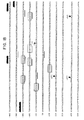
- Figure 1 depicts the promoter region of the TGF ⁇ -1 gene.
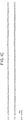
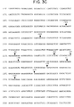
- FIG. 2 depicts the promoter region of the TGF ⁇ -2 gene.
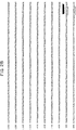
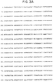
- FIG. 3 depicts the promoter region of the TGF ⁇ -3 gene.
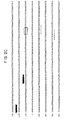
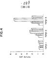
- Figure 4 depicts the relative levels of expression of reporter gene in cells transfected with expression constructs comprising the TGF ⁇ -1 promoter operably linked to the CAT gene, the TGF ⁇ -2 promoter operably linked to the CAT gene, or the TGF ⁇ -3 promoter operably linked to the CAT gene, in the presence of a control compound, estrogen, raloxifene, and tamoxifen.
- the TGF ⁇ -3 construct containing-cells express the highest levels of reporter gene, followed by those containing the TGF ⁇ -2 construct and the TGF ⁇ -1 construct.
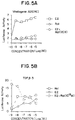
- Figure 5 depicts the induction of a reporter gene under the control of either the estrogen responsive element or a portion of the TGF ⁇ -3 promoter in the presence of estrogen, raloxifene, and combinations of estrogen and raloxifene. This figure shows the markedly different patterns of regulation exerted by the two regulatory sequences.
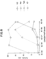
- Figure 6 is a bar graph showing the relative level of reporter gene expression in cells transfected with expression constructs containing a portion of the TGF ⁇ -3 promoter sequence and exposed to a control compound, estrogen, raloxifene, and combinations of estrogen and raloxifene in both the presence and absence of estrogen receptor.
- Estrogen receptor is necessary for induction of expression of the reporter gene by both estrogens and antiestrogens.
- Figure 7 depicts the various domains of estrogen receptor (ER) protein expressed by the deletion constructs set forth in Example XI. Additionally, the relative fold induction achievable in cells transfected with the various mutant ERs and an expression construct comprising the TGF ⁇ -3 promoter and the luciferase gene are shown.
- ER estrogen receptor
- Figure 8 depicts the levels of reporter gene expression achievable in cells transfected with an expression construct comprising the TGF ⁇ -3 promoter and the CAT gene in the presence of various concentrations of estradiol, raloxifene, tamoxifen, and ICI 164,384.
- raloxifene is the most potent inducer at all concentrations, followed by ICI 164,384, tamoxifen.
- Estrogen is the least potent inducer in this system.
- Figure 9 depicts the relative expression of reporter gene in CHO cells transfected with an expression construct comprising a portion of the TGF ⁇ -3 promoter and the luciferase gene in the presence of various concentrations of estradiol and raloxifene.
- raloxifene is the more potent inducer except at low concentrations.
- Figure 10 depicts the relative expression of reporter gene in MCF-7 cells transfected with an expression construct comprising the TGF ⁇ -3 promoter and the luciferase gene in the presence of various concentrations of estradiol and raloxifene.
- Raloxifene is the more potent inducer at all concentrations.
- Figure 11 represents the chemical structures of the compounds evaluated in Example X.
- Figure 12 represents the relative level of induction of reporter gene expression in MG63 cells transfected with TGF ⁇ -3 promoter/CAT expression constructs and exposed to various concentrations of the compounds set forth in Example X. Overall, compound 177366 is the most potent inducer, while 98005 shows no induction.
- Figure 13 depicts the portions of the TGF ⁇ -3 promoter used to identify the 41 base pair raloxifene responsive element, and depicts the relative induction of reporter gene expression by raloxifene in cells transformed with plasmids comprising the indicated portion of the TGF ⁇ -3 promoter sequence operably linked to a reporter gene.
- the fold induction achievable with the pB-301 construct is highest, presence of the raloxifene responsive element (base pairs +35 - +75) is clearly essential for any significant induction of transcription by raloxifene in these cells.
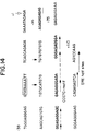
- Figure 14 depicts an analysis of the TGF ⁇ -3 promoter.
- the major transcriptional start site and a CCCTC-motif are depicted as described in Example XI.
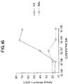
- Figure 15 depicts the relative expression of reporter gene in Hep2 cells transfected with an expression construct comprising the LDLR promoter and the luciferase gene in the presence of estrogen receptor and various concentrations of estradiol and raloxifene.
- raloxifene is the more potent inducer.
- Figure 16 depicts the relative expression of reporter gene in Hep2 cells transfected with an expression construct comprising the LDLR promoter and the luciferase gene in the presence of various concentrations of estradiol and raloxifene and in the absence of estrogen receptor. No induction is exhibited by either compound at concentrations at or below 10 ⁇ 6 M. High concentrations of raloxifene induce expression somewhat, suggesting an alternate, non-ER dependent induction mechanism at such concentrations.
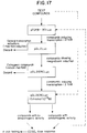
- Figure 17 is a flow diagram showing an example of a sequence of steps that can be carried out according to the teachings of the present invention to evaluate compounds with respect to their ability to induce transcription of reporter genes operably linked to the regulatory control element described herein. It is expected that a correlation will exist between compounds showing the induction profiles described in Example XIII and the ability of such compounds to act as anti-osteoporosis drugs in vivo.
- novel and efficient methods for screening chemical compounds to determine whether these compounds are capable of modulating steroid hormone-responsive gene expression from a promoter comprising a raloxifene responsive element There is also provided nucleic acids consisting essentially of a nucleotide sequence comprising a raloxifene responsive element isolated from the promoter region of a TGF ⁇ gene, and eukaryotic cells transfected therewith. There is also provided a recombinant expression construct comprising the raloxifene responsive element operably linked to a reporter gene.
- a method for inducing bone formation a method for treating osteoporosis, and a method for treating bone fractures which comprise administering a compound that, when bound to an estrogen receptor, potently induces transcription from a raloxifene responsive element of a promoter region of a TGF ⁇ gene.
- the present invention relates to novel and efficient methods of screening chemical compounds to determine whether those compounds are capable of modulating steroid hormone-responsive gene expression from a mammalian promoter comprising a raloxifene responsive element as discovered and described herein.
- the invention comprises nucleic acids consisting essentially of the nucleotide sequence of a mammalian promoter comprising such a raloxifene responsive element.
- the promoter comprising the raloxifene responsive element is derived from the promoter region of the gene for human TGF ⁇ -3 or TGF ⁇ -2.
- the invention further comprises recombinant eukaryotic expression constructs comprising a promoter having a raloxifene responsive element that is operably linked to a reporter gene.
- the reporter gene is the chloramphenicol acetyltransferase gene or the luciferase gene. Particularly, preferred is the luceferase gene.
- the present invention further comprises a method for inducing bone formation which comprises administering an effective amount of a compound that, when bound to an estrogen receptor, potently induces transcription from a raloxifene responsive element of a promoter region of a TGF ⁇ -3 gene.
- the present invention also includes a method for treating osteoporosis which comprises administering an effective amount of a compound that, when bound to an estrogen receptor, potently induces transcription from a raloxifene responsive element of a promoter region of a TGF ⁇ -3 gene.
- the present invention also provides a method for treating bone fractures which comprises administering an effective amount of a compound that, when bound to an estrogen receptor, potently induces transcription from a raloxifene responsive element of a promoter region of a TGF ⁇ -3 gene.
- the invention provides a nucleic acid consisting essentially of a nucleotide sequence comprising a raloxifene responsive element, where the element is isolated from the promoter region of a mammalian, preferably human, transforming growth factor ⁇ gene.
- the transforming growth factor ⁇ gene is the human TGF ⁇ -2 gene or the human TGF ⁇ -3 gene.
- the nucleic acid consists essentially of promoter sequences of the TGF ⁇ -3 gene as described in plasmids pB-301 (containing TGF ⁇ -3 promoter sequences from positions -301 to +110); pB-221 (containing TGF ⁇ -3 promoter sequences from positions -221 to +110); pB-91 (containing TGF ⁇ -3 promoter sequences from positions -91 to +110); pB-60 (containing TGF ⁇ -3 promoter sequence from positions -60 to +110); pB-47 (containing TGF ⁇ -3 promoter sequence from positions -47 to +110) and pB-38 (containing TGF ⁇ -3 promoter sequence from positions -38 to +110), as further described herein.
- the invention provides a recombinant expression construct comprising a nucleic acid consisting essentially of a nucleotide sequence comprising a raloxifene responsive element, where the element is isolated from the promoter region of a mammalian, preferably human, transforming growth factor ⁇ gene, operably linked to a reporter gene.
- the transforming growth factor ⁇ gene is the human TGF ⁇ -2 gene or the human TGF ⁇ -3 gene.
- the reporter gene is the chloramphenicol acetyltransferase gene or the luciferase gene. Particularly, preferred is the luciferase gene.
- the nucleic acid comprises a promoter sequence consisting essentially of the promoter sequences of the TGF ⁇ -3 gene comprising the plasmids pB-301 (containing TGF ⁇ -3 promoter sequence from positions -301 to +110); pB-221 (containing TGF ⁇ -3 promoter sequences from positions -221 to +110); pB-91 (containing TGF ⁇ -3 promoter sequences from positions -91 to +110); pB-60 (containing TGF ⁇ -3 promoter sequences from positions -60 to +110); pB-47 (containing TGF ⁇ -3 promoter sequence from positions -47 to +110) and pB-38 (containing TGF ⁇ -3 promoter sequence from positions -38 to +110), as further described herein and operably linked to a reporter gene.
- pB-301 containing TGF ⁇ -3 promoter sequence from positions -301 to +110
- pB-221 containing TGF ⁇ -3 promoter sequences from positions -221 to +110
- pB-91 containing TGF ⁇ -3
- the recombinant expression constructs of the invention are capable of expressing the reporter gene encoded by such a construct in eukaryotic cells transfected with such a construct.
- eukaryotic cells additionally express an estrogen receptor protein or mutant derivative thereof.
- expression of the reporter gene by the recombinant expression constructs of the invention is capable of being induced by treatment of such cells with raloxifene or other anti-estrogenic compounds as defined herein.
- a third aspect of the invention provides a eukaryotic cell into which has been introduced a recombinant expression construct of the invention.
- the eukaryotic cell is a cell transfected with a recombinant expression construct of the invention.
- the eukaryotic cells of the invention express an estrogen receptor protein or mutant derivative thereof.
- expression of the reporter gene in such cells is capable of being induced by treatment of such cells with raloxifene or other anti-estrogenic compounds as defined herein.
- the invention also provides methods for screening a multiplicity of compounds to identify those compounds having potential as anti-osteoporotic agents.
- a method for screening a multiplicity of compounds to identify compounds having potential as anti-osteoporosis agents comprises identifying a compound of the multiplicity that is capable of inducing transcription from a raloxifene-responsive element of a mammalian promoter, is a specific transcription inducer, is not capable of inducing transcription from an estrogen-responsive element of a mammalian promoter, and is an anti-estrogenic or non-estrogenic compound.
- the method provided by this embodiment further comprises the steps of (a) assaying for the ability of the compound to induce transcription from a raloxifene responsive element of a mammalian promoter; (b) assaying for the inability of the compound to induce transcription from a mammalian promoter not having a raloxifene responsive element; (c) assaying for the inability of the compound to induce transcription from an estrogen responsive promoter; and (d) assaying for the ability of the compound to inhibit estrogen induction of transcription from an estrogen responsive promoter in the presence of estrogen.
- the assay of subpart (a) comprises the step of determining the ability of the compound to induce expression of a reporter gene operably linked to the mammalian promoter comprising a raloxifene responsive element.
- the assay of subpart (b) comprises the step of determining the inability of the compound to induce expression of a reporter gene operably linked to the mammalian promoter wherein the promoter is not comprised of a raloxifene-responsive element.
- Another preferred embodiment of this aspect of the invention provides the assay of subpart (c) comprising the step of determining the inability of the compound to induce expression of a reporter gene operably linked to an estrogen responsive mammalian promoter.
- the invention provides the assay of subpart (d) comprising the step of determining the ability of the compound to inhibit estrogen-dependent induction of expression of a reporter gene operably linked to an estrogen responsive mammalian promoter in the presence of estrogen.
- the raloxifene responsive mammalian promoter is isolated from a mammalian, preferably human, transforming growth factor ⁇ gene.
- a mammalian, preferably human, transforming growth factor ⁇ gene is isolated from a mammalian, preferably human, transforming growth factor ⁇ gene.
- the transforming growth factor ⁇ gene is the human TGF ⁇ -2 gene or the human TGF ⁇ -3 gene.
- the reporter gene is the chloramphenicol acetyltransferase gene or the luciferase gene.
- the raloxifene responsive promoter sequences consisting essentially of the promoter sequences of the TGF ⁇ -3 gene comprising the plasmids pB-301 (containing TGF ⁇ -3 promoter sequences from positions -301 to +110); pB-221 (containing TGF ⁇ -3 promoter sequences from positions -221 to +110); pB-91 (containing TGF ⁇ -3 promoter sequences from positions -91 to +110); pB-60 (containing TGF ⁇ -3 promoter sequences from positions -60 to +110); pB-47 (containing TGF ⁇ -3 promoter sequences from positions -47 to +110) and pB-38 (containing TGF ⁇ -3 promoter sequences from positions -38 to +110), as further described herein and operably linked to a reporter gene.
- TGF ⁇ shall mean transforming growth factor ⁇ (without reference to isoform).
- TGF ⁇ -1, TGF ⁇ -2 and TGF ⁇ -3 shall have the meanings well-established in this art, i.e., to represent the three known isoforms of transforming growth factors ⁇ genes.
- CAT shall be taken to mean chloramphenicol acetyl CoA transferase.
- Estradiol is an estrogen and, at times, is abbreviated herein as E2.
- the abbreviation "ER” shall mean an estrogen receptor protein.
- raloxifene responsive element refers to nucleotide sequences of the nucleic acid comprising a mammalian promoter region of the TGF ⁇ gene that are capable of inducing transcription of any structural gene to which the raloxifene responsive element is operably linked in host cells that are exposed to raloxifene and estrogen receptor proteins.
- Raloxifene responsive elements include, but are not limited to the nucleotide sequences comprising the TGF ⁇ promoter sequences of the plasmids pB-301 (containing TGF ⁇ -3 promoter sequences from positions -301 to +110); pB-221 (containing TGF ⁇ -3 promoter sequences from positions -221 to +110); pB-91 (containing TGF ⁇ -3 promoter sequences from positions -91 to +110); pB-60 (containing TGF ⁇ -3 promoter sequences from positions -60 to +110); pB-47 (containing TGF ⁇ -3 promoter sequences from positions -47 to +110) and pB-38 (containing TGF ⁇ -3 promoter sequences from positions -38 to +110), as further described herein, and nucleic acids having substantially the same biological activity as those nucleic acids.
- Isolated raloxifene responsive elements of the present invention may be derived from TGF ⁇ promoters of any mammalian species of origin, but are preferably of human origin.
- anti-estrogens will be taken to include full and partial antagonists of estrogen. All estradiols used in the Examples described herein are 17 ⁇ -estradiol.
- the term "effective amount” represents an amount of a compound that, when bound to an estrogen receptor, is capable of inducing transcription from a raloxifene responsive element when administered to a mammal.
- the particular dose of compound administered will be determined by the particular circumstances surrounding the case, including the compound administered, the route of administration, the particular condition being treated, and similar considerations.
- the term “potently” represents a compound that, when bound to an estrogen receptor, induces transcription from a raloxifene responsive element at a minimum effective concentration (MEC) of less than or equal to 10nM (1x10 ⁇ 8M), when the compound is tested in the in vitro assay described herein. See Examples V, VI, X, and XIV.
- promoter sequences are identified in terms of their distance, in number of nucleotides, from the major transcriptional start site of the gene, taking this start site to be +1 as shown in Figures 1-3.
- a negative sign (-) preceding the number indicates the nucleotide is 5' to the start site, a positive sign (+) preceding the number indicates the nucleotide is 3' to the start site.
- the sequences are also identified by the numbering indicated in SEQ ID NOS:1-3, and are specifically correlated with numbering of Figures 1-3.
- DNA that encodes the raloxifene responsive elements of the present invention may be obtained, in view of the instant disclosure, by chemical synthesis, by in vitro amplification [including but not limited to the polymerase chain reaction (PCR)], or by combinations of these procedures from naturally-occurring sources, such as cultures of mammalian cells, genomic DNA from such cells, or libraries of such DNA.
- PCR polymerase chain reaction
- the raloxifene responsive elements may be advantageously operably linked to reporter genes and used to either transiently or stably transform appropriate host cells through the use of appropriate vectors, constructs, and means well known in the art, such as DNA mediated gene transfer means including but not limited to transfection, electroporation, and virally-mediated infection.
- DNA mediated gene transfer means including but not limited to transfection, electroporation, and virally-mediated infection.
- the term "recombinant expression construct" as used herein is intended to mean DNA constructs capable of directing the expression of reporter genes to which the raloxifene responsive elements of the invention are operably linked.
- DNA regions are operably linked when they are functionally related to each other.
- a promoter is operably linked to a coding sequence if it controls the transcription of the sequence;
- a ribosome binding site is operably linked to a coding sequence if it is positioned so as to permit translation.
- operably linked means contiguous.
- Reporter genes are genes that encode structural proteins capable of quantification by standard means such as measuring enzymatic activity, colorimetry, chemiluminescence, the presence of radioactivity in a sample, ELISA, antibody binding, radioimmunoassay or other methods of quantification known to those in the art.
- the reporter gene used will vary depending upon the host selected and the transformation method chosen.
- Useful reporter genes include but are not limited to chloramphenicol acetyltransferase, luciferase, ⁇ -galactosidase, alkaline phosphatase, or any other quantifiable protein product.
- the reporter gene is luciferase.
- Transfected cells are cells that have been transfected with raloxifene responsive element-reporter gene recombinant expression constructs made using recombinant DNA techniques.
- Cells that have been transfected with recombinant raloxifene responsive element-reporter gene expression constructs that express the estrogen receptor will express the gene product of the reporter gene under appropriate circumstances (i.e., exposure to an anti-estrogen or other inducer).
- a preferred cell line appropriate for use in the present invention, MCF-7 constitutively expresses the estrogen receptor.
- MG63 cells express reporter genes in a raloxifene-dependent manner, only upon cotransfection of a raloxifene responsive element-reporter gene expression construct and an estrogen receptor expression construct.
- raloxifene responsive element-reporter gene expression constructs are desirable hosts for expression of the raloxifene responsive element-reporter gene expression construct.
- any higher eukaryotic cell culture that either naturally expresses the estrogen receptor, or that has been genetically modified to express the estrogen receptor (or part of that receptor) is useable.
- Mammalian cells are preferred, as illustrated in the Examples. Propagation of such cells in cell culture has become a routine procedure. See Tissue Culture , Academic Press, Kruse & Patterson, editors (1973).
- useful host cell lines are MCF-7, MG63, HeLa, RL95.2, HepG2 and CHO cells (all available from the American Type Culture Collection, Rockville, Maryland).
- use of the MCF-7 cell line is particularly preferred, as this cell line constitutively expresses estrogen receptor.
- Host cells that express the estrogen receptor or part of that receptor and contain a raloxifene responsive element-reporter gene expression construct can be used to evaluate compounds for their ability to induce transcription via the raloxifene responsive element as described in the Examples infra .
- compounds will be considered to induce transcription via a regulatory element (including but not limited to nucleic acid derived from a TGF ⁇ promoter or deletion construct thereof) if transcription of the reporter gene is increased twofold in the presence of the compound compared with expression in the absence of the compound.
- compounds will be considered to be transcriptional inducers if they induce transcription to a level fifty percent above that of the control. In general, however, induction detectably above background is adequate to show induction by a chemical compound.
- plasmid pB-301 In the practice of the aspects of the invention embodying screening methods (see Example XIII), use of the plasmid pB-301 is preferred due to the high level of responsiveness to raloxifene exhibited by this plasmid.
- Other embodiments utilize constructs containing the TGF ⁇ -3 promoter region encompassing positions -38 to +75.
- the raloxifene responsive element is operably linked to a reporter gene in a context allowing transcription, as this element is necessary to allow the raloxifene responsive induction described herein.
- CAT chloramphenicol acetyltransferase gene
- TGF ⁇ -1 phTG12
- TGF ⁇ -2 pTGF-1
- TGF ⁇ -3 pB-499
- the sequences for each of these promoters can be found in Kim et al., J . Biol. Chem. , 264 , 402-408 (1989) (TGF ⁇ -1); Noma et al ., Growth Factor, 4 , 247-255 (1991) (TGF ⁇ -2); and Lafyatis et al., J. Biol. Chem ., 265 , 19128-19136 (1990) (TGF ⁇ -3).
- the promoter sequence of the TGF_-1 gene has been submitted to GenBank/EMBL Data Bank under accession number J04431.
- the TGF ⁇ -1, TGF ⁇ -2 and TGF ⁇ -3 promoter sequences are shown in Figures 1, 2 and 3, respectively, and as SEQ ID NOS: 1, 2 and 3, respectively.
- CAT-containing reporter plasmids operably linked to each of the TGF ⁇ promoter sequences can be produced by subcloning each TGF ⁇ promoter into a commercially-available CAT construct, for example , pCAT-Basic (Promega, Madison, WI), using conventional cloning techniques. See Sambrook, Fritsch, and Maniatis, Molecular Cloning: A Laboratory Manual , 2d ed., Cold Spring Harbor Press, Cold Spring Harbor, New York (hereinafter Sambrook et al .).
- TGF ⁇ -3 gene promoter responsive to the antiestrogen raloxifene In order to identify region(s) of the TGF ⁇ -3 gene promoter responsive to the antiestrogen raloxifene, CAT reporter gene expression directed by constructs containing partial sequences of the TGF ⁇ -3 gene promoter were analyzed. Six TGF ⁇ -3 promoter deletion/CAT reporter constructs were obtained from A. Roberts.
- Plasmid designations and the extent of the promoter region contained in each of these plasmids are set forth below: pB-301 -301 to +110 (corresponding to 1896 to 2306 shown in Figure 3 and SEQ ID NO:3) pB-221 -221 to +110 (corresponding to 1976 to 2306 shown in Figure 3 and SEQ ID NO:3) pB-91 -91 to +110 (corresponding to 2106 to 2306 shown in Figure 3 and SEQ ID NO:3) pB-60 -60 to +110 (corresponding to 2137 to 2306 shown in Figure 3 and SEQ ID NO:3) pB-47 -47 to +110 (corresponding to 2150 to 2306 shown in Figure 3 and SEQ ID NO:3) pB-38 -38 to +110 (corresponding to 2159 to 2306 shown in Figure 3 and SEQ ID NO:3)
- TGF ⁇ -3 promoter deletion constructs Two additional TGF ⁇ -3 promoter deletion constructs were constructed as described below. The first of these consisted of human TGF ⁇ -3 promoter region sequences corresponding to positions -38 to +75 in the promoter sequence, corresponding to 2159 to 2271 shown in Figure 3 and SEQ ID NO:3 ( see Lafyatis et al., ibid .). The second promoter deletion construct consisted of human TGF ⁇ -3 promoter region sequences corresponding to positions -38 to +35 in the promoter sequence, corresponding to 2159 to 2231 shown in Figure 3 and SEQ ID NO:3. The well-established practice in this art is to identify all promoter sequences identified with respect to the distance from the transcription start site.
- plasmids were generated as follows. Oligonucleotides corresponding to the extent of the TGF ⁇ -3 promoter sequence desired in each plasmid were synthesized using a DNA/RNA synthesizer (Model 394, Applied Biosystems Inc., Foster City, CA) under ⁇ -cyanoethyl phosphoamidite synthesis conditions specified by the manufacturer. Complementary pairs of oligonucleotides for each plasmid construct were synthesized, purified, mixed, and allowed to anneal to form double-stranded DNA corresponding to the appropriate TGF ⁇ -3 promoter sequences using conventional methods ( Sambrook et al ., ibid .).
- Hin dIII and Xba I restriction enzyme recognition sites were synthesized as the appropriate overhanging ends at the 5' and 3' ends of the sequences, respectively. Double-stranded promoter sequences were then ligated into the Hin dIII/ Xba I-digested CAT reporter plasmid pB-301 and propagated in bacteria under standard conditions.
- the reporter plasmid produced in this way that contained the -38 to +75 region of the TGF ⁇ -3 promoter was termed pTGF ⁇ +75CAT, and the plasmid that contained the -38 to +75 region of the TGF ⁇ -3 promoter was termed pTGF ⁇ +35CAT. These plasmids were used in CAT assays as described below in Example V.
- Luciferase reporter plasmids containing TGF ⁇ -3 promoter deletion constructs including control containing no portion of the promoter region: pTGF ⁇ -301LUC, pTGF ⁇ -38LUC, pTGF ⁇ +75LUC , pTGF ⁇ +35LUC and pLUC
- plasmids were constructed containing the luciferase gene (REF) expressed under the transcriptional control of TGF ⁇ -3 promoter sequences and varying deletion derivatives thereof.
- the plasmid pTGF-301LUC was made by digesting pB-301 with Hin dIII and thereafter the ends of the Hin dIII digestion-generated overhang were blunted by treatment with the Klenow fragment of bacterial DNA polymerase I (Boehringer-Mannheim, Indianapolis, IN). Xba I digestion was then performed to liberate the portion of the TGF ⁇ -3 promoter corresponding to positions -301 to +110. This fragment was subcloned into Sma I/ Xba I-digested pSP73 (Promega) to generate the shuttle vector pSPTGF ⁇ -301.
- pSPTGF ⁇ -301 After in vivo amplification in bacteria, a preparation of isolated and purified pSPTGF ⁇ -301 was digested with Nde I and Hin dIII, and the promoter sequence-containing fragment isolated after separation on a 0.8% agarose gel (BRL-LifeTechnologies, Inc., Gaithersburg, MD).
- the luciferase-containing construct pLDLRLUC10 (as described in U.S. Patent Application Ser. No. 08/018,985, filed March 3, 1993, and further described in Section C., below) was Nde I/ Hin dIII-digested and purified after agarose gel electrophoresis. These isolated fragments were then mixed, ligated and used to transform bacteria ( Sambrook et al ., ibid .).
- the plasmids pTGF ⁇ -38LUC, pTGF ⁇ +75LUC and pTGF ⁇ +35LUC were made by first excising the TGF ⁇ -3 promoter sequences from pB-38, pTGF ⁇ +75CAT and pTGF ⁇ +35CAT, respectively, by Bam HI/ Xba I double digestion.
- the luciferase-containing plasmid pGL2-Basic (or "pGL2LUC”) (Promega) was prepared by Nhe I/ Bam HI digestion, and each of the recombinant plasmids made by ligation of the appropriate TGF ⁇ -3 promoter sequences into the luciferase-containing plasmid. These plasmids were used in luciferase assays as described below in Example VI.
- a control plasmid containing the luciferase gene but harboring no portion of the TGF ⁇ -3 gene was constructed by digesting pTGF ⁇ +75LUC plasmid DNA with restriction endonuclease Xba I and Hin dIII. Protruding ends were filled by Klenow enzyme reaction in the presence of all four dNTPS under standard conditions ( Sambrook et al ., ibid .). The ends thus blunted were ligated with T4 DNA ligase (Boehringer Mannheim) under manufacturer suggested conditions. The resulting plasmid was designated pLUC.
- the plasmid pLDLRLUC10 was described in U.S. Patent Appln. Ser. No. 08/018,985, filed March 3, 1993 (hereinafter, the '985 application). The construction of this plasmid is described in detail as follows:
- a 1546 base pair sequence of the human LDL receptor promoter was amplified using the polymerase chain reaction.
- a reaction mixture containing 20 picomoles each of the synthetic oligonucleotides: and and 1 ⁇ g human genomic DNA purified from the adenocarcinoma cell line P3UCLA, 200 ⁇ M each of dATP, dGTP, dCTP and TTP, 2.5 units of Taq DNA polymerase, 10mM Tris-HCl pH9.3, 50 mM KCl, 15 mM MgCl2, 0.1% gelatin in a final volume of 100 ⁇ L was subjected to 30 cycles consisting of 15 sec at 96°C, 30 sec at 55°C, and 1 min at 72°C.
- the material was subject to gel electrophoresis on a 1% agarose gel and a 1546 basepair (bp) band isolated and restriction enzyme digested with Hin dIII and Nde I. This fragment was ligated into the plasmid pSP72 (Promega), which had previously been digested with Hin dIII and Nde I.
- the resulting vector, pNLDLRP was digested with Nde I and Hin dIII, the material was electrophoresed on a 1% agarose gel and the 1546 base pair LDL receptor sequence reisolated therefrom.
- Plasmid vector pSv2 was constructed by digesting plasmid pSv2-globin with Hin dIII and Bgl II then ligating an Nru I- Xho I linker into the vector. Plasmid pSv2 globin is disclosed in U.S. Patent No. 4,775,624, which is incorporated by reference. The linker contained the following sequences: The resulting vector was designated pSv2-H NXB because it contained a Bam HI site, an Nru I site, an Xho I site and a Bgl II site.
- the 1546 base pair fragment described above was isolated and cloned into the vector pSv2 containing firefly luciferase reporter gene that had been restriction enzyme digested to completion with NdeI and partially with Hin dIII.
- the resulting vector, pLDLRLUC10 contains the human LDL receptor promoter directing expression of the firefly luciferase gene, an ampicillin resistance marker and an origin of replication.
- the estrogen receptor-containing mammalian expression constructs pCMVER and pRSV-ER were obtained from B.S. Katzenellenbogen, Department of Physiology and Biophysics, University of Illinois (Urbana-Champaign, IL). See Reese and Katzenellenbogen, J. Biol. Chem ., 266 , 10880-10887 (1990). These plasmids were used in expression assays as described below, for example, in Example VII.
- Complementary oligonucleotides corresponding to the estrogen-responsive element (ERE) from the Xenopus laevis vitellogenin Az gene promoter, corresponding to positions -341 to -310 [(Metzger et a l., Nature , 334 , 31-36 (1988)] were designed, synthesized, and annealed to form a double-stranded region that is an estrogen responsive element essentially as described in Example I.
- a sequence comprising an Xho I restriction enzyme recognition site was synthesized to be flanking the ERE sequences, the element having the following nucleotide sequence (shown as SEQ ID NOS:8 and 9, respectively):
- the double-stranded ERE was subcloned into Xho I-digested pGL12-Basic (Promega), whereby the luciferase gene was placed under the transcriptional influence of the ERE.
- This plasmid was designated pGL2ERELUC and used in further experiments as described below (Example VI).
- Mammalian cells were cultured in media (termed 3:1 media) consisting of Dulbecco's modified Eagle's media and F12 media (mixed in a ratio of 3:1, obtained from GIBCO, Grand Island, NY), without phenol red, containing 10% fetal bovine serum (FBS; Hyclone, Logan, UT). Cells were passaged at four day intervals.
- csFBS charcoal-stripped FBS
- D-PBS Dulbecco's phosphate buffered saline
- GIBCO Dulbecco's phosphate buffered saline
- TGF ⁇ promoter containing plasmid DNA was mixed with DNA encoding a selectable marker.
- 60 ⁇ g of TGF ⁇ promoter-containing plasmid DNA pTGF ⁇ -301LUC, pGL2LUC or pGL2ERELUC
- 60 ⁇ g of the pSV2HYG-derivative, hygromycin resistance gene-containing plasmid pSV2HYGtB (further described in U.S. Patent Application Ser. No.
- PCR polymerase chain reaction
- Luciferase-containing hygromycin-resistant clones were incubated with estrogen or raloxifene as described above, and the effect on expression of reporter genes analyzed using assays for the amount on enzymatic activity present in cell extracts.
- luciferase assays cells were lysed in eukaryotic cell lysis reagent containing 0.1M phosphate buffer (pH7.8)/ 1% TritonX-100 (Boehringer Mannheim)/ 2mM EDTA and 1mM dithiothreitol (DTT, Boehringer Mannheim), and assayed using an optimized unenhanced luciferase assay protocol developed by the Analytical Luminescence Laboratory (San Diego, CA).
- TGF ⁇ -1, TGF ⁇ -2 and TGF ⁇ -3 genes were differentially inducible using estrogen and tamoxifen. See "Background of the Related Art” above. The extent and pattern of this inducibility was characterized using the TGF ⁇ promoter-containing plasmids described above in a series of in vitro expression assays as follows.
- Lysed cell preparations were centrifuged at 15,000 rpm for 5 minutes at 4°C to remove cell debris. Supernatants containing the soluble cell lysate were transferred to a new set of tubes for assaying chloramphenicol acetyltransferase (CAT) activity.
- CAT chloramphenicol acetyltransferase
- each lysate was first determined using a commercially-available assay (BioRad Laboratories, Richmond, CA). An amount of each cell lysate containing 100 ⁇ g total protein was then mixed with CAT assay buffer (0.4M Tris-HCl (pH 7.8)/ 0.5mM acetyl-CoA (Boehringer Mannheim)/ 0.1 ⁇ Ci D- threo -(dichloroacetyl-1,2-[14C]-chloramphenicol) for 15 hours. After this incubation, reactions were stopped by vigorously extracting the reaction mixture with 900 ⁇ L ethyl acetate.
- Acetylated and unacetylated chloramphenicol species were resolved by thin layer chromatography using a mixture of 95:5 (v/v) chloroform/methanol as the ascending buffer. Radioactivity from each species so resolved was measured using a Betascope 603 blot analyzer (Betagen, Intelligenetics Inc., Mountain View, CA). The percentage of acetylated counts relative to the total counts was calculated to yield relative CAT activities of each transfectant assayed (all CAT activities expressed herein were calculated on this basis). Each assay was performed in duplicate.
- the results obtained in the previous Example demonstrated that the TGF ⁇ -3 gene promoter is transcriptionally responsive to both estrogen and "antiestrogen” compounds such as raloxifene and tamoxifen, and that transcription was induced by raloxifene treatment to a relatively greater degree than the degree of transcriptional induction produced in response to estrogen.
- This example demonstrates that the gene encoding the Xenopus protein vitellogenin responds in vivo in exactly the opposite fashion, i.e., transcription of the vitellogenin gene is strongly induced by estrogen and only weakly induced by raloxifene.
- raloxifene strongly antagonizes estrogen-induced induction of vitellogenin production when the two compounds are given together.
- TGF_ promoter sequences directing transcription of the reporter genes in the reporter plasmids assayed above in Example VI contain a novel raloxifene-responsive element, characterized by a unique pattern of estrogen and antiestrogen responsiveness.
- Transfected cells were treated with varying amounts and combinations of hormones and then rinsed twice with D-PBS. Cells were then lysed upon incubation with 250 ⁇ L of eukaryotic cell lysis reagent (as described in Example VI) at 4°C for 20 min and transferred to microcentrifuge tubes by scraping with a rubber policeman. Cell lysates were centrifuged at 14,000 rpm for one minute to remove cell debris. Cell extracts (supernatant) were then assayed for protein content and luciferase activities.
- eukaryotic cell lysis reagent as described in Example VI
- Example IV Cultures of MG63 were prepared for co-transfection as in Example IV. One such culture was co-transfected with pB2-499 and pRSVER and another was co-transfected with pB2-499 and pRSV vector plasmid (control). The ability of the following compounds to induce transcription via the raloxifene responsive element of the TGF ⁇ -3 gene was then assayed essentially as described in Example V:
- TGF ⁇ promoter-mediated gene expression by estrogen and antiestrogen compounds was found to be concentration-dependent.
- Cultures of MG63 cells were transiently co-transfected with pB-301 and pRSVER as described in Example V. Such transiently transfected cell cultures were divided into four groups of twelve cultures, and each of the four groups was used to test the ability of one estrogen or antiestrogen compound to induce transcription from the TGF ⁇ -3 promoter individually. For each group of twelve cultures, the particular estrogen or antiestrogen compound was tested in replicate cultures at six concentrations, varying in tenfold increments from 10 ⁇ 9M to 10 ⁇ 5M, as well as one set of replicate cultures treated with vehicle only (for a total of twelve cultures per experimental treatment). Hormones were dissolved in ethanol and applied to the cultures in media as described above. The four estrogens and antiestrogens tested were:
- RRE raloxifene responsive element
- raloxifene to induce transcription from the TGF ⁇ -3 promoter distinct from estrogen-mediated induction was demonstrated in a variety of cell lines.
- Cultures of CHO cells were transiently co-transfected with pB-301 and pRSVER as described in Example IV and were used to determine the ability of both raloxifene and estradiol to induce transcription via the raloxifene responsive element. Twelve transiently transfected cultures were treated in replicate with either estradiol or raloxifene at six concentrations varying in tenfold increments from 10 ⁇ 9M to 10 ⁇ 5M (as well as a vehicle only control for a total of twelve cultures). Hormones were dissolved in ethanol and applied to the cultures in media as described above.
- Cultures of MCF-7 cells were transiently transfected with pTGF ⁇ -301LUC as described in Example IV. These cultures were used to test the ability of raloxifene and estradiol to induce transcription in this cell type. Cultures were treated with either estradiol or raloxifene at one of the following six concentrations: 0M, 10 ⁇ 9M, 10 ⁇ 8M, 10 ⁇ 7M, 10 ⁇ 6M and 10 ⁇ 5M. Hormones were dissolved in ethanol and applied to the cultures in media as described above.
- TGF ⁇ -3 promoter Similar assays were performed in human endometrial cancer cells (RL59.2), human cervical cancer cells (HeLa), and monkey kidney cells (COS-1) (American Type Culture Collection, Rockville, MD). Transcription initiated by the TGF ⁇ -3 promoter was found to be induced by estrogen and raloxifene in all cell types tested (with variations in the magnitude of induction). These results demonstrate that estrogen and raloxifene-mediated induction of reporter gene transcription from the TGF ⁇ -3 promoter is not restricted to specific cell types. The different levels of induction in different cells, however, might indicate the abundance of other factors in these cells involved in regulation. The fact that raloxifene and estrogen responsiveness of the TGF ⁇ -3 promoter were found using both luciferase and CAT as reporter genes indicates that this regulation is a general characteristic of gene expression from this promoter.
- Antiestrogen compounds were known in the prior art to be capable of inducing TGF ⁇ gene expression in a dose-dependent manner. Knabbe et al., Am. J. Clin. Onc ., 14 (Suppl.2), S15-S20 (1991). Furthermore, as shown in Example VIII above, raloxifene and tamoxifen were found to be capable of inducing TGF ⁇ -3 gene expression in a dose dependent manner.
- Raloxifene, LY81099, LY98005, LY112676, LY113526, LY13314, LY139482, and LY177366 were assayed to compare their ability to induce transcription from the promoter of the TGF ⁇ -3 gene at varying concentrations.
- Cultures of MG63 cells transiently co-transfected with pB-301 and pRSVER were treated as in Example VI with the above compounds at concentrations of 0M, 10 ⁇ 9M, 10 ⁇ 8M, 10 ⁇ 7M, 10 ⁇ 6M and 10 ⁇ 5M. The experimental results are shown in tabular form and depicted in Figure 12.
- results of these experiments demonstrate the utility of the reporter gene-containing expression plasmids described herein as a screening technique for identifying potential anti-osteoporosis agents, because those compounds that are raloxifene-like in their induction profiles and ED50's show relatively lower uterotropic effects than estrogen-like compounds having lower induction profiles and higher ED50's in the present assay.
- At least a portion of the raloxifene response element (RRE) in the human TGF ⁇ -3 gene promoter was approximately localized to a particular 41 nucleotide sequence found at positions +35 through +75. This sequence was found to be necessary for mediating raloxifene-induced transcriptional activation of reporter gene expression in the TGF ⁇ -reporter gene expression constructs described above.
- TGF ⁇ -3 promoter deletion reporter constructs including pB-499, pB-301, pB-221, pB-91, pB-60, pB-47, pTGF ⁇ -38LUC, pTGF ⁇ +75LUC, pTGF ⁇ +35LUC and pLUC.
- the nucleotide sequence of the TGF ⁇ -3 promoter from position -38 to +110 was depicted in Figure 14.
- the raloxifene responsive sequence was found above to be the sequence depicted in the Figure in outline form.
- the open arrow indicates the major transcription start site (+1).
- the two black arrows indicate the two minor transcription start sites.
- the "TATA" sequence is shown in the open box.
- a putative CCCTC-motif is indicated by a series of horizontal arrow heads under the sequence of the putative raloxifene responsive element. See Lobanenekov et al . Oncogene , 5 , 1743-1753 (1990).
- TGF ⁇ -3 Two conclusions can be drawn from the TGF ⁇ -3 analysis. The first is that no palindromic sequences homologous to the ERE was found in this region of the TGF ⁇ -3 promoter. This finding is consistent with the results shown in Example VII which demonstrated that DNA binding activity of ER is not required. Second, ER-mediated raloxifene activation of TGF ⁇ -3 most likely requires other factors that are capable of binding to the raloxifene responsive sequence. A good candidate for such a protein is the CTCF factor identified by Lobanenkov et a l. which is involved in c-myc gene regulation. These findings may lead to the identification of other genes as potential raloxifene inducible genes that have raloxifene responsive elements in their promoters. Furthermore, such genes could be used to identify genetic elements having the activity of raloxifene responsive elements for use in the screening procedure set forth in Example XIII.
- the raloxifene responsive element of the present invention was used to search the GenBank sequence library; significant homology was found between this element and elements in the following genes: GenBank/EMBL Data Bank Accession No.: Gene: X56595 Chicken type VI collagen _-2 X55373 Human ETS-2 promoter region M30137 Human ETS-2 (5' flank) D10231 Mouse glucose transporter (enhancer 2) M12731 Mouse N-myc proto-oncogene M13945 Mouse pim-1 proto-oncogene X63281 R.
- estrogen and Raloxifene Induce LDL Receptor Promoter Activation
- LDL receptor expression plays an essential role in regulation of serum LDL-cholesterol uptake. It has been known that estrogen induces LDL receptor messenger RNA in vivo . Ma et al., Proc. Natl. Acad. Sci . USA , 83 , 792-796 (1986). As shown in this Example, this activation of LDL receptor promoter sequence by estrogen is mediated by ER. Raloxifene also induces LDL receptor promoter; however, this induction is ER independent.
- ATCC strain HepG2 cells were co-transfected with pLDLRLUC10 and pRSVER as described in Example IV. These cells were exposed to estradiol and raloxifene under the conditions set forth in Example VI. The results are tabulated below and a series of experiments are depicted in Figure 15: TABLE X 0M 10 ⁇ 9M 10 ⁇ 8M 10 ⁇ 7M 10 ⁇ 6M 10 ⁇ 5M estradiol 20.9 16.6 15.4 40.7 74.0 69.0 raloxifene 20.9 20.9 19.9 26.9 14.6 14.6
- ATCC strain HepG2 cells were co-transfected with pLDLRLUC10 and pRSV vector plasmid as described in Example IV. These cells were exposed to estradiol and raloxifene under the conditions set forth in Example VI. The results are tabulated below and a representative series of such experiments are set forth in Figure 16: TABLE XI 0M 10 ⁇ 9M 10 ⁇ 8M 10 ⁇ 7M 10 ⁇ 6M 10 ⁇ 5M estradiol 1597 1645 1792 1574 1578 2445 raloxifene 1597 1652 1234 1025 1291 5561
- an assay using luciferase as a reporter gene was designed to screen for potential anti-osteoporosis agents.
- the first procedure that is used in the screening assay is to determine the ability of a compound to induce transcription from the TGF ⁇ -3 promoter.
- the assay is performed essentially as described in Example IX, using MCF-7 cells stably transfected with pTGF ⁇ -301LUC. Cell culture and assay conditions are adapted to the 96-well microtiter plate format. Cells are seeded in a 96-well plate at a density resulting in approximately 50% confluency. Test compounds may be selected from a variety of sources, including pharmaceutical research records, chemical manufacturers products lists, and naturally-occurring sources such as fermentation extracts. Cells are incubated in growth media (as described in Example IV) containing the test compound for about 24 hours.
- Cells are then lysed in situ on the plate, and the lysates subjected to both a quantitative protein assay and to the luciferase activity assay. Compounds that induce a greater than two-fold increase in luciferase activity are considered competent for further testing.
- Step 2 Assays are performed with compounds identified as described in Step 1 on cell cultures that have been stably transfected with pGL2LUC to determine whether such compounds are general transcription inducers.
- general transcriptional inducers lack the transcriptional induction specificity required for potential anti-osteoporetics that are modulators of raloxifene-responsive element-dependent gene expression, such general inducers are excluded from further testing.
- Step 4 Compounds that have fulfilled the criteria of Steps 1 through 3 are then further tested to determine whether such compounds are either anti-estrogenic or non-estrogenic/non-antiestrogenic. To this end, the compounds are assayed in the presence of estradiol in cells stably transfected with pGL2ERELUC. Inhibition of estrogen-induced luciferase activity in this assay indicates that such compounds have anti-estrogenic activity. Both anti-estrogenic and non-estrogenic compounds will be characterized for their dose-response profiles and ED50 values. Further experiments may be done to establish the dose-response profiles of such compounds and to compare them with known anti-estrogens like raloxifene. See Example X.
- the following experiments were performed to demonstrate the correlation between the in vitro assay and an in vivo model of post-menopausal osteoporosis.
- the in vitro assay measures the test compound's ability to induce transcription via the raloxifene responsive element of the TGF ⁇ -3 promoter.
- the in vivo model measures the changes in bone (femur) mineral density in ovariectomized rats.
- Raloxifene, 088074, 156678, 171147, and 309503 were assayed to determine their ability to produce a two-fold induction in the transcription from the promoter of the TGF ⁇ -3 gene, as measured by luciferase activity, at a concentration of less than 10 nM [i.e. minimum effective concentration (MEC) ⁇ 10 nM].
- MEC minimum effective concentration
- the cells were transiently co-transfected with pTGF ⁇ -301LUC (10 ⁇ g), pCMVER (1 ⁇ g), and pRSV ⁇ gal (1 ⁇ g) using the ProFection Mammalian Transfection System (Promega).
- a transfection solution was prepared by mixing pTGF ⁇ -301LUC (940 ⁇ g), pCMVER (94 ⁇ g), pRSV ⁇ gal (94 ⁇ g), CaCl2 (5.704mL), and nuclease free water (47ml) with 2XHEPES (47ml) while vortexing. The resulting solution was incubated at room temperature for 30 minutes. This transfection solution (3mL) was added to each flask.
- test compounds were dissolved in dimethyl sulfoxide and diluted initially to 1:1000. This dilution was accomplished by adding 2 ⁇ L of drug solution to 1000 ⁇ L of media in a deep well microtiter plate (1:500 dilution), then 100 ⁇ L of the dilute drug solution is added to 100 ⁇ L of media already in the plate (1:2 dilution). For compound screening, the compounds are diluted 1:1000, as previously described, then further diluted 1:100. Alternatively, the minimum effective concentration is determined using an eight-dilution dose-response curve. The compounds and cells are incubated in 96-well plates overnight.
- the media was removed from the cells and each well washed with Ca/Mg free phosphate buffered saline (200 ⁇ L).
- the saline solution was removed and the cells lysed with 60 ⁇ L of lysis buffer (100mM KPO4, 0.2% Triton X-100, 1mM DTT, pH 7.8).
- the resulting solution was used to assay for luciferase or ⁇ -gal activity.
- the luciferase assay was performed substantially as described in Example VI, except the luciferin solution comprises 100mg luciferin, 9mL 1M Glycyl-gylcine, 36mL 40mM EGTA, 720mL 1M DTT, 5.4mL 1M MgSO4, and 310mL H2O.
- the results of these experiments are shown in tabular form in Table XII.
- the above compounds were also tested for their ability to preserve bone mineral density in ovariectomized rats.
- Seventy-five day old female Sprague Dawley rats (weight range of 225 to 275 g) were obtained from Charles River Laboratories (Portage, MI). They were housed in groups of three and had ad libitum access to food (calcium context approximately 1%) and water.
- Room temperature was maintained at 22.2°C ⁇ 1.7°C with a minimum relative humidity of 40%. The photoperiod in the room was 12 hours light and 12 hours dark.
- the rats underwent bilateral ovariectomy under anesthesia [44 mg/kg Ketamine and 5 mg/kg Xylazine (Butler, Indianapolis, IN) administered intramuscularly]. Treatment with vehicle, or a test compound was initiated on the day of surgery following recovery from anesthesia. Oral dosage was by gavage in 0.5 mL of 1% carboxymethylcellulose (CMC). Body weight was determined at the time of surgery and weekly thereafter and the dosage was adjusted with changes in body weight. Vehicle or treated ovariectomized (ovex) rats and non-ovariectomized (intact) rats were evaluated in parallel with each experimental group to serve as negative and positive controls.
- CMC carboxymethylcellulose
- the rats were treated daily for 35 days (6 rats per treatment group) and sacrificed by decapitation on the 36th day. The 35 day time period was sufficient to allow maximal reduction in bone density, measured as described herein.
- the right femurs were excised and scanned at the distal metaphysis 1 mm from the patellar groove with single photon absorptiometry. Results of the densitometer measurements represent a calculation of bone density as a function of the bone mineral context and bone width. Generally, ovariectomy of the rats caused a reduction in femur density of about 25% as compared to intact vehicle treated controls. The results of these experiments are shown in Table XII.
- the compounds that fail to induce transcription for the raloxifene responsive element also fail to show a statistically significant protection against bone loss in mineral density over ovariectomized rats.
Abstract
Description
- The invention relates to methods for identifying therapeutic agents for the treatment of osteoporosis. The invention relates to isolating, cloning, and using nucleic acids comprising the promoter regions of mammalian transforming growth factor β genes that are novel regulatory elements designated "raloxifene responsive elements". The invention also encompasses genetically engineered eukaryotic cells containing the recombinant expression constructs wherein the raloxifene responsive elements are operably linked to reporter genes. In such cells the raloxifene responsive elements are capable of modulating transcription of the reporter genes in response to treatment with certain compounds. The invention also relates to methods for identifying anti-osteoporosis agents that induce transcription of certain genes via raloxifene responsive elements and that specifically do not induce deleterious or undesirable side effects that have been associated with estrogen replacement therapy, such as increased risk with uterine and breast cancer. The nucleic acids, cells, and methods of the invention provide effective methods for screening putative sources of anti-osteoporosis agents, and identifying those that advantageously lack the undesirable side effects associated with current anti-osteoporosis agents. The invention also relates to a method for inducing bone formation, a method for treating osteoporosis, and a method for treating bone fractures which comprise administering a compound that, when bound to an estrogen receptor, potently induces transcription from a raloxifene responsive element.
- In 1991, U.S. pharmaceutical companies spent an estimated $7.9 billion on research and development devoted to identifying new therapeutic agents (Pharmaceutical Manufacturer's Association). The magnitude of this amount is due, in part, to the fact that the hundreds, if not thousands, of chemical compounds must be tested in order to identify a single effective therapeutic agent that does not engender unacceptable levels of undesirable or deleterious side effects. There is an increasing demand for economical methods of testing large numbers of testing large number of chemical compounds to quickly identify those compounds that are likely to be effective in treating disease. At present, few such economical systems exist.
- One disease that conspicuously lacks a rapid method for screening large numbers of potential therapeutic agents is bone loss. Bone loss occurs in a wide variety of patients, including those who have undergone hysterectomy, who are undergoing or have undergone long-term administration of corticosteroids, who suffer from Cushing's syndrome or have gonadal dysgenesis, as well as post-menopausal women.
- The mechanism of bone loss is not well understood, but in practical effect, the disorder arises from an imbalance in the formation of new healthy bone and the resorption of old bone, skewed toward a net loss of bone tissue. This bone loss includes a decrease in both mineral content and protein matrix components of the bone, and leads to an increased fracture rate of the femoral bones and bones in the forearm and vertebrae predominantly. These fractures, in turn, lead to an increase in general morbidity, a marked loss of stature and mobility, and in many cases, an increase in mortality resulting from complications.
- Unchecked, bone loss can lead to osteoporosis, a major debilitating disease whose predominant feature is the loss of bone mass without a reduction in bone volume (by decreased density and enlargement of bone spaces), producing porosity and fragility. Osteoporosis among post-menopausal women is one of the most common types of bone disorders, affecting an estimated 20 to 25 million women in the United States alone.
- A significant feature of post-menopausal osteoporosis is the large and rapid loss of bone mass due to the cessation of estrogen production by the ovaries. Indeed, data clearly support the ability of estrogens to limit the progression of osteoporotic bone loss, and estrogen replacement is a recognized treatment for post-menopausal osteoporosis in the United States and many other countries.
- Estrogens, when administered at low levels, have beneficial effects on bone; however, long-term estrogen replacement therapy has been implicated in a variety of disorders, including an increased risk of uterine and breast cancer. These serious side effects cause many women to refuse this treatment. Alternative therapeutic regimens, designed to lessen the cancer risk, such as administering combinations of progestogen and estrogen, cause some patients to experience regular withdrawal bleeding, which is unacceptable to most older women. Concerns over these significant undesirable side effects associated with estrogen replacement therapy, and the limited ability of estrogens to reverse existing bone loss, provide a strong impetus for the development of effective alternative therapies for bone loss that do not cause undesirable side effects.
- Another approach in osteoporotic therapy is the use of antiestrogens. In general, antiestrogens inhibit (antagonize) the activity of estrogen in the body. Antiestrogens bind to the estrogen receptor, although it is believed that the interaction between antiestrogens and the estrogen receptor involves a different domain of the receptor than that to which estrogen binds. Some antiestrogens, on the other hand, display pharmacological properties that are a mixture of agonist and antagonist properties. In other words, these compounds cause certain effects that mimic estrogen, while antagonizing other effects that are commonly associated with estrogen administration in cells that express the receptor. Because of this mixed effect of some antiestrogens, they are subject to the same adverse effects associated with estrogen replacement therapy.
- One antiestrogen known to display such a mixed agonist/antagonist effect is tamoxifen, a drug used for the treatment of breast cancer. Tamoxifen acts as an estrogen antagonist in its ability to reduce the growth of breast tumors, but it also acts as an agonist in its ability to reduce the amount of serum cholesterol in both healthy women and women with breast cancer. Love et al., Annals Int. Med., 115, 860-864 (1991). Tamoxifen also act to increase bone density in breast cancer patients. Love et al., N. Eng. J. Med., 326, 852-856 (1991). At least one study has suggested that the increases in bone density possible with tamoxifen appear to be restricted to the lumbar spine, with bone loss being reported in the radius in some breast cancer patients treated with tamoxifen. Furthermore, tamoxifen treatment has also been suggested to contribute to weight gain among post-menopausal women. Love et al., Ann. Int. Med., ibid.
- Improved anti-osteoporotics that achieve increases in bone density without causing negative side effects are clearly needed. Unfortunately, no method currently exists for rapidly and efficiently screening large numbers of compounds to identify those that display the desired anti-osteoporotic effects. Because this screening process comprises the most time-consuming and expensive step in identifying improved anti-osteoporotic compounds, development of a rapid method for testing large numbers of compounds to identify those that are likely to possess anti-osteoporotic effect is highly desirable.
- It is well established that estrogens exert their effects by first binding to an estrogen receptor, and then the estrogen/estrogen receptor complex binding to DNA. The hormone/receptor complex modulates gene expression via this DNA binding. Kumar, Cell, 55, 145-156 (1988). Antiestrogens also bind to estrogen receptors. Although these antiestrogen/receptor complexes bind to DNA they generally fail to modulate gene expression. Both estradiol/estrogen receptor complexes and hydroxytamixofen/estrogen receptor complexes bind in vitro to DNA binding domains called estrogen responsive elements. Kumar, Cell, ibid.
- The conformation of the ligand/receptor complex is a matter of some debate. However, recent studies have suggested a conformational difference between estrogen receptor bound to estradiol and the same estrogen receptor bound to 4-hydroxytamoxifen or ICI 164,384. Klinge et al., J. Ster. Biochem. Mol. Biol., 43, 249-262 (1992).
- In an effort to rationally address the problem of developing improved anti-osteoporotic agents, researchers have investigated proteins known to play a role in bone maintenance. One protein known to influence bone remodelling and bone turnover is transforming growth factor β (TGFβ). Although commonly referred to as a single compound, TGFβ is actually a family of molecules that now is known to include at least three isoforms: TGFβ-1, TGFβ-2 and TGFβ-3. See Arrick et al., Canc. Res., 50, 299-303 (1990). The present inventor has noted that ovariectomy induces a significant decrease in TGFβ-3 in rat bone (data collected by present inventor is unpublished); others have noted the same type of correlation with respect to levels of TGFβ (isoform not specified). See Finkelman et al., Proc. Nat'l Acad. Sci. USA, 89, 12190-12193 (1992). Further, the present inventor has noted that administration of raloxifene, an antiestrogen, to ovariectomized rats restores TGFβ-3 concentrations to levels equal to or higher than that found in control animals. The direct correlation between TGFβ-3 levels and circulating levels of estrogen or antiestrogen, and the finding that TGFβ (isoform not specified) plays a significant role in bone remodelling and turnover, suggest that osteoporosis may result from reduced expression of TGFβ-3 in vivo. See Noda et al., Endocrin. 12, 2991-2994 (1989).
- The hypothesis that reduced levels of TGFβ-3 may allow bone loss is undermined by the findings that TGFβ has been isolated from a large number of sources and exhibits widely divergent effects. For example, it inhibits the growth of mesenchymal cells and epithelial cells, it induces biosynthesis of proteoglycans, fibronectins, and plasminogen activator, and is chemotactic for fibroblasts, macrophages, and smooth muscle cells. See, Flaumenhaft et al., J. Cell. Bio., 120(4), 995-1002 (1993).
- Furthermore, antiestrogens such as tamoxifen or toremifene induce human fetal fibroblasts to secrete TGFβ (without reference to isoform) in the absence of estrogen receptor Colletta et al., Br. J. Cancer, 62, 405-409 (1990). TGFβ has been found to stimulate osteoblastic bone formation and to inhibit osteoclast formation and osteoclast activities. Mundy, "Clinical Application of TGFβ", Ciba Foundation Symposium No. 157, 137-151, Wiley, Chichester. TGFβ repressed division of one human endometrial cancer cell line (Ishikawa), but was shown to be mitogenic with respect to another such cell line (HEC-50). Murphy et al., J. Ster. Biochem. Molec. Bio., 41, 309-314 (1992).
- Three months of antiestrogen treatment with tamoxifen has been correlated with induction of extracellular TGFβ-1 in breast cancer biopsies. Butta et al ., Cancer Res., 52, 4261-4264 (1992). Decreased concentrations of TGFβ-1 mRNA were found in one human endometrial cancer cell line (HEC-50) grown in media containing 1% ctFBS (twice charcoal stripped FBS) when such cells were exposed to either estradiol or certain antiestrogens. Gong et al., Canc. Res., 52, 1704-1709 (1992).
- TGFβ-2 mRNA is expressed by the T-47D and MDA-MB-231 cell lines. Treatment of these cell lines with estradiol reduced TGFβ-2 mRNA expression, but tamoxifen did not exhibit the same effect. TGFβ-3 induces mitogenesis, collagen synthesis, and alkaline phosphatase activity in osteoblast enriched bone cell cultures at a three to five fold higher rate than TGFβ-1. Arrick et al., Canc. Res., 50, 299-303 (1990).
- A general review of the properties of TGFβ are described in Sporn et al., J. Cell Bio., 105, 1039-1045 (1987); Massague, Cell, 49, 437-438 (1987); and Moses, Cell, 63, 245-247 (1990). These references generally describe the properties exerted by TGFβ in in vitro and in vivo systems.
- The complex pattern of expression described above suggests a unique and complex mechanism of regulation of expression of the various TGFβ isoforms. The promoter regions for each of the genes TGFβ-1, TGFβ-2 and TGFβ-3 have been cloned and described. Kim et al., J. Biol. Chem., 264, 402-408 (1989); Noma et al., Growth Factors, 4, 247-255 (1991); Lafyatis, et al., J. Biol. Chem., 265, 19128-19136 (1990).
- The promoters for TGFβ-2 and TGFβ-3 have been characterized and have been reported to contain cAMP responsive elements, AP-1 sites, AP-2 sites, and SP-1 sites. Noma et al. indicated that the TGFβ-2 promoter activity was dependent upon the region of the promoter investigated and the cell line selected for the induction assay.
- At least one method for efficiently screening the biological activity of a large number of compounds is described in International Patent Application No. PCT/US92/00419, which claims methods for transcriptionally regulating the expression of a growth factor. This patent disclosed assays to identify compounds capable of inducing transcription via the promoter regions of the human growth hormone gene, the c-ErbB2 gene, the promoter region of the K-ras sequence, and the early promoter and enhancers of cytomegalovirus. This application is directed primarily towards the problem of determining the regulation of various oncogenes.
- To date, identifying compounds that are likely to display an anti-osteoporotic effect has required virtually random investigation of individual compounds on the basis of epidemiological studies, the utility of related chemical compounds in achieving the desired effect, and other time consuming and inefficient methods. Both the delay caused by the current screening methods and the economic costs of such inefficient testing emphasize the need for economical and efficient methods for identifying potential anti-osteoporotic drugs worthy of additional investigation.
- Figure 1 depicts the promoter region of the TGFβ-1 gene.
- Figure 2 depicts the promoter region of the TGFβ-2 gene.
- Figure 3 depicts the promoter region of the TGFβ-3 gene.
- Figure 4 depicts the relative levels of expression of reporter gene in cells transfected with expression constructs comprising the TGFβ-1 promoter operably linked to the CAT gene, the TGFβ-2 promoter operably linked to the CAT gene, or the TGFβ-3 promoter operably linked to the CAT gene, in the presence of a control compound, estrogen, raloxifene, and tamoxifen. In general, the TGFβ-3 construct containing-cells express the highest levels of reporter gene, followed by those containing the TGFβ-2 construct and the TGFβ-1 construct.
- Figure 5 depicts the induction of a reporter gene under the control of either the estrogen responsive element or a portion of the TGFβ-3 promoter in the presence of estrogen, raloxifene, and combinations of estrogen and raloxifene. This figure shows the markedly different patterns of regulation exerted by the two regulatory sequences.
- Figure 6 is a bar graph showing the relative level of reporter gene expression in cells transfected with expression constructs containing a portion of the TGFβ-3 promoter sequence and exposed to a control compound, estrogen, raloxifene, and combinations of estrogen and raloxifene in both the presence and absence of estrogen receptor. Estrogen receptor is necessary for induction of expression of the reporter gene by both estrogens and antiestrogens.
- Figure 7 depicts the various domains of estrogen receptor (ER) protein expressed by the deletion constructs set forth in Example XI. Additionally, the relative fold induction achievable in cells transfected with the various mutant ERs and an expression construct comprising the TGFβ-3 promoter and the luciferase gene are shown.
- Figure 8 depicts the levels of reporter gene expression achievable in cells transfected with an expression construct comprising the TGFβ-3 promoter and the CAT gene in the presence of various concentrations of estradiol, raloxifene, tamoxifen, and ICI 164,384. In general, raloxifene is the most potent inducer at all concentrations, followed by ICI 164,384, tamoxifen. Estrogen is the least potent inducer in this system.
- Figure 9 depicts the relative expression of reporter gene in CHO cells transfected with an expression construct comprising a portion of the TGFβ-3 promoter and the luciferase gene in the presence of various concentrations of estradiol and raloxifene. In general, raloxifene is the more potent inducer except at low concentrations.
- Figure 10 depicts the relative expression of reporter gene in MCF-7 cells transfected with an expression construct comprising the TGFβ-3 promoter and the luciferase gene in the presence of various concentrations of estradiol and raloxifene. Raloxifene is the more potent inducer at all concentrations.
- Figure 11 represents the chemical structures of the compounds evaluated in Example X.
- Figure 12 represents the relative level of induction of reporter gene expression in MG63 cells transfected with TGFβ-3 promoter/CAT expression constructs and exposed to various concentrations of the compounds set forth in Example X. Overall,
compound 177366 is the most potent inducer, while 98005 shows no induction. - Figure 13 depicts the portions of the TGFβ-3 promoter used to identify the 41 base pair raloxifene responsive element, and depicts the relative induction of reporter gene expression by raloxifene in cells transformed with plasmids comprising the indicated portion of the TGFβ-3 promoter sequence operably linked to a reporter gene. Although the fold induction achievable with the pB-301 construct is highest, presence of the raloxifene responsive element (base pairs +35 - +75) is clearly essential for any significant induction of transcription by raloxifene in these cells.
- Figure 14 depicts an analysis of the TGFβ-3 promoter. The major transcriptional start site and a CCCTC-motif are depicted as described in Example XI.
- Figure 15 depicts the relative expression of reporter gene in Hep2 cells transfected with an expression construct comprising the LDLR promoter and the luciferase gene in the presence of estrogen receptor and various concentrations of estradiol and raloxifene. Generally, raloxifene is the more potent inducer.
- Figure 16 depicts the relative expression of reporter gene in Hep2 cells transfected with an expression construct comprising the LDLR promoter and the luciferase gene in the presence of various concentrations of estradiol and raloxifene and in the absence of estrogen receptor. No induction is exhibited by either compound at concentrations at or below 10⁻⁶ M. High concentrations of raloxifene induce expression somewhat, suggesting an alternate, non-ER dependent induction mechanism at such concentrations.
- Figure 17 is a flow diagram showing an example of a sequence of steps that can be carried out according to the teachings of the present invention to evaluate compounds with respect to their ability to induce transcription of reporter genes operably linked to the regulatory control element described herein. It is expected that a correlation will exist between compounds showing the induction profiles described in Example XIII and the ability of such compounds to act as anti-osteoporosis drugs in vivo.
- According to the present invention, there is provided novel and efficient methods for screening chemical compounds to determine whether these compounds are capable of modulating steroid hormone-responsive gene expression from a promoter comprising a raloxifene responsive element. There is also provided nucleic acids consisting essentially of a nucleotide sequence comprising a raloxifene responsive element isolated from the promoter region of a TGFβ gene, and eukaryotic cells transfected therewith. There is also provided a recombinant expression construct comprising the raloxifene responsive element operably linked to a reporter gene. There is also provided a method for inducing bone formation, a method for treating osteoporosis, and a method for treating bone fractures which comprise administering a compound that, when bound to an estrogen receptor, potently induces transcription from a raloxifene responsive element of a promoter region of a TGFβ gene.
- The present invention relates to novel and efficient methods of screening chemical compounds to determine whether those compounds are capable of modulating steroid hormone-responsive gene expression from a mammalian promoter comprising a raloxifene responsive element as discovered and described herein. The invention comprises nucleic acids consisting essentially of the nucleotide sequence of a mammalian promoter comprising such a raloxifene responsive element. In a preferred embodiment, the promoter comprising the raloxifene responsive element is derived from the promoter region of the gene for human TGFβ-3 or TGFβ-2.
- The invention further comprises recombinant eukaryotic expression constructs comprising a promoter having a raloxifene responsive element that is operably linked to a reporter gene. In preferred embodiments, the reporter gene is the chloramphenicol acetyltransferase gene or the luciferase gene. Particularly, preferred is the luceferase gene. Cells transfected with such eukaryotic expression constructs, that are capable of expressing the reporter gene when such cells are exposed to raloxifene or other anti-estrogenic compounds, are also provided by the invention.
- The present invention further comprises a method for inducing bone formation which comprises administering an effective amount of a compound that, when bound to an estrogen receptor, potently induces transcription from a raloxifene responsive element of a promoter region of a TGFβ-3 gene. The present invention also includes a method for treating osteoporosis which comprises administering an effective amount of a compound that, when bound to an estrogen receptor, potently induces transcription from a raloxifene responsive element of a promoter region of a TGFβ-3 gene. The present invention also provides a method for treating bone fractures which comprises administering an effective amount of a compound that, when bound to an estrogen receptor, potently induces transcription from a raloxifene responsive element of a promoter region of a TGFβ-3 gene.
- In the first aspect, the invention provides a nucleic acid consisting essentially of a nucleotide sequence comprising a raloxifene responsive element, where the element is isolated from the promoter region of a mammalian, preferably human, transforming growth factor β gene. In preferred embodiments, the transforming growth factor β gene is the human TGFβ-2 gene or the human TGFβ-3 gene. In further preferred embodiments of this aspect of the invention, the nucleic acid consists essentially of promoter sequences of the TGFβ-3 gene as described in plasmids pB-301 (containing TGFβ-3 promoter sequences from positions -301 to +110); pB-221 (containing TGFβ-3 promoter sequences from positions -221 to +110); pB-91 (containing TGFβ-3 promoter sequences from positions -91 to +110); pB-60 (containing TGFβ-3 promoter sequence from positions -60 to +110); pB-47 (containing TGFβ-3 promoter sequence from positions -47 to +110) and pB-38 (containing TGFβ-3 promoter sequence from positions -38 to +110), as further described herein.
- In a second aspect, the invention provides a recombinant expression construct comprising a nucleic acid consisting essentially of a nucleotide sequence comprising a raloxifene responsive element, where the element is isolated from the promoter region of a mammalian, preferably human, transforming growth factor β gene, operably linked to a reporter gene. In preferred embodiments, the transforming growth factor β gene is the human TGFβ-2 gene or the human TGFβ-3 gene. In preferred embodiments, the reporter gene is the chloramphenicol acetyltransferase gene or the luciferase gene. Particularly, preferred is the luciferase gene. In further preferred embodiments of this aspect of the invention, the nucleic acid comprises a promoter sequence consisting essentially of the promoter sequences of the TGFβ-3 gene comprising the plasmids pB-301 (containing TGFβ-3 promoter sequence from positions -301 to +110); pB-221 (containing TGFβ-3 promoter sequences from positions -221 to +110); pB-91 (containing TGFβ-3 promoter sequences from positions -91 to +110); pB-60 (containing TGFβ-3 promoter sequences from positions -60 to +110); pB-47 (containing TGFβ-3 promoter sequence from positions -47 to +110) and pB-38 (containing TGFβ-3 promoter sequence from positions -38 to +110), as further described herein and operably linked to a reporter gene.
- In another aspect, the recombinant expression constructs of the invention are capable of expressing the reporter gene encoded by such a construct in eukaryotic cells transfected with such a construct. In preferred embodiments, such eukaryotic cells additionally express an estrogen receptor protein or mutant derivative thereof. In particularly preferred embodiments, expression of the reporter gene by the recombinant expression constructs of the invention is capable of being induced by treatment of such cells with raloxifene or other anti-estrogenic compounds as defined herein.
- A third aspect of the invention provides a eukaryotic cell into which has been introduced a recombinant expression construct of the invention. In a preferred embodiment, the eukaryotic cell is a cell transfected with a recombinant expression construct of the invention. In a preferred embodiment, the eukaryotic cells of the invention express an estrogen receptor protein or mutant derivative thereof. In particularly preferred embodiments, expression of the reporter gene in such cells is capable of being induced by treatment of such cells with raloxifene or other anti-estrogenic compounds as defined herein.
- The invention also provides methods for screening a multiplicity of compounds to identify those compounds having potential as anti-osteoporotic agents. In one aspect of this embodiment of the invention is provided a method for screening a multiplicity of compounds to identify compounds having potential as anti-osteoporosis agents. The method provided by this aspect of the invention comprises identifying a compound of the multiplicity that is capable of inducing transcription from a raloxifene-responsive element of a mammalian promoter, is a specific transcription inducer, is not capable of inducing transcription from an estrogen-responsive element of a mammalian promoter, and is an anti-estrogenic or non-estrogenic compound. The method provided by this embodiment further comprises the steps of (a) assaying for the ability of the compound to induce transcription from a raloxifene responsive element of a mammalian promoter; (b) assaying for the inability of the compound to induce transcription from a mammalian promoter not having a raloxifene responsive element; (c) assaying for the inability of the compound to induce transcription from an estrogen responsive promoter; and (d) assaying for the ability of the compound to inhibit estrogen induction of transcription from an estrogen responsive promoter in the presence of estrogen.
- In a preferred embodiment, the assay of subpart (a) comprises the step of determining the ability of the compound to induce expression of a reporter gene operably linked to the mammalian promoter comprising a raloxifene responsive element. In another preferred embodiment, the assay of subpart (b) comprises the step of determining the inability of the compound to induce expression of a reporter gene operably linked to the mammalian promoter wherein the promoter is not comprised of a raloxifene-responsive element. Another preferred embodiment of this aspect of the invention provides the assay of subpart (c) comprising the step of determining the inability of the compound to induce expression of a reporter gene operably linked to an estrogen responsive mammalian promoter. In a final preferred embodiment, the invention provides the assay of subpart (d) comprising the step of determining the ability of the compound to inhibit estrogen-dependent induction of expression of a reporter gene operably linked to an estrogen responsive mammalian promoter in the presence of estrogen.
- In particularly preferred embodiments of this aspect of the invention, the raloxifene responsive mammalian promoter is isolated from a mammalian, preferably human, transforming growth factor β gene. Most preferred are embodiments wherein the transforming growth factor β gene is the human TGFβ-2 gene or the human TGFβ-3 gene. In other preferred embodiments, the reporter gene is the chloramphenicol acetyltransferase gene or the luciferase gene. In further preferred embodiments of this aspect of the invention, the raloxifene responsive promoter sequences consisting essentially of the promoter sequences of the TGFβ-3 gene comprising the plasmids pB-301 (containing TGFβ-3 promoter sequences from positions -301 to +110); pB-221 (containing TGFβ-3 promoter sequences from positions -221 to +110); pB-91 (containing TGFβ-3 promoter sequences from positions -91 to +110); pB-60 (containing TGFβ-3 promoter sequences from positions -60 to +110); pB-47 (containing TGFβ-3 promoter sequences from positions -47 to +110) and pB-38 (containing TGFβ-3 promoter sequences from positions -38 to +110), as further described herein and operably linked to a reporter gene.
- Specific preferred embodiments of the present invention will become evident from the following more detailed description of certain preferred embodiments and the claims.
- This specification contains a number of abbreviations. As used herein, "TGFβ" shall mean transforming growth factor β (without reference to isoform). TGFβ-1, TGFβ-2 and TGFβ-3 shall have the meanings well-established in this art, i.e., to represent the three known isoforms of transforming growth factors β genes. As used herein, the abbreviation "CAT" shall be taken to mean chloramphenicol acetyl CoA transferase. "Estradiol" is an estrogen and, at times, is abbreviated herein as E2. As used herein, the abbreviation "ER" shall mean an estrogen receptor protein.
- The term "raloxifene responsive element" as used herein refers to nucleotide sequences of the nucleic acid comprising a mammalian promoter region of the TGFβ gene that are capable of inducing transcription of any structural gene to which the raloxifene responsive element is operably linked in host cells that are exposed to raloxifene and estrogen receptor proteins. Raloxifene responsive elements include, but are not limited to the nucleotide sequences comprising the TGFβ promoter sequences of the plasmids pB-301 (containing TGFβ-3 promoter sequences from positions -301 to +110); pB-221 (containing TGFβ-3 promoter sequences from positions -221 to +110); pB-91 (containing TGFβ-3 promoter sequences from positions -91 to +110); pB-60 (containing TGFβ-3 promoter sequences from positions -60 to +110); pB-47 (containing TGFβ-3 promoter sequences from positions -47 to +110) and pB-38 (containing TGFβ-3 promoter sequences from positions -38 to +110), as further described herein, and nucleic acids having substantially the same biological activity as those nucleic acids. This definition is intended to encompass natural allelic variations in the promoter regions of the TGFβ genes. Isolated raloxifene responsive elements of the present invention may be derived from TGFβ promoters of any mammalian species of origin, but are preferably of human origin.
- As used herein, anti-estrogens will be taken to include full and partial antagonists of estrogen. All estradiols used in the Examples described herein are 17β-estradiol.
- The term "effective amount" represents an amount of a compound that, when bound to an estrogen receptor, is capable of inducing transcription from a raloxifene responsive element when administered to a mammal. The particular dose of compound administered will be determined by the particular circumstances surrounding the case, including the compound administered, the route of administration, the particular condition being treated, and similar considerations.
- The term "potently" represents a compound that, when bound to an estrogen receptor, induces transcription from a raloxifene responsive element at a minimum effective concentration (MEC) of less than or equal to 10nM (1x10⁻⁸M), when the compound is tested in the in vitro assay described herein. See Examples V, VI, X, and XIV.
- All portions of promoter sequences are identified in terms of their distance, in number of nucleotides, from the major transcriptional start site of the gene, taking this start site to be +1 as shown in Figures 1-3. A negative sign (-) preceding the number indicates the nucleotide is 5' to the start site, a positive sign (+) preceding the number indicates the nucleotide is 3' to the start site. The sequences are also identified by the numbering indicated in SEQ ID NOS:1-3, and are specifically correlated with numbering of Figures 1-3.
- DNA that encodes the raloxifene responsive elements of the present invention may be obtained, in view of the instant disclosure, by chemical synthesis, by in vitro amplification [including but not limited to the polymerase chain reaction (PCR)], or by combinations of these procedures from naturally-occurring sources, such as cultures of mammalian cells, genomic DNA from such cells, or libraries of such DNA.
- The raloxifene responsive elements may be advantageously operably linked to reporter genes and used to either transiently or stably transform appropriate host cells through the use of appropriate vectors, constructs, and means well known in the art, such as DNA mediated gene transfer means including but not limited to transfection, electroporation, and virally-mediated infection. The term "recombinant expression construct" as used herein is intended to mean DNA constructs capable of directing the expression of reporter genes to which the raloxifene responsive elements of the invention are operably linked.
- DNA regions are operably linked when they are functionally related to each other. For example, a promoter is operably linked to a coding sequence if it controls the transcription of the sequence; a ribosome binding site is operably linked to a coding sequence if it is positioned so as to permit translation. Generally, operably linked means contiguous.
- Reporter genes are genes that encode structural proteins capable of quantification by standard means such as measuring enzymatic activity, colorimetry, chemiluminescence, the presence of radioactivity in a sample, ELISA, antibody binding, radioimmunoassay or other methods of quantification known to those in the art. The reporter gene used will vary depending upon the host selected and the transformation method chosen. Useful reporter genes include but are not limited to chloramphenicol acetyltransferase, luciferase, β-galactosidase, alkaline phosphatase, or any other quantifiable protein product. In a preferred embodiment of the invention, the reporter gene is luciferase.
- Transfected cells are cells that have been transfected with raloxifene responsive element-reporter gene recombinant expression constructs made using recombinant DNA techniques. Cells that have been transfected with recombinant raloxifene responsive element-reporter gene expression constructs that express the estrogen receptor, either as a characteristic of such cells or due to the co-transfection of an estrogen receptor encoding expression construct, will express the gene product of the reporter gene under appropriate circumstances (i.e., exposure to an anti-estrogen or other inducer). For example, a preferred cell line appropriate for use in the present invention, MCF-7, constitutively expresses the estrogen receptor. For such cell lines, transfection with the recombinant raloxifene responsive element-reporter gene expression constructs alone will yield cells appropriate for use in the present invention. Alternatively, MG63 cells express reporter genes in a raloxifene-dependent manner, only upon cotransfection of a raloxifene responsive element-reporter gene expression construct and an estrogen receptor expression construct.
- Cultures of cells derived from multicellular organisms are desirable hosts for expression of the raloxifene responsive element-reporter gene expression construct. In principle, any higher eukaryotic cell culture that either naturally expresses the estrogen receptor, or that has been genetically modified to express the estrogen receptor (or part of that receptor) is useable. Mammalian cells are preferred, as illustrated in the Examples. Propagation of such cells in cell culture has become a routine procedure. See Tissue Culture, Academic Press, Kruse & Patterson, editors (1973). Examples of useful host cell lines are MCF-7, MG63, HeLa, RL95.2, HepG2 and CHO cells (all available from the American Type Culture Collection, Rockville, Maryland). For the purposes of the present invention, use of the MCF-7 cell line is particularly preferred, as this cell line constitutively expresses estrogen receptor.
- Host cells that express the estrogen receptor or part of that receptor and contain a raloxifene responsive element-reporter gene expression construct can be used to evaluate compounds for their ability to induce transcription via the raloxifene responsive element as described in the Examples infra. In a preferred embodiment of the invention, compounds will be considered to induce transcription via a regulatory element (including but not limited to nucleic acid derived from a TGFβ promoter or deletion construct thereof) if transcription of the reporter gene is increased twofold in the presence of the compound compared with expression in the absence of the compound. In a less preferred embodiment, compounds will be considered to be transcriptional inducers if they induce transcription to a level fifty percent above that of the control. In general, however, induction detectably above background is adequate to show induction by a chemical compound.
- In the practice of the aspects of the invention embodying screening methods (see Example XIII), use of the plasmid pB-301 is preferred due to the high level of responsiveness to raloxifene exhibited by this plasmid. Other embodiments utilize constructs containing the TGFβ-3 promoter region encompassing positions -38 to +75. In all operative embodiments of the invention, the raloxifene responsive element is operably linked to a reporter gene in a context allowing transcription, as this element is necessary to allow the raloxifene responsive induction described herein.
- The order of carrying out the steps of the screening methods of the invention may be varied, and in some instances, some of the steps may be omitted. The Examples that follow are illustrative of specific embodiments of the invention, and various uses thereof. They are set forth for explanatory purposes only, and are not to be taken as limiting the invention.
- As a first step in developing cell lines useful in screening potential anti-osteoporotic agents, a series of reporter plasmids encoding the chloramphenicol acetyltransferase gene (CAT) (Gorman et al., Molec. Cell. Biol., 2, 1044-1051 (1982)) operably linked to promoter sequences from the TGFβ-1, TGFβ-2 and TGFβ-3 genes were obtained from A. Roberts, National Institutes of Health, Laboratory of Chemoprevention (NIH/NCI, Bethesda, MD). These plasmids were designated phTG12 (TGFβ-1), pTGF-1 (TGFβ-2) and pB-499 (TGFβ-3), respectively. The sequences for each of these promoters can be found in Kim et al., J. Biol. Chem., 264, 402-408 (1989) (TGFβ-1); Noma et al., Growth Factor, 4, 247-255 (1991) (TGFβ-2); and Lafyatis et al., J. Biol. Chem., 265, 19128-19136 (1990) (TGFβ-3). The promoter sequence of the TGF_-1 gene has been submitted to GenBank/EMBL Data Bank under accession number J04431. The TGFβ-1, TGFβ-2 and TGFβ-3 promoter sequences are shown in Figures 1, 2 and 3, respectively, and as SEQ ID NOS: 1, 2 and 3, respectively.
- Alternatively, CAT-containing reporter plasmids operably linked to each of the TGFβ promoter sequences can be produced by subcloning each TGFβ promoter into a commercially-available CAT construct, for example, pCAT-Basic (Promega, Madison, WI), using conventional cloning techniques. See Sambrook, Fritsch, and Maniatis, Molecular Cloning: A Laboratory Manual, 2d ed., Cold Spring Harbor Press, Cold Spring Harbor, New York (hereinafter Sambrook et al.).
- In order to identify region(s) of the TGFβ-3 gene promoter responsive to the antiestrogen raloxifene, CAT reporter gene expression directed by constructs containing partial sequences of the TGFβ-3 gene promoter were analyzed. Six TGFβ-3 promoter deletion/CAT reporter constructs were obtained from A. Roberts. Plasmid designations and the extent of the promoter region contained in each of these plasmids are set forth below:
pB-301 -301 to +110 (corresponding to 1896 to 2306 shown in Figure 3 and SEQ ID NO:3) pB-221 -221 to +110 (corresponding to 1976 to 2306 shown in Figure 3 and SEQ ID NO:3) pB-91 -91 to +110 (corresponding to 2106 to 2306 shown in Figure 3 and SEQ ID NO:3) pB-60 -60 to +110 (corresponding to 2137 to 2306 shown in Figure 3 and SEQ ID NO:3) pB-47 -47 to +110 (corresponding to 2150 to 2306 shown in Figure 3 and SEQ ID NO:3) pB-38 -38 to +110 (corresponding to 2159 to 2306 shown in Figure 3 and SEQ ID NO:3) - Two additional TGFβ-3 promoter deletion constructs were constructed as described below. The first of these consisted of human TGFβ-3 promoter region sequences corresponding to positions -38 to +75 in the promoter sequence, corresponding to 2159 to 2271 shown in Figure 3 and SEQ ID NO:3 (see Lafyatis et al., ibid.). The second promoter deletion construct consisted of human TGFβ-3 promoter region sequences corresponding to positions -38 to +35 in the promoter sequence, corresponding to 2159 to 2231 shown in Figure 3 and SEQ ID NO:3. The well-established practice in this art is to identify all promoter sequences identified with respect to the distance from the transcription start site.
- These plasmids were generated as follows. Oligonucleotides corresponding to the extent of the TGFβ-3 promoter sequence desired in each plasmid were synthesized using a DNA/RNA synthesizer (Model 394, Applied Biosystems Inc., Foster City, CA) under β-cyanoethyl phosphoamidite synthesis conditions specified by the manufacturer. Complementary pairs of oligonucleotides for each plasmid construct were synthesized, purified, mixed, and allowed to anneal to form double-stranded DNA corresponding to the appropriate TGFβ-3 promoter sequences using conventional methods (Sambrook et al., ibid.). HindIII and XbaI restriction enzyme recognition sites were synthesized as the appropriate overhanging ends at the 5' and 3' ends of the sequences, respectively. Double-stranded promoter sequences were then ligated into the HindIII/XbaI-digested CAT reporter plasmid pB-301 and propagated in bacteria under standard conditions. The reporter plasmid produced in this way that contained the -38 to +75 region of the TGFβ-3 promoter was termed pTGFβ+75CAT, and the plasmid that contained the -38 to +75 region of the TGFβ-3 promoter was termed pTGFβ+35CAT. These plasmids were used in CAT assays as described below in Example V.
- Four plasmids were constructed containing the luciferase gene (REF) expressed under the transcriptional control of TGFβ-3 promoter sequences and varying deletion derivatives thereof. The plasmid pTGF-301LUC was made by digesting pB-301 with HindIII and thereafter the ends of the HindIII digestion-generated overhang were blunted by treatment with the Klenow fragment of bacterial DNA polymerase I (Boehringer-Mannheim, Indianapolis, IN). XbaI digestion was then performed to liberate the portion of the TGFβ-3 promoter corresponding to positions -301 to +110. This fragment was subcloned into SmaI/XbaI-digested pSP73 (Promega) to generate the shuttle vector pSPTGFβ-301.
- After in vivo amplification in bacteria, a preparation of isolated and purified pSPTGFβ-301 was digested with NdeI and HindIII, and the promoter sequence-containing fragment isolated after separation on a 0.8% agarose gel (BRL-LifeTechnologies, Inc., Gaithersburg, MD). The luciferase-containing construct pLDLRLUC10 (as described in U.S. Patent Application Ser. No. 08/018,985, filed March 3, 1993, and further described in Section C., below) was NdeI/HindIII-digested and purified after agarose gel electrophoresis. These isolated fragments were then mixed, ligated and used to transform bacteria (Sambrook et al., ibid.).
- The plasmids pTGFβ-38LUC, pTGFβ+75LUC and pTGFβ+35LUC were made by first excising the TGFβ-3 promoter sequences from pB-38, pTGFβ+75CAT and pTGFβ+35CAT, respectively, by BamHI/XbaI double digestion. The luciferase-containing plasmid pGL2-Basic (or "pGL2LUC") (Promega) was prepared by NheI/BamHI digestion, and each of the recombinant plasmids made by ligation of the appropriate TGFβ-3 promoter sequences into the luciferase-containing plasmid. These plasmids were used in luciferase assays as described below in Example VI.
- A control plasmid containing the luciferase gene but harboring no portion of the TGFβ-3 gene was constructed by digesting pTGFβ+75LUC plasmid DNA with restriction endonuclease XbaI and HindIII. Protruding ends were filled by Klenow enzyme reaction in the presence of all four dNTPS under standard conditions (Sambrook et al., ibid.). The ends thus blunted were ligated with T4 DNA ligase (Boehringer Mannheim) under manufacturer suggested conditions. The resulting plasmid was designated pLUC.
- The plasmid pLDLRLUC10 was described in U.S. Patent Appln. Ser. No. 08/018,985, filed March 3, 1993 (hereinafter, the '985 application). The construction of this plasmid is described in detail as follows:
- A 1546 base pair sequence of the human LDL receptor promoter was amplified using the polymerase chain reaction.
A reaction mixture containing 20 picomoles each of the synthetic oligonucleotides:
and
and 1 µg human genomic DNA purified from the adenocarcinoma cell line P3UCLA, 200 µM each of dATP, dGTP, dCTP and TTP, 2.5 units of Taq DNA polymerase, 10mM Tris-HCl pH9.3, 50 mM KCl, 15 mM MgCl₂, 0.1% gelatin in a final volume of 100 µL was subjected to 30 cycles consisting of 15 sec at 96°C, 30 sec at 55°C, and 1 min at 72°C. The material was subject to gel electrophoresis on a 1% agarose gel and a 1546 basepair (bp) band isolated and restriction enzyme digested with HindIII and NdeI. This fragment was ligated into the plasmid pSP72 (Promega), which had previously been digested with HindIII and NdeI. The resulting vector, pNLDLRP, was digested with NdeI and HindIII, the material was electrophoresed on a 1% agarose gel and the 1546 base pair LDL receptor sequence reisolated therefrom. - Plasmid vector pSv2 was constructed by digesting plasmid pSv2-globin with HindIII and BglII then ligating an NruI-XhoI linker into the vector. Plasmid pSv2 globin is disclosed in U.S. Patent No. 4,775,624, which is incorporated by reference. The linker contained the following sequences:


The resulting vector was designated pSv2-H NXB because it contained a BamHI site, an NruI site, an XhoI site and a BglII site. The HindIII-BglII fragment of plasmid pAlc4(NRRL B-18783), which contains the firefly luciferase gene (REF), was then ligated into the HindIII-BglII site of plasmid pSv2-HNXB. - The 1546 base pair fragment described above was isolated and cloned into the vector pSv2 containing firefly luciferase reporter gene that had been restriction enzyme digested to completion with NdeI and partially with HindIII. The resulting vector, pLDLRLUC10 contains the human LDL receptor promoter directing expression of the firefly luciferase gene, an ampicillin resistance marker and an origin of replication.
- The estrogen receptor-containing mammalian expression constructs pCMVER and pRSV-ER were obtained from B.S. Katzenellenbogen, Department of Physiology and Biophysics, University of Illinois (Urbana-Champaign, IL). See Reese and Katzenellenbogen, J. Biol. Chem., 266, 10880-10887 (1990). These plasmids were used in expression assays as described below, for example, in Example VII.
- Complementary oligonucleotides corresponding to the estrogen-responsive element (ERE) from the Xenopus laevis vitellogenin Az gene promoter, corresponding to positions -341 to -310 [(Metzger et al., Nature, 334, 31-36 (1988)] were designed, synthesized, and annealed to form a double-stranded region that is an estrogen responsive element essentially as described in Example I. A sequence comprising an XhoI restriction enzyme recognition site was synthesized to be flanking the ERE sequences, the element having the following nucleotide sequence (shown as SEQ ID NOS:8 and 9, respectively):

- The double-stranded ERE was subcloned into XhoI-digested pGL12-Basic (Promega), whereby the luciferase gene was placed under the transcriptional influence of the ERE. This plasmid was designated pGL2ERELUC and used in further experiments as described below (Example VI).
- Mammalian cells were cultured in media (termed 3:1 media) consisting of Dulbecco's modified Eagle's media and F12 media (mixed in a ratio of 3:1, obtained from GIBCO, Grand Island, NY), without phenol red, containing 10% fetal bovine serum (FBS; Hyclone, Logan, UT). Cells were passaged at four day intervals. One day prior to transfection, cells were trypsinized by incubating them with 1mL of a solution of 0.05% trypsin/5.3mM tetrasodium ethylenediamine tetraacetate (GIBCO) for 5min at room temperature, and then seeded in 3:1 media containing 10% charcoal-stripped FBS (csFBS; Hyclone) at densities of one million cells per 10cm culture dish.
- Co-transfection experiments were performed in MG63 (human osteosarcoma) cells, using the ProFection Mammalian Transfection System (Promega). Ten µg of TGFβ promoter-containing reporter plasmid DNA and 5 µg of human estrogen receptor (hER)-containing expression plasmid (pRSV-ER or pCMV-ER, obtained from B.S. Katzenellenbogen, Department of Physiology and Biophysics, University of Illinois [see Reese and Katzenellenbogen, J. Biol. Chem., 266, 10880-10887 (1990)] or pRSV (control) plasmid were mixed, co-precipitated, and transfected into 2-4 million cells in 10 cm culture dishes. After incubating the cells with the DNA precipitate at 37°C for 15h, the DNA precipitate was removed by twice washing the cells with Dulbecco's phosphate buffered saline (D-PBS; GIBCO). The cells were then refed with fresh 3:1 media containing 10% csFBS. Compounds to be evaluated for their ability to modulate reporter gene expression were added at the appropriate times and assayed as described below.
- MCF-7 cells were used for transfection experiments resulting in stable integration of transfected plasmid sequences into the recipient cell genome. In these experiments, TGFβ promoter containing plasmid DNA was mixed with DNA encoding a selectable marker. For example, 60µg of TGFβ promoter-containing plasmid DNA (pTGFβ-301LUC, pGL2LUC or pGL2ERELUC) were mixed with 60µg of the pSV2HYG-derivative, hygromycin resistance gene-containing plasmid pSV2HYGtB (further described in U.S. Patent Application Ser. No. 07/953,633, filed September 29, 1992, hereby incorporated by reference), and transfected onto two million MCF-7 cells using the ProFection system as described above (Promega). After overnight incubation at 37°C, the precipitate was washed from the cells as described above and the cells then refed with fresh 3:1 media containing 10% FBS and cultured for an additional 48h at 37°C, at which time the cells had typically reached confluency. Cells were trypsinized as described above and each culture dish replated into two 10 cm culture dishes in 3:1 media containing 10% FBS. Hygromycin resistance was selected by culturing the cells in media supplemented with 200µg/mL hygromycin B (Calbiochem-Novabiochem, LaJolla, CA). This selective media was replaced with fresh hygromycin B-supplemented media every 2 days without disturbing the cells for the duration of the selection experiment. Clonal colonies became visible after growth for approximately 14 days in selective media. Such clones were isolated and transferred to individual wells of 24-well cell culture dishes (Flow Laboratories, McLean, VA) using a sterile pipette tip. Such clones were grown and maintained in selective media.
- To identify hygromycin-resistant clones that had been successfully co-transfected with luciferase gene-containing plasmid sequences, the polymerase chain reaction (PCR) was used to detect luciferase cDNA-derived DNA sequences in transfectant DNA. Oligonucleotide PCR primers were synthesized corresponding to positions 355-373 (sense primer) and 929-911 (antisense primer) of the luciferase cDNA sequence. PCR was performed using Perkin-Elmer GeneAmp PCR System 9600 for 35 cycles under conditions essentially as described by the manufacturer; each PCR cycle included 45 sec at 94°C, 45 sec at 55°C and two minutes at 72°C.
- Luciferase-containing hygromycin-resistant clones were incubated with estrogen or raloxifene as described above, and the effect on expression of reporter genes analyzed using assays for the amount on enzymatic activity present in cell extracts. For luciferase assays, cells were lysed in eukaryotic cell lysis reagent containing 0.1M phosphate buffer (pH7.8)/ 1% TritonX-100 (Boehringer Mannheim)/ 2mM EDTA and 1mM dithiothreitol (DTT, Boehringer Mannheim), and assayed using an optimized unenhanced luciferase assay protocol developed by the Analytical Luminescence Laboratory (San Diego, CA). Light output was measured and recorded using a microtitre plate luminometer (ML3000, Dynatech Laboratories, Chantilly, VA). Clones of such cells stably transfected with TGFβ-reporter gene constructs were then used in new anti-osteoporotic screening assays as described below (Example XIII).
- It was known in the prior art that expression of the TGFβ-1, TGFβ-2 and TGFβ-3 genes was differentially inducible using estrogen and tamoxifen. See "Background of the Related Art" above. The extent and pattern of this inducibility was characterized using the TGFβ promoter-containing plasmids described above in a series of in vitro expression assays as follows.
- Cultures of MG63 cells were transiently co-transfected with pRSVER plasmid and either phTG12, pTGF-1, or pB2-499 using the ProFection system (Promega). For each transfected cell culture, DNA-containing calcium phosphate precipitates were added dropwise to each culture dish and mixed thoroughly in the media. The pH of the media was carefully maintained between pH 7.2 and pH 7.4. Transfected cell cultures were then incubated overnight in a 5% CO₂ atmosphere at 37°C. For all cultures, the precipitate was removed after overnight incubation by aspirating the media from the culture dishes, followed by rinsing each dish twice with D-PBS. For each co-transfected cell line, the buffer was replaced with 10 ml fresh medium containing 10% csFBS and one of the following compositions:
- a. 10 µl ethanol (hormone vehicle) = control;
- b. 10 µl 17β-estradiol (Sigma) ("estradiol") at a concentration of 10⁻⁴M in ethanol;
- c. 10 µl raloxifene (Eli Lilly Laboratories) at a concentration of 10⁻⁴M in ethanol;
- d. 10 µl Tamoxifen (Sigma) at a concentration of 10⁻⁴M in ethanol.
- After 24h, incubation with these hormonal preparations (or the vehicle control), cells were washed twice with D-PBS. The cells were then scraped from the culture dishes using a rubber policeman and 1 ml of D-PBS. Cells were collected by centrifugation at 8,000 rpm for two minutes in a tabletop centrifuge (MicroMax Model, IEC, Newark, NY). The supernatant was removed and the cell pellets were resuspended in 150µL of a 0.25M Tris-HCl solution (pH 7.8). Cells were lysed by three cycles of freezing in a dry ice/ethanol bath and thawing in a water bath at 37°C water bath (for 3 minutes each cycle). Lysed cell preparations were centrifuged at 15,000 rpm for 5 minutes at 4°C to remove cell debris. Supernatants containing the soluble cell lysate were transferred to a new set of tubes for assaying chloramphenicol acetyltransferase (CAT) activity.
- Before performing CAT assays on such cell lysates, the protein content of each lysate was first determined using a commercially-available assay (BioRad Laboratories, Richmond, CA). An amount of each cell lysate containing 100µg total protein was then mixed with CAT assay buffer (0.4M Tris-HCl (pH 7.8)/ 0.5mM acetyl-CoA (Boehringer Mannheim)/ 0.1 µCi D-threo-(dichloroacetyl-1,2-[¹⁴C]-chloramphenicol) for 15 hours. After this incubation, reactions were stopped by vigorously extracting the reaction mixture with 900µL ethyl acetate. The organic and aqueous phases were separated by brief centrifugation at 14,000 rpm for 1 minute, and approximately 800µL of the organic phase was transferred to a new set of tubes. Ethyl acetate was evaporated to concentrate the CAT-catalyzed reaction products.
- Acetylated and unacetylated chloramphenicol species were resolved by thin layer chromatography using a mixture of 95:5 (v/v) chloroform/methanol as the ascending buffer. Radioactivity from each species so resolved was measured using a Betascope 603 blot analyzer (Betagen, Intelligenetics Inc., Mountain View, CA). The percentage of acetylated counts relative to the total counts was calculated to yield relative CAT activities of each transfectant assayed (all CAT activities expressed herein were calculated on this basis). Each assay was performed in duplicate.
- A representation of the results of the above experiment for MG63 transfectant cell lines is shown in Figure 4. The results of a representative experiment are tabulated below:

- These experiments demonstrate that transcription of the CAT reporter gene is induced by estrogen and the antiestrogens raloxifene and tamoxifen, with raloxifene displaying a greater potency than estrogen, especially for the TGFβ-3 promoter. In contrast, the TGFβ-1 promoter region used in this experiment (positions -1032 to +727) showed no response to either estradiol or raloxifene.
- The results obtained in the previous Example demonstrated that the TGFβ-3 gene promoter is transcriptionally responsive to both estrogen and "antiestrogen" compounds such as raloxifene and tamoxifen, and that transcription was induced by raloxifene treatment to a relatively greater degree than the degree of transcriptional induction produced in response to estrogen. This example demonstrates that the gene encoding the Xenopus protein vitellogenin responds in vivo in exactly the opposite fashion, i.e., transcription of the vitellogenin gene is strongly induced by estrogen and only weakly induced by raloxifene. In addition, raloxifene strongly antagonizes estrogen-induced induction of vitellogenin production when the two compounds are given together. The instant results suggested that the TGF_ promoter sequences directing transcription of the reporter genes in the reporter plasmids assayed above in Example VI contain a novel raloxifene-responsive element, characterized by a unique pattern of estrogen and antiestrogen responsiveness.
- This pattern of estrogen and antiestrogen responsiveness was further investigated using the reporter gene assay system described in previous Examples. Cultures of MG63 cells were transiently co-transfected with pB-301 and pRSVER as described in Example IV. Such transiently transfected cultures were tested for transcriptional activation of reporter gene expression by treatment with estradiol and raloxifene at concentrations varying in ten-fold increments from 10⁻⁹M to 10⁻⁵M. The combination of estrogen and raloxifene was also tested by assaying the effects of raloxifene at 10⁻⁸M on reporter gene induction in response to estrogen using the same series of concentrations as with estrogen alone. These assays were performed essentially as in Example V.
- Similar assays were performed using cultures of MG63 cells transiently transfected with pGL2ERELUC and pRSVER. In these assays, however, the amount of raloxifene added in combination with estrogen was varied so that the raloxifene concentration was twenty times the concentration of estrogen in the mixture (i.e., 10⁻⁹M estrogen/2 x 10⁻⁸M raloxifene; 10⁻⁸M estrogen/ 2 x 10⁻⁷M raloxifene, etc.).
- Transfected cells were treated with varying amounts and combinations of hormones and then rinsed twice with D-PBS. Cells were then lysed upon incubation with 250µL of eukaryotic cell lysis reagent (as described in Example VI) at 4°C for 20 min and transferred to microcentrifuge tubes by scraping with a rubber policeman. Cell lysates were centrifuged at 14,000 rpm for one minute to remove cell debris. Cell extracts (supernatant) were then assayed for protein content and luciferase activities.
- Luciferase assays were performed as follows. 50µL of each cell extract was added to 100µL of reagent A buffer (containing 4.0mM ATP/15mM MgSO₄/ 30mM tricine buffer (pH7.8)/ 10mM DTT) in individual wells of a microtiter plate. 100µL of 1 mM D(-)-luciferin (in 0.1M KPO₄ (pH 7.8); Boehringer Mannheim) were added to each well and the amount of light produced measured by using a ML3000 microtiter plate luminometer (under conditions of integrate flash mode, high gain, integrate window = 10 seconds, at a temperature of 22°C). Luciferase activities were calculated as total light output relative to protein content in each cell lysate sample.
- The results of these tests are set forth in tabular form below:

- Representative results of these experiments are shown in Figure 5.
- These results clearly demonstrate the existence of a distinct promoter region in the TGFβ gene promoter that is responsive to antiestrogens. While raloxifene acts as a potent antagonist to transcription initiated by ERE-containing promoters of genes such as the vitellogenin gene, it was found herein to act as a super-agonist in inducing transcription from the TGFβ-3 promoter. Interestingly, at low concentrations, raloxifene and estrogen synergistically induced transcription from the TGFβ-3 promoter in the reporter plasmids of the invention, suggesting that raloxifene-induced gene transcription may be mediated by a novel mechanism.
- It was known in the prior art that the ability of both estrogens and antiestrogens to influence TGFβ production is dependent on the expression of estrogen receptor (ER), but the level at which this influence is exerted was not known (i.e. transcriptional, translational or post-translational). A series of experiments were therefore performed to investigate the putative dependence on ER expression of induction of reporter gene expression using the TGFβ promoter-containing constructs of the invention. Lack of ER expression virtually abolishes expression of TGFβ-3, regardless of the presence of estradiol or raloxifene. Through the use of mutant ER proteins, it has been determined that different domains of the ER molecule are responsible for estrogen and raloxifene induction.
- Cultures of MG63 were prepared for co-transfection as in Example IV. One such culture was co-transfected with pB2-499 and pRSVER and another was co-transfected with pB2-499 and pRSV vector plasmid (control). The ability of the following compounds to induce transcription via the raloxifene responsive element of the TGFβ-3 gene was then assayed essentially as described in Example V:
- (a) ethanol
- (b) 17β-estradiol (10⁻⁷M)
- (c) raloxifene (2 x 10⁻⁶M)
- (d) 17β-estradiol (10⁻⁷M) and raloxifene (2 x 10⁻⁶M)
- The results of one such experiment are set forth in the following Table, and a representative example of a thin-layer chromatogram produced thereby is shown in Figure 6.
TABLE III Hormone Vehicle Estradiol Raloxifene Estradiol + Raloxifene Control Plasmid 0.9 1.0 1.1 1.2 ER Expression Plasmid 1.3 4.6 45.7 33.7 - These results clearly indicate that ER expression is required for TGFβ-3 gene promoter-mediated induction of reporter gene expression in response to estrogen, antiestrogens and combinations thereof.
- It was disclosed in the prior art that the ER protein region designated "E" is necessary for estrogen binding, while region "C" is necessary for DNA binding. See Kumar et al., EMBO J., 9, 2231-2236 (1986). It has also been well established that the "C" region is essential for ER activation of ERE-containing genes, while the "E" region is required for estrogen-dependent inducibility.
- To determine the regions of the ER involved in induction of transcription from the TGFβ-3 promoter, cultures of MG63 cells were prepared for co-transfection as in Example IV. Cells were transfected with mixtures of pTGFβ-301LUC and one of the following expression plasmids comprising various deletion mutants of ER:
- a. pCMV-ER
- b. pCMV-ERΔA/B
- c. pCMV-ERΔB/C/D
- d. pCMV-ERΔE/F
- e. pCMV-ER₁₋₅₃₀
- f. pCMV-ERL540Q
- The ability of these mutant ERs to mediate raloxifene-induced TGFβ-3 activation was tested by treating co-transfected cells with vehicle (10µL ethanol) or 10⁻⁷M raloxifene. The increase of raloxifene-induced luciferase activity over basal activity was calculated as the fold induction by raloxifene in the presence of different mutant ER forms as depicted in Figure 7.
- The results of one such experiment are shown in the following Table:
TABLE IV ER mutant form vehicle raloxifene fold induction pCMV-ERwt 3.7 32.8 8.9 pCMV-ERΔA/B 14.2 39.4 2.8 pCMV-ERΔB/C/D 21.2 73.4 3.4 pCMV-ERΔE/F 17.4 20.9 1.2 pCMV-ER₁₋₅₃₀ 13.4 26.5 2.0 pCMV-ERL540 Q 12.2 39.8 3.3 - These results show that the hormone binding domain (i.e., the "E" region of the estrogen receptor molecule) is both necessary and sufficient to mediate raloxifene-stimulated, TGFβ-3 promoter-mediated transcription of reporter genes in the reporter plasmids of the invention. The "C" region of the ER molecule appears not to be required for this process. This finding further supports the suggestion that a novel mechanism of activating gene transcription involving ER may be involved in transcription from the TGFβ promoter.
- Transcriptional activation of TGFβ promoter-mediated gene expression by estrogen and antiestrogen compounds was found to be concentration-dependent. Cultures of MG63 cells were transiently co-transfected with pB-301 and pRSVER as described in Example V. Such transiently transfected cell cultures were divided into four groups of twelve cultures, and each of the four groups was used to test the ability of one estrogen or antiestrogen compound to induce transcription from the TGFβ-3 promoter individually. For each group of twelve cultures, the particular estrogen or antiestrogen compound was tested in replicate cultures at six concentrations, varying in tenfold increments from 10⁻⁹M to 10⁻⁵M, as well as one set of replicate cultures treated with vehicle only (for a total of twelve cultures per experimental treatment). Hormones were dissolved in ethanol and applied to the cultures in media as described above. The four estrogens and antiestrogens tested were:
- a. 17β-estradiol (Sigma Chemical Corp., St. Louis, MO);
- b. raloxifene (Eli Lilly, Indianapolis, IN);
- c. tamoxifen (Sigma);
- d. ICI 164,384 (described in European Patent No. EP138504, issued 20 July 1988).
- After 24 hours of hormone treatment (or control) the cells were washed, harvested, lysed, and assayed for CAT activity as described in Example V. The results of one such experiment are tabulated below and are depicted in Figure 8:
TABLE V vehicle 10⁻⁹M 10⁻⁸M 10⁻⁷M 10⁻⁶M 10⁻⁵M estradiol 0.85 0.96 1.1 1.6 4.3 3.4 raloxifene 0.85 6.9 19.2 18.1 19.6 14.8 tamoxifen 0.85 0.91 1.2 0.98 2.9 13.5 ICI 164,384 0.85 0.89 0.88 10.9 15.2 6.5 - Although all estrogens and antiestrogens influence TGFβ-3 promoter activity, each compound exhibits its own distinctive dose-response curve. Raloxifene is by far the more potent activator, displaying more than a 20-fold induction of reporter gene transcription and having an ED₅₀ at nanomolar concentrations. In contrast, estradiol showed only a 4-fold induction of reporter gene expression and an ED₅₀ that was two orders of magnitude higher than that of raloxifene. Tamoxifen activates the TGFβ-3 promoter only at high levels (i.e., greater than micromolar). ICI 164,384 showed an ED₅₀ of 10⁻⁷M, but this compound appears to be much less active at high concentrations (10⁻5M). These results demonstrate that a novel element has been found in the promoter region of the TGFβ-3 gene termed a raloxifene responsive element (RRE). This element induces transcription in the presence of both estrogens and antiestrogens and each of these compounds exhibits a characteristic dose-response profile of transcriptional activation.
- The ability of raloxifene to induce transcription from the TGFβ-3 promoter distinct from estrogen-mediated induction was demonstrated in a variety of cell lines.
- Cultures of CHO cells were transiently co-transfected with pB-301 and pRSVER as described in Example IV and were used to determine the ability of both raloxifene and estradiol to induce transcription via the raloxifene responsive element. Twelve transiently transfected cultures were treated in replicate with either estradiol or raloxifene at six concentrations varying in tenfold increments from 10⁻⁹M to 10⁻⁵M (as well as a vehicle only control for a total of twelve cultures). Hormones were dissolved in ethanol and applied to the cultures in media as described above.
- After 24 hours of incubation with the hormonal preparations (or the control), the cells were washed, harvested, lysed, and assayed for CAT activity as described in Example V. The results are tabulated below and depicted in Figure 9:
TABLE VI 0M 10⁻⁹M 10⁻⁸M 10⁻⁷M 10⁻⁶M 10⁻⁵M estradiol 10.6 11.9 22.6 29.4 32.9 33.3 raloxifene 10.6 21.3 21.2 19.7 20.8 10.1 - These results demonstrated that the previously-observed responsiveness of the TGFβ-3 promoter to estrogen and raloxifene was retained when assayed in CHO cells.
- Cultures of MCF-7 cells were transiently transfected with pTGFβ-301LUC as described in Example IV. These cultures were used to test the ability of raloxifene and estradiol to induce transcription in this cell type. Cultures were treated with either estradiol or raloxifene at one of the following six concentrations: 0M, 10⁻⁹M, 10⁻⁸M, 10⁻⁷M, 10⁻⁶M and 10⁻⁵M. Hormones were dissolved in ethanol and applied to the cultures in media as described above.
- After 24 hours of treatment with the hormonal preparation (or the vehicle control), the cells were washed, harvested, lysed, and assayed for LUC activity according to the method of Example VI. The results of this experiment are tabulated below, and the results from a series of such experiments are summarized in Figure 10:
TABLE VII 0M 10⁻⁹M 10⁻⁸M 10⁻⁷M 10⁻⁶M 10⁻⁵M estradiol 7.8 19.2 9.1 107.3 101.3 122 raloxifene 7.8 158 309 314 451 206
(Luciferase activity expressed in thousands of units) - Similar assays were performed in human endometrial cancer cells (RL59.2), human cervical cancer cells (HeLa), and monkey kidney cells (COS-1) (American Type Culture Collection, Rockville, MD). Transcription initiated by the TGFβ-3 promoter was found to be induced by estrogen and raloxifene in all cell types tested (with variations in the magnitude of induction). These results demonstrate that estrogen and raloxifene-mediated induction of reporter gene transcription from the TGFβ-3 promoter is not restricted to specific cell types. The different levels of induction in different cells, however, might indicate the abundance of other factors in these cells involved in regulation. The fact that raloxifene and estrogen responsiveness of the TGFβ-3 promoter were found using both luciferase and CAT as reporter genes indicates that this regulation is a general characteristic of gene expression from this promoter.
- Antiestrogen compounds were known in the prior art to be capable of inducing TGFβ gene expression in a dose-dependent manner. Knabbe et al., Am. J. Clin. Onc., 14 (Suppl.2), S15-S20 (1991). Furthermore, as shown in Example VIII above, raloxifene and tamoxifen were found to be capable of inducing TGFβ-3 gene expression in a dose dependent manner.
- The experiments described in this Example were performed in order to correlate the ability of compounds to induce transcription via the raloxifene responsive element of the TGFβ-3 promoter with their known uterotropic capacities as demonstrated in ovariectomized rats. Seven compounds that are structurally related to raloxifene were tested for their ability to induce transcription via the raloxifene responsive element of the TGFβ-3 promoter. These compounds can be distinguished on the basis of a spectrum of in vivo activity ranging from uterotropic (LY112676, LY81099, and LY13314) to anti-uterotropic (LY113526, LY139482 and LY177366), and included a compound known to be inert in vivo (LY98005).
- The IUPAC names for the compounds and the U.S. Patents in which they have been claimed are as follows:


- These compounds are depicted in Figure 11.
- Raloxifene, LY81099, LY98005, LY112676, LY113526, LY13314, LY139482, and LY177366 (Eli Lilly and Company, Indianapolis, Indiana) were assayed to compare their ability to induce transcription from the promoter of the TGFβ-3 gene at varying concentrations. Cultures of MG63 cells transiently co-transfected with pB-301 and pRSVER were treated as in Example VI with the above compounds at concentrations of 0M, 10⁻⁹M, 10⁻⁸M, 10⁻⁷M, 10⁻⁶M and 10⁻⁵M. The experimental results are shown in tabular form and depicted in Figure 12.
TABLE VIII 0M 10⁻⁹M 10⁻⁸M 10⁻⁷M 10⁻⁶M 10⁻⁵M raloxifene 0.61 2.1 6.4 7.5 7.0 4.0 LY81099 0.61 0.6 0.5 0.7 0.9 2.3 LY98005 0.61 0.4 0.5 0.5 0.4 0.5 LY112676 0.61 0.6 0.6 0.5 1.7 3.8 LY113526 0.61 0.7 0.7 0.6 2.0 2.1 LY133314 0.61 0.7 0.7 3.1 6.6 1.0 LY139482 0.61 0.8 2.5 2.7 2.5 2.0 LY177366 0.61 1.1 7.9 10.2 7.9 6.1 - Compounds that displayed uterotropic properties in vivo (i.e., estrogenic compounds) showed ED₅₀ values of approximately 10⁻⁷M and had a relatively lower-fold induction of reporter gene expression from the TGFβ-3 promoter, much like the profile exhibited by estrogen in Example VIII. In contrast, compounds that have a profile similar to raloxifene in vivo demonstrate an ED₅₀ of 10⁻⁹M and a relatively greater fold induction than estrogenic compounds. In vivo data regarding raloxifene was set forth in U.S. Patent Appln. Ser. No. 07/920,933, filed July 28, 1992, and is incorporated by reference. In summary, compounds exerting estrogen like qualities in vivo show significantly less induction of transcription via the TGFβ-3 promoter than compounds that exerted antiestrogen-like qualities in vivo.
- The results of these experiments demonstrate the utility of the reporter gene-containing expression plasmids described herein as a screening technique for identifying potential anti-osteoporosis agents, because those compounds that are raloxifene-like in their induction profiles and ED₅₀'s show relatively lower uterotropic effects than estrogen-like compounds having lower induction profiles and higher ED₅₀'s in the present assay.
- At least a portion of the raloxifene response element (RRE) in the human TGFβ-3 gene promoter was approximately localized to a particular 41 nucleotide sequence found at positions +35 through +75. This sequence was found to be necessary for mediating raloxifene-induced transcriptional activation of reporter gene expression in the TGFβ-reporter gene expression constructs described above.
- Cultures of MG63 cells transiently co-transfected with pCMVER and one of a variety of TGFβ-3 promoter deletion reporter constructs (including pB-499, pB-301, pB-221, pB-91, pB-60, pB-47, pTGFβ-38LUC, pTGFβ+75LUC, pTGFβ+35LUC and pLUC) were generated as described in Example I. These cultures were then treated with either ethanol (as a control) or 10⁻⁶M raloxifene. The degree of induction of reporter gene expression after treatment with raloxifene relative to that obtained by treatment with vehicle alone was calculated for each TGFβ-3 promoter deletion construct and are tabulated below and are depicted in Figure 13:
TABLE IX Vector Plasmid TGFβ-3 Promoter region Fold induction by raloxifene pB-499 -499 - +110 6.8 pB-301 -301 - +110 13.1 pB-221 -221 - +110 8.7 pB-91 -91 - +110 10.1 pB-60 -60 - +110 12.5 pB-47 -47 - +110 11.5 TGFβ-38LUC -38 - +110 7.1 TGFβ+75LUC -38 - +75 5.8 TGFβ+35LUC -38 - +35 1.2 pLUC vector alone 0.5 - These results localize at least one portion of the raloxifene responsive element to positions +35 to +75 in the TGFβ-3 promoter sequence.
- The nucleotide sequence of the TGFβ-3 promoter from position -38 to +110 was depicted in Figure 14. The raloxifene responsive sequence was found above to be the sequence depicted in the Figure in outline form. The open arrow indicates the major transcription start site (+1). The two black arrows indicate the two minor transcription start sites. The "TATA" sequence is shown in the open box. A putative CCCTC-motif is indicated by a series of horizontal arrow heads under the sequence of the putative raloxifene responsive element. See Lobanenekov et al. Oncogene, 5, 1743-1753 (1990).
- Two conclusions can be drawn from the TGFβ-3 analysis. The first is that no palindromic sequences homologous to the ERE was found in this region of the TGFβ-3 promoter. This finding is consistent with the results shown in Example VII which demonstrated that DNA binding activity of ER is not required. Second, ER-mediated raloxifene activation of TGFβ-3 most likely requires other factors that are capable of binding to the raloxifene responsive sequence. A good candidate for such a protein is the CTCF factor identified by Lobanenkov et al. which is involved in c-myc gene regulation. These findings may lead to the identification of other genes as potential raloxifene inducible genes that have raloxifene responsive elements in their promoters. Furthermore, such genes could be used to identify genetic elements having the activity of raloxifene responsive elements for use in the screening procedure set forth in Example XIII.
- The raloxifene responsive element of the present invention was used to search the GenBank sequence library; significant homology was found between this element and elements in the following genes:
GenBank/EMBL Data Bank Accession No.: Gene: X56595 Chicken type VI collagen _-2 X55373 Human ETS-2 promoter region M30137 Human ETS-2 (5' flank) D10231 Mouse glucose transporter (enhancer 2) M12731 Mouse N-myc proto-oncogene M13945 Mouse pim-1 proto-oncogene X63281 R. norvegicus N-myc gene X16995 Mouse N10 gene M94152 Rat adenosine receptor M20131 Rat cytochrome P450IIE1 M34111 Rat PTHrP J05097 Rat substance P receptor M64236 Rat substance P receptor
This finding supports the existence of this element as a discrete and important regulatory unit capable of mediating pleiotropic physiological effects in vivo in a variety of tissues and cell types. - LDL receptor expression plays an essential role in regulation of serum LDL-cholesterol uptake. It has been known that estrogen induces LDL receptor messenger RNA in vivo. Ma et al., Proc. Natl. Acad. Sci. USA, 83, 792-796 (1986). As shown in this Example, this activation of LDL receptor promoter sequence by estrogen is mediated by ER. Raloxifene also induces LDL receptor promoter; however, this induction is ER independent.
- ATCC strain HepG2 cells were co-transfected with pLDLRLUC10 and pRSVER as described in Example IV. These cells were exposed to estradiol and raloxifene under the conditions set forth in Example VI. The results are tabulated below and a series of experiments are depicted in Figure 15:
TABLE X 0M 10⁻⁹M 10⁻⁸M 10⁻⁷M 10⁻⁶M 10⁻⁵M estradiol 20.9 16.6 15.4 40.7 74.0 69.0 raloxifene 20.9 20.9 19.9 26.9 14.6 14.6 - ATCC strain HepG2 cells were co-transfected with pLDLRLUC10 and pRSV vector plasmid as described in Example IV. These cells were exposed to estradiol and raloxifene under the conditions set forth in Example VI. The results are tabulated below and a representative series of such experiments are set forth in Figure 16:
TABLE XI 0M 10⁻⁹M 10⁻⁸M 10⁻⁷M 10⁻⁶M 10⁻⁵M estradiol 1597 1645 1792 1574 1578 2445 raloxifene 1597 1652 1234 1025 1291 5561 - These results show that both raloxifene and estrogen have the ability to induce LDL receptor expression. This result provides an explanation of serum lipid lowering effect by estrogen and raloxifene in vivo in both animal models and humans.
- Based on the foregoing Examples, an assay using luciferase as a reporter gene was designed to screen for potential anti-osteoporosis agents.
- Cultures of MCF-7 cells were stably transfected with:
- 1. pTGFβ-301LUC;
- 2. pGL2LUC; or
- 3. pGL2ERELUC;
-
STEP 1. The first procedure that is used in the screening assay is to determine the ability of a compound to induce transcription from the TGFβ-3 promoter. The assay is performed essentially as described in Example IX, using MCF-7 cells stably transfected with pTGFβ-301LUC. Cell culture and assay conditions are adapted to the 96-well microtiter plate format. Cells are seeded in a 96-well plate at a density resulting in approximately 50% confluency. Test compounds may be selected from a variety of sources, including pharmaceutical research records, chemical manufacturers products lists, and naturally-occurring sources such as fermentation extracts. Cells are incubated in growth media (as described in Example IV) containing the test compound for about 24 hours. Cells are then lysed in situ on the plate, and the lysates subjected to both a quantitative protein assay and to the luciferase activity assay. Compounds that induce a greater than two-fold increase in luciferase activity are considered competent for further testing. -
STEP 2. Assays are performed with compounds identified as described inStep 1 on cell cultures that have been stably transfected with pGL2LUC to determine whether such compounds are general transcription inducers. As such general transcriptional inducers lack the transcriptional induction specificity required for potential anti-osteoporetics that are modulators of raloxifene-responsive element-dependent gene expression, such general inducers are excluded from further testing. -
STEP 3. Compounds having the required transcriptional induction specificity (that is, are capable of inducing transcription induction in pTGFβ-301LUC cells without inducing transcription in cells transfected with pGL2LUC) for potential anti-osteoporetics, that are modulators of raloxifene-responsive element-dependent gene expression, are then assayed to determine whether such compounds induce transcription from an estrogen responsive element. Cells stably transfected with pGL2ERELUC are assayed as described in Example VI both in the presence and in the absence of estradiol. Compounds that activate pGL2ERELUC in the absence of estrogen are disqualified for further testing, because the capacity of these compounds to induce transcription from an estrogen-responsive element in the absence of estrogen evidences potential estrogenic activity in vivo. -
STEP 4. Compounds that have fulfilled the criteria ofSteps 1 through 3 are then further tested to determine whether such compounds are either anti-estrogenic or non-estrogenic/non-antiestrogenic. To this end, the compounds are assayed in the presence of estradiol in cells stably transfected with pGL2ERELUC. Inhibition of estrogen-induced luciferase activity in this assay indicates that such compounds have anti-estrogenic activity. Both anti-estrogenic and non-estrogenic compounds will be characterized for their dose-response profiles and ED₅₀ values. Further experiments may be done to establish the dose-response profiles of such compounds and to compare them with known anti-estrogens like raloxifene. See Example X. - Following this screening protocol, conventional assays, particularly an in vivo assay involving appropriate animal model systems, may be used to further characterize the anti-estrogenic properties of the compounds identified as described herein. Development of such anti-estrogenic compounds having desirable anti-osteoporotic properties may then be advantageously and expeditiously achieved from the compounds identified in this assay.
- The following experiments were performed to demonstrate the correlation between the in vitro assay and an in vivo model of post-menopausal osteoporosis. The in vitro assay measures the test compound's ability to induce transcription via the raloxifene responsive element of the TGFβ-3 promoter. The in vivo model measures the changes in bone (femur) mineral density in ovariectomized rats. The following compounds were used in these experiments:
raloxifene 6-hydroxy-2-(4-hydroxyphenyl)-3-[4-(2-piperidinoethoxy)benzoyl]benzo[b]thiophene 088074 6-hydroxy-2-(4-hydroxyphenyl)-3-(4-hydroxybenzoyl)benzo[b]thiophene 156678 6-hydroxy-2-(4-hydroxyphenyl)-3-[4-(2-(3-methylpyrrolidine)ethoxyl)benzoyl]benzo[b]thiophene 171147 2-(4-hydroxyphenyl)-3-[4-(2-piperidinoethoxy)benzoyl]benzo[b]thiophene 309503 6-hydroxy-2-(4-hydroxyphenyl)-3-[4-(2-hydroxyethoxy)benzoyl]benzo[b]thiophene - These compounds are prepared as generally described in U.S. Patent No. 4,133,814 (issued January 9, 1979) which is incorporated herein by reference.
- Raloxifene, 088074, 156678, 171147, and 309503 (Eli Lilly and Company) were assayed to determine their ability to produce a two-fold induction in the transcription from the promoter of the TGFβ-3 gene, as measured by luciferase activity, at a concentration of less than 10 nM [i.e. minimum effective concentration (MEC) < 10 nM]. One day prior to transfection, HeLa cells were trypsinized with a solution of 0.05% trypsin/5.3mM tetrasodium ethylenediamine tetraacetate (EDTA) to achieve a single cell suspension. The detached confluent cells were counted and diluted to a concentration of 1,000,000 cells/mL. Three million cells and 3:1 media (25mL) were added to t-150 flasks and incubated overnight. The next day, the media was removed from the cells and replaced with fresh media (25mL). The cells were transiently co-transfected with pTGFβ-301LUC (10µg), pCMVER (1µg), and pRSVβgal (1µg) using the ProFection Mammalian Transfection System (Promega). A transfection solution was prepared by mixing pTGFβ-301LUC (940µg), pCMVER (94µg), pRSVβgal (94µg), CaCl₂ (5.704mL), and nuclease free water (47ml) with 2XHEPES (47ml) while vortexing. The resulting solution was incubated at room temperature for 30 minutes. This transfection solution (3mL) was added to each flask.
- After incubating the cells with the transfection solution overnight, the media was removed and each flask washed with Ca/Mg free phosphate buffered saline solution (10mL, GIBCO). The washed cells were trypsinized (3mL trypsin/EDTA) to detach the cells. The detached cells were treated with fresh media (3mL). A pellet of the cells was prepared by centrifuging and removing the media and trypsin. The cells were resuspended in approximately 20mL of media and counted. The cells were diluted to a concentration of 500,000 cells/mL. Next, 50,000 cells (100µL) were added to each well of a 96-well plate, and the cells incubated overnight.
- The test compounds were dissolved in dimethyl sulfoxide and diluted initially to 1:1000. This dilution was accomplished by adding 2µL of drug solution to 1000µL of media in a deep well microtiter plate (1:500 dilution), then 100µL of the dilute drug solution is added to 100µL of media already in the plate (1:2 dilution). For compound screening, the compounds are diluted 1:1000, as previously described, then further diluted 1:100. Alternatively, the minimum effective concentration is determined using an eight-dilution dose-response curve. The compounds and cells are incubated in 96-well plates overnight.
- The media was removed from the cells and each well washed with Ca/Mg free phosphate buffered saline (200µL). The saline solution was removed and the cells lysed with 60µL of lysis buffer (100mM KPO₄, 0.2% Triton X-100, 1mM DTT, pH 7.8). The resulting solution was used to assay for luciferase or β-gal activity. The luciferase assay was performed substantially as described in Example VI, except the luciferin solution comprises 100mg luciferin, 9mL 1M Glycyl-gylcine, 36mL 40mM EGTA, 720mL 1M DTT, 5.4mL 1M MgSO₄, and 310mL H₂O. The results of these experiments are shown in tabular form in Table XII.

- The above compounds were also tested for their ability to preserve bone mineral density in ovariectomized rats. Seventy-five day old female Sprague Dawley rats (weight range of 225 to 275 g) were obtained from Charles River Laboratories (Portage, MI). They were housed in groups of three and had ad libitum access to food (calcium context approximately 1%) and water. Room temperature was maintained at 22.2°C ± 1.7°C with a minimum relative humidity of 40%. The photoperiod in the room was 12 hours light and 12 hours dark.
- One week after arrival, the rats underwent bilateral ovariectomy under anesthesia [44 mg/kg Ketamine and 5 mg/kg Xylazine (Butler, Indianapolis, IN) administered intramuscularly]. Treatment with vehicle, or a test compound was initiated on the day of surgery following recovery from anesthesia. Oral dosage was by gavage in 0.5 mL of 1% carboxymethylcellulose (CMC). Body weight was determined at the time of surgery and weekly thereafter and the dosage was adjusted with changes in body weight. Vehicle or treated ovariectomized (ovex) rats and non-ovariectomized (intact) rats were evaluated in parallel with each experimental group to serve as negative and positive controls.
- The rats were treated daily for 35 days (6 rats per treatment group) and sacrificed by decapitation on the 36th day. The 35 day time period was sufficient to allow maximal reduction in bone density, measured as described herein. The right femurs were excised and scanned at the
distal metaphysis 1 mm from the patellar groove with single photon absorptiometry. Results of the densitometer measurements represent a calculation of bone density as a function of the bone mineral context and bone width. Generally, ovariectomy of the rats caused a reduction in femur density of about 25% as compared to intact vehicle treated controls. The results of these experiments are shown in Table XII. - The results of these experiments show that the compounds that potently induce transcription from the raloxifene responsive element of promoter region of the TBFβ-3 gene, such as raloxifene, 156678, and 171147, also show preservation of bone density in ovariectomized rats.
- The compounds that fail to induce transcription for the raloxifene responsive element, such as 088074 and 309503, also fail to show a statistically significant protection against bone loss in mineral density over ovariectomized rats.
- It should be understood that the foregoing disclosure emphasizes certain specific embodiments of the invention and that all modifications or alternatives equivalent thereto are within the spirit or scope of the invention as set forth in the appended claims.


See Reese & Katzenellenbogen, J. Biol. Chem., 266, 10880-10887 (1990) and Figure 7 for a further explanation of the extent of each deletion in these plasmids.
using the methods described in Example IV. The cells are then used in the inventive method depicted in Figure 17.
Claims (10)
- A nucleic acid comprising a raloxifene responsive element isolated from a promoter region of a TGFβ gene wherein the human TGFβ gene is TGFβ-2 or TGFβ-3.
- A nucleic acid according to Claim 1 wherein the nucleotide sequence of the nucleic acid comprises a sequence selected from the group consisting of sequences corresponding to 1896 to 2306 shown in Figure 3 and SEQ ID NO:3, sequences corresponding to 1976 to 2306 shown in Figure 3 and SEQ ID NO:3, sequences corresponding to 2106 to 2306 shown in Figure 3 and SEQ ID NO:3, sequences corresponding to 2137 to 2306 shown in Figure 3 and SEQ ID NO:3, sequences corresponding to 2150 to 2306 shown in Figure 3 and SEQ ID NO:3, sequences corresponding to 2159 to 2306 shown in Figure 3 and SEQ ID NO:3, sequences corresponding to 2159 to 2271 shown in Figure 3 and SEQ ID NO:3, and sequences corresponding to 2159 to 2231 shown in Figure 3 and SEQ ID NO:3.
- A recombinant expression construct comprising the nucleic acid according to Claim 1 or Claim 2 and a reporter gene.
- A eukaryotic cell transfected with the recombinant expression construct according to Claim 3.
- A method for screening a multiplicity of compounds to identify compounds having potential as anti-osteoporosis agents, the method comprising identifying a compound of the multiplicity that is capable of inducing transcription from a raloxifene responsive element of a mammalian promoter, that is not a non-specific transcription inducer, is not capable of inducing transcription from an estrogen-responsive element of a mammalian promoter and that is an anti-estrogenic or non-estrogenic compound, the method comprising the steps of:(a) assaying for the ability of the compound to induce transcription from a raloxifene responsive element of the mammalian promoter;(b) assaying for the inability of the compound to induce transcription from a mammalian promoter not having a raloxifene responsive element;(c) assaying for the inability of the compound to induce transcription from an estrogen responsive promoter; and(d) assaying for the ability of the compound to inhibit estrogen induction of transcription from an estrogen responsive promoter in the presence of estrogen.
- A method for inducing bone formation in a mammal which comprises administering a compound that, when bound to an estrogen receptor, potently induces transcription from a raloxifene responsive element of a promoter region of a TGFβ-3 gene, provided the compound is other than a compound of the formula

n is 0, 1 or 2;
R and R¹, independently, are hydrogen, hydroxyl, C₁-C₆-alkoxy, C₁-C₆-acyloxy, C₁-C₆-alkoxy-C₂-C₆-acyloxy, R³-substituted aryloxy, R³-substituted aroyloxy, R⁴-substituted carbonyloxy, chloro, or bromo;
R² is a heterocyclic ring selected from the group consisting of pyrrolidino, piperidino, or hexamethyleneimino;
R³ is C₁-C₃-alkyl, C₁-C₃-alkoxy, hydrogen, or halo; and
R⁴ is C₁-C₆-alkoxy or aryloxy; or
a pharmaceutically acceptable salt thereof. - A method for treating osteoporosis which comprises administering a compound that, when bound to an estrogen receptor, potently induces transcription from a raloxifene responsive element of a promoter region of a TGFβ-3 gene, provided the compound is other than a compound of the formula

n is 0, 1 or 2;
R and R¹, independently, are hydrogen, hydroxyl, C₁-C₆-alkoxy, C₁-C₆-acyloxy, C₁-C₆-alkoxy-C₂-C₆-acyloxy, R³-substituted aryloxy, R³-substituted aroyloxy, R⁴-substituted carbonyloxy, chloro, or bromo;
R² is a heterocyclic ring selected from the group consisting of pyrrolidino, piperidino, or hexamethyleneimino;
R³ is C₁-C₃-alkyl, C₁-C₃-alkoxy, hydrogen, or halo; and
R⁴ is C₁-C₆-alkoxy or aryloxy; or
a pharmaceutically acceptable salt thereof. - A method for treating bone fractures which comprises administering a compound that, when bound to an estrogen receptor, potently induces transcription from a raloxifene responsive element of a promoter region of a TGFβ-3 gene, provided the compound is other than a compound of the formula

n is 0, 1 or 2;
R and R¹, independently, are hydrogen, hydroxyl, C₁-C₆-alkoxy, C₁-C₆-acyloxy, C₁-C₆-alkoxy-C₂-C₆-acyloxy, R³-substituted aryloxy, R³-substituted aroyloxy, R⁴-substituted carbonyloxy, chloro, or bromo;
R² is a heterocyclic ring selected from the group consisting of pyrrolidino, piperidino, or hexamethyleneimino;
R³ is C₁-C₃-alkyl, C₁-C₃-alkoxy, hydrogen, or halo; and
R⁴ is C₁-C₆-alkoxy or aryloxy; or
a pharmaceutically acceptable salt thereof. - A method for inducing bone formation in a mammal which comprises administering a compound that:(a) potently induces transcription from a raloxifene responsive element of a promoter region of a TGFβ-3 gene;(b) does not induce transcription from a mammalian promoter not having a raloxifene responsive element;(c) does not induce transcription from an estrogen responsive promoter; and(d) inhibits estrogen-induced transcription from an estrogen responsive promoter in the presence of estrogen;provided the compound is other than a compound of the formula

n is 0, 1 or 2;
R and R¹, independently, are hydrogen, hydroxyl, C₁-C₆-alkoxy, C₁-C₆-acyloxy, C₁-C₆-alkoxy-C₂-C₆-acyloxy, R³-substituted aryloxy, R³-substituted aroyloxy, R⁴-substituted carbonyloxy, chloro, or bromo;
R² is a heterocyclic ring selected from the group consisting of pyrrolidino, piperidino, or hexamethyleneimino;
R³ is C₁-C₃-alkyl, C₁-C₃-alkoxy, hydrogen, or halo; and
R⁴ is C₁-C₆-alkoxy or aryloxy; or
a pharmaceutically acceptable salt thereof. - The use of a compound that, when bound to an estrogen receptor, potently induces transcription from a raloxifene responsive element of a promoter region of a TGFβ-3 gene, provided the compound is other than a compound of the formula

n is 0, 1 or 2;
R and R¹, independently, are hydrogen, hydroxyl, C₁-C₆-alkoxy, C₁-C₆-acyloxy, C₁-C₆-alkoxy-C₂-C₆-acyloxy, R³-substituted aryloxy, R³-substituted aroyloxy, R⁴-substituted carbonyloxy, chloro, or bromo;
R² is a heterocyclic ring selected from the group consisting of pyrrolidino, piperidino, or hexamethyleneimino;
R³ is C₁-C₃-alkyl, C₁-C₃-alkoxy, hydrogen, or halo; and
R⁴ is C₁-C₆-alkoxy or aryloxy; or
a pharmaceutically acceptable salt thereof; in the preparation of a medicament useful for treating osteoporosis.
Applications Claiming Priority (4)
| Application Number | Priority Date | Filing Date | Title |
|---|---|---|---|
| US08/081,610 US5445941A (en) | 1993-06-21 | 1993-06-21 | Method for screening anti-osteoporosis agents |
| US81610 | 1993-06-21 | ||
| US24699094A | 1994-05-18 | 1994-05-18 | |
| US246990 | 1994-05-18 |
Publications (2)
| Publication Number | Publication Date |
|---|---|
| EP0629697A2 true EP0629697A2 (en) | 1994-12-21 |
| EP0629697A3 EP0629697A3 (en) | 1995-04-19 |
Family
ID=26765750
Family Applications (1)
| Application Number | Title | Priority Date | Filing Date |
|---|---|---|---|
| EP94304432A Withdrawn EP0629697A3 (en) | 1993-06-21 | 1994-06-20 | Materials and methods for screening anti-osteoporosis agents. |
Country Status (14)
| Country | Link |
|---|---|
| EP (1) | EP0629697A3 (en) |
| JP (1) | JPH07184661A (en) |
| KR (1) | KR950000875A (en) |
| CN (1) | CN1102437A (en) |
| AU (2) | AU677319B2 (en) |
| BR (1) | BR9402480A (en) |
| CA (1) | CA2126294A1 (en) |
| CZ (1) | CZ147594A3 (en) |
| FI (1) | FI942958A (en) |
| HU (1) | HUT70326A (en) |
| IL (1) | IL109990A (en) |
| NO (1) | NO942313L (en) |
| PL (1) | PL177706B1 (en) |
| YU (1) | YU39594A (en) |
Cited By (12)
| Publication number | Priority date | Publication date | Assignee | Title |
|---|---|---|---|---|
| EP0659418A1 (en) * | 1993-12-21 | 1995-06-28 | Eli Lilly And Company | 2-Phenyl-3-azoylbenzothiophene for treating Alzheimer's disease |
| WO1996030505A1 (en) * | 1995-03-27 | 1996-10-03 | The Regents Of The University Of California | Methods for screening compounds for estrogenic activity |
| EP0754757A2 (en) * | 1995-07-14 | 1997-01-22 | Sumitomo Chemical Company, Limited | A plant promoter and an application thereof |
| EP0832297A1 (en) * | 1995-06-02 | 1998-04-01 | Osteoscreen, Inc. | Methods and compositions for identifying osteogenic agents |
| WO1998023740A1 (en) * | 1996-11-22 | 1998-06-04 | Akzo Nobel N.V. | Bmp-4 promoter and use thereof in screening of therapeutic agents for the prevention and/or treatment of osteoporosis |
| WO2000023618A1 (en) * | 1998-10-16 | 2000-04-27 | Gemini Genomics (Uk) Limited | Polymorphism in a tgf-beta gene correlated to osteoporosis |
| EP0998936A1 (en) * | 1998-10-01 | 2000-05-10 | Giovanni Gambaro | Substances for the prevention of pathological overexpression of TGF-Beta and methods for evaluating same |
| US6117911A (en) * | 1997-04-11 | 2000-09-12 | Neorx Corporation | Compounds and therapies for the prevention of vascular and non-vascular pathologies |
| WO2000076529A2 (en) * | 1999-06-11 | 2000-12-21 | Karo Bio Ab | Use of estrogen receptor agonists or antagonists for treating growth, bone disorders |
| WO2002020759A2 (en) * | 2000-09-08 | 2002-03-14 | The Government Of The United States Of America, As Represented By The Secretary, Department Of Health And Human Services | A non-steroidal anti-inflammatory drug activated gene with anti-tumorigenic properties |
| US6884577B2 (en) | 1993-09-01 | 2005-04-26 | The Regents Of The University Of California | Methods for screening compounds for estrogenic activity |
| US8067022B2 (en) | 1992-09-25 | 2011-11-29 | Boston Scientific Scimed, Inc. | Therapeutic inhibitor of vascular smooth muscle cells |
Families Citing this family (6)
| Publication number | Priority date | Publication date | Assignee | Title |
|---|---|---|---|---|
| US6515009B1 (en) | 1991-09-27 | 2003-02-04 | Neorx Corporation | Therapeutic inhibitor of vascular smooth muscle cells |
| US6395494B1 (en) | 1993-05-13 | 2002-05-28 | Neorx Corporation | Method to determine TGF-β |
| US6251920B1 (en) | 1993-05-13 | 2001-06-26 | Neorx Corporation | Prevention and treatment of cardiovascular pathologies |
| US6491938B2 (en) | 1993-05-13 | 2002-12-10 | Neorx Corporation | Therapeutic inhibitor of vascular smooth muscle cells |
| KR100504042B1 (en) * | 2002-10-23 | 2005-08-01 | 신경주 | Car with a self-washing device |
| CN106841258A (en) * | 2016-12-30 | 2017-06-13 | 遵义医学院附属医院 | A kind of method for screening the medicine for promoting knitting |
Citations (5)
| Publication number | Priority date | Publication date | Assignee | Title |
|---|---|---|---|---|
| US4075227A (en) * | 1975-10-28 | 1978-02-21 | Eli Lilly And Company | Antifertility compounds |
| US4133814A (en) * | 1975-10-28 | 1979-01-09 | Eli Lilly And Company | 2-Phenyl-3-aroylbenzothiophenes useful as antifertility agents |
| US4380635A (en) * | 1981-04-03 | 1983-04-19 | Eli Lilly And Company | Synthesis of acylated benzothiophenes |
| WO1988003168A1 (en) * | 1986-10-24 | 1988-05-05 | The Salk Institute For Biological Studies | Hormone receptor compositions and methods |
| WO1988007579A1 (en) * | 1987-03-30 | 1988-10-06 | Board Of Regents, The University Of Texas System | Sterol regulatory elements |
-
1994
- 1994-06-13 IL IL10999094A patent/IL109990A/en not_active IP Right Cessation
- 1994-06-14 AU AU64701/94A patent/AU677319B2/en not_active Withdrawn - After Issue
- 1994-06-15 CZ CZ941475A patent/CZ147594A3/en unknown
- 1994-06-17 NO NO942313A patent/NO942313L/en unknown
- 1994-06-20 FI FI942958A patent/FI942958A/en unknown
- 1994-06-20 KR KR1019940013849A patent/KR950000875A/en not_active Application Discontinuation
- 1994-06-20 CA CA002126294A patent/CA2126294A1/en not_active Abandoned
- 1994-06-20 EP EP94304432A patent/EP0629697A3/en not_active Withdrawn
- 1994-06-20 PL PL94303915A patent/PL177706B1/en unknown
- 1994-06-20 HU HU9401851A patent/HUT70326A/en unknown
- 1994-06-20 BR BR9402480A patent/BR9402480A/en not_active Application Discontinuation
- 1994-06-20 CN CN94106717A patent/CN1102437A/en active Pending
- 1994-06-20 JP JP6137287A patent/JPH07184661A/en not_active Withdrawn
- 1994-06-20 YU YU39594A patent/YU39594A/en unknown
-
1997
- 1997-07-17 AU AU28710/97A patent/AU2871097A/en not_active Abandoned
Patent Citations (5)
| Publication number | Priority date | Publication date | Assignee | Title |
|---|---|---|---|---|
| US4075227A (en) * | 1975-10-28 | 1978-02-21 | Eli Lilly And Company | Antifertility compounds |
| US4133814A (en) * | 1975-10-28 | 1979-01-09 | Eli Lilly And Company | 2-Phenyl-3-aroylbenzothiophenes useful as antifertility agents |
| US4380635A (en) * | 1981-04-03 | 1983-04-19 | Eli Lilly And Company | Synthesis of acylated benzothiophenes |
| WO1988003168A1 (en) * | 1986-10-24 | 1988-05-05 | The Salk Institute For Biological Studies | Hormone receptor compositions and methods |
| WO1988007579A1 (en) * | 1987-03-30 | 1988-10-06 | Board Of Regents, The University Of Texas System | Sterol regulatory elements |
Non-Patent Citations (7)
| Title |
|---|
| ACTA ONCOLOGICA, vol.31, no.2, 1992, NORWAY pages 143 - 146 KANGAS L. 'Agonistic and antagonistic effects of antiestrogens in different target organs' * |
| BONE AND MINERAL, vol.7, no.3, 1989 pages 245 - 254 FELDMANN S ET AL 'ANTIESTROGEN AND ANTIANDROGEN ADMINISTRATION REDUCE BONE MASS IN THE RAT' * |
| CALCIFIED TISSUE INTERNATIONAL, vol.54, no.4, May 1994, BERLIN page 342 Yang N N et al 'Estrogen receptor: One transcription factor, two genomic pathways' * |
| CIBA FOUNDATION SYMPOSIA, vol.157, 1991 pages 7 - 28 ROBERTS AB ET AL 'MULTIPLE FORMS OF TGF-BETA - DISTINCT PROMOTERS AND DIFFERENTIAL EXPRESSION' * |
| ENDOCRINOLOGY, 126 (3). 1990. 1449-1456., vol.126, no.3, 1990, BALTIMORE US pages 1449 - 1456 SUNDSTROM S A et al 'THE STIMULATION OF UTERINE COMPLEMENT COMPONENT C3 GENE EXPRESSION BY ANTIESTROGENS' * |
| JOURNAL OF BIOLOGICAL CHEMISTRY., vol.265, no.31, 1990, BALTIMORE US pages 19128 - 19136 R.L.LAFYATIST ET AL. 'Structural...' * |
| JOURNAL OF BONE AND MINERAL RESEARCH, vol.8, no.S1, August 1993 page S118 YANG NN ET AL 'RALOXIFENE, AN ANTIESTROGEN, SIMULATES THE EFFECTS OF ESTROGEN ON INHIBITING BONE-RESORPTION THROUGH REGULATING TGF-BETA-3 EXPRESSION IN BONE' * |
Cited By (22)
| Publication number | Priority date | Publication date | Assignee | Title |
|---|---|---|---|---|
| US8067022B2 (en) | 1992-09-25 | 2011-11-29 | Boston Scientific Scimed, Inc. | Therapeutic inhibitor of vascular smooth muscle cells |
| US5723291A (en) * | 1993-09-01 | 1998-03-03 | The Regents Of The University Of California | Methods for screening compounds for estrogenic activity |
| US6884577B2 (en) | 1993-09-01 | 2005-04-26 | The Regents Of The University Of California | Methods for screening compounds for estrogenic activity |
| EP0659418A1 (en) * | 1993-12-21 | 1995-06-28 | Eli Lilly And Company | 2-Phenyl-3-azoylbenzothiophene for treating Alzheimer's disease |
| WO1996030505A1 (en) * | 1995-03-27 | 1996-10-03 | The Regents Of The University Of California | Methods for screening compounds for estrogenic activity |
| EP0832297A1 (en) * | 1995-06-02 | 1998-04-01 | Osteoscreen, Inc. | Methods and compositions for identifying osteogenic agents |
| EP0832297A4 (en) * | 1995-06-02 | 1999-10-20 | Osteoscreen Inc | Methods and compositions for identifying osteogenic agents |
| US6083690A (en) * | 1995-06-02 | 2000-07-04 | Osteoscreen, Inc. | Methods and compositions for identifying osteogenic agents |
| US6187996B1 (en) | 1995-07-14 | 2001-02-13 | Sumitomo Chemical Co., Ltd. | Plant promoter comprising a G-box element, GCCACGTGCC or GCCACGTGAG, and an application thereof |
| EP0754757A2 (en) * | 1995-07-14 | 1997-01-22 | Sumitomo Chemical Company, Limited | A plant promoter and an application thereof |
| EP0754757A3 (en) * | 1995-07-14 | 1999-12-29 | Sumitomo Chemical Company, Limited | A plant promoter and an application thereof |
| WO1998023740A1 (en) * | 1996-11-22 | 1998-06-04 | Akzo Nobel N.V. | Bmp-4 promoter and use thereof in screening of therapeutic agents for the prevention and/or treatment of osteoporosis |
| US6159696A (en) * | 1996-11-22 | 2000-12-12 | Akzo Nobel N.V. | Isolated human BMP-4 promoter region |
| US6117911A (en) * | 1997-04-11 | 2000-09-12 | Neorx Corporation | Compounds and therapies for the prevention of vascular and non-vascular pathologies |
| US6410587B1 (en) | 1997-04-11 | 2002-06-25 | Neorx Corporation | Compounds and therapies for the prevention of vascular and non-vascular pathologies |
| EP0998936A1 (en) * | 1998-10-01 | 2000-05-10 | Giovanni Gambaro | Substances for the prevention of pathological overexpression of TGF-Beta and methods for evaluating same |
| WO2000023618A1 (en) * | 1998-10-16 | 2000-04-27 | Gemini Genomics (Uk) Limited | Polymorphism in a tgf-beta gene correlated to osteoporosis |
| WO2000076529A2 (en) * | 1999-06-11 | 2000-12-21 | Karo Bio Ab | Use of estrogen receptor agonists or antagonists for treating growth, bone disorders |
| WO2000076529A3 (en) * | 1999-06-11 | 2001-07-12 | Karobio Ab | Use of estrogen receptor agonists or antagonists for treating growth, bone disorders |
| WO2002020759A2 (en) * | 2000-09-08 | 2002-03-14 | The Government Of The United States Of America, As Represented By The Secretary, Department Of Health And Human Services | A non-steroidal anti-inflammatory drug activated gene with anti-tumorigenic properties |
| WO2002020759A3 (en) * | 2000-09-08 | 2003-03-13 | Us Gov Health & Human Serv | A non-steroidal anti-inflammatory drug activated gene with anti-tumorigenic properties |
| US7141661B2 (en) | 2000-09-08 | 2006-11-28 | The United States Of America As Represented By The Department Of Health And Human Services | Non-steroidal anti-inflammatory drug activated gene with anti-tumorigenic properties |
Also Published As
| Publication number | Publication date |
|---|---|
| CZ147594A3 (en) | 1995-06-14 |
| NO942313L (en) | 1994-12-22 |
| YU39594A (en) | 1997-03-07 |
| IL109990A (en) | 1999-06-20 |
| JPH07184661A (en) | 1995-07-25 |
| PL303915A1 (en) | 1995-01-09 |
| CN1102437A (en) | 1995-05-10 |
| AU2871097A (en) | 1997-09-25 |
| NO942313D0 (en) | 1994-06-17 |
| HUT70326A (en) | 1995-09-28 |
| KR950000875A (en) | 1995-01-03 |
| AU6470194A (en) | 1994-12-22 |
| CA2126294A1 (en) | 1994-12-22 |
| FI942958A0 (en) | 1994-06-20 |
| HU9401851D0 (en) | 1994-09-28 |
| BR9402480A (en) | 1995-01-25 |
| IL109990A0 (en) | 1994-10-07 |
| AU677319B2 (en) | 1997-04-17 |
| FI942958A (en) | 1994-12-22 |
| EP0629697A3 (en) | 1995-04-19 |
| PL177706B1 (en) | 2000-01-31 |
Similar Documents
| Publication | Publication Date | Title |
|---|---|---|
| US5445941A (en) | Method for screening anti-osteoporosis agents | |
| EP0629697A2 (en) | Materials and methods for screening anti-osteoporosis agents | |
| Fisch et al. | c-fos sequence necessary for basal expression and induction by epidermal growth factor, 12-O-tetradecanoyl phorbol-13-acetate and the calcium ionophore | |
| Sheng et al. | Calcium and growth factor pathways of c-fos transcriptional activation require distinct upstream regulatory sequences | |
| Waterman et al. | A single domain of the estrogen receptor confers deoxyribonucleic acid binding and transcriptional activation of the rat prolactin gene | |
| Hyder et al. | Interaction of human estrogen receptors α and β with the same naturally occurring estrogen response elements | |
| Slootweg et al. | Growth hormone induces expression of c-jun and jun B oncogenes and employs a protein kinase C signal transduction pathway for the induction of c-fos oncogene expression | |
| Kozak et al. | An upstream enhancer regulating brown-fat-specific expression of the mitochondrial uncoupling protein gene | |
| Klein-Hitpass et al. | A 13 bp palindrome is a functional estrogen responsive element and interacts specifically with estrogen receptor | |
| Pech et al. | Functional identification of regulatory elements within the promoter region of platelet-derived growth factor 2 | |
| Wu et al. | A TATA box implicated in E1A transcriptional activation of a simple adenovirus 2 promoter | |
| Courtois et al. | Nuclear factor-I and activator protein-2 bind in a mutually exclusive way to overlapping promoter sequences and trans-activate the human growth hormone gene | |
| Werner et al. | Wild-type and mutant p53 differentially regulate transcription of the insulin-like growth factor I receptor gene. | |
| El-Ashry et al. | Estrogen induction of TGF-α is mediated by an estrogen response element composed of two imperfect palindromes | |
| Nolten et al. | Expression of the insulin-like growth factor I gene is stimulated by the liver-enriched transcription factors C/EBP alpha and LAP. | |
| Truss et al. | Functional interaction of hybrid response elements with wild-type and mutant steroid hormone receptors | |
| Kasid et al. | Transcriptional control of thymidine kinase gene expression by estrogen and antiestrogens in MCF-7 human breast cancer cells. | |
| Weinberger et al. | Localization of a repressive sequence contributing to B-cell specificity in the immunoglobulin heavy-chain enhancer | |
| Celada et al. | Repression of major histocompatibility complex IA expression by glucocorticoids: the glucocorticoid receptor inhibits the DNA binding of the X box DNA binding protein. | |
| Tsiriyotis et al. | The mitochondrion as a primary site of action of glucocorticoids: mitochondrial nucleotide sequences, showing similarity to hormone response elements, confer dexamethasone inducibility to chimaeric genes transfected in LATK− cells | |
| Cato et al. | Estrogen and progesterone receptor-binding sites on the chicken vitellogenin II gene: synergism of steroid hormone action | |
| Katoh et al. | Identification of sequences responsible for positive and negative regulation by E1A in the promoter of H‐2Kbm1 class I MHC gene. | |
| Reem et al. | The human prolactin gene upstream promoter is regulated in lymphoid cells by activators of T-cells and by cAMP | |
| Plumb et al. | Characterisation of chicken erythroid nuclear proteins which bind to the nuclease hypersensitive regions upstream of the β A-and β H-globin genes | |
| AU6556194A (en) | Method for screening for receptor agonists |
Legal Events
| Date | Code | Title | Description |
|---|---|---|---|
| PUAI | Public reference made under article 153(3) epc to a published international application that has entered the european phase |
Free format text: ORIGINAL CODE: 0009012 |
|
| 17P | Request for examination filed |
Effective date: 19940628 |
|
| AK | Designated contracting states |
Kind code of ref document: A2 Designated state(s): AT BE CH DE DK ES FR GB GR IE IT LI LU NL PT SE |
|
| RAX | Requested extension states of the european patent have changed |
Free format text: SI PAYMENT 940628 |
|
| PUAL | Search report despatched |
Free format text: ORIGINAL CODE: 0009013 |
|
| AK | Designated contracting states |
Kind code of ref document: A3 Designated state(s): AT BE CH DE DK ES FR GB GR IE IT LI LU NL PT SE |
|
| RHK1 | Main classification (correction) |
Ipc: C12N 15/16 |
|
| 17Q | First examination report despatched |
Effective date: 20001117 |
|
| STAA | Information on the status of an ep patent application or granted ep patent |
Free format text: STATUS: THE APPLICATION IS DEEMED TO BE WITHDRAWN |
|
| 18D | Application deemed to be withdrawn |
Effective date: 20010103 |