EP0595659A2 - Extracellular matrix receptor ligands that modulate leukocyte function - Google Patents
Extracellular matrix receptor ligands that modulate leukocyte function Download PDFInfo
- Publication number
- EP0595659A2 EP0595659A2 EP93308688A EP93308688A EP0595659A2 EP 0595659 A2 EP0595659 A2 EP 0595659A2 EP 93308688 A EP93308688 A EP 93308688A EP 93308688 A EP93308688 A EP 93308688A EP 0595659 A2 EP0595659 A2 EP 0595659A2
- Authority
- EP
- European Patent Office
- Prior art keywords
- protein
- soluble
- extracellular matrix
- cell
- proteins
- Prior art date
- Legal status (The legal status is an assumption and is not a legal conclusion. Google has not performed a legal analysis and makes no representation as to the accuracy of the status listed.)
- Withdrawn
Links
- 239000003446 ligand Substances 0.000 title claims abstract description 30
- 210000000265 leukocyte Anatomy 0.000 title claims abstract description 23
- 108010036236 extracellular matrix receptor Proteins 0.000 title description 3
- 101000851370 Homo sapiens Tumor necrosis factor receptor superfamily member 9 Proteins 0.000 claims abstract description 144
- 102100036856 Tumor necrosis factor receptor superfamily member 9 Human genes 0.000 claims abstract description 143
- 108090000623 proteins and genes Proteins 0.000 claims abstract description 77
- 102000004169 proteins and genes Human genes 0.000 claims abstract description 68
- 102000010834 Extracellular Matrix Proteins Human genes 0.000 claims abstract description 58
- 108010037362 Extracellular Matrix Proteins Proteins 0.000 claims abstract description 58
- 238000000034 method Methods 0.000 claims abstract description 39
- 101150013553 CD40 gene Proteins 0.000 claims abstract description 23
- 102100040245 Tumor necrosis factor receptor superfamily member 5 Human genes 0.000 claims abstract description 22
- 210000002744 extracellular matrix Anatomy 0.000 claims abstract description 21
- 210000004698 lymphocyte Anatomy 0.000 claims abstract description 21
- 102000005962 receptors Human genes 0.000 claims abstract description 21
- 108020003175 receptors Proteins 0.000 claims abstract description 21
- 102100036857 Tumor necrosis factor receptor superfamily member 8 Human genes 0.000 claims abstract description 14
- 102100027207 CD27 antigen Human genes 0.000 claims abstract description 13
- 101000851376 Homo sapiens Tumor necrosis factor receptor superfamily member 8 Proteins 0.000 claims abstract description 13
- 101000914511 Homo sapiens CD27 antigen Proteins 0.000 claims abstract description 12
- 210000001519 tissue Anatomy 0.000 claims abstract description 11
- 230000003993 interaction Effects 0.000 claims description 44
- 102000037865 fusion proteins Human genes 0.000 claims description 23
- 108020001507 fusion proteins Proteins 0.000 claims description 23
- 108010032605 Nerve Growth Factor Receptors Proteins 0.000 claims description 22
- 239000000203 mixture Substances 0.000 claims description 10
- 230000002401 inhibitory effect Effects 0.000 claims description 7
- 238000002360 preparation method Methods 0.000 claims description 6
- 102000007339 Nerve Growth Factor Receptors Human genes 0.000 claims 2
- 238000002560 therapeutic procedure Methods 0.000 claims 1
- 230000004927 fusion Effects 0.000 abstract description 11
- 101000964894 Bos taurus 14-3-3 protein zeta/delta Proteins 0.000 abstract description 10
- 101000824278 Homo sapiens Acyl-[acyl-carrier-protein] hydrolase Proteins 0.000 abstract description 9
- 101000611023 Homo sapiens Tumor necrosis factor receptor superfamily member 6 Proteins 0.000 abstract description 9
- 108060008683 Tumor Necrosis Factor Receptor Proteins 0.000 abstract description 8
- 102000003298 tumor necrosis factor receptor Human genes 0.000 abstract description 8
- 102100022089 Acyl-[acyl-carrier-protein] hydrolase Human genes 0.000 abstract 1
- 210000004027 cell Anatomy 0.000 description 72
- 108010067306 Fibronectins Proteins 0.000 description 40
- 102000016359 Fibronectins Human genes 0.000 description 40
- 235000018102 proteins Nutrition 0.000 description 38
- 108090000765 processed proteins & peptides Proteins 0.000 description 23
- 239000012634 fragment Substances 0.000 description 22
- 102100033725 Tumor necrosis factor receptor superfamily member 16 Human genes 0.000 description 20
- IYMAXBFPHPZYIK-BQBZGAKWSA-N Arg-Gly-Asp Chemical group NC(N)=NCCC[C@H](N)C(=O)NCC(=O)N[C@@H](CC(O)=O)C(O)=O IYMAXBFPHPZYIK-BQBZGAKWSA-N 0.000 description 17
- 108010085895 Laminin Proteins 0.000 description 17
- 102000007547 Laminin Human genes 0.000 description 17
- YBJHBAHKTGYVGT-ZKWXMUAHSA-N (+)-Biotin Chemical compound N1C(=O)N[C@@H]2[C@H](CCCCC(=O)O)SC[C@@H]21 YBJHBAHKTGYVGT-ZKWXMUAHSA-N 0.000 description 16
- 239000002299 complementary DNA Substances 0.000 description 16
- 239000000463 material Substances 0.000 description 16
- 210000001744 T-lymphocyte Anatomy 0.000 description 15
- 108060008682 Tumor Necrosis Factor Proteins 0.000 description 13
- 102000000852 Tumor Necrosis Factor-alpha Human genes 0.000 description 13
- 102000004196 processed proteins & peptides Human genes 0.000 description 13
- 108020004414 DNA Proteins 0.000 description 12
- 229920002683 Glycosaminoglycan Polymers 0.000 description 12
- 108010031318 Vitronectin Proteins 0.000 description 12
- 102100035140 Vitronectin Human genes 0.000 description 12
- 101710178046 Chorismate synthase 1 Chemical group 0.000 description 11
- 101710152695 Cysteine synthase 1 Chemical group 0.000 description 11
- 102100032831 Protein ITPRID2 Human genes 0.000 description 11
- 230000000903 blocking effect Effects 0.000 description 11
- 230000029087 digestion Effects 0.000 description 11
- 230000000694 effects Effects 0.000 description 11
- 238000002474 experimental method Methods 0.000 description 10
- MZOFCQQQCNRIBI-VMXHOPILSA-N (3s)-4-[[(2s)-1-[[(2s)-1-[[(1s)-1-carboxy-2-hydroxyethyl]amino]-4-methyl-1-oxopentan-2-yl]amino]-5-(diaminomethylideneamino)-1-oxopentan-2-yl]amino]-3-[[2-[[(2s)-2,6-diaminohexanoyl]amino]acetyl]amino]-4-oxobutanoic acid Chemical compound OC[C@@H](C(O)=O)NC(=O)[C@H](CC(C)C)NC(=O)[C@H](CCCN=C(N)N)NC(=O)[C@H](CC(O)=O)NC(=O)CNC(=O)[C@@H](N)CCCCN MZOFCQQQCNRIBI-VMXHOPILSA-N 0.000 description 9
- 108060003951 Immunoglobulin Proteins 0.000 description 9
- 102000036639 antigens Human genes 0.000 description 9
- 108091007433 antigens Proteins 0.000 description 9
- 150000001720 carbohydrates Chemical class 0.000 description 9
- 230000006870 function Effects 0.000 description 9
- 102000018358 immunoglobulin Human genes 0.000 description 9
- 108010035532 Collagen Proteins 0.000 description 8
- 102000008186 Collagen Human genes 0.000 description 8
- 102000004127 Cytokines Human genes 0.000 description 8
- 108090000695 Cytokines Proteins 0.000 description 8
- 241001529936 Murinae Species 0.000 description 8
- 102100040403 Tumor necrosis factor receptor superfamily member 6 Human genes 0.000 description 8
- 239000000427 antigen Substances 0.000 description 8
- 229960002685 biotin Drugs 0.000 description 8
- 235000020958 biotin Nutrition 0.000 description 8
- 239000011616 biotin Substances 0.000 description 8
- 229920001436 collagen Polymers 0.000 description 8
- 230000001404 mediated effect Effects 0.000 description 8
- 238000003752 polymerase chain reaction Methods 0.000 description 8
- 108090000542 Lymphotoxin-alpha Proteins 0.000 description 7
- 102000004083 Lymphotoxin-alpha Human genes 0.000 description 7
- 102000003992 Peroxidases Human genes 0.000 description 7
- 102000018594 Tumour necrosis factor Human genes 0.000 description 7
- 108050007852 Tumour necrosis factor Proteins 0.000 description 7
- 125000000539 amino acid group Chemical group 0.000 description 7
- 210000003719 b-lymphocyte Anatomy 0.000 description 7
- 235000014633 carbohydrates Nutrition 0.000 description 7
- 150000001875 compounds Chemical class 0.000 description 7
- 235000018417 cysteine Nutrition 0.000 description 7
- XUJNEKJLAYXESH-UHFFFAOYSA-N cysteine Natural products SCC(N)C(O)=O XUJNEKJLAYXESH-UHFFFAOYSA-N 0.000 description 7
- -1 heparatinase Proteins 0.000 description 7
- 108040007629 peroxidase activity proteins Proteins 0.000 description 7
- 239000000872 buffer Substances 0.000 description 6
- 239000003795 chemical substances by application Substances 0.000 description 6
- 230000001419 dependent effect Effects 0.000 description 6
- 230000002068 genetic effect Effects 0.000 description 6
- 239000003550 marker Substances 0.000 description 6
- 238000010186 staining Methods 0.000 description 6
- 210000001541 thymus gland Anatomy 0.000 description 6
- 108090001008 Avidin Proteins 0.000 description 5
- 108091003079 Bovine Serum Albumin Proteins 0.000 description 5
- 241000283707 Capra Species 0.000 description 5
- HTTJABKRGRZYRN-UHFFFAOYSA-N Heparin Chemical compound OC1C(NC(=O)C)C(O)OC(COS(O)(=O)=O)C1OC1C(OS(O)(=O)=O)C(O)C(OC2C(C(OS(O)(=O)=O)C(OC3C(C(O)C(O)C(O3)C(O)=O)OS(O)(=O)=O)C(CO)O2)NS(O)(=O)=O)C(C(O)=O)O1 HTTJABKRGRZYRN-UHFFFAOYSA-N 0.000 description 5
- 125000000415 L-cysteinyl group Chemical group O=C([*])[C@@](N([H])[H])([H])C([H])([H])S[H] 0.000 description 5
- 108010006232 Neuraminidase Proteins 0.000 description 5
- 102000005348 Neuraminidase Human genes 0.000 description 5
- 125000000129 anionic group Chemical group 0.000 description 5
- 229940098773 bovine serum albumin Drugs 0.000 description 5
- 239000003085 diluting agent Substances 0.000 description 5
- 229920000669 heparin Polymers 0.000 description 5
- 229960002897 heparin Drugs 0.000 description 5
- 102000006495 integrins Human genes 0.000 description 5
- 108010044426 integrins Proteins 0.000 description 5
- 239000013612 plasmid Substances 0.000 description 5
- 239000004033 plastic Substances 0.000 description 5
- 229920003023 plastic Polymers 0.000 description 5
- 229920001184 polypeptide Polymers 0.000 description 5
- 229920001282 polysaccharide Polymers 0.000 description 5
- 230000002797 proteolythic effect Effects 0.000 description 5
- 238000006467 substitution reaction Methods 0.000 description 5
- 229920001287 Chondroitin sulfate Polymers 0.000 description 4
- 108090000819 Chondroitin-sulfate-ABC endolyases Proteins 0.000 description 4
- 102000037716 Chondroitin-sulfate-ABC endolyases Human genes 0.000 description 4
- 102000012422 Collagen Type I Human genes 0.000 description 4
- 108010022452 Collagen Type I Proteins 0.000 description 4
- 238000002965 ELISA Methods 0.000 description 4
- 102000004190 Enzymes Human genes 0.000 description 4
- 108090000790 Enzymes Proteins 0.000 description 4
- 229920000855 Fucoidan Polymers 0.000 description 4
- 108010022901 Heparin Lyase Proteins 0.000 description 4
- 108010003272 Hyaluronate lyase Proteins 0.000 description 4
- 102000001974 Hyaluronidases Human genes 0.000 description 4
- 229920000288 Keratan sulfate Polymers 0.000 description 4
- 102000007330 LDL Lipoproteins Human genes 0.000 description 4
- 108010007622 LDL Lipoproteins Proteins 0.000 description 4
- TWRXJAOTZQYOKJ-UHFFFAOYSA-L Magnesium chloride Chemical compound [Mg+2].[Cl-].[Cl-] TWRXJAOTZQYOKJ-UHFFFAOYSA-L 0.000 description 4
- 102000018697 Membrane Proteins Human genes 0.000 description 4
- 108010052285 Membrane Proteins Proteins 0.000 description 4
- 108010055817 Peptide-N4-(N-acetyl-beta-glucosaminyl) Asparagine Amidase Proteins 0.000 description 4
- 102000000447 Peptide-N4-(N-acetyl-beta-glucosaminyl) Asparagine Amidase Human genes 0.000 description 4
- 108010004729 Phycoerythrin Proteins 0.000 description 4
- 108010067787 Proteoglycans Proteins 0.000 description 4
- 102000016611 Proteoglycans Human genes 0.000 description 4
- 101710187830 Tumor necrosis factor receptor superfamily member 1B Proteins 0.000 description 4
- 239000002253 acid Substances 0.000 description 4
- 108010072041 arginyl-glycyl-aspartic acid Proteins 0.000 description 4
- SQVRNKJHWKZAKO-UHFFFAOYSA-N beta-N-Acetyl-D-neuraminic acid Natural products CC(=O)NC1C(O)CC(O)(C(O)=O)OC1C(O)C(O)CO SQVRNKJHWKZAKO-UHFFFAOYSA-N 0.000 description 4
- 229940059329 chondroitin sulfate Drugs 0.000 description 4
- 229960000633 dextran sulfate Drugs 0.000 description 4
- 229940088598 enzyme Drugs 0.000 description 4
- 239000013604 expression vector Substances 0.000 description 4
- MHMNJMPURVTYEJ-UHFFFAOYSA-N fluorescein-5-isothiocyanate Chemical compound O1C(=O)C2=CC(N=C=S)=CC=C2C21C1=CC=C(O)C=C1OC1=CC(O)=CC=C21 MHMNJMPURVTYEJ-UHFFFAOYSA-N 0.000 description 4
- 108010085617 glycopeptide alpha-N-acetylgalactosaminidase Proteins 0.000 description 4
- 229920002674 hyaluronan Polymers 0.000 description 4
- 229940099552 hyaluronan Drugs 0.000 description 4
- KIUKXJAPPMFGSW-MNSSHETKSA-N hyaluronan Chemical compound CC(=O)N[C@H]1[C@H](O)O[C@H](CO)[C@@H](O)C1O[C@H]1[C@H](O)[C@@H](O)[C@H](O[C@H]2[C@@H](C(O[C@H]3[C@@H]([C@@H](O)[C@H](O)[C@H](O3)C(O)=O)O)[C@H](O)[C@@H](CO)O2)NC(C)=O)[C@@H](C(O)=O)O1 KIUKXJAPPMFGSW-MNSSHETKSA-N 0.000 description 4
- 229960002773 hyaluronidase Drugs 0.000 description 4
- 239000004615 ingredient Substances 0.000 description 4
- 238000001000 micrograph Methods 0.000 description 4
- 150000003839 salts Chemical class 0.000 description 4
- 239000000243 solution Substances 0.000 description 4
- 210000000952 spleen Anatomy 0.000 description 4
- 230000001225 therapeutic effect Effects 0.000 description 4
- 102000002627 4-1BB Ligand Human genes 0.000 description 3
- 108010082808 4-1BB Ligand Proteins 0.000 description 3
- SQDAZGGFXASXDW-UHFFFAOYSA-N 5-bromo-2-(trifluoromethoxy)pyridine Chemical compound FC(F)(F)OC1=CC=C(Br)C=N1 SQDAZGGFXASXDW-UHFFFAOYSA-N 0.000 description 3
- CSCPPACGZOOCGX-UHFFFAOYSA-N Acetone Chemical compound CC(C)=O CSCPPACGZOOCGX-UHFFFAOYSA-N 0.000 description 3
- WQZGKKKJIJFFOK-GASJEMHNSA-N Glucose Natural products OC[C@H]1OC(O)[C@H](O)[C@@H](O)[C@@H]1O WQZGKKKJIJFFOK-GASJEMHNSA-N 0.000 description 3
- 208000017604 Hodgkin disease Diseases 0.000 description 3
- 208000010747 Hodgkins lymphoma Diseases 0.000 description 3
- OVRNDRQMDRJTHS-UHFFFAOYSA-N N-acelyl-D-glucosamine Natural products CC(=O)NC1C(O)OC(CO)C(O)C1O OVRNDRQMDRJTHS-UHFFFAOYSA-N 0.000 description 3
- MBLBDJOUHNCFQT-LXGUWJNJSA-N N-acetylglucosamine Natural products CC(=O)N[C@@H](C=O)[C@@H](O)[C@H](O)[C@H](O)CO MBLBDJOUHNCFQT-LXGUWJNJSA-N 0.000 description 3
- 108700026244 Open Reading Frames Proteins 0.000 description 3
- 108010076504 Protein Sorting Signals Proteins 0.000 description 3
- VMHLLURERBWHNL-UHFFFAOYSA-M Sodium acetate Chemical compound [Na+].CC([O-])=O VMHLLURERBWHNL-UHFFFAOYSA-M 0.000 description 3
- 102100033733 Tumor necrosis factor receptor superfamily member 1B Human genes 0.000 description 3
- 150000007513 acids Chemical class 0.000 description 3
- 239000011149 active material Substances 0.000 description 3
- 125000003275 alpha amino acid group Chemical group 0.000 description 3
- 235000001014 amino acid Nutrition 0.000 description 3
- 150000001413 amino acids Chemical class 0.000 description 3
- WQZGKKKJIJFFOK-VFUOTHLCSA-N beta-D-glucose Chemical compound OC[C@H]1O[C@@H](O)[C@H](O)[C@@H](O)[C@@H]1O WQZGKKKJIJFFOK-VFUOTHLCSA-N 0.000 description 3
- 239000007795 chemical reaction product Substances 0.000 description 3
- 238000000576 coating method Methods 0.000 description 3
- 108010052621 fas Receptor Proteins 0.000 description 3
- 102000018823 fas Receptor Human genes 0.000 description 3
- 239000000499 gel Substances 0.000 description 3
- 239000008103 glucose Substances 0.000 description 3
- 239000003102 growth factor Substances 0.000 description 3
- 238000010185 immunofluorescence analysis Methods 0.000 description 3
- 230000002055 immunohistochemical effect Effects 0.000 description 3
- 108010011519 keratan-sulfate endo-1,4-beta-galactosidase Proteins 0.000 description 3
- 230000000670 limiting effect Effects 0.000 description 3
- 238000004519 manufacturing process Methods 0.000 description 3
- 239000011159 matrix material Substances 0.000 description 3
- 231100000252 nontoxic Toxicity 0.000 description 3
- 230000003000 nontoxic effect Effects 0.000 description 3
- 239000008194 pharmaceutical composition Substances 0.000 description 3
- 239000006187 pill Substances 0.000 description 3
- 230000008569 process Effects 0.000 description 3
- 239000000047 product Substances 0.000 description 3
- 230000009257 reactivity Effects 0.000 description 3
- 230000010076 replication Effects 0.000 description 3
- 238000002415 sodium dodecyl sulfate polyacrylamide gel electrophoresis Methods 0.000 description 3
- 239000006228 supernatant Substances 0.000 description 3
- 239000013598 vector Substances 0.000 description 3
- QKNYBSVHEMOAJP-UHFFFAOYSA-N 2-amino-2-(hydroxymethyl)propane-1,3-diol;hydron;chloride Chemical compound Cl.OCC(N)(CO)CO QKNYBSVHEMOAJP-UHFFFAOYSA-N 0.000 description 2
- UXVMQQNJUSDDNG-UHFFFAOYSA-L Calcium chloride Chemical compound [Cl-].[Cl-].[Ca+2] UXVMQQNJUSDDNG-UHFFFAOYSA-L 0.000 description 2
- 108091026890 Coding region Proteins 0.000 description 2
- 102000002734 Collagen Type VI Human genes 0.000 description 2
- 108010043741 Collagen Type VI Proteins 0.000 description 2
- SHZGCJCMOBCMKK-UHFFFAOYSA-N D-mannomethylose Natural products CC1OC(O)C(O)C(O)C1O SHZGCJCMOBCMKK-UHFFFAOYSA-N 0.000 description 2
- WQZGKKKJIJFFOK-QTVWNMPRSA-N D-mannopyranose Chemical compound OC[C@H]1OC(O)[C@@H](O)[C@@H](O)[C@@H]1O WQZGKKKJIJFFOK-QTVWNMPRSA-N 0.000 description 2
- 238000012286 ELISA Assay Methods 0.000 description 2
- PNNNRSAQSRJVSB-SLPGGIOYSA-N Fucose Natural products C[C@H](O)[C@@H](O)[C@H](O)[C@H](O)C=O PNNNRSAQSRJVSB-SLPGGIOYSA-N 0.000 description 2
- 108060003393 Granulin Proteins 0.000 description 2
- 206010061218 Inflammation Diseases 0.000 description 2
- 108091092195 Intron Proteins 0.000 description 2
- SHZGCJCMOBCMKK-DHVFOXMCSA-N L-fucopyranose Chemical compound C[C@@H]1OC(O)[C@@H](O)[C@H](O)[C@@H]1O SHZGCJCMOBCMKK-DHVFOXMCSA-N 0.000 description 2
- OVRNDRQMDRJTHS-CBQIKETKSA-N N-Acetyl-D-Galactosamine Chemical compound CC(=O)N[C@H]1[C@@H](O)O[C@H](CO)[C@H](O)[C@@H]1O OVRNDRQMDRJTHS-CBQIKETKSA-N 0.000 description 2
- MBLBDJOUHNCFQT-UHFFFAOYSA-N N-acetyl-D-galactosamine Natural products CC(=O)NC(C=O)C(O)C(O)C(O)CO MBLBDJOUHNCFQT-UHFFFAOYSA-N 0.000 description 2
- OVRNDRQMDRJTHS-RTRLPJTCSA-N N-acetyl-D-glucosamine Chemical compound CC(=O)N[C@H]1C(O)O[C@H](CO)[C@@H](O)[C@@H]1O OVRNDRQMDRJTHS-RTRLPJTCSA-N 0.000 description 2
- SQVRNKJHWKZAKO-PFQGKNLYSA-N N-acetyl-beta-neuraminic acid Chemical compound CC(=O)N[C@@H]1[C@@H](O)C[C@@](O)(C(O)=O)O[C@H]1[C@H](O)[C@H](O)CO SQVRNKJHWKZAKO-PFQGKNLYSA-N 0.000 description 2
- 108010025020 Nerve Growth Factor Proteins 0.000 description 2
- 102000015336 Nerve Growth Factor Human genes 0.000 description 2
- 241000700564 Rabbit fibroma virus Species 0.000 description 2
- 101000679843 Rattus norvegicus Tumor necrosis factor receptor superfamily member 4 Proteins 0.000 description 2
- 229920001800 Shellac Polymers 0.000 description 2
- 108010090804 Streptavidin Proteins 0.000 description 2
- 230000006044 T cell activation Effects 0.000 description 2
- 101710187743 Tumor necrosis factor receptor superfamily member 1A Proteins 0.000 description 2
- 102100033732 Tumor necrosis factor receptor superfamily member 1A Human genes 0.000 description 2
- 241000700605 Viruses Species 0.000 description 2
- 230000009471 action Effects 0.000 description 2
- 239000004480 active ingredient Substances 0.000 description 2
- 150000003863 ammonium salts Chemical class 0.000 description 2
- 238000004458 analytical method Methods 0.000 description 2
- 239000002585 base Substances 0.000 description 2
- 239000001110 calcium chloride Substances 0.000 description 2
- 229910001628 calcium chloride Inorganic materials 0.000 description 2
- 235000011148 calcium chloride Nutrition 0.000 description 2
- 239000003153 chemical reaction reagent Substances 0.000 description 2
- DLGJWSVWTWEWBJ-HGGSSLSASA-N chondroitin Chemical compound CC(O)=N[C@@H]1[C@H](O)O[C@H](CO)[C@H](O)[C@@H]1OC1[C@H](O)[C@H](O)C=C(C(O)=O)O1 DLGJWSVWTWEWBJ-HGGSSLSASA-N 0.000 description 2
- 239000011248 coating agent Substances 0.000 description 2
- 230000000295 complement effect Effects 0.000 description 2
- 239000012141 concentrate Substances 0.000 description 2
- 125000000151 cysteine group Chemical group N[C@@H](CS)C(=O)* 0.000 description 2
- 230000001461 cytolytic effect Effects 0.000 description 2
- OPTASPLRGRRNAP-UHFFFAOYSA-N cytosine Chemical compound NC=1C=CNC(=O)N=1 OPTASPLRGRRNAP-UHFFFAOYSA-N 0.000 description 2
- 230000001086 cytosolic effect Effects 0.000 description 2
- 230000003111 delayed effect Effects 0.000 description 2
- BFMYDTVEBKDAKJ-UHFFFAOYSA-L disodium;(2',7'-dibromo-3',6'-dioxido-3-oxospiro[2-benzofuran-1,9'-xanthene]-4'-yl)mercury;hydrate Chemical compound O.[Na+].[Na+].O1C(=O)C2=CC=CC=C2C21C1=CC(Br)=C([O-])C([Hg])=C1OC1=C2C=C(Br)C([O-])=C1 BFMYDTVEBKDAKJ-UHFFFAOYSA-L 0.000 description 2
- 238000009826 distribution Methods 0.000 description 2
- 239000003814 drug Substances 0.000 description 2
- 239000002702 enteric coating Substances 0.000 description 2
- 239000012055 enteric layer Substances 0.000 description 2
- 210000002950 fibroblast Anatomy 0.000 description 2
- 239000007850 fluorescent dye Substances 0.000 description 2
- 238000001215 fluorescent labelling Methods 0.000 description 2
- 210000003714 granulocyte Anatomy 0.000 description 2
- UYTPUPDQBNUYGX-UHFFFAOYSA-N guanine Chemical compound O=C1NC(N)=NC2=C1N=CN2 UYTPUPDQBNUYGX-UHFFFAOYSA-N 0.000 description 2
- 239000005556 hormone Substances 0.000 description 2
- 229940088597 hormone Drugs 0.000 description 2
- 210000005260 human cell Anatomy 0.000 description 2
- 230000028993 immune response Effects 0.000 description 2
- 238000010166 immunofluorescence Methods 0.000 description 2
- 238000000338 in vitro Methods 0.000 description 2
- 230000004054 inflammatory process Effects 0.000 description 2
- 108010021518 integrin beta5 Proteins 0.000 description 2
- 239000012948 isocyanate Substances 0.000 description 2
- KXCLCNHUUKTANI-RBIYJLQWSA-N keratan Chemical compound CC(=O)N[C@@H]1[C@@H](O)C[C@@H](COS(O)(=O)=O)O[C@H]1O[C@@H]1[C@@H](O)[C@H](O[C@@H]2[C@H](O[C@@H](O[C@H]3[C@H]([C@@H](COS(O)(=O)=O)O[C@@H](O)[C@@H]3O)O)[C@H](NC(C)=O)[C@H]2O)COS(O)(=O)=O)O[C@H](COS(O)(=O)=O)[C@@H]1O KXCLCNHUUKTANI-RBIYJLQWSA-N 0.000 description 2
- 108010059345 keratinase Proteins 0.000 description 2
- 238000002372 labelling Methods 0.000 description 2
- 239000007788 liquid Substances 0.000 description 2
- 230000004807 localization Effects 0.000 description 2
- 229910001629 magnesium chloride Inorganic materials 0.000 description 2
- HQKMJHAJHXVSDF-UHFFFAOYSA-L magnesium stearate Chemical compound [Mg+2].CCCCCCCCCCCCCCCCCC([O-])=O.CCCCCCCCCCCCCCCCCC([O-])=O HQKMJHAJHXVSDF-UHFFFAOYSA-L 0.000 description 2
- 230000007246 mechanism Effects 0.000 description 2
- 239000012528 membrane Substances 0.000 description 2
- 230000004048 modification Effects 0.000 description 2
- 238000012986 modification Methods 0.000 description 2
- 210000000066 myeloid cell Anatomy 0.000 description 2
- 229920001542 oligosaccharide Polymers 0.000 description 2
- 150000002482 oligosaccharides Chemical class 0.000 description 2
- 108010049224 perlecan Proteins 0.000 description 2
- 238000002135 phase contrast microscopy Methods 0.000 description 2
- PHEDXBVPIONUQT-RGYGYFBISA-N phorbol 13-acetate 12-myristate Chemical compound C([C@]1(O)C(=O)C(C)=C[C@H]1[C@@]1(O)[C@H](C)[C@H]2OC(=O)CCCCCCCCCCCCC)C(CO)=C[C@H]1[C@H]1[C@]2(OC(C)=O)C1(C)C PHEDXBVPIONUQT-RGYGYFBISA-N 0.000 description 2
- 239000003755 preservative agent Substances 0.000 description 2
- 125000002924 primary amino group Chemical group [H]N([H])* 0.000 description 2
- 230000002285 radioactive effect Effects 0.000 description 2
- 230000002829 reductive effect Effects 0.000 description 2
- 230000002441 reversible effect Effects 0.000 description 2
- ZLGIYFNHBLSMPS-ATJNOEHPSA-N shellac Chemical compound OCCCCCC(O)C(O)CCCCCCCC(O)=O.C1C23[C@H](C(O)=O)CCC2[C@](C)(CO)[C@@H]1C(C(O)=O)=C[C@@H]3O ZLGIYFNHBLSMPS-ATJNOEHPSA-N 0.000 description 2
- 239000004208 shellac Substances 0.000 description 2
- 229940113147 shellac Drugs 0.000 description 2
- 235000013874 shellac Nutrition 0.000 description 2
- SQVRNKJHWKZAKO-OQPLDHBCSA-N sialic acid Chemical compound CC(=O)N[C@@H]1[C@@H](O)C[C@@](O)(C(O)=O)OC1[C@H](O)[C@H](O)CO SQVRNKJHWKZAKO-OQPLDHBCSA-N 0.000 description 2
- 235000021309 simple sugar Nutrition 0.000 description 2
- 210000003491 skin Anatomy 0.000 description 2
- 239000000758 substrate Substances 0.000 description 2
- RWQNBRDOKXIBIV-UHFFFAOYSA-N thymine Chemical compound CC1=CNC(=O)NC1=O RWQNBRDOKXIBIV-UHFFFAOYSA-N 0.000 description 2
- VZCYOOQTPOCHFL-UHFFFAOYSA-N trans-butenedioic acid Natural products OC(=O)C=CC(O)=O VZCYOOQTPOCHFL-UHFFFAOYSA-N 0.000 description 2
- 238000013518 transcription Methods 0.000 description 2
- 230000035897 transcription Effects 0.000 description 2
- 238000001890 transfection Methods 0.000 description 2
- 210000004881 tumor cell Anatomy 0.000 description 2
- 238000011144 upstream manufacturing Methods 0.000 description 2
- 238000005406 washing Methods 0.000 description 2
- 108091032973 (ribonucleotides)n+m Proteins 0.000 description 1
- BFSVOASYOCHEOV-UHFFFAOYSA-N 2-diethylaminoethanol Chemical compound CCN(CC)CCO BFSVOASYOCHEOV-UHFFFAOYSA-N 0.000 description 1
- HSTOKWSFWGCZMH-UHFFFAOYSA-N 3,3'-diaminobenzidine Chemical compound C1=C(N)C(N)=CC=C1C1=CC=C(N)C(N)=C1 HSTOKWSFWGCZMH-UHFFFAOYSA-N 0.000 description 1
- QTBSBXVTEAMEQO-UHFFFAOYSA-M Acetate Chemical compound CC([O-])=O QTBSBXVTEAMEQO-UHFFFAOYSA-M 0.000 description 1
- QTBSBXVTEAMEQO-UHFFFAOYSA-N Acetic acid Chemical compound CC(O)=O QTBSBXVTEAMEQO-UHFFFAOYSA-N 0.000 description 1
- 229930024421 Adenine Natural products 0.000 description 1
- GFFGJBXGBJISGV-UHFFFAOYSA-N Adenine Chemical compound NC1=NC=NC2=C1N=CN2 GFFGJBXGBJISGV-UHFFFAOYSA-N 0.000 description 1
- 102000002260 Alkaline Phosphatase Human genes 0.000 description 1
- 108020004774 Alkaline Phosphatase Proteins 0.000 description 1
- GUBGYTABKSRVRQ-XLOQQCSPSA-N Alpha-Lactose Chemical compound O[C@@H]1[C@@H](O)[C@@H](O)[C@@H](CO)O[C@H]1O[C@@H]1[C@@H](CO)O[C@H](O)[C@H](O)[C@H]1O GUBGYTABKSRVRQ-XLOQQCSPSA-N 0.000 description 1
- 101710116822 Atrochrysone carboxylic acid synthase Proteins 0.000 description 1
- 230000003844 B-cell-activation Effects 0.000 description 1
- 102100022005 B-lymphocyte antigen CD20 Human genes 0.000 description 1
- 102100036597 Basement membrane-specific heparan sulfate proteoglycan core protein Human genes 0.000 description 1
- 101710115912 CD27 antigen Proteins 0.000 description 1
- 108010063916 CD40 Antigens Proteins 0.000 description 1
- 206010006895 Cachexia Diseases 0.000 description 1
- 101100008050 Caenorhabditis elegans cut-6 gene Proteins 0.000 description 1
- OYPRJOBELJOOCE-UHFFFAOYSA-N Calcium Chemical compound [Ca] OYPRJOBELJOOCE-UHFFFAOYSA-N 0.000 description 1
- 229920002567 Chondroitin Polymers 0.000 description 1
- 108020004638 Circular DNA Proteins 0.000 description 1
- KRKNYBCHXYNGOX-UHFFFAOYSA-K Citrate Chemical compound [O-]C(=O)CC(O)(CC([O-])=O)C([O-])=O KRKNYBCHXYNGOX-UHFFFAOYSA-K 0.000 description 1
- 102000029816 Collagenase Human genes 0.000 description 1
- 108060005980 Collagenase Proteins 0.000 description 1
- 108020004635 Complementary DNA Proteins 0.000 description 1
- 229920002261 Corn starch Polymers 0.000 description 1
- FBPFZTCFMRRESA-FSIIMWSLSA-N D-Glucitol Natural products OC[C@H](O)[C@H](O)[C@@H](O)[C@H](O)CO FBPFZTCFMRRESA-FSIIMWSLSA-N 0.000 description 1
- FBPFZTCFMRRESA-JGWLITMVSA-N D-glucitol Chemical compound OC[C@H](O)[C@@H](O)[C@H](O)[C@H](O)CO FBPFZTCFMRRESA-JGWLITMVSA-N 0.000 description 1
- BCUVLMCXSDWQQC-SLPGGIOYSA-N D-glucose 6-sulfate Chemical compound OS(=O)(=O)OC[C@@H](O)[C@@H](O)[C@H](O)[C@@H](O)C=O BCUVLMCXSDWQQC-SLPGGIOYSA-N 0.000 description 1
- 102000016928 DNA-directed DNA polymerase Human genes 0.000 description 1
- 108010014303 DNA-directed DNA polymerase Proteins 0.000 description 1
- 229920000045 Dermatan sulfate Polymers 0.000 description 1
- 229920002307 Dextran Polymers 0.000 description 1
- FEWJPZIEWOKRBE-JCYAYHJZSA-N Dextrotartaric acid Chemical compound OC(=O)[C@H](O)[C@@H](O)C(O)=O FEWJPZIEWOKRBE-JCYAYHJZSA-N 0.000 description 1
- 235000019739 Dicalciumphosphate Nutrition 0.000 description 1
- BWGNESOTFCXPMA-UHFFFAOYSA-N Dihydrogen disulfide Chemical compound SS BWGNESOTFCXPMA-UHFFFAOYSA-N 0.000 description 1
- 239000006144 Dulbecco’s modified Eagle's medium Substances 0.000 description 1
- 241000588724 Escherichia coli Species 0.000 description 1
- 241001524679 Escherichia virus M13 Species 0.000 description 1
- 108700024394 Exon Proteins 0.000 description 1
- 101150034814 F gene Proteins 0.000 description 1
- 102000002090 Fibronectin type III Human genes 0.000 description 1
- 108050009401 Fibronectin type III Proteins 0.000 description 1
- VZCYOOQTPOCHFL-OWOJBTEDSA-N Fumaric acid Chemical compound OC(=O)\C=C\C(O)=O VZCYOOQTPOCHFL-OWOJBTEDSA-N 0.000 description 1
- BCUVLMCXSDWQQC-KCDKBNATSA-N Galactose 6-sulfate Chemical compound OS(=O)(=O)OC[C@@H](O)[C@H](O)[C@H](O)[C@@H](O)C=O BCUVLMCXSDWQQC-KCDKBNATSA-N 0.000 description 1
- 102000006395 Globulins Human genes 0.000 description 1
- 108010044091 Globulins Proteins 0.000 description 1
- 108010015776 Glucose oxidase Proteins 0.000 description 1
- 239000004366 Glucose oxidase Substances 0.000 description 1
- 102000003886 Glycoproteins Human genes 0.000 description 1
- 108090000288 Glycoproteins Proteins 0.000 description 1
- 102000009465 Growth Factor Receptors Human genes 0.000 description 1
- 108010009202 Growth Factor Receptors Proteins 0.000 description 1
- 229920002971 Heparan sulfate Polymers 0.000 description 1
- 101000897405 Homo sapiens B-lymphocyte antigen CD20 Proteins 0.000 description 1
- 101001027128 Homo sapiens Fibronectin Proteins 0.000 description 1
- 101001047681 Homo sapiens Tyrosine-protein kinase Lck Proteins 0.000 description 1
- 101000803709 Homo sapiens Vitronectin Proteins 0.000 description 1
- 108010001336 Horseradish Peroxidase Proteins 0.000 description 1
- 241000701024 Human betaherpesvirus 5 Species 0.000 description 1
- VEXZGXHMUGYJMC-UHFFFAOYSA-N Hydrochloric acid Chemical compound Cl VEXZGXHMUGYJMC-UHFFFAOYSA-N 0.000 description 1
- CPELXLSAUQHCOX-UHFFFAOYSA-N Hydrogen bromide Chemical compound Br CPELXLSAUQHCOX-UHFFFAOYSA-N 0.000 description 1
- DGAQECJNVWCQMB-PUAWFVPOSA-M Ilexoside XXIX Chemical compound C[C@@H]1CC[C@@]2(CC[C@@]3(C(=CC[C@H]4[C@]3(CC[C@@H]5[C@@]4(CC[C@@H](C5(C)C)OS(=O)(=O)[O-])C)C)[C@@H]2[C@]1(C)O)C)C(=O)O[C@H]6[C@@H]([C@H]([C@@H]([C@H](O6)CO)O)O)O.[Na+] DGAQECJNVWCQMB-PUAWFVPOSA-M 0.000 description 1
- 102000006496 Immunoglobulin Heavy Chains Human genes 0.000 description 1
- 108010019476 Immunoglobulin Heavy Chains Proteins 0.000 description 1
- 102100032817 Integrin alpha-5 Human genes 0.000 description 1
- 108010008212 Integrin alpha4beta1 Proteins 0.000 description 1
- 108010041014 Integrin alpha5 Proteins 0.000 description 1
- 108010042918 Integrin alpha5beta1 Proteins 0.000 description 1
- 108090000978 Interleukin-4 Proteins 0.000 description 1
- 108010044023 Ki-1 Antigen Proteins 0.000 description 1
- OUYCCCASQSFEME-QMMMGPOBSA-N L-tyrosine Chemical compound OC(=O)[C@@H](N)CC1=CC=C(O)C=C1 OUYCCCASQSFEME-QMMMGPOBSA-N 0.000 description 1
- JVTAAEKCZFNVCJ-UHFFFAOYSA-M Lactate Chemical compound CC(O)C([O-])=O JVTAAEKCZFNVCJ-UHFFFAOYSA-M 0.000 description 1
- GUBGYTABKSRVRQ-QKKXKWKRSA-N Lactose Natural products OC[C@H]1O[C@@H](O[C@H]2[C@H](O)[C@@H](O)C(O)O[C@@H]2CO)[C@H](O)[C@@H](O)[C@H]1O GUBGYTABKSRVRQ-QKKXKWKRSA-N 0.000 description 1
- 102100027448 Laminin subunit beta-1 Human genes 0.000 description 1
- 101710186246 Laminin subunit beta-1 Proteins 0.000 description 1
- WHXSMMKQMYFTQS-UHFFFAOYSA-N Lithium Chemical compound [Li] WHXSMMKQMYFTQS-UHFFFAOYSA-N 0.000 description 1
- 208000030289 Lymphoproliferative disease Diseases 0.000 description 1
- FYYHWMGAXLPEAU-UHFFFAOYSA-N Magnesium Chemical compound [Mg] FYYHWMGAXLPEAU-UHFFFAOYSA-N 0.000 description 1
- 108010037255 Member 7 Tumor Necrosis Factor Receptor Superfamily Proteins 0.000 description 1
- 102000012750 Membrane Glycoproteins Human genes 0.000 description 1
- 108010090054 Membrane Glycoproteins Proteins 0.000 description 1
- 241001465754 Metazoa Species 0.000 description 1
- 241000699670 Mus sp. Species 0.000 description 1
- OVRNDRQMDRJTHS-FMDGEEDCSA-N N-acetyl-beta-D-glucosamine Chemical compound CC(=O)N[C@H]1[C@H](O)O[C@H](CO)[C@@H](O)[C@@H]1O OVRNDRQMDRJTHS-FMDGEEDCSA-N 0.000 description 1
- 206010028851 Necrosis Diseases 0.000 description 1
- 206010028980 Neoplasm Diseases 0.000 description 1
- 101150026055 Ngfr gene Proteins 0.000 description 1
- 239000000020 Nitrocellulose Substances 0.000 description 1
- 108091028043 Nucleic acid sequence Proteins 0.000 description 1
- 101710160107 Outer membrane protein A Proteins 0.000 description 1
- MUBZPKHOEPUJKR-UHFFFAOYSA-N Oxalic acid Chemical compound OC(=O)C(O)=O MUBZPKHOEPUJKR-UHFFFAOYSA-N 0.000 description 1
- 229910019142 PO4 Inorganic materials 0.000 description 1
- 241000609499 Palicourea Species 0.000 description 1
- 235000019483 Peanut oil Nutrition 0.000 description 1
- 102000057297 Pepsin A Human genes 0.000 description 1
- 108090000284 Pepsin A Proteins 0.000 description 1
- 102000007079 Peptide Fragments Human genes 0.000 description 1
- 108010033276 Peptide Fragments Proteins 0.000 description 1
- 208000037581 Persistent Infection Diseases 0.000 description 1
- 229920001213 Polysorbate 20 Polymers 0.000 description 1
- 239000004793 Polystyrene Substances 0.000 description 1
- ZLMJMSJWJFRBEC-UHFFFAOYSA-N Potassium Chemical compound [K] ZLMJMSJWJFRBEC-UHFFFAOYSA-N 0.000 description 1
- 229920002684 Sepharose Polymers 0.000 description 1
- 206010040047 Sepsis Diseases 0.000 description 1
- 238000012300 Sequence Analysis Methods 0.000 description 1
- WOUIMBGNEUWXQG-VKHMYHEASA-N Ser-Gly Chemical group OC[C@H](N)C(=O)NCC(O)=O WOUIMBGNEUWXQG-VKHMYHEASA-N 0.000 description 1
- MTCFGRXMJLQNBG-UHFFFAOYSA-N Serine Natural products OCC(N)C(O)=O MTCFGRXMJLQNBG-UHFFFAOYSA-N 0.000 description 1
- FAPWRFPIFSIZLT-UHFFFAOYSA-M Sodium chloride Chemical compound [Na+].[Cl-] FAPWRFPIFSIZLT-UHFFFAOYSA-M 0.000 description 1
- 108091081024 Start codon Proteins 0.000 description 1
- 235000021355 Stearic acid Nutrition 0.000 description 1
- 101710172711 Structural protein Proteins 0.000 description 1
- 229930006000 Sucrose Natural products 0.000 description 1
- CZMRCDWAGMRECN-UGDNZRGBSA-N Sucrose Chemical compound O[C@H]1[C@H](O)[C@@H](CO)O[C@@]1(CO)O[C@@H]1[C@H](O)[C@@H](O)[C@H](O)[C@@H](CO)O1 CZMRCDWAGMRECN-UGDNZRGBSA-N 0.000 description 1
- QAOWNCQODCNURD-UHFFFAOYSA-L Sulfate Chemical compound [O-]S([O-])(=O)=O QAOWNCQODCNURD-UHFFFAOYSA-L 0.000 description 1
- 230000006052 T cell proliferation Effects 0.000 description 1
- 108091008874 T cell receptors Proteins 0.000 description 1
- 230000005867 T cell response Effects 0.000 description 1
- 102000016266 T-Cell Antigen Receptors Human genes 0.000 description 1
- 101150033527 TNF gene Proteins 0.000 description 1
- AYFVYJQAPQTCCC-UHFFFAOYSA-N Threonine Natural products CC(O)C(N)C(O)=O AYFVYJQAPQTCCC-UHFFFAOYSA-N 0.000 description 1
- 239000004473 Threonine Substances 0.000 description 1
- 101150009046 Tnfrsf1a gene Proteins 0.000 description 1
- 102000004142 Trypsin Human genes 0.000 description 1
- 108090000631 Trypsin Proteins 0.000 description 1
- 101710165473 Tumor necrosis factor receptor superfamily member 4 Proteins 0.000 description 1
- 102100024036 Tyrosine-protein kinase Lck Human genes 0.000 description 1
- 206010046865 Vaccinia virus infection Diseases 0.000 description 1
- 208000010399 Wasting Syndrome Diseases 0.000 description 1
- 229940022663 acetate Drugs 0.000 description 1
- 230000004913 activation Effects 0.000 description 1
- 239000012190 activator Substances 0.000 description 1
- 229960000643 adenine Drugs 0.000 description 1
- 239000000853 adhesive Substances 0.000 description 1
- 230000001070 adhesive effect Effects 0.000 description 1
- 210000001789 adipocyte Anatomy 0.000 description 1
- 238000001042 affinity chromatography Methods 0.000 description 1
- 229910052783 alkali metal Inorganic materials 0.000 description 1
- 150000001340 alkali metals Chemical class 0.000 description 1
- 229910052784 alkaline earth metal Inorganic materials 0.000 description 1
- 150000001342 alkaline earth metals Chemical class 0.000 description 1
- WQZGKKKJIJFFOK-PHYPRBDBSA-N alpha-D-galactose Chemical compound OC[C@H]1O[C@H](O)[C@H](O)[C@@H](O)[C@H]1O WQZGKKKJIJFFOK-PHYPRBDBSA-N 0.000 description 1
- 108010027597 alpha-chymotrypsin Proteins 0.000 description 1
- 230000004075 alteration Effects 0.000 description 1
- 230000003321 amplification Effects 0.000 description 1
- 239000003963 antioxidant agent Substances 0.000 description 1
- 230000006907 apoptotic process Effects 0.000 description 1
- 230000001580 bacterial effect Effects 0.000 description 1
- WPYMKLBDIGXBTP-UHFFFAOYSA-N benzoic acid Chemical compound OC(=O)C1=CC=CC=C1 WPYMKLBDIGXBTP-UHFFFAOYSA-N 0.000 description 1
- 239000011230 binding agent Substances 0.000 description 1
- 210000004369 blood Anatomy 0.000 description 1
- 239000008280 blood Substances 0.000 description 1
- 239000011575 calcium Substances 0.000 description 1
- 229910052791 calcium Inorganic materials 0.000 description 1
- VSGNNIFQASZAOI-UHFFFAOYSA-L calcium acetate Chemical compound [Ca+2].CC([O-])=O.CC([O-])=O VSGNNIFQASZAOI-UHFFFAOYSA-L 0.000 description 1
- 239000001506 calcium phosphate Substances 0.000 description 1
- 125000003178 carboxy group Chemical group [H]OC(*)=O 0.000 description 1
- 239000000969 carrier Substances 0.000 description 1
- 150000001768 cations Chemical class 0.000 description 1
- 230000030833 cell death Effects 0.000 description 1
- 230000003915 cell function Effects 0.000 description 1
- 230000001413 cellular effect Effects 0.000 description 1
- 229920002301 cellulose acetate Polymers 0.000 description 1
- 238000012512 characterization method Methods 0.000 description 1
- 238000006243 chemical reaction Methods 0.000 description 1
- 239000013599 cloning vector Substances 0.000 description 1
- 239000003240 coconut oil Substances 0.000 description 1
- 235000019864 coconut oil Nutrition 0.000 description 1
- 229960002424 collagenase Drugs 0.000 description 1
- 238000013329 compounding Methods 0.000 description 1
- 108091036078 conserved sequence Proteins 0.000 description 1
- 229920001577 copolymer Polymers 0.000 description 1
- 239000008120 corn starch Substances 0.000 description 1
- 235000012343 cottonseed oil Nutrition 0.000 description 1
- 239000002385 cottonseed oil Substances 0.000 description 1
- 150000001945 cysteines Chemical class 0.000 description 1
- 229940104302 cytosine Drugs 0.000 description 1
- 210000001151 cytotoxic T lymphocyte Anatomy 0.000 description 1
- 231100000135 cytotoxicity Toxicity 0.000 description 1
- 230000003013 cytotoxicity Effects 0.000 description 1
- 230000007547 defect Effects 0.000 description 1
- 230000002939 deleterious effect Effects 0.000 description 1
- 230000008021 deposition Effects 0.000 description 1
- 238000009795 derivation Methods 0.000 description 1
- AVJBPWGFOQAPRH-FWMKGIEWSA-L dermatan sulfate Chemical compound CC(=O)N[C@H]1[C@H](O)O[C@H](CO)[C@H](OS([O-])(=O)=O)[C@@H]1O[C@H]1[C@H](O)[C@@H](O)[C@H](O)[C@H](C([O-])=O)O1 AVJBPWGFOQAPRH-FWMKGIEWSA-L 0.000 description 1
- 229940051593 dermatan sulfate Drugs 0.000 description 1
- 210000004207 dermis Anatomy 0.000 description 1
- 229960002086 dextran Drugs 0.000 description 1
- NEFBYIFKOOEVPA-UHFFFAOYSA-K dicalcium phosphate Chemical compound [Ca+2].[Ca+2].[O-]P([O-])([O-])=O NEFBYIFKOOEVPA-UHFFFAOYSA-K 0.000 description 1
- 229940038472 dicalcium phosphate Drugs 0.000 description 1
- 229910000390 dicalcium phosphate Inorganic materials 0.000 description 1
- 230000004069 differentiation Effects 0.000 description 1
- 239000012895 dilution Substances 0.000 description 1
- 238000010790 dilution Methods 0.000 description 1
- 201000010099 disease Diseases 0.000 description 1
- 208000037265 diseases, disorders, signs and symptoms Diseases 0.000 description 1
- 238000010494 dissociation reaction Methods 0.000 description 1
- 230000005593 dissociations Effects 0.000 description 1
- 239000002552 dosage form Substances 0.000 description 1
- 239000003937 drug carrier Substances 0.000 description 1
- 230000009977 dual effect Effects 0.000 description 1
- 210000001198 duodenum Anatomy 0.000 description 1
- 239000000975 dye Substances 0.000 description 1
- 239000008157 edible vegetable oil Substances 0.000 description 1
- 238000010828 elution Methods 0.000 description 1
- 210000003989 endothelium vascular Anatomy 0.000 description 1
- 238000005516 engineering process Methods 0.000 description 1
- 239000003623 enhancer Substances 0.000 description 1
- 238000009505 enteric coating Methods 0.000 description 1
- 210000002615 epidermis Anatomy 0.000 description 1
- 210000002919 epithelial cell Anatomy 0.000 description 1
- 235000013861 fat-free Nutrition 0.000 description 1
- 239000012054 flavored emulsion Substances 0.000 description 1
- 239000000796 flavoring agent Substances 0.000 description 1
- 238000000799 fluorescence microscopy Methods 0.000 description 1
- 235000013355 food flavoring agent Nutrition 0.000 description 1
- 238000009472 formulation Methods 0.000 description 1
- 229930182830 galactose Natural products 0.000 description 1
- 230000005251 gamma ray Effects 0.000 description 1
- 229940116332 glucose oxidase Drugs 0.000 description 1
- 235000019420 glucose oxidase Nutrition 0.000 description 1
- 230000013595 glycosylation Effects 0.000 description 1
- 238000006206 glycosylation reaction Methods 0.000 description 1
- 238000010438 heat treatment Methods 0.000 description 1
- 230000002008 hemorrhagic effect Effects 0.000 description 1
- 239000000710 homodimer Substances 0.000 description 1
- QAOWNCQODCNURD-UHFFFAOYSA-M hydrogensulfate Chemical compound OS([O-])(=O)=O QAOWNCQODCNURD-UHFFFAOYSA-M 0.000 description 1
- 125000001165 hydrophobic group Chemical group 0.000 description 1
- 238000003364 immunohistochemistry Methods 0.000 description 1
- 210000004969 inflammatory cell Anatomy 0.000 description 1
- 239000003112 inhibitor Substances 0.000 description 1
- 230000005764 inhibitory process Effects 0.000 description 1
- 125000003010 ionic group Chemical group 0.000 description 1
- 210000003734 kidney Anatomy 0.000 description 1
- 239000008101 lactose Substances 0.000 description 1
- 108020001756 ligand binding domains Proteins 0.000 description 1
- 230000003520 lipogenic effect Effects 0.000 description 1
- 229910052744 lithium Inorganic materials 0.000 description 1
- 210000004185 liver Anatomy 0.000 description 1
- 239000000314 lubricant Substances 0.000 description 1
- 206010025135 lupus erythematosus Diseases 0.000 description 1
- 210000001165 lymph node Anatomy 0.000 description 1
- 210000003563 lymphoid tissue Anatomy 0.000 description 1
- 239000011777 magnesium Substances 0.000 description 1
- 229910052749 magnesium Inorganic materials 0.000 description 1
- 235000019359 magnesium stearate Nutrition 0.000 description 1
- VZCYOOQTPOCHFL-UPHRSURJSA-N maleic acid Chemical compound OC(=O)\C=C/C(O)=O VZCYOOQTPOCHFL-UPHRSURJSA-N 0.000 description 1
- 108020004999 messenger RNA Proteins 0.000 description 1
- 235000013336 milk Nutrition 0.000 description 1
- 239000008267 milk Substances 0.000 description 1
- 210000004080 milk Anatomy 0.000 description 1
- 150000007522 mineralic acids Chemical class 0.000 description 1
- 238000007799 mixed lymphocyte reaction assay Methods 0.000 description 1
- 239000003068 molecular probe Substances 0.000 description 1
- IHKWXDCSAKJQKM-SRQGCSHVSA-N n-[(1s,6s,7r,8r,8ar)-1,7,8-trihydroxy-1,2,3,5,6,7,8,8a-octahydroindolizin-6-yl]acetamide Chemical compound O[C@H]1[C@H](O)[C@@H](NC(=O)C)CN2CC[C@H](O)[C@@H]21 IHKWXDCSAKJQKM-SRQGCSHVSA-N 0.000 description 1
- 229950006780 n-acetylglucosamine Drugs 0.000 description 1
- 230000017074 necrotic cell death Effects 0.000 description 1
- 230000001613 neoplastic effect Effects 0.000 description 1
- 229940053128 nerve growth factor Drugs 0.000 description 1
- 210000003061 neural cell Anatomy 0.000 description 1
- 210000002569 neuron Anatomy 0.000 description 1
- 230000004031 neuronal differentiation Effects 0.000 description 1
- 230000006576 neuronal survival Effects 0.000 description 1
- 229920001220 nitrocellulos Polymers 0.000 description 1
- 238000003199 nucleic acid amplification method Methods 0.000 description 1
- 239000002773 nucleotide Substances 0.000 description 1
- 125000003729 nucleotide group Chemical group 0.000 description 1
- QIQXTHQIDYTFRH-UHFFFAOYSA-N octadecanoic acid Chemical compound CCCCCCCCCCCCCCCCCC(O)=O QIQXTHQIDYTFRH-UHFFFAOYSA-N 0.000 description 1
- OQCDKBAXFALNLD-UHFFFAOYSA-N octadecanoic acid Natural products CCCCCCCC(C)CCCCCCCCC(O)=O OQCDKBAXFALNLD-UHFFFAOYSA-N 0.000 description 1
- 229940049964 oleate Drugs 0.000 description 1
- ZQPPMHVWECSIRJ-KTKRTIGZSA-N oleic acid Chemical compound CCCCCCCC\C=C/CCCCCCCC(O)=O ZQPPMHVWECSIRJ-KTKRTIGZSA-N 0.000 description 1
- 230000003287 optical effect Effects 0.000 description 1
- 150000007524 organic acids Chemical class 0.000 description 1
- 229940039748 oxalate Drugs 0.000 description 1
- 238000007911 parenteral administration Methods 0.000 description 1
- 230000036961 partial effect Effects 0.000 description 1
- 230000037361 pathway Effects 0.000 description 1
- 239000000312 peanut oil Substances 0.000 description 1
- 229940111202 pepsin Drugs 0.000 description 1
- 230000007030 peptide scission Effects 0.000 description 1
- NBIIXXVUZAFLBC-UHFFFAOYSA-K phosphate Chemical compound [O-]P([O-])([O-])=O NBIIXXVUZAFLBC-UHFFFAOYSA-K 0.000 description 1
- 239000010452 phosphate Substances 0.000 description 1
- 229920002401 polyacrylamide Polymers 0.000 description 1
- 230000008488 polyadenylation Effects 0.000 description 1
- 229920000642 polymer Polymers 0.000 description 1
- 239000000256 polyoxyethylene sorbitan monolaurate Substances 0.000 description 1
- 235000010486 polyoxyethylene sorbitan monolaurate Nutrition 0.000 description 1
- 229920002223 polystyrene Polymers 0.000 description 1
- 239000013641 positive control Substances 0.000 description 1
- 229910052700 potassium Inorganic materials 0.000 description 1
- 239000011591 potassium Substances 0.000 description 1
- 230000035755 proliferation Effects 0.000 description 1
- 230000002035 prolonged effect Effects 0.000 description 1
- 230000006916 protein interaction Effects 0.000 description 1
- 230000006337 proteolytic cleavage Effects 0.000 description 1
- 150000003254 radicals Chemical class 0.000 description 1
- 239000000700 radioactive tracer Substances 0.000 description 1
- 238000009877 rendering Methods 0.000 description 1
- 108091008146 restriction endonucleases Proteins 0.000 description 1
- 239000000523 sample Substances 0.000 description 1
- 239000012723 sample buffer Substances 0.000 description 1
- 238000012216 screening Methods 0.000 description 1
- 230000003248 secreting effect Effects 0.000 description 1
- 230000028327 secretion Effects 0.000 description 1
- 238000002864 sequence alignment Methods 0.000 description 1
- 238000012163 sequencing technique Methods 0.000 description 1
- 239000008159 sesame oil Substances 0.000 description 1
- 235000011803 sesame oil Nutrition 0.000 description 1
- 230000035939 shock Effects 0.000 description 1
- 229910052708 sodium Inorganic materials 0.000 description 1
- 239000011734 sodium Substances 0.000 description 1
- 239000001632 sodium acetate Substances 0.000 description 1
- 235000017281 sodium acetate Nutrition 0.000 description 1
- 239000011780 sodium chloride Substances 0.000 description 1
- 210000001082 somatic cell Anatomy 0.000 description 1
- 239000000600 sorbitol Substances 0.000 description 1
- 235000010356 sorbitol Nutrition 0.000 description 1
- 238000001179 sorption measurement Methods 0.000 description 1
- 241000894007 species Species 0.000 description 1
- 238000010972 statistical evaluation Methods 0.000 description 1
- 239000008117 stearic acid Substances 0.000 description 1
- 210000002784 stomach Anatomy 0.000 description 1
- 210000002536 stromal cell Anatomy 0.000 description 1
- 239000000126 substance Substances 0.000 description 1
- KDYFGRWQOYBRFD-UHFFFAOYSA-L succinate(2-) Chemical compound [O-]C(=O)CCC([O-])=O KDYFGRWQOYBRFD-UHFFFAOYSA-L 0.000 description 1
- 239000005720 sucrose Substances 0.000 description 1
- 235000000346 sugar Nutrition 0.000 description 1
- 150000008163 sugars Chemical class 0.000 description 1
- 239000000829 suppository Substances 0.000 description 1
- 239000004094 surface-active agent Substances 0.000 description 1
- 230000004083 survival effect Effects 0.000 description 1
- 239000000725 suspension Substances 0.000 description 1
- 208000024891 symptom Diseases 0.000 description 1
- 239000000454 talc Substances 0.000 description 1
- 229910052623 talc Inorganic materials 0.000 description 1
- 235000012222 talc Nutrition 0.000 description 1
- 229940095064 tartrate Drugs 0.000 description 1
- JGVWCANSWKRBCS-UHFFFAOYSA-N tetramethylrhodamine thiocyanate Chemical compound [Cl-].C=12C=CC(N(C)C)=CC2=[O+]C2=CC(N(C)C)=CC=C2C=1C1=CC=C(SC#N)C=C1C(O)=O JGVWCANSWKRBCS-UHFFFAOYSA-N 0.000 description 1
- 239000002562 thickening agent Substances 0.000 description 1
- 229940113082 thymine Drugs 0.000 description 1
- JOXIMZWYDAKGHI-UHFFFAOYSA-N toluene-4-sulfonic acid Chemical compound CC1=CC=C(S(O)(=O)=O)C=C1 JOXIMZWYDAKGHI-UHFFFAOYSA-N 0.000 description 1
- 238000013519 translation Methods 0.000 description 1
- 239000012588 trypsin Substances 0.000 description 1
- 208000007089 vaccinia Diseases 0.000 description 1
- 229940070710 valerate Drugs 0.000 description 1
- NQPDZGIKBAWPEJ-UHFFFAOYSA-N valeric acid Chemical compound CCCCC(O)=O NQPDZGIKBAWPEJ-UHFFFAOYSA-N 0.000 description 1
- 239000003981 vehicle Substances 0.000 description 1
- 230000003612 virological effect Effects 0.000 description 1
- 238000001262 western blot Methods 0.000 description 1
Images

Classifications
-
- C—CHEMISTRY; METALLURGY
- C07—ORGANIC CHEMISTRY
- C07K—PEPTIDES
- C07K19/00—Hybrid peptides, i.e. peptides covalently bound to nucleic acids, or non-covalently bound protein-protein complexes
-
- A—HUMAN NECESSITIES
- A61—MEDICAL OR VETERINARY SCIENCE; HYGIENE
- A61P—SPECIFIC THERAPEUTIC ACTIVITY OF CHEMICAL COMPOUNDS OR MEDICINAL PREPARATIONS
- A61P37/00—Drugs for immunological or allergic disorders
-
- A—HUMAN NECESSITIES
- A61—MEDICAL OR VETERINARY SCIENCE; HYGIENE
- A61P—SPECIFIC THERAPEUTIC ACTIVITY OF CHEMICAL COMPOUNDS OR MEDICINAL PREPARATIONS
- A61P43/00—Drugs for specific purposes, not provided for in groups A61P1/00-A61P41/00
-
- C—CHEMISTRY; METALLURGY
- C07—ORGANIC CHEMISTRY
- C07K—PEPTIDES
- C07K14/00—Peptides having more than 20 amino acids; Gastrins; Somatostatins; Melanotropins; Derivatives thereof
- C07K14/435—Peptides having more than 20 amino acids; Gastrins; Somatostatins; Melanotropins; Derivatives thereof from animals; from humans
- C07K14/705—Receptors; Cell surface antigens; Cell surface determinants
- C07K14/70578—NGF-receptor/TNF-receptor superfamily, e.g. CD27, CD30, CD40, CD95
-
- C—CHEMISTRY; METALLURGY
- C07—ORGANIC CHEMISTRY
- C07K—PEPTIDES
- C07K14/00—Peptides having more than 20 amino acids; Gastrins; Somatostatins; Melanotropins; Derivatives thereof
- C07K14/435—Peptides having more than 20 amino acids; Gastrins; Somatostatins; Melanotropins; Derivatives thereof from animals; from humans
- C07K14/705—Receptors; Cell surface antigens; Cell surface determinants
- C07K14/71—Receptors; Cell surface antigens; Cell surface determinants for growth factors; for growth regulators
-
- C—CHEMISTRY; METALLURGY
- C07—ORGANIC CHEMISTRY
- C07K—PEPTIDES
- C07K14/00—Peptides having more than 20 amino acids; Gastrins; Somatostatins; Melanotropins; Derivatives thereof
- C07K14/435—Peptides having more than 20 amino acids; Gastrins; Somatostatins; Melanotropins; Derivatives thereof from animals; from humans
- C07K14/705—Receptors; Cell surface antigens; Cell surface determinants
- C07K14/715—Receptors; Cell surface antigens; Cell surface determinants for cytokines; for lymphokines; for interferons
-
- A—HUMAN NECESSITIES
- A61—MEDICAL OR VETERINARY SCIENCE; HYGIENE
- A61K—PREPARATIONS FOR MEDICAL, DENTAL OR TOILETRY PURPOSES
- A61K38/00—Medicinal preparations containing peptides
-
- C—CHEMISTRY; METALLURGY
- C07—ORGANIC CHEMISTRY
- C07K—PEPTIDES
- C07K2319/00—Fusion polypeptide
- C07K2319/01—Fusion polypeptide containing a localisation/targetting motif
- C07K2319/02—Fusion polypeptide containing a localisation/targetting motif containing a signal sequence
-
- C—CHEMISTRY; METALLURGY
- C07—ORGANIC CHEMISTRY
- C07K—PEPTIDES
- C07K2319/00—Fusion polypeptide
- C07K2319/30—Non-immunoglobulin-derived peptide or protein having an immunoglobulin constant or Fc region, or a fragment thereof, attached thereto
-
- C—CHEMISTRY; METALLURGY
- C07—ORGANIC CHEMISTRY
- C07K—PEPTIDES
- C07K2319/00—Fusion polypeptide
- C07K2319/32—Fusion polypeptide fusions with soluble part of a cell surface receptor, "decoy receptors"
Definitions
- the amino acid sequences of leukocyte surface molecules often contain motifs that have about 20 - 30% amino acid identity to motifs either within the molecule or in other proteins. These motifs are predicted to be evolutionarily related, have similar folding patterns and mediate similar types of interaction. A number of superfamilies have been described, containing conserved sequence motifs, such as the Ig-like domains of the immunoglobulin superfamily, fibronectin type III domains and a conserved motif found in many of the receptors for cytokines. The term domain is used where there are structural data to indicate that the motif may form a discrete structure, whereas the term motif is used where only sequence data are available.
- NGFR nerve growth factor receptor
- B-cell antigen CD40 the B-cell antigen CD40
- MRC OX-40 antigen which is a marker of activated T cells of the CD4 phenotype
- TNF tumour necrosis factor
- TNFR-I and TNFR-II two receptors for tumour necrosis factor
- FAS a polypeptide expressed in various human cells including myeloid cells, T lymphoblastoid cells and diploid fibroblasts
- CD27 a T cell activation antigen
- CD30 an antigen on the surface of T and B cells which is a characteristic marker for Hodgkin's disease.
- the members of the family are acids, in the extracellular part of the molecule. They are not associated with any other motif type in the extracellular region, although there is a hinge-like region, characterized by a lack of Cys residues and a high proportion of Ser, Thr and Pro, in MRC OX-40, NGFR and TNFR-II. This segment is likely to be glycosylated with O-linked sugars and could have an extended structure. Alignment of all the motifs shows that they contain either four or six cysteine residues in a characteristic and conserved pattern. There is considerable variation in the rest of the sequence, although some other residues are conserved in most of the motifs.
- the motifs in the 4-1BB protein are less homologous but two motifs separated by truncated motif can be found.
- the motifs contain a variety of residues found in the nonconserved positions. Although there are some differences in the number of residues between the Cys residues, the motifs are remarkably similar in length.
- the cysteine-rich motifs in the NGFR superfamily are clearly distinct from cysteine-rich repeats found in other proteins, such as the low-density lipoprotein (LDL) repeat.
- LDL low-density lipoprotein
- a FAS polypeptide has been produced which is encoded by the cDNA for the human cell surface antigen FAS, and it has been shown that this polypeptide can mediate apoptosis (Itoh, N. et al. (1991) Cell 66 :233-243). Defects in the FAS antigen have been associated with lymphoproliferative disorders in mice, producing lupus-like symptoms (Watanabe-Fukunaga, R. et al. (1992) Nature 356 :314-317). Binding of TNF to the cell surface FAS antigen has been shown to mediate cytolytic activity, suggesting that the FAS antigen serves as a receptor for TNF's cytolytic effects (Yonehara, S. et al. in Tumor Necrosis Factor: Structure-Function Relationships and Clinical Applications ., Basel (1992) pp. 58-65).
- CD30 is a marker for Hodgkin's disease and is interchangeably referred to as the Ki-1 antigen.
- CD30 has 5 cysteine-rich repeat regions that are homologous with the NGFR superfamily. Although the function of CD30 in Hodgkin's disease is unknown, it is a marker of T cell and B cell activation (Durkop, H. et al. (1992) Cell 68 :421-427).
- the T cell activation antigen CD27 is a member of the NGFR gene family, and it may be involved in T cell proliferation and survival following activation (Camerini, D. et al. (1991) J. Immunol. 147 :31653169).
- CD27 is a dimeric membrane glycoprotein and is found on the surface of most human T lymphocytes. Structural similarities of CD27 suggest that it belongs to a lymphocyte-specific subgroup of the NGFR family together with CD40, MRC OX40 and 4-1BB.
- NGFR Functions of four members of the NGFR superfamily are known: the NGFR, FAS, and the two TNFRs are growth factor receptors. Binding of polypeptide cytokines to these receptors results in a variety of functional effects.
- the binding of NGF to its high-affinity receptor significantly affects neuronal differentiation and survival (Barde, Y-A. (1989) Neuron 2 :1525-1534).
- Both TNF- ⁇ and TNF- ⁇ bind to the two TNFRs, leading to a diverse range of effects including inflammation and tumor cell death (Beutler, B. et al. (1989) Annu. Rev. Immunol. 7 :625-655; Sprang, S.R. (1990) Trend Biochem.
- the 4-1BB has been characterized only as a cDNA clone found in activated T-cell clones and there are not data on the product although it appears to code for a cell surface protein (Kwon, B.S., (1989) Proc. Natl. Acad. Sci. USA 86: 1963-1967).
- the vaccinia and Shope viral sequences are characterized only as sequences and it is not known whether the proteins are expressed (Upton, C. et al. (1987) Virology 160 :20-30; Howard, S.T. et al. (1991) Virology 180 :633-647).
- NGFR superfamily cell surface proteins may function as receptors for cytokines. Those characterized so far are unusual in that they can bind to more than one ligand, and these ligands are polymeric in nature.
- the size of the extracellular domains of the NGFR superfamily members is relatively homogeneous compared with other superfamilies. The characterized members share 3 - 4 motifs, and in some cases a hinge-like region. Presumably, new members have yet to be defined and sequence analysis is required to see whether these features are common to all members of this superfamily.
- TNFR-I and TNFR-II bind TNF- ⁇ and TNF- ⁇ with high affinity (Schall, T.J. et al. (1990) Cell 61 :361-370; Smith C.A. et al. (1990) Science 248 :1010-1023; Sprang, S.R. (1990) Trends Biochem. Sci. 15 :366-368). These two cytokines are only 30% identical in amino acid sequence.
- TNF- ⁇ is thought to have similar structure to TNF- ⁇ , which contains a ⁇ sheet structure in a trimeric arrangement (Jones, E.Y. et al. (1989) Nature 338 :225-228; Eck, M.J. et al. (1989) J. Biol. Chem. 264 :17595-17605).
- the two TNFRs are only 27% identical in the extracellular domains, which is the same order as that for other, functionally unrelated, members of this superfamily.
- the two receptor molecules are homologous only within a 150 residue cysteine-rich segment presumed to be the ligand-binding domain.
- An analog of this sequence is found also in the receptor for nerve growth factor (NGFR), a hormone which is unlikely to have any structural resemblance to TNF.
- NGFR nerve growth factor
- the surface of the binding scaffold presented by this cysteine-rich fold is potentially complementary to a variety of ligands.
- Tumor necrosis factor is the collective name for two related cytokine hormones, TNF- ⁇ and TNF- ⁇ .
- TNF- ⁇ is named for its ability to induce hemorrhagic necrosis of certain murine tumors and its cytotoxicity to some tumor cell lines (Beutler, B. et al. (1988) Annu. Rev. Biochem. 57 :505-518).
- Known also as cachectin, TNF- ⁇ is a mediator of inflammation, sepsis-induced shock and the wasting syndrome (cachexia) associated with chronic infection and neoplastic disease.
- TNF- ⁇ acts as an immunoregulator, represses lipogenic pathways in adipocytes, stimulates collagenase production in epithelial cells and is a growth factor for fibroblasts.
- TNF- ⁇ , or lymphotoxin is a lymphocyte-derived homologue of TNF- ⁇ and appears to effect many of the same functions.
- the present invention is directed to soluble lymphocyte ligand proteins and methods of modulating leukocyte function by inhibiting the interaction between extracellular matrix associated proteins and lymphocytes.
- the soluble ligand proteins are administered to a patient and bind to these extracellular matrix associated proteins. This blocks the ability of the lymphocytes to bind to the extracellular matrix receptors.
- the soluble lymphocyte ligand proteins of this invention contain the soluble extracellular portions of lymphocyte surface receptors, particularly lymphocyte proteins which are members of the nerve growth factor receptor superfamily.
- Specific proteins of the present invention include 4-1BB fusion proteins, such as 4-1BB Rg, as well as fusion proteins of CD27, CD30, FAS, etc.
- the present invention demonstrates that the soluble lymphocyte ligand proteins developed are involved in the binding of extracellular matrix associated proteins. Because interactions between leukocyte receptors and their ligands results in the modulation of leukocyte function, the inhibition of this interaction by administration of the soluble proteins of the present invention effectively alters leukocyte activity.
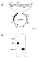
- FIGURE 1 illustrates the 4-1BB immunoglobulin fusion gene.
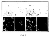
- FIGURE 2 illustrates the reactivity of 4-1BB Rg to murine tissue sections and binding of extracellular matrix proteins to COS cells expressing 4-1BB.
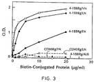
- FIGURE 3 illustrates the binding of 4-1BB Rg to extracellular matrix proteins.
- 4-1BB Rg was immobilized on plastic coated with affinity purified goat anti-human IgG antibodies and its ability to bind to increasing amounts of biotin-conjugated fibronectin, vitronectin, laminin or BSA was measured using an ELISA assay, as described in the materials and methods section.
- As a control the binding of a similarly immobilized CD40 Rg fusion protein to biotin-conjugated fibronectin and laminin was monitored.
- the data are expressed as the mean of triplicate samples. The SD were ⁇ 1%.
- the data shown are representative of at least three separate experiments.
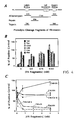
- FIGURE 4 illustrates that the binding of 4-1BB Rg to fibronectin is mediated by multiple fibronectin domains. Extracellular matrix proteins bind to 4-1BB Rg via overlapping binding sites.
- FIGURE 5 illustrates glycosaminoglycan blocking of the binding of 4-1BB to extracellular matrix proteins.
- 4-1BB Rg is modified with N-linked and O-linked oligosaccharides but not glycosaminoglycans.
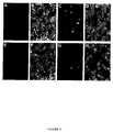
- FIGURE 6 illustrates mock transfection of COS cells stained with either anti-CD30 mAb (A, B) or anti-CD27 mAb (E, F) viewed under fluorescence (A, E) or phase contrast microscopy (B, F).
- COS cells transfected with CD30Rg (C, D) or CD27Rg (G, H) are similarly stained and viewed under fluorescence (C, G) or phase contrast microscopy (D, H), respectively.
- the present invention is directed to soluble fusion molecules of T cell surface ligands that are capable of inhibiting the interaction of leukocytes with proteins associated with the extracellular matrix of target tissue, and to methods for the inhibiting of leukocyte action.
- Soluble fusion molecules of the present invention include the extracellular domain of a T cell surface ligand fused to an immunoglobulin chain (Rg).
- Particularly preferred fusion molecules of the present invention include 4-1BBRg, CD30Rg, CD27Rg, CD40Rg and TNFRg.
- soluble lymphocyte ligand protein and grammatical variations thereof refers to a fusion protein composed of an extracellular region of a lymphocyte receptor protein such as 4-1BB, CD30, CD27, FAS, MRC OX40, TNF receptor (TNFR), etc. fused to a soluble structural protein such as in immunoglobulin (Rg) or T cell receptor region.
- a lymphocyte receptor protein such as 4-1BB, CD30, CD27, FAS, MRC OX40, TNF receptor (TNFR), etc.
- soluble structural protein such as in immunoglobulin (Rg) or T cell receptor region.
- Illustrative soluble lymphocyte ligand proteins of the present invention include 4-1BBRg, CD27Rg, and FAS Rg and the like.
- extracellular matrix associated proteins refers to proteins found within the extracellular matrix of tissues and include fibronectin, laminin, collagen type VI, and TNF-like molecules associated therewith.
- Codoning vector is any plasmid or virus into which a foreign DNA may be inserted to be cloned.
- Plasmid is an autonomous self-replicating extra-chromosomal circular DNA.
- Open Reading Frame is a DNA sequence which is (potentially) translatable into protein.
- Gene (cistron) is the segment of DNA that encodes the sequence of a peptide chain; it can include regions preceding and following the coding region (leader and trailer) as well as intervening sequences (introns) between individual coding segments (exons).
- “Expression”, as used herein, is the process undergone by a structural gene to produce a peptide or protein. It is a combination of transcription and translation.
- base pair is a partnership of adenine (A) with thymine (T), or of cytosine (C) with guanine (G) in a DNA double helix.
- expression vector is any plasmid or virus into which a foreign DNA may be inserted and/or expressed.
- downstream identifies sequences proceeding further in the direction of expression; for example, the peptide coding region of a gene is downstream from the initiation codon or in the 3' direction away from the gene; upstream is 5' to the sequence in question.
- PCR polymerase chain reaction
- unit dose refers to physically discrete units suitable as unitary dosages for animals, each unit contains a predetermined quantity of active material calculated to produce the desired therapeutic effect in association with the required diluent; that is, a carrier or vehicle.
- the specifications for the novel unit dose of this invention are dictated by, and are directly dependent upon, (a) the unique characteristics of the active material and the particular therapeutic effect to be achieved, and (b) the limitations inherent in the art of compounding such active material for therapeutic use.
- conservative substitution denotes that one amino acid residue has been replaced by another, biologically similar residue.
- conservative substitutions include the substitutions of one hydrophobic residue such as Ile, Val, Leu, or Met for another, or the substitution of one polar residue for another such as between Arg and Lys, between Glu and Asp or between Gln and Asn, and the like.
- an ionic residue by an oppositely charged ionic residue such as Asp by Lys has been determined conservative in the art in that those ionic groups are thought to merely provide solubility assistance. In general, however, since the replacements discussed herein are on a relatively short region, replacement of an ionic residue by another ionic residue of opposite charge is considered herein to be a "radical replacement" as are replacements by nonionic and ionic residues, and bulky residues such as Phe, Tyr or Trp and less bulky residues such as Gly, Ile and Val.
- nonionic and ionic residues are used herein in their usual sense to designate those amino acid residues that either bear no charge or normally bear a charge, respectively, at physiological pH value.
- exemplary nonionic residues include Thr and Gln, while exemplary ionic residues include Arg and Asp.
- phrases "pharmaceutically acceptable salts” and “physiologically tolerable salts”, as used interchangeably herein, refer to non-toxic alkali metal, alkaline earth metal and ammonium salts used in the pharmaceutical industry, including the sodium, potassium, lithium, calcium, magnesium and ammonium salts and the like that are prepared by methods well-known in the art.
- the phrase also includes non-toxic acid addition salts that are generally prepared by reacting the compounds in this invention with a suitable organic or inorganic acid.
- Representative salts include the hydrochloride, hydrobromide, sulfate, bisulfate, acetate, oxalate, valerate, oleate, laureate, vorate, benzoate, lactate, phosphate, tosylate, citrate, maleate, fumarate, succinate, tartrate and the like.
- cellular adhesion molecule refers to specific inflammatory cell surface molecules that are recognized and bind to vascular endothelium and/or granulocytes.
- the term "operatively attached” refers to the linkage of groups in a manner such that the binding affinity of the group is not inhibited by the attachment.
- IgG constant region refers to domains of the gamma chain of the IgG molecule that are adjacent to the variable region that corresponds to the first 107 amino acids of the gamma chain or fragments thereof.
- the four domains within the gamma chain constant region are designated CH1, H, CH2, and CH3.
- CH1 is adjacent to the variable region and encompasses amino acid residues 114 through 223.
- H is adjacent to CH1 and contains the cysteine residues that form the disulfide bonds which covalently link the two immunoglobulin heavy chains.
- CH2 is adjacent to the hinge and encompasses amino acid residues 246 through 361, followed by CH3 which contains amino acid residues 362 through 496.
- library refers to a large random collection of cloned DNA fragments obtained from the transcription system of interest. The gene library was then screened with labeled cDNA probes.
- a carrier is a material useful for administering the active compound and must be "acceptable” in the sense of being compatible with the other ingredients of the composition and not deleterious to the recipient thereof.
- compositions are prepared by any of the methods well known in the art of pharmacy all of which involve bringing into association the active compound and the carrier therefor.
- the agent utilized in the present invention can be administered in the form of conventional pharmaceutical compositions.
- Such compositions can be formulated so as to be suitable for oral or parenteral administration, or as suppositories.
- the agent is typically dissolved or dispersed in a physiologically tolerable carrier.
- the compounds of the present invention can be utilized in liquid compositions such as sterile suspensions or solutions, or as isotonic preparations containing suitable preservatives.
- liquid compositions such as sterile suspensions or solutions, or as isotonic preparations containing suitable preservatives.
- injectable media constituted by aqueous injectable isotonic and sterile saline or glucose solutions.
- Additional liquid forms in which the present compounds may be incorporated for administration include flavored emulsions with edible oils such as cottonseed oil, sesame oil, coconut oil, peanut oil, and the like, as well as elixirs and similar pharmaceutical vehicles.
- the present compounds can also be used in compositions such as tablets or pills, preferably containing a unit dose of the compound.
- the agent active ingredient
- conventional tabletting ingredients such as corn starch, lactose, sucrose, sorbitol, talc, stearic acid, magnesium stearate, dicalcium phosphate, gums or similar materials as non-toxic, physiologically tolerable carriers.
- the tablets or pills of the present compositions can be laminated or otherwise compounded to provide unit dosage forms affording prolonged or delayed action.
- the pharmaceutical formulation described herein can include, as appropriate, one or more additional carrier ingredients such as diluents, buffers, flavoring agents, binders, surface active agents, thickeners, lubricants, preservatives (including antioxidants) and the like, and substances included for the purpose of rendering the formulation isotonic with the blood of the intended recipient.
- additional carrier ingredients such as diluents, buffers, flavoring agents, binders, surface active agents, thickeners, lubricants, preservatives (including antioxidants) and the like, and substances included for the purpose of rendering the formulation isotonic with the blood of the intended recipient.
- the tablets or pills can also be provided with an enteric layer in the form of an envelope that serves to resist disintegration in the stomach and permits the active ingredient to pass intact into the duodenum or to be delayed in release.
- enteric layers or coatings including polymeric acids or mixtures of such acids with such materials as shellac, shellac and acetyl alcohol, cellulose acetate, and the like.
- a particularly suitable enteric coating comprises a styrene-maleic acid copolymer together with known materials that contribute to the enteric properties of the coating.
- label As used herein, the terms “label”, “indicating means” and “labeled indicating means”, in their various grammatical forms refer to single atoms and molecules that are either directly or indirectly involved in the production of a detectable signal to indicate or detect the presence of a reaction product. Such labels are themselves well known in clinical diagnostic chemistry and constitute a part of this invention only insofar as they are utilized with otherwise novel methods and/or systems.
- the indicating means can be a fluorescent labeling agent that chemically binds to antibodies or protein antigens without denaturing them to form a fluorochrome (dye) that is a useful immunofluorescent tracer.
- Suitable fluorescent labeling agents are fluorochrome, such as fluorescein isocyanate (FIC), fluorescein isothiocyanate (FITC), 5-dimethylamine-1-naphthalene sulfonyl chloride (DANSC), tetramethylrhodamine isocyanate (TRITC), lissamine and the like.
- Immunofluorescence analysis techniques are well known in the art, and for example, is described in DeLuca, "Immunofluorescence Analysis” in Immunofluorescence Analysis , Marchalonis et al., (1982) eds., John Wiley & Sons, Ltd., pp. 189-231, which is incorporated herein by reference.
- indicating means are colorimetric agents and enzymes, such as horseradish peroxidase, glucose oxidase or the like, linked as described above, as well as radioactive elements, preferably an element that produces gamma ray emissions.
- Elements which emit gamma rays such as 124I, 125I, 128I, 132 I, and 51Cr represent one class of radioactive indicating groups.
- Another group of useful labeling means are those elements such as 11C, 18F, 15O and 13N which emit positrons. The positrons so emitted produce gamma rays upon interaction with electrons present.
- the T cell activation marker 4-1BB was isolated by differential screening of a cDNA library prepared from the lymphokine activated cytotoxic T cell line L3 (Kwon, B.S. et al. (1987) Proc. Natl. Acad. Sci. USA 84 :2896-2900). Nucleotide sequencing of three overlapping cDNA cell lines of the 4-1BB gene revealed that it encodes a type I membrane protein of 256 amino acids. The extracellular domain of 4-1BB can be divided into two regions; a cysteine-rich region and a serine/threonine rich region.
- Ligand blocking experiments showed that various anionic carbohydrate polymers such as fucoidan, dextran sulfate or the glycosaminoglycans heparin, chondroitin sulfate, and hyaluronan could block the extracellular matrix protein-4-1BB interaction and suggests that carbohydrates participate in this interaction.
- Fibronectin binding to 4-1BB was not mediated by the RGD or CS-1 sequences which have been shown to mediate fibronectin binding to lymphocyte integrins (Springer T.A. (1990) Nature 346 :425-434). Binding studies with fibronectin peptides showed that distinct regions of the fibronectin molecule can bind to 4-1BB. Four out of the six proteolytic cleavage fragments of fibronectin were able to inhibit the 4-1BB-fibronectin interaction in a concentration dependent fashion. However, of these, only one, C120, contains sequences known previously to mediate fibronectin-cell attachment (Pytela, R. (1985) Cell 40 :191-198).
- T30 and T60 proteolytic fragments of fibronectin suggest that the binding sites of 4-1BB for all the extracellular matrix proteins are overlapping. T30 and T60 completely blocked the binding of laminin, vitronectin, and collagen VI to 4-1BB.
- Extracellular matrix proteins are known to bind to proteoglycans by an Arg-Gly-Asp (RGD) and CS-1 independent mechanism (Ruoslahti, E. et al. (1991) Cell 64 :867-869; Ruoslahti, E. (1988) Ann. Rev. Cell. Biol. 4 :229-255).
- RGD Arg-Gly-Asp
- CS-1 CS-1 independent mechanism
- 4-1BB Rg Digestion of 4-1BB Rg with chondroitin ABC lyase, hyaluronidase, heparinase, and heparatinase gave no evidence for the presence of these glycosaminoglycans on 4-1BB Rg. Keratanase digestion reduced the molecular mass of 4-1BB Rg, and 4-1BB Rg reacted with anti-keratin mAb suggesting that 4-1BB Rg contains carbohydrate moieties with the sequence GlcNac ( ⁇ -1,3)Gal( ⁇ -1,4)GlcNAc (Fukuda, M.N. (1981) J. Biol. Chem. 256 :3900-3905) and possibly keratan sulfate. Digestion with N-glycanase, O-glycanase and neuraminidase show that 4-1BB contains N-linked carbohydrates, O-linked carbohydrates and sialic acid. These results indicate that 4-1BB is
- 4-1BB is significantly homologous to the Cys rich domains of the short arms of laminin (Sasaki, M. et al. (1988) J. Biol. Chem. 263 :16536-16544), the proteoglycan perlecan (Noonan, D.M. et al. (1991) J. Biol. Chem. 266 :22939-22947) and the human ⁇ 5 integrin chain (Cheresh, D.A. et al.
- 4-1BB is a T cell surface protein involved in the binding of extracellular matrix proteins.
- An increasing body of evidence indicates that the interaction between leukocyte extracellular matrix receptors and their ligands result in the modulation of leukocyte function (Springer, T.A. (1990) Nature 346 :425-434; Nojim Y. et al. (1990) J. Exp. Med. 172 :11851192).
- This work suggests that the 4-1BB-extracellular matrix interaction results in the modulation of T cell function and that interactions between the cytoplasmic domain of 4-1BB and p56 lck are involved in this process.
- Soluble 4-1BB and antibodies to 4-1BB could be used to identify conditions under which normal T cells express high levels of 4-1BB and/or to identify cell lines which express high levels of this protein. Once these cells have been identified, the role of the interaction between extracellular matrix proteins and 4-1BB can be further characterized.
- soluble immunoglobulin fusion proteins of other members of the TNF gene family expressed by leukocytes have been prepared and are contemplated by the present invention.
- Figure 6 illustrates COS cell transfectants expressing fusion proteins of the T cell antigen CD27, and the B cell antigen CD30.
- a similar soluble immunoglobulin fusion molecule has been prepared for the leukoycte antigen FAS. These proteins were prepared in a maner analogous to that utilized for preparation of CD40 and 4-1BB fusion proteins. These fusion proteins can be utilized to inhibit leukocyte interactions thereby providing a method for modulating immune responses in a clinical setting.
- these proteins have ligands which are related to TNF as has been shown for CD40 and/or bind to extracellular matrix associated proteins as does 4-1BB.
- the fusion proteins of the present invention will be utilized to isolate specific ligands which may modulate immune responses.
- the full length 4-1BB cDNA sequence was amplified using the polymerase chain reaction (PCR) method using the conditions and reagents suggested by the vendor (Cetus).
- PCR polymerase chain reaction
- a 4-1BB synthetic primer encoding sequences immediately upstream of the signal sequence and including a Hind III site with the following sequence was synthesized:
- the soluble 4-1BB-immunoglobulin fusion protein (4-1BB-Rg) used in this study was created by isolating the cDNA fragment encoding the extracellular domain of 4-1BB by PCR as described (Aruffo, A. et al. (1990) Cell 61 :1303-1313).
- a 4-1BB primer encoding the sequence immediately downstream of the signal peptide cleavage point and including a Kpn I site was synthesized with the following sequence:
- a reverse primer containing a Bam HI restriction site was synthesized with the following sequence:
- the 4-1BB extracellular region PCR product was digested with Bam H1 and Kpn I and ligated to KpnI-Bam H1-cut CD5-IgG1 vector (Aruffo, A. et al. (1990) Cell 61 :1303-1313).
- the CD40 cDNA and the CD40-immunoglobulin fusion protein (CD40 Rg) used in these studies as controls have been previously described (Stamenkovic, I. et al. (1989) EMBO J 8 :1403-1410).
- the resulting constructs were transfected into COS cells and the desired fusion protein was recovered from the supernatant as described (Aruffo, A. et al. (1990) Cell 61 :1303-1313).
- Polystyrene 96-well plates were coated with affinity purified goat anti-human IgG Fc portion (Cappel) diluted to 5 ⁇ g/ml in PBS, 1 mM CaCl2, 1 mM MgCl2 (PBS/Ca2+/Mg2+) for one hour at 22° C.
- Wells were blocked with 10X Specimen Diluent Concentrate (Genetic Systems) diluted 1:10 with ddH20 overnight at 4 °C.
- Wells were washed and treated with 100 ⁇ l per well 4-1BB or CD40-Rg diluted in (PBS/Ca2+/Mg2+) at the appropriate concentrations (1.0 ⁇ g/ml for laminin) for one hour at 22 °C.
- Wells were treated with biotin-conjugated extracellular matrix proteins diluted in PBS/Ca2+/Mg2+ titrated from 20 ⁇ g/ml with 1:2 dilutions to 0.06 ⁇ g/ml for one hour at 22 °C.
- Bound material was detected using peroxidase-conjugated avidin (Sigma) diluted 1:500 in 1X Specimen Diluent Concentrate as above for one hour at 22 °C. Wells were washed and treated with EIA Chromogen Reagent (Genetic Systems) diluted 1:100 in EIA Buffered Substrate (Genetic Systems) for 15 min. at 22 °C. The colormetric reaction was quenched by adding Stop Buffer (Genetic Systems) and the optical density was measured on an ELISA reader at dual wavelengths 450,630.
- Blocking of the binding of 4-1BB-Rg to extracellular matrix proteins was tested using the ELISA method described above with the following alterations: After coating the wells with 4-1BB-Rg or CD40-Rg, fibronectin tryptic fragments were diluted to 4 nM, 0.8 nM, 0.16 nM, and 0.032 nM, and RGD and CS-1 peptides were diluted to 500 ⁇ g/ml, 100 ⁇ g/ml, 20 ⁇ g/ml and 4 ⁇ g/ml in PBS/Ca2+/Mg2+ added at 100 ⁇ l/well and incubated for one hour at 22 °C, after which biotin-conjugated extracellular matrix proteins were added as above to the well without washing and incubated for another hour at 22 °C.
- CDM8 vectors containing either the full length 4-1BB cDNA or the full length CD40 cDNA were transfected into COS cells using DEAE dextran method (Aruffo, A. et al. (1990) Cell 61 :1303-1313). Twenty-four hours post-transfection, cells were trypsinized, seeded onto 15 x 35 mm plates and allowed to grow another twenty-four hours. Before staining, cells were washed with staining buffer (PBS containing 2% FBS, 1 mM CaCl2, 1mM MgCl2). Cells were incubated with 4 ⁇ g of biotin-conjugated extracellular matrix protein for one hour at 22 °C.
- staining buffer PBS containing 2% FBS, 1 mM CaCl2, 1mM MgCl2
- Each scan measured the average fluorescence of a 1.8 mm2 area, and each plate was scanned three times. Background fluorescence was subtracted by removing all areas of fluorescence smaller than 400 um2. Each scan was expressed as the percent of the positive control (4-1BB expressing COS cells stained with biotinylated FN plus phycoerythrin-conjugated avidin), and the scans of each sample were averaged to generate a mean and standard deviation.
- COS cells transfected with either CD40-Rg or 4-1BB-Rg cDNAs were incubated in Cys/Met free DMEM for 30 min. before the addition of 1.0 mCi of 35 S-cysteine and 1.0 mCi 35S-methionine (Amersham). After 18 hours Rg fusion proteins were purified as described (Aruffo, A. et al. (1990) Cell 61 :1303-1313).
- 35 S-labeled 4-1BB-Rg or CD40-Rg were digested with heparinase (0.017 U/ml), heparatinase (ICN, 0.017 U/ml), keratinase (ICN, 0.33 U/ml) or hyaluronidase (Sigma, 17 U/ml) in PBS at pH 7.0, 7.0, 7.4 and 7.4, respectively for 18 hours at 37 °C.
- Chondroitin ABC lyase (ICN) digestions were performed with 1.0 U/ml of enzyme in 40 mM Tris-HCl (pH 8.6) containing 40 mM sodium acetate (NaOAc) for 18 hours at 37 °C.
- N-glycanase (Genzyme) digestions were performed with 35 U/ml enzyme in 0.1 Tris-HCl (pH 8.0) for 18 hours at 37 °C.
- Neuraminidase (Genzyme) digestions were done with 0.4 U/ml enzyme in 0.1 M NaOAc (pH 6.5) containing 10 mM Ca(OAc)2 for 2 hours at 37 °C.
- O-glycanase (Genzyme) digestions (0.17 U/ml) were performed on neuraminidase treated fusion proteins for 18 hours at 37 °C. Following digestions, SDS gel sample buffer was added and the digests subjected to SDS-PAGE analysis. Gels were fixed, dried and exposed to film.
- Proteins were electrophoresed in a 7.5% polyacrylamide gel, then electroblotted onto nitrocellulose for 1 hour at 80 mA.
- the membrane was blocked with 5% nonfat milk in PBS for 18 hours before including an anti-keratan sulfate antibody (5D4, ICN) diluted 1:100 in PBS 0.05% Tween-20 (PBS-TweenTM) for 2 hours at 22 °C.
- an anti-keratan sulfate antibody 5D4, ICN
- PBS-TweenTM PBS 0.05% Tween-20
- After one wash with the PBS-TweenTM solution the membrane was incubated with peroxidase-conjugated goat anti-mouse Ig (Tago) diluted 1:100 in PBS-TweenTM for 2 hours at 22 °C.
- the blot was washed and developed with 4-chloro-1-naphtol as substrate.
- a soluble immunoglobulin chimera of 4-1BB was prepared.
- This recombinant globulin (Rg) was used in immunohistochemical studies to identify tissues expressing the 4-1BB ligand.
- the fusion gene encoding the 4-1BB Rg chimera was prepared by genetic fusion of a cDNA fragment encoding the extracellular domain of 4-1BB to a genomic DNA fragment encoding the hinge, CH2, and CH3 domains of a human IgGl and subcloned into a mammalian expression vector ( Figure 1A). When transfected into COS cells this plasmid directs the expression and secretion of the 4-1BB Rg.
- the 4-1BB Rg is produced as a disulfide-linked homodimer ( Figure 1B) and accumulates in the supernatant of transfected cells in concentrations of ⁇ 14 mg/l.
- the 4-1BB Rg protein used in these studies was recovered from the supernatant by affinity chromatography on a protein A column.
- a CD40 Rg chimera was used or human IgG1.
- the human B cell surface protein CD40 is distantly related to 4-1BB (Mallett, S. et al. (1991) Immunol. Today 12 :220-223).
- Immunohistology studies with the 4-1BB Rg showed broad reactivity with a number of tissues including kidney, liver, epidermis and dermis of skin, in addition to primary and secondary lymphoid tissues.
- Isotype matched human IgG1 control showed no binding ( Figure 2).
- 4-1BB Rg bound to both lymphoid and stromal elements in the cortex and medulla, with more extensive labeling of the medulla.
- 4-1BB Rg bound red pulp more extensively than white pulp.
- the complete 4-1BB protein was expressed in a heterologous system and its ability to bind to extracellular matrix proteins was examined.
- a cDNA fragment encoding the complete 4-1BB protein was isolated from a murine T cell cDNA library, subcloned into the mammalian expression vector CDM8 (Seed, B. (1987) Nature 329 :840-843) and transfected into COS cells. Biotin-conjugated fibronectin, laminin, vitronectin and collagen VI bound to COS cells expressing 4-1BB whole biotin-conjugated collagen I and BSA or the phycoerythrin-conjugated strept-avidin alone did not ( Figure 2E-H). COS cells expressing CD40 (Stamenkovic, I. (1989) EMBO J 8 :1403-1410) showed no binding to any of the purified extracellular matrix or control proteins.
- integrins proteins which mediate the interaction between lymphocytes and the extracellular matrix have been identified, including the integrins which have been shown to bind to fibronectin, laminin, vitronectin and collagen (Springer, T.A. (1990) Nature 346 :425-434).
- the interaction between integrins and extracellular matrix proteins has been shown to be mediated by Arg-Gly-Asp (RGD) sequences in the extracellular matrix proteins (Ruoslahti, E. et al. (1987) Science 238 :491-497).
- RGD Arg-Gly-Asp
- the interaction between fibronectin and the VLA-5 integrin has been shown to be RGD dependent (Rytela, R.
- These peptides include RGD peptides which inhibit cell binding to either fibronectin or vitronectin, specifically, or RGD peptides which inhibit the binding to both fibronectin and vitronectin, or the CS-1 peptide, a scrambled version of the CS-1 peptide, or a mixture of RGD and CS-1 peptides.
- RGD peptides and peptide combinations had little or no effect on 4-1BB Rg-fibronectin adhesion even at concentrations 10X greater than those which inhibit the fibronectin-VLA-4 interaction (Nojim, Y. et al. (1990) J. Exp. Med. 172 :1185-1192).
- T30 and T60 fibronectin proteolytic fragments were found to block the binding of laminin, vitronectin, and collagen VI to immobilized 4-1BB Rg in a concentration dependent fashion ( Figure 4C).
- glycosaminoglycans as well as simple sugars were examined using COS cell transfectants expressing 4-1BB.
- the glycosaminoglycans heparin, hyaluronan and chondroitin sulfate partially inhibited the binding of extracellular matrix proteins to 4-1BB, while de-sulfated heparin had no effect (Figure 5A).
- the interaction between extracellular matrix proteins and 4-1BB was also inhibited by the anionic carbohydrate polymers fucoidan and dextran sulfate (Figure 5A).
- the simple carbohydrates fucose, galactose, mannose, glucose, galactose-6-sulfate, N-acetyl-galactosamine, N-acetyl-glucosamine, and N-acetyl-neuraminic acid had little or no effect on the 4-1BB-fibronectin interaction.
- 4-1BB contains no Ser-Gly sequences which are putative for chondroitin/dermatan sulfate attachment (Doege, K. et al. (1987) J. Biol. Chem. 262 :17757-17767; Oldberg, A. et al. (1987) Biochem. J. 243 :255-259) and SDS-PAGE analysis of 4-1BB Rg treated with chondroitin ABC lyase, heparinase, heparatinase, and hyaluronidase showed no evidence for the presence of these glycosaminoglycans (Figure 5B).
Abstract
Description
- The amino acid sequences of leukocyte surface molecules often contain motifs that have about 20 - 30% amino acid identity to motifs either within the molecule or in other proteins. These motifs are predicted to be evolutionarily related, have similar folding patterns and mediate similar types of interaction. A number of superfamilies have been described, containing conserved sequence motifs, such as the Ig-like domains of the immunoglobulin superfamily, fibronectin type III domains and a conserved motif found in many of the receptors for cytokines. The term domain is used where there are structural data to indicate that the motif may form a discrete structure, whereas the term motif is used where only sequence data are available.
- The members of a new superfamily, the nerve growth factor receptor (NGFR) superfamily include NGFR, found on neural cells; the B-cell antigen CD40; the MRC OX-40 antigen, which is a marker of activated T cells of the CD4 phenotype; two receptors for tumour necrosis factor (TNF), called TNFR-I and TNFR-II, which are found on a variety of cell types; a cDNA (4-1BB) obtained from T-cell clones; and SFV-T2, an open reading frame in Shope fibroma virus; FAS, a polypeptide expressed in various human cells including myeloid cells, T lymphoblastoid cells and diploid fibroblasts; CD27, a T cell activation antigen; and CD30, an antigen on the surface of T and B cells which is a characteristic marker for Hodgkin's disease. The members of the family are acids, in the extracellular part of the molecule. They are not associated with any other motif type in the extracellular region, although there is a hinge-like region, characterized by a lack of Cys residues and a high proportion of Ser, Thr and Pro, in MRC OX-40, NGFR and TNFR-II. This segment is likely to be glycosylated with O-linked sugars and could have an extended structure. Alignment of all the motifs shows that they contain either four or six cysteine residues in a characteristic and conserved pattern. There is considerable variation in the rest of the sequence, although some other residues are conserved in most of the motifs. The motifs in the 4-1BB protein are less homologous but two motifs separated by truncated motif can be found. The motifs contain a variety of residues found in the nonconserved positions. Although there are some differences in the number of residues between the Cys residues, the motifs are remarkably similar in length. The cysteine-rich motifs in the NGFR superfamily are clearly distinct from cysteine-rich repeats found in other proteins, such as the low-density lipoprotein (LDL) repeat. A statistical evaluation of the sequence similarities in the NGFR family and their comparison with other cysteine-rich repeats, such as the low-density lipoprotein (LDL) repeat, is discussed in Mallett, S. et al. (1990) EMBO. J. 9:1063-1068.
- A FAS polypeptide has been produced which is encoded by the cDNA for the human cell surface antigen FAS, and it has been shown that this polypeptide can mediate apoptosis (Itoh, N. et al. (1991) Cell 66:233-243). Defects in the FAS antigen have been associated with lymphoproliferative disorders in mice, producing lupus-like symptoms (Watanabe-Fukunaga, R. et al. (1992) Nature 356:314-317). Binding of TNF to the cell surface FAS antigen has been shown to mediate cytolytic activity, suggesting that the FAS antigen serves as a receptor for TNF's cytolytic effects (Yonehara, S. et al. in Tumor Necrosis Factor: Structure-Function Relationships and Clinical Applications., Basel (1992) pp. 58-65).
- CD30 is a marker for Hodgkin's disease and is interchangeably referred to as the Ki-1 antigen. CD30 has 5 cysteine-rich repeat regions that are homologous with the NGFR superfamily. Although the function of CD30 in Hodgkin's disease is unknown, it is a marker of T cell and B cell activation (Durkop, H. et al. (1992) Cell 68:421-427).
- The T cell activation antigen CD27 is a member of the NGFR gene family, and it may be involved in T cell proliferation and survival following activation (Camerini, D. et al. (1991) J. Immunol. 147:31653169). CD27 is a dimeric membrane glycoprotein and is found on the surface of most human T lymphocytes. Structural similarities of CD27 suggest that it belongs to a lymphocyte-specific subgroup of the NGFR family together with CD40, MRC OX40 and 4-1BB.
- Functions of four members of the NGFR superfamily are known: the NGFR, FAS, and the two TNFRs are growth factor receptors. Binding of polypeptide cytokines to these receptors results in a variety of functional effects. The binding of NGF to its high-affinity receptor (which contain two polypeptide chains) significantly affects neuronal differentiation and survival (Barde, Y-A. (1989) Neuron 2:1525-1534). Both TNF-α and TNF-β bind to the two TNFRs, leading to a diverse range of effects including inflammation and tumor cell death (Beutler, B. et al. (1989) Annu. Rev. Immunol. 7:625-655; Sprang, S.R. (1990) Trend Biochem. Sci. 15:366-368). There are limited in vitro functional data on the OX-40 and CD40 antigens. Antibodies against the OX40 antigen augment the T-cell response in a mixed lymphocyte reaction (Paterson, D.J. et al. (1987) Mol. Immunol. 24:1281-1290) and antibodies against CD40 enhance B-cell proliferation in the presence of a co-activator, such as tetradecanoylphorbol acetate (TPA) or CD20 antibodies (Ledbetter, J.A. et al. (1987) J. Immunol. 138:788-794), and synergize with IL-4 in vitro to induce B-cell differentiation and IgE production (Jabara, H.H. et al. (1990) J. Exp. Med. 172:1861-1864). The 4-1BB has been characterized only as a cDNA clone found in activated T-cell clones and there are not data on the product although it appears to code for a cell surface protein (Kwon, B.S., (1989) Proc. Natl. Acad. Sci. USA 86:1963-1967). The vaccinia and Shope viral sequences are characterized only as sequences and it is not known whether the proteins are expressed (Upton, C. et al. (1987) Virology 160:20-30; Howard, S.T. et al. (1991) Virology 180:633-647).
- The available evidence suggests that the NGFR superfamily cell surface proteins may function as receptors for cytokines. Those characterized so far are unusual in that they can bind to more than one ligand, and these ligands are polymeric in nature. The size of the extracellular domains of the NGFR superfamily members is relatively homogeneous compared with other superfamilies. The characterized members share 3 - 4 motifs, and in some cases a hinge-like region. Presumably, new members have yet to be defined and sequence analysis is required to see whether these features are common to all members of this superfamily.
- This superfamily is unusual in that three members have been shown to bind to more than one ligand and these ligands are dimeric or trimeric in contrast to many other cytokines. Both TNFR-I and TNFR-II bind TNF-α and TNF-β with high affinity (Schall, T.J. et al. (1990) Cell 61:361-370; Smith C.A. et al. (1990) Science 248:1010-1023; Sprang, S.R. (1990) Trends Biochem. Sci. 15:366-368). These two cytokines are only 30% identical in amino acid sequence. TNF-β is thought to have similar structure to TNF-α, which contains a β sheet structure in a trimeric arrangement (Jones, E.Y. et al. (1989) Nature 338:225-228; Eck, M.J. et al. (1989) J. Biol. Chem. 264:17595-17605). Surprisingly, the two TNFRs are only 27% identical in the extracellular domains, which is the same order as that for other, functionally unrelated, members of this superfamily.
- Remarkably, the two receptor molecules are homologous only within a 150 residue cysteine-rich segment presumed to be the ligand-binding domain. An analog of this sequence is found also in the receptor for nerve growth factor (NGFR), a hormone which is unlikely to have any structural resemblance to TNF. Thus, the surface of the binding scaffold presented by this cysteine-rich fold is potentially complementary to a variety of ligands.
- Tumor necrosis factor is the collective name for two related cytokine hormones, TNF-α and TNF-β. TNF-α is named for its ability to induce hemorrhagic necrosis of certain murine tumors and its cytotoxicity to some tumor cell lines (Beutler, B. et al. (1988) Annu. Rev. Biochem. 57:505-518). Known also as cachectin, TNF-α is a mediator of inflammation, sepsis-induced shock and the wasting syndrome (cachexia) associated with chronic infection and neoplastic disease. A multipotent cytokine, TNF-α acts as an immunoregulator, represses lipogenic pathways in adipocytes, stimulates collagenase production in epithelial cells and is a growth factor for fibroblasts. TNF-β, or lymphotoxin, is a lymphocyte-derived homologue of TNF-α and appears to effect many of the same functions.
- Consistent with its wide-ranging activities, receptors for TNF are expressed by many somatic cell types. Receptors bind both TNF-α and TNF-β (Stauber, G.B. et al. (1989) J. Biol. Chem. 264:3573-3576), but no other cytokines or growth factors, with dissociation constants in the nanomolar range. Recently it has become evident that there are at least two molecular species of TNF receptor (TNFR), a high affinity (Kd = 0.07 nM) 75-80 kDa myeloid cell type receptor and a 55-60 kDa receptor of epithelial origin with an affinity constant (Kd) of 0.3 nM (Brockhaus, M. et al. (1990) Proc. Natl. Acad. Sci. USA 87:3127-3131). The two molecules differ in their level of glycosylation, yield different peptide fragments upon tryptic digestion, and are immunologically distinct.
- The present invention is directed to soluble lymphocyte ligand proteins and methods of modulating leukocyte function by inhibiting the interaction between extracellular matrix associated proteins and lymphocytes. In this method the soluble ligand proteins are administered to a patient and bind to these extracellular matrix associated proteins. This blocks the ability of the lymphocytes to bind to the extracellular matrix receptors.
- The soluble lymphocyte ligand proteins of this invention contain the soluble extracellular portions of lymphocyte surface receptors, particularly lymphocyte proteins which are members of the nerve growth factor receptor superfamily. Specific proteins of the present invention include 4-1BB fusion proteins, such as 4-1BB Rg, as well as fusion proteins of CD27, CD30, FAS, etc.
- The present invention demonstrates that the soluble lymphocyte ligand proteins developed are involved in the binding of extracellular matrix associated proteins. Because interactions between leukocyte receptors and their ligands results in the modulation of leukocyte function, the inhibition of this interaction by administration of the soluble proteins of the present invention effectively alters leukocyte activity.
- FIGURE 1 illustrates the 4-1BB immunoglobulin fusion gene.
- A. Elements in the mammalian expression vector CDM7 plasmid containing the 4-1BB Rg fusion gene include: a human cytomegalovirus enhancer/promoter element (CMV); and SV40 virus derived intron and polyadenylation sequences (splice + An); and SV40 virus derived origin of replication (SV40 ori); and E. coli origin of replication (π VX ori); an M13 phage derived origin of replication (M13 ori); and as a bacterial genetic marker the Sup F gene (Sup F). The 4-1BB Rg is composed of: a CDS derived amino terminal secretory signal sequence (CD5ss); a cDNA fragment encoding the extracellular domain of 4-1BB (4-1BB); and a genomic DNA fragment encoding the hinge (H) and constant domains (CH2 and CH3) of human IgG1. Unique restriction enzyme sites flanking each of the elements in the 4-1BB Rg fusion are shown.
- B. Radiolabeled 4-1BB Rg was purified by adsorption to and elution from protein A-sepharose columns as described in materials and methods and electrophoresed under nonreducing (lane 1) or reducing (lane 2) conditions. The electrophoretic mobility of molecular mass standards are shown on the left.
- FIGURE 2 illustrates the reactivity of 4-1BB Rg to murine tissue sections and binding of extracellular matrix proteins to COS cells expressing 4-1BB.
- A. Immunoperoxidase localization of 4-1BB Rg binding to murine thymus in both the cortex (c) and medulla (m). Stromal cells in the medullary area of the thymus were more extensively labeled.
- B. Reactivity of murine thymus tissue with normal human IgG1 and peroxidase-labeled goat anti-human Ig. The only reaction product observed was associated with endogenous peroxidase activity displayed by granulocytes and other cells in the thymus medulla.
- C. Immunoperoxidase localization of 4-1BB binding to murine spleen. Both the white pulp (wp) and red pulp (rp) areas of the spleen bound the 4-1BB Rg, with more extensive binding in red pulp areas.
- D. When normal human IgG1 was substituted for the 4-1BB Rg, deposition of reaction product was indistinguishable from endogenous peroxidase.
Figures A - D 50X. - E and F. Phase and immunofluorescence micrographs depicting the binding of biotinylated laminin to COS cells transfected with 4-1BB. Arrowheads in phase micrographs identify COS cells binding laminin.
- G and H. Phase and immunofluorescence micrographs depicting the binding of biotinylated fibronectin to COS cells transfected with 4-1BB. Arrowheads in phase micrograph identify COS cells binding fibronectin.
Figures E - H 200 X. - FIGURE 3 illustrates the binding of 4-1BB Rg to extracellular matrix proteins. 4-1BB Rg was immobilized on plastic coated with affinity purified goat anti-human IgG antibodies and its ability to bind to increasing amounts of biotin-conjugated fibronectin, vitronectin, laminin or BSA was measured using an ELISA assay, as described in the materials and methods section. As a control the binding of a similarly immobilized CD40 Rg fusion protein to biotin-conjugated fibronectin and laminin was monitored. The data are expressed as the mean of triplicate samples. The SD were <1%. The data shown are representative of at least three separate experiments.
- FIGURE 4 illustrates that the binding of 4-1BB Rg to fibronectin is mediated by multiple fibronectin domains. Extracellular matrix proteins bind to 4-1BB Rg via overlapping binding sites.
- A. Schematic representation of the human fibronectin polypeptide and the proteolytic fragments used in the blocking experiments. The two trypsin derived fragments T30 and T60 have an approximate molecular mass of 30 and 60 kD, respectively. The α-chymotrypsin derived fragments C45, C120, and C40 have an approximate molecular mass of 456, 120, and 40 kD, respectively. The pepsin derived fragment P50 has an approximate molecular mass of 50 kD. These peptides were obtained from Telios Pharmaceuticals Inc. (San Diego, CA).
- B. The ability of the different proteolytic fragments of fibronectin to block the binding of fibronectin to 4-1BB Rg immobilized on plastic was measured using an ELISA assay as described in the material and methods section. Each fragment was tested at four different concentrations. The fibronectin fragments tested include T30, T60, C120, C40, C45 and P50. The data are expressed as the mean of triplicate samples. SD are shown. The data shown are representative of at least three separate experiments.
- C. The ability of increasing amounts of the T30 and C40 fragments of fibronectin to inhibit the binding of laminin, vitronectin and collagen VI to 4-1BB Rg immobilized on plastic was measured by the ELISA method as described in the materials and methods section. The data are expressed as the means of triplicate samples. The data shown are representative of at least three separate experiments.
- FIGURE 5 illustrates glycosaminoglycan blocking of the binding of 4-1BB to extracellular matrix proteins. 4-1BB Rg is modified with N-linked and O-linked oligosaccharides but not glycosaminoglycans.
- A. The ability of the anionic carbohydrate polymers fucoidan (H) and dextran sulfate (D), and the glycosaminoglycans heparin (C), chondroitin (E), and hyaluronan (F) and desulfated-heparin (G) to block the binding of fibronectin to COS cells transfectants expressing 4-1BB was tested as described in materials and methods. Binding is shown as % binding relative to the binding of 4-1BB COS cell transfectants to fibronectin in the absence of inhibitors (A). Controls included the binding of phycoerythrin conjugated strepavidin alone to COS cells expressing 4-1BB (B) and the binding of COS cell transfectants expressing CD40 to fibronectin (I). The data shown are expressed as the means of triplicate scans. The SD are calculated as described in the materials and methods. The data shown are representative of at least three separate experiments.
- B. Radiolabeled purified 4-1BB Rg was treated with chondroitin ABC lyase, heparinase, heparatinase, keratanase, hyaluronidase, N-glycanase, O-glycanase, and neuraminidase (lanes 2-9) as described in materials and methods section. The electrophoretic mobility of the treated 4-1BB Rg protein was compared to that of untreated 4-1BB Rg protein (lane 1) using SDS-PAGE as described in materials and methods section. The electrophoretic mobility of molecular mass standards are shown on the left.
- FIGURE 6 illustrates mock transfection of COS cells stained with either anti-CD30 mAb (A, B) or anti-CD27 mAb (E, F) viewed under fluorescence (A, E) or phase contrast microscopy (B, F). COS cells transfected with CD30Rg (C, D) or CD27Rg (G, H) are similarly stained and viewed under fluorescence (C, G) or phase contrast microscopy (D, H), respectively.
- The present invention is directed to soluble fusion molecules of T cell surface ligands that are capable of inhibiting the interaction of leukocytes with proteins associated with the extracellular matrix of target tissue, and to methods for the inhibiting of leukocyte action.
- Soluble fusion molecules of the present invention include the extracellular domain of a T cell surface ligand fused to an immunoglobulin chain (Rg). Particularly preferred fusion molecules of the present invention include 4-1BBRg, CD30Rg, CD27Rg, CD40Rg and TNFRg.
- In order to more clearly describe the present invention and its embodiments the following definitions are included.
- As used herein, the term "soluble lymphocyte ligand protein" and grammatical variations thereof refers to a fusion protein composed of an extracellular region of a lymphocyte receptor protein such as 4-1BB, CD30, CD27, FAS, MRC OX40, TNF receptor (TNFR), etc. fused to a soluble structural protein such as in immunoglobulin (Rg) or T cell receptor region. Illustrative soluble lymphocyte ligand proteins of the present invention include 4-1BBRg, CD27Rg, and FAS Rg and the like.
- As used herein, the term "extracellular matrix associated proteins" refers to proteins found within the extracellular matrix of tissues and include fibronectin, laminin, collagen type VI, and TNF-like molecules associated therewith.
- "Cloning vector", as used herein, is any plasmid or virus into which a foreign DNA may be inserted to be cloned.
- "Plasmid", as used herein, is an autonomous self-replicating extra-chromosomal circular DNA.
- "Open Reading Frame" (ORF), as used herein, is a DNA sequence which is (potentially) translatable into protein.
- "Gene (cistron)", as used herein, is the segment of DNA that encodes the sequence of a peptide chain; it can include regions preceding and following the coding region (leader and trailer) as well as intervening sequences (introns) between individual coding segments (exons).
- "Expression", as used herein, is the process undergone by a structural gene to produce a peptide or protein. It is a combination of transcription and translation.
- As used herein, the term "base pair" (bp) is a partnership of adenine (A) with thymine (T), or of cytosine (C) with guanine (G) in a DNA double helix.
- As used herein, the term "expression vector" is any plasmid or virus into which a foreign DNA may be inserted and/or expressed.
- As used herein, the term "downstream" identifies sequences proceeding further in the direction of expression; for example, the peptide coding region of a gene is downstream from the initiation codon or in the 3' direction away from the gene; upstream is 5' to the sequence in question.
- As used herein, the term "polymerase chain reaction" (PCR) refers to the amplification of DNA molecules by the successive use of a temperature stable DNA polymerase to copy the DNA chain, separating the complementary chains by heating, adding primers and repeating the process about 30 times to produce approximately 10⁹ copies of the DNA. By use of the PCR technique, minute amounts of DNA can be amplified to produce sufficient DNA for use in various procedures.
- The term "unit dose" refers to physically discrete units suitable as unitary dosages for animals, each unit contains a predetermined quantity of active material calculated to produce the desired therapeutic effect in association with the required diluent; that is, a carrier or vehicle. The specifications for the novel unit dose of this invention are dictated by, and are directly dependent upon, (a) the unique characteristics of the active material and the particular therapeutic effect to be achieved, and (b) the limitations inherent in the art of compounding such active material for therapeutic use.
- The term "correspond" in its various grammatical forms, as used herein and in the claims in relation to peptide sequences means the peptide sequence described plus or minus up to 3 amino acid residues at either or both of the amino and carboxy termini and containing only conservative substitutions in particular amino acid residues along the peptide and/or polypeptide sequence.
- The term "conservative substitution" as used above denotes that one amino acid residue has been replaced by another, biologically similar residue. Examples of conservative substitutions include the substitutions of one hydrophobic residue such as Ile, Val, Leu, or Met for another, or the substitution of one polar residue for another such as between Arg and Lys, between Glu and Asp or between Gln and Asn, and the like.
- In some instances the replacement of an ionic residue by an oppositely charged ionic residue such as Asp by Lys has been determined conservative in the art in that those ionic groups are thought to merely provide solubility assistance. In general, however, since the replacements discussed herein are on a relatively short region, replacement of an ionic residue by another ionic residue of opposite charge is considered herein to be a "radical replacement" as are replacements by nonionic and ionic residues, and bulky residues such as Phe, Tyr or Trp and less bulky residues such as Gly, Ile and Val.
- The terms "nonionic" and "ionic" residues are used herein in their usual sense to designate those amino acid residues that either bear no charge or normally bear a charge, respectively, at physiological pH value. Exemplary nonionic residues include Thr and Gln, while exemplary ionic residues include Arg and Asp.
- The phrases "pharmaceutically acceptable salts" and "physiologically tolerable salts", as used interchangeably herein, refer to non-toxic alkali metal, alkaline earth metal and ammonium salts used in the pharmaceutical industry, including the sodium, potassium, lithium, calcium, magnesium and ammonium salts and the like that are prepared by methods well-known in the art. The phrase also includes non-toxic acid addition salts that are generally prepared by reacting the compounds in this invention with a suitable organic or inorganic acid. Representative salts include the hydrochloride, hydrobromide, sulfate, bisulfate, acetate, oxalate, valerate, oleate, laureate, vorate, benzoate, lactate, phosphate, tosylate, citrate, maleate, fumarate, succinate, tartrate and the like.
- As used herein, the term "cellular adhesion molecule" refers to specific inflammatory cell surface molecules that are recognized and bind to vascular endothelium and/or granulocytes.
- As used herein, the term "operatively attached" refers to the linkage of groups in a manner such that the binding affinity of the group is not inhibited by the attachment.
- As used herein, the term "IgG constant region" refers to domains of the gamma chain of the IgG molecule that are adjacent to the variable region that corresponds to the first 107 amino acids of the gamma chain or fragments thereof. The four domains within the gamma chain constant region are designated CH₁, H, CH₂, and CH₃. CH₁ is adjacent to the variable region and encompasses amino acid residues 114 through 223. H (hinge; residues 224-245) is adjacent to CH₁ and contains the cysteine residues that form the disulfide bonds which covalently link the two immunoglobulin heavy chains. CH₂ is adjacent to the hinge and encompasses amino acid residues 246 through 361, followed by CH₃ which contains amino acid residues 362 through 496.
- As used herein, the term "library" refers to a large random collection of cloned DNA fragments obtained from the transcription system of interest. The gene library was then screened with labeled cDNA probes.
- A carrier is a material useful for administering the active compound and must be "acceptable" in the sense of being compatible with the other ingredients of the composition and not deleterious to the recipient thereof.
- The pharmaceutical compositions are prepared by any of the methods well known in the art of pharmacy all of which involve bringing into association the active compound and the carrier therefor.
- For therapeutic use, the agent utilized in the present invention can be administered in the form of conventional pharmaceutical compositions. Such compositions can be formulated so as to be suitable for oral or parenteral administration, or as suppositories. In these compositions, the agent is typically dissolved or dispersed in a physiologically tolerable carrier.
- As an example, the compounds of the present invention can be utilized in liquid compositions such as sterile suspensions or solutions, or as isotonic preparations containing suitable preservatives. Particularly well suited for the present purposes are injectable media constituted by aqueous injectable isotonic and sterile saline or glucose solutions. Additional liquid forms in which the present compounds may be incorporated for administration include flavored emulsions with edible oils such as cottonseed oil, sesame oil, coconut oil, peanut oil, and the like, as well as elixirs and similar pharmaceutical vehicles.
- The present compounds can also be used in compositions such as tablets or pills, preferably containing a unit dose of the compound. To this end, the agent (active ingredient) is mixed with conventional tabletting ingredients such as corn starch, lactose, sucrose, sorbitol, talc, stearic acid, magnesium stearate, dicalcium phosphate, gums or similar materials as non-toxic, physiologically tolerable carriers. The tablets or pills of the present compositions can be laminated or otherwise compounded to provide unit dosage forms affording prolonged or delayed action.
- It should be understood that in addition to the aforementioned carrier ingredients the pharmaceutical formulation described herein can include, as appropriate, one or more additional carrier ingredients such as diluents, buffers, flavoring agents, binders, surface active agents, thickeners, lubricants, preservatives (including antioxidants) and the like, and substances included for the purpose of rendering the formulation isotonic with the blood of the intended recipient.
- The tablets or pills can also be provided with an enteric layer in the form of an envelope that serves to resist disintegration in the stomach and permits the active ingredient to pass intact into the duodenum or to be delayed in release. A variety of materials can be used for such enteric layers or coatings, including polymeric acids or mixtures of such acids with such materials as shellac, shellac and acetyl alcohol, cellulose acetate, and the like. A particularly suitable enteric coating comprises a styrene-maleic acid copolymer together with known materials that contribute to the enteric properties of the coating.
- As used herein, the terms "label", "indicating means" and "labeled indicating means", in their various grammatical forms refer to single atoms and molecules that are either directly or indirectly involved in the production of a detectable signal to indicate or detect the presence of a reaction product. Such labels are themselves well known in clinical diagnostic chemistry and constitute a part of this invention only insofar as they are utilized with otherwise novel methods and/or systems.
- The indicating means can be a fluorescent labeling agent that chemically binds to antibodies or protein antigens without denaturing them to form a fluorochrome (dye) that is a useful immunofluorescent tracer. Suitable fluorescent labeling agents are fluorochrome, such as fluorescein isocyanate (FIC), fluorescein isothiocyanate (FITC), 5-dimethylamine-1-naphthalene sulfonyl chloride (DANSC), tetramethylrhodamine isocyanate (TRITC), lissamine and the like. Immunofluorescence analysis techniques are well known in the art, and for example, is described in DeLuca, "Immunofluorescence Analysis" in Immunofluorescence Analysis, Marchalonis et al., (1982) eds., John Wiley & Sons, Ltd., pp. 189-231, which is incorporated herein by reference.
- Other preferred indicating means are colorimetric agents and enzymes, such as horseradish peroxidase, glucose oxidase or the like, linked as described above, as well as radioactive elements, preferably an element that produces gamma ray emissions. Elements which emit gamma rays, such as ¹²⁴I, ¹²⁵I, ¹²⁸I, 132I, and ⁵¹Cr represent one class of radioactive indicating groups. Another group of useful labeling means are those elements such as ¹¹C, ¹⁸F, ¹⁵O and ¹³N which emit positrons. The positrons so emitted produce gamma rays upon interaction with electrons present.
- The T cell activation marker 4-1BB was isolated by differential screening of a cDNA library prepared from the lymphokine activated cytotoxic T cell line L3 (Kwon, B.S. et al. (1987) Proc. Natl. Acad. Sci. USA 84:2896-2900). Nucleotide sequencing of three overlapping cDNA cell lines of the 4-1BB gene revealed that it encodes a type I membrane protein of 256 amino acids. The extracellular domain of 4-1BB can be divided into two regions; a cysteine-rich region and a serine/threonine rich region. Published comparison between the predicted 4-1BB amino acid sequence and those of other known proteins show that the extracellular domain of 4-1BB has limited homology to members of the nerve growth factor receptor family (NGFR) (Mallett, S. et al. (1991) Immunol. Today 12:220-223) and that its cytoplasmic domain contains a putative binding site for the lymphocyte specific protein tyrosine kinase p561ck (Shaw, A.S. et al. (1990) Mol. Cell. Biol. 10:1853-1862). RNA blot studies showed that mRNAs encoding 4-1BB are expressed by a number of activated T-cell lines (Kwon, B.S. et al. (1987) Proc. Natl. Acad. Sci. USA 84:2986-2900; Kwon, B.S. et al. (1989) Cell. Immunol. 121:414-422). As a first step in elucidating the function of 4-1BB the 4-1BB ligand(s) were identified and characterized.
- Immunohistochemical studies with 4-1BB recombinant immunoglobulin fusion protein (4-1BB Rg) revealed that 4-1BB Rg bound extensively to a variety of tissues and suggested that extracellular matrix components might be involved. The interaction between 4-1BB and several extracellular-matrix proteins including fibronectin, laminin, vitronectin, and collagen VI was confirmed using COS cell transfectants expressing 4-1BB. Ligand blocking experiments showed that various anionic carbohydrate polymers such as fucoidan, dextran sulfate or the glycosaminoglycans heparin, chondroitin sulfate, and hyaluronan could block the extracellular matrix protein-4-1BB interaction and suggests that carbohydrates participate in this interaction.
- Immunohistochemical studies showing that 4-1BB Rg bound to virtually all tissues examined suggested that extracellular matrix components might be serving as ligands for this molecule. The interaction between extracellular matrix proteins and 4-1BB was confirmed using COS cell transfectants. COS cells expressing 4-1BB were found to bind fibronectin, laminin, vitronectin and collagen VI but not collagen I while those expressing CD40 (Stamenkovic, I. et al. (1989) EMBO J. 8:1403-1410) bound none of these proteins. This binding was found to be concentration dependent and saturable. Fibronectin binding to 4-1BB was not mediated by the RGD or CS-1 sequences which have been shown to mediate fibronectin binding to lymphocyte integrins (Springer T.A. (1990) Nature 346:425-434). Binding studies with fibronectin peptides showed that distinct regions of the fibronectin molecule can bind to 4-1BB. Four out of the six proteolytic cleavage fragments of fibronectin were able to inhibit the 4-1BB-fibronectin interaction in a concentration dependent fashion. However, of these, only one, C120, contains sequences known previously to mediate fibronectin-cell attachment (Pytela, R. (1985) Cell 40:191-198).
- Blocking studies with the T30 and T60 proteolytic fragments of fibronectin suggest that the binding sites of 4-1BB for all the extracellular matrix proteins are overlapping. T30 and T60 completely blocked the binding of laminin, vitronectin, and collagen VI to 4-1BB.
- Extracellular matrix proteins are known to bind to proteoglycans by an Arg-Gly-Asp (RGD) and CS-1 independent mechanism (Ruoslahti, E. et al. (1991) Cell 64:867-869; Ruoslahti, E. (1988) Ann. Rev. Cell. Biol. 4:229-255). Using blocking experiments, the possibility was investigated that the 4-1BB-extracellular matrix protein interaction could be inhibited with glycosaminoglycans and other anionic carbohydrate polymers and that 4-1BB Rg itself is a proteoglycan. The interaction between 4-1BB and extracellular matrix proteins was completely blocked by the anionic carbohydrate polymer fucoidan, and partially blocked by the glycosaminoglycans, heparin sulfate, chondroitin sulfate and hyaluronan, and by the oligosaccharide dextran sulfate. Conversely, de-sulfated-heparin and the simple sugars, fucose, mannose, glucose, glucose-6-sulfate, N-acetyl-glycosamine, N-acetyl-galactosamine, and N-acetyl-neuraminic acid did not affect the interaction. These results suggest that carbohydrates might be involved in mediating the interaction between 4-1BB and extracellular matrix proteins.
- Digestion of 4-1BB Rg with chondroitin ABC lyase, hyaluronidase, heparinase, and heparatinase gave no evidence for the presence of these glycosaminoglycans on 4-1BB Rg. Keratanase digestion reduced the molecular mass of 4-1BB Rg, and 4-1BB Rg reacted with anti-keratin mAb suggesting that 4-1BB Rg contains carbohydrate moieties with the sequence GlcNac (β-1,3)Gal(β-1,4)GlcNAc (Fukuda, M.N. (1981) J. Biol. Chem. 256:3900-3905) and possibly keratan sulfate. Digestion with N-glycanase, O-glycanase and neuraminidase show that 4-1BB contains N-linked carbohydrates, O-linked carbohydrates and sialic acid. These results indicate that 4-1BB is a glycoprotein.
- The interaction between 4-1BB and extracellular matrix proteins was unexpected, given the previous report that 4-1BB is homologous to the members of the nerve growth factor receptor (NGFR) family (Mallett, S. et al.(1991) Immunol. Today 12:220-223; Smith, C.A. et al. (1990) Science 248:1019-1023). This family of proteins consists of the NGFR (Radeke, M.J. et al. (1987) Nature 325:593-597) two receptors for tumor necrosis factor (Loetscher, H. et al. 1990) Cell 61:351-359; Schall, T.J. et al. (1990) Cell 61:361-370; Smith C.A. et al. (1990) Science 248:1019-1023), the B-cell antigens CD30 (Durkop, H. et al. (1992) Cell 68:421-427), CD40 (Stamenkovic, I. et al. (1989) EMBO J. 8:1403-1410) and FAS (Itoh, N. et al. (1991) Cell 66:233-243), the T cell surface antigens OX-40 (Mallett, S. et al. (1990) EMBO J. 9:1063-1068) and CD27 (Camerini, D. et al. (1991) J. Immunol. 147:3165-3169) and the open reading frame of the Shope fibroma virus designated SFV-T2 (Sprang, S.R. (1990) Trends in Biochem. Sci. 15:366 368). Ligands for only four of these proteins (NGFR, TNFR-1 and II and CD40) have been identified (Sprang, S.R. (1990) Trends in Biochem. Sci. 15:366-368; Bothwell, M. (1991) Cell 65:915-918) and none have previously been reported to bind to extracellular matrix proteins. Furthermore, the CD40 Rg and COS cell transfectants expressing CD40 (Stamenkovic, I. et al. (1989) EMBO J. 8:1403-1410) used as controls showed no binding to extracellular matrix proteins.
- These observations led to a reexamination of the homology between 4-1BB and other proteins in the nBRF and SWISPROT data bases using the FASTP program. In addition to the previously reported homology, 4-1BB is significantly homologous to the Cys rich domains of the short arms of laminin (Sasaki, M. et al. (1988) J. Biol. Chem. 263:16536-16544), the proteoglycan perlecan (Noonan, D.M. et al. (1991) J. Biol. Chem. 266:22939-22947) and the human β5 integrin chain (Cheresh, D.A. et al. (1989) Cell 57:59-69; McLean, J.W. et al. (1990) J. Biol. Chem. 265:17126-17131). Sequence alignment shows that the extracellular domain of 4-1BB can be divided into two laminin- like Cys rich domains each containing 7 Cys residues. These Cys residues are conserved among 4-1BB and the Cys rich domains of laminin and perlecan (data not shown). Interestingly, a non-RGD sequence responsible for mediating cell attachment has been mapped to one such Cys rich domain on the laminin B1 chain (Graf, J. et al. (1987) Cell 48:989-996). The possible significance of the homology between 4-1BB and the human β5 integrin chain is less clear. The homology algorithm matches a number of residues in the two sequences, however most of them are not conserved between members of the integrin family of proteins. In addition, although β5 integrins are known to bind to vitronectin and fibronectin, this adhesive interaction is mediated by RGD sequences (Springer, T.A. (1990) Nature 346:425-434). These observations in conjunction with the binding studies, suggest that 4-1BB might be distantly related to more than one gene family.
- The results presented here indicate that 4-1BB is a T cell surface protein involved in the binding of extracellular matrix proteins. An increasing body of evidence indicates that the interaction between leukocyte extracellular matrix receptors and their ligands result in the modulation of leukocyte function (Springer, T.A. (1990) Nature 346:425-434; Nojim Y. et al. (1990) J. Exp. Med. 172:11851192). This work suggests that the 4-1BB-extracellular matrix interaction results in the modulation of T cell function and that interactions between the cytoplasmic domain of 4-1BB and p56lck are involved in this process. Soluble 4-1BB and antibodies to 4-1BB could be used to identify conditions under which normal T cells express high levels of 4-1BB and/or to identify cell lines which express high levels of this protein. Once these cells have been identified, the role of the interaction between extracellular matrix proteins and 4-1BB can be further characterized.
- In order to further clarify and enable the present invention as illustrated in the Examples described herein below, a general description of the materials and methods utilized in producing the results disclosed is presented. These materials and methods illustrate the technology utilized and are not intended to be limiting of the present invention. Other variations and modifications of these methods are well-known and are contemplated by this invention.
- In addition to CD40 and 4-1BB, soluble immunoglobulin fusion proteins of other members of the TNF gene family expressed by leukocytes have been prepared and are contemplated by the present invention. Figure 6 illustrates COS cell transfectants expressing fusion proteins of the T cell antigen CD27, and the B cell antigen CD30. A similar soluble immunoglobulin fusion molecule has been prepared for the leukoycte antigen FAS. These proteins were prepared in a maner analogous to that utilized for preparation of CD40 and 4-1BB fusion proteins. These fusion proteins can be utilized to inhibit leukocyte interactions thereby providing a method for modulating immune responses in a clinical setting. Similar to studies with CD40 and 4-1BB, these proteins have ligands which are related to TNF as has been shown for CD40 and/or bind to extracellular matrix associated proteins as does 4-1BB. The fusion proteins of the present invention will be utilized to isolate specific ligands which may modulate immune responses.
- The full length 4-1BB cDNA sequence was amplified using the polymerase chain reaction (PCR) method using the conditions and reagents suggested by the vendor (Cetus). A 4-1BB synthetic primer encoding sequences immediately upstream of the signal sequence and including a Hind III site with the following sequence was synthesized:

- A reverse primer containing a Xho I restriction site with the following sequence was synthesized:

- Full length 4-1BB PCR products were digested with Hind III and Xho I and ligated to Hind III-Xho I-cut CDM8 vector (Seed, B. (1987) Nature 329:840-842).
- The soluble 4-1BB-immunoglobulin fusion protein (4-1BB-Rg) used in this study was created by isolating the cDNA fragment encoding the extracellular domain of 4-1BB by PCR as described (Aruffo, A. et al. (1990) Cell 61:1303-1313). A 4-1BB primer encoding the sequence immediately downstream of the signal peptide cleavage point and including a Kpn I site was synthesized with the following sequence:

- A reverse primer containing a Bam HI restriction site was synthesized with the following sequence:

- The 4-1BB extracellular region PCR product was digested with Bam H1 and Kpn I and ligated to KpnI-Bam H1-cut CD5-IgG1 vector (Aruffo, A. et al. (1990) Cell 61:1303-1313). The CD40 cDNA and the CD40-immunoglobulin fusion protein (CD40 Rg) used in these studies as controls have been previously described (Stamenkovic, I. et al. (1989) EMBO J 8:1403-1410). The resulting constructs were transfected into COS cells and the desired fusion protein was recovered from the supernatant as described (Aruffo, A. et al. (1990) Cell 61:1303-1313).
- The techniques used for light microscopic immunohistochemistry have been described in detail (Farr, A.G. et al. (1981) Immunol. Methods 47:129). Freshly cut 6 µm cryostat sections of murine thymus, lymph node, spleen or skin were mounted on amino alkylsilane-treated slides (Rentrop, M. et al. (1986) Histochem. J. 18:271) and allowed to air dry for at least two hours before fixation (Guan, J.L. et al. (1990) Cell 60:53-61; Nojim, Y. et al. (1990) J. Exp. Med. 172:1185-1192; Ruoslahti, E. (1988) Ann. Rev. Biochem. 57:375-413; Ruoslahti, E. et al. (1991) Cell 64:867-869; Ruoslahti, E. (1988) Ann. Rev. Cell. Biol. 4:229-255; Doege, K. (1986) J. Biol. Chem. 262:17757-17767) minutes in acetone at -20° C. After rinsing in PBS, the sections were incubated for 1 hour at room temperature with 4-1BB Rg or human IgG1 diluted in PBS containing Ca⁺² and Mg⁺² (2mM each). All subsequent solutions and diluents contained divalent cations. After several washes in PBS, slides were then incubated with peroxidase-labeled goat anti-human IgG1 antibodies using the same conditions as employed for the primary antibodies. Peroxidase activity was demonstrated using 3, 3'-diaminobenzidine as the electron donor. Slides were then hydrated, mounted with coverslips, and photographed without additional counterstaining.
- Binding of the purified 4-1BB-Rg to murine fibronectin, rat laminin, human vitronectin, human collagen type VI and I (Telios Pharmaceuticals) and bovine serum albumin (BSA) (Sigma) was tested using an ELISA method. Polystyrene 96-well plates were coated with affinity purified goat anti-human IgG Fc portion (Cappel) diluted to 5 µg/ml in PBS, 1 mM CaCl2, 1 mM MgCl2 (PBS/Ca²⁺/Mg²⁺) for one hour at 22° C. The wells were blocked with 10X Specimen Diluent Concentrate (Genetic Systems) diluted 1:10 with ddH₂0 overnight at 4 °C. Wells were washed and treated with 100 µl per well 4-1BB or CD40-Rg diluted in (PBS/Ca²⁺/Mg²⁺) at the appropriate concentrations (1.0 µg/ml for laminin) for one hour at 22 °C. Wells were treated with biotin-conjugated extracellular matrix proteins diluted in PBS/Ca²⁺/Mg²⁺ titrated from 20 µg/ml with 1:2 dilutions to 0.06 µg/ml for one hour at 22 °C. Bound material was detected using peroxidase-conjugated avidin (Sigma) diluted 1:500 in 1X Specimen Diluent Concentrate as above for one hour at 22 °C. Wells were washed and treated with EIA Chromogen Reagent (Genetic Systems) diluted 1:100 in EIA Buffered Substrate (Genetic Systems) for 15 min. at 22 °C. The colormetric reaction was quenched by adding Stop Buffer (Genetic Systems) and the optical density was measured on an ELISA reader at dual wavelengths 450,630.
- Blocking of the binding of 4-1BB-Rg to extracellular matrix proteins was tested using the ELISA method described above with the following alterations: After coating the wells with 4-1BB-Rg or CD40-Rg, fibronectin tryptic fragments were diluted to 4 nM, 0.8 nM, 0.16 nM, and 0.032 nM, and RGD and CS-1 peptides were diluted to 500 µg/ml, 100 µg/ml, 20 µg/ml and 4 µg/ml in PBS/Ca²⁺/Mg²⁺ added at 100 µl/well and incubated for one hour at 22 °C, after which biotin-conjugated extracellular matrix proteins were added as above to the well without washing and incubated for another hour at 22 °C.
- CDM8 vectors containing either the full length 4-1BB cDNA or the full length CD40 cDNA were transfected into COS cells using DEAE dextran method (Aruffo, A. et al. (1990) Cell 61:1303-1313). Twenty-four hours post-transfection, cells were trypsinized, seeded onto 15 x 35 mm plates and allowed to grow another twenty-four hours. Before staining, cells were washed with staining buffer (PBS containing 2% FBS, 1 mM CaCl₂, 1mM MgCl₂). Cells were incubated with 4 µg of biotin-conjugated extracellular matrix protein for one hour at 22 °C. Cells were washed twice with staining buffer and incubated with 5.0 µg of fluorescein isothiocyanate (FITC) conjugated avidin (Molecular Probes) or 1.0 µg of phycoerythrin-conjugated avidin (Biomeda) for 30 min. at 4 °C. Cells were washed twice with staining buffer and examined by fluorescence microscopy.
- In blocking experiments, cells were washed with staining buffer and incubated with 400 µg of the simple or 100 µg of the complex carbohydrates or polymers for one hour at 22 °C. Four µg of biotin-conjugated extracellular matrix protein was then added without washing and incubated for one hour at 22 °C. Staining proceeded as above. Fluorescence of COS cells expressing 4-1BB or CD40 was measured with an ACAS 570 microscope (Meridian) using a single image program and a combination of two filters: a diachroic filter which collects light longer than 515 nm, and a long pass filter which collects light longer than 605 nm. All 35 mm² plates contained cells at an equal density. Each scan measured the average fluorescence of a 1.8 mm² area, and each plate was scanned three times. Background fluorescence was subtracted by removing all areas of fluorescence smaller than 400 um². Each scan was expressed as the percent of the positive control (4-1BB expressing COS cells stained with biotinylated FN plus phycoerythrin-conjugated avidin), and the scans of each sample were averaged to generate a mean and standard deviation.
- COS cells transfected with either CD40-Rg or 4-1BB-Rg cDNAs were incubated in Cys/Met free DMEM for 30 min. before the addition of 1.0 mCi of 35S-cysteine and 1.0 mCi ³⁵S-methionine (Amersham). After 18 hours Rg fusion proteins were purified as described (Aruffo, A. et al. (1990) Cell 61:1303-1313).
- 35S-labeled 4-1BB-Rg or CD40-Rg were digested with heparinase (0.017 U/ml), heparatinase (ICN, 0.017 U/ml), keratinase (ICN, 0.33 U/ml) or hyaluronidase (Sigma, 17 U/ml) in PBS at pH 7.0, 7.0, 7.4 and 7.4, respectively for 18 hours at 37 °C. Chondroitin ABC lyase (ICN) digestions were performed with 1.0 U/ml of enzyme in 40 mM Tris-HCl (pH 8.6) containing 40 mM sodium acetate (NaOAc) for 18 hours at 37 °C. N-glycanase (Genzyme) digestions were performed with 35 U/ml enzyme in 0.1 Tris-HCl (pH 8.0) for 18 hours at 37 °C. Neuraminidase (Genzyme) digestions were done with 0.4 U/ml enzyme in 0.1 M NaOAc (pH 6.5) containing 10 mM Ca(OAc)₂ for 2 hours at 37 °C. O-glycanase (Genzyme) digestions (0.17 U/ml) were performed on neuraminidase treated fusion proteins for 18 hours at 37 °C. Following digestions, SDS gel sample buffer was added and the digests subjected to SDS-PAGE analysis. Gels were fixed, dried and exposed to film.
- Proteins were electrophoresed in a 7.5% polyacrylamide gel, then electroblotted onto nitrocellulose for 1 hour at 80 mA. The membrane was blocked with 5% nonfat milk in PBS for 18 hours before including an anti-keratan sulfate antibody (5D4, ICN) diluted 1:100 in PBS 0.05% Tween-20 (PBS-Tween™) for 2 hours at 22 °C. After one wash with the PBS-Tween™ solution the membrane was incubated with peroxidase-conjugated goat anti-mouse Ig (Tago) diluted 1:100 in PBS-Tween™ for 2 hours at 22 °C. The blot was washed and developed with 4-chloro-1-naphtol as substrate.
- Having generally described this invention, a further understanding can be obtained by reference to certain specific examples which are provided herein for purposes of illustration only and are not intended to be limiting unless otherwise specified.
- PREPARATION OF SOLUBLE 4-1BB IMMUNOGLOBULIN CHIMERA
- For the identification and characterization of 4-1BB ligand(s) a soluble immunoglobulin chimera of 4-1BB was prepared. This recombinant globulin (Rg) was used in immunohistochemical studies to identify tissues expressing the 4-1BB ligand. The fusion gene encoding the 4-1BB Rg chimera was prepared by genetic fusion of a cDNA fragment encoding the extracellular domain of 4-1BB to a genomic DNA fragment encoding the hinge, CH2, and CH3 domains of a human IgGl and subcloned into a mammalian expression vector (Figure 1A). When transfected into COS cells this plasmid directs the expression and secretion of the 4-1BB Rg. The 4-1BB Rg is produced as a disulfide-linked homodimer (Figure 1B) and accumulates in the supernatant of transfected cells in concentrations of ∼ 14 mg/l. The 4-1BB Rg protein used in these studies was recovered from the supernatant by affinity chromatography on a protein A column. As a control for nonspecific and Fc-mediated binding a CD40 Rg chimera was used or human IgG1. The human B cell surface protein CD40 is distantly related to 4-1BB (Mallett, S. et al. (1991) Immunol. Today 12:220-223).
- TISSUE DISTRIBUTION OF THE 4-1BB LIGAND
- Immunohistology studies with the 4-1BB Rg showed broad reactivity with a number of tissues including kidney, liver, epidermis and dermis of skin, in addition to primary and secondary lymphoid tissues. Isotype matched human IgG1 control showed no binding (Figure 2). Within the thymus, 4-1BB Rg bound to both lymphoid and stromal elements in the cortex and medulla, with more extensive labeling of the medulla. In the spleen, 4-1BB Rg bound red pulp more extensively than white pulp.
- The widespread distribution of ligand(s) for the 4-1BB Rg in tissues of epithelial or mesenchymal derivation raised the possibility that extracellular matrix components may function as ligands.
- EXTRACELLULAR MATRIX PROTEINS ARE LIGANDS FOR 4-1BB
- To investigate the possible interaction between 4-1BB and extracellular matrix proteins, the complete 4-1BB protein was expressed in a heterologous system and its ability to bind to extracellular matrix proteins was examined. A cDNA fragment encoding the complete 4-1BB protein was isolated from a murine T cell cDNA library, subcloned into the mammalian expression vector CDM8 (Seed, B. (1987) Nature 329:840-843) and transfected into COS cells. Biotin-conjugated fibronectin, laminin, vitronectin and collagen VI bound to COS cells expressing 4-1BB whole biotin-conjugated collagen I and BSA or the phycoerythrin-conjugated strept-avidin alone did not (Figure 2E-H). COS cells expressing CD40 (Stamenkovic, I. (1989) EMBO J 8:1403-1410) showed no binding to any of the purified extracellular matrix or control proteins.
- To evaluate the relative affinity of the interaction and extracellular matrix proteins, 4-1BB Rg was immobilized on plastic coated with anti-human IgG antibodies and its ability to bind to increasing concentrations of extracellular matrix proteins was tested (Figure 3). The binding of fibronectin, laminin, vitronectin, and collagen VI to 4-1BB was saturable and half maximal binding was reached at concentrations of ∼ 5.0, 1.5, 1.0 and 2.0 µg/ml, respectively. Collagen I, BSA and alkaline phosphatase-conjugated avidin alone showed no binding to all of the extracellular matrix proteins and control proteins tested (Figure 3).
- 4-1BB-EXTRACELLULAR MATRIX PROTEIN INTERACTIONS ARE NOT MEDIATED BY RGD OR CS-1 SEOUENCES
- A number of proteins which mediate the interaction between lymphocytes and the extracellular matrix have been identified, including the integrins which have been shown to bind to fibronectin, laminin, vitronectin and collagen (Springer, T.A. (1990) Nature 346:425-434). In some instances, the interaction between integrins and extracellular matrix proteins has been shown to be mediated by Arg-Gly-Asp (RGD) sequences in the extracellular matrix proteins (Ruoslahti, E. et al. (1987) Science 238:491-497). For example, the interaction between fibronectin and the VLA-5 integrin has been shown to be RGD dependent (Rytela, R. et al (1985) Cell 40:191-198), while the interaction between fibronectin and the VLA-4 integrin has been shown to be mediated by an alternatively spliced domain of fibronectin (CS-1) (Wayner, E. et al. (1989) J. Cell Biol. 109:1321-1330; Guan, J.L. et al. (1990) Cell 60:53-61). To investigate the role of RGD and CS-1 sequences in the 4-1BB extracellular matrix protein binding, the ability of several RGD and CS-1 peptides to block the binding of fibronectin to 4-1BB Rg immobilized on plastic was tested. These peptides include RGD peptides which inhibit cell binding to either fibronectin or vitronectin, specifically, or RGD peptides which inhibit the binding to both fibronectin and vitronectin, or the CS-1 peptide, a scrambled version of the CS-1 peptide, or a mixture of RGD and CS-1 peptides. These peptides and peptide combinations had little or no effect on 4-1BB Rg-fibronectin adhesion even at concentrations 10X greater than those which inhibit the fibronectin-VLA-4 interaction (Nojim, Y. et al. (1990) J. Exp. Med. 172:1185-1192).
- In an effort to identify the region or regions of the fibronectin molecule responsible for mediating the 4-1BB fibronectin interaction, a number of blocking studies were carried out with six overlapping proteolytic fragments of fibronectin (Figure 4A) (Ruoslahti, E. (1988) Ann. Rev. Biochem. 57:375-413). Three of these, T30, T60 and C120, blocked the binding of fibronectin to immobilized 4-1BB Rg in a concentration dependent fashion, the C45 peptide had a partial blocking effect and C40 and P50 had no blocking effect (Figure 4B). This experiment provides evidence that multiple sites within fibronectin mediate the 4-1BB-fibronectin interaction.
- THE EXTRACELLULAR MATRIX PROTEIN BINDING SITES ON 4-1BB ARE OVERLAPPING
- To determine whether the interaction between the different extracellular matrix proteins and 4-1BB was mediated by the same regions of the 4-1BB protein, blocking experiments were carried out with the T30 and T60 fibronectin proteolytic fragments. The T30 and T60 fragments were found to block the binding of laminin, vitronectin, and collagen VI to immobilized 4-1BB Rg in a concentration dependent fashion (Figure 4C).
- 4-1BB ADHESION IS INHIBITED BY GLYCOSAMINOGLYCANS
- In light of the inability of RGD or CS-1 peptides to interfere with the interaction between 4-1BB Rg and extracellular matrix components, the basis for this interaction was studied further. Because proteoglycans have been shown to bind to extracellular matrix proteins and growth factors by a largely RGD-independent mechanism (Ruoslahti, E. et al. (1990) Cell 64:867-869; Ruoslahti, E. (1988) Ann. Rev. Cell Biol. 4:229-255), we examined the effects of glycosaminoglycans on 4-1BB interactions with extracellular matrix components and determined whether 4-1BB was proteoglycan. The ability of glycosaminoglycans as well as simple sugars to inhibit the interaction between extracellular matrix proteins and 4-1BB was examined using COS cell transfectants expressing 4-1BB. The glycosaminoglycans heparin, hyaluronan and chondroitin sulfate partially inhibited the binding of extracellular matrix proteins to 4-1BB, while de-sulfated heparin had no effect (Figure 5A). The interaction between extracellular matrix proteins and 4-1BB was also inhibited by the anionic carbohydrate polymers fucoidan and dextran sulfate (Figure 5A). The simple carbohydrates fucose, galactose, mannose, glucose, galactose-6-sulfate, N-acetyl-galactosamine, N-acetyl-glucosamine, and N-acetyl-neuraminic acid had little or no effect on the 4-1BB-fibronectin interaction.
- 4-1BB contains no Ser-Gly sequences which are putative for chondroitin/dermatan sulfate attachment (Doege, K. et al. (1987) J. Biol. Chem. 262:17757-17767; Oldberg, A. et al. (1987) Biochem. J. 243:255-259) and SDS-PAGE analysis of 4-1BB Rg treated with chondroitin ABC lyase, heparinase, heparatinase, and hyaluronidase showed no evidence for the presence of these glycosaminoglycans (Figure 5B). Digestion with keratinase (endo-β-galactosidase) reduced the molecular mass of 4-1BB Rg (Figure 5B) and 4-1BB Rg reacted with the anti-keratan sulfate monoclonal antibody 5D4 (Caterson, B. et al. (1983) J. Biol. Chem. 258:8848-8854) suggesting that 4-1BB Rg is modified with carbohydrate moieties containing the sequence GlcNac(β-1, 3)Gal(β-1,4)GlcNAc (Fukuda, M.N. (1981) J. Biol. Chem. 256:3900-₃905) and possibly keratan sulfate. Parallel digests with N-glycanase, O-glycanase and neuraminidase demonstrate that 4-1BB Rg contains N-linked and O-linked carbohydrates as well as sialic acid (Figure 5B).
- The foregoing description and Examples are intended as illustrative of the present invention, but not as limiting. Numerous variations and modifications may be effected without departing from the true spirit and scope of the present invention.


Claims (20)
- A method of modulating leukocyte function comprising inhibiting the interaction between leukocyte receptors and extracellular matrix associated proteins on tissues with a soluble lymphocyte ligand protein such that said lymphocyte ligand protein binds to said extracellular matrix associated proteins and blocks interaction with said leukocytes.
- The method of Claim 1, wherein said lymphocyte ligand protein is a member of the nerve growth factor receptor superfamily.
- The method of Claim 2, wherein said protein is a soluble 4-1BB protein.
- The method of Claim 2, wherein said protein is a soluble CD27.
- The method of Claim 2, wherein said protein is a soluble CD30.
- The method of Claim 2, wherein said protein is a soluble CD40.
- The method of Claim 2, wherein said protein is a soluble TNF.
- A soluble lymphocyte receptor protein capable of inhibiting the interaction of leukocytes with extracellular matrix associated proteins.
- A soluble lymphocyte ligand protein capable of inhibiting the interaction of leukocytes with extracellular matrix associated proteins.
- The soluble protein of Claim 8 or Claim 9 comprising a 4-1BB fusion protein.
- The soluble protein of Claim 10, wherein the 4-1BB fusion protein is 4-1BB-Rg.
- The soluble protein of Claim 8 or Claim 9 comprising a CD27 fusion protein.
- The soluble protein of Claim 12, wherein the CD27 fusion protein is CD27 Rg.
- The soluble protein of Claim 8 or Claim 9 comprising a CD30 fusion protein.
- The soluble protein of Claim 14, wherein the CD30 fusion protein is CD30 Rg.
- The soluble protein of Claim 8 or Claim 9 comprising a FAS fusion protein.
- The soluble protein of Claim 16, wherein the FAS fusion protein is FAS Rg.
- Use of a soluble protein according to any one of Claims 8 top 17 in the preparation of a composition for use in the modulation of leukocyte function by inhibiting the interaction between leukocyte receptors and extracellular matrix proteins, wherein said soluble protein binds to the extracellular matrix protein and blocks interaction with the leukocytes.
- Use according to Claim 18, wherein said soluble protein is a member of the nerve growth factor receptor superfamily.
- A soluble protein according to any one of Claims 8 to 17 for use in therapy.
Applications Claiming Priority (2)
| Application Number | Priority Date | Filing Date | Title |
|---|---|---|---|
| US96970092A | 1992-10-30 | 1992-10-30 | |
| US969700 | 1992-10-30 |
Publications (2)
| Publication Number | Publication Date |
|---|---|
| EP0595659A2 true EP0595659A2 (en) | 1994-05-04 |
| EP0595659A3 EP0595659A3 (en) | 1995-01-11 |
Family
ID=25515876
Family Applications (1)
| Application Number | Title | Priority Date | Filing Date |
|---|---|---|---|
| EP93308688A Withdrawn EP0595659A3 (en) | 1992-10-30 | 1993-10-29 | Extracellular matrix receptor ligands that modulate leukocyte function. |
Country Status (8)
| Country | Link |
|---|---|
| EP (1) | EP0595659A3 (en) |
| JP (1) | JPH072899A (en) |
| AU (1) | AU5034493A (en) |
| CA (1) | CA2109398A1 (en) |
| FI (1) | FI934751A (en) |
| IL (1) | IL107432A0 (en) |
| MX (1) | MX9306682A (en) |
| NO (1) | NO933860L (en) |
Cited By (13)
| Publication number | Priority date | Publication date | Assignee | Title |
|---|---|---|---|---|
| WO1995028957A2 (en) * | 1994-04-25 | 1995-11-02 | Trustees Of Dartmouth College | Methods for inducing t cell tolerance to a tissue or organ graft |
| WO1999023229A1 (en) * | 1997-10-30 | 1999-05-14 | Cornell Research Foundation, Inc. | A method of inhibiting an immune response to a recombinant vector |
| US6087329A (en) * | 1991-10-25 | 2000-07-11 | Immunex Corporation | CD40 ligand polypeptide |
| US6210669B1 (en) | 1996-10-11 | 2001-04-03 | Bristol-Myers Squibb Co. | Methods and compositions for immunomodulation |
| US6290972B1 (en) | 1991-10-25 | 2001-09-18 | Immunex Corporation | Method of augmenting a vaccine response by administering CD40 ligand |
| WO2008080623A2 (en) | 2006-12-28 | 2008-07-10 | Deutsches Krebsforschungszentrum Stiftung des öffentlichen Rechts | Neutralization of cd95 activity blocks invasion of glioblastoma cells in vivo |
| US7405270B2 (en) | 1991-10-25 | 2008-07-29 | Immunex Corporation | CD40-Ligand lacking native-pattern glycosylation |
| WO2014013036A1 (en) | 2012-07-18 | 2014-01-23 | Apogenix Gmbh | Inhibitors of the cd95 signaling pathway for treatment of mds |
| EP3076179A1 (en) | 2015-03-30 | 2016-10-05 | Deutsches Krebsforschungszentrum Stiftung des Öffentlichen Rechts | Diagnosis and treatment of low grade gliomas |
| WO2017044752A1 (en) * | 2015-09-10 | 2017-03-16 | University Of Pittsburgh-Of The Commonwealth System Of Higher Education | Anti-fibrotic effect of cd70 |
| WO2020205662A1 (en) | 2019-03-29 | 2020-10-08 | Myst Therapeutics, Inc. | Ex vivo methods for producing a t cell therapeutic and related compositions and methods |
| WO2021108727A1 (en) | 2019-11-27 | 2021-06-03 | Myst Therapeutics, Inc. | Method of producing tumor-reactive t cell composition using modulatory agents |
| WO2021174208A1 (en) | 2020-02-27 | 2021-09-02 | Myst Therapeutics, Llc | Methods for ex vivo enrichment and expansion of tumor reactive t cells and related compositions thereof |
Families Citing this family (2)
| Publication number | Priority date | Publication date | Assignee | Title |
|---|---|---|---|---|
| US5246113A (en) * | 1992-02-11 | 1993-09-21 | Riverwood International Corporation | Carrier for stacked articles |
| JP2014184804A (en) | 2013-03-22 | 2014-10-02 | Toyota Motor Corp | Control unit of vehicular transmission system |
Citations (5)
| Publication number | Priority date | Publication date | Assignee | Title |
|---|---|---|---|---|
| EP0333517A2 (en) * | 1988-03-18 | 1989-09-20 | The Rockefeller University | Method and agent for inhibiting the binding of human polymorphonuclear leukocytes to endothelium and compositions therefor |
| WO1990003983A1 (en) * | 1988-10-03 | 1990-04-19 | Scripps Clinic And Research Foundation | Peptides and antibodies that inhibit integrin-ligand binding |
| WO1991009874A1 (en) * | 1990-01-05 | 1991-07-11 | La Jolla Cancer Research Foundation | Novel fibronectin receptor |
| US5057604A (en) * | 1988-08-03 | 1991-10-15 | Washington University | Novel monoclonal antibodies |
| WO1991019511A1 (en) * | 1990-06-18 | 1991-12-26 | The General Hospital Corporation | CONTROLLING CELLULAR IMMUNE/INFLAMMATORY RESPONSES WITH β2 INTEGRINS |
-
1993
- 1993-10-27 FI FI934751A patent/FI934751A/en not_active Application Discontinuation
- 1993-10-27 CA CA002109398A patent/CA2109398A1/en not_active Abandoned
- 1993-10-27 MX MX9306682A patent/MX9306682A/en unknown
- 1993-10-27 NO NO933860A patent/NO933860L/en unknown
- 1993-10-28 IL IL107432A patent/IL107432A0/en unknown
- 1993-10-29 AU AU50344/93A patent/AU5034493A/en not_active Abandoned
- 1993-10-29 JP JP5272526A patent/JPH072899A/en active Pending
- 1993-10-29 EP EP93308688A patent/EP0595659A3/en not_active Withdrawn
Patent Citations (5)
| Publication number | Priority date | Publication date | Assignee | Title |
|---|---|---|---|---|
| EP0333517A2 (en) * | 1988-03-18 | 1989-09-20 | The Rockefeller University | Method and agent for inhibiting the binding of human polymorphonuclear leukocytes to endothelium and compositions therefor |
| US5057604A (en) * | 1988-08-03 | 1991-10-15 | Washington University | Novel monoclonal antibodies |
| WO1990003983A1 (en) * | 1988-10-03 | 1990-04-19 | Scripps Clinic And Research Foundation | Peptides and antibodies that inhibit integrin-ligand binding |
| WO1991009874A1 (en) * | 1990-01-05 | 1991-07-11 | La Jolla Cancer Research Foundation | Novel fibronectin receptor |
| WO1991019511A1 (en) * | 1990-06-18 | 1991-12-26 | The General Hospital Corporation | CONTROLLING CELLULAR IMMUNE/INFLAMMATORY RESPONSES WITH β2 INTEGRINS |
Non-Patent Citations (2)
| Title |
|---|
| JOURNAL OF BIOLOGICAL CHEMISTRY., vol.267, no.9, February 1992, UNITED STATES pages 5747 - 5750 MARSTERS SA ET AL 'Identification of cysteine-rich domains of the type 1 tumor necrosis factor receptor involved in ligand binding.' * |
| PROCEEDINGS OF THE NATIONAL ACADEMY OF SCIENCES OF USA., vol.86, 1989, WASHINGTON US pages 1963 - 1967 KWON B.S ET AL. * |
Cited By (21)
| Publication number | Priority date | Publication date | Assignee | Title |
|---|---|---|---|---|
| US6087329A (en) * | 1991-10-25 | 2000-07-11 | Immunex Corporation | CD40 ligand polypeptide |
| US6290972B1 (en) | 1991-10-25 | 2001-09-18 | Immunex Corporation | Method of augmenting a vaccine response by administering CD40 ligand |
| US6391637B1 (en) | 1991-10-25 | 2002-05-21 | Richard J. Armitage | Use of CD40 ligand, a cytokine that binds CD40, to stimulate hybridoma cells |
| US7405270B2 (en) | 1991-10-25 | 2008-07-29 | Immunex Corporation | CD40-Ligand lacking native-pattern glycosylation |
| US7501124B2 (en) | 1994-04-25 | 2009-03-10 | Trustees Of Dartmouth College | Methods of inducing T-cell non-responsiveness with anti-gp39 24-31 antibodies |
| WO1995028957A3 (en) * | 1994-04-25 | 1995-11-23 | Dartmouth College | Methods for inducing t cell tolerance to a tissue or organ graft |
| EP1530973A2 (en) * | 1994-04-25 | 2005-05-18 | The Trustees Of Dartmouth College | Methods for inducing T cell tolerance to a tissue or organ graft |
| EP1530973A3 (en) * | 1994-04-25 | 2006-12-27 | The Trustees Of Dartmouth College | Methods for inducing T cell tolerance to a tissue or organ graft |
| WO1995028957A2 (en) * | 1994-04-25 | 1995-11-02 | Trustees Of Dartmouth College | Methods for inducing t cell tolerance to a tissue or organ graft |
| US6210669B1 (en) | 1996-10-11 | 2001-04-03 | Bristol-Myers Squibb Co. | Methods and compositions for immunomodulation |
| WO1999023229A1 (en) * | 1997-10-30 | 1999-05-14 | Cornell Research Foundation, Inc. | A method of inhibiting an immune response to a recombinant vector |
| US6525029B1 (en) | 1997-10-30 | 2003-02-25 | Cornell Research Foundation, Inc. | Method of inhibiting and immune response to a recombinant vector |
| WO2008080623A2 (en) | 2006-12-28 | 2008-07-10 | Deutsches Krebsforschungszentrum Stiftung des öffentlichen Rechts | Neutralization of cd95 activity blocks invasion of glioblastoma cells in vivo |
| EP2428252A1 (en) | 2006-12-28 | 2012-03-14 | Deutsches Krebsforschungszentrum Stiftung des Öffentlichen Rechts | Neutralization of CD95 activity blocks invasion of glioblastoma cells in vivo |
| WO2014013036A1 (en) | 2012-07-18 | 2014-01-23 | Apogenix Gmbh | Inhibitors of the cd95 signaling pathway for treatment of mds |
| EP3150224A1 (en) | 2012-07-18 | 2017-04-05 | Apogenix AG | Inhibitors of the cd95 signaling pathway for treatment of mds |
| EP3076179A1 (en) | 2015-03-30 | 2016-10-05 | Deutsches Krebsforschungszentrum Stiftung des Öffentlichen Rechts | Diagnosis and treatment of low grade gliomas |
| WO2017044752A1 (en) * | 2015-09-10 | 2017-03-16 | University Of Pittsburgh-Of The Commonwealth System Of Higher Education | Anti-fibrotic effect of cd70 |
| WO2020205662A1 (en) | 2019-03-29 | 2020-10-08 | Myst Therapeutics, Inc. | Ex vivo methods for producing a t cell therapeutic and related compositions and methods |
| WO2021108727A1 (en) | 2019-11-27 | 2021-06-03 | Myst Therapeutics, Inc. | Method of producing tumor-reactive t cell composition using modulatory agents |
| WO2021174208A1 (en) | 2020-02-27 | 2021-09-02 | Myst Therapeutics, Llc | Methods for ex vivo enrichment and expansion of tumor reactive t cells and related compositions thereof |
Also Published As
| Publication number | Publication date |
|---|---|
| MX9306682A (en) | 1994-04-29 |
| JPH072899A (en) | 1995-01-06 |
| NO933860D0 (en) | 1993-10-27 |
| EP0595659A3 (en) | 1995-01-11 |
| AU5034493A (en) | 1994-05-12 |
| FI934751A (en) | 1994-05-01 |
| FI934751A0 (en) | 1993-10-27 |
| IL107432A0 (en) | 1994-01-25 |
| CA2109398A1 (en) | 1994-05-01 |
| NO933860L (en) | 1994-05-02 |
Similar Documents
| Publication | Publication Date | Title |
|---|---|---|
| Chalupny et al. | T-cell activation molecule 4-1BB binds to extracellular matrix proteins. | |
| EP0595659A2 (en) | Extracellular matrix receptor ligands that modulate leukocyte function | |
| Stamenkovic et al. | The hematopoietic and epithelial forms of CD44 are distinct polypeptides with different adhesion potentials for hyaluronate‐bearing cells. | |
| US5808004A (en) | Fragments of a lymphocyte adhesion receptor for high endothelium, CD44 | |
| Hollenbaugh et al. | The human T cell antigen gp39, a member of the TNF gene family, is a ligand for the CD40 receptor: expression of a soluble form of gp39 with B cell co‐stimulatory activity. | |
| KR100319126B1 (en) | Water soluble ligand for CD40 | |
| EP0468257B1 (en) | Multimeric form of human rhinovirus receptor protein | |
| JP2003089654A (en) | Cd2-binding domain of lymphocyte function associated antigen 3 | |
| JPH05503009A (en) | Fusion protein consisting of a ligand-binding protein and a stable plasma protein | |
| JP2002112796A (en) | Method for preparing water-soluble polypeptide | |
| US20110039780A1 (en) | Platelet glycoprotein ib alpha variant fusion polypeptides and methods of use thereof | |
| Biancone et al. | Inhibition of the CD40-CD40ligand pathway prevents murine membranous glomerulonephritis | |
| PT2219675E (en) | Formulations for taci-immunoglobulin fusion proteins | |
| AU764020B2 (en) | Method of modulating memory effector T-cells and compositions | |
| US5830469A (en) | Fas antagonists and uses thereof | |
| WO1994013312A1 (en) | Mucosal vascular addressin, dna and expression | |
| KR102287180B1 (en) | Chimeric Antigen Receptor Specifically Binding to CD138, Immune Cell Expressing the Same, and Anti-Cancer Use Thereof | |
| Hansen et al. | Ectodomain shedding of CD30 is specifically regulated by peptide motifs in its cysteine‐rich domains 2 and 5 | |
| AU722388B2 (en) | CD6 ligand | |
| EP0856009A1 (en) | Cd6 ligand | |
| AU703694B2 (en) | CD6 ligand | |
| EP0689600B1 (en) | Process to induce the death of tumor cells | |
| CA2377489A1 (en) | Chimeric proteins mediating targeted apoptosis | |
| KR100418329B1 (en) | Concatameric Immunoadhesin | |
| US20230227525A1 (en) | Chimeric antigen receptor for treatment of cancer |
Legal Events
| Date | Code | Title | Description |
|---|---|---|---|
| PUAI | Public reference made under article 153(3) epc to a published international application that has entered the european phase |
Free format text: ORIGINAL CODE: 0009012 |
|
| AK | Designated contracting states |
Kind code of ref document: A2 Designated state(s): AT BE CH DE DK ES FR GB GR IE IT LI LU MC NL PT SE |
|
| RIN1 | Information on inventor provided before grant (corrected) |
Inventor name: LEDBETTER, JEFFREY A. Inventor name: MARKEN, JOHN Inventor name: CHALUPNY, JAN N. Inventor name: RUFFO, ALEJANDRO A. |
|
| PUAL | Search report despatched |
Free format text: ORIGINAL CODE: 0009013 |
|
| AK | Designated contracting states |
Kind code of ref document: A3 Designated state(s): AT BE CH DE DK ES FR GB GR IE IT LI LU MC NL PT SE |
|
| 17P | Request for examination filed |
Effective date: 19950509 |
|
| 17Q | First examination report despatched |
Effective date: 19980512 |
|
| STAA | Information on the status of an ep patent application or granted ep patent |
Free format text: STATUS: THE APPLICATION IS DEEMED TO BE WITHDRAWN |
|
| 18D | Application deemed to be withdrawn |
Effective date: 19980501 |