EP0593741B1 - Improved biliary catheter - Google Patents
Improved biliary catheter Download PDFInfo
- Publication number
- EP0593741B1 EP0593741B1 EP93911228A EP93911228A EP0593741B1 EP 0593741 B1 EP0593741 B1 EP 0593741B1 EP 93911228 A EP93911228 A EP 93911228A EP 93911228 A EP93911228 A EP 93911228A EP 0593741 B1 EP0593741 B1 EP 0593741B1
- Authority
- EP
- European Patent Office
- Prior art keywords
- catheter
- lumen
- wire guide
- cannula
- biliary
- Prior art date
- Legal status (The legal status is an assumption and is not a legal conclusion. Google has not performed a legal analysis and makes no representation as to the accuracy of the status listed.)
- Expired - Lifetime
Links
Images


Classifications
-
- A—HUMAN NECESSITIES
- A61—MEDICAL OR VETERINARY SCIENCE; HYGIENE
- A61M—DEVICES FOR INTRODUCING MEDIA INTO, OR ONTO, THE BODY; DEVICES FOR TRANSDUCING BODY MEDIA OR FOR TAKING MEDIA FROM THE BODY; DEVICES FOR PRODUCING OR ENDING SLEEP OR STUPOR
- A61M25/00—Catheters; Hollow probes
- A61M25/0021—Catheters; Hollow probes characterised by the form of the tubing
- A61M25/0023—Catheters; Hollow probes characterised by the form of the tubing by the form of the lumen, e.g. cross-section, variable diameter
- A61M25/0026—Multi-lumen catheters with stationary elements
-
- A—HUMAN NECESSITIES
- A61—MEDICAL OR VETERINARY SCIENCE; HYGIENE
- A61B—DIAGNOSIS; SURGERY; IDENTIFICATION
- A61B17/00—Surgical instruments, devices or methods, e.g. tourniquets
- A61B17/22—Implements for squeezing-off ulcers or the like on the inside of inner organs of the body; Implements for scraping-out cavities of body organs, e.g. bones; Calculus removers; Calculus smashing apparatus; Apparatus for removing obstructions in blood vessels, not otherwise provided for
- A61B2017/22072—Implements for squeezing-off ulcers or the like on the inside of inner organs of the body; Implements for scraping-out cavities of body organs, e.g. bones; Calculus removers; Calculus smashing apparatus; Apparatus for removing obstructions in blood vessels, not otherwise provided for with an instrument channel, e.g. for replacing one instrument by the other
-
- A—HUMAN NECESSITIES
- A61—MEDICAL OR VETERINARY SCIENCE; HYGIENE
- A61F—FILTERS IMPLANTABLE INTO BLOOD VESSELS; PROSTHESES; DEVICES PROVIDING PATENCY TO, OR PREVENTING COLLAPSING OF, TUBULAR STRUCTURES OF THE BODY, e.g. STENTS; ORTHOPAEDIC, NURSING OR CONTRACEPTIVE DEVICES; FOMENTATION; TREATMENT OR PROTECTION OF EYES OR EARS; BANDAGES, DRESSINGS OR ABSORBENT PADS; FIRST-AID KITS
- A61F2/00—Filters implantable into blood vessels; Prostheses, i.e. artificial substitutes or replacements for parts of the body; Appliances for connecting them with the body; Devices providing patency to, or preventing collapsing of, tubular structures of the body, e.g. stents
- A61F2/95—Instruments specially adapted for placement or removal of stents or stent-grafts
- A61F2/958—Inflatable balloons for placing stents or stent-grafts
-
- A—HUMAN NECESSITIES
- A61—MEDICAL OR VETERINARY SCIENCE; HYGIENE
- A61M—DEVICES FOR INTRODUCING MEDIA INTO, OR ONTO, THE BODY; DEVICES FOR TRANSDUCING BODY MEDIA OR FOR TAKING MEDIA FROM THE BODY; DEVICES FOR PRODUCING OR ENDING SLEEP OR STUPOR
- A61M25/00—Catheters; Hollow probes
- A61M2025/0008—Catheters; Hollow probes having visible markings on its surface, i.e. visible to the naked eye, for any purpose, e.g. insertion depth markers, rotational markers or identification of type
-
- A—HUMAN NECESSITIES
- A61—MEDICAL OR VETERINARY SCIENCE; HYGIENE
- A61M—DEVICES FOR INTRODUCING MEDIA INTO, OR ONTO, THE BODY; DEVICES FOR TRANSDUCING BODY MEDIA OR FOR TAKING MEDIA FROM THE BODY; DEVICES FOR PRODUCING OR ENDING SLEEP OR STUPOR
- A61M25/00—Catheters; Hollow probes
- A61M25/0043—Catheters; Hollow probes characterised by structural features
- A61M25/0045—Catheters; Hollow probes characterised by structural features multi-layered, e.g. coated
- A61M2025/0046—Coatings for improving slidability
-
- A—HUMAN NECESSITIES
- A61—MEDICAL OR VETERINARY SCIENCE; HYGIENE
- A61M—DEVICES FOR INTRODUCING MEDIA INTO, OR ONTO, THE BODY; DEVICES FOR TRANSDUCING BODY MEDIA OR FOR TAKING MEDIA FROM THE BODY; DEVICES FOR PRODUCING OR ENDING SLEEP OR STUPOR
- A61M25/00—Catheters; Hollow probes
- A61M25/01—Introducing, guiding, advancing, emplacing or holding catheters
- A61M2025/018—Catheters having a lateral opening for guiding elongated means lateral to the catheter
-
- A—HUMAN NECESSITIES
- A61—MEDICAL OR VETERINARY SCIENCE; HYGIENE
- A61M—DEVICES FOR INTRODUCING MEDIA INTO, OR ONTO, THE BODY; DEVICES FOR TRANSDUCING BODY MEDIA OR FOR TAKING MEDIA FROM THE BODY; DEVICES FOR PRODUCING OR ENDING SLEEP OR STUPOR
- A61M2205/00—General characteristics of the apparatus
- A61M2205/60—General characteristics of the apparatus with identification means
- A61M2205/6063—Optical identification systems
- A61M2205/6072—Bar codes
-
- A—HUMAN NECESSITIES
- A61—MEDICAL OR VETERINARY SCIENCE; HYGIENE
- A61M—DEVICES FOR INTRODUCING MEDIA INTO, OR ONTO, THE BODY; DEVICES FOR TRANSDUCING BODY MEDIA OR FOR TAKING MEDIA FROM THE BODY; DEVICES FOR PRODUCING OR ENDING SLEEP OR STUPOR
- A61M25/00—Catheters; Hollow probes
- A61M25/0021—Catheters; Hollow probes characterised by the form of the tubing
- A61M25/0023—Catheters; Hollow probes characterised by the form of the tubing by the form of the lumen, e.g. cross-section, variable diameter
- A61M25/0026—Multi-lumen catheters with stationary elements
- A61M25/0032—Multi-lumen catheters with stationary elements characterized by at least one unconventionally shaped lumen, e.g. polygons, ellipsoids, wedges or shapes comprising concave and convex parts
Definitions
- the present invention is directed to the field of Endoscopic Retrograde Cholangiopancreatography (ERCP) catheters.
- ERCP Endoscopic Retrograde Cholangiopancreatography
- the present invention is directed to an ERCP catheter which can more easily accommodate spring wire guide insertion and threading as well as contrast media infusion.
- Endoscopic Retrograde Cholangiopancreatography is an endoscopic technique which involves the placement of a sideviewing instrument within the descending duodenum.
- the Procedure eliminates the need for invasive surgical procedures for identifying biliary stones and other obstructions of the biliary and Pancreatic ducts.
- the Papilla of Vater and common biliary duct are cannulated, contrast media injected, and pancreatic ducts and the hepatobiliary tree visualized radiographically or examined with a duodeno-fiberscope. Skilled medical practitioners can visualize approximately 90 to 95 percent of the biliary and pancreatic ducts using this technique.
- ERCP is typically performed on an x-ray table. During the procedure, the patient's oropharynx is anesthetized with topical lidocaine, and the patient is sedated intravenously with diazepam. Atropine and glucagon are given intravenously to induce duodenal hypotonia.
- the ERCP procedure has heretofore typically been performed by the endoscopic introduction of a single lumen catheter into the pancreatic and common biliary ducts of a patient.
- ERCP catheters have typically been constructed from teflon.
- a spring wire guide may be placed in the lumen of the catheter to assist in cannulation of the ducts.
- a stylet, used to stiffen the catheter, must first be removed prior to spring wire guide insertion.
- the introduction of the spring wire guide eliminates the ability to inject contrast media, or makes it highly cumbersome.
- an EPCP catheter is initially inserted through the endoscope and into the biliary or pancreatic ducts. If diffficulty is encountered or if the operator so desires, a spring wire guide is threaded into the catheter to assist in the cannulation. After the catheter is inserted into the duct and threaded over the spring wire guide, the spring wire guide is removed. A radio-opaque contrast medium is then injected through the single lumen of the catheter in order to identify obstructions such as bile stones. Once located and identified, such stones can then be eliminated or destroyed by methods such as mechanical lithotripsy, utilizing a device such as Olympus BML-10/20 Mechanical Lithotriptor.
- This method of performing ERCP has several disadvantages. Most notably, it relies upon the use of a single lumen catheter which is threaded over the spring wire guide or pushed by a stylet and then, upon the removal of the stylet spring wire guide, used for infusing radio-opaque contrast media or dye into the biliary and pancreatic ducts. Unfortunately, the process of withdrawing the stylet spring wire guide in order to clear the single lumen for contrast media or dye infusion, frequently repositions the catheter. Thus, when the radio-opaque or contrast media is injected into the catheter, the catheter is often improperly positioned for proper fluoroscopy or x-ray visualization.
- teflon possesses a low coefficient of friction and can be extruded into a catheter having a long passageway
- teflon is an unsuitable material from which to construct a multiple lumen catheter for ERCP applications. Because it cannot be extruded properly, attempts at manufacturing a multiple lumen catheter from teflon have resulted in catheters having too narrow a wall thickness.
- a multi-lumen ERCP catheter in which one lumen may be utilized to inject a contrast media or dye and the second lumen may be utilized for spring wire guide insertion and threading or inserting other devices. It would also be desirable to provide a catheter having calibrated or digitised bands to determine precise points of insertion of the catheter. Such a catheter would facilitate both spring wire guide feeding and adjustment as well as the infusion of contrast media without the need to remove the spring wire guide. Such a catheter would be more hygienic and would further widen the pool of medical professionals who could perform ERCP procedures, and would reduce the time necessary to complete ERCP, thereby reducing the risks to the patient undergoing the procedure.
- Such a catheter would allow smooth manipulation of the guide wire and simultaneous contrast medium injection. This will result in safer more effective ERCP.
- Such a catheter may allow cannulation of the right and left hepatic ducts and cystic ducts.
- Such a catheter would also allow laser lithotrity in the bile duct while the simultaneous injection of contrast medium is taking place.
- an improved biliary catheter is contemplated by the present invention.
- the catheter of the present invention permits the cannulation and radiological examination of the biliary and pancreatic ducts of a patient during ERCP procedures.
- US-A-5 108 416 upon which the preamble of claim 1 is based, discloses a stent introducer system including a balloon catheter comprising a cannula having a proximal end and a distal end, a first lumen and a second lumen being defined within the cannula, each lumen extending from the proximal end to the distal end of the cannula, and a spring wire guide for the cannula, the second lumen being configured for receiving a spring wire guide and having an opening at the distal end of the cannula.
- the present invention provides a biliary catheter as defined in claim 1.
- the catheter is constructed from a material having a durometer of about 60D.
- a key feature of the present invention is the treatment of the catheter with a hydrophilic coating.
- the hydrophilic coating of the present invention provides a highly lubricated surface which is activated by the biliary fluids of the patient.
- the hydrophilic coating further functions to soften the material so as to increase the suppleness and kink resistance and lubricity of the catheter, and to reduce its durometer. This coating is applied to the outer surface of the catheter, and may optionally also be applied within the spring wire guide lumen.
- the catheter comprises a tube having substantially cylindrical sidewalls and having proximal end for connection to a source of a radiopaque contrast media and for the introduction of a spring wire guide and a distal end for entry into a biliary duct, said tube containing a first crescent-shaped lumen channel extending between said proximal end and said distal end, said first lumen channel transporting a radiopaque contrast medium from said proximal end to said distal end; and a second circular lumen channel extending between said proximal end and said distal end for facilitating the insertion and threading of a spring wire guide into said dual-lumen biliary catheter.
- the invention is directed to a biliary catheter comprising a tube constructed from a polyurethane or nylon having a durometer of about 60D and coated with a hydrophilic coating to provide kink resistance and suppleness to the polyurethane or nylon, said tube having substantially cylindrical sidewalls and having a proximal end for connection to a source of contrast media and a distal end for entry into a biliary duct; said tube containing a first crescent-shaped lumen channel extending coaxially between said proximal and said distal end, said first crescent-shaped lumen channel transporting said contrast medium from said source of contrast medium to said biliary duct; and a second circular lumen channel extending between said proximal end and said distal end for facilitating the insertion and threading of a spring wire guide into said dual-lumen biliary catheter.
- the present invention further includes means for locking the position of the guide wire.
- the present invention includes a calibrated tip which may be recessed.
- the present invention can be used for numerous ERCP applications and is particularly suited for simultaneous guidewire manipulation or stent placement with ongoing injection of contrast media.
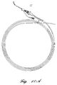
- Figure 1 is a perspective view of the dual-lumen biliary catheter of the present invention.
- Figure 2 is a partially broken away section view of the dual-lumen biliary catheter of the present invention.
- Figure 3 is a perspective view of the dual-lumen biliary catheter of the present invention which highlights the contrast stripes at the distal end of the catheter.
- Figure 3A is a section view highlighting the dual lumens of the biliary catheter of the present invention along line 3A-3A.
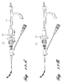
- Figure 4 is a section view of the branching connector which joins the spring wire guide infusion line and contrast medium infusion line of the present invention.
- Figure 5 is a perspective view of the contrast medium infusion line of the present invention.
- Figure 5A is a section view of the contrast medium infusion line of Figure 5.
- Figure 6 illustrates the introduction of the catheter of the present invention into a common biliary duct.
- Figure 7 illustrates the use of the dual-lumen catheter of the present invention with a fiberscope.
- Figure 8 is a section view of an alternative distal tip configuration for the biliary catheter of the present invention.
- Figure 8A is a further alternative embodiment tip configuration which illustrates the calibrated tip.
- Figure 9 is a section view of tubing clamp which may be utilized in the present invention to lock the relative positions of the catheter and spring wire guide which may be utilized with the present invention.
- Figure 9A is a section view of an alternative tubing clamp.
- Figure 10 is a side perspective view of spring wire guide feed apparatus which may be utilized with the present invention.
- Figure 10A is a side perspective view of an alternative spring wire guide feed arrangement including a snap lock adapter which may be utilized with the present invention.
- Figures 11A and 11B are side views of a catheter of an alternative embodiment having a tapered tip and digitized markings.
- Figure 12 is a section view of the catheter of Figure 11A along line 12-12.
- Figures 13A and 13B illustrate the alternative feed mechanism of Figure 10A including guide wire locking mechanism.
- the improved biliary catheter of the present invention is described with reference to the enclosed Figures wherein the same numbers are utilized where applicable.
- the present invention will be described in the context of its use in the cannulation and visualizing of the common biliary duct of a patient pursuant to an ERCP procedure. It is to be recognized that the present invention is applicable to all ERCP procedures involving the cannulation and radiological visualization of the common biliary, pancreatic, common hepatic and cystic ducts.
- a biliary catheter 10 in accordance with the present invention is illustrated.
- the catheter of the present invention comprises a cannula or tube 12 having a proximal end 12a for connection to a source of contrast media and a distal end 12b for insertion into the biliary duct of a patient.
- Tube 12 has a substantially circular cross-sectional shape.
- Tube 12, in a preferred embodiment, has a length of approximately 200 centimeters. This length permits the catheter 10 to be inserted endotracheally into a patient via an endoscope and reach biliary and pancreatic ducts located adjacent the patient's duodenum via an attached fiberscope during an ERCP procedure.
- branching means 14 which couples the tube 12 to spring wire guide feeding means 16 and contrast medium infusion means 18.
- branching means 14 comprises a wedge-shaped polymeric branching connector 15 which joins the spring wire guide feeding means 16 and contrast medium infusion means 18.
- the branching connector 15 may include a connector 19 having an affixed apertured wing 20.
- the spring wire guide feeding means 16 in a preferred embodiment comprises an eighteen gauge luer lock hub 17 which couples to the branching means 14 via a dovetail socket 17a.
- the spring wire guide feeding means 16 is utilized to feed a spring wire guide 24 into and out of the catheter 10.
- the spring wire guide utilized in a preferred embodiment should have a diameter of .035 inches. The use of a spring wire guide having this diameter permits the spring wire guide to be used for placing an indwelling stent, to be discussed below.
- the spring wire guide 24 may optionally be coated with teflon in order to add to its lubricity.
- the spring wire guide of the present invention may further be fed and withdrawn with the assistance of an auxiliary apparatus such the spring wire guide feed apparatus disclosed in U. S. Serial No. 07/608,234 entitled "Hand Held Device For Feeding A Spring Wire Guide", filed November 2, 1990, and now U.S. Patent No. 5,125,906 assigned to Arrow International Investment Corp., assignee of the present invention, and which is incorporated herein by reference as if set forth in full.
- a perspective view of such a device 27 is illustrated in Figure 10.
- the wire guide feed device 27 is affixed to snap lock adapter 29 for locking the position of the wire.
- Figures 13A and 13B illustrate the respective unlocked and locked positions of the adapter.
- the contrast medium infusion means 18 in a preferred embodiment preferably comprises a polymeric tube 26 which couples to a twenty-gauge connector 28.
- the connector 28 has a threaded outer surface 30 onto which may be affixed a cap or stopper (not shown).
- the interior 28a of the connector 28 is luer shaped and is designed to be coupled to a syringe containing radio-opaque contrast media or dye.
- the contrast media or dye is injected down tube 26 and into a contrast media lumen 34 to be discussed below.
- the distal end 12b of the tube 12 is shown in detail.
- the distal end of the catheter 12b includes means 25 for contrasting the outer distal surface of catheter radio-opaque. Contrast means 25 facilitates the visual identification of the distal end of catheter 10 by the endoscope.
- means 25 comprises a plurality of non-toxic ink stripes 25 such as sold under the specification LON-1342 by Colorcon, Inc. of West Point, Pennsylvania. It is to be appreciated that contrast stripes 25 comprising other materials may be utilized in the biliary catheter of the present invention. Moreover, it is to be appreciated by those skilled in the art that the entire catheter 10 or portions thereof may be or applied with any acceptable contrast medium.
- the tip 12b of the catheter may be calibrated 25a at predetermined intervals such as five millimeters.
- catheter tip 12b is tapered to facilitate ease of insertion of the guide wire. It is also to be appreciated that the catheter tip may be curved to facilitate entry of the guide wire.
- the lumens 32, 34 of catheter 10 of the present invention are shown so as to detail their cross-sectional shape.
- the catheter 10 includes a spring wire guide lumen means 32 and contrast media lumen means 34.
- the lumen means 32, 34 extend coaxially within the tube 12 along is entire length between the proximal end 12a and distal end 12b. Referring to Figures 8, 11, 11A and 12 alternative distal end 12b is shown tapered with the contrast media lumen means 34 terminating proximal of the spring wire guide lumen means 32.
- Spring wire guide lumen means 32 is circular in cross-section.
- the top of spring wire guide lumen 32 is defined by an arcuate septum 32a which defines the interior sidewall of the contrast medium lumen 34.
- contrast medium lumen 34 is crescent-shaped. While the present invention is described in the context of a biliary catheter having dual lumens, it is to be appreciated by those skilled in the art that the present invention also embodies catheters having more than two lumens. Further, while the present invention is described with respect to a contast media lumen 34 having a crescent shape, the present invention also embodies catheters in which the contrast media lumen has the different shape.
- the catheter 10 of the present invention is preferably constructed from a polyurethane or nylon having a durometer of about 60D or greater.
- a polyurethane known to be suitable in the present invention is a polyether-polyurethane called TECOFLEX sold under part number EG-60D-B20 by Thermedics. This material has a hardness of 60D, a yield elongation percentage of 275 to 475 and a yield tensile strength in pounds per square inch of 5,000.
- the resin of the present invention comprises a mixture of 20% barium sulfate, 60% Nylon 11 BESVOA and 20% PeBax (12055A00). Nylon II BESVOA and Pebax are manufactured by Atochem.
- a further important feature of the present invention is the addition of a hydrophilic coating on the outer surface of the catheter 10 and optionally within the spring wire guide lumen 32.
- the hydrophilic coating when applied to the catheter, imparts suppleness and kink resistance to the catheter.
- the hydrophilic coating further reduces the durometer of the polyurethane or nylon.
- the hydrophilic coating of the preferred embodiment comprises the following components: (a) 150 milliliters of Methylene Chloride (MeCl); (b) 50 milliliters Gensolv D DuPont Freon Solvent: (c) 2.25 milliliters Tyrite 7617 Adhesive, and (d) 2.13 grams of Polyethylene Oxide (PEO).
- the hydrophilic coating of the preferred embodiment is applied to the catheter pursuant to the following process. Initially, 150 milliliters of MeCl is poured into a beaker. Next, 50 milliliters of Gensolv D is added, and the beaker is placed on stirrer plate. A stirring magnet is then dropped into the beaker and the stirring plate is activated. Stirring is adjusted until a vortex forms. Next, 2.13 grams of PEO are slowly added to the stirring solution. The solution is stirred continuous for 10 minutes, in order to break up any lumps of PEO. Using a syringe, Tyrite 7617 adhesive is added to the stirring solution, which is permitted to stir for an additional five minutes. The stirred solution is then poured into a 200 milliliter graduated cylinder.
- the catheter 10 with its end sealed off, is then dipped into the cylinder until it reaches the bottom of the cylinder.
- the catheter 10 is left in the cylinder for 2-3 seconds, quickly retrieved and the excess solution shaken off.
- the catheter is then air-dried for a minimum 24 hours.
- solution may be injected down the spring wire guide lumen 32 of the catheter. Air is blown through the lumen immediately to drive out excess solution. Air is blown through the catheter for three to five minutes, and the catheter is left to dry for a minimum of 24 hours.
- the catheter 10 with hydrophilic coating provides a highly lubricated surface which is activated by the biliary fluids of the patient.
- the hydrophilic coating may also be activated by the gastric fluids which enter the endoscope.
- the hydrophilic coating reduces the durometer of the catheter, and imparts kink resistance and suppleness to the catheter.
- hydrophilic coating While the present invention is being described in the context of a preferred hydrophilic coating, it is to be appreciated that other hydrophilic coatings may be utilized in the present invention. Examples of such hydrophilic coatings are found and described in U. S. Patent No. 4,943,460 entitled “Process for Coating Polymer Surfaces and Coated Products Produced Using Such Process.” Another hydrophilic coating is Hydromer® "Slippery When Wet" coating manufactured by Hydromer, Inc.
- the operation and use of the biliary catheter 10 of the present invention is now described with reference to the Figures.
- the spring wire guide 24 is inserted through an endoscope and exits through the side of an attached fiberscope 36 as shown in Figure 7 situated in the patient's duodenum 38.
- the catheter 10 is then threaded over the spring wire guide 24 via spring wire guide lumen 32 and fed through the fiberscope 36 and into the common bile duct 40.
- a pre-filled syringe of radio-opaque dye or contrast media is attached to twenty-gauge luer shaped connector 28.
- a sufficient amount of dye to fill the catheter is then injected into tube 26.
- the catheter 10 is inserted through the accessory channel of the endoscope and threaded over the spring wire guide 24 via lumen 32.
- the catheter 10 exits the side of the fiberscope and enters the common bile duct, as shown in Figures 6 and 7.
- clamp 37 may be used to lock the relative positions of the catheter and spring wire guide.
- An example of clamps which achieves this function are the Series 340 clamps by Halkey Medical of St. Moscow, FLA.
- Contrast media is then injected into the contrast medium lumen 34 which exits at distal end 12b and into the common biliary duct 40 thereby permitting x-ray or fluoroscopic visualization of the duct 40.
- Digitized markings 25a facilitate precise adjustment of the catheter. If the position of the catheter needs to be adjusted, the spring wire guide 24 is advanced and the catheter 10 advanced accordingly. The catheter can be rapidly adjusted and contrast media and dye can be repeated infused without the need for repeated insertion and removal of the spring wire guide 24.
- the present invention thus provides for the probing with the spring wire guide 24, the injection of dye or contrast media, via contrast media lumen 34, further probing, and further injection of dye until a proper catheter position is achieved.
- the present invention eliminates the time consuming step of removing the spring wire guide 24 prior to each change in catheter position and contrast medium infusion.
- the use of the catheter of the present invention can save over twenty minutes of time during a typical ERCP procedure.
- a laser fiber for biliary lithotripty can be placed through one lumen with ongoing injection of contrast medium or fluid in the second lumen. Further, selective cannulation of the right and left hepatic ducts, cystic ducts, or pancreas becomes more directed, safe and efficient.
- a particular feature of the present invention is its adaptability for use in placing a stent around a biliary calculus 42 or cystic or pancreatic obstruction.
- surgery is mandated.
- surgery is often not always possible at the time of the ERCP procedure.
- a stent is typically placed within the common biliary or pancreatic duct around the calculus.
- the catheter 10 is utilized in association with a spring wire guide 24 having a length greater than twice the length of the catheter 10, or over 400 centimeters in length.
- the spring wire guide would be threaded with the catheter into endoscope as described above.
- the spring wire guide should preferably have a diameter of 0.036 inches (approx.0.0009m).
- the catheter 10 of the present invention is then threaded over the spring wire guide as discussed above, and fed into the common biliary duct. Contrast media or dye is infused, and the calculus 42 is located as shown in Figure 6. The catheter 10 is then removed from the endoscope.
- the catheter 10 can be completely removed from the endoscope over the spring wire guide 24 without the need for removing the spring wire guide from the endoscope.
- a stent may be placed forward of a guiding catheter and is threaded over the spring wire guide.
- the guiding catheter is utilized to push the stent into the endoscope, over the spring wire guide, into the common biliary duct and around the biliary calculus 42. With the stent in place, the spring wire guide 24 is then removed along with the guiding catheter.
- other accessories such as stone baskets, three pronged retrievers and fiber optic cameras can be used interchangeably in the first lumen with the guide wire.
Abstract
Description
Claims (13)
- A biliary catheter (10) for use in visualizing a duct of a patient, the catheter comprising:a cannula (12) having a proximal end (12a) and a distal end (12b), a first lumen (34) and a second lumen (32) being defined within the cannula, each lumen (34, 32) extending from the proximal end to the distal end of the cannula, and a spring wire guide (24) for the cannula, the second lumen (32) being configured for receiving the spring wire guide (24), and having an opening at the distal end of the cannula, characterised in that the first lumen (34) extends to an opening disposed at the distal end (12b) of the cannula and in that contrast medium infusion means (18) is connected to the proximal end of the first lumen (34).
- The biliary catheter of claim 1, wherein the first lumen (34) has a crescent shaped cross-section, and wherein the second lumen (32) has a round cross-section.
- The biliary catheter of any preceding claim, wherein the distal end (12b) has a tapered portion, the first lumen (34) extending to an opening in the tapered portion slightly proximal of the second lumen (32).
- The biliary catheter of any preceding claim, wherein the catheter (10) further comprises means (25) on the distal end of said catheter for rendering said distal end (12b) visible by an endoscope (36).
- The biliary catheter of claim 4, wherein the said means comprises a plurality of stripes (25) affixed to the cannula (12) adjacent the distal end (12b).
- The biliary catheter of any preceding claim, wherein the spring wire guide (24) is at least twice as long as the cannula (12).
- The biliary catheter of claim 6, further comprising feed means (27) for holding a portion of the spring wire guide (24) not disposed in the second lumen (32) of the cannula (12).
- The biliary catheter of any preceding claim, further comprising locking means (29) for maintaining the spring wire guide within the cannula (12).
- The biliary catheter of any preceding claim, including sufficient contrast fluid disposed in the first lumen so as to substantially fill the first lumen with contrast fluid from the proximal end (12a) to the distal end (12b) of the cannula (12).
- The biliary catheter of claim 9, wherein the spring wire guide (24) extends out of the distal end (12b) of the cannula (12) when the first lumen (34) is filled with contrast fluid.
- The biliary catheter of any preceding claim, wherein the second lumen (32) has a hydrophilic coating.
- The biliary catheter of claim 10, wherein the hydrophilic coating of the second lumen (32) is activatable in the present of biliary fluid.
- The biliary catheter of any preceding claim, further comprising a coating disposed on an outer surface of the cannula (12) for improving lubricity of the outer surface, and for increasing kink resistance in the cannula.
Applications Claiming Priority (5)
| Application Number | Priority Date | Filing Date | Title |
|---|---|---|---|
| US88084092A | 1992-05-11 | 1992-05-11 | |
| US880840 | 1992-05-11 | ||
| PCT/US1993/004434 WO1993023106A1 (en) | 1992-05-11 | 1993-05-11 | Improved biliary catheter |
| US60434 | 1993-05-11 | ||
| US08/060,434 US5397302A (en) | 1992-05-11 | 1993-05-11 | Method of using a dual lumen biliary catheter |
Publications (4)
| Publication Number | Publication Date |
|---|---|
| EP0593741A1 EP0593741A1 (en) | 1994-04-27 |
| EP0593741A4 EP0593741A4 (en) | 1994-12-07 |
| EP0593741B1 true EP0593741B1 (en) | 1998-08-05 |
| EP0593741B2 EP0593741B2 (en) | 2004-10-27 |
Family
ID=26739924
Family Applications (1)
| Application Number | Title | Priority Date | Filing Date |
|---|---|---|---|
| EP93911228A Expired - Lifetime EP0593741B2 (en) | 1992-05-11 | 1993-05-11 | Improved biliary catheter |
Country Status (6)
| Country | Link |
|---|---|
| US (2) | US5397302A (en) |
| EP (1) | EP0593741B2 (en) |
| JP (1) | JPH06511409A (en) |
| AU (1) | AU666554B2 (en) |
| DE (1) | DE69320136T3 (en) |
| WO (1) | WO1993023106A1 (en) |
Cited By (1)
| Publication number | Priority date | Publication date | Assignee | Title |
|---|---|---|---|---|
| US6770066B1 (en) | 1992-05-11 | 2004-08-03 | Ballard Medical Products | Multi-lumen endoscopic catheter |
Families Citing this family (163)
| Publication number | Priority date | Publication date | Assignee | Title |
|---|---|---|---|---|
| US5536248A (en) * | 1992-05-11 | 1996-07-16 | Arrow Precision Products, Inc. | Method and apparatus for electrosurgically obtaining access to the biliary tree and placing a stent therein |
| GB2274991B (en) * | 1993-02-11 | 1996-10-30 | Sara Kinal | Embryo replacement catheter |
| US5547469A (en) | 1994-05-13 | 1996-08-20 | Boston Scientific Corporation | Apparatus for performing diagnostic and therapeutic modalities in the biliary tree |
| US6743217B2 (en) | 1994-05-13 | 2004-06-01 | Scimed Life Systems, Inc. | Apparatus for performing diagnostic and therapeutic modalities in the biliary tree |
| US5599324A (en) * | 1995-05-04 | 1997-02-04 | Boston Scientific Corporation | Catheter for administering a liquid agent |
| GB9608483D0 (en) * | 1996-04-25 | 1996-07-03 | Smiths Industries Plc | Introducers and assemblies |
| US6520951B1 (en) | 1996-09-13 | 2003-02-18 | Scimed Life Systems, Inc. | Rapid exchange catheter with detachable hood |
| US6096009A (en) | 1996-09-13 | 2000-08-01 | Boston Scientific Corporation | Guidewire and catheter locking device and method |
| US6346093B1 (en) | 1996-09-13 | 2002-02-12 | Scimed Life Systems, Inc. | Single operator exchange biliary catheter with common distal lumen |
| US5921971A (en) | 1996-09-13 | 1999-07-13 | Boston Scientific Corporation | Single operator exchange biliary catheter |
| US6606515B1 (en) | 1996-09-13 | 2003-08-12 | Scimed Life Systems, Inc. | Guide wire insertion and re-insertion tools and methods of use |
| US6582401B1 (en) | 1996-09-13 | 2003-06-24 | Scimed Life Sytems, Inc. | Multi-size convertible catheter |
| US6007522A (en) * | 1996-09-13 | 1999-12-28 | Boston Scientific Corporation | Single operator exchange biliary catheter |
| US5795326A (en) * | 1997-01-29 | 1998-08-18 | Baxter International Inc. | Double lumen tubing design for catheter |
| US5968009A (en) * | 1997-01-29 | 1999-10-19 | Baxter International Inc. | Double lumen tubing design for catheter |
| US5817104A (en) * | 1997-04-30 | 1998-10-06 | C.R. Bard, Inc. | Dual purpose mechanism for expanding baskets |
| AU9681198A (en) | 1997-10-03 | 1999-04-27 | Boston Scientific Corporation | Device and method for facilitating access to a duct within the human body |
| US9586023B2 (en) | 1998-02-06 | 2017-03-07 | Boston Scientific Limited | Direct stream hydrodynamic catheter system |
| US6326396B1 (en) | 1998-11-20 | 2001-12-04 | Alteon, Inc. | Glucose and lipid lowering compounds |
| WO2000045691A2 (en) | 1999-02-04 | 2000-08-10 | Da Silva Branco Antonio Carlos | Kit for endovascular venous surgery |
| US7214229B2 (en) | 1999-03-18 | 2007-05-08 | Fossa Medical, Inc. | Radially expanding stents |
| US6709465B2 (en) | 1999-03-18 | 2004-03-23 | Fossa Medical, Inc. | Radially expanding ureteral device |
| US6458076B1 (en) | 2000-02-01 | 2002-10-01 | 5 Star Medical | Multi-lumen medical device |
| US7811250B1 (en) * | 2000-02-04 | 2010-10-12 | Boston Scientific Scimed, Inc. | Fluid injectable single operator exchange catheters and methods of use |
| WO2001056641A1 (en) * | 2000-02-04 | 2001-08-09 | C. R. Bard, Inc. | Triple lumen stone balloon catheter and method |
| US6663598B1 (en) | 2000-05-17 | 2003-12-16 | Scimed Life Systems, Inc. | Fluid seal for endoscope |
| DK1284775T3 (en) | 2000-05-18 | 2005-12-12 | Wilson Cook Medical Inc | Medical device with improved threading access |
| WO2002022072A2 (en) * | 2000-09-11 | 2002-03-21 | Closure Medical Corporation | Bronchial occlusion method and apparatus |
| EP1414513B1 (en) * | 2000-11-03 | 2009-04-01 | The Cleveland Clinic Foundation | Catheter for removal of solids from surgical drains |
| US6579300B2 (en) | 2001-01-18 | 2003-06-17 | Scimed Life Systems, Inc. | Steerable sphincterotome and methods for cannulation, papillotomy and sphincterotomy |
| US6764484B2 (en) | 2001-03-30 | 2004-07-20 | Scimed Life Systems, Inc. | C-channel to o-channel converter for a single operator exchange biliary catheter |
| US6620122B2 (en) * | 2001-04-26 | 2003-09-16 | Scimed Life Systems, Inc. | Gastric pseudocyst drainage and stent delivery system for use therein |
| US6517531B2 (en) | 2001-04-27 | 2003-02-11 | Scimed Life Systems, Inc. | Medical suction device |
| US6814739B2 (en) | 2001-05-18 | 2004-11-09 | U.S. Endoscopy Group, Inc. | Retrieval device |
| US6827718B2 (en) | 2001-08-14 | 2004-12-07 | Scimed Life Systems, Inc. | Method of and apparatus for positioning and maintaining the position of endoscopic instruments |
| US20030078473A1 (en) * | 2001-10-23 | 2003-04-24 | Scimed Life Systems, Inc. | Cone tip biliary catheter and method of use |
| DE10245009B4 (en) * | 2002-09-20 | 2007-09-06 | Karl Storz Gmbh & Co. Kg | Medical instrument for sucking and rinsing and process for its preparation |
| US7534223B2 (en) | 2002-10-08 | 2009-05-19 | Boston Scientific Scimed, Inc. | Catheter with formed guide wire ramp |
| WO2004041329A2 (en) * | 2002-11-01 | 2004-05-21 | C.R. Bard, Inc. | Low profile short tapered tip catheter |
| US7037293B2 (en) * | 2002-11-15 | 2006-05-02 | Boston Scientific Scimed, Inc. | Rapid exchange catheter with depressable channel |
| US6893393B2 (en) | 2003-02-19 | 2005-05-17 | Boston Scientific Scimed., Inc. | Guidewire locking device and method |
| US10413211B2 (en) | 2003-02-21 | 2019-09-17 | 3Dt Holdings, Llc | Systems, devices, and methods for mapping organ profiles |
| US10172538B2 (en) | 2003-02-21 | 2019-01-08 | 3Dt Holdings, Llc | Body lumen junction localization |
| US8078274B2 (en) | 2003-02-21 | 2011-12-13 | Dtherapeutics, Llc | Device, system and method for measuring cross-sectional areas in luminal organs |
| US7559934B2 (en) * | 2003-04-07 | 2009-07-14 | Scimed Life Systems, Inc. | Beaded basket retrieval device |
| US7654989B2 (en) | 2003-04-08 | 2010-02-02 | C. R. Bard, Inc. | Ureteral access sheath |
| WO2004098654A2 (en) * | 2003-05-02 | 2004-11-18 | Metolius Biomedical, Llc | Body-space drainage-tube debris removal |
| WO2005000096A2 (en) * | 2003-06-05 | 2005-01-06 | Hydrocision, Inc. | Disposable endoscope and method of making a disposable endoscope |
| WO2005011790A1 (en) * | 2003-07-31 | 2005-02-10 | Wilson-Cook Medical Inc. | System for introducing multiple medical devices |
| US8211087B2 (en) * | 2003-07-31 | 2012-07-03 | Cook Medical Technologies Llc | Distal wire stop |
| JP4898447B2 (en) | 2003-10-14 | 2012-03-14 | プルーロームド インコーポレイテッド | Confinement of kidney stone fragments during lithotripsy |
| US7025721B2 (en) * | 2004-01-29 | 2006-04-11 | Boston Scientific Scimed, Inc. | Endoscope channel cap |
| US20050234369A1 (en) * | 2004-04-16 | 2005-10-20 | Medical Components, Inc. | Lockable guide wire tip protector |
| WO2006015323A2 (en) * | 2004-07-29 | 2006-02-09 | Wilson-Cook Medical Inc. | Catheter with splittable wall shaft and peel tool |
| US20080275393A1 (en) * | 2004-08-24 | 2008-11-06 | Bonnette Michael J | Isolation thrombectomy catheter system |
| JP4901087B2 (en) * | 2004-09-24 | 2012-03-21 | オリンパス株式会社 | Stent introduction member, stent delivery catheter, and endoscope treatment system |
| US8480629B2 (en) * | 2005-01-28 | 2013-07-09 | Boston Scientific Scimed, Inc. | Universal utility board for use with medical devices and methods of use |
| BRPI0611278A2 (en) | 2005-05-02 | 2010-08-31 | Gen Hospital Corp | lithiasis treatment without lithotripsy |
| WO2007000159A1 (en) * | 2005-06-27 | 2007-01-04 | William Cook Europe Aps | A dilator for performing a percutaneous medical procedure |
| US8784336B2 (en) | 2005-08-24 | 2014-07-22 | C. R. Bard, Inc. | Stylet apparatuses and methods of manufacture |
| US8029494B2 (en) * | 2005-10-24 | 2011-10-04 | Cook Medical Technologies Llc | Method of removing biliary stones with coaxial catheter device |
| US8162878B2 (en) | 2005-12-05 | 2012-04-24 | Medrad, Inc. | Exhaust-pressure-operated balloon catheter system |
| AU2007254126A1 (en) * | 2006-05-19 | 2007-11-29 | Conmed Endoscopic Technologies, Inc. | Steerable medical instrument |
| US8196584B2 (en) | 2006-06-22 | 2012-06-12 | Nellcor Puritan Bennett Llc | Endotracheal cuff and technique for using the same |
| US20070296125A1 (en) * | 2006-06-22 | 2007-12-27 | Joel Colburn | Thin cuff for use with medical tubing and method and apparatus for making the same |
| US20070295337A1 (en) * | 2006-06-22 | 2007-12-27 | Nelson Donald S | Endotracheal cuff and technique for using the same |
| US8434487B2 (en) | 2006-06-22 | 2013-05-07 | Covidien Lp | Endotracheal cuff and technique for using the same |
| US20080053454A1 (en) * | 2006-09-01 | 2008-03-06 | Nellcor Puritan Bennett Incorporated | Endotracheal tube including a partially inverted cuff collar |
| US8684175B2 (en) | 2006-09-22 | 2014-04-01 | Covidien Lp | Method for shipping and protecting an endotracheal tube with an inflated cuff |
| US8561614B2 (en) * | 2006-09-28 | 2013-10-22 | Covidien Lp | Multi-layer cuffs for medical devices |
| US20080078405A1 (en) * | 2006-09-29 | 2008-04-03 | Crumback Gary L | Self-sizing adjustable endotracheal tube |
| US8807136B2 (en) * | 2006-09-29 | 2014-08-19 | Covidien Lp | Self-sizing adjustable endotracheal tube |
| US20080078399A1 (en) | 2006-09-29 | 2008-04-03 | O'neil Michael P | Self-sizing adjustable endotracheal tube |
| US20080078401A1 (en) * | 2006-09-29 | 2008-04-03 | Nellcor Puritan Bennett Incorporated | Self-sizing adjustable endotracheal tube |
| US8307830B2 (en) | 2006-09-29 | 2012-11-13 | Nellcor Puritan Bennett Llc | Endotracheal cuff and technique for using the same |
| WO2008042756A2 (en) * | 2006-09-29 | 2008-04-10 | Pluromed, Inc. | Methods for preventing retropulsion of concretions and fragments during lithotripsy |
| US7950393B2 (en) * | 2006-09-29 | 2011-05-31 | Nellcor Puritan Bennett Llc | Endotracheal cuff and technique for using the same |
| US8388546B2 (en) | 2006-10-23 | 2013-03-05 | Bard Access Systems, Inc. | Method of locating the tip of a central venous catheter |
| US7794407B2 (en) | 2006-10-23 | 2010-09-14 | Bard Access Systems, Inc. | Method of locating the tip of a central venous catheter |
| US7766909B2 (en) * | 2006-11-08 | 2010-08-03 | Boston Scientific Scimed, Inc. | Sphincterotome with stiffening member |
| WO2008085712A1 (en) | 2007-01-03 | 2008-07-17 | Boston Scientific Limited | Method and apparatus for biliary access and stone retrieval |
| US20080167628A1 (en) * | 2007-01-05 | 2008-07-10 | Boston Scientific Scimed, Inc. | Stent delivery system |
| US8480570B2 (en) | 2007-02-12 | 2013-07-09 | Boston Scientific Scimed, Inc. | Endoscope cap |
| US20080215034A1 (en) * | 2007-03-02 | 2008-09-04 | Jessica Clayton | Endotracheal cuff and technique for using the same |
| US20080210243A1 (en) * | 2007-03-02 | 2008-09-04 | Jessica Clayton | Endotracheal cuff and technique for using the same |
| US20090062769A1 (en) * | 2007-04-13 | 2009-03-05 | Boston Scientific Scimed, Inc. | Rapid exchange catheter converter |
| US20080294145A1 (en) * | 2007-05-25 | 2008-11-27 | Galt Medical Corporation | Catheter hub with flushable lumen and guidewire |
| US8591521B2 (en) | 2007-06-08 | 2013-11-26 | United States Endoscopy Group, Inc. | Retrieval device |
| US8292872B2 (en) * | 2007-06-29 | 2012-10-23 | Cook Medical Technologies Llc | Distal wire stop having adjustable handle |
| US20090043317A1 (en) * | 2007-08-08 | 2009-02-12 | Cavanaugh Brian J | Method and apparatus for delivery of a ligating suture |
| US9579496B2 (en) | 2007-11-07 | 2017-02-28 | C. R. Bard, Inc. | Radiopaque and septum-based indicators for a multi-lumen implantable port |
| US8781555B2 (en) | 2007-11-26 | 2014-07-15 | C. R. Bard, Inc. | System for placement of a catheter including a signal-generating stylet |
| US8849382B2 (en) | 2007-11-26 | 2014-09-30 | C. R. Bard, Inc. | Apparatus and display methods relating to intravascular placement of a catheter |
| US10751509B2 (en) | 2007-11-26 | 2020-08-25 | C. R. Bard, Inc. | Iconic representations for guidance of an indwelling medical device |
| ES2832713T3 (en) | 2007-11-26 | 2021-06-11 | Bard Inc C R | Integrated system for intravascular catheter placement |
| US10449330B2 (en) | 2007-11-26 | 2019-10-22 | C. R. Bard, Inc. | Magnetic element-equipped needle assemblies |
| US10524691B2 (en) | 2007-11-26 | 2020-01-07 | C. R. Bard, Inc. | Needle assembly including an aligned magnetic element |
| US9649048B2 (en) * | 2007-11-26 | 2017-05-16 | C. R. Bard, Inc. | Systems and methods for breaching a sterile field for intravascular placement of a catheter |
| US9521961B2 (en) | 2007-11-26 | 2016-12-20 | C. R. Bard, Inc. | Systems and methods for guiding a medical instrument |
| CN100544674C (en) * | 2007-12-05 | 2009-09-30 | 中国人民解放军第三军医大学第一附属医院 | The novel probe that is used for probing sinus tract and fistula |
| US20090182315A1 (en) * | 2007-12-07 | 2009-07-16 | Ceramoptec Industries Inc. | Laser liposuction system and method |
| WO2009079539A1 (en) * | 2007-12-17 | 2009-06-25 | Medrad, Inc. | Rheolytic thrombectomy catheter with self-inflation distal balloon |
| WO2009082669A1 (en) * | 2007-12-26 | 2009-07-02 | Medrad, Inc. | Rheolytic thrombectomy catheter with self-inflating proximal balloon with drug infusion capabilities |
| JP5430065B2 (en) * | 2007-12-28 | 2014-02-26 | テルモ株式会社 | Guide wire |
| US8750978B2 (en) * | 2007-12-31 | 2014-06-10 | Covidien Lp | System and sensor for early detection of shock or perfusion failure and technique for using the same |
| US8246752B2 (en) * | 2008-01-25 | 2012-08-21 | Clear Catheter Systems, Inc. | Methods and devices to clear obstructions from medical tubes |
| ES2758792T3 (en) | 2008-01-25 | 2020-05-06 | Clearflow Inc | Procedures and devices for clearing medical tube obstructions |
| US8388521B2 (en) * | 2008-05-19 | 2013-03-05 | Boston Scientific Scimed, Inc. | Integrated locking device with active sealing |
| US8478382B2 (en) | 2008-02-11 | 2013-07-02 | C. R. Bard, Inc. | Systems and methods for positioning a catheter |
| US8343041B2 (en) | 2008-05-19 | 2013-01-01 | Boston Scientific Scimed, Inc. | Integrated locking device with passive sealing |
| DE112009000700T5 (en) | 2008-03-20 | 2011-02-10 | Medrad, Inc. | Hydrodynamic direct current catheter system |
| US20110098528A1 (en) * | 2008-04-11 | 2011-04-28 | Lumenis Ltd. | Fibers and tips thereof used with devices |
| US20090312645A1 (en) * | 2008-06-16 | 2009-12-17 | Boston Scientific Scimed, Inc. | Methods and Devices for Accessing Anatomic Structures |
| US9901714B2 (en) | 2008-08-22 | 2018-02-27 | C. R. Bard, Inc. | Catheter assembly including ECG sensor and magnetic assemblies |
| US8437833B2 (en) | 2008-10-07 | 2013-05-07 | Bard Access Systems, Inc. | Percutaneous magnetic gastrostomy |
| US11890443B2 (en) | 2008-11-13 | 2024-02-06 | C. R. Bard, Inc. | Implantable medical devices including septum-based indicators |
| US8231519B2 (en) * | 2009-05-20 | 2012-07-31 | Thoratec Corporation | Multi-lumen cannula |
| US9532724B2 (en) | 2009-06-12 | 2017-01-03 | Bard Access Systems, Inc. | Apparatus and method for catheter navigation using endovascular energy mapping |
| CN102802514B (en) | 2009-06-12 | 2015-12-02 | 巴德阿克塞斯系统股份有限公司 | Catheter tip positioning equipment |
| US9445734B2 (en) | 2009-06-12 | 2016-09-20 | Bard Access Systems, Inc. | Devices and methods for endovascular electrography |
| US8590534B2 (en) * | 2009-06-22 | 2013-11-26 | Covidien Lp | Cuff for use with medical tubing and method and apparatus for making the same |
| AU2010300677B2 (en) | 2009-09-29 | 2014-09-04 | C.R. Bard, Inc. | Stylets for use with apparatus for intravascular placement of a catheter |
| US10639008B2 (en) | 2009-10-08 | 2020-05-05 | C. R. Bard, Inc. | Support and cover structures for an ultrasound probe head |
| US11103213B2 (en) * | 2009-10-08 | 2021-08-31 | C. R. Bard, Inc. | Spacers for use with an ultrasound probe |
| JP2013510652A (en) | 2009-11-17 | 2013-03-28 | シー・アール・バード・インコーポレーテッド | Overmolded access port including locking feature and identification feature |
| CN102821679B (en) | 2010-02-02 | 2016-04-27 | C·R·巴德股份有限公司 | For the apparatus and method that catheter navigation and end are located |
| EP2912999B1 (en) | 2010-05-28 | 2022-06-29 | C. R. Bard, Inc. | Apparatus for use with needle insertion guidance system |
| EP4122385A1 (en) | 2010-05-28 | 2023-01-25 | C. R. Bard, Inc. | Insertion guidance system for needles and medical components |
| KR101856267B1 (en) | 2010-08-20 | 2018-05-09 | 씨. 알. 바드, 인크. | Reconfirmation of ecg-assisted catheter tip placement |
| WO2012058461A1 (en) | 2010-10-29 | 2012-05-03 | C.R.Bard, Inc. | Bioimpedance-assisted placement of a medical device |
| WO2013003450A1 (en) | 2011-06-27 | 2013-01-03 | Boston Scientific Scimed, Inc. | Stent delivery systems and methods for making and using stent delivery systems |
| EP2729073A4 (en) | 2011-07-06 | 2015-03-11 | Bard Inc C R | Needle length determination and calibration for insertion guidance system |
| USD699359S1 (en) | 2011-08-09 | 2014-02-11 | C. R. Bard, Inc. | Ultrasound probe head |
| USD724745S1 (en) | 2011-08-09 | 2015-03-17 | C. R. Bard, Inc. | Cap for an ultrasound probe |
| US8942530B2 (en) | 2011-09-20 | 2015-01-27 | San Marino Capital, Inc. | Endoscope connector method and apparatus |
| US9211107B2 (en) | 2011-11-07 | 2015-12-15 | C. R. Bard, Inc. | Ruggedized ultrasound hydrogel insert |
| EP2833786A4 (en) | 2012-04-05 | 2015-11-11 | Bard Access Systems Inc | Devices and systems for navigation and positioning a central venous catheter within a patient |
| US11759268B2 (en) | 2012-04-05 | 2023-09-19 | C. R. Bard, Inc. | Apparatus and methods relating to intravascular positioning of distal end of catheter |
| US10159531B2 (en) | 2012-04-05 | 2018-12-25 | C. R. Bard, Inc. | Apparatus and methods relating to intravascular positioning of distal end of catheter |
| WO2013188833A2 (en) | 2012-06-15 | 2013-12-19 | C.R. Bard, Inc. | Apparatus and methods for detection of a removable cap on an ultrasound probe |
| US9872700B2 (en) | 2013-09-03 | 2018-01-23 | United States Endoscopy Group, Inc. | Endoscopic snare device |
| US9572591B2 (en) | 2013-09-03 | 2017-02-21 | United States Endoscopy Group, Inc. | Endoscopic snare device |
| EP3073910B1 (en) | 2014-02-06 | 2020-07-15 | C.R. Bard, Inc. | Systems for guidance and placement of an intravascular device |
| CA2939622C (en) | 2014-02-17 | 2022-12-06 | Clearflow, Inc. | Medical tube clearance device |
| EP3102277B1 (en) | 2014-02-17 | 2021-05-12 | Clearflow, Inc. | A device for clearing obstructions from a multi-lumen medical tube |
| US20170112518A1 (en) * | 2014-06-05 | 2017-04-27 | Mayo Foundation For Medical Education And Research | Cannulation devices |
| US10973584B2 (en) | 2015-01-19 | 2021-04-13 | Bard Access Systems, Inc. | Device and method for vascular access |
| US9889274B2 (en) | 2015-06-18 | 2018-02-13 | Medtronic Cryocath Lp | Skive-less sheath |
| US10349890B2 (en) | 2015-06-26 | 2019-07-16 | C. R. Bard, Inc. | Connector interface for ECG-based catheter positioning system |
| WO2017117190A1 (en) * | 2015-12-28 | 2017-07-06 | Gonzalez Luis Fernando | Delivery catheter with fixed guidewire and beveled elliptical port |
| CN108541222B (en) | 2016-01-29 | 2021-10-15 | 波士顿科学医学有限公司 | Medical device and method of use |
| US11000207B2 (en) | 2016-01-29 | 2021-05-11 | C. R. Bard, Inc. | Multiple coil system for tracking a medical device |
| WO2017155539A1 (en) * | 2016-03-11 | 2017-09-14 | Seftel Allen D | Catheter guide tube device |
| WO2018129546A1 (en) | 2017-01-09 | 2018-07-12 | United States Endoscopy Group, Inc. | Retrieval device |
| EP3664684B1 (en) | 2017-08-11 | 2022-11-30 | Boston Scientific Scimed, Inc. | Biopsy cap for use with endoscope |
| US11647899B2 (en) * | 2018-06-14 | 2023-05-16 | Boston Scientific Scimed, Inc. | Devices, systems and methods for accessing a body lumen |
| CN108742635A (en) * | 2018-06-26 | 2018-11-06 | 江苏省人民医院(南京医科大学第附属医院) | In-vivo length measuring method and device for measuring physical length of in-vivo cavity and guide wire ruler thereof |
| WO2020051310A1 (en) * | 2018-09-05 | 2020-03-12 | Vanderbilt University | Pediatric catheter |
| EP3852622A1 (en) | 2018-10-16 | 2021-07-28 | Bard Access Systems, Inc. | Safety-equipped connection systems and methods thereof for establishing electrical connections |
| EP4126182A4 (en) * | 2020-03-31 | 2024-03-20 | Dib UltraNav Medical LLC | Handle assembly for medical devices |
| CN116547031A (en) | 2020-11-17 | 2023-08-04 | 科里福罗公司 | Medical tube cleaning device |
| WO2022271482A1 (en) * | 2021-06-24 | 2022-12-29 | Dib UltraNav Medical LLC | Detachable medical devices, components, and methods of use thereof |
| US11883616B2 (en) | 2021-07-07 | 2024-01-30 | Mekal, LLC | Multi-lumen intravascular catheters with inner converging lumens for multiple guidewire control |
Family Cites Families (38)
| Publication number | Priority date | Publication date | Assignee | Title |
|---|---|---|---|---|
| US4033331A (en) † | 1975-07-17 | 1977-07-05 | Guss Stephen B | Cardiac catheter and method of using same |
| US4682596A (en) † | 1984-05-22 | 1987-07-28 | Cordis Corporation | Electrosurgical catheter and method for vascular applications |
| JPS60187737U (en) * | 1984-05-23 | 1985-12-12 | オリンパス光学工業株式会社 | Indwelling tube guide device |
| JPS60187738U (en) * | 1984-05-24 | 1985-12-12 | 日本シヤ−ウツド株式会社 | Catheter assembly for vascular placement |
| JPS6143442A (en) * | 1984-08-08 | 1986-03-03 | Toshiba Corp | Orientation device of wafer |
| US4807626A (en) * | 1985-02-14 | 1989-02-28 | Mcgirr Douglas B | Stone extractor and method |
| US4601713A (en) * | 1985-06-11 | 1986-07-22 | Genus Catheter Technologies, Inc. | Variable diameter catheter |
| US4781677A (en) * | 1985-07-17 | 1988-11-01 | Wilcox Gilbert M | Method of treatment utilizing a double balloon nasobiliary occlusion catheter |
| US4671291A (en) * | 1986-03-31 | 1987-06-09 | Siemens Medical Systems, Inc. | Angle encoding catheter |
| US4722344A (en) * | 1986-05-23 | 1988-02-02 | Critikon, Inc. | Radiopaque polyurethanes and catheters formed therefrom |
| US4893621A (en) * | 1986-08-22 | 1990-01-16 | Heyman Arnold M | Slipover antegrade loading calculus extraction instrument system |
| US4988356A (en) † | 1987-02-27 | 1991-01-29 | C. R. Bard, Inc. | Catheter and guidewire exchange system |
| US4832023A (en) † | 1987-06-03 | 1989-05-23 | Mcm Laboratories, Inc. | Method and apparatus for reducing blockage in body channels |
| US4850969A (en) * | 1987-10-01 | 1989-07-25 | Retroperfusion Systems, Inc. | Retroperfusion catheter and tip construction for use therewith |
| US4917667A (en) * | 1988-02-11 | 1990-04-17 | Retroperfusion Systems, Inc. | Retroperfusion balloon catheter and method |
| US4898591A (en) * | 1988-08-09 | 1990-02-06 | Mallinckrodt, Inc. | Nylon-PEBA copolymer catheter |
| JPH0426109Y2 (en) * | 1988-09-16 | 1992-06-23 | ||
| US4955377A (en) * | 1988-10-28 | 1990-09-11 | Lennox Charles D | Device and method for heating tissue in a patient's body |
| US5021044A (en) * | 1989-01-30 | 1991-06-04 | Advanced Cardiovascular Systems, Inc. | Catheter for even distribution of therapeutic fluids |
| US5024617A (en) † | 1989-03-03 | 1991-06-18 | Wilson-Cook Medical, Inc. | Sphincterotomy method and device having controlled bending and orientation |
| JPH02268769A (en) * | 1989-04-11 | 1990-11-02 | Olympus Optical Co Ltd | Treating device for endoscope |
| EP0408245B1 (en) * | 1989-07-13 | 1994-03-02 | American Medical Systems, Inc. | Stent placement instrument |
| JP2528011B2 (en) * | 1989-12-20 | 1996-08-28 | テルモ株式会社 | Catheter |
| US5108416A (en) * | 1990-02-13 | 1992-04-28 | C. R. Bard, Inc. | Stent introducer system |
| US5084054A (en) * | 1990-03-05 | 1992-01-28 | C.R. Bard, Inc. | Surgical gripping instrument |
| US5059177A (en) * | 1990-04-19 | 1991-10-22 | Cordis Corporation | Triple lumen balloon catheter |
| JP2521181B2 (en) * | 1990-07-16 | 1996-07-31 | テルモ株式会社 | Wire rod operation equipment |
| US5273527A (en) * | 1992-05-12 | 1993-12-28 | Ovamed Corporation | Delivery catheter |
| US5108366A (en) * | 1990-09-28 | 1992-04-28 | Ovamed Corporation | Delivery catheter |
| US5167623A (en) * | 1990-12-27 | 1992-12-01 | The Kendall Company | Multilumen catheter |
| US5241970A (en) * | 1991-05-17 | 1993-09-07 | Wilson-Cook Medical, Inc. | Papillotome/sphincterotome procedures and a wire guide specially |
| US5167239A (en) * | 1991-05-30 | 1992-12-01 | Endomedix Corporation | Anchorable guidewire |
| US5154725A (en) * | 1991-06-07 | 1992-10-13 | Advanced Cardiovascular Systems, Inc. | Easily exchangeable catheter system |
| US5147370A (en) * | 1991-06-12 | 1992-09-15 | Mcnamara Thomas O | Nitinol stent for hollow body conduits |
| NL9101534A (en) * | 1991-09-10 | 1993-04-01 | Cordis Europ | METHOD FOR MANUFACTURING A DOUBLE LUMEN CATHETER, CATHETER AND CATHETER ASSEMBLY MADE THEREOF |
| US5242428A (en) * | 1991-10-04 | 1993-09-07 | Aubrey Palestrant | Apparatus for wetting hydrophilic-coated guide wires and catheters |
| US5201732A (en) * | 1992-04-09 | 1993-04-13 | Everest Medical Corporation | Bipolar sphincterotomy utilizing side-by-side parallel wires |
| US5334143A (en) * | 1992-04-17 | 1994-08-02 | Carroll Brendon J | Method to remove common bile duct stones |
-
1993
- 1993-05-11 AU AU42435/93A patent/AU666554B2/en not_active Expired
- 1993-05-11 WO PCT/US1993/004434 patent/WO1993023106A1/en active IP Right Grant
- 1993-05-11 EP EP93911228A patent/EP0593741B2/en not_active Expired - Lifetime
- 1993-05-11 US US08/060,434 patent/US5397302A/en not_active Expired - Lifetime
- 1993-05-11 JP JP6502689A patent/JPH06511409A/en active Pending
- 1993-05-11 DE DE69320136T patent/DE69320136T3/en not_active Expired - Lifetime
-
1994
- 1994-01-31 US US08/189,317 patent/US5599299A/en not_active Expired - Lifetime
Cited By (1)
| Publication number | Priority date | Publication date | Assignee | Title |
|---|---|---|---|---|
| US6770066B1 (en) | 1992-05-11 | 2004-08-03 | Ballard Medical Products | Multi-lumen endoscopic catheter |
Also Published As
| Publication number | Publication date |
|---|---|
| EP0593741B2 (en) | 2004-10-27 |
| US5599299A (en) | 1997-02-04 |
| DE69320136T2 (en) | 1999-01-07 |
| AU4243593A (en) | 1993-12-13 |
| DE69320136D1 (en) | 1998-09-10 |
| AU666554B2 (en) | 1996-02-15 |
| JPH06511409A (en) | 1994-12-22 |
| EP0593741A4 (en) | 1994-12-07 |
| DE69320136T3 (en) | 2005-07-14 |
| EP0593741A1 (en) | 1994-04-27 |
| WO1993023106A1 (en) | 1993-11-25 |
| US5397302A (en) | 1995-03-14 |
Similar Documents
| Publication | Publication Date | Title |
|---|---|---|
| EP0593741B1 (en) | Improved biliary catheter | |
| US5843028A (en) | Multi-lumen endoscopic catheter | |
| US6770066B1 (en) | Multi-lumen endoscopic catheter | |
| EP0789599B1 (en) | Catheter for electrochsurgically obtaining access to the biliary tree | |
| US5599300A (en) | Method for electrosurgically obtaining access to the biliary tree with an adjustably positionable needle-knife | |
| JP4215823B2 (en) | Bile duct catheter replaceable by a single operator | |
| EP1286709B1 (en) | Multi-lumen biliary catheter with angled guidewire exit | |
| US7670316B2 (en) | Guidewire and catheter locking device and method | |
| JP2000510372A (en) | Guide wire feeding device | |
| JPH11504830A (en) | Catheter for administering liquids | |
| JPH04505112A (en) | Dural catheter made of two materials with different hardness | |
| CN115702966A (en) | Central catheter insertion assembly capable of being quickly inserted | |
| US6099496A (en) | Catheter having a variable length shaft segment and method of use | |
| US20060259009A1 (en) | Guidewire loader for bifurcated vessel | |
| CA2112589C (en) | Improved biliary catheter | |
| CA2203126C (en) | Method and apparatus for electrosurgically obtaining access to the biliary tree and placing a stent therein | |
| JPS612870A (en) | Medical flow guide wire and self-guide type catheter |
Legal Events
| Date | Code | Title | Description |
|---|---|---|---|
| PUAI | Public reference made under article 153(3) epc to a published international application that has entered the european phase |
Free format text: ORIGINAL CODE: 0009012 |
|
| AK | Designated contracting states |
Kind code of ref document: A1 Designated state(s): DE ES IT |
|
| 17P | Request for examination filed |
Effective date: 19940519 |
|
| A4 | Supplementary search report drawn up and despatched |
Effective date: 19941024 |
|
| AK | Designated contracting states |
Kind code of ref document: A4 Designated state(s): DE ES IT |
|
| 17Q | First examination report despatched |
Effective date: 19960318 |
|
| RAP1 | Party data changed (applicant data changed or rights of an application transferred) |
Owner name: MEDICAL INNOVATIONS CORPORATION |
|
| GRAG | Despatch of communication of intention to grant |
Free format text: ORIGINAL CODE: EPIDOS AGRA |
|
| GRAG | Despatch of communication of intention to grant |
Free format text: ORIGINAL CODE: EPIDOS AGRA |
|
| GRAH | Despatch of communication of intention to grant a patent |
Free format text: ORIGINAL CODE: EPIDOS IGRA |
|
| GRAH | Despatch of communication of intention to grant a patent |
Free format text: ORIGINAL CODE: EPIDOS IGRA |
|
| GRAA | (expected) grant |
Free format text: ORIGINAL CODE: 0009210 |
|
| AK | Designated contracting states |
Kind code of ref document: B1 Designated state(s): DE ES IT |
|
| PG25 | Lapsed in a contracting state [announced via postgrant information from national office to epo] |
Ref country code: ES Free format text: THE PATENT HAS BEEN ANNULLED BY A DECISION OF A NATIONAL AUTHORITY Effective date: 19980805 |
|
| REF | Corresponds to: |
Ref document number: 69320136 Country of ref document: DE Date of ref document: 19980910 |
|
| PLBQ | Unpublished change to opponent data |
Free format text: ORIGINAL CODE: EPIDOS OPPO |
|
| PLBI | Opposition filed |
Free format text: ORIGINAL CODE: 0009260 |
|
| PLBF | Reply of patent proprietor to notice(s) of opposition |
Free format text: ORIGINAL CODE: EPIDOS OBSO |
|
| 26 | Opposition filed |
Opponent name: BOSTON SCIENTIFIC CORPORATION Effective date: 19990505 |
|
| PLBF | Reply of patent proprietor to notice(s) of opposition |
Free format text: ORIGINAL CODE: EPIDOS OBSO |
|
| PLBF | Reply of patent proprietor to notice(s) of opposition |
Free format text: ORIGINAL CODE: EPIDOS OBSO |
|
| PLBF | Reply of patent proprietor to notice(s) of opposition |
Free format text: ORIGINAL CODE: EPIDOS OBSO |
|
| PLAW | Interlocutory decision in opposition |
Free format text: ORIGINAL CODE: EPIDOS IDOP |
|
| APAC | Appeal dossier modified |
Free format text: ORIGINAL CODE: EPIDOS NOAPO |
|
| APAC | Appeal dossier modified |
Free format text: ORIGINAL CODE: EPIDOS NOAPO |
|
| APBU | Appeal procedure closed |
Free format text: ORIGINAL CODE: EPIDOSNNOA9O |
|
| PUAH | Patent maintained in amended form |
Free format text: ORIGINAL CODE: 0009272 |
|
| STAA | Information on the status of an ep patent application or granted ep patent |
Free format text: STATUS: PATENT MAINTAINED AS AMENDED |
|
| 27A | Patent maintained in amended form |
Effective date: 20041027 |
|
| AK | Designated contracting states |
Kind code of ref document: B2 Designated state(s): DE ES IT |
|
| REG | Reference to a national code |
Ref country code: ES Ref legal event code: FD2A Effective date: 19990512 |
|
| APAA | Appeal reference recorded |
Free format text: ORIGINAL CODE: EPIDOS REFN |
|
| APAH | Appeal reference modified |
Free format text: ORIGINAL CODE: EPIDOSCREFNO |
|
| EN | Fr: translation not filed | ||
| PGFP | Annual fee paid to national office [announced via postgrant information from national office to epo] |
Ref country code: DE Payment date: 20120529 Year of fee payment: 20 |
|
| PGFP | Annual fee paid to national office [announced via postgrant information from national office to epo] |
Ref country code: IT Payment date: 20120524 Year of fee payment: 20 |
|
| REG | Reference to a national code |
Ref country code: DE Ref legal event code: R071 Ref document number: 69320136 Country of ref document: DE |
|
| REG | Reference to a national code |
Ref country code: DE Ref legal event code: R071 Ref document number: 69320136 Country of ref document: DE |
|
| PG25 | Lapsed in a contracting state [announced via postgrant information from national office to epo] |
Ref country code: DE Free format text: LAPSE BECAUSE OF EXPIRATION OF PROTECTION Effective date: 20130514 |