EP0561239A1 - Optical solid-phase biosensor, with fluorescence labeled polyionic layers - Google Patents
Optical solid-phase biosensor, with fluorescence labeled polyionic layers Download PDFInfo
- Publication number
- EP0561239A1 EP0561239A1 EP93103585A EP93103585A EP0561239A1 EP 0561239 A1 EP0561239 A1 EP 0561239A1 EP 93103585 A EP93103585 A EP 93103585A EP 93103585 A EP93103585 A EP 93103585A EP 0561239 A1 EP0561239 A1 EP 0561239A1
- Authority
- EP
- European Patent Office
- Prior art keywords
- dye
- biosensor according
- optical biosensor
- bound
- coumarin
- Prior art date
- Legal status (The legal status is an assumption and is not a legal conclusion. Google has not performed a legal analysis and makes no representation as to the accuracy of the status listed.)
- Granted
Links
Images

Classifications
-
- G—PHYSICS
- G01—MEASURING; TESTING
- G01N—INVESTIGATING OR ANALYSING MATERIALS BY DETERMINING THEIR CHEMICAL OR PHYSICAL PROPERTIES
- G01N33/00—Investigating or analysing materials by specific methods not covered by groups G01N1/00 - G01N31/00
- G01N33/48—Biological material, e.g. blood, urine; Haemocytometers
- G01N33/50—Chemical analysis of biological material, e.g. blood, urine; Testing involving biospecific ligand binding methods; Immunological testing
- G01N33/53—Immunoassay; Biospecific binding assay; Materials therefor
- G01N33/543—Immunoassay; Biospecific binding assay; Materials therefor with an insoluble carrier for immobilising immunochemicals
- G01N33/54366—Apparatus specially adapted for solid-phase testing
- G01N33/54373—Apparatus specially adapted for solid-phase testing involving physiochemical end-point determination, e.g. wave-guides, FETS, gratings
-
- G—PHYSICS
- G01—MEASURING; TESTING
- G01N—INVESTIGATING OR ANALYSING MATERIALS BY DETERMINING THEIR CHEMICAL OR PHYSICAL PROPERTIES
- G01N33/00—Investigating or analysing materials by specific methods not covered by groups G01N1/00 - G01N31/00
- G01N33/48—Biological material, e.g. blood, urine; Haemocytometers
- G01N33/50—Chemical analysis of biological material, e.g. blood, urine; Testing involving biospecific ligand binding methods; Immunological testing
- G01N33/58—Chemical analysis of biological material, e.g. blood, urine; Testing involving biospecific ligand binding methods; Immunological testing involving labelled substances
- G01N33/582—Chemical analysis of biological material, e.g. blood, urine; Testing involving biospecific ligand binding methods; Immunological testing involving labelled substances with fluorescent label
-
- G—PHYSICS
- G01—MEASURING; TESTING
- G01N—INVESTIGATING OR ANALYSING MATERIALS BY DETERMINING THEIR CHEMICAL OR PHYSICAL PROPERTIES
- G01N33/00—Investigating or analysing materials by specific methods not covered by groups G01N1/00 - G01N31/00
- G01N33/48—Biological material, e.g. blood, urine; Haemocytometers
- G01N33/50—Chemical analysis of biological material, e.g. blood, urine; Testing involving biospecific ligand binding methods; Immunological testing
- G01N33/94—Chemical analysis of biological material, e.g. blood, urine; Testing involving biospecific ligand binding methods; Immunological testing involving narcotics or drugs or pharmaceuticals, neurotransmitters or associated receptors
- G01N33/9453—Cardioregulators, e.g. antihypotensives, antiarrhythmics
-
- Y—GENERAL TAGGING OF NEW TECHNOLOGICAL DEVELOPMENTS; GENERAL TAGGING OF CROSS-SECTIONAL TECHNOLOGIES SPANNING OVER SEVERAL SECTIONS OF THE IPC; TECHNICAL SUBJECTS COVERED BY FORMER USPC CROSS-REFERENCE ART COLLECTIONS [XRACs] AND DIGESTS
- Y10—TECHNICAL SUBJECTS COVERED BY FORMER USPC
- Y10S—TECHNICAL SUBJECTS COVERED BY FORMER USPC CROSS-REFERENCE ART COLLECTIONS [XRACs] AND DIGESTS
- Y10S435/00—Chemistry: molecular biology and microbiology
- Y10S435/808—Optical sensing apparatus
Definitions
- the present invention relates to an optical biosensor for the detection of molecules which can be labeled with a fluorescent dye, for the detection of dissolved substances (hereinafter referred to as analytes) for which a biomolecule specifically recognizing them (hereinafter referred to as the receptor) exists.
- analytes dissolved substances
- the receptor a biomolecule specifically recognizing them
- This is a solid phase sensor with fluorescent dye, which allows the determination of its presence and amount via an energy transfer process to a molecule to be detected and labeled with a second fluorescent dye.
- Unlabeled analytes can also be determined using a displacement or a sandwich reaction.
- Immunoassays are generally based on the ability of a receptor molecule, for example an antibody, to specifically recognize the structure and molecular organization of a ligand molecule, be it defined by non-polar and / or polar interactions, and to bind this molecule in a very specific manner in this way.
- Immunoassays are carried out using various methods. These include the use of various labeling techniques, mostly radioactive, enzyme-linked and fluorescent in nature (EF Ulman, PL Khanna, Methods in Enzymology, 74 (1981), 28-60). A special case of the latter method is the radiation-free fluorescence energy transfer, with which the approximation of two fluorescent dyes and, indirectly, the approximation of a receptor / ligand pair can be measured (L. Stryer, Annual Reviews in Biochemistry 47 (1978), 819- 846). This principle has been mentioned several times in the technology of immunoassays and biosensors (SM Barnard, DR Walt, Science 251 , 927 (1991), EP 150 905, DE 3938598).
- Glasses such as float glass or quartz glass, semiconductor materials such as silicon, plastics such as polyester, vinyl polymers or polycarbonate, and metals are suitable as supports for the layers to be applied.
- the carrier surface To be on it To be able to adsorb a compound via ionic interactions, the carrier surface must have sufficient charge carriers. With some of the materials mentioned, such as the glasses or silicon oxidized on the surface, this is already to a certain extent. If this is not sufficient, the support materials can also be coated with ionic groups by chemical surface modification, for example with aminopropyl-ethoxy-dimethylsilane, as described in German patent application file number P 40 26 978.7.
- Oxidative treatment for example by wet chemical oxidation, by corona discharge or by plasma treatment of the carrier materials, is also a suitable method known to those skilled in the art to produce ionizable surfaces.
- the surfaces can also contain groups which can react chemically with the first layer to be applied to form covalent bonds, such as, for example, vinylidene chloride activation for coating with polyamino compounds.
- Polyanions and polycations are required as adsorbing polyions.
- Preferred polycations are compounds having amino functions, such as polylysine, polyallylamine, polyvinylpyridine, dextrans modified with amino functions (e.g. DEAE-dextran) and chitosan.
- the amino compounds can be converted into the ionized state either by simple protonation or by quaternization.
- the amino groups can also contain functional groups such as e.g. the donor dye and / or reactive groups for binding receptor molecules.
- Preferred polyanions are polycarboxylic acids and polysulfonic acids.
- examples include polyglutamate, polystyrene sulfonic acid or dextran sulfate.
- functional groups can be attached.
- the incorporation of acrylated coumarin dyes or aminofluorescein has proven to be suitable. Examples of compounds of this type are described in application example 1 and in the German patent application file number P 41 14 482.1.
- a functional multilayer is then built up by successively, alternately dipping the support in aqueous solutions, which may also contain additions of organic solvents, e.g. polyions, e.g. Polycation, polyanion, polycation, etc., with rinsing processes carried out in between.
- the polyions are preferably dissolved in concentrations of between 0.01 and 1 g / l in the solvent, the optimum concentration depending on the molecular weight and the type of coating technology used and must be optimized in each individual case.
- the pH of the solution is adjusted to 10 or less, it being important that the polyamino compounds, insofar as they are not quaternized anyway, are in protonated form and are not deprotonated and detached when the carrier is subsequently immersed in a polycation solution.
- One of the top layers should contain the polyion to which a fluorescent dye is bound.
- An antibody molecule or an antigen with a reactive anchor group, for example, is then immobilized on the top layer.
- the methods and reagents known from the literature are available for immobilizing antibodies, e.g. N-hydroxysuccinimide esters, isocyanates and isothiocyanates.
- Application example 2 shows, alternatively, the binding of activated digitoxigenin to polylysine.
- the following combinations of dyes are suitable as donor / acceptor pairs for Förster energy transfer: Donor (F1) Acceptor (F2) Fluorescein derivatives Tetramethylrhodamine Fluorescein derivatives Texas red Coumarin of formula I. Tetramethylrhodamine Coumarin of formula II Tetramethylrhodamine
- Tetramethylrhodamine Coumarin of formula II Tetramethylrhodamine
- the acceptor dyes mentioned can be reacted with protein receptors as well as with various low-molecular substances, provided that they have amino groups, in a manner known to those skilled in the art. A fluorescence-labeled binding partner can then be detected at these binding sites by a Förster energy transfer using a fluorescent dye which is complementary to the bound in the layer layers.
- a monoclonal antibody labeled with tetramethylrhodamine isothiocyanate (TRITC) or an Fab fragment thereof can be bound to the digitoxigenin-modified polylysine (application example 2).
- Free digoxin can be detected in this arrangement by displacement of the antibody from the solid phase or by prior reaction with the labeled antibody [analog: RG Sommer et al., Clin. Chem. 36 , 201-206 (1990)].
- Analytes are detected by simply bringing the coated carrier into contact with the sample solution and subsequent fluorescence measurement.
- a Förster energy transfer can be measured in conventional fluorescence spectrometers, but also in devices specially designed for it, as described, for example, in the German application with the file number P 41 16 116.5.
- the solution is placed in the polymerization apparatus.
- the apparatus is evacuated, charged with ultrapure nitrogen and this process is repeated three times; the solution is then heated to 65 ° C and reacted for 15 h.
- the reaction solution is added dropwise in 200 ml of acetone, the precipitate formed is filtered off and dried.
- the raw polymer is subjected to ultrafiltration (cutoff 10,000 daltons). Yield 70 - 80% of theory.
- poly-DL-lysine 25 mg poly-DL-lysine are dissolved in 15 ml methanol and mixed with 20 ul triethylamine. 22 mg of digitoxigenin (6-aminocaproyl-carboxy) -N-hydroxysuccinimide are added with stirring at room temperature added dropwise in 5 ml of isopropanol / chloroform (1: 1, v / v). The solution is then stirred under reflux for 2 h, then at room temperature for 8 h. The reaction mixture is evaporated in vacuo, taken up in 2 ml of water and chromatographed on a Sephadex G-25 column (50 cm ⁇ 16 cm ⁇ , UV detection 254 nm, eluent: water).
- a slide made of float glass is treated with water in an ultrasound bath for 1 minute and carefully dried with nitrogen, the surfaces being cleaned dust-free. Then the plate is pre-cleaned in a mixture of conc. Bring sulfuric acid and hydrogen peroxide (7: 3, v / v) and treated therein at 80 ° C for one hour in an ultrasonic bath. After cooling to room temperature, the plate is treated 3 times in water for 60 seconds in an ultrasonic bath and washed with water until it is acid-free. Then it is placed in a mixture of H2O / H2O2 / NH3 (5: 1: 1, v / v / v) and treated for 5 minutes at 80 ° C. The plate is then washed carefully with water until it is free of salt.
- the plate is treated in methanol, methanol / toluene (1: 1, v / v) and toluene for 2 minutes before the silanization reaction to remove traces of water. It is then left to react in a 5% (v / v) 3-aminopropyldimethylethoxysilane solution in toluene under a nitrogen atmosphere for 15 h. Finally, the plate is treated with toluene, toluene / dimethyl sulfoxide (DMSO) (1: 1, v / v) and DMSO in an ultrasonic bath for 1 minute each.
- DMSO dimethyl sulfoxide
- Solution A 5 mg polylysine in 10 ml 0.05 M carbonate buffer pH 8;
- Solution B 5 mg polymer C in 10 ml 0.05 M carbonate buffer pH 8.
- Solution A 0.5 mg DEAE dextran in 10 ml 0.05 M carbonate buffer pH 8;
- Solution B 0.5 mg polymer C in 10 ml 0.05 M carbonate buffer pH 8.
- Solution A 10 mg polylysine in 10 ml 0.05 M carbonate buffer pH 8;
- Solution B 1 mg polymer C in 10 ml 0.05 M carbonate buffer pH 8.
- Solution A 1 mg DEAE dextran in 10 ml 3 mM hydrochloric acid
- Solution B 1 mg polymer C in 10 ml 3 mM hydrochloric acid.
- Solution A 0.1 mg polyvinylpyridine (Reilline 2200) in 10 ml 3 mM hydrochloric acid
- Solution B 10 mg polymer A in 10 ml 3 mM hydrochloric acid.
- the slides made of float glass are made according to Example 3.2. pretreated and, in a modification of the general procedure, only occupied by dipping in solution B.
- the solutions from 4.1.1. up to 4.1.4.
- a piece of polyester film surface-activated with vinylidene chloride is coated without further cleaning steps analogously to the general procedure above.
- the solutions from 4.1.1. until 4.1.4. use.
- a according to example 4.1.3 The coated carrier is then coated with a specifically binding layer by immersion in a solution of 0.1 mg / ml digitoxigenin-derivatized polylysine (example 2) and washing according to the general procedure.
- the plate is then wetted with 50 ⁇ l of a solution of TRITC anti-digoxin IgG (produced by reaction of tetramethylrhodamine isothiocyanate and anti-digoxin IgG) in contact (protein concentration 0.1 mg / ml).
Abstract
Description
Die vorliegende Erfindung betrifft einen optischen Biosensor zur Detektion von Molekülen, die sich mit einem Fluoreszenzfarbstoff markieren lassen, zur Erkennung gelöster Substanzen (im folgenden Analyten genannt), für die ein diese spezifisch erkennendes Biomolekül (im folgenden Rezeptor genannt) existiert. Es handelt sich hierbei um einen Festphasensensor mit Fluoreszenzfarbstoff, der über einen Energietransferprozeß auf ein zu detektierendes, mit einem zweiten Fluoreszenzfarbstoff markiertes Molekül die Bestimmung von dessen Anwesenheit und Menge erlaubt. Über eine Verdrängungs- oder eine Sandwich- Reaktion ist die Bestimmung auch von unmarkierten Analyten möglich.The present invention relates to an optical biosensor for the detection of molecules which can be labeled with a fluorescent dye, for the detection of dissolved substances (hereinafter referred to as analytes) for which a biomolecule specifically recognizing them (hereinafter referred to as the receptor) exists. This is a solid phase sensor with fluorescent dye, which allows the determination of its presence and amount via an energy transfer process to a molecule to be detected and labeled with a second fluorescent dye. Unlabeled analytes can also be determined using a displacement or a sandwich reaction.
Es gibt verschiedene Methoden, Analyten, wie Hormone, Enzyme, andere Proteine, Kohlenhydrate, Nukleinsäuren, pharmakologische Wirkstoffe, Toxine und andere, in flüssigen Proben biologischen Ursprungs nachzuweisen. Unter den bekannten Methoden ragen speziell Immunoassays und damit verwandte Verfahren als eine empfindliche Nachweismethode zur Bestimmung sehr kleiner Mengen organischer Substanzen heraus. Immunoassay-Methoden beruhen allgemein auf der Fähigkeit eines Rezeptormoleküls, beispielsweise eines Antikörpers, die Struktur und molekulare Organisation eines Ligandenmoleküls, sei sie durch unpolare und/oder polare Wechselwirkungen definiert, spezifisch zu erkennen und dieses Molekül auf derartige Weise ganz spezifisch zu binden.There are various methods for detecting analytes, such as hormones, enzymes, other proteins, carbohydrates, nucleic acids, pharmacological agents, toxins and others, in liquid samples of biological origin. Among the known methods, immunoassays and related methods stand out as a sensitive detection method for the determination of very small amounts of organic substances. Immunoassay methods are generally based on the ability of a receptor molecule, for example an antibody, to specifically recognize the structure and molecular organization of a ligand molecule, be it defined by non-polar and / or polar interactions, and to bind this molecule in a very specific manner in this way.
Immunoassays werden mit verschiedenen Methoden durchgeführt. Dazu zählen der Einsatz verschiedener Markierungstechniken, meist radioaktiver, enzymgekoppelter und auf fluoreszierender Natur (E.F. Ulman, P.L. Khanna, Methods in Enzymology, 74 (1981), 28-60). Als Sonderfall der letztgenannten Methode kann der strahlungslose Fluoreszenz-Energietransfer betrachtet werden, mit dem die Annäherung zweier Fluoreszenzfarbstoffe und darüber indirekt die Annäherung eines Rezeptor/Ligand-Paares vermessen werden kann (L. Stryer, Annual Reviews in Biochemistry 47 (1978), 819-846). In der Technik der Immunoassays und der Biosensorik hat dieses Prinzip mehrfach Erwähnung gefunden (S.M. Barnard, D.R. Walt, Science 251, 927 (1991), EP 150 905, DE 3938598).Immunoassays are carried out using various methods. These include the use of various labeling techniques, mostly radioactive, enzyme-linked and fluorescent in nature (EF Ulman, PL Khanna, Methods in Enzymology, 74 (1981), 28-60). A special case of the latter method is the radiation-free fluorescence energy transfer, with which the approximation of two fluorescent dyes and, indirectly, the approximation of a receptor / ligand pair can be measured (L. Stryer, Annual Reviews in Biochemistry 47 (1978), 819- 846). This principle has been mentioned several times in the technology of immunoassays and biosensors (SM Barnard, DR Walt, Science 251 , 927 (1991), EP 150 905, DE 3938598).
Insbesondere bringt es Vorteile, einen Überschuß von Donorfarbstoff zu Akzeptorfarbstoff zu verwenden (DE 3938598). Dazu muß ersterer allerdings in einer entsprechend dem Förster- Radius kleiner als 5 bis 10 nm dicken, vorzugsweise monomolekularen Schicht auf einem festen Trägermaterial homogen aufgetragen sein. Dies ließ sich nach dem bisherigen Stand der Technik mittels der Langmuir-Blodgett-Technik oder mit Chemisorption erreichen. Beide Verfahren sind entweder apparativ aufwendig, unterliegen gewissen Einschränkungen bezüglich der Form der zu beschichtenden Körper oder erfordern relativ aufwendige, für die eigentliche Funktion des Sensorprinzips nicht notwendige funktionelle Gruppen an den jeweiligen aufgetragenen Molekülen. Eine Vereinfachung der Herstellbarkeit solcher dünner fluoreszierender Schichten mit vergleichsweise einfachen Molekülen ist demnach ein entscheidendes zu lösendes Problem.In particular, there are advantages in using an excess of donor dye to acceptor dye (DE 3938598). For this, however, the former must be applied homogeneously on a solid support material in a preferably monomolecular layer that is less than 5 to 10 nm thick, corresponding to the Förster radius. According to the prior art, this could be achieved using the Langmuir-Blodgett technique or with chemisorption. Both methods are either expensive in terms of equipment, are subject to certain restrictions with regard to the shape of the bodies to be coated or require relatively complex functional groups on the respective applied molecules which are not necessary for the actual function of the sensor principle. Simplifying the producibility of such thin fluorescent layers with comparatively simple molecules is therefore a crucial problem to be solved.
In der vorliegenden Erfindung wird diese Problem dadurch gelöst, daß poly-ionische Makromoleküle, an die ein Fluoreszenzfarbstoff gebunden ist, durch rein ionische Wechselwirkungen an geladene Trägermaterialien adsorbiert werden. Die Technik der Schichterzeugung mit Polyionen ist in der deutschen Patentanmeldung Aktenzeichen P 40 26 978.7 beschrieben. Übertaschenderweise wurde gefunden, daß mit diesem Verfahren höhere Fluoreszenzintensitäten als mit Langmuir-Blodgett-Schichten vergleichbarer Dicke und Anzahl bei wesentlich geringerem Herstellungsaufwand, vor allem auf der apparativen Seite, zu erzielen sind.In the present invention, this problem is solved in that poly-ionic macromolecules to which a fluorescent dye is bound are adsorbed onto charged carrier materials by purely ionic interactions. The technique of layer formation with polyions is described in the German patent application file number P 40 26 978.7. Surprisingly, it was found that higher fluorescence intensities can be achieved with this method than with Langmuir-Blodgett layers of comparable thickness and number with a significantly lower production outlay, especially on the apparatus side.
Gegenstand der Erfindung ist ein optischer Festphasenbiosensor, markierbar mit einem Farbstoff F₁ und einem Rezeptormolekül zur Detektion von Analytmolekülen in flüssiger Phase, markierbar mit einem Farbstoff F₂ unter Ausnutzung des Försterenergietransfers zwischen F₁ und F₂, der aus
- a) einem gegebenenfalls transparenten Träger,
- b) einer daraufliegenden Mono- oder Multischicht besteht,
- c) in der obersten Schicht oder einer der obersten Schichten den Fluoreszenzfarbstoff F₁ als Donorfarbstoff chemisch gebunden
- d) ein Antikörper oder Antigen als Rezeptor kovalent oder ionisch an die oberste Schicht
an welchem ein e) Antigen bzw. Antikörper als Analytmolekül gebunden werden kann, das wiederum mit dem Fluoreszenzfarbstoff F₂ markiert ist, wobei die Farbstoffmoleküle einen Abstand einnehmen, der einen strahlungslosen Försterenergietransfer ermöglicht, dadurch gekennzeichnet, daß als Mono- bzw. Multischicht b) eine oder mehrere alternierende Schichten aus Polyanionen oder Polykationen eingesetzt werden, und daß die Konzentration des gebundenen Analytmoleküls in Abhängigkeit von der Zunahme der relativen Fluoreszenzintensität von F₂ bzw. der Abnahme der Fluoreszenzintensität von F₁ oder der Veränderung des Verhältnisses von beiden Intensitäten gemessen wird.The invention relates to an optical solid-phase biosensor, markable with a dye F₁ and a receptor molecule for the detection of analyte molecules in the liquid phase, markable with a dye F₂ using the ranger energy transfer between F₁ and F₂, from
- a) an optionally transparent support,
- b) there is a monolayer or multilayer thereon,
- c) in the top layer or one of the top layers the fluorescent dye F₁ chemically bound as a donor dye
- d) an antibody or antigen as a receptor covalently or ionically to the top layer
to which an e) antigen or antibody can be bound as an analyte molecule, which in turn is labeled with the fluorescent dye F₂, the dye molecules being at a distance which enables radiation-free transfer of ranger energy, characterized in that a monolayer or multilayer b) is used or several alternating layers of polyanions or polycations are used, and that the concentration of the bound analyte molecule is measured depending on the increase in the relative fluorescence intensity of F₂ or the decrease in the fluorescence intensity of F₁ or the change in the ratio of the two intensities.
Als Träger für die aufzubringenden Schichten kommen beispielsweise Gläser wie Floatglas oder Quarzglas, Halbleitermaterialien wie Silizium, Kunststoffe wie Polyester, Vinylpolymere oder Polycarbonat, sowie Metalle in Frage. Um darauf eine Verbindung über ionische Wechselwirkungen adsorbieren zu können, muß die Trägeroberfläche in ausreichendem Maße Ladungsträger aufweisen. Bei einigen der genannten Materialien, wie den Gläsern oder an der Oberfläche oxidiertem Silizium, ist dies bereits in gewissem Maße gegeben. Wenn dies nicht ausreicht, können die Trägermaterialien auch durch chemische Oberflächenmodifikation mit ionischen Gruppen belegt werden, beispielsweise mit Aminopropyl-ethoxy-dimethylsilan, wie in der deutschen Patentanmeldung Aktenzeichen P 40 26 978.7 beschrieben. Auch eine oxidative Behandlung, beispielsweise durch naßchemische Oxidation, durch Koronaentladung oder durch Plasmabehandlung der Trägermaterialien, ist eine geeignete und dem Fachmann bekannte Methode, um ionisierbare Oberflächen zu erzeugen. Alternativ dazu können die Oberflächen auch Gruppen enthalten, die mit der ersten aufzubringenden Schicht chemisch unter Ausbildung kovalenter Bindungen reagieren können, wie zum Beispiel Vinylidenchlorid-Aktivierung zur Belegung mit Polyaminoverbindungen.Glasses such as float glass or quartz glass, semiconductor materials such as silicon, plastics such as polyester, vinyl polymers or polycarbonate, and metals are suitable as supports for the layers to be applied. To be on it To be able to adsorb a compound via ionic interactions, the carrier surface must have sufficient charge carriers. With some of the materials mentioned, such as the glasses or silicon oxidized on the surface, this is already to a certain extent. If this is not sufficient, the support materials can also be coated with ionic groups by chemical surface modification, for example with aminopropyl-ethoxy-dimethylsilane, as described in German patent application file number P 40 26 978.7. Oxidative treatment, for example by wet chemical oxidation, by corona discharge or by plasma treatment of the carrier materials, is also a suitable method known to those skilled in the art to produce ionizable surfaces. Alternatively, the surfaces can also contain groups which can react chemically with the first layer to be applied to form covalent bonds, such as, for example, vinylidene chloride activation for coating with polyamino compounds.
Als adsorbierende Polyionen werden Polyanionen und Polykationen benötigt. Als Polykationen kommen vorzugsweise Verbindungen mit Aminofunktionen in Betracht, wie Polylysin, Polyallylamin, Polyvinylpyridin, mit Aminofunktionen modifizierte Dextrane (z.B. DEAE-Dextran) sowie Chitosan. Die Aminoverbindungen können entweder durch einfache Protonierung oder durch Quaternisierung in den ionisierten Zustand überführt werden. Zu einem geringen Grade können die Aminogruppen auch mit funktionellen Gruppen, wie z.B. dem Donorfarbstoff und/oder Reaktivgruppen zur Anbindung von Rezeptormolekülen, versehen sein.Polyanions and polycations are required as adsorbing polyions. Preferred polycations are compounds having amino functions, such as polylysine, polyallylamine, polyvinylpyridine, dextrans modified with amino functions (e.g. DEAE-dextran) and chitosan. The amino compounds can be converted into the ionized state either by simple protonation or by quaternization. To a small extent, the amino groups can also contain functional groups such as e.g. the donor dye and / or reactive groups for binding receptor molecules.
Als Polyanionen kommen vorzugsweise Polycarbonsäuren und Polysulfonsäuren in Betracht Beispiele hierfür sind Polyglutamat, Polystyrolsulfonsäure, oder Dextransulfat. Auch hier können, wie im Falle der Polykationen, funktionelle Gruppen angebracht sein. Insbesondere im Falle von Polystyrolsulfonsäure und anderen vinylischen Sulfonsäuren erweist sich der Einbau von acrylierten Cumarin-Farbstoffen oder Aminofluorescein als geeignet. Beispiele für Verbindungen dieser Art sind im Anwendungsbeispiel 1 und in der deutschen Patentanmeldung Aktenzeichen P 41 14 482.1 beschrieben.Preferred polyanions are polycarboxylic acids and polysulfonic acids. Examples include polyglutamate, polystyrene sulfonic acid or dextran sulfate. Here too, as in the case of polycations, functional groups can be attached. In the case of polystyrene sulfonic acid and other vinylic sulfonic acids, the incorporation of acrylated coumarin dyes or aminofluorescein has proven to be suitable. Examples of compounds of this type are described in application example 1 and in the German patent application file number P 41 14 482.1.
Der Aufbau einer funktionellen Multischicht erfolgt nun durch sukzessives, alternierendes Tauchen des Trägers in wäßrige Lösungen, die auch Zusätze von organischen Lösungsmitteln enthalten können, von Polyionen, z.B. Polykation, Polyanion, Polykation, u.s.f., mit dazwischen ausgeführten Spülvorgängen. Die Polyionen werden vorzugsweise in Konzentrationen zwischen 0.01 und 1 g/l im Lösungsmittel gelöst, wobei das Konzentrationsoptimum vom Molekulargewicht und der Art der verwendeten Beschichtungstechnik abhängt und im Einzelfall optimiert werden muß. Der pH der Lösung wird auf 10 oder weniger eingestellt, wobei es darauf ankommt, daß die Polyaminoverbindungen, soweit sie nicht ohnehin quaternisiert sind, in protonierter Form vorliegen und auch nicht beim nachfolgenden Eintauchen des Trägers in eine Polykationen-Lösung deprotoniert und abgelöst werden. Eine der obersten Schichten (innerhalb des Förster-Radius von 5 bis 10 nm) sollte dasjenige Polyion enthalten, an das ein Fluoreszenzfarbstoff gebunden ist. An die oberste Schicht wird dann beispielsweise ein Antikörpermolekül oder ein Antigen mit Reaktivankergruppe immobilisiert. Zur Immobilisierung von Antikörpern bieten sich die literaturbekannten Methoden und Reagenzien an, wie z.B. N-Hydroxysuccinimidester, Isocyanate und Isothiocyanate. Anwendungsbeispiel 2 zeigt, alternativ dazu, die Anbindung von aktiviertem Digitoxigenin an Polylysin.A functional multilayer is then built up by successively, alternately dipping the support in aqueous solutions, which may also contain additions of organic solvents, e.g. polyions, e.g. Polycation, polyanion, polycation, etc., with rinsing processes carried out in between. The polyions are preferably dissolved in concentrations of between 0.01 and 1 g / l in the solvent, the optimum concentration depending on the molecular weight and the type of coating technology used and must be optimized in each individual case. The pH of the solution is adjusted to 10 or less, it being important that the polyamino compounds, insofar as they are not quaternized anyway, are in protonated form and are not deprotonated and detached when the carrier is subsequently immersed in a polycation solution. One of the top layers (within the Förster radius of 5 to 10 nm) should contain the polyion to which a fluorescent dye is bound. An antibody molecule or an antigen with a reactive anchor group, for example, is then immobilized on the top layer. The methods and reagents known from the literature are available for immobilizing antibodies, e.g. N-hydroxysuccinimide esters, isocyanates and isothiocyanates. Application example 2 shows, alternatively, the binding of activated digitoxigenin to polylysine.
Als Donor-/Akzeptorpaare für den Förster-Energietransfer eignen sich beispielsweise die folgenden Kombinationen von Farbstoffen:

Die genannten Akzeptorfarbstoffe lassen sich sowohl mit Proteinrezeptoren als auch mit verschiedenen niedermolekularen Substanzen, sofern sie über Aminogruppen verfügen, in dem Fachmann bekannter Weise zur Reaktion bringen. An diese Bindungsstellen lassen sich anschließend mit einem zu dem in den Schichtlagen gebundenen komplementären Fluoreszenzfarbstoff ein fluoreszenzmarkierter Bindungspartner durch Förster-Energietransfer nachweisen. Beispielsweise kann an das Digitoxigenin-modifizierte Polylysin (Anwendungsbeispiel 2) ein mit Tetramethylrhodamin-isothiocyanat (TRITC) markierter monoklonaler Antikörper oder ein Fab-Fragment desselben gebunden werden. Freies Digoxin läßt sich in dieser Anordnung durch Verdrängung des Antikörpers von der Festphase oder durch vorherige Reaktion mit dem markierten Antikörper nachweisen [analog: R.G. Sommer et al., Clin. Chem. 36, 201-206(1990)].The following combinations of dyes are suitable as donor / acceptor pairs for Förster energy transfer:
The acceptor dyes mentioned can be reacted with protein receptors as well as with various low-molecular substances, provided that they have amino groups, in a manner known to those skilled in the art. A fluorescence-labeled binding partner can then be detected at these binding sites by a Förster energy transfer using a fluorescent dye which is complementary to the bound in the layer layers. For example, a monoclonal antibody labeled with tetramethylrhodamine isothiocyanate (TRITC) or an Fab fragment thereof can be bound to the digitoxigenin-modified polylysine (application example 2). Free digoxin can be detected in this arrangement by displacement of the antibody from the solid phase or by prior reaction with the labeled antibody [analog: RG Sommer et al., Clin. Chem. 36 , 201-206 (1990)].
Der Nachweis von Analyten erfolgt durch einfaches Inkontaktbringen des beschichteten Trägers mit der Probenlösung und anschließender Fluoreszenzmessung. Ein Förster-Energietransfer läßt sich in üblichen Fluoreszenzspektrometern, aber auch in speziell dafür ausgelegten Geräten, wie z.B. in der deutschen Anmeldung mit dem Aktenzeichen P 41 16 116.5 beschrieben, messen.Analytes are detected by simply bringing the coated carrier into contact with the sample solution and subsequent fluorescence measurement. A Förster energy transfer can be measured in conventional fluorescence spectrometers, but also in devices specially designed for it, as described, for example, in the German application with the file number P 41 16 116.5.
In 25 ml Dimethylsulfoxid werden folgende Substanzen gelöst:


Die Lösung wird in die Polymerisationsapparatur gegeben.The solution is placed in the polymerization apparatus.
Die Apparatur wird evakuiert, mit Reinststickstoff beschickt und dieser Vorgang dreimal wiederholt; die Lösung wird anschließend auf 65°C erwärmt und 15 h reagiert. Die Reaktionslösung wird in 200 ml Aceton eingetropft, der entstandene Niederschlag abfiltriert und getrocknet. Das Rohpolymer wird einer Ultrafiltration (Cutoff 10.000 Dalton) unterworfen.
Ausbeute 70 - 80 % der Theorie.The apparatus is evacuated, charged with ultrapure nitrogen and this process is repeated three times; the solution is then heated to 65 ° C and reacted for 15 h. The reaction solution is added dropwise in 200 ml of acetone, the precipitate formed is filtered off and dried. The raw polymer is subjected to ultrafiltration (cutoff 10,000 daltons).
Yield 70 - 80% of theory.
Physikalische Daten der hergestellten Polymere:
Molekulargewichte (![]()
![]()
25 mg poly-DL-Lysin werden in 15 ml Methanol gelöst und mit 20 µl Triethylamin versetzt. Unter Rühren werden bei Raumtemperatur 22 mg Digitoxigenin-(6-aminocaproyl-carboxy)-N-hydroxysuccinimid
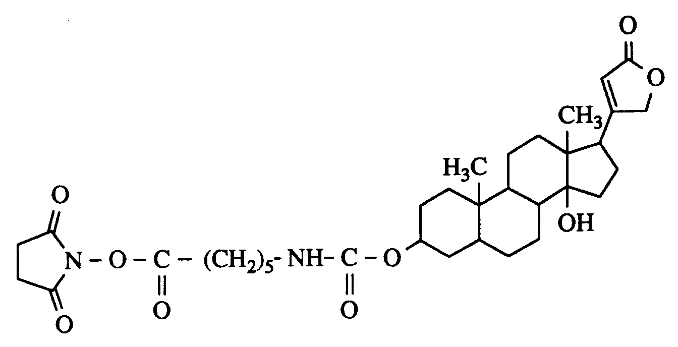
in 5 ml Isopropanol/Chloroform (1:1, v/v) zugetropft. Die Lösung wird anschließend 2 h unter Rückfluß, dann 8 h bei Raumtemperatur gerührt. Das Reaktionsgemisch wird im Vakuum eingedampft, in 2 ml Wasser aufgenommen und über eine Sephadex G-25 Säule (50 cm x 16 cm ⌀, UV-Detektion 254 nm, Laufmittel: Wasser) chromatographiert. Die produkthaltigen Fraktionen werden gefriergetrocknet. Ausbeute: 27 mg (60 % d. Th.)
¹H-NMR (250 MHz, CDCl₃): 1,45 (m), 1,71 (m), 2,66 (s), 2,99 (m), 4,30 (m), 4,80 (m)ppm.25 mg poly-DL-lysine are dissolved in 15 ml methanol and mixed with 20 ul triethylamine. 22 mg of digitoxigenin (6-aminocaproyl-carboxy) -N-hydroxysuccinimide are added with stirring at room temperature
added dropwise in 5 ml of isopropanol / chloroform (1: 1, v / v). The solution is then stirred under reflux for 2 h, then at room temperature for 8 h. The reaction mixture is evaporated in vacuo, taken up in 2 ml of water and chromatographed on a Sephadex G-25 column (50 cm × 16 cm ⌀, UV detection 254 nm, eluent: water). The product-containing fractions are freeze-dried. Yield: 27 mg (60% of theory)
1 H-NMR (250 MHz, CDCl₃): 1.45 (m), 1.71 (m), 2.66 (s), 2.99 (m), 4.30 (m), 4.80 (m ) ppm.
Eine immunologische Bestimmung des Digitoxigenin-Einbaus (Miles Seralyzer Digoxin Assay Kit) ergab einen Anteil Digoxinäquivalente von 2,5 Gew.-%).An immunological determination of the digitoxigenin incorporation (Miles Seralyzer Digoxin Assay Kit) showed a proportion of digoxin equivalents of 2.5% by weight).
Ein Objektträger aus Floatglas wird 1 Minute lang mit Wasser im Ultraschallbad behandelt und mit Stickstoff sorgfältig getrocknet, wobei die Oberflächen staubfrei gereinigt werden. Dann wird das Plättchen zur Vorteinigung in eine Mischung aus konz. Schwefelsäure und Wasserstoffperoxid (7:3, v/v) gebracht und darin bei 80°C eine Stunde lang im Ultraschallbad behandelt. Nach der Abkühlung auf Raumtemperatur wird das Plättchen 3 mal je 60 Sekunden in Wasser im Ultraschallbad behandelt und mit Wasser säurefrei gewaschen. Anschließend wird es in ein Gemisch aus H₂O/H₂O₂/NH₃ (5:1:1, v/v/v) gebracht und darin 5 Minuten lang bei 80°C behandelt. Danach wird das Plättchen sorgfältig mit Wasser salzfrei gewaschen.A slide made of float glass is treated with water in an ultrasound bath for 1 minute and carefully dried with nitrogen, the surfaces being cleaned dust-free. Then the plate is pre-cleaned in a mixture of conc. Bring sulfuric acid and hydrogen peroxide (7: 3, v / v) and treated therein at 80 ° C for one hour in an ultrasonic bath. After cooling to room temperature, the plate is treated 3 times in water for 60 seconds in an ultrasonic bath and washed with water until it is acid-free. Then it is placed in a mixture of H₂O / H₂O₂ / NH₃ (5: 1: 1, v / v / v) and treated for 5 minutes at 80 ° C. The plate is then washed carefully with water until it is free of salt.
Das Plättchen wird vor der Silanisierungsreaktion zur Entfernung von Wasserspuren je 2 Minuten lang in Methanol, Methanol/Toluol (1:1, v/v) und Toluol behandelt. Danach läßt man es unter Stickstoffatmosphäre 15 h in einer 5%igen (v/v) 3-Aminopropyldimethylethoxysilan-Lösung in Toluol reagieren. Schließlich wird das Plättchen je 1 Minute lang mit Toluol, Toluol/Dimethylsulfoxid (DMSO) (1:1, v/v) und DMSO im Ultraschallbad behandelt.The plate is treated in methanol, methanol / toluene (1: 1, v / v) and toluene for 2 minutes before the silanization reaction to remove traces of water. It is then left to react in a 5% (v / v) 3-aminopropyldimethylethoxysilane solution in toluene under a nitrogen atmosphere for 15 h. Finally, the plate is treated with toluene, toluene / dimethyl sulfoxide (DMSO) (1: 1, v / v) and DMSO in an ultrasonic bath for 1 minute each.
Allgemeines Beschichtungsverfahren
- A) Ein Probenplättchen wird 20 Minuten lang in Lösung A (Polykation) getaucht,
dann 3 mal je 20 Sekunden in je 10 ml Wasser getaucht und trocknen lassen. - B) Anschließend wird 20 Minuten in Lösung B (Polyanion),
wiederum 3 mal für 20 Sekunden in je 10 ml Wasser getaucht und trocknen gelassen.
- A) A sample plate is immersed in solution A (polycation) for 20 minutes, then immersed 3 times for 20 seconds in 10 ml of water and allowed to dry.
- B) Then it is immersed in solution B (polyanion) for 20 minutes, again 3 times for 20 seconds in 10 ml of water and allowed to dry.
1. Auf Floatglas, ohne Belegung mit Aminogruppen
Die Objektträger aus Floatglas werden nach Beispiel 3.1. vorbehandelt und nach obiger Prozedur durch sukzessives Tauchen in folgende Lösungen belegt:1. On float glass, without any amino groups
The slides made of float glass are made according to Example 3.1. pretreated and covered by successive dipping in the following solutions according to the above procedure:
Lösung A: 5 mg Polylysin in 10 ml 0.05 M Carbonatpuffer pH 8;
Lösung B: 5 mg Polymer C in 10 ml 0.05 M Carbonatpuffer pH 8.Solution A: 5 mg polylysine in 10 ml 0.05 M carbonate buffer pH 8;
Solution B: 5 mg polymer C in 10 ml 0.05 M carbonate buffer pH 8.
Lösung A: 0.5 mg DEAE-Dextran in 10 ml 0.05 M Carbonatpuffer pH 8;
Lösung B: 0.5 mg Polymer C in 10 ml 0.05 M Carbonatpuffer pH 8.Solution A: 0.5 mg DEAE dextran in 10 ml 0.05 M carbonate buffer pH 8;
Solution B: 0.5 mg polymer C in 10 ml 0.05 M carbonate buffer pH 8.
Lösung A: 10 mg Polylysin in 10 ml 0.05 M Carbonatpuffer pH 8;
Lösung B: 1 mg Polymer C in 10 ml 0.05 M Carbonatpuffer pH 8.Solution A: 10 mg polylysine in 10 ml 0.05 M carbonate buffer pH 8;
Solution B: 1 mg polymer C in 10 ml 0.05 M carbonate buffer pH 8.
Lösung A: 1 mg DEAE-Dextran in 10 ml 3 mM Salzsäure;
Lösung B:1 mg Polymer C in 10 ml 3 mM Salzsäure.Solution A: 1 mg DEAE dextran in 10
Solution B: 1 mg polymer C in 10
Lösung A: 0.1 mg Polyvinylpyridin (Reilline 2200) in 10 ml 3 mM Salzsäure;
Lösung B: 10 mg Polymer A in 10 ml 3 mM Salzsäure.Solution A: 0.1 mg polyvinylpyridine (Reilline 2200) in 10
Solution B: 10 mg polymer A in 10
Die Objektträger aus Floatglas werden nach Beispiel 3.2. vorbehandelt und in Abwandlung der allgemeinen Prozedur lediglich durch Tauchen in Lösung B belegt. Es lassen sich die Lösungen von 4.1.1. bis 4.1.4 verwenden.The slides made of float glass are made according to Example 3.2. pretreated and, in a modification of the general procedure, only occupied by dipping in solution B. The solutions from 4.1.1. up to 4.1.4.
Ein Stück von mit Vinylidenchlorid oberflächenaktivierter Polyesterfolie wird ohne weitere Reinigungsschritte analog der obigen allgemeinen Prozedur beschichtet. Es lassen sich die Lösungen von 4.1.1. bis 4.1.4. verwenden.A piece of polyester film surface-activated with vinylidene chloride is coated without further cleaning steps analogously to the general procedure above. The solutions from 4.1.1. until 4.1.4. use.
Ein nach Beispiel 4.1.3. beschichteter Träger wird daran anschließend durch Tauchen in eine Lösung von 0.1 mg/ml Digitoxigenin-derivatisiertem Polylysin (Beispiel 2) und Waschen gemäß der allgemeinen Prozedur mit einer spezifisch bindenden Schicht überzogen.
Das Plättchen wird anschließend mit 50 µl einer Lösung von TRITC-Anti-Digoxin-IgG (hergestellt durch Reaktion von Tetramethylrhodamin-isothiocyanat und Anti-Digoxin-IgG) in Kontakt benetzt (Proteinkonzentration 0.1 mg/ml). Nach Spülen mit einem Puffer (15 mM KH₂PO₄, 25 mM Citrat, 1 mM MgCl₂, 50 mM KCl, 0.4 g/l NaN₃, 1 g/l Rinderserumalbumin, pH 6.4) wird die Fluoreszenz nochmals gemessen, wobei die des Cumarins aus dem Polymer C (509 nm) zugunsten der Fluoreszenz des TRITC (587 nm) gelöscht wird (Figur 1, Kurve a). Zur Kontrolle wird anstelle des Digitoxigenin-derivatisierten Polylysin underivatisiertes Polylysin in gleicher Konzentration eingesetzt, wodurch die oberste Schicht nicht zur Bindung befähigt ist und sich kein Energietransfer ergibt (Figur 1, Kurve b).A according to example 4.1.3. The coated carrier is then coated with a specifically binding layer by immersion in a solution of 0.1 mg / ml digitoxigenin-derivatized polylysine (example 2) and washing according to the general procedure.
The plate is then wetted with 50 μl of a solution of TRITC anti-digoxin IgG (produced by reaction of tetramethylrhodamine isothiocyanate and anti-digoxin IgG) in contact (protein concentration 0.1 mg / ml). After rinsing with a buffer (15 mM KH₂PO₄, 25 mM citrate, 1 mM MgCl₂, 50 mM KCl, 0.4 g / l NaN₃, 1 g / l bovine serum albumin, pH 6.4) the fluorescence is measured again, the coumarin from the polymer C (509 nm) is deleted in favor of the fluorescence of the TRITC (587 nm) (FIG. 1, curve a). As a control, underivatized polylysine is used in the same concentration instead of the digitoxigenin-derivatized polylysine, as a result of which the top layer is incapable of binding and there is no energy transfer (FIG. 1, curve b).
Claims (12)
an welchem e) ein Antigen bzw. Antikörper als Analytmolekül gebunden werden kann, das wiederum mit dem Fluoreszenzfarbstoff F₂ markiert ist, wobei die Farbstoffmoleküle einen Abstand einnehmen, der einen strahlungslosen Försterenergietransfer ermöglicht, dadurch gekennzeichnet, daß als Mono- bzw. Multischicht b) eine oder mehrere alternierende Schichten aus Polyanionen oder Polykationen eingesetzt werden, und daß die Konzentration des gebundenen Analytmoleküls in Abhängigkeit von der Zunahme der relativen Fluoreszenzintensität von F₂ bzw. der Abnahme der Fluoreszenzintensität von F₁ oder der Änderung des Verhältnissses von beiden Intensitäten gemessen wird.Optical solid phase biosensor, markable with a dye F₁ and a receptor molecule for the detection of analyte molecules in the liquid phase, markable with a dye F₂ using the ranger energy transfer between F₁ and F₂, which
to which e) an antigen or antibody can be bound as an analyte molecule, which in turn is labeled with the fluorescent dye F₂, the dye molecules being at a distance which enables radiation-free transfer of ranger energy, characterized in that a monolayer or multilayer b) or more alternating layers of polyanions or polycations are used, and that the concentration of the bound analyte molecule is measured depending on the increase in the relative fluorescence intensity of F₂ or the decrease in the fluorescence intensity of F₁ or the change in the ratio of both intensities.
Fluoresceinderivate und Tetramethylrhodamin,
Fluoresceinderivate und Texasrot,
Cumarin der Formel I und Tetramethylrhodamin

Cumarin der Formel II und Tetramethylrhodamin,

Fluorescein derivatives and tetramethylrhodamine,
Fluorescein derivatives and Texas red,
Coumarin of formula I and tetramethylrhodamine
Coumarin of formula II and tetramethylrhodamine,
Cumarin der Formel I und Tetramethylrhodamin
oder
Cumarin der Formel II und Tetramethylrhodamin
als Farbstoffpaar F₁ und F₂ eingesetzt werden.Optical biosensor according to claim 8, in which
Coumarin of formula I and tetramethylrhodamine
or
Coumarin of formula II and tetramethylrhodamine
can be used as a dye pair F₁ and F₂.

Applications Claiming Priority (2)
| Application Number | Priority Date | Filing Date | Title |
|---|---|---|---|
| DE4208645A DE4208645A1 (en) | 1992-03-18 | 1992-03-18 | OPTICAL SOLID PHASE BIOSENSOR BASED ON FLUORESCENT COLOR-MARGINED POLYIONIC LAYERS |
| DE4208645 | 1992-03-18 |
Publications (2)
| Publication Number | Publication Date |
|---|---|
| EP0561239A1 true EP0561239A1 (en) | 1993-09-22 |
| EP0561239B1 EP0561239B1 (en) | 1998-06-03 |
Family
ID=6454353
Family Applications (1)
| Application Number | Title | Priority Date | Filing Date |
|---|---|---|---|
| EP93103585A Expired - Lifetime EP0561239B1 (en) | 1992-03-18 | 1993-03-05 | Optical solid-phase biosensor, with fluorescence labeled polyionic layers |
Country Status (6)
| Country | Link |
|---|---|
| US (1) | US5711915A (en) |
| EP (1) | EP0561239B1 (en) |
| JP (1) | JP3456660B2 (en) |
| AT (1) | ATE166974T1 (en) |
| CA (1) | CA2091635A1 (en) |
| DE (2) | DE4208645A1 (en) |
Cited By (3)
| Publication number | Priority date | Publication date | Assignee | Title |
|---|---|---|---|---|
| EP0762122A1 (en) * | 1995-08-16 | 1997-03-12 | Bayer Ag | Streptavadin and biotin-based optical solid-phase biosensor |
| WO2000065352A1 (en) * | 1999-04-28 | 2000-11-02 | Eidgenossisch Technische Hochschule Zurich | Polyionic coatings in analytic and sensor devices |
| US7316845B2 (en) | 1997-04-21 | 2008-01-08 | California Institute Of Technology | Multifunctional polymeric tissue coatings |
Families Citing this family (71)
| Publication number | Priority date | Publication date | Assignee | Title |
|---|---|---|---|---|
| GB9425138D0 (en) | 1994-12-12 | 1995-02-08 | Dynal As | Isolation of nucleic acid |
| US6232124B1 (en) | 1996-05-06 | 2001-05-15 | Verification Technologies, Inc. | Automated fingerprint methods and chemistry for product authentication and monitoring |
| US6120460A (en) * | 1996-09-04 | 2000-09-19 | Abreu; Marcio Marc | Method and apparatus for signal acquisition, processing and transmission for evaluation of bodily functions |
| US6544193B2 (en) * | 1996-09-04 | 2003-04-08 | Marcio Marc Abreu | Noninvasive measurement of chemical substances |
| US6156274A (en) * | 1997-02-27 | 2000-12-05 | St. John's University | Optical membrane films for polycation detection |
| US7745142B2 (en) * | 1997-09-15 | 2010-06-29 | Molecular Devices Corporation | Molecular modification assays |
| US20050227294A1 (en) * | 1997-09-15 | 2005-10-13 | Molecular Devices Corporation | Molecular modification assays involving lipids |
| US7632651B2 (en) * | 1997-09-15 | 2009-12-15 | Mds Analytical Technologies (Us) Inc. | Molecular modification assays |
| US6348322B1 (en) * | 1997-10-17 | 2002-02-19 | Duke University | Method of screening for specific binding interactions |
| US5976813A (en) * | 1997-12-12 | 1999-11-02 | Abbott Laboratories | Continuous format high throughput screening |
| US6331163B1 (en) | 1998-01-08 | 2001-12-18 | Microsense Cardiovascular Systems (1196) Ltd. | Protective coating for bodily sensor |
| JP3936058B2 (en) * | 1998-03-12 | 2007-06-27 | 株式会社メニコン | Contact lens solution |
| WO1999067640A1 (en) | 1998-06-22 | 1999-12-29 | The Regents Of The University Of California | Triggered optical biosensor |
| DE19850593A1 (en) * | 1998-11-03 | 2000-05-04 | Biochip Technologies Gmbh | DNA hybridization assay comprises detecting fluorescent resonance energy transfer between fluorochromes on different DNA strands |
| US6490030B1 (en) | 1999-01-18 | 2002-12-03 | Verification Technologies, Inc. | Portable product authentication device |
| EP1206213B1 (en) * | 1999-08-26 | 2005-01-26 | Novartis AG | Ocular analyte sensor |
| US6512580B1 (en) | 1999-10-27 | 2003-01-28 | Verification Technologies, Inc. | Method and apparatus for portable product authentication |
| EP1122535A3 (en) * | 2000-01-31 | 2004-09-22 | The Penn State Research Foundation | Interrogation of changes in the contents of a sealed container |
| US20030112423A1 (en) * | 2000-04-24 | 2003-06-19 | Rakesh Vig | On-line verification of an authentication mark applied to products or product packaging |
| US20040000787A1 (en) * | 2000-04-24 | 2004-01-01 | Rakesh Vig | Authentication mark for a product or product package |
| US6638593B2 (en) * | 2000-06-30 | 2003-10-28 | Verification Technologies, Inc. | Copy-protected optical media and method of manufacture thereof |
| US7124944B2 (en) * | 2000-06-30 | 2006-10-24 | Verification Technologies, Inc. | Product packaging including digital data |
| WO2002002301A1 (en) | 2000-06-30 | 2002-01-10 | Verification Technologies Inc. | Copy-protected optical media and method of manufacture thereof |
| US7660415B2 (en) * | 2000-08-03 | 2010-02-09 | Selinfreund Richard H | Method and apparatus for controlling access to storage media |
| US6706479B2 (en) | 2000-10-05 | 2004-03-16 | Virginia Tech Intellectual Properties, Inc. | Bio-chip, photoluminescent methods for identifying biological material, and apparatuses for use with such methods and bio-chips |
| US6740409B1 (en) | 2000-11-15 | 2004-05-25 | Board Of Trustees Of University Of Illinois | Polymer films |
| US7470420B2 (en) | 2000-12-05 | 2008-12-30 | The Regents Of The University Of California | Optical determination of glucose utilizing boronic acid adducts |
| AT410601B (en) * | 2000-12-29 | 2003-06-25 | Hoffmann La Roche | SENSOR FOR LUMINESCENCE-OPTICAL DETERMINATION OF ANALYTICAL AND REAGENT THAT WORKS ACCORDING TO THE FRET PRINCIPLE |
| WO2002054969A1 (en) * | 2001-01-10 | 2002-07-18 | Blood Cell Storage Inc. | System for growth, analysis, storage, validation and distribution of cells and tissues used for biomedical purposes |
| US20040265603A1 (en) * | 2001-08-03 | 2004-12-30 | Schlenoff Joseph B | Composite polyelectrolyte films for corrosion control |
| US6790709B2 (en) * | 2001-11-30 | 2004-09-14 | Intel Corporation | Backside metallization on microelectronic dice having beveled sides for effective thermal contact with heat dissipation devices |
| CA2473376A1 (en) | 2002-01-16 | 2003-07-31 | Dynal Biotech Asa | Method for isolating nucleic acids and protein from a single sample |
| US20040210289A1 (en) * | 2002-03-04 | 2004-10-21 | Xingwu Wang | Novel nanomagnetic particles |
| US20050084645A1 (en) * | 2002-02-07 | 2005-04-21 | Selinfreund Richard H. | Method and system for optical disc copy-protection |
| US8849379B2 (en) * | 2002-04-22 | 2014-09-30 | Geelux Holdings, Ltd. | Apparatus and method for measuring biologic parameters |
| US9848815B2 (en) | 2002-04-22 | 2017-12-26 | Geelux Holdings, Ltd. | Apparatus and method for measuring biologic parameters |
| US8328420B2 (en) | 2003-04-22 | 2012-12-11 | Marcio Marc Abreu | Apparatus and method for measuring biologic parameters |
| IL164685A0 (en) | 2002-04-22 | 2005-12-18 | Marcio Marc Aurelio Martins Ab | Apparatus and method for measuring biologic parameters |
| US7141369B2 (en) * | 2002-04-25 | 2006-11-28 | Semibio Technology, Inc. | Measuring cellular metabolism of immobilized cells |
| US7101947B2 (en) * | 2002-06-14 | 2006-09-05 | Florida State University Research Foundation, Inc. | Polyelectrolyte complex films for analytical and membrane separation of chiral compounds |
| US20040023397A1 (en) * | 2002-08-05 | 2004-02-05 | Rakesh Vig | Tamper-resistant authentication mark for use in product or product packaging authentication |
| US9034623B2 (en) | 2002-09-07 | 2015-05-19 | Jeffrey T. LaBelle | Nanoengineered biophotonic hybrid device |
| US7067293B2 (en) * | 2002-09-07 | 2006-06-27 | Labelle Jeffrey T | Nanoengineered biophotonic hybrid device |
| US8173407B2 (en) * | 2002-09-07 | 2012-05-08 | Labelle Jeffrey T | Nanoengineered biophotonic hybrid device |
| WO2004025300A1 (en) * | 2002-09-13 | 2004-03-25 | Hitachi Chemical Co., Ltd. | Fixation carrier and solid phase |
| CA2497645A1 (en) * | 2002-09-26 | 2004-04-08 | Verification Technologies, Inc. | Authentication of items using transient optical state change materials |
| GB0229287D0 (en) * | 2002-12-16 | 2003-01-22 | Dna Res Innovations Ltd | Polyfunctional reagents |
| US20060203700A1 (en) * | 2003-02-06 | 2006-09-14 | Verification Technologies, Inc. | Method and system for optical disk copy-protection |
| US20070010702A1 (en) * | 2003-04-08 | 2007-01-11 | Xingwu Wang | Medical device with low magnetic susceptibility |
| US20040254419A1 (en) * | 2003-04-08 | 2004-12-16 | Xingwu Wang | Therapeutic assembly |
| US20070149496A1 (en) * | 2003-10-31 | 2007-06-28 | Jack Tuszynski | Water-soluble compound |
| TWI227322B (en) * | 2003-11-13 | 2005-02-01 | Ind Tech Res Inst | Polyallylamine conjugates and applications thereof for biological signal amplification |
| US8481017B2 (en) | 2004-02-23 | 2013-07-09 | Florida State University Research Foundation, Inc. | Thin films for controlled protein interaction |
| US10227063B2 (en) | 2004-02-26 | 2019-03-12 | Geelux Holdings, Ltd. | Method and apparatus for biological evaluation |
| US7722752B2 (en) * | 2004-03-02 | 2010-05-25 | Florida State University Research Foundation | Variable charge films for controlling microfluidic flow |
| US7238536B1 (en) * | 2004-03-22 | 2007-07-03 | Florida State University Research Foundation, Inc. | Controlled transport through multiple reversible interaction point membranes |
| US20050249667A1 (en) * | 2004-03-24 | 2005-11-10 | Tuszynski Jack A | Process for treating a biological organism |
| US7713629B2 (en) | 2004-03-26 | 2010-05-11 | Florida State University Research Foundation | Hydrophobic fluorinated polyelectrolyte complex films and associated methods |
| US9056125B2 (en) | 2004-05-17 | 2015-06-16 | Florida State University Research Foundation, Inc. | Films for controlled cell growth and adhesion |
| DE102004046618A1 (en) * | 2004-09-25 | 2006-03-30 | Robert Bosch Gmbh | Circuit arrangement for analog / digital conversion |
| EP1951110B1 (en) | 2005-10-24 | 2012-10-03 | Marcio Marc Aurelio Martins Abreu | Apparatus for measuring biologic parameters |
| US20100233225A1 (en) * | 2007-02-09 | 2010-09-16 | Bingyun Li | Method of surface coating devices to incorporate bioactive molecules |
| JP2013120120A (en) * | 2011-12-07 | 2013-06-17 | Konica Minolta Medical & Graphic Inc | Test strip for lateral flow type chromatography, and method for detecting or quantifying analyte using the same |
| MX359376B (en) * | 2012-11-07 | 2018-09-24 | Canox4Drug S P A | Method and kit for determination of free copper in serum. |
| CA2980062A1 (en) | 2013-10-11 | 2015-04-16 | Marcio Marc Abreu | Method and apparatus for biological evaluation |
| JP2017501844A (en) | 2014-01-10 | 2017-01-19 | マーシオ マーク アブリュー | Device for measuring the infrared output of an Abreu brain thermal tunnel |
| CA2936235A1 (en) | 2014-01-10 | 2015-07-16 | Marcio Marc Abreu | Devices to monitor and provide treatment at an abreu brain tunnel |
| AU2015209304A1 (en) | 2014-01-22 | 2016-07-21 | Marcio Marc Abreu | Devices configured to provide treatment at an Abreu brain thermal tunnel |
| WO2016145215A1 (en) | 2015-03-10 | 2016-09-15 | Marcio Marc Abreu | Devices, apparatuses, systems, and methods for measuring temperature of an abtt terminus |
| CN114674791B (en) * | 2022-02-28 | 2023-01-17 | 江苏大学 | Preparation method and application of dye functionalized flexible up-conversion luminescence solid-phase sensor |
| US11773322B2 (en) | 2022-02-28 | 2023-10-03 | Jiangsu University | Preparation and application of dye-functionalized flexible upconversion-luminescence solid-phase sensor |
Citations (4)
| Publication number | Priority date | Publication date | Assignee | Title |
|---|---|---|---|---|
| EP0410323A2 (en) * | 1989-07-26 | 1991-01-30 | Perseptive Biosystems, Inc. | Immobilization of proteins and peptides on insoluble supports |
| EP0447133A2 (en) * | 1990-03-12 | 1991-09-18 | Orion-Yhtyma Oy | Paired polypeptides |
| EP0472990A2 (en) * | 1990-08-25 | 1992-03-04 | Bayer Ag | Mono or multilayer deposits on a substrat and process for making them |
| EP0429907B1 (en) * | 1989-11-21 | 1994-06-01 | Bayer Ag | Optical biosensor |
Family Cites Families (4)
| Publication number | Priority date | Publication date | Assignee | Title |
|---|---|---|---|---|
| EP0150905A3 (en) * | 1984-01-05 | 1987-01-14 | Ortho Diagnostic Systems Inc. | Anti-idiotype assay using fluorescent energy transfer |
| DE3938598A1 (en) * | 1989-11-21 | 1991-05-23 | Bayer Ag | Optical bio-sensor - based on fluorescent energy transfer between one dye incorporated in Langmuir-Blodgett film and marker dye |
| DE4114482A1 (en) * | 1991-05-03 | 1992-11-05 | Bayer Ag | POLYMERIC DYES, METHOD FOR THE PRODUCTION AND USE THEREOF |
| DE4116116A1 (en) * | 1991-05-17 | 1992-11-19 | Bayer Ag | Fluorescent light measuring instrument for joint testing - has optical passages generating quasi monochromatic excitation light and contg. collector lenses and detectors, for compact system |
-
1992
- 1992-03-18 DE DE4208645A patent/DE4208645A1/en not_active Withdrawn
-
1993
- 1993-03-05 EP EP93103585A patent/EP0561239B1/en not_active Expired - Lifetime
- 1993-03-05 AT AT93103585T patent/ATE166974T1/en active
- 1993-03-05 DE DE59308625T patent/DE59308625D1/en not_active Expired - Lifetime
- 1993-03-12 JP JP07745093A patent/JP3456660B2/en not_active Expired - Fee Related
- 1993-03-15 CA CA002091635A patent/CA2091635A1/en not_active Abandoned
-
1995
- 1995-10-24 US US08/547,272 patent/US5711915A/en not_active Expired - Lifetime
Patent Citations (4)
| Publication number | Priority date | Publication date | Assignee | Title |
|---|---|---|---|---|
| EP0410323A2 (en) * | 1989-07-26 | 1991-01-30 | Perseptive Biosystems, Inc. | Immobilization of proteins and peptides on insoluble supports |
| EP0429907B1 (en) * | 1989-11-21 | 1994-06-01 | Bayer Ag | Optical biosensor |
| EP0447133A2 (en) * | 1990-03-12 | 1991-09-18 | Orion-Yhtyma Oy | Paired polypeptides |
| EP0472990A2 (en) * | 1990-08-25 | 1992-03-04 | Bayer Ag | Mono or multilayer deposits on a substrat and process for making them |
Non-Patent Citations (5)
| Title |
|---|
| BIOMEDICINE Bd. 33, Nr. 7, November 1980, PARIS Seiten 210 - 211 PACHMANN ET AL. 'Poly-L-lysine as a coating agent for the attachment of active antibodies ....' * |
| DATABASE WPIL Week 8926, Derwent Publications Ltd., London, GB; AN 89-188469 & JP-A-1 126 557 (MEIDENSHA ELEC MFG KK) 18. Mai 1989 * |
| DATABASE WPIL Week 9032, Derwent Publications Ltd., London, GB; AN 90-242288 & JP-A-2 168 162 (SEKISUI CHEM IND KK) 28. Juni 1990 * |
| LANGONE ET AL. 'Methods in Enzymology. Immunochemical techniques' 1981 , ACADEMIC PRESS , NEW YORK Band 74, Teil C, Ullman et al.: 'Fluorescence excitation transfer immunoassay (FETI)', Seite 28 - Seite 60 * |
| SCIENCE Bd. 251, Nr. 4996, 22. Februar 1991, WASHINGTON, DC Seiten 927 - 929 BARNARD ET AL. 'Chemical sensors based on controlled-release polymer systems' * |
Cited By (4)
| Publication number | Priority date | Publication date | Assignee | Title |
|---|---|---|---|---|
| EP0762122A1 (en) * | 1995-08-16 | 1997-03-12 | Bayer Ag | Streptavadin and biotin-based optical solid-phase biosensor |
| US7316845B2 (en) | 1997-04-21 | 2008-01-08 | California Institute Of Technology | Multifunctional polymeric tissue coatings |
| WO2000065352A1 (en) * | 1999-04-28 | 2000-11-02 | Eidgenossisch Technische Hochschule Zurich | Polyionic coatings in analytic and sensor devices |
| US6884628B2 (en) | 1999-04-28 | 2005-04-26 | Eidgenossische Technische Hochschule Zurich | Multifunctional polymeric surface coatings in analytic and sensor devices |
Also Published As
| Publication number | Publication date |
|---|---|
| DE4208645A1 (en) | 1993-09-23 |
| ATE166974T1 (en) | 1998-06-15 |
| JP3456660B2 (en) | 2003-10-14 |
| CA2091635A1 (en) | 1993-09-19 |
| EP0561239B1 (en) | 1998-06-03 |
| JPH0627106A (en) | 1994-02-04 |
| DE59308625D1 (en) | 1998-07-09 |
| US5711915A (en) | 1998-01-27 |
Similar Documents
| Publication | Publication Date | Title |
|---|---|---|
| EP0561239B1 (en) | Optical solid-phase biosensor, with fluorescence labeled polyionic layers | |
| EP0429907B1 (en) | Optical biosensor | |
| DE69731306T2 (en) | METHOD AND KIT FOR DETERMINING AN ANALYSIS BY MEANS OF MASS ASSESSMENT | |
| EP0397113B1 (en) | Method of detecting specific binding substances in body fluids | |
| DE69735532T2 (en) | DETERMINATION OF ANTIGENS BY OLIGONUCLEOTIDE ANTIBODY CONJUGATES | |
| DE3705686C2 (en) | Methods for the determination of antibodies | |
| DE3105768C2 (en) | Use of a carrier for the immobilization of bioactive materials | |
| DE69817850T2 (en) | COVALENT IMMOBILIZATION WITH A HIGH SURFACE DENSITY OF OLIGONUCLEOTIDE MONO LAYERS | |
| EP0175973B1 (en) | Support material for the use in immunoassays | |
| DE69535273T2 (en) | DETECTION DEVICE AND METHOD | |
| DE3311889A1 (en) | METHOD FOR IRREVERSIBLE BINDING OF PROTEIN TO POLYSTYROL SURFACES WITH THE PRESERVATION OF BIOLOGICAL ACTIVITY, POLYSTYROL SURFACES OBTAINED AND THEIR USE | |
| EP0408078B1 (en) | Method for the preparation of a solid phase coated with an immunologically active substance | |
| EP2797680A1 (en) | Porous membranes having a polymeric coating and methods for their preparation and use | |
| EP0849595A1 (en) | Use of synthetic particles as reagents in agglutionation reactions | |
| EP0664452B1 (en) | Biotin-silane compounds and binding matrix containing these compounds | |
| EP0762122A1 (en) | Streptavadin and biotin-based optical solid-phase biosensor | |
| EP1332365B1 (en) | Method and test kit for detecting analytes in a sample | |
| WO1999027355A1 (en) | Method for producing laterally organized structures on supporting surfaces | |
| EP0215457B1 (en) | Method for the determination of a partner of an immuno-reaction | |
| EP0322813A2 (en) | Method for the determination of an immunologically active substance | |
| EP1525475B1 (en) | Sensor surface comprising an improved signal-noise ratio | |
| DE60109733T2 (en) | PROCESS FOR IMMOBILIZING AFFINE REAGENTS AT HYDROPHOBIC SOLID PHASES | |
| DE19925402A1 (en) | Screening of target-ligand interactions | |
| DE3733986A1 (en) | Means and process for manufacturing biosensors on the basis of an oscillating quartz crystal, and use thereof for determining antigens and affinity ligands | |
| EP0628819A2 (en) | Coated carrier, method for manufacturing it and its use in immobilising biomolecules on the surfaces of solids |
Legal Events
| Date | Code | Title | Description |
|---|---|---|---|
| PUAI | Public reference made under article 153(3) epc to a published international application that has entered the european phase |
Free format text: ORIGINAL CODE: 0009012 |
|
| AK | Designated contracting states |
Kind code of ref document: A1 Designated state(s): AT BE CH DE FR GB IT LI NL SE |
|
| 17P | Request for examination filed |
Effective date: 19940208 |
|
| 17Q | First examination report despatched |
Effective date: 19960904 |
|
| GRAG | Despatch of communication of intention to grant |
Free format text: ORIGINAL CODE: EPIDOS AGRA |
|
| GRAG | Despatch of communication of intention to grant |
Free format text: ORIGINAL CODE: EPIDOS AGRA |
|
| GRAH | Despatch of communication of intention to grant a patent |
Free format text: ORIGINAL CODE: EPIDOS IGRA |
|
| GRAH | Despatch of communication of intention to grant a patent |
Free format text: ORIGINAL CODE: EPIDOS IGRA |
|
| GRAA | (expected) grant |
Free format text: ORIGINAL CODE: 0009210 |
|
| AK | Designated contracting states |
Kind code of ref document: B1 Designated state(s): AT BE CH DE FR GB IT LI NL SE |
|
| PG25 | Lapsed in a contracting state [announced via postgrant information from national office to epo] |
Ref country code: NL Free format text: LAPSE BECAUSE OF FAILURE TO SUBMIT A TRANSLATION OF THE DESCRIPTION OR TO PAY THE FEE WITHIN THE PRESCRIBED TIME-LIMIT Effective date: 19980603 |
|
| REF | Corresponds to: |
Ref document number: 166974 Country of ref document: AT Date of ref document: 19980615 Kind code of ref document: T |
|
| REG | Reference to a national code |
Ref country code: CH Ref legal event code: EP |
|
| GBT | Gb: translation of ep patent filed (gb section 77(6)(a)/1977) |
Effective date: 19980604 |
|
| REF | Corresponds to: |
Ref document number: 59308625 Country of ref document: DE Date of ref document: 19980709 |
|
| ITF | It: translation for a ep patent filed |
Owner name: SOCIETA' ITALIANA BREVETTI S.P.A. |
|
| PG25 | Lapsed in a contracting state [announced via postgrant information from national office to epo] |
Ref country code: SE Free format text: LAPSE BECAUSE OF FAILURE TO SUBMIT A TRANSLATION OF THE DESCRIPTION OR TO PAY THE FEE WITHIN THE PRESCRIBED TIME-LIMIT Effective date: 19980903 |
|
| ET | Fr: translation filed | ||
| NLV1 | Nl: lapsed or annulled due to failure to fulfill the requirements of art. 29p and 29m of the patents act | ||
| PG25 | Lapsed in a contracting state [announced via postgrant information from national office to epo] |
Ref country code: AT Free format text: LAPSE BECAUSE OF NON-PAYMENT OF DUE FEES Effective date: 19990305 |
|
| PG25 | Lapsed in a contracting state [announced via postgrant information from national office to epo] |
Ref country code: LI Free format text: LAPSE BECAUSE OF NON-PAYMENT OF DUE FEES Effective date: 19990331 Ref country code: CH Free format text: LAPSE BECAUSE OF NON-PAYMENT OF DUE FEES Effective date: 19990331 Ref country code: BE Free format text: LAPSE BECAUSE OF NON-PAYMENT OF DUE FEES Effective date: 19990331 |
|
| PLBE | No opposition filed within time limit |
Free format text: ORIGINAL CODE: 0009261 |
|
| STAA | Information on the status of an ep patent application or granted ep patent |
Free format text: STATUS: NO OPPOSITION FILED WITHIN TIME LIMIT |
|
| 26N | No opposition filed | ||
| BERE | Be: lapsed |
Owner name: BAYER A.G. Effective date: 19990331 |
|
| REG | Reference to a national code |
Ref country code: CH Ref legal event code: PL |
|
| REG | Reference to a national code |
Ref country code: GB Ref legal event code: IF02 |
|
| PGFP | Annual fee paid to national office [announced via postgrant information from national office to epo] |
Ref country code: IT Payment date: 20100324 Year of fee payment: 18 Ref country code: FR Payment date: 20100325 Year of fee payment: 18 |
|
| PGFP | Annual fee paid to national office [announced via postgrant information from national office to epo] |
Ref country code: GB Payment date: 20100311 Year of fee payment: 18 |
|
| PGFP | Annual fee paid to national office [announced via postgrant information from national office to epo] |
Ref country code: DE Payment date: 20100521 Year of fee payment: 18 |
|
| REG | Reference to a national code |
Ref country code: GB Ref legal event code: 732E Free format text: REGISTERED BETWEEN 20100923 AND 20100929 |
|
| REG | Reference to a national code |
Ref country code: FR Ref legal event code: TP |
|
| GBPC | Gb: european patent ceased through non-payment of renewal fee |
Effective date: 20110305 |
|
| REG | Reference to a national code |
Ref country code: FR Ref legal event code: ST Effective date: 20111130 |
|
| PG25 | Lapsed in a contracting state [announced via postgrant information from national office to epo] |
Ref country code: FR Free format text: LAPSE BECAUSE OF NON-PAYMENT OF DUE FEES Effective date: 20110331 Ref country code: DE Free format text: LAPSE BECAUSE OF NON-PAYMENT OF DUE FEES Effective date: 20111001 |
|
| REG | Reference to a national code |
Ref country code: DE Ref legal event code: R119 Ref document number: 59308625 Country of ref document: DE Effective date: 20111001 |
|
| PG25 | Lapsed in a contracting state [announced via postgrant information from national office to epo] |
Ref country code: IT Free format text: LAPSE BECAUSE OF NON-PAYMENT OF DUE FEES Effective date: 20110305 Ref country code: GB Free format text: LAPSE BECAUSE OF NON-PAYMENT OF DUE FEES Effective date: 20110305 |