EP0550950A2 - Process and device for separating blood serum and plasma - Google Patents
Process and device for separating blood serum and plasma Download PDFInfo
- Publication number
- EP0550950A2 EP0550950A2 EP92306798A EP92306798A EP0550950A2 EP 0550950 A2 EP0550950 A2 EP 0550950A2 EP 92306798 A EP92306798 A EP 92306798A EP 92306798 A EP92306798 A EP 92306798A EP 0550950 A2 EP0550950 A2 EP 0550950A2
- Authority
- EP
- European Patent Office
- Prior art keywords
- blood
- separating
- serum
- plasma
- bean
- Prior art date
- Legal status (The legal status is an assumption and is not a legal conclusion. Google has not performed a legal analysis and makes no representation as to the accuracy of the status listed.)
- Withdrawn
Links
Images


Classifications
-
- G—PHYSICS
- G01—MEASURING; TESTING
- G01N—INVESTIGATING OR ANALYSING MATERIALS BY DETERMINING THEIR CHEMICAL OR PHYSICAL PROPERTIES
- G01N33/00—Investigating or analysing materials by specific methods not covered by groups G01N1/00 - G01N31/00
- G01N33/48—Biological material, e.g. blood, urine; Haemocytometers
- G01N33/483—Physical analysis of biological material
- G01N33/487—Physical analysis of biological material of liquid biological material
- G01N33/49—Blood
- G01N33/491—Blood by separating the blood components
-
- A—HUMAN NECESSITIES
- A61—MEDICAL OR VETERINARY SCIENCE; HYGIENE
- A61B—DIAGNOSIS; SURGERY; IDENTIFICATION
- A61B5/00—Measuring for diagnostic purposes; Identification of persons
- A61B5/15—Devices for taking samples of blood
- A61B5/150007—Details
- A61B5/150015—Source of blood
- A61B5/15003—Source of blood for venous or arterial blood
-
- A—HUMAN NECESSITIES
- A61—MEDICAL OR VETERINARY SCIENCE; HYGIENE
- A61B—DIAGNOSIS; SURGERY; IDENTIFICATION
- A61B5/00—Measuring for diagnostic purposes; Identification of persons
- A61B5/15—Devices for taking samples of blood
- A61B5/150007—Details
- A61B5/150206—Construction or design features not otherwise provided for; manufacturing or production; packages; sterilisation of piercing element, piercing device or sampling device
- A61B5/150236—Pistons, i.e. cylindrical bodies that sit inside the syringe barrel, typically with an air tight seal, and slide in the barrel to create a vacuum or to expel blood
-
- A—HUMAN NECESSITIES
- A61—MEDICAL OR VETERINARY SCIENCE; HYGIENE
- A61B—DIAGNOSIS; SURGERY; IDENTIFICATION
- A61B5/00—Measuring for diagnostic purposes; Identification of persons
- A61B5/15—Devices for taking samples of blood
- A61B5/150007—Details
- A61B5/150206—Construction or design features not otherwise provided for; manufacturing or production; packages; sterilisation of piercing element, piercing device or sampling device
- A61B5/150251—Collection chamber divided into at least two compartments, e.g. for division of samples
-
- A—HUMAN NECESSITIES
- A61—MEDICAL OR VETERINARY SCIENCE; HYGIENE
- A61B—DIAGNOSIS; SURGERY; IDENTIFICATION
- A61B5/00—Measuring for diagnostic purposes; Identification of persons
- A61B5/15—Devices for taking samples of blood
- A61B5/150007—Details
- A61B5/150374—Details of piercing elements or protective means for preventing accidental injuries by such piercing elements
- A61B5/150381—Design of piercing elements
- A61B5/150473—Double-ended needles, e.g. used with pre-evacuated sampling tubes
- A61B5/150496—Details of construction of hub, i.e. element used to attach the double-ended needle to a piercing device or sampling device
-
- A—HUMAN NECESSITIES
- A61—MEDICAL OR VETERINARY SCIENCE; HYGIENE
- A61B—DIAGNOSIS; SURGERY; IDENTIFICATION
- A61B5/00—Measuring for diagnostic purposes; Identification of persons
- A61B5/15—Devices for taking samples of blood
- A61B5/150007—Details
- A61B5/150732—Needle holders, for instance for holding the needle by the hub, used for example with double-ended needle and pre-evacuated tube
-
- A—HUMAN NECESSITIES
- A61—MEDICAL OR VETERINARY SCIENCE; HYGIENE
- A61B—DIAGNOSIS; SURGERY; IDENTIFICATION
- A61B5/00—Measuring for diagnostic purposes; Identification of persons
- A61B5/15—Devices for taking samples of blood
- A61B5/150007—Details
- A61B5/150755—Blood sample preparation for further analysis, e.g. by separating blood components or by mixing
-
- A—HUMAN NECESSITIES
- A61—MEDICAL OR VETERINARY SCIENCE; HYGIENE
- A61B—DIAGNOSIS; SURGERY; IDENTIFICATION
- A61B5/00—Measuring for diagnostic purposes; Identification of persons
- A61B5/15—Devices for taking samples of blood
- A61B5/153—Devices specially adapted for taking samples of venous or arterial blood, e.g. with syringes
- A61B5/154—Devices using pre-evacuated means
-
- B—PERFORMING OPERATIONS; TRANSPORTING
- B01—PHYSICAL OR CHEMICAL PROCESSES OR APPARATUS IN GENERAL
- B01D—SEPARATION
- B01D39/00—Filtering material for liquid or gaseous fluids
- B01D39/14—Other self-supporting filtering material ; Other filtering material
- B01D39/16—Other self-supporting filtering material ; Other filtering material of organic material, e.g. synthetic fibres
- B01D39/18—Other self-supporting filtering material ; Other filtering material of organic material, e.g. synthetic fibres the material being cellulose or derivatives thereof
-
- B—PERFORMING OPERATIONS; TRANSPORTING
- B01—PHYSICAL OR CHEMICAL PROCESSES OR APPARATUS IN GENERAL
- B01D—SEPARATION
- B01D39/00—Filtering material for liquid or gaseous fluids
- B01D39/14—Other self-supporting filtering material ; Other filtering material
- B01D39/20—Other self-supporting filtering material ; Other filtering material of inorganic material, e.g. asbestos paper, metallic filtering material of non-woven wires
- B01D39/2003—Glass or glassy material
- B01D39/2017—Glass or glassy material the material being filamentary or fibrous
-
- A—HUMAN NECESSITIES
- A61—MEDICAL OR VETERINARY SCIENCE; HYGIENE
- A61B—DIAGNOSIS; SURGERY; IDENTIFICATION
- A61B5/00—Measuring for diagnostic purposes; Identification of persons
- A61B5/15—Devices for taking samples of blood
- A61B5/150007—Details
- A61B5/150374—Details of piercing elements or protective means for preventing accidental injuries by such piercing elements
- A61B5/150381—Design of piercing elements
- A61B5/150389—Hollow piercing elements, e.g. canulas, needles, for piercing the skin
-
- B—PERFORMING OPERATIONS; TRANSPORTING
- B01—PHYSICAL OR CHEMICAL PROCESSES OR APPARATUS IN GENERAL
- B01D—SEPARATION
- B01D2239/00—Aspects relating to filtering material for liquid or gaseous fluids
- B01D2239/04—Additives and treatments of the filtering material
- B01D2239/0414—Surface modifiers, e.g. comprising ion exchange groups
-
- B—PERFORMING OPERATIONS; TRANSPORTING
- B01—PHYSICAL OR CHEMICAL PROCESSES OR APPARATUS IN GENERAL
- B01D—SEPARATION
- B01D2239/00—Aspects relating to filtering material for liquid or gaseous fluids
- B01D2239/04—Additives and treatments of the filtering material
- B01D2239/0464—Impregnants
-
- B—PERFORMING OPERATIONS; TRANSPORTING
- B01—PHYSICAL OR CHEMICAL PROCESSES OR APPARATUS IN GENERAL
- B01D—SEPARATION
- B01D2239/00—Aspects relating to filtering material for liquid or gaseous fluids
- B01D2239/04—Additives and treatments of the filtering material
- B01D2239/0471—Surface coating material
- B01D2239/0492—Surface coating material on fibres
-
- B—PERFORMING OPERATIONS; TRANSPORTING
- B01—PHYSICAL OR CHEMICAL PROCESSES OR APPARATUS IN GENERAL
- B01D—SEPARATION
- B01D2239/00—Aspects relating to filtering material for liquid or gaseous fluids
- B01D2239/12—Special parameters characterising the filtering material
- B01D2239/1225—Fibre length
-
- B—PERFORMING OPERATIONS; TRANSPORTING
- B01—PHYSICAL OR CHEMICAL PROCESSES OR APPARATUS IN GENERAL
- B01D—SEPARATION
- B01D2239/00—Aspects relating to filtering material for liquid or gaseous fluids
- B01D2239/12—Special parameters characterising the filtering material
- B01D2239/1233—Fibre diameter
-
- B—PERFORMING OPERATIONS; TRANSPORTING
- B01—PHYSICAL OR CHEMICAL PROCESSES OR APPARATUS IN GENERAL
- B01D—SEPARATION
- B01D2239/00—Aspects relating to filtering material for liquid or gaseous fluids
- B01D2239/12—Special parameters characterising the filtering material
- B01D2239/1291—Other parameters
-
- Y—GENERAL TAGGING OF NEW TECHNOLOGICAL DEVELOPMENTS; GENERAL TAGGING OF CROSS-SECTIONAL TECHNOLOGIES SPANNING OVER SEVERAL SECTIONS OF THE IPC; TECHNICAL SUBJECTS COVERED BY FORMER USPC CROSS-REFERENCE ART COLLECTIONS [XRACs] AND DIGESTS
- Y10—TECHNICAL SUBJECTS COVERED BY FORMER USPC
- Y10T—TECHNICAL SUBJECTS COVERED BY FORMER US CLASSIFICATION
- Y10T436/00—Chemistry: analytical and immunological testing
- Y10T436/25—Chemistry: analytical and immunological testing including sample preparation
- Y10T436/25375—Liberation or purification of sample or separation of material from a sample [e.g., filtering, centrifuging, etc.]
Definitions
- the present invention relates to a process and device for easily and efficiently separating and recovering serum or plasma components from whole blood.
- the present invention is used for separating and recovering serum or plasma components from blood samples when clinical chemistry analyses, bedside and other real-time assays are emergently needed.
- a conventionally, or even currently, available, typical method of separating serum or plasma from whole blood is centrifuging.
- blood usually collected through an injector which may or may not contain an anticoagulant, is placed in a tubular vessel, in which it is separated into cellular components and other cell-free constituents under the action of a force of about 650 to 1500 G.
- the separated cell-free constituents are used for a variety of clinical assays and tests available for various diagnoses and therapies.
- reagents themselves having a mechanism capable of separating serum from whole blood have been developed with the progress of dry chemistry reagents, opening a new way for emergent assays and rapid bedside tests.
- a testing paper including a filter paper form of glass fiber layer for collecting blood cells, which is made integral with a reagent-reacting layer.
- serum or plasma separated from a slight volume of whole blood (a few to 30 ⁇ l) through a blood cell separating region moves from there into the reagent reacting region in close contact with the blood cell separating region, where it reacts with the reagent to make a real-time assay of serum or plasma.
- Stover et al in EP-A-295526 discloses a blood cell separating region formed of a filter paper form of bibulous matrix comprising a lectin having a red blood cell agglutinizing activity and a bibulous material (hydrophilic organic powders, cellulosic material, etc.) and teaches a method of using this to separate serum and plasma components from a whole blood sample and guide them to the reaction region.
- the centrifuge or other method is suitable for large laboratories that assay a multitude of blood samples, and for institutions, such as hospitals, not requiring the assay results of (usually 1 to 10) tests in a matter of minutes.
- many small or private medical offices do not have such a costly, special blood separator on site. Therefore, the whole blood must be sent elsewhere for separation and assay.
- the assay results are not available in minutes but in hours or days.
- the whole blood when stored as such in days, would produce an assay error or make its assay impossible for hemolysis or other reasons. For patients with infectious diseases, much more care should be taken of disposal of the test vessel after use than of the operation of the centrifuging device.
- the problems mentioned above may be solved, because the blood cell separating regions are made integral with the reagent reacting regions.
- the object of these methods is to allow slight volumes of blood to react with slight volumes of reagents, in other words, it is impossible to obtain serum and plasma independently. Even when some modifications are made to these methods, e.g., even when a plurality of a filter form of blood cell separating regions are stacked up to prepare a separating layer, it is impossible to obtain serum and plasma due to hemolysis or other phenomenon.
- a process for separating and recovering serum and plasma components from a whole blood sample which comprises passing said whole blood sample through a separating filter formed of a blood cell-separating layer composed mainly of cellulose or glass fibers impregnated with a coating agent.
- the fibers have a diameter of 0.8 to 2.5 /1.m, a length of 100 to 3000 /1.m and a density of 100 to 300 mg/cm 3 .
- the coating agent is a surface active agent or agents having wetting power, which may or may not contain a nonblood group specific lectin that interacts on blood.
- This surface active agent may be polyethylene glycol hereinafter PEG for short)or docusate sodium.
- a device for separating and recovering serum and plasma from a whole blood sample which comprises a blood collector having a needle at one end and a blood sucker at the other end, and a separating filter including a blood cell-separating layer, said filter being provided in the intermediate zone of said blood collector, whereby said whole blood sample sucked through said needle by depressurizing said blood sucker is passed through the said separating filter.
- a device for separating and recovering serum and plasma from a whole blood sample which comprises an extruder for extruding said whole blood sample from within under pressure and a separator connected to said extruder and receiving a separating filter including a blood cell separating layer therein, whereby said whole blood sample is passed from said extruder through said separator.
- the coating agent can be a surface active agent having wetting power, for instance, polyethylene glycol or docusate sodium.
- polyethylene glycol be used in the form of a mixture with a polyacrylic ester derivative.
- the coating agent produces a sufficient effect at a concentration of 0.01 to 10%.
- the coating agent may additionally contain a lectin that interacts on blood. To this end any lectin is used, if it is a nonblood group specific one. Thus, especially when the coating agent contains the nonblood group specific lectin, it is well capable of separating serum and plasma from whole blood at a reduced concentration.
- poly(butyl acrylate) hereinafter PBA for short
- PBA poly(methyl acrylate)
- PEA poly(ethyl acrylate)
- the polyacrylic ester derivatives are preferably used in the form of a mixture of (a) PBA and (b) PMA or PEA. More preferably, a mixture of (a), (b) and (c) PEG is used at a ratio of (10-20):(1-4):(1-4) and in a total amount of 2 to 3%, because particularly favorable separation of blood cells is then achieved.
- Any blood cell agglutinizing agent may be used for the blood cell separating layer and lectin-impregnated layer, if it is a nonblood group specific one, and produces a sufficient effect in a slight amount as low as 0.01 to 0.005% by weight.
- the lectins used can be from Abrus precatorius (abrin, Jequirty bean), Bauhinia purpurea (camels foot tree), Caragana arborescens (Siberian pea tree), Codium fragile (Green marine algae), Concanavalin A (Jack bean), Glycine max (Soybean), Lathyrus odoratus (Sweet Pea), Lens culinaris (Lentil), Limulus polyphemus (Horseshoe crab, Limulin), Lycopersicon esculentum (Tomato), Maclura pomifera (Osage orange), Mycoplasma gallisepticum, Perseau americana (Avacado), Phaseolus coccineus sativum (garden pea), Psophocarpus tetragonolobus (winged bean), Ricinus communis (Castor bean), Solanum tuberosum (Potato), Triticum vulgaris (W
- Glass fibers having diameters of 0.8 to 2.5 ⁇ m are pulverized to lengths of 100 to 3000 /1.m.
- the pulverized glass fibers are suspended in a polyethylene glycol solution, then packed in a conical vessel at a density of 100 to 300 mg/cm 3 and finally dried to form a separating filter.
- Glass fibers having diameters of 0.8 to 2.5 ⁇ m are pulverized to lengths of 100 to 3000 /1.m.
- the pulverized glass fibers are suspended in a docusate sodium solution, then packed in a conical vessel at a density of 100 to 300 mg/cm 3 and finally dried to form a separating filter.
- Glass fibers having diameters of 0.8 to 2.5 ⁇ m are pulverized to lengths of 100 to 3000 ⁇ m.
- the pulverized glass fibers are suspended in a mixed solution of polyethylene glycol and Glycine max (Soybean) lectin, then packed in a conical vessel at a density of 100 to 300 mg/cm 3 and finally dried to form a separating filter.
- Glycine max Soybean
- Glass fibers having diameters of 0.8 to 2.5 ⁇ m are pulverized to lengths of 100 to 3000 ⁇ m.
- the pulverized glass fibers are suspended in a polyethylene glycol solution. After that, the suspension is packed in a cylindrical vessel with a needle connected to it at a density of 100 to 300 mg/cm 3 , followed by drying. Finally, a rubber plug is tightly put in the upper portion of the vessel to form a vacuum type of serum/plasma separating device.
- Hare's fresh blood was added dropwise to each of the separated filter obtained in Production Example 1 and the control sample obtained in Comparative Production Example 1 to separate plasma from it.
- the volume and degree of hemolysis of the collected plasma and the time of separation were measured. The results are set out below.
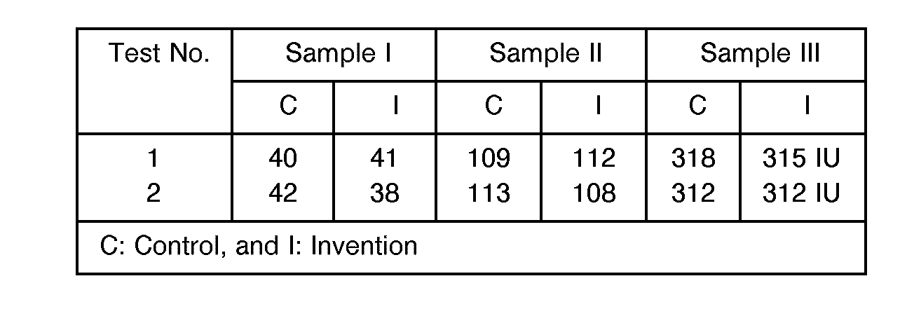
- glucose For the determination of glucose, use was made of three whole blood samples having different glucose concentrations, one subjected to centrifuging (for control) and the remaining two to serum separation with the serum separating device according to this invention. In all the cases, the concentrations of glucose were measured with a commercially available reagent (enzyme). The results are set out below.
- 3-hydroxybutyric acid For the determination of 3-hydroxybutyric acid, use was made of three whole blood samples having different 3-hydroxybutyric acid concentrations, one subjected to centrifuging (for control) and the remaining two to serum separation with the serum separating device according to this invention. In all the cases, the concentrations of glucose were measured with a commercially available reagent (enzyme). The results are set out below.
- Glass fibers GB100R (Toyo Roshi Co., Ltd.) having an average diameter of 0.5 to 2.5 /1.m are impregnated with a 5.5% methanol solution containing PBA, PMA and polyethylene glycol at 12:2:1, followed by drying.
- the dried fibers are finely pulverized, packed in a column of 70 mm in inner diameter and 30 mm in height, and impregnated with a 0.01% saline solution of Glycine max (Soybean) lectin.
- Nonwoven fabric QUINOCLOTH K70 (Honshu Seishi Co., Ltd.), dried at room temperature for about 1 hour and cut to the same size, was put on the thus impregnated fibers to form a mini-column.
- the separating device is built up of a bottomed, outer tube 2 including a needle 1 at one end, a tubular blood collector 3 fitted into the outer tube 2 and depressurized substantially to vacuum, a connector 4 fitted into the rear end of the blood collector 3 and a tubular form of separated fluid collector 5 fitted into the rear end of the connector 4 and depressurized substantially to vacuum.
- the blood collector 2, the connector 4 and the separated fluid collector 5 remain separated from each other.
- the outer tube 2 is in a bottomed, cylindrical form, and the needle 1 for collecting blood is fixed through the front end of the outer tube 2, said needle 1 being inwardly bent at 1 a at its roar end.
- the blood collector 3 is built up of a cylindrical body 3a open at both ends, and nonpermeable plug members 3b and 3c formed of rubber material, with which both ends of the body 3a are tightly closed.
- a blood cell separating filter 6 which is of a double-layer structure comprising a fibrous material layer 6a for separating blood cells from a whole blood sample and a a blood cell agglutinizing agent layer 6b impregnated with lectin. Note that this blood cell agglutinizing agent layer 6 may be dispensed with.
- the connector 4 is in a cylindrical form having a partition 4a substantially at its center, and an axially double-headed, hollow, connecting needle 4b is fixedly passed through the central region of the partition 4a.
- the separated fluid collector 5 has a closed-end, cylindrical form and has an opening tightly closed by a non-permeable plug member 5a formed of rubber material. This collector 5 is depressurized substantially to vacuum.
- the needle 1 For collecting blood, the needle 1 is first stuck in the blood vessel under the skin, and the blood collector 3 is then forced into the outer tube 2 to stick the inward part 1 a of the into the plug member 3b. Consequently, the blood is vacuum-sucked in the blood collector 3. After the required volume of blood has been collected, the needle 1 is removed from the skin. Thereafter, the collector 5 is pressed against the rear portion of the blood collector 3 through the connector 4, so that the connecting needle 4b can break through the plug member 3c of the blood collector 3 and the plug member 5a of the separated fluid collector 5. Consequently, the blood can be vacuum-sucked from the blood collector 3 into the separated fluid collector 5 through the blood cell separating filter 6. In the meantime, the blood is separated into serum and plasma components through the filter 6, which are then pooled in the separated fluid collector 5.
- this method enables hundreds of 1 of serum and plasma to be obtained from about 1 ml of a whole blood sample. This amount of serum and plasma enables as many as 10 or more blood tests to be done simultaneously with blood-collecting.
- FIG. 3 there is shown the second embodiment of the separating device according to this invention.
- the blood collector 3 is separated from the connector 4 and the fluid collector 5 prior to use.
- these parts are made integral with each other. More specifically, while the needle 1 remains stuck in the blood vessel, the fluid collector 5 is forced into the outer tube 2 together with the blood collector 3.
- FIG. 4 there is shown the third embodiment of the separating device according to this invention.
- an outer tube 2 is provided at its rear end with an extension, in which a separated fluid collector 5 is slidably pre-fitted.
- a hollow needle 10 is fixed at its one end in a blood collector 3 and extends from there through the rear end of the blood collector 3. The tip portion of this hollow needle 10 is supported by a partition 11 and located in opposition to the fluid collector 5.
- a funnel form of filter support plate 10a which supports and keeps the rear part of a filter from movement.
- the hollow needle 10 breaks, through the plug members 3b and 5a of the collectors 3 and 5, so that the blood can be vacuum-sucked into the blood collector 3 and the serum and plasma separated through the filter 6 can be sucked into the collector 5.
- This third embodiment of the separating device according to this invention enables serum and plasma to be separated and recovered from a whole blood sample simultaneously with blood sampling. Since the filter 6 is kept from co-movement by the support plate 19a during vacuum suction, the serum and plasma are more efficiently separated and recovered from the blood sample.
- this separating device is built up of a hard column 21 made of resin or glass, which has a tapering tip portion or a blood sucking or ejecting port 21a, a rubber plug 22 fitted into an opening in the rear end of the column 21 and an injector 23 fitted into a through-hole 22a formed centrally through the rubber plug 22.
- This injector 23 is used generally for collecting blood, and is made up of a cylinder 23a and a plunger 23b slidably inserted therein for vacuum suction or compression.
- This separating device is used as follows. When it is intended to collect blood in the cylinder 23a of the injector 23, the suction port 21 a of the column 21 is first immersed in blood stored in another vessel, e.g., a test tube. Then, the plunger 23b is pulled out to suck the blood from the column 21 into the cylinder 23a. While the blood passes through the blood cell separating layer 24, the serum and plasma components are selectively separated from the blood and sucked in the cylinder 23a. The serum and plasma components may then be used for the required tests, if the injector 23 is pulled out of the rubber plug 22 to transfer the serum and plasma components to other sample vessel.
- another vessel e.g., a test tube
- blood can be collected directly from a human or animal subject with the injector 23 to which the needle is attached. Then, the needle is removed from the injector 23, which is in turn connected to the column 21 to give a push on the plunger 23, thereby obtaining the serum and plasma components.
- a separate vessel for storing blood after collecting the blood can be dispensed with, and also the time of separation can be substantially reduced.
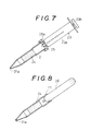
- FIG. 7 there is shown a modification of the fourth embodiment.
- This embodiment is similar to the fourth embodiment with the exception that a rubber plug 25 having a larger diameter is fitted onto an opening in the rear end of a column 21 and the tip of an injector 23 is fitted into a hole 25a formed centrally in the plug 25.
- the manner of using this embodiment is similar to that of the fourth embodiment.
- a syringe 26 is detachably received in an opening in the rear end of a column 21 provided therein with a blood cell separating layer 24.
- This syringe 26 is formed of an elastic, flexible resin material, and has a volume sufficient for blood suction and ejection.
- the column 21 is connected to the syringe 26, as illustrated. While the syringe 26 is deflated, the suction port 21 in the column 21 is immersed in a blood sample vessel, so that the serum and plasma components can be sucked and collected in the syringe 26 by its elastic inflation.
- the syringe 26, in which a blood sample has previously been collected is connected to the column 21, whereupon a push is given to the syringe 26 to eject the separated serum and plasma components from the ejecting port 21 in the column 21.
- a column 27 is built up of a funnel form of vessel 28 having a sucking or ejecting port 28a at the tip, a porous keep plate 29 located in the vessel 28, and a lid fitted over an opening in the rear portion of the vessel 28 and provided with a boss 30b having therein a through-hole 30a in communication with a cylinder 23a of an injector 23.
- a blood cell separating layer 24 is sandwiched between the keep plate 29 and the lid 30, so that it is kept from movement during suction and ejection. Note that the manner of using this embodiment is similar to that of the fourth or fifth embodiment.
- the present invention enables the volume of serum and plasma required for as many as ten or more tests to be simply and inexpensively separated and recovered from a whole blood sample just after sampling, without posing some problems which are associated with separating procedures such as centrifuging.
- serum and plasma can be separated from a blood sample just after sampling, high assay accuracy is achievable.
Abstract
A sample of whole blood is passed through a filter layer composed of cellulose or glass fibers 9if diameter 0.8 - 2.5 m, length 100 - 3000 m and density 100-300 mg/cm!) which are impregnated with a coating agent, preferably a surfactant, e.g. polyethylene glycol mixed with polyacrylic acid alkyl esters and optionally with a lectin obtained from a plant; the sample is thus separated into plasma and serum which can be separately analysed.
Separating devices are described which have at one end a needle (1) communicating with a blood collecting vessel portion (3), a separating filter layer (6a) and means for sucking blood through the filter and separately collecting the plasma and serum.
The invention allows numerous samples to be taken and separated rapidly and inexpensively without centrifuging.
Description
- The present invention relates to a process and device for easily and efficiently separating and recovering serum or plasma components from whole blood. The present invention is used for separating and recovering serum or plasma components from blood samples when clinical chemistry analyses, bedside and other real-time assays are emergently needed.
- A conventionally, or even currently, available, typical method of separating serum or plasma from whole blood is centrifuging. According to the centrifuge method, blood usually collected through an injector, which may or may not contain an anticoagulant, is placed in a tubular vessel, in which it is separated into cellular components and other cell-free constituents under the action of a force of about 650 to 1500 G. The separated cell-free constituents are used for a variety of clinical assays and tests available for various diagnoses and therapies. On the other hand, reagents themselves having a mechanism capable of separating serum from whole blood have been developed with the progress of dry chemistry reagents, opening a new way for emergent assays and rapid bedside tests.
- For instance, Vogel et al in U.S. Patent No. 4,477,575 discloses a testing paper including a filter paper form of glass fiber layer for collecting blood cells, which is made integral with a reagent-reacting layer. As set forth therein, serum or plasma separated from a slight volume of whole blood (a few to 30 µl) through a blood cell separating region moves from there into the reagent reacting region in close contact with the blood cell separating region, where it reacts with the reagent to make a real-time assay of serum or plasma. Lon R. Stover et al in EP-A-295526 discloses a blood cell separating region formed of a filter paper form of bibulous matrix comprising a lectin having a red blood cell agglutinizing activity and a bibulous material (hydrophilic organic powders, cellulosic material, etc.) and teaches a method of using this to separate serum and plasma components from a whole blood sample and guide them to the reaction region.
- The centrifuge or other method is suitable for large laboratories that assay a multitude of blood samples, and for institutions, such as hospitals, not requiring the assay results of (usually 1 to 10) tests in a matter of minutes. However, many small or private medical offices do not have such a costly, special blood separator on site. Therefore, the whole blood must be sent elsewhere for separation and assay. As a result, the assay results are not available in minutes but in hours or days. In some cases, the whole blood, when stored as such in days, would produce an assay error or make its assay impossible for hemolysis or other reasons. For patients with infectious diseases, much more care should be taken of disposal of the test vessel after use than of the operation of the centrifuging device.
- According to the methods disclosed in U.S. Patent No. 4,477,575 and EP-A-295526, the problems mentioned above may be solved, because the blood cell separating regions are made integral with the reagent reacting regions. However, because of relying on dry chemistry reactions, it is not possible to prepare reagent reaction regions for immunoassay. As regards the separated serum and plasma components, different reagent reaction regions are needed for individual tests. However, again, all reagent reaction regions are not prepared for all numerous tests. In addition, the object of these methods is to allow slight volumes of blood to react with slight volumes of reagents, in other words, it is impossible to obtain serum and plasma independently. Even when some modifications are made to these methods, e.g., even when a plurality of a filter form of blood cell separating regions are stacked up to prepare a separating layer, it is impossible to obtain serum and plasma due to hemolysis or other phenomenon.
- It is therefore a main object of this invention to provide a simple and rapid process for separating serum and plasma components from a whole blood sample for the purpose of real-time tests such as emergent clinical, chemical analyses and bedside examinations. Another object of this invention is to provide a simple device to simply and rapidly separate serum or plasma components from body fluids, e.g., whole blood for about a few to ten tests.
- As a result of investigating various fibrous materials in terms of their capabilities of separating blood cells from whole blood, we have found that they are unsuitable for obtaining serum or plasma in amounts well enough for about a few to ten tests, because hemolysis is likely to occur during separation. From the relations between the type, size and density of fibrous material and the coating material therefor, however, we have now discovered that serum or plasma can be efficiently and instantaneously separated from whole blood with a blood cell-separating layer comprising nonwoven fabric, cellulose or glass fibers and a coating agent, said fibers having a length lying in the range of 100 to 3000 /1.m and a density of 50 to 250 mg/cm3.
- According to one aspect of this invention, there is provided a process for separating and recovering serum and plasma components from a whole blood sample, which comprises passing said whole blood sample through a separating filter formed of a blood cell-separating layer composed mainly of cellulose or glass fibers impregnated with a coating agent.
- Preferably, the fibers have a diameter of 0.8 to 2.5 /1.m, a length of 100 to 3000 /1.m and a density of 100 to 300 mg/cm3.
- Preferably, the coating agent is a surface active agent or agents having wetting power, which may or may not contain a nonblood group specific lectin that interacts on blood. This surface active agent, for instance, may be polyethylene glycol hereinafter PEG for short)or docusate sodium.
- According to another aspect of this invention, there is provided a device for separating and recovering serum and plasma from a whole blood sample, which comprises a blood collector having a needle at one end and a blood sucker at the other end, and a separating filter including a blood cell-separating layer, said filter being provided in the intermediate zone of said blood collector, whereby said whole blood sample sucked through said needle by depressurizing said blood sucker is passed through the said separating filter.
- According to the third aspect of this invention, there is provided a device for separating and recovering serum and plasma from a whole blood sample, which comprises an extruder for extruding said whole blood sample from within under pressure and a separator connected to said extruder and receiving a separating filter including a blood cell separating layer therein, whereby said whole blood sample is passed from said extruder through said separator.
- The coating agent can be a surface active agent having wetting power, for instance, polyethylene glycol or docusate sodium. In particular, it is desired that polyethylene glycol be used in the form of a mixture with a polyacrylic ester derivative.
- According to this invention, it is found that the coating agent produces a sufficient effect at a concentration of 0.01 to 10%. If desired, the coating agent may additionally contain a lectin that interacts on blood. To this end any lectin is used, if it is a nonblood group specific one. Thus, especially when the coating agent contains the nonblood group specific lectin, it is well capable of separating serum and plasma from whole blood at a reduced concentration.
- Of the polyacrylic ester derivatives, particular preference is given to poly(butyl acrylate) (hereinafter PBA for short), poly(methyl acrylate) (PMA) and poly(ethyl acrylate) (PEA). With glass fibers containing these derivatives together with PEG, it has been found that hundreds of 1 of serum or plasma can be easily separated from about 1 ml of a whole blood sample in minutes. In addition, it has been found that the provision of the lectin-impregnated layer makes that separation more efficient.
- The polyacrylic ester derivatives are preferably used in the form of a mixture of (a) PBA and (b) PMA or PEA. More preferably, a mixture of (a), (b) and (c) PEG is used at a ratio of (10-20):(1-4):(1-4) and in a total amount of 2 to 3%, because particularly favorable separation of blood cells is then achieved.
- Any blood cell agglutinizing agent may be used for the blood cell separating layer and lectin-impregnated layer, if it is a nonblood group specific one, and produces a sufficient effect in a slight amount as low as 0.01 to 0.005% by weight. The lectins used, by way of example, can be from Abrus precatorius (abrin, Jequirty bean), Bauhinia purpurea (camels foot tree), Caragana arborescens (Siberian pea tree), Codium fragile (Green marine algae), Concanavalin A (Jack bean), Glycine max (Soybean), Lathyrus odoratus (Sweet Pea), Lens culinaris (Lentil), Limulus polyphemus (Horseshoe crab, Limulin), Lycopersicon esculentum (Tomato), Maclura pomifera (Osage orange), Mycoplasma gallisepticum, Perseau americana (Avacado), Phaseolus coccineus sativum (garden pea), Psophocarpus tetragonolobus (winged bean), Ricinus communis (Castor bean), Solanum tuberosum (Potato), Triticum vulgaris (Wheat germ), Vicia faba (faba bean, broad bean), Vigna radiata (Mung bean), Viscum album (European bean) and Wisteria floribunda (Japanese wisteria).
- These lectins are extracted and isolated in known manner from these plants.
- The present invention will now be explained more specifically but not exclusively with reference to several examples.
- Glass fibers having diameters of 0.8 to 2.5 µm are pulverized to lengths of 100 to 3000 /1.m. The pulverized glass fibers are suspended in a polyethylene glycol solution, then packed in a conical vessel at a density of 100 to 300 mg/cm3 and finally dried to form a separating filter.
- Glass fibers having diameters of 0.8 to 2.5 µm are pulverized to lengths of 100 to 3000 /1.m. The pulverized glass fibers are suspended in a docusate sodium solution, then packed in a conical vessel at a density of 100 to 300 mg/cm3 and finally dried to form a separating filter.
- Glass fibers having diameters of 0.8 to 2.5 µm are pulverized to lengths of 100 to 3000 µm. The pulverized glass fibers are suspended in a mixed solution of polyethylene glycol and Glycine max (Soybean) lectin, then packed in a conical vessel at a density of 100 to 300 mg/cm3 and finally dried to form a separating filter.
- Glass fibers having diameters of 0.8 to 2.5 µm are pulverized to lengths of 100 to 3000 µm. The pulverized glass fibers are suspended in a polyethylene glycol solution. After that, the suspension is packed in a cylindrical vessel with a needle connected to it at a density of 100 to 300 mg/cm3, followed by drying. Finally, a rubber plug is tightly put in the upper portion of the vessel to form a vacuum type of serum/plasma separating device.
- Similar glass fibers as used in the above examples are suspended under agitation in a polyethylene glycol-free solution, and then packed in a conical vessel at a density of 100 to 300 mg/cm3. Subsequent drying gives a sample for comparative testing.
- The following tests were conducted to find whether or not there was a significant difference between the volumes of the components in serum or plasma actually separated from whole blood by each of the separating filters and conventional centrifuging.
- Hare's fresh blood was added dropwise to each of the separated filter obtained in Production Example 1 and the control sample obtained in Comparative Production Example 1 to separate plasma from it. The volume and degree of hemolysis of the collected plasma and the time of separation were measured. The results are set out below.

- For the determination of glucose, use was made of three whole blood samples having different glucose concentrations, one subjected to centrifuging (for control) and the remaining two to serum separation with the serum separating device according to this invention. In all the cases, the concentrations of glucose were measured with a commercially available reagent (enzyme). The results are set out below.

- For the determination of 3-hydroxybutyric acid, use was made of three whole blood samples having different 3-hydroxybutyric acid concentrations, one subjected to centrifuging (for control) and the remaining two to serum separation with the serum separating device according to this invention. In all the cases, the concentrations of glucose were measured with a commercially available reagent (enzyme). The results are set out below.

- For the determination of total bile acids, use was made of three whole blood samples having different total bile acids concentrations, one subjected to centrifuging (for control) and the remaining two to serum separation with the serum separating device according to this invention. In all the cases, the concentrations of glucose were measured with a commercially available reagent (enzyme). The results are set out below.

- For the determination of GPT, use was made of three whole blood samples having different GPT concentrations, one subjected to centrifuging (for control) and the remaining two to serum separation with the serum separating device according to this invention. In all the cases, the concentrations of GPT were measured with a commercially available reagent (enzyme). The results are set out below.
- Glass fibers GB100R (Toyo Roshi Co., Ltd.) having an average diameter of 0.5 to 2.5 /1.m are impregnated with a 5.5% methanol solution containing PBA, PMA and polyethylene glycol at 12:2:1, followed by drying. The dried fibers are finely pulverized, packed in a column of 70 mm in inner diameter and 30 mm in height, and impregnated with a 0.01% saline solution of Glycine max (Soybean) lectin. Finally, Nonwoven fabric QUINOCLOTH K70 (Honshu Seishi Co., Ltd.), dried at room temperature for about 1 hour and cut to the same size, was put on the thus impregnated fibers to form a mini-column.
- The following tests were conducted to find whether or not there was a significant difference between the volumes of the components in serum or plasma actually separated from whole blood by the mini-column obtained in Ex. 5 and conventional centrifuging.
- For the determination of glucose, use was made of three whole blood samples having different glucose concentrations, one subjected to centrifuging (for control) and the remaining two to serum separation with the serum separating column according to this invention. In all the cases, the concentrations of glucose were measured with a commercially available reagent (enzyme). From the results set out in Table 1, it is noted that there is no significant difference between the samples.

- For the determination of 3-hydroxybutyric acid, use was made of three whole blood samples having different 3-hydroxybutyric acid concentrations, one subjected to centrifuging (for control) and the remaining two to serum separation with the serum separating column according to this invention. In all the cases, the concentrations of glucose were measured with a commercially available reagent (enzyme). From the results set out in Table 2, it is noted that there is no significant difference between the samples.

- For the determination of total bile acids, use was made of three whole blood samples having different total bile acids concentrations, one subjected to centrifuging (for control) and the remaining two to serum separation with the serum separating column according to this invention. In all the cases, the concentrations of glucose were measured with a commercially available reagent (enzyme). From the results set out below in Table 3, it is noted that there is no significant difference between the samples.

- In what follows, some embodiments of the serum/plasma separating device according to this invention will be explained more specifically but not exclusively with reference to the accompanying drawings, in which:
- FIGURE 1 is a perspective view of the first embodiment of the serum/plasma separating device according to this invention,
- FIGURE 2 is a sectional view of the first embodiment,
- FIGURE 3 is a sectional view of the second embodiment of the serum/plasma separating device according to this invention,
- FIGURE 4 is a sectional view of the third embodiment of the serum/plasma separating device according to this invention,
- FIGURE 5 is an exploded, perspective view of the fourth embodiment of the serum/plasma separating device according to this invention,
- FIGURE 6 is a sectional view of the fourth embodiment being assembled,
- FIGURE 7 is a sectional view of a modification of the fourth embodiment,
- FIGURE 8 is a sectional view of the fifth embodiment of the serum/plasma separating device according to this invention,
- FIGURE 9 is an exploded, perspective view of the sixth embodiment of the serum/plasma separating device according to this invention, and
- FIGURE 10 is a sectional view of the sixth embodiment, assembled.
- Referring now to Figs. 1 and 2, there is the first embodiment of the serum/plasma separating device according to this invention. As illustrated, the separating device is built up of a bottomed,
outer tube 2 including aneedle 1 at one end, atubular blood collector 3 fitted into theouter tube 2 and depressurized substantially to vacuum, a connector 4 fitted into the rear end of theblood collector 3 and a tubular form of separatedfluid collector 5 fitted into the rear end of the connector 4 and depressurized substantially to vacuum. When not in use, theblood collector 2, the connector 4 and the separatedfluid collector 5 remain separated from each other. - The
outer tube 2 is in a bottomed, cylindrical form, and theneedle 1 for collecting blood is fixed through the front end of theouter tube 2, saidneedle 1 being inwardly bent at 1 a at its roar end. Theblood collector 3 is built up of acylindrical body 3a open at both ends, andnonpermeable plug members body 3a are tightly closed. Within thecollector body 3a there is a bloodcell separating filter 6, which is of a double-layer structure comprising afibrous material layer 6a for separating blood cells from a whole blood sample and a a blood cellagglutinizing agent layer 6b impregnated with lectin. Note that this blood cellagglutinizing agent layer 6 may be dispensed with. - The connector 4 is in a cylindrical form having a
partition 4a substantially at its center, and an axially double-headed, hollow, connectingneedle 4b is fixedly passed through the central region of thepartition 4a. The separatedfluid collector 5 has a closed-end, cylindrical form and has an opening tightly closed by anon-permeable plug member 5a formed of rubber material. Thiscollector 5 is depressurized substantially to vacuum. - How to use this separating device will now be explained. For collecting blood, the
needle 1 is first stuck in the blood vessel under the skin, and theblood collector 3 is then forced into theouter tube 2 to stick the inward part 1 a of the into theplug member 3b. Consequently, the blood is vacuum-sucked in theblood collector 3. After the required volume of blood has been collected, theneedle 1 is removed from the skin. Thereafter, thecollector 5 is pressed against the rear portion of theblood collector 3 through the connector 4, so that the connectingneedle 4b can break through theplug member 3c of theblood collector 3 and theplug member 5a of the separatedfluid collector 5. Consequently, the blood can be vacuum-sucked from theblood collector 3 into the separatedfluid collector 5 through the bloodcell separating filter 6. In the meantime, the blood is separated into serum and plasma components through thefilter 6, which are then pooled in the separatedfluid collector 5. - It has been confirmed that this method enables hundreds of 1 of serum and plasma to be obtained from about 1 ml of a whole blood sample. This amount of serum and plasma enables as many as 10 or more blood tests to be done simultaneously with blood-collecting.
- Referring to Fig. 3, there is shown the second embodiment of the separating device according to this invention. In the first embodiment, the
blood collector 3 is separated from the connector 4 and thefluid collector 5 prior to use. In the second embodiment, however, these parts are made integral with each other. More specifically, while theneedle 1 remains stuck in the blood vessel, thefluid collector 5 is forced into theouter tube 2 together with theblood collector 3. - Referring to Fig. 4, there is shown the third embodiment of the separating device according to this invention. As illustrated, an
outer tube 2 is provided at its rear end with an extension, in which a separatedfluid collector 5 is slidably pre-fitted. Ahollow needle 10 is fixed at its one end in ablood collector 3 and extends from there through the rear end of theblood collector 3. The tip portion of thishollow needle 10 is supported by a partition 11 and located in opposition to thefluid collector 5. - At the rear end of the
needle 10 there is integrally provided a funnel form of filter support plate 10a, which supports and keeps the rear part of a filter from movement. According to this arrangement, as thefluid collector 5 is forced into theouter tube 2 for collecting blood, thehollow needle 10 breaks, through theplug members collectors blood collector 3 and the serum and plasma separated through thefilter 6 can be sucked into thecollector 5. This third embodiment of the separating device according to this invention enables serum and plasma to be separated and recovered from a whole blood sample simultaneously with blood sampling. Since thefilter 6 is kept from co-movement by the support plate 19a during vacuum suction, the serum and plasma are more efficiently separated and recovered from the blood sample. - Referring to Figs. 5 and 6, there is shown the forth embodiment of the separating device according to this invention. As illustrated, this separating device is built up of a
hard column 21 made of resin or glass, which has a tapering tip portion or a blood sucking or ejectingport 21a, arubber plug 22 fitted into an opening in the rear end of thecolumn 21 and aninjector 23 fitted into a through-hole 22a formed centrally through therubber plug 22. - This
injector 23 is used generally for collecting blood, and is made up of acylinder 23a and aplunger 23b slidably inserted therein for vacuum suction or compression. - Within the
column 21, there is fitted and fixed a filter form of bloodcell separating layer 24. - This separating device is used as follows. When it is intended to collect blood in the
cylinder 23a of theinjector 23, thesuction port 21 a of thecolumn 21 is first immersed in blood stored in another vessel, e.g., a test tube. Then, theplunger 23b is pulled out to suck the blood from thecolumn 21 into thecylinder 23a. While the blood passes through the bloodcell separating layer 24, the serum and plasma components are selectively separated from the blood and sucked in thecylinder 23a. The serum and plasma components may then be used for the required tests, if theinjector 23 is pulled out of therubber plug 22 to transfer the serum and plasma components to other sample vessel. - On the contrary, if the required amount of a blood sample stored in the
cylinder 23a of thesyringe 23 is ejected by theplunger 23b, it is then possible to feed only the serum and plasma components separated from the blood sample through the bloodcell separating layer 24 in thecolumn 21 to a sample vessel. - According to this method of using the separating device, blood can be collected directly from a human or animal subject with the
injector 23 to which the needle is attached. Then, the needle is removed from theinjector 23, which is in turn connected to thecolumn 21 to give a push on theplunger 23, thereby obtaining the serum and plasma components. In other words, a separate vessel for storing blood after collecting the blood can be dispensed with, and also the time of separation can be substantially reduced. - Referring to Fig. 7, there is shown a modification of the fourth embodiment. This embodiment is similar to the fourth embodiment with the exception that a
rubber plug 25 having a larger diameter is fitted onto an opening in the rear end of acolumn 21 and the tip of aninjector 23 is fitted into ahole 25a formed centrally in theplug 25. The manner of using this embodiment is similar to that of the fourth embodiment. - Referring to Fig. 8, there is shown the fifth embodiment of the separating device according to this invention. As illustrated, a
syringe 26 is detachably received in an opening in the rear end of acolumn 21 provided therein with a bloodcell separating layer 24. - This
syringe 26 is formed of an elastic, flexible resin material, and has a volume sufficient for blood suction and ejection. - For use, the
column 21 is connected to thesyringe 26, as illustrated. While thesyringe 26 is deflated, thesuction port 21 in thecolumn 21 is immersed in a blood sample vessel, so that the serum and plasma components can be sucked and collected in thesyringe 26 by its elastic inflation. Alternatively, thesyringe 26, in which a blood sample has previously been collected, is connected to thecolumn 21, whereupon a push is given to thesyringe 26 to eject the separated serum and plasma components from the ejectingport 21 in thecolumn 21. - Referring to Figs. 9 and 10, there is shown the sixth embodiment of the separating device according to this invention. As illustrated, a
column 27 is built up of a funnel form ofvessel 28 having a sucking or ejectingport 28a at the tip, aporous keep plate 29 located in thevessel 28, and a lid fitted over an opening in the rear portion of thevessel 28 and provided with aboss 30b having therein a through-hole 30a in communication with acylinder 23a of aninjector 23. - In the
vessel 28, a bloodcell separating layer 24 is sandwiched between thekeep plate 29 and thelid 30, so that it is kept from movement during suction and ejection. Note that the manner of using this embodiment is similar to that of the fourth or fifth embodiment. - As explained above, the present invention enables the volume of serum and plasma required for as many as ten or more tests to be simply and inexpensively separated and recovered from a whole blood sample just after sampling, without posing some problems which are associated with separating procedures such as centrifuging. In addition, since serum and plasma can be separated from a blood sample just after sampling, high assay accuracy is achievable.
Claims (11)
1. A process for separating and recovering serum and plasma components from a sample of whole blood, which comprises passing said blood sample through a separating filter formed of a blood cell separating layer composed mainly of fibers impregnated with a coating agent.
2. A process as claimed in Claim 1, wherein said fibers have an average diameter of 0.8 to 2.5 /1.m, a length of 100 to 3000 /1.m and a density of 100 to 300 mg/cm3.
3. A process as claimed in Claim 1 or 2, wherein said fibers are selected from cellulose and glass fibers.
4. A process as claimed in any one of Claims 1-3, wherein said coating agent is a surface active agent having wetting power.
5. A process as claimed in Claim 4, wherein said surface active agent is polyethylene glycol and/or docusate sodium, which may or may not contain a nonblood group specific lectin that interacts on blood.
6. A process as claimed in Claim 5, wherein said lectin is present and is selected from those obtained from Abrus precatorius (abrin, Jequirty bean), Bauhinia purpurea (camels foot tree), Caragana arborescens (Siberian pea tree), Codium fragile (Green marine algae), Concanavalin A (Jack bean), Glycine max (Soybean), Lathyrus odoratus (Sweet Pea), Lens culinaris (Lentil), Limulus polyphemus (Horseshoe crab, Limulin), Lycopersicon esculentum (Tomato), Maclura pomifera (Osage orange), Mycoplasma gallisepticum, Perseau americana (Avacado), Phaseolus coccineus sativum (garden pea), Psophocarpus tetragonolobus (winged bean), Ricinus communis (Castor bean), Solanum tuberosum (Potato), Triticum vulgaris (Wheat germ), Vicia faba (faba bean, broad bean), Vigna radiata (Mung bean), Viscum album (European bean) and Wisteria floribunda (Japanese wisteria) and mixtures thereof.
7. A process as claimed in any preceding claim, wherein said blood cell separating layer is made up of glass fibers having an average diameter 0.5 to 2.5 /1.m and impregnated with a coating agent comprising a polyacrylic ester derivative and polyethylene glycol.
8. A process as claimed in Claim 7, wherein said polyacrylic ester derivative is selected from poly(butyl acrylate), poly(methyl acrylate) and poly(ethyl acrylate), which may be used in admixture at a respective ratio of (10-12):(1-4):(1-4).
9. A device for separating and recovering serum and plasma from a whole blood sample, which comprises a blood collector (3) having a needle (1) at one end and a blood sucker (5) at the other end, and a separating filter including a blood cell separating layer (6a), said filter being provided in an intermediate zone of said blood collector (3), whereby said whole blood sample can be sucked through said needle (1) by depressurizing said blood sucker and then passed through the said separating filter.
10. A device for separating and recovering serum and plasma from a whole blood sample, which comprises an extruder for extruding said whole blood sample from within under pressure and a separator connected to said extruder and receiving a separating filter including a blood cell separating layer therein, whereby said whole blood sample can be passed from said extruder through said separator.
11. A device as claimed in Claim 9 or 10, wherein the blood cell separating layer used is as defined in any of Claims 1 to 8.
Applications Claiming Priority (2)
| Application Number | Priority Date | Filing Date | Title |
|---|---|---|---|
| JP4003215A JPH05188053A (en) | 1992-01-10 | 1992-01-10 | Instrument for separating serum or plasma component from blood |
| JP3215/92 | 1992-01-10 |
Publications (2)
| Publication Number | Publication Date |
|---|---|
| EP0550950A2 true EP0550950A2 (en) | 1993-07-14 |
| EP0550950A3 EP0550950A3 (en) | 1993-10-06 |
Family
ID=11551223
Family Applications (1)
| Application Number | Title | Priority Date | Filing Date |
|---|---|---|---|
| EP19920306798 Withdrawn EP0550950A3 (en) | 1992-01-10 | 1992-07-24 | Process and device for separating blood serum and plasma |
Country Status (3)
| Country | Link |
|---|---|
| US (1) | US5364533A (en) |
| EP (1) | EP0550950A3 (en) |
| JP (1) | JPH05188053A (en) |
Cited By (25)
| Publication number | Priority date | Publication date | Assignee | Title |
|---|---|---|---|---|
| WO1996020402A1 (en) * | 1994-12-24 | 1996-07-04 | Fsm Technologies Limited | Fluid sampling device |
| WO1997029369A1 (en) * | 1996-02-09 | 1997-08-14 | Micro Diagnostic Innovations Nederland B.V. | Method and kit for separating plasma from whole blood |
| FR2768632A1 (en) * | 1997-09-23 | 1999-03-26 | Andre Cohen | Self-sampling and assay device |
| WO2000013764A1 (en) * | 1998-09-03 | 2000-03-16 | University Of Florida | Novel methods and apparatus for improved filtration of submicron particles |
| EP1245943A2 (en) * | 2001-03-30 | 2002-10-02 | Becton, Dickinson and Company | Blunt cannula and filter assembly and method of use with point-of-care testing cartridge |
| EP1618940A1 (en) * | 2004-07-23 | 2006-01-25 | Fuji Photo Film Co., Ltd. | Glass-fiber filter for blood filtration, blood filtration device and blood analysis element |
| EP1618958A2 (en) * | 2004-07-24 | 2006-01-25 | Samsung Electronics Co., Ltd. | Sample processing apparatus and method using vacuum chamber |
| EP1618939A1 (en) * | 2004-07-23 | 2006-01-25 | Fuji Photo Film Co., Ltd. | Liquid filtering instrument and dry type analysis device |
| EP1725867A1 (en) * | 2004-03-18 | 2006-11-29 | Fuji Photo Film Co., Ltd. | Analysis element for use in method of testing specimen |
| EP2695655A1 (en) | 2012-08-09 | 2014-02-12 | F. Hoffmann-La Roche AG | Multi-part device for extracting plasma from blood |
| EP2695652A1 (en) | 2012-08-09 | 2014-02-12 | F. Hoffmann-La Roche AG | System for separating plasma from blood |
| CN103748469A (en) * | 2011-06-16 | 2014-04-23 | 贝勒研究院 | Analysis of total homocysteine and methylmalonic acid in plasma by LC-MS/MS from a plasma separator device (PSD) |
| EP2968059A4 (en) * | 2013-03-15 | 2017-01-04 | Theranos, Inc. | Systems, devices, and methods for bodily fluid sample collection |
| US9636062B2 (en) | 2012-09-06 | 2017-05-02 | Theranos, Inc. | Systems, devices, and methods for bodily fluid sample collection |
| EP3236259A1 (en) * | 2011-04-29 | 2017-10-25 | Seventh Sense Biosystems, Inc. | Plasma or serum production and removal of fluids under reduced pressure |
| US9835531B2 (en) | 2012-08-09 | 2017-12-05 | Roche Diagnostics Operations, Inc. | Method and separation device for separating a filtrate from a sample fluid |
| US9877674B2 (en) | 2012-09-06 | 2018-01-30 | Theranos Ip Company, Llc | Systems, devices, and methods for bodily fluid sample collection |
| US9908113B2 (en) | 2013-03-15 | 2018-03-06 | Theranos Ip Company, Llc | Methods and devices for sample collection and sample separation |
| US9993816B2 (en) | 2007-02-09 | 2018-06-12 | Fabpulous B.V. | Apparatus and method for separating and analyzing blood |
| US10248765B1 (en) | 2012-12-05 | 2019-04-02 | Theranos Ip Company, Llc | Systems, devices, and methods for bodily fluid sample collection, transport, and handling |
| US10244973B2 (en) | 2012-12-05 | 2019-04-02 | Theranos Ip Company, Llc | Systems, devices, and methods for bodily fluid sample transport |
| US10371606B2 (en) | 2015-07-21 | 2019-08-06 | Theraos IP Company, LLC | Bodily fluid sample collection and transport |
| US10856791B2 (en) | 2014-03-12 | 2020-12-08 | Labrador Diagnostics Llc | Systems, devices, and methods for bodily fluid sample collection |
| US11007527B2 (en) | 2015-09-09 | 2021-05-18 | Labrador Diagnostics Llc | Devices for sample collection and sample separation |
| US11857966B1 (en) | 2017-03-15 | 2024-01-02 | Labrador Diagnostics Llc | Methods and devices for sample collection and sample separation |
Families Citing this family (47)
| Publication number | Priority date | Publication date | Assignee | Title |
|---|---|---|---|---|
| US5976824A (en) * | 1993-11-24 | 1999-11-02 | Abbott Laboratories | Method and apparatus for collecting a cell sample from a liquid specimen |
| US5578459A (en) * | 1993-11-24 | 1996-11-26 | Abbott Laboratories | Method and apparatus for collecting a cell sample from a liquid specimen |
| WO1995031720A1 (en) * | 1994-05-15 | 1995-11-23 | Troell, Martha, T. | Method and apparatus for the collection, storage, and real time analysis of blood and other bodily fluids |
| GB9422504D0 (en) * | 1994-11-08 | 1995-01-04 | Robertson Patricia M B | Blood testing |
| WO1996035952A1 (en) * | 1995-05-09 | 1996-11-14 | Smithkline Diagnostics, Inc. | Devices and methods for separating cellular components of blood from liquid portion of blood |
| CA2178523C (en) | 1995-06-09 | 2001-08-28 | Tomohiro Kitagawa | Plasma separation filter, plasma separation method using the same and plasma separation apparatus |
| CA2203759A1 (en) * | 1995-08-28 | 1997-03-06 | Ryusuke Okamoto | Serum or plasma separating compositions |
| JPH09196911A (en) * | 1996-01-19 | 1997-07-31 | Fuji Photo Film Co Ltd | Blood filter unit |
| JP3664286B2 (en) * | 1996-12-24 | 2005-06-22 | 富士写真フイルム株式会社 | Blood filtration unit |
| US5972869A (en) * | 1996-12-17 | 1999-10-26 | Colgate-Palmolive Co | Mildly acidic laundry detergent composition providing improved protection of fine fabrics during washing and enhanced rinsing in hand wash |
| US5879951A (en) | 1997-01-29 | 1999-03-09 | Smithkline Diagnostics, Inc. | Opposable-element assay device employing unidirectional flow |
| JP3685283B2 (en) * | 1997-02-13 | 2005-08-17 | 富士写真フイルム株式会社 | Plasma collection tool |
| US5939252A (en) | 1997-05-09 | 1999-08-17 | Lennon; Donald J. | Detachable-element assay device |
| US6506167B1 (en) * | 1997-12-24 | 2003-01-14 | I-Design Co., Ltd. | Blood-collecting tubes |
| JP4638986B2 (en) | 1998-10-16 | 2011-02-23 | テルモ メディカル コーポレイション | Blood processing equipment |
| US6376210B1 (en) | 1999-07-06 | 2002-04-23 | General Atomics | Methods and compositions for assaying analytes |
| US7192729B2 (en) | 1999-07-06 | 2007-03-20 | General Atomics | Methods for assaying homocysteine |
| US20030038087A1 (en) * | 2000-01-24 | 2003-02-27 | Garvin Alex M. | Physical separation of cells by filtration |
| AU2001227679A1 (en) * | 2000-02-25 | 2001-09-03 | General Atomics | Mutant nucleic binding enzymes and use thereof in diagnostic, detection and purification methods |
| US6610504B1 (en) | 2000-04-10 | 2003-08-26 | General Atomics | Methods of determining SAM-dependent methyltransferase activity using a mutant SAH hydrolase |
| US6659288B2 (en) | 2000-05-16 | 2003-12-09 | Fuji Photo Film Co., Ltd. | Plasma- or serum-collecting device |
| JP2001321368A (en) * | 2000-05-16 | 2001-11-20 | Fuji Photo Film Co Ltd | Plasma taking tool |
| JP3475355B2 (en) * | 2000-08-11 | 2003-12-08 | 株式会社クニムネ | Urine sample collection and storage equipment |
| US6852874B2 (en) * | 2000-10-02 | 2005-02-08 | The Scripps Research Institute | Second cycle asymmetric dihydroxylation reaction |
| US6582665B2 (en) * | 2001-01-25 | 2003-06-24 | Biomedical Polymers, Inc. | Universal collection and transfer system |
| MXPA03008081A (en) * | 2001-03-08 | 2004-11-12 | Exelixis Inc | Multi-well apparatus. |
| DE10252223A1 (en) * | 2002-11-11 | 2004-05-27 | Roche Diagnostics Gmbh | Device for separating and dispensing plasma |
| US7500569B2 (en) * | 2003-12-24 | 2009-03-10 | Becton, Dickinson And Company | Plasma on demand tube |
| JP4332451B2 (en) * | 2004-03-05 | 2009-09-16 | シーエステック株式会社 | Method for producing blood filter tube |
| US7384760B2 (en) * | 2004-04-30 | 2008-06-10 | General Atomics | Methods for assaying inhibitors of S-adenosylhomocysteine (SAH) hydrolase and S-adenosylmethionine (SAM)-dependent methyltransferase |
| AU2006321289B2 (en) * | 2005-05-23 | 2011-12-08 | Phadia Ab | Two step lateral flow assay methods and devices |
| EP1752755B1 (en) * | 2005-08-10 | 2015-05-06 | Roche Diagnostics GmbH | sampling and dosing device with an integrate container for fluid |
| US20080017577A1 (en) * | 2006-07-21 | 2008-01-24 | Becton, Dickinson And Company | Membrane-based Double-layer Tube for Sample Collections |
| JP2009039350A (en) * | 2007-08-09 | 2009-02-26 | Hitachi Chem Co Ltd | Blood cell separating material and production method of blood cell separating material |
| KR100900655B1 (en) * | 2007-09-28 | 2009-06-01 | 신은정 | Blood analyzing apparatus equipped with blood suction collection device maintaining regular collection volume |
| FR2923151B1 (en) * | 2007-11-02 | 2010-09-03 | Commissariat Energie Atomique | BLOOD SAMPLING DEVICE COMPRISING AT LEAST ONE FILTER. |
| WO2010114183A1 (en) * | 2009-03-30 | 2010-10-07 | Shin Eunjung | Blood testing kit having suction type sampling equipment which collects a predetermined volume of blood |
| US20130112622A1 (en) * | 2010-07-14 | 2013-05-09 | Qiagen Gmbh | New liquid processing device |
| CN103168222A (en) | 2010-07-14 | 2013-06-19 | 恰根有限公司 | Device for isolation and/or purification of biomolecules |
| JP2013533979A (en) * | 2010-07-14 | 2013-08-29 | キアゲン ゲーエムベーハー | New storage, collection or isolation device |
| US8956859B1 (en) | 2010-08-13 | 2015-02-17 | Aviex Technologies Llc | Compositions and methods for determining successful immunization by one or more vaccines |
| US9427707B2 (en) | 2012-08-10 | 2016-08-30 | Jean I. Montagu | Filtering blood |
| US20140323911A1 (en) * | 2013-03-15 | 2014-10-30 | Theranos, Inc. | Methods and devices for sample collection and sample separation |
| JP6367923B2 (en) * | 2013-04-15 | 2018-08-01 | ベクトン・ディキンソン・アンド・カンパニーBecton, Dickinson And Company | Body fluid collection device and body fluid separation and inspection system |
| CA3005826C (en) * | 2013-04-15 | 2021-11-23 | Becton, Dickinson And Company | Biological fluid collection device and biological fluid separation and testing system |
| US20150129488A1 (en) * | 2013-11-11 | 2015-05-14 | Ching-Chao Lin | Filtering solid |
| CA3181809A1 (en) * | 2020-06-29 | 2022-01-06 | Aaron Kauffmann | Devices and methods for plasma separation and metering |
Citations (5)
| Publication number | Priority date | Publication date | Assignee | Title |
|---|---|---|---|---|
| US4477575A (en) * | 1980-08-05 | 1984-10-16 | Boehringer Mannheim Gmbh | Process and composition for separating plasma or serum from whole blood |
| JPS60247163A (en) * | 1984-05-22 | 1985-12-06 | Toyobo Co Ltd | Serum separator for examination |
| EP0295526A1 (en) * | 1987-06-19 | 1988-12-21 | Miles Inc. | Process and device for separating and testing whole blood |
| EP0305803A2 (en) * | 1987-08-31 | 1989-03-08 | BEHRINGWERKE Aktiengesellschaft | Apparatus and method for separating blood cells from erythrocyte containing body fluids and its use |
| EP0457183A1 (en) * | 1990-05-15 | 1991-11-21 | Roche Diagnostics GmbH | Device and its use in the separation of plasma from blood |
Family Cites Families (18)
| Publication number | Priority date | Publication date | Assignee | Title |
|---|---|---|---|---|
| US3493503A (en) * | 1967-05-19 | 1970-02-03 | Haematronics Inc | Method of producing a protein-free fluid |
| US3814079A (en) * | 1972-04-28 | 1974-06-04 | Upjohn Co | Liquid collecting and filtering device |
| US4816224A (en) * | 1980-08-05 | 1989-03-28 | Boehringer Mannheim Gmbh | Device for separating plasma or serum from whole blood and analyzing the same |
| US4426295A (en) * | 1981-09-28 | 1984-01-17 | Evans Deborah A | Cell suspension chamber process |
| JPS6138608A (en) * | 1984-07-31 | 1986-02-24 | Fuji Photo Film Co Ltd | Equipment and process for separating solid from liquid |
| JPS61161103A (en) * | 1985-01-10 | 1986-07-21 | Terumo Corp | Hydrophilic porous membrane and its preparation |
| US4839296A (en) * | 1985-10-18 | 1989-06-13 | Chem-Elec, Inc. | Blood plasma test method |
| JPS639449A (en) * | 1986-07-01 | 1988-01-16 | テルモ株式会社 | Instrument for separating blood component |
| US5135719A (en) * | 1986-10-29 | 1992-08-04 | Biotrack, Inc. | Blood separation device comprising a filter and a capillary flow pathway exiting the filter |
| US4980297A (en) * | 1987-02-27 | 1990-12-25 | Becton, Dickinson And Company | Device for the membrane separation of the components of a liquid sample |
| JPH0825890B2 (en) * | 1987-06-18 | 1996-03-13 | 呉羽化学工業株式会社 | Antiviral agent |
| US5064541A (en) * | 1989-04-07 | 1991-11-12 | Abbott Laboratories | Devices and methods for the collection of a predetermined volume of plasma or serum |
| GB2232599A (en) * | 1989-06-08 | 1990-12-19 | Summers Dr Julie Andrea | One-step blood to plasma device |
| US5000854A (en) * | 1989-06-14 | 1991-03-19 | The University Of Michigan | Protamine-based filter device for removal of heparin from blood samples |
| DE3923128A1 (en) * | 1989-07-13 | 1991-01-24 | Akzo Gmbh | FLAX OR CAPILLARY MEMBRANE BASED ON A HOMOGENEOUS MIXTURE OF POLYVINYLIDE FLUORIDE AND OF A SECOND, BY CHEMICAL IMPROVEMENT, HYDROPHILIBLABLE POLYMERS |
| US5244578A (en) * | 1989-09-28 | 1993-09-14 | Terumo Kabushiki Kaisha | Blood plasma-separating membrane and blood plasma separator using the membrane |
| JPH04187206A (en) * | 1990-11-19 | 1992-07-03 | Toyobo Co Ltd | Leucocyte separator and production of leucocyte separating material |
| US5186843A (en) * | 1991-07-22 | 1993-02-16 | Ahlstrom Filtration, Inc. | Blood separation media and method for separating plasma from whole blood |
-
1992
- 1992-01-10 JP JP4003215A patent/JPH05188053A/en active Pending
- 1992-07-14 US US07/913,169 patent/US5364533A/en not_active Expired - Fee Related
- 1992-07-24 EP EP19920306798 patent/EP0550950A3/en not_active Withdrawn
Patent Citations (6)
| Publication number | Priority date | Publication date | Assignee | Title |
|---|---|---|---|---|
| US4477575A (en) * | 1980-08-05 | 1984-10-16 | Boehringer Mannheim Gmbh | Process and composition for separating plasma or serum from whole blood |
| US4477575B1 (en) * | 1980-08-05 | 1992-04-21 | Boehringer Mannheim Gmbh | |
| JPS60247163A (en) * | 1984-05-22 | 1985-12-06 | Toyobo Co Ltd | Serum separator for examination |
| EP0295526A1 (en) * | 1987-06-19 | 1988-12-21 | Miles Inc. | Process and device for separating and testing whole blood |
| EP0305803A2 (en) * | 1987-08-31 | 1989-03-08 | BEHRINGWERKE Aktiengesellschaft | Apparatus and method for separating blood cells from erythrocyte containing body fluids and its use |
| EP0457183A1 (en) * | 1990-05-15 | 1991-11-21 | Roche Diagnostics GmbH | Device and its use in the separation of plasma from blood |
Non-Patent Citations (2)
| Title |
|---|
| PATENT ABSTRACTS OF JAPAN vol. 10, no. 119 (P-453)6 May 1986 & JP-A-60 247 163 ( TOYO BOSEKI KK ) 6 December 1985 * |
| PATENT ABSTRACTS OF JAPAN vol. 16, no. 501 (C-0996)16 October 1992 * |
Cited By (41)
| Publication number | Priority date | Publication date | Assignee | Title |
|---|---|---|---|---|
| WO1996020402A1 (en) * | 1994-12-24 | 1996-07-04 | Fsm Technologies Limited | Fluid sampling device |
| US5919356A (en) * | 1994-12-24 | 1999-07-06 | Fsm Technologies Ltd. | Fluid sampling device |
| WO1997029369A1 (en) * | 1996-02-09 | 1997-08-14 | Micro Diagnostic Innovations Nederland B.V. | Method and kit for separating plasma from whole blood |
| US6245244B1 (en) | 1996-02-09 | 2001-06-12 | Micro Diagnostic Innovations Nederland B.V. | Method and kit for separating plasma from whole blood |
| FR2768632A1 (en) * | 1997-09-23 | 1999-03-26 | Andre Cohen | Self-sampling and assay device |
| WO2000013764A1 (en) * | 1998-09-03 | 2000-03-16 | University Of Florida | Novel methods and apparatus for improved filtration of submicron particles |
| EP1245943A2 (en) * | 2001-03-30 | 2002-10-02 | Becton, Dickinson and Company | Blunt cannula and filter assembly and method of use with point-of-care testing cartridge |
| EP1245943A3 (en) * | 2001-03-30 | 2004-03-17 | Becton, Dickinson and Company | Blunt cannula and filter assembly and method of use with point-of-care testing cartridge |
| US6869405B2 (en) | 2001-03-30 | 2005-03-22 | Becton, Dickinson And Company | Blunt cannula and filter assembly and method of use with point-of-care testing cartridge |
| EP1725867A4 (en) * | 2004-03-18 | 2009-04-08 | Fujifilm Corp | Analysis element for use in method of testing specimen |
| EP1725867A1 (en) * | 2004-03-18 | 2006-11-29 | Fuji Photo Film Co., Ltd. | Analysis element for use in method of testing specimen |
| EP1618939A1 (en) * | 2004-07-23 | 2006-01-25 | Fuji Photo Film Co., Ltd. | Liquid filtering instrument and dry type analysis device |
| US7407578B2 (en) | 2004-07-23 | 2008-08-05 | Fujifilm Corporation | Liquid filtering instrument and dry type analysis device |
| EP1618940A1 (en) * | 2004-07-23 | 2006-01-25 | Fuji Photo Film Co., Ltd. | Glass-fiber filter for blood filtration, blood filtration device and blood analysis element |
| EP1618958A2 (en) * | 2004-07-24 | 2006-01-25 | Samsung Electronics Co., Ltd. | Sample processing apparatus and method using vacuum chamber |
| EP1618958A3 (en) * | 2004-07-24 | 2006-06-21 | Samsung Electronics Co., Ltd. | Sample processing apparatus and method using vacuum chamber |
| US7795011B2 (en) | 2004-07-24 | 2010-09-14 | Samsung Electronics Co., Ltd. | Sample processing apparatus and method using vacuum chamber |
| US9993816B2 (en) | 2007-02-09 | 2018-06-12 | Fabpulous B.V. | Apparatus and method for separating and analyzing blood |
| EP3236259A1 (en) * | 2011-04-29 | 2017-10-25 | Seventh Sense Biosystems, Inc. | Plasma or serum production and removal of fluids under reduced pressure |
| EP2721416A1 (en) * | 2011-06-16 | 2014-04-23 | Baylor Research Institute | Analysis of total homocysteine and methylmalonic acid in plasma by lc-ms/ms from a plasma separator device (psd) |
| EP2721416A4 (en) * | 2011-06-16 | 2015-01-21 | Baylor Res Inst | Analysis of total homocysteine and methylmalonic acid in plasma by lc-ms/ms from a plasma separator device (psd) |
| CN103748469A (en) * | 2011-06-16 | 2014-04-23 | 贝勒研究院 | Analysis of total homocysteine and methylmalonic acid in plasma by LC-MS/MS from a plasma separator device (PSD) |
| WO2014023761A1 (en) | 2012-08-09 | 2014-02-13 | F. Hoffmann-La Roche Ag | Multi-part device for extracting plasma from blood |
| EP2695655A1 (en) | 2012-08-09 | 2014-02-12 | F. Hoffmann-La Roche AG | Multi-part device for extracting plasma from blood |
| US9283313B2 (en) | 2012-08-09 | 2016-03-15 | Roche Diagnostics Operations, Inc. | Device and filter cartridge for separating plasma from whole blood |
| WO2014023756A1 (en) | 2012-08-09 | 2014-02-13 | F. Hoffmann-La Roche Ag | Plasma separation system and method for plasma separation |
| US9835531B2 (en) | 2012-08-09 | 2017-12-05 | Roche Diagnostics Operations, Inc. | Method and separation device for separating a filtrate from a sample fluid |
| EP2695652A1 (en) | 2012-08-09 | 2014-02-12 | F. Hoffmann-La Roche AG | System for separating plasma from blood |
| US9877674B2 (en) | 2012-09-06 | 2018-01-30 | Theranos Ip Company, Llc | Systems, devices, and methods for bodily fluid sample collection |
| US9636062B2 (en) | 2012-09-06 | 2017-05-02 | Theranos, Inc. | Systems, devices, and methods for bodily fluid sample collection |
| US10842424B2 (en) | 2012-09-06 | 2020-11-24 | Labrador Diagnostics Llc | Systems, devices, and methods for bodily fluid sample collection |
| US10248765B1 (en) | 2012-12-05 | 2019-04-02 | Theranos Ip Company, Llc | Systems, devices, and methods for bodily fluid sample collection, transport, and handling |
| US10244973B2 (en) | 2012-12-05 | 2019-04-02 | Theranos Ip Company, Llc | Systems, devices, and methods for bodily fluid sample transport |
| US9908113B2 (en) | 2013-03-15 | 2018-03-06 | Theranos Ip Company, Llc | Methods and devices for sample collection and sample separation |
| EP2968059A4 (en) * | 2013-03-15 | 2017-01-04 | Theranos, Inc. | Systems, devices, and methods for bodily fluid sample collection |
| US10856791B2 (en) | 2014-03-12 | 2020-12-08 | Labrador Diagnostics Llc | Systems, devices, and methods for bodily fluid sample collection |
| US10371606B2 (en) | 2015-07-21 | 2019-08-06 | Theraos IP Company, LLC | Bodily fluid sample collection and transport |
| US11305275B2 (en) | 2015-07-21 | 2022-04-19 | Labrador Diagnostics Llc | Bodily fluid sample collection and transport |
| US11007527B2 (en) | 2015-09-09 | 2021-05-18 | Labrador Diagnostics Llc | Devices for sample collection and sample separation |
| US11247208B2 (en) | 2015-09-09 | 2022-02-15 | Labrador Diagnostics Llc | Methods and devices for sample collection and sample separation |
| US11857966B1 (en) | 2017-03-15 | 2024-01-02 | Labrador Diagnostics Llc | Methods and devices for sample collection and sample separation |
Also Published As
| Publication number | Publication date |
|---|---|
| EP0550950A3 (en) | 1993-10-06 |
| JPH05188053A (en) | 1993-07-27 |
| US5364533A (en) | 1994-11-15 |
Similar Documents
| Publication | Publication Date | Title |
|---|---|---|
| US5364533A (en) | Process for separating serum and plasma | |
| CA2909229C (en) | Biological fluid collection device and biological fluid separation and testing system | |
| CA2909359C (en) | Biological fluid collection device and biological fluid separation and testing system | |
| US6869405B2 (en) | Blunt cannula and filter assembly and method of use with point-of-care testing cartridge | |
| EP3085307B1 (en) | Biological fluid collection device | |
| EP0317847A1 (en) | Apparatus and method for blood chemistry analysis | |
| AU695988B2 (en) | Method and apparatus for the collection, storage, and real time analysis of blood and other bodily fluids | |
| JP2009503550A (en) | Apparatus and method for taking a sample of a substance | |
| JPS63224703A (en) | Membrane separator for component of liquid sample | |
| EP1924201A1 (en) | Method and apparatus for collecting and diluting a liquid sample | |
| EP1106065A2 (en) | Method of and container for storing and transporting vital tissue, fluid or cell materials | |
| JP2001324500A (en) | Body fluid filtering unit and body fluid filtering device | |
| JPH05501965A (en) | Method and apparatus for collecting amniotic fluid samples | |
| US6245244B1 (en) | Method and kit for separating plasma from whole blood | |
| JPH05184949A (en) | Microquantitative pipette | |
| JPH05196620A (en) | Serum/plasma separating instrument | |
| US6265223B1 (en) | Diagnostic assay | |
| CN111610235A (en) | Clinical blood on-line analysis and detection device and method | |
| US6177283B1 (en) | Diagnostic assay | |
| CN213345662U (en) | Finger blood collection and storage device for blood morphology examination and PCR detection | |
| CN214667729U (en) | Double-filtering quantitative biological sample collector | |
| CN211674348U (en) | Saliva collection system | |
| JPH0611443Y2 (en) | Stool collection container | |
| JPS62278456A (en) | Blood inspection | |
| CN115200943A (en) | Quantitative blood sampling device and blood sampling method |
Legal Events
| Date | Code | Title | Description |
|---|---|---|---|
| PUAI | Public reference made under article 153(3) epc to a published international application that has entered the european phase |
Free format text: ORIGINAL CODE: 0009012 |
|
| AK | Designated contracting states |
Kind code of ref document: A2 Designated state(s): AT BE CH DE DK ES FR GB GR IT LI LU NL SE |
|
| PUAL | Search report despatched |
Free format text: ORIGINAL CODE: 0009013 |
|
| AK | Designated contracting states |
Kind code of ref document: A3 Designated state(s): AT BE CH DE DK ES FR GB GR IT LI LU NL SE |
|
| STAA | Information on the status of an ep patent application or granted ep patent |
Free format text: STATUS: THE APPLICATION HAS BEEN WITHDRAWN |
|
| 17P | Request for examination filed |
Effective date: 19940318 |
|
| 18W | Application withdrawn |
Withdrawal date: 19940408 |