EP0488900A1 - Protein with cytokine activity, recombinant DNA, expression vector and hosts for obtaining it - Google Patents
Protein with cytokine activity, recombinant DNA, expression vector and hosts for obtaining it Download PDFInfo
- Publication number
- EP0488900A1 EP0488900A1 EP91403243A EP91403243A EP0488900A1 EP 0488900 A1 EP0488900 A1 EP 0488900A1 EP 91403243 A EP91403243 A EP 91403243A EP 91403243 A EP91403243 A EP 91403243A EP 0488900 A1 EP0488900 A1 EP 0488900A1
- Authority
- EP
- European Patent Office
- Prior art keywords
- sequence
- protein
- cells
- recombinant dna
- protein according
- Prior art date
- Legal status (The legal status is an assumption and is not a legal conclusion. Google has not performed a legal analysis and makes no representation as to the accuracy of the status listed.)
- Granted
Links
Images


Classifications
-
- C—CHEMISTRY; METALLURGY
- C07—ORGANIC CHEMISTRY
- C07K—PEPTIDES
- C07K14/00—Peptides having more than 20 amino acids; Gastrins; Somatostatins; Melanotropins; Derivatives thereof
- C07K14/435—Peptides having more than 20 amino acids; Gastrins; Somatostatins; Melanotropins; Derivatives thereof from animals; from humans
- C07K14/52—Cytokines; Lymphokines; Interferons
- C07K14/521—Chemokines
- C07K14/523—Beta-chemokines, e.g. RANTES, I-309/TCA-3, MIP-1alpha, MIP-1beta/ACT-2/LD78/SCIF, MCP-1/MCAF, MCP-2, MCP-3, LDCF-1, LDCF-2
-
- A—HUMAN NECESSITIES
- A61—MEDICAL OR VETERINARY SCIENCE; HYGIENE
- A61P—SPECIFIC THERAPEUTIC ACTIVITY OF CHEMICAL COMPOUNDS OR MEDICINAL PREPARATIONS
- A61P35/00—Antineoplastic agents
-
- A—HUMAN NECESSITIES
- A61—MEDICAL OR VETERINARY SCIENCE; HYGIENE
- A61P—SPECIFIC THERAPEUTIC ACTIVITY OF CHEMICAL COMPOUNDS OR MEDICINAL PREPARATIONS
- A61P37/00—Drugs for immunological or allergic disorders
-
- A—HUMAN NECESSITIES
- A61—MEDICAL OR VETERINARY SCIENCE; HYGIENE
- A61K—PREPARATIONS FOR MEDICAL, DENTAL OR TOILETRY PURPOSES
- A61K38/00—Medicinal preparations containing peptides
-
- Y—GENERAL TAGGING OF NEW TECHNOLOGICAL DEVELOPMENTS; GENERAL TAGGING OF CROSS-SECTIONAL TECHNOLOGIES SPANNING OVER SEVERAL SECTIONS OF THE IPC; TECHNICAL SUBJECTS COVERED BY FORMER USPC CROSS-REFERENCE ART COLLECTIONS [XRACs] AND DIGESTS
- Y02—TECHNOLOGIES OR APPLICATIONS FOR MITIGATION OR ADAPTATION AGAINST CLIMATE CHANGE
- Y02A—TECHNOLOGIES FOR ADAPTATION TO CLIMATE CHANGE
- Y02A50/00—TECHNOLOGIES FOR ADAPTATION TO CLIMATE CHANGE in human health protection, e.g. against extreme weather
- Y02A50/30—Against vector-borne diseases, e.g. mosquito-borne, fly-borne, tick-borne or waterborne diseases whose impact is exacerbated by climate change
Abstract
Description
La présente invention a pour objet une nouvelle protéine présentant une activité de type cytokine, les outils de génie génétique pour la produire, à savoir un ADN recombinant, un vecteur d'expression portant cet ADN recombinant, les microorganismes procaryotes et les cellules eucaryotes contenant cet ADN recombinant ainsi qu'un médicament, utile notamment comme anticancéreux ou immunomodulateur, contenant à titre de principe actif cette protéine.The subject of the present invention is a new protein exhibiting cytokine-type activity, the genetic engineering tools for producing it, namely a recombinant DNA, an expression vector carrying this recombinant DNA, the prokaryotic microorganisms and the eukaryotic cells containing this Recombinant DNA and a drug, useful in particular as an anticancer or immunomodulatory agent, containing this protein as an active principle.
Il est bien connu que le système immunitaire comprend des éléments cellulaires et des substances solubles sécrétées par ces derniers, appelées cytokines. Celles-ci sont des protéines qui assurent la communication entre une cellule émettrice et une cellule cible appartenant, soit au système immunitaire, soit à un autre système biologique de l'organisme. Les cytokines ont en général une activité biologique dite pléiotropique : elles peuvent avoir de multiples actions sur la cellule cible : prolifération, différenciation, cytolyse, activation, chimiotactisme, etc... Plusieurs de ces molécules ont déjà trouvé des applications en thérapeutique : par exemple, l'interleukine-2 ou l'interféron-α utilisées pour le traitement de certaines tumeurs par immunothérapie et les facteurs myélopoiëtiques tels que le GCSF (Granulocyte Colony Stimulating Factor) ou le GMCSF (Granulocyte Monocyte Colony Stimulating Factor) qui stimulent la croissance et la différenciation des cellules sanguines et permettent ainsi l'enrichissement en ces dernières du sang appauvri en celles-ci suite à une chimiothérapie.It is well known that the immune system includes cellular elements and soluble substances secreted by them, called cytokines. These are proteins that provide communication between an emitting cell and a target cell belonging either to the immune system or to another biological system in the body. Cytokines generally have a so-called pleiotropic biological activity: they can have multiple actions on the target cell: proliferation, differentiation, cytolysis, activation, chemotaxis, etc. Several of these molecules have already found applications in therapy: for example , interleukin-2 or interferon-α used for the treatment of certain tumors by immunotherapy and myelopoietic factors such as GCSF (Granulocyte Colony Stimulating Factor) or GMCSF (Granulocyte Monocyte Colony Stimulating Factor) the differentiation of blood cells and thus allow the enrichment in the latter of the blood depleted in these following chemotherapy.
Une des premières cytokines découvertes est l'interleukine-1, à laquelle on a d'abord attribué une activité centrale dans l'inflammation : le chimiotactisme des neutrophiles, suite à des expériences montrant une activité de ce type in vivo après injection (J. Oppenheim et al, 1986, Imm. Today, 7, 45-56). On sait aujourd'hui que l'interleukine-1 stimule in vivo l'expression d'une autre cytokine chimiotactique vis-à-vis de neutrophiles, l'interleukine-8 (initialement appelée Neutrophil Chemotactic Factor NCF). Cette cytokine, dont la séquence d'acides aminés a été déterminée en 1987 après isolement et purification ainsi que par clonage et séquençage de son ADN complémentaire (K. Matsushima et al. 1988. J. Exp. Med., 1883-1893), est homologue à d'autres cytokines déjà connues à la date de sa découverte : les cytokines produites par les granules α des plaquettes telles que le PF4 (Platelet Factor 4) et le PBP (Platelet Basic Protein). La famille des protéines connues homologues à l'interleukine-8, appelée habituellement famille SIS (pour Small Induced Secreted proteins), s'est considérablement agrandie depuis 1987 [J. Oppenheim et al., 1991, Ann. Rev. Immun., 9, 617]. Elle compte aujourd'hui notamment: gro (également appelée MGSA : Melanoma Growth Stimulatory Activity) décrite par A. Anisowicz et al, 1987, Proc. Ntl. Acad. Sci. USA, 84, 7188-7192 et A. Richmond et al, 1988, EMBO J., 7, 2025-2033, RANTES (Regulated upon Activation Normal T Expressed and presumably Secreted) décrite par T. Schall et al, 1988, J. Imm., 141, 1018-1025, MIP-1 (Macrophage Inflammatory Protein 1) décrite par S.D. Wolpe et al, 1988, J. Exp. Med., 167, 570-581, MIP2 (Macrophage Inflammatory Protein 2) décrite par S.D. Wolpe et al, 1989, Proc. Ntl. Acad. Sci. USA, 86, 612-616 et MCP-1 (Monocyte Chemoattractant Protein 1 également appelée MCAF : Monocyte chemotactic and Activating Factor) décrite par Yoshimura et al, 1989, Proc. Ntl. Acad. Sci. USA, 84, 9233-9237 et K. Matsushima et al, 1989, J. Expr. Med., 169, 1485-1489.One of the first cytokines discovered was interleukin-1, to which a central activity in inflammation was first attributed: the chemotaxis of neutrophils, following experiments showing activity of this type in vivo after injection (J. Oppenheim et al, 1986, Imm. Today, 7, 45-56). We now know that interleukin-1 stimulates in vivo the expression of another chemotactic cytokine towards neutrophils, interleukin-8 (initially called Neutrophil Chemotactic Factor NCF). This cytokine, whose amino acid sequence was determined in 1987 after isolation and purification as well as by cloning and sequencing of its complementary DNA (K. Matsushima et al. 1988. J. Exp. Med., 1883-1893), is homologous to other cytokines already known at the time of its discovery: the cytokines produced by α granules in platelets such as PF4 (Platelet Factor 4) and PBP (Platelet Basic Protein). The family of known proteins homologous to interleukin-8, usually called the SIS family (for Small Induced Secreted proteins), has grown considerably since 1987 [J. Oppenheim et al., 1991, Ann. Rev. Immun., 9 , 617]. Today it includes: gro (also called MGSA: Melanoma Growth Stimulatory Activity) described by A. Anisowicz et al, 1987, Proc. Ntl. Acad. Sci. USA, 84, 7188-7192 and A. Richmond et al, 1988, EMBO J., 7, 2025-2033, RANTES (Regulated upon Activation Normal T Expressed and presumably Secreted) described by T. Schall et al, 1988, J. Imm., 141, 1018-1025, MIP-1 (Macrophage Inflammatory Protein 1) described by SD Wolpe et al, 1988, J. Exp. Med., 167, 570-581, MIP2 (Macrophage Inflammatory Protein 2) described by SD Wolpe et al, 1989, Proc. Ntl. Acad. Sci. USA, 86, 612-616 and MCP-1 (Monocyte
La cytokine MCP-1, isolée d'une lignée de gliome par T. Yoshimura, référence ci-dessus, ainsi que d'une lignée monocytaire par K. Matsushima et al, référence ci-dessus, existe sous deux formes de masses moléculaires apparentes 13 et 15 kDa, appelées MCP-1α et MCP-1β qui semblent correspondre à des modifications post-traductionnelles [Y. Jiang et al., 1990, J. Biol. Chem., 265, 1318-321]. La cytokine MCP-1 a une activité chimiotactique pour les monocytes et les basophiles mais pas pour les neutrophiles (E.J. Leonard, 1990, Immunology Today, 11, 3, 97-101) et un effet stimulateur de l'activité cytostatique des monocytes sur certaines lignées tumorales (K. Matsushima et al, 1989, J. Exp. Med., 169, 1485-1490).The cytokine MCP-1, isolated from a glioma line by T. Yoshimura, reference above, as well as from a monocytic line by K. Matsushima et al, reference above, exists in two forms of apparent
Les études de structures tridimensionnelles par spectroscopie de résonnance magnétique nucléaire ou diffraction des rayons X de l'interleukine-8 et de PF4 ont montré que ces deux cytokines ont la même conformation, avec un peptide carboxyterminal de 12 à 15 acides aminés en forme d'hélice a (R. St. Charles et al, 1988, J. Biol. Chem., 264, 4, 2092-2099 et G.M. Clore, 1990, Biochem., 29, 1689-1696). Selon G.M. Clore, la plupart des cytokines de la famille SIS ont une telle partie carboxy terminale en forme d'hélice a, le rôle de cette hélice restant à déterminer [C. Herbert et al., 1991, J. Biol. Chem., 266, 18989-994].Studies of three-dimensional structures by nuclear magnetic resonance spectroscopy or X-ray diffraction of interleukin-8 and of PF4 have shown that these two cytokines have the same conformation, with a carboxyterminal peptide of 12 to 15 amino acids in the form of helix a (R. St. Charles et al, 1988, J. Biol. Chem., 264, 4, 2092-2099 and GM Clore, 1990, Biochem., 29, 1689-1696). According to GM Clore, most cytokines of the SIS family have such a carboxy terminal part in the form of an α-helix, the role of this helix remaining to be determined [C. Herbert et al., 1991, J. Biol. Chem., 266 , 18989-994].
On a montré récemment que le peptide carboxy-terminal de 13 acides aminés peut présenter l'activité antiangiogénique (inhibition de la prolifération des vaisseaux sanguins) de la cytokine PF4 (T. Maione et al., 1990, Science, 247, 77-79). Selon D.G. Osterman, 1982, Biochem. Biophys. Res. Commun., 107, 130-135, ce peptide a une activité chimiotactique vis-à-vis des monocytes trente fois supérieure à celle de PF4.It has recently been shown that the carboxy-terminal peptide of 13 amino acids can exhibit antiangiogenic activity (inhibition of proliferation blood vessels) of cytokine PF4 (T. Maione et al., 1990, Science, 247 , 77-79). According to DG Osterman, 1982, Biochem. Biophys. Res. Commun., 107, 130-135, this peptide has a chemotactic activity with respect to monocytes thirty times greater than that of PF4.
La présente invention concerne une nouvelle protéine présentant une activité de type cytokine, caractérisée en ce qu'elle comprend la séquence (a1) suivante :![]()
et immédiatement en amont de la séquence (a1), une partie de la séquence (a2) suivante :
ou une séquence différent de ladite séquence (a2) par un ou plusieurs acides aminés et conférant à la protéine la même activité, ou en ce qu'elle comprend la séquence (a2)
et immédiatement en aval de (a2) la séquence (a1)![]()
ou une séquence différent de la séquence (a1) par un ou plusieurs acides aminés et conférant à la protéine la même activité.The present invention relates to a new protein exhibiting cytokine-type activity, characterized in that it comprises the following sequence (a1): ![]()
and immediately upstream of the sequence (a1), part of the following sequence (a2):
or a sequence different from said sequence (a2) by one or more amino acids and conferring on the protein the same activity, or in that it comprises the sequence (a2)
and immediately downstream of (a2) the sequence (a1) ![]()
or a sequence different from the sequence (a1) by one or more amino acids and conferring on the protein the same activity.
La partie de la séquence (a2) immédiatement en amont de la séquence (a1), peut être choisie parmi les séquences suivantes :![]()
![]()
De préférence la partie de la séquence (a2) immédiatement en amont de la séquence (a1), est choisie parmi la séquence (a2) et les séquences (a3) et (a4) ci-après :
et
ou parmi les séquences qui différent de un ou plusieurs acides aminés de la séquence (a2), (a3) ou (a4), et conférent à la protéine la même activité.Preferably, the part of the sequence (a2) immediately upstream of the sequence (a1) is chosen from the sequence (a2) and the sequences (a3) and (a4) below:
and
or among the sequences which differ from one or more amino acids of the sequence (a2), (a3) or (a4), and confer on the protein the same activity.
La séquence (a2) est particulièrement appréciée, la protéine comportant alors éventuellement un blocage aminoterminal. La séquence de la protéine et le blocage aminoterminal correspondent probablement à ceux de la protéine exporté de cellules mononucléaires du sang périphérique dans des conditions stimulant l'expression de cytokines. Cette protéine présente, en ce qui concerne sa séquence d'acides aminés, une certaine ressemblance avec celle de la cytokine MCP-1, et, comme cette dernière elle possède une activité de type cytokine. Elle constitue un nouveau membre de la famille SIS.The sequence (a2) is particularly appreciated, the protein then optionally comprising an amino terminal block. The protein sequence and the aminoterminal blocking probably correspond to those of the protein exported from peripheral blood mononuclear cells under conditions stimulating the expression of cytokines. This protein has, with regard to its amino acid sequence, a certain resemblance to that of the cytokine MCP-1, and, like the latter it has a cytokine-like activity. She is a new member of the SIS family.
Cette protéine est de préférence sous une forme qui présente une masse moléculaire apparente, déterminée par électrophorèse sur gel de polyacrylamide en présence de SDS de 9 ± 2, 11 ± 2, ou 16 ± 2 kDa. Cette protéine est avantageusement N-glycosylée en particulier lorsqu'elle est sous une forme de masse moléculaire apparente 16 ± 2 kDa. Cette protéine est avantageusement O-glycosylée en particulier lorsqu'elle est sous une forme de masse moléculaire apparente de 11 ± 2 kDa.This protein is preferably in a form which has an apparent molecular mass, determined by polyacrylamide gel electrophoresis in the presence of SDS of 9 ± 2, 11 ± 2, or 16 ± 2 kDa. This protein is advantageously N-glycosylated in particular when it is in the form of an apparent molecular mass 16 ± 2 kDa. This protein is advantageously O-glycosylated in particular when it is in the form of an apparent molecular mass of 11 ± 2 kDa.
Cette protéine a de préférence un degré de pureté, déterminé par électrophorèse sur gel de polyacrylamide en présence de SDS et révélation au nitrate d'argent, supérieur à 90 %, et en particulier supérieur à 95 %.This protein preferably has a degree of purity, determined by polyacrylamide gel electrophoresis in the presence of SDS and revelation with silver nitrate, greater than 90%, and in particular greater than 95%.
L'invention a aussi pour objet un ADN recombinant caractérisé en ce qu'il code pour la protéine précédente, laquelle peut être alors obtenue à partir du lysat cellulaire ou, avantageusement, pour un précurseur de la protéine précédente. Ce précurseur comprend de préférence une séquence signalThe subject of the invention is also a recombinant DNA characterized in that it codes for the preceding protein, which can then be obtained from the cell lysate or, advantageously, for a precursor of the preceding protein. This precursor preferably comprises a signal sequence
Cette séquence signal, choisie en fonction de la cellule hôte, a pour fonction de permettre l'exportation de la protéine recombinante en dehors du cytoplasme, ce qui permet à la protéine recombinante de prendre une conformation proche de celle de la protéine naturelle et facilite considérablement sa purification. Cette séquence signal peut être clivée, soit en une seule étape par une signal-peptidase que libère la protéine mature, soit en plusieurs étapes lorsque cette séquence signal comprend en plus de la séquence éliminée par la signal-peptidase, appelée peptide signal ou séquence pré, une séquence éliminée plus tardivement au cours de un ou plusieurs évènements protéolytiques, appelée séquence pro.This signal sequence, chosen according to the host cell, has the function of allowing the export of the recombinant protein outside the cytoplasm, which allows the recombinant protein to take a conformation close to that of the natural protein and considerably facilitates its purification. This signal sequence can be cleaved, either in a single step by a signal-peptidase released by the mature protein, or in several stages when this signal sequence comprises in addition to the sequence eliminated by the signal-peptidase, called signal peptide or pre sequence , a sequence later eliminated during one or more proteolytic events, called a pro sequence.
Pour une expression dans les microorganismes procaryotes, tels que par exemple Escherichia coli, cette séquence signal peut être, soit une séquence dérivée d'un précurseur naturel d'une protéine exportée par un microorganisme procaryote (par exemple le peptide signal OMPa (Grayeb et al, 1984, EMBO Journal, 3, 2437-2442) ou celui de la phosphatase alcaline (Michaelis et al, J. Bact. 1983, 154, 366-374), soit une séquence non endogène provenant d'un précurseur eucaryote (par exemple le peptide signal de l'un des précurseurs naturels de l'hormone de croissance humaine), soit un peptide signal synthétique (par exemple celui décrit dans la demande de brevet français n° 2 636 643, de séquence :
Pour une expression dans les cellules eucaryotes telles que les ascomycètes, par exemple la levure Saccharomyces cerevisiae ou le champignon filamenteux Cryphonectria parasitica, cette séquence signal est de préférence une séquence dérivée d'un précurseur naturel d'une protéine sécrétée par ces cellules, par exemple pour la levure le précurseur de l'invertase (demande de brevet EP-0123289) ou le précueseur de la séquence prépro de la phéromone alpha, (demande de brevet DK 2484/84), ou pour Cryphonectria parasitica, celui de la séquence prépro de l'endothiapepsine, de séquence :
Pour une expression dans les cellules animales, on utilise comme séquence signal, soit une séquence signal d'une protéine de cellule animale connue pour être exportée -par exemple le peptide signal de l'un des précurseurs naturels de l'hormone de croissance humaine, déjà connu pour permettre la sécrétion de l'interleukine-2 (cf. la demande de brevet français n° 2 619 711)-, soit l'une des trois séquences signal explicitées ci-dessous.
et
avantageusement codées par les séquences (Nb1), (Nb2) et (Nb3) suivantes :
et
and
advantageously encoded by the following sequences (Nb1), (Nb2) and (Nb3):
and
La séquence nucléotidique codant pour la protéine mature est par exemple la séquence (Na2) suivante :
L'invention concerne également un vecteur d'expression qui porte, avec les moyens nécessaires à son expression, l'ADN recombinant défini précédemment.The invention also relates to an expression vector which carries, with the means necessary for its expression, the recombinant DNA defined above.
Pour une expression dans les micro-organismes procaryotes, en particulier dans Escherichia coli, l'ADN recombinant doit être inséré dans un vecteur d'expression comportant notamment un promoteur efficace, suivi d'un site de fixation des ribosomes en amont du gène à exprimer, ainsi qu'une séquence d'arrêt de transcription efficace en aval du gène à exprimer. Ce vecteur doit également comporter une origine de réplication et un marqueur de sélection. Toutes ces séquences doivent être choisies en fonction de la cellule hôte.For expression in prokaryotic microorganisms, in particular in Escherichia coli , the recombinant DNA must be inserted into an expression vector comprising in particular an effective promoter, followed by a ribosome binding site upstream of the gene to be expressed. , as well as an efficient transcription stop sequence downstream of the gene to be expressed. This vector must also include an origin of replication and a selection marker. All these sequences must be chosen according to the host cell.
Pour une expression dans les cellules eucaryotes, le vecteur d'expression selon l'invention porte l'ADN recombinant défini précédemment avec les moyens nécessaires à son expression, ainsi qu'éventuellement les moyens nécessaires à sa réplication dans les cellules eucaryotes et/ou à la sélection des cellules transformées. De préférence, ce vecteur porte un marqueur de sélection, choisi par exemple pour complémenter une mutation des cellules eucaryotes réceptrices, qui permet la sélection des cellules qui ont intégré l'ADN recombinant en un nombre de copies élevé soit dans leur génome, soit dans un vecteur multicopie.For expression in eukaryotic cells, the expression vector according to the invention carries the recombinant DNA defined above with the means necessary for its expression, as well as possibly the means necessary for its replication in eukaryotic cells and / or selection of transformed cells. Preferably, this vector carries a selection marker, chosen for example to complement a mutation of the eukaryotic receptor cells, which allows the selection of cells which have integrated the recombinant DNA into a high copy number either in their genome or in a multicopy vector.
Pour une expression dans les cellules eucaryotes telles que la levure, par exemple Saccharomyces cerevisiae, il convient d'insérer l'ADN recombinant entre, d'une part, des séquences reconnues comme promoteur efficace, d'autre part, un terminateur de transcription. L'ensemble promoteur-séquence codante-terminateur, appelé cassette d'expression, est cloné soit dans un vecteur plasmidique monocopie ou polycopie pour la levure, soit intégré en multicopie dans le génome de la levure.For expression in eukaryotic cells such as yeast, for example Saccharomyces cerevisiae , the recombinant DNA should be inserted between, on the one hand, sequences recognized as an effective promoter, on the other hand, a transcription terminator. The promoter-coding-terminator assembly, called the expression cassette, is cloned either into a single-copy or mimeographed plasmid vector for yeast, or integrated into the yeast genome by multicopy.
L'invention concerne aussi la levure qui contient, avec les moyens nécessaires à son expression, l'ADN recombinant défini précédemment.The invention also relates to yeast which contains, with the means necessary for its expression, the recombinant DNA defined above.
L'invention a également trait à un procédé de préparation de la protéine ci-dessus, caractérisé en ce qu'il comprend une étape de culture de cette levure, suivie de l'isolement et de la purification de la protéine recombinante.The invention also relates to a process for the preparation of the above protein, characterized in that it comprises a stage of culture of this yeast, followed by the isolation and purification of the recombinant protein.
Pour une expression dans les cellules eucaryotes telles que celles des champignons filamenteux, du groupe des ascomycètes, par exemple ceux des genres Asperpillus, Neurospora, Podospora, Trichoderma ou Cryphonectria, le vecteur d'expression selon l'invention porte l'ADN recombinant défini précédemment avec les moyens nécessaires à son expression, et éventuellement un marqueur de sélection et/ou des séquences télomériques. Il est en effet possible de sélectionner les transformants ayant intégré un ADN d'intérêt à l'aide d'un marqueur de sélection situé soit sur le même vecteur que l'ADN d'intérêt, soit sur un autre vecteur, ces deux vecteurs étant alors introduits par cotransformation. L'ADN recombinant de l'invention peut être soit intégré au génome des champignons filamenteux, soit conservé sous forme extrachromosomique grâce à des séquences permettant la réplication et la partition de cet ADN.For expression in eukaryotic cells such as those of filamentous fungi, of the group of ascomycetes, for example those of the genera Asperpillus , Neurospora , Podospora , Trichoderma or Cryphonectria , the expression vector according to the invention carries the recombinant DNA defined above with the means necessary for its expression, and optionally a selection marker and / or telomeric sequences. It is indeed possible to select the transformants having integrated a DNA of interest using a selection marker located either on the same vector as the DNA of interest, or on another vector, these two vectors being then introduced by cotransformation. The recombinant DNA of the invention can either be integrated into the genome of filamentous fungi, or conserved in extrachromosomal form thanks to sequences allowing the replication and partitioning of this DNA.
Pour une expression dans les cellules animales, notamment dans les cellules d'ovaires de hamster chinois CHO, l'ADN recombinant est de préférence inséré dans un plasmide (par exemple dérivé du pBR322) comportant soit une seule unité d'expression dans laquelle est inséré l'ADN recombinant de l'invention et éventuellement un marqueur de sélection, devant un promoteur efficace, soit deux unités d'expression. La première unité d'expression comporte l'ADN recombinant ci-dessus, précédé d'un promoteur efficace (par exemple le promoteur précoce de SV40). La séquence autour de l'ATG d'initiation est de préférence choisie en fonction de la séquence consensus décrite par KOZAK (M. KOZAK (1978) Cell., 15, 1109-1123). Une séquence intronique, par exemple de l'intron de l'α-globine de souris, peut être insérée en amont de l'ADN recombinant ainsi qu'une séquence comportant un site de polyadénylation, par exemple une séquence de polyadénylation du SV40, en aval du gène recombinant. La deuxième unité d'expression comporte un marqueur de sélection, par exemple une séquence d'ADN codant pour la dihydrofolate réductase (enzyme ci-après abrégée DHFR). Le plasmide est transfecté dans les cellules animales, par exemple les cellules CHO dhfr⁻ (incapables d'exprimer la DHFR). Une lignée est sélectionnée pour sa résistance au méthotréxate : elle a intégré dans son génome un nombre élevé de copies de l'ADN recombinant et exprime ce dernier à un niveau suffisant.For expression in animal cells, in particular in Chinese hamster ovary CHO cells, the recombinant DNA is preferably inserted into a plasmid (for example derived from pBR322) comprising either a single expression unit in which is inserted the recombinant DNA of the invention and optionally a selection marker, in front of an effective promoter, ie two expression units. The first expression unit comprises the above recombinant DNA, preceded by an effective promoter (for example the SV40 early promoter). The sequence around the initiation ATG is preferably chosen according to the consensus sequence described by KOZAK (M. KOZAK (1978) Cell., 15, 1109-1123). An intronic sequence, for example of the mouse α-globin intron, can be inserted upstream of the recombinant DNA as well as a sequence comprising a polyadenylation site, for example a polyadenylation sequence of SV40, in downstream of the recombinant gene. The second expression unit comprises a selection marker, for example a DNA sequence coding for dihydrofolate reductase (enzyme hereinafter abbreviated DHFR). The plasmid is transfected into animal cells, for example CHO dhfr⁻ cells (unable to express DHFR). A line is selected for its resistance to methotrexate: it has integrated into its genome a high number of copies of the recombinant DNA and expresses the latter at a sufficient level.
L'invention a également trait aux cellules animales contenant, avec les moyens nécessaires à son expression, cet ADN recombinant. Ce dernier peut, par exemple, avoir été introduit dans les cellules par transfection par le vecteur d'expression ci-dessus, par infection au moyen d'un virus ou d'un rétro-virus le portant, ou par microinjection. Des cellules animales préférées sont des cellules CHO, en particulier les cellules CHO dhfr⁻ à partir desquelles il est possible d'obtenir des lignées hautement productrices en la protéine de l'invention. Les cellules COS constituent également un hôte avantageux pour l'obtention de cette protéine.The invention also relates to animal cells containing, with the means necessary for its expression, this recombinant DNA. The latter may, for example, have been introduced into the cells by transfection with the above expression vector, by infection using a virus or a retro-virus carrying it, or by microinjection. Preferred animal cells are cells CHO, in particular the CHO dhfr⁻ cells from which it is possible to obtain lines which are highly productive of the protein of the invention. COS cells also constitute an advantageous host for obtaining this protein.
L'invention concerne aussi un procédé de préparation de la protéine définie précédemment qui comprend une étape de culture des cellules animales ci-dessus, suivie de l'isolement et de la purification de la protéine recombinante.The invention also relates to a method for preparing the protein defined above, which comprises a stage of culturing the above animal cells, followed by the isolation and purification of the recombinant protein.
L'invention se rapporte également à la protéine recombinante susceptible d'être obtenue par un procédé qui comprend une étape de culture de ces cellules animales, suivie de l'isolement et de la purification de la protéine recombinante.The invention also relates to the recombinant protein capable of being obtained by a method which comprises a stage of culture of these animal cells, followed by the isolation and purification of the recombinant protein.
La protéine de l'invention est une cytokine possédant une activité chimiotactique vis-à-vis des monocytes, cellules qui peuvent inhiber la croissance des tumeurs (B.J. Rollins et al, 1991, Molecular and Cellular Biology, 11, 6, 3125-3131) et éliminer certains parasites tels que Leishmania major (S. Stenger et al, 1991, Eur. J. Immunol., 21, 327-33).The protein of the invention is a cytokine possessing chemotactic activity with respect to monocytes, cells which can inhibit the growth of tumors (BJ Rollins et al, 1991, Molecular and Cellular Biology, 11, 6, 3125-3131) and eliminate certain parasites such as Leishmania major (S. Stenger et al, 1991, Eur. J. Immunol., 21, 327-33).
L'invention a donc également pour objet le médicament, utile notamment en cancérologie et dans le traitement de certains états infectieux, au cours desquels les défenses immunitaires sont affaiblies, suite par exemple à la présence de certains parasites (par exemple la leishmaniose, la lèpre ou le shagas), qui contient comme principe actif la protéine ou le peptide définis précédemment dans un excipient pharmaceutiquement acceptable. Ceux-ci peuvent être utilisés seuls ou en association avec d'autres agents actifs : par exemple une ou plusieurs autres cytokines.A subject of the invention is therefore also the medicament, useful in particular in oncology and in the treatment of certain infectious states, during which the immune defenses are weakened, following for example the presence of certain parasites (for example leishmaniasis, leprosy or shagas), which contains as active ingredient the protein or peptide defined above in a pharmaceutically acceptable excipient. These can be used alone or in combination with other active agents: for example one or more other cytokines.
L'invention sera mieux comprise à l'aide de l'exposé ci-après, divisé en sections, qui comprend des résultats expérimentaux et une discussion de ceux-ci. Certaines de ces sections concernent des expériences effectuées dans le but de réaliser l'invention, d'autres des exemples de réalisation de l'invention donnés bien sûr à titre purement illustratif.The invention will be better understood from the following discussion, divided into sections, which includes experimental results and a discussion thereof. Some of these sections relate to experiments carried out with the aim of carrying out the invention, others to examples of carrying out the invention given of course for purely illustrative purposes.
Une grande partie de l'ensemble des techniques ci-après, bien connues de l'homme de l'art, est exposée en détail dans l'ouvrage de Sambrook et al : "Molecular cloning : a Laboratory manual" publié en 1989 par les éditions Cold Spring Harbor Press à New-York (2ème édition).A large part of the following set of techniques, well known to those skilled in the art, is explained in detail in the work by Sambrook et al: "Molecular cloning: a Laboratory manual" published in 1989 by Cold Spring Harbor Press editions in New York (2nd edition).
La description ci-après sera mieux comprises à l'aide des figures 1a, 1b, 2 à 5, 6a, 6b et 6c.The description below will be better understood using FIGS. 1a, 1b, 2 to 5, 6a, 6b and 6c.
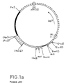
La figure 1a, représente une carte d'assemblage du plasmide pSE1, plasmide de clonage dans E. coli et d'expression dans les cellules animales, les sites ayant disparu par ligation étant notés entre parenthèses. Les symboles utilisés dans cette figure seront précisés lors de la description de ce plasmide (section 2).FIG. 1a represents an assembly map of the plasmid pSE1, a plasmid for cloning in E. coli and for expression in animal cells, the sites having disappeared by ligation being noted in parentheses. The symbols used in this figure will be specified during the description of this plasmid (section 2).
La figure 1b représente la séquence du fragment synthétique "site liant à HIndIII"-HindIII utilisé dans l'assemblage du plasmide pSE1.FIG. 1b represents the sequence of the synthetic fragment "HIndIII binding site" -HindIII used in the assembly of the plasmid pSE1.
La figure 2 représente la séquence nucléotidique de l'ADNc NC28 et en vis-à-vis la séquence d'acides aminés déduite, les trois Met susceptibles d'initier la traduction étant soulignés, le site de clivage probable du peptide signal étant indiqué par un trait vertical et le site potentiel de N-glycosylation étant souligné en pointillés.FIG. 2 represents the nucleotide sequence of the cDNA NC28 and opposite the deduced amino acid sequence, the three Met likely to initiate translation being underlined, the probable site of cleavage of the signal peptide being indicated by a vertical line and the potential site of N-glycosylation being underlined in dotted lines.
La figure 3 et la figure 4 représentent respectivement l'alignement d'après l'homologie maximale selon la méthode de Needleman et Wunsch, 1970, J. Mol. Biol., 48, 443-453 de la séquence d'acides aminés déduite de l'ADNc NC28 (ligne supérieure) et de la séquence d'acides aminés déduite de l'ADNc de la cytokine MCP-1 (ligne inférieure) et l'alignement selon cette méthode de l'ADNc NC28 (ligne supérieure) et de l'ADNc de la cytokine MCP-1 (ligne inférieure).FIG. 3 and FIG. 4 respectively represent the alignment according to the maximum homology according to the method of Needleman and Wunsch, 1970, J. Mol. Biol., 48, 443-453 of the amino acid sequence deduced from the cDNA NC28 (upper line) and the amino acid sequence deduced from the cDNA of the cytokine MCP-1 (lower line) and l alignment by this method of the cDNA NC28 (upper line) and the cDNA of the cytokine MCP-1 (lower line).
La figure 5 représente la séquence du fragment B utilisé dans la construction du plasmide pEMR617, vecteur d'expression dans la levure.FIG. 5 represents the sequence of fragment B used in the construction of the plasmid pEMR617, expression vector in yeast.
Les figures 6a, 6b et 6c sont relatives aux expériences de mise en évidence de l'activité chimiotactique.Figures 6a, 6b and 6c relate to experiments demonstrating chemotactic activity.
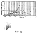
La figure 6a représente le nombre de cellules par champ microscopique en fonction de la concentration exprimée, en nM pour la protéine NC28 purifiée issue de la levure, la protéine NC28 purifiée issue de cellules COS, les peptides C13, C16, C20 et fMLP.FIG. 6a represents the number of cells per microscopic field as a function of the concentration expressed, in nM for the purified NC28 protein derived from yeast, the purified NC28 protein derived from COS cells, the C13, C16, C20 and fMLP peptides.
La figure 6b représente le nombre de cellules par champ microcospique en fonction de la concentration exprimée en ng/ml pour la protéine NC28 purifiée issue de levure et la cytokine MCP-1 purifiée issue des cellules COS.FIG. 6b represents the number of cells per microcospic field as a function of the concentration expressed in ng / ml for the purified NC28 protein derived from yeast and the purified cytokine MCP-1 derived from COS cells.
La figure 6c représente le nombre de cellules par champ microscopique en fonction de la concentration exprimée en ng/ml, pour la protéine NC28 purifiée issue de levure, la cytokine IL-8 et le peptide fMLP.FIG. 6c represents the number of cells per microscopic field as a function of the concentration expressed in ng / ml, for the purified NC28 protein derived from yeast, the cytokine IL-8 and the peptide fMLP.
A partir de poches de sang périphérique (prélevées sur trois volontaires sains dans un centre de transfusion sanguine), ayant préalablement subi une cytaphérèse et un gradient de Ficoll (Gauchat et al, 1989, Eur. J. Imm. 19, 1079) on prélève une fraction de cellules enrichie en cellules mononucléaires du sang périphérique PBMNC (peripheral blood mononuclear cells) de composition approximative suivante : 70 % de lymphocytes, 25 % de monocytes et 5 % de granulocytes (comptage des cellules à l'aide du compteur de cellules Coulter-Modèle S-Plus IV)From pockets of peripheral blood (taken from three healthy volunteers in a blood transfusion center), having previously undergone cytapheresis and a Ficoll gradient (Gauchat et al, 1989, Eur. J. Imm. 19, 1079) a fraction of cells enriched in peripheral blood mononuclear cells (PBMNC) with the following approximate composition: 70% lymphocytes, 25% monocytes and 5% granulocytes (counting of cells using the Coulter cell counter -Model S-Plus IV)
Les cellules sont recueillies dans un flacon de 250 ml, puis centrifugées pendant 10 min à 37°C. Le surnageant est éliminé et le culot de cellules est rincé avec 50 ml de milieu à base de glucose, sels minéraux, acides aminés et vitamines, appelé milieu RPMI (milieu RPMI 1640 de Gibco BRL), puis de nouveau centrifugé dans les mêmes conditions.The cells are collected in a 250 ml flask, then centrifuged for 10 min at 37 ° C. The supernatant is eliminated and the cell pellet is rinsed with 50 ml of medium based on glucose, mineral salts, amino acids and vitamins, called RPMI medium (RPMI 1640 medium from Gibco BRL), then again centrifuged under the same conditions.
Le culot de cellules est alors repris avec 500 ml du milieu RPMI complété avec 10 % de sérum de veau foetal (Gibco BRL- réf. 013-06290H), additionné de 10 unités de pénicilline et de 10 µg de streptomycine (solution de pénicilline/streptomycine de Gibco réf. 043-05140D) par ml de milieu ainsi que de L-glutamine (Gibco BRL-réf. 043-05030D) jusqu'à 2mM final.The cell pellet is then taken up with 500 ml of RPMI medium supplemented with 10% fetal calf serum (Gibco BRL- ref. 013-06290H), supplemented with 10 units of penicillin and 10 μg of streptomycin (penicillin solution / streptomycin from Gibco ref. 043-05140D) per ml of medium as well as L-glutamine (Gibco BRL-ref. 043-05030D) up to 2 mM final.
Une partie de la suspension cellulaire est répartie en vue de la séparation des cellules adhérentes et des cellules non adhérentes, à raison d'environ 100 ml par boîte, dans quatre grandes boîtes de culture carrées (245 x 245 x 20 mm-Nunc - réf. 166508) et incubée pendant 1 h à 37°C. On sait en effet que la plupart des cellules monocytaires adhérent à la boîte de culture tandis que la plupart des cellules lymphocytaires restent en suspension.Part of the cell suspension is distributed for the separation of adherent cells and non-adherent cells, at a rate of approximately 100 ml per dish, in four large square culture dishes (245 x 245 x 20 mm-Nunc - ref 166508) and incubated for 1 h at 37 ° C. It is known in fact that most of the monocytic cells adhere to the culture dish while most of the lymphocyte cells remain in suspension.
Les cellules non adhérentes sont aspirées à l'aide d'une pipette et cultivées dans des flacons de culture du type Falcon de surface 175 cm² en présence de milieu RPMI complété comme décrit ci-dessus, additionné de 10 ng/ml de phorbol myristate-2 acétate-3 (PMA) (Sigma-réf. P8139) et de 5µg/ml de phytohémaglutinine (PHA-P) (Sigma-réf. L8754), à 37°C en présence de 5 % de CO₂ pendant 24 h.The non-adherent cells are aspirated using a pipette and cultured in culture flasks of the Falcon type with a surface area of 175 cm 2 in the presence of RPMI medium supplemented as described above, supplemented with 10 ng / ml of phorbol myristate- 2 acetate-3 (PMA) (Sigma-ref. P8139) and 5µg / ml of phytohemaglutinin (PHA-P) (Sigma-ref. L8754), at 37 ° C in the presence of 5% CO₂ for 24 h.
Sur les cellules adhérentes on rajoute 100 ml de milieu RPMI complété comme décrit ci-dessus additionné de 10 ng/ml de PMA et de 5 µg/ml de PHA-P. Les cellules sont incubées à 37°C en présence de 5 % (v/v) de CO₂ pendant 5 h.100 ml of RPMI medium, supplemented as described above, added with 10 ng / ml of PMA and 5 μg / ml of PHA-P are added to the adherent cells. The cells are incubated at 37 ° C. in the presence of 5% (v / v) of CO₂ for 5 h.
Le reste de la suspension cellulaire, appelé ci-après les cellules totales, est réparti dans 4 grandes boîtes de culture carrées et incubé en présence de milieu RPMI complété comme décrit ci-dessus, additionné de 10 ng/ml de PMA et 5 µg/ml de PHA-P à 37°C en présence de 5 % (v/v) de CO₂ pendant 5 h pour les deux premières boîtes et 24 h pour les deux autres.The rest of the cell suspension, hereinafter called the total cells, is distributed into 4 large square culture dishes and incubated in the presence of RPMI medium supplemented as described above, supplemented with 10 ng / ml of PMA and 5 μg / ml of PHA-P at 37 ° C in the presence of 5% (v / v) of CO₂ for 5 h for the first two dishes and 24 h for the other two.
2 h environ avant la fin de l'incubation on ajoute dans le milieu de culture de ces différentes cellules 10µg/ml de cycloheximide (Sigma réf.C6255) (inhibiteur de traduction qui augmente la stabilité des ARN des cytokines : cf. Lindsten et al, 1989, Science 244, 339-344) et l'incubation est poursuivie pendant 2 h à 37°C.About 2 h before the end of the incubation, 10 μg / ml of cycloheximide (Sigma ref. C6255) (translation inhibitor which increases the stability of cytokine RNAs) is added to the culture medium of these different cells. See Lindsten et al. , 1989, Science 244, 339-344) and the incubation is continued for 2 h at 37 ° C.
Les cellules sont récupérées de la façon suivante :
- les cellules adhérentes sont lavées 2 fois avec du tampon PBS (Phosphate buffered saline réf. 04104040-Gibco BRL) puis grattées avec un grattoir en caoutchouc et centrifugées. On obtient ainsi un culot cellulaire, appelé culot A.
- pour les cellules non adhérentes, après agitation du flacon contenant la suspension des cellules, la suspension cellulaire est prélevée et centrifugée. On obtient ainsi un culot cellulaire, appelé culot cellulaire NA.
- pour les cellules totales, la fraction adhérente est lavée 2 fois avec du tampon PBS et grattée comme précédemment puis centrifugée. La fraction non adhérente est centrifugée. Les deux culots cellulaires obtenus seront réunis par la suite. Leur réunion est appelée culot cellulaire T(5 h) pour les cellules totales incubées pendant 5 h et T(24 h) pour les cellules totales incubées pendant 24 h.
- the adherent cells are washed twice with PBS buffer (phosphate buffered saline ref. 04104040-Gibco BRL) then scraped with a rubber scraper and centrifuged. This gives a cell pellet, called pellet A.
- for non-adherent cells, after shaking the bottle containing the cell suspension, the cell suspension is removed and centrifuged. This produces a cell pellet, called the NA cell pellet.
- for total cells, the adherent fraction is washed twice with PBS buffer and scraped off as above and then centrifuged. The non-adherent fraction is centrifuged. The two cell pellets obtained will be combined subsequently. Their union is called cell pellet T (5 h) for the total cells incubated for 5 h and T (24 h) for the total cells incubated for 24 h.
Les culots cellulaires A, NA, T(5 h) et T(24 h) sont congelés et conservés à -80°C.Cell pellets A, NA, T (5 h) and T (24 h) are frozen and stored at -80 ° C.
Chaque culot cellulaire congelé est mis en suspension dans le tampon de lyse de composition suivante : guanidine- thiocyanate 5M ; Tris-(hydroxyméthyl)-aminométhane 50mM pH 7,5 EDTA 10mM. La suspension est soniquée à l'aide d'un sonicateur Ultra Turax n° 231 256 (Janke et Kunkel) à puissance maximale pendant 4 cycles de 20 s. On ajoute du β-mercaptoéthanol jusqu'à 0,2M et on refait un cycle de sonication de 30 s. On ajoute du chlorure de lithium jusqu'à 3M. On refroidit la suspension à 4°C et on la laisse reposer à cette température pendant 48 h. L'ARN est ensuite isolé par centrifugation pendant 60 min. Le culot d'ARN est lavé une fois avec une solution de chlorure de lithium 3M, recentrifugé, puis repris dans un tampon de composition suivante : SDS 1 %, EDTA 5mM et Tris HCl 10mM pH 7,5, additionné de 1 mg/ml de protéinase K (Boehringer Mannheim, GmbH). Après incubation à 40°C pendant 1 h la solution d'ARN est extraite avec un mélange phénol/chloroforme. On précipite à -20°C l'ARN contenu dans la phase aqueuse à l'aide d'une solution d'acétate d'ammonium 0,3M final et 2,5 volumes d'éthanol. On centrifuge à 15 000 g pendant 30 min et on garde le culot.Each frozen cell pellet is suspended in the lysis buffer of the following composition: guanidine-thiocyanate 5M; Tris- (hydroxymethyl) -aminomethane 50mM pH 7.5 EDTA 10mM. The suspension is sonicated using an Ultra Turax sonicator No. 231 256 (Janke and Kunkel) at maximum power for 4 cycles of 20 s. Β-mercaptoethanol is added up to 0.2M and a sonication cycle of 30 s is repeated. Lithium chloride is added up to 3M. The suspension is cooled to 4 ° C and allowed to stand at this temperature for 48 h. The RNA is then isolated by centrifugation for 60 min. The RNA pellet is washed once with a 3M lithium chloride solution, recentrifuged, then taken up in a buffer of the following composition:
Le culot est repris dans 1 ml de tampon de composition Tris-HCl 10mM pH 7,5 EDTA 1mM, appelé tampon TE et mis en suspension par agitation au vortex. L'oligo dT-cellulose type 3 (commercialisé par Collaborative Research Inc, Biomedicals Product Division) est préparé suivant les recommandations du fabricant. L'ARN est déposé sur l'oligo dT-cellulose, agité doucement pour mettre en suspension les billes, puis chauffé pendant 1 min à 65°C.The pellet is taken up in 1 ml of 10 mM Tris-HCl composition buffer pH 7.5 1 mM EDTA, called TE buffer and suspended by vortexing. The oligo dT-cellulose type 3 (marketed by Collaborative Research Inc, Biomedicals Product Division) is prepared according to the manufacturer's recommendations. The RNA is deposited on the oligo dT-cellulose, stirred gently to suspend the beads, then heated for 1 min at 65 ° C.
La suspension est ajustée à 0,5 M NaCl, puis mise à agiter doucement pendant 10 min. La suspension est alors centrifugée pendant 1 min à 1000 g, le surnageant est éliminé, le culot est lavé 2 fois avec 1 ml de tampon TE contenant 0,5 M NaCl. Les sumageants sont éliminés. L'élution de la fraction polyadénylée des ARN (constituée des ARN messagers) est obtenue par suspension des billes dans 1 ml de tampon TE, puis chauffage de cette suspension à 60°C pendant 1 min, suivie d'une agitation pendant 10 min sur plaque basculante. On centrifuge ensuite pendant 1 min à 1000 g, ce qui permet de récupérer le surnageant contenant des ARN messagers libres en solution. L'ensemble des opérations ci-dessus (à partir de l'élution) est répété 2 fois. Les surnageants ainsi obtenus sont rassemblés, on élimine les billes résiduelles par centrifugation et on précipite le surnageant avec 3 volumes d'éthanol et une solution de NaCl à 0,3M final.The suspension is adjusted to 0.5 M NaCl, then stirred gently for 10 min. The suspension is then centrifuged for 1 min at 1000 g, the supernatant is removed, the pellet is washed 2 times with 1 ml of TE buffer containing 0.5 M NaCl. The supernatants are eliminated. The elution of the polyadenylated fraction of RNA (consisting of messenger RNA) is obtained by suspending the beads in 1 ml of TE buffer, then heating this suspension at 60 ° C for 1 min, followed by stirring for 10 min on a tilting plate. Then centrifuged for 1 min at 1000 g, which allows the supernatant containing free messenger RNA in solution to be recovered. All of the above operations (from elution) are repeated 2 times. The supernatants thus obtained are combined, the residual beads are removed by centrifugation and the supernatant is precipitated with 3 volumes of ethanol and a final 0.3M NaCl solution.
On obtient ainsi à partir des culots cellulaires A, NA, T(5 h) et T(24 h), quatre échantillons d'ARN-poly A⁺, nommés par la suite ARN-poly A⁺-A, ARN-poly A⁺-NA; ARN-polyA ⁺-T(5 h) et ARN-poly A⁺-T(24 h).There are thus obtained from cell pellets A, NA, T (5 h) and T (24 h), four samples of RNA-poly A⁺, subsequently named RNA-poly A⁺-A, RNA-poly A ⁺-NA; RNA-polyA ⁺-T (5 h) and RNA-poly A⁺-T (24 h).
La stratégie mise en oeuvre fait appel à des fragments obtenus à partir de plasmides préexistants accessibles au public et à des fragments préparés par voie de synthèse selon les techniques maintenant couramment utilisées. Les techniques de clonage employées sont celles décrites par T. Maniatis, EF. Fritsch et J. Sambrook dans "Molecular Cloning, a Laboratory manual" (Cold Spring Harbor Laboratory, 1984). La synthèse des oligonucléotides est réalisée à l'aide d'un synthétiseur d'ADN Biosearch 8700.The strategy implemented uses fragments obtained from pre-existing plasmids accessible to the public and fragments prepared by synthesis using the techniques now commonly used. The cloning techniques used are those described by T. Maniatis, EF. Fritsch and J. Sambrook in "Molecular Cloning, a Laboratory manual" (Cold Spring Harbor Laboratory, 1984). The synthesis of oligonucleotides is carried out using a Biosearch 8700 DNA synthesizer.
La description ci-après sera mieux comprise en référence à la figure 1a.The description below will be better understood with reference to FIG. 1 a .
Ce plasmide a été construit par ligations successives des éléments suivants :
- a)- un fragment PvuII-PvuII - symbolisé par +++++++ sur la figure 1a - de 2525pb, obtenu par digestion complète du plasmide pTZ18R (Pharmacia) à l'aide de l'enzyme de restriction PvuII. Ce fragment contient l'origine de réplication du phage f1 (notée ORI F1 sur la figure 1a), un gène (noté AmpR sur la figure 1a) portant la résistance à l'ampicilline et l'origine de réplication (notée ORI pBR322 sur la figure 1a) permettant la réplication de ce plasmide dans E. coli. Le premier site franc PvuII disparaît par ligation avec le site franc EcoRV (qui disparaît également) du fragment décrit en g).
- b)- un fragment PvuII-HpaI - symbolisé par sur la figure 1a - de 1060 pb de l'ADN d'adénovirus type 5 entre les positions 11299 (site de restriction PvuII) et 10239 (site de restriction HpaI) (Dekker et Van Ormondt, Gene 27, 1984, 115-120) contenant l'information pour les ARN VA-I et VA-II. Le site franc HpaI disparaît par ligation avec le site franc PvuII (qui disparaît également) du fragment décrit en c). Le site ApaI en

position 11 218 a été enlevé par clivage à l'aide de l'enzyme ApaI, traitement à l'aide de l'exonucléase : ADN polymérase du phage T4 et religation. - c)- un fragment PvuII-HindIII - symbolisé par sur la figure 1a - de 344 pb, issu de l'ADN du virus SV40 obtenu par digestion complète à l'aide des enzymes de restriction PvuII et HindIII. Ce fragment comporte l'origine de réplication et le promoteur précoce de l'ADN du virus SV40 (réf. B.J. Byrne et al. Proc. Ntl. Acad. Sci. USA (1983), 80, 721-725).

Le site HindIII disparaît par ligation avec le site liant à HindIII du fragment décrit en d). - d)- un fragment synthétique "site liant à HindIII" - HindIII - symbolisé par sur la figure 1a -

de 419 pb dont la séquence, donnée sur la figure 1b, contient une séquence proche de la séquence 5′ non traduite du virus HTLV1 (R. WEISS et al, "Molecular Biology of Tumor Viruses" - part 2-2e ed - 1985 - Cold Spring Harbor Laboratory - p. 1057) et l'intron distal du gène de l'α-globine de souris (y. Nishioka et al, 1979, Cell, 18, 875-882). - e)- un fragment synthétique HindIII-"site liant à BamHI" - symbolisé par XXXXXXX sur la figure 1a - contenant le promoteur de l'ARN-polymérase du phage T7 ainsi qu'un polylinker contenant notamment les sites de clonage ApaI et BamHI

- f)- un fragment BamHI-
BclI de 240 pb - représenté parsur la figure 1a -, petit fragment obtenu par digestion complète à l'aide des enzymes BclI et BamHI de l'ADN du virus SV40 qui contient le site de polyadénylation tardif de ce dernier. (M. Fitzgerald et al. Cell, 24, 1981, 251-260). Les sites BamHI et BclI disparaissent par ligations avec respectivement le "site liant à BamHI" du fragment décrit en e) et le site BamHI (qui disparaît également) du fragment décrit en g).
- g)- un fragment BamHI-EcoRV - symbolisé par OOOOOO sur la figure 1a- de 190 pb, petit fragment issu du plasmide pBR322 après digestion complète à l'aide des enzymes EcoRV et BamHI.
Le plasmide pSE1 comporte donc les éléments nécessaires pour son utilisation comme vecteur de clonage dans E. coli (origine de replication dans E. coli et gène de résistance à l'ampicilline, provenant du plasmide pTZ18R) ainsi que comme vecteur d'expression dans les cellules animales (promoteur, intron, site de polyadénylation, origine de replication du virus SV40), et pour sa copie en simple brin dans un but de séquençage (origine de réplication du phage f1 ).
- a) - a PvuII-PvuII fragment - symbolized by +++++++ in FIG. 1 a - of 2525 bp, obtained by complete digestion of the plasmid pTZ18R (Pharmacia) using the restriction enzyme PvuII. This fragment contains the origin of replication of phage f1 (denoted ORI F1 in FIG. 1 a ), a gene (denoted Amp R in FIG. 1 a ) carrying the resistance to ampicillin and the origin of replication (denoted ORI pBR322 in FIG. 1 a ) allowing replication of this plasmid in E. coli. The first free site PvuII disappears by ligation with the free site EcoRV (which also disappears) from the fragment described in g).
- b) - a PvuII-HpaI fragment - symbolized by in FIG. 1 a - 1060 bp of adenovirus type 5 DNA between positions 11299 (restriction site PvuII) and 10239 (restriction site HpaI) (Dekker and Van Ormondt, Gene 27, 1984, 115-120 ) containing the information for the VA-I and VA-II RNAs. The free HpaI site disappears by ligation with the free site PvuII (which also disappears) from the fragment described in c). The ApaI site at
position 11 218 was removed by cleavage using the enzyme ApaI, treatment with the exonuclease: phage T4 DNA polymerase and religation. - c) - a PvuII-HindIII fragment - symbolized by in FIG. 1a - 344 bp, derived from the DNA of the SV40 virus obtained by complete digestion using the restriction enzymes PvuII and HindIII. This fragment contains the origin of replication and the early promoter of the DNA of the SV40 virus (ref. BJ Byrne et al. Proc. Ntl. Acad. Sci. USA (1983), 80, 721-725).
The HindIII site disappears by ligation with the HindIII binding site of the fragment described in d). - d) - a synthetic fragment "site binding to HindIII" - HindIII - symbolized by in FIG. 1 a - of 419 bp whose sequence, given in FIG. 1 b , contains a sequence close to the 5 ′ untranslated sequence of the HTLV1 virus (R. WEISS et al, "Molecular Biology of Tumor Viruses" - share 2-2 th ed - 1985 - Cold Spring Harbor Laboratory - p. 1057) and the distal intron of the α-globin gene mouse (there Nishioka et al, 1979, Cell, 18, 875-882)..
- e) - a synthetic HindIII fragment - "BamHI binding site" - symbolized by XXXXXXX in FIG. 1 a - containing the promoter of phage T7 RNA polymerase as well as a polylinker containing in particular the cloning sites ApaI and BamHI
- f) - a BamHI-BclI fragment of 240 bp - represented by in FIG. 1 a -, small fragment obtained by complete digestion using the enzymes BclI and BamHI of the DNA of the SV40 virus which contains the late polyadenylation site of the latter. (M. Fitzgerald et al. Cell, 24, 1981, 251-260). The BamHI and BclI sites disappear by ligations with the "BamHI binding site" of the fragment described in e) respectively and the BamHI site (which also disappears) of the fragment described in g).
- g) - a BamHI-EcoRV fragment - symbolized by OOOOOO in FIG. 1 a - of 190 bp, small fragment derived from the plasmid pBR322 after complete digestion using the enzymes EcoRV and BamHI.
The plasmid pSE1 therefore contains the elements necessary for its use as a cloning vector in E. coli (origin of replication in E. coli and ampicillin resistance gene, originating from the plasmid pTZ18R) as well as as an expression vector in the animal cells (promoter, intron, polyadenylation site, origin of replication of the SV40 virus), and for its single-strand copy for the purpose of sequencing (origin of replication of phage f1).
La technique de clonage utilisée est celle décrite par Caput et al, (technique de l'amorce-adaptateur (Primer-adapter) : Caput et al, Proc. Natl. Acad. Sci. (U.S.A.), 1986, 83, 1670-1674).The cloning technique used is that described by Caput et al, (Primer-adapter technique: Caput et al, Proc. Natl. Acad. Sci. (USA), 1986, 83, 1670-1674 ).
Elle consiste d'une part à digérer le vecteur pSE1 par ApaI, ajouter une queue de polydC sur l'extrémité 3′ protubérante, puis à digérer les plasmides ainsi obtenus par l'endonucléase BamHI. Le fragment correspondant au vecteur est purifié sur colonne de Sépharose CL4B (Pharmacia). Il comprend donc une queue polydC à une extrémité, l'autre extrémité étant cohésive, du type BamHI.It consists on the one hand in digesting the vector pSE1 with ApaI, adding a tail of polydC on the protruding 3 ′ end, then in digesting the plasmids thus obtained by the endonuclease BamHI. The Shard corresponding to the vector is purified on a Sepharose CL4B column (Pharmacia). It therefore comprises a polydC tail at one end, the other end being cohesive, of the BamHI type.
D'autre part, les ARN polyA⁺ obtenus à l'issue de la section 1 sont soumis à la transcription inverse à partir d'une amorce dont la séquence est la suivante :![]()
Ainsi, les ADNc présentent à leur extrémité 5′ la séquence GATCC complémentaire de l'extrémité cohésive BamHI.On the other hand, the polyA⁺ RNAs obtained at the end of ![]()
Thus, the cDNAs have at their 5 ′ end the GATCC sequence complementary to the BamHI cohesive end.
Les hybrides ARN-ADN obtenus par action de la transcriptase inverse sont soumis à une hydrolyse alcaline qui permet de se débarrasser de l'ARN. Les ADNc simple brin sont alors soumis à un traitement à la terminale transférase, de façon à ajouter des polydG en 3′ et purifiés par 2 cycles sur colonne de sépharose CL4B.The RNA-DNA hybrids obtained by the action of reverse transcriptase are subjected to alkaline hydrolysis which makes it possible to get rid of the RNA. The single-stranded cDNAs are then subjected to a terminal transferase treatment, so as to add polydG at 3 ′ and purified by 2 cycles on a CL4B sepharose column.
Ces ADNc sont hybridés avec de l'ARN-polyA⁺ provenant de cellules de la lignée COS3 (lignée de cellules de reins de singe exprimant l'antigène T du virus SV40 : cf. Y. Gluzman, 1981, Cell, 23, 175-182 préparées comme décrit dans la Section 1.2).These cDNAs are hybridized with RNA-polyA⁺ originating from cells of the COS3 line (monkey kidney cell line expressing the T antigen of the SV40 virus: cf. Y. Gluzman, 1981, Cell, 23, 175- 182 prepared as described in Section 1.2).
Les ADNc non hybridés sont isolés (fraction enrichie en ADN complémentaire aux ARN messagers spécifiques des cellules mononucléaires du sang périphérique).The non-hybridized cDNAs are isolated (fraction enriched in DNA complementary to the messenger RNAs specific for peripheral blood mononuclear cells).
Ces ADNc sont insérés sous forme simple brin dans le vecteur pSE1. Un second oligonucléotide (l'adaptateur) complémentaire de l'amorce est nécessaire pour générer un site BamHI à l'extrémité 5′ des ADNc. Après hybridation du vecteur, de l'ADNc et de l'adaptateur, les molécules recombinantes sont circularisées par l'action de la ligase du phage T4. Les régions simple brin sont alors réparées grâce à l'ADN polymérase du phage T4. Le pool de plasmides ainsi obtenu sert à transformer la souche E. coli MC 1061 (Casabadan et S. Cohen, J. Bact. (1980) 143, 971-980) par électroporation.These cDNAs are inserted in single-stranded form into the vector pSE1. A second oligonucleotide (the adapter) complementary to the primer is necessary to generate a BamHI site at the 5 ′ end of the cDNAs. After hybridization of the vector, the cDNA and the adapter, the recombinant molecules are circularized by the action of the ligase of phage T4. The single-stranded regions are then repaired using DNA polymerase from phage T4. The pool of plasmids thus obtained is used to transform the E. coli MC 1061 strain (Casabadan and S. Cohen, J. Bact. (1980) 143, 971-980) by electroporation.
A partir de 5 µg des ARN-poly A⁺ de cellules mononucléaires de sang périphérique obtenus à l'issue de la section 1 de composition suivante : ARN-poly A⁺ A : 0,5 µg, ARN-poly A⁺ NA: 2 µg, ARN-poly A⁺ T(5 h): 2 µg et ARN-poly A⁺ T(24 h) : 0,5 µg, on prépare l'ADN complémentaire simple-brin marqué au ³²P-dCTP (l'ADN complémentaire obtenu présente une activité spécifique de 3000 dpm/ng) avec l'amorce synthétique de séquence suivante (comprenant un site BamHI) :![]()
dans un volume de 100µl. Après 30 min d'incubation à 46°C avec 100 unités de l'enzyme transcriptase inverse (Genofit-E1 022) on ajoute 4µl d'EDTA 0,5M. On extrait une première fois avec du phénol (saturé en tampon TE) puis une seconde fois avec du chloroforme. On ajoute 10 µg d'ARN de transfert de foie de veau, 1/10ème de volume d'une solution d'acétate d'ammonium 10M et 2,5 volumes d'éthanol pour précipiter l'ADN complémentaire. On centrifuge, on dissout le culot dans 30µl de tampon TE puis on retire les petites molécules telles que sels, phénol et chloroforme par chromatographie d'exclusion sur une colonne de polyacrylamide P10 (Biogel P10-200-400mesh, réf. 1501050-Biorad).From 5 μg of RNA-poly A⁺ of peripheral blood mononuclear cells obtained at the end of ![]()
in a volume of 100µl. After 30 min of incubation at 46 ° C with 100 units of the reverse transcriptase enzyme (Genofit-E1 022), 4 μl of 0.5M EDTA is added. Extraction is carried out a first time with phenol (saturated in TE buffer) and then a second time with chloroform. 10 μg of calf liver transfer RNA, 1 / 10th of a volume of a 10M ammonium acetate solution and 2.5 volumes of ethanol are added to precipitate the complementary DNA. Centrifuge, dissolve the pellet in 30 μl of TE buffer and then remove the small molecules such as salts, phenol and chloroform by exclusion chromatography on a column of polyacrylamide P10 (Biogel P10-200-400mesh, ref. 1501050-Biorad) .
On ajoute 4,6µl d'une solution de NaOH 2N, on incube pendant 30 min à 68°C, puis on ajoute 4,6µl d'acide acétique 2N et on fait passer la solution obtenue sur une colonne de polyacrylamide P10.4.6 μl of a 2N NaOH solution are added, the mixture is incubated for 30 min at 68 ° C., then 4.6 μl of 2N acetic acid is added and the solution obtained is passed through a column of polyacrylamide P10.
On allonge l'ADN complémentaire en 3′ avec une "queue" de dG avec 66 unités de l'enzyme terminale transférase (Pharmacia 27073001). On incube pendant 30 min à 37°C, puis on ajoute 4 µl d'EDTA 0,5M.The complementary DNA is extended at 3 ′ with a “tail” of dG with 66 units of the terminal enzyme transferase (Pharmacia 27073001). Incubate for 30 min at 37 ° C., then add 4 μl of 0.5M EDTA.
Afin d'éliminer l'amorce synthétique, on purifie l'ADN complémentaire sur deux colonnes successives de 1 ml de sépharose CL4B (Pharmacia), équilibrées avec une solution NaOH 30mM/EDTA 2mM.In order to remove the synthetic primer, the complementary DNA is purified on two successive columns of 1 ml of CL4B sepharose (Pharmacia), equilibrated with a 30 mM NaOH / 2 mM EDTA solution.
Les trois premières fractions radioactives (d'environ 80µl chacune) sont regroupées et précipitées avec 1/10ème de volume d'une solution d'acétate d'ammonium et 2,5 volumes d'éthanol. La quantité d'ADN complémentaire est de 1 µg.The first three radioactive fractions (approximately 80 μl each) are combined and precipitated with 1 / 10th of a volume of an ammonium acetate solution and 2.5 volumes of ethanol. The amount of complementary DNA is 1 µg.
Le culot d'ADN complémentaire est mis en suspension dans 25 µl de tampon TE, on ajoute 15µg d'ARN-polyA⁺ extrait de cellules de la lignée COS, puis 1/10ème de volume d'une solution de NaCl 3M, 2,5 volumes d'éthanol et on laisse précipiter à -20°C.The complementary DNA pellet is suspended in 25 μl of TE buffer, 15 μg of RNA-polyA⁺ extracted from cells of the COS line are added, then 1 / 10th of a volume of a 3M NaCl solution, 2, 5 volumes of ethanol and allowed to precipitate at -20 ° C.
On centrifuge, on lave à l'éthanol 70 %, on sèche, on dissout le culot dans 5 µl de tampon de composition suivante : Tris-HCl 0,1M pH 7,5 ; NaCl 0,3M, EDTA 1mM, on met la solution obtenue dans un tube capillaire que l'on scelle, puis on incube à 65°C pendant 40 h.Centrifuged, washed with 70% ethanol, dried, the pellet is dissolved in 5 μl of buffer with the following composition: 0.1 M Tris-HCl pH 7.5; 0.3M NaCl, 1mM EDTA, the solution obtained is placed in a capillary tube which is sealed, then incubated at 65 ° C for 40 h.
On dilue le contenu du capillaire dans 100µl de tampon TE auquel on ajoute 300µl de tampon phosphate de sodium 50mM pH6,8. On fait passer la solution obtenue sur une colonne d'hydroxyapatite (Biorad réf. 130.0520) à 60°C, équilibrée avec ce tampon phosphate. On sépare le simple brin (l'ADN complémentaire non-hybridé) et le double brin (ARN messager de COS hybridé à l'ADN complémentaire) par un gradient de tampon phosphate de 0,1M à 0,2M à travers la colonne d'hydroxyapatite. On regroupe les fractions correspondant à l'ADN complémentaire simple brin (25 % en poids de l'ADNc élué, ce qui correspond à un enrichissement d'environ 4 fois en séquences spécifiques de cellules mononucléaires du sang périphérique), on ajoute 20 µg d'ARN de transfert, on précipite le volume total avec 1/10ème de volume d'une solution d'acétate d'ammonium 10M et 2,5 volumes d'éthanol. On centrifuge, le culot est dissout dans 200 µl de TE, on retire le phosphate résiduel sur polyacrylamide P10, on précipite de nouveau avec 1/10ème de volume d'une solution d'acétate d'ammonium 10M et 2,5 volumes d'éthanol.The content of the capillary is diluted in 100 μl of TE buffer to which 300 μl of 50 mM sodium phosphate buffer pH 6.8 are added. The solution obtained is passed through a hydroxyapatite column (Biorad ref. 130.0520) at 60 ° C, equilibrated with this phosphate buffer. The single strand (non-hybridized complementary DNA) and the double strand (COS messenger RNA hybridized to complementary DNA) are separated by a phosphate buffer gradient of 0.1M to 0.2M through the column of hydroxyapatite. The fractions corresponding to the complementary single-stranded DNA are combined (25% by weight of the eluted cDNA, which corresponds to an enrichment of approximately 4 times in specific sequences of mononuclear cells of the peripheral blood), 20 μg of d are added. 'Transfer RNA, the total volume is precipitated with 1 / 10th of a volume of a 10M ammonium acetate solution and 2.5 volumes of ethanol. Centrifuged, the pellet is dissolved in 200 μl of TE, the residual phosphate is removed on polyacrylamide P10, again precipitated with 1/10 th volume of a 10M ammonium acetate solution and 2.5 volumes d ethanol.
Le culot est dissous dans 30 µl d'une solution de NaOH 30mM ; EDTA 2mM. On charge l'ADN complémentaire sur une colonne de sépharose CL4B (Pharmacia) de 1 ml, équilibrée avec une solution de NaOH 30mM ; EDTA 2mM, afin d'éliminer le reste d'amorce synthétique. On regroupe les 3 premières fractions radioactives d'environ 80 µl chacune. On précipite l'ADNc contenu dans ces fractions avec 1/10ème de volume d'une solution d'acétate d'ammonium 10 M et 2,5 volume d'éthanol. La quantité d'ADN complémentaire ainsi récupérée est de 20 ng.The pellet is dissolved in 30 μl of a 30 mM NaOH solution; EDTA 2mM. The complementary DNA is loaded onto a 1 ml CL4B (Pharmacia) sepharose column, equilibrated with a 30 mM NaOH solution; 2mM EDTA, to remove the rest of the synthetic primer. The first 3 radioactive fractions of around 80 µl each are grouped together. The cDNA contained in these fractions is precipitated with 1/10 th volume of a 10 M ammonium acetate solution and 2.5 volume of ethanol. The amount of complementary DNA thus recovered is 20 ng.
On centrifuge, le culot est dissous dans 33 µl de tampon TE, on ajoute 5 µl (125 ng) de vecteur de clonage pSE1, 1 µl (120 ng) de l'adaptateur de séquence suivante (comprenant un site ApaI) :![]()
10µl d'une solution de NaCl 200mM, on incube 5 min à 65°C puis on laisse refroidir le mélange réactionnel jusqu'à température ambiante.Centrifuged, the pellet is dissolved in 33 μl of TE buffer, 5 μl (125 ng) of cloning vector pSE1, 1 μl (120 ng) of the adapter with the following sequence (comprising an ApaI site) are added: ![]()
10 μl of a 200 mM NaCl solution, incubated for 5 min at 65 ° C. and then the reaction mixture is allowed to cool to room temperature.
On ligue le vecteur de clonage et l'ADNc simple brin dans un volume de 100µl avec 32,5 unités de l'enzyme ADN ligase du phage T4 (Pharmacia ref: 270 87002) pendant une nuit à 15°C.The cloning vector and the single-stranded cDNA are ligated in a volume of 100 μl with 32.5 units of the enzyme DNA ligase from phage T4 (Pharmacia ref: 270 87002) overnight at 15 ° C.
On élimine les protéines par extraction au phénol suivie d'une extraction au chloroforme, puis on ajoute 1/10ème de volume d'une solution d'acétate d'ammonium 10 mM, puis 2,5 volumes d'éthanol. On centrifuge, le culot est dissous dans le tampon de composition Tris acétate pH 7,9, 33 mM, acétate de potassium 62,5 mM, acétate de magnésium 1 mM et dithiothreitol (DTT) 1 mM. Le deuxième brin d'ADN complémentaire est synthétisé dans un volume de 30µl avec 30 unités de l'enzyme ADN polymérase du phage T4 (Pharmacia : réf. 27-0718) et un mélange des quatre déoxynucléotides triphosphates dATP, dCTP, dGTP et dTTP, ainsi que deux unités de la protéine du gène 32 du phage T4 (Pharmacia: réf. 27-0213), pendant 1 h à 37°C. On extrait au phénol et on retire les traces de phénol par une colonne de polyacrylamide P10 (Biogel P10-200-400 mesh- Réf 15011050 - Biorad).The proteins are eliminated by phenol extraction followed by chloroform extraction, then 1/10 th of a volume of a 10 mM ammonium acetate solution is added, then 2.5 volumes of ethanol. Centrifuged, the pellet is dissolved in the Tris acetate composition buffer, pH 7.9, 33 mM, potassium acetate 62.5 mM, 1 mM magnesium acetate and 1 mM dithiothreitol (DTT). The second strand of complementary DNA is synthesized in a volume of 30 μl with 30 units of the DNA polymerase enzyme of phage T4 (Pharmacia: ref. 27-0718) and a mixture of the four deoxynucleotide triphosphates dATP, dCTP, dGTP and dTTP, as well as two units of the gene 32 protein phage T4 (Pharmacia: ref. 27-0213), for 1 hour at 37 ° C. Extraction is carried out with phenol and the traces of phenol are removed by a column of polyacrylamide P10 (Biogel P10-200-400 mesh - Ref 15011050 - Biorad).
On transforme des cellules E. coli MC1061 (Clontech) avec l'ADN recombinant obtenu précedemment par électroporation à l'aide de l'appareil Biorad Gene Pulser (Biorad) utilisé à 2,5 kV dans les conditions prescrites par le fabricant, puis on fait pousser les bactéries pendant 6 h 30 dans du milieu dit milieu LB (Sambrook, op cité) de composition : bactotryptone 10 g/l ; extrait de levure 5 g/l ; NaCl 10 g/l, additionné de 100 µg/ml d'ampicilline. E. coli MC1061 cells (Clontech) are transformed with the recombinant DNA obtained previously by electroporation using the Biorad Gene Pulser device (Biorad) used at 2.5 kV under the conditions prescribed by the manufacturer, then makes the bacteria grow for 6
On détermine le nombre de clones indépendants en étalant une dilution au 1/1000ème de la transformation avant l'amplification sur une boîte de milieu LB additionné de 1,5 % d'agar (p/v) et de 100 µg/ml d'ampicilline, appelé par la suite milieu LB gélosé. Le nombre de clones indépendants est de 500 000.Determining the number of independent clones by spreading a 1/1000 dilution of the transformation before amplification on an LB medium plate supplemented with 1.5% agar (w / v) and 100 ug / ml ampicillin, hereinafter called LB agar medium. The number of independent clones is 500,000.
On distribue environ 40 000 bactéries recombinantes de la banque d'ADNc sur des boîtes de Pétri de (245 x 245) mm contenant du milieu LB gélosé (environ 2000 colonies/boîte).About 40,000 recombinant bacteria from the cDNA library are distributed on Petri dishes of (245 × 245) mm containing agar LB medium (approximately 2000 colonies / dish).
A partir de chacune de ces boîtes, on réalise un transfert des colonies sur une membrane de nylon (Hybond N-Amersham) par dépôt de la membrane sur la surface de la boîte et mise de repères par perçage de la membrane à l'aide d'une aiguille. La membrane est ensuite retirée et déposée sur la surface d'une nouvelle boîte de Pétri contenant du milieu LB gélosé. On laisse pendant quelques heures à 37°C pour obtenir la repousse des colonies. A partir de cette première membrane on réalise deux répliques sur de nouvelles membranes (préalablement déposées sur du milieu LB gélosé pour les humidifier) par contacts successifs avec la première membrane. Les répliques sur membrane obtenues sont finalement déposées sur des boîtes de milieu LB gélosé et mises à incuber pendant une nuit à 30°C.From each of these dishes, colonies are transferred to a nylon membrane (Hybond N-Amersham) by depositing the membrane on the surface of the dish and setting marks by piercing the membrane using 'a needle. The membrane is then removed and deposited on the surface of a new Petri dish containing LB agar medium. It is left for a few hours at 37 ° C. to obtain the regrowth of the colonies. From this first membrane, two replicas are made on new membranes (previously deposited on LB agar medium to moisten them) by successive contacts with the first membrane. The replicas on membrane obtained are finally deposited on plates of agar LB medium and incubated overnight at 30 ° C.
Les répliques sur membrane sont déposées avec les colonies vers le haut, sur une feuille de Whatman 3MM saturée avec une solution de composition: NaOH 0,5M; NaCl 1,5M, pendant 5 min, ce qui permet de lyser les bactéries et de fixer l'ADN. Les répliques sur membrane sont ensuite posées sur une deuxième feuille de Whatman 3MM ,saturée cette fois avec une solution neutralisante de composition: NaCl 1,5M ; Tris-HCl pH8 0,5M, pendant 5 min. Les répliques sur membrane sont ensuite plongées dans une solution de 2 x SSC (composition de la solution SSC: NaCl 0,15M ; citrate de sodium 0,015M) et les débris bactériens sont partiellement éliminés en frottant doucement à l'aide d'ouate de nettoyage.The membrane replicas are deposited with the colonies upwards, on a sheet of Whatman 3MM saturated with a solution of composition: 0.5M NaOH; 1.5M NaCl, for 5 min, which makes it possible to lyse the bacteria and fix the DNA. The replicas on the membrane are then placed on a second sheet of Whatman 3MM, this time saturated with a neutralizing solution of composition: NaCl 1.5M; Tris-HCl pH8 0.5M, for 5 min. The replicas on the membrane are then immersed in a solution of 2 x SSC (composition of the SSC solution: 0.15M NaCl; 0.015M sodium citrate) and the bacterial debris is partially removed by gently rubbing with cotton wool. cleaning.
On traite ensuite les répliques sur membrane avec de la protéinase K (Boehringer Mannheim GmbH) à 100 µg/ml dans une solution de composition: Tris-HCl 10mM pH8; EDTA 10mM; NaCl 50mM; SDS 0,1 % à raison de 20 ml par membrane. On incube pendant30 min à 37°C avec agitation. Les répliques sur membrane sont de nouveau plongées dans une solution de 2 x SSC pour éliminer définitivement toute trace de débris bactériens. Elles sont finalement mises à sécher sur du papier filtre pendant quelques minutes puis pendant 30 min sous vide à + 80°C. On obtient ainsi, pour chaque boîte, deux répliques sur membrane, appelées par la suite réplique 1 et réplique 2.The replicas are then treated on a membrane with proteinase K (Boehringer Mannheim GmbH) at 100 μg / ml in a solution of composition: Tris-HCl 10mM pH8; 10mM EDTA; 50 mM NaCl; 0.1% SDS at a rate of 20 ml per membrane. Incubate for 30 min at 37 ° C with shaking. The replicas on the membrane are again immersed in a solution of 2 x SSC to permanently eliminate all traces of bacterial debris. They are finally put to dry on filter paper for a few minutes and then for 30 min under vacuum at + 80 ° C. There are thus obtained, for each box, two replicas on the membrane, hereinafter called
La lignée monocytaire U937 ATCC 1593 est cultivée dans du milieu RPMI complété avec 10 % de sérum de veau foetal (Gibco BRL-réf. 013-06290H), additionné de 10 unités de pénicilline et de 10µg de streptomycine par ml ainsi que de 2 mM final de L-glutamine. Pour l'activation de ces cellules, on les met dans le milieu RPMI additionné de pénicilline, streptomycine et de L-glutamine et de 20 ng/ml de phorbol myristate-2 acétate-3 (PMA) (Sigma-réf. P8139) pendant 24 h. Les cellules ainsi activées sont grattées et centrifugées. Le culot cellulaire obtenu est appelé culot cellulaire U937P.The monocytic line U937 ATCC 1593 is cultivated in RPMI medium supplemented with 10% fetal calf serum (Gibco BRL-ref. 013-06290H), supplemented with 10 units of penicillin and 10 μg of streptomycin per ml as well as 2 mM final L-glutamine. For the activation of these cells, they are placed in RPMI medium supplemented with penicillin, streptomycin and L-glutamine and 20 ng / ml of phorbol myristate-2 acetate-3 (PMA) (Sigma-ref. P8139) for 24h. The cells thus activated are scraped and centrifuged. The cell pellet obtained is called the U937P cell pellet.
La moitié des cellules est induite de surcroît avec du cycloheximide (10µg/ml) pendant les deux dernières heures de culture. Cet inhibiteur de la synthèse protéique rend plus stables les ARN messagers instables (dont les cytokines) (cf. T. Lindsten et al., 1989, Science, 244, 339-343). Les cellules ainsi activées sont grattées et centrifugées. Le culot cellulaire obtenu est appelé culot cellulaire U937PC.Half of the cells are additionally induced with cycloheximide (10 μg / ml) during the last two hours of culture. This inhibitor of protein synthesis makes unstable messenger RNAs (including cytokines) more stable (cf. T. Lindsten et al., 1989, Science, 244, 339-343). The cells thus activated are scraped and centrifuged. The cell pellet obtained is called the U937PC cell pellet.
A partir des culots cellulaires U937P et U937PC, l'ARN est extrait et la fraction polyA⁺ est purifiée comme décrit dans la section 1-2) a) et b). On obtient ainsi deux fractions ARN-polyA⁺, appelées respectivement fraction polyA⁺1 et fraction polyA⁺2.From the U937P and U937PC cell pellets, the RNA is extracted and the polyA⁺ fraction is purified as described in section 1-2) a) and b). Two RNA-polyA⁺ fractions are thus obtained, called respectively polyA⁺1 fraction and polyA⁺2 fraction.
Les sondes ADNc radiomarquées, appelées respectivement sonde 1 et sonde 2 sont synthétisées à partir des deux fractions ARN-polyA⁺ ci-dessus, préparées comme décrit ci-après.The radiolabelled cDNA probes, called
1 µg d'ARN polyA⁺ est hybridé avec 200 ng d'oligo dT₁₂₋₁₈ (Pharmacia) dans 2 à 3 µl de tampon de composition Tris HCl, pH 7,5, 50 mM et EDTA 1 mM par incubation pendant 2 min à 65°C et refroidissement jusqu'à température ambiante. La synthèse de l'ADNc radiomarqué est réalisée dans un volume réactionnel de 10 µl en tampon de composition: Tris-HCl 50mM pH8,3; MgCl₂ 5mM; dithiotreitol 10mM, contenant 50µM de dATP dGTP et dTTP, 10 µM de dCTP et 150 µCi de dCTP α32P (3000 Ci/ mmol Amersham) et 40 unités de RNasine (inhibiteur de RNAse-Genofit). La réaction s'effectue à 46°C pendant 30 min en présence de 10 à 20 unités de transcriptase inverse (Genofit). Cette synthèse est suivie de l'hydrolyse alcaline de l'ARN par une solution de NaOH 0,3M dans un volume final de 20µl pendant 30 min à 65°C. On neutralise par ajout d'acide acétique 3M. On ajuste le volume à 50 µl avec du milieu TE. On fait une extraction avec un même volume de phénol suivie d'une deuxième extraction avec un même volume d'un mélange chloroforme/ isoamylalcool (dans les proportions respectives 24/1). On élimine le dCTP α32P non incorporé lors de la synthèse du brin ADNc par chromatographie d'exclusion sur une colonne de polyacrylamide P10 (Biogel-200-400 mesh-Biorad).1 μg of polyA⁺ RNA is hybridized with 200 ng of oligo dT₁₂₋₁₈ (Pharmacia) in 2 to 3 μl of Tris HCl composition buffer, pH 7.5, 50 mM and 1 mM EDTA by incubation for 2 min at 65 ° C and cooling to room temperature. The synthesis of the radiolabelled cDNA is carried out in a reaction volume of 10 μl in a buffer of composition: Tris-HCl 50mM pH8.3; MgCl₂ 5mM; 10mM dithiotreitol, containing 50µM of dATP dGTP and dTTP, 10 µM of dCTP and 150 µCi of dCTP α32P (3000 Ci / mmol Amersham) and 40 units of RNasine (RNAse-Genofit inhibitor). The reaction is carried out at 46 ° C for 30 min in the presence of 10 to 20 units of reverse transcriptase (Genofit). This synthesis is followed by alkaline hydrolysis of the RNA by a 0.3M NaOH solution in a final volume of 20 μl for 30 min at 65 ° C. Neutralized by adding 3M acetic acid. The volume is adjusted to 50 μl with TE medium. An extraction is carried out with the same volume of phenol followed by a second extraction with the same volume of a chloroform / isoamyl alcohol mixture (in the respective proportions 24/1). The non-incorporated α32P dCTP is eliminated during the synthesis of the cDNA strand by exclusion chromatography on a column of polyacrylamide P10 (Biogel-200-400 mesh-Biorad).
La quantité d'ADNc est de 60 à 100 ng possédant une activité spécifique de 1 x 10⁹dpm/µg.The amount of cDNA is 60 to 100 ng with a specific activity of 1 x 10⁹dpm / µg.
Les répliques sur membrane sont préhybridées pendant 2 h à 42°C dans un tampon de composition: 50 % formamide ; 6 x SSC; 5 x solution de Denhardt ; 0,1 % SDS et 100 µg/ml d'ADN de sperme de saumon soniqué, rajouté après dénaturation pendant 10 min à 100°C. Les répliques sur membrane sont hybridées pendant deux jours : la réplique 1 avec la sonde 1 et la réplique 2 avec la sonde 2, ces sondes étant utilisées à une concentration de 4 ng/ml dans le tampon ci-dessus. La 5 x solution de Denhardt (cf. Sambrook, op. cité) a la composition : Ficoll (type 400-Pharmacia) 1 g/l, polyvinylpyrrolidone 1 g/l et la sérumalbumine bovine (BSA) 1 g/l.The membrane replicas are prehybridized for 2 h at 42 ° C in a buffer of composition: 50% formamide; 6 x SSC; 5 x Denhardt's solution; 0.1% SDS and 100 µg / ml of sonicated salmon sperm DNA, added after denaturing for 10 min at 100 ° C. The membrane replicas are hybridized for two days:
Les préhybridation et hybridation s'effectuent en tubes dans un four à hybrider (Hybaid) avec respectivement 25 ml et 10 ml de tampon par membrane.The prehybridization and hybridization are carried out in tubes in a hybridization oven (Hybaid) with respectively 25 ml and 10 ml of buffer per membrane.
Ensuite les répliques sur membrane sont successivement lavées plusieurs fois pendant 15 min à 20°C dans le tampon de composition 2 x SSC; 0,1 % SDS, puis deux fois pendant 15 min dans une solution 0,1 x SSC, 0,1 % SDS à 55°C, séchées sur papier Whatman 3MM et autoradiographiées sur films Kodak XAR5.Then the replicas on the membrane are successively washed several times for 15 min at 20 ° C in the 2 x SSC composition buffer; 0.1% SDS, then twice for 15 min in a solution 0.1 x SSC, 0.1% SDS at 55 ° C, dried on Whatman 3MM paper and autoradiographed on Kodak XAR5 films.
Pour identifier les clones qui contiennent les ADN complémentaires aux ARN messagers des cytokines connues, une autre série de répliques sur membrane, préparées comme décrit ci-dessus, est hybridée avec un mélange - appelé mélange C - de 28 oligonucléotides comportant chacun 20 nucléotides, correspondant aux ADN complémentaires des cytokines suivantes: Interleukine-1 α(Furutani Y. et al, 1985, Nucl. Ac. Res., 13, 5869-5882), Interleukine-1 β(Auron P. et al, 1984, Proc. Natl. Acad. Sci. USA, 81, 7907-7911), Interleukine-2 (Degrave W. et al, 1983, EMBO J., 2, 3249-3253), Interleukine-3 (Yang Y.C. et al, 1986, 47, 3-10), Interleukine-4 (Yokoto T. et al, 1986, Proc. Ntl. Acad. Sci., 83, 5894-5898), Interleukine-5 (Hirano T. et al, 1986, Nature, 324, 73-75), Interleukine- 6 (May L. et al, 1986, Proc. Natl. Acad. Sci. USA, 83, 8957-8961), Interleukine-7 (Namen A. et al, 1988, Nature, 333, 571-573), Interleukine-8 (Matsushima K. et al, 1988, J. Exp. Med., 167, 1883-1893), Interleukine-9 (Yang Y.C. et al, 1989, Blood, 74, 1880-1884), TNFα (Pennica D. et al, 1984, Nature, 312, 724-729), TNFβ (Gray P. et al, 1984, Nature, 312, 721-724), G-CSF (Nagata S. et al, 1986, 319, 415-418), M-CSF (Kawasaki E. et al, 1985, Science, 230, 291-296), GM-CSF (Wong G. et al, 1985, Science, 228, 810-815), LIF (Grough N. et al, 1988, Proc. Natl. Acad. Sci. USA, 85, 2623-2627), Interféron α (Goeddel D. et al, 1981, Nature, 290, 20-26), Interféron β1 (Taniguchi T. et al, 1980, Gene, 10, 11-15), Interféron γ (Gray P. et al, 1982, Nature, 295, 503-508), TGFα(Derynck R. et al, 1984, Cell, 38, 287-297), TGFβ1 (Derynck R. et al, 1985, Nature, 316, 701-705), bFGF (Prats H. et al, 1989, Proc. Natl. Acad. Sci. USA, 86, 1836-1840), Erythropoiétine (Jacobs K. et al, 1985, Nature, 313, 806-810), BCGF (Sharma S. et al, 1987, Science, 235, 1489-1492), MIF (Weiser W. et al, 1989, Proc. Natl. Acad. Sci USA, 86, 7522-7526), MCP-1 (Yoshimura T. et al, FEBS Lett., 244, 487-493), Oncostatine-M (Malik N. et al, 1989, Mol. Cell. Biol., 9, 2847-2853) et EDF (Murata M. et al, 1988, Proc. Natl. Acad. Sci. USA, 85, 2434-2438).To identify the clones which contain the DNAs complementary to the messenger RNAs of known cytokines, another series of membrane replicas, prepared as described above, is hybridized with a mixture - called mixture C - of 28 oligonucleotides each comprising 20 nucleotides, corresponding to complementary DNAs of the following cytokines: Interleukin-1 α (Furutani Y. et al, 1985, Nucl. Ac. Res., 13, 5869-5882), Interleukin-1 β (Auron P. et al, 1984, Proc. Natl Acad. Sci. USA, 81, 7907-7911), Interleukin-2 (Degrave W. et al, 1983, EMBO J., 2, 3249-3253), Interleukin-3 (Yang YC et al, 1986, 47, 3-10), Interleukin-4 (Yokoto T. et al, 1986, Proc. Ntl. Acad. Sci., 83, 5894-5898), Interleukin-5 (Hirano T. et al, 1986, Nature, 324, 73 -75), Interleukin-6 (May L. et al, 1986, Proc. Natl. Acad. Sci. USA, 83, 8957-8961), Interleukin-7 (Namen A. et al, 1988, Nature, 333, 571 -573), Interleukin-8 (Matsushima K. et al, 1988, J. Exp. Med., 167, 1883-1893), Inter leukine-9 (Yang Y.C. et al, 1989, Blood, 74, 1880-1884), TNFα (Pennica D. et al, 1984, Nature, 312, 724-729), TNFβ (Gray P. et al, 1984, Nature, 312, 721-724 ), G-CSF (Nagata S. et al, 1986, 319, 415-418), M-CSF (Kawasaki E. et al, 1985, Science, 230, 291-296), GM-CSF (Wong G. and al, 1985, Science, 228, 810-815), LIF (Grough N. et al, 1988, Proc. Natl. Acad. Sci. USA, 85, 2623-2627), Interferon α (Goeddel D. et al, 1981 , Nature, 290, 20-26), Interferon β1 (Taniguchi T. et al, 1980, Gene, 10, 11-15), Interferon γ (Gray P. et al, 1982, Nature, 295, 503-508), TGFα (Derynck R. et al, 1984, Cell, 38, 287-297), TGFβ1 (Derynck R. et al, 1985, Nature, 316, 701-705), bFGF (Prats H. et al, 1989, Proc. Natl. Acad. Sci. USA, 86, 1836-1840), Erythropoietin (Jacobs K. et al, 1985, Nature, 313, 806-810), BCGF (Sharma S. et al, 1987, Science, 235, 1489- 1492), MIF (Weiser W. et al, 1989, Proc. Natl. Acad. Sci USA, 86, 7522-7526), MCP-1 (Yoshimura T. et al, FEBS Lett., 244, 487-493), Oncostatin-M (Malik N. et al, 1989, Mol. Cell. Biol., 9, 2847-2853) and EDF (Murata M. et al, 1988, Proc. Natl. Acad. Sci. USA, 85, 2434-2438).
Ces oligonucléotides, fabriqués à l'aide du synthétiseur d'ADN Biosearch 8700, sont couplés avec de la peroxydase de raifort EC 1.11.17 (Boehringer Mannheim-Réf. 814-407) selon le protocole suivant :
- on fait réagir sur la colonne de synthèse les oligonucléotides avec du carbonyl-diimidazole (Aldrich - 11, 553-3) et du diamino-1,6-hexane (Aldrich - H1.169-6) selon la méthode de Wachter et al, 1986, Nucl. Ac. Res., 14, 7985-7994.
- après déprotection des bases et clivage du support par traitement ammoniacal les oligonucléotides sont purifiés sur une résine d'échange d'ions (Quiagen - Diagen-500051) avec changement du contre-ion ammonium en ion lithium.
- les 5′-amino-oligonucléotides sont couplés à la peroxydase de raifort (Boehringer Mannheim-814407) selon la méthode de M. Urdea et al, Nucl. Ac. Res., 1988, 16, 4937-4956.
- the oligonucleotides are reacted on the synthesis column with carbonyl-diimidazole (Aldrich - 11, 553-3) and diamino-1,6-hexane (Aldrich - H1.169-6) according to the method of Wachter et al, 1986, Nucl. Ac. Res., 14, 7985-7994.
- after deprotection of the bases and cleavage of the support by ammoniacal treatment, the oligonucleotides are purified on an ion exchange resin (Quiagen - Diagen-500051) with change of the ammonium counterion to lithium ion.
- the 5′-amino-oligonucleotides are coupled to horseradish peroxidase (Boehringer Mannheim-814407) according to the method of M. Urdea et al, Nucl. Ac. Res., 1988, 16, 4937-4956.
Le mélange d'oligonucléotides s'hybride avec environ 10 % des clones de la banque.The mixture of oligonucleotides hybridizes with approximately 10% of the clones of the library.
Les clones donnant un signal autoradiographique plus fort avec la sonde 2 qu'avec la sonde 1, et ne s'hybridant pas avec le mélange C ont été partiellement séquencés comme décrit dans la section 4 ci-après. Un de ces clones, appelé clone A dans la demande de brevet FR 90 14 961 et désigné par clone NC28 dans la présente demande, a été retenu.Clones giving a stronger autoradiographic signal with probe 2 than with
Le clone NC28 contient le vecteur pSE1, lequel porte un ADNc entre les sites ApaI et BamHI, appelé par la suite ADNc NC28.The clone NC28 contains the vector pSE1, which carries a cDNA between the ApaI and BamHI sites, hereinafter called cDNA NC28.
Le vecteur pSE1, qui contient l'origine de replication du phage f1 permet de produire de l'ADN simple brin par culture du clone NC28 en présence du bactériophage M13K07 (Pharmacia- réf: 27-1524) de la manière suivante :The vector pSE1, which contains the origin of replication of phage f1, makes it possible to produce single-stranded DNA by culture of the clone NC28 in the presence of the bacteriophage M13K07 (Pharmacia- ref: 27-1524) in the following manner:
Le clone NC28 est cultivé, dans un tube de 15 ml, sous agitation à 37°C dans 2 ml de milieu 2 x YT de composition : bactotryptone 16 g/l, extrait de levure 10 g/l NaCl 5 g/l (décrit dans Sambrook et al, op cité), complémenté avec 0,001 % de thiamine et 100 µg/ml d'ampicilline jusqu'à une densité optique à 660 nm d'environ 0,60.
- 100 µl de cette culture sont infectés avec du bactériophage M13K07 (Pharmacia- réf: 27-1524) à une multiplicité d'infection de l'ordre de 10 dans un tube de 15 ml. La culture est mise à agiter à 37°C.
- Au bout de 1 h, 2 ml de milieu sont ajoutés. La culture est alors mise à incuber pendant environ 16 h à 37°C sous agitation.
- 1,5 ml de la culture est centrifugé, dans un microtube, à 15 000 g pendant 2 min.
- 1 ml de surnageant est transféré dans un microtube et additionné de 250 µl d'une solution de polyéthylène glycol (de masse moléculaire 6000) 20 % contenant 2,5M de NaCl. Le mélange est incubé pendant 5 min à 4°C pour faciliter la précipitation du phage puis centrifugé pendant 5 min à 15 000 g. Le surnageant est éliminé et le culot phagique est remis en
suspension dans 500 µl de tampon de composition Tris-HCl 10mM pH8, EDTA 1mM. - La suspension est extraite une fois au phénol saturé avec du Tris-HCl 100mM pH8, puis deux fois au chloroforme.
- La préparation est ensuite précipitée par adjonction de 1/10 de volume d'une solution d'acétate de sodium 3M pH4,8 et 2,5 volumes d'éthanol. La précipitation est réalisée à -20°C pendant un minimum de 20 min. L'ADN
est centrifugé pendant 10 min à 15 000 g, le culot est lavé avec une solution d'éthanol à 70 % puis remis ensuspension dans 30 µl de tampon de composition: Tris-HCl 10mM pH8,EDTA 1 mM.
- 100 µl of this culture are infected with bacteriophage M13K07 (Pharmacia- ref: 27-1524) at a multiplicity of infection of the order of 10 in a 15 ml tube. The culture is stirred at 37 ° C.
- After 1 hour, 2 ml of medium are added. The culture is then incubated for approximately 16 h at 37 ° C. with shaking.
- 1.5 ml of the culture is centrifuged, in a microtube, at 15,000 g for 2 min.
- 1 ml of supernatant is transferred to a microtube and added with 250 μl of a 20% polyethylene glycol solution (of molecular weight 6000) containing 2.5M NaCl. The mixture is incubated for 5 min at 4 ° C. to facilitate the precipitation of the phage and then centrifuged for 5 min at 15,000 g. The supernatant is eliminated and the phage pellet is resuspended in 500 μl of 10mM Tris-HCl composition buffer pH8, 1mM EDTA.
- The suspension is extracted once with saturated phenol with 100 mM Tris-HCl pH8, then twice with chloroform.
- The preparation is then precipitated by adding 1/10 of a volume of a 3M sodium acetate solution pH4.8 and 2.5 volumes of ethanol. Precipitation is carried out at -20 ° C for a minimum of 20 min. The DNA is centrifuged for 10 min at 15,000 g, the pellet is washed with a 70% ethanol solution and then resuspended in 30 μl of composition buffer: 10 mM Tris-HCl pH8, 1 mM EDTA.
Les réactions de séquençage sont réalisées à l'aide du kit de séquençage United States Biochemical (réf: 70770), qui utilise la méthode de Sanger et al , Proc. Ntl. Acad. Sci. USA, 1977, 14, 5463-5467. Les amorces utilisées sont des oligonucléotides de 18 nucléotides, complémentaires soit au vecteur pSE1 dans la région immédiatement en 5′ de l'ADNc NC28, soit à la séquence de l'ADNc NC28.The sequencing reactions are carried out using the United States Biochemical sequencing kit (ref: 70770), which uses the method of Sanger et al, Proc. Ntl. Acad. Sci. USA, 1977, 14, 5463-5467. The primers used are oligonucleotides of 18 nucleotides, complementary either to the vector pSE1 in the region immediately 5 ′ of the cDNA NC28, or to the sequence of the cDNA NC28.
La description ci-après sera mieux comprise à l'aide des figures 2, 3 et 4.The description below will be better understood using FIGS. 2, 3 and 4.
La figure 2 représente la séquence nucléotidique de l'ADNc NC28 et en vis-à-vis la séquence d'acides aminés déduite, les trois Met susceptibles d'initier la traduction étant soulignés, le site de N-glycosylation étant souligné en pointillés et le site de clivage probable du peptide signal étant indiqué par une flèche verticale.FIG. 2 represents the nucleotide sequence of the cDNA NC28 and opposite the deduced amino acid sequence, the three Met capable of initiating translation being underlined, the N-glycosylation site being underlined in dotted lines and the site of probable cleavage of the signal peptide being indicated by a vertical arrow.
La figure 3 et la figure 4 représentent respectivement l'alignement d'après l'homologie maximale selon la méthode de Needleman et Wunsch, 1970, J. Mol. Biol., 48, 443-453 de la séquence d'acides aminés déduite de l'ADNc NC28 (ligne supérieure) et de la séquence d'acides aminés déduite de l'ADNc de la cytokine MCP-1 (ligne inférieure) et l'alignement selon cette méthode de l'ADNc NC28 (ligne supérieure) et de l'ADNc de la cytokine MCP-1 (ligne inférieure).FIG. 3 and FIG. 4 respectively represent the alignment according to the maximum homology according to the method of Needleman and Wunsch, 1970, J. Mol. Biol., 48, 443-453 of the amino acid sequence deduced from the cDNA NC28 (upper line) and the amino acid sequence deduced from the cDNA of the cytokine MCP-1 (lower line) and l alignment by this method of the cDNA NC28 (upper line) and the cDNA of the cytokine MCP-1 (lower line).
- (1) L'ADNc NC28 comporte 804 nucléotides et se termine par une séquence poly-A.(1) The NC28 cDNA contains 804 nucleotides and ends in a poly-A sequence.
- (2) Ce nombre de nucléotides est en accord avec la taille de l'ARN messager correspondant (environ 0,8 kb), mesurée par électrophorèse sur gel d'agarose 1 % en présence de formaldéhyde (Sambrook, op cité), suivie d'un transfert sur membrane de nylon (Hybond N+- Amersham) et hybridation selon le protocole décrit ci-après.(2) This number of nucleotides is in agreement with the size of the corresponding messenger RNA (approximately 0.8 kb), measured by electrophoresis on 1% agarose gel in the presence of formaldehyde (Sambrook, op cited), followed by '' transfer to a nylon membrane (Hybond N + - Amersham) and hybridization according to the protocol described below.
Cette membrane est hybridée avec une sonde radiomarquée avec du ³²P-dCTP (Amersham) fabriquée à partir de l'ADNc NC28 par coupure partielle de ce dernier à l'aide de la DNAse I, suivie d'une polymérisation à l'aide de l'enzyme ADN polymérase I (technique dite de "nick-translation"), comme décrit par Sambrook et al, op cité. L'hybridation se fait à 42°C pendant 16 h en milieu aqueux contenant 50 % de formamide, 1M de NaCl, une solution de 5 x Denhardt et 0,1 % de SDS. Les membranes sont lavées plusieurs fois à température ambiante avec une solution 2 x SSC contenant 0,1 % de SDS, puis lavées deux fois à 50°C pendant 15 min avec une solution 0,1 x SSC contenant 0,1 % de SDS. La solution de 5 x Denhardt a la composition suivante: Ficoll (type 400 - Pharmacia) 1 g/l, polyvinylpyrrolidone 1 g/l et BSA 1 g/l. La solution 1 x SSC contient 0,15M de NaCl et 0,015M de citrate de sodium.
- (3) A la position 787-792 la séquence CATAAA , qui ressemble à la séquence consensus AATAAA décrite par M. Birnstiel et al, 1985, Cell, 41, 349, est probablement un signal de polyadénylation. A la position 534-554, une région riche en A et T: TTATTAATATTTTAATTTAAT contient le motif consensus d'instabilité ATTTA décrit par G. Shaw et al, 1986, Cell, 46, 659-667. La plupart des cytokines connues possèdent une telle région riche en A et T, qui contient ce motif consensus d'instabilité.
- (4) La séquence d'ADN comporte une phase de lecture ouverte pour la traduction d'une protéine à partir de l'ATG en position 41-43 jusqu'au TGA à la position 368-370 qui correspond à un codon d'arrêt de la traduction. Dans cette phase de lecture il y a trois codons ATG aux positions 41-43, 53-55 et 71-73, susceptibles d'initier la traduction. Parmi ceux-ci, l'environnement nucléotidique de l'ATG aux positions 71-73 est celui qui se rapproche le plus de la séquence consensus décrite par Kozak M., 1978, Cell, 15, 1109-1123, pour l'initiation de la traduction dans les cellules eucaryotes
- (5) Un logiciel de recherche de peptide signal, appelé par la suite logiciel PS, a été développé par la Demanderesse d'après la méthode et les informations décrites par Von Heijne, 1986, Nucl. Ac. Res. 14, 483-490. Ce logiciel prévoit dans cette phase de lecture une région hydrophobe ressemblant à un peptide signal et un site probable de clivage protéique à la position 139-140 (entre Ala et Gln). Le peptide signal prévu est compris entre l'une des trois Met susceptibles d'initier la traduction et ce site de clivage. La protéine mature (protéine traduite clivée de son peptide signal) comprend donc 76 acides aminés (cf. figure 2)
- (6) La séquence d'acides aminés de la protéine déduite de l'ADNc NC28 et celle de l'ADNc NC28 ont été comparées respectivement à la séquence de la protéine déduite de l'ADNc de la cytokine MCP-1, et à la séquence de l'ADNc de la cytokine MCP-1, à l'aide d'un logiciel approprié, à savoir le logiciel UWGCG de l'Université du Wisconsin : Devereux et al., 1984, Nucl. Ac. Res., 12, 8711-8721 - Option GAP : alignement optimal des séquences d'après la méthode de Needleman et Wunsch, 1970 J. Mol. Biol., 48, 443-453. Cette comparaison a montré environ 74 % d'identité entre la séquence d'acides aminés de la protéine déduite de l'ADNc NC28 et celle de la cytokine MCP-1 (73 acides aminés identiques sur les 99 acides aminés) et environ 79 % d'identité entre la partie de l'ADNc NC28 codant pour la protéine déduite et l'ADNc de la cytokine MCP-1 (235 nucléotides identiques sur les 297 nucléotides).
- (3) At position 787-792 the CATAAA sequence, which resembles the AATAAA consensus sequence described by M. Birnstiel et al, 1985, Cell, 41, 349, is probably a polyadenylation signal. At position 534-554, a region rich in A and T: TTATTAATATTTTAATTTAAT contains the ATTTA consensus instability motif described by G. Shaw et al, 1986, Cell, 46, 659-667. Most known cytokines have such a region rich in A and T, which contains this consensus pattern of instability.
- (4) The DNA sequence includes an open reading phase for the translation of a protein from ATG at position 41-43 to the TGA at position 368-370 which corresponds to a stop codon. of the translation. In this reading phase there are three ATG codons at positions 41-43, 53-55 and 71-73, capable of initiating translation. Among these, the nucleotide environment of ATG at positions 71-73 is that which most closely resembles the consensus sequence described by Kozak M., 1978, Cell, 15, 1109-1123, for the initiation of translation in eukaryotic cells
- (5) Signal peptide research software, hereinafter called PS software, was developed by the Applicant according to the method and the information described by Von Heijne, 1986, Nucl. Ac. Res. 14, 483-490. This software provides in this reading phase a hydrophobic region resembling a signal peptide and a probable protein cleavage site at position 139-140 (between Ala and Gln). The expected signal peptide is between one of the three Met capable of initiating translation and this cleavage site. The mature protein (translated protein cleaved from its signal peptide) therefore comprises 76 amino acids (cf. FIG. 2)
- (6) The amino acid sequence of the protein deduced from the cDNA NC28 and that of the cDNA NC28 were compared respectively to the sequence of the protein deduced from the cDNA of the cytokine MCP-1, and to the sequence of the cytokine MCP-1 cDNA, using appropriate software, namely the UWGCG software from the University of Wisconsin: Devereux et al., 1984, Nucl. Ac. Res., 12, 8711-8721 - GAP option: optimal sequence alignment according to the method of Needleman and Wunsch, 1970 J. Mol. Biol., 48, 443-453. This comparison showed approximately 74% identity between the amino acid sequence of the protein deduced from the cDNA NC28 and that of the cytokine MCP-1 (73 identical amino acids out of the 99 amino acids) and approximately 79% of identity between the part of the cDNA NC28 coding for the deduced protein and the cDNA of the cytokine MCP-1 (235 identical nucleotides out of the 297 nucleotides).
Le site de clivage prédit par le logiciel PS, à la position 139-140 (entre Ala et Gln) correspond à celui constaté expérimentalement pour la cytokine MCP-1 par E. Robinson et al, 1989, Proc. Ntl. Acad. Sci. USA, 86, 1850-1854. L'ATG en position 71-73 de l'ADNc NC28 correspond à l'ATG initiateur de la traduction de la cytokine MCP-1The cleavage site predicted by the PS software, at position 139-140 (between Ala and Gln) corresponds to that observed experimentally for the cytokine MCP-1 by E. Robinson et al, 1989, Proc. Ntl. Acad. Sci. USA, 86, 1850-1854. The ATG at position 71-73 of the cDNA NC28 corresponds to the ATG initiating the translation of the cytokine MCP-1
L'homologie entre la séquence d'acides aminés de la protéine traduite de l'ADNc NC28 et celle de la cytokine MCP-1 indique que la protéine traduite de l'ADNc NC28 est une protéine sécrétée, de type cytokine.The homology between the amino acid sequence of the translated protein of cDNA NC28 and that of cytokine MCP-1 indicates that the translated protein of cDNA NC28 is a secreted protein, of cytokine type.
Les cellules COS sont des cellules de reins de singe exprimant l'antigène T du virus SV40 (Gluzman, Y. Cell 23, 1981, 175-182). Ces cellules, qui permettent la réplication des vecteurs contenant l'origine de réplication de l'ADN du virus SV40 (cas du vecteur pSE1), constituent des hôtes de choix pour l'étude de l'expression de gènes dans les cellules animales.COS cells are monkey kidney cells expressing the SV40 virus T antigen (Gluzman, Y. Cell 23 , 1981, 175-182). These cells, which allow the replication of the vectors containing the origin of replication of the DNA of the SV40 virus (case of the vector pSE1), constitute hosts of choice for the study of gene expression in animal cells.
5 x 10⁵ cellules COS sont ensemencées dans une boîte de Pétri de 6 cm de diamètre (Corning) dans 5 ml de milieu modifié d'Eagle selon Dulbecco ci-après appelé DMEM (le Dulbecco's Modified Eagle's medium de Gibco réf.041-01965), lequel contient 0,6 g/l glutamine, 3,7 g/l NaHCO₃ et est complémenté avec du sérum de veau foetal (Gibco) à raison de 5 %. Après environ 16 h de culture à 37°C dans une atmosphère contenant 5 % de dioxyde de carbone, le milieu de culture est enlevé par aspiration et les cellules sont lavées avec 3 ml de tampon PBS (le Phosphate Buffered Saline de GIBCO). On y ajoute alors le mélange suivant: 1000 µl de (DMEM + 10 % sérum de veau foetal (Gibco)), 110 µl de diéthylamino-éthyl-dextrane de poids moléculaire moyen 500 000, à une concentration de 2 mg/ml (pharmacia), 1,1 µl de chloroquine 100mM (Sigma) et 6 µg d'ADN plasmidique du clone NC28, préparé selon la technique de lyse alcaline suivie de la purification de l'ADN plasmidique sur un gradient de chlorure de césium (Sambrook et al, op cité). Après 5 h d'incubation à 37°C dans une atmosphère contenant 5 % de dioxyde de carbone, le mélange est retiré des cellules. On y ajoute alors 2 ml de tampon PBS contenant 10 % de diméthyl sulfoxyde (qualité Spectroscopie, Merck). Après 1 min d'incubation à la température ambiante, le mélange est retiré et les cellules sont lavées deux fois avec du PBS et sont incubées dans du milieu DMEM contenant 2 % de sérum de veau foetal . L'incubation est poursuivie pendant 40 h à 37°C sous atmosphère contenant 5 % de dioxyde de carbone.5 x 10⁵ COS cells are seeded in a 6 cm diameter Petri dish (Corning) in 5 ml of modified Eagle's medium according to Dulbecco hereinafter called DMEM (Dulbecco's Modified Eagle's medium from Gibco ref. 041-01965) , which contains 0.6 g / l glutamine, 3.7 g / l NaHCO₃ and is supplemented with fetal calf serum (Gibco) at a rate of 5%. After approximately 16 h of culture at 37 ° C. in an atmosphere containing 5% of carbon dioxide, the culture medium is removed by aspiration and the cells are washed with 3 ml of PBS buffer (Phosphate Buffered Saline from GIBCO). The following mixture is then added to it: 1000 μl of (DMEM + 10% fetal calf serum (Gibco)), 110 μl of diethylamino-ethyl-dextran of average molecular weight 500,000, at a concentration of 2 mg / ml (pharmacia ), 1.1 µl of 100mM chloroquine (Sigma) and 6 µg of plasmid DNA from clone NC28, prepared according to the alkaline lysis technique followed by purification of the plasmid DNA on a cesium chloride gradient (Sambrook et al , op cited). After 5 h of incubation at 37 ° C in an atmosphere containing 5% carbon dioxide, the mixture is removed from the cells. 2 ml of PBS buffer containing 10% of dimethyl sulfoxide (spectroscopy quality, Merck) is then added thereto. After 1 min of incubation at room temperature, the mixture is removed and the cells are washed twice with PBS and are incubated in DMEM medium containing 2% of fetal calf serum. Incubation is continued for 40 h at 37 ° C. under an atmosphere containing 5% of carbon dioxide.
D'autre part, on a préparé des cellules COS témoins en effectuant les opérations décrites ci-dessus avec l'ADN du plasmide pSE1.On the other hand, control COS cells were prepared by carrying out the operations described above with the DNA of the plasmid pSE1.
L'ensemble des opérations décrites ci-dessous est effectué avec les cellules COS transfectées par l'ADN plasmidique du clone NC28 et les cellules COS témoins.All the operations described below are carried out with the COS cells transfected with the plasmid DNA of the clone NC28 and the control COS cells.
Le milieu de culture est enlevé par aspiration et les cellules sont lavées deux fois avec 3 ml de tampon PBS. On ajoute 5 ml de milieu MEM (Minimal Eagle's Medium) sans méthionine (Gibco - réf.041-01900H), complété avec 3 g/ml de glucose et 4 mM de glutamine. On incube pendant 2 h à 37°C. On enlève le milieu de culture et on rajoute 2 ml de même milieu additionné de 200 µCi de méthionine ³⁵S (réf. SJ1015 Amersham). On incube pendant 6 h à 37°C. On prélève le milieu de culture, on le centrifuge pendant 5 min pour éliminer les débris cellulaires et les cellules en suspension, et on garde le surnageant. Les cellules adhérentes sont rincées deux fois avec du tampon PBS, grattées avec un grattoir en caoutchouc et centrifugées.The culture medium is removed by suction and the cells are washed twice with 3 ml of PBS buffer. 5 ml of MEM medium (Minimal Eagle's Medium) without methionine are added (Gibco - ref. 041-01900H), supplemented with 3 g / ml of glucose and 4 mM of glutamine. Incubate for 2 h at 37 ° C. The culture medium is removed and 2 ml of the same medium added with 200 μCi of methionine ³⁵S (ref. SJ1015 Amersham) are added. Incubate for 6 h at 37 ° C. The culture medium is removed, centrifuged for 5 min to remove cell debris and suspended cells, and the supernatant is kept. The adherent cells are rinsed twice with PBS buffer, scraped with a rubber scraper and centrifuged.
On précipite 1 ml du surnageant des cellules COS transfectées et 9 ml d'acétone à -20°C. On centrifuge et on récupère les culots de protéines. On les reprend dans un tampon de composition : Tris 0,125M pH 6,8, SDS 4 %, glycérol 20 %. Une partie aliquote de la suspension obtenue correspondant à une radioactivité de 200 000 cpm est analysée par électrophorèse sur un gel de polyacrylamide 15 % en présence de SDS selon la technique décrite par U.K. Laemmli, Anal. Biochem., 1977, 78, 459. Le gel est séché sous vide. Les protéines radiomarquées sont révélées par autoradiographie.1 ml of the supernatant of the transfected COS cells and 9 ml of acetone are precipitated at -20 ° C. Centrifuge and recover the protein pellets. They are taken up in a buffer of composition: Tris 0.125M pH 6.8, SDS 4%,
On constate sur l'autoradiogramme la présence de quatre bandes surnuméraires pour les cellules transfectées par l'ADN plasmidique du clone NC28 par rapport aux cellules témoins : deux bandes de forte intensité correspondant à des masses moléculaires apparentes de 9 ± 2 et 16 ± 2 kDa et deux bandes de faible intensité correspondant à des masses moléculaires apparentes de 11 ± 2 et 17 ± 2 kDa (cette dernière bande est diffuse).La masse moléculaire pour la protéine mature de 76 acides aminés est de 8956 kDa, donc proche de la masse moléculaire apparente correspondant à la première de ces bandes.The autoradiogram shows the presence of four supernumerary bands for the cells transfected with the plasmid DNA of the clone NC28 compared to the control cells: two high intensity bands corresponding to apparent molecular masses of 9 ± 2 and 16 ± 2 kDa and two low intensity bands corresponding to apparent molecular masses of 11 ± 2 and 17 ± 2 kDa (the latter band is diffuse). The molecular mass for the mature protein of 76 amino acids is 8956 kDa, therefore close to the mass apparent molecular corresponding to the first of these bands.
Les différentes bandes observées ou certaines d'entre elles peuvent correspondre à des degrés de glycosylation divers de la protéine de l'invention. Celle-ci présente en effet (cf. fig. 2) un résidu asparagine susceptible d'être N-glycosylé (souligné en pointillés sur la figure 2 et correspondant à la séquence consensus décrite par DONNER et al. J. Cell. Biol. 1987, 105, 2665) et plusieurs résidus sérine et thréonine susceptibles d'être O-glycosylés.The different bands observed or some of them may correspond to different degrees of glycosylation of the protein of the invention. This indeed presents (cf. fig. 2) an asparagine residue capable of being N-glycosylated (underlined in dotted lines in FIG. 2 and corresponding to the consensus sequence described by DONNER et al. J. Cell. Biol. 1987 , 105 , 2665) and several serine and threonine residues capable of being O-glycosylated.
Le marquage des protéines est effectué comme en 2) ci-dessus mais en présence de 10 µg/ml de tunicanycine (Sigma-réf. T7765), agent inhibiteur de la N-glycosylation des protéines.Protein labeling is carried out as in 2) above but in the presence of 10 μg / ml of tunicanycin (Sigma-ref. T7765), an agent inhibiting the N-glycosylation of proteins.
L'analyse des protéines sur gel de polyacrylamide est effectuée comme en 3).The analysis of the proteins on polyacrylamide gel is carried out as in 3).
On constate sur l'autoradiogramme la présence de deux bandes surnuméraires pour les cellules transfectées par l'ADN plasmidique du clone NC28, par rapport aux cellules témoins : une bande de forte intensité correspondant à une masse moléculaire apparente de 9 ± 2 kDa et une bande de faible intensité correspondant à une masse moléculaire apparente de 11 ± 2 kDa. Ces résultats montrent que les deux formes de la protéine recombinante observées en 3) correspondant à une masse moléculaire de 16 ± 2 et 17 ± 2 kDa sont N-glycosylées.The autoradiogram shows the presence of two supernumerary bands for the cells transfected with the plasmid DNA of the clone NC28, compared to the control cells: a high intensity band corresponding to an apparent molecular mass of 9 ± 2 kDa and a band of low intensity corresponding to an apparent molecular mass of 11 ± 2 kDa. These results show that the two forms of the recombinant protein observed in 3) corresponding to a molecular mass of 16 ± 2 and 17 ± 2 kDa are N-glycosylated.
4 x 10⁷ cellules COS sont ensemencées dans un flacon de culture cylindrique, dénommé habituellement "roller", de surface 850 cm² dans 150 ml de milieu modifié d'Eagle selon Dubelcco ci-après appelé DMEM (le Dulbecco's Modified Eagle's medium de Gibco réf: 041-01965), lequel contient 0,6 g/l glutamine, 3,7 g/l NaHCO₃ et est complémenté avec du sérum de veau foetal (Gibco) à raison de 5 % puis tamponné avec du dioxyde de carbone. Après environ 16 h de culture à 37°C avec une vitesse de rotation d'environ 0,2 tr/min, le milieu de culture est enlevé par aspiration et les cellules sont lavées avec du tampon PBS (le Phosphate Buffered Saline de Gibco). On y ajoute alors le mélange suivant : 36 ml de (DMEM + 10 % sérum de veau foetal (Gibco), 4 ml de diéthyl-aminoéthyl-dextrane de poids moléculaire moyen 500 000, à une concentration de 2 mg/ml (Pharmacia), 40 µl de chloroquine 100mM (Sigma) et 128 µg d'ADN plasmidique du clone NC28, préparé selon la technique de lyse alcaline suivie de la purification de l'ADN plasmidique sur un gradient de chlorure de césium (Sambrook et al, op cité). Après 5 h d'incubation à 37°C dans une atmosphère contenant 5 % de dioxyde de carbone, le mélange est retiré des cellules. On y ajoute alors 35 ml de tampon PBS contenant 7 % de diméthyl sulfoxyde (qualité Spectroscopie, Merck). Après 1 min et 30 s de rotation à la température ambiante, le mélange est retiré et les cellules sont lavées deux fois avec du PBS et sont incubées pendant 5 jours à 37°C en rotation dans du milieu DMEM sans rouge de phénol. On prélève le surnageant, de volume environ 130 ml.4 x 10⁷ COS cells are seeded in a cylindrical culture flask, usually called "roller", with an area of 850 cm² in 150 ml of modified Eagle medium according to Dubelcco hereinafter called DMEM (Dulbecco's Modified Eagle's medium from Gibco ref: 041-01965), which contains 0.6 g / l glutamine, 3.7 g / l NaHCO₃ and is supplemented with fetal calf serum (Gibco) at a rate of 5% and then buffered with carbon dioxide. After approximately 16 h of culture at 37 ° C. with a rotation speed of approximately 0.2 rpm, the culture medium is removed by suction and the cells are washed with PBS buffer (Phosphate Buffered Saline from Gibco) . The following mixture is then added thereto: 36 ml of (DMEM + 10% fetal calf serum (Gibco), 4 ml of diethyl-aminoethyl-dextran of average molecular weight 500,000, at a concentration of 2 mg / ml (Pharmacia) , 40 µl of 100mM chloroquine (Sigma) and 128 µg of plasmid DNA from clone NC28, prepared according to the alkaline lysis technique followed by the purification of plasmid DNA on a cesium chloride gradient (Sambrook et al, op cited After 5 h of incubation at 37 ° C. in an atmosphere containing 5% of carbon dioxide, the mixture is removed from the cells and 35 ml of PBS buffer containing 7% of dimethyl sulfoxide (Spectroscopy quality, Merck is added thereto). ) After 1 min and 30 s of rotation at room temperature, the mixture is removed and the cells are washed twice with PBS and are incubated for 5 days at 37 ° C. in rotation in DMEM medium without phenol red. The supernatant is removed, with a volume of approximately 130 ml.
La protéine recombinante a été isolée et purifiée à partir du surnageant obtenu ci-dessus, avec les étapes successives suivantes :
- Chromatographie d'échange d'ions sur une colonne DEAE-Sepharose (Pharmacia) préalablement équilibrée avec une solution de Tris-
HCl 50mM pH 8,0. Dans ces conditions opératoires, la protéine ne se fixe pas sur le gel. - Chromatographie d'affinité sur colonne d'héparine sépharose (Pharmacia) préalablement équilibrée avec une solution de Tris-
HCl 50 mM pH 8, avec comme éluant un gradient linéaire de NaCl de 0 à 1 M dans une solution de Tris-HCl 50mM pH 8,0. - Dialyse des fractions contenant la protéine recombinante, dont le degré de pureté, déterminé par analyse électrophorétique sur gel de polyacrylamide en présence de SDS, et révélation au nitrate d'argent est supérieur à 90 %, soit contre une solution de PBS pour l'échantillon soumis au test de chimiotactisme décrit dans la
section 11, soit contre une solution de tamponMOPS 20mM pH 6,4contenant 0,1 M NaCl,appelé solution 1, pour l'étape de chromatographie ci-après. - Chromatographie d'échange cationique de ces fractions sur une colonne Mono S HR 5/5 (Pharmacia), préalablement équilibrée avec la
solution 1, avec comme éluant un gradient linéaire de 0,1 à 0,4 M de NaCl dans la solution 1 (pendant 60 min) et une détection à 280 nm.
- Ion exchange chromatography on a DEAE-Sepharose column (Pharmacia) previously equilibrated with a 50 mM Tris-HCl solution pH 8.0. Under these operating conditions, the protein does not bind to the gel.
- Affinity chromatography on a heparin-sepharose column (Pharmacia) previously equilibrated with a 50 mM Tris-HCl solution pH 8, using as eluent a linear gradient of NaCl from 0 to 1 M in a 50 mM Tris-HCl solution pH 8.0.
- Dialysis of the fractions containing the recombinant protein, the degree of purity of which, determined by electrophoretic analysis on polyacrylamide gel in the presence of SDS, and revelation with silver nitrate is greater than 90%, ie against a solution of PBS for the sample subjected to the chemotaxis test described in
section 11, either against a 20 mM MOPS buffer solution pH 6.4 containing 0.1 M NaCl, calledsolution 1, for the chromatography step below. - Cation exchange chromatography of these fractions on a Mono S HR 5/5 column (Pharmacia), previously equilibrated with
solution 1, using as eluent a linear gradient of 0.1 to 0.4 M NaCl in solution 1 ( for 60 min) and detection at 280 nm.
La protéine recombinante se trouve dans les fractions correspondant à deux pics, appelés fraction 1 et fraction 2, qui donnent respectivement, par analyse électrophorétique en présence de SDS, pour l'un, une bande majoritaire de masse moléculaire apparente de 9 ± 2 kDa et une bande minoritaire de masse moléculaire apparente de 11 ± 2 kDa, pour l'autre, une bande majoritaire de masse moléculaire apparente de 16 ± 2 kDa et une série de bandes faibles de masses moléculaires apparentes comprise entre (16 et 18) ± 2 kDa.The recombinant protein is found in the fractions corresponding to two peaks, called
L'analyse de chacune de ces deux fractions par électrophorèse sur gel de polyacrylamide en présence de SDS et révélation au nitrate d'argent, ne fait pas apparaître d'autres bandes que les formes de la protéine NC28 décrites ci-dessus, ce qui montre que dans chacune des fractions la protéine recombinante a un degré de pureté d'au moins 95 %.The analysis of each of these two fractions by electrophoresis on polyacrylamide gel in the presence of SDS and revelation with silver nitrate, does not show any bands other than the forms of the NC28 protein described above, which shows that in each of the fractions the recombinant protein has a degree of purity of at least 95%.
Pour chacune des fractions 1 et 2, la séquence amino-terminale a été analysée à l'aide d'un séquenceur Applied Biosystem modèle 470A, couplé à un analyseur de dérivés phénylthiohydantoïques Applied Biosystem modele 120A. La protéine purifiée (200 pmoles contrôlées par analyse d'acides aminés) a été déposée sur le séquenceur en présence de 20 pmol de β-lactoglobuline, protéine témoin.For each of
Aucune séquence amino-terminale correspondant à la séquence d'acides aminés codée par l'ADNc NC28 n'a été détectée (par contre la séquence amino-terminale de la protéine témoin a été détectée, donc le séquenceur fonctionne).No amino-terminal sequence corresponding to the amino acid sequence coded by the cDNA NC28 was detected (on the other hand the amino-terminal sequence of the control protein was detected, therefore the sequencer functions).
Il y a donc probablement un blocage amino-terminal de chacune des différentes formes de la protéine recombinante.There is therefore probably an amino-terminal blocking of each of the different forms of the recombinant protein.
Les fractions 1 et 2 ont été soumises à des digestions à l'aide des 3 enzymes, N-glycanase, neuraminidase et O-glycanase (Genzyme), suivant le protocole décrit dans la fiche technique livrée avec ces enzymes.
Les produits de ces digestions enzymatiques ont été analysés par électrophorèse en présence de SDS suivant la technique de Laemmli UK, 1977, Anal Biochem., 78, 459.
Les résultats sont les suivants :The products of these enzymatic digestions were analyzed by electrophoresis in the presence of SDS according to the technique of Laemmli UK, 1977, Anal Biochem., 78, 459.
The results are as follows:
La bande majoritaire de masse moléculaire apparente de 9 ± 2 kDa n'est pas modifiée en présence de N-glycanase, ni de neuraminidase, ni de O-glycanase.The majority band of apparent molecular mass of 9 ± 2 kDa is not modified in the presence of N-glycanase, neither of neuraminidase, nor of O-glycanase.
La bande minoritaire de masse moléculaire apparente 11 ± 2 kDa n'est pas modifiée par la N-glycanase, mais est modifiée par la neuraminidase en une bande de masse moléculaire apparente de 10 ± 2 kDa, cette bande étant elle-même modifiée par la O-glycanase en une bande se confondant avec la bande majoritaire de masse moléculaire apparente de 9 ± 2 kDa.The minority band of apparent
La bande majoritaire de masse moléculaire apparente 16 ± 2 kDa n'est pas modifiée par la neuraminidase, ni par la O-glycanase, mais après action de la N-glycanase, elle se confond avec la bande majoritaire de masse moléculaire apparente de 9 ± 2 kDa.The majority band of apparent molecular mass 16 ± 2 kDa is not modified by neuraminidase or by O-glycanase, but after the action of N-glycanase, it merges with the majority band of apparent molecular mass of 9 ± 2 kDa.
Les bandes de masses moléculaires apparentes comprises entre (16 et 18) ± 2 kDa, disparaissent sous l'action de chacune des trois enzymes.The bands of apparent molecular masses between (16 and 18) ± 2 kDa, disappear under the action of each of the three enzymes.
Ces expériences montrent que la forme majoritaire de masse moléculaire apparente de 9 ± 2 kDa est une forme de la protéine NC28 non glycosylée, la forme minoritaire de masse moléculaire apparente 11 ± 2 kDa est une forme O-glycosylée de la protéine NC28 contenant 1 ou plusieurs acides sialiques, la forme majoritaire de masse moléculaire apparente 16 ± 2 kDa est une forme N-glycosylée de la protéine NC28.These experiments show that the majority apparent molecular mass of 9 ± 2 kDa is a form of the non-glycosylated NC28 protein, the minority apparent
Les formes minoritaires de masses moléculaires apparentes comprises entre 16 et 18 kDa représentent probablement un mélange complexe de formes N- et O-glycosylées.The minority forms of apparent molecular weights between 16 and 18 kDa probably represent a complex mixture of N- and O-glycosylated forms.
La protéine NC28 est le premier membre connu de la famille des cytokines SIS humaines, qui est N-glycosylé (A. Minty, 1991, Médecine Sciences, 7, 578). La cytokine MCP-1 existe dans une forme O-glycosylée mais pas dans une forme N-glycosylée (Jiang et al, 1990, J. Biol. Chem., 30, 18318).The NC28 protein is the first known member of the family of human SIS cytokines, which is N-glycosylated (A. Minty, 1991, Médecine Sciences, 7, 578). The cytokine MCP-1 exists in an O-glycosylated form but not in an N-glycosylated form (Jiang et al, 1990, J. Biol. Chem., 30, 18318).
Le plasmide pEMR583 (décrit dans la demande de brevet EP-435776) a été soumis à une digestion complète par les enzymes HindIII et BamHI. Le grand fragment (appelé ci-après fragment A) comprenant l'origine de replication et le locus STB du 2 micron, le gène LEU2d, le gène de résistance à l'ampicilline, l'origine de pBR322, le terminateur du gène PGK, le gène URA3, le promoteur artificiel et le début de la région prépro de la phéromone alpha a été purifié.The plasmid pEMR583 (described in patent application EP-435776) was subjected to a complete digestion with the enzymes HindIII and BamHI. The large fragment (hereinafter called fragment A) comprising the origin of replication and the 2 micron STB locus, the LEU2d gene, the ampicillin resistance gene, the origin of pBR322, the terminator of the PGK gene, the URA3 gene, the artificial promoter and the start of the alpha pheromone prepro region has been purified.
Le fragment HindIII - BamHI (nommé ci-après fragment B) comprenant la fin de la région prépro de la phéromone alpha et l'ADNc codant pour la protéine mature, flanqué du site de restriction BamHI en 3′, a été obtenu par amplification par la technique PCR à partir du plasmide pSE₁-NC28 . La séquence de ce fragment est précisée sur la figure 5. Les fragments A et B ont été ligués de façon à obtenir le plasmide pEMR617The HindIII-BamHI fragment (hereinafter called fragment B) comprising the end of the prepro region of the alpha pheromone and the cDNA coding for the mature protein, flanked by the BamHI restriction site at 3 ′, was obtained by amplification by the PCR technique from the plasmid pSE₁-NC28. The sequence of this fragment is specified in FIG. 5. The fragments A and B were ligated so as to obtain the plasmid pEMR617
La technique de la réaction de polymérase en chaîne (PCR) est une méthode bien connue de l'homme de l'art qui permet de copier simultanément les deux brins d'une séquence d'ADN préalablement dénaturé en utilisant deux oligonucléotides comme amorces (cf. notamment l'ouvrage de H.A. Erlich : "PCR Technology : Principles and Applications for DNA amplification" publié en 1989 par les éditions Macmillan Publishers Ltd, Royaume-Uni et celui de M.A. Innis et al. "PCR Protocols" publié en 1990 par Academic Press Inc. San Diego, Californie 92101, USA). Le principe de cette technique est résumé ci-après.The polymerase chain reaction (PCR) technique is a method well known to those skilled in the art which makes it possible to simultaneously copy the two strands of a previously denatured DNA sequence using two oligonucleotides as primers (cf. in particular the work of HA Erlich: "PCR Technology: Principles and Applications for DNA amplification" published in 1989 by Macmillan Publishers Ltd, UK and that of MA Innis et al. "PCR Protocols" published in 1990 by Academic Press Inc. San Diego, California 92101, USA). The principle of this technique is summarized below.
La technique du PCR est basée sur la répétition de trois étapes, permettant d'obtenir, après entre 10 et 30 cycles, des centaines de milliers de copies de la matrice originale, à l'aide d'une ADN polymérase de Thermus aquaticus, habituellement appelée polymérase Taq. Les trois étapes sont les suivantes :The PCR technique is based on the repetition of three stages, making it possible, after between 10 and 30 cycles, hundreds of thousands of copies of the original matrix, using a DNA polymerase from Thermus aquaticus, usually called Taq polymerase. The three steps are:
L'ADN double brin est dénaturé en ADN simple brin par incubation à haute température (de 92°C à 96°C) pendant approximativement 2 min.Double stranded DNA is denatured to single stranded DNA by incubation at high temperature (92 ° C to 96 ° C) for approximately 2 min.
Ces amorces sont une paire d'oligonucléotides synthétiques qui s'hybrident avec les extrémités de la région à amplifier. Les deux amorces s'hybrident avec les brins opposés. Les amorces sont ajoutées en excès, de façon que la formation du complexe amorce-matrice soit favorisée.These primers are a pair of synthetic oligonucleotides which hybridize with the ends of the region to be amplified. The two primers hybridize with opposite strands. The primers are added in excess, so that the formation of the primer-template complex is favored.
L'étape au cours de laquelle la polymérase Taq assure l'extension du complexe amorce-matrice de 5′ vers 3′ est effectuée à 72°C.The step during which the Taq polymerase ensures the extension of the primer-template complex from 5 ′ to 3 ′ is carried out at 72 ° C.
Dans la technique du PCR, le produit d'intérêt apparaît au troisième cycle et il est alors amplifié de manière significative. Au cours du déroulement des cycles, le produit d'amplification devient rapidement la matrice avec laquelle les amorces s'hybrident.In the PCR technique, the product of interest appears in the third cycle and is then amplified significantly. During the course of the cycles, the amplification product quickly becomes the matrix with which the primers hybridize.
Deux oligonucléotides synthétiques ont été préparés.Two synthetic oligonucleotides were prepared.
Le premier oligonucléotide, appelé amorce 1, dont la séquence est la suivante :
possède deux régions distinctes: la région 1, qui contient la fin de la région prépro de la phéromone a modifiée par rapport à la séquence naturelle décrite par Kurjan et al, Cell, 1982, 30, 933-943 par une mutation silencieuse qui permet d'introduire un site HindIII juste avant la partie codante de la région 1 (cinquième nucléotide de la région 1), et la région 2, qui est une région destinée à s'hybrider avec la région codante correspondant au début de la protéine mature de 76 acides aminés (cf. section 4), du brin non codant de la partie du plasmide pSE1-NC28 qui porte l'ADNc NC28.The first oligonucleotide, called
has two distinct regions:
Le deuxième oligonucléotide, appelé amorce 2, dont la séquence est la suivante :
On utilise comme matrice le plasmide pSE1 - NC28 qui porte l'ADNc codant pour la protéine NC28.The plasmid pSE1 - NC28 which carries the cDNA coding for the protein NC28 is used as a template.
Dans un tube on ajoute 100 ng du plamide pSE1 - NC28, 100 ng de l'amorce 1, 100 ng de l'amorce 2, 5 µl de mélange réactionnel concentré 10 fois (quantité finale: 67mM Tris-HCl pH 8,8, 16,6 mM (NH₄)₂SO₄, 1 mM β mercaptoéthanol, 6,7 mM EDTA, 0,15 % Triton x 100, 2 mM MgCl₂, 0,2 mM de dNTP, 200 ng de gelatine) le volume du mélange est ensuite porté à 50 ml en ajoutant de l'eau.100 ng of the pSE1 - NC28 plamide are added to a tube, 100 ng of the
On ajoute 0,5 µl, soit 2,5 unités de Polymérase Taq (Boehringer Manheim réf 1146 - 173). On couvre alors le mélange avec de la paraffine afin d'éviter l'évaporation de la solution aqueuse.0.5 μl is added, ie 2.5 units of Taq Polymerase (Boehringer Manheim ref 1146 - 173). The mixture is then covered with paraffin to avoid evaporation of the aqueous solution.
L'amplification se fait au cours de 15 cycles réactionnels, dont les étapes d'un cycle sont les suivantes :
- 1 min à 94°C → dénaturation
- 1 min à 55°C → hybridation
- 1 min à 72°C → polymérisation
- 1 min at 94 ° C → denaturation
- 1 min at 55 ° C → hybridization
- 1 min at 72 ° C → polymerization
Après les 15 cycles, la réaction enzymatique est arrétée par l'addition de 20 mM EDTA.After the 15 cycles, the enzymatic reaction is stopped by the addition of 20 mM EDTA.
Le fragment d'ADN ainsi amplifié, qui présente la taille attendue d'environ 250 pb, est ensuite isolé et purifié sur gel d'agarose 1 %, soumis pour dialyse à une chromatographie sur colonne de gel de polyacrylamide P10 (Pharmacia), puis hydrolysé totalement et simultanément par les enzymes HindIII et BamHI selon les techniques habituelles bien connues de l'homme de l'art (Sambrook et al, op cité) afin de former les extrémités cohésives HindIII et BamHI. Après hydrolyse le fragment est purifié sur colonne P10.The DNA fragment thus amplified, which has the expected size of approximately 250 bp, is then isolated and purified on 1% agarose gel, subjected for dialysis by chromatography on a column of polyacrylamide gel P10 (Pharmacia), then completely and simultaneously hydrolyzed by the enzymes HindIII and BamHI according to the usual techniques well known to those skilled in the art (Sambrook et al, op cited) in order to to form the HindIII and BamHI cohesive ends. After hydrolysis, the fragment is purified on column P10.
La séquence du fragment B obtenu est représentée sur la figure 5. Elle comprend dans sa partie codant pour la protéine NC28 une mutation silencieuse par rapport à l'ADNc NC28, indiquée par un astérisque sur la figure 5.The sequence of the fragment B obtained is represented in FIG. 5. It includes, in its part coding for the protein NC28, a silent mutation with respect to the cDNA NC28, indicated by an asterisk in FIG. 5.
Les fragments A et B ont été ligués de manière à obtenir le plasmide pEMR617.Fragments A and B were ligated so as to obtain the plasmid pEMR617.
La souche EMY761 (Mat alpha, leu2, ura3, his3) décrite dans le brevet EP-0408461 et pouvant être obtenue par curage plasmidique de la souche déposée à la CNCM le 27 Décembre 1989 sous le n° I-1021, contient des mutations (leu2 et ura3), susceptibles d'être complémentées par le marqueur de sélection défectif LEU2d et le marqueur de sélection URA3, présents dans le plasmide pEMR617. Elle a été transformée par le plasmide pEMR617 avec sélection pour la prototrophie de leucine selon une variante de la technique de transformation décrite par Beggs et al. (Beggs et al, 1978, Nature 275, 104-109) qui consiste à soumettre les levures à un traitement de protoplastisation en présence d'un stabilisant osmotique, le sorbitol en concentration 1 M.The strain EMY761 (Mat alpha, leu2, ura3, his3) described in patent EP-0408461 and which can be obtained by plasmid cleaning of the strain deposited at the CNCM on December 27, 1989 under the number I-1021, contains mutations ( leu2 and ura3 ), capable of being complemented by the defective selection marker LEU2d and the selection marker URA3, present in the plasmid pEMR617. It was transformed by the plasmid pEMR617 with selection for the prototrophy of leucine according to a variant of the transformation technique described by Beggs et al. (Beggs et al, 1978, Nature 275, 104-109) which consists in subjecting the yeasts to a protoplastization treatment in the presence of an osmotic stabilizer, sorbitol in 1 M concentration.
Le protocole précis de transformation est indiqué ci-après :
- a) 200 ml du milieu YPG liquide (cf.
tableau 1 ci-après) sont inoculés avec environ 5 x 10⁶cellules d'une culture en phase stationnaire et la culture ainsi inoculée est placée pendant une nuit sous agitation à 30°C. - b) Lorsque la
culture atteint environ 10⁷ cellules par ml, les cellules sont centrifugées à 4 000 tr/min pendant 5 min et le culot est lavé avec une solution de sorbitol 1 M. - c) Les cellules sont mises en suspension dans 5 ml de solution de sorbitol 1 M contenant 25 mM EDTA et 50 mM de dithiothréitol et incubées
pendant 10 min à 30°C. - d) Les cellules sont lavées une fois avec 10 ml de solution de sorbitol 1 M et mises en suspension dans 20 ml de solution de sorbitol. De la zymolase-100T (préparation, commercialisée par Seykagaku Kogyo Co. Ltd., obtenue par purification partielle sur une colonne d'affinité du surnageant de culture d'Arthobacter luteus et contenant de la β-1,3-glucanase-laminaripentahydrolase) est ajoutée à une concentration finale de 20 µg/ml et on incube la suspension à température ambiante pendant environ 15 min.
- e) Les cellules sont remises en suspension dans 20 ml d'un milieu contenant du sorbitol appelé milieu YPG sorbitol (cf. tableau I ci-après) et incubées
pendant 20 min à 30°C sous agitation douce. - f) On centrifuge pendant 3 min à 2 500 tr/min.
- g) On remet en suspension les cellules dans 9 ml de tampon de transformation de composition : sorbitol 1 M, Tris-
HCl pH 7,5 10 mM et CaCl₂ 10 mM). - h) On
ajoute suspension obtenue pendant 10 à 15 min à température ambiante. - i) On
ajoute 1 ml de la solution : polyéthylène glycol PEG 4000 20 %, Tris-HCl 10mM pH 7,5 et CaCl₂ 10 mM. - j) On
verse
- a) 200 ml of the liquid YPG medium (cf. table 1 below) are inoculated with approximately 5 × 10⁶ cells of a culture in stationary phase and the culture thus inoculated is placed overnight with stirring at 30 ° C.
- b) When the culture reaches approximately 10⁷ cells per ml, the cells are centrifuged at 4000 rpm for 5 min and the pellet is washed with a 1 M sorbitol solution.
- c) The cells are suspended in 5 ml of 1 M sorbitol solution containing 25 mM EDTA and 50 mM dithiothreitol and incubated for 10 min at 30 ° C.
- d) The cells are washed once with 10 ml of 1 M sorbitol solution and suspended in 20 ml of sorbitol solution. Zymolase-100T (preparation, marketed by Seykagaku Kogyo Co. Ltd., obtained by partial purification on an affinity column of the culture supernatant of Arthobacter luteus and containing β-1,3-glucanase-laminaripentahydrolase) is added to a final concentration of 20 µg / ml and the suspension is incubated at room temperature for approximately 15 min.
- e) The cells are resuspended in 20 ml of a medium containing sorbitol called YPG sorbitol medium (cf. table I below) and incubated for 20 min at 30 ° C. with gentle shaking.
- f) Centrifuge for 3 min at 2,500 rpm.
- g) The cells are resuspended in 9 ml of transformation transformation buffer: 1 M sorbitol, Tris-HCl pH 7.5 10 mM and
CaCl₂ 10 mM). - h) 0.1 ml of cells and 5 μl of DNA solution (approximately 5 mg) are added and the suspension obtained is left for 10 to 15 min at room temperature.
- i) 1 ml of the solution is added: 20%
polyethylene glycol PEG 4000, 10 mM Tris-HCl pH 7.5 and 10 mM CaClM. - j) 0.1 ml of the suspension obtained in i) is poured into a tube containing solid regeneration medium without leucine (cf. table I below) previously melted and kept liquid at approximately 45 ° C. The suspension is poured onto a Petri dish containing a solidified layer of 15 ml of solid regeneration medium without leucine.
Les transformés commencent à apparaître au bout de trois jours. On a ainsi obtenu un transformé, appelé souche EMY761 pEMR617.Transformers begin to appear after three days. We thus obtained a transform, called strain EMY761 pEMR617.
-
milieu YPG liquide
10 g d'extrait de levure (Bacto-Yeast extract de Difco) 20 g de peptone (Bacto-peptone de Difco) 20 g de glucose
mélanger les ingrédients dans de l'eau distillée. Compléter le volume final à 1 l avec de l'eau distillée - Autoclaver pendant 15 min à 120°C. liquid YPG medium
10 g yeast extract (Bacto-Yeast extract from Difco) 20 g peptone (Bacto-peptone from Difco) 20 g glucose
mix the ingredients in distilled water. Make up the final volume to 1 l with distilled water - Autoclave for 15 min at 120 ° C. -
milieu YPG sorbitol
utiliser la formule du milieu YPG liquide auquel on ajoute, après autoclavage, du sorbitol à une concentration de 1 M. sorbitol YPG medium
use the formula of the liquid YPG medium to which sorbitol is added after autoclaving at a concentration of 1 M. -
milieu solide de régénération sans leucine
6,7 g de base azotée de levure sans acides aminés
(Yeast nitrogen base without Amino Acids de Difco)
20 mg d'adénine
20 mg d'uracile
20 mg de L-tryptophane
20 mg de L-histidine
20 mg de L-arginine
20 mg de L-méthionine
30 mg de L-tyrosine
30 mg de L-isoleucine
30 mg de L-lysine
50 mg de L-phénylalanine
100 mg de L-acide glutamique
150 mg de L-valine
20 g de glucose
30 g d'agar
182 g de sorbitol
mélanger tous les ingrédients dans l'eau distillée. Compléter le volume final à 1 l, avec de l'eau distillée. Autoclaver pendant 15 min à 120°C. Après autoclavage, ajouter 200 mg de L-thréonine et 100 mg d'acide L-aspartique. solid regeneration medium without leucine
6.7 g of nitrogenous yeast base without amino acids
(Yeast nitrogen base without Amino Acids from Difco)
20 mg adenine
20 mg uracil
20 mg L-tryptophan
20 mg L-histidine
20 mg L-arginine
20 mg L-methionine
30 mg L-tyrosine
30 mg L-isoleucine
30 mg L-lysine
50 mg L-phenylalanine
100 mg L-glutamic acid
150 mg L-valine
20 g glucose
30 g agar
182 g sorbitol
mix all the ingredients in distilled water. Make up the final volume to 1 l with distilled water. Autoclave for 15 min at 120 ° C. After autoclaving, add 200 mg of L-threonine and 100 mg of L-aspartic acid.
Une colonie de la souche EMY761 pEMR617 (obtenue dans la section 7) a été mise en culture dans 50 ml de milieu liquide sans uracile.
Ce milieu contient pour 1 litre :
- 6,7 g de base azotée de levure sans acides aminés (Yeast nitrogen base without aminoacids de Difco).
- 5,0 g d'hydrolysat de caséine (casaminoacids de Difco)
- 10 g de glucose
This medium contains for 1 liter:
- 6.7 g of yeast nitrogen base without amino acids (Difco Yeast nitrogen base without aminoacids).
- 5.0 g casein hydrolyzate (Difco casaminoacids)
- 10 g glucose
Après une nuit à 30°C sous agitation, la culture a été centrifugée pendant 10 min, le culot a été repris dans 10 ml d'eau stérile et de nouveau centrifugée pendant 10 min. L'expression de la protéine NC28 a été induite en reprenant les cellules dans 50 ml de milieu de composition suivante :
- 6,7 g/l de base azotée de levure sans acides aminés (Difco)
- 5,0 g/l d'hydrolysat de caséine (casaaminoacids de Difco)
- 30,0 g/l de glycérol
- 30,0 g/l de galactose
- 10 ml/l d'éthanol.
- 6.7 g / l of nitrogenous yeast base without amino acids (Difco)
- 5.0 g / l casein hydrolyzate (Difco casaaminoacids)
- 30.0 g / l glycerol
- 30.0 g / l galactose
- 10 ml / l of ethanol.
La culture a été replacée à 30°C sous agitation pendant 24 h.The culture was replaced at 30 ° C with stirring for 24 h.
Une partie des cellules cultivées pendant 1 nuit dans un milieu appelé milieu liquide sans uracile avec glucose dont la composition est précisée dans le tableau 2 ci-après, a été centrifugée : échantillon non induit. Les cellules cultivées pendant 1 nuit dans un milieu, appelé milieu liquide sans uracile avec éthanol, glycérol et galactose (tableau 2 ci-après) ont été centrifugées : échantillon induit. Le surnageant a été recueilli. A 10 ml de surnageant, 5 ml d'acide trichloroacétique à 50 % contenant 2 mg/ml de désoxycholate ont été rajoutés.A part of the cells cultivated overnight in a medium called liquid medium without uracil with glucose, the composition of which is specified in Table 2 below, was centrifuged: sample not induced. The cells cultivated overnight in a medium, called liquid medium without uracil with ethanol, glycerol and galactose (table 2 below) were centrifuged: induced sample. The supernatant was collected. To 10 ml of supernatant, 5 ml of 50% trichloroacetic acid containing 2 mg / ml of deoxycholate were added.
Le mélange a été mis à la température de + 4°C pendant 30 min, puis centrifugé pendant 30 min. Le culot a été repris dans environ 1 ml d'acétone froid (+ 4°C) et de nouveau centrifugé pendant 30 min. Le culot, après avoir été séché, est repris dans environ 20 µl d'un tampon dénommé tampon de charge constitué de Tris-HCl 0,125 M pH 6,8 SDS 4 %, bleu de bromophénol 0,002 %, glycérol 20 %, β-mercaptoéthanol 10 % selon le protocole décrit par Laemmli (cité précédemment). Le culot est solubilisé par ébullition pendant 15 min puis neutralisé jusqu'à ce que le bleu de bromophénol vire au bleu.The mixture was brought to the temperature of + 4 ° C for 30 min, then centrifuged for 30 min. The pellet was taken up in approximately 1 ml of cold acetone (+ 4 ° C) and centrifuged again for 30 min. The pellet, after having been dried, is taken up in approximately 20 μl of a buffer called loading buffer consisting of 0.125 M Tris-HCl pH 6.8 SDS 4%, bromophenol blue 0.002%,
Les échantillons sont déposés sur un gel de polyacrylamide en présence de SDS et soumis à une électrophorèseThe samples are deposited on a polyacrylamide gel in the presence of SDS and subjected to electrophoresis
L'analyse du gel (révélation au bleu de Coomassie) montre pour l'échantillon induit la présence de plusieurs bandes supplémentaires par rapport à l'échantillon non induit, dont les deux principales correspondent à un masse moléculaire apparent de 9 ± 2 (forme majoritaire) et 11 ± 2 kDa. Les autres bandes supplémentaires observées d'une masse moléculaire apparente supérieure à 16 ± 2 kDa, qui sont assez nombreuses et diffuses, correspondent probablement à un degré variable de glycosylation. On observe également une bande de faible intensité correspondant à une masse moléculaire apparente de 8 kDa.Analysis of the gel (revelation with Coomassie blue) shows for the sample induced the presence of several additional bands compared to the sample not induced, the two main ones of which correspond to a mass apparent molecular of 9 ± 2 (majority form) and 11 ± 2 kDa. The other additional bands observed with an apparent molecular mass greater than 16 ± 2 kDa, which are quite numerous and diffuse, probably correspond to a variable degree of glycosylation. There is also a low intensity band corresponding to an apparent molecular mass of 8 kDa.
On sait que la N-glycosylation d'une protéine par la levure fait intervenir une N-glycosylation simple ("core glycosylation") dans le reticulum endoplasmique et une N-hyperglycosylation ("outer-chain glycosylation") dans l'appareil de Golgi (R.A. Hitzeman et al, 1990, "Methods in Enzymology, n° 185", Academic Press, p. 421-440). En général la N-glycosylation simple conduit à une glyco-protéine de poids moléculaire apparent homogène (une bande) et la N-hyperglycosylation à une glycoprotéine de poids moléculaire apparent hétérogène (pluralité de bandes diffuses). On sait également que certaines protéines peuvent être O-glycosylées par la levure (J. Zueco et al, 1986, Biochemica et Biophysica Acta, 884, 93-100).We know that the N-glycosylation of a protein by yeast involves a simple N-glycosylation ("core glycosylation") in the endoplasmic reticulum and an N-hyperglycosylation ("outer-chain glycosylation") in the Golgi apparatus (RA Hitzeman et al, 1990, "Methods in Enzymology, No. 185", Academic Press, p. 421-440). In general, simple N-glycosylation leads to a glycoprotein of homogeneous apparent molecular weight (a band) and N-hyperglycosylation to a heterogeneous apparent molecular weight glycoprotein (plurality of diffuse bands). It is also known that certain proteins can be O-glycosylated by yeast (J. Zueco et al, 1986, Biochemica and Biophysica Acta, 884, 93-100).
La N-glycosylation peut être mise en évidence de plusieurs manières (P. Oleans et al, 1991, Methods in Enzymology, 194, 682-697). L'une d'elles consiste à observer la diminution de la masse moléculaire apparente de la protéine lorsqu'elle est traitée à l'endoglycosydase H (Endo-b-N-acétylglucosaminisidase : E.C. 32.1.96) qui coupe spécifiquement les chaînes N-liées carbohydratées. La O-glycosylation peut être présumée lorsque la protéine est résistante à l'endoglycosydase H et que sa masse moléculaire apparente diminue après traitement à l'α-mannosidase (α-D-manoside mannohydrolase: E.C. 3.2.1.24), comme décrit par Biemans et al, 1991, DNA and Cell Biology, 10, 191-200.N-glycosylation can be demonstrated in several ways (P. Oleans et al, 1991, Methods in Enzymology, 194, 682-697). One of them consists in observing the decrease in the apparent molecular mass of the protein when it is treated with endoglycosydase H (Endo-bN-acetylglucosaminisidase: EC 32.1.96) which specifically cuts the N-linked carbohydrate chains . O-glycosylation can be presumed when the protein is resistant to endoglycosydase H and its apparent molecular mass decreases after treatment with α-mannosidase (α-D-manoside mannohydrolase: EC 3.2.1.24), as described by Biemans et al, 1991, DNA and Cell Biology, 10, 191-200.
Une partie des cellules cultivées pendant une nuit en milieu liquide sans uracile avec glucose (tableau 2) a été centrifugée : échantillon non induit.
Les cellules cultivées pendant une nuit en milieu liquide sans uracile avec éthanol, avec glycérol et galactose,(tableau 2) ont été centrifugées : échantillon induit. Le surnageant a été recueilli. A 10 ml de surnageant, 5 ml d'acide trichloroacétique à 50 % contenant 2 mg/ml de désoxycholate ont été rajoutés.Part of the cells cultivated overnight in liquid medium without uracil with glucose (Table 2) was centrifuged: sample not induced.
The cells cultivated overnight in a liquid medium without uracil with ethanol, with glycerol and galactose (table 2) were centrifuged: induced sample. The supernatant was collected. To 10 ml of supernatant, 5 ml of 50% trichloroacetic acid containing 2 mg / ml of deoxycholate were added.
Le mélange a été mis à la température de + 4°C pendant 30 min, puis centrifugé pendant 30 min. Le culot a été repris dans environ 1 ml d'acétone froid (+ 4°C) et de nouveau centrifugé pendant 30 min. Le culot est repris dans 40 µl d'un tampon de solubilisation (de composition: Tris-HCl pH 6.8 10 mM, β mercaptoéthanol 2 %, SDS 1 %). Le culot est porté à 100°C pendant 15 min.The mixture was brought to the temperature of + 4 ° C for 30 min, then centrifuged for 30 min. The pellet was taken up in approximately 1 ml of cold acetone (+ 4 ° C) and centrifuged again for 30 min. The pellet is taken up in 40 μl of a solubilization buffer (composition: Tris-HCl pH 6.8 10 mM, β mercaptoethanol 2%,
L'échantillon est ensuite partagé en quatre parties
- à la première partie de 10 µl, on
ajoute 10 µl d'un tampon citrate desodium 50 mM pH 5 et 5 µl d'endoglycosidase H (5mUI-Boehringer réf. 1088726). L'échantillon est placé à 37°C pendant environ 1 nuit. On ajoute ensuite 20 µl de tampon de charge ; - à la deuxième partie de 10 µl, on
ajoute 10 µl de tampon citrate desodium 50 mM pH 5 et 2,5 µl d'α-mannosidase (Sigma Réf. M7257). L'échantillon est placé à 37°C pendant environ 1 nuit. On ajoute ensuite 20 µl de tampon de charge ; - à la troisième partie de 10 µl, on
ajoute 10 µl de tampon citrate desodium 50 mM pH 5 et 5 µl d'endoglycosidase H (Boehringer). L'échantillon est placé à 37°C pendant environ 1 nuit. On ajoute ensuite 2,5 µl d'α-mannosidase (Sigma). L'échantillon est placé à 37°C pendant 1 nuit. Onajoute 20 µl de tampon de charge ; - à la quatrième partie de 10 µl, on
ajoute 10 µl de tampon de charge. Les échantillons sont portés à ébullitionpendant 10 min. On dépose les échantillons sur gel de polyacrylamide en présence de SDS et on effectue une électrophorèse selon le protocole de Laemmli, Anal. Biochem., 1977, 78, 459.
- to the first 10 μl part, 10 μl of a 50 mM sodium citrate buffer pH 5 and 5 μl of endoglycosidase H (5mII-Boehringer ref. 1088726) are added. The sample is placed at 37 ° C for approximately 1 night. 20 μl of loading buffer are then added;
- to the second 10 μl part, 10 μl of 50 mM sodium citrate buffer pH 5 and 2.5 μl of α-mannosidase (Sigma Ref. M7257) are added. The sample is placed at 37 ° C for approximately 1 night. 20 μl of loading buffer are then added;
- to the third 10 μl part, 10 μl of 50 mM sodium citrate buffer pH 5 and 5 μl of endoglycosidase H (Boehringer) are added. The sample is placed at 37 ° C for approximately 1 night. 2.5 μl of α-mannosidase (Sigma) are then added. The sample is placed at 37 ° C for 1 night. 20 μl of loading buffer are added;
- to the fourth part of 10 μl, 10 μl of loading buffer is added. The samples are brought to the boil for 10 min. The samples are deposited on polyacrylamide gel in the presence of SDS and electrophoresis is carried out according to the protocol of Laemmli, Anal. Biochem., 1977, 78, 459.
Les protéines contenues dans le gel sont ensuite transférées sur membrane de nitrocellulose (selon la technique de H. Towbin et al, 1979, Proc. Natl. Acad. Sci. USA, 76, 4350-4354). L'immunodétection, réalisée selon le protocole décrit dans l'immuno-Blot Assay Kit de Bio-Rad (réf. 170-6450) implique les étapes suivantes :
- la saturation de la membrane de nitrocellulose avec un tampon TBS ("Tris Buffered Saline") qui contient 3 g/100 ml de gélatine
pendant 30 min ; - le rinçage de la membrane avec un tampon dénommé T. TBS (Tampon
TBS qui contient - la mise en contact de la membrane avec l'immunsérum préparé dans la
section 10, pendant 1 h à température ambiante ; - le rinçage de la membrane avec le tampon T.TBS, deux fois pendant 5 min et une fois pendant 5 min avec le tampon TBS ;
- le complexe antigène-anticorps est révélé par la mise en contact de la membrane avec un tampon de développement contenant du chloro-4 naphtol-1 dans du diéthylène et du peroxyde d'hydrogène ;
- le rinçage de la membrane avec de l'eau.
- saturation of the nitrocellulose membrane with a TBS buffer ("Tris Buffered Saline") which contains 3 g / 100 ml of gelatin for 30 min;
- rinsing the membrane with a buffer called T. TBS (TBS buffer which contains 0.05% Tween 20), twice for 5 min;
- bringing the membrane into contact with the immune serum prepared in
section 10, for 1 h at room temperature; - rinsing the membrane with the T.TBS buffer, twice for 5 min and once for 5 min with the TBS buffer;
- the antigen-antibody complex is revealed by bringing the membrane into contact with a development buffer containing 4-chloro-naphthol-1 in diethylene and hydrogen peroxide;
- rinsing the membrane with water.
L'analyse de l'immunoempreinte montre pour l'échantillon induit non traité à l'endoglycosidase H la présence de plusieurs bandes supplémentaires par rapport à l'échantillon non induit, dont les deux principales correspondent à une masse moléculaire apparente de 9 ± 2 et11 ± 2 kDa. Ces deux bandes sont reconnues par l'immunsérum. Des bandes diffuses de poids moléculaire apparent supérieur à 16 ± 2 kDa sont également mises en évidence.The immunoblot analysis shows for the induced sample not treated with endoglycosidase H the presence of several additional bands compared to the non-induced sample, the two main ones of which correspond to an apparent molecular mass of 9 ± 2 and 11 ± 2 kDa. These two bands are recognized by the immune serum. Diffuse bands of apparent molecular weight greater than 16 ± 2 kDa are also highlighted.
Dans l'échantillon induit les bandes diffuses correspondant au poids moléculaire supérieur à 16 ± 2 kDa tendent à disparaître après traitement à l'endoglycosidase H pour laisser place à une bande de masse moléculaire apparente de 16 ± 2 kDa pouvant correspondre au précurseur ayant gardé la séquence pro de la phéromone. Dans ce même échantillon traité, on note que les deux bandes principales de poids moléculaires apparents de 9 ± 2 et 11 ± 2 kDa sont toujours présentes.In the induced sample, the diffuse bands corresponding to the molecular weight greater than 16 ± 2 kDa tend to disappear after treatment with endoglycosidase H to give way to a band of apparent molecular mass of 16 ± 2 kDa which may correspond to the precursor having kept the pro pheromone sequence. In this same treated sample, it is noted that the two main bands of apparent molecular weights of 9 ± 2 and 11 ± 2 kDa are always present.
Dans l'échantillon induit la bande correspondant à un poids moléculaire apparent de 11 ± 2 kDa tend à disparaître après traitement à l'α-mannosidase tandis que la bande de 9 ± 2 kDa augmente en intensité dans les mêmes conditions. Ceci semble indiquer que la protéine correspondant à une masse moléculaire apparente de 11 ± 2 kDa est O-glycosylée.In the induced sample the band corresponding to an apparent molecular weight of 11 ± 2 kDa tends to disappear after treatment with α-mannosidase while the band of 9 ± 2 kDa increases in intensity under the same conditions. This seems to indicate that the protein corresponding to an apparent molecular mass of 11 ± 2 kDa is O-glycosylated.
-
Milieu liquide sans uracile avec glucose :
- 6,7 g de base azotée de levure sans acides aminés (Yeast nitrogen base without amino acids de Difco)
- 5,0 g d'hydrolysat de caséine (casaminoacids de Difco)
- 10,0 g de glucose
Autoclaver pendant 10 min à 120°C.Liquid medium without uracil with glucose:- 6.7 g of yeast nitrogen base without amino acids (Difco's Yeast nitrogen base without amino acids)
- 5.0 g casein hydrolyzate (Difco casaminoacids)
- 10.0 g glucose
Autoclave for 10 min at 120 ° C. -
Milieu liquide sans uracile avec éthanol, glycérol et galactose :
utiliser la formule du milieu liquide sans uracile décrite ci-dessus mais sans glucose. Après autoclavage ajouter 10 ml d'éthanol 100 %, 30 g de glycérol et 30 g de galactoseLiquid medium without uracil with ethanol, glycerol and galactose:
use the liquid medium formula without uracil described above but without glucose. After autoclaving add 10 ml of 100% ethanol, 30 g of glycerol and 30 g of galactose
La forme majoritaire de la protéine recombinante de masse moléculaire apparente de 9 ± 2 kDa (cf. section 8-2)-a)) a été isolée et purifiée à partir du milieu de culture obtenu à l'issue de la section 8-1), à l'aide d'un protocole comportant les étapes successives suivantes :
- Chromatographie d'échange d'ions sur une colonne DEAE-Sepharose (Pharmacia préalablement équilibrée avec une solution de Tris-
HCl 50mM pH 8,0. Dans ces conditions opératoires, la protéine ne se fixe pas sur le gel. - Chromatographie d'affinité sur colonne d'héparine sépharose (Pharmacia) préalablement équilibrée avec une solution de Tris-
HCl 50 mM pH 8, avec comme éluant un gradient linéaire de NaCl de 0 à 1 M dans une solution de Tris-HCl 50mM pH 8,0. - Dialyse des fractions contenant la protéine recombinante (déterminées par analyse électrophorétique sur gel de polyacrylamide en présence de SDS) contre une solution de tampon PBS.
- Ion exchange chromatography on a DEAE-Sepharose column (Pharmacia previously equilibrated with a 50 mM Tris-HCl solution pH 8.0. Under these operating conditions, the protein does not bind to the gel.
- Affinity chromatography on a heparin sepharose column (Pharmacia) previously equilibrated with a 50 mM Tris-HCl solution pH 8, with as eluting a linear gradient of NaCl from 0 to 1 M in a solution of Tris-
HCl 50 mM pH 8.0. - Dialysis of the fractions containing the recombinant protein (determined by electrophoretic analysis on polyacrylamide gel in the presence of SDS) against a solution of PBS buffer.
L'analyse par électrophorèse sur gel de polyacrylamide en présence de SDS et révélation au nitrate d'argent montre la présence d'une bande intense correspondant à une masse moléculaire apparente de 9 ± 2 kDa et de deux bandes de très faible intensité correspondant à des masses moléculaires apparentes de 8 ± 2 et 11 ± 2 kDa. La pureté de la protéine recombinante correspondant à ces trois bandes est supérieure à 90 %.Analysis by electrophoresis on polyacrylamide gel in the presence of SDS and revelation with silver nitrate shows the presence of an intense band corresponding to an apparent molecular mass of 9 ± 2 kDa and of two very low intensity bands corresponding to apparent molecular masses of 8 ± 2 and 11 ± 2 kDa. The purity of the recombinant protein corresponding to these three bands is greater than 90%.
La séquence amino-terminale est déterminée selon le principe de la dégradation d'Edman (Kia-Ki Han et al, 1977, Biochimie, 59, 557). Le premier acide aminé amino-terminal est couplé au phényl-isothiocyanate (PITC), puis clivé. Le dérivé obtenu est converti en un phénylthioidantoîne-acide aminé stable, absorbant à 269 nm. Le produit de chaque cycle est analysé par HPLC.The amino-terminal sequence is determined according to the principle of Edman degradation (Kia-Ki Han et al, 1977, Biochemistry, 59, 557). The first amino-terminal amino acid is coupled to phenyl-isothiocyanate (PITC), then cleaved. The derivative obtained is converted into a stable phenylthioidantoin amino acid, absorbing at 269 nm. The product of each cycle is analyzed by HPLC.
On détecte trois séquences aminoterminales ci-après dans les proportions respectives 70 %, 20 % et 10 % :
La séquence 1 est la séquence aminoterminale attendue : celle de la protéine mature de 76 acides aminés (cf. figure 2) décrite dans la section 4, dont la séquence codante est introduite dans le vecteur pEMR617 décrit dans la section 7.
Les séquences 1 et 2 sont les séquences amino-terminales de la protéine mature de 76 acides aminés respectivement clivée dans sa partie aminoterminale de 2 et 18 acides aminés.
Plusieurs peptides ont été synthétisés, soit manuellement par la méthode dite du "sachet de thé" ("tea bags"), décrite par Houghten, 1985, Proc. Natl. Acad. Sci-USA, 82, 5131, soit par une méthode utilisant un synthétiseur Milligen. Dans la première méthode, le support de synthèse est enfermé dans une enveloppe perméable et résistante aux solvants ; ainsi, les étapes communes de synthèse (activations, lavages, etc..) peuvent être réalisées simultanément pour un grand nombre de peptides. La deuxième méthode permet de faire une synthèse complètement automatisée. Dans les deux cas, la chimie de synthèse est celle utilisée couramment pour la synthèse de peptides en phase solide (Merrifield et al, 1963, J. Am. Chem Soc. 85, 2149-2154). Dans cette méthode , le résidu carboxy-terminal du peptide à synthétiser est attaché à un polymère insoluble, ensuite différents acides aminés sont ajoutés. La chaîne polypeptidique augmente de taille dans le sens amino-terminal. Après la synthèse, le peptide est séparé du support par l'acide fluorhydrique et récupéré en solution.Several peptides have been synthesized, either manually by the so-called "tea bag" method, described by Houghten, 1985, Proc. Natl. Acad. Sci-USA, 82, 5131, either by a method using a Milligen synthesizer. In the first method, the synthesis support is enclosed in a permeable and solvent-resistant envelope; thus, the common synthesis steps (activations, washes, etc.) can be carried out simultaneously for a large number of peptides. The second method allows for a fully automated synthesis. In both cases, the synthetic chemistry is that commonly used for the synthesis of peptides in solid phase (Merrifield et al, 1963, J. Am. Chem Soc. 85, 2149-2154). In this method, the carboxy-terminal residue of the peptide to be synthesized is attached to an insoluble polymer, then different amino acids are added. The polypeptide chain increases in size in the amino-terminal direction. After synthesis, the peptide is separated from the support by hydrofluoric acid and recovered in solution.
Par la première méthode, on a synthétisé les peptides correspondant aux acides aminés 90-109, 94-109 et 97-109 de la protéine traduite NC28 (cf. figure 2), appelés respectivement peptides C20, peptide C16 et peptide C13. Le peptide correspondant aux acides aminés 48-69 de la protéine traduite de l'ADNc NC28, appelé peptide 48-69, a été synthétisé automatiquement. La totalité des peptides a été purifiée par HPLC, la détermination de la composition en acides aminés et le séquençage du peptide ont été effectués sur les produits purifiés.By the first method, the peptides corresponding to amino acids 90-109, 94-109 and 97-109 of the translated protein NC28 (cf. FIG. 2) were synthesized, called respectively peptides C20, peptide C16 and peptide C13. The peptide corresponding to amino acids 48-69 of the translated protein of cDNA NC28, called peptide 48-69, was synthesized automatically. All the peptides were purified by HPLC, the determination of the amino acid composition and the sequencing of the peptide were carried out on the purified products.
L'analyse de la composition en acides aminés est entièrement automatisée. L'analyseur d'acides aminés (modèle 420A, Applied Biosystems) effectue l'hydrolyse, puis la dérivation des acides aminés libérés. La séparation des acides aminés dérivés est réalisée par un système HPLC Brownlee 130A connecté en ligne avec le système 420A.The analysis of the amino acid composition is fully automated. The amino acid analyzer (model 420A, Applied Biosystems) performs the hydrolysis, then the derivation of the released amino acids. The separation derived amino acids is produced by a Brownlee 130A HPLC system connected in line with the 420A system.
La séquence amino-terminale est déterminée selon le principe de la dégradation d'Edman. Le premier acide aminé amino-terminal est couplé au phényl-isocyanate (PITC), puis clivé. Le dérivé obtenu est converti en un phénylthioidantoîne-acide aminé stable, absorbant à 254 nM. Le produit de chaque cycle est analysé par HPLC.The amino-terminal sequence is determined according to the principle of Edman degradation. The first amino-terminal amino acid is coupled to phenyl-isocyanate (PITC), then cleaved. The derivative obtained is converted into a stable phenylthioidantoin amino acid, absorbing at 254 nM. The product of each cycle is analyzed by HPLC.
Afin de produire des immunsérums, le peptide 48-69 est couplé à une protéine porteuse l'hémocyanine de Fisurella (KLH) par son résidu cystéine carboxy-terminal avec le m-maleimidobenzoyl-N-hydroxysuccinimide ester (MBS) comme agent couplant.In order to produce immune sera, peptide 48-69 is coupled to a carrier protein Fisurella hemocyanin (KLH) by its carboxy-terminal cysteine residue with the m-maleimidobenzoyl-N-hydroxysuccinimide ester (MBS) as a coupling agent.
Des lapins (New Zealand, mâles de 2 Kg environ) sont immunisés avec le conjugé peptide 48-69/KLH (quantité correspondant à 800 µg de peptide) (lapin N° 43) ou avec 50 µg de protéine NC28 (purifiée à partir de levure - section 9) (lapin N° 44) toutes les deux semaines selon le protocole décrit par Vaitukaitis, 1981, Methods in Enzymology, 73, 46. Pour la première injection, un volume de solution antigénique est émulsifié par un volume d'adjuvant complet de Freund (Sigma-réf. 4258). Les rappels (3 pour le lapin N° 43 et 6 pour le lapin N° 44) sont administrés dans l'adjuvant incomplet de Freund (Sigma-réf. 5506).Rabbits (New Zealand, males weighing approximately 2 kg) are immunized with the peptide 48-69 / KLH conjugate (amount corresponding to 800 µg of peptide) (rabbit N ° 43) or with 50 µg of NC28 protein (purified from yeast - section 9) (rabbit N ° 44) every two weeks according to the protocol described by Vaitukaitis, 1981, Methods in Enzymology, 73, 46. For the first injection, one volume of antigenic solution is emulsified by one volume of adjuvant complete by Freund (Sigma-ref. 4258). The boosters (3 for rabbit No. 43 and 6 for rabbit No. 44) are administered in Freund's incomplete adjuvant (Sigma-ref. 5506).
Les immunsérums obtenus sont capables de reconnaître la protéine NC28 produite dans la levure et dans les cellules COS par immunodétection après électrophorèse sur gel de polyacrylamide en présence de SDS.The immune sera obtained are capable of recognizing the protein NC28 produced in yeast and in COS cells by immunodetection after electrophoresis on polyacrylamide gel in the presence of SDS.
A partir du sang périphérique on enlève la plupart des hématies par sédimentation à 37°C pendant 30 min dans une solution contenant 0,6 % dextran T500 (Pharmacia -réf: 17-0320-01) et 0,09 % NaCl. Par la suite les cellules sont déposées par dessus une couche de Ficoll-Paque (Pharmacia) et centrifugées à 400 g pendant 30 min. Les cellules mononucléaires du sang périphérique (PBMNC) sont présentes à l'interface entre le Ficoll et le surnageant alors que les hématies résiduelles et les polynucléaires (principalement des neutrophiles) se trouvent dans le culot cellulaire. Ce culot est remis en suspension dans une solution de NH₄Cl 0,8 %, 10mM Hepes et incubé à 37 °C pendant 7 min pour faire éclater les hématies. Les cellules résiduelles (principalement des neutrophiles) sont centrifugées et lavées dans du tampon HBSS de Hanks Solution Saline équilibrée(Gibco BRL-Réf. 041-04020 H), appelée ci-après solution HBSS.From the peripheral blood, most of the red cells are removed by sedimentation at 37 ° C. for 30 min in a solution containing 0.6% dextran T500 (Pharmacia -ref: 17-0320-01) and 0.09% NaCl. Subsequently the cells are deposited on top of a layer of Ficoll-Paque (Pharmacia) and centrifuged at 400 g for 30 min. Peripheral blood mononuclear cells (PBMNC) are present at the interface between Ficoll and the supernatant while residual red blood cells and polynuclear cells (mainly neutrophils) are found in the cell pellet. This pellet is resuspended in a 0.8% NH₄Cl solution, 10mM Hepes and incubated at 37 ° C for 7 min to burst the red cells. The residual cells (mainly neutrophils) are centrifuged and washed in HBSS buffer from Hanks Balanced Saline Solution (Gibco BRL-Ref. 041-04020 H), hereinafter called HBSS solution.
Le principe d'isolement des monocytes a été décrit par A. Boyum, 1983, Scan. J. Immunol., 17, 429-436. Il est résumé ci-après. La méthode consiste à séparer les monocytes du sang en utilisant un milieu de gradient iodé, le Nycodenz (N,N′-Bis (dihydroxypropyl-2,3), [N-(dihydroxy propyl-2,3) acétamido]-5-triodo-isophtalamide-2,4,6). Pour accentuer la différence de densité entre les monocytes et des lymphocytes, l'osmolarité de la solution est augmentée,donc les lymphocytes rejettent de l'eau et deviennent plus denses. On peut utiliser du milieu "NycoPrep 1.068", qui contient du Nycodenz, du chlorure de sodium et de la tricine/NaOH à des concentrations optimales pour la séparation des monocytes (Nycomed Pharma AS, Norvège-réf. 223510).The principle of isolation of monocytes has been described by A. Boyum, 1983, Scan. J. Immunol., 17, 429-436. It is summarized below. The method consists in separating the monocytes from the blood using an iodine gradient medium, Nycodenz (N, N′-Bis (2,3-dihydroxypropyl), [N- (2,3-dihydroxy propyl) acetamido] -5- triodoisophthalamide-2,4,6). To accentuate the difference in density between monocytes and lymphocytes, the osmolarity of the solution is increased, so the lymphocytes reject water and become denser. Medium "NycoPrep 1.068" can be used, which contains Nycodenz, sodium chloride and tricin / NaOH at optimum concentrations for the separation of monocytes (Nycomed Pharma AS, Norway-ref. 223510).
Le protocole utilisé est le suivant :The protocol used is as follows:
A partir du sang périphérique on enlève la plupart des hématies par sédimentation à 37°C pendant 30 min dans une solution contenant 0,6 % de dextran et 0,09 % NaCl. La phase supérieure du plasma contenant les monocytes, les lymphocytes et les neutrophiles est prélevée. Pour séparer les monocytes des autres cellules, des tubes sont préparés de la façon suivante : 6 ml de plasma sont déposés sur une couche de 3 ml de NycoPrep 1.068 (Nycomed Pharma AS, Norvège, réf. 223510) dans un tube de diamètre 13-14 mm. Après centrifugation à 600 g pendant 15 min, on prélève le plasma clarifié jusqu'à 3-4 mm au dessus de l'interphase et on recueille le reste du plasma et toute la solution de NycoPrep jusqu'à environ 1 cm au dessus du culot cellulaire, ce qui permet de ne pas prélever les lymphocytes. La suspension de monocytes recueillie est complétée jusqu'à un volume de 6-7 ml avec une solution de composition: 0,9 % NaCl 0,13 % EDTA 1 % BSA, puis centrifugée pendant 7 min à 600 g.From the peripheral blood, most of the red cells are removed by sedimentation at 37 ° C. for 30 min in a solution containing 0.6% dextran and 0.09% NaCl. The upper phase of the plasma containing monocytes, lymphocytes and neutrophils is removed. To separate the monocytes from the other cells, tubes are prepared in the following manner: 6 ml of plasma are deposited on a layer of 3 ml of NycoPrep 1.068 (Nycomed Pharma AS, Norway, ref. 223510) in a tube of diameter 13- 14 mm. After centrifugation at 600 g for 15 min, the clarified plasma is taken up to 3-4 mm above the interphase and the rest of the plasma and all of the NycoPrep solution are collected up to about 1 cm above the pellet cell, which does not collect lymphocytes. The suspension of monocytes collected is completed to a volume of 6-7 ml with a solution of composition: 0.9% NaCl 0.13
Les monocytes sont contaminés avec des plaquettes. Pour éliminer ces dernières, on centrifuge puis on enlève le surnageant et on remet en suspension avec la même solution, en répétant ces opérations 3 fois.Monocytes are contaminated with platelets. To remove these, centrifuge then remove the supernatant and resuspend with the same solution, repeating these operations 3 times.
Les cellules sont remises en suspension dans du milieu RPMI 1640 (Gibco) contenant de l'albumine sérique bovine (BSA) 0,5 %The cells are resuspended in RPMI 1640 medium (Gibco) containing 0.5% bovine serum albumin (BSA)
Le test utilisé est celui décrit par W. Falk et al, 1980, J. Imm. Meth., 33, 239-247. Le protocole précis utilisé est exposé ci-après :The test used is that described by W. Falk et al, 1980, J. Imm. Meth., 33, 239-247. The specific protocol used is set out below:
On utilise la chambre de Boyden modifiée pour mesure de chimiotactisme, commercialisée par Neuroprobe (réf. AP48). On met les échantillons à tester dilués dans une solution HBSS pour les tests sur les neutrophiles, et le milieu RPMI contenant 0,5 % BSA pour le test sur les monocytes, dans les puits de la plaque inférieure. On dépose sur celle-ci un filtre de polycarbonate (taille des pores : 5 mm-Nuclepore réf. 155845) avec le côté brillant en bas. On dépose la plaque supérieure sur le filtre. On met les cellules (50 000 pour 50 µl tampon) dans les puits de la plaque supérieure. On incube la chambre à 37°C dans une étuve humidifiée ou dans une boîte contenant du coton mouillé pendant 1 h pour le test sur les neutrophiles, et 3 h pour le test sur les monocytes. On retire le filtre et on enlève les cellules qui sont sur le côté mat (cellules qui n'ont pas migré) en essuyant le filtre et en le raclant avec un grattoir en caoutchouc, ces deux dernières opérations étant répétées 1 fois. Les cellules qui ont migré sont colorées et fixées en utilisant le kit "Diff-quick" (Dade-réf. 130832). Par observation microscopique, on compte le nombre de cellules sur le côté brillant du filtre (cellules qui ont migré)The modified Boyden chamber is used for chemotaxis measurement, sold by Neuroprobe (ref. AP48). The test samples diluted in an HBSS solution for the tests on the neutrophils, and the RPMI medium containing 0.5% BSA for the test on the monocytes are placed in the wells of the lower plate. A polycarbonate filter (pore size: 5 mm-Nuclepore ref. 155845) is placed on it with the shiny side at the bottom. The top plate is placed on the filter. The cells (50,000 for 50 μl buffer) are placed in the wells of the upper plate. The chamber is incubated at 37 ° C. in a humidified oven or in a box containing wet cotton for 1 h for the neutrophil test, and 3 h for the monocyte test. The filter is removed and the cells which are on the matt side (cells which have not migrated) are removed by wiping the filter and scraping it with a rubber scraper, these last two operations being repeated 1 time. The cells which have migrated are stained and fixed using the "Diff-quick" kit (Dade-ref. 130832). By microscopic observation, count the number of cells on the shiny side of the filter (cells that have migrated)
-
protéine NC28 issue de cellules COS, purifiée comme décrit dans la section 6.NC28 protein from COS cells, purified as described in
section 6. - protéine NC28 issue de levure, purifiée comme décrit dans la section 9NC28 protein from yeast, purified as described in section 9
- cytokine MCP-1, obtenue de la façon suivante : isolement d'un plasmide pSE1 porteur de l'ADNc de la cytokine MCP-1 par criblage de la banque préparée dans la section 2 à l'aide d'un oligonucléotide correspondant à une partie de l'ADNc de la cytokine MCP-1 décrite par Yoshimura T. et al, 1989, Febs Lett., 244, 487-493, puis transfection des cellules COS, culture des cellules COS transfectées et purification de la cytokine MCP-1, comme décrit pour la protéine NC28.cytokine MCP-1, obtained as follows: isolation of a plasmid pSE1 carrying the cDNA of cytokine MCP-1 by screening the library prepared in section 2 using an oligonucleotide corresponding to a portion cDNA of the cytokine MCP-1 described by Yoshimura T. et al, 1989, Febs Lett., 244, 487-493, then transfection of the COS cells, culture of the transfected COS cells and purification of the cytokine MCP-1, as described for the NC28 protein.
- protéine IL-8 (Endogen -réf: CY-09025)protein IL-8 (Endogen -ref: CY-09025)
- peptide C13) préparés comme décrit danspeptide C13) prepared as described in
-
peptide C16) la section 10peptide C16)
section 10 - peptide C20)C20 peptide)
- peptide formyl-Met-Leu-Phe, généralement appelé fMLP (Sigma- réf. F 3506)formyl-Met-Leu-Phe peptide, generally called fMLP (Sigma- ref. F 3506)
Certains résultats obtenus sont illustrés sur la figure 6a qui représente le nombre de cellules par champ microscopique en fonction de la concentration exprimée en nM pour la protéine NC28 purifiée issue de levure, la protéine NC28 purifiée issue de cellules COS, les peptides C13, C16, C20 et fMLP et sur la figure 6b qui représente le nombre de cellules par champ microscopique en fonction de la concentration exprimée en ng/ml pour la protéine NC28 purifiée issue de levure et la cytokine MCP-1 purifiée issue des cellules COS.Certain results obtained are illustrated in FIG. 6a which represents the number of cells per microscopic field as a function of the concentration expressed in nM for the purified NC28 protein derived from yeast, the purified NC28 protein derived from COS cells, the C13, C16 peptides, C20 and fMLP and in FIG. 6b which represents the number of cells per microscopic field as a function of the concentration expressed in ng / ml for the purified NC28 protein derived from yeast and the purified cytokine MCP-1 derived from COS cells.
On constate sur la figure 6a que la protéine NC28 purifiée issue de levure et la protéine NC28 purifiée issue de cellules COS, présentent après 3 h d'incubation une activité de chimiotactisme sur les monocytes nettement supérieure à celle du fMLP, peptide connu comme possédant une activité de chimiotactisme sur les monocytes (Schiffmann E. et al, J. Immunol. 114, 1831). Les peptides carboxyterminaux de la protéine NC28 possèdent également cette activité.It can be seen in FIG. 6a that the purified NC28 protein derived from yeast and the purified NC28 protein derived from COS cells exhibit, after 3 h of incubation, a chemotaxis activity on monocytes significantly greater than that of fMLP, a peptide known to have a chemotaxis activity on monocytes (Schiffmann E. et al, J. Immunol. 114, 1831). The carboxyterminal peptides of the NC28 protein also have this activity.
On constate sur la figure 6b que l'activité de chimiotactisme sur les monocytes de la protéine NC28 est du même ordre de grandeur que celle de la protéine MCP-1, connue pour cette activité (Rollins B. J. et al, 1991, Blood, 78, 1112) et que cette activité est constatée à des concentrations de 0,1 à 10 ng/mlIt can be seen in FIG. 6b that the activity of chemotaxis on the monocytes of the protein NC28 is of the same order of magnitude as that of the protein MCP-1, known for this activity (Rollins BJ et al, 1991, Blood, 78, 1112) and that this activity is observed at concentrations of 0.1 to 10 ng / ml
Certains résultats obtenus sont illustrés sur la figure 6c, qui représente le nombre de cellules par champ microcospique en fonction de la concentration exprimée en ng/ml, pour la protéine NC28 purifiée issue de levure, la cytokine IL-8 et le peptide fMLP.Certain results obtained are illustrated in FIG. 6c, which represents the number of cells per microcospic field as a function of the concentration expressed in ng / ml, for the purified NC28 protein derived from yeast, the cytokine IL-8 and the peptide fMLP.
On constate que la protéine NC28 ne présente qu'une faible activité de chimiotactisme sur les neutrophiles, (à des concentrations élevées supérieures à 100 ng/ml) contrairement à la cytokine IL-8 et au peptide fMLP, tous deux connus pour posséder cette activité (Yoshimura T. et al, 1987, Proc. Natl. Acad. Sci-USA, 84, 9233,1 et Schiffman E. et al, 1975, J. Immunol., 114, 1831).The NC28 protein is found to have only a weak chemotaxis activity on neutrophils (at high concentrations greater than 100 ng / ml), unlike the cytokine IL-8 and the peptide fMLP, both known to possess this activity (Yoshimura T. et al, 1987, Proc. Natl. Acad. Sci-USA, 84, 9233.1 and Schiffman E. et al, 1975, J. Immunol., 114, 1831).
La protéine NC28 est donc un chimioattractant puissant et spécifique des monocytes.The NC28 protein is therefore a powerful chemoattractant specific for monocytes.
Claims (27)


Applications Claiming Priority (2)
| Application Number | Priority Date | Filing Date | Title |
|---|---|---|---|
| FR9014961 | 1990-11-29 | ||
| FR9014961A FR2669930A1 (en) | 1990-11-29 | 1990-11-29 | PROTEIN HAVING ACTIVITY OF CYTOKINE TYPE, RECOMBINANT DNA, EXPRESSION VECTOR AND HOSTS FOR OBTAINING SAME. |
Publications (2)
| Publication Number | Publication Date |
|---|---|
| EP0488900A1 true EP0488900A1 (en) | 1992-06-03 |
| EP0488900B1 EP0488900B1 (en) | 2001-02-28 |
Family
ID=9402736
Family Applications (1)
| Application Number | Title | Priority Date | Filing Date |
|---|---|---|---|
| EP91403243A Expired - Lifetime EP0488900B1 (en) | 1990-11-29 | 1991-11-29 | Protein with cytokine activity, recombinant DNA, expression vector and hosts for obtaining it |
Country Status (7)
| Country | Link |
|---|---|
| US (1) | US6001649A (en) |
| EP (1) | EP0488900B1 (en) |
| JP (1) | JPH05503945A (en) |
| AT (1) | ATE199377T1 (en) |
| DE (1) | DE69132545D1 (en) |
| FR (1) | FR2669930A1 (en) |
| WO (1) | WO1992009629A1 (en) |
Cited By (14)
| Publication number | Priority date | Publication date | Assignee | Title |
|---|---|---|---|---|
| EP0767796A1 (en) * | 1994-05-16 | 1997-04-16 | Human Genome Sciences, Inc. | Monocyte chemotactic protein-4 |
| WO1998002459A1 (en) * | 1996-07-15 | 1998-01-22 | Incyte Pharmaceuticals, Inc. | Human monocyte chemotactic proprotein |
| US5910431A (en) * | 1996-03-19 | 1999-06-08 | Human Genome Sciences, Inc. | Polynucleotides encoding chemokine α-2 |
| US6028169A (en) * | 1997-03-31 | 2000-02-22 | Human Genome Sciences, Inc. | Chemokine β-6 antagonists |
| US6075124A (en) * | 1994-05-16 | 2000-06-13 | Human Genome Sciences, Inc. | Human chemotactin protein |
| US6100389A (en) * | 1995-04-21 | 2000-08-08 | Human Genome Sciences, Inc. | Polynucleotides encoding a human chemotactic protein |
| US6139832A (en) * | 1995-02-08 | 2000-10-31 | Human Genome Sciences, Inc. | Leukocyte adhesion inhibitor-1 (LAI-1) Polypeptides |
| US6274342B1 (en) | 1996-09-18 | 2001-08-14 | Center For Blood Research, Inc. | Nucleic acid molecules encoding monocyte chemotactic protein 5 (MCP-5) molecules and uses therefor |
| EP1125948A1 (en) * | 1994-05-16 | 2001-08-22 | Human Genome Sciences, Inc. | Monocyte chemotactic protein-4 |
| US6391589B1 (en) | 1994-08-23 | 2002-05-21 | Human Genome Sciences, Inc. | Human chemokine beta-10 mutant polypeptides |
| US6410268B1 (en) | 1996-03-18 | 2002-06-25 | Human Genome Sciences, Inc. | Polynucleotides encoding chemokine alpha-3 |
| US6586185B2 (en) | 2000-06-20 | 2003-07-01 | Switch Biotech Ag | Use of polypeptides or nucleic acids for the diagnosis or treatment of skin disorders and wound healing and for the identification of pharmacologically active substances |
| US7943741B2 (en) | 2002-05-01 | 2011-05-17 | Human Genome Sciences, Inc. | Antibodies that specifically bind to chemokine β-4 |
| US8491901B2 (en) | 2010-11-19 | 2013-07-23 | Toshio Imai | Neutralizing anti-CCL20 antibodies |
Families Citing this family (8)
| Publication number | Priority date | Publication date | Assignee | Title |
|---|---|---|---|---|
| US7005509B1 (en) * | 1995-02-17 | 2006-02-28 | Incyte Corporation | Chemokine PANEC-1 polynucleotides and compositions and methods related thereto |
| US6713605B1 (en) * | 1996-04-10 | 2004-03-30 | Kansas State University Research Foundation | Synthetic peptides that inhibit leukocyte superoxide anion production and/or attract leukocytes |
| US7265201B1 (en) | 1995-06-23 | 2007-09-04 | Millennium Pharmaceuticals, Inc. | Human chemotactic cytokine |
| AU2903999A (en) * | 1998-03-12 | 1999-09-27 | Government Of The United States Of America, As Represented By The Secretary Of The Department Of Health And Human Services, The | Methods and compositions of chemokine-tumor antigen fusion proteins as cancer vaccines |
| US7157418B1 (en) | 1998-07-22 | 2007-01-02 | Osprey Pharmaceuticals, Ltd. | Methods and compositions for treating secondary tissue damage and other inflammatory conditions and disorders |
| US7754676B2 (en) * | 2000-09-15 | 2010-07-13 | The United States Of America As Represented By The Department Of Health And Human Services | Defensin-antigen fusion proteins |
| AU2001291050A1 (en) | 2000-09-15 | 2002-03-26 | The Government Of The United States Of America, As Represented By The Secretary Of The Department Of Health And Human Services | Viral chemokine-tumur antigen fusion proteins |
| FR3031100A1 (en) | 2014-12-31 | 2016-07-01 | Equipement De Chantiers Et Locations | IMPROVED CONSTRUCTION DEVICE FOR RECYCLING WASHING WATER |
Citations (2)
| Publication number | Priority date | Publication date | Assignee | Title |
|---|---|---|---|---|
| WO1990007863A1 (en) * | 1989-01-01 | 1990-07-26 | The United States Of America, Represented By The Secretary, United States Department Of Commerce | A polypeptide having human monocyte chemotactic factor activity and a dna encoding said polypeptide |
| WO1990008777A1 (en) * | 1989-01-31 | 1990-08-09 | The United States Of America, Represented By The Secretary, United States Department Of Commerce | HUMAN DERIVED MONOCYTE ATTRACTING PURIFIED PROTEIN PRODUCT USEFUL IN A METHOD OF TREATING INFECTION AND NEOPLASM IN HUMAN BODY, AND THE CLONING OF FULL LENGTH cDNA THEREOF |
Family Cites Families (2)
| Publication number | Priority date | Publication date | Assignee | Title |
|---|---|---|---|---|
| JPS5155297A (en) * | 1974-10-28 | 1976-05-14 | Toppan Moore Kk | Mihirakya bunryoinakirokubutsuno sakuseihoho |
| JPS6274652A (en) * | 1985-09-30 | 1987-04-06 | Tokyo Kikai Seisakusho:Kk | Delivering and sorting system for products |
-
1990
- 1990-11-29 FR FR9014961A patent/FR2669930A1/en active Granted
- 1990-11-29 US US07/927,391 patent/US6001649A/en not_active Expired - Fee Related
-
1991
- 1991-11-29 DE DE69132545T patent/DE69132545D1/en not_active Expired - Lifetime
- 1991-11-29 AT AT91403243T patent/ATE199377T1/en active
- 1991-11-29 JP JP4501436A patent/JPH05503945A/en active Pending
- 1991-11-29 WO PCT/FR1991/000949 patent/WO1992009629A1/en unknown
- 1991-11-29 EP EP91403243A patent/EP0488900B1/en not_active Expired - Lifetime
Patent Citations (2)
| Publication number | Priority date | Publication date | Assignee | Title |
|---|---|---|---|---|
| WO1990007863A1 (en) * | 1989-01-01 | 1990-07-26 | The United States Of America, Represented By The Secretary, United States Department Of Commerce | A polypeptide having human monocyte chemotactic factor activity and a dna encoding said polypeptide |
| WO1990008777A1 (en) * | 1989-01-31 | 1990-08-09 | The United States Of America, Represented By The Secretary, United States Department Of Commerce | HUMAN DERIVED MONOCYTE ATTRACTING PURIFIED PROTEIN PRODUCT USEFUL IN A METHOD OF TREATING INFECTION AND NEOPLASM IN HUMAN BODY, AND THE CLONING OF FULL LENGTH cDNA THEREOF |
Cited By (25)
| Publication number | Priority date | Publication date | Assignee | Title |
|---|---|---|---|---|
| EP0767796A4 (en) * | 1994-05-16 | 1997-06-11 | Human Genome Sciences Inc | Monocyte chemotactic protein-4 |
| US6867006B2 (en) | 1994-05-16 | 2005-03-15 | Human Genome Sciences, Inc. | Antibodies to human chemotactic protein |
| US6419917B1 (en) | 1994-05-16 | 2002-07-16 | Human Genome Sciences, Inc. | Human chemotactic protein |
| US6075124A (en) * | 1994-05-16 | 2000-06-13 | Human Genome Sciences, Inc. | Human chemotactin protein |
| EP0767796A1 (en) * | 1994-05-16 | 1997-04-16 | Human Genome Sciences, Inc. | Monocyte chemotactic protein-4 |
| EP1125948A1 (en) * | 1994-05-16 | 2001-08-22 | Human Genome Sciences, Inc. | Monocyte chemotactic protein-4 |
| US6391589B1 (en) | 1994-08-23 | 2002-05-21 | Human Genome Sciences, Inc. | Human chemokine beta-10 mutant polypeptides |
| US6485719B1 (en) | 1995-02-08 | 2002-11-26 | Human Genome Sciences, Inc. | Methods for inhibiting angiogenesis with leukocyte adhesion inhibitor-1 (LAI-1) polypeptides |
| US6139832A (en) * | 1995-02-08 | 2000-10-31 | Human Genome Sciences, Inc. | Leukocyte adhesion inhibitor-1 (LAI-1) Polypeptides |
| US6100389A (en) * | 1995-04-21 | 2000-08-08 | Human Genome Sciences, Inc. | Polynucleotides encoding a human chemotactic protein |
| US6410268B1 (en) | 1996-03-18 | 2002-06-25 | Human Genome Sciences, Inc. | Polynucleotides encoding chemokine alpha-3 |
| US6908986B2 (en) | 1996-03-18 | 2005-06-21 | Human Genome Sciences, Inc. | Chemokine alpha 3 |
| US7419662B2 (en) | 1996-03-18 | 2008-09-02 | Human Genome Sciences, Inc. | Methods of making chemokine alpha 3 antibodies |
| US6479633B1 (en) | 1996-03-19 | 2002-11-12 | Human Genome Sciences, Inc. | Chemokine alpha 2 |
| US5910431A (en) * | 1996-03-19 | 1999-06-08 | Human Genome Sciences, Inc. | Polynucleotides encoding chemokine α-2 |
| US7122639B2 (en) | 1996-03-19 | 2006-10-17 | Human Genome Sciences, Inc. | Chemokine alpha-2 antibodies |
| WO1998002459A1 (en) * | 1996-07-15 | 1998-01-22 | Incyte Pharmaceuticals, Inc. | Human monocyte chemotactic proprotein |
| US6274342B1 (en) | 1996-09-18 | 2001-08-14 | Center For Blood Research, Inc. | Nucleic acid molecules encoding monocyte chemotactic protein 5 (MCP-5) molecules and uses therefor |
| US6379926B1 (en) | 1997-03-31 | 2002-04-30 | Human Genome Sciences, Inc. | Polynucleotides encoding chemokine β-6 antagonists |
| US6028169A (en) * | 1997-03-31 | 2000-02-22 | Human Genome Sciences, Inc. | Chemokine β-6 antagonists |
| US6586185B2 (en) | 2000-06-20 | 2003-07-01 | Switch Biotech Ag | Use of polypeptides or nucleic acids for the diagnosis or treatment of skin disorders and wound healing and for the identification of pharmacologically active substances |
| US7943741B2 (en) | 2002-05-01 | 2011-05-17 | Human Genome Sciences, Inc. | Antibodies that specifically bind to chemokine β-4 |
| US8491901B2 (en) | 2010-11-19 | 2013-07-23 | Toshio Imai | Neutralizing anti-CCL20 antibodies |
| US9133273B2 (en) | 2010-11-19 | 2015-09-15 | Eisai R&D Management Co., Ltd. | Nucleic acids encoding neutralizing anti-CCL20 antibodies |
| US9809647B2 (en) | 2010-11-19 | 2017-11-07 | Eisai R&D Management Co., Ltd. | Neutralizing anti-CCL20 antibodies |
Also Published As
| Publication number | Publication date |
|---|---|
| ATE199377T1 (en) | 2001-03-15 |
| DE69132545D1 (en) | 2001-04-05 |
| FR2669930A1 (en) | 1992-06-05 |
| EP0488900B1 (en) | 2001-02-28 |
| WO1992009629A1 (en) | 1992-06-11 |
| JPH05503945A (en) | 1993-06-24 |
| FR2669930B1 (en) | 1995-04-14 |
| US6001649A (en) | 1999-12-14 |
Similar Documents
| Publication | Publication Date | Title |
|---|---|---|
| EP0488900B1 (en) | Protein with cytokine activity, recombinant DNA, expression vector and hosts for obtaining it | |
| EP0506574B1 (en) | Protein having cytokin type activity, recombinant DNA coding for this protein, transformed cells and microorganisms | |
| CH672790A5 (en) | ||
| JP2682858B2 (en) | Leukemia inhibitory factor | |
| JP2553829B2 (en) | Recombinant colony stimulating factor-1 | |
| JP2999579B2 (en) | DNA and its uses | |
| JPH06506195A (en) | Cytokine-induced protein, TSG-14, DNA encoding therefor and uses thereof | |
| FR2514783A1 (en) | HUMAN IMMUNE INTERFERON | |
| JP2003038179A (en) | Cd53 cell surface antigen and use thereof | |
| DK168048B1 (en) | CLEANED HUMAN INTERLEUKIN-1, ITS MANUFACTURING AND USE IN PHARMACEUTICAL PREPARATIONS | |
| KR20010015711A (en) | POLYPEPTIDE, cDNA ENCODING THE POLYPEPTIDE, AND USE OF THE BOTH | |
| EP0567575A1 (en) | Cytokine-induced protein, tsg-6, dna coding therefor and uses thereof | |
| CA2035900C (en) | Protein having urate oxidase activity, recombinant gene coding for this protein, expression vector, microorganisms and transformed cells | |
| WO1997044462A1 (en) | Use of chemokine antagonist molecules for their antiviral activity, particularly against an hiv type retrovirus | |
| JP2633295B2 (en) | Gene encoding human ADF | |
| González et al. | Cloning and characterization of a gene coding for a protein (KAP) associated with the kinetoplast of epimastigotes and amastigotes of Trypanosoma cruzi | |
| FR2685919A1 (en) | Protein with cytokine-type activity, recombinant DNA encoding this protein, transformed animal cells | |
| FR2674537A1 (en) | Protein with cytokine type activity, recombinant DNA encoding this protein, transformed animal cells | |
| Mérien et al. | Sequencing and analysis of genomic DNA and cDNA encoding TNF-α in the squirrel monkey (Saimiri sciureus) | |
| JP2000510684A (en) | Interleukin-3 (IL-3) receptor agonist | |
| KR20010030984A (en) | POLYPEPTIDE, cDNA ENCODING THE SAME, AND USE OF THEM | |
| Schall | Prolegomena to a molecular understanding of T cell function: A novel late activation gene and a new cytokine family | |
| JPH08131178A (en) | Reductive protein, and dna coding the same | |
| JPH10136983A (en) | Novel polypeptide, its production, dna coding the same, vector comprising the same, host cell transformed with this vector, antibody against the polypeptide, and pharmaceutical composition containing the peptide or antibody | |
| JPH08301898A (en) | New polypeptide, its production, dna coding the polypeptide, vector comprising the dna, host cell transformed with the vector, antibody of the polypeptide, and pharmacological composition containing the peptide or antibody |
Legal Events
| Date | Code | Title | Description |
|---|---|---|---|
| PUAI | Public reference made under article 153(3) epc to a published international application that has entered the european phase |
Free format text: ORIGINAL CODE: 0009012 |
|
| AK | Designated contracting states |
Kind code of ref document: A1 Designated state(s): AT BE CH DE DK ES FR GB GR IT LI LU NL SE |
|
| 17P | Request for examination filed |
Effective date: 19921005 |
|
| RAP1 | Party data changed (applicant data changed or rights of an application transferred) |
Owner name: SANOFI |
|
| 17Q | First examination report despatched |
Effective date: 19980814 |
|
| RAP1 | Party data changed (applicant data changed or rights of an application transferred) |
Owner name: SANOFI |
|
| RAP1 | Party data changed (applicant data changed or rights of an application transferred) |
Owner name: SANOFI-SYNTHELABO |
|
| GRAG | Despatch of communication of intention to grant |
Free format text: ORIGINAL CODE: EPIDOS AGRA |
|
| GRAG | Despatch of communication of intention to grant |
Free format text: ORIGINAL CODE: EPIDOS AGRA |
|
| GRAG | Despatch of communication of intention to grant |
Free format text: ORIGINAL CODE: EPIDOS AGRA |
|
| GRAH | Despatch of communication of intention to grant a patent |
Free format text: ORIGINAL CODE: EPIDOS IGRA |
|
| RIC1 | Information provided on ipc code assigned before grant |
Free format text: 7C 07K 14/00 A, 7C 12P 21/02 B, 7C 12N 5/10 B, 7C 12N 1/19 B, 7C 12N 15/19 B, 7A 61K 38/00 B |
|
| GRAH | Despatch of communication of intention to grant a patent |
Free format text: ORIGINAL CODE: EPIDOS IGRA |
|
| GRAA | (expected) grant |
Free format text: ORIGINAL CODE: 0009210 |
|
| AK | Designated contracting states |
Kind code of ref document: B1 Designated state(s): AT BE CH DE DK ES FR GB GR IT LI LU NL SE |
|
| PG25 | Lapsed in a contracting state [announced via postgrant information from national office to epo] |
Ref country code: IT Free format text: LAPSE BECAUSE OF FAILURE TO SUBMIT A TRANSLATION OF THE DESCRIPTION OR TO PAY THE FEE WITHIN THE PRE;WARNING: LAPSES OF ITALIAN PATENTS WITH EFFECTIVE DATE BEFORE 2007 MAY HAVE OCCURRED AT ANY TIME BEFORE 2007. THE CORRECT EFFECTIVE DATE MAY BE DIFFERENT FROM THE ONE RECORDED.SCRIBED TIME-LIMIT Effective date: 20010228 Ref country code: GB Free format text: LAPSE BECAUSE OF FAILURE TO SUBMIT A TRANSLATION OF THE DESCRIPTION OR TO PAY THE FEE WITHIN THE PRESCRIBED TIME-LIMIT Effective date: 20010228 Ref country code: AT Free format text: LAPSE BECAUSE OF FAILURE TO SUBMIT A TRANSLATION OF THE DESCRIPTION OR TO PAY THE FEE WITHIN THE PRESCRIBED TIME-LIMIT Effective date: 20010228 Ref country code: NL Free format text: LAPSE BECAUSE OF FAILURE TO SUBMIT A TRANSLATION OF THE DESCRIPTION OR TO PAY THE FEE WITHIN THE PRESCRIBED TIME-LIMIT Effective date: 20010228 Ref country code: ES Free format text: THE PATENT HAS BEEN ANNULLED BY A DECISION OF A NATIONAL AUTHORITY Effective date: 20010228 Ref country code: GR Free format text: LAPSE BECAUSE OF NON-PAYMENT OF DUE FEES Effective date: 20010228 Ref country code: SE Free format text: LAPSE BECAUSE OF FAILURE TO SUBMIT A TRANSLATION OF THE DESCRIPTION OR TO PAY THE FEE WITHIN THE PRESCRIBED TIME-LIMIT Effective date: 20010228 |
|
| REF | Corresponds to: |
Ref document number: 199377 Country of ref document: AT Date of ref document: 20010315 Kind code of ref document: T |
|
| REG | Reference to a national code |
Ref country code: CH Ref legal event code: EP |
|
| REF | Corresponds to: |
Ref document number: 69132545 Country of ref document: DE Date of ref document: 20010405 |
|
| PG25 | Lapsed in a contracting state [announced via postgrant information from national office to epo] |
Ref country code: DE Free format text: LAPSE BECAUSE OF FAILURE TO SUBMIT A TRANSLATION OF THE DESCRIPTION OR TO PAY THE FEE WITHIN THE PRESCRIBED TIME-LIMIT Effective date: 20010529 |
|
| PG25 | Lapsed in a contracting state [announced via postgrant information from national office to epo] |
Ref country code: DK Free format text: LAPSE BECAUSE OF FAILURE TO SUBMIT A TRANSLATION OF THE DESCRIPTION OR TO PAY THE FEE WITHIN THE PRESCRIBED TIME-LIMIT Effective date: 20010531 |
|
| NLV1 | Nl: lapsed or annulled due to failure to fulfill the requirements of art. 29p and 29m of the patents act | ||
| GBV | Gb: ep patent (uk) treated as always having been void in accordance with gb section 77(7)/1977 [no translation filed] |
Effective date: 20010228 |
|
| PG25 | Lapsed in a contracting state [announced via postgrant information from national office to epo] |
Ref country code: LU Free format text: LAPSE BECAUSE OF NON-PAYMENT OF DUE FEES Effective date: 20011129 |
|
| PG25 | Lapsed in a contracting state [announced via postgrant information from national office to epo] |
Ref country code: LI Free format text: LAPSE BECAUSE OF NON-PAYMENT OF DUE FEES Effective date: 20011130 Ref country code: CH Free format text: LAPSE BECAUSE OF NON-PAYMENT OF DUE FEES Effective date: 20011130 Ref country code: BE Free format text: LAPSE BECAUSE OF NON-PAYMENT OF DUE FEES Effective date: 20011130 |
|
| PLBE | No opposition filed within time limit |
Free format text: ORIGINAL CODE: 0009261 |
|
| STAA | Information on the status of an ep patent application or granted ep patent |
Free format text: STATUS: NO OPPOSITION FILED WITHIN TIME LIMIT |
|
| 26N | No opposition filed | ||
| BERE | Be: lapsed |
Owner name: SANOFI-SYNTHELABO Effective date: 20011130 |
|
| REG | Reference to a national code |
Ref country code: CH Ref legal event code: PL |
|
| PG25 | Lapsed in a contracting state [announced via postgrant information from national office to epo] |
Ref country code: FR Free format text: LAPSE BECAUSE OF NON-PAYMENT OF DUE FEES Effective date: 20020730 |
|
| REG | Reference to a national code |
Ref country code: FR Ref legal event code: ST |
|
| REG | Reference to a national code |
Ref country code: FR Ref legal event code: ST |