EP0461201B1 - Prosthetic meniscus - Google Patents
Prosthetic meniscus Download PDFInfo
- Publication number
- EP0461201B1 EP0461201B1 EP90905141A EP90905141A EP0461201B1 EP 0461201 B1 EP0461201 B1 EP 0461201B1 EP 90905141 A EP90905141 A EP 90905141A EP 90905141 A EP90905141 A EP 90905141A EP 0461201 B1 EP0461201 B1 EP 0461201B1
- Authority
- EP
- European Patent Office
- Prior art keywords
- fibers
- matrix
- meniscus
- prosthetic meniscus
- prosthetic
- Prior art date
- Legal status (The legal status is an assumption and is not a legal conclusion. Google has not performed a legal analysis and makes no representation as to the accuracy of the status listed.)
- Expired - Lifetime
Links
Images


Classifications
-
- A—HUMAN NECESSITIES
- A61—MEDICAL OR VETERINARY SCIENCE; HYGIENE
- A61L—METHODS OR APPARATUS FOR STERILISING MATERIALS OR OBJECTS IN GENERAL; DISINFECTION, STERILISATION OR DEODORISATION OF AIR; CHEMICAL ASPECTS OF BANDAGES, DRESSINGS, ABSORBENT PADS OR SURGICAL ARTICLES; MATERIALS FOR BANDAGES, DRESSINGS, ABSORBENT PADS OR SURGICAL ARTICLES
- A61L27/00—Materials for grafts or prostheses or for coating grafts or prostheses
- A61L27/50—Materials characterised by their function or physical properties, e.g. injectable or lubricating compositions, shape-memory materials, surface modified materials
- A61L27/58—Materials at least partially resorbable by the body
-
- A—HUMAN NECESSITIES
- A61—MEDICAL OR VETERINARY SCIENCE; HYGIENE
- A61F—FILTERS IMPLANTABLE INTO BLOOD VESSELS; PROSTHESES; DEVICES PROVIDING PATENCY TO, OR PREVENTING COLLAPSING OF, TUBULAR STRUCTURES OF THE BODY, e.g. STENTS; ORTHOPAEDIC, NURSING OR CONTRACEPTIVE DEVICES; FOMENTATION; TREATMENT OR PROTECTION OF EYES OR EARS; BANDAGES, DRESSINGS OR ABSORBENT PADS; FIRST-AID KITS
- A61F2/00—Filters implantable into blood vessels; Prostheses, i.e. artificial substitutes or replacements for parts of the body; Appliances for connecting them with the body; Devices providing patency to, or preventing collapsing of, tubular structures of the body, e.g. stents
- A61F2/02—Prostheses implantable into the body
- A61F2/28—Bones
-
- A—HUMAN NECESSITIES
- A61—MEDICAL OR VETERINARY SCIENCE; HYGIENE
- A61F—FILTERS IMPLANTABLE INTO BLOOD VESSELS; PROSTHESES; DEVICES PROVIDING PATENCY TO, OR PREVENTING COLLAPSING OF, TUBULAR STRUCTURES OF THE BODY, e.g. STENTS; ORTHOPAEDIC, NURSING OR CONTRACEPTIVE DEVICES; FOMENTATION; TREATMENT OR PROTECTION OF EYES OR EARS; BANDAGES, DRESSINGS OR ABSORBENT PADS; FIRST-AID KITS
- A61F2/00—Filters implantable into blood vessels; Prostheses, i.e. artificial substitutes or replacements for parts of the body; Appliances for connecting them with the body; Devices providing patency to, or preventing collapsing of, tubular structures of the body, e.g. stents
- A61F2/02—Prostheses implantable into the body
- A61F2/30—Joints
- A61F2/38—Joints for elbows or knees
- A61F2/3872—Meniscus for implantation between the natural bone surfaces
-
- A—HUMAN NECESSITIES
- A61—MEDICAL OR VETERINARY SCIENCE; HYGIENE
- A61L—METHODS OR APPARATUS FOR STERILISING MATERIALS OR OBJECTS IN GENERAL; DISINFECTION, STERILISATION OR DEODORISATION OF AIR; CHEMICAL ASPECTS OF BANDAGES, DRESSINGS, ABSORBENT PADS OR SURGICAL ARTICLES; MATERIALS FOR BANDAGES, DRESSINGS, ABSORBENT PADS OR SURGICAL ARTICLES
- A61L27/00—Materials for grafts or prostheses or for coating grafts or prostheses
- A61L27/14—Macromolecular materials
- A61L27/20—Polysaccharides
-
- A—HUMAN NECESSITIES
- A61—MEDICAL OR VETERINARY SCIENCE; HYGIENE
- A61L—METHODS OR APPARATUS FOR STERILISING MATERIALS OR OBJECTS IN GENERAL; DISINFECTION, STERILISATION OR DEODORISATION OF AIR; CHEMICAL ASPECTS OF BANDAGES, DRESSINGS, ABSORBENT PADS OR SURGICAL ARTICLES; MATERIALS FOR BANDAGES, DRESSINGS, ABSORBENT PADS OR SURGICAL ARTICLES
- A61L27/00—Materials for grafts or prostheses or for coating grafts or prostheses
- A61L27/14—Macromolecular materials
- A61L27/22—Polypeptides or derivatives thereof, e.g. degradation products
- A61L27/227—Other specific proteins or polypeptides not covered by A61L27/222, A61L27/225 or A61L27/24
-
- A—HUMAN NECESSITIES
- A61—MEDICAL OR VETERINARY SCIENCE; HYGIENE
- A61L—METHODS OR APPARATUS FOR STERILISING MATERIALS OR OBJECTS IN GENERAL; DISINFECTION, STERILISATION OR DEODORISATION OF AIR; CHEMICAL ASPECTS OF BANDAGES, DRESSINGS, ABSORBENT PADS OR SURGICAL ARTICLES; MATERIALS FOR BANDAGES, DRESSINGS, ABSORBENT PADS OR SURGICAL ARTICLES
- A61L27/00—Materials for grafts or prostheses or for coating grafts or prostheses
- A61L27/14—Macromolecular materials
- A61L27/22—Polypeptides or derivatives thereof, e.g. degradation products
- A61L27/24—Collagen
-
- A—HUMAN NECESSITIES
- A61—MEDICAL OR VETERINARY SCIENCE; HYGIENE
- A61L—METHODS OR APPARATUS FOR STERILISING MATERIALS OR OBJECTS IN GENERAL; DISINFECTION, STERILISATION OR DEODORISATION OF AIR; CHEMICAL ASPECTS OF BANDAGES, DRESSINGS, ABSORBENT PADS OR SURGICAL ARTICLES; MATERIALS FOR BANDAGES, DRESSINGS, ABSORBENT PADS OR SURGICAL ARTICLES
- A61L27/00—Materials for grafts or prostheses or for coating grafts or prostheses
- A61L27/14—Macromolecular materials
- A61L27/26—Mixtures of macromolecular compounds
-
- A—HUMAN NECESSITIES
- A61—MEDICAL OR VETERINARY SCIENCE; HYGIENE
- A61L—METHODS OR APPARATUS FOR STERILISING MATERIALS OR OBJECTS IN GENERAL; DISINFECTION, STERILISATION OR DEODORISATION OF AIR; CHEMICAL ASPECTS OF BANDAGES, DRESSINGS, ABSORBENT PADS OR SURGICAL ARTICLES; MATERIALS FOR BANDAGES, DRESSINGS, ABSORBENT PADS OR SURGICAL ARTICLES
- A61L27/00—Materials for grafts or prostheses or for coating grafts or prostheses
- A61L27/40—Composite materials, i.e. containing one material dispersed in a matrix of the same or different material
- A61L27/44—Composite materials, i.e. containing one material dispersed in a matrix of the same or different material having a macromolecular matrix
- A61L27/46—Composite materials, i.e. containing one material dispersed in a matrix of the same or different material having a macromolecular matrix with phosphorus-containing inorganic fillers
-
- A—HUMAN NECESSITIES
- A61—MEDICAL OR VETERINARY SCIENCE; HYGIENE
- A61L—METHODS OR APPARATUS FOR STERILISING MATERIALS OR OBJECTS IN GENERAL; DISINFECTION, STERILISATION OR DEODORISATION OF AIR; CHEMICAL ASPECTS OF BANDAGES, DRESSINGS, ABSORBENT PADS OR SURGICAL ARTICLES; MATERIALS FOR BANDAGES, DRESSINGS, ABSORBENT PADS OR SURGICAL ARTICLES
- A61L27/00—Materials for grafts or prostheses or for coating grafts or prostheses
- A61L27/40—Composite materials, i.e. containing one material dispersed in a matrix of the same or different material
- A61L27/44—Composite materials, i.e. containing one material dispersed in a matrix of the same or different material having a macromolecular matrix
- A61L27/48—Composite materials, i.e. containing one material dispersed in a matrix of the same or different material having a macromolecular matrix with macromolecular fillers
-
- A—HUMAN NECESSITIES
- A61—MEDICAL OR VETERINARY SCIENCE; HYGIENE
- A61L—METHODS OR APPARATUS FOR STERILISING MATERIALS OR OBJECTS IN GENERAL; DISINFECTION, STERILISATION OR DEODORISATION OF AIR; CHEMICAL ASPECTS OF BANDAGES, DRESSINGS, ABSORBENT PADS OR SURGICAL ARTICLES; MATERIALS FOR BANDAGES, DRESSINGS, ABSORBENT PADS OR SURGICAL ARTICLES
- A61L27/00—Materials for grafts or prostheses or for coating grafts or prostheses
- A61L27/50—Materials characterised by their function or physical properties, e.g. injectable or lubricating compositions, shape-memory materials, surface modified materials
- A61L27/54—Biologically active materials, e.g. therapeutic substances
-
- A—HUMAN NECESSITIES
- A61—MEDICAL OR VETERINARY SCIENCE; HYGIENE
- A61L—METHODS OR APPARATUS FOR STERILISING MATERIALS OR OBJECTS IN GENERAL; DISINFECTION, STERILISATION OR DEODORISATION OF AIR; CHEMICAL ASPECTS OF BANDAGES, DRESSINGS, ABSORBENT PADS OR SURGICAL ARTICLES; MATERIALS FOR BANDAGES, DRESSINGS, ABSORBENT PADS OR SURGICAL ARTICLES
- A61L27/00—Materials for grafts or prostheses or for coating grafts or prostheses
- A61L27/50—Materials characterised by their function or physical properties, e.g. injectable or lubricating compositions, shape-memory materials, surface modified materials
- A61L27/56—Porous materials, e.g. foams or sponges
-
- A—HUMAN NECESSITIES
- A61—MEDICAL OR VETERINARY SCIENCE; HYGIENE
- A61F—FILTERS IMPLANTABLE INTO BLOOD VESSELS; PROSTHESES; DEVICES PROVIDING PATENCY TO, OR PREVENTING COLLAPSING OF, TUBULAR STRUCTURES OF THE BODY, e.g. STENTS; ORTHOPAEDIC, NURSING OR CONTRACEPTIVE DEVICES; FOMENTATION; TREATMENT OR PROTECTION OF EYES OR EARS; BANDAGES, DRESSINGS OR ABSORBENT PADS; FIRST-AID KITS
- A61F2/00—Filters implantable into blood vessels; Prostheses, i.e. artificial substitutes or replacements for parts of the body; Appliances for connecting them with the body; Devices providing patency to, or preventing collapsing of, tubular structures of the body, e.g. stents
- A61F2/02—Prostheses implantable into the body
- A61F2/30—Joints
- A61F2/30756—Cartilage endoprostheses
-
- A—HUMAN NECESSITIES
- A61—MEDICAL OR VETERINARY SCIENCE; HYGIENE
- A61F—FILTERS IMPLANTABLE INTO BLOOD VESSELS; PROSTHESES; DEVICES PROVIDING PATENCY TO, OR PREVENTING COLLAPSING OF, TUBULAR STRUCTURES OF THE BODY, e.g. STENTS; ORTHOPAEDIC, NURSING OR CONTRACEPTIVE DEVICES; FOMENTATION; TREATMENT OR PROTECTION OF EYES OR EARS; BANDAGES, DRESSINGS OR ABSORBENT PADS; FIRST-AID KITS
- A61F2/00—Filters implantable into blood vessels; Prostheses, i.e. artificial substitutes or replacements for parts of the body; Appliances for connecting them with the body; Devices providing patency to, or preventing collapsing of, tubular structures of the body, e.g. stents
- A61F2/02—Prostheses implantable into the body
- A61F2/30—Joints
- A61F2/3094—Designing or manufacturing processes
- A61F2/30965—Reinforcing the prosthesis by embedding particles or fibres during moulding or dipping
-
- A—HUMAN NECESSITIES
- A61—MEDICAL OR VETERINARY SCIENCE; HYGIENE
- A61F—FILTERS IMPLANTABLE INTO BLOOD VESSELS; PROSTHESES; DEVICES PROVIDING PATENCY TO, OR PREVENTING COLLAPSING OF, TUBULAR STRUCTURES OF THE BODY, e.g. STENTS; ORTHOPAEDIC, NURSING OR CONTRACEPTIVE DEVICES; FOMENTATION; TREATMENT OR PROTECTION OF EYES OR EARS; BANDAGES, DRESSINGS OR ABSORBENT PADS; FIRST-AID KITS
- A61F2/00—Filters implantable into blood vessels; Prostheses, i.e. artificial substitutes or replacements for parts of the body; Appliances for connecting them with the body; Devices providing patency to, or preventing collapsing of, tubular structures of the body, e.g. stents
- A61F2/02—Prostheses implantable into the body
- A61F2/30—Joints
- A61F2/38—Joints for elbows or knees
-
- A—HUMAN NECESSITIES
- A61—MEDICAL OR VETERINARY SCIENCE; HYGIENE
- A61F—FILTERS IMPLANTABLE INTO BLOOD VESSELS; PROSTHESES; DEVICES PROVIDING PATENCY TO, OR PREVENTING COLLAPSING OF, TUBULAR STRUCTURES OF THE BODY, e.g. STENTS; ORTHOPAEDIC, NURSING OR CONTRACEPTIVE DEVICES; FOMENTATION; TREATMENT OR PROTECTION OF EYES OR EARS; BANDAGES, DRESSINGS OR ABSORBENT PADS; FIRST-AID KITS
- A61F2/00—Filters implantable into blood vessels; Prostheses, i.e. artificial substitutes or replacements for parts of the body; Appliances for connecting them with the body; Devices providing patency to, or preventing collapsing of, tubular structures of the body, e.g. stents
- A61F2/02—Prostheses implantable into the body
- A61F2/30—Joints
- A61F2002/30001—Additional features of subject-matter classified in A61F2/28, A61F2/30 and subgroups thereof
- A61F2002/30003—Material related properties of the prosthesis or of a coating on the prosthesis
- A61F2002/3006—Properties of materials and coating materials
- A61F2002/30062—(bio)absorbable, biodegradable, bioerodable, (bio)resorbable, resorptive
-
- A—HUMAN NECESSITIES
- A61—MEDICAL OR VETERINARY SCIENCE; HYGIENE
- A61F—FILTERS IMPLANTABLE INTO BLOOD VESSELS; PROSTHESES; DEVICES PROVIDING PATENCY TO, OR PREVENTING COLLAPSING OF, TUBULAR STRUCTURES OF THE BODY, e.g. STENTS; ORTHOPAEDIC, NURSING OR CONTRACEPTIVE DEVICES; FOMENTATION; TREATMENT OR PROTECTION OF EYES OR EARS; BANDAGES, DRESSINGS OR ABSORBENT PADS; FIRST-AID KITS
- A61F2/00—Filters implantable into blood vessels; Prostheses, i.e. artificial substitutes or replacements for parts of the body; Appliances for connecting them with the body; Devices providing patency to, or preventing collapsing of, tubular structures of the body, e.g. stents
- A61F2/02—Prostheses implantable into the body
- A61F2/30—Joints
- A61F2002/30001—Additional features of subject-matter classified in A61F2/28, A61F2/30 and subgroups thereof
- A61F2002/30108—Shapes
- A61F2002/3011—Cross-sections or two-dimensional shapes
- A61F2002/30112—Rounded shapes, e.g. with rounded corners
- A61F2002/30131—Rounded shapes, e.g. with rounded corners horseshoe- or crescent- or C-shaped or U-shaped
-
- A—HUMAN NECESSITIES
- A61—MEDICAL OR VETERINARY SCIENCE; HYGIENE
- A61F—FILTERS IMPLANTABLE INTO BLOOD VESSELS; PROSTHESES; DEVICES PROVIDING PATENCY TO, OR PREVENTING COLLAPSING OF, TUBULAR STRUCTURES OF THE BODY, e.g. STENTS; ORTHOPAEDIC, NURSING OR CONTRACEPTIVE DEVICES; FOMENTATION; TREATMENT OR PROTECTION OF EYES OR EARS; BANDAGES, DRESSINGS OR ABSORBENT PADS; FIRST-AID KITS
- A61F2/00—Filters implantable into blood vessels; Prostheses, i.e. artificial substitutes or replacements for parts of the body; Appliances for connecting them with the body; Devices providing patency to, or preventing collapsing of, tubular structures of the body, e.g. stents
- A61F2/02—Prostheses implantable into the body
- A61F2/30—Joints
- A61F2002/30001—Additional features of subject-matter classified in A61F2/28, A61F2/30 and subgroups thereof
- A61F2002/30108—Shapes
- A61F2002/30199—Three-dimensional shapes
- A61F2002/30205—Three-dimensional shapes conical
-
- A—HUMAN NECESSITIES
- A61—MEDICAL OR VETERINARY SCIENCE; HYGIENE
- A61F—FILTERS IMPLANTABLE INTO BLOOD VESSELS; PROSTHESES; DEVICES PROVIDING PATENCY TO, OR PREVENTING COLLAPSING OF, TUBULAR STRUCTURES OF THE BODY, e.g. STENTS; ORTHOPAEDIC, NURSING OR CONTRACEPTIVE DEVICES; FOMENTATION; TREATMENT OR PROTECTION OF EYES OR EARS; BANDAGES, DRESSINGS OR ABSORBENT PADS; FIRST-AID KITS
- A61F2/00—Filters implantable into blood vessels; Prostheses, i.e. artificial substitutes or replacements for parts of the body; Appliances for connecting them with the body; Devices providing patency to, or preventing collapsing of, tubular structures of the body, e.g. stents
- A61F2/02—Prostheses implantable into the body
- A61F2/30—Joints
- A61F2002/30001—Additional features of subject-matter classified in A61F2/28, A61F2/30 and subgroups thereof
- A61F2002/30108—Shapes
- A61F2002/30199—Three-dimensional shapes
- A61F2002/30224—Three-dimensional shapes cylindrical
- A61F2002/3023—Three-dimensional shapes cylindrical wedge-shaped cylinders
-
- A—HUMAN NECESSITIES
- A61—MEDICAL OR VETERINARY SCIENCE; HYGIENE
- A61F—FILTERS IMPLANTABLE INTO BLOOD VESSELS; PROSTHESES; DEVICES PROVIDING PATENCY TO, OR PREVENTING COLLAPSING OF, TUBULAR STRUCTURES OF THE BODY, e.g. STENTS; ORTHOPAEDIC, NURSING OR CONTRACEPTIVE DEVICES; FOMENTATION; TREATMENT OR PROTECTION OF EYES OR EARS; BANDAGES, DRESSINGS OR ABSORBENT PADS; FIRST-AID KITS
- A61F2/00—Filters implantable into blood vessels; Prostheses, i.e. artificial substitutes or replacements for parts of the body; Appliances for connecting them with the body; Devices providing patency to, or preventing collapsing of, tubular structures of the body, e.g. stents
- A61F2/02—Prostheses implantable into the body
- A61F2/30—Joints
- A61F2002/30001—Additional features of subject-matter classified in A61F2/28, A61F2/30 and subgroups thereof
- A61F2002/30667—Features concerning an interaction with the environment or a particular use of the prosthesis
- A61F2002/30677—Means for introducing or releasing pharmaceutical products, e.g. antibiotics, into the body
-
- A—HUMAN NECESSITIES
- A61—MEDICAL OR VETERINARY SCIENCE; HYGIENE
- A61F—FILTERS IMPLANTABLE INTO BLOOD VESSELS; PROSTHESES; DEVICES PROVIDING PATENCY TO, OR PREVENTING COLLAPSING OF, TUBULAR STRUCTURES OF THE BODY, e.g. STENTS; ORTHOPAEDIC, NURSING OR CONTRACEPTIVE DEVICES; FOMENTATION; TREATMENT OR PROTECTION OF EYES OR EARS; BANDAGES, DRESSINGS OR ABSORBENT PADS; FIRST-AID KITS
- A61F2/00—Filters implantable into blood vessels; Prostheses, i.e. artificial substitutes or replacements for parts of the body; Appliances for connecting them with the body; Devices providing patency to, or preventing collapsing of, tubular structures of the body, e.g. stents
- A61F2/02—Prostheses implantable into the body
- A61F2/30—Joints
- A61F2/30721—Accessories
- A61F2/30734—Modular inserts, sleeves or augments, e.g. placed on proximal part of stem for fixation purposes or wedges for bridging a bone defect
- A61F2002/30736—Augments or augmentation pieces, e.g. wedges or blocks for bridging a bone defect
-
- A—HUMAN NECESSITIES
- A61—MEDICAL OR VETERINARY SCIENCE; HYGIENE
- A61F—FILTERS IMPLANTABLE INTO BLOOD VESSELS; PROSTHESES; DEVICES PROVIDING PATENCY TO, OR PREVENTING COLLAPSING OF, TUBULAR STRUCTURES OF THE BODY, e.g. STENTS; ORTHOPAEDIC, NURSING OR CONTRACEPTIVE DEVICES; FOMENTATION; TREATMENT OR PROTECTION OF EYES OR EARS; BANDAGES, DRESSINGS OR ABSORBENT PADS; FIRST-AID KITS
- A61F2/00—Filters implantable into blood vessels; Prostheses, i.e. artificial substitutes or replacements for parts of the body; Appliances for connecting them with the body; Devices providing patency to, or preventing collapsing of, tubular structures of the body, e.g. stents
- A61F2/02—Prostheses implantable into the body
- A61F2/30—Joints
- A61F2/30767—Special external or bone-contacting surface, e.g. coating for improving bone ingrowth
- A61F2/30771—Special external or bone-contacting surface, e.g. coating for improving bone ingrowth applied in original prostheses, e.g. holes or grooves
- A61F2002/3082—Grooves
-
- A—HUMAN NECESSITIES
- A61—MEDICAL OR VETERINARY SCIENCE; HYGIENE
- A61F—FILTERS IMPLANTABLE INTO BLOOD VESSELS; PROSTHESES; DEVICES PROVIDING PATENCY TO, OR PREVENTING COLLAPSING OF, TUBULAR STRUCTURES OF THE BODY, e.g. STENTS; ORTHOPAEDIC, NURSING OR CONTRACEPTIVE DEVICES; FOMENTATION; TREATMENT OR PROTECTION OF EYES OR EARS; BANDAGES, DRESSINGS OR ABSORBENT PADS; FIRST-AID KITS
- A61F2/00—Filters implantable into blood vessels; Prostheses, i.e. artificial substitutes or replacements for parts of the body; Appliances for connecting them with the body; Devices providing patency to, or preventing collapsing of, tubular structures of the body, e.g. stents
- A61F2/02—Prostheses implantable into the body
- A61F2/30—Joints
- A61F2/30767—Special external or bone-contacting surface, e.g. coating for improving bone ingrowth
- A61F2/30771—Special external or bone-contacting surface, e.g. coating for improving bone ingrowth applied in original prostheses, e.g. holes or grooves
- A61F2002/30878—Special external or bone-contacting surface, e.g. coating for improving bone ingrowth applied in original prostheses, e.g. holes or grooves with non-sharp protrusions, for instance contacting the bone for anchoring, e.g. keels, pegs, pins, posts, shanks, stems, struts
- A61F2002/30879—Ribs
-
- A—HUMAN NECESSITIES
- A61—MEDICAL OR VETERINARY SCIENCE; HYGIENE
- A61F—FILTERS IMPLANTABLE INTO BLOOD VESSELS; PROSTHESES; DEVICES PROVIDING PATENCY TO, OR PREVENTING COLLAPSING OF, TUBULAR STRUCTURES OF THE BODY, e.g. STENTS; ORTHOPAEDIC, NURSING OR CONTRACEPTIVE DEVICES; FOMENTATION; TREATMENT OR PROTECTION OF EYES OR EARS; BANDAGES, DRESSINGS OR ABSORBENT PADS; FIRST-AID KITS
- A61F2/00—Filters implantable into blood vessels; Prostheses, i.e. artificial substitutes or replacements for parts of the body; Appliances for connecting them with the body; Devices providing patency to, or preventing collapsing of, tubular structures of the body, e.g. stents
- A61F2/02—Prostheses implantable into the body
- A61F2/30—Joints
- A61F2/30767—Special external or bone-contacting surface, e.g. coating for improving bone ingrowth
- A61F2/30771—Special external or bone-contacting surface, e.g. coating for improving bone ingrowth applied in original prostheses, e.g. holes or grooves
- A61F2002/30904—Special external or bone-contacting surface, e.g. coating for improving bone ingrowth applied in original prostheses, e.g. holes or grooves serrated profile, i.e. saw-toothed
-
- A—HUMAN NECESSITIES
- A61—MEDICAL OR VETERINARY SCIENCE; HYGIENE
- A61F—FILTERS IMPLANTABLE INTO BLOOD VESSELS; PROSTHESES; DEVICES PROVIDING PATENCY TO, OR PREVENTING COLLAPSING OF, TUBULAR STRUCTURES OF THE BODY, e.g. STENTS; ORTHOPAEDIC, NURSING OR CONTRACEPTIVE DEVICES; FOMENTATION; TREATMENT OR PROTECTION OF EYES OR EARS; BANDAGES, DRESSINGS OR ABSORBENT PADS; FIRST-AID KITS
- A61F2/00—Filters implantable into blood vessels; Prostheses, i.e. artificial substitutes or replacements for parts of the body; Appliances for connecting them with the body; Devices providing patency to, or preventing collapsing of, tubular structures of the body, e.g. stents
- A61F2/02—Prostheses implantable into the body
- A61F2/30—Joints
- A61F2/3094—Designing or manufacturing processes
- A61F2/30942—Designing or manufacturing processes for designing or making customized prostheses, e.g. using templates, CT or NMR scans, finite-element analysis or CAD-CAM techniques
- A61F2002/30957—Designing or manufacturing processes for designing or making customized prostheses, e.g. using templates, CT or NMR scans, finite-element analysis or CAD-CAM techniques using a positive or a negative model, e.g. moulds
-
- A—HUMAN NECESSITIES
- A61—MEDICAL OR VETERINARY SCIENCE; HYGIENE
- A61F—FILTERS IMPLANTABLE INTO BLOOD VESSELS; PROSTHESES; DEVICES PROVIDING PATENCY TO, OR PREVENTING COLLAPSING OF, TUBULAR STRUCTURES OF THE BODY, e.g. STENTS; ORTHOPAEDIC, NURSING OR CONTRACEPTIVE DEVICES; FOMENTATION; TREATMENT OR PROTECTION OF EYES OR EARS; BANDAGES, DRESSINGS OR ABSORBENT PADS; FIRST-AID KITS
- A61F2/00—Filters implantable into blood vessels; Prostheses, i.e. artificial substitutes or replacements for parts of the body; Appliances for connecting them with the body; Devices providing patency to, or preventing collapsing of, tubular structures of the body, e.g. stents
- A61F2/02—Prostheses implantable into the body
- A61F2/30—Joints
- A61F2/42—Joints for wrists or ankles; for hands, e.g. fingers; for feet, e.g. toes
- A61F2/4261—Joints for wrists or ankles; for hands, e.g. fingers; for feet, e.g. toes for wrists
- A61F2002/4271—Carpal bones
- A61F2002/4274—Distal carpal row, i.e. bones adjacent the metacarpal bones
- A61F2002/4276—Trapezium
-
- A—HUMAN NECESSITIES
- A61—MEDICAL OR VETERINARY SCIENCE; HYGIENE
- A61F—FILTERS IMPLANTABLE INTO BLOOD VESSELS; PROSTHESES; DEVICES PROVIDING PATENCY TO, OR PREVENTING COLLAPSING OF, TUBULAR STRUCTURES OF THE BODY, e.g. STENTS; ORTHOPAEDIC, NURSING OR CONTRACEPTIVE DEVICES; FOMENTATION; TREATMENT OR PROTECTION OF EYES OR EARS; BANDAGES, DRESSINGS OR ABSORBENT PADS; FIRST-AID KITS
- A61F2/00—Filters implantable into blood vessels; Prostheses, i.e. artificial substitutes or replacements for parts of the body; Appliances for connecting them with the body; Devices providing patency to, or preventing collapsing of, tubular structures of the body, e.g. stents
- A61F2/02—Prostheses implantable into the body
- A61F2/30—Joints
- A61F2/42—Joints for wrists or ankles; for hands, e.g. fingers; for feet, e.g. toes
- A61F2/4261—Joints for wrists or ankles; for hands, e.g. fingers; for feet, e.g. toes for wrists
- A61F2002/4271—Carpal bones
- A61F2002/4274—Distal carpal row, i.e. bones adjacent the metacarpal bones
- A61F2002/4279—Trapezoid
-
- A—HUMAN NECESSITIES
- A61—MEDICAL OR VETERINARY SCIENCE; HYGIENE
- A61F—FILTERS IMPLANTABLE INTO BLOOD VESSELS; PROSTHESES; DEVICES PROVIDING PATENCY TO, OR PREVENTING COLLAPSING OF, TUBULAR STRUCTURES OF THE BODY, e.g. STENTS; ORTHOPAEDIC, NURSING OR CONTRACEPTIVE DEVICES; FOMENTATION; TREATMENT OR PROTECTION OF EYES OR EARS; BANDAGES, DRESSINGS OR ABSORBENT PADS; FIRST-AID KITS
- A61F2/00—Filters implantable into blood vessels; Prostheses, i.e. artificial substitutes or replacements for parts of the body; Appliances for connecting them with the body; Devices providing patency to, or preventing collapsing of, tubular structures of the body, e.g. stents
- A61F2/02—Prostheses implantable into the body
- A61F2/30—Joints
- A61F2/42—Joints for wrists or ankles; for hands, e.g. fingers; for feet, e.g. toes
- A61F2/4261—Joints for wrists or ankles; for hands, e.g. fingers; for feet, e.g. toes for wrists
- A61F2002/4271—Carpal bones
- A61F2002/4274—Distal carpal row, i.e. bones adjacent the metacarpal bones
- A61F2002/4282—Capitate
-
- A—HUMAN NECESSITIES
- A61—MEDICAL OR VETERINARY SCIENCE; HYGIENE
- A61F—FILTERS IMPLANTABLE INTO BLOOD VESSELS; PROSTHESES; DEVICES PROVIDING PATENCY TO, OR PREVENTING COLLAPSING OF, TUBULAR STRUCTURES OF THE BODY, e.g. STENTS; ORTHOPAEDIC, NURSING OR CONTRACEPTIVE DEVICES; FOMENTATION; TREATMENT OR PROTECTION OF EYES OR EARS; BANDAGES, DRESSINGS OR ABSORBENT PADS; FIRST-AID KITS
- A61F2/00—Filters implantable into blood vessels; Prostheses, i.e. artificial substitutes or replacements for parts of the body; Appliances for connecting them with the body; Devices providing patency to, or preventing collapsing of, tubular structures of the body, e.g. stents
- A61F2/02—Prostheses implantable into the body
- A61F2/30—Joints
- A61F2/42—Joints for wrists or ankles; for hands, e.g. fingers; for feet, e.g. toes
- A61F2/4261—Joints for wrists or ankles; for hands, e.g. fingers; for feet, e.g. toes for wrists
- A61F2002/4271—Carpal bones
- A61F2002/4274—Distal carpal row, i.e. bones adjacent the metacarpal bones
- A61F2002/4284—Hamate
-
- A—HUMAN NECESSITIES
- A61—MEDICAL OR VETERINARY SCIENCE; HYGIENE
- A61F—FILTERS IMPLANTABLE INTO BLOOD VESSELS; PROSTHESES; DEVICES PROVIDING PATENCY TO, OR PREVENTING COLLAPSING OF, TUBULAR STRUCTURES OF THE BODY, e.g. STENTS; ORTHOPAEDIC, NURSING OR CONTRACEPTIVE DEVICES; FOMENTATION; TREATMENT OR PROTECTION OF EYES OR EARS; BANDAGES, DRESSINGS OR ABSORBENT PADS; FIRST-AID KITS
- A61F2/00—Filters implantable into blood vessels; Prostheses, i.e. artificial substitutes or replacements for parts of the body; Appliances for connecting them with the body; Devices providing patency to, or preventing collapsing of, tubular structures of the body, e.g. stents
- A61F2/02—Prostheses implantable into the body
- A61F2/30—Joints
- A61F2/42—Joints for wrists or ankles; for hands, e.g. fingers; for feet, e.g. toes
- A61F2/4261—Joints for wrists or ankles; for hands, e.g. fingers; for feet, e.g. toes for wrists
- A61F2002/4271—Carpal bones
- A61F2002/4287—Proximal carpal row, i.e. bones adjacent the radius and the ulna
- A61F2002/4289—Scaphoid or navicular bone
-
- A—HUMAN NECESSITIES
- A61—MEDICAL OR VETERINARY SCIENCE; HYGIENE
- A61F—FILTERS IMPLANTABLE INTO BLOOD VESSELS; PROSTHESES; DEVICES PROVIDING PATENCY TO, OR PREVENTING COLLAPSING OF, TUBULAR STRUCTURES OF THE BODY, e.g. STENTS; ORTHOPAEDIC, NURSING OR CONTRACEPTIVE DEVICES; FOMENTATION; TREATMENT OR PROTECTION OF EYES OR EARS; BANDAGES, DRESSINGS OR ABSORBENT PADS; FIRST-AID KITS
- A61F2/00—Filters implantable into blood vessels; Prostheses, i.e. artificial substitutes or replacements for parts of the body; Appliances for connecting them with the body; Devices providing patency to, or preventing collapsing of, tubular structures of the body, e.g. stents
- A61F2/02—Prostheses implantable into the body
- A61F2/30—Joints
- A61F2/42—Joints for wrists or ankles; for hands, e.g. fingers; for feet, e.g. toes
- A61F2/4261—Joints for wrists or ankles; for hands, e.g. fingers; for feet, e.g. toes for wrists
- A61F2002/4271—Carpal bones
- A61F2002/4287—Proximal carpal row, i.e. bones adjacent the radius and the ulna
- A61F2002/4292—Lunate
-
- A—HUMAN NECESSITIES
- A61—MEDICAL OR VETERINARY SCIENCE; HYGIENE
- A61F—FILTERS IMPLANTABLE INTO BLOOD VESSELS; PROSTHESES; DEVICES PROVIDING PATENCY TO, OR PREVENTING COLLAPSING OF, TUBULAR STRUCTURES OF THE BODY, e.g. STENTS; ORTHOPAEDIC, NURSING OR CONTRACEPTIVE DEVICES; FOMENTATION; TREATMENT OR PROTECTION OF EYES OR EARS; BANDAGES, DRESSINGS OR ABSORBENT PADS; FIRST-AID KITS
- A61F2/00—Filters implantable into blood vessels; Prostheses, i.e. artificial substitutes or replacements for parts of the body; Appliances for connecting them with the body; Devices providing patency to, or preventing collapsing of, tubular structures of the body, e.g. stents
- A61F2/02—Prostheses implantable into the body
- A61F2/30—Joints
- A61F2/42—Joints for wrists or ankles; for hands, e.g. fingers; for feet, e.g. toes
- A61F2/4261—Joints for wrists or ankles; for hands, e.g. fingers; for feet, e.g. toes for wrists
- A61F2002/4271—Carpal bones
- A61F2002/4287—Proximal carpal row, i.e. bones adjacent the radius and the ulna
- A61F2002/4294—Triquetrum
-
- A—HUMAN NECESSITIES
- A61—MEDICAL OR VETERINARY SCIENCE; HYGIENE
- A61F—FILTERS IMPLANTABLE INTO BLOOD VESSELS; PROSTHESES; DEVICES PROVIDING PATENCY TO, OR PREVENTING COLLAPSING OF, TUBULAR STRUCTURES OF THE BODY, e.g. STENTS; ORTHOPAEDIC, NURSING OR CONTRACEPTIVE DEVICES; FOMENTATION; TREATMENT OR PROTECTION OF EYES OR EARS; BANDAGES, DRESSINGS OR ABSORBENT PADS; FIRST-AID KITS
- A61F2/00—Filters implantable into blood vessels; Prostheses, i.e. artificial substitutes or replacements for parts of the body; Appliances for connecting them with the body; Devices providing patency to, or preventing collapsing of, tubular structures of the body, e.g. stents
- A61F2/02—Prostheses implantable into the body
- A61F2/30—Joints
- A61F2/42—Joints for wrists or ankles; for hands, e.g. fingers; for feet, e.g. toes
- A61F2/4261—Joints for wrists or ankles; for hands, e.g. fingers; for feet, e.g. toes for wrists
- A61F2002/4271—Carpal bones
- A61F2002/4287—Proximal carpal row, i.e. bones adjacent the radius and the ulna
- A61F2002/4297—Pisiform bone
-
- A—HUMAN NECESSITIES
- A61—MEDICAL OR VETERINARY SCIENCE; HYGIENE
- A61F—FILTERS IMPLANTABLE INTO BLOOD VESSELS; PROSTHESES; DEVICES PROVIDING PATENCY TO, OR PREVENTING COLLAPSING OF, TUBULAR STRUCTURES OF THE BODY, e.g. STENTS; ORTHOPAEDIC, NURSING OR CONTRACEPTIVE DEVICES; FOMENTATION; TREATMENT OR PROTECTION OF EYES OR EARS; BANDAGES, DRESSINGS OR ABSORBENT PADS; FIRST-AID KITS
- A61F2210/00—Particular material properties of prostheses classified in groups A61F2/00 - A61F2/26 or A61F2/82 or A61F9/00 or A61F11/00 or subgroups thereof
- A61F2210/0004—Particular material properties of prostheses classified in groups A61F2/00 - A61F2/26 or A61F2/82 or A61F9/00 or A61F11/00 or subgroups thereof bioabsorbable
-
- A—HUMAN NECESSITIES
- A61—MEDICAL OR VETERINARY SCIENCE; HYGIENE
- A61F—FILTERS IMPLANTABLE INTO BLOOD VESSELS; PROSTHESES; DEVICES PROVIDING PATENCY TO, OR PREVENTING COLLAPSING OF, TUBULAR STRUCTURES OF THE BODY, e.g. STENTS; ORTHOPAEDIC, NURSING OR CONTRACEPTIVE DEVICES; FOMENTATION; TREATMENT OR PROTECTION OF EYES OR EARS; BANDAGES, DRESSINGS OR ABSORBENT PADS; FIRST-AID KITS
- A61F2230/00—Geometry of prostheses classified in groups A61F2/00 - A61F2/26 or A61F2/82 or A61F9/00 or A61F11/00 or subgroups thereof
- A61F2230/0002—Two-dimensional shapes, e.g. cross-sections
- A61F2230/0004—Rounded shapes, e.g. with rounded corners
- A61F2230/0013—Horseshoe-shaped, e.g. crescent-shaped, C-shaped, U-shaped
-
- A—HUMAN NECESSITIES
- A61—MEDICAL OR VETERINARY SCIENCE; HYGIENE
- A61F—FILTERS IMPLANTABLE INTO BLOOD VESSELS; PROSTHESES; DEVICES PROVIDING PATENCY TO, OR PREVENTING COLLAPSING OF, TUBULAR STRUCTURES OF THE BODY, e.g. STENTS; ORTHOPAEDIC, NURSING OR CONTRACEPTIVE DEVICES; FOMENTATION; TREATMENT OR PROTECTION OF EYES OR EARS; BANDAGES, DRESSINGS OR ABSORBENT PADS; FIRST-AID KITS
- A61F2230/00—Geometry of prostheses classified in groups A61F2/00 - A61F2/26 or A61F2/82 or A61F9/00 or A61F11/00 or subgroups thereof
- A61F2230/0063—Three-dimensional shapes
- A61F2230/0067—Three-dimensional shapes conical
-
- A—HUMAN NECESSITIES
- A61—MEDICAL OR VETERINARY SCIENCE; HYGIENE
- A61F—FILTERS IMPLANTABLE INTO BLOOD VESSELS; PROSTHESES; DEVICES PROVIDING PATENCY TO, OR PREVENTING COLLAPSING OF, TUBULAR STRUCTURES OF THE BODY, e.g. STENTS; ORTHOPAEDIC, NURSING OR CONTRACEPTIVE DEVICES; FOMENTATION; TREATMENT OR PROTECTION OF EYES OR EARS; BANDAGES, DRESSINGS OR ABSORBENT PADS; FIRST-AID KITS
- A61F2230/00—Geometry of prostheses classified in groups A61F2/00 - A61F2/26 or A61F2/82 or A61F9/00 or A61F11/00 or subgroups thereof
- A61F2230/0063—Three-dimensional shapes
- A61F2230/0069—Three-dimensional shapes cylindrical
-
- A—HUMAN NECESSITIES
- A61—MEDICAL OR VETERINARY SCIENCE; HYGIENE
- A61F—FILTERS IMPLANTABLE INTO BLOOD VESSELS; PROSTHESES; DEVICES PROVIDING PATENCY TO, OR PREVENTING COLLAPSING OF, TUBULAR STRUCTURES OF THE BODY, e.g. STENTS; ORTHOPAEDIC, NURSING OR CONTRACEPTIVE DEVICES; FOMENTATION; TREATMENT OR PROTECTION OF EYES OR EARS; BANDAGES, DRESSINGS OR ABSORBENT PADS; FIRST-AID KITS
- A61F2310/00—Prostheses classified in A61F2/28 or A61F2/30 - A61F2/44 being constructed from or coated with a particular material
- A61F2310/00005—The prosthesis being constructed from a particular material
- A61F2310/00365—Proteins; Polypeptides; Degradation products thereof
-
- A—HUMAN NECESSITIES
- A61—MEDICAL OR VETERINARY SCIENCE; HYGIENE
- A61L—METHODS OR APPARATUS FOR STERILISING MATERIALS OR OBJECTS IN GENERAL; DISINFECTION, STERILISATION OR DEODORISATION OF AIR; CHEMICAL ASPECTS OF BANDAGES, DRESSINGS, ABSORBENT PADS OR SURGICAL ARTICLES; MATERIALS FOR BANDAGES, DRESSINGS, ABSORBENT PADS OR SURGICAL ARTICLES
- A61L2300/00—Biologically active materials used in bandages, wound dressings, absorbent pads or medical devices
- A61L2300/20—Biologically active materials used in bandages, wound dressings, absorbent pads or medical devices containing or releasing organic materials
- A61L2300/23—Carbohydrates
- A61L2300/236—Glycosaminoglycans, e.g. heparin, hyaluronic acid, chondroitin
-
- A—HUMAN NECESSITIES
- A61—MEDICAL OR VETERINARY SCIENCE; HYGIENE
- A61L—METHODS OR APPARATUS FOR STERILISING MATERIALS OR OBJECTS IN GENERAL; DISINFECTION, STERILISATION OR DEODORISATION OF AIR; CHEMICAL ASPECTS OF BANDAGES, DRESSINGS, ABSORBENT PADS OR SURGICAL ARTICLES; MATERIALS FOR BANDAGES, DRESSINGS, ABSORBENT PADS OR SURGICAL ARTICLES
- A61L2300/00—Biologically active materials used in bandages, wound dressings, absorbent pads or medical devices
- A61L2300/40—Biologically active materials used in bandages, wound dressings, absorbent pads or medical devices characterised by a specific therapeutic activity or mode of action
- A61L2300/452—Lubricants
-
- A—HUMAN NECESSITIES
- A61—MEDICAL OR VETERINARY SCIENCE; HYGIENE
- A61L—METHODS OR APPARATUS FOR STERILISING MATERIALS OR OBJECTS IN GENERAL; DISINFECTION, STERILISATION OR DEODORISATION OF AIR; CHEMICAL ASPECTS OF BANDAGES, DRESSINGS, ABSORBENT PADS OR SURGICAL ARTICLES; MATERIALS FOR BANDAGES, DRESSINGS, ABSORBENT PADS OR SURGICAL ARTICLES
- A61L2430/00—Materials or treatment for tissue regeneration
- A61L2430/06—Materials or treatment for tissue regeneration for cartilage reconstruction, e.g. meniscus
-
- A—HUMAN NECESSITIES
- A61—MEDICAL OR VETERINARY SCIENCE; HYGIENE
- A61L—METHODS OR APPARATUS FOR STERILISING MATERIALS OR OBJECTS IN GENERAL; DISINFECTION, STERILISATION OR DEODORISATION OF AIR; CHEMICAL ASPECTS OF BANDAGES, DRESSINGS, ABSORBENT PADS OR SURGICAL ARTICLES; MATERIALS FOR BANDAGES, DRESSINGS, ABSORBENT PADS OR SURGICAL ARTICLES
- A61L2430/00—Materials or treatment for tissue regeneration
- A61L2430/38—Materials or treatment for tissue regeneration for reconstruction of the spine, vertebrae or intervertebral discs
Definitions
- the present invention is in the field of implantable medical devices, and more particularly, is directed to devices useful as prosthetic menisci, and in vivo scaffolds for regeneration of meniscal tissue and to methods for their fabrication.
- the medial and lateral menisci are a pair of cartilaginous structures in the knee joint which together act as a crucial stabilizer, a mechanism for force distribution, and a lubricant in the area of contact between the tibia and femur. Without the menisci, stress concentration occurs in the knee in conjunction with abnormal joint mechanics, and premature development of arthritic changes occurs.
- meniscal fibrochondrocytes have the ability to migrate into a defect filled with a fibrin clot and form tissue apparently similar to normal meniscal fibrocartilage. When an adequate matrix scaffold is present within a meniscal defect, such meniscal fibrocartilage may be formed.
- Meniscal tissue is also capable of self-repair when exposed to bleeding tissues, and additionally, it is also known in the prior art that meniscal cells in tissue culture are capable of cell division and matrix synthesis. Replacement of an injured meniscus in an otherwise healthy joint may prevent arthritic changes and may stabilize the joint.
- menisci have been replaced with prostheses composed of permanent artificial materials.
- Such prosthesis have been constructed of purely artificial materials in order to minimize the possibility of an immunological response.
- the use of such materials is believed to be advantageous because it permits construction of a structure which can withstand the high and repeated loads which are encountered in the knee joint, and because it can alter the joint mechanics in beneficial ways that biological materials would not tolerate.
- a Teflon net has been used to replace the resected meniscus of a dog upon which fibrous ingrowth or regeneration was observed, although accompanied by significant chondral abrasion.
- a prosthetic meniscus has also been constructed from resilient materials such as silicone rubber or Teflon with reinforcing materials of stainless steel or nylon strands (U.S. Patent No. 4,502,161)
- a meniscal component has also been made from resilient plastic materials (U.S. Patent No. 4,085,466).
- a prosthetic meniscus according to the features of the preamble of claim 1 is known from WO-A-8900413.
- Another object is to provide a meniscal replacement or prosthesis which is biomechanically able to withstand normal joint forces and is able to function at those loads to protect the cartilage and stabilize the joint.
- Yet another object is to provide a resorbable meniscal prosthesis which acts as a temporary in vivo scaffold for meniscal fibrocartilage infiltration and regeneration.
- Still another object is to provide a meniscal prosthesis which is composed of biocompatible materials having an organization equivalent to that of the normal meniscus.
- a further object is to provide a meniscal prosthesis which is adapted for implantation by standard operative techniques.
- Still a further object is to provide a method by which such prosthetic menisci can be fabricated.
- the present invention provides a biocompatible and bioresorbable structure for implantation into the knee joint which assumes the form and role of a meniscus.
- This prosthetic meniscus promotes and provides a scaffold for the regeneration of tissue having the physical characteristics of a natural meniscus.
- the prosthetic meniscus of the present invention is generally a dry, porous matrix of biocompatible bioresorbable fibers, including natural polymers or analogs or mixtures thereof.
- the matrix is adapted to have in vivo an outer surface contour substantially the same as that of a natural meniscus. Further, the matrix has pore size in the range of greater than 50 microns to less than about 500 microns. With this configuration, the matrix establishes an at least partially bioresorbable scaffold adapted for ingrowth of meniscal fibrochondrocytes.
- the matrix may have the shape of a circumferentially extending wedge spanning a predetermined angle greater than 0 degrees, and less than or equal to 360 degrees, and having a thickness in its central region which is less than its thickness in its peripheral regions.
- the matrix may assume the shape of a simple wedge, a crescent-shaped wedge with a wide central region between two narrow distal tip regions, or a circumferentially extending wedge spanning an angle of 360 degrees and having a depressed (concave) central region, for example.
- the matrix is composed of biocompatible and bioresorbable fibers, a portion of which may be crosslinked.
- the fibers include a natural material or an analog of a natural material such as a biosynthetic analog.
- the fibers of the matrix are polymers of, for example, natural molecules such as those obtained from animal or human tissue. Natural fibers useful for the same purpose include collagen, elastin, reticulin, analogs thereof, and mixtures thereof.
- the fibers may be randomly orientated throughout the matrix, or may be ordered at specified regions. Alternatively, the fibers may assume substantially circumferentially extending or substantially radially extending orientations throughout the prosthetic meniscus.
- the matrix may also include glycosaminoglycan molecules (GAGs) interspersed with the fibers.
- GAGs are any mucopolysaccharide molecules which provide lubrication and crosslinks for the prosthetic meniscus of the invention.
- GAGs such as chondroitin 4-sulfate, chondroitin 6-sulfate, keratan sulfate, dermatan sulfate, heparin sulfate, hyaluronic acid, and mixtures thereof are a component of the matrix.
- GAGs may be uniformly dispersed throughout the prosthetic meniscus as individual molecules, or may be present in varying amounts in different regions of the structure.
- GAGs may directly participate in covalent crosslinking formation with the fibers, or may interact with the fibers mechanically in the form of entanglement or through interlocking mechanisms, forming stable fiber-GAG complexes.
- the matrix include about 75-100% natural and/or synthetic fibers and about 0-25% GAGs by dry weight, the proportions of which may be constant throughout the structure or may be variable.
- the matrix has a density of about 0.07 to 0.50 g matrix/cm3 where "g matrix/cm3" is a unit connoting the number of grams in a cubic centimeter of the matrix.
- g matrix/cm3 is a unit connoting the number of grams in a cubic centimeter of the matrix.
- it has an interfibrillary and interfibrillary space of about 2 to 25 cm3/g matrix.
- the prosthetic meniscus may further comprise a mesh composed of a bioresorbable, biocompatible material which is attached to portions of the outer surface of the matrix.
- the mesh aids in the successful implantation of the prosthetic meniscus into the knee joint by providing a temporary anchoring mechanism.
- the invention includes a method for fabricating a prosthetic meniscus of the type described above.
- the method includes placing a plurality of fibers and/or fibers and GAGs into a mold having a shape useful for knee joint function, subjecting the fibers (and GAGs) in the mold to two cycles of freezing and thawing, contacting said fibers or said fibers and GAGs with a chemical crosslinking reagent such that the fibers then assume the shape of the mold, and lyophilizing the resulting structure to obtain a dry, porous, volume matrix.
- the fibers may be laid down in a circumferential orientation by rotating the mold as they are placed therein.
- the fibers in the mold may be compressed with a rotating piston.
- Radial orientation of the fibers is produced by manually painting the fibers in a linear, radially directed fashion.
- Specific densities and pore sizes may be obtained in various regions of the matrix by compressing the fibers or fibers and GAGs in the mold prior to the second freeze-thaw cycle, subsequent to the chemical crosslinking step. This may be accomplished by applying pressure to a specific region of the matrix with a piston of a predetermined shape.
- the cross linking step is performed using chemical agents which form intramolecular and intermolecular crosslinks.
- useful chemical agents include, for example, glutaraldehyde, formaldehyde, biocompatible bifunctional aldehydes, carbodiimides, hexamethylene diisocyanate, bis-ionidates, glyoxal, polyglycerol polyglycidyl ether, glyoxal, and mixtures thereof.
- Particularly useful crosslinking agents are 1-ethyl, 3-(3-dimethylaminopropyl), polyglycerol polyglycidyl ether, and glutaraldehyde.
- an additional crosslinking step is performed by lyophilizing the chemically crosslinked matrix and then subjecting it to dehydrothermal crosslinking procedures.
- a prosthetic meniscus fabricated from biocompatible and bioresorbable fibers can be surgically implanted into the knee joint so as to provide normal joint motion and strength.
- This prosthetic meniscus also acts as a scaffold for regenerating meniscal tissue whose ingrowth is encouraged by the physical characteristics of the implanted device.
- FIG. 1 shows a diagramatic representation of the normal positioning of medial meniscus 7 and lateral meniscus 8 in the human knee joint 3 between the femur 2 and tibia 4. These menisci, when compressed between the femur 2 and tibia 4, become tough except at their points of attachment.
- FIG. 1A shows the in vivo structure of medial meniscus 7 and lateral meniscus 8 in the knee joint 3. The menisci conform to the shapes of the surfaces between which they are positioned, thereby resulting in two distinct in vivo forms. For example, the medial meniscus 7 has a relatively open crescent shape, while the lateral meniscus 8 has a relatively closed crescent shape.
- the prosthetic meniscus 10 is a generally wedge-shaped, porous dry matrix or scaffold which extends circumferentially or laterally at least in part about a central axis 11.
- the prosthetic meniscus 10 has the shape of a crescent-shaped wedge, extending circumferentially about the axis 11, and comprising a relatively wide central region 12 between two narrow distal regions 14 and 16.
- the wedge has maximum height A at its peripheral edge of approximately 1 cm (0.4 inches) a height D at its central point of approximately 0,5 cm (0.2 inches), and a maximum radial dimension C of approximately 2,5 cm (1.0 inches).
- the crescent shaped wedge subtends an angle B about axis 11 substantially in the range of about 135 to about 155 degrees, and preferably of about 150 degrees.
- the prosthetic meniscus 10 includes a mesh member 20 extending from its peripheral edge.
- the mesh member 20 is composed of a biocompatible, bioresorbable material, and provides a readily used means for anchoring the array 10 in place.
- the mesh member 20 may function in this capacity until sufficient tissue ingrowth occurs to then provide that function.
- the mesh member 20 may be a #1 mesh screen composed of absorbable suture materials such as polyglyconate, Dexon, or polydioxane (PDS) woven into a mesh.
- Non-absorbable suture materials such as Goretex® may also be used.
- FIGs. 4 and 5 show an additional embodiment of the present invention which is similar in composition to the prosthetic meniscus depicted in FIG. 2. More particularly, FIG. 4 depicts a right circular cylinder-shaped meniscus 22, extending fully about axis 11, i.e. where angle B equals 0 degrees. (i.e. the meniscus subtends 360 degrees.) FIG. 5 shows a sectional view along line 5-5 of the meniscus shown in FIG. 4.
- the device illustrated in Figs. 4 and 5 show the shape of the meniscus 22 when implanted; that is, the height D at areas 11 is less than the peripheral height A of the device. Prior to implantation, the device 22 may in some cases not have this relationship but upon implantation, the normal loads applied by the body force this conformation.
- wedges may have densities of collagen fibers and dispersions of GAG molecules and crosslinks, permitting accommodation of differing stress levels, rates of ingrowth, and resiliency. Differing densities may be obtained in vivo where a device having uniform density is implanted, and body loading causes non-uniform compression of the device.
- the prosthetic meniscus may be fabricated of any biocompatible, bioresorbable fibers which include a natural material or an analog thereof; preferably polymeric in structure, which can provide mechanical strength and protection and lubrication while encouraging tissue ingrowth (e.g., collagen, reticulin, elastin, cellulose, or biosynthetic analogs thereof).
- These fibers may be ordered in substantially circumferentially-extending or substantially radially-extending orientations, with the density of fibers being substantially uniform throughout the matrix.
- the matrix fibers may be unordered. In either the ordered or unordered configuration, the density of the fibers may be non-uniform. In the non-uniform configuration, relatively high densities of fibers may be established at anticipated points of high stress by local application.
- the intrafibrillary and interfibrillary space is relatively high, a condition which promotes ingrowth of regenerated meniscal tissue.
- the density of the meniscus may be in the range of about 10-25 g matrix/cm3.
- the intrafibrillary and interfibrillary space is relatively low, a condition which provides cushioning, lubrication, and mechanical support for the knee joint and which retards tissue and cell ingrowth, thereby diminishing the rate of scaffold resorption (e.g., density is in the range of about 2-10 g matrix/cm3).
- the temporary stability of the shape of the structure when in vivo , and the rate of meniscal resorption, are both attributed to the effective crosslinking formation between at least one portion of the fibers.
- the crosslinking reagents used may be any biocompatible bifunctional reagents which interacts with amino groups, carboxyl, or hydroxyl groups on a single fiber (intramolecular crosslinks), or the fibers or on the fibers and the GAGs, resulting in covalent bond formation between adjacent molecules (intermolecular crosslinks).
- Useful crosslinking reagents include aldehydes, hexamethylene diisocyanate, bis-imidates, polyglycerol polyglycidyl ether, and carbodiimides.
- the crosslinked device maintains sufficient degree of hydrophilicity and elasticity which simulates the properties of the natural meniscus, i.e., ability to sustain mechanical stress and to protect and lubricate articular surfaces.
- the structure provides an ideal environment for cell infiltration and extracellular matrix synthesis and deposition resulting in regeneration of natural meniscal tissue.
- GAGs may be dispersed throughout the fibers. Alternatively, they may act as intermolecular crosslinks between fibers. These GAG crosslinks are composed typically of at least one of the group of molecules consisting of chondroitin 4-sulfate, chondroitin 6-sulfate, keratin sulfate, dermatan sulfate, heparin sulfate, and hyaluronic acid.
- the dispersion of GAG crosslinks is preferably uniform, but may be more concentrated at anticipated points of high stress, typically at the distal regions 14 and 16, and less concentrated in the central region 12 (FIG. 1).
- the GAG concentration may be in the range of about 0-25% in the distal regions 14 and 16, and in the range of about 0-10% in the central region 12.
- the dispersion of GAG throughout the prosthetic meniscus may be, for example, in the range of about 1-15%.
- Intermolecular cross linkages can also be established through a dehydrothermal process (heat and vacuum) which results in peptide bond formation between an epsilon amino group of lysine or hydroxylysine and a carboxyl group of aspartic or glutamic acid.
- the cross linked device has a relatively high thermal stability between about 55-85° C, preferably between about 65-75° C, for sufficient in vivo stability. This may be achieved through manipulation of the crosslinking conditions, including reagent concentration, temperature, pH, and time.
- the prosthetic meniscus is constructed mainly of Type I collagen fibers without GAG crosslinks.
- Type I collagen fibers may be obtained from the Achilles tendons of animals. However, the fibers may also be obtained from animal skin or from the skin or tendon of humans.
- the tissues are treated with a series of mechanical and chemical means to either totally remove the non-collagenous materials or reduce them to a minimal level.
- the tendon or skin is mechanically disintegrated into fine pieces useful for further processing. The disintegration may be achieved by grinding the tissue at liquid nitrogen temperature, or by cutting the tissue into small pieces with a sharp knife.
- the tendons are mechanically disintegrated along the fiber direction in order to maintain the length of the fibers for mechanical strength.
- Salt extraction of tendon at neutral pH removes a small portion of the collagen molecules that are newly synthesized and have not yet been incorporated into the stable fibrils. Salt also removes some glycoproteins and proteoglycans that are associated with collagen through electrostatic interactions. Other salts such as KCl and the like can be used as a substitute for NaCl.
- Lipids that are associated with the cell membranes or collagenous matrices may be removed by first extracting with detergents such as Triton X-100, followed by extracting with ether-ethanol mixtures.
- concentration of Triton X-100 is usually about 2-4%, but is preferably about 3%.
- the preferred mixture of ether-ethanol is usually at about a 1:1 ratio (v/v).
- the period of extraction is usually from 8 hours to 96 hours, as is preferably from about 24 to 48 hours.
- Further extraction may be accomplished by matrix swelling conducted at two extreme pHs. Both acidic and basic swelling weakens the non-covalent intermolecular interactions, thus facilitating the release of non-covalently attached glycoproteins, glycosaminoglycans (GAGs), and other non-collagenous molecules through the open pores of the collagenous matrices.
- GAGs glycosaminoglycans
- the swelling of matrix at alkaline pH is done by treating the collagen at high pH with Ca(OH)2, NaOH, or the like, for a period of about 8-96 hours.
- Alkali extraction in the presence of triple-helical stabilizing salts such at (CH3)NCl, NH3S04, or the like reduces the potential risk of denaturation of the collagen.
- Alkali treatment dissociates the non-cross-linked glycoproteins and GAGs from the collagen matrices.
- the alkali also removed the residual lipids through saponification.
- the acid swelling may be conducted at a low pH in the presence of acetic acid, HCl, or the like. Like the alkali treatment, the acid swelling removes non-cross-linked glycoproteins and GAGs.
- telopeptides The non-triple helical portions of the molecule (telopeptides) are involved in intermolecular crosslinking formation. They are weak antigens and are susceptible to attack by proteases, such as pepsin, trypsin, and the like. Prolonged digestion with such proteases dissociates the fibrils (fibers) into individual molecules. However, if the digestion process is properly controlled such that maximal telopeptides are removed without complete dissociation, the immunogenic properties of the fibrils can be reduced to a minimal level without compromising the mechanical strength.
- proteases such as pepsin, trypsin, and the like. Prolonged digestion with such proteases dissociates the fibrils (fibers) into individual molecules. However, if the digestion process is properly controlled such that maximal telopeptides are removed without complete dissociation, the immunogenic properties of the fibrils can be reduced to a minimal level without compromising the mechanical strength.
- the digestion of skin or tendon with pepsin is usually conducted at an
- Collagen fibers obtained according to this methodology are then used to fabricate the prosthetic meniscus of the present invention.
- collagen obtained from other sources such as biosynthetically-produced collagen or analogs thereof - may also be used in the construction of the prosthetic meniscus.
- the prosthetic meniscus further includes an adhesion molecule or adhesive portion or analog thereof which is incorporated within the network of fibers, and which aids in meniscal tissue regeneration
- adhesion molecules include peptides such as fibronectin (see e.g., U.S. Patent No. 4,589,881, 4,661,111 and 4,578,079), a portion of which can be conjugated to, for example, chondroitin sulfate.
- the method of fabrication includes molding the collagen fibers into a predetermined shape using, for example, the mold forms described below in conjunction with FIGs. 6-8.
- the fibers may be placed randomly in the mold, or may be oriented in specific directions to achieve a meniscus having specific structure characteristics.
- Other components such as GAGs which may participate in the crosslinking reaction, can be mixed in with the fibers in a random or non-random fashion before the structure is subjected to various crosslinking and dehydrating procedures including various chemical and/or dehydrothermal methods.
- Adhesion molecules or adhesive fragments or analogs thereof may be added to the structure before the final drying step by soaking the structure in a solution containing that molecule, or by specifically coupling it to an existing fiber or crosslink.
- the adhesion portion of fibronectin may be cross linked to chondroitin sulfate at a concentration of 3 peptide molecules per molecule chondroitin sulfate by soaking the prosthetic meniscus in a 50 mg/ml solution thereof.
- a prosthetic meniscus of the form shown in FIGs. 2 or 3 may be constructed having the characteristics listed below in TABLE 1.
- a mold 100 useful for fabricating the prosthetic meniscus is made of implantable stainless steel or biocompatible plastics such as teflon, polypropylene, delrin, or combination of these materials.
- the mold 100 is composed of three pieces 102, 104, and 106 as shown in FIGs. 6-8.
- the mold 100 of FIG. 6 is used.
- the first piece 102 is disk-like and has a diameter substantially equal to that of the desired meniscus.
- Piece 102 is perforated to allow liquid to pass through under pressure.
- the inner surface 103 of piece 102 has the desired shape of one side of the meniscus-to-be-formed.
- the second piece 104 is a hollow cylinder which has the same inner dimension as the first piece 102.
- the third piece 106 is a cylindrical piston which has an outer diameter slightly less than the inner diameter of piece 104.
- the "top", or crown, surface 108 of piston 106 has the desired shape of one side of the meniscus-to-be-formed.
- the mold of FIG. 7 is used where the shape of piece 102, and cross-section of piece 104 have the shape of an angular segment.
- the mold 100 of FIG. 8 is used where pieces 102 and 104 are the same as in FIG. 6 and piece 106 is similar to that piece in FIG. 108 but has a flat crown surface 108.
- the piece 102 is first assembled within piece 104, as shown in FIGs. 6-8.
- the constituent fibers (in a fluid) are placed against the surface 103 of piece 102.
- the crown surface 108 of piston 106 is driven toward surface 103 along a compression axis 106a until the fibers are compressed, the fluid is driven out through piece 102, and the desired axial dimension of the compressed fiber array is attained.
- the mold is then frozen in preparation for chemical crosslinking.
- Bovine, porcine, or sheep Achilles tendon is obtained from USDA-approved slaughter houses.
- the preferred age of the animals is between 12-18 months.
- the tissue is kept cold during the purification process except where specified to minimize bacteria contamination and tissue degradation.
- the adhering tissues of carefully selected tendons are first scrapped off mechanically.
- the tendons are then minced or cut into fine pieces and washed in excess quantities (10 volumes) of cold water to remove residual blood proteins and water soluble materials.
- the washed tendons are extracted in ten volumes of 5% NaCl, 0.01 M Tris, pH 7.4, for 24 (+/-4) hours to remove salt soluble materials.
- the salt extracted tendons are repeatedly washed in about 10 volumes of water to remove the salt.
- the material is extracted in 3% Triton X-100 for 24 (+/- 2) hours.
- the detergent is removed by extensive washing with water.
- the material is then extracted in 3-4 volumes of ether-ethanol (1:1 vol/vol) for 24 (+/- 2) hours to further minimize the lipid content.
- the lipid extracted material is extensively washed in water to remove the ether and ethanol.
- the material is then subjected to two extreme pH extractions to remove non-collagenous materials.
- Alkaline extraction is conducted with 3-4 volumes of 0.2 M NaOH at pH 12.5 - 13.5 at room temperature (RT) in the presence of 1.0 M (CH)NCl for 24 (+/- 2) hours with mild agitation.
- the pH is neutralized with HCl and the material is washed with water.
- the pH is then adjusted to 2.5 - 3.0 by adding concentrated acetic acid to a final concentration of 0.5 M.
- the acid extraction is continued for 24 (+/- 2) hours with agitation.
- the pepsin and telopeptides are removed through dialysis.
- the swollen fibrillar material is then coacervated by adjusting the pH to its isoionic point with 1 M NaOH or HCl or by adjusting the ionic strength to 0.7 with NaCl.
- the aggregated collagen fibers are harvested by filtration, and the filtered material extensively washed with cold buffered solution.
- the highly purified type I collagen may be stored (-20 to -40° C) until used.
- prosthetic menisci were evaluated in vivo using animal models and in vitro to determine ability to function or to serve as a regeneration template for normal meniscal tissues.
- Seventeen prosthetic menisci (device III type) were implanted into eleven immature Yorkshire pigs. Seven joints underwent a two-thirds subtotal resection of the medial meniscus with replacement by a matched prosthetic meniscus; two joints underwent a two-thirds subtotal resection alone; two joints received a similar subtotal meniscectomy with the resected portion immediately replaced with suture fixation; and two joints received total meniscectomy alone.
- FIG. 11 is a photographic representation of two canine knee joints three months after surgical resection. The joints were protected by the prosthetic implant with subsequent regrowth of a new meniscus. The joint on the right in FIG. 11 underwent an 80% meniscal resection alone. The dramatic articular cartilage protection is highlighted by India ink.
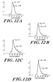
- FIG. 12 is a series of graphs showing the hydrodynamic profiles of the proteoglycan aggregates in the regenerated meniscus (FIG. 12B) compared to the resected rim alone (FIG. 12D), and compared with control samples including the remaining fibrocartilage post-resection in the medium (FIG. 12A) and the remaining fibrocartilage post-resection in the associative extract (FIG. 12C).
- Menisci were aseptically harvested from mature dogs, trimmed of all adherent tissue, and placed into Gey's balanced saline solution. Each meniscus was bisected in the coronal plane and 3 mm full-thickness circular defects were made in each meniscal half. The defects were filled with a 3 mm diameter plug of one of two prototypes of a complex collagen-based biosynthetic scaffold (prosthetic meniscus). The menisci were placed in six well culture plates containing six ml of Dulbecco's modified Eagle's medium supplemented with 10% fetal bovine serum, sodium ascorbate, and 0.1% penicillin/streptomycin.
- Cultures were maintained at 37°C in a humidified atmosphere of 10% C02/90% air, fed three times per week, and placed in fresh culture wells every week to prevent the formation of explant cell cultures. At intervals of one, four, and six weeks after initiation of culture, three menisci from each group were removed, fixed, and evaluated with serial sections and staining.
Abstract
Description
- The present invention is in the field of implantable medical devices, and more particularly, is directed to devices useful as prosthetic menisci, and in vivo scaffolds for regeneration of meniscal tissue and to methods for their fabrication.
- The medial and lateral menisci are a pair of cartilaginous structures in the knee joint which together act as a crucial stabilizer, a mechanism for force distribution, and a lubricant in the area of contact between the tibia and femur. Without the menisci, stress concentration occurs in the knee in conjunction with abnormal joint mechanics, and premature development of arthritic changes occurs.
- In the prior art, treatment of injured or diseased menisci has generally been both by surgical repair and by excision. With excision, regeneration of meniscal tissue may occur. Additionally, it is known that meniscal fibrochondrocytes have the ability to migrate into a defect filled with a fibrin clot and form tissue apparently similar to normal meniscal fibrocartilage. When an adequate matrix scaffold is present within a meniscal defect, such meniscal fibrocartilage may be formed. Meniscal tissue is also capable of self-repair when exposed to bleeding tissues, and additionally, it is also known in the prior art that meniscal cells in tissue culture are capable of cell division and matrix synthesis. Replacement of an injured meniscus in an otherwise healthy joint may prevent arthritic changes and may stabilize the joint. In diseased joints, replacement of the meniscus may reduce the progression of the disease process, and may provide pain relief. Allografting or meniscal transplantation, is one method of replacement which has been executed both in dogs and in humans. However, this approach has been only partially successful over the long term due to the host's immunologic response to the graft, to failures in the cryopreservation process, and to failures of the attachment sites.
- In alternative prior art replacement approaches, menisci have been replaced with prostheses composed of permanent artificial materials. Such prosthesis have been constructed of purely artificial materials in order to minimize the possibility of an immunological response. In addition, the use of such materials is believed to be advantageous because it permits construction of a structure which can withstand the high and repeated loads which are encountered in the knee joint, and because it can alter the joint mechanics in beneficial ways that biological materials would not tolerate.
- For example, a Teflon net has been used to replace the resected meniscus of a dog upon which fibrous ingrowth or regeneration was observed, although accompanied by significant chondral abrasion. A prosthetic meniscus has also been constructed from resilient materials such as silicone rubber or Teflon with reinforcing materials of stainless steel or nylon strands (U.S. Patent No. 4,502,161) A meniscal component has also been made from resilient plastic materials (U.S. Patent No. 4,085,466). In addition, reconstruction of meniscal lesions has been attempted with carbon-fiber-polyurethane-poly (L-lactide), but its success with these materials is minimal (Leeslag et al., Biological and Biomechanical Performance of Biomaterials (Christel et al., eds.) Elsevier Science Publishers B.V., Amsterdam. 1986, pp: 347-352).
- A prosthetic meniscus according to the features of the preamble of claim 1 is known from WO-A-8900413.
- However, the replacement of meniscal tissue with structures consisting of permanent artificial materials generally has been unsuccessful, principally because the opposing articular cartilage of human and animal joints is fragile. The articular cartilage in the knee will not withstand abrasive interfaces, nor compliance variances from normal, which eventually results from the implantation of prior art artificial menisci. Additionally, joint forces are multiples of body weight which, in the case of the knee and hip, are typically encountered over a million cycles per year. Thus far, prior art permanent artificial menisci have not been composed of materials having natural meniscal properties, nor have they been able to be positioned securely enough to withstand such routine forces.
- Therefore, what is needed is an improved prosthetic meniscus composed of biocompatible materials which are soft and lubricating.
- Repair of other tissues such as skin and nerve has been attempted using both synthetic and natural materials. For example, Yannas et al., fashioned endodermal implants, and artificial epidermis out of natural collagen and glycosaminoglycans (U.S. Patent No. 4,060,081). Nyiles et al. (Trans. Am. Soc. Artif. Intern. Organs (1983) 29:307-312) reported the use of synthetic resorbable polyesters for peripheral nerve regeneration applications, and the use of collagen conduits as a scaffold for nerve regeneration.
- However, even with the foregoing technologies which have been applied to the reconstruction of anatomical structures other than knee joints, a structure suitable as a prosthetic meniscus and constructed from totally resorbable natural materials, or analogs thereof, has not been developed in the prior art.
- Accordingly, it is an object of this invention to provide an improved meniscal prosthesis which allows for normal joint motion.
- Another object is to provide a meniscal replacement or prosthesis which is biomechanically able to withstand normal joint forces and is able to function at those loads to protect the cartilage and stabilize the joint.
- Yet another object is to provide a resorbable meniscal prosthesis which acts as a temporary in vivo scaffold for meniscal fibrocartilage infiltration and regeneration.
- Still another object is to provide a meniscal prosthesis which is composed of biocompatible materials having an organization equivalent to that of the normal meniscus.
- A further object is to provide a meniscal prosthesis which is adapted for implantation by standard operative techniques.
- Still a further object is to provide a method by which such prosthetic menisci can be fabricated.
- The present invention provides a biocompatible and bioresorbable structure for implantation into the knee joint which assumes the form and role of a meniscus. This prosthetic meniscus promotes and provides a scaffold for the regeneration of tissue having the physical characteristics of a natural meniscus.
- The prosthetic meniscus of the present invention is generally a dry, porous matrix of biocompatible bioresorbable fibers, including natural polymers or analogs or mixtures thereof. The matrix is adapted to have in vivo an outer surface contour substantially the same as that of a natural meniscus. Further, the matrix has pore size in the range of greater than 50 microns to less than about 500 microns. With this configuration, the matrix establishes an at least partially bioresorbable scaffold adapted for ingrowth of meniscal fibrochondrocytes. The matrix may have the shape of a circumferentially extending wedge spanning a predetermined angle greater than 0 degrees, and less than or equal to 360 degrees, and having a thickness in its central region which is less than its thickness in its peripheral regions. In some forms of the invention, the matrix may assume the shape of a simple wedge, a crescent-shaped wedge with a wide central region between two narrow distal tip regions, or a circumferentially extending wedge spanning an angle of 360 degrees and having a depressed (concave) central region, for example.
- The matrix is composed of biocompatible and bioresorbable fibers, a portion of which may be crosslinked. The fibers include a natural material or an analog of a natural material such as a biosynthetic analog. In a preferred embodiment of the invention, the fibers of the matrix are polymers of, for example, natural molecules such as those obtained from animal or human tissue. Natural fibers useful for the same purpose include collagen, elastin, reticulin, analogs thereof, and mixtures thereof.
- In some forms of the invention, the fibers may be randomly orientated throughout the matrix, or may be ordered at specified regions. Alternatively, the fibers may assume substantially circumferentially extending or substantially radially extending orientations throughout the prosthetic meniscus.
- The matrix may also include glycosaminoglycan molecules (GAGs) interspersed with the fibers. GAGs are any mucopolysaccharide molecules which provide lubrication and crosslinks for the prosthetic meniscus of the invention. In the preferred aspects of the invention, GAGs such as chondroitin 4-sulfate, chondroitin 6-sulfate, keratan sulfate, dermatan sulfate, heparin sulfate, hyaluronic acid, and mixtures thereof are a component of the matrix. These GAGs may be uniformly dispersed throughout the prosthetic meniscus as individual molecules, or may be present in varying amounts in different regions of the structure.
- In various forms of the invention, GAGs may directly participate in covalent crosslinking formation with the fibers, or may interact with the fibers mechanically in the form of entanglement or through interlocking mechanisms, forming stable fiber-GAG complexes.
- The matrix include about 75-100% natural and/or synthetic fibers and about 0-25% GAGs by dry weight, the proportions of which may be constant throughout the structure or may be variable.
- In a preferred embodiment of the invention, the matrix has a density of about 0.07 to 0.50 g matrix/cm³ where "g matrix/cm³" is a unit connoting the number of grams in a cubic centimeter of the matrix. In addition, it has an interfibrillary and interfibrillary space of about 2 to 25 cm³/g matrix.
- In another form of the invention, the prosthetic meniscus may further comprise a mesh composed of a bioresorbable, biocompatible material which is attached to portions of the outer surface of the matrix. The mesh aids in the successful implantation of the prosthetic meniscus into the knee joint by providing a temporary anchoring mechanism.
- Further, the invention includes a method for fabricating a prosthetic meniscus of the type described above. Generally, the method includes placing a plurality of fibers and/or fibers and GAGs into a mold having a shape useful for knee joint function, subjecting the fibers (and GAGs) in the mold to two cycles of freezing and thawing, contacting said fibers or said fibers and GAGs with a chemical crosslinking reagent such that the fibers then assume the shape of the mold, and lyophilizing the resulting structure to obtain a dry, porous, volume matrix.
- The fibers may be laid down in a circumferential orientation by rotating the mold as they are placed therein. Alternatively the fibers in the mold may be compressed with a rotating piston. Radial orientation of the fibers is produced by manually painting the fibers in a linear, radially directed fashion.
- Specific densities and pore sizes may be obtained in various regions of the matrix by compressing the fibers or fibers and GAGs in the mold prior to the second freeze-thaw cycle, subsequent to the chemical crosslinking step. This may be accomplished by applying pressure to a specific region of the matrix with a piston of a predetermined shape.
- In a preferred aspect of the invention, the cross linking step is performed using chemical agents which form intramolecular and intermolecular crosslinks. Useful chemical agents include, for example, glutaraldehyde, formaldehyde, biocompatible bifunctional aldehydes, carbodiimides, hexamethylene diisocyanate, bis-ionidates, glyoxal, polyglycerol polyglycidyl ether, glyoxal, and mixtures thereof. Particularly useful crosslinking agents are 1-ethyl, 3-(3-dimethylaminopropyl), polyglycerol polyglycidyl ether, and glutaraldehyde.
- In other aspects of the invention, an additional crosslinking step is performed by lyophilizing the chemically crosslinked matrix and then subjecting it to dehydrothermal crosslinking procedures.
- The invention will next be described in connection with certain illustrated embodiments. However, it should be clear that various modifications, additions, and deletions can be made without departing from the spirit or scope of the invention.
- The foregoing and other objects of this invention, the various features thereof, as well as the invention, itself, may be more fully understood from the following description, when read together with the accompanying drawings.
- FIG. 1 shows a simplified diagramatic representation of a human knee joint, with menisci in native positioning;
- FIG. 1A is a diagrammatic representation of a cut-away view of the knee joint showing the medial and lateral menisci as they are positioned in vivo over the medial and lateral condyles.
- FIG. 2 shows a perspective view of an exemplary prosthetic meniscus in accordance with the present invention;
- FIG. 3 shows a perspective radial section of the prosthetic meniscus of FIG. 2;
- FIG. 4 shows a perspective view of an alternative embodiment of the present invention;
- FIG. 5 shows a sectional view along line 5-5 of the prosthetic meniscus of FIG. 4.
- FIG. 6 shows a mold designed for the fabrication of a prosthetic meniscus having a cylindrical pad shape.
- FIG. 7 shows a mold designed for the fabrication of a prosthetic meniscus having a crescent-shaped wedge form.
- FIG. 8 shows a mold designed for the fabrication of a cylindrical prosthetic meniscus.
- FIG. 9 is a photographic representation of in vivo meniscal regrowth after 80% resection and implantation of the prosthetic meniscus.
- FIG. 10 is a photographic representation of an explanted canine meniscus containing a section of scaffold, and demonstrating in vitro regrowth of meniscal tissues into the scaffold.
- FIG. 11 is a photographic representation of two canine knee joints three months after surgical resection.
- FIG. 12 is a series of graphs showing the hydrodynamic profiles of the proteoglycan aggregates in the regenerated meniscus (FIG. 12B) compared to the resected rim alone (FIG. 12D), and compared with control samples including the remaining fibrocartilage post-resection in the medium (FIG. 12A) and the remaining fibrocartilage post-resection in the associative extract (FIG. 12C).
- It has been discovered that a prosthetic meniscus fabricated from biocompatible and bioresorbable fibers can be surgically implanted into the knee joint so as to provide normal joint motion and strength. This prosthetic meniscus also acts as a scaffold for regenerating meniscal tissue whose ingrowth is encouraged by the physical characteristics of the implanted device.
- FIG. 1 shows a diagramatic representation of the normal positioning of
medial meniscus 7 andlateral meniscus 8 in the human knee joint 3 between thefemur 2 and tibia 4. These menisci, when compressed between thefemur 2 and tibia 4, become tough except at their points of attachment. FIG. 1A shows the in vivo structure ofmedial meniscus 7 andlateral meniscus 8 in theknee joint 3. The menisci conform to the shapes of the surfaces between which they are positioned, thereby resulting in two distinct in vivo forms. For example, themedial meniscus 7 has a relatively open crescent shape, while thelateral meniscus 8 has a relatively closed crescent shape. - An exemplary
prosthetic meniscus 10 is shown in FIG. 2. Theprosthetic meniscus 10 is a generally wedge-shaped, porous dry matrix or scaffold which extends circumferentially or laterally at least in part about a central axis 11. In the preferred form, theprosthetic meniscus 10 has the shape of a crescent-shaped wedge, extending circumferentially about the axis 11, and comprising a relatively wide central region 12 between two narrowdistal regions - In the embodiment illustrated in Fig. 2, the
prosthetic meniscus 10 includes amesh member 20 extending from its peripheral edge. Themesh member 20 is composed of a biocompatible, bioresorbable material, and provides a readily used means for anchoring thearray 10 in place. Themesh member 20 may function in this capacity until sufficient tissue ingrowth occurs to then provide that function. By way of an example, themesh member 20 may be a #1 mesh screen composed of absorbable suture materials such as polyglyconate, Dexon, or polydioxane (PDS) woven into a mesh. Non-absorbable suture materials such as Goretex® may also be used. - FIGs. 4 and 5 show an additional embodiment of the present invention which is similar in composition to the prosthetic meniscus depicted in FIG. 2. More particularly, FIG. 4 depicts a right circular cylinder-shaped
meniscus 22, extending fully about axis 11, i.e. where angle B equals 0 degrees. (i.e. the meniscus subtends 360 degrees.) FIG. 5 shows a sectional view along line 5-5 of the meniscus shown in FIG. 4. The device illustrated in Figs. 4 and 5 show the shape of themeniscus 22 when implanted; that is, the height D at areas 11 is less than the peripheral height A of the device. Prior to implantation, thedevice 22 may in some cases not have this relationship but upon implantation, the normal loads applied by the body force this conformation. - In alternative forms of the invention, still other shapes may be used. For example, it is not required that the wedge be symmetrical. These embodiments may have densities of collagen fibers and dispersions of GAG molecules and crosslinks, permitting accommodation of differing stress levels, rates of ingrowth, and resiliency. Differing densities may be obtained in vivo where a device having uniform density is implanted, and body loading causes non-uniform compression of the device.
- The prosthetic meniscus may be fabricated of any biocompatible, bioresorbable fibers which include a natural material or an analog thereof; preferably polymeric in structure, which can provide mechanical strength and protection and lubrication while encouraging tissue ingrowth (e.g., collagen, reticulin, elastin, cellulose, or biosynthetic analogs thereof). These fibers may be ordered in substantially circumferentially-extending or substantially radially-extending orientations, with the density of fibers being substantially uniform throughout the matrix. Alternatively, the matrix fibers may be unordered. In either the ordered or unordered configuration, the density of the fibers may be non-uniform. In the non-uniform configuration, relatively high densities of fibers may be established at anticipated points of high stress by local application.
- In an alternative aspect of the invention, the intrafibrillary and interfibrillary space is relatively high, a condition which promotes ingrowth of regenerated meniscal tissue. For example, the density of the meniscus may be in the range of about 10-25 g matrix/cm³. Alternatively, the intrafibrillary and interfibrillary space is relatively low, a condition which provides cushioning, lubrication, and mechanical support for the knee joint and which retards tissue and cell ingrowth, thereby diminishing the rate of scaffold resorption (e.g., density is in the range of about 2-10 g matrix/cm³).
- The temporary stability of the shape of the structure when in vivo, and the rate of meniscal resorption, are both attributed to the effective crosslinking formation between at least one portion of the fibers. The crosslinking reagents used may be any biocompatible bifunctional reagents which interacts with amino groups, carboxyl, or hydroxyl groups on a single fiber (intramolecular crosslinks), or the fibers or on the fibers and the GAGs, resulting in covalent bond formation between adjacent molecules (intermolecular crosslinks). Useful crosslinking reagents include aldehydes, hexamethylene diisocyanate, bis-imidates, polyglycerol polyglycidyl ether, and carbodiimides.
- The crosslinked device maintains sufficient degree of hydrophilicity and elasticity which simulates the properties of the natural meniscus, i.e., ability to sustain mechanical stress and to protect and lubricate articular surfaces. In addition, the structure provides an ideal environment for cell infiltration and extracellular matrix synthesis and deposition resulting in regeneration of natural meniscal tissue.
- GAGs may be dispersed throughout the fibers. Alternatively, they may act as intermolecular crosslinks between fibers. These GAG crosslinks are composed typically of at least one of the group of molecules consisting of chondroitin 4-sulfate, chondroitin 6-sulfate, keratin sulfate, dermatan sulfate, heparin sulfate, and hyaluronic acid. The dispersion of GAG crosslinks is preferably uniform, but may be more concentrated at anticipated points of high stress, typically at the
distal regions distal regions - Intermolecular cross linkages can also be established through a dehydrothermal process (heat and vacuum) which results in peptide bond formation between an epsilon amino group of lysine or hydroxylysine and a carboxyl group of aspartic or glutamic acid.
- The cross linked device has a relatively high thermal stability between about 55-85° C, preferably between about 65-75° C, for sufficient in vivo stability. This may be achieved through manipulation of the crosslinking conditions, including reagent concentration, temperature, pH, and time.
- In a one embodiment the prosthetic meniscus is constructed mainly of Type I collagen fibers without GAG crosslinks. Type I collagen fibers may be obtained from the Achilles tendons of animals. However, the fibers may also be obtained from animal skin or from the skin or tendon of humans. The tissues are treated with a series of mechanical and chemical means to either totally remove the non-collagenous materials or reduce them to a minimal level. In the preferred processing steps, the tendon or skin is mechanically disintegrated into fine pieces useful for further processing. The disintegration may be achieved by grinding the tissue at liquid nitrogen temperature, or by cutting the tissue into small pieces with a sharp knife. In certain applications, the tendons are mechanically disintegrated along the fiber direction in order to maintain the length of the fibers for mechanical strength.
- Salt extraction of tendon at neutral pH removes a small portion of the collagen molecules that are newly synthesized and have not yet been incorporated into the stable fibrils. Salt also removes some glycoproteins and proteoglycans that are associated with collagen through electrostatic interactions. Other salts such as KCl and the like can be used as a substitute for NaCl.
- Lipids that are associated with the cell membranes or collagenous matrices may be removed by first extracting with detergents such as Triton X-100, followed by extracting with ether-ethanol mixtures. The concentration of Triton X-100 is usually about 2-4%, but is preferably about 3%. The preferred mixture of ether-ethanol is usually at about a 1:1 ratio (v/v). The period of extraction is usually from 8 hours to 96 hours, as is preferably from about 24 to 48 hours.
- Further extraction may be accomplished by matrix swelling conducted at two extreme pHs. Both acidic and basic swelling weakens the non-covalent intermolecular interactions, thus facilitating the release of non-covalently attached glycoproteins, glycosaminoglycans (GAGs), and other non-collagenous molecules through the open pores of the collagenous matrices.
- The swelling of matrix at alkaline pH is done by treating the collagen at high pH with Ca(OH)₂, NaOH, or the like, for a period of about 8-96 hours. Alkali extraction in the presence of triple-helical stabilizing salts such at (CH₃)NCl, NH₃S0₄, or the like reduces the potential risk of denaturation of the collagen. Alkali treatment dissociates the non-cross-linked glycoproteins and GAGs from the collagen matrices. The alkali also removed the residual lipids through saponification.
- The acid swelling may be conducted at a low pH in the presence of acetic acid, HCl, or the like. Like the alkali treatment, the acid swelling removes non-cross-linked glycoproteins and GAGs.
- The non-triple helical portions of the molecule (telopeptides) are involved in intermolecular crosslinking formation. They are weak antigens and are susceptible to attack by proteases, such as pepsin, trypsin, and the like. Prolonged digestion with such proteases dissociates the fibrils (fibers) into individual molecules. However, if the digestion process is properly controlled such that maximal telopeptides are removed without complete dissociation, the immunogenic properties of the fibrils can be reduced to a minimal level without compromising the mechanical strength. For example, to isolate molecular collagen, the digestion of skin or tendon with pepsin is usually conducted at an enzyme:collagen ratio of about 1:10 for about 24-96 hours at below room temperature. In comparison, fibrils may be obtained by limited pepsin digestion achieved at a ratio of about 1:100 (enzyme:collagen) for about 24-96 hours at 4° C.
- Collagen fibers obtained according to this methodology are then used to fabricate the prosthetic meniscus of the present invention. However, it must be appreciated that collagen obtained from other sources, such as biosynthetically-produced collagen or analogs thereof - may also be used in the construction of the prosthetic meniscus.
- In one embodiment, the prosthetic meniscus further includes an adhesion molecule or adhesive portion or analog thereof which is incorporated within the network of fibers, and which aids in meniscal tissue regeneration Useful adhesion molecules include peptides such as fibronectin (see e.g., U.S. Patent No. 4,589,881, 4,661,111 and 4,578,079), a portion of which can be conjugated to, for example, chondroitin sulfate.
- The method of fabrication includes molding the collagen fibers into a predetermined shape using, for example, the mold forms described below in conjunction with FIGs. 6-8. The fibers may be placed randomly in the mold, or may be oriented in specific directions to achieve a meniscus having specific structure characteristics. Other components such as GAGs which may participate in the crosslinking reaction, can be mixed in with the fibers in a random or non-random fashion before the structure is subjected to various crosslinking and dehydrating procedures including various chemical and/or dehydrothermal methods. Adhesion molecules or adhesive fragments or analogs thereof may be added to the structure before the final drying step by soaking the structure in a solution containing that molecule, or by specifically coupling it to an existing fiber or crosslink. For example, the adhesion portion of fibronectin may be cross linked to chondroitin sulfate at a concentration of 3 peptide molecules per molecule chondroitin sulfate by soaking the prosthetic meniscus in a 50 mg/ml solution thereof.
- By following the processes described in the above examples set forth herein below, a prosthetic meniscus of the form shown in FIGs. 2 or 3 may be constructed having the characteristics listed below in TABLE 1.

- The following non-limiting examples describe methods of fabrication and in vivo testing of the prosthetic meniscus of the invention.
- A
mold 100 useful for fabricating the prosthetic meniscus is made of implantable stainless steel or biocompatible plastics such as teflon, polypropylene, delrin, or combination of these materials. Themold 100 is composed of threepieces - By way of example for the disk-shaped meniscus illustrated in FIGs. 4 and 5, the
mold 100 of FIG. 6 is used. Thefirst piece 102 is disk-like and has a diameter substantially equal to that of the desired meniscus.Piece 102 is perforated to allow liquid to pass through under pressure. Theinner surface 103 ofpiece 102 has the desired shape of one side of the meniscus-to-be-formed. - The
second piece 104 is a hollow cylinder which has the same inner dimension as thefirst piece 102. Thethird piece 106 is a cylindrical piston which has an outer diameter slightly less than the inner diameter ofpiece 104. The "top", or crown,surface 108 ofpiston 106 has the desired shape of one side of the meniscus-to-be-formed. - For the meniscus of FIG. 3, the mold of FIG. 7 is used where the shape of
piece 102, and cross-section ofpiece 104 have the shape of an angular segment. For a flat circular disk meniscus, themold 100 of FIG. 8 is used wherepieces piece 106 is similar to that piece in FIG. 108 but has aflat crown surface 108. - During fabrication of the
meniscus 10, thepiece 102 is first assembled withinpiece 104, as shown in FIGs. 6-8. The constituent fibers (in a fluid) are placed against thesurface 103 ofpiece 102. Then thecrown surface 108 ofpiston 106 is driven towardsurface 103 along acompression axis 106a until the fibers are compressed, the fluid is driven out throughpiece 102, and the desired axial dimension of the compressed fiber array is attained. The mold is then frozen in preparation for chemical crosslinking. - Bovine, porcine, or sheep Achilles tendon is obtained from USDA-approved slaughter houses. The preferred age of the animals is between 12-18 months. The tissue is kept cold during the purification process except where specified to minimize bacteria contamination and tissue degradation.
- The adhering tissues of carefully selected tendons are first scrapped off mechanically. The tendons are then minced or cut into fine pieces and washed in excess quantities (10 volumes) of cold water to remove residual blood proteins and water soluble materials.
- The washed tendons are extracted in ten volumes of 5% NaCl, 0.01 M Tris, pH 7.4, for 24 (+/-4) hours to remove salt soluble materials. The salt extracted tendons are repeatedly washed in about 10 volumes of water to remove the salt.
- The material is extracted in 3% Triton X-100 for 24 (+/- 2) hours. The detergent is removed by extensive washing with water. The material is then extracted in 3-4 volumes of ether-ethanol (1:1 vol/vol) for 24 (+/- 2) hours to further minimize the lipid content. The lipid extracted material is extensively washed in water to remove the ether and ethanol.
- The material is then subjected to two extreme pH extractions to remove non-collagenous materials. Alkaline extraction is conducted with 3-4 volumes of 0.2 M NaOH at pH 12.5 - 13.5 at room temperature (RT) in the presence of 1.0 M (CH)NCl for 24 (+/- 2) hours with mild agitation.
- Following alkaline extraction, the pH is neutralized with HCl and the material is washed with water. The pH is then adjusted to 2.5 - 3.0 by adding concentrated acetic acid to a final concentration of 0.5 M. The acid extraction is continued for 24 (+/- 2) hours with agitation.
- The acid swollen tendon is then subjected to a limited proteolytic digestion with pepsin (enzyme : collagen = 1 : 100) for 24 (+/-) 2 hours. The pepsin and telopeptides are removed through dialysis.
- The swollen fibrillar material is then coacervated by adjusting the pH to its isoionic point with 1 M NaOH or HCl or by adjusting the ionic strength to 0.7 with NaCl. The aggregated collagen fibers are harvested by filtration, and the filtered material extensively washed with cold buffered solution. The highly purified type I collagen may be stored (-20 to -40° C) until used.
-
- A) The collagen content of the highly purified type I collagen fibrils from EXAMPLE 2 is determined either by gravimetric methods or by determining the hydroxyproline content assuming a 13.5% by weight of hydroxyproline in type I collagen. The amount of purified material needed to fabricate a given density of a meniscus device is then determined and weighed.
- B) A solution of fibrillar collagen is carefully fit into a mold of specified dimensions, e.g. according to the exemplary meniscus described above in conjunction with FIG. 2-5 (see EXAMPLE I and FIGs. 6-8 for the description of molds). Collagen fibers are laid down in random manner or in an oriented manner. In the oriented manner, circumferential orientation of the fibers is produced by rotation of the piston about its principal axis as the material is compressed in the mold; radial orientation is produced by manual painting of the collagen fibers in a linear, radially directed fashion.
- C) The fibers are frozen at -20° C, turned out of the mold, and thawed at RT.
- D) The fibers are then resuspended in phosphate buffered saline, put back into the mold in the desired orientation(s), and compressed with the piston.
- E) The compressed fibers are then refrozen at -20 C and then thawed at RT.
- F) The resulting structure is cross linked by soaking in a 0.2% glutaraldehyde solution, pH 7.6, for 24 (+/- 0.5) hours. Each glutaraldehyde-cross-linked meniscal device is subsequently rinsed repeatedly in 500 ml of phosphate buffered saline (PBS) solution, pH 7.4, for 4, 8, 24 and 48 hours.
- G) The rinsed matrix is then lyophilized.
-
- A)-G) (same as in EXAMPLE 3)
- H) The lyophilized matrix is subjected to dehydrothermal crosslinking by vacuum and heat. The vacuum is first applied to reduce the residual water content to a minimal level (some structural water, about 3%, may still be associated with collagen triple-helix as part of the structure stabilizing factor). The heat is increasing in steps to 110° C (+/- 5°), and continually applied at 110° C under vacuum for 24 (+/- 2) hours.
-
- A) (same as in EXAMPLE 3)
- B) The collagen material is dispersed in 0.01 M HCl solution at pH 2-2.5. Predetermined amounts of various GAGs are weighed and dissolved in water. For example, for a given density of 0.25 g/cm, the collagen content will be 0.244 g, the hyaluronic acid content will be 0.003 g, and the chondroitin sulfate content will be 0.003 g for a 2.5% GAG content. The GAG solution is mixed in with the collagen solution and placed in the mold in the desired orientation as described in EXAMPLE 2.
- C)-G) (same as in EXAMPLE 3)
-
- A)-C) (same as in EXAMPLE 3)
- D) (same as in EXAMPLE 3 except that the fibers laid down are not compressed.
- E)-G) (same as in EXAMPLE 3)
-
- A)-E) (same as in EXAMPLE 3)
- F) The molded collagen is cross linked in 5% polyglycerol polyglycidyl ether in 50% ethanol and 0.1 M Na₂CO₃ at pH 10.0 for 24 (+/- 2) hours. The crosslinked device is rinsed for 4, 8, 24 and 48 hours, each with 500 ml of PBS, pH 7.4.
- G) (same as in EXAMPLE 3)
-
- A)-E) (same as in EXAMPLE 3)
- F) The molded collagen is crosslinked in the presence of 1-ethyl-3-(3-dimethylaminopropyl) carbodiimide (10 mg/g matrix) in 0.9% NaCl, pH 4.7 at room temperature for 24 (+/- 2) hours. The addition of carbodiimide is made every 3-4 hours, and the pH is adjusted to 4.7 after each addition of carbodiimide.
- G) (same as in EXAMPLE 3)
-
- (A) - (D) same as steps (A) - (D) as described in Example II.
- (E) For attachment purposes, a mesh of absorbable polyglyconate suture material, matched to the size of the mold, is laid in the dispersed collagen such that it protrudes from the structure's periphery to form a skirt which may extend over the tibial plateau. This mesh provides both immediate attachment sites and long term fibrous ingrowth.
- The prosthetic menisci were evaluated in vivo using animal models and in vitro to determine ability to function or to serve as a regeneration template for normal meniscal tissues.
- Seventeen prosthetic menisci (device III type) were implanted into eleven immature Yorkshire pigs. Seven joints underwent a two-thirds subtotal resection of the medial meniscus with replacement by a matched prosthetic meniscus; two joints underwent a two-thirds subtotal resection alone; two joints received a similar subtotal meniscectomy with the resected portion immediately replaced with suture fixation; and two joints received total meniscectomy alone.
- Evaluation of all joints was made at 3 or 6 weeks. All arthrotomies healed well, and all animals progressed to full weight bearing. At final evaluation all prosthetic material had been partially or completely resorbed without evidence of joint destruction or cartilage abrasion. Neovascularization was observed as the basic healing mechanism in both the prosthetic implanted menisci as well as in the controls, and in all joints there was evidence of early meniscal regeneration. The prosthetic meniscus material conformed to the appropriate joint shape. In addition, there was no clinical evidence of implant rejection over a 6 week period. Histologically, there was acute inflammation followed by neovascularization and extensive fibroplasia with early hyalinization of the newly formed collagen. (See FIG. 9)
- In vivo studies of the invention in mature dogs have demonstrated induced meniscal regeneration through the prosthetic material. Normally, the mature canine stifle is known to not regenerate a meniscus and is known to develop significant arthritic changes. However, six weeks after meniscectomy and implantation of scaffolds in accordance with the present invention, there occurred significant regeneration of the meniscus through the scaffolds. The scaffolds provided joint protection, as determined by diminished cartilage erosions, osteophyte formation and affinity for India ink.
- FIG. 11 is a photographic representation of two canine knee joints three months after surgical resection. The joints were protected by the prosthetic implant with subsequent regrowth of a new meniscus. The joint on the right in FIG. 11 underwent an 80% meniscal resection alone. The dramatic articular cartilage protection is highlighted by India ink.
- New collagen and glycosaminoglycan formation was evidenced histologically, by Alcian Blue and Masson's Trichrome stains.
The hydrodynamic size and chromatographic profiles of the proteoglycans synthesized within both the meniscal implants and the controls were similar when analyzed on a Sephacryl S-500 column as shown in FIG. 12. FIG. 12 is a series of graphs showing the hydrodynamic profiles of the proteoglycan aggregates in the regenerated meniscus (FIG. 12B) compared to the resected rim alone (FIG. 12D), and compared with control samples including the remaining fibrocartilage post-resection in the medium (FIG. 12A) and the remaining fibrocartilage post-resection in the associative extract (FIG. 12C). - Menisci were aseptically harvested from mature dogs, trimmed of all adherent tissue, and placed into Gey's balanced saline solution. Each meniscus was bisected in the coronal plane and 3 mm full-thickness circular defects were made in each meniscal half. The defects were filled with a 3 mm diameter plug of one of two prototypes of a complex collagen-based biosynthetic scaffold (prosthetic meniscus). The menisci were placed in six well culture plates containing six ml of Dulbecco's modified Eagle's medium supplemented with 10% fetal bovine serum, sodium ascorbate, and 0.1% penicillin/streptomycin. Cultures were maintained at 37°C in a humidified atmosphere of 10% C0₂/90% air, fed three times per week, and placed in fresh culture wells every week to prevent the formation of explant cell cultures. At intervals of one, four, and six weeks after initiation of culture, three menisci from each group were removed, fixed, and evaluated with serial sections and staining.
- The results (shown in FIG. 10) demonstrated increasing cellular migration and invasion over time. There was no apparent toxicity from the material. The morphologic characteristics of the migrating cells were more fusiform and elongated than native fibrochondrocytes The depth of cellular penetration into the scaffold appeared to be limited by the density of the prosthetic complex.
- The present invention may be embodied in other specific forms without departing from the essential characteristics thereof. The present embodiments are therefore to be considered in all respects as illustrative and not restrictive, the scope of the invention being indicated by the appended claims rather than by the foregoing description, and all changes which come within the meaning of the claims are therefore intended to be embraced therein.
Claims (21)
- A prosthetic meniscus comprising a dry porous matrix of biocompatible and at least partially bioresorbable fibers,
said fibers including natural polymers, e.g. collagen, elastin, reticulin, cellulose, or analogs or mixtures thereof,
said matrix being adapted to have in vivo an outer surface contour substantially the same as that of a natural meniscus, characterised in that
said matrix has pore size in the range of greater than 50 microns to less than about 500 microns,
whereby said matrix establishes an at least partially bioresorbable scaffold adapted for ingrowth of meniscal fibrochondrocytes. - A prosthetic meniscus according to claim 1 wherein said matrix has a substantially wedge shape including a wide central region between two narrow distal tip regions.
- A prosthetic meniscus according to claim 1 wherein said matrix has a density of about 0.07 to 0.50 gram matrix per cubic centimeter.
- A prosthetic meniscus according to claim 1 wherein said matrix has an interfibrillary and interfibrillary space of about 2 - 25 cubic centimeters per gram matrix.
- A prosthetic meniscus according to claim 1, wherein said natural polymers comprise Type I collagen, and wherein said collagen fibers are present at a concentration of about 75-100% by dry weight, and said glycosaminoglycan molecules are present at a concentration of about 0-25% by dry weight.
- A prosthetic meniscus according to claim 1 further comprising a plurality of glycosaminoglycan molecules, e.g. chondroitin 4-sulfate, chondroitin 6-sulfate, keratan sulfate, dermatan sulfate, heparin sulfate, hyaluronic acid, and mixtures thereof, interspersed with said fibers, at least a portion of said molecules providing crosslinks between ones of said fibers.
- A prosthetic meniscus according to claim 6 wherein said fibers are present at a concentration of about 75-100% by dry weight, and said glycosaminoglycan molecules are present at a concentration of about 0-25% by dry weight.
- A prosthetic meniscus according to claim 1 wherein said matrix has the shape of a circumferentially extending wedge, e.g. a crescent-shaped wedge, having a central region and a region peripheral thereto, and spanning a predetermined angle greater than 0 degrees and less than or equal to 360 degrees about said central region, and
where the thickness in said central region of said wedge is less than the thickness in the peripheral region of said wedge. - A prosthetic meniscus according to claim 1 further comprising a mesh extending from portions of the outer surface of said matrix, said mesh being resorbable and biocompatible.
- The prosthetic meniscus according to any one of claims 1 to 9 for use in therapy, e.g. in a method of regenerating meniscal tissue in vivo comprising the steps of:(a) fabricating the prosthetic meniscus according to any one of claims 1-9; and(b) implanting said prosthetic meniscus into a knee joint by surgical procedures.
- The prosthetic meniscus of claim 10 wherein said prosthetic meniscus fabricated in step (a) assumes the shape of a native meniscus when implanted in step (b).
- A method for fabricating a prosthetic meniscus according to any one of claims 1-9, comprising the steps of:(a) placing a plurality of biocompatible and bioresorbable fibers into a mold, said fibers including natural polymers, e.g. collagen, elastin, reticulin, cellulose, or analogs or mixtures thereof, and said mold defining a shape for a meniscal prosthesis that enables knee joint function;(b) subjecting said fibers to a first and a second cycle of freezing and thawing;(c) contacting said fibers with a chemical crosslinking agent, e.g. glutaraldehyde, formaldehyde, biocompatible and bifunctional aldehydes, carbodiimides, hexamethylene diisocyanate, bis-imidates, polyglycerol polyglycidylether, glyoxal, and mixtures thereof, such that said fibers assume the shape of said mold; and(d) lyophilizing said crosslinked fibers.
- The method of claim 12 wherein said placing step further comprises placing a plurality of glycosaminoglycan molecules into said mold.
- The method of claim 12 wherein said placing step further comprises the step of orienting said fibers substantially circumferentially.
- The method of claim 14 wherein said orienting step comprises compressing said fibers in said mold with a piston, wherein piston motion is substantially directed along a compression axis, while during said compressing step said piston is rotated with respect to said mold about said compression axis.
- The method of claim 14 wherein said orienting step comprises rotating said mold as said fibers are placed therein.
- The method of claim 12 wherein said placing step further comprises the step of orienting said fibers substantially radially.
- The method of claim 12 further comprising the step of compressing said fibers prior to said second cycle of freezing and thawing.
- The method of claim 18 wherein said compressing step comprises applying a predetermined amount of pressure to a region of said matrix with a piston, said piston having a predetermined shape.
- The method of claim 18 further comprising the additional step of subjecting said lyophilized matrix to a dehydrothermal crosslinking procedure.
- The method of claim 12 wherein said placing step further comprises interspersing a plurality of glycosaminoglycan molecules such as chondroitin 4-sulfate, chondroitin 6-sulfate, keratin sulfate, dermatan sulfate, heparin sulfate, hyaluronic acid, and mixtures thereof.
Applications Claiming Priority (3)
| Application Number | Priority Date | Filing Date | Title |
|---|---|---|---|
| US07/317,951 US5007934A (en) | 1987-07-20 | 1989-03-02 | Prosthetic meniscus |
| US317951 | 1989-03-02 | ||
| PCT/US1990/001141 WO1990009769A1 (en) | 1989-03-02 | 1990-03-01 | Prosthetic meniscus |
Publications (3)
| Publication Number | Publication Date |
|---|---|
| EP0461201A1 EP0461201A1 (en) | 1991-12-18 |
| EP0461201A4 EP0461201A4 (en) | 1992-05-13 |
| EP0461201B1 true EP0461201B1 (en) | 1995-07-26 |
Family
ID=23235970
Family Applications (1)
| Application Number | Title | Priority Date | Filing Date |
|---|---|---|---|
| EP90905141A Expired - Lifetime EP0461201B1 (en) | 1989-03-02 | 1990-03-01 | Prosthetic meniscus |
Country Status (9)
| Country | Link |
|---|---|
| US (1) | US5007934A (en) |
| EP (1) | EP0461201B1 (en) |
| JP (1) | JP3100156B2 (en) |
| AT (1) | ATE125441T1 (en) |
| AU (1) | AU637605B2 (en) |
| CA (1) | CA2050471C (en) |
| DE (1) | DE69021204T2 (en) |
| ES (1) | ES2077676T3 (en) |
| WO (1) | WO1990009769A1 (en) |
Cited By (2)
| Publication number | Priority date | Publication date | Assignee | Title |
|---|---|---|---|---|
| US7666230B2 (en) | 2003-12-08 | 2010-02-23 | Depuy Products, Inc. | Implant device for cartilage regeneration in load bearing articulation regions |
| US9445908B2 (en) | 2009-07-06 | 2016-09-20 | Aesculap Ag | Elevated implant for the reconstruction of meniscus defects or partial meniscus defects |
Families Citing this family (229)
| Publication number | Priority date | Publication date | Assignee | Title |
|---|---|---|---|---|
| US5306311A (en) * | 1987-07-20 | 1994-04-26 | Regen Corporation | Prosthetic articular cartilage |
| US5735902A (en) * | 1987-07-20 | 1998-04-07 | Regen Biologics, Inc. | Hand implant device |
| US5263984A (en) * | 1987-07-20 | 1993-11-23 | Regen Biologics, Inc. | Prosthetic ligaments |
| US5681353A (en) * | 1987-07-20 | 1997-10-28 | Regen Biologics, Inc. | Meniscal augmentation device |
| US5376118A (en) * | 1989-05-10 | 1994-12-27 | United States Surgical Corporation | Support material for cell impregnation |
| US20030077825A1 (en) * | 1994-07-22 | 2003-04-24 | Bhatnagar Rajendra S. | Structures useful for bone engineering and methods |
| US5674848A (en) * | 1989-08-14 | 1997-10-07 | The Regents Of The University Of California | Bioreactor compositions with enhanced cell binding |
| US5354736A (en) * | 1989-08-14 | 1994-10-11 | Regents Of The University Of California | Synthetic compounds and compositions with enhanced cell binding |
| US5635482A (en) * | 1989-08-14 | 1997-06-03 | The Regents Of The University Of California | Synthetic compounds and compositions with enhanced cell binding |
| US5645591A (en) | 1990-05-29 | 1997-07-08 | Stryker Corporation | Synthetic bone matrix |
| US5376376A (en) * | 1992-01-13 | 1994-12-27 | Li; Shu-Tung | Resorbable vascular wound dressings |
| US6334872B1 (en) | 1994-02-18 | 2002-01-01 | Organogenesis Inc. | Method for treating diseased or damaged organs |
| US20020055786A1 (en) * | 1994-08-16 | 2002-05-09 | Anthony Atala | Reconstruction of urological structures with polymeric matrices |
| US8603095B2 (en) | 1994-09-02 | 2013-12-10 | Puget Bio Ventures LLC | Apparatuses for femoral and tibial resection |
| US6695848B2 (en) * | 1994-09-02 | 2004-02-24 | Hudson Surgical Design, Inc. | Methods for femoral and tibial resection |
| CA2204753A1 (en) * | 1994-11-07 | 1996-05-17 | The University Of Medicine And Dentistry Of New Jersey | Synthetic collagen orthopaedic structures such as grafts, tendons and other structures |
| AUPN174495A0 (en) | 1995-03-15 | 1995-04-06 | Ketharanathan, Vettivetpillai | Surgical prostheses |
| US20020095218A1 (en) * | 1996-03-12 | 2002-07-18 | Carr Robert M. | Tissue repair fabric |
| US7128763B1 (en) * | 1995-05-26 | 2006-10-31 | Gerald Blatt | Joint treating method |
| US5865849A (en) * | 1995-06-07 | 1999-02-02 | Crosscart, Inc. | Meniscal heterografts |
| US6231608B1 (en) | 1995-06-07 | 2001-05-15 | Crosscart, Inc. | Aldehyde and glycosidase-treated soft and bone tissue xenografts |
| US5913900A (en) * | 1995-06-07 | 1999-06-22 | Corsscart, Inc. | Substantially native meniscal cartilage heterografts |
| US5984858A (en) * | 1995-06-07 | 1999-11-16 | Crosscart, Inc. | Meniscal xenografts |
| US6046379A (en) * | 1995-06-07 | 2000-04-04 | Stone; Kevin R. | Meniscal xenografts |
| US5944755A (en) * | 1995-09-15 | 1999-08-31 | Crosscart, Inc. | Articular cartilage xenografts |
| US6210440B1 (en) | 1995-09-15 | 2001-04-03 | Kevin R. Stone | Anterior cruciate ligament xenografts |
| US5782915A (en) * | 1995-09-15 | 1998-07-21 | Stone; Kevin R. | Articular cartilage heterografts |
| US6049025A (en) * | 1995-09-15 | 2000-04-11 | Stone; Kevin R. | Articular cartilage xenografts |
| US6110206A (en) * | 1995-09-15 | 2000-08-29 | Crosscart, Inc. | Anterior cruciate ligament xenografts |
| US5902338A (en) * | 1995-09-15 | 1999-05-11 | Crosscart, Inc. | Anterior cruciate ligament heterograft |
| US8480754B2 (en) | 2001-05-25 | 2013-07-09 | Conformis, Inc. | Patient-adapted and improved articular implants, designs and related guide tools |
| US8556983B2 (en) | 2001-05-25 | 2013-10-15 | Conformis, Inc. | Patient-adapted and improved orthopedic implants, designs and related tools |
| US8083745B2 (en) | 2001-05-25 | 2011-12-27 | Conformis, Inc. | Surgical tools for arthroplasty |
| US8545569B2 (en) | 2001-05-25 | 2013-10-01 | Conformis, Inc. | Patient selectable knee arthroplasty devices |
| US8882847B2 (en) | 2001-05-25 | 2014-11-11 | Conformis, Inc. | Patient selectable knee joint arthroplasty devices |
| US9603711B2 (en) | 2001-05-25 | 2017-03-28 | Conformis, Inc. | Patient-adapted and improved articular implants, designs and related guide tools |
| US10085839B2 (en) | 2004-01-05 | 2018-10-02 | Conformis, Inc. | Patient-specific and patient-engineered orthopedic implants |
| US7534263B2 (en) | 2001-05-25 | 2009-05-19 | Conformis, Inc. | Surgical tools facilitating increased accuracy, speed and simplicity in performing joint arthroplasty |
| US7468075B2 (en) | 2001-05-25 | 2008-12-23 | Conformis, Inc. | Methods and compositions for articular repair |
| GB9704749D0 (en) * | 1997-03-07 | 1997-04-23 | Univ London | Tissue Implant |
| US6017366A (en) * | 1997-04-18 | 2000-01-25 | W. L. Gore & Associates, Inc. | Resorbable interposition arthroplasty implant |
| US5993844A (en) * | 1997-05-08 | 1999-11-30 | Organogenesis, Inc. | Chemical treatment, without detergents or enzymes, of tissue to form an acellular, collagenous matrix |
| GB9713330D0 (en) * | 1997-06-25 | 1997-08-27 | Bridport Gundry Plc | Surgical implant |
| US6056777A (en) * | 1998-02-27 | 2000-05-02 | Mcdowell; Charles L. | Method and device for regenerating cartilage in articulating |
| EP1059891A4 (en) | 1998-03-06 | 2004-05-19 | Crosscart Inc | Soft tissue xenografts |
| US6383732B1 (en) | 1999-02-11 | 2002-05-07 | Crosscart, Inc. | Method of preparing xenograft heart valves |
| CA2334364C (en) * | 1998-06-05 | 2011-01-04 | Organogenesis Inc. | Bioengineered flat sheet graft prostheses |
| AU754437B2 (en) * | 1998-06-05 | 2002-11-14 | Organogenesis Inc. | Bioengineered tubular graft prostheses |
| WO1999062427A1 (en) * | 1998-06-05 | 1999-12-09 | Organogenesis Inc. | Bioengineered vascular graft support prostheses |
| JP4341050B2 (en) * | 1998-06-05 | 2009-10-07 | オルガノジェネシス インク. | Vascular graft prosthesis made by bioengineering |
| AU772012B2 (en) | 1998-09-14 | 2004-04-08 | Board Of Trustees Of The Leland Stanford Junior University | Assessing the condition of a joint and preventing damage |
| US7184814B2 (en) | 1998-09-14 | 2007-02-27 | The Board Of Trustees Of The Leland Stanford Junior University | Assessing the condition of a joint and assessing cartilage loss |
| US7239908B1 (en) | 1998-09-14 | 2007-07-03 | The Board Of Trustees Of The Leland Stanford Junior University | Assessing the condition of a joint and devising treatment |
| US6267786B1 (en) * | 1999-02-11 | 2001-07-31 | Crosscart, Inc. | Proteoglycan-reduced soft tissue xenografts |
| US6206927B1 (en) | 1999-04-02 | 2001-03-27 | Barry M. Fell | Surgically implantable knee prothesis |
| US20050209703A1 (en) * | 1999-04-02 | 2005-09-22 | Fell Barry M | Surgically implantable prosthetic system |
| CN1183884C (en) * | 1999-04-02 | 2005-01-12 | 巴里M·费尔 | Surgically implantable knee prosthesis |
| US7341602B2 (en) * | 1999-05-10 | 2008-03-11 | Fell Barry M | Proportioned surgically implantable knee prosthesis |
| US7338524B2 (en) * | 1999-05-10 | 2008-03-04 | Fell Barry M | Surgically implantable knee prosthesis |
| US6866684B2 (en) | 1999-05-10 | 2005-03-15 | Barry M. Fell | Surgically implantable knee prosthesis having different tibial and femoral surface profiles |
| US7297161B2 (en) * | 1999-05-10 | 2007-11-20 | Fell Barry M | Surgically implantable knee prosthesis |
| US7491235B2 (en) * | 1999-05-10 | 2009-02-17 | Fell Barry M | Surgically implantable knee prosthesis |
| US6893463B2 (en) * | 1999-05-10 | 2005-05-17 | Barry M. Fell | Surgically implantable knee prosthesis having two-piece keyed components |
| US6966928B2 (en) | 1999-05-10 | 2005-11-22 | Fell Barry M | Surgically implantable knee prosthesis having keels |
| US6911044B2 (en) * | 1999-05-10 | 2005-06-28 | Barry M. Fell | Surgically implantable knee prosthesis having medially shifted tibial surface |
| US6923831B2 (en) * | 1999-05-10 | 2005-08-02 | Barry M. Fell | Surgically implantable knee prosthesis having attachment apertures |
| US20050033424A1 (en) * | 1999-05-10 | 2005-02-10 | Fell Barry M. | Surgically implantable knee prosthesis |
| US6855165B2 (en) * | 1999-05-10 | 2005-02-15 | Barry M. Fell | Surgically implantable knee prosthesis having enlarged femoral surface |
| US6964685B2 (en) * | 1999-06-22 | 2005-11-15 | The Brigham And Women's Hospital, Inc. | Biologic replacement for fibrin clot |
| US20040059416A1 (en) * | 1999-06-22 | 2004-03-25 | Murray Martha M. | Biologic replacement for fibrin clot |
| US20020095157A1 (en) * | 1999-07-23 | 2002-07-18 | Bowman Steven M. | Graft fixation device combination |
| US6179840B1 (en) | 1999-07-23 | 2001-01-30 | Ethicon, Inc. | Graft fixation device and method |
| BR0016474A (en) | 1999-12-17 | 2002-08-20 | Prosthetic device | |
| US8366787B2 (en) | 2000-08-04 | 2013-02-05 | Depuy Products, Inc. | Hybrid biologic-synthetic bioabsorbable scaffolds |
| US6638312B2 (en) | 2000-08-04 | 2003-10-28 | Depuy Orthopaedics, Inc. | Reinforced small intestinal submucosa (SIS) |
| AU2001291092B2 (en) * | 2000-09-18 | 2007-08-23 | Organogenesis Inc. | Bioengineered flat sheet graft prosthesis and its use |
| DE10051438B4 (en) * | 2000-10-17 | 2006-11-09 | Tutech Innovation Gmbh | Tibial component of a knee endoprosthesis with a three-dimensional fiber-reinforced structure and method of manufacture |
| CA2365376C (en) | 2000-12-21 | 2006-03-28 | Ethicon, Inc. | Use of reinforced foam implants with enhanced integrity for soft tissue repair and regeneration |
| US8062377B2 (en) * | 2001-03-05 | 2011-11-22 | Hudson Surgical Design, Inc. | Methods and apparatus for knee arthroplasty |
| US6913762B2 (en) * | 2001-04-25 | 2005-07-05 | Mayo Foundation For Medical Education And Research | Stent having non-woven framework containing cells |
| CA2446362A1 (en) * | 2001-05-07 | 2002-11-14 | Crosscart, Inc. | Submucosal xenografts |
| US6444222B1 (en) * | 2001-05-08 | 2002-09-03 | Verigen Transplantation Services International Ag | Reinforced matrices |
| US8439926B2 (en) | 2001-05-25 | 2013-05-14 | Conformis, Inc. | Patient selectable joint arthroplasty devices and surgical tools |
| US9308091B2 (en) | 2001-05-25 | 2016-04-12 | Conformis, Inc. | Devices and methods for treatment of facet and other joints |
| WO2002096268A2 (en) | 2001-05-25 | 2002-12-05 | Imaging Therapeutics, Inc. | Methods and compositions for articular resurfacing |
| US8951260B2 (en) | 2001-05-25 | 2015-02-10 | Conformis, Inc. | Surgical cutting guide |
| WO2003007786A2 (en) * | 2001-07-16 | 2003-01-30 | Depuy Products, Inc. | Porous delivery scaffold and method |
| US7819918B2 (en) * | 2001-07-16 | 2010-10-26 | Depuy Products, Inc. | Implantable tissue repair device |
| US7914808B2 (en) * | 2001-07-16 | 2011-03-29 | Depuy Products, Inc. | Hybrid biologic/synthetic porous extracellular matrix scaffolds |
| JP4294474B2 (en) * | 2001-07-16 | 2009-07-15 | デピュイ・プロダクツ・インコーポレイテッド | Meniscus reproduction device |
| EP1416866A4 (en) | 2001-07-16 | 2007-04-18 | Depuy Products Inc | Devices form naturally occurring biologically derived |
| AU2002320512B2 (en) * | 2001-07-16 | 2008-01-31 | Depuy Products, Inc. | Cartilage repair and regeneration device and method |
| AU2002313694B2 (en) | 2001-07-16 | 2007-08-30 | Depuy Products, Inc. | Cartilage repair apparatus and method |
| US8025896B2 (en) * | 2001-07-16 | 2011-09-27 | Depuy Products, Inc. | Porous extracellular matrix scaffold and method |
| WO2003007788A2 (en) | 2001-07-16 | 2003-01-30 | Depuy Products, Inc. | Unitary surgical device and method |
| US20040166169A1 (en) * | 2002-07-15 | 2004-08-26 | Prasanna Malaviya | Porous extracellular matrix scaffold and method |
| US7744651B2 (en) * | 2002-09-18 | 2010-06-29 | Warsaw Orthopedic, Inc | Compositions and methods for treating intervertebral discs with collagen-based materials |
| US20040054414A1 (en) * | 2002-09-18 | 2004-03-18 | Trieu Hai H. | Collagen-based materials and methods for augmenting intervertebral discs |
| US7309359B2 (en) * | 2003-08-21 | 2007-12-18 | Warsaw Orthopedic, Inc. | Allogenic/xenogenic implants and methods for augmenting or repairing intervertebral discs |
| CN1697634A (en) * | 2002-09-18 | 2005-11-16 | Sdgi控股股份有限公司 | Natural tissue devices and methods of implantation |
| DE60336002D1 (en) | 2002-10-07 | 2011-03-24 | Conformis Inc | MINIMALLY INVASIVE JOINT IMPLANT WITH A THREE-DIMENSIONAL GEOMETRY TAILORED TO THE JOINTS |
| US20040070107A1 (en) * | 2002-10-09 | 2004-04-15 | Pragtech, Inc. | Method of making lubricious polyacrylonitrile artificial joint components and resulting product |
| US20040078090A1 (en) * | 2002-10-18 | 2004-04-22 | Francois Binette | Biocompatible scaffolds with tissue fragments |
| US7824701B2 (en) | 2002-10-18 | 2010-11-02 | Ethicon, Inc. | Biocompatible scaffold for ligament or tendon repair |
| EP3075356B1 (en) | 2002-11-07 | 2023-07-05 | ConforMIS, Inc. | Method of selecting a meniscal implant |
| JP2006515765A (en) * | 2002-11-15 | 2006-06-08 | エスディージーアイ・ホールディングス・インコーポレーテッド | Collagen-based materials and methods for treating synovial joints |
| US20040186471A1 (en) * | 2002-12-07 | 2004-09-23 | Sdgi Holdings, Inc. | Method and apparatus for intervertebral disc expansion |
| US6982298B2 (en) | 2003-01-10 | 2006-01-03 | The Cleveland Clinic Foundation | Hydroxyphenyl cross-linked macromolecular network and applications thereof |
| US8137688B2 (en) | 2003-01-10 | 2012-03-20 | The Cleveland Clinic Foundation | Hydroxyphenyl cross-linked macromolecular network and applications thereof |
| US7465766B2 (en) | 2004-01-08 | 2008-12-16 | The Cleveland Clinic Foundation | Hydroxyphenyl cross-linked macromolecular network and applications thereof |
| US8138265B2 (en) | 2003-01-10 | 2012-03-20 | The Cleveland Clinic Foundation | Hydroxyphenyl cross-linked macromolecular network and applications thereof |
| US6994730B2 (en) * | 2003-01-31 | 2006-02-07 | Howmedica Osteonics Corp. | Meniscal and tibial implants |
| WO2004075940A1 (en) * | 2003-02-26 | 2004-09-10 | Zimmer Orthobiologics, Inc. | Preparation for repairing cartilage tissue, especially articular cartilage defects |
| US8197837B2 (en) | 2003-03-07 | 2012-06-12 | Depuy Mitek, Inc. | Method of preparation of bioabsorbable porous reinforced tissue implants and implants thereof |
| US7067123B2 (en) * | 2003-04-29 | 2006-06-27 | Musculoskeletal Transplant Foundation | Glue for cartilage repair |
| US7901457B2 (en) | 2003-05-16 | 2011-03-08 | Musculoskeletal Transplant Foundation | Cartilage allograft plug |
| US7819880B2 (en) * | 2003-06-30 | 2010-10-26 | Depuy Products, Inc. | Implant delivery instrument |
| US7559941B2 (en) * | 2003-06-30 | 2009-07-14 | Depuy Products, Inc. | Instrument for delivery of implant |
| US7473259B2 (en) | 2003-06-30 | 2009-01-06 | Depuy Products, Inc. | Implant stabilizing instrument, kit and method |
| US7563266B2 (en) * | 2003-06-30 | 2009-07-21 | Depuy Products, Inc. | Slide and kit for delivering implants |
| US8226715B2 (en) | 2003-06-30 | 2012-07-24 | Depuy Mitek, Inc. | Scaffold for connective tissue repair |
| US10583220B2 (en) * | 2003-08-11 | 2020-03-10 | DePuy Synthes Products, Inc. | Method and apparatus for resurfacing an articular surface |
| CA2463301A1 (en) * | 2004-04-22 | 2005-10-22 | Atanas Iliev Lalev | Method for detecting protein interactions - affinity purification with switching the ligand |
| GB0322145D0 (en) * | 2003-09-22 | 2003-10-22 | Howmedica Internat S De R L | Apparatus for use in the regeneration of structured human tissue |
| US7316822B2 (en) * | 2003-11-26 | 2008-01-08 | Ethicon, Inc. | Conformable tissue repair implant capable of injection delivery |
| US7901461B2 (en) * | 2003-12-05 | 2011-03-08 | Ethicon, Inc. | Viable tissue repair implants and methods of use |
| US8114083B2 (en) | 2004-01-14 | 2012-02-14 | Hudson Surgical Design, Inc. | Methods and apparatus for improved drilling and milling tools for resection |
| US9814539B2 (en) * | 2004-01-14 | 2017-11-14 | Puget Bioventures Llc | Methods and apparatus for conformable prosthetic implants |
| US20060030854A1 (en) | 2004-02-02 | 2006-02-09 | Haines Timothy G | Methods and apparatus for wireplasty bone resection |
| US7857814B2 (en) * | 2004-01-14 | 2010-12-28 | Hudson Surgical Design, Inc. | Methods and apparatus for minimally invasive arthroplasty |
| US8021368B2 (en) | 2004-01-14 | 2011-09-20 | Hudson Surgical Design, Inc. | Methods and apparatus for improved cutting tools for resection |
| US7815645B2 (en) * | 2004-01-14 | 2010-10-19 | Hudson Surgical Design, Inc. | Methods and apparatus for pinplasty bone resection |
| WO2005069957A2 (en) * | 2004-01-20 | 2005-08-04 | Alexander Michalow | Unicondylar knee implant |
| US11395865B2 (en) | 2004-02-09 | 2022-07-26 | DePuy Synthes Products, Inc. | Scaffolds with viable tissue |
| EP1734814B1 (en) | 2004-03-17 | 2015-01-21 | Revivicor, Inc. | Tissue products derived from animals lacking any expression of functional alpha 1,3 galactosyltransferase |
| AU2005225208B2 (en) * | 2004-03-26 | 2009-11-19 | Nuvasive, Inc. | Porous implant for spinal disc nucleus replacement |
| AU2005225205B9 (en) * | 2004-03-26 | 2009-11-19 | Nuvasive, Inc. | Prosthetic spinal disc |
| US8221780B2 (en) * | 2004-04-20 | 2012-07-17 | Depuy Mitek, Inc. | Nonwoven tissue scaffold |
| US8657881B2 (en) * | 2004-04-20 | 2014-02-25 | Depuy Mitek, Llc | Meniscal repair scaffold |
| US8137686B2 (en) | 2004-04-20 | 2012-03-20 | Depuy Mitek, Inc. | Nonwoven tissue scaffold |
| US7282165B2 (en) * | 2004-04-27 | 2007-10-16 | Howmedica Osteonics Corp. | Wear resistant hydrogel for bearing applications |
| US20050249772A1 (en) * | 2004-05-04 | 2005-11-10 | Prasanna Malaviya | Hybrid biologic-synthetic bioabsorbable scaffolds |
| US7569233B2 (en) * | 2004-05-04 | 2009-08-04 | Depuy Products, Inc. | Hybrid biologic-synthetic bioabsorbable scaffolds |
| US20080269900A1 (en) * | 2004-05-20 | 2008-10-30 | Christopher Reah | Surgical Implants |
| WO2005123905A1 (en) * | 2004-06-17 | 2005-12-29 | Ken Nakata | Cell culturing method by biomechanical stimulus load and its device |
| US20060002967A1 (en) * | 2004-07-01 | 2006-01-05 | Smestad Thomas L | Resorbable implant with lubricious coating |
| US7837740B2 (en) | 2007-01-24 | 2010-11-23 | Musculoskeletal Transplant Foundation | Two piece cancellous construct for cartilage repair |
| EP1807007A4 (en) * | 2004-10-29 | 2014-03-19 | Depuy Products Inc | Coordinate instrument set |
| AU2005227405B2 (en) | 2004-10-29 | 2012-02-02 | Depuy Products, Inc. | Instruments, kit and method for suture management |
| US7513866B2 (en) * | 2004-10-29 | 2009-04-07 | Depuy Products, Inc. | Intestine processing device and associated method |
| US7857851B2 (en) | 2004-10-29 | 2010-12-28 | Depuy Products, Inc. | Implant system with sizing templates |
| US7354627B2 (en) * | 2004-12-22 | 2008-04-08 | Depuy Products, Inc. | Method for organizing the assembly of collagen fibers and compositions formed therefrom |
| US20070042326A1 (en) * | 2005-06-01 | 2007-02-22 | Osseous Technologies Of America | Collagen antral membrane expander |
| GB0514891D0 (en) * | 2005-07-20 | 2005-08-24 | Pearsalls Ltd | Improvements in and relating to implants |
| US20090105826A1 (en) * | 2005-06-03 | 2009-04-23 | Mcleod Alan | Surgical Implants |
| US20060293760A1 (en) * | 2005-06-24 | 2006-12-28 | Dedeyne Patrick G | Soft tissue implants with improved interfaces |
| US7815926B2 (en) | 2005-07-11 | 2010-10-19 | Musculoskeletal Transplant Foundation | Implant for articular cartilage repair |
| US7595062B2 (en) | 2005-07-28 | 2009-09-29 | Depuy Products, Inc. | Joint resurfacing orthopaedic implant and associated method |
| GB0516846D0 (en) * | 2005-08-17 | 2005-09-21 | Knight David P | Meniscal repair device |
| WO2007035778A2 (en) | 2005-09-19 | 2007-03-29 | Histogenics Corporation | Cell-support matrix and a method for preparation thereof |
| EP1764117A1 (en) * | 2005-09-20 | 2007-03-21 | Zimmer GmbH | Implant for the repair of a cartilage defect and method for manufacturing the implant |
| US20070112360A1 (en) * | 2005-11-15 | 2007-05-17 | Patrick De Deyne | Bioprosthetic device |
| EP1981435A2 (en) | 2006-01-25 | 2008-10-22 | Children's Medical Center Corporation | Methods and procedures for ligament repair |
| EP1981409B1 (en) | 2006-02-06 | 2017-01-11 | ConforMIS, Inc. | Patient selectable joint arthroplasty devices and surgical tools |
| US8623026B2 (en) | 2006-02-06 | 2014-01-07 | Conformis, Inc. | Patient selectable joint arthroplasty devices and surgical tools incorporating anatomical relief |
| GB0605114D0 (en) * | 2006-03-14 | 2006-04-26 | Isis Innovation | Fibre-reinforced scaffold |
| US8399619B2 (en) * | 2006-06-30 | 2013-03-19 | Warsaw Orthopedic, Inc. | Injectable collagen material |
| US8118779B2 (en) * | 2006-06-30 | 2012-02-21 | Warsaw Orthopedic, Inc. | Collagen delivery device |
| EP4162936A1 (en) | 2006-09-28 | 2023-04-12 | The Children's Medical Center Corporation | Methods and collagen products for tissue repair |
| US8192491B2 (en) * | 2006-10-09 | 2012-06-05 | Active Implants Corporation | Meniscus prosthetic device |
| WO2008045807A2 (en) | 2006-10-09 | 2008-04-17 | Active Implants Corporation | Meniscus prosthetic device |
| US7871440B2 (en) * | 2006-12-11 | 2011-01-18 | Depuy Products, Inc. | Unitary surgical device and method |
| US7942104B2 (en) * | 2007-01-22 | 2011-05-17 | Nuvasive, Inc. | 3-dimensional embroidery structures via tension shaping |
| US7946236B2 (en) * | 2007-01-31 | 2011-05-24 | Nuvasive, Inc. | Using zigzags to create three-dimensional embroidered structures |
| WO2008098125A2 (en) * | 2007-02-08 | 2008-08-14 | Nuvasive, Inc. | Medical implants with pre-settled cores and related methods |
| EP2591756A1 (en) | 2007-02-14 | 2013-05-15 | Conformis, Inc. | Implant device and method for manufacture |
| US10278947B2 (en) | 2007-02-28 | 2019-05-07 | Orthopeutics, L.P. | Crosslinker enhanced repair of connective tissues |
| US8435551B2 (en) | 2007-03-06 | 2013-05-07 | Musculoskeletal Transplant Foundation | Cancellous construct with support ring for repair of osteochondral defects |
| US20080255665A1 (en) * | 2007-04-11 | 2008-10-16 | Active Implants Corporation | Anchored prosthetic meniscus device |
| AU2008255048B2 (en) | 2007-05-15 | 2014-05-01 | Barry M. Fell | Surgically implantable knee prosthesis with captured keel |
| US7947081B2 (en) * | 2007-07-11 | 2011-05-24 | Linares Medical Devices, Llc | Skeletal implant for replacing a human bone |
| US8282681B2 (en) * | 2007-08-13 | 2012-10-09 | Nuvasive, Inc. | Bioresorbable spinal implant and related methods |
| CN101835442A (en) * | 2007-09-17 | 2010-09-15 | 利纳雷斯医疗设备有限责任公司 | Artificial joint support between first and second bones |
| US8979938B2 (en) * | 2007-11-08 | 2015-03-17 | Linares Medical Devices, Llc | Artificial knee implant including liquid ballast supporting / rotating surfaces and incorporating flexible multi-material and natural lubricant retaining matrix applied to a joint surface |
| US9539097B2 (en) | 2007-11-08 | 2017-01-10 | Linares Medical Devices, Llc | Hip and knee joint assemblies incorporating debris collection architecture between the ball and seat interface |
| US8828088B2 (en) * | 2007-11-08 | 2014-09-09 | Linares Medical Devices, Llc | Joint assembly incorporating undercut surface design to entrap accumulating wear debris from plastic joint assembly |
| US8764837B2 (en) * | 2008-03-26 | 2014-07-01 | Linares Medical Devices, Llc | Reinforced joint assembly |
| EP2249891B1 (en) | 2008-02-13 | 2016-05-25 | The Cleveland Clinic Foundation | Molecular enhancement of extracellular matrix and methods of use |
| US8702801B2 (en) | 2008-02-25 | 2014-04-22 | Linares Medical Devices, Llc | Artificial wear resistant plug for mounting to existing joint bone |
| US9216085B2 (en) | 2008-02-28 | 2015-12-22 | Biopoly, Llc | Partial joint resurfacing implant, instrumentation, and method |
| WO2009108591A1 (en) | 2008-02-28 | 2009-09-03 | Schwartz Biomedical, Llc | Partial joint resurfacing implant, instrumentation, and method |
| WO2009111626A2 (en) | 2008-03-05 | 2009-09-11 | Conformis, Inc. | Implants for altering wear patterns of articular surfaces |
| EP2265220A1 (en) * | 2008-03-05 | 2010-12-29 | Musculoskeletal Transplant Foundation | Cancellous constructs, cartilage particles and combinations of cancellous constructs and cartilage particles |
| US8377135B1 (en) | 2008-03-31 | 2013-02-19 | Nuvasive, Inc. | Textile-based surgical implant and related methods |
| US7991599B2 (en) | 2008-04-09 | 2011-08-02 | Active Implants Corporation | Meniscus prosthetic device selection and implantation methods |
| US7611653B1 (en) * | 2008-04-09 | 2009-11-03 | Active Implants Corporation | Manufacturing and material processing for prosthetic devices |
| US8361147B2 (en) * | 2008-04-09 | 2013-01-29 | Active Implants Corporation | Meniscus prosthetic devices with anti-migration features |
| US8016884B2 (en) | 2008-04-09 | 2011-09-13 | Active Implants Corporation | Tensioned meniscus prosthetic devices and associated methods |
| WO2009135029A2 (en) | 2008-04-30 | 2009-11-05 | The Cleveland Clinic Foundation | Compositions and methods to treat urinary incontinence |
| US8114156B2 (en) * | 2008-05-30 | 2012-02-14 | Edwin Burton Hatch | Flexibly compliant ceramic prosthetic meniscus for the replacement of damaged cartilage in orthopedic surgical repair or reconstruction of hip, knee, ankle, shoulder, elbow, wrist and other anatomical joints |
| US20100145451A1 (en) | 2008-12-04 | 2010-06-10 | Derek Dee | Joint support and subchondral support system |
| US10610364B2 (en) | 2008-12-04 | 2020-04-07 | Subchondral Solutions, Inc. | Method for ameliorating joint conditions and diseases and preventing bone hypertrophy |
| US20100274362A1 (en) * | 2009-01-15 | 2010-10-28 | Avner Yayon | Cartilage particle tissue mixtures optionally combined with a cancellous construct |
| US8808303B2 (en) | 2009-02-24 | 2014-08-19 | Microport Orthopedics Holdings Inc. | Orthopedic surgical guide |
| WO2010099231A2 (en) | 2009-02-24 | 2010-09-02 | Conformis, Inc. | Automated systems for manufacturing patient-specific orthopedic implants and instrumentation |
| US9017334B2 (en) | 2009-02-24 | 2015-04-28 | Microport Orthopedics Holdings Inc. | Patient specific surgical guide locator and mount |
| US8808297B2 (en) | 2009-02-24 | 2014-08-19 | Microport Orthopedics Holdings Inc. | Orthopedic surgical guide |
| WO2010117766A1 (en) * | 2009-03-30 | 2010-10-14 | Osseous Technologies Of America | Collagen biomaterial wedge |
| WO2010121147A1 (en) | 2009-04-16 | 2010-10-21 | Conformis, Inc. | Patient-specific joint arthroplasty devices for ligament repair |
| US8393862B2 (en) * | 2009-08-07 | 2013-03-12 | Chia-Teh Chen | Ceiling fan positioning structure for shielding a hanging portion of a ceiling fan |
| US8926705B2 (en) | 2010-05-10 | 2015-01-06 | Linares Medical Devices, Llc | Implantable joint assembly featuring debris entrapment chamber subassemblies along with opposing magnetic fields generated between articulating implant components in order to minimize frictional force and associated wear |
| DE102011002530A1 (en) * | 2011-01-11 | 2012-07-12 | Aesculap Ag | Medical product and process for its preparation, in particular for the regenerative treatment of cartilage damage |
| EP2754419B1 (en) | 2011-02-15 | 2024-02-07 | ConforMIS, Inc. | Patient-adapted and improved orthopedic implants |
| US11484578B2 (en) | 2012-02-01 | 2022-11-01 | Children's Medical Center Corporation | Biomaterial for articular cartilage maintenance and treatment of arthritis |
| US9486226B2 (en) | 2012-04-18 | 2016-11-08 | Conformis, Inc. | Tibial guides, tools, and techniques for resecting the tibial plateau |
| US9675471B2 (en) | 2012-06-11 | 2017-06-13 | Conformis, Inc. | Devices, techniques and methods for assessing joint spacing, balancing soft tissues and obtaining desired kinematics for joint implant components |
| FR2994185B1 (en) | 2012-08-02 | 2015-07-31 | Sofradim Production | PROCESS FOR THE PREPARATION OF A POROUS CHITOSAN LAYER |
| WO2014121067A1 (en) | 2013-02-01 | 2014-08-07 | Children's Medical Center Corporation | Collagen scaffolds |
| GB2522861A (en) * | 2014-02-05 | 2015-08-12 | Biomet Uk Ltd | Flexible prosthetic bearing for joint |
| FR3017537B1 (en) * | 2014-02-19 | 2016-03-25 | Sofradim Production | METHOD FOR MANUFACTURING AN IMPLANTABLE FILM AND PROSTHETIC COMPRISING SUCH A FILM |
| US10077420B2 (en) | 2014-12-02 | 2018-09-18 | Histogenics Corporation | Cell and tissue culture container |
| EP3059255B1 (en) | 2015-02-17 | 2020-05-13 | Sofradim Production | Method for preparing a chitosan-based matrix comprising a fiber reinforcement member |
| CA2980504C (en) | 2015-03-20 | 2024-01-30 | Massachusetts Eye And Ear Infirmary | Artificial tympanic membrane devices and uses |
| CN108495595B (en) | 2015-11-25 | 2022-02-01 | 软骨解决方案股份有限公司 | Methods, systems, and devices for repairing anatomical joint conditions |
| EP3512476A4 (en) | 2016-09-16 | 2019-10-09 | Massachusetts Eye & Ear Infirmary | Ear canal grafts |
| US11116640B2 (en) | 2017-02-13 | 2021-09-14 | Rutgers, The State University Of New Jersey | Polymer filament reinforced scaffold for partial meniscus regeneration |
| EP3400912B1 (en) | 2017-05-10 | 2019-11-20 | Howmedica Osteonics Corporation | Patient specific composite knee replacement |
| US20220249739A1 (en) * | 2019-08-09 | 2022-08-11 | Shu-Tung And Alice Li Foundation Inc. | Biopolymer-based meniscus implant |
| EP4209232A1 (en) | 2022-01-06 | 2023-07-12 | Geistlich Pharma AG | Collagen device for meniscus regeneration |
Family Cites Families (30)
| Publication number | Priority date | Publication date | Assignee | Title |
|---|---|---|---|---|
| US3551560A (en) * | 1967-10-02 | 1970-12-29 | Heinrich F Thiele | Process of reconstructing tendons,cartilage,nerve sheaths,and products |
| CA962806A (en) * | 1970-06-04 | 1975-02-18 | Ontario Research Foundation | Surgical prosthetic device |
| US4085466A (en) * | 1974-11-18 | 1978-04-25 | National Research Development Corporation | Prosthetic joint device |
| US4280954A (en) * | 1975-07-15 | 1981-07-28 | Massachusetts Institute Of Technology | Crosslinked collagen-mucopolysaccharide composite materials |
| GB1515963A (en) * | 1975-07-15 | 1978-06-28 | Massachusetts Inst Technology | Crosslinked collagen-mucopolysaccharide composite materials |
| US4060081A (en) * | 1975-07-15 | 1977-11-29 | Massachusetts Institute Of Technology | Multilayer membrane useful as synthetic skin |
| US4000525A (en) * | 1975-08-21 | 1977-01-04 | The United States Of America As Represented By The Secretary Of The Navy | Ceramic prosthetic implant suitable for a knee joint plateau |
| US4064567A (en) * | 1976-09-15 | 1977-12-27 | The Sampson Corporation | Prosthesis-to-bone interface system |
| CA1170001A (en) * | 1980-07-17 | 1984-07-03 | Ioannis V. Yannas | Method for preserving porosity in porous materials |
| US4378224A (en) * | 1980-09-19 | 1983-03-29 | Nimni Marcel E | Coating for bioprosthetic device and method of making same |
| US4344193A (en) * | 1980-11-28 | 1982-08-17 | Kenny Charles H | Meniscus prosthesis |
| US4400833A (en) * | 1981-06-10 | 1983-08-30 | Kurland Kenneth Z | Means and method of implanting bioprosthetics |
| US4350629A (en) * | 1981-07-29 | 1982-09-21 | Massachusetts Institute Of Technology | Procedures for preparing composite materials from collagen and glycosaminoglycan |
| US4502161A (en) * | 1981-09-21 | 1985-03-05 | Wall W H | Prosthetic meniscus for the repair of joints |
| US4418691A (en) * | 1981-10-26 | 1983-12-06 | Massachusetts Institute Of Technology | Method of promoting the regeneration of tissue at a wound |
| US4458678A (en) * | 1981-10-26 | 1984-07-10 | Massachusetts Institute Of Technology | Cell-seeding procedures involving fibrous lattices |
| US4505266A (en) * | 1981-10-26 | 1985-03-19 | Massachusetts Institute Of Technology | Method of using a fibrous lattice |
| US4542539A (en) * | 1982-03-12 | 1985-09-24 | Artech Corp. | Surgical implant having a graded porous coating |
| WO1983003536A1 (en) * | 1982-04-19 | 1983-10-27 | Massachusetts Inst Technology | A multilayer bioreplaceable blood vessel prosthesis |
| US4544516A (en) * | 1982-07-28 | 1985-10-01 | Battelle Development Corporation | Collagen orientation |
| US4661111A (en) * | 1982-08-04 | 1987-04-28 | La Jolla Cancer Research Foundation | Polypeptide |
| US4589881A (en) * | 1982-08-04 | 1986-05-20 | La Jolla Cancer Research Foundation | Polypeptide |
| US4578079A (en) * | 1982-08-04 | 1986-03-25 | La Jolla Cancer Research Foundation | Tetrapeptide |
| US4448718A (en) * | 1983-09-13 | 1984-05-15 | Massachusetts Institute Of Technology | Method for the preparation of collagen-glycosaminoglycan composite materials |
| GB2148901A (en) * | 1983-10-04 | 1985-06-05 | Johnson & Johnson | Protein/polysaccharide complexes |
| US4627853A (en) * | 1985-05-29 | 1986-12-09 | American Hospital Supply Corporation | Method of producing prostheses for replacement of articular cartilage and prostheses so produced |
| NL8700113A (en) * | 1987-01-19 | 1988-08-16 | Groningen Science Park | INK, SUITABLE FOR TREATMENT BY RECONSTRUCTIVE SURGERY, WITH TISSUE-SPECIFIC POROSITY, AND METHOD FOR MANUFACTURING THE ENTAGMENT. |
| JP3050879B2 (en) * | 1987-04-28 | 2000-06-12 | ザ・リージエント・オブ・ザ・ユニバーシテイ・オブ・カリフオルニア | Method and apparatus for preparing composite skin substitute |
| US4880429A (en) * | 1987-07-20 | 1989-11-14 | Stone Kevin R | Prosthetic meniscus |
| EP1515963B1 (en) * | 2002-06-27 | 2007-02-14 | Dr. Reddy's Laboratories Ltd. | A process for preparation of optically pure or optically enriched sulfoxide compounds, including amorphous esomeprazole and salts thereof |
-
1989
- 1989-03-02 US US07/317,951 patent/US5007934A/en not_active Expired - Lifetime
-
1990
- 1990-03-01 CA CA002050471A patent/CA2050471C/en not_active Expired - Lifetime
- 1990-03-01 AT AT90905141T patent/ATE125441T1/en not_active IP Right Cessation
- 1990-03-01 WO PCT/US1990/001141 patent/WO1990009769A1/en active IP Right Grant
- 1990-03-01 DE DE69021204T patent/DE69021204T2/en not_active Expired - Lifetime
- 1990-03-01 JP JP02505044A patent/JP3100156B2/en not_active Expired - Lifetime
- 1990-03-01 EP EP90905141A patent/EP0461201B1/en not_active Expired - Lifetime
- 1990-03-01 AU AU53379/90A patent/AU637605B2/en not_active Expired
- 1990-03-01 ES ES90905141T patent/ES2077676T3/en not_active Expired - Lifetime
Cited By (2)
| Publication number | Priority date | Publication date | Assignee | Title |
|---|---|---|---|---|
| US7666230B2 (en) | 2003-12-08 | 2010-02-23 | Depuy Products, Inc. | Implant device for cartilage regeneration in load bearing articulation regions |
| US9445908B2 (en) | 2009-07-06 | 2016-09-20 | Aesculap Ag | Elevated implant for the reconstruction of meniscus defects or partial meniscus defects |
Also Published As
| Publication number | Publication date |
|---|---|
| AU637605B2 (en) | 1993-06-03 |
| CA2050471A1 (en) | 1990-09-03 |
| JPH04504968A (en) | 1992-09-03 |
| EP0461201A1 (en) | 1991-12-18 |
| ES2077676T3 (en) | 1995-12-01 |
| DE69021204D1 (en) | 1995-08-31 |
| US5007934A (en) | 1991-04-16 |
| DE69021204T2 (en) | 1995-12-07 |
| EP0461201A4 (en) | 1992-05-13 |
| ATE125441T1 (en) | 1995-08-15 |
| AU5337990A (en) | 1990-09-26 |
| JP3100156B2 (en) | 2000-10-16 |
| WO1990009769A1 (en) | 1990-09-07 |
| CA2050471C (en) | 2001-12-25 |
Similar Documents
| Publication | Publication Date | Title |
|---|---|---|
| EP0461201B1 (en) | Prosthetic meniscus | |
| US5116374A (en) | Prosthetic meniscus | |
| US5158574A (en) | Prosthetic meniscus | |
| AU647144B2 (en) | Prosthetic intervertebral disc | |
| US5258043A (en) | Method for making a prosthetic intervertebral disc | |
| US5624463A (en) | Prosthetic articular cartilage | |
| EP0760598B1 (en) | Meniscal augmentation device | |
| US4880429A (en) | Prosthetic meniscus | |
| CA2571803A1 (en) | Resorbable implant with lubricious coating | |
| AU2003245388A1 (en) | Galactosidase-treated prosthetic devices |
Legal Events
| Date | Code | Title | Description |
|---|---|---|---|
| PUAI | Public reference made under article 153(3) epc to a published international application that has entered the european phase |
Free format text: ORIGINAL CODE: 0009012 |
|
| 17P | Request for examination filed |
Effective date: 19911010 |
|
| AK | Designated contracting states |
Kind code of ref document: A1 Designated state(s): AT BE CH DE DK ES FR GB IT LI LU NL SE |
|
| A4 | Supplementary search report drawn up and despatched |
Effective date: 19920326 |
|
| AK | Designated contracting states |
Kind code of ref document: A4 Designated state(s): AT BE CH DE DK ES FR GB IT LI LU NL SE |
|
| RAP1 | Party data changed (applicant data changed or rights of an application transferred) |
Owner name: REGEN BIOLOGICS, INC. |
|
| 17Q | First examination report despatched |
Effective date: 19931119 |
|
| GRAA | (expected) grant |
Free format text: ORIGINAL CODE: 0009210 |
|
| AK | Designated contracting states |
Kind code of ref document: B1 Designated state(s): AT BE CH DE DK ES FR GB IT LI LU NL SE |
|
| PG25 | Lapsed in a contracting state [announced via postgrant information from national office to epo] |
Ref country code: DK Effective date: 19950726 Ref country code: NL Free format text: LAPSE BECAUSE OF FAILURE TO SUBMIT A TRANSLATION OF THE DESCRIPTION OR TO PAY THE FEE WITHIN THE PRESCRIBED TIME-LIMIT Effective date: 19950726 Ref country code: LI Effective date: 19950726 Ref country code: CH Effective date: 19950726 |
|
| REF | Corresponds to: |
Ref document number: 125441 Country of ref document: AT Date of ref document: 19950815 Kind code of ref document: T |
|
| REF | Corresponds to: |
Ref document number: 69021204 Country of ref document: DE Date of ref document: 19950831 |
|
| ET | Fr: translation filed | ||
| ITF | It: translation for a ep patent filed |
Owner name: BARZANO' E ZANARDO MILANO S.P.A. |
|
| PG25 | Lapsed in a contracting state [announced via postgrant information from national office to epo] |
Ref country code: SE Effective date: 19951026 |
|
| REG | Reference to a national code |
Ref country code: ES Ref legal event code: FG2A Ref document number: 2077676 Country of ref document: ES Kind code of ref document: T3 |
|
| NLV1 | Nl: lapsed or annulled due to failure to fulfill the requirements of art. 29p and 29m of the patents act | ||
| PG25 | Lapsed in a contracting state [announced via postgrant information from national office to epo] |
Ref country code: LU Free format text: LAPSE BECAUSE OF NON-PAYMENT OF DUE FEES Effective date: 19960331 |
|
| PLBE | No opposition filed within time limit |
Free format text: ORIGINAL CODE: 0009261 |
|
| STAA | Information on the status of an ep patent application or granted ep patent |
Free format text: STATUS: NO OPPOSITION FILED WITHIN TIME LIMIT |
|
| 26N | No opposition filed | ||
| REG | Reference to a national code |
Ref country code: GB Ref legal event code: IF02 |
|
| PGFP | Annual fee paid to national office [announced via postgrant information from national office to epo] |
Ref country code: AT Payment date: 20090313 Year of fee payment: 20 |
|
| PGFP | Annual fee paid to national office [announced via postgrant information from national office to epo] |
Ref country code: GB Payment date: 20090225 Year of fee payment: 20 |
|
| PGFP | Annual fee paid to national office [announced via postgrant information from national office to epo] |
Ref country code: ES Payment date: 20090428 Year of fee payment: 20 |
|
| PGFP | Annual fee paid to national office [announced via postgrant information from national office to epo] |
Ref country code: IT Payment date: 20090320 Year of fee payment: 20 Ref country code: DE Payment date: 20090226 Year of fee payment: 20 |
|
| PGFP | Annual fee paid to national office [announced via postgrant information from national office to epo] |
Ref country code: BE Payment date: 20090401 Year of fee payment: 20 |
|
| PGFP | Annual fee paid to national office [announced via postgrant information from national office to epo] |
Ref country code: FR Payment date: 20090316 Year of fee payment: 20 |
|
| REG | Reference to a national code |
Ref country code: GB Ref legal event code: PE20 Expiry date: 20100228 |
|
| BE20 | Be: patent expired |
Owner name: *REGEN BIOLOGICS INC. Effective date: 20100301 |
|
| REG | Reference to a national code |
Ref country code: ES Ref legal event code: FD2A Effective date: 20100302 |
|
| PG25 | Lapsed in a contracting state [announced via postgrant information from national office to epo] |
Ref country code: GB Free format text: LAPSE BECAUSE OF EXPIRATION OF PROTECTION Effective date: 20100228 |
|
| PG25 | Lapsed in a contracting state [announced via postgrant information from national office to epo] |
Ref country code: ES Free format text: LAPSE BECAUSE OF EXPIRATION OF PROTECTION Effective date: 20100302 |
|
| PG25 | Lapsed in a contracting state [announced via postgrant information from national office to epo] |
Ref country code: DE Free format text: LAPSE BECAUSE OF EXPIRATION OF PROTECTION Effective date: 20100301 |