EP0440741B1 - Improved perinatal pulse oximetry sensor - Google Patents
Improved perinatal pulse oximetry sensor Download PDFInfo
- Publication number
- EP0440741B1 EP0440741B1 EP89912721A EP89912721A EP0440741B1 EP 0440741 B1 EP0440741 B1 EP 0440741B1 EP 89912721 A EP89912721 A EP 89912721A EP 89912721 A EP89912721 A EP 89912721A EP 0440741 B1 EP0440741 B1 EP 0440741B1
- Authority
- EP
- European Patent Office
- Prior art keywords
- probe
- cavity
- fetal
- sensor
- light
- Prior art date
- Legal status (The legal status is an assumption and is not a legal conclusion. Google has not performed a legal analysis and makes no representation as to the accuracy of the status listed.)
- Expired - Lifetime
Links
Images


Classifications
-
- A—HUMAN NECESSITIES
- A61—MEDICAL OR VETERINARY SCIENCE; HYGIENE
- A61B—DIAGNOSIS; SURGERY; IDENTIFICATION
- A61B5/00—Measuring for diagnostic purposes; Identification of persons
- A61B5/145—Measuring characteristics of blood in vivo, e.g. gas concentration, pH value; Measuring characteristics of body fluids or tissues, e.g. interstitial fluid, cerebral tissue
- A61B5/1455—Measuring characteristics of blood in vivo, e.g. gas concentration, pH value; Measuring characteristics of body fluids or tissues, e.g. interstitial fluid, cerebral tissue using optical sensors, e.g. spectral photometrical oximeters
- A61B5/1464—Measuring characteristics of blood in vivo, e.g. gas concentration, pH value; Measuring characteristics of body fluids or tissues, e.g. interstitial fluid, cerebral tissue using optical sensors, e.g. spectral photometrical oximeters specially adapted for foetal tissue
-
- A—HUMAN NECESSITIES
- A61—MEDICAL OR VETERINARY SCIENCE; HYGIENE
- A61B—DIAGNOSIS; SURGERY; IDENTIFICATION
- A61B5/00—Measuring for diagnostic purposes; Identification of persons
- A61B5/145—Measuring characteristics of blood in vivo, e.g. gas concentration, pH value; Measuring characteristics of body fluids or tissues, e.g. interstitial fluid, cerebral tissue
- A61B5/14542—Measuring characteristics of blood in vivo, e.g. gas concentration, pH value; Measuring characteristics of body fluids or tissues, e.g. interstitial fluid, cerebral tissue for measuring blood gases
-
- A—HUMAN NECESSITIES
- A61—MEDICAL OR VETERINARY SCIENCE; HYGIENE
- A61B—DIAGNOSIS; SURGERY; IDENTIFICATION
- A61B5/00—Measuring for diagnostic purposes; Identification of persons
- A61B5/24—Detecting, measuring or recording bioelectric or biomagnetic signals of the body or parts thereof
- A61B5/25—Bioelectric electrodes therefor
- A61B5/251—Means for maintaining electrode contact with the body
- A61B5/252—Means for maintaining electrode contact with the body by suction
-
- A—HUMAN NECESSITIES
- A61—MEDICAL OR VETERINARY SCIENCE; HYGIENE
- A61B—DIAGNOSIS; SURGERY; IDENTIFICATION
- A61B5/00—Measuring for diagnostic purposes; Identification of persons
- A61B5/24—Detecting, measuring or recording bioelectric or biomagnetic signals of the body or parts thereof
- A61B5/25—Bioelectric electrodes therefor
- A61B5/279—Bioelectric electrodes therefor specially adapted for particular uses
- A61B5/28—Bioelectric electrodes therefor specially adapted for particular uses for electrocardiography [ECG]
-
- A—HUMAN NECESSITIES
- A61—MEDICAL OR VETERINARY SCIENCE; HYGIENE
- A61B—DIAGNOSIS; SURGERY; IDENTIFICATION
- A61B5/00—Measuring for diagnostic purposes; Identification of persons
- A61B5/43—Detecting, measuring or recording for evaluating the reproductive systems
- A61B5/4306—Detecting, measuring or recording for evaluating the reproductive systems for evaluating the female reproductive systems, e.g. gynaecological evaluations
- A61B5/4343—Pregnancy and labour monitoring, e.g. for labour onset detection
- A61B5/4362—Assessing foetal parameters
-
- A—HUMAN NECESSITIES
- A61—MEDICAL OR VETERINARY SCIENCE; HYGIENE
- A61B—DIAGNOSIS; SURGERY; IDENTIFICATION
- A61B5/00—Measuring for diagnostic purposes; Identification of persons
- A61B5/68—Arrangements of detecting, measuring or recording means, e.g. sensors, in relation to patient
- A61B5/6801—Arrangements of detecting, measuring or recording means, e.g. sensors, in relation to patient specially adapted to be attached to or worn on the body surface
- A61B5/683—Means for maintaining contact with the body
- A61B5/6834—Means for maintaining contact with the body using vacuum
Definitions
- the present invention relates to a non-invasive pulse oximetry intrauterine sensor.
- Pulse oximetry is typically used to measure various blood flow characteristics including, but not limited to, the blood-oxygen saturation of hemoglobin in arterial blood, the volume of individual blood pulsations supplying the tissue, and the rate of blood pulsations corresponding to each heartbeat of a patient. Measurement of these characteristics has been accomplished by use of a non-invasive sensor which passes light through a portion of the patient's tissue where blood perfuses the tissue, and photoelectrically senses the absorption of light in such tissue. The amount of light absorbed is then used to calculate the amount of blood constituent being measured.
- the light passed through the tissue is selected to be of one or more wavelengths that are absorbed by the blood in an amount representative of the amount of the blood constituent present in the blood.
- the amount of transmitted light passed through the tissue will vary in accordance with the changing amount of blood constituent in the tissue and the related light absorption.
- such sensors For measuring blood oxygen level, such sensors have been provided with two sets of light sources and photodetectors that are adapted to operate at different wavelengths, in accordance with known techniques for measuring blood oxygen saturation.
- Non-invasive sensors include devices that are secured to a portion of the body, such as a finger, ear or the scalp. In animals and humans, the tissue of these body portions is perfused with blood and the tissue surface is readily accessible to the sensor.
- photoelectric pulse oximetry also be useful for monitoring the blood flow characteristics and constituents of a fetus.
- monitoring fetal oxygen levels provides an effective way to detect and provide indications for treating hypoxia in the fetus during labor.
- known sensors adapted for use on infants or adults are not suited for intrauterine placement.
- the environment in which the non-invasive intrauterine sensor must operate is fluid-filled (e.g., by amniotic fluid) and is only accessible through the restricted opening of the cervix. Visual inspection of the fetus and the sensor is likewise restricted.
- the operating environment presents certain variants that interfere with detection of the fetal blood flow characteristics using known pulse oximetry techniques. For example, the presence of the waxy vernix caseosa, hair, mucus, blood and dead tissue cells on top of the fetal tissue surface against which the sensor is to be positioned create a problem in establishing contact between the optical components of the sensor and the surface of blood-perfused tissue. Detection of fetal blood flow characteristics by pulse oximetry is particularly complicated by the relatively low perfusion and low oxygen saturation of blood in fetal tissue. These environmental factors prevent known sensors from providing reliable information needed to calculate fetal blood characteristics.
- Positive attachment of a sensor to the tissue surface improves the quality of the photoelectric signal provided by the sensor.
- Positive attachment to a human's tissue may be obtained by vacuum, adhesives, tapes or devices such as clothespin-type clips.
- fetal tissue is relatively moist and there is limited access to the tissue surface. Consequently, conventional adhesives or tapes or clips are not adapted for intrauterine use.
- Non-invasive attachment such as by vacuum, may also result in disfigurement if the sensor includes sharp surfaces that press into the fetal tissue surface, or if the sensor is attached to the fetal tissue surface with excessive force (e.g., heavy vacuum suction).
- the intrauterine probe sensor must be safely and reliably deliverable to the point of contact with the fetus. It is desirable that intrauterine fetal monitoring be available early in labor, for example, to detect and treat hypoxia in the fetus during labor. Contact with the fetus can be made after natural rupture of the amniotic membrane by manually inserting a probe sensor into the uterus from the vagina, but access to the fetus through the vaginal canal is restricted by the cervix, which may be only slightly dilated to one or two centimetres when the membrane ruptures. Thus there is need for a fetal probe sensor that can be delivered to the fetus through a slightly dilated cervix, and a delivery system for doing so safely and reliably.
- European Patent Application No. 84110306.2 (EP-A-135 840) relates to an intravaginal and intrauterine sensor for transillumination of a blood-perfused portion of fetal tissue.
- the sensor comprises a light source and a photosensor housed within shielding.
- the sensor may be mounted onto a paddle shaped probe or may include an opaque cup for disposition on a protruding portion of the foetus.
- German Gebrauchemuster No. G8431502.4 relates to a suction electrode having a filter element disposed therein for diffusing the air as it is withdrawn from within the electrode.
- the present invention is directed to measurement of the fetal blood flow characteristics using a probe sensor adapted for intrauterine placement.
- the sensor can be adapted to operate in accordance with the photoelectric pulse oximetry measuring techniques described above as well as to accomplish other measurement techniques for monitoring the well-being of the fetus.
- electrical heart activity corresponding to the heartbeat can be monitored externally and characterised by the electrocardiogram ("ECG") waveform.
- ECG waveform of the fetus can be measured by providing the probe sensor with an ECG electrode which is in electrical contact with the fetus when the probe sensor is in place in the uterus.
- maternal blood flow characteristics and the maternal ECG waveform may be measured by providing the probe sensor with one or more light sources, one or more photoelectric detectors and an ECG electrode directed toward the uterine wall.
- the intrauterine probe sensor may include a thermistor to measure the temperature of the fetus, and a heat flux sensor to provide an indication of the adequacy of fetal tissue perfusion.
- non-invasive positive attachment can be accomplished without disfigurement of the fetus by using a deformable probe sensor which is positively attached by creating a partial vacuum in a cavity formed in the probe to cause the probe to conform to the tissue surface of the fetus and to form a gasket-type seal with the fetal tissue surface.
- One aspect of the present invention provides a sensor for insertion into a uterus arranged to measure fetal blood flow characteristics, comprising a deformable outer body having at least one cavity open to the exterior of the outer body, a light source secured in the cavity, a light detector secured in the cavity, and means for connecting the cavity to a vacuum source to create a partial vacuum in the cavity at a predetermined time characterised in that the sensor further incudes at least one filter pad associated with the cavity for diffusing air as it is drawn from within the cavity into the means connecting the cavity to the vacuum source and for preventing fetal tissue from being drawn into cavity and from being damaged and in that the outer body further includes a peripheral contacting surface associated with an open end of the cavity and having a compliancy greater than that of fetal tissue, the peripheral contacting surface being constructed and arranged to operate as a gasket to form a seal with the fetal tissue surface when the outer body is in place and a partial vacuum is created in the cavity.
- the sensor preferably has a narrow, oblong body that permits it to be inserted through a slightly dilated cervix.
- the body of the sensor preferably includes one or more hollow cavities and is preferably made of a flexible, inert biomaterial such as silicone rubber.
- a cream or gel sealant may be applied to the peripheral contacting surface to improve the seal between the gasket and the fetal tissue surface.
- the partial vacuum may be created in the interior cavity of the sensor by connecting the sensor cavity to a pump in a sump configuration.
- a vent tube preferably connects the interior cavity of the sensor to the open atmosphere, creating a constant flow of air through the sensor to prevent clogging of the vacuum line and to control the vacuum pressure in the sensor.
- Humid air or a saline solution also may be pumped through the sensor to prevent the fetal tissue surface from becoming dry.
- the pump can be protected against contamination by a suction trap which contains filtering stages comprising hydrophobic filter elements to prevent passage to the pump of any fluid or airborne contaminants that have been evacuated from the probe, and which operates independently of the orientation of the suction trap.
- the suction trap may be replaced by filter elements in the vacuum line including an absorbent medium for absorbing fluid and a bacterial filter for removing contaminants from the air.
- the suction trap or serial in-line elements may be housed in a disposable cassette.
- the body preferably includes a structure located on one or more interior cavities for supporting the light source and light detector, at least one of the cavities being connectable to a vacuum source.
- the light source which may include two or more light emitting diodes ("LEDs") for generating light at a plurality of selected wavelengths
- the light detector which may comprise a photoelectric detector responsive to the wavelengths of a light generated by the LEDs
- the substrate can be lengthened, if desired, to permit the electrical leads to extend out of the uterus when the sensor is in place, such that the connection between the leads and the electrical cable can be accomplished outside of the uterus.
- the sensor is preferably configured to assure that an adequate amount of light passes from the light sources through the blood-perfused fetal tissue to the photodetector to provide a signal for measuring the fetal blood flow characteristics.
- An optical barrier may be provided between the LEDs and the photoelectric detector to prevent light from passing from the LEDs directly to the photoelectric detector without travelling through the fetal tissue.
- a reflective material surrounding the LEDs and shaped for example as a cylindrical or parabolic reflector is provided to direct the light generated by the LEDs into the fetal tissue beneath the probe.
- a second reflector surrounding the photoelectric detector may also be provided to direct light from the fetal tissue to the photoelectric detector and to prevent the detector from sensing shunted light.
- the optical barrier or either reflector may be formed of a conductive material and may operate as an ECG electrode. Alternatively, a separate ECG electrode, which is spring-loaded to provide good electrical contact with the fetal tissue, may be provided.
- the devices housed in the sensor are preferably mounted in a manner which protects the sensor from the intrauterine environment, protects the fetal tissue surface from the sensor and imposes a contour on the fetal tissue surface in the vicinity of the optical devices to improve the consistency of measurements made by the sensor.
- the sensor may be provided with clear windows to cover and protect the optical devices.
- one or both of the windows have curved surface portions which, when brought into a contact with the fetal tissue surface, cause the tissue surface to dimple around the window.
- the dimpling effect thus created helps to prevent light from being shunted between the light source and photodetector devices and improves the coupling between the optical devices and blood-perfused fetal tissue.
- the perfusion of blood in the fetal tissue between the light source and the photodetector can be increased by creating a concave area in the surface of the sensor between the optical devices.
- the sensor may further be provided with optics and an ECG electrode for measuring maternal blood flow characteristics.
- the maternal ECG electrode may for example comprise a metallic button mounted on the surface of the sensor body.
- the differential voltage between the fetal ECG electrode and the maternal ECG electrode can be used to determine the fetal ECG.
- other sensors such as a thermistor or a heat flux sensor or both may be provided, for example, to measure the temperature of the fetus and the perfusion of blood in the fetal tissue.
- the sensor body preferably also includes one or more receptacles for receiving an end portion of the insertion tool.
- the insertion tool can be a narrow, shaped member preferably comprising a flexible material formed into a single or double curve.
- the insertion tool preferably has a shaped end portion for insertion into one or more corresponding receptacles in the body of the probe to safely and reliably deliver the probe.
- the sensor may comprise a probe adapted for insertion into a uterus, the probe comprising a deformable outer body having the cavity therein, the peripheral contacting surface comprising deformable seal means mounted to an outer surface of the body, the light source and light detector being adapted to generate a signal for use in measuring at least one fetal blood flow characteristic by pulse oximetry and means being provided for connecting the light source to an external source of power and for connecting the light detector to external components adapted to receive and monitor the signals generated thereby.
- the probe may further include first electrode means for detecting a fetal body function and for generating a signal responsive to the detected body function, the first electrode means being mounted in the probe and adapted to contact the fetal tissue surface when the probe is in place.
- the probe preferably further includes a light barrier element between the light source and the light detector.
- the probe may further include a first reflector mounted adjacent one of the light source and the light detector.
- the first reflector means preferably reflects light between the light source and the fetal tissue surface and the probe preferably further includes a second reflector means for reflecting light between the light detector and the fetal tissue surface, the first and second reflector means being formed by a single electrically conductive element.
- Second electrode means may be associated with the probe for detecting a body function and for generating a signal responsive to the detected body function; and means may be provided for connecting the second electrode means to external components adapted to receive and monitor the signal generated by the second electrode means.
- the deformable seal is preferably a gasket having sufficient compressibility to seal against the ingress into the cavity of biological fluids which can form an electrical path between the first and second electrode means.
- Temperature sensing means may be mounted in the probe for sensing the temperature of a portion of the fetal tissue surface and for generating a signal responsive to the sensed temperature; and means may be provided for connecting the temperature sensing means to external components adapted to receive and monitor the signal generated by the temperature sensing means.
- Means is preferably mounted in the probe for sensing heat flux in at least a portion of one of the maternal biological fluid and the fetal tissue surface and for generating a signal responsive to the sensed heat flux, and means is preferably provided for connecting the heat flux sensing means to external components adapted to receive and monitor the signal generated by the heat flux sensing means.
- FIGS. 11(a)-(b) Various embodiments of the probe of the present invention are described herein and are shown in the figures.
- probe 100 comprises outer body 102 and inner boot 104 which house the photoelectric pulse oximetry measuring devices of the sensor.
- Inner boot 104 is shown apart from outer body 102 in FIG. 2(c).
- Outer body 102 is molded in a substantially rectangular, oblong shape, and has a hollow interior cavity 106 in which inner boot 104 is positioned.
- Outer body 102 and inner boot 104 are molded or cast from a flexible, inert biocompatible material such as silicone rubber.
- the outer body is preferably made opaque, such as by addition of a coloring agent (e.g., white titanium dioxide).
- Inner boot 104 which encases the pulse oximetry optics, is preferably colored black to provide optical isolation between the probe's pulse oximetry light sources and photoelectric detector, which are described in greater detail below.
- the height (or thickness) and width of outer body 102 are preferably sized to fit probe 100 through a cervix which is approximately 1-2 centimeters dilated, although a probe having larger dimensions can of course be used after the cervix has become further dilated. Accordingly, the probe of the present invention has a slender appearance.
- a typical length of probe 100 is approximately 3.81 cm (1.5 inches), although its length can be extended as necessary to encapsulate the electrical components of the probe (including electrical leads connected to those components) to isolate them from biological fluids.
- a typical height of probe 100 is 1.14 cm (0.45 inches), and a typical width is 1.27 cm (0.5 inches).
- Outer body 102 has a pair of narrow channels 108 and 110 for receiving respectively legs 112 and 114 of U-shaped wire 116.
- Wire 116 forces outer body 102 to bend along its length in a concave curve defined by the strength of the wire, the curvature of legs 112 and 114 and the stiffness of probe 100.
- curved neck 118 of wire 116 abuts the underside of lip 120 of outer body 102, and is fixed in place by a flexible biocompatible adhesive.
- Wire 116 is formed from stainless steel, and is heat treated to harden the steel and to help maintain its form. With wire 116 in place, probe 100 has a concave curvature that preferably conforms approximately to the typical curvature of a fetal head. A typical value for the diameter of the fetal head is 10.16 cm (4 inches).
- the approximate conformance of the pre-set concave curvature of probe 100 to the fetal tissue surface allows a partial vacuum to be created in cavity 106 when gasket 122 of probe 100 is positioned against the fetal tissue surface and a negative pressure (vacuum) is applied to cavity 106.
- the negative pressure causes gasket 122 bonded to lip 120 of outer body 102 to create a soft-contact seal with the fetal tissue surface.
- outer body 102 may be molded with the desired concave curvature to eliminate the need for wire 116 and to increase the flexibility of outer body 102.
- Outer body 102 further includes a pair of lengthwise receptacles 124 and 126 (not shown) for receiving the end portion of an insertion tool during insertion and placement of the probe in the uterus.
- Outer body also includes at one end conduits 128, 130 and 132 which receive respectively vacuum tube 134, vent tube 136 and electrical cable 138.
- Vacuum tube 134 and vent tube 136 are connected respectively via conduits 128 and 130 to cavity 106 to provide a partial vacuum in and a flow of air through cavity 106, and may comprise conventional double-lumen tubing.
- the partial vacuum created in the probe also is referred to simply as a "vacuum".
- the end 139 of vacuum tube 134 is connected via a suction trap to a vacuum pump (trap and pump not shown in FIG. 1).
- the end 140 of vent tube 136 is open to the atmosphere, such that when the pump is operated it draws air into vent tube 136 from the atmosphere and causes the air to pass through cavity 106 and into the suction trap via vacuum tube 134.
- a conventional quick-disconnect (or "Luer") connector 142 is provided for releasably connecting vacuum tube 134 to the suction trap, and a conventional clamp 144 is provided to clamp vacuum tube 134 when desired, such as during insertion, positioning and removal of probe 100.
- vent tube 136 is not necessary to create a vacuum in cavity 106, it prevents vacuum tube 134 from becoming clogged by fluids and solid matter drawn into vacuum tube 134 from cavity 106.
- the level of vacuum in cavity 106 is a function of the length and diameter of vacuum and vent tubes 134 and 136, and the flow rate of the vacuum pump.
- the vacuum in cavity 106 is maintained at a level which is sufficient to achieve positive attachment to the fetal surface. Because of the pre-set curvature and flexibility of outer body 102 and the compliancy of gasket 122 (discussed below), positive attachment can be achieved at a vacuum level which is not so high as to cause disfigurement of the fetal surface from the contact force of the probe.
- positive attachment has been achieved using a diaphragm pump, set to a pressure within the range of 0.007 - 0.01 Kg/cm 2 (75-150 mmHg), with a 1.98 cm (.78 inch) inner diameter vacuum tube 134 and a 0.05 cm (.020 inch) inner diameter vent tube 136, both tubes being made of medical implant grade clear silicone and having equal lengths of approximately 91.44 cm (3 feet).
- humid air or a saline solution may be suctioned from a source connected to vent tube 136, through cavity 106 and into vacuum tube 134, to maintain a flow of liquid through cavity 106 for moistening the fetal tissue surface under probe 100.
- Inner boot 104 supports a substrate 155 on which two light sources are mounted in light source region 156, and on which a photodetector is mounted in photodetector region 158. Regions 156 and 158 of the substrate are each coated by a thin layer of clear epoxy to protect the optical devices. Electrical connections to the light sources and photodetector from electrical cable 138 are provided by leads 146 which are electrically attached to the substrate. The exposed wire portions of leads 146 and the solder joints by which they are attached to the substrate are insulated by encapsulating the ends of inner boot 104 with silicone adhesive 148. Adhesive is also used to hold inner boot 104 in place in outer body 102.
- the inner walls of outer body 102 that surround photodetector region 158 are coated with a layer of black silicone rubber to prevent shunting. Shunting occurs when light emitted by the light sources reaches the photodetector without first passing through fetal tissue.
- a flexible, black silicone rubber optical barrier 152 having a height when mounted approximately equal to the inner rim 154 of outer body 102 is provided between light source region 156 and photodetector region 158 to further optically isolate the photodetector from light generated by the light sources which does not pass through the fetal tissue.
- the optical barrier 152 preferably is formed from the same material as outer body 102 and inner boot 104, although other inert biomaterials may be used.
- Light source region 156 and photodetector region 158 are each encapsulated by a clear layer (not shown) of epoxy or silicone rubber which has a height approximately equal to the inner rim 154 of outer body 102. These layers fill cavity 106 of outer body 102 above light source and photodetector regions 156 and 158 to provide light-transmissive surfaces for making contact with the fetal tissue surface.
- Open cell silicone foam pads 160 and 162 are positioned over the exposed portions of cavity 106 and are bonded with adhesive, respectively, to U-shaped shelves 164 and 166 of outer body 102.
- the open-cell silicone foam diffuses the air drawn into cavity 106 from the vicinity of the fetal surface under probe 100 through numerous small vacuum ports formed by the open cell structure of the silicone foam. In this manner a vacuum is created over a large area without risk that a large piece of solid matter may be drawn into the vacuum line.
- the pads also prevent fetal tissue from being drawn into cavity 106 and damaged.
- open cell silicone foam pads are preferable because of their flexibility, more rigid materials such as porous plastic may also be used for filter pads 160 and 162.
- Gasket 122 is bonded to the upper surface 167 of lip 120. Lip 120 thins as it extends outwardly and upwardly from the upper portion of outer body 102. Gasket 122 is shaped as a ring having a substantially flat top surface 168, which is elevated at outer edge 174 by lip 120. When probe 100 is in place and a vacuum is created in cavity 106, lip 120 and gasket 122 operate as a cantilever, bending outwardly and flattening to absorb the contact forces between probe 100 and the fetal tissue surface by distributing the forces in directions perpendicular to the fetal tissue surface.
- the vacuum causes a seal to form between the fetal tissue surface and outer edge 174 of gasket 122.
- the vacuum created in cavity 106 is thus distributed over a large surface area to provide a substantially even attractive force over that area. As a result, a good seal can be obtained even where the fetal surface is covered by hair.
- a sealant cream or gel may also be used if desired between gasket 122 and the fetal tissue surface to facilitate the formation of a seal which minimizes the likelihood of a continuous flow of biological fluid into cavity 106.
- Gasket 122 is made from a soft material such as silicone rubber or silicone gel having a compliancy that is greater than that of the fetal tissue, so that when a vacuum is created in cavity 106, gasket 122 deforms before the fetal tissue to create a gasket-type seal. The deformation of gasket 122 allows the formation of a seal over fetal hair, and minimizes the risk that the fetal tissue will be marked or disfigured by probe 100.
- FIGS. 2(a)-2(c) show a typical assembly for mounting the pulse oximetry optics in probe 100.
- Substrate 202 comprises a thin layer of flexible insulating material such as polyamide film, one side of which is plated with solder pads 204 and 206 and conducting paths 208 and 210, preferably comprising layered metals such as copper, nickel and gold, for connecting wires 146 of electrical cable 138, respectively, to light sources 212 and photoelectric detector 214.
- the back of substrate 202 is copper-coated to prevent light from traveling through the substrate to the photodetector and a stiffener 224 is attached to the back of substrate 202 to provide support when electrical connections are made between the substrate and light sources 212 and photodetector 214.
- Solder pad 216 and conducting path 218 may be provided also for making electrical connections to a fetal ECG electrode (not shown in FIGS. 2(a)-2(c)).
- Light sources 212 preferably comprise two or more LEDs, at least one of which has a nominal discrete frequency in the red light range, for example 660 nanometers (nm), and another having a nominal discrete frequency in the infrared range, for example 960 nm, in accordance with pulse oximetry techniques for measurement of oxygen saturation. Additional LEDs of different wavelengths may also be provided if desired to obtain information relating to factors such as tissue density or the average amount of blood in the tissue. To compensate for the relatively low level of oxygen saturation in the fetal blood, which creates a low signal to noise ratio in the red LED signal, it is preferred that a bright (e.g., 1.0 mW) red LED having zero secondary emission be used.
- a bright (e.g., 1.0 mW) red LED having zero secondary emission be used.
- Photodetector 214 preferably comprises a photoelectric detector having discrete frequency responsivities at the different peak frequencies of the LEDs.
- the LEDs and the photodetector are mounted to substrate 202 using electrically conductive epoxy with additional electrical connections provided by wire bonding.
- the light sources and photodetector are encapsulated with clear epoxy 226.
- the flexible substrate 202 is folded as shown in FIG. 2(b) to reduce its dimensions, and is inserted in inner boot 104.
- Inner boot 104 has two openings 227 and 228 which correspond respectively to light source region 156 and photodetector region 158.
- Slot 230 is formed in inner boot 104 between openings 227 and 228 to support optical barrier 152 (shown in FIG. 1).
- a silicone tube 232 is attached underneath inner boot 104 as a spacer to maintain an air flow channel between inner boot 104 and the rear inner surface of outer body 102 when a vacuum is created in cavity 106.
- the mounting of the LEDs and the photodetector onto substrate 202 herein disclosed can be accomplished through other configurations.
- a single layer substrate may be provided to reduce the height of the probe.
- the length of the substrate can be extended in the direction of the longer dimension of the probe 100, with the solder pads being removed away from the LEDs and the photodetector, such that electrical connections to the substrate are made outside of outer body 102. This reduces the required height of outer body 102.
- the extended substrate and the electrical connections thereto can be electrically isolated from the biological fluids by encapsulation with silicone rubber.
- the encapsulated substrate can be extended to permit the electrical connections to be made outside the uterus when the probe is in position.
- an integrated chip or a thin film construction may be used if desired for mass production of either light sources 212 or photodetector 214.
- fiber optic connections between substrate 202 and the external pulse oximeter are also suitable.
- probe 100 is such that when the light sources are operating, the entire probe functions to assure that an adequate level of light is transmitted to a blood-perfused portion of the fetal skin despite the presence of hair, blood, mucus or other substances which tend to decrease the light level.
- the transmission efficiency of the probe may be increased by supplementing one or both of the light sources and photodetector with means for imposing a contour on the fetal tissue surface to minimize shunting, maximize coupling through interfering substances and improve blood perfusion of the fetal tissue between the optical devices.
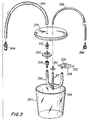
- FIG. 3 is an exploded view of an embodiment of a suction trap 300 for handling fluids and solid matter drawn into vacuum tube 134 (shown in FIG. 1) and for removing contaminants from the air in vacuum tube 134 to prevent contamination of the vacuum pump.
- Intake tube 302 and outflow tube 304 comprise conventional silicone rubber tubing.
- Intake tube 302 and outflow tube 304 are connected respectively to connector 142 of vacuum tube 134 via conventional quick-disconnect connector 306 and to the air intake of the vacuum pump via pneumatic connector 308.
- Connectors 306 and 308 facilitate simple connection of suction trap 300, which also can be quickly disconnected for easy disposal.
- Intake tube 302 and outflow tube 304 are connected respectively via connectors 310 and 312 to plastic lid 314 of suction trap 300, which forms an airtight seal with and is securely attached to plastic jar 316.
- a length of silicone tubing 317 is attached to connector 310 for passing air and fluids into jar 316 from intake tube 302.
- Filters 318 and 320 respectively prevent fluid and solid matter from being drawn into outflow tube 304, and remove contaminants from the air that is drawn into outflow tube 304.
- Filter 318 comprises a hydrophobic filter surface, which is formed into a sealed pouch 322 and which encases a sponge 324. Sponge 324 causes filter 318 to float on any fluid collected in jar 316, and thereby prevents the hydrophobic filter surface from being entirely submerged in fluid, which would block the flow of air into outflow tube 304.
- Filter 318 is connected to a length of flexible silicone rubber tubing 326 which permits filter 318 to move freely about jar 316.
- Filter 320 which is connected to tubing 326 by connector 328, and to outflow tube 304 by connector 312, comprises a hydrophobic bacterial filter having a pore size of approximately 0.2 microns.
- Suction trap 300 is capable of removing fluids and air-borne contaminants from the air drawn into jar 316 independent of the orientation of jar 316. This permits suction trap 300 to be moved with the patient (the mother), and to be attached, for example, to the leg of the patient.
- the suction trap is disposable, and can be made opaque if desired for aesthetic purposes.
- the suction trap typically will be required to handle approximately 5-10cc of fluid, and therefore can be made sufficiently small to be comfortably carried by the patient.
- suction trap 300 can be replaced by one or more disposable in-line filters, including an absorbent medium for absorbing fluids in vacuum tube 134 and a bacterial filter for removing air-borne contaminants.
- Suction trap 300 or the in-line filters may be housed in a disposable cassette for mounting in a module connected to the vacuum pump and the intrauterine probe.
- the cassette may further include a rubber diaphragm which is punctured by a needle in the module to make a vacuum connection to the filters housed in the cassette.
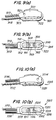
- FIGS. 4(a)-(b) show an embodiment of an insertion tool 400 that can be used to safely and reliably position probe 100.
- Insertion tool 400 comprises a shaped plastic member having a curved body portion 402 and a forked end portion 404.
- Body portion 402 may be shaped in a single, curve (as shown) or double curve as desired to facilitate insertion of the probe into the uterus.
- the forks 406 and 408 of end portion 404 are relatively flexible from side to side, and are relatively inflexible in directions perpendicular to the plane of insertion tool 400. Forks 406 and 408 are inserted into receptables 124 and 126 of probe 100 (shown in FIG.
- Insertion tool 400 permits probe 100 to be placed between the fetus and the uterine wall at a distance from the vaginal canal, thereby allowing the probe to be attached to a particular portion of the fetus such as the head, which may be turned away from the vaginal canal.
- the insertion tool may have a spatula-shaped end portion for insertion into a corresponding sleeve in the probe. The spatula end portion is useful if less flexibility from side to side than that possessed by the forked end portion is desired.
- FIG . 5 illustrates a second embodiment of the intrauterine probe of the present invention having different outer body structures for achieving flexibility.
- Fig. 5 shows an intrauterine probe 500 having first and second segmented sections 502 and 504 defined by silicone rubber outer body 506.
- First segmented section 502 houses fetal pulse oximetry optics 508, to which electrical connections are made by electrical cable 510.
- Second segmented section 504 forms a cavity 512 covered by open cell silicone foam filter pad 514. Cavity 512 is connected to double-lumen vacuum and vent tubes 516.
- first and second segmented sections 502 and 504 When probe 500 is in place and air is suctioned from cavity 512, a vacuum is created between the fetal tissue surface and first and second segmented sections 502 and 504 which causes the outer body 506 to bend at juncture 518 of first and second segmented sections 502 and 504 as necessary to conform the curvature of gasket 520 to that of the fetal tissue surface and to form a seal with the fetal tissue surface.
- outer body 506 may be divided into additional segmented sections to provide greater flexibility.
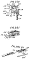
- FIGS. 7-11 fourth, fifth, sixth, seventh, and eighth embodiments of an intrauterine probe having different arrangements of sensing devices are illustrated.
- FIGS. 7(a)-(b) shows top and bottom views of an intrauterine probe 700 in which optical barrier 702 also serves as a fetal ECG electrode.
- the electrode is made from an electrically conductive material such as silver, stainless steel or conductive silicone which, with the probe in place, contacts the fetal tissue.
- the electrode is mounted between light sources 704 and photodetector 706 to serve the dual purpose of preventing light from shunting between the light sources and the photodetector, and of providing a conductive surface useful as an ECG sensor.
- the electrode is mounted on and electrically connected to substrate 707, on which the light sources and photodetector are mounted, using conductive adhesive or soldering.
- An electrical signal from the ECG electrode is provided to EGG monitoring equipment by electrical cable 708, which includes a wire electrically connected to the EGG electrode via the substrate and also provides electrical connections for light sources 704 and photodetector 706.
- Probe 700 also includes a silicone rubber outer body 710 having a gasket 712 for creating a seal with the fetal tissue surface when a vacuum is created in outer body 710.
- Double-lumen vacuum and vent tube 714 provide pneumatic connections between the probe and a vacuum source (not shown).
- Open cell silicone foam filter pads 716 and 718 are provided to diffuse the flow of air and fluid in the probe, and to prevent fetal tissue from being drawn into and damaged by the vacuum cavity of the probe.
- Probe 700 further includes a maternal ECG button 720 mounted flush with rear surface 721 of probe 700.
- Button 720 comprises an electrically conductive material such as silver or stainless steel, and is connected to a wire in electrical cable 708 for providing an ECG signal to ECG monitoring equipment.
- button 720 is in direct electrical contact with the uterine wall or with biological fluids in the uterus which convey to button 720 an electrical signal representative, at least in part, of maternal and fetal electrical heart activity.
- Button 720 may have any desired shape and be placed in any location on rear surface 721 useful for establishing electrical contact with the uterine wall or the biological fluid in the uterus.
- the signal detected by button 720 can be used, in conjunction with the signal detected by ECG sensor 702 in contact with the fetal tissue surface, to determine fetal electrical heart activity.
- gasket 712 serves the dual purposes of positively attaching the probe to the fetal tissue surface (without disfigurement and despite the presence of hair on the fetus, as discussed above in connection with probe 100), and of electrically insulating fetal ECG electrode 702 from maternal ECG electrode 720 to attenuate any undesirable interference or cross-talk between the two ECG electrodes caused by conductive biological fluids in the uterus.
- Gasket 712 preferably provides a seal that prevents biological fluids from forming a continuous electrical conduction path between the fetal and maternal ECG electrodes after the probe is in place.
- a gel-like or cream-like sealant may be used on the surface of gasket 712 to improve the seal formed by gasket 712 with the fetal tissue surface, or gasket 712 may be provided with an open channel in the surface thereof in which sealant can be disposed and applied to the fetal tissue surface to achieve the same effect.
- FIGS. 8(a)-(b) show top and bottom views of an intrauterine probe 800 having a maternal ECG electrode button 802 mounted flush with rear surface 804 of probe 800, and a fetal ECG electrode 806.
- Fetal ECG electrode 806 comprises a substantially rectangular block of electrically conductive material such as silver, stainless steel or conductive silicone. Electrode 806 has a curved end portion 808 which fits into the inside of curved end portion 810 of outer body 814 of probe 800. Electrode 806 has a circular opening 816 which has optically reflective walls, and which is aligned directly above light sources 818.
- Circular opening 816 is filled with a clear encapsulant such as clear silicone rubber to reduce the possibility that the fetal tissue surface may be damaged by the hard edges of the circular opening, and to keep the circular opening free of material that might reduce the intensity of the light reaching the fetal tissue surface.
- the outer edges of electrode 806, as well as those of other metallic components of the intrauterine probes described in connection with alternative embodiments of the present invention, may be rounded to reduce the possibility of tissue damage.
- Electrode 806 is mounted on substrate 812 on which light sources 818 and photodetector 820 are mounted, and is electrically connected to the substrate by conductive epoxy. The electrode is connected via the substrate to a wire of electrical cable 809, which provides an electrical signal from the electrode to ECG monitoring equipment.
- electrode 806 contacts the fetal tissue surface and serves the multiple purposes of preventing light from shunting between light sources 818 and photodetector 820, of focusing and directing light from light sources 818 toward the fetal tissue surface to increase the intensity of light passing through the fetal tissue, and of providing an electrically conductive surface suitable for detecting a fetal ECG signal.
- Probe 800 is asymmetric in that ECG electrode 806, light sources 818 and photodetector 820 are located at one end of the probe, and a porous filter pad 824 is located at the other end of the probe. In this manner, the relatively stiff components of probe 800, including the ECG electrode, light sources, photodetector and substrate, are located at one end of the probe, and do not straddle the midsection of the probe, thereby increasing the flexibility of the probe at its midsection to provide a good seal between gasket 826 and the fetal tissue surface.
- FIGS. 9(a)-(b) show top and bottom views of an intrauterine probe 900 in which individual reflectors 902 and 904 are mounted over light sources 906 and photodetector 908, respectively, to serve the multiple purposes of focusing and directing light from light sources 906 toward the fetal tissue surface, of minimizing the amount of shunted light detected by photodetector 908, and of focusing and directing light from the fetal tissue surface toward photodetector 908.
- Reflectors 902 and 904 comprise metal parts having thin, circular walls with reflective inner surfaces, preferably shaped in the form of a cylinder or a paraboloid centered respectively on light sources 906 and photodetector 908.
- Reflectors 902 and 904 are mounted on substrate 909 on which light sources 906 and photodetector 908 are mounted, and are filled with and encased in clear, soft silicone rubber 910 which minimizes the possibility of damage to the fetal tissue surface caused by the hard edges of the reflectors.
- Open cell silicone foam filter pads 912 and 914 are attached to the probe to cover the remaining exposed portions of the cavity (not shown) of the probe. Double-lumen vacuum and vent tubes 913 provide pneumatic connections between the probe and a vacuum source (not shown).
- a fetal ECG electrode 916 comprising an electrically conductive material such as silver or stainless steel is attached to the surface of filter pad 914, such as by forcing the electrode through a hole in the filter pad, to provide a spring-loaded electrically-conductive surface for contacting the fetal tissue surface.
- a wire is connected to the back surface of electrode 916 and passed through filter pad 914 to electrical cable 918 for making electrical connection to electrode 916.
- electrode 916 When probe 900 is in place, electrode 916 is urged against the fetal tissue surface by the spring action of filter pad 914 to form a firm electrical contact useful for sensing the fetal ECG signal.
- This spring action also can be achieved if desired by using other types of conventional springs such as a coil spring or a cantilever spring.
- the fetal ECG electrode 916 is electrically decoupled from the reflectors. Since the reflectors are not performing the function of an electrical contact, they can be formed from non-conductive materials softer than metal.
- a block of silicone rubber can be used to form the reflectors.
- the body of the block is made of black-colored silicone rubber to act as a light barrier between the light sources and photodetector, and includes two circular openings corresponding, respectively, to the light sources and the photodetector.
- the openings have a coating of reflective white-colored silicone rubber on their walls and are positioned to form cylindrical or parabolic reflectors over the light sources and photodetector.
- the openings can be filled with a clear encapsulant to reduce the possibility of interference caused by fluid or tissue that may reduce the intensity of light transmitted from the light sources through the fetal tissue surface to the photodetectors.
- a clear encapsulant to reduce the possibility of interference caused by fluid or tissue that may reduce the intensity of light transmitted from the light sources through the fetal tissue surface to the photodetectors.
- separate flexible reflectors comprising layers of white and black-colored silicone rubber, or silicone rubber coated with a layer of reflective metallic material, also may be used.
- Probe 900 also includes a maternal ECG electrode 920 mounted flush with rear surface 921 of outer body 923 for providing a maternal ECG signal, and for providing a fetal ECG signal in conjunction with fetal ECG electrode 916.
- Gasket 922 serves the dual purposes of attaching the probe to the fetal surface, and of electrically insulating fetal ECG electrode 916 from maternal ECG electrode 920.
- a sealant also may be used as described above to improve the sealing quality of gasket 922.
- FIGS. 10(a)-(b) show top and bottom views of an intrauterine sensor 1000 in which an electrically conductive block 1002, formed from metal or metal-impregnated silicone, serves the multiple purposes of focusing and directing light from light sources 1004 toward the fetal tissue surface, of minimizing the amount of shunted light detected by photodetector 1006, of focusing and directing light from the fetal tissue surface toward photodetector 1006, and of providing an electrically conductive surface suitable for a fetal ECG sensor.
- an electrically conductive block 1002 formed from metal or metal-impregnated silicone
- Block 1002 is mounted on the substrate on which light sources 1004 and photodetector 1006 are mounted, and is aligned such that openings 1008 and 1010 are positioned directly over light sources 1004 and photodetector 1006, respectively, to provide reflective surfaces for coupling light between blood-perfused fetal tissue and the probe more efficiently.
- Electrical cable 1012 includes wires for providing separate electrical connection to light sources 1004, photodetector 1006, fetal ECG electrode 1002, and maternal ECG electrode 1014.
- double-lumen vacuum and vent tubes 1016 provide pneumatic connections to the cavity (not shown) of probe 1000, the exposed portions of which are covered by open cell silicone foam filter pads 1018 and 1019 attached to probe 1000, and gasket 1020 operates to form a seal with the fetal surface.
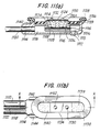
- FIGS. 11(a)-(b) show top and cross-sectional side views of a preferred embodiment of an intrauterine probe 1100.
- probe 1100 includes light sources 1102 and photodetector 1104 mounted on and electrically connected to substrate 1106, which in turn is supported by inner boot 1108 and has electrical leads (not shown) for connection to wires (also not shown) in electrical cable 1110 for connection to an external pulse oximeter.
- a maternal ECG electrode 1112 is mounted through a hole in the rear surface of outer body 1114, and is electrically connected at its inner surface 1113 to a wire (not shown) in electrical cable 1110.
- Outer body 1114 includes cavity 1116 to which vacuum pressure is applied by a vacuum pump (not shown) through vacuum tube 1118 which is connected to cavity 1116 through conduit 1120. Outer body 1114 also includes separate parallel conduits (not shown) for receiving vent tube 1122 and electrical cable 1110.
- Light sources 1102 and photodetector 1104 are separated by an opaque barrier block 1124 mounted on substrate 1106.
- Barrier block 1124 comprises a conductive material such as metal or impregnated silicone rubber, and includes two circular openings 1126 and 1128 positioned respectively above light sources 1102 and photodetector 1104 for transmitting light between the fetal tissue surface and the optical devices. Openings 1126 and 1128 have reflective walls for focusing the transmitted light. Preferably, opening 1126 slopes inwardly at the bottom to reflect transversely emitted light. Openings 1126 and 1128 are filled with a clear encapsulant such as epoxy or silicone rubber on top of which are located protrusions 1130 and 1132 and conductive mask 1134.
- a clear encapsulant such as epoxy or silicone rubber
- Protrusions 1130 and 1132 have smoothly rounded upper surfaces, and are made from a hard, light-transmissive material such as clear epoxy, although softer clear materials such as silicone rubber can also be used. Protrusions 1130 and 1132 may be formed as part of the encapsulant filling openings 1126 and 1128, or as separate pieces mounted on top of the encapsulant. Protrusions 1130 and 1132 extend through openings in conductive mask 1134 above rim 1136 of outer body 1114 to which gasket 1138 is attached.
- Protrusions 1130 and 1132 serve the purpose of imposing a dimpled contour on the fetal tissue surface.
- the dimpling effect facilitates coupling light to and from blood-perfused portions of the fetal tissue, and prevents light from being shunted directly from light sources 1102 to photodetector 1104 as a result of any mismatch between the curvature of the fetal tissue surface and the probe surface.
- the perfusion of blood in the fetal tissue between the optical devices can be increased by making an area of the surface of barrier block 1124 concave, such that blood-perfused fetal tissue fills the concave area between the optical devices when the probe is secured to the fetal tissue surface.
- Gasket 1138 has a substantially rectangular cross-section and is supported by rim 1136 such that the top surface of the gasket is elevated at its outer edge as shown in FIG. 11(a).
- Gasket 1138 may have other cross-sectional shapes (e.g., triangular, with the outer edge being the thicker edge), although it may be more difficult to manufacture compliant gaskets in such shapes.
- Conductive mask 1134 is electrically connected to barrier block 1124 to form a fetal ECG electrode.
- conductive mask comprises a thin sheet of soft metal the edges of which are curved downward as shown to eliminate sharp edges.
- Barrier block 1124 is connected to a wire (not shown) of electrical cable 1110 for connecting the fetal ECG electrode to an external ECG monitor. Gaps between the fetal ECG electrode and the inner walls of outer body 1114 are covered with open-cell foam pads 1139 and 1140 to provide a filtered vacuum port between cavity 1116 and the fetal tissue surface.
- probe 1100 is shown without curvature, it is to be understood that this is for purposes of illustration only.
- outer body 1114 of probe 1100 is molded or bent (as by insertion of a curved wire in receptacles 1142 and 1144) to provide a concave curvature closely approximating the curvature of the desired point of placement on the fetus (e.g., the head).
- probe 1100 can be modified to increase the amount of light reaching detector 1104 by eliminating protrusion 1132 and increasing the diameter of the opening in mask 1134 above the photodetector.
- the amount of light transmitted between the optical devices and the blood-perfused fetal tissue also can be increased by bringing the optical devices closer to the fetal tissue surface. This can be achieved, for example, by mounting the optical devices on a thick substrate and by reducing the thickness of barrier block 1124.
- the diameter of opening 1128 in barrier block 1124 can be made equal to the diameter of protrusion 1130, such that conductive mask 1134 may be eliminated.
- the above-described probe embodiments may include additional sensing devices such as a thermistor and a heat flux sensor adapted to provide electrical signals to external monitoring equipment (not shown) for measuring the temperature of and heat flux at a portion of the fetal tissue surface or the maternal tissue fluids, in accordance with temperature and heat flux measuring techniques.
- a second pair of light sources 906 and photodetectors 908 may be provided on the rear surface of the probe for measuring blood flow characteristics of the uterine tissue, including but not limited to blood oxygen saturation of hemoglobin in arterial blood, volume of individual blood pulsations supplying the tissue, and the rate of blood pulsations corresponding to each maternal heart beat.
- the maternal light source-photodetector pair would preferably operate at different wavelengths than the fetal light source-photodetector pair.
Abstract
Description
- The present invention relates to a non-invasive pulse oximetry intrauterine sensor.
- Pulse oximetry is typically used to measure various blood flow characteristics including, but not limited to, the blood-oxygen saturation of hemoglobin in arterial blood, the volume of individual blood pulsations supplying the tissue, and the rate of blood pulsations corresponding to each heartbeat of a patient. Measurement of these characteristics has been accomplished by use of a non-invasive sensor which passes light through a portion of the patient's tissue where blood perfuses the tissue, and photoelectrically senses the absorption of light in such tissue. The amount of light absorbed is then used to calculate the amount of blood constituent being measured.
- The light passed through the tissue is selected to be of one or more wavelengths that are absorbed by the blood in an amount representative of the amount of the blood constituent present in the blood. The amount of transmitted light passed through the tissue will vary in accordance with the changing amount of blood constituent in the tissue and the related light absorption. For measuring blood oxygen level, such sensors have been provided with two sets of light sources and photodetectors that are adapted to operate at different wavelengths, in accordance with known techniques for measuring blood oxygen saturation.
- Known non-invasive sensors include devices that are secured to a portion of the body, such as a finger, ear or the scalp. In animals and humans, the tissue of these body portions is perfused with blood and the tissue surface is readily accessible to the sensor.
- It is desirable that photoelectric pulse oximetry also be useful for monitoring the blood flow characteristics and constituents of a fetus. For example, monitoring fetal oxygen levels provides an effective way to detect and provide indications for treating hypoxia in the fetus during labor. However, known sensors adapted for use on infants or adults are not suited for intrauterine placement.
- The environment in which the non-invasive intrauterine sensor must operate is fluid-filled (e.g., by amniotic fluid) and is only accessible through the restricted opening of the cervix. Visual inspection of the fetus and the sensor is likewise restricted. Moreover, the operating environment presents certain variants that interfere with detection of the fetal blood flow characteristics using known pulse oximetry techniques. For example, the presence of the waxy vernix caseosa, hair, mucus, blood and dead tissue cells on top of the fetal tissue surface against which the sensor is to be positioned create a problem in establishing contact between the optical components of the sensor and the surface of blood-perfused tissue. Detection of fetal blood flow characteristics by pulse oximetry is particularly complicated by the relatively low perfusion and low oxygen saturation of blood in fetal tissue. These environmental factors prevent known sensors from providing reliable information needed to calculate fetal blood characteristics.
- It is known that positive attachment of a sensor to the tissue surface improves the quality of the photoelectric signal provided by the sensor. Positive attachment to a human's tissue may be obtained by vacuum, adhesives, tapes or devices such as clothespin-type clips. However, fetal tissue is relatively moist and there is limited access to the tissue surface. Consequently, conventional adhesives or tapes or clips are not adapted for intrauterine use.
- Known techniques involving invasive attachment to fetal tissue, such as by a screw attachment penetrating the tissue, creates a risk that the fetus will suffer an infection or disfigurement. Non-invasive attachment, such as by vacuum, may also result in disfigurement if the sensor includes sharp surfaces that press into the fetal tissue surface, or if the sensor is attached to the fetal tissue surface with excessive force (e.g., heavy vacuum suction).
- Moreover, the intrauterine probe sensor must be safely and reliably deliverable to the point of contact with the fetus. It is desirable that intrauterine fetal monitoring be available early in labor, for example, to detect and treat hypoxia in the fetus during labor. Contact with the fetus can be made after natural rupture of the amniotic membrane by manually inserting a probe sensor into the uterus from the vagina, but access to the fetus through the vaginal canal is restricted by the cervix, which may be only slightly dilated to one or two centimetres when the membrane ruptures. Thus there is need for a fetal probe sensor that can be delivered to the fetus through a slightly dilated cervix, and a delivery system for doing so safely and reliably.
- European Patent Application No. 84110306.2 (EP-A-135 840) relates to an intravaginal and intrauterine sensor for transillumination of a blood-perfused portion of fetal tissue. The sensor comprises a light source and a photosensor housed within shielding. The sensor may be mounted onto a paddle shaped probe or may include an opaque cup for disposition on a protruding portion of the foetus. German Gebrauchemuster No. G8431502.4 relates to a suction electrode having a filter element disposed therein for diffusing the air as it is withdrawn from within the electrode.
- The present invention is directed to measurement of the fetal blood flow characteristics using a probe sensor adapted for intrauterine placement. The sensor can be adapted to operate in accordance with the photoelectric pulse oximetry measuring techniques described above as well as to accomplish other measurement techniques for monitoring the well-being of the fetus. For example, it is well known that electrical heart activity corresponding to the heartbeat can be monitored externally and characterised by the electrocardiogram ("ECG") waveform. In a preferred embodiment of the present invention the ECG waveform of the fetus can be measured by providing the probe sensor with an ECG electrode which is in electrical contact with the fetus when the probe sensor is in place in the uterus. Also, maternal blood flow characteristics and the maternal ECG waveform may be measured by providing the probe sensor with one or more light sources, one or more photoelectric detectors and an ECG electrode directed toward the uterine wall.
- In one embodiment of the present invention the intrauterine probe sensor may include a thermistor to measure the temperature of the fetus, and a heat flux sensor to provide an indication of the adequacy of fetal tissue perfusion.
- Preferably non-invasive positive attachment can be accomplished without disfigurement of the fetus by using a deformable probe sensor which is positively attached by creating a partial vacuum in a cavity formed in the probe to cause the probe to conform to the tissue surface of the fetus and to form a gasket-type seal with the fetal tissue surface.
- One aspect of the present invention provides a sensor for insertion into a uterus arranged to measure fetal blood flow characteristics, comprising a deformable outer body having at least one cavity open to the exterior of the outer body, a light source secured in the cavity, a light detector secured in the cavity, and means for connecting the cavity to a vacuum source to create a partial vacuum in the cavity at a predetermined time characterised in that the sensor further incudes at least one filter pad associated with the cavity for diffusing air as it is drawn from within the cavity into the means connecting the cavity to the vacuum source and for preventing fetal tissue from being drawn into cavity and from being damaged and in that the outer body further includes a peripheral contacting surface associated with an open end of the cavity and having a compliancy greater than that of fetal tissue, the peripheral contacting surface being constructed and arranged to operate as a gasket to form a seal with the fetal tissue surface when the outer body is in place and a partial vacuum is created in the cavity.
- The sensor preferably has a narrow, oblong body that permits it to be inserted through a slightly dilated cervix. The body of the sensor preferably includes one or more hollow cavities and is preferably made of a flexible, inert biomaterial such as silicone rubber.
- A cream or gel sealant may be applied to the peripheral contacting surface to improve the seal between the gasket and the fetal tissue surface.
- The partial vacuum may be created in the interior cavity of the sensor by connecting the sensor cavity to a pump in a sump configuration. A vent tube preferably connects the interior cavity of the sensor to the open atmosphere, creating a constant flow of air through the sensor to prevent clogging of the vacuum line and to control the vacuum pressure in the sensor. Humid air or a saline solution also may be pumped through the sensor to prevent the fetal tissue surface from becoming dry.
- The pump can be protected against contamination by a suction trap which contains filtering stages comprising hydrophobic filter elements to prevent passage to the pump of any fluid or airborne contaminants that have been evacuated from the probe, and which operates independently of the orientation of the suction trap. Alternatively, the suction trap may be replaced by filter elements in the vacuum line including an absorbent medium for absorbing fluid and a bacterial filter for removing contaminants from the air. In another alternative embodiment, the suction trap or serial in-line elements may be housed in a disposable cassette.
- The body preferably includes a structure located on one or more interior cavities for supporting the light source and light detector, at least one of the cavities being connectable to a vacuum source. In a preferred embodiment, the light source, which may include two or more light emitting diodes ("LEDs") for generating light at a plurality of selected wavelengths, and the light detector which may comprise a photoelectric detector responsive to the wavelengths of a light generated by the LEDs, are mounted on a flat substrate having electrical leads for connection to electrical cables extending outside of the uterus. The substrate can be lengthened, if desired, to permit the electrical leads to extend out of the uterus when the sensor is in place, such that the connection between the leads and the electrical cable can be accomplished outside of the uterus.
- The sensor is preferably configured to assure that an adequate amount of light passes from the light sources through the blood-perfused fetal tissue to the photodetector to provide a signal for measuring the fetal blood flow characteristics. An optical barrier may be provided between the LEDs and the photoelectric detector to prevent light from passing from the LEDs directly to the photoelectric detector without travelling through the fetal tissue. In a preferred embodiment, a reflective material surrounding the LEDs and shaped for example as a cylindrical or parabolic reflector is provided to direct the light generated by the LEDs into the fetal tissue beneath the probe. A second reflector surrounding the photoelectric detector may also be provided to direct light from the fetal tissue to the photoelectric detector and to prevent the detector from sensing shunted light. The optical barrier or either reflector may be formed of a conductive material and may operate as an ECG electrode. Alternatively, a separate ECG electrode, which is spring-loaded to provide good electrical contact with the fetal tissue, may be provided.
- The devices housed in the sensor are preferably mounted in a manner which protects the sensor from the intrauterine environment, protects the fetal tissue surface from the sensor and imposes a contour on the fetal tissue surface in the vicinity of the optical devices to improve the consistency of measurements made by the sensor. For example, the sensor may be provided with clear windows to cover and protect the optical devices. In a preferred embodiment, one or both of the windows have curved surface portions which, when brought into a contact with the fetal tissue surface, cause the tissue surface to dimple around the window. The dimpling effect thus created helps to prevent light from being shunted between the light source and photodetector devices and improves the coupling between the optical devices and blood-perfused fetal tissue. The perfusion of blood in the fetal tissue between the light source and the photodetector can be increased by creating a concave area in the surface of the sensor between the optical devices.
- The sensor may further be provided with optics and an ECG electrode for measuring maternal blood flow characteristics. The maternal ECG electrode may for example comprise a metallic button mounted on the surface of the sensor body. In accordance with fetal ECG measurement techniques, the differential voltage between the fetal ECG electrode and the maternal ECG electrode can be used to determine the fetal ECG. In addition, other sensors such as a thermistor or a heat flux sensor or both may be provided, for example, to measure the temperature of the fetus and the perfusion of blood in the fetal tissue.
- The sensor body preferably also includes one or more receptacles for receiving an end portion of the insertion tool. The insertion tool can be a narrow, shaped member preferably comprising a flexible material formed into a single or double curve. The insertion tool preferably has a shaped end portion for insertion into one or more corresponding receptacles in the body of the probe to safely and reliably deliver the probe.
- The sensor may comprise a probe adapted for insertion into a uterus, the probe comprising a deformable outer body having the cavity therein, the peripheral contacting surface comprising deformable seal means mounted to an outer surface of the body, the light source and light detector being adapted to generate a signal for use in measuring at least one fetal blood flow characteristic by pulse oximetry and means being provided for connecting the light source to an external source of power and for connecting the light detector to external components adapted to receive and monitor the signals generated thereby.
- The probe may further include first electrode means for detecting a fetal body function and for generating a signal responsive to the detected body function, the first electrode means being mounted in the probe and adapted to contact the fetal tissue surface when the probe is in place.
- The probe preferably further includes a light barrier element between the light source and the light detector.
- The probe may further include a first reflector mounted adjacent one of the light source and the light detector.
- The first reflector means preferably reflects light between the light source and the fetal tissue surface and the probe preferably further includes a second reflector means for reflecting light between the light detector and the fetal tissue surface, the first and second reflector means being formed by a single electrically conductive element.
- Second electrode means may be associated with the probe for detecting a body function and for generating a signal responsive to the detected body function; and means may be provided for connecting the second electrode means to external components adapted to receive and monitor the signal generated by the second electrode means.
- The deformable seal is preferably a gasket having sufficient compressibility to seal against the ingress into the cavity of biological fluids which can form an electrical path between the first and second electrode means.
- Temperature sensing means may be mounted in the probe for sensing the temperature of a portion of the fetal tissue surface and for generating a signal responsive to the sensed temperature; and means may be provided for connecting the temperature sensing means to external components adapted to receive and monitor the signal generated by the temperature sensing means.
- Means is preferably mounted in the probe for sensing heat flux in at least a portion of one of the maternal biological fluid and the fetal tissue surface and for generating a signal responsive to the sensed heat flux, and means is preferably provided for connecting the heat flux sensing means to external components adapted to receive and monitor the signal generated by the heat flux sensing means.
- It is therefore an object of the present invention to provide an improved intrauterine probe sensor which can be positively attached to fetal tissue without risk of disfigurement of the fetus.
- It is another object of the present invention to provide an improved fetal pulse oximetry sensor that efficiently couples light signals between the sensor and blood-perfused fetal tissue.
- It is an additional object of the present invention to provide an improved fetal pulse oximetry sensor in which the sensor geometry imposes a contour on the fetal tissue surface in the vicinity of the optical devices of the sensor to improve the consistency of the oximetry measurements.
- The above and other objects and advantages of the present invention will be apparent upon consideration of the following detailed description, taken in conjunction with the accompanying drawings, in which like reference characters refer to like parts throughout, and in which:
- FIG. 1 is a partially-exploded elevated perspective view of a first embodiment of the probe sensor of the present invention.
- FIGS. 2(a)-2(c) are perspective views of the optics, electrical cable, substrate and inner boot of the probe of FIG. 1.
- FIG. 3 is an exploded perspective view of an embodiment of a suction trap for use with the probe sensor of Fig. 1.
- FIGS 4(a)-(b) are perspective views of an embodiment of an insertion tool of the present invention.
- FIG. 5 is a cross-sectional side view of a second embodiment of the probe sensor of the present invention having a segmented outer body.
- FIGS. 7(a)-(b) are top plan and bottom perspective views of a fourth embodiment of the probe sensor of the present invention including a fetal ECG electrode which also serves as an optical barrier.
- FIGS. 8(a)-(b) are top plan and bottom perspective views of a fifth embodiment of the probe sensor of the present invention including a fetal ECG electrode which also serves as an optical barrier and a reflector.
- FIGS. 9(a)-(b) are top plan and bottom perspective views of a sixth embodiment of the probe of the present invention including two reflectors for increasing the efficiency of light transmission, and a spring-loaded fetal ECG electrode.
- FIGS. 10(a)-(b) are top plan and bottom perspective views of a seventh embodiment of the probe of the present invention.
- FIGS. 11(a)-(b) are top plan and cross-sectional side views of a preferred eighth embodiment of the probe of the present invention having protrusions over the light source and photodetector regions.
- Various embodiments of the probe of the present invention are described herein and are shown in the figures. The preferred embodiment, which incorporates many features described in connection with other embodiments, is shown in FIGS. 11(a)-(b) and is described last.
- Referring to FIG. 1,
probe 100 comprisesouter body 102 andinner boot 104 which house the photoelectric pulse oximetry measuring devices of the sensor.Inner boot 104 is shown apart fromouter body 102 in FIG. 2(c).Outer body 102 is molded in a substantially rectangular, oblong shape, and has a hollowinterior cavity 106 in whichinner boot 104 is positioned. -
Outer body 102 andinner boot 104 are molded or cast from a flexible, inert biocompatible material such as silicone rubber. The outer body is preferably made opaque, such as by addition of a coloring agent (e.g., white titanium dioxide).Inner boot 104, which encases the pulse oximetry optics, is preferably colored black to provide optical isolation between the probe's pulse oximetry light sources and photoelectric detector, which are described in greater detail below. - The height (or thickness) and width of
outer body 102 are preferably sized to fitprobe 100 through a cervix which is approximately 1-2 centimeters dilated, although a probe having larger dimensions can of course be used after the cervix has become further dilated. Accordingly, the probe of the present invention has a slender appearance. A typical length ofprobe 100 is approximately 3.81 cm (1.5 inches), although its length can be extended as necessary to encapsulate the electrical components of the probe (including electrical leads connected to those components) to isolate them from biological fluids. A typical height ofprobe 100 is 1.14 cm (0.45 inches), and a typical width is 1.27 cm (0.5 inches). -
Outer body 102 has a pair ofnarrow channels legs U-shaped wire 116.Wire 116 forcesouter body 102 to bend along its length in a concave curve defined by the strength of the wire, the curvature oflegs probe 100. Whenlegs wire 116 are inserted fully inchannels curved neck 118 ofwire 116 abuts the underside oflip 120 ofouter body 102, and is fixed in place by a flexible biocompatible adhesive.Wire 116 is formed from stainless steel, and is heat treated to harden the steel and to help maintain its form. Withwire 116 in place,probe 100 has a concave curvature that preferably conforms approximately to the typical curvature of a fetal head. A typical value for the diameter of the fetal head is 10.16 cm (4 inches). - The approximate conformance of the pre-set concave curvature of
probe 100 to the fetal tissue surface allows a partial vacuum to be created incavity 106 whengasket 122 ofprobe 100 is positioned against the fetal tissue surface and a negative pressure (vacuum) is applied tocavity 106. As described further below, the negative pressure causesgasket 122 bonded tolip 120 ofouter body 102 to create a soft-contact seal with the fetal tissue surface. By providing the probe with a pre-set curvature that closely approximates the curvature of the fetal tissue surface, the amount of negative pressure required to deformprobe 100 sufficiently to create the seal betweengasket 122 and the fetal tissue surface is minimized. In this manner the contact between the probe and the fetal tissue surface is softened, and the risk of potential disfigurement caused by the positive attachment of the probe to the fetal tissue surface is minimized. - In an alternative embodiment of
probe 100,outer body 102 may be molded with the desired concave curvature to eliminate the need forwire 116 and to increase the flexibility ofouter body 102. -
Outer body 102 further includes a pair oflengthwise receptacles 124 and 126 (not shown) for receiving the end portion of an insertion tool during insertion and placement of the probe in the uterus. Outer body also includes at oneend conduits vacuum tube 134,vent tube 136 andelectrical cable 138.Vacuum tube 134 and venttube 136 are connected respectively viaconduits cavity 106 to provide a partial vacuum in and a flow of air throughcavity 106, and may comprise conventional double-lumen tubing. Hereafter, the partial vacuum created in the probe also is referred to simply as a "vacuum". Theend 139 ofvacuum tube 134 is connected via a suction trap to a vacuum pump (trap and pump not shown in FIG. 1). Theend 140 ofvent tube 136 is open to the atmosphere, such that when the pump is operated it draws air intovent tube 136 from the atmosphere and causes the air to pass throughcavity 106 and into the suction trap viavacuum tube 134. A conventional quick-disconnect (or "Luer")connector 142 is provided for releasably connectingvacuum tube 134 to the suction trap, and aconventional clamp 144 is provided to clampvacuum tube 134 when desired, such as during insertion, positioning and removal ofprobe 100. - Although
vent tube 136 is not necessary to create a vacuum incavity 106, it preventsvacuum tube 134 from becoming clogged by fluids and solid matter drawn intovacuum tube 134 fromcavity 106. - The level of vacuum in
cavity 106 is a function of the length and diameter of vacuum and venttubes cavity 106 is maintained at a level which is sufficient to achieve positive attachment to the fetal surface. Because of the pre-set curvature and flexibility ofouter body 102 and the compliancy of gasket 122 (discussed below), positive attachment can be achieved at a vacuum level which is not so high as to cause disfigurement of the fetal surface from the contact force of the probe. For example, positive attachment has been achieved using a diaphragm pump, set to a pressure within the range of 0.007 - 0.01 Kg/cm2 (75-150 mmHg), with a 1.98 cm (.78 inch) innerdiameter vacuum tube 134 and a 0.05 cm (.020 inch) innerdiameter vent tube 136, both tubes being made of medical implant grade clear silicone and having equal lengths of approximately 91.44 cm (3 feet). - In an alternative embodiment, humid air or a saline solution may be suctioned from a source connected to vent
tube 136, throughcavity 106 and intovacuum tube 134, to maintain a flow of liquid throughcavity 106 for moistening the fetal tissue surface underprobe 100. -
Inner boot 104 supports asubstrate 155 on which two light sources are mounted inlight source region 156, and on which a photodetector is mounted inphotodetector region 158.Regions electrical cable 138 are provided byleads 146 which are electrically attached to the substrate. The exposed wire portions ofleads 146 and the solder joints by which they are attached to the substrate are insulated by encapsulating the ends ofinner boot 104 withsilicone adhesive 148. Adhesive is also used to holdinner boot 104 in place inouter body 102. - The inner walls of
outer body 102 that surroundphotodetector region 158 are coated with a layer of black silicone rubber to prevent shunting. Shunting occurs when light emitted by the light sources reaches the photodetector without first passing through fetal tissue. A flexible, black silicone rubberoptical barrier 152 having a height when mounted approximately equal to theinner rim 154 ofouter body 102 is provided betweenlight source region 156 andphotodetector region 158 to further optically isolate the photodetector from light generated by the light sources which does not pass through the fetal tissue. Theoptical barrier 152 preferably is formed from the same material asouter body 102 andinner boot 104, although other inert biomaterials may be used. -
Light source region 156 andphotodetector region 158 are each encapsulated by a clear layer (not shown) of epoxy or silicone rubber which has a height approximately equal to theinner rim 154 ofouter body 102. These layers fillcavity 106 ofouter body 102 above light source andphotodetector regions - Open cell
silicone foam pads cavity 106 and are bonded with adhesive, respectively, toU-shaped shelves 164 and 166 ofouter body 102. The open-cell silicone foam diffuses the air drawn intocavity 106 from the vicinity of the fetal surface underprobe 100 through numerous small vacuum ports formed by the open cell structure of the silicone foam. In this manner a vacuum is created over a large area without risk that a large piece of solid matter may be drawn into the vacuum line. The pads also prevent fetal tissue from being drawn intocavity 106 and damaged. Although open cell silicone foam pads are preferable because of their flexibility, more rigid materials such as porous plastic may also be used forfilter pads -
Gasket 122 is bonded to theupper surface 167 oflip 120.Lip 120 thins as it extends outwardly and upwardly from the upper portion ofouter body 102.Gasket 122 is shaped as a ring having a substantially flattop surface 168, which is elevated atouter edge 174 bylip 120. Whenprobe 100 is in place and a vacuum is created incavity 106,lip 120 andgasket 122 operate as a cantilever, bending outwardly and flattening to absorb the contact forces betweenprobe 100 and the fetal tissue surface by distributing the forces in directions perpendicular to the fetal tissue surface. - The vacuum causes a seal to form between the fetal tissue surface and
outer edge 174 ofgasket 122. The vacuum created incavity 106 is thus distributed over a large surface area to provide a substantially even attractive force over that area. As a result, a good seal can be obtained even where the fetal surface is covered by hair. As described in greater detail below, a sealant cream or gel may also be used if desired betweengasket 122 and the fetal tissue surface to facilitate the formation of a seal which minimizes the likelihood of a continuous flow of biological fluid intocavity 106. -
Gasket 122 is made from a soft material such as silicone rubber or silicone gel having a compliancy that is greater than that of the fetal tissue, so that when a vacuum is created incavity 106,gasket 122 deforms before the fetal tissue to create a gasket-type seal. The deformation ofgasket 122 allows the formation of a seal over fetal hair, and minimizes the risk that the fetal tissue will be marked or disfigured byprobe 100. - FIGS. 2(a)-2(c) show a typical assembly for mounting the pulse oximetry optics in
probe 100.Substrate 202 comprises a thin layer of flexible insulating material such as polyamide film, one side of which is plated withsolder pads paths wires 146 ofelectrical cable 138, respectively, tolight sources 212 andphotoelectric detector 214. The back ofsubstrate 202 is copper-coated to prevent light from traveling through the substrate to the photodetector and astiffener 224 is attached to the back ofsubstrate 202 to provide support when electrical connections are made between the substrate andlight sources 212 andphotodetector 214.Solder pad 216 and conductingpath 218 may be provided also for making electrical connections to a fetal ECG electrode (not shown in FIGS. 2(a)-2(c)). -
Light sources 212 preferably comprise two or more LEDs, at least one of which has a nominal discrete frequency in the red light range, for example 660 nanometers (nm), and another having a nominal discrete frequency in the infrared range, for example 960 nm, in accordance with pulse oximetry techniques for measurement of oxygen saturation. Additional LEDs of different wavelengths may also be provided if desired to obtain information relating to factors such as tissue density or the average amount of blood in the tissue. To compensate for the relatively low level of oxygen saturation in the fetal blood, which creates a low signal to noise ratio in the red LED signal, it is preferred that a bright (e.g., 1.0 mW) red LED having zero secondary emission be used. -
Photodetector 214 preferably comprises a photoelectric detector having discrete frequency responsivities at the different peak frequencies of the LEDs. The LEDs and the photodetector are mounted tosubstrate 202 using electrically conductive epoxy with additional electrical connections provided by wire bonding. - As described earlier, the light sources and photodetector are encapsulated with
clear epoxy 226. Theflexible substrate 202 is folded as shown in FIG. 2(b) to reduce its dimensions, and is inserted ininner boot 104.Inner boot 104 has twoopenings 227 and 228 which correspond respectively tolight source region 156 andphotodetector region 158.Slot 230 is formed ininner boot 104 betweenopenings 227 and 228 to support optical barrier 152 (shown in FIG. 1). Asilicone tube 232 is attached underneathinner boot 104 as a spacer to maintain an air flow channel betweeninner boot 104 and the rear inner surface ofouter body 102 when a vacuum is created incavity 106. - The mounting of the LEDs and the photodetector onto
substrate 202 herein disclosed can be accomplished through other configurations. For example, a single layer substrate may be provided to reduce the height of the probe. To maintain a suitable surface area for mounting and making electrical connections to the LEDs and the photodetector without increasing the width of theprobe 100, the length of the substrate can be extended in the direction of the longer dimension of theprobe 100, with the solder pads being removed away from the LEDs and the photodetector, such that electrical connections to the substrate are made outside ofouter body 102. This reduces the required height ofouter body 102. The extended substrate and the electrical connections thereto can be electrically isolated from the biological fluids by encapsulation with silicone rubber. If desired, the encapsulated substrate can be extended to permit the electrical connections to be made outside the uterus when the probe is in position. Further, an integrated chip or a thin film construction may be used if desired for mass production of eitherlight sources 212 orphotodetector 214. Alternatively, fiber optic connections betweensubstrate 202 and the external pulse oximeter are also suitable. - The structure and arrangement of
probe 100 is such that when the light sources are operating, the entire probe functions to assure that an adequate level of light is transmitted to a blood-perfused portion of the fetal skin despite the presence of hair, blood, mucus or other substances which tend to decrease the light level. As discussed below in connection with alternative probe embodiments, the transmission efficiency of the probe may be increased by supplementing one or both of the light sources and photodetector with means for imposing a contour on the fetal tissue surface to minimize shunting, maximize coupling through interfering substances and improve blood perfusion of the fetal tissue between the optical devices. - FIG. 3 is an exploded view of an embodiment of a
suction trap 300 for handling fluids and solid matter drawn into vacuum tube 134 (shown in FIG. 1) and for removing contaminants from the air invacuum tube 134 to prevent contamination of the vacuum pump.Intake tube 302 andoutflow tube 304 comprise conventional silicone rubber tubing.Intake tube 302 andoutflow tube 304 are connected respectively toconnector 142 ofvacuum tube 134 via conventional quick-disconnect connector 306 and to the air intake of the vacuum pump viapneumatic connector 308.Connectors suction trap 300, which also can be quickly disconnected for easy disposal.Intake tube 302 andoutflow tube 304 are connected respectively viaconnectors plastic lid 314 ofsuction trap 300, which forms an airtight seal with and is securely attached toplastic jar 316. A length ofsilicone tubing 317 is attached toconnector 310 for passing air and fluids intojar 316 fromintake tube 302. -
Filters outflow tube 304, and remove contaminants from the air that is drawn intooutflow tube 304.Filter 318 comprises a hydrophobic filter surface, which is formed into a sealedpouch 322 and which encases asponge 324.Sponge 324 causes filter 318 to float on any fluid collected injar 316, and thereby prevents the hydrophobic filter surface from being entirely submerged in fluid, which would block the flow of air intooutflow tube 304.Filter 318 is connected to a length of flexiblesilicone rubber tubing 326 which permits filter 318 to move freely aboutjar 316. -
Filter 320, which is connected totubing 326 byconnector 328, and tooutflow tube 304 byconnector 312, comprises a hydrophobic bacterial filter having a pore size of approximately 0.2 microns. -
Suction trap 300 is capable of removing fluids and air-borne contaminants from the air drawn intojar 316 independent of the orientation ofjar 316. This permitssuction trap 300 to be moved with the patient (the mother), and to be attached, for example, to the leg of the patient. The suction trap is disposable, and can be made opaque if desired for aesthetic purposes. The suction trap typically will be required to handle approximately 5-10cc of fluid, and therefore can be made sufficiently small to be comfortably carried by the patient. - Alternatively,
suction trap 300 can be replaced by one or more disposable in-line filters, including an absorbent medium for absorbing fluids invacuum tube 134 and a bacterial filter for removing air-borne contaminants.Suction trap 300 or the in-line filters may be housed in a disposable cassette for mounting in a module connected to the vacuum pump and the intrauterine probe. The cassette may further include a rubber diaphragm which is punctured by a needle in the module to make a vacuum connection to the filters housed in the cassette. - FIGS. 4(a)-(b) show an embodiment of an
insertion tool 400 that can be used to safely and reliably positionprobe 100.Insertion tool 400 comprises a shaped plastic member having acurved body portion 402 and a forkedend portion 404.Body portion 402 may be shaped in a single, curve (as shown) or double curve as desired to facilitate insertion of the probe into the uterus. Theforks end portion 404 are relatively flexible from side to side, and are relatively inflexible in directions perpendicular to the plane ofinsertion tool 400.Forks receptables Insertion tool 400permits probe 100 to be placed between the fetus and the uterine wall at a distance from the vaginal canal, thereby allowing the probe to be attached to a particular portion of the fetus such as the head, which may be turned away from the vaginal canal. In an alternative embodiment, the insertion tool may have a spatula-shaped end portion for insertion into a corresponding sleeve in the probe. The spatula end portion is useful if less flexibility from side to side than that possessed by the forked end portion is desired. - FIG . 5 illustrates a second embodiment of the intrauterine probe of the present invention having different outer body structures for achieving flexibility. Fig. 5 shows an
intrauterine probe 500 having first and secondsegmented sections outer body 506. Firstsegmented section 502 houses fetalpulse oximetry optics 508, to which electrical connections are made byelectrical cable 510. Secondsegmented section 504 forms acavity 512 covered by open cell siliconefoam filter pad 514.Cavity 512 is connected to double-lumen vacuum and venttubes 516. Whenprobe 500 is in place and air is suctioned fromcavity 512, a vacuum is created between the fetal tissue surface and first and secondsegmented sections outer body 506 to bend atjuncture 518 of first and secondsegmented sections gasket 520 to that of the fetal tissue surface and to form a seal with the fetal tissue surface. Although only two segmented sections are illustrated,outer body 506 may be divided into additional segmented sections to provide greater flexibility. - Referring now to FIGS. 7-11, fourth, fifth, sixth, seventh, and eighth embodiments of an intrauterine probe having different arrangements of sensing devices are illustrated.
- FIGS. 7(a)-(b) shows top and bottom views of an
intrauterine probe 700 in whichoptical barrier 702 also serves as a fetal ECG electrode. The electrode is made from an electrically conductive material such as silver, stainless steel or conductive silicone which, with the probe in place, contacts the fetal tissue. The electrode is mounted betweenlight sources 704 andphotodetector 706 to serve the dual purpose of preventing light from shunting between the light sources and the photodetector, and of providing a conductive surface useful as an ECG sensor. The electrode is mounted on and electrically connected tosubstrate 707, on which the light sources and photodetector are mounted, using conductive adhesive or soldering. An electrical signal from the ECG electrode is provided to EGG monitoring equipment byelectrical cable 708, which includes a wire electrically connected to the EGG electrode via the substrate and also provides electrical connections forlight sources 704 andphotodetector 706. - Probe 700 also includes a silicone rubber
outer body 710 having agasket 712 for creating a seal with the fetal tissue surface when a vacuum is created inouter body 710. Double-lumen vacuum and venttube 714 provide pneumatic connections between the probe and a vacuum source (not shown). Open cell siliconefoam filter pads - Probe 700 further includes a
maternal ECG button 720 mounted flush withrear surface 721 ofprobe 700.Button 720 comprises an electrically conductive material such as silver or stainless steel, and is connected to a wire inelectrical cable 708 for providing an ECG signal to ECG monitoring equipment. Whenprobe 700 is in place,button 720 is in direct electrical contact with the uterine wall or with biological fluids in the uterus which convey tobutton 720 an electrical signal representative, at least in part, of maternal and fetal electrical heart activity.Button 720 may have any desired shape and be placed in any location onrear surface 721 useful for establishing electrical contact with the uterine wall or the biological fluid in the uterus. The signal detected bybutton 720 can be used, in conjunction with the signal detected byECG sensor 702 in contact with the fetal tissue surface, to determine fetal electrical heart activity. - Applicants have discovered that the seal provided by
gasket 712 serves the dual purposes of positively attaching the probe to the fetal tissue surface (without disfigurement and despite the presence of hair on the fetus, as discussed above in connection with probe 100), and of electrically insulatingfetal ECG electrode 702 from maternal ECG electrode 720 to attenuate any undesirable interference or cross-talk between the two ECG electrodes caused by conductive biological fluids in the uterus.Gasket 712 preferably provides a seal that prevents biological fluids from forming a continuous electrical conduction path between the fetal and maternal ECG electrodes after the probe is in place. A gel-like or cream-like sealant may be used on the surface ofgasket 712 to improve the seal formed bygasket 712 with the fetal tissue surface, orgasket 712 may be provided with an open channel in the surface thereof in which sealant can be disposed and applied to the fetal tissue surface to achieve the same effect. - FIGS. 8(a)-(b) show top and bottom views of an
intrauterine probe 800 having a maternalECG electrode button 802 mounted flush withrear surface 804 ofprobe 800, and afetal ECG electrode 806.Fetal ECG electrode 806 comprises a substantially rectangular block of electrically conductive material such as silver, stainless steel or conductive silicone.Electrode 806 has acurved end portion 808 which fits into the inside ofcurved end portion 810 ofouter body 814 ofprobe 800.Electrode 806 has acircular opening 816 which has optically reflective walls, and which is aligned directly abovelight sources 818.Circular opening 816 is filled with a clear encapsulant such as clear silicone rubber to reduce the possibility that the fetal tissue surface may be damaged by the hard edges of the circular opening, and to keep the circular opening free of material that might reduce the intensity of the light reaching the fetal tissue surface. The outer edges ofelectrode 806, as well as those of other metallic components of the intrauterine probes described in connection with alternative embodiments of the present invention, may be rounded to reduce the possibility of tissue damage.Electrode 806 is mounted onsubstrate 812 on whichlight sources 818 andphotodetector 820 are mounted, and is electrically connected to the substrate by conductive epoxy. The electrode is connected via the substrate to a wire ofelectrical cable 809, which provides an electrical signal from the electrode to ECG monitoring equipment. - When
probe 800 is in place,electrode 806 contacts the fetal tissue surface and serves the multiple purposes of preventing light from shunting betweenlight sources 818 andphotodetector 820, of focusing and directing light fromlight sources 818 toward the fetal tissue surface to increase the intensity of light passing through the fetal tissue, and of providing an electrically conductive surface suitable for detecting a fetal ECG signal. -
Probe 800 is asymmetric in thatECG electrode 806,light sources 818 andphotodetector 820 are located at one end of the probe, and aporous filter pad 824 is located at the other end of the probe. In this manner, the relatively stiff components ofprobe 800, including the ECG electrode, light sources, photodetector and substrate, are located at one end of the probe, and do not straddle the midsection of the probe, thereby increasing the flexibility of the probe at its midsection to provide a good seal betweengasket 826 and the fetal tissue surface. - FIGS. 9(a)-(b) show top and bottom views of an
intrauterine probe 900 in whichindividual reflectors light sources 906 andphotodetector 908, respectively, to serve the multiple purposes of focusing and directing light fromlight sources 906 toward the fetal tissue surface, of minimizing the amount of shunted light detected byphotodetector 908, and of focusing and directing light from the fetal tissue surface towardphotodetector 908. -
Reflectors light sources 906 andphotodetector 908.Reflectors substrate 909 on whichlight sources 906 andphotodetector 908 are mounted, and are filled with and encased in clear,soft silicone rubber 910 which minimizes the possibility of damage to the fetal tissue surface caused by the hard edges of the reflectors. - Open cell silicone
foam filter pads tubes 913 provide pneumatic connections between the probe and a vacuum source (not shown). Afetal ECG electrode 916 comprising an electrically conductive material such as silver or stainless steel is attached to the surface offilter pad 914, such as by forcing the electrode through a hole in the filter pad, to provide a spring-loaded electrically-conductive surface for contacting the fetal tissue surface. A wire is connected to the back surface ofelectrode 916 and passed throughfilter pad 914 toelectrical cable 918 for making electrical connection toelectrode 916. Whenprobe 900 is in place,electrode 916 is urged against the fetal tissue surface by the spring action offilter pad 914 to form a firm electrical contact useful for sensing the fetal ECG signal. This spring action also can be achieved if desired by using other types of conventional springs such as a coil spring or a cantilever spring. - In
probe 900, thefetal ECG electrode 916 is electrically decoupled from the reflectors. Since the reflectors are not performing the function of an electrical contact, they can be formed from non-conductive materials softer than metal. For example, a block of silicone rubber can be used to form the reflectors. The body of the block is made of black-colored silicone rubber to act as a light barrier between the light sources and photodetector, and includes two circular openings corresponding, respectively, to the light sources and the photodetector. The openings have a coating of reflective white-colored silicone rubber on their walls and are positioned to form cylindrical or parabolic reflectors over the light sources and photodetector. The openings can be filled with a clear encapsulant to reduce the possibility of interference caused by fluid or tissue that may reduce the intensity of light transmitted from the light sources through the fetal tissue surface to the photodetectors. Alternately, separate flexible reflectors comprising layers of white and black-colored silicone rubber, or silicone rubber coated with a layer of reflective metallic material, also may be used. - Probe 900 also includes a
maternal ECG electrode 920 mounted flush withrear surface 921 ofouter body 923 for providing a maternal ECG signal, and for providing a fetal ECG signal in conjunction withfetal ECG electrode 916.Gasket 922 serves the dual purposes of attaching the probe to the fetal surface, and of electrically insulatingfetal ECG electrode 916 frommaternal ECG electrode 920. A sealant also may be used as described above to improve the sealing quality ofgasket 922. - FIGS. 10(a)-(b) show top and bottom views of an
intrauterine sensor 1000 in which an electricallyconductive block 1002, formed from metal or metal-impregnated silicone, serves the multiple purposes of focusing and directing light from light sources 1004 toward the fetal tissue surface, of minimizing the amount of shunted light detected byphotodetector 1006, of focusing and directing light from the fetal tissue surface towardphotodetector 1006, and of providing an electrically conductive surface suitable for a fetal ECG sensor. -
Block 1002 is mounted on the substrate on which light sources 1004 andphotodetector 1006 are mounted, and is aligned such thatopenings photodetector 1006, respectively, to provide reflective surfaces for coupling light between blood-perfused fetal tissue and the probe more efficiently.Electrical cable 1012 includes wires for providing separate electrical connection to light sources 1004,photodetector 1006,fetal ECG electrode 1002, andmaternal ECG electrode 1014. As in previously described embodiments, double-lumen vacuum and venttubes 1016 provide pneumatic connections to the cavity (not shown) ofprobe 1000, the exposed portions of which are covered by open cell siliconefoam filter pads gasket 1020 operates to form a seal with the fetal surface. - FIGS. 11(a)-(b) show top and cross-sectional side views of a preferred embodiment of an intrauterine probe 1100. As in previously described embodiments, probe 1100 includes
light sources 1102 andphotodetector 1104 mounted on and electrically connected tosubstrate 1106, which in turn is supported byinner boot 1108 and has electrical leads (not shown) for connection to wires (also not shown) inelectrical cable 1110 for connection to an external pulse oximeter. Amaternal ECG electrode 1112 is mounted through a hole in the rear surface ofouter body 1114, and is electrically connected at itsinner surface 1113 to a wire (not shown) inelectrical cable 1110.Outer body 1114 includescavity 1116 to which vacuum pressure is applied by a vacuum pump (not shown) throughvacuum tube 1118 which is connected tocavity 1116 throughconduit 1120.Outer body 1114 also includes separate parallel conduits (not shown) for receivingvent tube 1122 andelectrical cable 1110. -
Light sources 1102 andphotodetector 1104 are separated by anopaque barrier block 1124 mounted onsubstrate 1106.Barrier block 1124 comprises a conductive material such as metal or impregnated silicone rubber, and includes twocircular openings light sources 1102 andphotodetector 1104 for transmitting light between the fetal tissue surface and the optical devices.Openings Openings protrusions conductive mask 1134. -
Protrusions Protrusions encapsulant filling openings Protrusions conductive mask 1134 aboverim 1136 ofouter body 1114 to whichgasket 1138 is attached. -
Protrusions light sources 1102 tophotodetector 1104 as a result of any mismatch between the curvature of the fetal tissue surface and the probe surface. The perfusion of blood in the fetal tissue between the optical devices can be increased by making an area of the surface ofbarrier block 1124 concave, such that blood-perfused fetal tissue fills the concave area between the optical devices when the probe is secured to the fetal tissue surface. -
Gasket 1138 has a substantially rectangular cross-section and is supported byrim 1136 such that the top surface of the gasket is elevated at its outer edge as shown in FIG. 11(a).Gasket 1138 may have other cross-sectional shapes (e.g., triangular, with the outer edge being the thicker edge), although it may be more difficult to manufacture compliant gaskets in such shapes. -
Conductive mask 1134 is electrically connected tobarrier block 1124 to form a fetal ECG electrode. Preferably, conductive mask comprises a thin sheet of soft metal the edges of which are curved downward as shown to eliminate sharp edges.Barrier block 1124 is connected to a wire (not shown) ofelectrical cable 1110 for connecting the fetal ECG electrode to an external ECG monitor. Gaps between the fetal ECG electrode and the inner walls ofouter body 1114 are covered with open-cell foam pads cavity 1116 and the fetal tissue surface. - Although probe 1100 is shown without curvature, it is to be understood that this is for purposes of illustration only. As discussed with respect to previously illustrated embodiments,
outer body 1114 of probe 1100 is molded or bent (as by insertion of a curved wire inreceptacles 1142 and 1144) to provide a concave curvature closely approximating the curvature of the desired point of placement on the fetus (e.g., the head). - If desired, probe 1100 can be modified to increase the amount of
light reaching detector 1104 by eliminatingprotrusion 1132 and increasing the diameter of the opening inmask 1134 above the photodetector. The amount of light transmitted between the optical devices and the blood-perfused fetal tissue also can be increased by bringing the optical devices closer to the fetal tissue surface. This can be achieved, for example, by mounting the optical devices on a thick substrate and by reducing the thickness ofbarrier block 1124. Also, for purposes of simplifying the manufacture of probe 1100, the diameter of opening 1128 inbarrier block 1124 can be made equal to the diameter ofprotrusion 1130, such thatconductive mask 1134 may be eliminated. - The above-described probe embodiments may include additional sensing devices such as a thermistor and a heat flux sensor adapted to provide electrical signals to external monitoring equipment (not shown) for measuring the temperature of and heat flux at a portion of the fetal tissue surface or the maternal tissue fluids, in accordance with temperature and heat flux measuring techniques. A second pair of
light sources 906 andphotodetectors 908 may be provided on the rear surface of the probe for measuring blood flow characteristics of the uterine tissue, including but not limited to blood oxygen saturation of hemoglobin in arterial blood, volume of individual blood pulsations supplying the tissue, and the rate of blood pulsations corresponding to each maternal heart beat. In such an arrangement, the maternal light source-photodetector pair would preferably operate at different wavelengths than the fetal light source-photodetector pair.
Claims (17)
- A sensor for insertion into a uterus arranged to measure fetal blood flow characteristics, comprising a deformable outer body (102) having at least one cavity (106) open to the exterior of the outer body, a light source (156) secured in the cavity (106), a light detector (158) secured in the cavity (106), and means (134) for connecting the cavity (106) to a vacuum source to create a partial vacuum in the cavity (106) at a predetermined time; characterized in that the sensor further includes at least one filter pad (160,162) associated with the cavity (106) for diffusing air as it is drawn from within the cavity (106) into the means (134) connecting the cavity (106) to the vacuum source and for preventing fetal tissue from being drawn into the cavity (106) and from being damaged and in that the outer body (102) further includes a peripheral contacting surface (122) associated with an open end of the cavity and having a compliancy greater than that of fetal tissue surface, the peripheral contacting surface (122) being constructed and arranged to operate as a gasket to form a seal with the fetal tissue surface when the outer body (102) is in place and a partial vacuum is created in the cavity (106).
- A sensor according to claim 1 characterized in that the filter pad (160,162) comprises at least one flexible porous filter element (160,162).
- A sensor according to claim 1 or 2 characterized in that a reflector (152) is mounted adjacent one of the light source (156) and light detector (158).
- A sensor according to any one of claims 1, 2 or 3 characterized in that said at least one cavity (106) comprises a plurality of cavities, with at least one of the cavities containing the light source (156), at least one of the cavities containing the light detector (158), and at least one of the cavities being connectable to a vacuum source (134).
- A sensor according to any preceding claim characterized in that light-transmissive projections (226) at least partially enclose the light source (156) and light detector (158).
- A sensor according to claim 5 characterized in that an area of the surface of the outer body (102) between the projections (226) is concave.
- The sensor of any preceding claim characterized in that venting means (136) is provided for venting air into the cavity (106) to provide a flow of air through the cavity.
- A sensor according to claim 1 characterized in that the sensor comprises a probe (100) adapted for insertion into a uterus, in that the probe (100) comprises a deformable outer body (102) having the cavity (106) therein, in that the peripheral contacting surface comprises deformable seal means (122) mounted to an outer surface of the body (102), in that the light source (156) and light detector (158) are adapted to generate a signal for use in measuring at least one fetal blood flow characteristic by pulse oximetry and in that means (146) is provided for connecting the light source (156) to an external source of power and for connecting the light detector to external components adapted to receive and monitor the signals generated thereby.
- A sensor system according to claim 8 characterized in that the probe (100) further includes first electrode means for detecting a fetal body function and for generating a signal responsive to the detected body function, the first electrode means being mounted in the probe and adapted to contact the fetal tissue surface when the probe is in place.
- A sensor system according to claim 8 or 9 characterized in that the probe (100) further includes a light barrier element (152) between the light source (156) and the light detector (158).
- A sensor system according to claim 8, 9 or 10 characterized in that the probe (900) further includes a first reflector (902,904) being mounted adjacent one of the light source (906) and the light detector (908).
- A sensor system according to claim 11 characterized in that the first reflector means (1002) reflects light between the light source (1004) and the fetal tissue surface and the probe (1000) further includes a second reflector means (1002) for reflecting light between the light detector (1006) and the fetal tissue surface, and wherein the first and second reflector means (1002) are formed by a single electrically conductive element (1002).
- A sensor system according to claim 9 characterized by the system further comprising second electrode means (1014) associated with the probe (1000) for detecting a body function and for generating a signal responsive to the detected body function; and means (1012) for connecting the second electrode means (1014) to external components adapted to receive and monitor the signal generated by the second electrode means (1014).
- A sensor system according to claim 13 characterized in that the deformable seal is a gasket (1020) having sufficient compressibility to seal against the ingress into the cavity of biological fluids which can form an electrical path between the first and second electrode means.
- A sensor system according to any of claims 8 to 14 characterized in that there is provided a second light source (906) and a second light detector (908), mounted on a surface of the probe (900) directed toward the uterine wall when the probe is in place and adapted to generate a signal for use in measuring at least one maternal blood flow characteristic by pulse oximetry; and means for connecting the second light source to an external source of power, and for connecting the second light detector to external components adapted to receive and monitor the signal generated by the second light detector.
- A sensor system according to any of claims 8 to 15 characterized in that there is provided temperature sensing means (924) mounted in the probe for sensing the temperature of a portion of the fetal tissue surface and for generating a signal responsive to the sensed temperature; and means for connecting the temperature sensing means (924) to external components adapted to receive and monitor the signal generated by the temperature sensing means (924).
- A sensor system according to any of claims 8 to 16 characterised in that means (926) is mounted in the probe for sensing heat flux in at least a portion of one of the maternal biological fluid and the fetal tissue surface and for generating a signal responsive to the sensed heat flux, and means for connecting the heat flux sensing means (926) to external components adapted to receive and monitor the signal generated by the heat flux sensing means (926).
Applications Claiming Priority (3)
| Application Number | Priority Date | Filing Date | Title |
|---|---|---|---|
| US264196 | 1988-10-28 | ||
| US07/264,196 US4938218A (en) | 1983-08-30 | 1988-10-28 | Perinatal pulse oximetry sensor |
| PCT/US1989/004837 WO1990004352A1 (en) | 1988-10-28 | 1989-10-27 | Improved perinatal pulse oximetry sensor |
Publications (2)
| Publication Number | Publication Date |
|---|---|
| EP0440741A1 EP0440741A1 (en) | 1991-08-14 |
| EP0440741B1 true EP0440741B1 (en) | 1997-04-09 |
Family
ID=23004999
Family Applications (1)
| Application Number | Title | Priority Date | Filing Date |
|---|---|---|---|
| EP89912721A Expired - Lifetime EP0440741B1 (en) | 1988-10-28 | 1989-10-27 | Improved perinatal pulse oximetry sensor |
Country Status (9)
| Country | Link |
|---|---|
| US (1) | US4938218A (en) |
| EP (1) | EP0440741B1 (en) |
| JP (1) | JP2740561B2 (en) |
| AT (1) | ATE151248T1 (en) |
| AU (1) | AU645962B2 (en) |
| CA (1) | CA1334210C (en) |
| DE (1) | DE68927958T2 (en) |
| FI (1) | FI912032A0 (en) |
| WO (1) | WO1990004352A1 (en) |
Families Citing this family (284)
| Publication number | Priority date | Publication date | Assignee | Title |
|---|---|---|---|---|
| DE3810008C1 (en) * | 1988-03-24 | 1989-10-26 | Johannes Dr. 8000 Muenchen De Buschmann | |
| US5361758A (en) * | 1988-06-09 | 1994-11-08 | Cme Telemetrix Inc. | Method and device for measuring concentration levels of blood constituents non-invasively |
| US5024226A (en) * | 1989-08-17 | 1991-06-18 | Critikon, Inc. | Epidural oxygen sensor |
| KR100213554B1 (en) * | 1989-11-28 | 1999-08-02 | 제이슨 오토 가도시 | Fetal probe |
| US5080098A (en) * | 1989-12-18 | 1992-01-14 | Sentinel Monitoring, Inc. | Non-invasive sensor |
| WO1991018549A1 (en) * | 1990-05-29 | 1991-12-12 | Yue Samuel K | Fetal probe apparatus |
| US5361759A (en) * | 1990-07-04 | 1994-11-08 | Charing Cross & Westminster Medical School | Meconium monitoring system |
| ES2128297T3 (en) * | 1990-08-22 | 1999-05-16 | Nellcor Puritan Bennett Inc | APPARATUS FOR THE OXIMETRY OF THE PULSE OF A FETUS. |
| US6541756B2 (en) | 1991-03-21 | 2003-04-01 | Masimo Corporation | Shielded optical probe having an electrical connector |
| US5638818A (en) | 1991-03-21 | 1997-06-17 | Masimo Corporation | Low noise optical probe |
| US5247932A (en) * | 1991-04-15 | 1993-09-28 | Nellcor Incorporated | Sensor for intrauterine use |
| US5218962A (en) * | 1991-04-15 | 1993-06-15 | Nellcor Incorporated | Multiple region pulse oximetry probe and oximeter |
| US5271736A (en) * | 1991-05-13 | 1993-12-21 | Applied Medical Research | Collagen disruptive morphology for implants |
| DK0626820T3 (en) * | 1992-02-20 | 2000-04-25 | Quickels Systems Ab | Device for attaching an object to a surface by vacuum |
| US5263244A (en) * | 1992-04-17 | 1993-11-23 | Gould Inc. | Method of making a flexible printed circuit sensor assembly for detecting optical pulses |
| US5425360A (en) * | 1992-07-24 | 1995-06-20 | Sensormedics Corporation | Molded pulse oximeter sensor |
| EP0690692A4 (en) * | 1992-12-01 | 1999-02-10 | Somanetics Corp | Patient sensor for optical cerebral oximeters |
| DE4304693C2 (en) * | 1993-02-16 | 2002-02-21 | Gerhard Rall | Sensor device for measuring vital parameters of a fetus during childbirth |
| US5377673A (en) * | 1993-03-22 | 1995-01-03 | Van Dell; Peter | Intrauterine monitoring device |
| US5687719A (en) * | 1993-03-25 | 1997-11-18 | Ikuo Sato | Pulse oximeter probe |
| US5339810A (en) * | 1993-05-03 | 1994-08-23 | Marquette Electronics, Inc. | Pulse oximetry sensor |
| AU7170094A (en) * | 1993-05-20 | 1994-12-20 | Somanetics Corporation | Improved electro-optical sensor for spectrophotometric medical devices |
| WO1994027493A1 (en) * | 1993-05-28 | 1994-12-08 | Somanetics Corporation | Method and apparatus for spectrophotometric cerebral oximetry |
| US5472438A (en) * | 1993-07-22 | 1995-12-05 | Case Western Reserve University | Laproscopic vacuum delivery apparatus for a diaphragm daper |
| SE503420C2 (en) * | 1993-11-16 | 1996-06-10 | Humanteknik Ab | Absorbent unit for use in a biomedical electrode |
| US5411024A (en) * | 1993-12-15 | 1995-05-02 | Corometrics Medical Systems, Inc. | Fetal pulse oximetry sensor |
| EP0744911B1 (en) * | 1994-02-11 | 2004-06-30 | Quickels Systems AB | A sealing element for a biomedical electrode |
| US6662033B2 (en) * | 1994-04-01 | 2003-12-09 | Nellcor Incorporated | Pulse oximeter and sensor optimized for low saturation |
| US5421329A (en) * | 1994-04-01 | 1995-06-06 | Nellcor, Inc. | Pulse oximeter sensor optimized for low saturation |
| US5474065A (en) * | 1994-04-04 | 1995-12-12 | Graphic Controls Corporation | Non-invasive fetal probe |
| US5833622A (en) * | 1994-04-04 | 1998-11-10 | Graphic Controls Corporation | Non-invasive fetal probe having improved mechanical and electrical properties |
| US5697367A (en) * | 1994-10-14 | 1997-12-16 | Somanetics Corporation | Specially grounded sensor for clinical spectrophotometric procedures |
| DE4442855B4 (en) * | 1994-12-01 | 2004-04-01 | Gerhard Dipl.-Ing. Rall | Use of a pulse oximetry sensor device |
| WO1996027325A1 (en) * | 1995-03-03 | 1996-09-12 | Huch Albert W | Device for measuring oxygen saturation in the blood present in the body |
| US6185442B1 (en) * | 1995-06-08 | 2001-02-06 | Ilan Zadik Samson | Valve controlled flow into a tube |
| US6456865B2 (en) | 1995-06-08 | 2002-09-24 | Ilan Zadik Samson | Non-invasive medical probe |
| AU708051B2 (en) * | 1995-06-09 | 1999-07-29 | Conmed Israel Ltd | Sensor, method and device for optical blood oximetry |
| US5827181A (en) * | 1995-09-07 | 1998-10-27 | Hewlett-Packard Co. | Noninvasive blood chemistry measurement method and system |
| US5836311A (en) * | 1995-09-20 | 1998-11-17 | Medtronic, Inc. | Method and apparatus for temporarily immobilizing a local area of tissue |
| US5839439A (en) * | 1995-11-13 | 1998-11-24 | Nellcor Puritan Bennett Incorporated | Oximeter sensor with rigid inner housing and pliable overmold |
| US5776058A (en) | 1996-08-13 | 1998-07-07 | Nellcor Puritan Bennett Incorporated | Pressure-attached presenting part fetal pulse oximetry sensor |
| US6482224B1 (en) | 1996-08-22 | 2002-11-19 | The Trustees Of Columbia University In The City Of New York | Endovascular flexible stapling device |
| US6018673A (en) | 1996-10-10 | 2000-01-25 | Nellcor Puritan Bennett Incorporated | Motion compatible sensor for non-invasive optical blood analysis |
| US5851179A (en) | 1996-10-10 | 1998-12-22 | Nellcor Puritan Bennett Incorporated | Pulse oximeter sensor with articulating head |
| US9042952B2 (en) | 1997-01-27 | 2015-05-26 | Lawrence A. Lynn | System and method for automatic detection of a plurality of SPO2 time series pattern types |
| US8932227B2 (en) | 2000-07-28 | 2015-01-13 | Lawrence A. Lynn | System and method for CO2 and oximetry integration |
| US9468378B2 (en) | 1997-01-27 | 2016-10-18 | Lawrence A. Lynn | Airway instability detection system and method |
| US6009339A (en) | 1997-02-27 | 1999-12-28 | Terumo Cardiovascular Systems Corporation | Blood parameter measurement device |
| WO1998037801A1 (en) * | 1997-02-27 | 1998-09-03 | Minnesota Mining And Manufacturing Company | Cassette for measuring parameters of blood |
| US6113541A (en) * | 1997-03-07 | 2000-09-05 | Agilent Technologies, Inc. | Noninvasive blood chemistry measurement method and system |
| US20070191697A1 (en) | 2006-02-10 | 2007-08-16 | Lynn Lawrence A | System and method for SPO2 instability detection and quantification |
| US9521971B2 (en) | 1997-07-14 | 2016-12-20 | Lawrence A. Lynn | System and method for automatic detection of a plurality of SPO2 time series pattern types |
| US6144866A (en) * | 1998-10-30 | 2000-11-07 | Medtronic, Inc. | Multiple sensor assembly for medical electric lead |
| US6142149A (en) * | 1997-10-23 | 2000-11-07 | Steen; Scot Kenneth | Oximetry device, open oxygen delivery system oximetry device and method of controlling oxygen saturation |
| US5895349A (en) * | 1997-12-15 | 1999-04-20 | Contimed, Inc. | Female incontinence device |
| US6289229B1 (en) | 1998-01-20 | 2001-09-11 | Scimed Life Systems, Inc. | Readable probe array for in vivo use |
| FI104461B (en) | 1998-03-13 | 2000-02-15 | Instrumentarium Oy | Tonometric measuring probe and measuring procedure |
| US6104941A (en) * | 1998-03-26 | 2000-08-15 | Ge Marquette Medical Systems, Inc. | Physiological sensor |
| US6391005B1 (en) | 1998-03-30 | 2002-05-21 | Agilent Technologies, Inc. | Apparatus and method for penetration with shaft having a sensor for sensing penetration depth |
| US6285896B1 (en) | 1998-07-13 | 2001-09-04 | Masimo Corporation | Fetal pulse oximetry sensor |
| US6061584A (en) * | 1998-10-28 | 2000-05-09 | Lovejoy; David A. | Pulse oximetry sensor |
| US6856821B2 (en) * | 2000-05-26 | 2005-02-15 | Kci Licensing, Inc. | System for combined transcutaneous blood gas monitoring and vacuum assisted wound closure |
| US6675031B1 (en) | 1999-04-14 | 2004-01-06 | Mallinckrodt Inc. | Method and circuit for indicating quality and accuracy of physiological measurements |
| US20030018243A1 (en) * | 1999-07-07 | 2003-01-23 | Gerhardt Thomas J. | Selectively plated sensor |
| DE19954756A1 (en) * | 1999-11-17 | 2001-05-31 | Odim Gmbh | Device and method for examining biological tissue |
| EP1251770A4 (en) * | 2000-01-28 | 2005-05-25 | Gen Hospital Corp | Fetal pulse oximetry |
| US8224412B2 (en) | 2000-04-17 | 2012-07-17 | Nellcor Puritan Bennett Llc | Pulse oximeter sensor with piece-wise function |
| ES2392818T3 (en) | 2000-04-17 | 2012-12-14 | Nellcor Puritan Bennett Llc | Pulse oximeter sensor with section function |
| US6616613B1 (en) * | 2000-04-27 | 2003-09-09 | Vitalsines International, Inc. | Physiological signal monitoring system |
| US8641644B2 (en) | 2000-11-21 | 2014-02-04 | Sanofi-Aventis Deutschland Gmbh | Blood testing apparatus having a rotatable cartridge with multiple lancing elements and testing means |
| US9053222B2 (en) | 2002-05-17 | 2015-06-09 | Lawrence A. Lynn | Patient safety processor |
| US20060195041A1 (en) | 2002-05-17 | 2006-08-31 | Lynn Lawrence A | Centralized hospital monitoring system for automatically detecting upper airway instability and for preventing and aborting adverse drug reactions |
| US9795747B2 (en) | 2010-06-02 | 2017-10-24 | Sanofi-Aventis Deutschland Gmbh | Methods and apparatus for lancet actuation |
| US7749174B2 (en) | 2001-06-12 | 2010-07-06 | Pelikan Technologies, Inc. | Method and apparatus for lancet launching device intergrated onto a blood-sampling cartridge |
| ATE485766T1 (en) | 2001-06-12 | 2010-11-15 | Pelikan Technologies Inc | ELECTRICAL ACTUATING ELEMENT FOR A LANCET |
| US9226699B2 (en) | 2002-04-19 | 2016-01-05 | Sanofi-Aventis Deutschland Gmbh | Body fluid sampling module with a continuous compression tissue interface surface |
| US7981056B2 (en) | 2002-04-19 | 2011-07-19 | Pelikan Technologies, Inc. | Methods and apparatus for lancet actuation |
| US9427532B2 (en) | 2001-06-12 | 2016-08-30 | Sanofi-Aventis Deutschland Gmbh | Tissue penetration device |
| US7025774B2 (en) | 2001-06-12 | 2006-04-11 | Pelikan Technologies, Inc. | Tissue penetration device |
| DE60234598D1 (en) | 2001-06-12 | 2010-01-14 | Pelikan Technologies Inc | SELF-OPTIMIZING LANZET DEVICE WITH ADAPTANT FOR TEMPORAL FLUCTUATIONS OF SKIN PROPERTIES |
| US8337419B2 (en) | 2002-04-19 | 2012-12-25 | Sanofi-Aventis Deutschland Gmbh | Tissue penetration device |
| US6697658B2 (en) | 2001-07-02 | 2004-02-24 | Masimo Corporation | Low power pulse oximeter |
| US6754516B2 (en) | 2001-07-19 | 2004-06-22 | Nellcor Puritan Bennett Incorporated | Nuisance alarm reductions in a physiological monitor |
| US6748254B2 (en) | 2001-10-12 | 2004-06-08 | Nellcor Puritan Bennett Incorporated | Stacked adhesive optical sensor |
| US6934570B2 (en) * | 2002-01-08 | 2005-08-23 | Masimo Corporation | Physiological sensor combination |
| US9795334B2 (en) | 2002-04-19 | 2017-10-24 | Sanofi-Aventis Deutschland Gmbh | Method and apparatus for penetrating tissue |
| US7232451B2 (en) | 2002-04-19 | 2007-06-19 | Pelikan Technologies, Inc. | Method and apparatus for penetrating tissue |
| US7976476B2 (en) | 2002-04-19 | 2011-07-12 | Pelikan Technologies, Inc. | Device and method for variable speed lancet |
| US9248267B2 (en) | 2002-04-19 | 2016-02-02 | Sanofi-Aventis Deustchland Gmbh | Tissue penetration device |
| US7226461B2 (en) | 2002-04-19 | 2007-06-05 | Pelikan Technologies, Inc. | Method and apparatus for a multi-use body fluid sampling device with sterility barrier release |
| US7547287B2 (en) | 2002-04-19 | 2009-06-16 | Pelikan Technologies, Inc. | Method and apparatus for penetrating tissue |
| US7297122B2 (en) | 2002-04-19 | 2007-11-20 | Pelikan Technologies, Inc. | Method and apparatus for penetrating tissue |
| US8267870B2 (en) | 2002-04-19 | 2012-09-18 | Sanofi-Aventis Deutschland Gmbh | Method and apparatus for body fluid sampling with hybrid actuation |
| US7909778B2 (en) | 2002-04-19 | 2011-03-22 | Pelikan Technologies, Inc. | Method and apparatus for penetrating tissue |
| US7892183B2 (en) | 2002-04-19 | 2011-02-22 | Pelikan Technologies, Inc. | Method and apparatus for body fluid sampling and analyte sensing |
| US7175642B2 (en) | 2002-04-19 | 2007-02-13 | Pelikan Technologies, Inc. | Methods and apparatus for lancet actuation |
| US8784335B2 (en) | 2002-04-19 | 2014-07-22 | Sanofi-Aventis Deutschland Gmbh | Body fluid sampling device with a capacitive sensor |
| US9314194B2 (en) | 2002-04-19 | 2016-04-19 | Sanofi-Aventis Deutschland Gmbh | Tissue penetration device |
| US8360992B2 (en) | 2002-04-19 | 2013-01-29 | Sanofi-Aventis Deutschland Gmbh | Method and apparatus for penetrating tissue |
| US8702624B2 (en) | 2006-09-29 | 2014-04-22 | Sanofi-Aventis Deutschland Gmbh | Analyte measurement device with a single shot actuator |
| US7892185B2 (en) | 2002-04-19 | 2011-02-22 | Pelikan Technologies, Inc. | Method and apparatus for body fluid sampling and analyte sensing |
| US7331931B2 (en) | 2002-04-19 | 2008-02-19 | Pelikan Technologies, Inc. | Method and apparatus for penetrating tissue |
| US7229458B2 (en) | 2002-04-19 | 2007-06-12 | Pelikan Technologies, Inc. | Method and apparatus for penetrating tissue |
| US7901362B2 (en) | 2002-04-19 | 2011-03-08 | Pelikan Technologies, Inc. | Method and apparatus for penetrating tissue |
| US8579831B2 (en) | 2002-04-19 | 2013-11-12 | Sanofi-Aventis Deutschland Gmbh | Method and apparatus for penetrating tissue |
| US7674232B2 (en) | 2002-04-19 | 2010-03-09 | Pelikan Technologies, Inc. | Method and apparatus for penetrating tissue |
| US7491178B2 (en) | 2002-04-19 | 2009-02-17 | Pelikan Technologies, Inc. | Method and apparatus for penetrating tissue |
| US8221334B2 (en) | 2002-04-19 | 2012-07-17 | Sanofi-Aventis Deutschland Gmbh | Method and apparatus for penetrating tissue |
| US6711425B1 (en) | 2002-05-28 | 2004-03-23 | Ob Scientific, Inc. | Pulse oximeter with calibration stabilization |
| US7190986B1 (en) | 2002-10-18 | 2007-03-13 | Nellcor Puritan Bennett Inc. | Non-adhesive oximeter sensor for sensitive skin |
| US8574895B2 (en) | 2002-12-30 | 2013-11-05 | Sanofi-Aventis Deutschland Gmbh | Method and apparatus using optical techniques to measure analyte levels |
| US7006856B2 (en) | 2003-01-10 | 2006-02-28 | Nellcor Puritan Bennett Incorporated | Signal quality metrics design for qualifying data for a physiological monitor |
| US7016715B2 (en) | 2003-01-13 | 2006-03-21 | Nellcorpuritan Bennett Incorporated | Selection of preset filter parameters based on signal quality |
| US7252659B2 (en) * | 2003-02-07 | 2007-08-07 | Alfred E. Mann Institute For Biomedical Engineering At The University Of Southern California | Implanted surgical drain with sensing and transmitting elements for monitoring internal tissue condition |
| US8021362B2 (en) | 2003-03-27 | 2011-09-20 | Terumo Kabushiki Kaisha | Methods and apparatus for closing a layered tissue defect |
| US7165552B2 (en) | 2003-03-27 | 2007-01-23 | Cierra, Inc. | Methods and apparatus for treatment of patent foramen ovale |
| US7186251B2 (en) | 2003-03-27 | 2007-03-06 | Cierra, Inc. | Energy based devices and methods for treatment of patent foramen ovale |
| US7972330B2 (en) | 2003-03-27 | 2011-07-05 | Terumo Kabushiki Kaisha | Methods and apparatus for closing a layered tissue defect |
| CA2519636A1 (en) | 2003-03-27 | 2004-10-14 | Cierra, Inc. | Methods and apparatus for treatment of patent foramen ovale |
| US7293562B2 (en) | 2003-03-27 | 2007-11-13 | Cierra, Inc. | Energy based devices and methods for treatment of anatomic tissue defects |
| US6939348B2 (en) | 2003-03-27 | 2005-09-06 | Cierra, Inc. | Energy based devices and methods for treatment of patent foramen ovale |
| EP1628567B1 (en) | 2003-05-30 | 2010-08-04 | Pelikan Technologies Inc. | Method and apparatus for fluid injection |
| DK1633235T3 (en) | 2003-06-06 | 2014-08-18 | Sanofi Aventis Deutschland | Apparatus for sampling body fluid and detecting analyte |
| WO2006001797A1 (en) | 2004-06-14 | 2006-01-05 | Pelikan Technologies, Inc. | Low pain penetrating |
| US8282576B2 (en) | 2003-09-29 | 2012-10-09 | Sanofi-Aventis Deutschland Gmbh | Method and apparatus for an improved sample capture device |
| EP1680014A4 (en) | 2003-10-14 | 2009-01-21 | Pelikan Technologies Inc | Method and apparatus for a variable user interface |
| WO2005053530A1 (en) * | 2003-12-02 | 2005-06-16 | Deepbreeze Ltd. | Holder for affixing signal pick-up devices to a body surface |
| US7305262B2 (en) * | 2003-12-11 | 2007-12-04 | Ge Medical Systems Information Technologies, Inc. | Apparatus and method for acquiring oximetry and electrocardiogram signals |
| US7822454B1 (en) | 2005-01-03 | 2010-10-26 | Pelikan Technologies, Inc. | Fluid sampling device with improved analyte detecting member configuration |
| EP1706026B1 (en) | 2003-12-31 | 2017-03-01 | Sanofi-Aventis Deutschland GmbH | Method and apparatus for improving fluidic flow and sample capture |
| US7162288B2 (en) | 2004-02-25 | 2007-01-09 | Nellcor Purtain Bennett Incorporated | Techniques for detecting heart pulses and reducing power consumption in sensors |
| US7120479B2 (en) * | 2004-02-25 | 2006-10-10 | Nellcor Puritan Bennett Inc. | Switch-mode oximeter LED drive with a single inductor |
| US7190985B2 (en) | 2004-02-25 | 2007-03-13 | Nellcor Puritan Bennett Inc. | Oximeter ambient light cancellation |
| US7194293B2 (en) | 2004-03-08 | 2007-03-20 | Nellcor Puritan Bennett Incorporated | Selection of ensemble averaging weights for a pulse oximeter based on signal quality metrics |
| US7534212B2 (en) | 2004-03-08 | 2009-05-19 | Nellcor Puritan Bennett Llc | Pulse oximeter with alternate heart-rate determination |
| US8828203B2 (en) | 2004-05-20 | 2014-09-09 | Sanofi-Aventis Deutschland Gmbh | Printable hydrogels for biosensors |
| US9775553B2 (en) | 2004-06-03 | 2017-10-03 | Sanofi-Aventis Deutschland Gmbh | Method and apparatus for a fluid sampling device |
| EP1765194A4 (en) | 2004-06-03 | 2010-09-29 | Pelikan Technologies Inc | Method and apparatus for a fluid sampling device |
| US7367975B2 (en) | 2004-06-21 | 2008-05-06 | Cierra, Inc. | Energy based devices and methods for treatment of anatomic tissue defects |
| WO2006134421A2 (en) * | 2004-11-05 | 2006-12-21 | Envitec-Wismar Gmbh | Apparatus for improved pulse oximetry measurement |
| US8652831B2 (en) | 2004-12-30 | 2014-02-18 | Sanofi-Aventis Deutschland Gmbh | Method and apparatus for analyte measurement test time |
| US8190223B2 (en) | 2005-03-01 | 2012-05-29 | Masimo Laboratories, Inc. | Noninvasive multi-parameter patient monitor |
| US7392075B2 (en) | 2005-03-03 | 2008-06-24 | Nellcor Puritan Bennett Incorporated | Method for enhancing pulse oximetry calculations in the presence of correlated artifacts |
| US8109274B2 (en) | 2005-04-11 | 2012-02-07 | Terumo Kabushiki Kaisha | Methods and electrode apparatus to achieve a closure of a layered tissue defect |
| US7657294B2 (en) | 2005-08-08 | 2010-02-02 | Nellcor Puritan Bennett Llc | Compliant diaphragm medical sensor and technique for using the same |
| US7657295B2 (en) | 2005-08-08 | 2010-02-02 | Nellcor Puritan Bennett Llc | Medical sensor and technique for using the same |
| US7590439B2 (en) | 2005-08-08 | 2009-09-15 | Nellcor Puritan Bennett Llc | Bi-stable medical sensor and technique for using the same |
| US9924886B2 (en) * | 2005-08-09 | 2018-03-27 | Ingo Flore | Medical measuring device |
| US20070060808A1 (en) | 2005-09-12 | 2007-03-15 | Carine Hoarau | Medical sensor for reducing motion artifacts and technique for using the same |
| US7725147B2 (en) * | 2005-09-29 | 2010-05-25 | Nellcor Puritan Bennett Llc | System and method for removing artifacts from waveforms |
| US7725146B2 (en) | 2005-09-29 | 2010-05-25 | Nellcor Puritan Bennett Llc | System and method for pre-processing waveforms |
| US8092379B2 (en) | 2005-09-29 | 2012-01-10 | Nellcor Puritan Bennett Llc | Method and system for determining when to reposition a physiological sensor |
| US7899510B2 (en) | 2005-09-29 | 2011-03-01 | Nellcor Puritan Bennett Llc | Medical sensor and technique for using the same |
| US7904130B2 (en) | 2005-09-29 | 2011-03-08 | Nellcor Puritan Bennett Llc | Medical sensor and technique for using the same |
| US7869850B2 (en) | 2005-09-29 | 2011-01-11 | Nellcor Puritan Bennett Llc | Medical sensor for reducing motion artifacts and technique for using the same |
| US7555327B2 (en) | 2005-09-30 | 2009-06-30 | Nellcor Puritan Bennett Llc | Folding medical sensor and technique for using the same |
| US7483731B2 (en) | 2005-09-30 | 2009-01-27 | Nellcor Puritan Bennett Llc | Medical sensor and technique for using the same |
| US7881762B2 (en) | 2005-09-30 | 2011-02-01 | Nellcor Puritan Bennett Llc | Clip-style medical sensor and technique for using the same |
| US7486979B2 (en) | 2005-09-30 | 2009-02-03 | Nellcor Puritan Bennett Llc | Optically aligned pulse oximetry sensor and technique for using the same |
| US20070106126A1 (en) | 2005-09-30 | 2007-05-10 | Mannheimer Paul D | Patient monitoring alarm escalation system and method |
| US8233954B2 (en) | 2005-09-30 | 2012-07-31 | Nellcor Puritan Bennett Llc | Mucosal sensor for the assessment of tissue and blood constituents and technique for using the same |
| US8062221B2 (en) | 2005-09-30 | 2011-11-22 | Nellcor Puritan Bennett Llc | Sensor for tissue gas detection and technique for using the same |
| US20070100220A1 (en) | 2005-10-28 | 2007-05-03 | Baker Clark R Jr | Adjusting parameters used in pulse oximetry analysis |
| US7753902B1 (en) * | 2005-11-17 | 2010-07-13 | Hebah Noshy Mansour | Methods and devices for tissue monitoring |
| US7668579B2 (en) | 2006-02-10 | 2010-02-23 | Lynn Lawrence A | System and method for the detection of physiologic response to stimulation |
| US20070208259A1 (en) * | 2006-03-06 | 2007-09-06 | Mannheimer Paul D | Patient monitoring alarm escalation system and method |
| US8702606B2 (en) | 2006-03-21 | 2014-04-22 | Covidien Lp | Patient monitoring help video system and method |
| US7522948B2 (en) | 2006-05-02 | 2009-04-21 | Nellcor Puritan Bennett Llc | Medical sensor and technique for using the same |
| US7477924B2 (en) | 2006-05-02 | 2009-01-13 | Nellcor Puritan Bennett Llc | Medical sensor and technique for using the same |
| US8073518B2 (en) | 2006-05-02 | 2011-12-06 | Nellcor Puritan Bennett Llc | Clip-style medical sensor and technique for using the same |
| US10188348B2 (en) | 2006-06-05 | 2019-01-29 | Masimo Corporation | Parameter upgrade system |
| WO2007145562A1 (en) * | 2006-06-14 | 2007-12-21 | Jolife Ab | Ecg electrode and electrode support |
| US8380271B2 (en) | 2006-06-15 | 2013-02-19 | Covidien Lp | System and method for generating customizable audible beep tones and alarms |
| US8145288B2 (en) | 2006-08-22 | 2012-03-27 | Nellcor Puritan Bennett Llc | Medical sensor for reducing signal artifacts and technique for using the same |
| US8219170B2 (en) | 2006-09-20 | 2012-07-10 | Nellcor Puritan Bennett Llc | System and method for practicing spectrophotometry using light emitting nanostructure devices |
| US8396527B2 (en) | 2006-09-22 | 2013-03-12 | Covidien Lp | Medical sensor for reducing signal artifacts and technique for using the same |
| US8190224B2 (en) | 2006-09-22 | 2012-05-29 | Nellcor Puritan Bennett Llc | Medical sensor for reducing signal artifacts and technique for using the same |
| US8175671B2 (en) | 2006-09-22 | 2012-05-08 | Nellcor Puritan Bennett Llc | Medical sensor for reducing signal artifacts and technique for using the same |
| US7869849B2 (en) | 2006-09-26 | 2011-01-11 | Nellcor Puritan Bennett Llc | Opaque, electrically nonconductive region on a medical sensor |
| US7574245B2 (en) | 2006-09-27 | 2009-08-11 | Nellcor Puritan Bennett Llc | Flexible medical sensor enclosure |
| US7796403B2 (en) | 2006-09-28 | 2010-09-14 | Nellcor Puritan Bennett Llc | Means for mechanical registration and mechanical-electrical coupling of a faraday shield to a photodetector and an electrical circuit |
| US7890153B2 (en) | 2006-09-28 | 2011-02-15 | Nellcor Puritan Bennett Llc | System and method for mitigating interference in pulse oximetry |
| US8068890B2 (en) * | 2006-09-29 | 2011-11-29 | Nellcor Puritan Bennett Llc | Pulse oximetry sensor switchover |
| US8175667B2 (en) | 2006-09-29 | 2012-05-08 | Nellcor Puritan Bennett Llc | Symmetric LED array for pulse oximetry |
| US7684842B2 (en) | 2006-09-29 | 2010-03-23 | Nellcor Puritan Bennett Llc | System and method for preventing sensor misuse |
| US7680522B2 (en) | 2006-09-29 | 2010-03-16 | Nellcor Puritan Bennett Llc | Method and apparatus for detecting misapplied sensors |
| US7476131B2 (en) | 2006-09-29 | 2009-01-13 | Nellcor Puritan Bennett Llc | Device for reducing crosstalk |
| US8728059B2 (en) | 2006-09-29 | 2014-05-20 | Covidien Lp | System and method for assuring validity of monitoring parameter in combination with a therapeutic device |
| US8068891B2 (en) | 2006-09-29 | 2011-11-29 | Nellcor Puritan Bennett Llc | Symmetric LED array for pulse oximetry |
| US7880626B2 (en) | 2006-10-12 | 2011-02-01 | Masimo Corporation | System and method for monitoring the life of a physiological sensor |
| EP2096989B1 (en) * | 2006-11-23 | 2012-11-21 | Flore, Ingo | Medical measuring device |
| US7894869B2 (en) | 2007-03-09 | 2011-02-22 | Nellcor Puritan Bennett Llc | Multiple configuration medical sensor and technique for using the same |
| US8280469B2 (en) | 2007-03-09 | 2012-10-02 | Nellcor Puritan Bennett Llc | Method for detection of aberrant tissue spectra |
| US8265724B2 (en) | 2007-03-09 | 2012-09-11 | Nellcor Puritan Bennett Llc | Cancellation of light shunting |
| WO2008118993A1 (en) | 2007-03-27 | 2008-10-02 | Masimo Laboratories, Inc. | Multiple wavelength optical sensor |
| US20080249393A1 (en) * | 2007-04-04 | 2008-10-09 | Alexander Finarov | Method and apparatus for enhancement and quality improvement of analyte measurement signals |
| US8374665B2 (en) | 2007-04-21 | 2013-02-12 | Cercacor Laboratories, Inc. | Tissue profile wellness monitor |
| JP6125743B2 (en) * | 2007-09-07 | 2017-05-10 | フローレ,インゴ | Medical measuring device for bioelectrical impedance measurement |
| US8275432B2 (en) * | 2007-12-12 | 2012-09-25 | Medtronic, Inc. | Implantable optical sensor and method for manufacture |
| US8204567B2 (en) * | 2007-12-13 | 2012-06-19 | Nellcor Puritan Bennett Llc | Signal demodulation |
| US8352004B2 (en) | 2007-12-21 | 2013-01-08 | Covidien Lp | Medical sensor and technique for using the same |
| US8998892B2 (en) | 2007-12-21 | 2015-04-07 | Atricure, Inc. | Ablation device with cooled electrodes and methods of use |
| US8346328B2 (en) | 2007-12-21 | 2013-01-01 | Covidien Lp | Medical sensor and technique for using the same |
| US8353907B2 (en) | 2007-12-21 | 2013-01-15 | Atricure, Inc. | Ablation device with internally cooled electrodes |
| US8366613B2 (en) | 2007-12-26 | 2013-02-05 | Covidien Lp | LED drive circuit for pulse oximetry and method for using same |
| US20090171167A1 (en) * | 2007-12-27 | 2009-07-02 | Nellcor Puritan Bennett Llc | System And Method For Monitor Alarm Management |
| US8577434B2 (en) | 2007-12-27 | 2013-11-05 | Covidien Lp | Coaxial LED light sources |
| US8452364B2 (en) | 2007-12-28 | 2013-05-28 | Covidien LLP | System and method for attaching a sensor to a patient's skin |
| US8442608B2 (en) | 2007-12-28 | 2013-05-14 | Covidien Lp | System and method for estimating physiological parameters by deconvolving artifacts |
| US8199007B2 (en) | 2007-12-31 | 2012-06-12 | Nellcor Puritan Bennett Llc | Flex circuit snap track for a biometric sensor |
| US8070508B2 (en) | 2007-12-31 | 2011-12-06 | Nellcor Puritan Bennett Llc | Method and apparatus for aligning and securing a cable strain relief |
| US8897850B2 (en) | 2007-12-31 | 2014-11-25 | Covidien Lp | Sensor with integrated living hinge and spring |
| US8092993B2 (en) | 2007-12-31 | 2012-01-10 | Nellcor Puritan Bennett Llc | Hydrogel thin film for use as a biosensor |
| US8275553B2 (en) | 2008-02-19 | 2012-09-25 | Nellcor Puritan Bennett Llc | System and method for evaluating physiological parameter data |
| US8750953B2 (en) | 2008-02-19 | 2014-06-10 | Covidien Lp | Methods and systems for alerting practitioners to physiological conditions |
| US20090247851A1 (en) * | 2008-03-26 | 2009-10-01 | Nellcor Puritan Bennett Llc | Graphical User Interface For Monitor Alarm Management |
| US8140272B2 (en) * | 2008-03-27 | 2012-03-20 | Nellcor Puritan Bennett Llc | System and method for unmixing spectroscopic observations with nonnegative matrix factorization |
| US8437822B2 (en) | 2008-03-28 | 2013-05-07 | Covidien Lp | System and method for estimating blood analyte concentration |
| US8112375B2 (en) | 2008-03-31 | 2012-02-07 | Nellcor Puritan Bennett Llc | Wavelength selection and outlier detection in reduced rank linear models |
| US8292809B2 (en) | 2008-03-31 | 2012-10-23 | Nellcor Puritan Bennett Llc | Detecting chemical components from spectroscopic observations |
| US8364224B2 (en) | 2008-03-31 | 2013-01-29 | Covidien Lp | System and method for facilitating sensor and monitor communication |
| WO2009126900A1 (en) | 2008-04-11 | 2009-10-15 | Pelikan Technologies, Inc. | Method and apparatus for analyte detecting device |
| WO2009137682A1 (en) | 2008-05-07 | 2009-11-12 | Lynn Lawrence A | Medical failure pattern search engine |
| US20090327515A1 (en) * | 2008-06-30 | 2009-12-31 | Thomas Price | Medical Monitor With Network Connectivity |
| US7887345B2 (en) | 2008-06-30 | 2011-02-15 | Nellcor Puritan Bennett Llc | Single use connector for pulse oximetry sensors |
| US8071935B2 (en) | 2008-06-30 | 2011-12-06 | Nellcor Puritan Bennett Llc | Optical detector with an overmolded faraday shield |
| US7880884B2 (en) | 2008-06-30 | 2011-02-01 | Nellcor Puritan Bennett Llc | System and method for coating and shielding electronic sensor components |
| USD626561S1 (en) | 2008-06-30 | 2010-11-02 | Nellcor Puritan Bennett Llc | Circular satseconds indicator and triangular saturation pattern detection indicator for a patient monitor display panel |
| USD626562S1 (en) | 2008-06-30 | 2010-11-02 | Nellcor Puritan Bennett Llc | Triangular saturation pattern detection indicator for a patient monitor display panel |
| US9895068B2 (en) * | 2008-06-30 | 2018-02-20 | Covidien Lp | Pulse oximeter with wait-time indication |
| US8577431B2 (en) | 2008-07-03 | 2013-11-05 | Cercacor Laboratories, Inc. | Noise shielding for a noninvasive device |
| US8630691B2 (en) | 2008-08-04 | 2014-01-14 | Cercacor Laboratories, Inc. | Multi-stream sensor front ends for noninvasive measurement of blood constituents |
| US8364220B2 (en) | 2008-09-25 | 2013-01-29 | Covidien Lp | Medical sensor and technique for using the same |
| US8386000B2 (en) | 2008-09-30 | 2013-02-26 | Covidien Lp | System and method for photon density wave pulse oximetry and pulse hemometry |
| US8417309B2 (en) | 2008-09-30 | 2013-04-09 | Covidien Lp | Medical sensor |
| US8968193B2 (en) | 2008-09-30 | 2015-03-03 | Covidien Lp | System and method for enabling a research mode on physiological monitors |
| US8423112B2 (en) * | 2008-09-30 | 2013-04-16 | Covidien Lp | Medical sensor and technique for using the same |
| US8433382B2 (en) | 2008-09-30 | 2013-04-30 | Covidien Lp | Transmission mode photon density wave system and method |
| US8914088B2 (en) | 2008-09-30 | 2014-12-16 | Covidien Lp | Medical sensor and technique for using the same |
| US9375169B2 (en) | 2009-01-30 | 2016-06-28 | Sanofi-Aventis Deutschland Gmbh | Cam drive for managing disposable penetrating member actions with a single motor and motor and control system |
| DE102009011381A1 (en) * | 2009-03-05 | 2010-09-09 | Flore, Ingo, Dr. | Diagnostic measuring device |
| US8452366B2 (en) | 2009-03-16 | 2013-05-28 | Covidien Lp | Medical monitoring device with flexible circuitry |
| US8221319B2 (en) | 2009-03-25 | 2012-07-17 | Nellcor Puritan Bennett Llc | Medical device for assessing intravascular blood volume and technique for using the same |
| US8515515B2 (en) * | 2009-03-25 | 2013-08-20 | Covidien Lp | Medical sensor with compressible light barrier and technique for using the same |
| US8509869B2 (en) | 2009-05-15 | 2013-08-13 | Covidien Lp | Method and apparatus for detecting and analyzing variations in a physiologic parameter |
| US8571619B2 (en) | 2009-05-20 | 2013-10-29 | Masimo Corporation | Hemoglobin display and patient treatment |
| US8634891B2 (en) | 2009-05-20 | 2014-01-21 | Covidien Lp | Method and system for self regulation of sensor component contact pressure |
| US20100331640A1 (en) * | 2009-06-26 | 2010-12-30 | Nellcor Puritan Bennett Llc | Use of photodetector array to improve efficiency and accuracy of an optical medical sensor |
| US8311601B2 (en) | 2009-06-30 | 2012-11-13 | Nellcor Puritan Bennett Llc | Reflectance and/or transmissive pulse oximeter |
| US9010634B2 (en) | 2009-06-30 | 2015-04-21 | Covidien Lp | System and method for linking patient data to a patient and providing sensor quality assurance |
| US8505821B2 (en) | 2009-06-30 | 2013-08-13 | Covidien Lp | System and method for providing sensor quality assurance |
| US8391941B2 (en) | 2009-07-17 | 2013-03-05 | Covidien Lp | System and method for memory switching for multiple configuration medical sensor |
| US8494786B2 (en) | 2009-07-30 | 2013-07-23 | Covidien Lp | Exponential sampling of red and infrared signals |
| US8417310B2 (en) | 2009-08-10 | 2013-04-09 | Covidien Lp | Digital switching in multi-site sensor |
| US8494606B2 (en) | 2009-08-19 | 2013-07-23 | Covidien Lp | Photoplethysmography with controlled application of sensor pressure |
| US8428675B2 (en) | 2009-08-19 | 2013-04-23 | Covidien Lp | Nanofiber adhesives used in medical devices |
| US8494604B2 (en) | 2009-09-21 | 2013-07-23 | Covidien Lp | Wavelength-division multiplexing in a multi-wavelength photon density wave system |
| US8788001B2 (en) | 2009-09-21 | 2014-07-22 | Covidien Lp | Time-division multiplexing in a multi-wavelength photon density wave system |
| US8704666B2 (en) | 2009-09-21 | 2014-04-22 | Covidien Lp | Medical device interface customization systems and methods |
| US8798704B2 (en) | 2009-09-24 | 2014-08-05 | Covidien Lp | Photoacoustic spectroscopy method and system to discern sepsis from shock |
| US8376955B2 (en) | 2009-09-29 | 2013-02-19 | Covidien Lp | Spectroscopic method and system for assessing tissue temperature |
| US9554739B2 (en) | 2009-09-29 | 2017-01-31 | Covidien Lp | Smart cable for coupling a medical sensor to an electronic patient monitor |
| US8515511B2 (en) | 2009-09-29 | 2013-08-20 | Covidien Lp | Sensor with an optical coupling material to improve plethysmographic measurements and method of using the same |
| US20110077470A1 (en) * | 2009-09-30 | 2011-03-31 | Nellcor Puritan Bennett Llc | Patient Monitor Symmetry Control |
| EP2498675A1 (en) * | 2009-11-12 | 2012-09-19 | Nellcor Puritan Bennett LLC | Systems and methods for combined physiological sensors |
| US9839381B1 (en) | 2009-11-24 | 2017-12-12 | Cercacor Laboratories, Inc. | Physiological measurement system with automatic wavelength adjustment |
| DE112010004682T5 (en) | 2009-12-04 | 2013-03-28 | Masimo Corporation | Calibration for multi-level physiological monitors |
| US8965476B2 (en) | 2010-04-16 | 2015-02-24 | Sanofi-Aventis Deutschland Gmbh | Tissue penetration device |
| US8930145B2 (en) | 2010-07-28 | 2015-01-06 | Covidien Lp | Light focusing continuous wave photoacoustic spectroscopy and its applications to patient monitoring |
| JP5710767B2 (en) | 2010-09-28 | 2015-04-30 | マシモ コーポレイション | Depth of consciousness monitor including oximeter |
| US9775545B2 (en) | 2010-09-28 | 2017-10-03 | Masimo Corporation | Magnetic electrical connector for patient monitors |
| AU2012222860A1 (en) * | 2011-02-28 | 2013-09-26 | Scimotana Pty Ltd | Surgical implant and method |
| US9833146B2 (en) | 2012-04-17 | 2017-12-05 | Covidien Lp | Surgical system and method of use of the same |
| JP6189701B2 (en) | 2013-10-04 | 2017-08-30 | 日本光電工業株式会社 | Sensor and fixture |
| US10058254B2 (en) * | 2014-04-07 | 2018-08-28 | Physical Enterprises Inc. | Systems and methods for optical sensor arrangements |
| WO2016057553A1 (en) | 2014-10-07 | 2016-04-14 | Masimo Corporation | Modular physiological sensors |
| WO2016065469A1 (en) | 2014-10-27 | 2016-05-06 | Jesse Goodman | System and method for monitoring aortic pulse wave velocity and blood pressure |
| JP6464736B2 (en) * | 2014-12-26 | 2019-02-06 | カシオ計算機株式会社 | Pulse wave sensor and electronic device |
| US9696199B2 (en) | 2015-02-13 | 2017-07-04 | Taiwan Biophotonic Corporation | Optical sensor |
| US11304643B2 (en) * | 2016-08-09 | 2022-04-19 | Koninklijke Philips N.V. | Maternal monitoring transducer and operating method |
| KR20200092665A (en) * | 2019-01-25 | 2020-08-04 | 삼성전자주식회사 | Texture interface for measuring biological signal and biological signal measurement apparatus comprising the same |
| CN114173654A (en) * | 2019-07-04 | 2022-03-11 | 迪彼蒂有限公司 | Apparatus, system and method for evaluating internal organs |
| KR20210151591A (en) * | 2020-06-05 | 2021-12-14 | 삼성전자주식회사 | Biosensor and biosensor array and device |
| GB2614138A (en) * | 2020-06-22 | 2023-06-28 | Owlet Baby Care Inc | Infant monitoring device |
Family Cites Families (34)
| Publication number | Priority date | Publication date | Assignee | Title |
|---|---|---|---|---|
| CA671279A (en) * | 1963-10-01 | F. Veling William | Cardiac monitor | |
| US3167658A (en) * | 1961-07-17 | 1965-01-26 | Air Shields | Apparatus for use in sensing the pulse |
| US3602213A (en) * | 1968-02-13 | 1971-08-31 | Prototypes Inc | Apparatus for photoelectric dermachromography |
| US3599629A (en) * | 1968-08-28 | 1971-08-17 | Lexington Instr | Oxidized surface biopotential skin electrode |
| DE1909882B2 (en) * | 1969-02-27 | 1976-11-11 | Arnold, Karl M., Dr., 8000 München; Beck, Harald, Prof., 6078 Neu-Isenburg | Photoelectric reflection measurement of pulsing tissue - uses miniature solid state components set in synthetic resin on glass plate |
| US3659586A (en) * | 1969-05-20 | 1972-05-02 | Univ Johns Hopkins | Percutaneous carbon dioxide sensor and process for measuring pulmonary efficiency |
| US3704706A (en) * | 1969-10-23 | 1972-12-05 | Univ Drexel | Heart rate and respiratory monitor |
| US3647299A (en) * | 1970-04-20 | 1972-03-07 | American Optical Corp | Oximeter |
| US3769974A (en) * | 1971-06-29 | 1973-11-06 | Martin Marietta Corp | Blood pulse measuring employing reflected red light |
| US3983866A (en) * | 1971-11-16 | 1976-10-05 | Siemens Aktiengesellschaft | Photoelectric pulsemeter |
| DE2208653B1 (en) * | 1972-02-24 | 1973-04-05 | Robert Bosch Elektronik Gmbh, 1000 Berlin U. 7000 Stuttgart | Suction electrode |
| US3858574A (en) * | 1972-11-03 | 1975-01-07 | Robert E Page | Pulse rate and amplitude monitor |
| US3841314A (en) * | 1973-07-16 | 1974-10-15 | R Page | Pulse activity indicator |
| US4013067A (en) * | 1974-06-26 | 1977-03-22 | Siemens Aktiengesellschaft | Warning apparatus for indicating a threat of impending shock |
| US4041932A (en) * | 1975-02-06 | 1977-08-16 | Fostick Moshe A | Method for monitoring blood gas tension and pH from outside the body |
| GB1542850A (en) * | 1975-02-17 | 1979-03-28 | Orr T | Transducers for detecting heartbeats |
| US4281645A (en) * | 1977-06-28 | 1981-08-04 | Duke University, Inc. | Method and apparatus for monitoring metabolism in body organs |
| GB2005418B (en) * | 1977-07-26 | 1982-04-21 | Searle & Co | Electrochemical sensor system |
| DK143246C (en) * | 1978-03-28 | 1981-11-30 | Radiometer As | ELECTRIC DEVICE FOR TRANSCUTAN P (CO2) MEASUREMENT |
| DE2830412A1 (en) * | 1978-07-07 | 1980-01-17 | Erich Prof Dr Med Saling | Foetal detector fitting instrument - draws tissue inside suction cup containing detectors and permits attachment of instruments |
| US4244375A (en) * | 1979-02-07 | 1981-01-13 | Hoffmann-La Roche Inc. | Transcutaneous electrode with finger operative attachment assembly |
| US4281659A (en) * | 1979-03-12 | 1981-08-04 | Roche Medical Electronics Inc. | Applying and securing percutaneous or transcutaneous probes to the skin especially for fetal monitoring |
| US4370984A (en) * | 1979-04-30 | 1983-02-01 | Ndm Corporation | X-Ray transparent medical electrode |
| US4299232A (en) * | 1979-06-19 | 1981-11-10 | Mario Zilianti | Bipolar electrodes for fetal heart-rate recording during labor |
| US4350165A (en) * | 1980-05-23 | 1982-09-21 | Trw Inc. | Medical electrode assembly |
| US4396017A (en) * | 1981-01-26 | 1983-08-02 | Vickers Limited | Transcutaneous gas sensor |
| US4537197A (en) * | 1981-03-06 | 1985-08-27 | Hulka Jaroslav F | Disposable fetal oxygen monitor |
| IE53020B1 (en) * | 1981-08-05 | 1988-05-11 | Ici Plc | Reflected light measuring apparatus |
| US4541439A (en) * | 1982-04-23 | 1985-09-17 | American Home Products Corporation (Del.) | Monitoring of capillary blood flow |
| JPS5980230A (en) * | 1982-09-24 | 1984-05-09 | アボット・ラボラトリーズ | Connector of electrocardiographic electrode and ph probe |
| EP0135840A3 (en) * | 1983-08-30 | 1986-06-11 | Nellcor Incorporated | Perinatal oximeter |
| DE8431502U1 (en) * | 1984-10-26 | 1985-04-18 | Thome, Peter, 6460 Gelnhausen | Suction electrode |
| US4825879A (en) * | 1987-10-08 | 1989-05-02 | Critkon, Inc. | Pulse oximeter sensor |
| JPH0953585A (en) * | 1995-08-18 | 1997-02-25 | Sanden Corp | Compressor |
-
1988
- 1988-10-28 US US07/264,196 patent/US4938218A/en not_active Expired - Lifetime
-
1989
- 1989-09-29 CA CA000614769A patent/CA1334210C/en not_active Expired - Fee Related
- 1989-10-27 WO PCT/US1989/004837 patent/WO1990004352A1/en active IP Right Grant
- 1989-10-27 JP JP1511804A patent/JP2740561B2/en not_active Expired - Fee Related
- 1989-10-27 AT AT89912721T patent/ATE151248T1/en not_active IP Right Cessation
- 1989-10-27 DE DE68927958T patent/DE68927958T2/en not_active Expired - Fee Related
- 1989-10-27 EP EP89912721A patent/EP0440741B1/en not_active Expired - Lifetime
- 1989-10-27 AU AU45238/89A patent/AU645962B2/en not_active Ceased
-
1991
- 1991-04-26 FI FI912032A patent/FI912032A0/en not_active Application Discontinuation
Also Published As
| Publication number | Publication date |
|---|---|
| FI912032A0 (en) | 1991-04-26 |
| DE68927958D1 (en) | 1997-05-15 |
| CA1334210C (en) | 1995-01-31 |
| JPH04501520A (en) | 1992-03-19 |
| AU645962B2 (en) | 1994-02-03 |
| ATE151248T1 (en) | 1997-04-15 |
| US4938218A (en) | 1990-07-03 |
| DE68927958T2 (en) | 1998-01-22 |
| AU4523889A (en) | 1990-05-14 |
| WO1990004352A1 (en) | 1990-05-03 |
| EP0440741A1 (en) | 1991-08-14 |
| JP2740561B2 (en) | 1998-04-15 |
Similar Documents
| Publication | Publication Date | Title |
|---|---|---|
| EP0440741B1 (en) | Improved perinatal pulse oximetry sensor | |
| US5109849A (en) | Perinatal pulse oximetry sensor | |
| US6195575B1 (en) | Fetal sensor which self-inflates using capillary force | |
| US5916155A (en) | Fetal sensor with securing balloons remote from optics | |
| EP0135840A2 (en) | Perinatal oximeter | |
| US5823952A (en) | Pulse oximeter sensor with differential slip coefficient | |
| US10478107B2 (en) | Non-invasive physiological sensor cover | |
| US4537197A (en) | Disposable fetal oxygen monitor | |
| US6285896B1 (en) | Fetal pulse oximetry sensor | |
| US5776058A (en) | Pressure-attached presenting part fetal pulse oximetry sensor | |
| AU2006297302B2 (en) | Sensor holder | |
| US5154175A (en) | Intrauterine fetal EKG-oximetry cable apparatus | |
| US5842982A (en) | Infant neonatal pulse oximeter sensor | |
| US5584296A (en) | Patient sensor for optical cerebral oximeters and the like | |
| US4510938A (en) | Body-mounted light source-detector apparatus | |
| US20050209516A1 (en) | Vital signs probe | |
| CA2217666A1 (en) | Non-invasive fetal probe having improved mechanical and electrical properties | |
| GB2135074A (en) | A spectrophotometric metabolism monitoring apparatus | |
| JPH10509352A (en) | Low noise optical probe | |
| WO2011051888A2 (en) | Medical optical sensor | |
| US8543181B2 (en) | Sensor holder for medical sensor | |
| JPS6090535A (en) | Oxygen meter during delivery | |
| WO2012162665A1 (en) | Non-invasive trans-reflective monitoring apparatus | |
| MXPA01002503A (en) | Nasopharyngeal airway with reflectance pulse oximeter sensor |
Legal Events
| Date | Code | Title | Description |
|---|---|---|---|
| PUAI | Public reference made under article 153(3) epc to a published international application that has entered the european phase |
Free format text: ORIGINAL CODE: 0009012 |
|
| 17P | Request for examination filed |
Effective date: 19910424 |
|
| AK | Designated contracting states |
Kind code of ref document: A1 Designated state(s): AT BE CH DE FR GB IT LI LU NL SE |
|
| 17Q | First examination report despatched |
Effective date: 19930401 |
|
| GRAG | Despatch of communication of intention to grant |
Free format text: ORIGINAL CODE: EPIDOS AGRA |
|
| RAP1 | Party data changed (applicant data changed or rights of an application transferred) |
Owner name: NELLCOR PURITAN BENNETT INC. |
|
| GRAH | Despatch of communication of intention to grant a patent |
Free format text: ORIGINAL CODE: EPIDOS IGRA |
|
| GRAH | Despatch of communication of intention to grant a patent |
Free format text: ORIGINAL CODE: EPIDOS IGRA |
|
| GRAA | (expected) grant |
Free format text: ORIGINAL CODE: 0009210 |
|
| AK | Designated contracting states |
Kind code of ref document: B1 Designated state(s): AT BE CH DE FR GB IT LI LU NL SE |
|
| PG25 | Lapsed in a contracting state [announced via postgrant information from national office to epo] |
Ref country code: IT Free format text: LAPSE BECAUSE OF FAILURE TO SUBMIT A TRANSLATION OF THE DESCRIPTION OR TO PAY THE FEE WITHIN THE PRESCRIBED TIME-LIMIT;WARNING: LAPSES OF ITALIAN PATENTS WITH EFFECTIVE DATE BEFORE 2007 MAY HAVE OCCURRED AT ANY TIME BEFORE 2007. THE CORRECT EFFECTIVE DATE MAY BE DIFFERENT FROM THE ONE RECORDED. Effective date: 19970409 Ref country code: LI Effective date: 19970409 Ref country code: CH Effective date: 19970409 Ref country code: AT Effective date: 19970409 Ref country code: NL Effective date: 19970409 Ref country code: BE Effective date: 19970409 |
|
| REF | Corresponds to: |
Ref document number: 151248 Country of ref document: AT Date of ref document: 19970415 Kind code of ref document: T |
|
| REG | Reference to a national code |
Ref country code: CH Ref legal event code: EP |
|
| REF | Corresponds to: |
Ref document number: 68927958 Country of ref document: DE Date of ref document: 19970515 |
|
| PG25 | Lapsed in a contracting state [announced via postgrant information from national office to epo] |
Ref country code: SE Effective date: 19970709 |
|
| ET | Fr: translation filed | ||
| NLV1 | Nl: lapsed or annulled due to failure to fulfill the requirements of art. 29p and 29m of the patents act | ||
| REG | Reference to a national code |
Ref country code: CH Ref legal event code: PL |
|
| PG25 | Lapsed in a contracting state [announced via postgrant information from national office to epo] |
Ref country code: LU Free format text: LAPSE BECAUSE OF NON-PAYMENT OF DUE FEES Effective date: 19971027 |
|
| PLBE | No opposition filed within time limit |
Free format text: ORIGINAL CODE: 0009261 |
|
| STAA | Information on the status of an ep patent application or granted ep patent |
Free format text: STATUS: NO OPPOSITION FILED WITHIN TIME LIMIT |
|
| 26N | No opposition filed | ||
| REG | Reference to a national code |
Ref country code: GB Ref legal event code: IF02 |
|
| PGFP | Annual fee paid to national office [announced via postgrant information from national office to epo] |
Ref country code: DE Payment date: 20071130 Year of fee payment: 19 |
|
| PGFP | Annual fee paid to national office [announced via postgrant information from national office to epo] |
Ref country code: FR Payment date: 20071017 Year of fee payment: 19 Ref country code: GB Payment date: 20071029 Year of fee payment: 19 |
|
| GBPC | Gb: european patent ceased through non-payment of renewal fee |
Effective date: 20081027 |
|
| REG | Reference to a national code |
Ref country code: FR Ref legal event code: ST Effective date: 20090630 |
|
| PG25 | Lapsed in a contracting state [announced via postgrant information from national office to epo] |
Ref country code: DE Free format text: LAPSE BECAUSE OF NON-PAYMENT OF DUE FEES Effective date: 20090501 |
|
| PG25 | Lapsed in a contracting state [announced via postgrant information from national office to epo] |
Ref country code: FR Free format text: LAPSE BECAUSE OF NON-PAYMENT OF DUE FEES Effective date: 20081031 |
|
| PG25 | Lapsed in a contracting state [announced via postgrant information from national office to epo] |
Ref country code: GB Free format text: LAPSE BECAUSE OF NON-PAYMENT OF DUE FEES Effective date: 20081027 |