EP0416817A2 - Amplification processes - Google Patents
Amplification processes Download PDFInfo
- Publication number
- EP0416817A2 EP0416817A2 EP90309502A EP90309502A EP0416817A2 EP 0416817 A2 EP0416817 A2 EP 0416817A2 EP 90309502 A EP90309502 A EP 90309502A EP 90309502 A EP90309502 A EP 90309502A EP 0416817 A2 EP0416817 A2 EP 0416817A2
- Authority
- EP
- European Patent Office
- Prior art keywords
- primer
- amplification
- primer extension
- extension product
- moiety
- Prior art date
- Legal status (The legal status is an assumption and is not a legal conclusion. Google has not performed a legal analysis and makes no representation as to the accuracy of the status listed.)
- Granted
Links
Images


Classifications
-
- C—CHEMISTRY; METALLURGY
- C12—BIOCHEMISTRY; BEER; SPIRITS; WINE; VINEGAR; MICROBIOLOGY; ENZYMOLOGY; MUTATION OR GENETIC ENGINEERING
- C12Q—MEASURING OR TESTING PROCESSES INVOLVING ENZYMES, NUCLEIC ACIDS OR MICROORGANISMS; COMPOSITIONS OR TEST PAPERS THEREFOR; PROCESSES OF PREPARING SUCH COMPOSITIONS; CONDITION-RESPONSIVE CONTROL IN MICROBIOLOGICAL OR ENZYMOLOGICAL PROCESSES
- C12Q1/00—Measuring or testing processes involving enzymes, nucleic acids or microorganisms; Compositions therefor; Processes of preparing such compositions
- C12Q1/68—Measuring or testing processes involving enzymes, nucleic acids or microorganisms; Compositions therefor; Processes of preparing such compositions involving nucleic acids
- C12Q1/6844—Nucleic acid amplification reactions
- C12Q1/686—Polymerase chain reaction [PCR]
-
- C—CHEMISTRY; METALLURGY
- C07—ORGANIC CHEMISTRY
- C07H—SUGARS; DERIVATIVES THEREOF; NUCLEOSIDES; NUCLEOTIDES; NUCLEIC ACIDS
- C07H21/00—Compounds containing two or more mononucleotide units having separate phosphate or polyphosphate groups linked by saccharide radicals of nucleoside groups, e.g. nucleic acids
-
- C—CHEMISTRY; METALLURGY
- C12—BIOCHEMISTRY; BEER; SPIRITS; WINE; VINEGAR; MICROBIOLOGY; ENZYMOLOGY; MUTATION OR GENETIC ENGINEERING
- C12Q—MEASURING OR TESTING PROCESSES INVOLVING ENZYMES, NUCLEIC ACIDS OR MICROORGANISMS; COMPOSITIONS OR TEST PAPERS THEREFOR; PROCESSES OF PREPARING SUCH COMPOSITIONS; CONDITION-RESPONSIVE CONTROL IN MICROBIOLOGICAL OR ENZYMOLOGICAL PROCESSES
- C12Q1/00—Measuring or testing processes involving enzymes, nucleic acids or microorganisms; Compositions therefor; Processes of preparing such compositions
- C12Q1/68—Measuring or testing processes involving enzymes, nucleic acids or microorganisms; Compositions therefor; Processes of preparing such compositions involving nucleic acids
- C12Q1/6813—Hybridisation assays
-
- C—CHEMISTRY; METALLURGY
- C12—BIOCHEMISTRY; BEER; SPIRITS; WINE; VINEGAR; MICROBIOLOGY; ENZYMOLOGY; MUTATION OR GENETIC ENGINEERING
- C12Q—MEASURING OR TESTING PROCESSES INVOLVING ENZYMES, NUCLEIC ACIDS OR MICROORGANISMS; COMPOSITIONS OR TEST PAPERS THEREFOR; PROCESSES OF PREPARING SUCH COMPOSITIONS; CONDITION-RESPONSIVE CONTROL IN MICROBIOLOGICAL OR ENZYMOLOGICAL PROCESSES
- C12Q1/00—Measuring or testing processes involving enzymes, nucleic acids or microorganisms; Compositions therefor; Processes of preparing such compositions
- C12Q1/68—Measuring or testing processes involving enzymes, nucleic acids or microorganisms; Compositions therefor; Processes of preparing such compositions involving nucleic acids
- C12Q1/6844—Nucleic acid amplification reactions
- C12Q1/6853—Nucleic acid amplification reactions using modified primers or templates
-
- C—CHEMISTRY; METALLURGY
- C12—BIOCHEMISTRY; BEER; SPIRITS; WINE; VINEGAR; MICROBIOLOGY; ENZYMOLOGY; MUTATION OR GENETIC ENGINEERING
- C12Q—MEASURING OR TESTING PROCESSES INVOLVING ENZYMES, NUCLEIC ACIDS OR MICROORGANISMS; COMPOSITIONS OR TEST PAPERS THEREFOR; PROCESSES OF PREPARING SUCH COMPOSITIONS; CONDITION-RESPONSIVE CONTROL IN MICROBIOLOGICAL OR ENZYMOLOGICAL PROCESSES
- C12Q1/00—Measuring or testing processes involving enzymes, nucleic acids or microorganisms; Compositions therefor; Processes of preparing such compositions
- C12Q1/68—Measuring or testing processes involving enzymes, nucleic acids or microorganisms; Compositions therefor; Processes of preparing such compositions involving nucleic acids
- C12Q1/6844—Nucleic acid amplification reactions
- C12Q1/6858—Allele-specific amplification
-
- C—CHEMISTRY; METALLURGY
- C12—BIOCHEMISTRY; BEER; SPIRITS; WINE; VINEGAR; MICROBIOLOGY; ENZYMOLOGY; MUTATION OR GENETIC ENGINEERING
- C12Q—MEASURING OR TESTING PROCESSES INVOLVING ENZYMES, NUCLEIC ACIDS OR MICROORGANISMS; COMPOSITIONS OR TEST PAPERS THEREFOR; PROCESSES OF PREPARING SUCH COMPOSITIONS; CONDITION-RESPONSIVE CONTROL IN MICROBIOLOGICAL OR ENZYMOLOGICAL PROCESSES
- C12Q1/00—Measuring or testing processes involving enzymes, nucleic acids or microorganisms; Compositions therefor; Processes of preparing such compositions
- C12Q1/68—Measuring or testing processes involving enzymes, nucleic acids or microorganisms; Compositions therefor; Processes of preparing such compositions involving nucleic acids
- C12Q1/6876—Nucleic acid products used in the analysis of nucleic acids, e.g. primers or probes
-
- C—CHEMISTRY; METALLURGY
- C12—BIOCHEMISTRY; BEER; SPIRITS; WINE; VINEGAR; MICROBIOLOGY; ENZYMOLOGY; MUTATION OR GENETIC ENGINEERING
- C12Q—MEASURING OR TESTING PROCESSES INVOLVING ENZYMES, NUCLEIC ACIDS OR MICROORGANISMS; COMPOSITIONS OR TEST PAPERS THEREFOR; PROCESSES OF PREPARING SUCH COMPOSITIONS; CONDITION-RESPONSIVE CONTROL IN MICROBIOLOGICAL OR ENZYMOLOGICAL PROCESSES
- C12Q2600/00—Oligonucleotides characterized by their use
- C12Q2600/156—Polymorphic or mutational markers
Definitions
- the present invention relates to amplification processes, primers for use in such processes and kits comprising such primers.
- the invention relates to processes for preparing amplification products having a polynucleotide tail, which tail may for example be employed for solid phase capture or for linkage to a labelling or signalling system.
- the present invention also relates to primers for use in the amplification processes of the invention and to kits for performing such processes.
- K. Kleppe et al in J. Mol. Biol., (1971), 56, 341-361 disclose a method for the amplification of a desired DNA sequence.
- the method involves denaturation of a DNA duplex to form single strands.
- the denaturation step is carried out in the presence of a sufficiently large excess of two nucleic acid primers which hybridise to regions adjacent to the desired DNA sequence.
- DNA polymerase and a sufficient amount of each required nucleoside triphosphate are added whereby two molecules of the original duplex are obtained.
- the above cycle of denaturation, primer addition and extension are repeated until the appropriate number of copies of the desired DNA sequence is obtained. It is indicated that adjustment of the primer concentration may be required.
- PCR polymerase chain reaction
- amplification of a given nucleic acid sequence on a template is effected by extension of a nucleic acid primer in the presence of Thermus aquaticus (Taq) DNA polymerase or the Klenow fragment of E.coli DNA polymerase I.
- the amplification procedure is generally repeated for up to about 50 cycles.
- the examples provided only relate to short DNA sequences, generally of a few hundred base pairs.
- Amplification processes such as the PCR reaction provide a useful tool for amplifying nucleic acid sequences, but it is commonly desirable that the amplified product be capable of attachment to a further species for example for capture or signalling purposes.
- the further species may therefore be for example a solid phase or signalling moiety.
- it may be desirable to isolate the amplified product for example as a purification step in the production of nucleotide probes or as a quality control step in their production or in assay methods such as for example in the diagnosis of genetic disorders. It may also be desirable to label or mark, or augment the label or marker associated with or carried by the amplified product.
- the primer(s) used for amplification may for example carry an antibody or antigen, for example, for capture onto a solid phase carrying corresponding antigen or antibody, but such an antibody and antigen would need to be thermostable to withstand PCR temperature cycling.
- An alternative technique would be to use a primer carrying one member of a complex forming pair such as biotin, the further species carrying the other member of the complex forming pair such as avidin. Such techniques tend to be unsatisfactory because these techniques inter alia , are not readily adaptable to the simultaneous performance of more than one test.
- the present invention is based on the discovery that the above-identified problems may be overcome, at least in part, if the amplification process is effected using a first primer comprising 1) a target binding-nucleotide moiety which is substantially complementary to the desired portion of the target nucleotide sequence; and 2) a polynucleotide tail; the first primer being such that whilst an extension product thereof may itself serve as a template for primer extension to form an amplification primer extension product, formation of an amplification primer extension product comprising a sequence complementary to the polynucleotide tail is inhibited.
- a first primer comprising 1) a target binding-nucleotide moiety which is substantially complementary to the desired portion of the target nucleotide sequence; and 2) a polynucleotide tail; the first primer being such that whilst an extension product thereof may itself serve as a template for primer extension to form an amplification primer extension product, formation of an amplification primer extension product comprising a sequence complementary to
- a method for the amplification of a target nucleotide sequence comprises contacting the target nucleotide sequence under hybridising conditions, together or sequentially, with a first primer for a desired portion thereof, a corresponding amplification primer, appropriate nucleotides and an agent for polymerisation of the nucleotides the first primer comprising:- 1) a target binding nucleotide moiety which is substantially complementary to the desired portion of the target nucleotide sequence; and 2) a polynucleotide tail; such that the first primer may be subjected to primer extension whereby a first primer extension product is synthesised based on the target polynucleotide sequence as template, and after denaturation of the first primer extension product from its template and hybridisation of the amplification primer to the desired portion of the first primer extension product, primer extension may be effected to form an amplification primer extension product, the presence of the first primer in the first primer extension product being effective to inhibit formation
- primer extension product whether the extension product be first primer extension product or amplification primer extension product
- steps of separating primer extension product may be repeated as many times as necessary to obtain the desired level of sequence amplification.
- the first primer may if desired comprise the target binding nucleotide moiety and the polynucleotide tail with a nucleotide polymerisation blocking moiety therebetween.
- the presence of a nucleotide polymerisation blocking moiety is not however essential since the agent for polymerisation may be inhibited from forming an extension product based on the polynucleotide tail as template for example if more than one polynucleotide tail is bonded to the target binding nucleotide moiety or for example if the target binding nucleotide moiety and the polynucleotide tail are linked to one another but the moiety and the tail are in the opposite sense to one another, the target binding nucleotide moiety generally being in the 5′- ⁇ 3′ sense and the polynucleotide tail being therefore generally in the 3′- ⁇ 5′ sense, with linkage via their 5′ termini.
- the amplification primer may, in addition to the first primer, comprise:
- a nucleotide polymerisation blocking moiety may if desired be positioned between the target binding nucleotide moiety and the polynucleotide tail although this not necessary.
- the method of the present invention may, for example, be employed to capture amplified product for example on to solid phase and/or to bind the amplified product to label or marker, regardless of whether the amplified product itself carries a label or marker.
- the method of the present invention enables the signal associated with an amplified product to be enhanced and moreover renders an amplified product capable of detection and/or capture on to solid phase by any convenient technique such as for example techniques as described in European Patent Publication Nos. 79,139; 124,221; 128,332; 153,873; 185,494; 204,510; or 225,807 as described in U.K. Patent Publication Nos.
- the polynucleotide tail of a primer in respect of a first locus may be distinguishable from the polynucleotide tail in respect of a further locus or loci such that amplified product in respect of the first locus may for example be captured onto a first and distinguishable solid phase whilst amplified product(s) in respect of a further locus or loci are captured on one or more further solid phase(s) which solid phases are preferably distinguishable from one another.
- amplified product in respect of a first locus may be distinguishably labelled or marked via the primer's polynucleotide tail whilst amplified product(s) in respect of a further locus or loci may also be labelled or marked preferably such that amplified product in respect of each locus may be distinguished by a different label or marker.
- the polynucleotide tail may be employed to match a given signal to a given locus.
- the method of the present invention may also be effected in the solution phase, conveniently using an oligonucleotide primer which is essentialy complementary to the 3′end of the non-amplifiable tail primer as far as the nucleotide polymerisation blocking moiety and which is for example conjugated to a hapten capable of isolating unincorporated non-amplifiable tail primer.
- the prefered hapten is a magnetic bead.
- the reaction mixture including unincorporated non-amplifiable tail primers is then further contacted under hybridising conditions with a detection primer conjugated, for example, to a fluorophore.
- a detection primer conjugated for example, to a fluorophore.
- Any convenient fluorophore may be used, such as fluorescein and rhodamine, for example fluorescein.
- the presence of the amplified product may then be detected for example by fluorescence polarisation according to the methods described in our European Patent Application No. 90301135.1.
- the hybridised complex is conveniently excited with ultra violet light, preferably at 495nm wavelength when fluorescein is the chosen fluorophore or preferably at 554nm when rhodamine is the chosen fluorophore. Whilst we do not wish to be bound by theoretical considerations it is believed that the solution phase embodiments outlined above are not suitable for use with the single stranded products of linear amplification.
- the method of the present invention is applicable to all areas of diagnostic medicine and other diagnostic sciences, for example forensic, agricultural, veterinary, food sciences or molecular biology research where it is necessary to detect or measure specific nucleic acid sequences.
- diagnostic medicine and other diagnostic sciences for example forensic, agricultural, veterinary, food sciences or molecular biology research where it is necessary to detect or measure specific nucleic acid sequences.
- it is applicable to the detection of infectious micro-organisms and to the detection of point mutations, gene deletions and rearrangements which give rise to various inherited diseases, such as for example cystic fibrosis, alpha-1-antitrypsin deficiency and predisposition to disease.
- the method of the present invention may advantageously be employed in conjunction with the allele specific amplification method referred to as the Amplification Refractory Mutation System (ARMS) which is described and claimed in our European Patent Application No.89302331.7, Publication No. 0332435 and additionally described by Newton et al in Nucleic Acids Research 17 (7) 2503-2516 (1989).
- the method as described in Nucleic Acids Research may also be effected by linear amplification or by other amplification techniques as opposed to the polymerase chain reaction as described and claimed in our above-mentioned European Patent Application.
- ARMS uses primers that allow amplification in an allele specific manner.
- Allele specificity is provided by the complementarity of the 3′-terminal base of a primer with its' respective allele. Amplification is inhibited when the 3′ terminal base of the primer is mismatched. Where specificity of a given primer is not absolute, this can be achieved by destabilising its 3′-terminus by including an additional mismatched base. Both ARMS and PCR can be performed on small ammounts of crude DNA from biological samples and therefore have considerable diagnostic utility.
- Allele specific amplification as outlined above may conveniently be effected using a single solid phase for a reaction to detect the presence of both normal and variant alleles of a genetic locus.
- the internal control signal primers are labelled with a fluorophore such as fluorescein and both ARMS signal primers are labelled with a different fluorophore such as rhodamine.
- the different colours green (fluorescein), red (rhodamine) and their combination yellow (red + green) allow the detection of all normal and variant heterozygotes and homozygotes.
- two solid phases bind respectively an internal control capture oligonucleotide plus a capture oligonucleotide for the variant sequence of AAT-S locus as well as a further internal control capture oligonucleotide and a capture oligonucleotide for the normal sequence of the AAT-S locus.
- Subsequent hybridisation with the ARMS amplification products having non-amplifiable tails allows different coloured fluorescent products to be identified.
- a normal individual would show up on the first solid phase as green (fluorescein only), similarly a homozygous S variant individual would show up as green on the second solid phase.
- a heterozygote would appear yellow on both solid phases (green + red).
- Allele specific amplification as outlined above may alternatively be carried out in the solution phase.
- each unincorporated non-amplifiable tail primer is removed as hereinbefore described.
- the reaction mixture is then contacted with a plurality of detection primers under hybridising conditions with each detection primer being conjugated to a different fluorophore.
- Any convenient fluorophore such as fluorescein, rhodamine, Texas red or lucifer yellow may be used.
- the presence of each amplification reaction product is then detected for example using fluorescence polarisation as hereinbefore described.
- kits for the amplification of a target nucleotide sequence which comprises:- a first primer and a corresponding amplification primer for each target nucleotide sequence to be amplified, each first primer comprising:-
- the first primer may additionally include a nucleotide polymerisation blocking moiety, the said moiety being positioned between the target binding nucleotide moiety and the polynucleotide tail.
- the amplification primer may also comprise:-
- the amplification primer may additionally include a nucleotide polymerisation blocking moiety, the said moiety being positioned between the target binding nucleotide moiety and the polynucleotide tail.
- the kit of the present invention may, if desired include internal control primers, where appropriate.
- the kit of the present invention may, if desired, include appropriate different nucleotides and/or an agent for polymerisation of the nucleotides.
- the kit of the present invention may, if desired include solid phase, for binding directly or indirectly to the polynucleotide tails of the primers for example in respect of each target nucleotide sequence to be amplified.
- the solid phase may carry a nucleotide sequence substantially complementary to the polynucleotide tail of the primer. If more than than one target nucleotide sequence is to be amplified in the same reaction vessel, then the solid phase for binding directly or indirectly to the polynucleotide tail of the primer is preferably distinguishable in respect of each target nucleotide sequence to be amplified.
- target nucleotide sequence means a nucleotide sequence comprising the sequence to be amplified.
- a sample may contain as many as 60, for example 50, separate potential variant sequences and therefore the sample would contain as many as 60 target nucleotide sequences all of which may, if desired, be amplified (for example as discussed in European Patent Publication No. 237,362) and the amplified products distinguished according to the present invention.
- nucleotide can refer to nucleotides present in either DNA or RNA and thus includes nucleotides which incorporate adenine, cytosine, guanine, thymine and uracil as base, the sugar moiety being deoxyribose or ribose.
- modified bases capable of base pairing with one of the conventional bases, adenine, cytosine, guanine, thymine and uracil, may be used in the diagnostic first primer and amplification primer employed in the present invention.
- modified bases include for example 8-azaguanine and hypoxanthine.
- the nucleotides may carry a label or marker so that on incorporation into a primer extension product, they augment the signal associated with the primer extension product, for example for capture on to solid phase.
- an extension product of the first primer and if desired, an extension product of the amplification primer may be formed in the presence of only the appropriate corresponding nucleoside triphosphates and all four different nucleoside triphosphates would not be necessary and the expression "appropriate nucleotides" as used herein is to be understood accordingly.
- the agent for polymerisation of the nucleotides may be any compound or system which will function to accomplish the synthesis of primer extension products, including enzymes.
- Suitable enzymes for this purpose include, for example, E.coli DNA Polymerase I, Klenow fragment of E.coli DNA polymerase I, T4 DNA polymerase, other available DNA polymerases, reverse transcriptase, and other enzymes, including thermostable enzymes.
- thermostable enzyme refers to any enzyme which is stable to heat and is heat resistant and catalyses (facilitates) combination of the nucleotides in the proper manner to form the primer extension products which are complementary to each nucleic acid strand.
- thermostable enzymes for example, thermostable enzymes, however, which inititate synthesis at the 5′ end and proceed in the other direction, using the same process as described above.
- a preferred thermostable enzyme which may be employed in the process of the present invention is that which can be extracted and purified from Thermus aquaticus . Such an enzyme has a molecular weight of about 86,000 - 90,000 daltons as described in European Patent Publication No. 237,362 (see also European Patent Publication No 258,017).
- Thermus aquaticus strain YT1 is available without restriction from the American Type Culture Collection, 12301 Parklawn Drive, Rockville, Maryland, USA as ATCC 25,104.
- primer is used herein to refer to a binding element which comprises an oligonucleotide, whether occurring naturally as in a purified restriction digest or produced synthetically, which is capable of acting as a point of initiation of synthesis when placed under conditions in which synthesis of a primer extension product which is complementary to a nucleic acid strand is induced, ie., in the presence of appropriate nucleotides and an agent for polymerisation such as a DNA polymerase in an appropriate buffer ("buffer” includes pH, ionic strength, cofactors, etc.) and at a suitable temperature.
- buffer includes pH, ionic strength, cofactors, etc.
- oligonucleotide as used herein is defined as a molecule comprised of two or more deoxyribonucleotides or ribonucleotides, preferably more than three. Its exact size will depend on many factors, such as the reaction temperature, salt concentration, the presence of denaturants such as formamide, and the degree of complementarity with the sequence to which the oligonucleotide is intended to hybridise.
- the primer is preferably single stranded for maximum efficiency in amplification, but may alternatively be double standed. If double stranded, the primer is first treated to separate its strands before being used to prepare extension products.
- the primer is an oligodeoxyribonucleotide.
- the primer must be sufficiently long to prime the synthesis of extension products in the presence of the agent for polymerization. The exact lengths of the primers will depend on many factors, including temperature and source of primer and use of the method.
- the first and amplification primers typically contain 12-35, for example, 15-35 nucleotides capable of hybridisation to the target nucleotide sequence, although they may contain more or fewer such nucleotides. Primers having only short sequences capable of hybridisation to the target nucleotide sequence generally require lower temperatures to form sufficiently stable hybrid complexes with the template.
- adenosine triphosphate is complementary to uridine triphosphate or thymidine triphosphate and guanosine triphosphate is complementary to cytidine triphosphate. It is appreciated that whilst thymidine triphosphate and guanosine triphosphate may base pair under certain circumstances they are not regarded as complementary for the purposes of this specification. It will also be appreciated that whilst cytosine triphosphate and adenosine triphosphate may base pair under certain circumstances they are not regarded as complementary for the purposes of this specification. The same applies to cytosine triphosphate and uracil triphosphate.
- the primers herein are selected to be “substantially” complementary to the different strands of each specific sequence to be amplified. This means that the primers must be sufficiently complementary to hybridize with their respective strands. Therefore, the primer sequence need not reflect the exact sequence of the template. Commonly, however, the primers have exact complementarity except with respect to analyses effected according to the method described in Nucleic Acids Research 17 (7) 2503-2516, (1989) or a corresponding method employing linear amplification or an amplification technique other than the polymerase chain reaction.
- the first primer and if desired the amplification primer comprise a target binding nucleotide moiety (as hereinbefore defined), a polynucleotide tail and, if desired, a polymerisation blocking moiety.
- the polynucleotide tail can be any chosen sequence, provided
- the polynucleotide tail may code for a particular gene product or products, or may code for no gene product at all. Thus, any structural gene or portion thereof could be used as the polynucleotide tail.
- a preferred sequence would not code for a given gene since such coding may cross hybridise with complementary gene sequences present in the analyte. It is thus preferred to choose a polynucleotide sequence which is non-coding, and not likely to be complementary to sequences in the analyte such as, for example, sequences comprising poly deoxy G, poly deoxy A, poly deoxy GA, poly deoxy GAT, poly deoxy GTA, or any other low complexity (repeating) sequence or any randomly generated sequence.
- Any such (randomly generated) sequence may be checked against an appropriate nucleotide sequence database to ensure that binding will not be expected to take place between the randomly generated sequence and the target sequence.
- the randomly generated sequence may be further checked to ensure that no spurious binding takes place with the target or other sequences in the analyte by dot blot analysis of samples of the analyte using methods known per se .
- polynucleotide as used herein means a sequence of more than one nucleotide (as hereinbefore defined) and thus includes the term “oligonucleotide” (as hereinbefore defined).
- polymerisation blocking moiety means any moiety which when linked for example covalently linked, between a first nucleotide sequence(s) and a second nucleotide sequence(s) is effective to inhibit and preferably prevent, more preferably completely prevent amplification (which term includes any detectable copying beyond the polymerisation blocking moiety) of either the first or the second nucleotide sequence(s) but not both such first and second sequences.
- an agent for polymerisation will initiate polynucleotide synthesis at the 3′ end of a primer and will proceed in the 5′ direction along the template strand and thus in general the polynucleotide tail will be 5′ of the polymerisation blocking moiety and the target binding nucleotide moiety will be 3′ of the polymerisation blocking moiety.
- an agent for polymerisation is to be used which initiates synthesis at the 5′ end of a primer and proceeds in the 3′ direction along the template strand
- the target binding nucleotide moiety will be 5′ of the polymerisation blocking moiety and the polynucleotide tail will be 3′ of the polymerisation blocking moiety.
- the polymerisation blocking moiety comprises a moiety inert under the conditions of the method of the invention.
- the target binding nucleotide moiety may carry more than one polynucleotide tail and/or the polynucleotide tail(s) may carry more than one target binding nucleotide moiety.
- the presence of a polymerisation blocking moiety between the polynucleotide tail(s) and the target binding nucleotide moiety (moieties) may be unnecessary as for example disclosed in European Patent application No. 317077.
- the target binding nucleotide moiety and the polynucleotide tail may be linked to one another, the moiety and tail however being disposed in the opposite sense to one another.
- the presence of a polymerisation blocking moiety between the polynucleotide tail and the target binding nucleotide moiety may not be necessary.
- the target binding moiety would generally be disposed in the 5′- ⁇ 3′ sense and the polynucleotide tail generally disposed in the 3′- ⁇ 5′ sense, with linkage via their 5′ termini.
- a polymerisation blocking moiety comprises a moiety inert under the conditions of the method of the invention employed and linked, for example covalently linked, between the polynucleotide tail and the target binding nucleotide moiety.
- a polymerisation blocking moiety comprises a moiety inert under the conditions of the method of the invention employed and linked, for example covalently linked, between the polynucleotide tail and the target binding nucleotide moiety.
- a wide range of such moieties may be envisaged for this purpose as exemplified below.
- the polymerisation blocking moiety may comprise a bead, for example a polystyrene, glass or polyacrylamide bead or the polymerisation blocking moiety may comprise a transition metal such as for example iron, chromium, cobalt or nickel (for example in the form of a transition metal complex with the polynucleotide tail and the target binding nucleotide moiety) or an element capable of substituting phosphorus such as for example arsenic, antimony or bismuth linked between the polynucleotide tail and the target binding nucleotide moiety.
- a transition metal such as for example iron, chromium, cobalt or nickel (for example in the form of a transition metal complex with the polynucleotide tail and the target binding nucleotide moiety) or an element capable of substituting phosphorus such as for example arsenic, antimony or bismuth linked between the polynucleotide tail and the target binding nucleotide moiety.
- the blocking moiety might similarly but less preferably involve substitution of the usual phosphate linking groups for example where oxygen is replaced leading to inter alia phosphorodithioates, phosphorothioates, methylphosphonates, phosphoramidates such as phosphormorpholidates, or other residues known per se .
- Alternative blocking moieties include any 3′-deoxynucleotide not recognised by restriction endonucleases and seco nucleotides which have no 2′-3′ bond in the sugar ring and are also not recognised by restriction endonucleases.
- the minimum molecular length between the said tail and the target binding nucleotide moiety is generally one atom.
- the said molecular length is conveniently the equivalent of from 1 to 200 nucleotides, advantageously the equivalent of from 1 to 10 nucleotides, preferably the equivalent of 2-4 nucleotides, for example the equivalent of about 2 or about 4 nucleotides, depending on the nature of the moiety or combination of different moieties.
- the polymerisation blocking moiety may comprise at least one deoxy ribofuranosyl naphthalene or ribofuranosyl naphthalene moiety, advantageously at least 2 (such as 2 to 10), preferably at least 3 (such as 3 to 8), more preferably at least 4 (such as 4 to 6) such moieties.
- the deoxyribofuranosyl or ribofuranosyl naphthalene moiety may be linked to the adjacent nucleotides via a 3′-furanosyl linkage or preferably via a 2′-furanosyl linkage.
- deoxyribofuranosyl naphthalene moieties may be used, linkage to the adjacent nucleotides of the polynucleotide tail and the target binding nucleotide moiety being via a 2′-deoxyribofuranosyl group or more preferably a 3′-deoxyribofuranosyl group.
- the polymerisation blocking moiety may comprise a straight chain alkylene grouping having one or more carbon atoms, for example at least 2 conveniently at least 3, advantageously at least 4, preferably at least 5, more preferably at least 6 carbon atoms. The upper limit on the number of carbon atoms in the alkylene chain is only determined by synthetic convenience.
- the grouping may contain from 6 to 20 carbon atoms, which alkylene grouping may if desired carry at least one, for example 1-6 C1 ⁇ 3 alkyl substituents.
- the alkylene grouping, or the backbone thereof where substitution is present conveniently has at least 7, 8, 9, 10, 11 or 12 carbon atoms (the order of preference being 7 ⁇ 8 ⁇ 9 ⁇ 10 ⁇ 11 ⁇ 12) as the minimum number and 19, 18, 17 and 16 carbon atoms (the order of preference being 19 ⁇ 18 ⁇ 17 ⁇ 16) as the maximum number of carbon atoms.
- amplification primer is used herein to refer to a primer which is capable of hybridising to the nucleic acid strand which is complementary to the nucleic acid strand to which the first primer is capable of hydridising, the "amplification primer” having a nucleotide sequence such that it is capable of hybridising to a first primer extension product, after separation from its complement, whereby the first primer extension product serves as a template for synthesis of an extension product of the amplification primer, thereby facilitating amplification.
- the nucleotide primary structures of the non-amplifiable tails were generated using a computer program to generate random nucleotide sequences.
- the nucleotide sequences of candidate non-amplifiable tails were inspected for guanosine content; particularly G-rich sequences or sequences with more than three consecutive G residues were discarded. The remaining candidates were tested against the primate DNA nucleotide sequences in Genbank version 63.0 (Nucleic Acids Research, 12 , 387-395, 1984) allowing 2 mismatches. Candidates that matched were also discarded. All oligonucleotides were synthesised using an Applied Biosystems 380A or 380B DNA synthesiser according to the manufacturer's protocols:-
- Oligonucleotide 1 AATTCCGTGCATAAGGCTGTGCTGACCATCGACGAGAAAGGGACTGAAGCTGCTGGGGCCATG
- Oligonucleotide 2 AATTCCGTGCATAAGGCTGTGCTG NNNN TCGACGAGAAAGGGACTGAAGCTGCTGGGGCCATG (wherein N represents a 2′-deoxyribofuranosyl naphthalene moiety, the 5′ and 3′ terminal naphthalene moieties being linked via the 3′-PO4 grouping to the adjacent nucleotide).
- Oligonucleotide 3 AATTCCGTGCATAAGGCTGTGCTG NN TCGACGAGAAAGGGACTGAAGCTGCTGGGGCCATG (wherein N represents a 3′-deoxyribofuranosyl naphthalene moiety, the naphthalene moieties being linked via the 2′-PO4 grouping to the adjacent nucleotide thereby introducing a kink into the DNA chain).
- Oligonucleotide 4 AATTCCGTGCATAAGGCTGTGCTG NNNN TCGACGAGAAAGGGACTGAAGCTGCTGGGGCCATG (wherein N represents a 3′-deoxyribofuranoysl naphthalene moiety, the naphthalene moieties being linked via the 2′-PO4 grouping to the adjacent nucleotide thereby introducing a kink into the DNA chain).
- Oligonucleotide 5 CATGGCCCCAGCAGCTTCAGTCCCTTTCTCTC Oligonucleotide 5 is a 30-mer which is complementary to the 3′ end of all the oligonucleotides 1-4 described above and oligonucleotides 6 and 8 below; the purpose of the experiment being to see whether oligonucleotide 5 can be extended using the above-described oligonucleotides 1-4 and the below described oligonucleotides 6 and 8 as template.
- Oligonucleotide 6 AATTCCGTGCATAAGGCTGTGCTG NNNN CGACGAGAAAGGGACTGAAGCTGCTGGGGCCATG (wherein NNNN represents a straight chain alkylene moiety of 16 carbon atoms).
- Oligonucleotide 7 TTTTTTTTTTTATCAACTTACTTGCCTATA this oligonucleotide may be used for attachment to a solid phase via an amino link on the 5′ end of the final T of the poly T region.
- the sequence of nucleotides 3′ of the poly T region is complementary to the polynucleotide tail of an ARMS primer specific for the normal S locus of the alpha-1-antitrypsin gene.
- Oligonucleotide 8 AATTCCGTGCATAAGGCTGTGCTG NNNN CGACGAGAAAGGGACTGAAGCTGCTGGGGCCATG in which N represents a 4-methyl phosphonate substituted thymidine residue.
- oligonucleotides 6 and 8 are used as template to see whether oligonucleotide 5 can be extended.
- oligonucleotides were used to demonstrate the failure of Taq DNA polymerase to extend an oligonucleotide primer across a nucleotide polymerisation blocking moiety present in a template DNA strand.
- Oligonucleotide 5 (30 p moles) was labelled at the 5′ terminal hydroxylgroup using ⁇ 32P-ATP 75 ⁇ ci (15 pmoles) (Amersham) and T4 polynucleotide kinase (4 units) in a 50 ⁇ l reaction volume containing 50nM Tris.HCl pH7.6, 10mM MgCl2, 5mM DTT 100 ⁇ M spermidine and 100 ⁇ M EDTA.
- the kinase reaction was carried out at 37°C for 20 minutes. To completely destroy any remaining kinase activity the reaction mixture was boiled for 5 minutes, followed by a phenol: chloroform (1:1) extraction (37.5 ⁇ l of each component). This was followed by mixing and centrifugation of the mixture for 2 minutes at 13,500 r.p.m. The aqueous layer was removed and kept for the reactions.
- Oligonucleotides 1-4, 6, and 8 were purified by acrylamide gel electrophoresis, u.v. shadowing, butan-1-ol concentration, phenol/chloroform extraction and ethanol precipitation.
- Oligonucleotide 5 was purified by HPLC using an anion exchange column (Mono Q, Pharmacia) equilibrated with Buffer A (10mM NaOH, 0.5M NaCl). The oligonucleotide was eluted with a gradient of Buffer B (10mM NaOH, 0.9M NaCl) 0-100% in 60 minutes. Fractions corresponding to the product peak were pooled and desalted using a PD10 column (Pharmacia) then concentrated by drying under vacuum.
- Buffer A 10mM NaOH, 0.5M NaCl
- Buffer B 10mM NaOH, 0.9M NaCl
- the purity of the oligonucleotides was checked by 5′ 32P labelling using 5′32P ATP (Amersham) and T4 polynucleotide kinase followed by polyacrylamide gel electrophoresis under denaturing (7M urea) conditions.
- DNA polymerisation blocking moiety If DNA polymerisation is not prevented by the polymerisation blocking moiety the full length oligonucleotide will be copied to give a 63 mer product as expected in control reaction (h). However, if the blocking moiety stops the enzyme (Taq DNA polymerase) then a 35 mer product is expected, this representing chain extension up to the start of the polymerisation blocking moiety.
- Each reaction contained 1 pmole of labelled oligonucleotide 5 and 3 pmoles of the appropriate long oligonucleotide 1-4, 6 and 8.
- the dNTPs where required as shown, were added to a final concentration of 100 ⁇ M each.
- the tubes were placed in a 91°C water bath for 2 minutes then at 60°C for 58 minutes. A 5 ⁇ l aliquot was taken from each tube.
- oligonucleotide 5 was added to 89 ⁇ l autoclaved deionised water (Milli Q Standard) and 10 ⁇ l of 10x buffer A (1x buffer A being as herein defined) and 5 ⁇ l of this taken to act as a marker for oligonucleotide 5. All the 5 ⁇ l aliquots were mixed with 5 ⁇ l formamide dye (80% formamide, 10mM NaOH 1mM EDTA and 0.1% Bromophenol Blue) and denatured by boiling for 5 minutes.
- 5 ⁇ l formamide dye 80% formamide, 10mM NaOH 1mM EDTA and 0.1% Bromophenol Blue
- the aliquots were electrophoresed on a pre-run (1hr, 500V) 15% denaturing polyacrylamide gel, 8M Urea in 1 x TBE buffer (0.089M TrisBorate, 0.089M boric acid, 0.002M EDTA) for 5 hours at 900V and autoradiographed for two hours at room temperature with X-ray film.
- the fully protected oligodeoxyribonucleotide of sequence (2) was prepared on an Applied Biosystems 380A DNA synthesiser from 5′- ⁇ , ⁇ -bis(p-methoxyphenyl)benzyl-N2-isobutyryl-2′ -deoxyguanosine bound to controlled pore glass via 3′-OH and a succinylglycylglycylaminopropyl spacer (Applied Biosystems Inc) and the 2-cyanoethyl- N , N -diisopropylaminophosphoramidite of 5′- ⁇ , ⁇ -bis(p-methoxyphenyl)benzyl- N6-benzoyl-2′-deoxyadenosine, 5′- ⁇ , ⁇ -bis(p-methoxyphenyl)benzyl-N4-benzoyl-2′- deoxycytidine, 5′- ⁇ , ⁇ -bis(p-methoxyphenyl)benzyl-N2-isobutyryl
- oligodeoxyribonucleotide sequence and those referred to below may be prepared by the manual methods as described by Atkinson and Smith in 'Oligonucleotide Synthesis, a Practical Approach' (M.J. Gait Editor, IRL Press, Oxford, Washington DC, pages 35-81).
- the detritylated oligodeoxyribonucleotide sequence was cleaved from the solid support and completely deprotected by treatment with ammonium hydroxide solution (sp.g. 0.88) for 16h. at 55°C.
- ammonium hydroxide solution was evaporated and the residue dissolved in water (1ml).
- An aliquot of the solution containing the oligodeoxyribonucleotide was purified as follows:-
- the sample was boiled for 5 minutes, immediately put on ice and loaded onto a 15% polyacrylamide denaturing gel (7M Urea) in 100mM TrisBorate, 2mM EDTA, pH 8.3 buffer. The gel had been prerun for 1 hour at 500V. The samples were electrophoresed overnight at 50V, that being until the bromophenol dye had run to the bottom of the (0.3 x 25 x 17 cm3) gel.
- the oligodeoxyribonucleotide was visualised by ultraviolet shadowing at 254nM on Merck 5554 20 x 20 aluminium backed thin layer chromotography plates covered with Saran wrap. The product bands were excised using a new scalpel for each oligodeoxyribonucleotide to prevent contamination.
- the gel slices were placed into 1 inch lengths of treated 1 inch wide dialysis tubing containing 1ml 10mM TrisBorate, 0.2mM EDTA, pH 8.3. Treatment of the tubing consisted of boiling in 1mM EDTA for 10 minutes and rinsing three times by boiling in autoclaved deionised water (Milli Q).
- the oligodeoxyribonucleotide was eluted from the gel slices by electroelution at 200V for one and a half hours at the end of which the polarity was reversed for about 45 seconds to remove any DNA stuck to the tubing.
- the electroeluate was transferred into a quartz spectrophotometer cuvette and the optical density read against the elution buffer.
- the concentration of the oligodeoxyribonucleotide was calculated from the optical density of the solution.
- the oligodeoxyribonucleotide was then concentrated by butanol extraction until the volume of the aqueous layer was about 500 ⁇ l.
- a phenol chloroform 1:1 mixture 500 ⁇ l was then added, mixed and the mixture centrifuged at 13,500 r.p.m for 2 minutes.
- the aqueous phase was removed and concentrated further by butanol extraction to about 50 ⁇ l. This was then dried under vacuum then reconstituted to 1pmole/ ⁇ l with autoclaved deionised water (Milli Q).
- the purity of the oligodeoxyribonucleotide was checked by labelling 1pmole of the purifed deoxynucleotide at the 5′ terminal hydroxyl group using ⁇ 32P ATP 100 ⁇ Ci (2 pmoles) (Amersham) and T4 polynucleotide kinase (4 units) in a 20 ⁇ l reaction volume containing 50mM Tris HCl pH7.6, 10mm MgCl2, 5mM DTT, 100 ⁇ M spermidine and 100 ⁇ M EDTA. The kinase reaction was carried out at 37°C for 20 minutes.
- 1-(2′-Deoxy- ⁇ -D-ribofuranosyl)naphthalene (0.88g, 4 mmole) prepared as described in 2b below] was dried by co-evaporations with anhydrous pyridine (2 x 10ml) and then stirred at ambient temperature for 2h with ⁇ , ⁇ -bis(p-methoxyphenyl)benzyl chloride (1.3g, 4.3mmole) in anhydrous pyridine (10ml). Methanol (5ml) was added and the residual oil remaining after the solvent was removed in vacuo taken up in chloroform (50ml).
- the crude product was obtained by successively washing the organic solution with equal volumes of a saturated solution of sodium hydrogen carbonate (2x) and with water (2x) and then removal of the solvent under reduced pressure after drying over magnesium sulphate (MgSO4).
- the product 1-[2′-deoxy- ⁇ -D-ribofuranosyl-5′-O- ⁇ , ⁇ -bis(p-methoxyphenyl)benzyl] naphthalene (0.6g; 30.5%) was isolated as a pale brown solid by chromatography on silica gel (Merck Art 9385) with elution by pyridine: methanol: dichloromethane (1:3:196), follwed by co-evaporation with toluene.
- naphthalene [(0.12g, 0.24m mole) prepared as described in 2c below] in a mixture of pyridine (10%); water (10%) and tetrahydrofuran (80%), (2ml) was added to a solution of n-tetrabutylammonium fluoride (0.73m mole) in THF and allowed to stand at ambient temperature for 16 hours.
- the crude product, (2.6g) was obtained after drying the organic solution over magnesium sulphate and removal of the solvent under reduced pressure.
- the product, 1-[ ⁇ -Ribofuranosyl-3′,5′-(1,1,3,3-tetraisopropyldisiloxyl)] naphthalene (0.8g, 1.6 mmole, 42%) was isolated as a colourless oil by flash chromatography on silica gel (Merck Art 9385) eluted with dichloromethane.
- oligodeoxyribonucleotides of sequence (3) and (4) as hereinbefore defined were prepared as described in Preparation A1 above, except that the position 5 on an Applied Biosystems DNA synthesiser contained a solution of 1-[3′-deoxy-5′-O- ⁇ , ⁇ -bis(p-methoxyphenyl)benzyl-2′-(methoxy-N,N-diiso propylaminophosphine)- ⁇ -D-ribofuranosyl]naphthalene [(70.0mg) prepared as described in B2 below] in anhydrous CH3CN (1ml) and the phosphoramidite was coupled by a standard coupling reaction used for normal phosphoramidites.
- the oligodeoxyribonucleotide was cleaved from the solid support, deprotected and purified by the procedure described in Preparation A1 above.
- the crude product was obtained by successively washing the organic solution with NaHCO3 aq (2x50ml) and with water (2x50ml) and by evaporation of the solvent in vacuo after drying over MgSO4.
- 1-[3′-Deoxy-5′-O- ⁇ , ⁇ -bis(p-methoxyphenyl)benzyl- ⁇ -D-ribofuranosyl]nap hthalene (0.7g, 71%) was isolated as a brown foam after flash chromatography on silica gel (Merck, Art 9385) with eluant of MeOH:CH2Cl2 (1:99) and by removal of the solvent in vacuo followed by co-evaporation with toluene.
- the crude product was obtained by the addition of butan-1-ol (300ml) and by successively washing the organic solution with NaHCO3 aq (2x150ml) and water (2x150ml) followed by evaporation under reduced pressure.
- the product, 1-(3′-deoxy- ⁇ -D-ribofuranosyl)naphthalene (0.44g, 17%) was isolated as a white crystalline solid after chromatography on silica gel (Merck, Art No. 9385) with eluant of MeOH:CH2Cl2 (1:9) and evaporation of the solvent in vacuo .
- 1-( ⁇ -D-ribofuranosyl)naphthalene (5.0g, 19.2mmole) prepared as described by Ohrui, H; Kuzuhara, H.; Emote,S. Agr.Biol.Chem 1972, 36 (9), 1651] in acetonitrile (50ml) was treated with acetonitrile: water (99:1) (5ml) and 2-acetoxyisobutyryl chloride (13.0g, 80mmole) and heated at 80°C for 1 hour. The reaction mixture was concentrated under reduced pressure and EtOAC (200ml) added.
- the fully protected oligodeoxyribonucleotide (6) was prepared as described in Preparation A1 except that position 5 on the Applied Biosystems DNA synthesiser contained a solution of N , N -diisopropylamino-[16- O - ⁇ , ⁇ -bis(p-methoxyphenyl)benzyl)hexadecan-1- O ] methoxyphosphine [72mg prepared as described in 1a below] in acetonitrile: 1,2-dichloroethane (3:2) (1ml) and the C-16 phosphoramidite was coupled by a standard coupling reaction used for normal phosphoramidites.
- the oligodeoxyribonucleotide was cleaved from the solid support, and 2ml of concentrated ammonia added. Deprotection was carried out overnight at 55°C in a waterbath. The oligonucleotide was dried under vacuum and resuspended in 1ml of autoclaved deionised water (Milli Q) and stored at -20°C ready for purification. 100 ⁇ l of the deprotected oligonucleotide was applied to an anion exchange column (Mono Q, Pharmacia) equilibrated with Buffer A (10mm NaOH, 0.5M NaCl).
- Flow rate was set to 1ml/min, chart speed 5mm/min, Absorbance Scale 1.0 Fraction size 0.5ml.
- the oligonucleotide was eluted with a gradient of Buffer B (10mM NaOH, 0.9M NaCl) 0-100% in 60 minutes.
- Peak 1 corresponds approximately to a 30mer (or less) whereas peak 2 corresponds to about a 60 mer. It is therefore probable that peak 2 contains the full length product.
- N -diisopropyl [16-O- ⁇ , ⁇ -bis(p-methoxyphenyl)benzyl hexadecan-1- O -] methoxyphosphine (0.62g, 48%) was isolated as a yellow oil by removing the solvent in vacuo and by chromatography on silica gel (Merck Art 9385) with eluant of Et3N:hexane (1:9).
- 1,16-Hexadecanediol (1.0g, 3.8mmole) was treated with ⁇ , ⁇ -bis(p-methoxyphenyl)benzyl chloride (1.28g, 3.8mmole) in pyridine (30ml) at ambient temperature for 16 hours. The solvent was removed in vacuo and the product, 16-O- ⁇ , ⁇ -bis(p-methoxyphenyl)benzylhexadecan-1-ol, (0.35g, 16%) was isolated as a brown oil by flash chromatography on silica gel (Merck Art 9385) with eluant of MeOH:CH2Cl2 (1:99). M/S (M+H)+561
- the fully protected oligodeoxyribonucleotide (8) was prepared as described in Preparation A1 above, except that position 5 on the Applied Biosystems DNA synthesiser contained a solution of the N , N -diisopropylaminomethylphosphoramidite of 5′-dimethoxytrityl-2′-deoxythymidine (0.25g) in anhydrous acetonitile (3.8ml) and the methylphosphoramidite was coupled by a standard coupling reaction used for normal phosphoramidites.
- the fully protected oligodeoxyribonucleotide sequence may be prepared by the manual methods as described by Atkinson and Smith in 'Oligonucleotide Synthesis, a Practical Approach' (M.J. Gait Editor, IRL Press, Oxford, Washington DC, pages 35-81).
- the detritylated oligodeoxyribonucleotide sequence was cleaved from the solid support and deprotected by treatment with ethylene diamine: ethanol (1:7 0.4ml) at 55°C for 55 minutes.
- the ethylene diamine in ethanol solution was evaporated at 50°C under reduced pressure and the residue was treated again with the ethylene diamine:ethanol solution at 55°C for 55 minutes.
- Oligonucleotide 2a AATTCCGTGCATAAGGCTGTGCTG N TCGACGAGAAAGGGACTGAAGCTGCTGGGGCCATG (where N is as described in oligonucleotide 2 in Example 1)
- Oligonucleotide 4a AATTCCGTGCATAAGGCTGTGCTG N TCGACGAGAAAGGGACTGAAGCTGCTGGGGCCATG (where N is as described in oligonucleotide 3 in Example 1).
- Oligonucleotide 8a AATTCCGTGCATAAGGCTGTGCTG N TCGACGAGAAAGGGACTGAAGCTGCTGGGGCCATG (where N is as described in oligonucleotide 8 in Example 1).
- This example demonstrates the use of metaphosphate and thymidine phosphoramidate polymerisation blocking moieties to prevent polynucleotide tail amplification.
- Oligonucleotide 20 AATTCCGTGCATAAGGCTGTGCTG NNNN TCGACGAGAAAGGGACTGAAGCTGCTGGGGCCATG wherein N represents a thymidine phosphormorpholidate group
- Oligonucleotide 21 AATTCCGTGCATAAGGCTGTGCTG N TCGACGAGAAAGGGACTGAAGCTGCTGGGGCCATG wherein N represents a thymidine phosphormorpholidate group
- Oligonucleotide 22 AATTCCGTGCATAAGGCTGTGCTG N TCGACGAGAAAGGGACTGAAGCTGCTGGGGCCATG wherein N represents a metaphosphate group
- Oligonucleotide 23 AATTCCGTGCATAAGGCTGTGCTG NNN TCGACGAGAAAGGGACTGAAGCTGCTGGGGCCATG wherein N represents a metaphosphate group
- the phosphoramidite at position 5 was coupled to the oligonucleotide bound to a controlled pore glass support by the standard procedures used for coupling normal phosphoramidites except that the trityl protecting group was removed by manual treatment with an aqueous solution of silver nitrate (0.25M, 1ml) at ambient temperature for 16 hours and, after washing with distilled water (2 x 1ml), by treatment with an aqueous solution of dithiothreitol (0.3M, 1ml, PH8.3) at ambient temperature for 16 hours.
- the detritylated oligonucleotide sequences were cleaved from the solid support and all the protecting groups removed by treatment with aqueous ammonia solution (sp.gr. 0.88, 55°C for 16 hours). After removal of the ammonia solution under reduced pressure at 50°C, the residue was dissolved in sterile water (1ml) and the metaphosphate bridged oligonucleotides isolated by polyacrylamide gel electrophoresis.
- the detritylated oligonucleotide sequences were cleaved from the controlled pore glass support and the protecting groups removed by treatment with aqueous ammonia solution (sp.gr. 0.88, 55°C, 16 hours). After removal of the ammonia solution under reduced pressure at 50°C, the residue was taken up in sterile water (1ml)and the phosphormorpholidate bridged oligonucleotides isolated by pholyacrylamide gel electrophoresis.
- oligonucleotide sequences were completed using normal cyanoethylphosphoramidite reagents and synthesis cycle and the detritylated oligonucleotide sequences were cleaved from the controlled pore glass support and deptrotected by standard treatment with ammonia solution (sp.gr. 0.88, 55°C, 16 hours). After removal of the ammonia solution (reduced pressure at 50°C) and the addition of sterile water (1ml), the tetraphosphormorpholidate bridged oligonucleotide was isolated by polacrylamide gel electrophoresis.
- the polymerase blocking activities may be underestimated because of the presence of the 63 mer bands in the controls with dNTPs omitted.
- the presence of these bands may be due to a carry over of polynucleotide kinase activity during combination of the template oligonucleotides with 32P phosphorylated oligonucleotide 5.
- This example demonstrates a microtitre plate test capture system to determine whether:-
- Oligonucleotide 13 TTTTTTTTTATCAACTTACTTGCCTATA Oligonucleotide 14; TTTTTTTTAGATCTTGGTTAACAATCGT Oligonucleotide 15; TTTTTTTTTTCGGTCCTCATAATACATACT these are used as capture oligonucleotides for complementary hybridisation with oligonucleotides 11, 12 and 10 respectively as set out in Example 5 below.
- Oligonucleotide 13 is identical to olgionucleotide 7 as disclosed in Example 1. The 5′(dT)10 residues were included as spacer elements between the hybridisation regions and their respective conjugants.
- oligonucleotides were immobilised on protein coated microtitre plates as follows with reference to Figure 4:- The chosen concentration was such that 100% binding represents 1 nmole oligonucleotide per well.
- Each oligonucleotide is a unique sequence, complementary to one of the capture oligonucleotides described above, and conjugated to the enzyme alkaline phosphatase (AP) using the E-linkTM oligonucleotide labelling kit (Cambridge Research Biochemicals). The labelling was carried out with a nominal concentration of each oligonucleotide (1.5 ⁇ M).
- Oligonucleotide 16 TTTTTTTTAGTATGTATTATGAGGACCG Oligonucleotide 17; TTTTTTTTTATAGGCAAGTAAGTTGATA Oligonucleotide 18; TTTTTTTTACGATTGTTAACCAAGATCT these are used as capture control oligonucleotides
- prehybridisation solution containing 0.6M NaCl, 20mM sodium phosphate pH 7.5, 1mM EDTA, 0.02% Ficoll, 0.02% polyvinylpyrrolidine and 0.02% bovine serum albumin, was added to each well and incubated in the dark at 37°C minutes. This was then removed by pipetting.
- the appropriate oligonucleotide enzyme conjugate was diluted 1 in 10 in a polymerase chain reaction (PCR) mix (100 ⁇ M dATP, 100 ⁇ M dGTP, 100 ⁇ M dCTP & 100 ⁇ M dTTP), 1.2mM MgCl2, 50mM KCl, 10mM Tris HCl pH 8.3, 0.01 gelatine) and 10 ⁇ l of diluted conjugate, ie. approximately 1.5 pmoles were added to 40 ⁇ l of 5X SSC (1.1 MNaCl, 0.825M sodium citrate pH 7.0 already added to each well). This was allowed to incubate in the dark at 37°C for 1 hour. Solutions were then removed by pipetting. Each of the test capture control oligonucleotides was added to a different set of microtitre wells as prepared above and with reference to Figure 4:-
- the final step was addition of 200 ⁇ l of a colour development solution comprising alkaline phosphatase (AP) 9.5 buffer (0. 1M Tris HCl pR 9.5, 0.1M NaCl, 5mM MgCl2) and containing NBT (0.33mg/ml) and BCIP (0.17mg/ml).
- the colour development solution was prepared as follows:- for 15 ml of reagent, 5mg of NBT was supsended in 1.5ml AP 9.5 buffer in a microcentrifuge tube, vortexed vigorously for 1-2 minutes and then centrifgued briefly. The supernatant was decanted into 10ml of AP 9.5 buffer, and warmed to 37°C in a polypropylene tube.
- the residual NBT pellet was extracted twice more with 1.5ml of AP 9.5 buffer and these supernatants pooled with the original solution.
- the tube was rinsed with a final 0.5ml of AP 9.5 buffer and also decanted into the 15 ml NBT stock solution.
- BCIP 2.5mg was dissolved in 50 ⁇ l of N,N-dimethylformamide and added dropwise with gentle mixing into the NBT solution.
- the solution was frozen at -20°C in 1 ml aliquots and then before use the solution was prewarmed to 37°C in a water bath. Colour was allowed to develop in the dark at 37°C and photographs were taken after 15 minutes and 2 hours 15 minutes.
- This example demonstrates the specific capture and detection of amplification products based on the S locus of the alpha-1-antitrypsin gene and using an internal amplification control based on sequence from exon V of the same gene.
- Detection of the immobilised products was achieved using an alkaline phosphatase (AP) conjugated common detection primer.
- AP alkaline phosphatase conjugated common detection primer.
- This common primer was prepared using the E-linkTM conjugation kit (Cambridge Research Biochemicals) as directed by the supplier and is complementary to the detection tail of all the amplified products.
- N represents a 3′deoxyribofuranosyl naphthalene moiety, the naphthalene moieties being linked via the 2′-PO4 grouping to the adjacent nucleotide:-
- Oligonucleotide 8b GTGTCGTCCCGCAGTCAATG NNNN CCCACCTTCCCCTCTCTCCAGGCAAATGGG this oligonucleotide is the common signal primer for carrying out the ARMS assay method.
- Oligonucleotide 9 GTCTCGTCCCGCAGTCAATG NNNN GAGACTTGGTATTTTGTTCAATCATTAAG Oligonucleotide 10 AGTATGTATTATGAGGACCG NNNN GTCCACGTGAGCCTTGCTCGAGGCCTGGG these oligonucleotides are for the exon V control reaction and are the signal and capture primers respectively.
- Oligonucleotide 11 TATAGGCAAGTAAGTTGATA NNNN TGGTGATGATATCGTGGGTGAGTTCATTTT Oligonucleotide 12; ACGATTGTTAACCAAGATCT NNNN TGGTGATGATATCGTGGGTGAGTTCATTTA oligonucleotides 11 and 12 are both capture primers and comprise respectively regions for complementary hybridisation to the published normal and mutant sequences of the S locus of the alpha-1-antitrypsin gene.
- Oligonucleotide 13 TTTTTTTTTATCAACTTACTTGCCTATA Oligonucleotide 14; TTTTTTTTAGATCTTGGTTAACAATCGT Oligonucleotide 15; TTTTTTTTTTCGGTCCTCATAATACATACT these are used as capture oligonucleotides for complementary hybridisation with oligonucleotides 11, 12 and 10 respectively.
- Oligonucleotide 13 is identical to olgionucleotide 7 as disclosed in Example 1. The 5′(dT)10 residues were included as spacer elements between the hybridisation regions and their respective conjugants.
- Oligonucleotide 16 TTTTTTTTAGTATGTATTATGAGGACCG Oligonucleotide 17; TTTTTTTTTATAGGCAAGTAAGTTGATA Oligonucleotide 18; TTTTTTTTACGATTGTTAACCAAGATCT these are used as capture control oligonucleotides
- Oligonucleotide 19 TTTTTTTTTTCATTGACTGCGGGACGACAC this is the common signal oligonucleotide for the ARMS assay method
- Oligonucleotides 13-15 and 16-19 were synthesised as 5′ aminoalkyl derivatives using aminoalkyl phosphoramidite (Amino-link 2, Applied Biosystems) in the final synthesis cycle on the DNA synthesiser. These oligonucleotides were conjugated to alkaline phosphatase (AP) using the E-LINKTM oligonucleotide labelling kit as directed by the supplier (Cambridge Research Biochemicals).
- AP alkaline phosphatase
- the samples were electrophoresed overnight at 140V, ie. until the dye had run to the bottom of the gel (0.3 x 24 x 17 cm3).
- the oligonucleotides were visualised by ultra violet shadowing at 254nm on a 20 x 20 aluminium backed thin layer chromatography plate (Merck 5554) covered with Saran wrap.
- the bands were excised using a new scalpel for each band to prevent cross-contamination.
- the gel slices were placed separately into a length of treated dialysis tubing (boiled in 1mM EDTA for ten minutes then rinsed 3X by boiling in milli Q water) containing 1ml 10mm Tris borate EDTA buffer pH 8.3.
- the oligonucleotides were electroeluted from the gel slice at 2100V for 1.5 hours, the polarity was then reversed for 45 seconds.
- the eluates were removed into 1.5ml quartz spectrophotometer cuvettes, the optical density at 220-330 nanometres was then scanned against the elution buffer and the concentration of oligonucleotide calculated based on the optical density at 260nm.
- the oligonucleotides were then concentrated by butanol extraction until the volume of the aqueous layer was ⁇ 500 ⁇ l. A phenol chloroform 1:1 (500 ⁇ l) extraction was included.
- the aqueous phase was removed and further concentrated by butanol extraction to ⁇ 50 ⁇ l. This was then dried under vacuum and reconstituted with filtered MilliQ water to 50 pmoles/ ⁇ l ready for the amplification reactions.
- the capture oligonucleotides (200 ⁇ l, 0.2 ⁇ Mol synthesis) were mixed separately with 4mg/ml iminothiolane in 0.2M Na2CO3/ HCO3 pH 9.6 (300 ⁇ l) and incubated at ambient temperature for 1 hour.
- the reactions were diluted to 1ml with phosphate buffered saline (PBS) and desalted on a Nap-25 column (Pharmacia). The columns were washed with PBS (2.2ml). The first 1.6ml eluted was retained and the oligonucleotide concentrations were measured by u.v. spectrometry.
- PBS phosphate buffered saline
- Oligonucleotide immobilisation to microtitre wells was performed using a modification of the method described by Running, J.A. and Urdea, M.S., (1990), Biotechniques, 8 , 276-277.
- Poly (Phe-Lys) (Sigma) was dissolved to 100ug/ml in 50mM Na2CO3/HCO3, pH 9.6 and 100 ⁇ l of this solution was added to each well of five clear polystyrene microtitre dishes (Nunc). The dishes were incubated overnight at 4 o C then washed 3 times with PBS/0.05% Tween.
- SMCC Succinimidyl 4-(N-maleimidomethyl)cyclohexane-1-carboxylate
- the wells were washed twice with PBS/0.05% Tween, twice with 5% lactose, 0.5% gelatin, 0.1% N3Na, 6mM PBS, pH 7.5. Finally the coated dishes were dried overnight in a laminar flow cabinet. The dishes were stored at 4 o C in vacuo .
- 1 ⁇ l DNA was added to a tube containing 1.2mM MgCl2, 50mM KCl, 10mM Tris HCl pH 8.3, 0.01% gelatin, and 100 ⁇ M dATP, 100 ⁇ M dGTP, 100 ⁇ M dCTP & 100 ⁇ M dTTP to make a reaction volume of 98 ⁇ l to a final concentration of 100 ⁇ M each and 50 p moles each of oligonucleotides 8b, 9, 10 and 11 (the ARMS primer specific for the normal allele).
- 1 ⁇ l DNA was also added to a similar tube containing oligonucleotide 12 (the ARMS primer specific for the S variant allele) instead of oligonucleotide 11.
- Oligonucleotide 19 the common signal primer was conjgated to alkaline phosphatase using the E-linkTM oligonucleotide labelling kit (Cambridge Research Biochemicals). The solutions are the same as those described in detail in Example 4 above unless otherwise stated:-
Abstract
Description
- The present invention relates to amplification processes, primers for use in such processes and kits comprising such primers. In particular the invention relates to processes for preparing amplification products having a polynucleotide tail, which tail may for example be employed for solid phase capture or for linkage to a labelling or signalling system. The present invention also relates to primers for use in the amplification processes of the invention and to kits for performing such processes.
- Processes for amplifying a desired specific nucleic acid sequence are known and have been described in the literature. K. Kleppe et al in J. Mol. Biol., (1971), 56, 341-361 disclose a method for the amplification of a desired DNA sequence. The method involves denaturation of a DNA duplex to form single strands. The denaturation step is carried out in the presence of a sufficiently large excess of two nucleic acid primers which hybridise to regions adjacent to the desired DNA sequence. Upon cooling two structures are obtained each containing the full length of the template strand appropriately complexed with primer. DNA polymerase and a sufficient amount of each required nucleoside triphosphate are added whereby two molecules of the original duplex are obtained. The above cycle of denaturation, primer addition and extension are repeated until the appropriate number of copies of the desired DNA sequence is obtained. It is indicated that adjustment of the primer concentration may be required.
- The above method is now referred to as polymerase chain reaction (PCR) as claimed in United States patents nos. 4683195 and 4683202 wherein amplification of a given nucleic acid sequence on a template is effected by extension of a nucleic acid primer in the presence of Thermus aquaticus (Taq) DNA polymerase or the Klenow fragment of E.coli DNA polymerase I. The amplification procedure is generally repeated for up to about 50 cycles. The examples provided only relate to short DNA sequences, generally of a few hundred base pairs.
- Amplification processes such as the PCR reaction provide a useful tool for amplifying nucleic acid sequences, but it is commonly desirable that the amplified product be capable of attachment to a further species for example for capture or signalling purposes. The further species may therefore be for example a solid phase or signalling moiety. Thus for example it may be desirable to isolate the amplified product, for example as a purification step in the production of nucleotide probes or as a quality control step in their production or in assay methods such as for example in the diagnosis of genetic disorders. It may also be desirable to label or mark, or augment the label or marker associated with or carried by the amplified product.
- In order to render the amplified product capable of attachment to a further species the primer(s) used for amplification may for example carry an antibody or antigen, for example, for capture onto a solid phase carrying corresponding antigen or antibody, but such an antibody and antigen would need to be thermostable to withstand PCR temperature cycling. An alternative technique would be to use a primer carrying one member of a complex forming pair such as biotin, the further species carrying the other member of the complex forming pair such as avidin. Such techniques tend to be unsatisfactory because these techniques inter alia, are not readily adaptable to the simultaneous performance of more than one test. An assay in which attachment of the amplified product to the further species was based on nucleic acid hybridisation would overcome at least in part the above-identified problems but if a primer for a PCR reaction were provided with a polynucleotide tail, for example for binding to solid phase immobilised DNA, the Taq DNA polymerase present would cause amplification of the polynucleotide tail as well as the extended target binding portion of the primer. In such a case, where the polynucleotide tail is itself amplified, competition for attachment to the further species (for example to a solid phase or signalling moiety) arises which thereby inhibits binding. The present invention is based on the discovery that the above-identified problems may be overcome, at least in part, if the amplification process is effected using a first primer comprising 1) a target binding-nucleotide moiety which is substantially complementary to the desired portion of the target nucleotide sequence; and 2) a polynucleotide tail; the first primer being such that whilst an extension product thereof may itself serve as a template for primer extension to form an amplification primer extension product, formation of an amplification primer extension product comprising a sequence complementary to the polynucleotide tail is inhibited.
- Thus according to one feature of the present invention there is provided a method for the amplification of a target nucleotide sequence, which method comprises contacting the target nucleotide sequence under hybridising conditions, together or sequentially, with a first primer for a desired portion thereof, a corresponding amplification primer, appropriate nucleotides and an agent for polymerisation of the nucleotides the first primer comprising:-
1) a target binding nucleotide moiety which is substantially complementary to the desired portion of the target nucleotide sequence;
and 2) a polynucleotide tail;
such that the first primer may be subjected to primer extension whereby a first primer extension product is synthesised based on the target polynucleotide sequence as template, and after denaturation of the first primer extension product from its template and hybridisation of the amplification primer to the desired portion of the first primer extension product, primer extension may be effected to form an amplification primer extension product, the presence of the first primer in the first primer extension product being effective to inhibit formation of a sequence complementary to the polynucleotide tail in the amplification primer extension product. - It will be appreciated that the steps of separating primer extension product (whether the extension product be first primer extension product or amplification primer extension product) from its template and contacting the single stranded molecules thus obtained with first primer and amplification primer under conditions such that further primer extension products are synthesised may be repeated as many times as necessary to obtain the desired level of sequence amplification.
- The first primer may if desired comprise the target binding nucleotide moiety and the polynucleotide tail with a nucleotide polymerisation blocking moiety therebetween. The presence of a nucleotide polymerisation blocking moiety is not however essential since the agent for polymerisation may be inhibited from forming an extension product based on the polynucleotide tail as template for example if more than one polynucleotide tail is bonded to the target binding nucleotide moiety or for example if the target binding nucleotide moiety and the polynucleotide tail are linked to one another but the moiety and the tail are in the opposite sense to one another, the target binding nucleotide moiety generally being in the 5′-→3′ sense and the polynucleotide tail being therefore generally in the 3′-→5′ sense, with linkage via their 5′ termini.
- If desired the amplification primer may, in addition to the first primer, comprise:-
- 1) a target binding nucleotide moiety which is substantially complementary to the desired portion of the target nucleotide sequence; and
- 2) a polynucleotide tail;
- As stated in relation to the first primer, a nucleotide polymerisation blocking moiety may if desired be positioned between the target binding nucleotide moiety and the polynucleotide tail although this not necessary.
- The method of the present invention may, for example, be employed to capture amplified product for example on to solid phase and/or to bind the amplified product to label or marker, regardless of whether the amplified product itself carries a label or marker. Thus the method of the present invention enables the signal associated with an amplified product to be enhanced and moreover renders an amplified product capable of detection and/or capture on to solid phase by any convenient technique such as for example techniques as described in European Patent Publication Nos. 79,139; 124,221; 128,332; 153,873; 185,494; 204,510; or 225,807 as described in U.K. Patent Publication Nos. 2,019,408 or 2,169,403 or as described in PCT Patent Publications W087/03622 or W088/02785. Furthermore the method of the present invention enables more than one amplification product to be identified in a single test by appropriate design of the polynucleotide tail. Thus for example the polynucleotide tail of a primer in respect of a first locus may be distinguishable from the polynucleotide tail in respect of a further locus or loci such that amplified product in respect of the first locus may for example be captured onto a first and distinguishable solid phase whilst amplified product(s) in respect of a further locus or loci are captured on one or more further solid phase(s) which solid phases are preferably distinguishable from one another. Similarly if desired, amplified product in respect of a first locus may be distinguishably labelled or marked via the primer's polynucleotide tail whilst amplified product(s) in respect of a further locus or loci may also be labelled or marked preferably such that amplified product in respect of each locus may be distinguished by a different label or marker. Thus for example the polynucleotide tail may be employed to match a given signal to a given locus.
- The method of the present invention may also be effected in the solution phase, conveniently using an oligonucleotide primer which is essentialy complementary to the 3′end of the non-amplifiable tail primer as far as the nucleotide polymerisation blocking moiety and which is for example conjugated to a hapten capable of isolating unincorporated non-amplifiable tail primer. The prefered hapten is a magnetic bead. After contacting the reaction mixture with the hapten conjugated oligonucleotide and placing this for example in a magnetic field, the unincorporated non-amplifiable tail primer is retained, preferably within a microtitre dish well. The reaction mixture including unincorporated non-amplifiable tail primers is then further contacted under hybridising conditions with a detection primer conjugated, for example, to a fluorophore. Any convenient fluorophore may be used, such as fluorescein and rhodamine, for example fluorescein. The presence of the amplified product may then be detected for example by fluorescence polarisation according to the methods described in our European Patent Application No. 90301135.1. The hybridised complex is conveniently excited with ultra violet light, preferably at 495nm wavelength when fluorescein is the chosen fluorophore or preferably at 554nm when rhodamine is the chosen fluorophore. Whilst we do not wish to be bound by theoretical considerations it is believed that the solution phase embodiments outlined above are not suitable for use with the single stranded products of linear amplification.
- The method of the present invention is applicable to all areas of diagnostic medicine and other diagnostic sciences, for example forensic, agricultural, veterinary, food sciences or molecular biology research where it is necessary to detect or measure specific nucleic acid sequences. In particular it is applicable to the detection of infectious micro-organisms and to the detection of point mutations, gene deletions and rearrangements which give rise to various inherited diseases, such as for example cystic fibrosis, alpha-1-antitrypsin deficiency and predisposition to disease.
- In this regard the method of the present invention may advantageously be employed in conjunction with the allele specific amplification method referred to as the Amplification Refractory Mutation System (ARMS) which is described and claimed in our European Patent Application No.89302331.7, Publication No. 0332435 and additionally described by Newton et al in Nucleic Acids Research 17 (7) 2503-2516 (1989). The method as described in Nucleic Acids Research may also be effected by linear amplification or by other amplification techniques as opposed to the polymerase chain reaction as described and claimed in our above-mentioned European Patent Application. ARMS uses primers that allow amplification in an allele specific manner. Allele specificity is provided by the complementarity of the 3′-terminal base of a primer with its' respective allele. Amplification is inhibited when the 3′ terminal base of the primer is mismatched. Where specificity of a given primer is not absolute, this can be achieved by destabilising its 3′-terminus by including an additional mismatched base. Both ARMS and PCR can be performed on small ammounts of crude DNA from biological samples and therefore have considerable diagnostic utility.
- Allele specific amplification as outlined above may conveniently be effected using a single solid phase for a reaction to detect the presence of both normal and variant alleles of a genetic locus. For a given locus, for example the S locus of the alpha-1-antitrypsin gene (AAT-S locus), the internal control signal primers are labelled with a fluorophore such as fluorescein and both ARMS signal primers are labelled with a different fluorophore such as rhodamine. The different colours green (fluorescein), red (rhodamine) and their combination yellow (red + green) allow the detection of all normal and variant heterozygotes and homozygotes. By way of example two solid phases bind respectively an internal control capture oligonucleotide plus a capture oligonucleotide for the variant sequence of AAT-S locus as well as a further internal control capture oligonucleotide and a capture oligonucleotide for the normal sequence of the AAT-S locus. Subsequent hybridisation with the ARMS amplification products having non-amplifiable tails allows different coloured fluorescent products to be identified. Thus for example a normal individual would show up on the first solid phase as green (fluorescein only), similarly a homozygous S variant individual would show up as green on the second solid phase. A heterozygote would appear yellow on both solid phases (green + red). Similarly a normal individual would show up yellow on the second solid phase and a homozygous S would show up yellow on the first solid phase. The above mentioned aspects are particularly useful in respect of dipstick type assay formats. Also a reduction in the number of solid phases required simplifies the chosen assay.
- Allele specific amplification as outlined above may alternatively be carried out in the solution phase. Thus for example each unincorporated non-amplifiable tail primer is removed as hereinbefore described. The reaction mixture is then contacted with a plurality of detection primers under hybridising conditions with each detection primer being conjugated to a different fluorophore. Any convenient fluorophore such as fluorescein, rhodamine, Texas red or lucifer yellow may be used. The presence of each amplification reaction product is then detected for example using fluorescence polarisation as hereinbefore described.
- According to a still further feature of the present invention there is provided a kit for the amplification of a target nucleotide sequence which comprises:-
a first primer and a corresponding amplification primer for each target nucleotide sequence to be amplified, each first primer comprising:- - 1) a target binding nucleotide moiety which is substantially complementary to the desired portion of the target nucleotide sequence; and
- 2) a polynucleotide tail;
- If desired the first primer may additionally include a nucleotide polymerisation blocking moiety, the said moiety being positioned between the target binding nucleotide moiety and the polynucleotide tail.
- If desired the amplification primer may also comprise:-
- 1) a target binding nucleotide moiety which is substantially complementary to the desired portion of the target nucleotide sequence; and
- 2) a polynucleotide tail;
- If desired the amplification primer may additionally include a nucleotide polymerisation blocking moiety, the said moiety being positioned between the target binding nucleotide moiety and the polynucleotide tail.
- The kit of the present invention may, if desired include internal control primers, where appropriate.
- The kit of the present invention may, if desired, include appropriate different nucleotides and/or an agent for polymerisation of the nucleotides.
- Furthermore, the kit of the present invention may, if desired include solid phase, for binding directly or indirectly to the polynucleotide tails of the primers for example in respect of each target nucleotide sequence to be amplified. Thus for example, the solid phase may carry a nucleotide sequence substantially complementary to the polynucleotide tail of the primer. If more than than one target nucleotide sequence is to be amplified in the same reaction vessel, then the solid phase for binding directly or indirectly to the polynucleotide tail of the primer is preferably distinguishable in respect of each target nucleotide sequence to be amplified.
- The expression "target nucleotide sequence" as used herein means a nucleotide sequence comprising the sequence to be amplified. Thus for example if the present invention is applied to the diagnosis of β-thalassaemias a sample may contain as many as 60, for example 50, separate potential variant sequences and therefore the sample would contain as many as 60 target nucleotide sequences all of which may, if desired, be amplified (for example as discussed in European Patent Publication No. 237,362) and the amplified products distinguished according to the present invention.
- The term "nucleotide" as used herein can refer to nucleotides present in either DNA or RNA and thus includes nucleotides which incorporate adenine, cytosine, guanine, thymine and uracil as base, the sugar moiety being deoxyribose or ribose. It will be appreciated however that other modified bases capable of base pairing with one of the conventional bases, adenine, cytosine, guanine, thymine and uracil, may be used in the diagnostic first primer and amplification primer employed in the present invention. Such modified bases include for example 8-azaguanine and hypoxanthine. If desired the nucleotides may carry a label or marker so that on incorporation into a primer extension product, they augment the signal associated with the primer extension product, for example for capture on to solid phase.
- It will be appreciated that where the process of the present invention is to be used to amplify a target nucleotide sequence which does not contain all four different nucleotides, then an extension product of the first primer and if desired, an extension product of the amplification primer may be formed in the presence of only the appropriate corresponding nucleoside triphosphates and all four different nucleoside triphosphates would not be necessary and the expression "appropriate nucleotides" as used herein is to be understood accordingly.
- The agent for polymerisation of the nucleotides may be any compound or system which will function to accomplish the synthesis of primer extension products, including enzymes. Suitable enzymes for this purpose include, for example, E.coli DNA Polymerase I, Klenow fragment of E.coli DNA polymerase I, T4 DNA polymerase, other available DNA polymerases, reverse transcriptase, and other enzymes, including thermostable enzymes. The term "thermostable enzyme" as used herein refers to any enzyme which is stable to heat and is heat resistant and catalyses (facilitates) combination of the nucleotides in the proper manner to form the primer extension products which are complementary to each nucleic acid strand. Generally, the synthesis will be initiated at the 3′ end of each primer and will proceed in the 5′ direction along the template strand, until synthesis terminates, producing molecules of different lengths. There may be enzymes for example, thermostable enzymes, however, which inititate synthesis at the 5′ end and proceed in the other direction, using the same process as described above. A preferred thermostable enzyme which may be employed in the process of the present invention is that which can be extracted and purified from Thermus aquaticus. Such an enzyme has a molecular weight of about 86,000 - 90,000 daltons as described in European Patent Publication No. 237,362 (see also European Patent Publication No 258,017). Thermus aquaticus strain YT1 is available without restriction from the American Type Culture Collection, 12301 Parklawn Drive, Rockville, Maryland, USA as ATCC 25,104.
- The term "primer" is used herein to refer to a binding element which comprises an oligonucleotide, whether occurring naturally as in a purified restriction digest or produced synthetically, which is capable of acting as a point of initiation of synthesis when placed under conditions in which synthesis of a primer extension product which is complementary to a nucleic acid strand is induced, ie., in the presence of appropriate nucleotides and an agent for polymerisation such as a DNA polymerase in an appropriate buffer ("buffer" includes pH, ionic strength, cofactors, etc.) and at a suitable temperature. In respect of the first primer the oligonucleotide portion of the binding element is termed the "target binding nucleotide moiety".
- The term "oligonucleotide" as used herein is defined as a molecule comprised of two or more deoxyribonucleotides or ribonucleotides, preferably more than three. Its exact size will depend on many factors, such as the reaction temperature, salt concentration, the presence of denaturants such as formamide, and the degree of complementarity with the sequence to which the oligonucleotide is intended to hybridise.
- The primer is preferably single stranded for maximum efficiency in amplification, but may alternatively be double standed. If double stranded, the primer is first treated to separate its strands before being used to prepare extension products. Preferably, the primer is an oligodeoxyribonucleotide. The primer must be sufficiently long to prime the synthesis of extension products in the presence of the agent for polymerization. The exact lengths of the primers will depend on many factors, including temperature and source of primer and use of the method. For example, depending on the complexity of the target sequence, the first and amplification primers typically contain 12-35, for example, 15-35 nucleotides capable of hybridisation to the target nucleotide sequence, although they may contain more or fewer such nucleotides. Primers having only short sequences capable of hybridisation to the target nucleotide sequence generally require lower temperatures to form sufficiently stable hybrid complexes with the template.
- The term "complementary to" is used herein in relation to nucleotides to mean a nucleotide which will base pair with another specific nucleotide. Thus adenosine triphosphate is complementary to uridine triphosphate or thymidine triphosphate and guanosine triphosphate is complementary to cytidine triphosphate. It is appreciated that whilst thymidine triphosphate and guanosine triphosphate may base pair under certain circumstances they are not regarded as complementary for the purposes of this specification. It will also be appreciated that whilst cytosine triphosphate and adenosine triphosphate may base pair under certain circumstances they are not regarded as complementary for the purposes of this specification. The same applies to cytosine triphosphate and uracil triphosphate.
- The primers herein are selected to be "substantially" complementary to the different strands of each specific sequence to be amplified. This means that the primers must be sufficiently complementary to hybridize with their respective strands. Therefore, the primer sequence need not reflect the exact sequence of the template. Commonly, however, the primers have exact complementarity except with respect to analyses effected according to the method described in Nucleic Acids Research 17 (7) 2503-2516, (1989) or a corresponding method employing linear amplification or an amplification technique other than the polymerase chain reaction.
- The first primer and if desired the amplification primer comprise a target binding nucleotide moiety (as hereinbefore defined), a polynucleotide tail and, if desired, a polymerisation blocking moiety. The polynucleotide tail can be any chosen sequence, provided
- 1) that it is long enough to provide a stable hybrid with a substantially complementary sequence under given stringency conditions, and
- 2) that it be sufficiently different from the target binding nucleotide moiety, to avoid significant hybrid formation between
- a) the target nucleotide sequence and the polynucleotide tail; and
- b) the polynucleotide tail and the target binding nucleotide moiety.
- This latter condition is required to prevent molecular confusion with concommitant appearance of false results.
- The polynucleotide tail may code for a particular gene product or products, or may code for no gene product at all. Thus, any structural gene or portion thereof could be used as the polynucleotide tail. A preferred sequence, however, would not code for a given gene since such coding may cross hybridise with complementary gene sequences present in the analyte. It is thus preferred to choose a polynucleotide sequence which is non-coding, and not likely to be complementary to sequences in the analyte such as, for example, sequences comprising poly deoxy G, poly deoxy A, poly deoxy GA, poly deoxy GAT, poly deoxy GTA, or any other low complexity (repeating) sequence or any randomly generated sequence. Any such (randomly generated) sequence may be checked against an appropriate nucleotide sequence database to ensure that binding will not be expected to take place between the randomly generated sequence and the target sequence. The randomly generated sequence may be further checked to ensure that no spurious binding takes place with the target or other sequences in the analyte by dot blot analysis of samples of the analyte using methods known per se.
- The term "polynucleotide" as used herein means a sequence of more than one nucleotide (as hereinbefore defined) and thus includes the term "oligonucleotide" (as hereinbefore defined).
- The term "polymerisation blocking moiety" as used herein means any moiety which when linked for example covalently linked, between a first nucleotide sequence(s) and a second nucleotide sequence(s) is effective to inhibit and preferably prevent, more preferably completely prevent amplification (which term includes any detectable copying beyond the polymerisation blocking moiety) of either the first or the second nucleotide sequence(s) but not both such first and second sequences. In general an agent for polymerisation will initiate polynucleotide synthesis at the 3′ end of a primer and will proceed in the 5′ direction along the template strand and thus in general the polynucleotide tail will be 5′ of the polymerisation blocking moiety and the target binding nucleotide moiety will be 3′ of the polymerisation blocking moiety. Where however an agent for polymerisation is to be used which initiates synthesis at the 5′ end of a primer and proceeds in the 3′ direction along the template strand, then the target binding nucleotide moiety will be 5′ of the polymerisation blocking moiety and the polynucleotide tail will be 3′ of the polymerisation blocking moiety. Preferably the polymerisation blocking moiety comprises a moiety inert under the conditions of the method of the invention.
- If desired the target binding nucleotide moiety may carry more than one polynucleotide tail and/or the polynucleotide tail(s) may carry more than one target binding nucleotide moiety. In such circumstances the presence of a polymerisation blocking moiety between the polynucleotide tail(s) and the target binding nucleotide moiety (moieties) may be unnecessary as for example disclosed in European Patent application No. 317077. Similarly the target binding nucleotide moiety and the polynucleotide tail may be linked to one another, the moiety and tail however being disposed in the opposite sense to one another. In such a situation, again, the presence of a polymerisation blocking moiety between the polynucleotide tail and the target binding nucleotide moiety may not be necessary. In such a situation the target binding moiety would generally be disposed in the 5′-→3′ sense and the polynucleotide tail generally disposed in the 3′-→5′ sense, with linkage via their 5′ termini.
- Preferably a polymerisation blocking moiety, where present, comprises a moiety inert under the conditions of the method of the invention employed and linked, for example covalently linked, between the polynucleotide tail and the target binding nucleotide moiety. A wide range of such moieties may be envisaged for this purpose as exemplified below. For example the polymerisation blocking moiety may comprise a bead, for example a polystyrene, glass or polyacrylamide bead or the polymerisation blocking moiety may comprise a transition metal such as for example iron, chromium, cobalt or nickel (for example in the form of a transition metal complex with the polynucleotide tail and the target binding nucleotide moiety) or an element capable of substituting phosphorus such as for example arsenic, antimony or bismuth linked between the polynucleotide tail and the target binding nucleotide moiety. The blocking moiety might similarly but less preferably involve substitution of the usual phosphate linking groups for example where oxygen is replaced leading to inter alia phosphorodithioates, phosphorothioates, methylphosphonates, phosphoramidates such as phosphormorpholidates, or other residues known per se. Alternative blocking moieties include any 3′-deoxynucleotide not recognised by restriction endonucleases and seco nucleotides which have no 2′-3′ bond in the sugar ring and are also not recognised by restriction endonucleases.
- The minimum molecular length between the said tail and the target binding nucleotide moiety is generally one atom. The said molecular length is conveniently the equivalent of from 1 to 200 nucleotides, advantageously the equivalent of from 1 to 10 nucleotides, preferably the equivalent of 2-4 nucleotides, for example the equivalent of about 2 or about 4 nucleotides, depending on the nature of the moiety or combination of different moieties.
- Thus for example the polymerisation blocking moiety may comprise at least one deoxy ribofuranosyl naphthalene or ribofuranosyl naphthalene moiety, advantageously at least 2 (such as 2 to 10), preferably at least 3 (such as 3 to 8), more preferably at least 4 (such as 4 to 6) such moieties. If desired the deoxyribofuranosyl or ribofuranosyl naphthalene moiety may be linked to the adjacent nucleotides via a 3′-furanosyl linkage or preferably via a 2′-furanosyl linkage. In particular deoxyribofuranosyl naphthalene moieties may be used, linkage to the adjacent nucleotides of the polynucleotide tail and the target binding nucleotide moiety being via a 2′-deoxyribofuranosyl group or more preferably a 3′-deoxyribofuranosyl group. If desired the polymerisation blocking moiety may comprise a straight chain alkylene grouping having one or more carbon atoms, for example at least 2 conveniently at least 3, advantageously at least 4, preferably at least 5, more preferably at least 6 carbon atoms. The upper limit on the number of carbon atoms in the alkylene chain is only determined by synthetic convenience. Thus for example the grouping may contain from 6 to 20 carbon atoms, which alkylene grouping may if desired carry at least one, for example 1-6 C₁₋₃ alkyl substituents. The alkylene grouping, or the backbone thereof where substitution is present, conveniently has at least 7, 8, 9, 10, 11 or 12 carbon atoms (the order of preference being 7<8<9<10<11<12) as the minimum number and 19, 18, 17 and 16 carbon atoms (the order of preference being 19<18<17<16) as the maximum number of carbon atoms.
- The term "amplification primer" is used herein to refer to a primer which is capable of hybridising to the nucleic acid strand which is complementary to the nucleic acid strand to which the first primer is capable of hydridising, the "amplification primer" having a nucleotide sequence such that it is capable of hybridising to a first primer extension product, after separation from its complement, whereby the first primer extension product serves as a template for synthesis of an extension product of the amplification primer, thereby facilitating amplification.
-
- Figure 1 is an autoradiograph showing the results of using deoxyribofuranosyl, alkylene and methyl phosphonate polymerisation blocking moieties in primers employed in the method of the present invention.
- Figure 2 is an autoradiograph showing the results of using single deoxyribofuranosyl and methyl phosphonate polymerisation blocking moieties in primers employed in the method of the present invention.
- Figure 3 is an autoradiograph showing the results of using phosphate and phosporamidate polymerisation blocking moieties in primers employed in the method of the present invention.
- Figure 4 is a photograph showing the results obtained using a microtitre plate test capture system.
- Figure 5 illustrates a convenient assay format for performing the method of the present invention. The illustration shows one well of a microtitre plate with a protein coated internal surface. Immobilised on the internal surface is a solid phase capture oligonuclotide. The free end of this oligonucleotide is hybridised to a unique tail sequence on a double stranded amplification product. The other end of the PCR product likewise comprises a tail sequence which is hybridised to a signal oligonucleotide including an enzyme label represented by E. This label is used to react with a substrate leading to a detectable colour change as represented by the arrow and accompanying asterisk. NN represents a polymerisation blocking moiety which prevents formation of a double stranded tail.
- Figure 6 is a photograph which illustrates the generation of PCR products in an allele specific amplification (ARMS) assay of the S locus of the alpha-1-antitrypsin gene.
- Figure 7 is a photograph which illustrates the microtitre well arrangement for the above ARMS assay of the S locus of the alpha-1-antitrypsin gene and using an internal control based on exon V of the alpha-1-antitrypsin gene.
- The invention is illustrated, but not limited, by the following Examples in which the oligonucleotides (5′-3′) are as stated below:-
- The nucleotide primary structures of the non-amplifiable tails were generated using a computer program to generate random nucleotide sequences. The nucleotide sequences of candidate non-amplifiable tails were inspected for guanosine content; particularly G-rich sequences or sequences with more than three consecutive G residues were discarded. The remaining candidates were tested against the primate DNA nucleotide sequences in Genbank version 63.0 (Nucleic Acids Research, 12, 387-395, 1984) allowing 2 mismatches. Candidates that matched were also discarded. All oligonucleotides were synthesised using an Applied Biosystems 380A or 380B DNA synthesiser according to the manufacturer's protocols:-
-
Oligonucleotide 1
AATTCCGTGCATAAGGCTGTGCTGACCATCGACGAGAAAGGGACTGAAGCTGCTGGGGCCATG -
Oligonucleotide 2
AATTCCGTGCATAAGGCTGTGCTGNNNNTCGACGAGAAAGGGACTGAAGCTGCTGGGGCCATG (wherein N represents a 2′-deoxyribofuranosyl naphthalene moiety, the 5′ and 3′ terminal naphthalene moieties being linked via the 3′-PO₄ grouping to the adjacent nucleotide). -
Oligonucleotide 3
AATTCCGTGCATAAGGCTGTGCTGNNTCGACGAGAAAGGGACTGAAGCTGCTGGGGCCATG (wherein N represents a 3′-deoxyribofuranosyl naphthalene moiety, the naphthalene moieties being linked via the 2′-PO₄ grouping to the adjacent nucleotide thereby introducing a kink into the DNA chain). -
Oligonucleotide 4
AATTCCGTGCATAAGGCTGTGCTGNNNNTCGACGAGAAAGGGACTGAAGCTGCTGGGGCCATG (wherein N represents a 3′-deoxyribofuranoysl naphthalene moiety, the naphthalene moieties being linked via the 2′-PO₄ grouping to the adjacent nucleotide thereby introducing a kink into the DNA chain). -
Oligonucleotide 5
CATGGCCCCAGCAGCTTCAGTCCCTTTCTC
Oligonucleotide 5 is a 30-mer which is complementary to the 3′ end of all the oligonucleotides 1-4 described above andoligonucleotides 6 and 8 below; the purpose of the experiment being to see whetheroligonucleotide 5 can be extended using the above-described oligonucleotides 1-4 and the below describedoligonucleotides 6 and 8 as template. -
Oligonucleotide 6
AATTCCGTGCATAAGGCTGTGCTGNNNNCGACGAGAAAGGGACTGAAGCTGCTGGGGCCATG (wherein NNNN represents a straight chain alkylene moiety of 16 carbon atoms). -
Oligonucleotide 7
TTTTTTTTTTTATCAACTTACTTGCCTATA
this oligonucleotide may be used for attachment to a solid phase via an amino link on the 5′ end of the final T of the poly T region. The sequence ofnucleotides 3′ of the poly T region is complementary to the polynucleotide tail of an ARMS primer specific for the normal S locus of the alpha-1-antitrypsin gene. - Oligonucleotide 8
AATTCCGTGCATAAGGCTGTGCTGNNNNCGACGAGAAAGGGACTGAAGCTGCTGGGGCCATG in which N represents a 4-methyl phosphonate substituted thymidine residue. - Similarly
oligonucleotides 6 and 8 are used as template to see whetheroligonucleotide 5 can be extended. - The above-mentioned oligonucleotides were used to demonstrate the failure of Taq DNA polymerase to extend an oligonucleotide primer across a nucleotide polymerisation blocking moiety present in a template DNA strand.
- Oligonucleotide 5 (30 p moles) was labelled at the 5′ terminal hydroxylgroup using γ ³²P-ATP 75µci (15 pmoles) (Amersham) and T4 polynucleotide kinase (4 units) in a 50µl reaction volume containing 50nM Tris.HCl pH7.6, 10mM MgCl₂, 5mM DTT 100µM spermidine and 100µM EDTA. The kinase reaction was carried out at 37°C for 20 minutes. To completely destroy any remaining kinase activity the reaction mixture was boiled for 5 minutes, followed by a phenol: chloroform (1:1) extraction (37.5 µl of each component). This was followed by mixing and centrifugation of the mixture for 2 minutes at 13,500 r.p.m. The aqueous layer was removed and kept for the reactions.
- Oligonucleotides 1-4, 6, and 8 were purified by acrylamide gel electrophoresis, u.v. shadowing, butan-1-ol concentration, phenol/chloroform extraction and ethanol precipitation.
Oligonucleotide 5 was purified by HPLC using an anion exchange column (Mono Q, Pharmacia) equilibrated with Buffer A (10mM NaOH, 0.5M NaCl). The oligonucleotide was eluted with a gradient of Buffer B (10mM NaOH, 0.9M NaCl) 0-100% in 60 minutes. Fractions corresponding to the product peak were pooled and desalted using a PD10 column (Pharmacia) then concentrated by drying under vacuum. The purity of the oligonucleotides was checked by 5′ ³²P labelling using 5′³²P ATP (Amersham) and T4 polynucleotide kinase followed by polyacrylamide gel electrophoresis under denaturing (7M urea) conditions. - The following reactions were effected:

- If DNA polymerisation is not prevented by the polymerisation blocking moiety the full length oligonucleotide will be copied to give a 63 mer product as expected in control reaction (h). However, if the blocking moiety stops the enzyme (Taq DNA polymerase) then a 35 mer product is expected, this representing chain extension up to the start of the polymerisation blocking moiety.
- Each reaction contained 1 pmole of labelled
oligonucleotide oligonucleotide 5 was added to 89µl autoclaved deionised water (Milli Q Standard) and 10µl of 10x buffer A (1x buffer A being as herein defined) and 5µl of this taken to act as a marker foroligonucleotide 5. All the 5µl aliquots were mixed with 5µl formamide dye (80% formamide, 10mM NaOH 1mM EDTA and 0.1% Bromophenol Blue) and denatured by boiling for 5 minutes. The aliquots were electrophoresed on a pre-run (1hr, 500V) 15% denaturing polyacrylamide gel, 8M Urea in 1 x TBE buffer (0.089M TrisBorate, 0.089M boric acid, 0.002M EDTA) for 5 hours at 900V and autoradiographed for two hours at room temperature with X-ray film. - The results obtained are shown in the autoradiograph depicted in Figure 1. These demonstrate that the polymerisation blocking moieties present in
oligonucleotides - The fully protected oligodeoxyribonucleotide of sequence (2) was prepared on an Applied Biosystems 380A DNA synthesiser from 5′-α,α-bis(p-methoxyphenyl)benzyl-N²-isobutyryl-2′ -deoxyguanosine bound to controlled pore glass via 3′-OH and a succinylglycylglycylaminopropyl spacer (Applied Biosystems Inc) and the 2-cyanoethyl-N,N-diisopropylaminophosphoramidite of 5′-α,α-bis(p-methoxyphenyl)benzyl- N⁶-benzoyl-2′-deoxyadenosine, 5′-α,α-bis(p-methoxyphenyl)benzyl-N⁴-benzoyl-2′- deoxycytidine, 5′-α,α-bis(p-methoxyphenyl)benzyl-N²-isobutyryl-2′-deoxyguanosine, 5′-α,α-bis(p-methoxyphenyl)benzyl-2′-deoxythymidine (Applied Biosystems Inc) and 1-[2′-deoxy-β-D-ribofuranosyl-5′-O-α,α-bis(p-methoxyphenyl)benzyl-3′-(methoxymorpholinophosphine)]naphthalene (prepared as described in (2) below). Alternatively, the fully protected oligodeoxyribonucleotide sequence and those referred to below may be prepared by the manual methods as described by Atkinson and Smith in 'Oligonucleotide Synthesis, a Practical Approach' (M.J. Gait Editor, IRL Press, Oxford, Washington DC, pages 35-81).
- 1-[2-deoxy-β-D-ribofuranosyl-5′-O-α,α-bis(p-methoxyphenyl) benzyl-3′-(methoxy-N-morpholinophosphine)]naphthalene (70mg) in anhydrous acetonitrile (1ml) was used in place of a normal phosphoramidite at
position 5 on a Applied Biosystems 380A DNA synthesiser to introduce the naphthalene substituted residues into the oligonucleotide where indicated by "N" inoligonucleotide 2 above. The procedures consisted briefly of: (1) removal of the α,α-bis(p-methoxyphenyl)benzyl group with 3% trichloroacetic acid in dichloromethane; (2) Coupling of 1-[2′-deoxy-β-D-ribofuranosyl-5′-O-α,α-bis(p-methoxyphenyl)benzyl-3′-(methoxy-N-morpholino-phosphine)]naphthalene (0.1M solution) activated by tetrazole for 3 minutes; (3) iodine oxidation of the intermediate phosphite to a phosphate; (4) a capping step with acetic anhydride. The detritylated oligodeoxyribonucleotide sequence was cleaved from the solid support and completely deprotected by treatment with ammonium hydroxide solution (sp.g. 0.88) for 16h. at 55°C. The ammonium hydroxide solution was evaporated and the residue dissolved in water (1ml). An aliquot of the solution containing the oligodeoxyribonucleotide was purified as follows:- - An aliquot of the crude oligodeoxyribonucleotide (225µl) was precipitated by the addition of 25µl of 3M sodium acetate (pH 5.2), mixing and addition of 750 µl of absolute ethanol to the mixture. After further mixing the mixture was left at ambient temperature (25°C) for one hour and then at 4°C for a further one hour in order to encourage precipitation of the DNA. The mixtures were centrifuged for 10 minutes at 13,500 rpm and the supernatant removed. The pellets were left to air dry for an hour and then reconstitued with 50 µl of formamide dye (80% formamide, 10mM NaOH, 1mM EDTA 0.1% bromophenol blue) at 37°C for 20 minutes. The sample was boiled for 5 minutes, immediately put on ice and loaded onto a 15% polyacrylamide denaturing gel (7M Urea) in 100mM TrisBorate, 2mM EDTA, pH 8.3 buffer. The gel had been prerun for 1 hour at 500V. The samples were electrophoresed overnight at 50V, that being until the bromophenol dye had run to the bottom of the (0.3 x 25 x 17 cm³) gel. The oligodeoxyribonucleotide was visualised by ultraviolet shadowing at 254nM on Merck 5554 20 x 20 aluminium backed thin layer chromotography plates covered with Saran wrap. The product bands were excised using a new scalpel for each oligodeoxyribonucleotide to prevent contamination. The gel slices were placed into 1 inch lengths of treated 1 inch wide dialysis tubing containing 1ml 10mM TrisBorate, 0.2mM EDTA, pH 8.3. Treatment of the tubing consisted of boiling in 1mM EDTA for 10 minutes and rinsing three times by boiling in autoclaved deionised water (Milli Q). The oligodeoxyribonucleotide was eluted from the gel slices by electroelution at 200V for one and a half hours at the end of which the polarity was reversed for about 45 seconds to remove any DNA stuck to the tubing. The electroeluate was transferred into a quartz spectrophotometer cuvette and the optical density read against the elution buffer. The concentration of the oligodeoxyribonucleotide was calculated from the optical density of the solution. The oligodeoxyribonucleotide was then concentrated by butanol extraction until the volume of the aqueous layer was about 500 µl. A phenol chloroform 1:1 mixture (500 µl) was then added, mixed and the mixture centrifuged at 13,500 r.p.m for 2 minutes. The aqueous phase was removed and concentrated further by butanol extraction to about 50 µl. This was then dried under vacuum then reconstituted to 1pmole/µl with autoclaved deionised water (Milli Q).
- The purity of the oligodeoxyribonucleotide was checked by labelling 1pmole of the purifed deoxynucleotide at the 5′ terminal hydroxyl group using γ³²P ATP 100µCi (2 pmoles) (Amersham) and T₄ polynucleotide kinase (4 units) in a 20 µl reaction volume containing 50mM Tris HCl pH7.6, 10mm MgCl₂, 5mM DTT, 100µM spermidine and 100µM EDTA. The kinase reaction was carried out at 37°C for 20 minutes. Then 2µl of the reaction mix were added to 5µl of the formamide dye, boiled for 5 minutes put immediately on ice and run on a 15% denaturing polyacrylamide gel (7M Urea) prerun for 1 hour at 500V and electrophoresed for 3 hours at 800V. The gel was exposed to X-ray film for 30 minutes and the resulting autoradiograph showed that the gel purifed oligodeoxynucleotide was of the required high purity.
- 1-[2′-Deoxy-β-D-ribofuranosyl-5′-O-α,α-bis(p-methoxyphenyl) benzyl]naphthalene (0.87g, 1.6mmole) [prepared as described in 2a below] was stirred with chloromethoxy)-N-morpholinophosphine (0.3g, 1.6mmole) and N,N′ diisopropylethylamine (0.85g, 6.6mmole) in CH₂Cl₂ (30ml) at -40°C under N₂. The crude product was obtained after 10 min at ambient temperature by removal of the solvent under reduced pressure. The product 1-[β-D-2′-deoxyribofuranosyl-5′-O-α,α-bis(p-methoxyphenyl)benzyl-3′-(methoxy-N-morpholinophosphine)]naphthalene (0.8g, 73%) was isolated as a white solid by flash chromatography on silica gel (Merck Art 9385) with eluant of EtOAc: Et₃N (66:1).
- M/S (M+H)⁺694 δ (CDCl₃,200MHz); 7.83 (3H,m,ArH), 7.33 (13H,m,ArH), 6.82(4H,m,ArH), 5.88 (1H,dd,H-1′), 4.65 and 4.48 (1H,m,H-3′), 4.3 and 4.15 (1H,m,H- 4′), 3.8 (7H,m,OCH₃ and H-5′) 3.6 and 3.52 (4H,2xt morpholino protons), 3.46 and 3.39 (3H, 2xd,POCH₃), 3.12 and 3.03 (4H,m,Morpholino protons), 2.57 (1H,m,H-2′), 2.18 (1H,m,H-2′).
- 1-(2′-Deoxy-β-D-ribofuranosyl)naphthalene [(0.88g, 4 mmole) prepared as described in 2b below] was dried by co-evaporations with anhydrous pyridine (2 x 10ml) and then stirred at ambient temperature for 2h with α,α-bis(p-methoxyphenyl)benzyl chloride (1.3g, 4.3mmole) in anhydrous pyridine (10ml). Methanol (5ml) was added and the residual oil remaining after the solvent was removed in vacuo taken up in chloroform (50ml). The crude product was obtained by successively washing the organic solution with equal volumes of a saturated solution of sodium hydrogen carbonate (2x) and with water (2x) and then removal of the solvent under reduced pressure after drying over magnesium sulphate (MgSO₄). The product 1-[2′-deoxy-β-D-ribofuranosyl-5′-O-α,α-bis(p-methoxyphenyl)benzyl] naphthalene (0.6g; 30.5%) was isolated as a pale brown solid by chromatography on silica gel (Merck Art 9385) with elution by pyridine: methanol: dichloromethane (1:3:196), follwed by co-evaporation with toluene.
- δ(CDCl₃, 200MHz); 8.03 to 6.82, (20H,m, ArH), 5.88 (1H, m, H-1′), 4.47 (1H, m, H-3′), 4.12 (1H, m, H-4′), 3.78 (7H, m, 2x OCH₃ and H-5′), 3.43 (1H, m, H-5′), 2.50 (1H, m, H-2′), 2.12 (1H, m, H-2′).
- 1-[2′-deoxy-β-D-ribofuranosyl-3′,5′-(1,1,3,3 tetraisopropyldisiloxyl)] naphthalene [(0.12g, 0.24m mole) prepared as described in 2c below] in a mixture of pyridine (10%); water (10%) and tetrahydrofuran (80%), (2ml) was added to a solution of n-tetrabutylammonium fluoride (0.73m mole) in THF and allowed to stand at ambient temperature for 16 hours. The product, 1-(2′deoxy-β-D-ribofuranosyl) naphthalene (19mg, 35%) was isolated as a white solid after removal of the solvent in vacuo and purification by chromatography on silica gel (Merck Art 9385) with elution by methanol:dichloromethane (1:9).
- δ(CD₃OD, 200MHz); 8.05(1H,m,ArH), 7.80(3H,m,ArH) 7.48(3H,m,ArH), 5.87(1H,dd,H-1′), 4.35(1H,ddd,H-3′), 4.05 (1H,ddd,H-4′), 3.75(2H,dd,2H-5′), 2.48(1H,ddd,H-2′), 1.99 (1H,ddd,H-2′)
- 1-[β-D-Ribofuranosyl-2′-phenylthioformate-3′,5′-(1,1,3,3-traisopropyldisiloxyl)]naphthalene, [(0.4g, 0.6mmole) prepared as described in 2d below] and 2,2′-azobis(2-methylpropionitrile), (0.1g, 0.6mmole) and n- tributyltin hydride (2.8g, 2.7mmole) were refluxed in toluene (20ml) for 30 minutes. The product, 1-[β-D-2′-deoxyribofuranosyl-3′-5′-(1,1,3,3-tetraisopropyldisiloxyl)]naphthalene (0.12g, 51%) was isolated by removal of the solvent under reduced pressure and purification on silica gel (Merck Art 9385) with elution by petrol (b.p.40/60): dichloromethane (1:1.5). M/S M (486).
- 1-[β-D-Ribofuranosyl-3′,5′-(1,1,3,3-tetraisopropyldisiloxyl)]naphthalene [(0.4g, 0.8mmole) prepared as described in 2e below] in dry acetonitrite (25ml) was stirred for 24 hours at ambient temperature with 4-dimethylaminopyridine (0.6g, 4.8mmole) and phenylthiochloroformate (0.7g, 4.0mmole). Ethyl acetate (200ml) was added and the solution sequentially washed with equal volumes of a saturated solution of sodium hydrogen carbonate and with water. The crude product was obtained after drying the solution over magnesium sulphate and removing the solvent under reduced pressure. The product 1[β-D-ribofuranosyl-2′-phenylthioformate-3′,5′-(1,1,3,3-tetraisopropyldisiloxyl)]naphthalene (0.45g, 88%) was isolated as a white solid by flash chromatography on silica gel (Merck Art 9385) with elution by ethyl acetate: petrol (b.p.40/60) (1:12.3). M/S (M+NH₄)⁺656, (M+H)⁺639.
- 1(β-D-Ribofuranosyl)naphthalene [(1.0g, 3.8mmole) prepared as described by Ohrui,H.; Kuzuhara,H.; Emoto,S. Agr, Biol. Chem. 1972, 36 (9), 1651] in dry pyridine (15ml) was stirred for 2 hours at room temperature with 1,3-dichloro-1,1,3,3-tetraisopropyldisiloxane (1.6g, 5mmole). Dichloromethane (450ml) was added and the solution washed sequentially with equal volumes of a saturated solution of sodium hydrogen carbonate and saturated brine. The crude product, (2.6g) was obtained after drying the organic solution over magnesium sulphate and removal of the solvent under reduced pressure. The product, 1-[β-Ribofuranosyl-3′,5′-(1,1,3,3-tetraisopropyldisiloxyl)] naphthalene (0.8g, 1.6 mmole, 42%) was isolated as a colourless oil by flash chromatography on silica gel (Merck Art 9385) eluted with dichloromethane.
- M/S 520 (M+NH₄)⁺, 503(M+H)⁺, δ(CDCl₃, 200 MHz); 8.12, 7.82, 7.52, (7H,m,ArH), 5.68 (1H,s,H-1′), 4.38 (1H,dd,H-3′), 4.28 (1H,d,H-5′), 4.18 (1H,d,H-2′), 4.13 (2H,m,H-4′ and H-5′), 1.0 (28H,m,4 x CH(CH₃)2)
- The fully protected oligodeoxyribonucleotides of sequence (3) and (4) as hereinbefore defined were prepared as described in Preparation A1 above, except that the
position 5 on an Applied Biosystems DNA synthesiser contained a solution of 1-[3′-deoxy-5′-O-α,α-bis(p-methoxyphenyl)benzyl-2′-(methoxy-N,N-diiso propylaminophosphine)-β-D-ribofuranosyl]naphthalene [(70.0mg) prepared as described in B2 below] in anhydrous CH₃CN (1ml) and the phosphoramidite was coupled by a standard coupling reaction used for normal phosphoramidites. - The oligodeoxyribonucleotide was cleaved from the solid support, deprotected and purified by the procedure described in Preparation A1 above.
- 1-[3′-deoxy-5′-O-α,α-bis(p-methoxyphenyl)benzyl-β-D-ribofuranosyl]nap hthalene (0.78, 1.3mmole) [prepared as described in 2a below] and N,N-diisopropylethylamine (0.67g, 5.2mmole) in anhydrous CH₂Cl₂ (30ml) at -40°C was stirred with chloro-N-diisopropylmethoxyphosphine (0.26g, 1.3mmole). The reaction mixture was maintained under an atmosphere of dry N₂ and attained ambient temperature after 1 hour. The product, 1-[3′-deoxy-5′-O-α,α-bis(p-methoxyphenyl)benzyl-2′-(methoxy-N,Ndiisopropylaminophosphine)-β-D-ribofuranosyl]naphthalene (0.8g, 87%) was isolated as a yellow solid (mixture of diastereoisomers) by removing the solvent in vacuo followed by flash chromatography on silica gel (Merck Art No. 9385) with elution by Et₃N:EtOAc (1:66) and by evaporation of the solvent.
- δ (CDCl₃, 200 MHz); 8.3 and 8.17 (1H,m,naphthalene protons), 7.80 (3H,m,naphthalene protons), 7.53 (3H,m,naphthalene protons), 7.28 (9H,m,ArH), 6.83 (4H,m,ArH), 5.8 and 5.75 (1H,s,H-1′), 4.65 (1H,m,H-4′), 4.5(18,m,H-2′), 3.78 (6H,s, 2x OCH₃), 3.65 (2H,m,2H-5′), 3.45 and 3.36 (3H,d,POCH₃), 2.09 and 2.02 (2H,m,2H-3′), 1.21 (14H,m,2CH(CH₃)₂).
- 1-(3′-Deoxy-β-D-ribofuranosyl)naphthalene [(0.44g, 1.8mmole) prepared as described in 2b below] in dry pyridine (10ml) was treated with O-α,α-bis(p-methoxyphenyl)benzyl chloride (0.97g, 2.9mmole) for 2 hours at ambient temperature. Excess O-α,α-bis(p-methoxyphenyl)benzyl chloride was destroyed by the addition of MeOH (10ml) and CH₂Cl₂ (50ml) was added. The crude product was obtained by successively washing the organic solution with NaHCO₃ aq (2x50ml) and with water (2x50ml) and by evaporation of the solvent in vacuo after drying over MgSO₄. 1-[3′-Deoxy-5′-O-α,α-bis(p-methoxyphenyl)benzyl-β-D-ribofuranosyl]nap hthalene (0.7g, 71%) was isolated as a brown foam after flash chromatography on silica gel (Merck, Art 9385) with eluant of MeOH:CH₂Cl₂ (1:99) and by removal of the solvent in vacuo followed by co-evaporation with toluene.
- δ(CD₃OD, 200MHz); 8.18 (1H,m,naphthalene protons), 7.83 (3H,m,naphthalene protons), 7.5 (3H,m,naphthalene protons), 7.24 and 6.86 (13H,m,ArH), 5.6 (1H,br.s.H-1′), 4.62 (1H,m,H-4′), 4.37 (1H,m,H-2′) 3.78(6H,d,OCH₃), 3.43 (2H,dd,2H-5′), 2.07 (1H,ddd,H-3′), 1.86 (1H,ddd,H-3′).
- 1-(2′,3′-Anhydro-β-D-ribofuranosyl)naphthalene [(2.5g, 10.3mmole) prepared as described in 2c below] in dry TKF (50ml) was added gradually to a stirred suspension of LiAlH₄ (1.52g, 40mmole) in THF (50ml), then maintained in an atmosphere of N₂ for 16 hours at ambient temperature. Excess LiAlH₄ was destroyed by the gradual addition of THF/H₂O (95:5, 50ml) and the Li/Al complex decomposed with dilute HC1 (1M, 50ml). The crude product was obtained by the addition of butan-1-ol (300ml) and by successively washing the organic solution with NaHCO₃ aq (2x150ml) and water (2x150ml) followed by evaporation under reduced pressure. The product, 1-(3′-deoxy-β-D-ribofuranosyl)naphthalene (0.44g, 17%) was isolated as a white crystalline solid after chromatography on silica gel (Merck, Art No. 9385) with eluant of MeOH:CH₂Cl₂ (1:9) and evaporation of the solvent in vacuo.
- δ(CD₃OD,200MHz); 8.18 (1H,m,ArH), 7.82 (3H,m,ArH), 7.5 (3H,m,ArH), 5.54 (1H,d,H-1′),4.48 (1H,m,H-4′), 4.52 (1H,m,H-2′), 3.88 (2H,dd,2H-5′) 1.93 (2H,m,2H-3′). M/S (M+NH₄)⁺262, (M+H)⁺244. mp. 145-147oC.
- 1-(β-D-ribofuranosyl)naphthalene [(5.0g, 19.2mmole) prepared as described by Ohrui, H; Kuzuhara, H.; Emote,S. Agr.Biol.Chem 1972, 36 (9), 1651] in acetonitrile (50ml) was treated with acetonitrile: water (99:1) (5ml) and 2-acetoxyisobutyryl chloride (13.0g, 80mmole) and heated at 80°C for 1 hour. The reaction mixture was concentrated under reduced pressure and EtOAC (200ml) added. The solution was sequentially washed with saturated NaHCO₃ aq (3x200ml), and H₂O (3x200ml) and concentrated under reduced pressure after drying over MgSO₄. The resulting brown oil was taken up in MeOH (30ml) and treated with sodium methoxide (2M, 30ml) at ambient temperature for 16 hours. The crude product was obtained by stirring with Amberlite IRC 50 (5g, 50mg) for 16 hours and by removing the solvent in vacuo. The product 1-(2′,3′-anhydro-β-D-ribofuranosyl)naphthalene (2.5g, 54%), was isolated as a white solid by chromatography on silica gel (Merck Art No 9385) with elution by Et₃N:MeOH:CH₂Cl₂ (1:10:189)
- δ ((CD₃)₂SO, 200MHz); 8.12, 7.93 and 7.58 (7H,m,ArH), 5.77 (1H,s,H-1′), 4.94 (1H,t,OH), 4.16 (1H,dd,H-4′), 4.13 (1H,d,H-2′ or H-3′), 3.99 (1H,d,H-3′ or H-2′), 3.5 (2H,m,2H-5′).
- The fully protected oligodeoxyribonucleotide (6) was prepared as described in Preparation A1 except that
position 5 on the Applied Biosystems DNA synthesiser contained a solution of N,N-diisopropylamino-[16-O-α,α-bis(p-methoxyphenyl)benzyl)hexadecan-1-O] methoxyphosphine [72mg prepared as described in 1a below] in acetonitrile: 1,2-dichloroethane (3:2) (1ml) and the C-16 phosphoramidite was coupled by a standard coupling reaction used for normal phosphoramidites. The oligodeoxyribonucleotide was cleaved from the solid support, and 2ml of concentrated ammonia added. Deprotection was carried out overnight at 55°C in a waterbath. The oligonucleotide was dried under vacuum and resuspended in 1ml of autoclaved deionised water (Milli Q) and stored at -20°C ready for purification. 100µl of the deprotected oligonucleotide was applied to an anion exchange column (Mono Q, Pharmacia) equilibrated with Buffer A (10mm NaOH, 0.5M NaCl). Flow rate was set to 1ml/min, chart speed 5mm/min, Absorbance Scale 1.0 Fraction size 0.5ml. The oligonucleotide was eluted with a gradient of Buffer B (10mM NaOH, 0.9M NaCl) 0-100% in 60 minutes. - A number of small peaks eluted as the gradient increased followed by two larger peaks.
Peak 1 corresponds approximately to a 30mer (or less) whereaspeak 2 corresponds to about a 60 mer. It is therefore probable thatpeak 2 contains the full length product. Fractions corresponding to each of the peaks were pooled (for each peak), separately concentrated by drying under vacuum and desalted, on a desalting and buffer exchange column (PD10, Pharmacia) ready for use. - Confirmation of the identity of the correct peak was determined by checking the purity of the oligodeoxyribonucleotides as described in Preparation A1, except that both peaks of
oligonucleotide 6 were labelled. Autoradiography reveals thatpeak 2 represents the required oligodeoxyribonucleotide. - 16-O-α,α-bis(p-methoxyphenyl)benzylhexadecan-1-ol prepared as described in 1b below] in CH₂Cl₂ (15ml) was stirred with chloro(methoxy)-N-diisopropylphosphine (0.36g, 1.79mmole) in the presence of N,N-diisopropylethylamine (0.92g, 7.2mmole) at ambient temperature for 2 hours. The product, N-diisopropyl [16-O-α,α-bis(p-methoxyphenyl)benzyl hexadecan-1-O-] methoxyphosphine, (0.62g, 48%) was isolated as a yellow oil by removing the solvent in vacuo and by chromatography on silica gel (Merck Art 9385) with eluant of Et₃N:hexane (1:9).
- M/S 722 (M+H)⁺, δ (CdCl₃,200MHz); 7.3 and 6.8 (13H,m,Ar), 3.80 (6H,s,OCl₃), 3.59 (2H,m CH₂OP), 3.4 (3H,d,POCH₃), 3.03 (2H,t,CH₂ODMTr), 1.25 (42H,m,14CH₂, 2CH(CH₃)₂).
- 1,16-Hexadecanediol (1.0g, 3.8mmole) was treated with α,α-bis(p-methoxyphenyl)benzyl chloride (1.28g, 3.8mmole) in pyridine (30ml) at ambient temperature for 16 hours. The solvent was removed in vacuo and the product, 16-O-α,α-bis(p-methoxyphenyl)benzylhexadecan-1-ol, (0.35g, 16%) was isolated as a brown oil by flash chromatography on silica gel (Merck Art 9385) with eluant of MeOH:CH₂Cl₂ (1:99). M/S (M+H)⁺561
- The fully protected oligodeoxyribonucleotide (8) was prepared as described in Preparation A1 above, except that
position 5 on the Applied Biosystems DNA synthesiser contained a solution of the N,N-diisopropylaminomethylphosphoramidite of 5′-dimethoxytrityl-2′-deoxythymidine (0.25g) in anhydrous acetonitile (3.8ml) and the methylphosphoramidite was coupled by a standard coupling reaction used for normal phosphoramidites. The procedures consisted briefly of: (1) removal of the α,α-bis(p-methoxyphenyl)benzyl group with 3% trichloroacetic acid in dichloromethane; (2) coupling of N,N-diisopropylaminomethylphosphoramidite of 5′-dimethoxytrityl-2′-deoxythymidine activated by tetrazole for 30 sec; (3) iodine oxidation of the intermediate phosphite to a phosphate; and (4) a capping step with acetic anhydride. - Alternatively, the fully protected oligodeoxyribonucleotide sequence may be prepared by the manual methods as described by Atkinson and Smith in 'Oligonucleotide Synthesis, a Practical Approach' (M.J. Gait Editor, IRL Press, Oxford, Washington DC, pages 35-81). The detritylated oligodeoxyribonucleotide sequence was cleaved from the solid support and deprotected by treatment with ethylene diamine: ethanol (1:7 0.4ml) at 55°C for 55 minutes. The ethylene diamine in ethanol solution was evaporated at 50°C under reduced pressure and the residue was treated again with the ethylene diamine:ethanol solution at 55°C for 55 minutes. After a second evaporation under reduced pressure the residue was dissolved in water (1ml). An aliquot of the solution containing the oligodeoxyribonucleotide was purified by polyacrylamide gel electrophoresis as described in Preparation A1.
- This example demonstrates the ability of as little as one modified base to prevent polynucleotide tail amplification. The following oligonucleotides were prepared using methods analogous to those outlined in Example 1 above:-
- Oligonucleotide 2a;
AATTCCGTGCATAAGGCTGTGCTGNTCGACGAGAAAGGGACTGAAGCTGCTGGGGCCATG (where N is as described inoligonucleotide 2 in Example 1) - Oligonucleotide 4a;
AATTCCGTGCATAAGGCTGTGCTGNTCGACGAGAAAGGGACTGAAGCTGCTGGGGCCATG (where N is as described inoligonucleotide 3 in Example 1). - Oligonucleotide 8a;
AATTCCGTGCATAAGGCTGTGCTGNTCGACGAGAAAGGGACTGAAGCTGCTGGGGCCATG (where N is as described in oligonucleotide 8 in Example 1). - The following reactions were then effected using procedures identical to those disclosed in Example 1:-

- The results are shown in Figure 2 and demonstrate that a single 3′ deoxynaphthalene residue will stop extension by Taq DNA polymerase, a single 2′ deoxynaphthalene will partially stop extension by Taq DNA polymerase and a single 4-methyl phosphonate substituted thymidine residue is ineffective in stopping extension by Taq DNA polymerase.
- This example demonstrates the use of metaphosphate and thymidine phosphoramidate polymerisation blocking moieties to prevent polynucleotide tail amplification.
- The following oligonucleotides were prepared:-
- Oligonucleotide 20;
AATTCCGTGCATAAGGCTGTGCTGNNNNTCGACGAGAAAGGGACTGAAGCTGCTGGGGCCATG wherein N represents a thymidine phosphormorpholidate group - Oligonucleotide 21;
AATTCCGTGCATAAGGCTGTGCTGNTCGACGAGAAAGGGACTGAAGCTGCTGGGGCCATG wherein N represents a thymidine phosphormorpholidate group - Oligonucleotide 22;
AATTCCGTGCATAAGGCTGTGCTGNTCGACGAGAAAGGGACTGAAGCTGCTGGGGCCATG wherein N represents a metaphosphate group - Oligonucleotide 23;
AATTCCGTGCATAAGGCTGTGCTGNNNTCGACGAGAAAGGGACTGAAGCTGCTGGGGCCATG wherein N represents a metaphosphate group - (a) The fully protected oligonucleotide of sequence:
5′AATTCCGTGCATAAGGCTGTGCTG 5TCGACGAGAAGGGACTGAAGCTGCTGGGGCCATG 3′
was prepared as described previously on an Applied Biosystems 380 A DNA Synthesiser, except thatposition 5 contained a solution of N,N-diisopropylamino-O-cyanoethyl-O-tritylmercaptamoethylphosphine (52 mgs) (Sinha N.D, Biarnot J., McManus J., and Koster A., Nucleic Acids Research, 1984, 12, 4539-4557) in anhydrous acetonitrile (1ml). The phosphoramidite atposition 5 was coupled to the oligonucleotide bound to a controlled pore glass support by the standard procedures used for coupling normal phosphoramidites except that the trityl protecting group was removed by manual treatment with an aqueous solution of silver nitrate (0.25M, 1ml) at ambient temperature for 16 hours and, after washing with distilled water (2 x 1ml), by treatment with an aqueous solution of dithiothreitol (0.3M, 1ml, PH8.3) at ambient temperature for 16 hours. After washing the supported oligonucleotide sequence with distilled water (2 x 1ml) followed by drying under reduced pressure at 50°C (5 minutes), the synthesis of the fully protected oligonucleotide sequences completed using a standard cyanomethylphosphoramidite cycle on the DNA Synthesiser. - The detritylated oligonucleotide sequences were cleaved from the solid support and all the protecting groups removed by treatment with aqueous ammonia solution (sp.gr. 0.88, 55°C for 16 hours). After removal of the ammonia solution under reduced pressure at 50°C, the residue was dissolved in sterile water (1ml) and the metaphosphate bridged oligonucleotides isolated by polyacrylamide gel electrophoresis.
- (b) The fully protected oligonucleotide of sequence:
5′ AATTCCGTGCATAAGGCTGTGCTG 555TCGACGAGAAAGGGACTGAAGCTGCTGGGGCCCATG 3′
was prepared as described in preparation (a) except that the phosphoramidite at position 5 (N,N-diisopropylamino-O-cyanoethyl-O-tritylmercaptoethlyphosphine) was used in three standard cyanoethylphosphoramidite cycles with manual removal of the trityl protecting group before each coupling reaction. The synthesis, cleavage from the solid support and the deprotection of the bridged oligonucleotide sequences was completed as described in preparation (a) and the trimetaphosphate bridged oligonucleotides were isolated by polyacrylamide gel electrophoresis. - (c) The fully protected oligonucleotide of sequence:
5′AATTCCGTGCATAAGGCTCTGCTG 5TCGACGAGAAAGGGACTGAAGCTGCTGGGGCCATG 3′
were prepared as described previously using a standard cyanoethylphophoramidite cycle on an Applied Biosystems 380A DNA Synthesiser, except thatpoition 5 contained a solution of 5′-dimethoxytrityl-3′-thymidine H-phosphonate (250 mg) in anhydrous acetonitile: pyridine (1:1, 6ml) and H-phosphonate reagents and protocols (Oligonucleotide Synthesis with Hydrogen-Phosphonate Monomers, Applied Biosystems User Bulletin, No 44, November 20, 1987) were used to incorporate the reagent atposition 5 into the oligonucleotide sequence. The procedures consisted briefly of: (1) removal of the dimethoytrityl group with 7% dichloroacetic acid in dichloromethane (2) coupling of the 5′-dimethoxytrityl-3′-thymidine H-phosphonate activated with 1-adamantanecarbonyl chloride for 40 seconds (3) a capping step with isopropylphosphite and 1-adamantanecarbonyl chloride and (4) the manual oxidation of the H-phosphonate diester with a solution of freshly distilled morpholine (10%) in carbon tetrachloride for 5 minutes. After incorporation of the reagent atposition 5 into the oligonucleotide sequence, the synthesis of the fully protected oligonucleotide sequences was completed using standard cyanoethylphophoramidite reagents and a standard cycle on the DNA synthesiser. - The detritylated oligonucleotide sequences were cleaved from the controlled pore glass support and the protecting groups removed by treatment with aqueous ammonia solution (sp.gr. 0.88, 55°C, 16 hours). After removal of the ammonia solution under reduced pressure at 50°C, the residue was taken up in sterile water (1ml)and the phosphormorpholidate bridged oligonucleotides isolated by pholyacrylamide gel electrophoresis.
- (d) The fully protected oligonucleotide of sequence:
5′ AATTCCGTGCATAAGGCTGTGCTG 5555TCGACGAGAAAGGGACTGAAGCTGCTGGGGCCATG 3′
was prepared as described in preparation (c) except that the detritylation, coupling and capping steps with 5-dimethoxytrityl-3′-thymidine H-phosphonate atposition 5 on an Applied Biosystems 380A DNA Synthesiser were repeated a further three times before the manual oxidation of the four H-phosphonate linkages with morpholine in carbon tetrachloride (10% solution for 5 minutes). The synthesis of the oligonucleotide sequences were completed using normal cyanoethylphosphoramidite reagents and synthesis cycle and the detritylated oligonucleotide sequences were cleaved from the controlled pore glass support and deptrotected by standard treatment with ammonia solution (sp.gr. 0.88, 55°C, 16 hours). After removal of the ammonia solution (reduced pressure at 50°C) and the addition of sterile water (1ml), the tetraphosphormorpholidate bridged oligonucleotide was isolated by polacrylamide gel electrophoresis. - Isolation:- An aliquot of the solutions containing the oligodeoxyribonucleotides was purified by polyacrylamide gel electrophoresis as described in preparation A1.
- The following reactions were then effected using procedures identical to those disclosed in Example 1:-
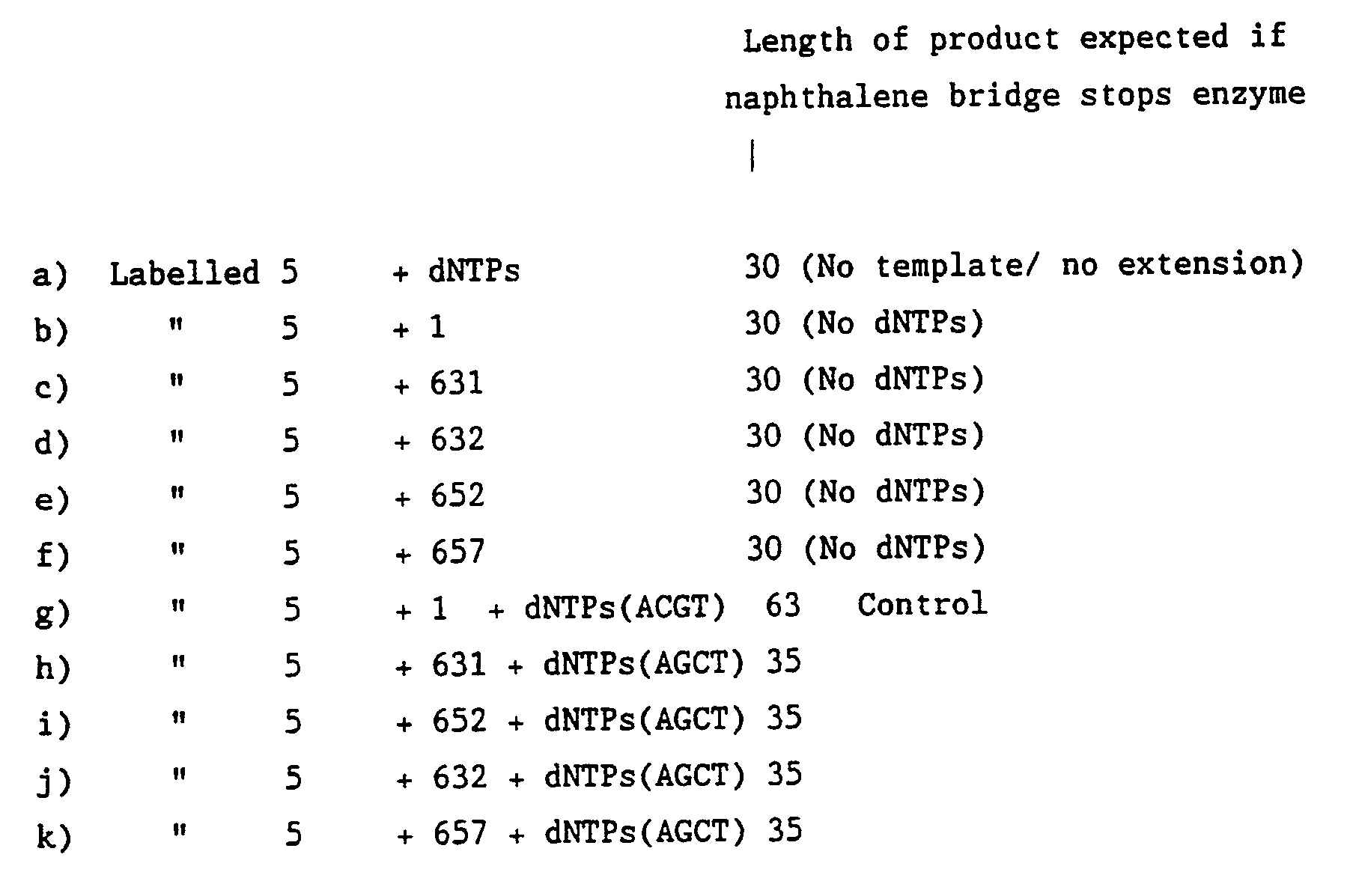
- The results are shown in Figure 3 and demonstrate that one and three metaphosphate moieties significantly inhibit extension by Taq DNA polymerase beyond the bridge. Four thymidine phosphormorpholidate moieties show useful blocking activity but a single thymidine phosphormorpholidate moiety shows negligible blocking activity.
- Without wishing to be bound by any theoretical considerations the polymerase blocking activities may be underestimated because of the presence of the 63 mer bands in the controls with dNTPs omitted. The presence of these bands may be due to a carry over of polynucleotide kinase activity during combination of the template oligonucleotides with ³²P phosphorylated
oligonucleotide 5. - This example demonstrates a microtitre plate test capture system to determine whether:-
- a) capture oligonucleotides are bound to a microtitre plate,
- b) there is nonspecific binding of the test capture oligonucleotide sequences to immobilised sequences other than their complement, or to the protein coating on the microtitre wells.
- c) an alkaline phosphatase, nitro blue tetrazolium/5-bromo-4-chloro-3-indoyl phosphate p-toluidine salt (AP/NBT/BCIP) colour development system performs satisfactorily with the above capture oligonucleotides.
- Oligonucleotide 13; TTTTTTTTTTTATCAACTTACTTGCCTATA
Oligonucleotide 14; TTTTTTTTTTAGATCTTGGTTAACAATCGT
Oligonucleotide 15; TTTTTTTTTTCGGTCCTCATAATACATACT
these are used as capture oligonucleotides for complementary hybridisation with oligonucleotides 11, 12 and 10 respectively as set out in Example 5 below. Oligonucleotide 13 is identical toolgionucleotide 7 as disclosed in Example 1. The 5′(dT)10 residues were included as spacer elements between the hybridisation regions and their respective conjugants. - These oligonucleotides were immobilised on protein coated microtitre plates as follows with reference to Figure 4:-The chosen concentration was such that 100% binding represents 1 nmole oligonucleotide per well.

- Each oligonucleotide is a unique sequence, complementary to one of the capture oligonucleotides described above, and conjugated to the enzyme alkaline phosphatase (AP) using the E-link™ oligonucleotide labelling kit (Cambridge Research Biochemicals). The labelling was carried out with a nominal concentration of each oligonucleotide (1.5 µM).
- Oligonucleotide 16;
TTTTTTTTTTAGTATGTATTATGAGGACCG
Oligonucleotide 17;
TTTTTTTTTTTATAGGCAAGTAAGTTGATA
Oligonucleotide 18;
TTTTTTTTTTACGATTGTTAACCAAGATCT
these are used as capture control oligonucleotides - 200 µl of prehybridisation solution, containing 0.6M NaCl, 20mM sodium phosphate pH 7.5, 1mM EDTA, 0.02% Ficoll, 0.02% polyvinylpyrrolidine and 0.02% bovine serum albumin, was added to each well and incubated in the dark at 37°C minutes. This was then removed by pipetting. The appropriate oligonucleotide enzyme conjugate was diluted 1 in 10 in a polymerase chain reaction (PCR) mix (100 µM dATP, 100µM dGTP, 100µM dCTP & 100µM dTTP), 1.2mM MgCl₂, 50mM KCl, 10mM Tris HCl pH 8.3, 0.01 gelatine) and 10µl of diluted conjugate, ie. approximately 1.5 pmoles were added to 40µl of 5X SSC (1.1 MNaCl, 0.825M sodium citrate pH 7.0 already added to each well). This was allowed to incubate in the dark at 37°C for 1 hour. Solutions were then removed by pipetting. Each of the test capture control oligonucleotides was added to a different set of microtitre wells as prepared above and with reference to Figure 4:-

- Following hybridisation, the wells were washed for 2 x 5 minutes at room temperature using 200µl of 2X SSC (0.44M NaCl 0.33M sodium citrate pH 7.0). Solutions were again removed by pipetting.
- The final step was addition of 200µl of a colour development solution comprising alkaline phosphatase (AP) 9.5 buffer (0. 1M Tris HCl pR 9.5, 0.1M NaCl, 5mM MgCl₂) and containing NBT (0.33mg/ml) and BCIP (0.17mg/ml). The colour development solution was prepared as follows:- for 15 ml of reagent, 5mg of NBT was supsended in 1.5ml AP 9.5 buffer in a microcentrifuge tube, vortexed vigorously for 1-2 minutes and then centrifgued briefly. The supernatant was decanted into 10ml of AP 9.5 buffer, and warmed to 37°C in a polypropylene tube. The residual NBT pellet was extracted twice more with 1.5ml of AP 9.5 buffer and these supernatants pooled with the original solution. The tube was rinsed with a final 0.5ml of AP 9.5 buffer and also decanted into the 15 ml NBT stock solution. BCIP (2.5mg) was dissolved in 50µl of N,N-dimethylformamide and added dropwise with gentle mixing into the NBT solution. The solution was frozen at -20°C in 1 ml aliquots and then before use the solution was prewarmed to 37°C in a water bath. Colour was allowed to develop in the dark at 37°C and photographs were taken after 15 minutes and 2 hours 15 minutes.
- The results are shown in Figure 4 and show colour development only in wells A1 B1 C2 and F2 which is entirely consistent with the test capture oligonucleotides hybridising only to their complementary immobilised oligonucleotides under the conditions employed.
- This example demonstrates the specific capture and detection of amplification products based on the S locus of the alpha-1-antitrypsin gene and using an internal amplification control based on sequence from exon V of the same gene. Detection of the immobilised products was achieved using an alkaline phosphatase (AP) conjugated common detection primer. This common primer was prepared using the E-link™ conjugation kit (Cambridge Research Biochemicals) as directed by the supplier and is complementary to the detection tail of all the amplified products.
- The following oligonucleotides were used wherein N represents a 3′deoxyribofuranosyl naphthalene moiety, the naphthalene moieties being linked via the 2′-PO₄ grouping to the adjacent nucleotide:-
- Oligonucleotide 8b;
GTGTCGTCCCGCAGTCAATGNNNNCCCACCTTCCCCTCTCTCCAGGCAAATGGG
this oligonucleotide is the common signal primer for carrying out the ARMS assay method. - Oligonucleotide 9
GTCTCGTCCCGCAGTCAATGNNNNGAGACTTGGTATTTTGTTCAATCATTAAG
Oligonucleotide 10
AGTATGTATTATGAGGACCGNNNNGTCCACGTGAGCCTTGCTCGAGGCCTGGG
these oligonucleotides are for the exon V control reaction and are the signal and capture primers respectively. - Oligonucleotide 11;
TATAGGCAAGTAAGTTGATANNNNTGGTGATGATATCGTGGGTGAGTTCATTTT
Oligonucleotide 12;
ACGATTGTTAACCAAGATCTNNNNTGGTGATGATATCGTGGGTGAGTTCATTTA
oligonucleotides 11 and 12 are both capture primers and comprise respectively regions for complementary hybridisation to the published normal and mutant sequences of the S locus of the alpha-1-antitrypsin gene. - Oligonucleotide 13;
TTTTTTTTTTTATCAACTTACTTGCCTATA
Oligonucleotide 14;
TTTTTTTTTTAGATCTTGGTTAACAATCGT
Oligonucleotide 15;
TTTTTTTTTTCGGTCCTCATAATACATACT
these are used as capture oligonucleotides for complementary hybridisation with oligonucleotides 11, 12 and 10 respectively. Oligonucleotide 13 is identical toolgionucleotide 7 as disclosed in Example 1. The 5′(dT)10 residues were included as spacer elements between the hybridisation regions and their respective conjugants. - Oligonucleotide 16;
TTTTTTTTTTAGTATGTATTATGAGGACCG
Oligonucleotide 17;
TTTTTTTTTTTATAGGCAAGTAAGTTGATA
Oligonucleotide 18;
TTTTTTTTTTACGATTGTTAACCAAGATCT
these are used as capture control oligonucleotides - Oligonucleotide 19;
TTTTTTTTTTCATTGACTGCGGGACGACAC
this is the common signal oligonucleotide for the ARMS assay method - Oligonucleotides 13-15 and 16-19 were synthesised as 5′ aminoalkyl derivatives using aminoalkyl phosphoramidite (Amino-
link 2, Applied Biosystems) in the final synthesis cycle on the DNA synthesiser. These oligonucleotides were conjugated to alkaline phosphatase (AP) using the E-LINK™ oligonucleotide labelling kit as directed by the supplier (Cambridge Research Biochemicals). - Aliquots of crude oligonucleotides 8b, 9, 10, 11 and 12 (225µl) were precipitated by the addition of 25µl of 3M sodium acetate pH 5.2, mixing and then adding 750µl ethanol. After further mixing the tubes were left at -20°C for one hour and spun for 10 minutes at 13,500 rpm before removing the supernatant. The pellets were left to air dry for 20 minutes then reconstituted with 50µl formamide dye (80% formamide, 10mM NaOH, 1mM EDTA, 0.1% bromophenol blue). The samples were then loaded onto a 15% acrylamide gel in 7M urea, 100mM Tris borate EDTA buffer pH 8.3, which had been prerun for 1 hour at 300V. The samples were electrophoresed overnight at 140V, ie. until the dye had run to the bottom of the gel (0.3 x 24 x 17 cm³). The oligonucleotides were visualised by ultra violet shadowing at 254nm on a 20 x 20 aluminium backed thin layer chromatography plate (Merck 5554) covered with Saran wrap. The bands were excised using a new scalpel for each band to prevent cross-contamination. The gel slices were placed separately into a length of treated dialysis tubing (boiled in 1mM EDTA for ten minutes then rinsed 3X by boiling in milli Q water) containing 1ml 10mm Tris borate EDTA buffer pH 8.3. The oligonucleotides were electroeluted from the gel slice at 2100V for 1.5 hours, the polarity was then reversed for 45 seconds. The eluates were removed into 1.5ml quartz spectrophotometer cuvettes, the optical density at 220-330 nanometres was then scanned against the elution buffer and the concentration of oligonucleotide calculated based on the optical density at 260nm. The oligonucleotides were then concentrated by butanol extraction until the volume of the aqueous layer was ∼500 µl. A phenol chloroform 1:1 (500µl) extraction was included. The aqueous phase was removed and further concentrated by butanol extraction to ∼50µl. This was then dried under vacuum and reconstituted with filtered MilliQ water to 50 pmoles/µl ready for the amplification reactions.
- The capture oligonucleotides (200µl, 0.2µMol synthesis) were mixed separately with 4mg/ml iminothiolane in 0.2M Na₂CO₃/ HCO₃ pH 9.6 (300µl) and incubated at ambient temperature for 1 hour. The reactions were diluted to 1ml with phosphate buffered saline (PBS) and desalted on a Nap-25 column (Pharmacia). The columns were washed with PBS (2.2ml). The first 1.6ml eluted was retained and the oligonucleotide concentrations were measured by u.v. spectrometry.
- Oligonucleotide immobilisation to microtitre wells was performed using a modification of the method described by Running, J.A. and Urdea, M.S., (1990), Biotechniques, 8, 276-277. Poly (Phe-Lys) (Sigma) was dissolved to 100ug/ml in 50mM Na₂CO₃/HCO₃, pH 9.6 and 100µl of this solution was added to each well of five clear polystyrene microtitre dishes (Nunc). The dishes were incubated overnight at 4oC then washed 3 times with PBS/0.05% Tween. Succinimidyl 4-(N-maleimidomethyl)cyclohexane-1-carboxylate (SMCC) was dissolved at 8.7mg/ml in dry dimethylformamide and this solution was diluted 10 times with conjugation buffer (100mM triethanolamine HC1, 1mM MgCl₂, 1mM ZnSO₄, pH 7.4). 100µl of this solution was added to each microtitre well and the plates were incubated at ambient temperature for 1 hour. The wells were washed 5 times with water. Thiolated oligonucleotides, 10µM in PBS, were added to appropriate microtitre wells at a rate of 100µl per well. These were incubated overnight at 4oC. The wells were washed twice with PBS/0.05% Tween, twice with 5% lactose, 0.5% gelatin, 0.1% N₃Na, 6mM PBS, pH 7.5. Finally the coated dishes were dried overnight in a laminar flow cabinet. The dishes were stored at 4oC in vacuo.
- 1µl DNA was added to a tube containing 1.2mM MgCl₂, 50mM KCl, 10mM Tris HCl pH 8.3, 0.01% gelatin, and 100 µM dATP, 100µM dGTP, 100µM dCTP & 100µM dTTP to make a reaction volume of 98 µl to a final concentration of 100 µM each and 50 p moles each of oligonucleotides 8b, 9, 10 and 11 (the ARMS primer specific for the normal allele). 1µl DNA was also added to a similar tube containing oligonucleotide 12 (the ARMS primer specific for the S variant allele) instead of oligonucleotide 11. 2 drops of mineral oil were added to each tube and spun for 1 minute at 13500 rpm. The tubes were transferred into a PCR machine (Techne) where they were denatured at 95°C for 5 minutes before 2 units of Taq polymerase (Perkin Elmer, Ampli-Taq) were added still at 95°C to make the final volume 100 µl. The Techne machine was switched into the 2 step PCR programme of 2 minutes at 94°C, 4 minutes at 60°C, 35 cycles with a final 9.9 minutes extension step at 60oC. A 20 µl aliquot of the reaction mixtures was electophoresed on 1.5% agarose gel. This was to ascertain the genotype of the DNA sample by traditional analytical methods.
- The results are shown in Figure 6:-
Lane 4 S normal allele specific ARMS primer
Lane 5 S variant allele specific ARMS primer
The control (lower band) is present in both lanes whereas there is only a product band in lane 5 (S variant). This confirms that the DNA is from a homozygote for the S variant allele of the alpha-1-antitrypsin gene. - Oligonucleotide 19, the common signal primer was conjgated to alkaline phosphatase using the E-link™ oligonucleotide labelling kit (Cambridge Research Biochemicals). The solutions are the same as those described in detail in Example 4 above unless otherwise stated:-
- 1. 200µl prehybridisation solution was added to each well. This was then incubated at 37°C with shaking for 30 minutes. The solution was removed by pipetting.
- 2. A 20µl aliquot of PCR product from the S normal primer tube was added to wells [A1, A2 and A3 with reference to Figure 7] containing 60µl 5X SSC. A 20 µl aliquot of PCR product from the S variant primer tube was added to wells [B1, B2 and B3 with reference to Figure 7] also containing 60 µl 5 x SSC. Hybridisation lasted for 1 hour at 37°C with shaking. The solutions were then removed by pipetting.
Signal generation was as described in Example 4. - 3. 10µl diluted conjugated oligonucleotide 19 ie. ∼1.5 pmoles was added to 40µl 5X SSC already added to each well. Hybridisation lasted for 1 hour at 37°C with shaking. The solutions were removed by pipetting.
- 4.
2x 5 minutes washes with 200µl 2 X SSC at 37°C were carried out with shaking. The solutions were removed by pipetting. - 5. 200µl of colour development solution was added to each well [A1, A2, A3, B1, B2 and B3 with reference to Figure 7] and these were incubated for ∼1 hour at 37°C with shaking. The wells were then scanned using a Hewlett Packard Scan Jet Plus scanner and digitised using Apple Mackintosh software.
- The results are shown in Figure 7. It can be seen that both the internal control wells A1 and B1 are coloured. In addition only the variant well B2 is coloured providing a clear diagnosis that the DNA is from a homozygous S variant of alpha-1-antitrypsin. There is no significant nonspecific binding, ie. there is no colour in the control wells A3 and B3.


Claims (15)
1) a target binding nucleotide moiety which is substantially complementary to the desired portion of the target nucleotide sequence; and
2) a polynucleotide tail;
such that the first primer may be subjected to primer extension whereby a first primer extension product is synthesised based on the target polynucleotide sequence as template, and after denaturation of the first primer extension product from its template and hybridisation of the amplification primer to the desired portion of the first primer extension product, primer extension may be effected to form an amplification primer extension product, the presence of the first primer in the first primer extension product being effective to inhibit formation of a sequence complementary to the polynucleotide tail in the amplification primer extension product.
1) a target binding nucleotide moiety which is substantially complementary to the desired portion of the target nucleotide sequence; and
2) a polynucleotide tail;
such that the amplification primer may be subjected to primer extension whereby an amplification primer extension product is synthesised based on the first primer extension product as template, and after denaturation of the amplification primer extension product from its template and hybridisation of the first primer to the desired portion of the amplification primer extension product, primer extension may be effected to form a first primer extension product, the presence of the amplification primer in the amplification primer extension product being effective to inhibit formation of a sequence complementary to the polynucleotide tail in the first primer extension product.
a first primer and a corresponding amplification primer for each target nucleotide sequence to be amplified, each first primer comprising:-
1) a target binding nucleotide moiety which is substantially complementary to the desired portion of the target nucleotide sequence; and
2) a polynucleotide tail;
the presence of the first primer in the first primer extension product being effective to inhibit formation of a sequence complementary to the polynucleotide tail in the amplification primer extension product.
1) a target binding nucleotide moiety which is substantially complementary to the desired portion of the target nucleotide sequence; and
2) a polynucleotide tail;
the presence of the amplification primer in the amplification primer extension product being effective to inhibit formation of a sequence complementary to the polynucleotide tail in a first primer extension product.
Applications Claiming Priority (2)
| Application Number | Priority Date | Filing Date | Title |
|---|---|---|---|
| GB898920097A GB8920097D0 (en) | 1989-09-06 | 1989-09-06 | Amplification processes |
| GB8920097 | 1989-09-06 |
Publications (3)
| Publication Number | Publication Date |
|---|---|
| EP0416817A2 true EP0416817A2 (en) | 1991-03-13 |
| EP0416817A3 EP0416817A3 (en) | 1991-09-04 |
| EP0416817B1 EP0416817B1 (en) | 1996-10-16 |
Family
ID=10662595
Family Applications (1)
| Application Number | Title | Priority Date | Filing Date |
|---|---|---|---|
| EP90309502A Expired - Lifetime EP0416817B1 (en) | 1989-09-06 | 1990-08-30 | Amplification processes |
Country Status (8)
| Country | Link |
|---|---|
| US (1) | US5525494A (en) |
| EP (1) | EP0416817B1 (en) |
| JP (1) | JP3165431B2 (en) |
| AT (1) | ATE144290T1 (en) |
| AU (1) | AU622349B2 (en) |
| DE (1) | DE69028891T2 (en) |
| GB (2) | GB8920097D0 (en) |
| NZ (1) | NZ235015A (en) |
Cited By (45)
| Publication number | Priority date | Publication date | Assignee | Title |
|---|---|---|---|---|
| WO1992018646A1 (en) * | 1991-04-18 | 1992-10-29 | The Solicitor For The Affairs Of Her Majesty's Treasury | Nucleotide probes and primers for genetic analysis |
| EP0521111A1 (en) * | 1990-03-23 | 1993-01-07 | Chiron Corporation | Polynucleotide capture assay employing in vitro amplification |
| EP0530009A2 (en) * | 1991-08-27 | 1993-03-03 | Zeneca Limited | Method of characterising genomic DNA |
| EP0607151A1 (en) * | 1992-06-17 | 1994-07-27 | City Of Hope | A method of detecting and discriminating between nucleic acid sequences |
| US5426039A (en) * | 1993-09-08 | 1995-06-20 | Bio-Rad Laboratories, Inc. | Direct molecular cloning of primer extended DNA containing an alkane diol |
| EP0710296A1 (en) * | 1993-07-23 | 1996-05-08 | Bio-Rad Laboratories, Inc. | Linked linear amplification of nucleic acids |
| GB2312747A (en) * | 1996-05-04 | 1997-11-05 | Zeneca Ltd | Primers with non-complementary tails for detection of diagnostic base sequences |
| WO1997042345A1 (en) * | 1996-05-04 | 1997-11-13 | Zeneca Limited | Method for detecting a nucleic acid base sequence |
| EP0832278A1 (en) * | 1995-06-07 | 1998-04-01 | Bio-Rad Laboratories, Inc. | Linked linear amplification of nucleic acids |
| WO1998014610A2 (en) * | 1996-10-04 | 1998-04-09 | The Perkin-Elmer Corporation | Multiplex polynucleotide capture methods and compositions |
| WO1998045475A1 (en) * | 1997-04-09 | 1998-10-15 | Cis Bio International | System for detecting nucleic acid hybridisation, preparation method and application thereof |
| US5853989A (en) * | 1991-08-27 | 1998-12-29 | Zeneca Limited | Method of characterisation of genomic DNA |
| US5981176A (en) * | 1992-06-17 | 1999-11-09 | City Of Hope | Method of detecting and discriminating between nucleic acid sequences |
| US6027923A (en) * | 1993-07-23 | 2000-02-22 | Bio-Rad Laboratories, Inc. | Linked linear amplification of nucleic acids |
| WO2000047766A1 (en) * | 1999-02-11 | 2000-08-17 | Astrazeneca Ab | Method for detecting variant nucleotides using arms multiplex amplification |
| WO2000047767A1 (en) * | 1999-02-11 | 2000-08-17 | Astrazeneca Ab | Oligonucleotide array and methods of use |
| WO2000058516A2 (en) * | 1999-03-26 | 2000-10-05 | Whitehead Institute For Biomedical Research | Universal arrays |
| US6326145B1 (en) | 1998-06-13 | 2001-12-04 | Zeneca Limited | Methods for detecting target nucleic acid sequences |
| US6335184B1 (en) | 1993-07-23 | 2002-01-01 | Bio-Rad Laboratories, Inc. | Linked linear amplification of nucleic acids |
| WO2002024944A2 (en) * | 2000-09-18 | 2002-03-28 | november Aktiengesellschaft Gesellschaft für Molekulare Medizin | Method for detecting at least one nucleic acid sequence |
| WO2002061143A2 (en) * | 2001-01-31 | 2002-08-08 | Ambion, Inc. | Comparative analysis of nucleic acids using population tagging |
| US6458530B1 (en) | 1996-04-04 | 2002-10-01 | Affymetrix Inc. | Selecting tag nucleic acids |
| EP1302547A2 (en) * | 1992-06-17 | 2003-04-16 | City Of Hope | A method of detecting and discriminating between nucleic acid sequences |
| WO2001090417A3 (en) * | 2000-05-19 | 2003-08-07 | Eragen Biosciences Inc | Materials and methods for detection of nucleic acids |
| EP1481088A2 (en) * | 2002-03-01 | 2004-12-01 | Integrated Dna Technologies, Inc. | Polynomial amplification of nucleic acids |
| US7056666B2 (en) | 1990-12-06 | 2006-06-06 | Affymetrix, Inc. | Analysis of surface immobilized polymers utilizing microfluorescence detection |
| US7192700B2 (en) | 2002-12-20 | 2007-03-20 | Orchid Cellmark Inc. | Methods and compositions for conducting primer extension and polymorphism detection reactions |
| US7238476B2 (en) | 2002-08-02 | 2007-07-03 | Orchid Cellmark, Inc. | Methods and compositions for genotyping |
| WO2011018774A1 (en) | 2009-08-13 | 2011-02-17 | Centre National De La Recherche Scientifique | Method for detecting a circularized dna, and use of said method for detecting mutations |
| US7892796B2 (en) | 2000-10-14 | 2011-02-22 | Eragen Biosciences, Inc. | Solid support assay systems and methods utilizing non-standard bases |
| US8071562B2 (en) * | 2007-12-01 | 2011-12-06 | Mirna Therapeutics, Inc. | MiR-124 regulated genes and pathways as targets for therapeutic intervention |
| US8313932B2 (en) | 2003-04-01 | 2012-11-20 | Luminex Corporation | Polymerase inhibitor and method of using same |
| US8399197B2 (en) | 2002-03-01 | 2013-03-19 | Integrated Dna Technologies, Inc. | Methods for amplifying polymeric nucleic acids |
| US8440406B2 (en) | 2009-10-23 | 2013-05-14 | Luminex Corporation | Amplification primers with non-standard bases for increased reaction specificity |
| US8765709B2 (en) | 2004-11-12 | 2014-07-01 | Asuragen, Inc. | Methods and compositions involving miRNA and miRNA inhibitor molecules |
| US8936913B2 (en) | 2005-06-07 | 2015-01-20 | Luminex Corporation | Methods for detection and typing of nucleic acids |
| EP2369007B1 (en) * | 1996-05-29 | 2015-07-29 | Cornell Research Foundation, Inc. | Detection of nucleic acid sequence differences using coupled ligase detection and polymerase chain reactions |
| US9222085B2 (en) | 2011-02-03 | 2015-12-29 | Mirna Therapeutics, Inc. | Synthetic mimics of MIR-124 |
| US9365852B2 (en) | 2008-05-08 | 2016-06-14 | Mirna Therapeutics, Inc. | Compositions and methods related to miRNA modulation of neovascularization or angiogenesis |
| US9644241B2 (en) | 2011-09-13 | 2017-05-09 | Interpace Diagnostics, Llc | Methods and compositions involving miR-135B for distinguishing pancreatic cancer from benign pancreatic disease |
| US9783844B2 (en) | 2012-04-27 | 2017-10-10 | Kaneka Corporation | Method for amplifying nucleic acid and method for detecting amplified nucleic acid |
| US9863001B2 (en) | 2003-08-15 | 2018-01-09 | Epigenomics Ag | Method for the detection of cytosine methylations in DNA |
| US9920356B2 (en) | 2010-11-24 | 2018-03-20 | Kaneka Corporation | Amplified nucleic acid detection method and detection device |
| US10047388B2 (en) | 2004-05-28 | 2018-08-14 | Asuragen, Inc. | Methods and compositions involving MicroRNA |
| US10392652B2 (en) | 2013-11-22 | 2019-08-27 | Kaneka Corporation | Micro RNA detection method using two primers to produce an amplified double stranded DNA fragment having a single stranded region at one end |
Families Citing this family (38)
| Publication number | Priority date | Publication date | Assignee | Title |
|---|---|---|---|---|
| US5916776A (en) * | 1997-08-27 | 1999-06-29 | Sarnoff Corporation | Amplification method for a polynucleotide |
| AU4058100A (en) * | 1999-04-09 | 2000-11-14 | Arcturus Engineering, Inc. | Generic cdna or protein array for customized assays |
| US20030165913A1 (en) * | 1999-06-17 | 2003-09-04 | Sha-Sha Wang | Methods for detecting nucleic acid sequence variations |
| US6316200B1 (en) * | 2000-06-08 | 2001-11-13 | Becton, Dickinson And Company | Probes and methods for detection of nucleic acids |
| US20020025519A1 (en) * | 1999-06-17 | 2002-02-28 | David J. Wright | Methods and oligonucleotides for detecting nucleic acid sequence variations |
| US6232104B1 (en) | 1999-08-17 | 2001-05-15 | Dade Behring Inc. | Detection of differences in nucleic acids by inhibition of spontaneous DNA branch migration |
| US6709816B1 (en) | 1999-10-18 | 2004-03-23 | Affymetrix, Inc. | Identification of alleles |
| US6287778B1 (en) * | 1999-10-19 | 2001-09-11 | Affymetrix, Inc. | Allele detection using primer extension with sequence-coded identity tags |
| US6844154B2 (en) * | 2000-04-04 | 2005-01-18 | Polygenyx, Inc. | High throughput methods for haplotyping |
| US7238505B2 (en) * | 2000-10-04 | 2007-07-03 | Ahram Biosystems Inc. | Immobilized DNA polymerase |
| DE10119006A1 (en) | 2001-04-18 | 2002-10-24 | Roche Diagnostics Gmbh | Method for improving the stability of linear DNA in cell-free in vitro transcription / translation systems |
| US6632938B2 (en) * | 2001-06-07 | 2003-10-14 | Isis Pharmaceuticals, Inc. | Processes of purifying oligonucleotides |
| US6977163B1 (en) | 2001-06-13 | 2005-12-20 | Caliper Life Sciences, Inc. | Methods and systems for performing multiple reactions by interfacial mixing |
| US7510829B2 (en) * | 2001-11-19 | 2009-03-31 | Affymetrix, Inc. | Multiplex PCR |
| US20110151438A9 (en) | 2001-11-19 | 2011-06-23 | Affymetrix, Inc. | Methods of Analysis of Methylation |
| US8394944B2 (en) * | 2002-09-20 | 2013-03-12 | Siemens Healthcare Diagnostics Inc. | Dual-purpose primers and probes for providing enhanced hybridization assays by disruption of secondary structure formation |
| US20040197803A1 (en) * | 2002-12-06 | 2004-10-07 | Hidenobu Yaku | Method, primer and kit for determining base type |
| EP1590482B1 (en) | 2003-01-17 | 2008-12-10 | Eragen Biosciences, Inc. | Nucleic acid amplification using non-standard bases |
| AU2004201842A1 (en) | 2003-05-01 | 2004-11-18 | Veridex, Llc | Intraoperative lymph node assay |
| EP1766079A4 (en) * | 2004-06-04 | 2010-06-23 | Veridex Llc | Diagnosing or predicting the course of breast cancer |
| US20070154889A1 (en) * | 2004-06-25 | 2007-07-05 | Veridex, Llc | Methods and reagents for the detection of melanoma |
| US8883487B2 (en) * | 2004-12-23 | 2014-11-11 | Abbott Point Of Care Inc. | Molecular diagnostics system and methods |
| WO2006101913A2 (en) | 2005-03-18 | 2006-09-28 | Eragen Biosciences, Inc. | Methods for detecting multiple species and subspecies of neiserria |
| US20070099195A1 (en) * | 2005-11-02 | 2007-05-03 | Huang Xiaohua C | Methods and compositions for separating nucleic acids from a solid support |
| EP1966366A4 (en) | 2005-12-29 | 2011-06-15 | I Stat Corp | Molecular diagnostics amplification system and methods |
| EP3118333B1 (en) * | 2006-12-13 | 2019-04-03 | Luminex Corporation | Systems and methods for multiplex analysis of pcr in real time |
| US9938641B2 (en) | 2006-12-18 | 2018-04-10 | Fluidigm Corporation | Selection of aptamers based on geometry |
| DE102007029772B4 (en) * | 2007-06-22 | 2011-12-08 | Aj Innuscreen Gmbh | Method and rapid test for the detection of specific nucleic acid sequences |
| US8361714B2 (en) | 2007-09-14 | 2013-01-29 | Asuragen, Inc. | Micrornas differentially expressed in cervical cancer and uses thereof |
| EP2218019A4 (en) * | 2007-11-02 | 2012-04-18 | Hunch Inc | Interactive machine learning advice facility |
| US20090148849A1 (en) * | 2007-11-02 | 2009-06-11 | Barbara Galvan-Goldman | One-step target detection assay |
| US8008019B2 (en) * | 2007-11-28 | 2011-08-30 | Luminex Molecular Diagnostics | Use of dual-tags for the evaluation of genomic variable repeat regions |
| CN102686728B (en) | 2009-06-29 | 2018-09-18 | 卢米耐克斯公司 | Chimeric primers with hairpin conformation and its application method |
| MX2012012908A (en) * | 2010-05-07 | 2013-05-01 | Quantibact As | Method for generating a double stranded nucleic acid with a single stranded overhang. |
| SG11201400635UA (en) * | 2011-09-14 | 2014-09-26 | Ngk Insulators Ltd | Method for detecting target nucleic acid |
| WO2013038534A1 (en) * | 2011-09-14 | 2013-03-21 | 日本碍子株式会社 | Method for detecting target nucleic acid |
| US9115394B2 (en) * | 2011-12-22 | 2015-08-25 | Roche Molecular Systems, Inc. | Methods and reagents for reducing non-specific amplification |
| US9745633B2 (en) * | 2013-06-28 | 2017-08-29 | Diacarta Ltd | Detection of PNA clamping |
Citations (4)
| Publication number | Priority date | Publication date | Assignee | Title |
|---|---|---|---|---|
| EP0237362A1 (en) * | 1986-03-13 | 1987-09-16 | F. Hoffmann-La Roche Ag | Process for detecting specific nucleotide variations and genetic polymorphisms present in nucleic acids and kits therefor |
| GB2202328A (en) * | 1987-03-11 | 1988-09-21 | Orion Yhtymae Oy | An improved method for assaying of nucleic acids, a reagent combination and a kit therefore |
| GB2221909A (en) * | 1988-07-28 | 1990-02-21 | Ici Plc | A method for the amplification of nucleotide sequences. |
| WO1990011372A1 (en) * | 1989-03-21 | 1990-10-04 | Collaborative Research, Inc. | Multiplex dna diagnostic test |
Family Cites Families (4)
| Publication number | Priority date | Publication date | Assignee | Title |
|---|---|---|---|---|
| US4683195A (en) * | 1986-01-30 | 1987-07-28 | Cetus Corporation | Process for amplifying, detecting, and/or-cloning nucleic acid sequences |
| ES8706823A1 (en) * | 1985-03-28 | 1987-06-16 | Cetus Corp | Process for amplifying, detecting, and/or cloning nucleic acid sequences. |
| US4683202A (en) * | 1985-03-28 | 1987-07-28 | Cetus Corporation | Process for amplifying nucleic acid sequences |
| IE61148B1 (en) * | 1988-03-10 | 1994-10-05 | Ici Plc | Method of detecting nucleotide sequences |
-
1989
- 1989-09-06 GB GB898920097A patent/GB8920097D0/en active Pending
-
1990
- 1990-08-21 AU AU61184/90A patent/AU622349B2/en not_active Expired
- 1990-08-22 NZ NZ235015A patent/NZ235015A/en unknown
- 1990-08-30 DE DE69028891T patent/DE69028891T2/en not_active Expired - Lifetime
- 1990-08-30 GB GB9018939A patent/GB2235689B/en not_active Revoked
- 1990-08-30 AT AT90309502T patent/ATE144290T1/en not_active IP Right Cessation
- 1990-08-30 EP EP90309502A patent/EP0416817B1/en not_active Expired - Lifetime
- 1990-09-06 JP JP23689590A patent/JP3165431B2/en not_active Expired - Lifetime
-
1994
- 1994-02-23 US US08/200,296 patent/US5525494A/en not_active Expired - Lifetime
Patent Citations (4)
| Publication number | Priority date | Publication date | Assignee | Title |
|---|---|---|---|---|
| EP0237362A1 (en) * | 1986-03-13 | 1987-09-16 | F. Hoffmann-La Roche Ag | Process for detecting specific nucleotide variations and genetic polymorphisms present in nucleic acids and kits therefor |
| GB2202328A (en) * | 1987-03-11 | 1988-09-21 | Orion Yhtymae Oy | An improved method for assaying of nucleic acids, a reagent combination and a kit therefore |
| GB2221909A (en) * | 1988-07-28 | 1990-02-21 | Ici Plc | A method for the amplification of nucleotide sequences. |
| WO1990011372A1 (en) * | 1989-03-21 | 1990-10-04 | Collaborative Research, Inc. | Multiplex dna diagnostic test |
Non-Patent Citations (3)
| Title |
|---|
| AGR. BIOL. CHEM., vol. 36, no. 9, 1972, pages 1651-1653; H. OHRUI et al: "Facile synthesis of C-glycosides of aromatic compounds" * |
| NUCLEIC ACIDS RESEARCH, vol. 12, no. 11, 1984, pages 4539-4557, IRL Press Ltd, Oxford, GB; N.D. SHINHA et al.: "Polymer support oligonucleotide synthesis XVIII 1,2): use of beta-cyanoethyl-N,N-dialkylamino-/N-morpholino phosphoramidite of deoxynucleosides for the synthesis of DNA fragments simplifying deprotection and isolation of the final product" * |
| NUCLEIC ACIDS RESEARCH, vol. 17, no. 7, 11th April 1989, pages 2503-2516, Eynsham, Oxford, GB; C.R. NEWTON et al.: "Analysis of any point mutation in DNA. The amplification refractory mutation system (ARMS)" * |
Cited By (88)
| Publication number | Priority date | Publication date | Assignee | Title |
|---|---|---|---|---|
| EP0521111A1 (en) * | 1990-03-23 | 1993-01-07 | Chiron Corporation | Polynucleotide capture assay employing in vitro amplification |
| EP0521111A4 (en) * | 1990-03-23 | 1994-06-01 | Chiron Corp | Polynucleotide capture assay employing in vitro amplification |
| US7056666B2 (en) | 1990-12-06 | 2006-06-06 | Affymetrix, Inc. | Analysis of surface immobilized polymers utilizing microfluorescence detection |
| WO1992018646A1 (en) * | 1991-04-18 | 1992-10-29 | The Solicitor For The Affairs Of Her Majesty's Treasury | Nucleotide probes and primers for genetic analysis |
| EP0731177A3 (en) * | 1991-08-27 | 1996-11-06 | Zeneca Ltd | Method of characterising genomic DNA |
| EP0530009A2 (en) * | 1991-08-27 | 1993-03-03 | Zeneca Limited | Method of characterising genomic DNA |
| EP0530009A3 (en) * | 1991-08-27 | 1993-11-03 | Ici Plc | Method of characterising genomic dna |
| US5853989A (en) * | 1991-08-27 | 1998-12-29 | Zeneca Limited | Method of characterisation of genomic DNA |
| US5811235A (en) * | 1991-08-27 | 1998-09-22 | Zeneca Limited | Method of characterisation |
| EP0731177A2 (en) * | 1991-08-27 | 1996-09-11 | Zeneca Limited | Method of characterising genomic DNA |
| EP0607151A1 (en) * | 1992-06-17 | 1994-07-27 | City Of Hope | A method of detecting and discriminating between nucleic acid sequences |
| EP0607151A4 (en) * | 1992-06-17 | 1996-11-27 | Hope City | A method of detecting and discriminating between nucleic acid sequences. |
| EP1302547A3 (en) * | 1992-06-17 | 2003-11-12 | City Of Hope | A method of detecting and discriminating between nucleic acid sequences |
| US6627402B2 (en) | 1992-06-17 | 2003-09-30 | City Of Hope | Method of detecting and discriminating between nucleic acid sequences |
| EP1302547A2 (en) * | 1992-06-17 | 2003-04-16 | City Of Hope | A method of detecting and discriminating between nucleic acid sequences |
| US5981176A (en) * | 1992-06-17 | 1999-11-09 | City Of Hope | Method of detecting and discriminating between nucleic acid sequences |
| US6335184B1 (en) | 1993-07-23 | 2002-01-01 | Bio-Rad Laboratories, Inc. | Linked linear amplification of nucleic acids |
| US6027923A (en) * | 1993-07-23 | 2000-02-22 | Bio-Rad Laboratories, Inc. | Linked linear amplification of nucleic acids |
| EP0710296A1 (en) * | 1993-07-23 | 1996-05-08 | Bio-Rad Laboratories, Inc. | Linked linear amplification of nucleic acids |
| EP0710296A4 (en) * | 1993-07-23 | 1999-05-26 | Bio Rad Laboratories | Linked linear amplification of nucleic acids |
| EP0722487A4 (en) * | 1993-09-08 | 1997-08-20 | Bio Rad Laboratories | Direct molecular cloning of primer extended dna |
| EP0722487A1 (en) * | 1993-09-08 | 1996-07-24 | Bio-Rad Laboratories, Inc. | Direct molecular cloning of primer extended dna |
| US5426039A (en) * | 1993-09-08 | 1995-06-20 | Bio-Rad Laboratories, Inc. | Direct molecular cloning of primer extended DNA containing an alkane diol |
| EP0832278A4 (en) * | 1995-06-07 | 1999-05-26 | Bio Rad Laboratories | Linked linear amplification of nucleic acids |
| EP0832278A1 (en) * | 1995-06-07 | 1998-04-01 | Bio-Rad Laboratories, Inc. | Linked linear amplification of nucleic acids |
| US6458530B1 (en) | 1996-04-04 | 2002-10-01 | Affymetrix Inc. | Selecting tag nucleic acids |
| WO1997042345A1 (en) * | 1996-05-04 | 1997-11-13 | Zeneca Limited | Method for detecting a nucleic acid base sequence |
| GB2312747B (en) * | 1996-05-04 | 1998-07-22 | Zeneca Ltd | Method for the detection of diagnostic base sequences using tailed primers having a detector region |
| GB2312747A (en) * | 1996-05-04 | 1997-11-05 | Zeneca Ltd | Primers with non-complementary tails for detection of diagnostic base sequences |
| US6270967B1 (en) | 1996-05-04 | 2001-08-07 | Zeneca Limited | Method for detecting a nucleic acid base sequence |
| EP2369007B1 (en) * | 1996-05-29 | 2015-07-29 | Cornell Research Foundation, Inc. | Detection of nucleic acid sequence differences using coupled ligase detection and polymerase chain reactions |
| US6124092A (en) * | 1996-10-04 | 2000-09-26 | The Perkin-Elmer Corporation | Multiplex polynucleotide capture methods and compositions |
| WO1998014610A2 (en) * | 1996-10-04 | 1998-04-09 | The Perkin-Elmer Corporation | Multiplex polynucleotide capture methods and compositions |
| WO1998014610A3 (en) * | 1996-10-04 | 1998-05-22 | Perkin Elmer Corp | Multiplex polynucleotide capture methods and compositions |
| US6514699B1 (en) | 1996-10-04 | 2003-02-04 | Pe Corporation (Ny) | Multiplex polynucleotide capture methods and compositions |
| US6210932B1 (en) | 1997-04-09 | 2001-04-03 | Cis Bio International | System for detecting nucleic acid hybridization, preparation method and application thereof |
| FR2762013A1 (en) * | 1997-04-09 | 1998-10-16 | Cis Bio Int | NUCLEIC ACID HYBRIDIZATION DETECTION SYSTEMS, PROCESS FOR PREPARING THEM AND USES THEREOF |
| WO1998045475A1 (en) * | 1997-04-09 | 1998-10-15 | Cis Bio International | System for detecting nucleic acid hybridisation, preparation method and application thereof |
| US6326145B1 (en) | 1998-06-13 | 2001-12-04 | Zeneca Limited | Methods for detecting target nucleic acid sequences |
| US9273350B2 (en) | 1998-06-13 | 2016-03-01 | Qiagen Manchester Limited | Methods and primers for detecting target nucleic acid sequences |
| WO2000047766A1 (en) * | 1999-02-11 | 2000-08-17 | Astrazeneca Ab | Method for detecting variant nucleotides using arms multiplex amplification |
| WO2000047767A1 (en) * | 1999-02-11 | 2000-08-17 | Astrazeneca Ab | Oligonucleotide array and methods of use |
| WO2000058516A2 (en) * | 1999-03-26 | 2000-10-05 | Whitehead Institute For Biomedical Research | Universal arrays |
| WO2000058516A3 (en) * | 1999-03-26 | 2001-07-19 | Whitehead Biomedical Inst | Universal arrays |
| WO2001090417A3 (en) * | 2000-05-19 | 2003-08-07 | Eragen Biosciences Inc | Materials and methods for detection of nucleic acids |
| US10240189B2 (en) | 2000-05-19 | 2019-03-26 | Luminex Corporation | Solid support assay systems and methods utilizing non-standard bases |
| US9611509B2 (en) | 2000-05-19 | 2017-04-04 | Luminex Corporation | Materials and methods for detection of nucleic acids |
| US8969047B2 (en) | 2000-05-19 | 2015-03-03 | Luminex Corporation | Solid support assay systems and methods utilizing non-standard bases |
| US8518671B2 (en) | 2000-05-19 | 2013-08-27 | Luminex Corporation | Solid support assay systems and methods utilizing non-standard bases |
| US7422850B2 (en) | 2000-05-19 | 2008-09-09 | Eragen Biosciences, Inc. | Materials and methods for detection of nucleic acids |
| US7541147B2 (en) | 2000-05-19 | 2009-06-02 | Eragen Biosciences, Inc. | Materials and methods for detection of nucleic acids |
| US8519117B2 (en) | 2000-05-19 | 2013-08-27 | Luminex Corporation | Materials and methods for detection of nucleic acids |
| US8217160B2 (en) | 2000-05-19 | 2012-07-10 | Eragen Biosciences, Inc. | Solid support assay systems and methods utilizing non-standard bases |
| WO2002024944A2 (en) * | 2000-09-18 | 2002-03-28 | november Aktiengesellschaft Gesellschaft für Molekulare Medizin | Method for detecting at least one nucleic acid sequence |
| WO2002024944A3 (en) * | 2000-09-18 | 2003-05-08 | November Ag Molekulare Medizin | Method for detecting at least one nucleic acid sequence |
| US7892796B2 (en) | 2000-10-14 | 2011-02-22 | Eragen Biosciences, Inc. | Solid support assay systems and methods utilizing non-standard bases |
| WO2002061143A2 (en) * | 2001-01-31 | 2002-08-08 | Ambion, Inc. | Comparative analysis of nucleic acids using population tagging |
| WO2002061143A3 (en) * | 2001-01-31 | 2003-03-27 | Ambion Inc | Comparative analysis of nucleic acids using population tagging |
| EP1481088A4 (en) * | 2002-03-01 | 2006-08-23 | Integrated Dna Tech Inc | Polynomial amplification of nucleic acids |
| US8399197B2 (en) | 2002-03-01 | 2013-03-19 | Integrated Dna Technologies, Inc. | Methods for amplifying polymeric nucleic acids |
| EP1481088A2 (en) * | 2002-03-01 | 2004-12-01 | Integrated Dna Technologies, Inc. | Polynomial amplification of nucleic acids |
| US7238476B2 (en) | 2002-08-02 | 2007-07-03 | Orchid Cellmark, Inc. | Methods and compositions for genotyping |
| US7192700B2 (en) | 2002-12-20 | 2007-03-20 | Orchid Cellmark Inc. | Methods and compositions for conducting primer extension and polymorphism detection reactions |
| US8313932B2 (en) | 2003-04-01 | 2012-11-20 | Luminex Corporation | Polymerase inhibitor and method of using same |
| US9863001B2 (en) | 2003-08-15 | 2018-01-09 | Epigenomics Ag | Method for the detection of cytosine methylations in DNA |
| US10047388B2 (en) | 2004-05-28 | 2018-08-14 | Asuragen, Inc. | Methods and compositions involving MicroRNA |
| US9382537B2 (en) | 2004-11-12 | 2016-07-05 | Asuragen, Inc. | Methods and compositions involving miRNA and miRNA inhibitor molecules |
| US9506061B2 (en) | 2004-11-12 | 2016-11-29 | Asuragen, Inc. | Methods and compositions involving miRNA and miRNA inhibitor molecules |
| US9051571B2 (en) | 2004-11-12 | 2015-06-09 | Asuragen, Inc. | Methods and compositions involving miRNA and miRNA inhibitor molecules |
| US8765709B2 (en) | 2004-11-12 | 2014-07-01 | Asuragen, Inc. | Methods and compositions involving miRNA and miRNA inhibitor molecules |
| US9068219B2 (en) | 2004-11-12 | 2015-06-30 | Asuragen, Inc. | Methods and compositions involving miRNA and miRNA inhibitor molecules |
| US9447414B2 (en) | 2004-11-12 | 2016-09-20 | Asuragen, Inc. | Methods and compositions involving miRNA and miRNA inhibitor molecules |
| US10385412B2 (en) | 2005-06-07 | 2019-08-20 | Luminex Corporation | Methods for detection and typing of nucleic acids |
| US8936913B2 (en) | 2005-06-07 | 2015-01-20 | Luminex Corporation | Methods for detection and typing of nucleic acids |
| US9562261B2 (en) | 2005-06-07 | 2017-02-07 | Luminex Corporation | Methods for detection and typing of nucleic acids |
| US8071562B2 (en) * | 2007-12-01 | 2011-12-06 | Mirna Therapeutics, Inc. | MiR-124 regulated genes and pathways as targets for therapeutic intervention |
| US9365852B2 (en) | 2008-05-08 | 2016-06-14 | Mirna Therapeutics, Inc. | Compositions and methods related to miRNA modulation of neovascularization or angiogenesis |
| US9499859B2 (en) | 2009-08-13 | 2016-11-22 | Ulrich Bockelmann | Method for detecting a circularized DNA, and use of said method for detecting mutations |
| WO2011018774A1 (en) | 2009-08-13 | 2011-02-17 | Centre National De La Recherche Scientifique | Method for detecting a circularized dna, and use of said method for detecting mutations |
| FR2949120A1 (en) * | 2009-08-13 | 2011-02-18 | Centre Nat Rech Scient | METHOD FOR DETECTING CIRCULARIZED DNA AND USE OF SAID METHOD FOR DETECTING MUTATIONS |
| US8440406B2 (en) | 2009-10-23 | 2013-05-14 | Luminex Corporation | Amplification primers with non-standard bases for increased reaction specificity |
| US9920356B2 (en) | 2010-11-24 | 2018-03-20 | Kaneka Corporation | Amplified nucleic acid detection method and detection device |
| US10829805B2 (en) | 2010-11-24 | 2020-11-10 | Kaneka Corporation | Amplified nucleic acid detection method and detection device |
| US9222085B2 (en) | 2011-02-03 | 2015-12-29 | Mirna Therapeutics, Inc. | Synthetic mimics of MIR-124 |
| US9644241B2 (en) | 2011-09-13 | 2017-05-09 | Interpace Diagnostics, Llc | Methods and compositions involving miR-135B for distinguishing pancreatic cancer from benign pancreatic disease |
| US10655184B2 (en) | 2011-09-13 | 2020-05-19 | Interpace Diagnostics, Llc | Methods and compositions involving miR-135b for distinguishing pancreatic cancer from benign pancreatic disease |
| US9783844B2 (en) | 2012-04-27 | 2017-10-10 | Kaneka Corporation | Method for amplifying nucleic acid and method for detecting amplified nucleic acid |
| US10392652B2 (en) | 2013-11-22 | 2019-08-27 | Kaneka Corporation | Micro RNA detection method using two primers to produce an amplified double stranded DNA fragment having a single stranded region at one end |
Also Published As
| Publication number | Publication date |
|---|---|
| NZ235015A (en) | 1992-03-26 |
| EP0416817B1 (en) | 1996-10-16 |
| JPH03272686A (en) | 1991-12-04 |
| DE69028891D1 (en) | 1996-11-21 |
| AU622349B2 (en) | 1992-04-02 |
| GB9018939D0 (en) | 1990-10-17 |
| JP3165431B2 (en) | 2001-05-14 |
| AU6118490A (en) | 1991-03-14 |
| ATE144290T1 (en) | 1996-11-15 |
| DE69028891T2 (en) | 1997-03-13 |
| EP0416817A3 (en) | 1991-09-04 |
| GB8920097D0 (en) | 1989-10-18 |
| US5525494A (en) | 1996-06-11 |
| GB2235689B (en) | 1993-10-20 |
| GB2235689A (en) | 1991-03-13 |
Similar Documents
| Publication | Publication Date | Title |
|---|---|---|
| EP0416817B1 (en) | Amplification processes | |
| JP2622327B2 (en) | Nucleic acid sequence amplification means | |
| AU770217B2 (en) | Ligation assembly and detection of polynucleotides on solid-support | |
| EP0566751B1 (en) | DNA detection method | |
| US6461816B1 (en) | Methods for controlling cross-hybridization in analysis of nucleic acid sequences | |
| EP1368489B1 (en) | Asynchronous primed pcr | |
| JP4360904B2 (en) | Single nucleotide amplification and polymerase detection | |
| US5434047A (en) | Assay for polynucleotides employing oligonucleotides to eliminate undesireable cross reactions | |
| EP1375676A2 (en) | Methods of synthesizing polynucleotides by ligation of multiple oligomers | |
| JP2001514859A (en) | Oligonucleotide probes with quenchable fluorescent labels and methods of use | |
| EP0541693B1 (en) | Large comb-type branched polynucleotides | |
| JPH067166A (en) | Method of cloning nucleic acid sequence | |
| EP1937702A2 (en) | Labeled nucleotide analogs and uses therefor | |
| US5650271A (en) | Enzymatic synthesis of oligonucleotides | |
| US6063571A (en) | Process for amplifying nucleic acids using DNA/PNA primers | |
| AU4599797A (en) | Compositions and methods for enhancing hybridization specificity | |
| EP1017855B1 (en) | Methods of synthesizing polynucleotides by ligation of multiple oligomers | |
| EP1198593B1 (en) | Amplification method for detection of target nucleic acids involving fluorescence energy transfer | |
| JP4643263B2 (en) | Allele-specific primer extension | |
| JP3942079B2 (en) | Methods for exogenous control and internal control during nucleic acid amplification | |
| EP0439968A1 (en) | Hybridization probes for the detection of haemophilus ducreyi strains | |
| McGuire | Synthesis and studies of modified nucleotides and oligonucleotides | |
| AU2001268235B2 (en) | Asynchronous primed PCR | |
| WO2002004679A1 (en) | A process for detecting a nucleic acid target | |
| AU2001268235A1 (en) | Asynchronous primed PCR |
Legal Events
| Date | Code | Title | Description |
|---|---|---|---|
| PUAI | Public reference made under article 153(3) epc to a published international application that has entered the european phase |
Free format text: ORIGINAL CODE: 0009012 |
|
| AK | Designated contracting states |
Kind code of ref document: A2 Designated state(s): AT BE CH DE DK ES FR GB GR IT LI LU NL SE |
|
| PUAL | Search report despatched |
Free format text: ORIGINAL CODE: 0009013 |
|
| AK | Designated contracting states |
Kind code of ref document: A3 Designated state(s): AT BE CH DE DK ES FR GB GR IT LI LU NL SE |
|
| 17P | Request for examination filed |
Effective date: 19920123 |
|
| 17Q | First examination report despatched |
Effective date: 19930909 |
|
| RAP1 | Party data changed (applicant data changed or rights of an application transferred) |
Owner name: ZENECA LIMITED |
|
| GRAH | Despatch of communication of intention to grant a patent |
Free format text: ORIGINAL CODE: EPIDOS IGRA |
|
| GRAA | (expected) grant |
Free format text: ORIGINAL CODE: 0009210 |
|
| AK | Designated contracting states |
Kind code of ref document: B1 Designated state(s): AT BE CH DE DK ES FR GB GR IT LI LU NL SE |
|
| PG25 | Lapsed in a contracting state [announced via postgrant information from national office to epo] |
Ref country code: IT Free format text: LAPSE BECAUSE OF FAILURE TO SUBMIT A TRANSLATION OF THE DESCRIPTION OR TO PAY THE FEE WITHIN THE PRE;WARNING: LAPSES OF ITALIAN PATENTS WITH EFFECTIVE DATE BEFORE 2007 MAY HAVE OCCURRED AT ANY TIME BEFORE 2007. THE CORRECT EFFECTIVE DATE MAY BE DIFFERENT FROM THE ONE RECORDED.SCRIBED TIME-LIMIT Effective date: 19961016 Ref country code: NL Free format text: LAPSE BECAUSE OF FAILURE TO SUBMIT A TRANSLATION OF THE DESCRIPTION OR TO PAY THE FEE WITHIN THE PRESCRIBED TIME-LIMIT Effective date: 19961016 Ref country code: BE Effective date: 19961016 Ref country code: GR Free format text: LAPSE BECAUSE OF FAILURE TO SUBMIT A TRANSLATION OF THE DESCRIPTION OR TO PAY THE FEE WITHIN THE PRESCRIBED TIME-LIMIT Effective date: 19961016 Ref country code: ES Free format text: THE PATENT HAS BEEN ANNULLED BY A DECISION OF A NATIONAL AUTHORITY Effective date: 19961016 Ref country code: AT Effective date: 19961016 Ref country code: DK Effective date: 19961016 |
|
| REF | Corresponds to: |
Ref document number: 144290 Country of ref document: AT Date of ref document: 19961115 Kind code of ref document: T |
|
| REG | Reference to a national code |
Ref country code: CH Ref legal event code: NV Representative=s name: A. BRAUN, BRAUN, HERITIER, ESCHMANN AG PATENTANWAE |
|
| REF | Corresponds to: |
Ref document number: 69028891 Country of ref document: DE Date of ref document: 19961121 |
|
| ET | Fr: translation filed | ||
| PG25 | Lapsed in a contracting state [announced via postgrant information from national office to epo] |
Ref country code: SE Effective date: 19970116 |
|
| NLV1 | Nl: lapsed or annulled due to failure to fulfill the requirements of art. 29p and 29m of the patents act | ||
| PLBE | No opposition filed within time limit |
Free format text: ORIGINAL CODE: 0009261 |
|
| STAA | Information on the status of an ep patent application or granted ep patent |
Free format text: STATUS: NO OPPOSITION FILED WITHIN TIME LIMIT |
|
| PG25 | Lapsed in a contracting state [announced via postgrant information from national office to epo] |
Ref country code: LU Free format text: LAPSE BECAUSE OF NON-PAYMENT OF DUE FEES Effective date: 19970831 |
|
| 26N | No opposition filed | ||
| REG | Reference to a national code |
Ref country code: GB Ref legal event code: 732E |
|
| REG | Reference to a national code |
Ref country code: CH Ref legal event code: PUE Owner name: ZENECA LIMITED TRANSFER- ASTRAZENECA AB |
|
| REG | Reference to a national code |
Ref country code: GB Ref legal event code: IF02 |
|
| REG | Reference to a national code |
Ref country code: FR Ref legal event code: CD Ref country code: FR Ref legal event code: TP |
|
| REG | Reference to a national code |
Ref country code: CH Ref legal event code: PFA Owner name: ASTRAZENECA AB Free format text: ASTRAZENECA AB# #151 85 SOEDERTAELJE (SE) -TRANSFER TO- ASTRAZENECA AB# #151 85 SOEDERTAELJE (SE) |
|
| PGFP | Annual fee paid to national office [announced via postgrant information from national office to epo] |
Ref country code: FR Payment date: 20090806 Year of fee payment: 20 |
|
| PGFP | Annual fee paid to national office [announced via postgrant information from national office to epo] |
Ref country code: GB Payment date: 20090708 Year of fee payment: 20 Ref country code: CH Payment date: 20090811 Year of fee payment: 20 Ref country code: DE Payment date: 20090831 Year of fee payment: 20 |
|
| REG | Reference to a national code |
Ref country code: CH Ref legal event code: PL |
|
| REG | Reference to a national code |
Ref country code: GB Ref legal event code: PE20 Expiry date: 20100829 |
|
| PG25 | Lapsed in a contracting state [announced via postgrant information from national office to epo] |
Ref country code: GB Free format text: LAPSE BECAUSE OF EXPIRATION OF PROTECTION Effective date: 20100829 |
|
| PG25 | Lapsed in a contracting state [announced via postgrant information from national office to epo] |
Ref country code: DE Free format text: LAPSE BECAUSE OF EXPIRATION OF PROTECTION Effective date: 20100830 |