EP0407942A2 - Streptokinase proteins, corresponding genes, corresponding plasmid recombinants, corresponding transformants and processes for preparing same - Google Patents
Streptokinase proteins, corresponding genes, corresponding plasmid recombinants, corresponding transformants and processes for preparing same Download PDFInfo
- Publication number
- EP0407942A2 EP0407942A2 EP90113099A EP90113099A EP0407942A2 EP 0407942 A2 EP0407942 A2 EP 0407942A2 EP 90113099 A EP90113099 A EP 90113099A EP 90113099 A EP90113099 A EP 90113099A EP 0407942 A2 EP0407942 A2 EP 0407942A2
- Authority
- EP
- European Patent Office
- Prior art keywords
- asp
- leu
- lys
- thr
- glu
- Prior art date
- Legal status (The legal status is an assumption and is not a legal conclusion. Google has not performed a legal analysis and makes no representation as to the accuracy of the status listed.)
- Granted
Links
- 108010023197 Streptokinase Proteins 0.000 title claims abstract description 211
- 108090000623 proteins and genes Proteins 0.000 title claims abstract description 208
- 239000013612 plasmid Substances 0.000 title claims abstract description 35
- 238000000034 method Methods 0.000 title description 150
- 230000008569 process Effects 0.000 title description 4
- 229960005202 streptokinase Drugs 0.000 claims abstract description 206
- 102000004169 proteins and genes Human genes 0.000 claims abstract description 144
- 230000002950 deficient Effects 0.000 claims abstract description 83
- 230000000694 effects Effects 0.000 claims abstract description 44
- 125000000539 amino acid group Chemical group 0.000 claims abstract description 34
- 125000002924 primary amino group Chemical group [H]N([H])* 0.000 claims abstract description 29
- 238000004519 manufacturing process Methods 0.000 claims abstract description 12
- 210000004899 c-terminal region Anatomy 0.000 claims abstract description 9
- 210000004027 cell Anatomy 0.000 claims description 74
- 241000588724 Escherichia coli Species 0.000 claims description 49
- 239000013600 plasmid vector Substances 0.000 claims description 9
- 238000012986 modification Methods 0.000 claims description 5
- 230000004048 modification Effects 0.000 claims description 5
- 238000011534 incubation Methods 0.000 abstract description 15
- 235000018102 proteins Nutrition 0.000 description 115
- 108020004414 DNA Proteins 0.000 description 103
- 108091008146 restriction endonucleases Proteins 0.000 description 83
- 239000013598 vector Substances 0.000 description 83
- 239000012634 fragment Substances 0.000 description 77
- 150000001413 amino acids Chemical group 0.000 description 57
- 239000002585 base Substances 0.000 description 56
- 229940024606 amino acid Drugs 0.000 description 55
- 235000001014 amino acid Nutrition 0.000 description 55
- 239000013604 expression vector Substances 0.000 description 46
- 239000012264 purified product Substances 0.000 description 42
- 238000004458 analytical method Methods 0.000 description 34
- 239000000203 mixture Substances 0.000 description 29
- 108091034117 Oligonucleotide Proteins 0.000 description 28
- 210000001322 periplasm Anatomy 0.000 description 28
- 238000002360 preparation method Methods 0.000 description 28
- 239000000047 product Substances 0.000 description 24
- 108010076504 Protein Sorting Signals Proteins 0.000 description 23
- 239000000243 solution Substances 0.000 description 22
- 239000011541 reaction mixture Substances 0.000 description 20
- 108010001014 Plasminogen Activators Proteins 0.000 description 17
- 102000001938 Plasminogen Activators Human genes 0.000 description 17
- 229940127126 plasminogen activator Drugs 0.000 description 17
- QKNYBSVHEMOAJP-UHFFFAOYSA-N 2-amino-2-(hydroxymethyl)propane-1,3-diol;hydron;chloride Chemical compound Cl.OCC(N)(CO)CO QKNYBSVHEMOAJP-UHFFFAOYSA-N 0.000 description 15
- KDXKERNSBIXSRK-UHFFFAOYSA-N Lysine Natural products NCCCCC(N)C(O)=O KDXKERNSBIXSRK-UHFFFAOYSA-N 0.000 description 15
- 230000003248 secreting effect Effects 0.000 description 15
- ISWSIDIOOBJBQZ-UHFFFAOYSA-N Phenol Chemical compound OC1=CC=CC=C1 ISWSIDIOOBJBQZ-UHFFFAOYSA-N 0.000 description 14
- FAPWRFPIFSIZLT-UHFFFAOYSA-M Sodium chloride Chemical compound [Na+].[Cl-] FAPWRFPIFSIZLT-UHFFFAOYSA-M 0.000 description 14
- 238000010276 construction Methods 0.000 description 14
- 108020004705 Codon Proteins 0.000 description 13
- DHMQDGOQFOQNFH-UHFFFAOYSA-N Glycine Chemical compound NCC(O)=O DHMQDGOQFOQNFH-UHFFFAOYSA-N 0.000 description 13
- 239000000872 buffer Substances 0.000 description 13
- 238000010586 diagram Methods 0.000 description 13
- XLYOFNOQVPJJNP-UHFFFAOYSA-N water Substances O XLYOFNOQVPJJNP-UHFFFAOYSA-N 0.000 description 13
- VEXZGXHMUGYJMC-UHFFFAOYSA-N Hydrochloric acid Chemical compound Cl VEXZGXHMUGYJMC-UHFFFAOYSA-N 0.000 description 12
- COLNVLDHVKWLRT-QMMMGPOBSA-N L-phenylalanine Chemical compound OC(=O)[C@@H](N)CC1=CC=CC=C1 COLNVLDHVKWLRT-QMMMGPOBSA-N 0.000 description 12
- JLCPHMBAVCMARE-UHFFFAOYSA-N [3-[[3-[[3-[[3-[[3-[[3-[[3-[[3-[[3-[[3-[[3-[[5-(2-amino-6-oxo-1H-purin-9-yl)-3-[[3-[[3-[[3-[[3-[[3-[[5-(2-amino-6-oxo-1H-purin-9-yl)-3-[[5-(2-amino-6-oxo-1H-purin-9-yl)-3-hydroxyoxolan-2-yl]methoxy-hydroxyphosphoryl]oxyoxolan-2-yl]methoxy-hydroxyphosphoryl]oxy-5-(5-methyl-2,4-dioxopyrimidin-1-yl)oxolan-2-yl]methoxy-hydroxyphosphoryl]oxy-5-(6-aminopurin-9-yl)oxolan-2-yl]methoxy-hydroxyphosphoryl]oxy-5-(6-aminopurin-9-yl)oxolan-2-yl]methoxy-hydroxyphosphoryl]oxy-5-(6-aminopurin-9-yl)oxolan-2-yl]methoxy-hydroxyphosphoryl]oxy-5-(6-aminopurin-9-yl)oxolan-2-yl]methoxy-hydroxyphosphoryl]oxyoxolan-2-yl]methoxy-hydroxyphosphoryl]oxy-5-(5-methyl-2,4-dioxopyrimidin-1-yl)oxolan-2-yl]methoxy-hydroxyphosphoryl]oxy-5-(4-amino-2-oxopyrimidin-1-yl)oxolan-2-yl]methoxy-hydroxyphosphoryl]oxy-5-(5-methyl-2,4-dioxopyrimidin-1-yl)oxolan-2-yl]methoxy-hydroxyphosphoryl]oxy-5-(5-methyl-2,4-dioxopyrimidin-1-yl)oxolan-2-yl]methoxy-hydroxyphosphoryl]oxy-5-(6-aminopurin-9-yl)oxolan-2-yl]methoxy-hydroxyphosphoryl]oxy-5-(6-aminopurin-9-yl)oxolan-2-yl]methoxy-hydroxyphosphoryl]oxy-5-(4-amino-2-oxopyrimidin-1-yl)oxolan-2-yl]methoxy-hydroxyphosphoryl]oxy-5-(4-amino-2-oxopyrimidin-1-yl)oxolan-2-yl]methoxy-hydroxyphosphoryl]oxy-5-(4-amino-2-oxopyrimidin-1-yl)oxolan-2-yl]methoxy-hydroxyphosphoryl]oxy-5-(6-aminopurin-9-yl)oxolan-2-yl]methoxy-hydroxyphosphoryl]oxy-5-(4-amino-2-oxopyrimidin-1-yl)oxolan-2-yl]methyl [5-(6-aminopurin-9-yl)-2-(hydroxymethyl)oxolan-3-yl] hydrogen phosphate Polymers Cc1cn(C2CC(OP(O)(=O)OCC3OC(CC3OP(O)(=O)OCC3OC(CC3O)n3cnc4c3nc(N)[nH]c4=O)n3cnc4c3nc(N)[nH]c4=O)C(COP(O)(=O)OC3CC(OC3COP(O)(=O)OC3CC(OC3COP(O)(=O)OC3CC(OC3COP(O)(=O)OC3CC(OC3COP(O)(=O)OC3CC(OC3COP(O)(=O)OC3CC(OC3COP(O)(=O)OC3CC(OC3COP(O)(=O)OC3CC(OC3COP(O)(=O)OC3CC(OC3COP(O)(=O)OC3CC(OC3COP(O)(=O)OC3CC(OC3COP(O)(=O)OC3CC(OC3COP(O)(=O)OC3CC(OC3COP(O)(=O)OC3CC(OC3COP(O)(=O)OC3CC(OC3COP(O)(=O)OC3CC(OC3COP(O)(=O)OC3CC(OC3CO)n3cnc4c(N)ncnc34)n3ccc(N)nc3=O)n3cnc4c(N)ncnc34)n3ccc(N)nc3=O)n3ccc(N)nc3=O)n3ccc(N)nc3=O)n3cnc4c(N)ncnc34)n3cnc4c(N)ncnc34)n3cc(C)c(=O)[nH]c3=O)n3cc(C)c(=O)[nH]c3=O)n3ccc(N)nc3=O)n3cc(C)c(=O)[nH]c3=O)n3cnc4c3nc(N)[nH]c4=O)n3cnc4c(N)ncnc34)n3cnc4c(N)ncnc34)n3cnc4c(N)ncnc34)n3cnc4c(N)ncnc34)O2)c(=O)[nH]c1=O JLCPHMBAVCMARE-UHFFFAOYSA-N 0.000 description 12
- 238000000605 extraction Methods 0.000 description 12
- 238000002415 sodium dodecyl sulfate polyacrylamide gel electrophoresis Methods 0.000 description 12
- WHUUTDBJXJRKMK-UHFFFAOYSA-N Glutamic acid Natural products OC(=O)C(N)CCC(O)=O WHUUTDBJXJRKMK-UHFFFAOYSA-N 0.000 description 11
- CKLJMWTZIZZHCS-REOHCLBHSA-N L-aspartic acid Chemical compound OC(=O)[C@@H](N)CC(O)=O CKLJMWTZIZZHCS-REOHCLBHSA-N 0.000 description 11
- AGPKZVBTJJNPAG-WHFBIAKZSA-N L-isoleucine Chemical compound CC[C@H](C)[C@H](N)C(O)=O AGPKZVBTJJNPAG-WHFBIAKZSA-N 0.000 description 11
- ROHFNLRQFUQHCH-YFKPBYRVSA-N L-leucine Chemical compound CC(C)C[C@H](N)C(O)=O ROHFNLRQFUQHCH-YFKPBYRVSA-N 0.000 description 11
- OUYCCCASQSFEME-QMMMGPOBSA-N L-tyrosine Chemical compound OC(=O)[C@@H](N)CC1=CC=C(O)C=C1 OUYCCCASQSFEME-QMMMGPOBSA-N 0.000 description 11
- KZSNJWFQEVHDMF-UHFFFAOYSA-N Valine Chemical compound CC(C)C(N)C(O)=O KZSNJWFQEVHDMF-UHFFFAOYSA-N 0.000 description 11
- 238000000246 agarose gel electrophoresis Methods 0.000 description 11
- 230000035939 shock Effects 0.000 description 11
- 239000006228 supernatant Substances 0.000 description 11
- 238000001262 western blot Methods 0.000 description 11
- TWRXJAOTZQYOKJ-UHFFFAOYSA-L Magnesium chloride Chemical compound [Mg+2].[Cl-].[Cl-] TWRXJAOTZQYOKJ-UHFFFAOYSA-L 0.000 description 10
- 239000002253 acid Substances 0.000 description 10
- 239000000284 extract Substances 0.000 description 10
- BPHPUYQFMNQIOC-NXRLNHOXSA-N isopropyl beta-D-thiogalactopyranoside Chemical compound CC(C)S[C@@H]1O[C@H](CO)[C@H](O)[C@H](O)[C@H]1O BPHPUYQFMNQIOC-NXRLNHOXSA-N 0.000 description 10
- 230000003204 osmotic effect Effects 0.000 description 10
- 238000000746 purification Methods 0.000 description 10
- 239000000725 suspension Substances 0.000 description 10
- 102000012410 DNA Ligases Human genes 0.000 description 9
- 108010061982 DNA Ligases Proteins 0.000 description 9
- 239000007788 liquid Substances 0.000 description 9
- 238000006243 chemical reaction Methods 0.000 description 8
- 239000003638 chemical reducing agent Substances 0.000 description 8
- 238000003776 cleavage reaction Methods 0.000 description 8
- 230000007017 scission Effects 0.000 description 8
- 102220489870 Cofilin-1_H46A_mutation Human genes 0.000 description 7
- 241000264435 Streptococcus dysgalactiae subsp. equisimilis Species 0.000 description 7
- 239000012528 membrane Substances 0.000 description 7
- 239000011780 sodium chloride Substances 0.000 description 7
- 238000003786 synthesis reaction Methods 0.000 description 7
- LFQSCWFLJHTTHZ-UHFFFAOYSA-N Ethanol Chemical compound CCO LFQSCWFLJHTTHZ-UHFFFAOYSA-N 0.000 description 6
- 239000007864 aqueous solution Substances 0.000 description 6
- 230000027455 binding Effects 0.000 description 6
- 239000000499 gel Substances 0.000 description 6
- FEMOMIGRRWSMCU-UHFFFAOYSA-N ninhydrin Chemical compound C1=CC=C2C(=O)C(O)(O)C(=O)C2=C1 FEMOMIGRRWSMCU-UHFFFAOYSA-N 0.000 description 6
- 229920001184 polypeptide Polymers 0.000 description 6
- 108090000765 processed proteins & peptides Proteins 0.000 description 6
- 102000004196 processed proteins & peptides Human genes 0.000 description 6
- 239000013049 sediment Substances 0.000 description 6
- KCXVZYZYPLLWCC-UHFFFAOYSA-N EDTA Chemical compound OC(=O)CN(CC(O)=O)CCN(CC(O)=O)CC(O)=O KCXVZYZYPLLWCC-UHFFFAOYSA-N 0.000 description 5
- 241001131785 Escherichia coli HB101 Species 0.000 description 5
- DBMJMQXJHONAFJ-UHFFFAOYSA-M Sodium laurylsulphate Chemical compound [Na+].CCCCCCCCCCCCOS([O-])(=O)=O DBMJMQXJHONAFJ-UHFFFAOYSA-M 0.000 description 5
- 230000009471 action Effects 0.000 description 5
- 239000011543 agarose gel Substances 0.000 description 5
- 238000004128 high performance liquid chromatography Methods 0.000 description 5
- 229910001629 magnesium chloride Inorganic materials 0.000 description 5
- 238000002264 polyacrylamide gel electrophoresis Methods 0.000 description 5
- 235000019333 sodium laurylsulphate Nutrition 0.000 description 5
- 238000003756 stirring Methods 0.000 description 5
- 230000002537 thrombolytic effect Effects 0.000 description 5
- 230000009466 transformation Effects 0.000 description 5
- 108091003079 Bovine Serum Albumin Proteins 0.000 description 4
- BFNBIHQBYMNNAN-UHFFFAOYSA-N ammonium sulfate Chemical compound N.N.OS(O)(=O)=O BFNBIHQBYMNNAN-UHFFFAOYSA-N 0.000 description 4
- 229910052921 ammonium sulfate Inorganic materials 0.000 description 4
- 235000011130 ammonium sulphate Nutrition 0.000 description 4
- 229960000723 ampicillin Drugs 0.000 description 4
- AVKUERGKIZMTKX-NJBDSQKTSA-N ampicillin Chemical compound C1([C@@H](N)C(=O)N[C@H]2[C@H]3SC([C@@H](N3C2=O)C(O)=O)(C)C)=CC=CC=C1 AVKUERGKIZMTKX-NJBDSQKTSA-N 0.000 description 4
- 210000004369 blood Anatomy 0.000 description 4
- 239000008280 blood Substances 0.000 description 4
- 229940098773 bovine serum albumin Drugs 0.000 description 4
- AIYUHDOJVYHVIT-UHFFFAOYSA-M caesium chloride Chemical compound [Cl-].[Cs+] AIYUHDOJVYHVIT-UHFFFAOYSA-M 0.000 description 4
- 108010030074 endodeoxyribonuclease MluI Proteins 0.000 description 4
- 108020004707 nucleic acids Proteins 0.000 description 4
- 102000039446 nucleic acids Human genes 0.000 description 4
- 150000007523 nucleic acids Chemical class 0.000 description 4
- 239000002773 nucleotide Substances 0.000 description 4
- 125000003729 nucleotide group Chemical group 0.000 description 4
- -1 phosphite triester Chemical class 0.000 description 4
- 230000006798 recombination Effects 0.000 description 4
- 238000005215 recombination Methods 0.000 description 4
- 210000003705 ribosome Anatomy 0.000 description 4
- 238000012216 screening Methods 0.000 description 4
- 239000007790 solid phase Substances 0.000 description 4
- 239000000126 substance Substances 0.000 description 4
- 238000011144 upstream manufacturing Methods 0.000 description 4
- QTBSBXVTEAMEQO-UHFFFAOYSA-N Acetic acid Chemical compound CC(O)=O QTBSBXVTEAMEQO-UHFFFAOYSA-N 0.000 description 3
- WEVYAHXRMPXWCK-UHFFFAOYSA-N Acetonitrile Chemical compound CC#N WEVYAHXRMPXWCK-UHFFFAOYSA-N 0.000 description 3
- 241000894006 Bacteria Species 0.000 description 3
- UXVMQQNJUSDDNG-UHFFFAOYSA-L Calcium chloride Chemical compound [Cl-].[Cl-].[Ca+2] UXVMQQNJUSDDNG-UHFFFAOYSA-L 0.000 description 3
- 108010088842 Fibrinolysin Proteins 0.000 description 3
- 241000283973 Oryctolagus cuniculus Species 0.000 description 3
- HEMHJVSKTPXQMS-UHFFFAOYSA-M Sodium hydroxide Chemical compound [OH-].[Na+] HEMHJVSKTPXQMS-UHFFFAOYSA-M 0.000 description 3
- 229930006000 Sucrose Natural products 0.000 description 3
- CZMRCDWAGMRECN-UGDNZRGBSA-N Sucrose Chemical compound O[C@H]1[C@H](O)[C@@H](CO)O[C@@]1(CO)O[C@@H]1[C@H](O)[C@@H](O)[C@H](O)[C@@H](CO)O1 CZMRCDWAGMRECN-UGDNZRGBSA-N 0.000 description 3
- 239000007984 Tris EDTA buffer Substances 0.000 description 3
- 238000002835 absorbance Methods 0.000 description 3
- 239000001110 calcium chloride Substances 0.000 description 3
- 229910001628 calcium chloride Inorganic materials 0.000 description 3
- 238000005119 centrifugation Methods 0.000 description 3
- KRKNYBCHXYNGOX-UHFFFAOYSA-N citric acid Chemical compound OC(=O)CC(O)(C(O)=O)CC(O)=O KRKNYBCHXYNGOX-UHFFFAOYSA-N 0.000 description 3
- 239000012228 culture supernatant Substances 0.000 description 3
- 230000008021 deposition Effects 0.000 description 3
- 239000003480 eluent Substances 0.000 description 3
- 238000005516 engineering process Methods 0.000 description 3
- ZMMJGEGLRURXTF-UHFFFAOYSA-N ethidium bromide Chemical compound [Br-].C12=CC(N)=CC=C2C2=CC=C(N)C=C2[N+](CC)=C1C1=CC=CC=C1 ZMMJGEGLRURXTF-UHFFFAOYSA-N 0.000 description 3
- 229960005542 ethidium bromide Drugs 0.000 description 3
- 238000000855 fermentation Methods 0.000 description 3
- 230000004151 fermentation Effects 0.000 description 3
- 230000002209 hydrophobic effect Effects 0.000 description 3
- 238000004255 ion exchange chromatography Methods 0.000 description 3
- 238000013507 mapping Methods 0.000 description 3
- 229940012957 plasmin Drugs 0.000 description 3
- 125000006239 protecting group Chemical group 0.000 description 3
- 239000011535 reaction buffer Substances 0.000 description 3
- 238000011160 research Methods 0.000 description 3
- 239000005720 sucrose Substances 0.000 description 3
- 230000014616 translation Effects 0.000 description 3
- 238000013519 translation Methods 0.000 description 3
- OSBLTNPMIGYQGY-UHFFFAOYSA-N 2-amino-2-(hydroxymethyl)propane-1,3-diol;2-[2-[bis(carboxymethyl)amino]ethyl-(carboxymethyl)amino]acetic acid;boric acid Chemical compound OB(O)O.OCC(N)(CO)CO.OC(=O)CN(CC(O)=O)CCN(CC(O)=O)CC(O)=O OSBLTNPMIGYQGY-UHFFFAOYSA-N 0.000 description 2
- 229920000936 Agarose Polymers 0.000 description 2
- NLXLAEXVIDQMFP-UHFFFAOYSA-N Ammonia chloride Chemical compound [NH4+].[Cl-] NLXLAEXVIDQMFP-UHFFFAOYSA-N 0.000 description 2
- IJGRMHOSHXDMSA-UHFFFAOYSA-N Atomic nitrogen Chemical compound N#N IJGRMHOSHXDMSA-UHFFFAOYSA-N 0.000 description 2
- 244000063299 Bacillus subtilis Species 0.000 description 2
- 235000014469 Bacillus subtilis Nutrition 0.000 description 2
- 102000053602 DNA Human genes 0.000 description 2
- RTZKZFJDLAIYFH-UHFFFAOYSA-N Diethyl ether Chemical compound CCOCC RTZKZFJDLAIYFH-UHFFFAOYSA-N 0.000 description 2
- WQZGKKKJIJFFOK-GASJEMHNSA-N Glucose Natural products OC[C@H]1OC(O)[C@H](O)[C@@H](O)[C@@H]1O WQZGKKKJIJFFOK-GASJEMHNSA-N 0.000 description 2
- 101000605403 Homo sapiens Plasminogen Proteins 0.000 description 2
- MHAJPDPJQMAIIY-UHFFFAOYSA-N Hydrogen peroxide Chemical compound OO MHAJPDPJQMAIIY-UHFFFAOYSA-N 0.000 description 2
- KFZMGEQAYNKOFK-UHFFFAOYSA-N Isopropanol Chemical compound CC(C)O KFZMGEQAYNKOFK-UHFFFAOYSA-N 0.000 description 2
- ONIBWKKTOPOVIA-BYPYZUCNSA-N L-Proline Chemical compound OC(=O)[C@@H]1CCCN1 ONIBWKKTOPOVIA-BYPYZUCNSA-N 0.000 description 2
- DCXYFEDJOCDNAF-REOHCLBHSA-N L-asparagine Chemical compound OC(=O)[C@@H](N)CC(N)=O DCXYFEDJOCDNAF-REOHCLBHSA-N 0.000 description 2
- 241001465754 Metazoa Species 0.000 description 2
- 108091028043 Nucleic acid sequence Proteins 0.000 description 2
- 108010021757 Polynucleotide 5'-Hydroxyl-Kinase Proteins 0.000 description 2
- 102000008422 Polynucleotide 5'-hydroxyl-kinase Human genes 0.000 description 2
- 240000004808 Saccharomyces cerevisiae Species 0.000 description 2
- 102000040739 Secretory proteins Human genes 0.000 description 2
- 108091058545 Secretory proteins Proteins 0.000 description 2
- 229920005654 Sephadex Polymers 0.000 description 2
- 239000012507 Sephadex™ Substances 0.000 description 2
- VMHLLURERBWHNL-UHFFFAOYSA-M Sodium acetate Chemical compound [Na+].CC([O-])=O VMHLLURERBWHNL-UHFFFAOYSA-M 0.000 description 2
- 239000008051 TBE buffer Substances 0.000 description 2
- 108020005038 Terminator Codon Proteins 0.000 description 2
- 208000007536 Thrombosis Diseases 0.000 description 2
- 241000700605 Viruses Species 0.000 description 2
- 235000010724 Wisteria floribunda Nutrition 0.000 description 2
- 238000005273 aeration Methods 0.000 description 2
- 230000004075 alteration Effects 0.000 description 2
- 230000015572 biosynthetic process Effects 0.000 description 2
- 125000000484 butyl group Chemical group [H]C([*])([H])C([H])([H])C([H])([H])C([H])([H])[H] 0.000 description 2
- 238000004587 chromatography analysis Methods 0.000 description 2
- 238000004440 column chromatography Methods 0.000 description 2
- OPTASPLRGRRNAP-UHFFFAOYSA-N cytosine Chemical compound NC=1C=CNC(=O)N=1 OPTASPLRGRRNAP-UHFFFAOYSA-N 0.000 description 2
- 238000010828 elution Methods 0.000 description 2
- 108020001507 fusion proteins Proteins 0.000 description 2
- 102000037865 fusion proteins Human genes 0.000 description 2
- 238000002523 gelfiltration Methods 0.000 description 2
- 230000002068 genetic effect Effects 0.000 description 2
- 239000008103 glucose Substances 0.000 description 2
- UYTPUPDQBNUYGX-UHFFFAOYSA-N guanine Chemical compound O=C1NC(N)=NC2=C1N=CN2 UYTPUPDQBNUYGX-UHFFFAOYSA-N 0.000 description 2
- 238000010438 heat treatment Methods 0.000 description 2
- 230000003053 immunization Effects 0.000 description 2
- 239000004615 ingredient Substances 0.000 description 2
- 238000001155 isoelectric focusing Methods 0.000 description 2
- 238000002955 isolation Methods 0.000 description 2
- 238000007169 ligase reaction Methods 0.000 description 2
- 238000005259 measurement Methods 0.000 description 2
- 229920001220 nitrocellulos Polymers 0.000 description 2
- 125000002467 phosphate group Chemical group [H]OP(=O)(O[H])O[*] 0.000 description 2
- 229960002429 proline Drugs 0.000 description 2
- 239000011347 resin Substances 0.000 description 2
- 229920005989 resin Polymers 0.000 description 2
- 229920006395 saturated elastomer Polymers 0.000 description 2
- 230000028327 secretion Effects 0.000 description 2
- 238000012163 sequencing technique Methods 0.000 description 2
- 239000001632 sodium acetate Substances 0.000 description 2
- 235000017281 sodium acetate Nutrition 0.000 description 2
- 229940083575 sodium dodecyl sulfate Drugs 0.000 description 2
- 239000007787 solid Substances 0.000 description 2
- 238000010186 staining Methods 0.000 description 2
- 230000002194 synthesizing effect Effects 0.000 description 2
- 238000012360 testing method Methods 0.000 description 2
- RWQNBRDOKXIBIV-UHFFFAOYSA-N thymine Chemical compound CC1=CNC(=O)NC1=O RWQNBRDOKXIBIV-UHFFFAOYSA-N 0.000 description 2
- 231100000331 toxic Toxicity 0.000 description 2
- 230000002588 toxic effect Effects 0.000 description 2
- 235000013311 vegetables Nutrition 0.000 description 2
- 108091032973 (ribonucleotides)n+m Proteins 0.000 description 1
- VKIGAWAEXPTIOL-UHFFFAOYSA-N 2-hydroxyhexanenitrile Chemical compound CCCCC(O)C#N VKIGAWAEXPTIOL-UHFFFAOYSA-N 0.000 description 1
- LVSPDZAGCBEQAV-UHFFFAOYSA-N 4-chloronaphthalen-1-ol Chemical compound C1=CC=C2C(O)=CC=C(Cl)C2=C1 LVSPDZAGCBEQAV-UHFFFAOYSA-N 0.000 description 1
- TYMLOMAKGOJONV-UHFFFAOYSA-N 4-nitroaniline Chemical compound NC1=CC=C([N+]([O-])=O)C=C1 TYMLOMAKGOJONV-UHFFFAOYSA-N 0.000 description 1
- 241000186361 Actinobacteria <class> Species 0.000 description 1
- 229930024421 Adenine Natural products 0.000 description 1
- GFFGJBXGBJISGV-UHFFFAOYSA-N Adenine Chemical compound NC1=NC=NC2=C1N=CN2 GFFGJBXGBJISGV-UHFFFAOYSA-N 0.000 description 1
- 229920001817 Agar Polymers 0.000 description 1
- 102000002260 Alkaline Phosphatase Human genes 0.000 description 1
- 108020004774 Alkaline Phosphatase Proteins 0.000 description 1
- 239000004475 Arginine Substances 0.000 description 1
- DCXYFEDJOCDNAF-UHFFFAOYSA-N Asparagine Natural products OC(=O)C(N)CC(N)=O DCXYFEDJOCDNAF-UHFFFAOYSA-N 0.000 description 1
- KHMHKQMHXBWBKU-UHFFFAOYSA-N C(C1)C1C12OC3N1CC23 Chemical compound C(C1)C1C12OC3N1CC23 KHMHKQMHXBWBKU-UHFFFAOYSA-N 0.000 description 1
- OYPRJOBELJOOCE-UHFFFAOYSA-N Calcium Chemical compound [Ca] OYPRJOBELJOOCE-UHFFFAOYSA-N 0.000 description 1
- OKTJSMMVPCPJKN-UHFFFAOYSA-N Carbon Chemical compound [C] OKTJSMMVPCPJKN-UHFFFAOYSA-N 0.000 description 1
- 102000000496 Carboxypeptidases A Human genes 0.000 description 1
- 108010080937 Carboxypeptidases A Proteins 0.000 description 1
- 108020004635 Complementary DNA Proteins 0.000 description 1
- 101100364969 Dictyostelium discoideum scai gene Proteins 0.000 description 1
- 108090000204 Dipeptidase 1 Proteins 0.000 description 1
- 108090000790 Enzymes Proteins 0.000 description 1
- 102000004190 Enzymes Human genes 0.000 description 1
- 241000192125 Firmicutes Species 0.000 description 1
- 239000004471 Glycine Substances 0.000 description 1
- 241000282412 Homo Species 0.000 description 1
- QNAYBMKLOCPYGJ-REOHCLBHSA-N L-alanine Chemical compound C[C@H](N)C(O)=O QNAYBMKLOCPYGJ-REOHCLBHSA-N 0.000 description 1
- FFEARJCKVFRZRR-BYPYZUCNSA-N L-methionine Chemical compound CSCC[C@H](N)C(O)=O FFEARJCKVFRZRR-BYPYZUCNSA-N 0.000 description 1
- 229930182821 L-proline Natural products 0.000 description 1
- QIVBCDIJIAJPQS-VIFPVBQESA-N L-tryptophane Chemical compound C1=CC=C2C(C[C@H](N)C(O)=O)=CNC2=C1 QIVBCDIJIAJPQS-VIFPVBQESA-N 0.000 description 1
- KZSNJWFQEVHDMF-BYPYZUCNSA-N L-valine Chemical compound CC(C)[C@H](N)C(O)=O KZSNJWFQEVHDMF-BYPYZUCNSA-N 0.000 description 1
- ROHFNLRQFUQHCH-UHFFFAOYSA-N Leucine Natural products CC(C)CC(N)C(O)=O ROHFNLRQFUQHCH-UHFFFAOYSA-N 0.000 description 1
- NVGBPTNZLWRQSY-UWVGGRQHSA-N Lys-Lys Chemical compound NCCCC[C@H](N)C(=O)N[C@H](C(O)=O)CCCCN NVGBPTNZLWRQSY-UWVGGRQHSA-N 0.000 description 1
- 239000004472 Lysine Substances 0.000 description 1
- 102000016943 Muramidase Human genes 0.000 description 1
- 108010014251 Muramidase Proteins 0.000 description 1
- 101100364971 Mus musculus Scai gene Proteins 0.000 description 1
- 108010062010 N-Acetylmuramoyl-L-alanine Amidase Proteins 0.000 description 1
- 101710163270 Nuclease Proteins 0.000 description 1
- 101710116435 Outer membrane protein Proteins 0.000 description 1
- 241000609499 Palicourea Species 0.000 description 1
- 108091005804 Peptidases Proteins 0.000 description 1
- 102000035195 Peptidases Human genes 0.000 description 1
- ONIBWKKTOPOVIA-UHFFFAOYSA-N Proline Natural products OC(=O)C1CCCN1 ONIBWKKTOPOVIA-UHFFFAOYSA-N 0.000 description 1
- 239000004365 Protease Substances 0.000 description 1
- 108020005091 Replication Origin Proteins 0.000 description 1
- MTCFGRXMJLQNBG-UHFFFAOYSA-N Serine Natural products OCC(N)C(O)=O MTCFGRXMJLQNBG-UHFFFAOYSA-N 0.000 description 1
- 108020004682 Single-Stranded DNA Proteins 0.000 description 1
- 108091081024 Start codon Proteins 0.000 description 1
- 239000004098 Tetracycline Substances 0.000 description 1
- AYFVYJQAPQTCCC-UHFFFAOYSA-N Threonine Natural products CC(O)C(N)C(O)=O AYFVYJQAPQTCCC-UHFFFAOYSA-N 0.000 description 1
- 239000004473 Threonine Substances 0.000 description 1
- 229920004890 Triton X-100 Polymers 0.000 description 1
- 239000013504 Triton X-100 Substances 0.000 description 1
- QIVBCDIJIAJPQS-UHFFFAOYSA-N Tryptophan Natural products C1=CC=C2C(CC(N)C(O)=O)=CNC2=C1 QIVBCDIJIAJPQS-UHFFFAOYSA-N 0.000 description 1
- 229930003451 Vitamin B1 Natural products 0.000 description 1
- 239000013543 active substance Substances 0.000 description 1
- 206010000891 acute myocardial infarction Diseases 0.000 description 1
- 229960000643 adenine Drugs 0.000 description 1
- 239000002671 adjuvant Substances 0.000 description 1
- 238000005377 adsorption chromatography Methods 0.000 description 1
- 239000008272 agar Substances 0.000 description 1
- 235000004279 alanine Nutrition 0.000 description 1
- 239000003513 alkali Substances 0.000 description 1
- 150000001371 alpha-amino acids Chemical class 0.000 description 1
- 235000008206 alpha-amino acids Nutrition 0.000 description 1
- 235000019270 ammonium chloride Nutrition 0.000 description 1
- 230000003321 amplification Effects 0.000 description 1
- 238000005571 anion exchange chromatography Methods 0.000 description 1
- 238000000137 annealing Methods 0.000 description 1
- 239000003146 anticoagulant agent Substances 0.000 description 1
- 239000000427 antigen Substances 0.000 description 1
- 102000036639 antigens Human genes 0.000 description 1
- 108091007433 antigens Proteins 0.000 description 1
- ODKSFYDXXFIFQN-UHFFFAOYSA-N arginine Natural products OC(=O)C(N)CCCNC(N)=N ODKSFYDXXFIFQN-UHFFFAOYSA-N 0.000 description 1
- 235000009582 asparagine Nutrition 0.000 description 1
- 229960001230 asparagine Drugs 0.000 description 1
- 235000003704 aspartic acid Nutrition 0.000 description 1
- OQFSQFPPLPISGP-UHFFFAOYSA-N beta-carboxyaspartic acid Natural products OC(=O)C(N)C(C(O)=O)C(O)=O OQFSQFPPLPISGP-UHFFFAOYSA-N 0.000 description 1
- 102000006635 beta-lactamase Human genes 0.000 description 1
- 230000003115 biocidal effect Effects 0.000 description 1
- 238000009835 boiling Methods 0.000 description 1
- 239000004327 boric acid Substances 0.000 description 1
- 239000006227 byproduct Substances 0.000 description 1
- 238000010804 cDNA synthesis Methods 0.000 description 1
- 239000011575 calcium Substances 0.000 description 1
- 229910052791 calcium Inorganic materials 0.000 description 1
- 229940041514 candida albicans extract Drugs 0.000 description 1
- 229910052799 carbon Inorganic materials 0.000 description 1
- 125000003178 carboxy group Chemical group [H]OC(*)=O 0.000 description 1
- 230000008859 change Effects 0.000 description 1
- 239000007795 chemical reaction product Substances 0.000 description 1
- 239000003795 chemical substances by application Substances 0.000 description 1
- 238000010367 cloning Methods 0.000 description 1
- 239000013599 cloning vector Substances 0.000 description 1
- 230000000295 complement effect Effects 0.000 description 1
- 239000002299 complementary DNA Substances 0.000 description 1
- 235000018417 cysteine Nutrition 0.000 description 1
- XUJNEKJLAYXESH-UHFFFAOYSA-N cysteine Natural products SCC(N)C(O)=O XUJNEKJLAYXESH-UHFFFAOYSA-N 0.000 description 1
- 210000000805 cytoplasm Anatomy 0.000 description 1
- 229940104302 cytosine Drugs 0.000 description 1
- RGWHQCVHVJXOKC-SHYZEUOFSA-J dCTP(4-) Chemical compound O=C1N=C(N)C=CN1[C@@H]1O[C@H](COP([O-])(=O)OP([O-])(=O)OP([O-])([O-])=O)[C@@H](O)C1 RGWHQCVHVJXOKC-SHYZEUOFSA-J 0.000 description 1
- 238000012217 deletion Methods 0.000 description 1
- 230000037430 deletion Effects 0.000 description 1
- 238000001514 detection method Methods 0.000 description 1
- 238000011161 development Methods 0.000 description 1
- 238000000502 dialysis Methods 0.000 description 1
- AAOVKJBEBIDNHE-UHFFFAOYSA-N diazepam Chemical compound N=1CC(=O)N(C)C2=CC=C(Cl)C=C2C=1C1=CC=CC=C1 AAOVKJBEBIDNHE-UHFFFAOYSA-N 0.000 description 1
- 238000010790 dilution Methods 0.000 description 1
- 239000012895 dilution Substances 0.000 description 1
- BNIILDVGGAEEIG-UHFFFAOYSA-L disodium hydrogen phosphate Chemical compound [Na+].[Na+].OP([O-])([O-])=O BNIILDVGGAEEIG-UHFFFAOYSA-L 0.000 description 1
- 229910000397 disodium phosphate Inorganic materials 0.000 description 1
- 235000019800 disodium phosphate Nutrition 0.000 description 1
- 238000006073 displacement reaction Methods 0.000 description 1
- 239000012153 distilled water Substances 0.000 description 1
- VHJLVAABSRFDPM-QWWZWVQMSA-N dithiothreitol Chemical compound SC[C@@H](O)[C@H](O)CS VHJLVAABSRFDPM-QWWZWVQMSA-N 0.000 description 1
- 238000001962 electrophoresis Methods 0.000 description 1
- 239000000839 emulsion Substances 0.000 description 1
- 238000006911 enzymatic reaction Methods 0.000 description 1
- 229940088598 enzyme Drugs 0.000 description 1
- 239000003527 fibrinolytic agent Substances 0.000 description 1
- 238000011049 filling Methods 0.000 description 1
- 238000001914 filtration Methods 0.000 description 1
- 238000005194 fractionation Methods 0.000 description 1
- 238000001502 gel electrophoresis Methods 0.000 description 1
- 235000013922 glutamic acid Nutrition 0.000 description 1
- 239000004220 glutamic acid Substances 0.000 description 1
- ZDXPYRJPNDTMRX-UHFFFAOYSA-N glutamine Natural products OC(=O)C(N)CCC(N)=O ZDXPYRJPNDTMRX-UHFFFAOYSA-N 0.000 description 1
- 230000002949 hemolytic effect Effects 0.000 description 1
- HNDVDQJCIGZPNO-UHFFFAOYSA-N histidine Natural products OC(=O)C(N)CC1=CN=CN1 HNDVDQJCIGZPNO-UHFFFAOYSA-N 0.000 description 1
- 125000002887 hydroxy group Chemical group [H]O* 0.000 description 1
- 238000002649 immunization Methods 0.000 description 1
- 238000003780 insertion Methods 0.000 description 1
- 230000037431 insertion Effects 0.000 description 1
- 229960000310 isoleucine Drugs 0.000 description 1
- AGPKZVBTJJNPAG-UHFFFAOYSA-N isoleucine Natural products CCC(C)C(N)C(O)=O AGPKZVBTJJNPAG-UHFFFAOYSA-N 0.000 description 1
- 238000005304 joining Methods 0.000 description 1
- 210000004072 lung Anatomy 0.000 description 1
- 239000004325 lysozyme Substances 0.000 description 1
- 229960000274 lysozyme Drugs 0.000 description 1
- 235000010335 lysozyme Nutrition 0.000 description 1
- 108010054155 lysyllysine Proteins 0.000 description 1
- 229930182817 methionine Natural products 0.000 description 1
- 244000005700 microbiome Species 0.000 description 1
- 229910000402 monopotassium phosphate Inorganic materials 0.000 description 1
- 235000019796 monopotassium phosphate Nutrition 0.000 description 1
- 229910052757 nitrogen Inorganic materials 0.000 description 1
- 238000003199 nucleic acid amplification method Methods 0.000 description 1
- 102000013415 peroxidase activity proteins Human genes 0.000 description 1
- 108040007629 peroxidase activity proteins Proteins 0.000 description 1
- 150000002989 phenols Chemical class 0.000 description 1
- COLNVLDHVKWLRT-UHFFFAOYSA-N phenylalanine Natural products OC(=O)C(N)CC1=CC=CC=C1 COLNVLDHVKWLRT-UHFFFAOYSA-N 0.000 description 1
- PJNZPQUBCPKICU-UHFFFAOYSA-N phosphoric acid;potassium Chemical compound [K].OP(O)(O)=O PJNZPQUBCPKICU-UHFFFAOYSA-N 0.000 description 1
- 230000000865 phosphorylative effect Effects 0.000 description 1
- 239000002504 physiological saline solution Substances 0.000 description 1
- 230000033885 plasminogen activation Effects 0.000 description 1
- 229920002401 polyacrylamide Polymers 0.000 description 1
- 238000004321 preservation Methods 0.000 description 1
- 235000019419 proteases Nutrition 0.000 description 1
- 238000005185 salting out Methods 0.000 description 1
- 150000003839 salts Chemical class 0.000 description 1
- 210000002966 serum Anatomy 0.000 description 1
- 239000007974 sodium acetate buffer Substances 0.000 description 1
- 239000001488 sodium phosphate Substances 0.000 description 1
- 230000007480 spreading Effects 0.000 description 1
- 238000003892 spreading Methods 0.000 description 1
- 239000007858 starting material Substances 0.000 description 1
- 239000000758 substrate Substances 0.000 description 1
- 239000012134 supernatant fraction Substances 0.000 description 1
- 208000024891 symptom Diseases 0.000 description 1
- 229960002180 tetracycline Drugs 0.000 description 1
- 229930101283 tetracycline Natural products 0.000 description 1
- 235000019364 tetracycline Nutrition 0.000 description 1
- 150000003522 tetracyclines Chemical class 0.000 description 1
- 229960003495 thiamine Drugs 0.000 description 1
- DPJRMOMPQZCRJU-UHFFFAOYSA-M thiamine hydrochloride Chemical compound Cl.[Cl-].CC1=C(CCO)SC=[N+]1CC1=CN=C(C)N=C1N DPJRMOMPQZCRJU-UHFFFAOYSA-M 0.000 description 1
- 229960000103 thrombolytic agent Drugs 0.000 description 1
- 229940113082 thymine Drugs 0.000 description 1
- 238000013518 transcription Methods 0.000 description 1
- 230000035897 transcription Effects 0.000 description 1
- 230000014621 translational initiation Effects 0.000 description 1
- 108010087967 type I signal peptidase Proteins 0.000 description 1
- OUYCCCASQSFEME-UHFFFAOYSA-N tyrosine Natural products OC(=O)C(N)CC1=CC=C(O)C=C1 OUYCCCASQSFEME-UHFFFAOYSA-N 0.000 description 1
- 238000005199 ultracentrifugation Methods 0.000 description 1
- 241001515965 unidentified phage Species 0.000 description 1
- 239000004474 valine Substances 0.000 description 1
- 239000011782 vitamin Substances 0.000 description 1
- 235000013343 vitamin Nutrition 0.000 description 1
- 229940088594 vitamin Drugs 0.000 description 1
- 229930003231 vitamin Natural products 0.000 description 1
- 239000011691 vitamin B1 Substances 0.000 description 1
- 235000010374 vitamin B1 Nutrition 0.000 description 1
- 238000005406 washing Methods 0.000 description 1
- 239000003643 water by type Substances 0.000 description 1
- 238000005303 weighing Methods 0.000 description 1
- 239000012138 yeast extract Substances 0.000 description 1
Images


Classifications
-
- C—CHEMISTRY; METALLURGY
- C07—ORGANIC CHEMISTRY
- C07K—PEPTIDES
- C07K14/00—Peptides having more than 20 amino acids; Gastrins; Somatostatins; Melanotropins; Derivatives thereof
- C07K14/195—Peptides having more than 20 amino acids; Gastrins; Somatostatins; Melanotropins; Derivatives thereof from bacteria
- C07K14/315—Peptides having more than 20 amino acids; Gastrins; Somatostatins; Melanotropins; Derivatives thereof from bacteria from Streptococcus (G), e.g. Enterococci
- C07K14/3153—Streptokinase
-
- C—CHEMISTRY; METALLURGY
- C12—BIOCHEMISTRY; BEER; SPIRITS; WINE; VINEGAR; MICROBIOLOGY; ENZYMOLOGY; MUTATION OR GENETIC ENGINEERING
- C12N—MICROORGANISMS OR ENZYMES; COMPOSITIONS THEREOF; PROPAGATING, PRESERVING, OR MAINTAINING MICROORGANISMS; MUTATION OR GENETIC ENGINEERING; CULTURE MEDIA
- C12N15/00—Mutation or genetic engineering; DNA or RNA concerning genetic engineering, vectors, e.g. plasmids, or their isolation, preparation or purification; Use of hosts therefor
- C12N15/09—Recombinant DNA-technology
- C12N15/11—DNA or RNA fragments; Modified forms thereof; Non-coding nucleic acids having a biological activity
- C12N15/52—Genes encoding for enzymes or proenzymes
Definitions
- the present invention relates to a novel chemically synthesized gene including a base sequence coding for the primary amino acid sequence of streptokinase, a corresponding plasmid recombinant, corresponding transformant and process for preparing streptokinase by the incubation of the transformant, the invention further relating to novel streptokinase derivative proteins having a modified primary amino acid sequence, chemically synthesized gene including a base sequence coding for the protein, plasmid recombinant containing the gene, transformant transformed by the recombinant, and process for preparing the streptokinase derivative protein by the incubation of the transformant.
- Streptokinase is a protein produced or secreted by hemolytic streptococci and having thrombolytic activity and a molecular weight of about 47000.
- the primary amino acid sequence of the protein is represented by the formula (1) given below.
- the total base sequence coding for the protein has also been determined from the DNA of Streptococcus equisimilis H46A (Group C) which produces the protein.
- Streptokinase is already in clinical use as a thrombolytic agent in U.S. and European countries, is widely used for treating patients with lung thrombus and acute myocardial infarction and has been proved fully effective.
- streptokinase when streptokinase is prepared by incubating original bacteria, the resulting culture contains some extracellular secretions, many of which are toxic or likely to be toxic to humans, so that the preparation of streptokinase from the culture for clinical use requires many purification steps which must be executed with utmost care. Further it has been reported that the streptokinase obtained by the incubation of original bacteria cause various symptoms of shock due to the antigenicity thereof. It has therefore been desired in the art to develop techniques for reducing the antigenicity or to provide novel streptokinase derivatives with reduced antigenicity. For clinical use, it is also desired to improve streptokinase in its stability in blood and its specificity of thrombolytic activity.
- An object of the present invention is to provide a process for preparing a large quantity of streprokinase with ease and high purity by resorting to gene recombination techniques, chemically synthesized gene of streptokinase wherein nucleotide codons are used to practice the process easily with use of E . coli , corresponding plasmid recombinant (streptokinase expression vector) having the gene introduced therein, host cell (transformant) transformed with the recombinant, and techniques for incubating the cell.
- Another object of the invention is to provide a novel streptokinase derivative protein which has activities inherent in streptokinase, especially thrombolytic activity, and which is reduced in the binding activity to a streptokinase-specific antibody and in the productivity of the specific antibody (antigenicity) when the protein is administered.
- Another object of the invention is to provide the derivative protein which is improved in its stability in blood and in thrombolytic specificity.
- Another object of the invention is to provide a process for preparing the novel streptokinase derivative protein by utilizing gene recombination techniques capable of producing the protein in a large quantity with ease and high purify, chemically synthesized gene wherein nucleotide codons are used to practice the process easily with use of E . coli , corresponding plasmid recombinant (expression vector) having the gene inserted therein, host cell (transformant) transformed with the reccmbinant, and techniques for preparing the protein by incubating the cell.
- the present invention provides a chemically synthesized gene coding for streptokinase of the natural-type having a primary amino acid sequence represented by the following formula (1), a corresponding plasmid recombinant having the gene introduced therein (natural-type streptokinase expression vector), a corresponding transformant obtained from the recombinant by transformation, and a process for preparing natural-type streptokinase by incubating the transformant.
- the present invention further provides a streptokinase derivative protein which has a modified primary amino acid sequence corresponding to the amino acid sequence respresented by the formula (1) of natural-type streptokinase which is deficient in the amino acid residues at the 373-position to the C-terminal and wherein at least one amino acid residue may optionally be deficient, replaced or inserted.
- amino acid residues in amino acid sequences are all expressed according to the amino acid sequence of the formula (1) even when an amino acid or amino acids are deficient or inserted.
- streptokinase derivative proteins of the invention having modified primary amino acid sequences.
- the other amino acid residue which can be substituted or inserted in the above modified amino acid sequences can be any of those constituting the protein of streptokinase and is preferably selected from among the ⁇ -amino acids constituting the proteins of the human body.
- Examples of such amino acids are Pro, Gln, Thr, Ser, His and the like.
- examples of preferred amino acid residues to be substituted in the 256-position are Gln, Thr, His and the like.
- Those to be substituted in the 257-position are Pro, Gln, Ser and His.
- Those to be inserted in the 256- and 257-positions include Pro.
- the present invention further provides a chemically synthesized gene including a base sequence coding for the streptokinase derivative protein having the modified amino acid sequence, a corresponding plasmid recombinant having the gene inserted therein (streptokinase derivative protein expression vector), a host cell transformed with the recombinant, and a process for preparing the streptokinase derivative protein by incubating the transformant.
- streptokinase derivative protein of the invention A detail description will be given of the streptokinase derivative protein of the invention, the process for preparing the same and the process for preparing natural-type streptokinase of the invention as well as the chemically synthesized gene, the expression vector and the transformant which are used in these processes.
- various base sequences can be determined for the chemically synthesized gene coding for the primary amino acid sequence of natural-type streptokinase of the invention. It is desirable to employ the following standards for this purpose.
- the formula (2) given below represents a preferred example of base sequence thus determined of chemically synthesized gene of natural-type streptokinase. The corresponding amino acid sequence is also given.
- the base sequence represented by the formula (2) is in the form of a double-stranded DNA sequence coding for the primary amino acid sequence (formula (1)) of natural-type streptokinase and comprising single-stranded DNA sequences which are complementary to each other. These complementary DNA sequences are also included in the present invention.
- codons which are frequently used for E . coli are preferentially selected for designing the base sequence of the formula (2), whereas the codons to be used for the present gene are not limited to such codons.
- the host cells to be used in the present invention are not limited to those of E . coli as will be described later. Accordingly, codons which are frequently used in the host cell can be selected for use in combination.
- the base sequence of the desired gene can be the base sequence of any chemically synthesized gene insofar as the sequence has the same genetic information as the base sequence of the formula (2), i.e., insofar as the base sequence contains the one coding for the primary amino acid sequence of streptokinase so that streptokinase can be expressed and produced by gene recombination techniques.
- the base sequence of the formula (2) can be altered or modified by local change, deletion or addition of some nucleic acid bases.
- Such alteration or modification of the base sequence includes use of genetic codons coding for the same primary amino acid sequence as the one coded for by the base sequence of the formula (2), and provision of various restriction enzyme recognition sites for effecting ligation with regulators, such as promoter, which are required in actually inserting the resulting base sequence into a suitable vector for expression in a microorganism.
- regulators such as promoter
- SD sequence Shine-Dalgarno sequence
- like ribosomal binding sites protein synthesis initiation codon and termination codon, etc.
- the formula (3) given below represents an example of gene wherein such suitable restriction enzyme recognition sites are attached.
- the gene comprises the base sequence of the formula (2) and specific restriction enzyme recognition sites provided at the upstream and downstream ends thereof for attaching promoter and like regulators which are required for the expression of the desired protein encoded by the base sequence.
- the gene is desirable for the subsequent construction of expression vector.
- the restriction enzyme recognition sites present in the base sequence of the formula (3) are shown by the formula (4).
- the formula (4) shows only examples of recognition sites; those to be present in the gene of the invention can be suitably modified or selected in accordance with the kind of expression vector to be constructed.
- the chemically synthesized gene coding for the streptokinase derivative protein of the invention having a modified primary amino acid sequence can be designed based on the gene coding for natural-type streptokinase, using a DNA sequence (codons) coding for amino acid residues corresponding to the modification of the primary amino acid sequence.
- a specific portion of the DNA sequence can be modified by various methods known in the art.
- the modification procedure includes cleavage or removal of a specified region with use of restriction enzymes, or replacement of a specific region by a chemically synthesized oligonucleotide including a modified portion which is separately prepared.
- the chemically synthesized gene of the present invention is prepared by chemically synthesizing some oligonucleotide fragments according to the base sequence designed and ligating these fragments.
- the oligonucleotide fragments can be chemically synthesized with ease by usual methods using a commercial DNA synthesizer or the like. These methods include, for example, the solid-phase phosphite triester method [Nature, 310 , 105(1984)] and the solid-phase phosphoamidide method (S.L. Beaucage and M.H. Caruthers, Tetrahedron Letters, 22 , 1859(1981)).
- the oligonucleotide fragments prepared can be isolated and purified by a usual method such as high performance liquid chromatography.
- the purified base sequences can be checked, for example, by the Maxam-Gilbert method [A.M. Maxam and W. Gilbert, Proc. Natl. Acad. Sci., U.S.A., 74 , 560 (1977); A.M. Maxam and W. Gilbert, Methods in Enzymol., 65 , 499, Acad. Press (1980)].
- the desired gene of the invention can be constructed by phosphorylating the resulting oligonucleotides at the hydroxyl groups of their 5′ ends with T4 polynucleotidekinase, followed by annealing, ligating the oligonucleotides into blocks with use of T4 DNA ligase, similarly ligating the blocks into some subunits, and ligating the subunits.
- T4 polynucleotidekinase similarly ligating the blocks into some subunits, and ligating the subunits.
- These subunits and the desired gene can be incorporated into suitable vectors, e.g. pBR322, for preservation or amplification. It is desirable to isolate the subunits or the gene from the vector for use in the subsequent procedure.
- suitable vectors e.g. pBR322
- the plasmid vector can be constructed by employing usual procedures or methods of gene engineering techniques. These include cleavage of DNA with restriction enzymes, S1 nuclease and the like, ligation of DNA fragments with T4 DNA ligase or the like, isolation and purification of DNA by agarose gel electrophoresis, polyacrylamide gel electrophoresis or the like, collection and purification of DNA by the phenol extraction method, etc.
- the host cells can be checked for the presence of the plasmid vector by the alkali SDS extraction method [H.C. Birnboim and J.
- nucleic Acids Res. 7 , 1513 (1979)] i.e., by treating the plasmid DNA collected therefrom with restriction enzymes and checking the DNA for the presence of corresponding restriction enzyme recognition sites or checking the length of DNA fragment produced.
- the presence of the vector can be checked also by analyzing the base sequence of the gene by the direct dideoxy method [F. Sanger et al., Proc., Natl. Acad. Sci., U.S.A., 74 , 5463 (1977)] or like method.
- Suitable vectors for use in constructing the plasmid recombinant incorporating the gene of the invention are pBR322 and various plasmid vectors derived therefrom.
- useful vectors are not limited to these but include various known ones such as bacteriophage, virus vectors inclusive of animal and vegetable viruses, plasmids, cosmids and the like.
- the vector needs to have, in addition to the gene of the invention, various regulators such as promoter, terminator, poly-A tail adding signal (in the case where the host cells are eucaryotic cells) and the like for transcription, and ribosomal binding site and the like for translation.
- various regulators such as promoter, terminator, poly-A tail adding signal (in the case where the host cells are eucaryotic cells) and the like for transcription, and ribosomal binding site and the like for translation.
- Various promoters are known in different host cells. Examples of promoters in E .
- the desired vector of the invention can be obtained by selecting a plasmid containing such regulators as a vector, or isolating such regulators from a plasmid by the usual method or chemically synthesizing the regulators and incorporating the regulators into a suitable vector.
- the vector (recombinant) obtained for expressing streptokinase or derivative protein thereof has the gene of the invention, promoter and ribosomal binding site which are bound to the upstream of the gene, and a terminator binding to the downstream thereof, i.e., information for expressing streptokinase (natural type or derivative) protein.
- the vector is introduced into suitable host cells, whereby the cells were caused to express (produce or accumulate) the protein of contemplated streptokinase or a derivative thereof.
- the expression vector includes a system for directly expressing the desired protein in the cell, and a system for causing the periplasm to secrete the protein for expression.
- the secreting expression system it is necessary to construct a vector having the chemically synthesized gene of the invention and a gene coding for a signal peptide bound to the upstream end of the gene. The secreting expression vector will be described below in detail.
- signal peptide refers an amino acid sequence of more than ten to several tens of hydrophobic amino acid residues which is present at the amino ends of various secretory proteins.
- the signal peptide acts to withdraw the protein from the cytoplasm and is excised from the secretory protein by signal peptidase.
- the use of such signal peptides in gene recombination methods and the resulting advantages are known as described in detail, for example, in Unexamined Japanese Patent Publication SHO 61-149089.
- the signal peptide to be used in the present invention may be the same as, or different from, those disclosed in the publication.
- Examples of useful signal peptides are E . coli ⁇ -lactamase ( bla ), alkaline phosphatase ( pho S ), E . coli outer membrane protein ( Omp A , Omp F , Lpp ) and the like.
- the base sequence coding for such a signal peptide can be chemically synthesized, or a natural one can be utilized.
- pKTN mentioned in examples given later is an example of vector containing such a base sequence.
- pKTN is a vector containing a base sequence coding for E . coli bla signal peptide, more specifically, a vector having gene information comprising tac promoter, SD sequence, and base sequence coding for bla signal peptide, as arranged in the same direction.
- E . coli the strain JM103 harboring pKTN has been deposited in Fermentation Research Institute, Agency of Industrial Science and Technology, MITI, with the designation " Escherichia coli , JM-103, pKTN-2-2 ⁇ and the deposition number FERM BP-1398
- a preferred example of secreting expression system is the plasmid pSKXT constructed with use of pKTN as will be described later in an example.
- the secreting expression vector pSKXT has a base sequence which comprises the bla signal peptide encoded base sequence and a first codon coding for the amino acid residue at the first position of the streptokinase and directly attached to the downstream end of the base sequence, i.e., a base sequence coding for the fusion protein of the signal peptide and streptokinase.
- E . coli the strain JM109 harboring the plasmid pSKXT has been deposited in Fermentation Research Institute, Agency of Industrial Science and Technology, MITI with the designation " Escherichia coli , JM-109, pSKT" and the deposition number FERM BP-2464.
- the desired expression vector thus obtained is introduced into suitable host cells (transformation), whereby the cells are given ability to produce streptokinase or protein of derivative thereof.
- the host cell to be used is not limited specifically but can be any of those known, such as cells of E . coli and like gram-negative bacteria, Bacillus subtilis and like gram-positive bacteria, actinomycetes and yeast, and animal or vegetable cells.
- E . coli is preferable, which are more preferably the strain HB101 [H. W. Boyer and D. Roulland-Dussoix., J. Mol. Biol., 41 , 459 (1969)] and the strain JM109 [J. Messing et al., Nucleic Acids Res., 9 , 309 (1981)] derived from the strain K12.
- the vector of the invention can be introduced into the host cell for transformation by a usual method, for example, by treating the host cell in an aqueous solution containing calcium chloride at a low temperature and adding the vector to the solution [E. Lederberg and S. Cohen, J. Bacteriol., 119 , 1072 (1974)].
- the present invention also provides the host cell thus transformed (transformant having introduced therein the vector for expressing natural-type streptokinase of the invention or streptokinase derivative protein of the invention).
- the transformant can be incubated using usual media, which include, for example, L-broth medium, E medium, M-9 medium, etc. These media are usable with addition of various carbon sources, nitrogen sources, inorganic salts, vitamins, natural extracts, physiologically active substances and the like which are generally known.
- the incubation can be carried out by various methods under conditions which are suitable to the growth of the host cell in respect of pH, temperature, aeration, stirring, etc.
- the scale of incubation is not limited specifically.
- the composition of the medium and incubation conditions can be altered suitably in order to produce an increased amount of expressed protein or promote or inhibit the secretion of the desired protein.
- the desired protein is secreted and accumulated in the periplasm, and the protein can be separated off, collected and purified by usual methods.
- the periplasm prepared by the osmotic shock method can be subjected to gel filtration, adsorption chromatography, ion-exchange chromatography, high performance liquid chromatography or the like method. Such methods can be employed in a suitable combination.
- the protein secreted in the periplasm or obtained in the culture supernatant is advantageous in that it is easy to separate off and purify by the above procedure.
- streptokinase and protein of derivative thereof can be prepared by gene engineering techniques according to the present invention.
- the protein obtained can be readily identified with reference to the fact that it exhibits a single peak when subjected to high performance liquid chromatography or a single band when subjected to polyacrylamide gel electrophoresis.
- the desired streptokinase or derivative protein thereof, as purified to a higher degree can be identified by the same methods as usually used for analyzing the structure of polypeptides or proteins, for example, by analyzing the molecular weight by SDS-PAGE, measuring isoelectric point by isoelectric focusing, determining the amino acid composition by amino acid analyzer, analyzing the amino acid sequence by protein sequencer, etc.
- the streptokinase derivative protein having a modified primary amino acid sequence of the invention can be produced by preparing a natural-type streptokinase expression vector by the foregoing method, cleaving the vector with suitable restriction enzyme to remove a specific region, repairing the gene with a chemically synthesized oligonucleotide fragment having the desired base sequence, incorporating the gene into a suitable vector again similarly to construct the desired derivative protein expression vector, introducing the vector into host cells and incubating the cells.
- the amino acid residue at an optional position in the natural-type streptokinase of the formula (1) can be removed or replaced or a contemplated amino acid residue can be inserted in a desired position by procedures already described or to be described in detail in the examples to follow.
- natural-type streptokinase and derivative protein thereof can be produced in large quantities with high purity and ease by gene engineering techniques.
- the streptokinase derivative protein obtained according to the invention is lower in antigenicity, more stable in blood and higher in the selectivity of thrombolytic activity and specificity thereof (thrombus selectivity) than natural-type streptokinase.
- the protein is therefore advantageously usable for medical applications wherein natural-type streptokinase is used.
- Examples 1 to 4 wherein natural-type streptokinase was prepared, and Examples 5 to 12 wherein streptokinase derivative proteins were prepared according to the invention.
- the restriction enzymes used were those produced by Takara Shuzo Co., Ltd., Toyobo Co., Ltd. and Nippon Gene Co., Ltd.
- the mixtures to be reacted were those of compositions specified by the respective companies.
- For reaction each mixture was allowed to stand in a water bath at 37°C for 3 hours.
- the restriction enzyme was used in an amount of one unit per microgram of DNA, such that the amount of reaction mixture eventually obtained was 100 ⁇ l.
- a sterilized 1.5-ml Eppendorf tube was used as the reactor.
- This extraction method was practiced after the completion of the enzymatic reaction to inactivate the enzyme and terminate the reaction.
- TE-saturated phenol phenol saturated with 10 mM tris-HCl (pH 8.0) containing 1 mM EDTA
- To the aqueous layer was added the same amount of ether, and the mixture was similarly stirred and centrifuged to collect an aqueous layer. This procedure was repeated two to three times.
- the DNA was dissolved in an aqueous solution containing 67 mM tris-HCl (pH 7.6), 6.7 mM magnesium chloride, 10 mM dithiothreitol and 1 mM ATP, and T4 DNA ligase (product of Takara Shuzo Co., Ltd.) was added to the solution in an amount of one unit per microgram of DNA.
- the mixture was reacted at 12°C for at least 5 hours or at 4°C overnight to ligate the DNA fragments.
- the amount of reaction mixture finally obtained was 100 ⁇ l. After the reaction, the DNA was collected by the phenol extraction method.
- E . coli the strain HB-101 or JM-109 was used as the host cell.
- the strain was incubated with shaking in L-broth medium (1% bacto trypton, 0.5% bacto yeast extract and 0.5% sodium chloride) at 37°C until the absorbance at 600 nm reached 0.25.
- L-broth medium 1% bacto trypton, 0.5% bacto yeast extract and 0.5% sodium chloride
- the culture (10 ml) was centrifuged (7000 r.p.m., 5 minutes) to collect the cells, which were then ice-cooled.
- the cells were suspended with addition of 5 ml of 0.1 M magnesium chloride and washed, followed by centrifugation (7000 r.p.m., 1 minute) to collect the cells.
- a 5 ml quantity of ice-cooled mixture of 0.1 M calcium chloride and 0.05 M magnesium chloride was added to the cells to obtained a suspension, which was then allowed to stand in ice for at least 30 minutes and thereafter centrifuged (7000 r.p.m., 1 minute).
- the cells collected were suspended in 0.5 ml of the same solution again.
- To 0.2 ml of the suspension was added 20 ⁇ l of the reaction mixture of DNA ligated with T4 DNA ligase, and the mixture was ice-cooled for 30 minutes. Subsequently, the mixture was heated in a water bath at 42.5 °C for 30 seconds. With addition of 1.0 ml of L-broth medium, the mixture was allowed to stand in a water bath at 37°C for 1 hour.
- the transformant to be obtained was selected by the following procedure utilizing antibiotic resistance for identification, i.e., by spreading 0.2 ml portions of the reaction mixture over a plate medium prepared by adding 50 ⁇ g/ml of ampicillin to L-broth medium containing 1.5% agar, and allowing the mixture to stand overnight at 37°C. The active colonies were isolated.
- the strain harboring plasmids was incubated with shaking at 37°C for 12 to 16 hours in 400 ml of L-broth medium containing 50 ⁇ g/ml of ampicillin.
- the culture was centrifuged (6000 r.p.m., 10 minutes) to collect the cells, to which was added 14 ml of solution I (50 mM glucose, 10 mM EDTA, 25 mM tris-HCl (pH 8.0) and 2 mg/ml of lysozyme, as sterilized) to obtained a suspension.
- the suspension was allowed to stand in ice for 30 minutes.
- solution II 0.2N sodium hydroxide and 1% sodium dodecylsulfate
- solution II 0.2N sodium hydroxide and 1% sodium dodecylsulfate
- solution III 3 M sodium acetate (pH 4.8), as sterilized
- the sediment was dissolved in 11 ml of solution IV (0.1 M sodium acetate and 0.05 M tris-HCl (pH 8.0)), cold ethanol was added to the resulting solution in 2.5 times the amount thereof, and the mixture was allowed to stand at -80°C for 30 minutes.
- the mixture was centrifuged again (12000 r.p.m., 15 minutes) to collect a sediment.
- the sediment was dissolved in 4 ml of TE buffer (solution of 10 mM tris-HCl (pH 7.5) and 1 mM EDTA), 4.62 g of cesium chloride was dissolved in the solution with stirring, and 0.42 ml of ethidium bromide solution (5 mg/ml) was added.
- the combined solution was centrifuged (3000 r.p.m., 10 minutes) to remove the suspended matter, and the resulting solution was ultracentrifuged (50000 r.p.m., for 15 hours).
- the resulting product was irradiated with ultraviolet rays to collect a plasmid DNA portion emitting fluorescence.
- DNA was chemically synthesized using a DNA synthesizer, Model 381A type, product of Applied Biochemicals (solid-phase ⁇ -cyanoethyl phosphoamidide method, with use of 0.2 ⁇ M column).
- the procedure followed was according to the manufacturer's manual.
- the oligonucleotide obtained by the synthesizer has the column carrier resin attached to the 3′ end, protective groups to the active groups, and dimethoxytrityl group to the 5′ end, so that the resin and these groups need to be removed.
- the removal procedure was also according to the manual.
- HPLC HPLC was conducted until a single peak was obtained for fractionation and purification to separate the by-product, free protective groups and protective group removing agents from the desired oligonucleotide.
- the column used was YMCA-PACK AM-303 ODS (4.6 x 250 mm, Waters type, product of Yamamura Chemical Laboratories). Gradient elution was conducted using as eluents 5% to 40% acetonitrile/0.1 M triethyl ammonium acetate aqueous solutions (pH 7.2).
- the system used had a pump CCPM, UV visible detector UV-8000 and controller CCP which were products of Tosoh Corporation.
- Agarose gel was used at a concentration of 0.9 or 1.6%.
- the agarose gel was prepared by weighing out an amount of Agarose I (product of Dojin Chemical Lab.) and dissolving the agarose in TBE buffer (containing 0.089 M tris-boric acid and 0.02 M EDTA) to the specified concentration with heating. Electrophoresis was performed using a mini gel electrophoresis system Mupid-2 (product of Cosmo-Bio Co., Ltd.) and TBE buffer as an electrophoretic buffer. The resulting gel was dipped in a solution of 0.5 ⁇ g/ml of ethidium bromide and observed under ultraviolet light to detect DNA fragments emitting fluorescence.
- the DNA was eluted from the gel by removing the desired band portions with a knife, placing the portions into a dialysis tube (3500 MW cut), filling the tube with TE buffer and electrophoresing the portions for 30 minutes using the same system as above.
- the DNA obtained was concentrated to dryness, a small amount of distilled water was added thereto, and the mixture was treated by the phenol extraction method to collect the DNA.
- the base sequences of streptokinase and the protein of derivative thereof were analyzed by the M-13 dideoxy method [J. Messing., Methods in Enzymology, 101 , 20 (1983)] using M-13 sequencing kit (with use of ( ⁇ -32P) dCTP, product of Amersham International Ltd.) manufactured by Toyobo Co., Ltd. according to the manufacturer's manual.
- the sequencing gel used was FUJI GENSOR GEL SHEET (SO802, concentration 8%) manufactured by Fuji Photo Film Co., Ltd.
- the base sequence was first divided into 52 oligonucleotide fragments (A-1 to A-14, B-1 to B-12, C-1 to C-12 and D-1 to D-14) having 43 to 56 bases and listed in Tables 1 to 4 below, and the individual fragments were chemically synthesized by the solid-phase ⁇ -cyanoethyl phosphoamidide method.
- the whole base sequence of the formula (3) was constructed as divided into subunits SKA, SKB, SKC and SKD.
- Subunit SKA comprises, as shown by the following formula (5), a base sequence starting with EcoR I restriction enzyme recognition site of the whole base sequence, including Apa I and Nhe I restriction enzyme recognition sites and ending with Sal I restriction enzyme recognition site.
- Subunit SKB comprises, as shown by the following formula (6), a base sequence starting with EcoR I restriction enzyme recognition site, including Nhe I and Sac I restriction enzyme recognition sites and ending with Sal I restriction enzyme recognition site.
- Subunit SKC comprises, as shown by the following formula (7), a base sequence starting with EcoR I restriction enzyme recognition site, including Sca I and AatI I restriction enzyme recognition sites and ending with Sal I restriction enzyme recognition site.
- Subunit SKD comprises, as shown by the following formula (8), a base sequence starting with EcoR I restriction enzyme recognition site, including Aat II restriction enzyme recognition site and ending with Sal I restriction enzyme recognition site.
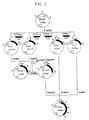
- the subunits were constructed as divided into blocks 1 to 8, which were constructed in the manner to be described below, as schematically shown in Fig. 1.
- the solid dot at one end of the line representing a chemically synthesized oligonucleotide represents introduction of a phosphate group at the 5′ end.
- the 44 oligonucleotides other than A-1, A-8, B-1, B-7, C-1, C-7, D-1 and D-8 were treated in the following manner to introduce the phosphate group into each oligonucleotide at the 5′ end.
- a 20 ⁇ l quantity of 1 to 3 ⁇ g/ml solution of each oligonucleotide was prepared, 5 units of T4 DNA polynucleotidekinase (product of Takara Shuzo Co., Ltd.), 5 ⁇ l of reaction buffer specified by the company and 1 ⁇ l of 100 mM ATP were added to the solution, and the amount of the mixture was adjusted to 50 ⁇ l with addition of water. The mixture was then reacted at 37°C for 1 hour.
- oligonucleotides serving as the starting materials for each of the blocks to be constructed e.g. oligonucleotides A-1, A-2, A-3, A-12, A-13 and A-14 in the case of block 1
- ligase reaction buffer solution of 660 mM tris-HCl (pH 7.6), 66 mM magnesium chloride and 100 mM dithiothreithol
- the amount of mixture to be reacted was adjusted to 100 ⁇ l.
- the mixture was heated in a water bath at 100°C for 2 minutes and thereafter cooled spontaneously.
- 2.5 units of T4 DNA ligase product of Takara Shuzo Co., Ltd.
- Blocks 1 to 8 were constructed by the same procedure as above.
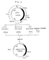
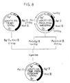
- Subunit SKA comprising block 1 and block 2 constructed by the procedure 2) was incorporated into plasmid pBR322 by the following procedure to obtain vector pSKA. Similarly, pSKB, pSKC and pSKD were prepared.
- Fig. 2 schematically shows the procedures.
- Ap r stands for ampicillin resistance
- Tc r for tetracycline resistance
- ori for a replication origin.
- the restriction enzyme recognition sites of base sequences are to be represented by the following symbols, the same as the diagrams to follow.
- a ... Aat II E ... EcoR I Na .. Nae I Sc .. Sac I
- pBR322 was treated with restriction enzymes EcoR I and Sal I and then electrophoresced on 0.9% agarose gel to obtain a DNA fragment of about 3.7 kb.
- the DNA fragment was mixed with block 1 and block 2 prepared above, T4 DNA ligase, ATP and ligase reaction buffer, followed by reaction at 4°C overnight to ligate DNA.
- E . coli HB-101 was transformed by the calcium method using the reaction mixture. DNA was collected from the colonies obtained by the simplified boiling method, followed by screening to select the colonies containing the desired pSKA.
- SKA introduction portion of pSKA was checked for base sequence by the M-13 dideoxy method. More specifically, EcoR I- Sal I fragment was subcloned in M-13 mp18 or mp19 to analyze the base sequence. As a result, the desired sequence was identified.
- E . coli HB-101 was transformed with subunit SKB comprising block 3 and block 4, followed by colony screening, and collection and purification of plasmid DNA, to prepare pSKB, which was similarly identified.
- subunit SKC comprising block 5 and block 6 was introduced in E . coli HB-101 for transformation, followed by screening of colonies, and collection and purification of plasmid DNA to prepare pSKC, which was identified similarly.
- E . coli HB-101 was likewise transformed with subunit SKD comprising block 7 and block 8, followed by screening of colonies, and collection and purification of plasmid DNA.
- This plasmid is an intermediate vector for constructing pSKX contemplated.
- the procedure for preparing the vector which is schematically illustrated in Fig. 2, is as follows.
- pSKA obtained by the procedure 3
- restriction enzyme Pst I and Nhe I was treated with restriction enzyme Pst I and Nhe I and electrophoresced on agarose gel to isolate and purify a 1072 bp DNA fragment.
- pSKB obtained by the procedure 3 was treated with the same restriction enzymes as above to isolate and purify a 3250 bp DNA fragment.
- the two DNA fragments were ligated with T4 DNA ligase, and E . coli HB-101 was transformed with the resulting reaction mixture to obtain colonies containing the desired pSKAB. Plasmid DNA was collected from the colonies and purified. pSKAB of 4323 bp was confirmed by restriction enzyme cleavage mapping.
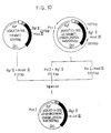
- streptokinase secreting expression vector requires a plasmid vector with a base sequence not having the codon for Met attached to the front of the first-position amino acid of streptokinase. Accordingly pSKABX was prepared by altering the N-terminal of pSKAB through the following procedure.
- Fig. 2 shows the procedure schematically.
- pSKAB was treated with restriction enzymes EcoR I and Apa I and ligated with oligonucleotides E-1 and E-2 freshly chemically synthesized, in the presence of T4 DNA ligase.
- E . coli was transformed with the reaction mixture, vector DNA was collected from the resulting colonies and purified, and a restriction enzyme cleavage map was prepared to obtain the desired pSKABX.
- the plasmid vector thus obtained can be used for constructing a system for secreting and expressing streptokinase.
- the E-1 and E-2 introducing portion of pSKABX was found to have the above base sequence by the M-13 dideoxy method [J. Messing, Methods in Enzymology, 101 , 20 (1983)].
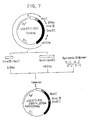
- Fig. 2 schematically shows a procedure for preparing pSKX.
- pSKABX was treated with restriction enzymes Sac I and Pst I to obtain a DNA fragment having 1352 bp, which was ligated with a DNA fragment of 309 bp obtained by treating pSKC with restriction enzymes Sac I and Aat II and a DNA fragment of 3305 pb obtained by treating pSKD with restriction enzymes Aat II and Pst I.
- E . coli was transformed with the reaction mixture, followed by the same procedures as above, i.e. collection and purification of vector DNA and restriction enzyme mapping.
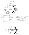
- a vector was constructed for secreting and expressing streptokinase in the periplasm of E . coli .
- the chemically synthesized streptokinase gene was ligated to tac promoter and bla signal peptide so that the codon frame of the streptokinase structural gene would not be displaced from the codon stream of amino acid sequence of bla signal peptide, whereby the desired streptokinase secreting expression vector was constructed.
- Fig. 3 shows the construction procedure. Same symbols as above were used for indicating restriction enzyme recognition sites.
- Spbla stands for bla signal peptide
- SD for ribosomal binding site
- Ptac for tac promoter.
- tac promoter is represented by the blank area, bla signal peptide by the hatched area, and the base sequence coding for the streptokinase gene by the solid black area.
- pSKX was treated with restriction enzymes EcoR V and Sal I to obtain a DNA fragment with 1251 bp.
- pSKX was treated with restriction enzymes EcoR I and Sal I to obtain a DNA fragment with 3711 bp.
- pKTN2-2 having tac promoter and bla signal peptide portions was treated with restriction enzymes EcoR I and Nae I to obtain a DNA fragment with 460 bp.
- E . coli JM-103 harboring pKTN2-2 has been deposited as FERM BP-1398.
- Vector DNA was collected from the colonies obtained, purified, followed by restriction enzyme cleavage mapping to obtain the desired vector pSKXT.
- E . coli JM-109 harboring pSKXT produces within the cell the fused protein of bla signal peptide and streptokinase through the action of tac promoter, the fused protein is then transferred by the action of bla signal peptide to the inner membrane where the signal peptide is excised by protease present in the membrane, and streptokinase only is secreted in the periplasm between the inner membrane and the outer membrane.
- E . coli JM-109 harboring vector pSKXT and obtained in Example 2 was incubated with shaking in the following manner using M-9 casamino acid liquid medium of the composition listed in Table 5 below.
- Table 5 Ingredient Amount Disodium phosphate 5.8 g Potassium dihydrogen phosphate 3.0 g Sodium chloride 5.0 g Ammonium chloride 1.0 g 1 M calcium chloride* 0.1 ml 1 M magnesium chloride* 1.0 ml Glucose* 5.0 g Casamino acid (product of Difco) 5.0 g L-proline 50 mg Vitamin B1 1 mg Water amount needed to make 1 liter of medium
- Each asterisked ingredient was separately sterilized by autoclaving (at 121°C for 15 minutes).
- ampicillin sterilized by filtration was added to a final concentration of 50 ⁇ g/ml.
- IPTG isopropyl- ⁇ -D-thiogalactoside, product of Sigma
- the culture was centrifuged (5000 r.p.m., 10 minutes) to separate cells and culture supernatant.
- the supernatant thus obtained will be referred to as the "medium fraction”.
- the cells obtained were suspended in 30 mM tris-HCl (pH 8.0)-20% sucrose buffer in the same amount as the culture, EDTA aqueous solution was added to the suspension to a final concentration of 0.01 M, and the mixture was stirred by a rotary shaker at 24°C at 180 r.p.m. for 10 minutes and then centrifuged (6000 r.p.m., 10 minutes). The resulting supernatant will be referred to as the "sucrose buffer fraction".
- the sediment resulting from the centrifugation was suspended again in ice-cooled water in the same amount as the culture, and the suspension was allowed to stand in ice for 15 minutes with occasional stirring and thereafter centrifuged (10000 r.p.m., 5 minutes). The resulting supernatant will be referred to as the "periplasm fraction.”
- the plasminogen activator activity of the streptokinase was determined by measuring the activity of plasmin produced by the plasminogen activation of the streptokinase by the method of Jackson et al. [K.W. Jackson et al, Methods in Enzymology, 80 , 387 (1981)].
- the activity of plasmin was measured using a synthetic substrate, i.e., S-2251 (D-Val-Leu-Lys-p-nitroaniline, product of Kabi Vitrum), that is, by determining p-nitroaniline released by plasmin in terms of an increase in the absorbance at 405 nm, as will be described in detail below.
- S-2251 D-Val-Leu-Lys-p-nitroaniline, product of Kabi Vitrum
- the specimen was diluted with 0.025% aqueous solution of Triton X-100, a 25 ⁇ l portion of the dilution and 25 ⁇ l of 50 mM tris-HCl buffer (pH 7.5) were placed into a test tube, and the mixture was incubated at 37°C for 5 minutes.
- Human plasminogen (product of Sigma) was diluted with 50 mM tris-HCl buffer (pH 7.5) containing 1 mg/ml of bovine serum albumin (BSA) to a concentration of 80 ⁇ g/ml.
- BSA bovine serum albumin
- the activity unit of the specimen was determined with reference to a standard curve for a standard substance, i.e., streptokinase derived from streptococcus equisimilis H46A (Group C) and having specific activity of 100 international units/ ⁇ g, the curve being obtained by using the standard substance in the same manner as above.
- a standard substance i.e., streptokinase derived from streptococcus equisimilis H46A (Group C) and having specific activity of 100 international units/ ⁇ g
- streptokinase present in the cells obtained by the procedure 2) above was detected by the Western blotting method [H. Towbin, T. Staehelin and J. Gordon, Proc. Natl. Acad. Sci., U.S.A., 76 , 4350 (1979)] using streptokinase-specific antibody as will be described below in detail.
- Streptokinase-specific anti-serum was prepared by immunizing rabbits with purified streptokinase derived from Streptococcus equisimilis H46A (Group C) as an antigen, i.e., by dissolving 1 mg of freeze-dried Streptokinase in 0.5 ml of physiological saline, adding 0.5 ml of Freunds complete adjuvant to the solution to emulsify the streptokinase, subcutaneously giving the emulsion to the back of three rabbits, similarly giving the emulsified streptokinase at the same dose every two weeks to thus immunize the rabbits four times in total, collecting the total blood 10 days after the final immunization and separating off the serum. The anti-serum thus obtained was used for detecting streptokinase.
- SDS-PAGE A specimen for polyacrylamide gel electrophoresis to be performed in the presence of sodium dodecylsulfate (hereinafter referred to as "SDS-PAGE") was prepared, for example, by suspending cells obtained from 1 ml of culture in 1 ml of 10 mM tris-HCl buffer (pH 8.0) containing 2% SDS and 2% dithiothreithol, heating the suspension at 100°C for 5 minutes and the centrifuging the suspension (12000 r.p.m., 10 minutes) to obtain a supernatant.
- SDS-PAGE was performed by electrophrescing the specimen thus prepared at 2 mA/cm on 15% polyacrylamide containing 0.1% of SDS according to the Laemmli method [U.K. Laemmli, Nature, 227 , 680 (1970)].
- the streptokinase was detected by electrophoretically transferring to a nitrocellulose film (product of Bio Rad) the protein in the resulting gel and specifically staining the streptokinase in the protein with use of specific antibody.
- the nitrocellulose film having the protein transferred thereto was shaken at room temperature for 1 hour in a 10% solution of bovine serum albumin (BSA) in 20 mM tris-HCl buffer (pH 7.5, hereinafter referred to briefly as "TBS") containing 0.15 M sodium chloride and then reacted, at room temperature for 3 hours, with the streptokinase anti-serum as diluted to 1000-fold with 0.1% BSA-TBS.
- the resulting reaction mixture was washed with TBS four times and treated for 60 minutes with peroxidase-bound anti-rabbit IgG (product of Cappel) as diluted to 2000-fold with 0.1% BSA-TBS.
- the resulting mixture was washed again and thereafter treated with 0.02 M citric acid buffer (pH 6.5) containing 0.03% hydrogen peroxide and 0.5 mg/ml 4 chloro-1-naphthol for color development.
- E . coli JM-109 harboring secreting expression vector pSKXT was incubated and fractionated by the procedures 1) and 2) above and checked by the procedure 3) to determine the amount of streptokinase expressed.
- streptokinase detected by the invention is the same as natural-type streptokinase and was produced by translation within the cell, then passed through the inner membrane by the action of the signal peptide, further processed for the removal of the signal peptide and secreted.
- Streptokinase can be collected from the extract of the fraction by a combination of separating methods such as salting-out with ammonium sulfate, isoelectric focusing, gel filtration, ion-exchange chromatography and hydrophobic chromatography, whereby a single purified product can be obtained.
- separating methods such as salting-out with ammonium sulfate, isoelectric focusing, gel filtration, ion-exchange chromatography and hydrophobic chromatography, whereby a single purified product can be obtained.
- hydrophobic chromatography and anion-exchange chromatography will be described below.
- ammonium sulfate was added to 600 ml of the periplasm fraction to a concentration of 0.6 M, and the mixture was passed through a Butyl Toyo pearl column (3 x 20 cm, product of Tosoh Corporation) equilibrated with 0.6 M ammonium sulfate. After washing the column with 500 ml of 0.3 M ammonium sulfate, 50 mM tris-HCl buffer (pH 8.5) was passed through the column to elute a fraction having streptokinase activity.
- the fraction was then dialyzed against 50 mM tris-HCl buffer (pH 8.5), and the dialyzate was separated by a fractionating HPLC system (Model HLC-837, product of Tosoh Corporation) including DEAE-5PW (2.15 x 15 cm, product of Tosoh Corporation) at a flow rate of 4 ml/min using 50 mM tris-HCl buffer (pH 8.5) as an eluent at a straight concentration gradient of 0.1 M to 0.3 M sodium chloride.
- the Butyl Toyo pearl eluate fraction after dialyzation was injected into the HPLC system, and the main protein peak portion was pooled.
- the streptokinase activity fraction was eluted at a peak in match with the protein peak.
- the active eluate fraction pooled was further desalted by Sephadex G-25 column (2.5 x 25 cm, product of Pharmacia) and then separated again by a DEAE-5PW column under the same condition as above. Finally, the streptokinase active fraction was desalted by the same Sephadex G-25 column and lyophilized to obtain a final purified product.
- a sample of the purified product obtained by the procedure 2) was analyzed by SDS-PAGE in the presence of and also in the absence of a reducing agent.
- Table 6 shows the result. For comparison, Table 6 also shows the result obtained by similarly analyzing the streptokinase derived from Streptococcus equisimilis H46A (Group G) (designated as "Natural” in Table 6.
- the amount of recombinant streptokinase collected by the foregoing method was found to be 2.5 mg by the amino acid analysis of the final purified product sample.
- the specific plasminogen activator activity was 101.5 international units/ ⁇ g, which was comparable to that (100 international units/ ⁇ g) of the natural type.
- streptokinase obtained was identical with the streptokinase (of the natural type) derived from Streptococcus equisimilis H46A (Group C).
- streptokinase derivative protein (1-372) expression vector was prepared by the following procedure using streptokinase expression vector pSKXT obtained in Example 2.
- Fig. 4 schematically shows the procedure.
- streptokinase derivative protein corresponding to streptokinase wherein amino acid residues toward the C-terminal are deficient
- DNA oligonucleotide
- the vector for expressing the derivative protein was prepared by removing fragments having restriction enzyme recognition site Acc I (present at the 367-position of streptokinase and at three other positions) and restriction enzyme recognition site Sal I (present downstream from the termination codon of streptokinase and at another position) from pSKXT, and inserting the fragments F-1 and F-2 into the plasmid.
- pSKXT was treated with restriction enzymes Pst I and Mlu I to obtain a DNA fragment of 1635 bp, with restriction enzymes Pst I and Sal I to produce a DNA fragment of 2962 bp, and further with restriction enzymes Acc I and Mlu I to produce a DNA fragment of 677 bp. These fragments were collected by agarose gel electrophoresis and reacted with the chemically synthesized DNA fragment obtained by the procedure 1) above for ligation. E . coli JM109 was transformed with the reaction mixture, vector DNA collected from the resulting colonies and purified, and a restriction enzyme map was prepared to obtain the desired vector pSKXT (1-372).
- E . coli JM-109 harboring vector pSKXT (1-372) obtained by the procedure 2) was incubated with shaking in the same manner as in Example 3, 1) using M-9 casamino acid liquid medium of the composition listed in Table 5.
- streptokinase derivative protein (1-372) was produced by translation within the cell, then passed through the inner membrane by the action of the signal peptide, further processed for the removal of the signal peptide and secreted.
- the purified product When analyzed by SDS-PAGE in the presence of a reducing agent, the purified product exhibited a single band at the position of contemplated molecular weight of about 42300.
- the sequence of 20 amino acid residues from the amino end of the purified product was analyzed by the method of Heiwick et al., and the sequence of 5 residues from the carboxyl terminal thereof was analyzed with use of carboxypeptidases A and B. As a result, the product was identified as the desired streptokinase derivative protein (1-372).
- the amount of recombinant streptokinase derivative protein (1-372) obtained by the above method was found to be 9 mg by the amino acid analysis of a sample of the purified product.
- the specific plasminogen activator activity was 107 in mole ratio based on that of the natural-type streptokinase which was taken as 100 and was comparable to that of the natural type.
- a vector for expressing a streptokinase derivative protein (1-372, 118 deficient) was prepared by the following procedure using streptokinase derivative expression vector pSKXT (1-372) obtained in Example 5.
- Fig. 5 schematically shows the procedure.
- the desired vector is shown as "pSKXT (1-372, 118 def)" in the figure.
- the desired derivative protein expression vector was prepared by replacing the sequence of pSKXT (1-372) between restriction enzyme recognition sites Nhe I and Hinf I by the following freshly chemically synthesized oligonucleotide (DNA) fragments a-1 to a-4.
- pSKXT (1-372) was treated with restriction enzymes Pst I and Nhe I to form a DNA fragment of 1522 bp, with restriction enzymes Pst I and Sac I to form a DNA fragment of 3488 bp and further with restriction enzymes Sac I and Hinf I to form a DNA fragment of 212 bp. These fragments were collected by agarose gel electrophoresis and reacted with the four DNA fragments chemically synthesized by the procedure 1) above for ligation. (Oligomers a-2 and a-4 were used as phosphorylated at their 5 ends). E . coli JM109 was transformed with the reaction mixture, vector DNA was collected from the resulting colonies and purified, and a restriction enzyme map was prepared to obtain the desired vector pSKXT (1-372, 118 deficient).
- Example 5 In the same manner as in Example 5, 4), 1.6 liters of culture was centrifuged to collect the cells, from which a periplasm fraction was extracted by the osmotic shock method. The desired derivative protein (1-372, 118 deficient) was collected from the extract and purified.
- the purified product When analyzed by SDS-PAGE in the presence of a reducing agent, the purified product exhibited a single band at the position of contemplated molecular weight of about 42200.
- the amount of recombinant streptokinase derivative protein (1-372, 118 deficient) obtained by the above method was found to be 2.4 mg by the amino acid analysis of a sample of the purified product.
- the specific plasminogen activator activity was 100 in mole ratio based on that of the natural-type streptokinase which was taken as 100 and was equivalent to that of the natural type.
- a vector for expressing a streptokinase derivative protein (1-372, 45-68 deficient) was prepared by the following procedure using streptokinase derivative expression vector pSKXT (1-372) obtained in Example 5.
- Fig. 6 schematically shows the procedure. The desired vector is shown as “pSKXT (1-372, 45-68 def)" in the figure.
- the desired derivative protein expression vector was prepared by replacing the sequence of pSKXT (1-372) at the region between restriction enzyme sites Nsp(7524) V and Xho I by the following freshly chemically synthesized oligonucleotide (DNA) fragments (b-1 and b-2).
- pSKXT (1-372) was treated with restriction enzymes Pst I and Nsp(7524) V to form a DNA fragment of 1325 bp and with restriction enzymes Pst I and Xho I to form a DNA fragment of 3864 bp. These fragments were collected by agarose gel electrophoresis and reacted with the two DNA fragments chemically synthesized by the procedure 1) above for ligation. E . coli JM109 was transformed with the reaction mixture, vector DNA was collected from the resulting colonies and purified, and a restriction enzyme map was prepared to obtain the desired vector pSKXT (1-372, 45-48 deficient).
- E . coli JM109 harboring vector pSKXT (1-372, 45-68 deficient) obtained by the procedure 2) was incubated with shaking similarly using the same M-9 casamino acid liquid medium, and the cells were collected and checked for streptokinase activity (plasminogen activator activity). Consequently, an amount of expression of about 1200 international units/ml was detected from the cell fraction and was found almost entirely in the periplasm fraction. Further analysis by Western blotting revealed the immunoactivity at the position of calculated molecular weight of about 39600.
- Example 5 In the same manner as in Example 5, 4), cells were collected from 800 ml of culture, and a periplasm fraction was extracted from the cells by the osmotic shock method.
- the desired derivative protein (1-372, 45-68 deficient) was collected from the extract, purified and lyophilized to obtain a purified product.
- Table 9 shows the result of amino acid analysis of the product.
- Amino acid Purified product Calculated Asp 54.3 57 Thr 24.9 25 Ser 19.2 20 Glu 36.7 36 Pro 13.9 14 Gly 13.9 14 Ala 17.0 17 Val 22.0 22 1/2 Cys Not detected 0 Met 3.8 4 Ile 20.0 22 Leu 36.6 36 Tyr 14.7 16 Phe 13.9 14 Trp Not detected 1 Lys 27.2 27 His 6.8 7 Arg 15.1 16
- the amount of recombinant streptokinase derivative protein (1-372, 45-68 deficient) obtained by the above method was found to be 2 mg by the amino acid analysis of a sample of the purified product.
- the specific plasminogen activator activity was 112 in mole ratio based on that of the natural-type streptokinase which was taken as 100 and was comparable to that of the natural type.
- a vector for expressing a streptokinase derivative protein (1-372, replaced by Gln at each of the 256- and 257-positions) was prepared by the following procedure using streptokinase derivative expression vector pSKXT (1-372) obtained in Example 5.
- Fig. 7 schematically shows the procedure.
- the desired vector is shown as "pSKXT (1-372, 256Gln, 257Gln sub)" in the figure.
- the desired derivative protein expression vector was prepared by replacing the sequence of pSKXT (1-372) at the region between restriction enzyme sites Hind III and SanB I by the following freshly chemically synthesized oligonucleotide (DNA) fragments (c-1 to c-4).
- fragments include a region of streptokinase wherein Gln is substituted at each of the 256- and 257-positions.
- the four fragments c-1 to c-4 were individually chemically synthesized in the same manner as above.
- restriction enzyme recognition site EcoR V was further provided to the sequence.
- pSKXT (1-372) was treated with restriction enzymes Hind IIII and Nco I to form a DNA fragment of 608 bp and with restriction enzymes Nco I and SnaB I to form a DNA fragment of 4612 bp. These fragments were collected by agarose gel electrophoresis and reacted with the four DNA fragments chemically synthesized by the procedure 1) above for ligation. (Oligomers c-2 and c-4 were used as phosphorylated at their 5′ ends). E . coli JM109 was transformed with the reaction mixture, vector DNA was collected from the resulting colonies and purified, and a restriction enzyme map was prepared to obtain the desired vector.
- E . coli JM109 harboring the vector obtained by the procedure 2 was incubated with shaking in the same manner as already stated using the same M-9 casamino acid liquid medium as above. After the addition of IPTG, the cells were collected and checked for streptokinase activity (plasminogen activator activity) similarly. As a result, no activity was detected from the medium supernatant but expression of about 70 international units/ml was detected from the cell fraction, i.e., almost entirely from the periplasm fraction. Further analysis by Western blotting revealed the immunoactivity at the position of calculated molecular weight of about 42300.
- Example 5 In the same manner as in Example 5, 4), 2.4 liters of culture was centrifuged to collect the cells, from which a periplasm fraction was extracted by the osmotic shock method.
- the desired derivative protein (1-372, 256Gln, 257Gln replaced) was collected from the extract, purified and lyophilized to obtain a purified product.
- the purified product When analyzed by SDS-PAGE in the presence of a reducing agent, the purified product exhibited a single band at the position of contemplated molecular weight of about 42300.
- Table 10 shows the result obtained by subjecting the purified product to amino acid analysis.
- Amino acid Purified product Calculated Asp 56.6 58 Thr 25.8 27 Ser 21.0 23 Glu 40.7 40 Pro 17.5 17 Gly 18.3 18 Ala 19.3 19 Val 19.9 22 1/2 Cys Not detected 0 Met 3.6 4 Ile 19.5 22 Leu 35.9 37 Tyr 15.5 16 Phe 15.1 15 Trp Not detected 1 Lys 27.9 28 His 7.5 8 Arg 16.8 17
- the amount of recombinant streptokinase derivative protein (1-372, 256Gln, 257Gln replaced) obtained by the above method was found to be 4 mg by the amino acid analysis of the purified product.
- the specific plasminogen activator activity was 7 in mole ratio based on that of the natural-type streptokinase which was taken as 100.
- a vector for expressing a streptokinase derivative protein (1-372, 118 deficient, 256Gln, 257Gln replaced) was prepared by the following procedure using streptokinase derivative expression vector pSKXT (1-372, 118 deficient) obtained in Example 6 and streptokinase derivative protein expression vector pSKXT (1-372, 256Gln, 257Gln replaced) obtained in Example 8.
- Fig. 8 schematically shows the procedure.
- the desired vector is shown as "pSKXT (1-372, 118 def, 256Gln, 257Gln sub)" in the figure.
- pSKXT (1-372, 118 deficient) was treated with restriction enzymes Bgl II and Hind III to form a DNA fragment of 515 bp.
- pSKXT (1-372, 256Gln, 257Gln replaced) was treated with restriction enzymes Pst I and Bgl II to form a DNA fragment of 1443 bp and with restriction enzymes Pst I and Hind III to form a DNA fragment of 3335 bp. These DNA fragments were collected by agarose gel electrophoresis and ligated together by reaction. E .
- coli JM109 was transformed with the reaction mixture, vector DNA was collected from the resulting colonies and purified, and a restriction enzyme map was prepared to obtain the desired vector pSKXT (1-372, 118 deficient, 256Gln, 257Gln replaced).
- E . coli JM109 harboring the vector obtained by the procedure 1) was incubated with shaking in the same manner as already stated using the same M-9 casamino acid liquid medium as above.
- Example 5 In the same manner as in Example 5, 4), 1.2 liters of culture was centrifuged to collect the cells, from which a periplasm fraction was extracted by the osmotic shock method.
- the desired derivative protein (1-372, 118 deficient, 256Gln, 257Gln replaced) was collected from the extract and purified.
- the purified product When analyzed by SDS-PAGE in the presence of a reducing agent, the purified product exhibited a single band at the position of contemplated molecular weight of about 42200.
- the amount of recombinant streptokinase derivative protein (1-372, 118 deficient, 256Gln, 257Gln replaced) obtained by the above method was found to be 5.4 mg by the amino acid analysis of the purified product.
- the specific plasminogen activator activity was 9.98 in mole ratio based on that of the natural-type streptokinase which was taken as 100.
- a vector for expressing a streptokinase derivative protein (1-372, 45-68 deficient, 256Gln, 257Gln replaced) was prepared by the following procedure using streptokinase derivative expression vectors pSKXT (1-372, 45-68 decicient) and pSKXT (1-372, 256Gln, 257Gln replaced) obtained in Examples 7 and 8, respectively.
- Fig. 9 schematically shows the procedure.
- the desired vector is shown as "pSKXT (1-372, 45-68 def, 256Gln, 257Gln sub)" in the figure.
- pSKXT (1-372, 45-68 deficient) was treated with restriction enzymes Pst I and Mlu I to form a DNA fragment of 1563 bp.
- pSKXT (1-372, 256Gln, 257Gln replaced) was treated with restriction enzymes Pst I and Mlu I to form a DNA fragment of 3661 bp.
- These DNA fragments were collected by agarose gel electrophoresis and ligated together by reaction.
- E . coli JM109 was transformed with the reaction mixture, vector DNA was collected from the resulting colonies and purified, and a restriction enzyme map was prepared to obtain the desired vector pSKXT (1-372, 45-68 deficient, 256Gln, 257Gln replaced).
- E . coli JM109 harboring the vector obtained by the procedure 1) was incubated with shaking in the same manner as already stated using the same M-9 casamino acid liquid medium as above.
- Example 5 In the same manner as in Example 5, 4), 0.8 liter of culture was centrifuged to collect the cells, from which a periplasm fraction was extracted by the osmotic shock method.
- the desired derivative protein (1-372, 45-68 deficient, 256Gln, 257Gln replaced) was collected from the extract and purified.
- the purified product When analyzed by SDS-PAGE in the presence of a reducing agent, the purified product exhibited a single band at the position of contemplated molecular weight of about 39800.
- the amount of recombinant streptokinase derivative protein (1-372, 45-68 deficient, 256Gln, 257Gln replaced) obtained by the above method was found to be 0.56 mg by the amino acid analysis of the purified product.
- the specific plasminogen activator activity was 6.30 in mole ratio based on that of the natural-type streptokinase which was taken as 100.
- a vector for expressing a streptokinase derivative protein (1-372, 45-68 deficient, 118 deficient, 256Gln, 257Gln replaced) was prepared by the following procedure using streptokinase derivative expression vectors pSKXT (1-372, 118 deficient) and pSKXT (1-372, 45-68 deficient, 256Gln, 257Gln replaced) obtained in Example 6 and 10, respectively.
- Fig. 10 schematically shows the procedure.
- the desired vector is shown as "pSKXT (1-372, 45-68 def, 118 def, 256Gln, 257Gln sub)" in the figure.
- pSKXT (1-372, 118 deficient) was treated with restriction enzymes Bgl II and Hind III to form a DNA fragment of 515 bp.
- pSKXT (1-372, 45-68 deficient, 256Gln, 257Gln replaced) was treated with restriction enzymes Pst I and Bgl II to form a DNA fragment of 1371 bp and with restriction enzymes Pst I and Hind III to form a DNA fragment of 3335 bp. These DNA fragments were collected by agarose gel electrophoresis and ligated together by reaction. E .
- coli JM109 was transformed with the reaction mixture, vector DNA was collected from the resulting colonies and purified, and a restriction enzyme map was prepared to obtain the desired vector pSKXT (1-372, 45-68 deficient, 118 deficient, 256Gln, 257Gln replaced).
- E . coli JM109 harboring the above vector has been deposited in Fermentation Research Institute, Agency of Industrial Science and Technology, MITI with the designation " Escherichia coli , JM-109, pSKXT (1-372, ⁇ 45-68, ⁇ 118, QQ") and the deposition number FERM BP-2828.
- E . coli JM109 (FERM BP-2828) harboring the vector obtained by the procedure 1) was incubated with shaking in the same manner as already stated using the same M-9 casamino acid liquid medium as above.
- Example 5 In the same manner as in Example 5, 4), 1.2 liters of culture was centrifuged to collect the cells, from which a periplasm fraction was extracted by the osmotic shock method.
- the desired derivative protein (1-372, 45-68 deficient, 118 deficient, 256Gln, 257Gln replaced) was collected from the extract and purified.
- the purified product When analyzed by SDS-PAGE in the presence of a reducing agent, the purified product exhibited a single band at the position of contemplated molecular weight of about 39700.
- the amount of recombinant streptokinase derivative protein (1-372, 45-68 deficient, 118 deficient, 256Gln, 257Gln replaced) obtained by the above method was found to be 2.4 mg by the amino acid analysis of the purified product.
- the specific plasminogen activator activity was 7.74 in mole ratio based on that of the natural-type streptokinase which was taken as 100.
- a vector for expressing a streptokinase derivative protein (1-372, altered to -Lys-Pro-Lys-Pro- at the 256- and 257-positions) was prepared by the following procedure using streptokinase derivative expression vector pSKXT (1-372) obtained in Example 5.
- Fig. 11 schematically shows the procedure.
- the desired vector is shown as "pSKXT (1-372, 256, 257LysProLysPro rep)" in the figure.
- the desired derivative protein expression vector was prepared by replacing the sequence of pSKXT (1-372) at the region thereof between restriction enzyme recognition sites Hind III and SnaB I by the new DNA fragments d-1 to d-4 chemically synthesized and represented below.
- fragments include Lys-Pro-Lys-Pro substituted for Lys-Lys at the 256- and 257-positions of streptokinase and are used for an alteration by replacing some amino acid residues by other amino acid residues according to the invention.
- the four oligonucleotides d-1 to d-4 were individually synthesized chemically. For the analysis to be performed later, new restriction enzyme recognition site EcoR V was provided.
- pSKXT (1-372) was treated with restriction enzymes Hind III and Nco I to form a DNA fragment of 608 bp and with restriction enzymes Nco I and SnaB I to form a DNA fragment of 4612 bp. These fragments were collected by agarose gel electrophoresis and reacted with the four chemically synthesized DNA fragments (with the oligomers d-2 and d-4 phosphorylated at the 5′ ends). E . coli JM109 was transformed with the reaction mixture, vector DNA was collected from the resulting colonies and purified, and a restriction enzyme map was prepared to obtain the desired vector.
- E . coli JM109 harboring the vector obtained by the procedure 2) above was incubated with shaking in the same manner as already stated using the same M-9 casamino acid liquid medium as above.
- Example 5 In the same manner as in Example 5, 4), 2.4 liters of culture was centrifuged to collect the cells, from which a periplasm fraction was extracted by the osmotic shock method.
- the desired derivative protein (1-372, 256, 257Lys-Pro-Lys-Pro altered) was collected from the extract and purified to obtain a lyophilized purified product.
- the purified product When analyzed by SDS-PAGE in the presence of a reducing agent, the purified product exhibited a single band at the position of contemplated molecular weight of about 42500.
- Table 14 shows the result obtained by subjecting the purified product to amino acid analysis.
- Amino acid Purified product Calculated Asp 60.6 58 Thr 27.2 27 Ser 22.4 23 Glu 38.7 38 Pro 18.1 19 Gly 18.5 18 Ala 19.9 19 Val 20.7 22 1/2 Cys Not detected 0 Met 3.4 4 Ile 20.2 22 Leu 38.3 37 Tyr 15.8 16 Phe 14.8 15 Trp Not detected 1 Lys 30.1 30 His 8.1 8 Arg 16.1 17
- the amount of recombinant streptokinase derivative protein (1-372, 256, 257Lys-Pro-Lys-Pro altered) thus obtained was found to be 0.46 mg by the amino acid analysis of the purified product.
- the specific plasminogen activator activity was 17.7 in mole ratio based on that of the natural-type streptokinase which was taken as 100.
Abstract
Description
- The present invention relates to a novel chemically synthesized gene including a base sequence coding for the primary amino acid sequence of streptokinase, a corresponding plasmid recombinant, corresponding transformant and process for preparing streptokinase by the incubation of the transformant, the invention further relating to novel streptokinase derivative proteins having a modified primary amino acid sequence, chemically synthesized gene including a base sequence coding for the protein, plasmid recombinant containing the gene, transformant transformed by the recombinant, and process for preparing the streptokinase derivative protein by the incubation of the transformant.
- Streptokinase is a protein produced or secreted by hemolytic streptococci and having thrombolytic activity and a molecular weight of about 47000. The primary amino acid sequence of the protein is represented by the formula (1) given below. The total base sequence coding for the protein has also been determined from the DNA of Streptococcus equisimilis H46A (Group C) which produces the protein.
- Streptokinase is already in clinical use as a thrombolytic agent in U.S. and European countries, is widely used for treating patients with lung thrombus and acute myocardial infarction and has been proved fully effective.
- However, when streptokinase is prepared by incubating original bacteria, the resulting culture contains some extracellular secretions, many of which are toxic or likely to be toxic to humans, so that the preparation of streptokinase from the culture for clinical use requires many purification steps which must be executed with utmost care. Further it has been reported that the streptokinase obtained by the incubation of original bacteria cause various symptoms of shock due to the antigenicity thereof. It has therefore been desired in the art to develop techniques for reducing the antigenicity or to provide novel streptokinase derivatives with reduced antigenicity. For clinical use, it is also desired to improve streptokinase in its stability in blood and its specificity of thrombolytic activity.
- An object of the present invention is to provide a process for preparing a large quantity of streprokinase with ease and high purity by resorting to gene recombination techniques, chemically synthesized gene of streptokinase wherein nucleotide codons are used to practice the process easily with use of E. coli, corresponding plasmid recombinant (streptokinase expression vector) having the gene introduced therein, host cell (transformant) transformed with the recombinant, and techniques for incubating the cell.
- Another object of the invention is to provide a novel streptokinase derivative protein which has activities inherent in streptokinase, especially thrombolytic activity, and which is reduced in the binding activity to a streptokinase-specific antibody and in the productivity of the specific antibody (antigenicity) when the protein is administered.
- Another object of the invention is to provide the derivative protein which is improved in its stability in blood and in thrombolytic specificity.
- Another object of the invention is to provide a process for preparing the novel streptokinase derivative protein by utilizing gene recombination techniques capable of producing the protein in a large quantity with ease and high purify, chemically synthesized gene wherein nucleotide codons are used to practice the process easily with use of E. coli, corresponding plasmid recombinant (expression vector) having the gene inserted therein, host cell (transformant) transformed with the reccmbinant, and techniques for preparing the protein by incubating the cell.
- The present invention provides a chemically synthesized gene coding for streptokinase of the natural-type having a primary amino acid sequence represented by the following formula (1), a corresponding plasmid recombinant having the gene introduced therein (natural-type streptokinase expression vector), a corresponding transformant obtained from the recombinant by transformation, and a process for preparing natural-type streptokinase by incubating the transformant.
Formula (1):
Ile-Ala-Gly-Pro-Glu-Trp-Leu-Leu-Asp-Arg-
Pro-Ser-Val-Asn-Asn-Ser-Gln-Leu-Val-Val-
Ser-Val-Ala-Gly-Thr-Val-Glu-Gly-Thr-Asn-
Gln-Asp-Ile-Ser-Leu-Lys-Phe-Phe-Glu-Ile-
Asp-Leu-Thr-Ser-Arg-Pro-Ala-His-Gly-Gly-
Lys-Thr-Glu-Gln-Gly-Leu-Ser-Pro-Lys-Ser-
Lys-Pro-Phe-Ala-Thr-Asp-Ser-Gly-Ala-Met-
Ser-His-Lys-Leu-Glu-Lys-Ala-Asp-Leu-Leu-
Lys-Ala-Ile-Gln-Glu-Gln-Leu-Ile-Ala-Asn-
Val-His-Ser-Asn-Asp-Asp-Tyr-Phe-Glu-Val-
Ile-Asp-Phe-Ala-Ser-Asp-Ala-Thr-Ile-Thr-
Asp-Arg-Asn-Gly-Lys-Val-Tyr-Phe-Ala-Asp-
Lys-Asp-Gly-Ser-Val-Thr-Leu-Pro-Thr-Gln-
Pro-Val-Gln-Glu-Phe-Leu-Leu-Ser-Gly-His-
Val-Arg-Val-Arg-Pro-Tyr-Lys-Glu-Lys-Pro-
Ile-Gln-Asn-Gln-Ala-Lys-Ser-Val-Asp-Val-
Glu-Tyr-Thr-Val-Gln-Phe-Thr-Pro-Leu-Asn-
Pro-Asp-Asp-Asp-Phe-Arg-Pro-Gly-Leu-Lys-
Asp-Thr-Lys-Leu-Leu-Lys-Thr-Leu-Ala-Ile-
Gly-Asp-Thr-Ile-Thr-Ser-Gln-Glu-Leu-Leu-
Ala-Gln-Ala-Gln-Ser-Ile-Leu-Asn-Lys-Asn-
His-Pro-Gly-Tyr-Thr-Ile-Tyr-Glu-Arg-Asp-
Ser-Ser-Ile-Val-Thr-His-Asp-Asn-Asp-Ile-
Phe-Arg-Thr-Ile-Leu-Pro-Met-Asp-Gln-Glu-
Phe-Thr-Tyr-Arg-Val-Lys-Asn-Arg-Glu-Gln-
Ala-Tyr-Arg-Ile-Asn-Lys-Lys-Ser-Gly-Leu-
Asn-Glu-Glu-Ile-Asn-Asn-Thr-Asp-Leu-Ile-
Ser-Glu-Lys-Tyr-Tyr-Val-Leu-Lys-Lys-Gly-
Glu-Lys-Pro-Tyr-Asp-Pro-Phe-Asp-Arg-Ser-
His-Leu-Lys-Leu-Phe-Thr-Ile-Lys-Tyr-Val-
Asp-Val-Asp-Thr-Asn-Glu-Leu-Leu-Lys-Ser-
Glu-Gln-Leu-Leu-Thr-Ala-Ser-Glu-Arg-Asn-
Leu-Asp-Phe-Arg-Asp-Leu-Tyr-Asp-Pro-Arg-
Asp-Lys-Ala-Lys-Leu-Leu-Tyr-Asn-Asn-Leu-
Asp-Ala-Phe-Gly-Ile-Met-Asp-Tyr-Thr-Leu-
Thr-Gly-Lys-Val-Glu-Asp-Asn-His-Asp-Asp-
Thr-Asn-Arg-Ile-Ile-Thr-Val-Tyr-Met-Gly-
Lys-Arg-Pro-Glu-Gly-Glu-Asn-Ala-Ser-Tyr-
His-Leu-Ala-Tyr-Asp-Lys-Asp-Arg-Tyr-Thr-
Glu-Glu-Glu-Arg-Glu-Val-Tyr-Ser-Tyr-Leu-
Arg-Tyr-Thr-Gly-Thr-Pro-Ile-Pro-Asp-Asn-
Pro-Asn-Asp-Lys - The present invention further provides a streptokinase derivative protein which has a modified primary amino acid sequence corresponding to the amino acid sequence respresented by the formula (1) of natural-type streptokinase which is deficient in the amino acid residues at the 373-position to the C-terminal and wherein at least one amino acid residue may optionally be deficient, replaced or inserted.
- The symbols as used in the formula (1) and hereinafter to represent amino acid sequences and amino acid residues, as well as base sequences, nucleic acid bases, etc., are according to the nomenclature of IUPAC-IUB and to the practice in the art, as exemplified below.
Ala: alanine
Asn: asparagine
Cys: cysteine
Glu: glutamic acid
His: histidine
Leu: leucine
Met: methionine
Pro: proline
Thr: threonine
Tyr: tyrosine
A: adenine
G: guanine
Arg: arginine
Asp: aspartic acid
Gln: glutamine
Gly: glycine
Ile: isoleucine
Lys: lysine
Phe: phenylalanine
Ser: serine
Try: tryptophan
Val: valine
T: thymine
C: cytosine - The positions of the amino acid residues in amino acid sequences are all expressed according to the amino acid sequence of the formula (1) even when an amino acid or amino acids are deficient or inserted.
- Given below are preferred examples of streptokinase derivative proteins of the invention having modified primary amino acid sequences.
- (1) Polypeptide having the amino acid sequence (represented by the formula (1)) of natural-type streptokinase wherein the amino acid residues at the 373-position to the C-terminal are deficient.
- (2) Polypeptide having the amino acid sequence of natural-type streptokinase wherein the amino acid residues at the 373-position to the C-terminal are deficient, and further the amino acid sequence of at least from Arg at the 45-position to Gly at the 68-position is deficient.
- (3) Polypeptide having the amino acid sequence of natural-type streptokinase wherein the amino acid residues at the 373-position to the C-terminal are deficient, and further at least Phe at the 118-position is deficient or replaced by other amino acid residue.
- (4) Polypeptide having the amino acid sequence of natural-type streptokinase wherein the amino acid residues at the 373-position to the C-terminal are deficient, and further at least Lys at the 256-position and Lys at the 257-position are deficient or replaced by other amino acid residues.
- (5) Polypeptide having the amino acid sequence of natural-type streptokinase wherein the amino acid residues at the 373-position to the C-terminal are deficient, and further other amino acid residue is inserted in the position next to at least each of Lys at the 256-position and Lys at the 257-position.
- The other amino acid residue which can be substituted or inserted in the above modified amino acid sequences can be any of those constituting the protein of streptokinase and is preferably selected from among the α-amino acids constituting the proteins of the human body. Examples of such amino acids are Pro, Gln, Thr, Ser, His and the like. More specifically, examples of preferred amino acid residues to be substituted in the 256-position are Gln, Thr, His and the like. Those to be substituted in the 257-position are Pro, Gln, Ser and His. Those to be inserted in the 256- and 257-positions include Pro.
- The present invention further provides a chemically synthesized gene including a base sequence coding for the streptokinase derivative protein having the modified amino acid sequence, a corresponding plasmid recombinant having the gene inserted therein (streptokinase derivative protein expression vector), a host cell transformed with the recombinant, and a process for preparing the streptokinase derivative protein by incubating the transformant.
- A detail description will be given of the streptokinase derivative protein of the invention, the process for preparing the same and the process for preparing natural-type streptokinase of the invention as well as the chemically synthesized gene, the expression vector and the transformant which are used in these processes.
- In accordance with the primary amino acid sequence of the formula (1), various base sequences can be determined for the chemically synthesized gene coding for the primary amino acid sequence of natural-type streptokinase of the invention. It is desirable to employ the following standards for this purpose.
- 1) Select triplet codons which are frequently used in the host cell, e.g., in E. coli.
- 2) Give specific restriction enzyme recognition sites within the base sequence to be determined and at the opposite ends thereof to ensure facilitated ligation to the base sequence of other gene or the like and facilitated insertion into the plasmid vector utilizing these sites as desired.
- 3) In joining or ligating chemically synthesized genes (DNA fragments), avoid or minimize ligations other than the desired ligation.
- 4) Eliminate objectionable sequences, such as terminator, from the base sequence to be designed.
- 5) Provide a restriction enzyme recognition site at a suitable position so as to facilitate the subsequent modification of the gene.
- The formula (2) given below represents a preferred example of base sequence thus determined of chemically synthesized gene of natural-type streptokinase. The corresponding amino acid sequence is also given.


- The base sequence represented by the formula (2) is in the form of a double-stranded DNA sequence coding for the primary amino acid sequence (formula (1)) of natural-type streptokinase and comprising single-stranded DNA sequences which are complementary to each other. These complementary DNA sequences are also included in the present invention.
- In view of the use of E. coli as the host cell, codons which are frequently used for E. coli are preferentially selected for designing the base sequence of the formula (2), whereas the codons to be used for the present gene are not limited to such codons. Furthermore, the host cells to be used in the present invention are not limited to those of E. coli as will be described later. Accordingly, codons which are frequently used in the host cell can be selected for use in combination. More specifically, the base sequence of the desired gene can be the base sequence of any chemically synthesized gene insofar as the sequence has the same genetic information as the base sequence of the formula (2), i.e., insofar as the base sequence contains the one coding for the primary amino acid sequence of streptokinase so that streptokinase can be expressed and produced by gene recombination techniques. Thus, the base sequence of the formula (2) can be altered or modified by local change, deletion or addition of some nucleic acid bases. Such alteration or modification of the base sequence includes use of genetic codons coding for the same primary amino acid sequence as the one coded for by the base sequence of the formula (2), and provision of various restriction enzyme recognition sites for effecting ligation with regulators, such as promoter, which are required in actually inserting the resulting base sequence into a suitable vector for expression in a microorganism. In constructing the desired expression vector by gene engineering techniques utilizing the gene of the present invention, it is necessary to suitably attach various regulators such as promoter, Shine-Dalgarno sequence (SD sequence) and like ribosomal binding sites, protein synthesis initiation codon and termination codon, etc. Since the ligation and cleavage of base sequences are effected with use of restriction enzymes, there is a need to provide suitable restriction enzyme recognition site at the upstream and downstream ends thereof. The formula (3) given below represents an example of gene wherein such suitable restriction enzyme recognition sites are attached. The gene comprises the base sequence of the formula (2) and specific restriction enzyme recognition sites provided at the upstream and downstream ends thereof for attaching promoter and like regulators which are required for the expression of the desired protein encoded by the base sequence. The gene is desirable for the subsequent construction of expression vector. The restriction enzyme recognition sites present in the base sequence of the formula (3) are shown by the formula (4). Of course, the formula (4) shows only examples of recognition sites; those to be present in the gene of the invention can be suitably modified or selected in accordance with the kind of expression vector to be constructed.


- The chemically synthesized gene coding for the streptokinase derivative protein of the invention having a modified primary amino acid sequence can be designed based on the gene coding for natural-type streptokinase, using a DNA sequence (codons) coding for amino acid residues corresponding to the modification of the primary amino acid sequence. A specific portion of the DNA sequence can be modified by various methods known in the art. The modification procedure includes cleavage or removal of a specified region with use of restriction enzymes, or replacement of a specific region by a chemically synthesized oligonucleotide including a modified portion which is separately prepared.
- Preferably, the chemically synthesized gene of the present invention is prepared by chemically synthesizing some oligonucleotide fragments according to the base sequence designed and ligating these fragments. The oligonucleotide fragments can be chemically synthesized with ease by usual methods using a commercial DNA synthesizer or the like. These methods include, for example, the solid-phase phosphite triester method [Nature, 310, 105(1984)] and the solid-phase phosphoamidide method (S.L. Beaucage and M.H. Caruthers, Tetrahedron Letters, 22, 1859(1981)). The oligonucleotide fragments prepared can be isolated and purified by a usual method such as high performance liquid chromatography. The purified base sequences can be checked, for example, by the Maxam-Gilbert method [A.M. Maxam and W. Gilbert, Proc. Natl. Acad. Sci., U.S.A., 74, 560 (1977); A.M. Maxam and W. Gilbert, Methods in Enzymol., 65, 499, Acad. Press (1980)].
- The desired gene of the invention can be constructed by phosphorylating the resulting oligonucleotides at the hydroxyl groups of their 5′ ends with T4 polynucleotidekinase, followed by annealing, ligating the oligonucleotides into blocks with use of T4 DNA ligase, similarly ligating the blocks into some subunits, and ligating the subunits. These subunits and the desired gene can be incorporated into suitable vectors, e.g. pBR322, for preservation or amplification. It is desirable to isolate the subunits or the gene from the vector for use in the subsequent procedure. The above gene preparation procedure is described specifically in examples given later.
- The plasmid vector can be constructed by employing usual procedures or methods of gene engineering techniques. These include cleavage of DNA with restriction enzymes, S1 nuclease and the like, ligation of DNA fragments with T4 DNA ligase or the like, isolation and purification of DNA by agarose gel electrophoresis, polyacrylamide gel electrophoresis or the like, collection and purification of DNA by the phenol extraction method, etc. The host cells can be checked for the presence of the plasmid vector by the alkali SDS extraction method [H.C. Birnboim and J. Doly, Nucleic Acids Res., 7, 1513 (1979)], i.e., by treating the plasmid DNA collected therefrom with restriction enzymes and checking the DNA for the presence of corresponding restriction enzyme recognition sites or checking the length of DNA fragment produced. The presence of the vector can be checked also by analyzing the base sequence of the gene by the direct dideoxy method [F. Sanger et al., Proc., Natl. Acad. Sci., U.S.A., 74, 5463 (1977)] or like method.
- Suitable vectors for use in constructing the plasmid recombinant incorporating the gene of the invention are pBR322 and various plasmid vectors derived therefrom. However, useful vectors are not limited to these but include various known ones such as bacteriophage, virus vectors inclusive of animal and vegetable viruses, plasmids, cosmids and the like.
- For the transformant obtained by incorporating the vector to express the desired streptokinase or the protein of a derivative thereof, the vector needs to have, in addition to the gene of the invention, various regulators such as promoter, terminator, poly-A tail adding signal (in the case where the host cells are eucaryotic cells) and the like for transcription, and ribosomal binding site and the like for translation. Various promoters are known in different host cells. Examples of promoters in E. coli are trp promoter, lac promoter, tac promoter, λP L promoter, lpp promoter and the like, those in Bacillus subtilis are SP01 promoter, SP02 promoter, pen promoter and the like, those in yeast and other eucaryotic cells are PH05 promoter, PGK promoter, SV40-derived promoter and the like. The desired vector of the invention can be obtained by selecting a plasmid containing such regulators as a vector, or isolating such regulators from a plasmid by the usual method or chemically synthesizing the regulators and incorporating the regulators into a suitable vector.
- The vector (recombinant) obtained for expressing streptokinase or derivative protein thereof has the gene of the invention, promoter and ribosomal binding site which are bound to the upstream of the gene, and a terminator binding to the downstream thereof, i.e., information for expressing streptokinase (natural type or derivative) protein. The vector is introduced into suitable host cells, whereby the cells were caused to express (produce or accumulate) the protein of contemplated streptokinase or a derivative thereof.
- Especially in the case where E. coli is used as the host cell, the expression vector includes a system for directly expressing the desired protein in the cell, and a system for causing the periplasm to secrete the protein for expression. In the secreting expression system, it is necessary to construct a vector having the chemically synthesized gene of the invention and a gene coding for a signal peptide bound to the upstream end of the gene. The secreting expression vector will be described below in detail.
- The term "signal peptide" refers an amino acid sequence of more than ten to several tens of hydrophobic amino acid residues which is present at the amino ends of various secretory proteins. The signal peptide acts to withdraw the protein from the cytoplasm and is excised from the secretory protein by signal peptidase. The use of such signal peptides in gene recombination methods and the resulting advantages are known as described in detail, for example, in Unexamined Japanese Patent Publication SHO 61-149089.
- The signal peptide to be used in the present invention may be the same as, or different from, those disclosed in the publication. Examples of useful signal peptides are E. coli β-lactamase (bla), alkaline phosphatase (pho S), E. coli outer membrane protein (Omp A, Omp F, Lpp) and the like.
- The base sequence coding for such a signal peptide can be chemically synthesized, or a natural one can be utilized. pKTN mentioned in examples given later is an example of vector containing such a base sequence.
- pKTN is a vector containing a base sequence coding for E. coli bla signal peptide, more specifically, a vector having gene information comprising tac promoter, SD sequence, and base sequence coding for bla signal peptide, as arranged in the same direction. E. coli, the strain JM103 harboring pKTN has been deposited in Fermentation Research Institute, Agency of Industrial Science and Technology, MITI, with the designation "Escherichia coli, JM-103, pKTN-2-2˝ and the deposition number FERM BP-1398
- A preferred example of secreting expression system is the plasmid pSKXT constructed with use of pKTN as will be described later in an example. The secreting expression vector pSKXT has a base sequence which comprises the bla signal peptide encoded base sequence and a first codon coding for the amino acid residue at the first position of the streptokinase and directly attached to the downstream end of the base sequence, i.e., a base sequence coding for the fusion protein of the signal peptide and streptokinase. With this vector there is no likelihood of displacement of the reading frame, permitting E. coli cell to express the fusion protein by virtue of the action of tac promoter, such that the desired protein only is passed through the inner membrane and secreted and accumulated in the periplasm.
- E. coli, the strain JM109 harboring the plasmid pSKXT has been deposited in Fermentation Research Institute, Agency of Industrial Science and Technology, MITI with the designation "Escherichia coli, JM-109, pSKT" and the deposition number FERM BP-2464.
- The desired expression vector thus obtained is introduced into suitable host cells (transformation), whereby the cells are given ability to produce streptokinase or protein of derivative thereof. The host cell to be used is not limited specifically but can be any of those known, such as cells of E. coli and like gram-negative bacteria, Bacillus subtilis and like gram-positive bacteria, actinomycetes and yeast, and animal or vegetable cells. Among these, E. coli is preferable, which are more preferably the strain HB101 [H. W. Boyer and D. Roulland-Dussoix., J. Mol. Biol., 41, 459 (1969)] and the strain JM109 [J. Messing et al., Nucleic Acids Res., 9, 309 (1981)] derived from the strain K12.
- The vector of the invention can be introduced into the host cell for transformation by a usual method, for example, by treating the host cell in an aqueous solution containing calcium chloride at a low temperature and adding the vector to the solution [E. Lederberg and S. Cohen, J. Bacteriol., 119, 1072 (1974)].
- The present invention also provides the host cell thus transformed (transformant having introduced therein the vector for expressing natural-type streptokinase of the invention or streptokinase derivative protein of the invention).
- The transformant can be incubated using usual media, which include, for example, L-broth medium, E medium, M-9 medium, etc. These media are usable with addition of various carbon sources, nitrogen sources, inorganic salts, vitamins, natural extracts, physiologically active substances and the like which are generally known.
- The incubation can be carried out by various methods under conditions which are suitable to the growth of the host cell in respect of pH, temperature, aeration, stirring, etc. For example, it is desirable to incubate E. coli at a pH of about 5 to about 8, more preferably 7, at a temperature of about 20 to about 43 °C with aeration and stirring. The scale of incubation is not limited specifically. The composition of the medium and incubation conditions can be altered suitably in order to produce an increased amount of expressed protein or promote or inhibit the secretion of the desired protein.
- For example, when E. coli is thus incubated which is transformed with a vector for secreting and expressing the streptokinase of the invention or protein of a derivative thereof, the desired protein is secreted and accumulated in the periplasm, and the protein can be separated off, collected and purified by usual methods. For this purpose, for example, the periplasm prepared by the osmotic shock method can be subjected to gel filtration, adsorption chromatography, ion-exchange chromatography, high performance liquid chromatography or the like method. Such methods can be employed in a suitable combination. The protein secreted in the periplasm or obtained in the culture supernatant is advantageous in that it is easy to separate off and purify by the above procedure.
- In this way, streptokinase and protein of derivative thereof can be prepared by gene engineering techniques according to the present invention. The protein obtained can be readily identified with reference to the fact that it exhibits a single peak when subjected to high performance liquid chromatography or a single band when subjected to polyacrylamide gel electrophoresis. The desired streptokinase or derivative protein thereof, as purified to a higher degree, can be identified by the same methods as usually used for analyzing the structure of polypeptides or proteins, for example, by analyzing the molecular weight by SDS-PAGE, measuring isoelectric point by isoelectric focusing, determining the amino acid composition by amino acid analyzer, analyzing the amino acid sequence by protein sequencer, etc.
- More advantageously, the streptokinase derivative protein having a modified primary amino acid sequence of the invention can be produced by preparing a natural-type streptokinase expression vector by the foregoing method, cleaving the vector with suitable restriction enzyme to remove a specific region, repairing the gene with a chemically synthesized oligonucleotide fragment having the desired base sequence, incorporating the gene into a suitable vector again similarly to construct the desired derivative protein expression vector, introducing the vector into host cells and incubating the cells. With this method, the amino acid residue at an optional position in the natural-type streptokinase of the formula (1) can be removed or replaced or a contemplated amino acid residue can be inserted in a desired position by procedures already described or to be described in detail in the examples to follow.
- In this way, natural-type streptokinase and derivative protein thereof can be produced in large quantities with high purity and ease by gene engineering techniques. Especially, the streptokinase derivative protein obtained according to the invention is lower in antigenicity, more stable in blood and higher in the selectivity of thrombolytic activity and specificity thereof (thrombus selectivity) than natural-type streptokinase. The protein is therefore advantageously usable for medical applications wherein natural-type streptokinase is used.
- Given below for a better understanding of the present invention are Examples 1 to 4 wherein natural-type streptokinase was prepared, and Examples 5 to 12 wherein streptokinase derivative proteins were prepared according to the invention.
- The following drawings be referred to in the examples.
- Fig. 1 is a diagram showing procedures in Example 1 for constructing
block 1 to block 8 from synthetic oligonucleotides and for constructing streptokinase gene subunits SKA, SKB, SKC and SKD from the blocks; - Fig. 2 is a diagram showing procedures in Example 1 for preparing plasmids pSKA, pSKB, pSKC and pSKD having the respective subunits and for preparing streptokinase cloning vector pSKX from these plasmids via plasmid vectors pSKAB and pSKABX;
- Fig. 3 is a diagram showing a procedure for preparing streptokinase secreting expression vector pSKXT from vector pSKX and plasmid pKTN2-2 in Example 2;
- Fig. 4 is a diagram showing a procedure for preparing streptokinase derivative protein (1-372) expression vector in Example 5;
- Fig. 5 is a diagram showing a procedure for preparing a vector for expressing streptokinase derivative protein (1-372, 118 deficient) in Example 6;
- Fig. 6 is a diagram showing a procedure for preparing a vector for expressing streptokinase derivative protein (1-372, 45-68 deficient) in Example 7;
- Fig. 7 is a diagram showing a procedure for preparing a vector for expressing streptokinase derivative protein (1-372, 256Gln, 257Gln replaced) in Example 8;
- Fig. 8 is a diagram showing a procedure for preparing a vector for expressing streptokinase derivative protein (1-372, 118 deficient, 256Gln, 257Gln replaced) in Example 9;
- Fig. 9 is a diagram showing a procedure for preparing a vector for expressing streptokinase derivative protein (1-372, 45-68 deficient, 256Gln, 257Gln replaced) in Example 10;
- Fig. 10 is a diagram showing a procedure for preparing a vector for expressing streptokinase derivative protein (1-372, 45-68 deficient, 118 deficient, 256Gln, 257Gln replaced) in Example 11; and
- Fig. 11 is a diagram showing a procedure for preparing a vector for expressing streptokinase derivative protein (1-372, 256, 257Lys-Pro-Lys-Pro altered) in Example 12.
- The methods and procedures employed in Examples are as follows unless otherwise stated.
- The restriction enzymes used were those produced by Takara Shuzo Co., Ltd., Toyobo Co., Ltd. and Nippon Gene Co., Ltd. The mixtures to be reacted were those of compositions specified by the respective companies. For reaction, each mixture was allowed to stand in a water bath at 37°C for 3 hours. As a standard, the restriction enzyme was used in an amount of one unit per microgram of DNA, such that the amount of reaction mixture eventually obtained was 100 µl. A sterilized 1.5-ml Eppendorf tube was used as the reactor.
- This extraction method was practiced after the completion of the enzymatic reaction to inactivate the enzyme and terminate the reaction. To the reaction mixture was added TE-saturated phenol (phenol saturated with 10 mM tris-HCl (pH 8.0) containing 1 mM EDTA) in one-half the amount of the reaction mixture, followed by full stirring by shaking and then by centrifugation (12000 r.p.m., 5 minutes) to obtain an aqueous layer containing DNA. To the aqueous layer was added the same amount of ether, and the mixture was similarly stirred and centrifuged to collect an aqueous layer. This procedure was repeated two to three times. To the resulting aqueous layer were added 3 M sodium acetate buffer (pH 5.0) in 0.1 times the amount of the layer and cold ethanol in 2.5 times the amount of the layer. The mixture was stirred by shaking, then allowed to stand at -80°C for at least 30 minutes and thereafter centrifuged (12000 r.p.m., 5 minutes), whereby the DNA was collected as a sediment.
- The DNA was dissolved in an aqueous solution containing 67 mM tris-HCl (pH 7.6), 6.7 mM magnesium chloride, 10 mM dithiothreitol and 1 mM ATP, and T4 DNA ligase (product of Takara Shuzo Co., Ltd.) was added to the solution in an amount of one unit per microgram of DNA. The mixture was reacted at 12°C for at least 5 hours or at 4°C overnight to ligate the DNA fragments. The amount of reaction mixture finally obtained was 100 µl. After the reaction, the DNA was collected by the phenol extraction method.
- E. coli, the strain HB-101 or JM-109 was used as the host cell.
- The strain was incubated with shaking in L-broth medium (1% bacto trypton, 0.5% bacto yeast extract and 0.5% sodium chloride) at 37°C until the absorbance at 600 nm reached 0.25. The culture (10 ml) was centrifuged (7000 r.p.m., 5 minutes) to collect the cells, which were then ice-cooled. The cells were suspended with addition of 5 ml of 0.1 M magnesium chloride and washed, followed by centrifugation (7000 r.p.m., 1 minute) to collect the cells. A 5 ml quantity of ice-cooled mixture of 0.1 M calcium chloride and 0.05 M magnesium chloride was added to the cells to obtained a suspension, which was then allowed to stand in ice for at least 30 minutes and thereafter centrifuged (7000 r.p.m., 1 minute). The cells collected were suspended in 0.5 ml of the same solution again. To 0.2 ml of the suspension was added 20 µl of the reaction mixture of DNA ligated with T4 DNA ligase, and the mixture was ice-cooled for 30 minutes. Subsequently, the mixture was heated in a water bath at 42.5 °C for 30 seconds. With addition of 1.0 ml of L-broth medium, the mixture was allowed to stand in a water bath at 37°C for 1 hour.
- The transformant to be obtained was selected by the following procedure utilizing antibiotic resistance for identification, i.e., by spreading 0.2 ml portions of the reaction mixture over a plate medium prepared by adding 50 µg/ml of ampicillin to L-broth medium containing 1.5% agar, and allowing the mixture to stand overnight at 37°C. The active colonies were isolated.
- The strain harboring plasmids was incubated with shaking at 37°C for 12 to 16 hours in 400 ml of L-broth medium containing 50 µg/ml of ampicillin. The culture was centrifuged (6000 r.p.m., 10 minutes) to collect the cells, to which was added 14 ml of solution I (50 mM glucose, 10 mM EDTA, 25 mM tris-HCl (pH 8.0) and 2 mg/ml of lysozyme, as sterilized) to obtained a suspension. The suspension was allowed to stand in ice for 30 minutes. With addition of 28 ml of solution II (0.2N sodium hydroxide and 1% sodium dodecylsulfate), the suspension was stirred and allowed to stand in ice for 5 minutes. A 21 ml quantity of solution III (3 M sodium acetate (pH 4.8), as sterilized) was then added to the suspension, and the mixture was allowed to stand in ice for at least 60 minutes and thereafter centrifuged (8000 r.p.m., 10 minutes) to obtain a sediment. The sediment was dissolved in 11 ml of solution IV (0.1 M sodium acetate and 0.05 M tris-HCl (pH 8.0)), cold ethanol was added to the resulting solution in 2.5 times the amount thereof, and the mixture was allowed to stand at -80°C for 30 minutes. The mixture was centrifuged again (12000 r.p.m., 15 minutes) to collect a sediment.
- The sediment was dissolved in 4 ml of TE buffer (solution of 10 mM tris-HCl (pH 7.5) and 1 mM EDTA), 4.62 g of cesium chloride was dissolved in the solution with stirring, and 0.42 ml of ethidium bromide solution (5 mg/ml) was added. The combined solution was centrifuged (3000 r.p.m., 10 minutes) to remove the suspended matter, and the resulting solution was ultracentrifuged (50000 r.p.m., for 15 hours). The resulting product was irradiated with ultraviolet rays to collect a plasmid DNA portion emitting fluorescence. This portion was subjected to extraction five to six times using isopropanol saturated with 5 M sodium chloride solution to remove the ethidium bromide. Finally, the portion was subjected to Biogel A-50 (product of Bio Rad Laboratories) column chromatography (column size: 2.5 cm x 15-20 cm, eluent: TE buffer + 0.5 M sodium chloride solution, detection at UV 254 nm) to remove the cesium chloride and RNA and the like present. Plasmid DNA was collected by the phenol extraction method.
- The amount of purified plasmid DNA was determined from OD260 nm measurement, with OD₂₆₀=0.022 calculated as 1 µg/ml of DNA.
- DNA was chemically synthesized using a DNA synthesizer, Model 381A type, product of Applied Biochemicals (solid-phase β-cyanoethyl phosphoamidide method, with use of 0.2 µM column). The procedure followed was according to the manufacturer's manual. The oligonucleotide obtained by the synthesizer has the column carrier resin attached to the 3′ end, protective groups to the active groups, and dimethoxytrityl group to the 5′ end, so that the resin and these groups need to be removed. The removal procedure was also according to the manual.
- Next, HPLC was conducted until a single peak was obtained for fractionation and purification to separate the by-product, free protective groups and protective group removing agents from the desired oligonucleotide. The column used was YMCA-PACK AM-303 ODS (4.6 x 250 mm, Waters type, product of Yamamura Chemical Laboratories). Gradient elution was conducted using as
eluents 5% to 40% acetonitrile/0.1 M triethyl ammonium acetate aqueous solutions (pH 7.2). The system used had a pump CCPM, UV visible detector UV-8000 and controller CCP which were products of Tosoh Corporation. - Agarose gel was used at a concentration of 0.9 or 1.6%. The agarose gel was prepared by weighing out an amount of Agarose I (product of Dojin Chemical Lab.) and dissolving the agarose in TBE buffer (containing 0.089 M tris-boric acid and 0.02 M EDTA) to the specified concentration with heating. Electrophoresis was performed using a mini gel electrophoresis system Mupid-2 (product of Cosmo-Bio Co., Ltd.) and TBE buffer as an electrophoretic buffer. The resulting gel was dipped in a solution of 0.5 µg/ml of ethidium bromide and observed under ultraviolet light to detect DNA fragments emitting fluorescence. The DNA was eluted from the gel by removing the desired band portions with a knife, placing the portions into a dialysis tube (3500 MW cut), filling the tube with TE buffer and electrophoresing the portions for 30 minutes using the same system as above. The DNA obtained was concentrated to dryness, a small amount of distilled water was added thereto, and the mixture was treated by the phenol extraction method to collect the DNA.
- The base sequences of streptokinase and the protein of derivative thereof were analyzed by the M-13 dideoxy method [J. Messing., Methods in Enzymology, 101, 20 (1983)] using M-13 sequencing kit (with use of (α-32P) dCTP, product of Amersham International Ltd.) manufactured by Toyobo Co., Ltd. according to the manufacturer's manual. The sequencing gel used was FUJI GENSOR GEL SHEET (SO802,
concentration 8%) manufactured by Fuji Photo Film Co., Ltd. - A detailed description will be given of the construction of plasmid pSKX which has incorporated therein a chemically synthesized gene of natural-type streptokinase by cloning.
- In constructing the whole base sequence represented by the formula (3) and including the structural gene of streptokinase, the base sequence was first divided into 52 oligonucleotide fragments (A-1 to A-14, B-1 to B-12, C-1 to C-12 and D-1 to D-14) having 43 to 56 bases and listed in Tables 1 to 4 below, and the individual fragments were chemically synthesized by the solid-phase β-cyanoethyl phosphoamidide method.


- The whole base sequence of the formula (3) was constructed as divided into subunits SKA, SKB, SKC and SKD.
- Subunit SKA comprises, as shown by the following formula (5), a base sequence starting with EcoRI restriction enzyme recognition site of the whole base sequence, including ApaI and NheI restriction enzyme recognition sites and ending with SalI restriction enzyme recognition site. Subunit SKB comprises, as shown by the following formula (6), a base sequence starting with EcoRI restriction enzyme recognition site, including NheI and SacI restriction enzyme recognition sites and ending with SalI restriction enzyme recognition site. Subunit SKC comprises, as shown by the following formula (7), a base sequence starting with EcoRI restriction enzyme recognition site, including ScaI and AatII restriction enzyme recognition sites and ending with SalI restriction enzyme recognition site. Subunit SKD comprises, as shown by the following formula (8), a base sequence starting with EcoRI restriction enzyme recognition site, including AatII restriction enzyme recognition site and ending with SalI restriction enzyme recognition site.


- The subunits were constructed as divided into
blocks 1 to 8, which were constructed in the manner to be described below, as schematically shown in Fig. 1. With reference to Fig. 1, the solid dot at one end of the line representing a chemically synthesized oligonucleotide represents introduction of a phosphate group at the 5′ end. - Of the 52 chemically synthesized oligonucleotides A-1 to D-14, the 44 oligonucleotides other than A-1, A-8, B-1, B-7, C-1, C-7, D-1 and D-8 were treated in the following manner to introduce the phosphate group into each oligonucleotide at the 5′ end. A 20 µl quantity of 1 to 3 µg/ml solution of each oligonucleotide was prepared, 5 units of T4 DNA polynucleotidekinase (product of Takara Shuzo Co., Ltd.), 5 µl of reaction buffer specified by the company and 1 µl of 100 mM ATP were added to the solution, and the amount of the mixture was adjusted to 50 µl with addition of water. The mixture was then reacted at 37°C for 1 hour.
- Next, to DNA solutions (2 µl each) of oligonucleotides serving as the starting materials for each of the blocks to be constructed (e.g. oligonucleotides A-1, A-2, A-3, A-12, A-13 and A-14 in the case of block 1) were added 1 µl of 100 mM ATP and 10 µl of ligase reaction buffer (solution of 660 mM tris-HCl (pH 7.6), 66 mM magnesium chloride and 100 mM dithiothreithol), and the amount of mixture to be reacted was adjusted to 100 µl. The mixture was heated in a water bath at 100°C for 2 minutes and thereafter cooled spontaneously. Subsequently, with addition 2.5 units of T4 DNA ligase (product of Takara Shuzo Co., Ltd.), the mixture was reacted at 4°C overnight to ligate the nucleotides.
- The ligated reaction product was subjected to phenol extraction and then to 10% polyacrylamide gel electrophoresis. A double-stranded portion which was the block of the desired size was removed from the gel and subjected to elution.
Blocks 1 to 8 were constructed by the same procedure as above. - Subunit
SKA comprising block 1 and block 2 constructed by the procedure 2) was incorporated into plasmid pBR322 by the following procedure to obtain vector pSKA. Similarly, pSKB, pSKC and pSKD were prepared. - Fig. 2 schematically shows the procedures. With reference to the diagram, Apr stands for ampicillin resistance, Tcr for tetracycline resistance, and ori for a replication origin. The restriction enzyme recognition sites of base sequences are to be represented by the following symbols, the same as the diagrams to follow.
A ... AatII
E ... EcoRI
Na .. NaeI
Sc .. SacI
Ap .. ApaI
EV .. EcoRV
P ... PstI
B ... BamHI
N ... NheI
S ... SalI - First, pBR322 was treated with restriction enzymes EcoRI and SalI and then electrophoresced on 0.9% agarose gel to obtain a DNA fragment of about 3.7 kb. The DNA fragment was mixed with
block 1 and block 2 prepared above, T4 DNA ligase, ATP and ligase reaction buffer, followed by reaction at 4°C overnight to ligate DNA. E. coli HB-101 was transformed by the calcium method using the reaction mixture. DNA was collected from the colonies obtained by the simplified boiling method, followed by screening to select the colonies containing the desired pSKA. The alkali-SDS extraction method, CsCl equilibrium density ultracentrifugation method and Biogel A-50 column chromatography were further practiced to collect purified DNA, and a restriction enzyme cleavage map was prepared using restriction enzymes (EcoRI, BglII, ApaI, NcoI, SalI, XhoII, HinfI, etc.) - Consequently, it was confirmed that the colonies selected contained the desired pSKA.
- Further the SKA introduction portion of pSKA was checked for base sequence by the M-13 dideoxy method. More specifically, EcoRI-SalI fragment was subcloned in M-13 mp18 or mp19 to analyze the base sequence. As a result, the desired sequence was identified.
- By the same procedure as the above 3)-1, E. coli HB-101 was transformed with subunit
SKB comprising block 3 andblock 4, followed by colony screening, and collection and purification of plasmid DNA, to prepare pSKB, which was similarly identified. - Similarly, subunit
SKC comprising block 5 andblock 6 was introduced in E. coli HB-101 for transformation, followed by screening of colonies, and collection and purification of plasmid DNA to prepare pSKC, which was identified similarly. E. coli HB-101 was likewise transformed with subunitSKD comprising block 7 andblock 8, followed by screening of colonies, and collection and purification of plasmid DNA. - This plasmid is an intermediate vector for constructing pSKX contemplated. The procedure for preparing the vector, which is schematically illustrated in Fig. 2, is as follows.
- pSKA obtained by the procedure 3) was treated with restriction enzyme PstI and NheI and electrophoresced on agarose gel to isolate and purify a 1072 bp DNA fragment.
- Similarly, pSKB obtained by the procedure 3) was treated with the same restriction enzymes as above to isolate and purify a 3250 bp DNA fragment.
- The two DNA fragments were ligated with T4 DNA ligase, and E. coli HB-101 was transformed with the resulting reaction mixture to obtain colonies containing the desired pSKAB. Plasmid DNA was collected from the colonies and purified. pSKAB of 4323 bp was confirmed by restriction enzyme cleavage mapping.
- Construction of the streptokinase secreting expression vector requires a plasmid vector with a base sequence not having the codon for Met attached to the front of the first-position amino acid of streptokinase. Accordingly pSKABX was prepared by altering the N-terminal of pSKAB through the following procedure.
- Fig. 2 shows the procedure schematically.
- pSKAB was treated with restriction enzymes EcoRI and ApaI and ligated with oligonucleotides E-1 and E-2 freshly chemically synthesized, in the presence of T4 DNA ligase. E. coli was transformed with the reaction mixture, vector DNA was collected from the resulting colonies and purified, and a restriction enzyme cleavage map was prepared to obtain the desired pSKABX.
- Given below are the base sequences of oligonucleotides E-1 and E-2 and of a linker obtained with use of these nucleotides for constructing the secreting expression vector.


- The plasmid vector thus obtained can be used for constructing a system for secreting and expressing streptokinase. The E-1 and E-2 introducing portion of pSKABX was found to have the above base sequence by the M-13 dideoxy method [J. Messing, Methods in Enzymology, 101, 20 (1983)].
- Fig. 2 schematically shows a procedure for preparing pSKX.
- pSKABX was treated with restriction enzymes SacI and PstI to obtain a DNA fragment having 1352 bp, which was ligated with a DNA fragment of 309 bp obtained by treating pSKC with restriction enzymes SacI and AatII and a DNA fragment of 3305 pb obtained by treating pSKD with restriction enzymes AatII and PstI. E. coli was transformed with the reaction mixture, followed by the same procedures as above, i.e. collection and purification of vector DNA and restriction enzyme mapping.
- The procedure revealed that the desired pSKX (4966 pb) was obtained.
- A vector was constructed for secreting and expressing streptokinase in the periplasm of E. coli. The chemically synthesized streptokinase gene was ligated to tac promoter and bla signal peptide so that the codon frame of the streptokinase structural gene would not be displaced from the codon stream of amino acid sequence of bla signal peptide, whereby the desired streptokinase secreting expression vector was constructed.
- Fig. 3 shows the construction procedure. Same symbols as above were used for indicating restriction enzyme recognition sites. Spbla stands for bla signal peptide, SD for ribosomal binding site, and Ptac for tac promoter. tac promoter is represented by the blank area, bla signal peptide by the hatched area, and the base sequence coding for the streptokinase gene by the solid black area.
- pSKX was treated with restriction enzymes EcoRV and SalI to obtain a DNA fragment with 1251 bp. Similarly, pSKX was treated with restriction enzymes EcoRI and SalI to obtain a DNA fragment with 3711 bp. pKTN2-2 having tac promoter and bla signal peptide portions was treated with restriction enzymes EcoRI and NaeI to obtain a DNA fragment with 460 bp. (E. coli JM-103 harboring pKTN2-2 has been deposited as FERM BP-1398). These fragments were collected by agarose gel or polyacrylamide gel electrophoresis and reacted for ligation. E. coli JM-109 was then transformed with the resulting reaction mixture.
- Vector DNA was collected from the colonies obtained, purified, followed by restriction enzyme cleavage mapping to obtain the desired vector pSKXT.
- E. coli JM-109 harboring pSKXT produces within the cell the fused protein of bla signal peptide and streptokinase through the action of tac promoter, the fused protein is then transferred by the action of bla signal peptide to the inner membrane where the signal peptide is excised by protease present in the membrane, and streptokinase only is secreted in the periplasm between the inner membrane and the outer membrane.
-
- E. coli JM-109 harboring vector pSKXT and obtained in Example 2 was incubated with shaking in the following manner using M-9 casamino acid liquid medium of the composition listed in Table 5 below.
Table 5 Ingredient Amount Disodium phosphate 5.8 g Potassium dihydrogen phosphate 3.0 g Sodium chloride 5.0 g Ammonium chloride 1.0 g 1 M calcium chloride* 0.1 ml 1 M magnesium chloride* 1.0 ml Glucose* 5.0 g Casamino acid (product of Difco) 5.0 g L-proline 50 mg Vitamin B₁ 1 mg Water amount needed to make 1 liter of medium - Each asterisked ingredient was separately sterilized by autoclaving (at 121°C for 15 minutes). When required, ampicillin sterilized by filtration was added to a final concentration of 50 µg/ml.
- The culture (1 ml) prepared above was placed into a Sakaguchi flask containing 100 ml of the medium and incubated at 37°C with reciprocating shaking. About 3 hours (OD₆₀₀=about 0.3) after the start of incubation, IPTG (isopropyl-β-D-thiogalactoside, product of Sigma) was added to the culture to a final concentration of 0.25 mg/ml, followed by further incubation.
- Incubation conducted under the above conditions was discontinued about 4 hours after the addition of IPTG, and fractions were obtained by the following steps.
- First, the culture was centrifuged (5000 r.p.m., 10 minutes) to separate cells and culture supernatant. The supernatant thus obtained will be referred to as the "medium fraction".
- The cells obtained were suspended in 30 mM tris-HCl (pH 8.0)-20% sucrose buffer in the same amount as the culture, EDTA aqueous solution was added to the suspension to a final concentration of 0.01 M, and the mixture was stirred by a rotary shaker at 24°C at 180 r.p.m. for 10 minutes and then centrifuged (6000 r.p.m., 10 minutes). The resulting supernatant will be referred to as the "sucrose buffer fraction".
- The sediment resulting from the centrifugation was suspended again in ice-cooled water in the same amount as the culture, and the suspension was allowed to stand in ice for 15 minutes with occasional stirring and thereafter centrifuged (10000 r.p.m., 5 minutes). The resulting supernatant will be referred to as the "periplasm fraction."
- The plasminogen activator activity of the streptokinase was determined by measuring the activity of plasmin produced by the plasminogen activation of the streptokinase by the method of Jackson et al. [K.W. Jackson et al, Methods in Enzymology, 80, 387 (1981)].
- The activity of plasmin was measured using a synthetic substrate, i.e., S-2251 (D-Val-Leu-Lys-p-nitroaniline, product of Kabi Vitrum), that is, by determining p-nitroaniline released by plasmin in terms of an increase in the absorbance at 405 nm, as will be described in detail below.
- The specimen was diluted with 0.025% aqueous solution of Triton X-100, a 25 µl portion of the dilution and 25 µl of 50 mM tris-HCl buffer (pH 7.5) were placed into a test tube, and the mixture was incubated at 37°C for 5 minutes. Human plasminogen (product of Sigma) was diluted with 50 mM tris-HCl buffer (pH 7.5) containing 1 mg/ml of bovine serum albumin (BSA) to a concentration of 80µ g/ml. The diluted human plasminogen (25 µl) was placed into the test tube, and the mixture was incubated at 37°C. Fifteen minutes thereafter, 25 µl of S-2251 diluted to 2.5 mg/ml with 50 mM tris-HCl buffer (pH 7.5) containing 1.6 M sodium chloride was added to the culture, followed by incubation at 37°C for 10 minutes. The reaction was then terminated by addition of 1.5 ml of 0.2 M acetic acid aqueous solution, and the absorbance of the culture was measured at 405 nm.
- The activity unit of the specimen was determined with reference to a standard curve for a standard substance, i.e., streptokinase derived from streptococcus equisimilis H46A (Group C) and having specific activity of 100 international units/µg, the curve being obtained by using the standard substance in the same manner as above.
- The streptokinase present in the cells obtained by the procedure 2) above was detected by the Western blotting method [H. Towbin, T. Staehelin and J. Gordon, Proc. Natl. Acad. Sci., U.S.A., 76, 4350 (1979)] using streptokinase-specific antibody as will be described below in detail.
- Streptokinase-specific anti-serum was prepared by immunizing rabbits with purified streptokinase derived from Streptococcus equisimilis H46A (Group C) as an antigen, i.e., by dissolving 1 mg of freeze-dried Streptokinase in 0.5 ml of physiological saline, adding 0.5 ml of Freunds complete adjuvant to the solution to emulsify the streptokinase, subcutaneously giving the emulsion to the back of three rabbits, similarly giving the emulsified streptokinase at the same dose every two weeks to thus immunize the rabbits four times in total, collecting the
total blood 10 days after the final immunization and separating off the serum. The anti-serum thus obtained was used for detecting streptokinase. - A specimen for polyacrylamide gel electrophoresis to be performed in the presence of sodium dodecylsulfate (hereinafter referred to as "SDS-PAGE") was prepared, for example, by suspending cells obtained from 1 ml of culture in 1 ml of 10 mM tris-HCl buffer (pH 8.0) containing 2% SDS and 2% dithiothreithol, heating the suspension at 100°C for 5 minutes and the centrifuging the suspension (12000 r.p.m., 10 minutes) to obtain a supernatant.
- SDS-PAGE was performed by electrophrescing the specimen thus prepared at 2 mA/cm on 15% polyacrylamide containing 0.1% of SDS according to the Laemmli method [U.K. Laemmli, Nature, 227, 680 (1970)]. The streptokinase was detected by electrophoretically transferring to a nitrocellulose film (product of Bio Rad) the protein in the resulting gel and specifically staining the streptokinase in the protein with use of specific antibody.
- For the staining, the nitrocellulose film having the protein transferred thereto was shaken at room temperature for 1 hour in a 10% solution of bovine serum albumin (BSA) in 20 mM tris-HCl buffer (pH 7.5, hereinafter referred to briefly as "TBS") containing 0.15 M sodium chloride and then reacted, at room temperature for 3 hours, with the streptokinase anti-serum as diluted to 1000-fold with 0.1% BSA-TBS. The resulting reaction mixture was washed with TBS four times and treated for 60 minutes with peroxidase-bound anti-rabbit IgG (product of Cappel) as diluted to 2000-fold with 0.1% BSA-TBS. The resulting mixture was washed again and thereafter treated with 0.02 M citric acid buffer (pH 6.5) containing 0.03% hydrogen peroxide and 0.5 mg/
ml 4 chloro-1-naphthol for color development. - E. coli JM-109 harboring secreting expression vector pSKXT was incubated and fractionated by the procedures 1) and 2) above and checked by the procedure 3) to determine the amount of streptokinase expressed.
- Consequently, no activity was detected from the medium supernatant fractions but 200 international units/ml of activity was detected from the cell fraction, i.e., almost entirely from the periplasm fraction. Further when analyzed by the procedure 4) above, the immunoactivity appeared at the position of about 47000 in molecular weight the same as in the case of natural type.
- These results indicate that the streptokinase detected by the invention is the same as natural-type streptokinase and was produced by translation within the cell, then passed through the inner membrane by the action of the signal peptide, further processed for the removal of the signal peptide and secreted.
- A 2.4-liter quantity of culture obtained by the incubation method of Example 3, 1) was centrifuged (8000 r.p.m., 10 minutes) to collect the cells, which were treated by the osmotic shock method described in Example 3, 2) to obtain a periplasm fraction.
- Streptokinase can be collected from the extract of the fraction by a combination of separating methods such as salting-out with ammonium sulfate, isoelectric focusing, gel filtration, ion-exchange chromatography and hydrophobic chromatography, whereby a single purified product can be obtained. As an example, the combination of hydrophobic chromatography and anion-exchange chromatography will be described below.
- First, ammonium sulfate was added to 600 ml of the periplasm fraction to a concentration of 0.6 M, and the mixture was passed through a Butyl Toyo pearl column (3 x 20 cm, product of Tosoh Corporation) equilibrated with 0.6 M ammonium sulfate. After washing the column with 500 ml of 0.3 M ammonium sulfate, 50 mM tris-HCl buffer (pH 8.5) was passed through the column to elute a fraction having streptokinase activity. The fraction was then dialyzed against 50 mM tris-HCl buffer (pH 8.5), and the dialyzate was separated by a fractionating HPLC system (Model HLC-837, product of Tosoh Corporation) including DEAE-5PW (2.15 x 15 cm, product of Tosoh Corporation) at a flow rate of 4 ml/min using 50 mM tris-HCl buffer (pH 8.5) as an eluent at a straight concentration gradient of 0.1 M to 0.3 M sodium chloride. The Butyl Toyo pearl eluate fraction after dialyzation was injected into the HPLC system, and the main protein peak portion was pooled. The streptokinase activity fraction was eluted at a peak in match with the protein peak. The active eluate fraction pooled was further desalted by Sephadex G-25 column (2.5 x 25 cm, product of Pharmacia) and then separated again by a DEAE-5PW column under the same condition as above. Finally, the streptokinase active fraction was desalted by the same Sephadex G-25 column and lyophilized to obtain a final purified product.
- A sample of the purified product obtained by the procedure 2) was analyzed by SDS-PAGE in the presence of and also in the absence of a reducing agent.
- Consequently, the sample exhibited a single band at the position of 47300 in molecular weight the same as the streptokinase derived from Streptococcus equisimilis H46A (Group C).
- With addition of 6N hydrochloric acid, a sample of the purified product was hydrolyzed at 110°C for 24 hours and subjected to amino acid analysis by the ninhydrin method using an automatic amino acid analyzer, Model Hitachi 835.
- Table 6 below shows the result. For comparison, Table 6 also shows the result obtained by similarly analyzing the streptokinase derived from Streptococcus equisimilis H46A (Group G) (designated as "Natural" in Table 6.
Table 6 Amino acid Natural Recombinant Calculated Asp 65.5 63.8 65 Thr 29.4 29.2 30 Ser 23.6 23.0 25 Glu 46.1 45.4 44 Pro 21.2 22.6 21 Gly 20.8 20.4 20 Ala 21.9 21.6 21 Val 21.5 21.1 23 1/2 Cys Not detected Not detected 0 Met 3.6 3.6 4 Ile 20.8 21.0 23 Leu 38.2 38.6 39 Tyr 22.1 21.3 22 Phe 15.2 15.1 15 Trp Not detected Not detected 1 Lys 32.4 32.2 32 His 8.8 9.0 9 Arg 19.8 19.9 20 - The sequence of 20 amino acid residues at the amino end of a sample of the purified product was analyzed by the method of Heiwick et al. [R.M. Heiwick et al., J. Biol. Chem., 256, 7990 (1981)].
- Consequently, the sequence of 20 amino acid residues at the amino end of the sample was found identical with that of the streptokinase (natural type) derived from Streptococcus equisimilis H46A (Group C).
- It was possible for the above analysis procedure to identify Trp which was the sixth amino acid residue from the N-terminal and which was not detectable by the amino acid analysis procedure 3)-2 above.
- The amount of recombinant streptokinase collected by the foregoing method was found to be 2.5 mg by the amino acid analysis of the final purified product sample.
- The specific plasminogen activator activity was 101.5 international units/µg, which was comparable to that (100 international units/µg) of the natural type.
- These results revealed that the streptokinase obtained was identical with the streptokinase (of the natural type) derived from Streptococcus equisimilis H46A (Group C).
- A streptokinase derivative protein (1-372) expression vector was prepared by the following procedure using streptokinase expression vector pSKXT obtained in Example 2. Fig. 4 schematically shows the procedure.
- For the preparation of a streptokinase derivative protein corresponding to streptokinase wherein amino acid residues toward the C-terminal are deficient, the following freshly chemically synthesized oligonucleotide (DNA) fragments F-1 and F-2 were used.

- These DNA fragments were prepared by the same method as already described.
- The vector for expressing the derivative protein was prepared by removing fragments having restriction enzyme recognition site AccI (present at the 367-position of streptokinase and at three other positions) and restriction enzyme recognition site SalI (present downstream from the termination codon of streptokinase and at another position) from pSKXT, and inserting the fragments F-1 and F-2 into the plasmid.
- pSKXT was treated with restriction enzymes PstI and MluI to obtain a DNA fragment of 1635 bp, with restriction enzymes PstI and SalI to produce a DNA fragment of 2962 bp, and further with restriction enzymes AccI and MluI to produce a DNA fragment of 677 bp. These fragments were collected by agarose gel electrophoresis and reacted with the chemically synthesized DNA fragment obtained by the procedure 1) above for ligation. E. coli JM109 was transformed with the reaction mixture, vector DNA collected from the resulting colonies and purified, and a restriction enzyme map was prepared to obtain the desired vector pSKXT (1-372).
- E. coli JM-109 harboring vector pSKXT (1-372) obtained by the procedure 2) was incubated with shaking in the same manner as in Example 3, 1) using M-9 casamino acid liquid medium of the composition listed in Table 5.
- About 4 hours after the addition of IPTG, incubation was discontinued, and the culture was separated into cells and a culture supernatant in the same manner as in Example 3, 2), and a sucrose buffer fraction and periplasm fraction were obtained.
- In the same manner as in Example 3, 3) and 4), the fractions were checked for streptokinase activity and subjected to Western blotting, and the amount of expression of protein was determined.
- Consequently, no activity was detected from the medium supernatant but expression of about 1000 international units/ml was detected from the cell fraction, i.e. almost entirely from the periplasm fraction. Further analysis by Western blotting revealed the immunoactivity at the position of calculated molecular weight of about 42300.
- These results indicate that the streptokinase derivative protein (1-372) was produced by translation within the cell, then passed through the inner membrane by the action of the signal peptide, further processed for the removal of the signal peptide and secreted.
- In the same manner as in the procedure 3), 2.4 liters of culture was centrifuged (8000 r.p.m., 10 minutes) to collect the cells, from which a periplasm fraction was extracted by the osmotic shock method. Streptokinase derivative protein (1-372) was collected from the extract and purified in the same manner as in Example 4, 2) and finally lyophilized to obtain a purified product.
- When analyzed by SDS-PAGE in the presence of a reducing agent, the purified product exhibited a single band at the position of contemplated molecular weight of about 42300.
- With addition of 6N hydrochloric acid, the purified product was hydrolyzed at 110°C for 24 hours and subjected to amino acid analysis by the ninhydrin method using the automatic amino acid analyzer, Model Hitachi 835. Table 7 below shows the result.
Table 7 Amino acid Purified product Calculated Asp 56.6 58 Thr 25.9 27 Ser 20.8 23 Glu 39.5 38 Pro 18.5 17 Gly 18.4 18 Ala 19.4 19 Val 20.3 22 1/2 Cys Not detected 0 Met 3.6 4 Ile 19.9 22 Leu 36.3 37 Tyr 15.8 16 Phe 15.0 15 Trp Not detected 1 Lys 30.2 30 His 7.7 8 Arg 16.7 17 - The sequence of 20 amino acid residues from the amino end of the purified product was analyzed by the method of Heiwick et al., and the sequence of 5 residues from the carboxyl terminal thereof was analyzed with use of carboxypeptidases A and B. As a result, the product was identified as the desired streptokinase derivative protein (1-372).
- The amount of recombinant streptokinase derivative protein (1-372) obtained by the above method was found to be 9 mg by the amino acid analysis of a sample of the purified product.
- The specific plasminogen activator activity was 107 in mole ratio based on that of the natural-type streptokinase which was taken as 100 and was comparable to that of the natural type.
- A vector for expressing a streptokinase derivative protein (1-372, 118 deficient) was prepared by the following procedure using streptokinase derivative expression vector pSKXT (1-372) obtained in Example 5. Fig. 5 schematically shows the procedure. The desired vector is shown as "pSKXT (1-372, 118 def)" in the figure.
- The desired derivative protein expression vector was prepared by replacing the sequence of pSKXT (1-372) between restriction enzyme recognition sites NheI and HinfI by the following freshly chemically synthesized oligonucleotide (DNA) fragments a-1 to a-4.

- These DNA fragments were chemically prepared individually in the same manner as already stated. For the analysis to be performed later, restriction enzyme recognition site BamHI was provided which was not present in pSKXT (1-372).
- pSKXT (1-372) was treated with restriction enzymes PstI and NheI to form a DNA fragment of 1522 bp, with restriction enzymes PstI and SacI to form a DNA fragment of 3488 bp and further with restriction enzymes SacI and HinfI to form a DNA fragment of 212 bp. These fragments were collected by agarose gel electrophoresis and reacted with the four DNA fragments chemically synthesized by the procedure 1) above for ligation. (Oligomers a-2 and a-4 were used as phosphorylated at their 5 ends). E. coli JM109 was transformed with the reaction mixture, vector DNA was collected from the resulting colonies and purified, and a restriction enzyme map was prepared to obtain the desired vector pSKXT (1-372, 118 deficient).
- E. coli JM109 harboring vector pSKXT (1-372, 118 deficient) obtained by the procedure 2) was incubated with shaking in the same manner as already stated using the same M-9 casamino acid liquid medium as above.
- After the addition of IPTG, the cells were collected and checked for streptokinase activity (plasminogen activator activity) similarly. As a result, no activity was detected from the medium supernatant but expression of about 250 international units/ml was detected from the cell fraction, i.e., almost entirely from the periplasm fraction. Further analysis by Western blotting revealed the immunoactivity at the position of calculated molecular weight of about 42200.
- In the same manner as in Example 5, 4), 1.6 liters of culture was centrifuged to collect the cells, from which a periplasm fraction was extracted by the osmotic shock method. The desired derivative protein (1-372, 118 deficient) was collected from the extract and purified.
- When analyzed by SDS-PAGE in the presence of a reducing agent, the purified product exhibited a single band at the position of contemplated molecular weight of about 42200.
- With addition of 6N hydrochloric acid, the purified product was hydrolyzed at 110°C for 24 hours and subjected to amino acid analysis by the ninhydrin method using the automatic amino acid analyzer, Model Hitachi 835. Table 8 below shows the result.
Table 8 Amino acid Purified product Calculated Asp 57.7 58 Thr 26.5 27 Ser 21.7 23 Glu 39.0 38 Pro 17.4 17 Gly 19.1 18 Ala 19.7 19 Val 19.8 22 1/2 Cys Not detected 0 Met 3.3 4 Ile 20.1 22 Leu 37.5 37 Tyr 15.4 16 Phe 14.0 14 Trp Not detected 1 Lys 30.0 30 His 7.9 8 Arg 16.5 17 - The amount of recombinant streptokinase derivative protein (1-372, 118 deficient) obtained by the above method was found to be 2.4 mg by the amino acid analysis of a sample of the purified product.
- The specific plasminogen activator activity was 100 in mole ratio based on that of the natural-type streptokinase which was taken as 100 and was equivalent to that of the natural type.
- A vector for expressing a streptokinase derivative protein (1-372, 45-68 deficient) was prepared by the following procedure using streptokinase derivative expression vector pSKXT (1-372) obtained in Example 5. Fig. 6 schematically shows the procedure. The desired vector is shown as "pSKXT (1-372, 45-68 def)" in the figure.
- The desired derivative protein expression vector was prepared by replacing the sequence of pSKXT (1-372) at the region between restriction enzyme sites Nsp(7524)V and XhoI by the following freshly chemically synthesized oligonucleotide (DNA) fragments (b-1 and b-2).

- These fragments correspond to a region of streptokinase wherein the 45- to 68-positions are deficient and were individually chemically synthesized in the same manner as above.
- pSKXT (1-372) was treated with restriction enzymes PstI and Nsp(7524)V to form a DNA fragment of 1325 bp and with restriction enzymes PstI and XhoI to form a DNA fragment of 3864 bp. These fragments were collected by agarose gel electrophoresis and reacted with the two DNA fragments chemically synthesized by the procedure 1) above for ligation. E. coli JM109 was transformed with the reaction mixture, vector DNA was collected from the resulting colonies and purified, and a restriction enzyme map was prepared to obtain the desired vector pSKXT (1-372, 45-48 deficient).
- E. coli JM109 harboring vector pSKXT (1-372, 45-68 deficient) obtained by the procedure 2) was incubated with shaking similarly using the same M-9 casamino acid liquid medium, and the cells were collected and checked for streptokinase activity (plasminogen activator activity). Consequently, an amount of expression of about 1200 international units/ml was detected from the cell fraction and was found almost entirely in the periplasm fraction. Further analysis by Western blotting revealed the immunoactivity at the position of calculated molecular weight of about 39600.
- In the same manner as in Example 5, 4), cells were collected from 800 ml of culture, and a periplasm fraction was extracted from the cells by the osmotic shock method. The desired derivative protein (1-372, 45-68 deficient) was collected from the extract, purified and lyophilized to obtain a purified product.
- Table 9 below shows the result of amino acid analysis of the product.
Table 9 Amino acid Purified product Calculated Asp 54.3 57 Thr 24.9 25 Ser 19.2 20 Glu 36.7 36 Pro 13.9 14 Gly 13.9 14 Ala 17.0 17 Val 22.0 22 1/2 Cys Not detected 0 Met 3.8 4 Ile 20.0 22 Leu 36.6 36 Tyr 14.7 16 Phe 13.9 14 Trp Not detected 1 Lys 27.2 27 His 6.8 7 Arg 15.1 16 - The amount of recombinant streptokinase derivative protein (1-372, 45-68 deficient) obtained by the above method was found to be 2 mg by the amino acid analysis of a sample of the purified product.
- The specific plasminogen activator activity was 112 in mole ratio based on that of the natural-type streptokinase which was taken as 100 and was comparable to that of the natural type.
- A vector for expressing a streptokinase derivative protein (1-372, replaced by Gln at each of the 256- and 257-positions) was prepared by the following procedure using streptokinase derivative expression vector pSKXT (1-372) obtained in Example 5.
- Fig. 7 schematically shows the procedure. The desired vector is shown as "pSKXT (1-372, 256Gln, 257Gln sub)" in the figure.
- The desired derivative protein expression vector was prepared by replacing the sequence of pSKXT (1-372) at the region between restriction enzyme sites HindIII and SanBI by the following freshly chemically synthesized oligonucleotide (DNA) fragments (c-1 to c-4).

- These fragments include a region of streptokinase wherein Gln is substituted at each of the 256- and 257-positions. The four fragments c-1 to c-4 were individually chemically synthesized in the same manner as above. For the analysis to be practiced later, restriction enzyme recognition site EcoRV was further provided to the sequence.
- pSKXT (1-372) was treated with restriction enzymes HindIIII and NcoI to form a DNA fragment of 608 bp and with restriction enzymes NcoI and SnaBI to form a DNA fragment of 4612 bp. These fragments were collected by agarose gel electrophoresis and reacted with the four DNA fragments chemically synthesized by the procedure 1) above for ligation. (Oligomers c-2 and c-4 were used as phosphorylated at their 5′ ends). E. coli JM109 was transformed with the reaction mixture, vector DNA was collected from the resulting colonies and purified, and a restriction enzyme map was prepared to obtain the desired vector.
- E. coli JM109 harboring the vector obtained by the procedure 2) was incubated with shaking in the same manner as already stated using the same M-9 casamino acid liquid medium as above. After the addition of IPTG, the cells were collected and checked for streptokinase activity (plasminogen activator activity) similarly. As a result, no activity was detected from the medium supernatant but expression of about 70 international units/ml was detected from the cell fraction, i.e., almost entirely from the periplasm fraction. Further analysis by Western blotting revealed the immunoactivity at the position of calculated molecular weight of about 42300.
- In the same manner as in Example 5, 4), 2.4 liters of culture was centrifuged to collect the cells, from which a periplasm fraction was extracted by the osmotic shock method. The desired derivative protein (1-372, 256Gln, 257Gln replaced) was collected from the extract, purified and lyophilized to obtain a purified product.
- When analyzed by SDS-PAGE in the presence of a reducing agent, the purified product exhibited a single band at the position of contemplated molecular weight of about 42300.
- Table 10 below shows the result obtained by subjecting the purified product to amino acid analysis.
Table 10 Amino acid Purified product Calculated Asp 56.6 58 Thr 25.8 27 Ser 21.0 23 Glu 40.7 40 Pro 17.5 17 Gly 18.3 18 Ala 19.3 19 Val 19.9 22 1/2 Cys Not detected 0 Met 3.6 4 Ile 19.5 22 Leu 35.9 37 Tyr 15.5 16 Phe 15.1 15 Trp Not detected 1 Lys 27.9 28 His 7.5 8 Arg 16.8 17 - The amount of recombinant streptokinase derivative protein (1-372, 256Gln, 257Gln replaced) obtained by the above method was found to be 4 mg by the amino acid analysis of the purified product.
- The specific plasminogen activator activity was 7 in mole ratio based on that of the natural-type streptokinase which was taken as 100.
- A vector for expressing a streptokinase derivative protein (1-372, 118 deficient, 256Gln, 257Gln replaced) was prepared by the following procedure using streptokinase derivative expression vector pSKXT (1-372, 118 deficient) obtained in Example 6 and streptokinase derivative protein expression vector pSKXT (1-372, 256Gln, 257Gln replaced) obtained in Example 8.
- Fig. 8 schematically shows the procedure. The desired vector is shown as "pSKXT (1-372, 118 def, 256Gln, 257Gln sub)" in the figure.
- pSKXT (1-372, 118 deficient) was treated with restriction enzymes BglII and HindIII to form a DNA fragment of 515 bp. pSKXT (1-372, 256Gln, 257Gln replaced) was treated with restriction enzymes PstI and BglII to form a DNA fragment of 1443 bp and with restriction enzymes PstI and HindIII to form a DNA fragment of 3335 bp. These DNA fragments were collected by agarose gel electrophoresis and ligated together by reaction. E. coli JM109 was transformed with the reaction mixture, vector DNA was collected from the resulting colonies and purified, and a restriction enzyme map was prepared to obtain the desired vector pSKXT (1-372, 118 deficient, 256Gln, 257Gln replaced).
- E. coli JM109 harboring the vector obtained by the procedure 1) was incubated with shaking in the same manner as already stated using the same M-9 casamino acid liquid medium as above.
- After the addition of IPTG, the cells were collected and checked for streptokinase activity (plasminogen activator activity) similarly. As a result, no activity was detected from the medium supernatant but expression of about 160 international units/ml was detected from the cell fraction, i.e., almost entirely from the periplasm fraction. Further analysis by Western blotting revealed the immunoactivity at the position of calculated molecular weight of about 42200.
-
- In the same manner as in Example 5, 4), 1.2 liters of culture was centrifuged to collect the cells, from which a periplasm fraction was extracted by the osmotic shock method. The desired derivative protein (1-372, 118 deficient, 256Gln, 257Gln replaced) was collected from the extract and purified.
- When analyzed by SDS-PAGE in the presence of a reducing agent, the purified product exhibited a single band at the position of contemplated molecular weight of about 42200.
- With addition of 6N hydrochloric acid, the purified product was hydrolyzed at 110°C for 24 hours and subjected to amino acid analysis by the ninhydrin method using the automatic amino acid analyzer, Model Hitachi 835. Table 11 shows the result.
Table 11 Amino acid Purified product Calculated Asp 58.8 58 Thr 26.4 27 Ser 21.4 23 Glu 40.0 40 Pro 17.2 17 Gly 18.4 18 Ala 19.3 19 Val 19.9 22 1/2 Cys Not detected 0 Met 3.7 4 Ile 19.5 22 Leu 37.6 37 Tyr 15.7 16 Phe 13.9 14 Trp Not detected 1 Lys 27.7 28 His 7.7 8 Arg 16.7 17 - The amount of recombinant streptokinase derivative protein (1-372, 118 deficient, 256Gln, 257Gln replaced) obtained by the above method was found to be 5.4 mg by the amino acid analysis of the purified product.
- The specific plasminogen activator activity was 9.98 in mole ratio based on that of the natural-type streptokinase which was taken as 100.
- A vector for expressing a streptokinase derivative protein (1-372, 45-68 deficient, 256Gln, 257Gln replaced) was prepared by the following procedure using streptokinase derivative expression vectors pSKXT (1-372, 45-68 decicient) and pSKXT (1-372, 256Gln, 257Gln replaced) obtained in Examples 7 and 8, respectively.
- Fig. 9 schematically shows the procedure. The desired vector is shown as "pSKXT (1-372, 45-68 def, 256Gln, 257Gln sub)" in the figure.
- pSKXT (1-372, 45-68 deficient) was treated with restriction enzymes PstI and MluI to form a DNA fragment of 1563 bp. pSKXT (1-372, 256Gln, 257Gln replaced) was treated with restriction enzymes PstI and MluI to form a DNA fragment of 3661 bp. These DNA fragments were collected by agarose gel electrophoresis and ligated together by reaction. E. coli JM109 was transformed with the reaction mixture, vector DNA was collected from the resulting colonies and purified, and a restriction enzyme map was prepared to obtain the desired vector pSKXT (1-372, 45-68 deficient, 256Gln, 257Gln replaced).
- E. coli JM109 harboring the vector obtained by the procedure 1) was incubated with shaking in the same manner as already stated using the same M-9 casamino acid liquid medium as above.
- After the addition of IPTG, the cells were collected and checked for streptokinase activity (plasminogen activator activity) similarly. As a result, no activity was detected from the medium supernatant but expression of about 40 international units/ml was detected from the cell fraction, i.e., almost entirely from the periplasm fraction. Further analysis by Western blotting revealed the immunoactivity at the position of calculated molecular weight of about 39800.
- In the same manner as in Example 5, 4), 0.8 liter of culture was centrifuged to collect the cells, from which a periplasm fraction was extracted by the osmotic shock method. The desired derivative protein (1-372, 45-68 deficient, 256Gln, 257Gln replaced) was collected from the extract and purified.
- When analyzed by SDS-PAGE in the presence of a reducing agent, the purified product exhibited a single band at the position of contemplated molecular weight of about 39800.
- With addition of 6N hydrochloric acid, the purified product was hydrolyzed at 110°C for 24 hours and subjected to amino acid analysis by the ninhydrin method using the automatic amino acid analyzer, Model Hitachi 835. Table 12 shows the result.
Table 12 Amino acid Purified product Calculated Asp 54.4 57 Thr 24.9 25 Ser 18.9 20 Glu 39.0 38 Pro 14.6 14 Gly 14.1 14 Ala 16.9 17 Val 21.5 22 1/2 Cys Not detected 0 Met 3.8 4 Ile 20.1 22 Leu 36.7 36 Tyr 15.2 16 Phe 13.9 14 Trp Not detected 1 Lys 25.0 25 His 6.5 7 Arg 15.3 16 - The amount of recombinant streptokinase derivative protein (1-372, 45-68 deficient, 256Gln, 257Gln replaced) obtained by the above method was found to be 0.56 mg by the amino acid analysis of the purified product.
- The specific plasminogen activator activity was 6.30 in mole ratio based on that of the natural-type streptokinase which was taken as 100.
- A vector for expressing a streptokinase derivative protein (1-372, 45-68 deficient, 118 deficient, 256Gln, 257Gln replaced) was prepared by the following procedure using streptokinase derivative expression vectors pSKXT (1-372, 118 deficient) and pSKXT (1-372, 45-68 deficient, 256Gln, 257Gln replaced) obtained in Example 6 and 10, respectively.
- Fig. 10 schematically shows the procedure. The desired vector is shown as "pSKXT (1-372, 45-68 def, 118 def, 256Gln, 257Gln sub)" in the figure.
- pSKXT (1-372, 118 deficient) was treated with restriction enzymes BglII and HindIII to form a DNA fragment of 515 bp. pSKXT (1-372, 45-68 deficient, 256Gln, 257Gln replaced) was treated with restriction enzymes PstI and BglII to form a DNA fragment of 1371 bp and with restriction enzymes PstI and HindIII to form a DNA fragment of 3335 bp. These DNA fragments were collected by agarose gel electrophoresis and ligated together by reaction. E. coli JM109 was transformed with the reaction mixture, vector DNA was collected from the resulting colonies and purified, and a restriction enzyme map was prepared to obtain the desired vector pSKXT (1-372, 45-68 deficient, 118 deficient, 256Gln, 257Gln replaced).
- E. coli JM109 harboring the above vector has been deposited in Fermentation Research Institute, Agency of Industrial Science and Technology, MITI with the designation "Escherichia coli, JM-109, pSKXT (1-372, Δ45-68, Δ118, QQ") and the deposition number FERM BP-2828.
- E. coli JM109 (FERM BP-2828) harboring the vector obtained by the procedure 1) was incubated with shaking in the same manner as already stated using the same M-9 casamino acid liquid medium as above.
- After the addition of IPTG, the cells were collected and checked for streptokinase activity (plasminogen activator activity) similarly. Consequently, no activity was detected from the medium supernatant but an amount of expression of about 170 international units/ml was detected from the cell fraction and was almost entirely found in the periplasm fraction. Further analysis by Western blotting revealed the immunoactivity at the position of calculated molecular weight of about 39700.
- In the same manner as in Example 5, 4), 1.2 liters of culture was centrifuged to collect the cells, from which a periplasm fraction was extracted by the osmotic shock method. The desired derivative protein (1-372, 45-68 deficient, 118 deficient, 256Gln, 257Gln replaced) was collected from the extract and purified.
- When analyzed by SDS-PAGE in the presence of a reducing agent, the purified product exhibited a single band at the position of contemplated molecular weight of about 39700.
- With addition of 6N hydrochloric acid, the purified product was hydrolyzed at 110°C for 24 hours and subjected to amino acid analysis by the ninhydrin method using the automatic amino acid analyzer, Model Hitachi 835. Table 13 shows the result.
Table 13 Amino acid Purified product Calculated Asp 57.8 57 Thr 24.6 25 Ser 18.7 20 Glu 37.9 38 Pro 14.4 14 Gly 14.1 14 Ala 17.0 17 Val 19.6 22 1/2 Cys Not detected 0 Met 3.6 4 Ile 19.5 22 Leu 36.2 36 Tyr 15.7 16 Phe 12.9 13 Trp Not detected 1 Lys 24.5 25 His 7.1 7 Arg 16.1 16 - The amount of recombinant streptokinase derivative protein (1-372, 45-68 deficient, 118 deficient, 256Gln, 257Gln replaced) obtained by the above method was found to be 2.4 mg by the amino acid analysis of the purified product.
- The specific plasminogen activator activity was 7.74 in mole ratio based on that of the natural-type streptokinase which was taken as 100.
- A vector for expressing a streptokinase derivative protein (1-372, altered to -Lys-Pro-Lys-Pro- at the 256- and 257-positions) was prepared by the following procedure using streptokinase derivative expression vector pSKXT (1-372) obtained in Example 5.
- Fig. 11 schematically shows the procedure. The desired vector is shown as "pSKXT (1-372, 256, 257LysProLysPro rep)" in the figure.
- The desired derivative protein expression vector was prepared by replacing the sequence of pSKXT (1-372) at the region thereof between restriction enzyme recognition sites HindIII and SnaBI by the new DNA fragments d-1 to d-4 chemically synthesized and represented below.

- These fragments include Lys-Pro-Lys-Pro substituted for Lys-Lys at the 256- and 257-positions of streptokinase and are used for an alteration by replacing some amino acid residues by other amino acid residues according to the invention. The four oligonucleotides d-1 to d-4 were individually synthesized chemically. For the analysis to be performed later, new restriction enzyme recognition site EcoRV was provided.
- pSKXT (1-372) was treated with restriction enzymes HindIII and NcoI to form a DNA fragment of 608 bp and with restriction enzymes NcoI and SnaBI to form a DNA fragment of 4612 bp. These fragments were collected by agarose gel electrophoresis and reacted with the four chemically synthesized DNA fragments (with the oligomers d-2 and d-4 phosphorylated at the 5′ ends). E. coli JM109 was transformed with the reaction mixture, vector DNA was collected from the resulting colonies and purified, and a restriction enzyme map was prepared to obtain the desired vector.
- E. coli JM109 harboring the vector obtained by the procedure 2) above was incubated with shaking in the same manner as already stated using the same M-9 casamino acid liquid medium as above.
- After the addition of IPTG, the cells were collected and checked for streptokinase activity (plasminogen activator activity) similarly. Consequently, no activity was detected from the medium supernatant but an amount of expression of about 7 international units/ml was detected from the cell fraction and was almost entirely found in the periplasm fraction. Further analysis by Western blotting revealed the immunoactivity at the position of calculated molecular weight of about 42500.
- In the same manner as in Example 5, 4), 2.4 liters of culture was centrifuged to collect the cells, from which a periplasm fraction was extracted by the osmotic shock method. The desired derivative protein (1-372, 256, 257Lys-Pro-Lys-Pro altered) was collected from the extract and purified to obtain a lyophilized purified product.
- When analyzed by SDS-PAGE in the presence of a reducing agent, the purified product exhibited a single band at the position of contemplated molecular weight of about 42500.
- Table 14 below shows the result obtained by subjecting the purified product to amino acid analysis.
Table 14 Amino acid Purified product Calculated Asp 60.6 58 Thr 27.2 27 Ser 22.4 23 Glu 38.7 38 Pro 18.1 19 Gly 18.5 18 Ala 19.9 19 Val 20.7 22 1/2 Cys Not detected 0 Met 3.4 4 Ile 20.2 22 Leu 38.3 37 Tyr 15.8 16 Phe 14.8 15 Trp Not detected 1 Lys 30.1 30 His 8.1 8 Arg 16.1 17 - The amount of recombinant streptokinase derivative protein (1-372, 256, 257Lys-Pro-Lys-Pro altered) thus obtained was found to be 0.46 mg by the amino acid analysis of the purified product.
- The specific plasminogen activator activity was 17.7 in mole ratio based on that of the natural-type streptokinase which was taken as 100.
Claims (17)
Ile-Ala-Gly-Pro-Glu-Trp-Leu-Leu-Asp-Arg-
Pro-Ser-Val-Asn-Asn-Ser-Gln-Leu-Val-Val-
Ser-Val-Ala-Gly-Thr-Val Glu-Gly-Thr-Asn-
Gln-Asp-Ile-Ser-Leu-Lys-Phe-Phe-Glu-Ile-
Asp-Leu-Thr-Ser-Arg-Pro-Ala-His-Gly-Gly-
Lys-Thr-Glu-Gln-Gly-Leu-Ser-Pro-Lys-Ser-
Lys-Pro-Phe-Ala-Thr-Asp-Ser-Gly-Ala-Met-
Ser-His-Lys-Leu-Glu-Lys-Ala-Asp-Leu-Leu-
Lys-Ala-Ile-Gln-Glu-Gln-Leu-Ile-Ala-Asn-
Val-His-Ser-Asn-Asp-Asp-Tyr-Phe-Glu-Val-
Ile-Asp-Phe-Ala-Ser-Asp-Ala-Thr-Ile-Thr-
Asp-Arg-Asn-Gly-Lys-Val-Tyr-Phe-Ala-Asp-
Lys-Asp-Gly-Ser-Val-Thr-Leu-Pro-Thr-Gln-
Pro-Val-Gln-Glu-Phe-Leu-Leu-Ser-Gly-His-
Val-Arg-Val-Arg-Pro-Tyr-Lys-Glu-Lys-Pro-
Ile-Gln-Asn-Gln-Ala-Lys-Ser-Val-Asp-Val-
Glu-Tyr-Thr-Val-Gln-Phe-Thr-Pro-Leu-Asn-
Pro-Asp-Asp-Asp-Phe-Arg-Pro-Gly-Leu-Lys-
Asp-Thr-Lys-Leu-Leu-Lys-Thr-Leu-Ala-Ile-
Gly-Asp-Thr-Ile-Thr-Ser-Gln-Glu-Leu-Leu-
Ala-Gln-Ala-Gln-Ser-Ile-Leu-Asn-Lys-Asn-
His-Pro-Gly-Tyr-Thr-Ile-Tyr-Glu-Arg-Asp-
Ser-Ser-Ile-Val-Thr-His-Asp-Asn-Asp-Ile-
Phe-Arg-Thr-Ile-Leu-Pro-Met-Asp-Gln-Glu-
Phe-Thr-Tyr-Arg-Val-Lys-Asn-Arg-Glu-Gln-
Ala-Tyr-Arg-Ile-Asn-Lys-Lys-Ser-Gly-Leu-
Asn-Glu-Glu-Ile-Asn-Asn-Thr-Asp-Leu-Ile-
Ser-Glu-Lys-Tyr-Tyr-Val-Leu-Lys-Lys-Gly-
Glu-Lys-Pro-Tyr-Asp-Pro-Phe-Asp-Arg-Ser-
His-Leu-Lys-Leu-Phe-Thr-Ile-Lys-Tyr-Val-
Asp-Val-Asp-Thr-Asn-Glu-Leu-Leu-Lys-Ser-
Glu-Gln-Leu-Leu-Thr-Ala-Ser-Glu-Arg-Asn-
Leu-Asp-Phe-Arg-Asp-Leu-Tyr-Asp-Pro-Arg-
Asp-Lys-Ala-Lys-Leu-Leu-Tyr-Asn-Asn-Leu-
Asp-Ala-Phe-Gly-Ile-Met-Asp-Tyr-Thr-Leu-
Thr-Gly-Lys-Val-Glu-Asp-Asn-His-Asp-Asp-
Thr-Asn-Arg-Ile-Ile-Thr-Val-Tyr-Met-Gly-
Lys-Arg-Pro-Glu-Gly-Glu-Asn-Ala-Ser-Tyr-
His-Leu-Ala-Tyr-Asp-Lys-Asp-Arg-Tyr-Thr-
Glu-Glu-Glu-Arg-Glu-Val-Tyr-Ser-Tyr-Leu-
Arg-Tyr-Thr-Gly-Thr-Pro-Ile-Pro-Asp-Asn-
Pro-Asn-Asp-Lys


Formula (1) :
Ile-Ala-Gly-Pro-Glu-Trp-Leu-Leu-Asp-Arg-
Pro-Ser-Val-Asn-Asn-Ser-Gln-Leu-Val-Val-
Ser-Val-Ala-Gly-Thr-Val-Glu-Gly-Thr-Asn-
Gln-Asp-Ile-Ser-Leu-Lys-Phe-Phe-Glu-Ile-
Asp-Leu-Thr-Ser-Arg-Pro-Ala-His-Gly-Gly-
Lys-Thr-Glu-Gln-Gly-Leu-Ser-Pro-Lys-Ser-
Lys-Pro-Phe-Ala-Thr-Asp-Ser-Gly-Ala-Met-
Ser-His-Lys-Leu-Glu-Lys-Ala-Asp-Leu-Leu-
Lys-Ala-Ile-Gln-Glu-Gln-Leu-Ile-Ala-Asn-
Val-His-Ser-Asn-Asp-Asp-Tyr-Phe-Glu-Val-
Ile-Asp-Phe-Ala-Ser-Asp-Ala-Thr-Ile-Thr-
Asp-Arg-Asn-Gly-Lys-Val-Tyr-Phe-Ala-Asp-
Lys-Asp-Gly-Ser-Val-Thr-Leu-Pro-Thr-Gln-
Pro-Val-Gln-Glu-Phe-Leu-Leu-Ser-Gly-His-
Val-Arg-Val-Arg-Pro-Tyr-Lys-Glu-Lys-Pro-
Ile-Gln-Asn-Gln-Ala-Lys-Ser-Val-Asp-Val-
Glu-Tyr-Thr-Val-Gln-Phe-Thr-Pro-Leu-Asn-
Pro-Asp-Asp-Asp-Phe-Arg-Pro-Gly-Leu-Lys-
Asp-Thr-Lys-Leu-Leu-Lys-Thr-Leu-Ala-Ile-
Gly-Asp-Thr-Ile-Thr-Ser-Gln-Glu-Leu-Leu-
Ala-Gln-Ala-Gln-Ser-Ile-Leu-Asn-Lys-Asn-
His-Pro-Gly-Tyr-Thr-Ile-Tyr-Glu-Arg-Asp-
Ser-Ser-Ile-Val-Thr-His-Asp-Asn-Asp-Ile-
Phe-Arg-Thr-Ile-Leu-Pro-Met-Asp-Gln-Glu-
Phe-Thr-Tyr-Arg-Val-Lys-Asn-Arg-Glu-Gln-
Ala-Tyr-Arg-Ile-Asn-Lys-Lys-Ser-Gly-Leu-
Asn-Glu-Glu-Ile-Asn-Asn-Thr-Asp-Leu-Ile-
Ser-Glu-Lys-Tyr-Tyr-Val-Leu-Lys-Lys-Gly-
Glu-Lys-Pro-Tyr-Asp-Pro-Phe-Asp-Arg-Ser-
His-Leu-Lys-Leu-Phe-Thr-Ile-Lys-Tyr-Val-
Asp-Val-Asp-Thr-Asn-Glu-Leu-Leu-Lys-Ser-
Glu-Gln-Leu-Leu-Thr-Ala-Ser-Glu-Arg-Asn-
Leu-Asp-Phe-Arg-Asp-Leu-Tyr-Asp-Pro-Arg-
Asp-Lys-Ala-Lys-Leu-Leu-Tyr-Asn-Asn-Leu-
Asp-Ala-Phe-Gly-Ile-Met-Asp-Tyr-Thr-Leu-
Thr-Gly-Lys-Val-Glu-Asp-Asn-His-Asp-Asp-
Thr-Asn-Arg-Ile-Ile-Thr-Val-Tyr-Met-Gly-
Lys-Arg-Pro-Glu-Gly-Glu-Asn-Ala-Ser-Tyr-
His-Leu-Ala-Tyr-Asp-Lys-Asp-Arg-Tyr-Thr-
Glu-Glu-Glu-Arg-Glu-Val-Tyr-Ser-Tyr-Leu-
Arg-Tyr-Thr-Gly-Thr-Pro-Ile-Pro-Asp-Asn-
Pro-Asn-Asp-Lys
Applications Claiming Priority (6)
| Application Number | Priority Date | Filing Date | Title |
|---|---|---|---|
| JP179432/89 | 1989-07-11 | ||
| JP17943289 | 1989-07-11 | ||
| JP307957/89 | 1989-11-27 | ||
| JP30795789 | 1989-11-27 | ||
| JP96830/90 | 1990-04-11 | ||
| JP9683090 | 1990-04-11 |
Publications (3)
| Publication Number | Publication Date |
|---|---|
| EP0407942A2 true EP0407942A2 (en) | 1991-01-16 |
| EP0407942A3 EP0407942A3 (en) | 1991-09-04 |
| EP0407942B1 EP0407942B1 (en) | 1995-10-11 |
Family
ID=27308224
Family Applications (1)
| Application Number | Title | Priority Date | Filing Date |
|---|---|---|---|
| EP90113099A Expired - Lifetime EP0407942B1 (en) | 1989-07-11 | 1990-07-09 | Streptokinase proteins, corresponding genes, corresponding plasmid recombinants, corresponding transformants and processes for preparing same |
Country Status (9)
| Country | Link |
|---|---|
| US (1) | US5240845A (en) |
| EP (1) | EP0407942B1 (en) |
| KR (1) | KR910003104A (en) |
| AT (1) | ATE129014T1 (en) |
| AU (1) | AU648029B2 (en) |
| CA (1) | CA2020828A1 (en) |
| DE (1) | DE69022901T2 (en) |
| DK (1) | DK0407942T3 (en) |
| ES (1) | ES2078925T3 (en) |
Cited By (6)
| Publication number | Priority date | Publication date | Assignee | Title |
|---|---|---|---|---|
| WO1993014209A1 (en) * | 1992-01-17 | 1993-07-22 | Smithkline Beecham Corporation | Vaccines based on streptokinase |
| US5240845A (en) * | 1989-07-11 | 1993-08-31 | Otsuka Pharmaceutical Factory, Ltd. | Mutated streptokinase proteins |
| WO1995027050A1 (en) * | 1994-04-04 | 1995-10-12 | Shanghai Medical University | A method of preparing recombinant streptokinase |
| WO1999031247A1 (en) * | 1997-12-15 | 1999-06-24 | The Presidents And Fellows Of Harvard College | Bacterial fibrin-dependent plasminogen activator |
| WO1999057251A2 (en) * | 1998-05-06 | 1999-11-11 | Oklahoma Medical Research Foundation | Thrombolytic agents derived from streptokinase |
| EP1997900A2 (en) | 1996-04-05 | 2008-12-03 | Novartis Vaccines and Diagnostics, Inc. | Recombinant alphavirus-based vectors with reduced inhibition of cellular macromolecular synthesis |
Families Citing this family (5)
| Publication number | Priority date | Publication date | Assignee | Title |
|---|---|---|---|---|
| US6335160B1 (en) * | 1995-02-17 | 2002-01-01 | Maxygen, Inc. | Methods and compositions for polypeptide engineering |
| GB9819851D0 (en) * | 1998-09-12 | 1998-11-04 | Rolls Royce Plc | Data processing method and system |
| US7084256B2 (en) * | 1999-09-24 | 2006-08-01 | Large Scale Biology Corporation | Self antigen vaccines for treating B cell lymphomas and other cancers |
| US20030044420A1 (en) * | 1999-09-24 | 2003-03-06 | Mccormick Alison A. | Self antigen vaccines for treating B cell lymphomas and other cancers |
| CA2726908C (en) * | 2008-06-04 | 2017-01-24 | Talecris Biotherapeutics, Inc. | Composition, method, and kit for preparing plasmin |
Citations (4)
| Publication number | Priority date | Publication date | Assignee | Title |
|---|---|---|---|---|
| EP0150424A2 (en) * | 1983-12-16 | 1985-08-07 | The Board Of Regents Of The University Of Oklahoma | Unique, monogenetic streptokinase, its preparation and use |
| EP0151337A2 (en) * | 1983-10-10 | 1985-08-14 | The Board of Regents for the University of Oklahoma | Streptokinase-coding recombinant vectors |
| EP0200451A1 (en) * | 1985-04-22 | 1986-11-05 | Genentech, Inc. | Protease resistant urokinase composition, its production and use |
| EP0210279A1 (en) * | 1985-01-25 | 1987-02-04 | Sagami Chemical Research Center | Stabilized human prourokinase |
Family Cites Families (1)
| Publication number | Priority date | Publication date | Assignee | Title |
|---|---|---|---|---|
| US5240845A (en) * | 1989-07-11 | 1993-08-31 | Otsuka Pharmaceutical Factory, Ltd. | Mutated streptokinase proteins |
-
1990
- 1990-07-06 US US07/549,049 patent/US5240845A/en not_active Expired - Fee Related
- 1990-07-09 DK DK90113099.7T patent/DK0407942T3/en active
- 1990-07-09 DE DE69022901T patent/DE69022901T2/en not_active Expired - Fee Related
- 1990-07-09 ES ES90113099T patent/ES2078925T3/en not_active Expired - Lifetime
- 1990-07-09 EP EP90113099A patent/EP0407942B1/en not_active Expired - Lifetime
- 1990-07-09 AU AU58806/90A patent/AU648029B2/en not_active Ceased
- 1990-07-09 AT AT90113099T patent/ATE129014T1/en not_active IP Right Cessation
- 1990-07-10 CA CA002020828A patent/CA2020828A1/en not_active Abandoned
- 1990-07-11 KR KR1019900010501A patent/KR910003104A/en not_active Application Discontinuation
Patent Citations (4)
| Publication number | Priority date | Publication date | Assignee | Title |
|---|---|---|---|---|
| EP0151337A2 (en) * | 1983-10-10 | 1985-08-14 | The Board of Regents for the University of Oklahoma | Streptokinase-coding recombinant vectors |
| EP0150424A2 (en) * | 1983-12-16 | 1985-08-07 | The Board Of Regents Of The University Of Oklahoma | Unique, monogenetic streptokinase, its preparation and use |
| EP0210279A1 (en) * | 1985-01-25 | 1987-02-04 | Sagami Chemical Research Center | Stabilized human prourokinase |
| EP0200451A1 (en) * | 1985-04-22 | 1986-11-05 | Genentech, Inc. | Protease resistant urokinase composition, its production and use |
Non-Patent Citations (5)
| Title |
|---|
| Biochemistry, Vol. 21, 1982, pages 6620-6625, American Chemical Society, Washington, US; K.W. JAKSON et al.: "Complete amino acid sequence of streptokinase and its homology with serine proteases", Abstract; page 6621, figure 1. * |
| Gene, Vol. 34, 1985, pages 357-362, Elsevier Science Publishers, Amsterdam, NL; H. MALKE et al.: "Nucleotide sequence of the streptokinase gene from Streptococcus equisimilis H46A", whole article. * |
| NUCLEIC ACIDS RESEARCH, vol. 17, no. 3, 11 February 1989, Oxford (GB); F. WALTER et al., p. 1261; and F. WALTER et al., p. 1262 * |
| Nucleic Acids Research, Vol. 17, No. 3, 11 February 1989, page 1261, Oxford, GB; F. WALTER et al.: "Nucleotide sequence of the streptokinase gene from a Streptococcus pyogenes type I strain", whole article. * |
| Nucleic Acids Research, Vol. 17, No. 3, 11 February 1989, page 1262, Oxford, GB; F. WALTER et al.: "Nucleotide sequence of the streptokinase gene from a group-G Streptococcus", whole article. * |
Cited By (10)
| Publication number | Priority date | Publication date | Assignee | Title |
|---|---|---|---|---|
| US5240845A (en) * | 1989-07-11 | 1993-08-31 | Otsuka Pharmaceutical Factory, Ltd. | Mutated streptokinase proteins |
| WO1993014209A1 (en) * | 1992-01-17 | 1993-07-22 | Smithkline Beecham Corporation | Vaccines based on streptokinase |
| AU673344B2 (en) * | 1992-01-17 | 1996-11-07 | Pfizer Inc. | Vaccines based on streptokinase |
| US5840315A (en) * | 1992-01-17 | 1998-11-24 | Pfizer Inc. | Vaccines based on streptokinase |
| WO1995027050A1 (en) * | 1994-04-04 | 1995-10-12 | Shanghai Medical University | A method of preparing recombinant streptokinase |
| EP1997900A2 (en) | 1996-04-05 | 2008-12-03 | Novartis Vaccines and Diagnostics, Inc. | Recombinant alphavirus-based vectors with reduced inhibition of cellular macromolecular synthesis |
| WO1999031247A1 (en) * | 1997-12-15 | 1999-06-24 | The Presidents And Fellows Of Harvard College | Bacterial fibrin-dependent plasminogen activator |
| US6210667B1 (en) | 1997-12-15 | 2001-04-03 | The President And Fellows Of Harvard College | Bacterial fibrin-dependent plasminogen activator |
| WO1999057251A2 (en) * | 1998-05-06 | 1999-11-11 | Oklahoma Medical Research Foundation | Thrombolytic agents derived from streptokinase |
| WO1999057251A3 (en) * | 1998-05-06 | 1999-12-16 | Oklahoma Med Res Found | Thrombolytic agents derived from streptokinase |
Also Published As
| Publication number | Publication date |
|---|---|
| DE69022901D1 (en) | 1995-11-16 |
| AU648029B2 (en) | 1994-04-14 |
| DE69022901T2 (en) | 1996-06-13 |
| AU5880690A (en) | 1991-01-17 |
| US5240845A (en) | 1993-08-31 |
| EP0407942B1 (en) | 1995-10-11 |
| KR910003104A (en) | 1991-02-26 |
| DK0407942T3 (en) | 1996-02-12 |
| CA2020828A1 (en) | 1991-01-12 |
| ES2078925T3 (en) | 1996-01-01 |
| ATE129014T1 (en) | 1995-10-15 |
| EP0407942A3 (en) | 1991-09-04 |
Similar Documents
| Publication | Publication Date | Title |
|---|---|---|
| Bedouelle et al. | Mutations which alter the function of the signal sequence of the maltose binding protein of Escherichia coli | |
| Shirai et al. | Cloning and expression in Escherichia coli of the gene for human tumour necrosis factor | |
| Magota et al. | Nucleotide sequence of the phoS gene, the structural gene for the phosphate-binding protein of Escherichia coli | |
| US4839293A (en) | DNA encoding streptavidin, streptavidin produced therefrom, fused polypeptides which include amino acid sequences present in streptavidin and uses thereof | |
| KR100316347B1 (en) | Recombinant microorganisms expressing a fusion protein of Escherichia coli enterotoxin II signal peptide and fusion protein of human growth hormone and a method of producing human growth hormone using the same | |
| Foltmann et al. | Human progastricsin: analysis of intermediates during activation into gastricsin and determination of the amino acid sequence of the propart | |
| US5391490A (en) | Ubiquitin-specific protease | |
| JP2621025B2 (en) | Recombinant core streptavidin | |
| EP0322533B1 (en) | Bordetella pertussis toxin with altered toxicity | |
| Taniyama et al. | Role of disulfide bonds in folding and secretion of human lysozyme in saccharomycescerevisiae | |
| EP0205475B2 (en) | Recombinant methods for production of serine protease inhibitors and dna sequences useful for same | |
| Niewiarowski et al. | Identification and separation of secreted platelet proteins by isoelectric focusing. Evidence that low-affinity platelet factor 4 is converted to beta-thromboglobulin by limited proteolysis | |
| KR940005585B1 (en) | Gm-csf protein its derivatives the preparation of proteins of this type and their use | |
| EP0187991A2 (en) | Antitumor active substance, process for preparing the same, drug containing the substance, gene coding for the substance, vector containing the gene and recombinant microorganism | |
| US5240845A (en) | Mutated streptokinase proteins | |
| EP0386752B1 (en) | Novel polypeptide and production thereof | |
| US5889172A (en) | DNA sequences for immunologically active peptides of pertussis toxin | |
| WO1991009952A1 (en) | Lipoprotein signal peptide fused to antigenic polypeptides | |
| EP0255233A2 (en) | Production of human lysozyme | |
| CA2016018A1 (en) | Purification of proteins employing ctap-iii fusions | |
| JP3205331B2 (en) | Achromobacter protease I gene and gene product thereof | |
| US5541288A (en) | Peptide having elastase inhibitory activity and producing method thereof | |
| EP0139216A2 (en) | Adult T cell leukemia virus antigen peptide | |
| EP0207165B1 (en) | Polypeptide secretion-causing vector, microorganisms transformed by said vector, and process for preparing polypeptide using said microorganisms | |
| US4929547A (en) | Antitumor protein gene of streptococcus pyogenes Su, plasmids containing the gene, transformant cells harboring the plasmids, and process for producing the antitumor protein |
Legal Events
| Date | Code | Title | Description |
|---|---|---|---|
| PUAI | Public reference made under article 153(3) epc to a published international application that has entered the european phase |
Free format text: ORIGINAL CODE: 0009012 |
|
| AK | Designated contracting states |
Kind code of ref document: A2 Designated state(s): AT CH DE DK ES FR GB IT LI NL SE |
|
| PUAL | Search report despatched |
Free format text: ORIGINAL CODE: 0009013 |
|
| AK | Designated contracting states |
Kind code of ref document: A3 Designated state(s): AT CH DE DK ES FR GB IT LI NL SE |
|
| 17P | Request for examination filed |
Effective date: 19911031 |
|
| 17Q | First examination report despatched |
Effective date: 19940616 |
|
| GRAA | (expected) grant |
Free format text: ORIGINAL CODE: 0009210 |
|
| AK | Designated contracting states |
Kind code of ref document: B1 Designated state(s): AT CH DE DK ES FR GB IT LI NL SE |
|
| REF | Corresponds to: |
Ref document number: 129014 Country of ref document: AT Date of ref document: 19951015 Kind code of ref document: T |
|
| REF | Corresponds to: |
Ref document number: 69022901 Country of ref document: DE Date of ref document: 19951116 |
|
| ITF | It: translation for a ep patent filed |
Owner name: MANZONI & MANZONI |
|
| ET | Fr: translation filed | ||
| REG | Reference to a national code |
Ref country code: ES Ref legal event code: FG2A Ref document number: 2078925 Country of ref document: ES Kind code of ref document: T3 |
|
| REG | Reference to a national code |
Ref country code: DK Ref legal event code: T3 |
|
| PLBE | No opposition filed within time limit |
Free format text: ORIGINAL CODE: 0009261 |
|
| STAA | Information on the status of an ep patent application or granted ep patent |
Free format text: STATUS: NO OPPOSITION FILED WITHIN TIME LIMIT |
|
| 26N | No opposition filed | ||
| PGFP | Annual fee paid to national office [announced via postgrant information from national office to epo] |
Ref country code: FR Payment date: 19970626 Year of fee payment: 8 |
|
| PGFP | Annual fee paid to national office [announced via postgrant information from national office to epo] |
Ref country code: GB Payment date: 19970630 Year of fee payment: 8 |
|
| PGFP | Annual fee paid to national office [announced via postgrant information from national office to epo] |
Ref country code: DK Payment date: 19970702 Year of fee payment: 8 |
|
| PGFP | Annual fee paid to national office [announced via postgrant information from national office to epo] |
Ref country code: SE Payment date: 19970703 Year of fee payment: 8 |
|
| PGFP | Annual fee paid to national office [announced via postgrant information from national office to epo] |
Ref country code: CH Payment date: 19970707 Year of fee payment: 8 |
|
| PGFP | Annual fee paid to national office [announced via postgrant information from national office to epo] |
Ref country code: ES Payment date: 19970708 Year of fee payment: 8 |
|
| PGFP | Annual fee paid to national office [announced via postgrant information from national office to epo] |
Ref country code: AT Payment date: 19970718 Year of fee payment: 8 |
|
| PGFP | Annual fee paid to national office [announced via postgrant information from national office to epo] |
Ref country code: NL Payment date: 19970731 Year of fee payment: 8 |
|
| PGFP | Annual fee paid to national office [announced via postgrant information from national office to epo] |
Ref country code: DE Payment date: 19970929 Year of fee payment: 8 |
|
| PG25 | Lapsed in a contracting state [announced via postgrant information from national office to epo] |
Ref country code: GB Free format text: LAPSE BECAUSE OF NON-PAYMENT OF DUE FEES Effective date: 19980709 Ref country code: AT Free format text: LAPSE BECAUSE OF NON-PAYMENT OF DUE FEES Effective date: 19980709 |
|
| PG25 | Lapsed in a contracting state [announced via postgrant information from national office to epo] |
Ref country code: SE Free format text: LAPSE BECAUSE OF NON-PAYMENT OF DUE FEES Effective date: 19980710 Ref country code: ES Free format text: LAPSE BECAUSE OF THE APPLICANT RENOUNCES Effective date: 19980710 |
|
| PG25 | Lapsed in a contracting state [announced via postgrant information from national office to epo] |
Ref country code: LI Free format text: LAPSE BECAUSE OF NON-PAYMENT OF DUE FEES Effective date: 19980731 Ref country code: DK Free format text: LAPSE BECAUSE OF NON-PAYMENT OF DUE FEES Effective date: 19980731 Ref country code: CH Free format text: LAPSE BECAUSE OF NON-PAYMENT OF DUE FEES Effective date: 19980731 |
|
| PG25 | Lapsed in a contracting state [announced via postgrant information from national office to epo] |
Ref country code: NL Free format text: LAPSE BECAUSE OF NON-PAYMENT OF DUE FEES Effective date: 19990201 |
|
| GBPC | Gb: european patent ceased through non-payment of renewal fee |
Effective date: 19980709 |
|
| REG | Reference to a national code |
Ref country code: CH Ref legal event code: PL |
|
| EUG | Se: european patent has lapsed |
Ref document number: 90113099.7 |
|
| PG25 | Lapsed in a contracting state [announced via postgrant information from national office to epo] |
Ref country code: FR Free format text: LAPSE BECAUSE OF NON-PAYMENT OF DUE FEES Effective date: 19990331 |
|
| NLV4 | Nl: lapsed or anulled due to non-payment of the annual fee |
Effective date: 19990201 |
|
| REG | Reference to a national code |
Ref country code: FR Ref legal event code: ST |
|
| PG25 | Lapsed in a contracting state [announced via postgrant information from national office to epo] |
Ref country code: DE Free format text: LAPSE BECAUSE OF NON-PAYMENT OF DUE FEES Effective date: 19990601 |
|
| REG | Reference to a national code |
Ref country code: DK Ref legal event code: EBP |
|
| REG | Reference to a national code |
Ref country code: ES Ref legal event code: FD2A Effective date: 20001009 |
|
| PG25 | Lapsed in a contracting state [announced via postgrant information from national office to epo] |
Ref country code: IT Free format text: LAPSE BECAUSE OF NON-PAYMENT OF DUE FEES;WARNING: LAPSES OF ITALIAN PATENTS WITH EFFECTIVE DATE BEFORE 2007 MAY HAVE OCCURRED AT ANY TIME BEFORE 2007. THE CORRECT EFFECTIVE DATE MAY BE DIFFERENT FROM THE ONE RECORDED. Effective date: 20050709 |