EP0403139A1 - Cell culture sytems and media - Google Patents
Cell culture sytems and media Download PDFInfo
- Publication number
- EP0403139A1 EP0403139A1 EP90306084A EP90306084A EP0403139A1 EP 0403139 A1 EP0403139 A1 EP 0403139A1 EP 90306084 A EP90306084 A EP 90306084A EP 90306084 A EP90306084 A EP 90306084A EP 0403139 A1 EP0403139 A1 EP 0403139A1
- Authority
- EP
- European Patent Office
- Prior art keywords
- cells
- accordance
- medium
- cell
- growth
- Prior art date
- Legal status (The legal status is an assumption and is not a legal conclusion. Google has not performed a legal analysis and makes no representation as to the accuracy of the status listed.)
- Granted
Links
Images
Classifications
-
- C—CHEMISTRY; METALLURGY
- C12—BIOCHEMISTRY; BEER; SPIRITS; WINE; VINEGAR; MICROBIOLOGY; ENZYMOLOGY; MUTATION OR GENETIC ENGINEERING
- C12N—MICROORGANISMS OR ENZYMES; COMPOSITIONS THEREOF; PROPAGATING, PRESERVING, OR MAINTAINING MICROORGANISMS; MUTATION OR GENETIC ENGINEERING; CULTURE MEDIA
- C12N5/00—Undifferentiated human, animal or plant cells, e.g. cell lines; Tissues; Cultivation or maintenance thereof; Culture media therefor
- C12N5/06—Animal cells or tissues; Human cells or tissues
- C12N5/0602—Vertebrate cells
- C12N5/0625—Epidermal cells, skin cells; Cells of the oral mucosa
- C12N5/0629—Keratinocytes; Whole skin
-
- A—HUMAN NECESSITIES
- A61—MEDICAL OR VETERINARY SCIENCE; HYGIENE
- A61L—METHODS OR APPARATUS FOR STERILISING MATERIALS OR OBJECTS IN GENERAL; DISINFECTION, STERILISATION OR DEODORISATION OF AIR; CHEMICAL ASPECTS OF BANDAGES, DRESSINGS, ABSORBENT PADS OR SURGICAL ARTICLES; MATERIALS FOR BANDAGES, DRESSINGS, ABSORBENT PADS OR SURGICAL ARTICLES
- A61L27/00—Materials for grafts or prostheses or for coating grafts or prostheses
- A61L27/50—Materials characterised by their function or physical properties, e.g. injectable or lubricating compositions, shape-memory materials, surface modified materials
- A61L27/60—Materials for use in artificial skin
-
- C—CHEMISTRY; METALLURGY
- C12—BIOCHEMISTRY; BEER; SPIRITS; WINE; VINEGAR; MICROBIOLOGY; ENZYMOLOGY; MUTATION OR GENETIC ENGINEERING
- C12N—MICROORGANISMS OR ENZYMES; COMPOSITIONS THEREOF; PROPAGATING, PRESERVING, OR MAINTAINING MICROORGANISMS; MUTATION OR GENETIC ENGINEERING; CULTURE MEDIA
- C12N5/00—Undifferentiated human, animal or plant cells, e.g. cell lines; Tissues; Cultivation or maintenance thereof; Culture media therefor
- C12N5/0068—General culture methods using substrates
- C12N5/0075—General culture methods using substrates using microcarriers
-
- C—CHEMISTRY; METALLURGY
- C12—BIOCHEMISTRY; BEER; SPIRITS; WINE; VINEGAR; MICROBIOLOGY; ENZYMOLOGY; MUTATION OR GENETIC ENGINEERING
- C12N—MICROORGANISMS OR ENZYMES; COMPOSITIONS THEREOF; PROPAGATING, PRESERVING, OR MAINTAINING MICROORGANISMS; MUTATION OR GENETIC ENGINEERING; CULTURE MEDIA
- C12N2500/00—Specific components of cell culture medium
- C12N2500/05—Inorganic components
- C12N2500/10—Metals; Metal chelators
- C12N2500/12—Light metals, i.e. alkali, alkaline earth, Be, Al, Mg
- C12N2500/14—Calcium; Ca chelators; Calcitonin
-
- C—CHEMISTRY; METALLURGY
- C12—BIOCHEMISTRY; BEER; SPIRITS; WINE; VINEGAR; MICROBIOLOGY; ENZYMOLOGY; MUTATION OR GENETIC ENGINEERING
- C12N—MICROORGANISMS OR ENZYMES; COMPOSITIONS THEREOF; PROPAGATING, PRESERVING, OR MAINTAINING MICROORGANISMS; MUTATION OR GENETIC ENGINEERING; CULTURE MEDIA
- C12N2500/00—Specific components of cell culture medium
- C12N2500/05—Inorganic components
- C12N2500/10—Metals; Metal chelators
- C12N2500/20—Transition metals
- C12N2500/24—Iron; Fe chelators; Transferrin
- C12N2500/25—Insulin-transferrin; Insulin-transferrin-selenium
-
- C—CHEMISTRY; METALLURGY
- C12—BIOCHEMISTRY; BEER; SPIRITS; WINE; VINEGAR; MICROBIOLOGY; ENZYMOLOGY; MUTATION OR GENETIC ENGINEERING
- C12N—MICROORGANISMS OR ENZYMES; COMPOSITIONS THEREOF; PROPAGATING, PRESERVING, OR MAINTAINING MICROORGANISMS; MUTATION OR GENETIC ENGINEERING; CULTURE MEDIA
- C12N2500/00—Specific components of cell culture medium
- C12N2500/90—Serum-free medium, which may still contain naturally-sourced components
-
- C—CHEMISTRY; METALLURGY
- C12—BIOCHEMISTRY; BEER; SPIRITS; WINE; VINEGAR; MICROBIOLOGY; ENZYMOLOGY; MUTATION OR GENETIC ENGINEERING
- C12N—MICROORGANISMS OR ENZYMES; COMPOSITIONS THEREOF; PROPAGATING, PRESERVING, OR MAINTAINING MICROORGANISMS; MUTATION OR GENETIC ENGINEERING; CULTURE MEDIA
- C12N2501/00—Active agents used in cell culture processes, e.g. differentation
- C12N2501/01—Modulators of cAMP or cGMP, e.g. non-hydrolysable analogs, phosphodiesterase inhibitors, cholera toxin
-
- C—CHEMISTRY; METALLURGY
- C12—BIOCHEMISTRY; BEER; SPIRITS; WINE; VINEGAR; MICROBIOLOGY; ENZYMOLOGY; MUTATION OR GENETIC ENGINEERING
- C12N—MICROORGANISMS OR ENZYMES; COMPOSITIONS THEREOF; PROPAGATING, PRESERVING, OR MAINTAINING MICROORGANISMS; MUTATION OR GENETIC ENGINEERING; CULTURE MEDIA
- C12N2501/00—Active agents used in cell culture processes, e.g. differentation
- C12N2501/10—Growth factors
- C12N2501/11—Epidermal growth factor [EGF]
-
- C—CHEMISTRY; METALLURGY
- C12—BIOCHEMISTRY; BEER; SPIRITS; WINE; VINEGAR; MICROBIOLOGY; ENZYMOLOGY; MUTATION OR GENETIC ENGINEERING
- C12N—MICROORGANISMS OR ENZYMES; COMPOSITIONS THEREOF; PROPAGATING, PRESERVING, OR MAINTAINING MICROORGANISMS; MUTATION OR GENETIC ENGINEERING; CULTURE MEDIA
- C12N2501/00—Active agents used in cell culture processes, e.g. differentation
- C12N2501/30—Hormones
- C12N2501/38—Hormones with nuclear receptors
- C12N2501/39—Steroid hormones
- C12N2501/392—Sexual steroids
-
- C—CHEMISTRY; METALLURGY
- C12—BIOCHEMISTRY; BEER; SPIRITS; WINE; VINEGAR; MICROBIOLOGY; ENZYMOLOGY; MUTATION OR GENETIC ENGINEERING
- C12N—MICROORGANISMS OR ENZYMES; COMPOSITIONS THEREOF; PROPAGATING, PRESERVING, OR MAINTAINING MICROORGANISMS; MUTATION OR GENETIC ENGINEERING; CULTURE MEDIA
- C12N2501/00—Active agents used in cell culture processes, e.g. differentation
- C12N2501/30—Hormones
- C12N2501/38—Hormones with nuclear receptors
- C12N2501/395—Thyroid hormones
-
- C—CHEMISTRY; METALLURGY
- C12—BIOCHEMISTRY; BEER; SPIRITS; WINE; VINEGAR; MICROBIOLOGY; ENZYMOLOGY; MUTATION OR GENETIC ENGINEERING
- C12N—MICROORGANISMS OR ENZYMES; COMPOSITIONS THEREOF; PROPAGATING, PRESERVING, OR MAINTAINING MICROORGANISMS; MUTATION OR GENETIC ENGINEERING; CULTURE MEDIA
- C12N2533/00—Supports or coatings for cell culture, characterised by material
- C12N2533/50—Proteins
- C12N2533/54—Collagen; Gelatin
Definitions
- This invention relates to media and systems for cell growth and to methods of making and using such media and systems.
- Epithelial cells such as epidermal cells
- Living skin and other tissue equivalents and methods of making and using such tissue equivalents are disclosed in U.S. Patent No. 4,485,096; U. S. Patent No. 4,835,102; U.S. Patent No. 4,837,379; U.S.S.N. 07/505,678, filed April 6, 1990; and U.S.S.N. 07/408,052, filed September 15, 1989; all of which are incorporated herein by reference, and referred to hereinafter as the "Patents.”
- These applications require the growth of large quantities of epidermal cells in culture and presently available media do not meet this need.
- the mammalian epidermis is composed principally of a single cell type, the keratinocyte, in various stages of differentiation.
- the basal layer separated from the fibroblasts of the underlying dermis, contains the dividing keratinocytes. These give rise to progeny, some of which no longer divide but move outward from the basal layer, terminally differentiate to form the stratum corneum, and are eventually shed from the surface.
- it is essential that living skin equivalents include a fully developed keratinocyte layer which requires the differentiation and maturation of the cultured epidermal cells.
- keratinocyte sheets akin to those seen in vivo , presents additional challenges for existing cell growth media and systems.
- the formation of such sheets requires a calcium concentration sufficient to allow differentiation of the epidermal cells and to allow the stratification of older, more mature keratinocytes while still maintaining a proliferative, small basal cell population.
- the culture and maturation of keratinocytes has been attempted by various methods and with varying degrees of success, even when serum or protein supplements have been used. For example, it has been found that the growth requirements of keratinocytes are not met by conventional serum supplementation of chemically defined media. In any event, growth and maturation of keratinocytes is preferably achieved using a chemically defined culture media with little or no added serum or other undefined factors and without the use of feeder cells such as embryonic mouse fibroblasts (3T3 cells).
- Rheinwald and Green reported a method for growing keratinocytes using a 3T3 feeder cell layer which allowed clonal growth and multiple passage (Rheinwald and Green, Cell 6(1975)331-344). This work was considered a breakthrough methodology, because the keratinocyte cells were able to form stratifying, differentiating cultures which still maintained a proliferative or relatively undifferentiated basal cell population capable of further clonal growth when passaged. Notwithstanding the foregoing advantages of the Rheinwald and Green methodology, the use of feeder cells,. e. g., mouse 3T3 cells, is undesirable because of the possibility that undefined proteins from the 3T3 cells, a transformed line, will be present in the cultured cells.
- feeder cells e. g., mouse 3T3 cells
- MCDB 153 The reported usefulness of MCDB 153 is in the areas of keratinocyte cell biology, toxicology, pathology and the growth of cells for epidermal cell grafts (Boyce and Ham 1985, supra ). Although MCDB 153 is a serum free medium which does not require the use of feeder cells, it suffers certain disadvantages. For example, its use is limited in that MCDB 153 is not a very flexible medium, e.g., in the presence of calcium and/or serum.
- the ability to cultivate keratinocytes in MCDB 153 is critically dependent on the calcium concentration of the medium further limiting its flexibility. Proliferative ability in MCDB 153 is maintained by keeping the calcium concentration below about 1.0 mM, about 0.3 mM being optimal (See Boyce and Ham 1983, Fig. 1, p. 35S, supra ). When calcium is added to concentrations of about 1.0 mM or above in MCDB 153 to cause stratification, the cells will no longer divide, will terminally differentiate and will not be capable of further cultivation, i.e., the proliferating basal cell population is lost because the cells cannot withstand high calcium concentrations in this medium. (Boyce and Ham 1983, supra ).
- MCDB 153 is not an appropriate medium to achieve a fully developed keratinocyte layer.
- Pittelkow and Scott Mayo Clin. Proc. 61 (1986) 771-777 report formed sheets of graftable epidermis using cells cultivated in MCDB 153 but found it necessary to return to a DMEM-high serum (10%) medium to achieve stratification and at the same time maintain adequate viability.
- MCDB 153 is further limited in that cells grown in MCDB 153 dramatically lose their ability to be passaged if grown to confluence (Boyce and Ham 1983, supra ). This finding indicates that the great majority of cells continue to differentiate even though the calcium concentration is kept below 1.0 mM and is possibly one reason why the MCDB 153 system is also so intolerant of calcium, since calcium is known to induce the final process of terminal differentiation in committed cells (Rice and Green, Cell , Vol 18, 681-694, November 1979). Furthermore, colony forming efficiencies of 30% and population doubling times of approximately 24 hours are achieved in MCDB 153 only at passages 2-3. These properties are seen to decline beyond passage 3 (Boyce and Ham 1983, supra ).
- Cyclic AMP elevating agents See, e.g., U.S. Patent No. 4,456,687 and the use of bovine neural extract (Gilchrest et al., J. Cell Phys. 120 (1984) 377-383) or placental extract (O'Keefe and Chiu, Soc. Invest. Dermatol. 90 (1988) 2-7) are also of benefit in a number of cell culture systems.
- the 3T3, feeder system requires the use of cAMP elevating agent, typically cholera toxin, in order to achieve optimal growth (Green, Cell 15 (1978) 801-811). This artificial elevation of cAMP levels complicates the study of actual growth factor effects and mechanisms.
- cAMP elevating agent typically cholera toxin
- Cell culture media in accordance with the present invention comprise, insulin or an insulin-like growth factor; ferrous ion; triiodothyronine or thyroxin; at least one of ethanolamine or o-phosphoryl-ethanolamine; calcium; and a nutrient source.
- a preferred medium in accordance with the present invention comprises:
- Cells which are preferably cultured by the practice of the present invention are epithelial cells.
- Preferred epithelial cells include epidermal cells or keratinocytes.
- Cell culture systems of the present invention comprise a cell culture medium as described above and a substrate for the cells comprising plastic, glass, collagen, fibronectin, laminin, heparan sulfate proteoglycan, microcarriers coated with collagen, fibronectin, laminin or heparan sulfate proteoglycan, or a tissue equivalent produced by a method comprising:
- One method of culturing epithelial cells in accordance with the present invention comprises, inoculating a growth substrate as described above with epithelial cells, and maintaining the cell culture system under conditions to promote cell growth.
- Cell growth achieved in such methods is characterized by a population doubling time of from about 16 to about 33 hours.
- cells may be serially cultured to achieve from about 20 to about 50 population doublings.
- the method further comprises adding calcium at a concentration greater than about 1.0mM to enable stratification and differentiation of the cells while maintaining a colony forming efficiency of from about 20 to about 60%. This is important in formation of living skin equivalents and sheets of epidermis for grafting.
- the practice of the present invention also provides for clonal growth of cells, i.e., seeding at a cell density of from about 30 to about 1000 cells.
- the present invention also provides a method of culturing epithelial cells on microcarriers, the method comprising plating epithelial cells onto collagen-coated microcarriers in a cell culture system as described above at a plating efficiency of from about 20 to about 40%, and maintaining the microcarriers and plated cells under conditions to promote cell growth.
- a colony forming efficiency of from about 20 to about 100% is maintained even as cell growth reaches confluence.
- calcium concentration may be increased from 0.08 to about 1.8mM during the rapid proliferative phase when cell growth is approaching confluence to increase adherence of the cells to microcarriers and, thus, reduce cell loss through the shearing forces exerted on the microcarriers.
- Fully epidermalized living tissue equivalents may also be made in accordance with the present invention.
- the method comprises the steps of:
- Media in accordance with the present invention will be conditioned by rapidly proliferating epithelial cells and it is expected that such conditioned media will be useful as growth supplements and in therapeutic applications such as wound healing.
- Chemically defined media in accordance with the present invention provide for culture of cells in the absence of feeder cells, serum or other components which may contribute undefined proteins to the media.
- Some of the advantages of the media of the present invention are: ease of fabrication, flexibility of use, i.e, it can be used with varied calcium concentration, varied serum concentrations, varied growth factor additions and varied extracellular matrix components.
- the media and systems of the present invention provide the cell biologist and others interested in the fabrication and/or study of epithelium in vitro with a system to meet most of their needs:
- One medium in accordance with the present invention comprises: insulin or an insulin-like growth factor; transferrin or ferrous ion; triiodothyronine or thyroxin; ethanolamine and/or o-phosphoryl-ethanolamine; calcium; and a nutrient source.
- Other components may be added to the media, depending upon, e.g., the particular cell being cultured, including but not limited to, epidermal growth factor (EGF), hydrocortisone, strontium, Bovine Hypothalamic Extract, progesterone, selenium and cAMP elevating agents.
- Hydrocortisone is reported to have value in the long term culture of normal human keratinocytes (Rheinwald and Green, supra ) and is, therefore, a preferred component of complete mSBM. When minimal supplementation is desired, however, hydrocortisone is not required, and its absence may then be preferred.
- Insulin is present at a concentration of from about 0.05 to about 500 ug/ml, a preferred range being from about 0.5 to about 50 ug/ml, and a particularly preferred concentration being 5.0 ug/ml.
- Proinsulin, IGF-1 (10 ⁇ 10 to 10 ⁇ 8M) or other insulin-like growth factors may be substituted for insulin, although insulin is presently preferred for reasons of economy.
- Insulin-like growth factor as used herein means compositions which are structurally similar to insulin and stimulate the insulin-like growth factor receptors, e.g., Insulin-like Growth Factors I and II.
- Ferrous ion may be provided by a ferrous salt such as ferrous sulfate at a concentration of from about 5x10 ⁇ 8 to about 5x10 ⁇ 5M, preferably at about 5x10 ⁇ 6M. More preferably, ferrous is supplied by transferrin at a concentration of from about 0.05 to about 50 ug/ml, a preferred concentration being about 5 ug/ml.
- a ferrous salt such as ferrous sulfate at a concentration of from about 5x10 ⁇ 8 to about 5x10 ⁇ 5M, preferably at about 5x10 ⁇ 6M. More preferably, ferrous is supplied by transferrin at a concentration of from about 0.05 to about 50 ug/ml, a preferred concentration being about 5 ug/ml.
- Triiodothyronine is preferred over thyroxin because it has been found to have a more potent effect in the claimed cell culture systems. Triiodothyronine is preferably present at a concentration of from about 2 to about 200pM, more preferably at about 20 pM.
- Ethanolamine and/or o-phosphoryl-ethanolamine may be used in the practice of the present invention. However, it is preferred to use these components in combination. Ethanolamine is present at a concentration of from about 10 ⁇ 6 to about 10 ⁇ 2 M, more preferably about 10 ⁇ 4 M. o-phosphoryl-ethanolamine is present at a concentration of from about 10 ⁇ 6 to about 10 ⁇ 2 M, more preferably 10 ⁇ 4 M.
- the calcium concentration can be varied over a wide range in the media of the present invention while still maintaining an actively proliferating cell culture.
- cells may be grown in the media of the present invention at serum concentrations of 5% and at physiological calcium levels of 1.8 mM, growth and lack of terminal differentiation (i.e., maintenance of the proliferating basaloid cell population) are optimized by reducing the serum to about 0.3% or less, preferably 0%, and having a calcium concentration at from about 0.005 to about 2.0 mM, more preferably from about 0.08 to about 1.0 mM.
- Epidermal Growth Factor is present at from about 1 to about 50 ng/ml, preferably at about 10 ng/ml (mouse EGF), and at about 1 ng/ml (human EGF).
- EGF is preferred, transforming growth factor-alpha, human or mouse, may be substituted for EGF, preferably at about 10 ng/ml (mouse) and about 1 ng/ml (human).
- EGF is an optional ingredient in media in accordance with the present invention, it may be preferred for some applications, e.g., large scale batch cultures.
- Hydrocortisone is preferably at a concentration of from about 0.04 to about 4.0 ug/ml, a particularly preferred concentration being 0.4 ug/ml.
- the medium of the present invention may be supplemented with additional components.
- cholera toxin components which inhibit fibroblast growth, such as cholera toxin.
- Bovine hypothalmic extract at from about 5 to about 200 ug/ml, preferably about 50 ug/ml, may be included to increase plating efficiency and colony formation in primary and in clonal density culture, particularly when initial cell density is less than about 1 x 10³ cells/cm2.
- Members of the fibroblast growth factor family including acidic FGF, keratinocyte growth factor, and basic FGF (at a slightly higher concentration) may be substituted for BHE with similar effects at from about 10 ⁇ 10 g/ml to about 10 ⁇ 6 g/ml, preferably about 10 ⁇ 7 g/ml.
- BHE provides little or no advantage when cells are plated at 1x10³ cells/cm2 or higher.
- adenine from about 0.02 mM to about 2.0 mM
- chelexed serum from 0% to about 2.00%
- progesterone from about 2 x 10 ⁇ 10 to about 2 x 10 ⁇ 8M, preferably 2 x 10 ⁇ 9 M
- selenium from about 1x10 ⁇ 9 to about 1x10 ⁇ 7M.
- Progesterone, selenium, BHE may be added in some instances to achieve optimal growth.
- Cyclic AMP elevating agents, serum and bovine hypothalamic extract are not necessary to establish primary cell cultures and to pass human epidermal cells in the systems of the present invention.
- Previous investigators have shown both the addition of cAMP elevating agents and the presence of bovine pituitary extract to be beneficial, if not essential, for establishing primary cultures of epidermal cells (See Green, 1978, supra. , and Boyce and Ham, 1985, supra ).
- BHE may be slightly beneficial
- the data in Example 9, below show that neither BHE nor cAMP elevating agents are necessary for primary culture and subsequent passage of epidermal cells using the media of the present invention.
- cAMP elevating agents may be used in the practice of the present invention if desired.
- Cyclic AMP elevating agents which may be used in the practice of the present invention include cholera toxin, foreskolin, ⁇ -adrenergic agents such as adrenalin, theophylline, dibutyryl cyclic AMP, methyl isobutyl xanthine, isoproterenol, caffeine and pertussis toxin.
- Preferred agents include cholera toxin and foreskolin.
- Cholera toxin is preferably used at from about 10 ⁇ 8 to about 10 ⁇ 5 M; and foreskolin from about 10 ⁇ 9 to about 10 ⁇ 3 M, preferably from about 10 ⁇ 7 to about 10 ⁇ 5 M, and more preferably at 10 ⁇ 6 M.
- c-AMP elevating agents in the systems and media of the present invention may be remedial in some cases when establishing primary cultures or in reestablishing primary cultures or other cells which have been frozen.
- cells can be readily established without the addition of a cAMP elevating agent or BHE. Such cells can subsequently be passed for multiple passages using the media of the current invention without a cAMP elevating agent.
- Nutrient sources useful in the practice of the present invention provide known essential nutrients for cultured cells, such as: an energy source such as glucose, fructose or galactose; both essential and nonessential amino acids; both water-soluble (B group, biotin, folic acid, nicotinamide, panthothenic acid, pyroxidine, riboflavin and thiamine) and fat-soluble (A, D, E, K, and ubiquinone) vitamins; major inorganic ions such as bicarbonate, calcium, chloride, magnesium, phosphate, potassium and sodium; trace elements such as As, Co, Cr, Cu, F, Fe, Mn, Mo, Ni, Se, Si, Sn, V and Zn; lipids; buffers, e.g., like CO2/HCO3 and HEPES; gases (oxygen and carbon dioxide); and nucleic acid precursors like adenine, cytidine, hypoxanthine, and thymidine.
- an energy source such as glucose,
- nutrient sources which are expected to be useful in the practice of the present invention. These include commerically available nutrient sources which supply inorganic salts, an energy source, amino acids, and B-vitamins, such as Dulbecco's Modified Eagle's Medium (DMEM); Minimum Essential Medium (MEM); M199; RPMI 1640; (all available from Flow Laboratories); and Iscove's Modified Dulbecco's Medium (EDMEM, Gibco Labs). Minimum Essential Medium and M199 require additional supplementation with phospholipid precursors and non-essential amino acids.
- DMEM Dulbecco's Modified Eagle's Medium
- MEM Minimum Essential Medium
- M199 RPMI 1640
- Iscove's Modified Dulbecco's Medium EMEM, Gibco Labs.
- Minimum Essential Medium and M199 require additional supplementation with phospholipid precursors and non-essential amino acids.
- vitamin rich mixtures which supply additional amino acids, nucleic acids, enzyme cofactors, phospholipid precursors, and inorganic salts, include Ham's F12; Ham's F10; NCTC 109; (all available from Flow Laboratories) and NCTC 135 (Irvine Scientific).
- Dulbecco's Modified Eagle's Medium COMPONENT 1X MEM (Modified) Dulbecco's Modification Liquid without L-Glutamine Product No. 12-322 mg/l INORGANIC SALTS Cacl2 Cacl2 ⁇ 2H2O 264 90 Fe(NO3)3 ⁇ 9H2O 0.10 KCl 400 0 MgSO4 MgSO4 ⁇ 7H2O 200 00 NaCl 6400 00 NaHCO3 3700 00 NaH3PO4 ⁇ H2O 125 00 OTHER COMPONENTS Fructose Glucose 4500 0 Lipoic Acid Phenol Red, Na 15 0 Sodium Pyruvate 110 0 Aminopterin Hypoxanthine Thymidine AMINO ACIDS L-arginine ⁇ HCl 84 00 L-cystine 48 00 L-cystine, Na2 L-glutamine Glycine 30
- a preferred nutrient source for use in the present invention comprises Ca++ Free DMEM from about 90 to about 10% and Ham's F 12 from about 10% to about 90%.
- a particularly preferred source comprises Ca++ Free DMEM at about 75% and Ham's F 12 at about 25%.
- the media of the present invention comprises at least about 90% and preferably greater than 95% nutrient source.
- One preferred medium of the present invention comprises: Insulin: 5 ug/ml; Transferrin: 5 ug/ml; Triiodothyronine: 20 pM; Ethanolamine: 1x10 ⁇ 4 M; o-phosphorylethanolamine: 1x10 ⁇ 4 M; Adenine 0.18 mM; Selenium 3x10 ⁇ 8 M; Strontium 1 mM Calcium: 0.08mM; and Nutrient source: Ca++ Free DMEM from about 90 to about 10% and Ham's F 12 from about 10% to about 90%; If desired, hydrocortisone may be added at about 1.1 mM, EGF at 10 ng/ml; progesterone at 2 x 10 ⁇ 9M and either BHE at about 50 ug/ml or cholera toxin at about 9 ng/ml may be added to further enhance growth rate or prolong life span.
- the pH of the medium is typically around neutrality, e.g., about pH 6.8-7.4.
- a significant measure of the effectiveness of a culture medium is the growth rate of a cell type as given by population doublings per day.
- epithelial cells may be cultured in the absence of feeder cells and serum at low density to allow for expansion of primary culture in excess of 50 population doublings at a growth rate comparable to that in a feeder cell system.
- keratinocytes from newborn foreskins rarely drop below 0.5 doublings per day in the media and systems of the present invention, averaging one doubling per day through passage 6, and can reach growth rates of 1.3 to 2.0 doublings per day between passages 2 and 5 (See Table 2, below). Growth rates of such magnitude indicate a population doubling in number every 12-18 hours.
- the culture systems and media of the present invention allow for the addition of calcium in excess of 1.0 mM without loss of a proliferative cell population, and, therefore, allow use of such cells in fabrication of living skin equivalents or in the formation of continuous sheets of epidermal cells suitable for grafting (See Example 6 below).
- the ability to vary calcium concentration without detrimental effects on cell growth now makes it possible to grow epidermal cells in large scale mirocarrier culture (See Example 7 below).
- the calcium level has to be low in order to get cell growth but when the calcium level is low cells do not adhere well to the surface of the microcarrier, a disadvantage when the microcarrier is subjected to shear force.
- the microcarrier would first have to be coated with the 3T3 cells and the epithelial cells then grown on the 3T3 cells.
- the media of the present invention are currently prepared from components which are readily available commercially, including the nutrient source which may conveniently be obtained commercially calcium free. Furthermore, the components of the subject media are prepared and assembled using techniques available to the skilled artisan. In contrast, it is believed, based on culture studies with commercially available equivalents to MCDB 153, that limited success has been achieved in making these equivalents.
- the systems of the present invention are not only comparable to, but in some respects clearly superior to the 3T3 system, one of the most successful systems heretofore known for the culture of epithelial cells.
- the present invention provides cell growth media which is chemically defined and does not depend on serum or feeder cells to provide acceptable growth and expansion of the cultured cells.
- the media and systems of the present invention enable establishment of primary cultures with from about 0.5 to about 2.0 populations doublings (PDL) per 24 hour period.
- Various substrates can be used in the practice of the present invention including collagen, fibronectin, laminin, heparan sulfate proteoglycan and tissue equivalents, as described in the Patents.
- Native Type I collagen from bovine tendon is a preferred collagen substrate.
- growth may be enhanced under certain conditions using either fibronectin or collagen coated substrate (See Example 3).
- It can be advantageous to use, e.g., collagen, in the establishment of primary cultures or when serum is included in the media, at greater than about 1% or to establish cells from frozen stocks. However, when the serum concentration is reduced to about 0.3% or less, the detrimental or differentiation promoting serum effects are lessened to the extent that a matrix component no longer provides such advantages.
- a substrate it must be of high quality because, it was found that poorly coated dishes with either partially degraded fibronectin or uneven collagen could also inhibit cell growth and spreading.
- a substrate e.g., a matrix component appears to allow the cells to establish colonies when conditions are less than optimal or more stringent, e.g., in the presence of serum factors,. low cell density, in establishment of cells from tissue, or, possibly, in application where even greater longevity is required.
- mSBM the basic medium used, designated mSBM, consisted of the following components: Hydrocortisone 1.1uM Insulin 5 ug/ml Transferrin 5 ug/ml Triiodothyronine 20 pM Ethanolamine 1 x 10 ⁇ 4 M o-phosphorylethanolamine 1 x 10 ⁇ 4 M Adenine 0.18 mM Progesterone 2 x 10 ⁇ 9 M Selenium 3 x 10 ⁇ 8 M Cholera Toxin 9 ng/ml Epidermal Growth Factor 10 ng/ml Calcium Free DMEM 75% Ham's F-12 25% Unless otherwise noted, mSBM also included chelexed Fetal Bovine Serum (cFBS) at 0.3%. Although cFBS is not a necessary component of the medium, it was used because it was traditionally used in the cholera toxin supplement to allow prolonged frozen storage without loss of activity of the cholera toxin.
- cFBS Fetal Bovine Serum
- emSBM Triiodothyronine Increased to 1.002 x 10 ⁇ 8 M Cholera Toxin Increased to 100 ng/ml
- Example 8 strontium (Sr) was added to medium in certain instances and in Example 9 below a cAMP elevating agent was not included in the medium in certain instances.
- Cells were grown in accordance with Example 10 without exogenous cAMP elevating agents, progesterone, serum or bovine hypothalmic extract.
- Example 11 cells were grown without EGF and in Example 13 without hydrocortisone.
- the medium described above is typically prepared as set forth below. However, it should be understood that the components of the present invention may be prepared and assembled using conventional methodology compatible with their physical properties.
- Media in accordance with the present invention are sterile. Sterile components are bought or rendered sterile by conventional procedures after preparation. Proper sterile procedures were used throughout the following examples. Stock solutions of all components can be stored at -20 o C, with the exception of nutrient source which can be stored at 4 o C.
- Ethanolamine and o-phosphorylethanolamine are dissolved in water to 500x concentration, filter sterilized.
- Progesterone (Sigma) is dissolved in absolute ethanol and diluted with water.
- Bovine serum albumin (BSA) may be added for prolonged storage to maintain the activity.
- Selenium (Sigma) is prepared as is ethanolamine. Cholera toxin is purchased sterile from Sigma and is dissolved in water. EGF is purchased sterile from Biomedical Technologies, Inc. and is dissolved in PBS. Adenine is difficult to dissolve but may be dissolved by any number of methods known to those skilled in the art.
- Sterile calcium free DMEM is ordered from J. R. Scientific or Hazelton.
- Ham's F-12 is ordered from M. A.
- Bioproducts. BHE Cold Engineering Research, i.e., ECGS
- BSA may be added to prolong the storage life of the EGF stock solution.
- DMEM and F-12 are combined and the individual components are then added to complete the medium.
- the medium can be either used immediately after preparation or, stored at 4 o C. If stored frozen, EGF should not be added until the time of use. The resulting media is sterile.
- the activity of both BHE and EGF are evaluated on a lot by lot basis. Growth achieved in medium lacking BHE or EGF is compared with growth in medium containing these ingredients, from a range of concentrations from about 0 to about 50 ng/ml EGF and from about 0 to about 200 ug/ml BHE in order to determine the concentration which will support maxium growth.
- EXAMPLE 1 Establishment of Primary Cultures.
- PBS phosphate buffered saline
- the components of the collagenease dissociation mix used are as follows: collagenase 55.5 mg/ml; trypsin 2.0 mg/ml; glucose 0.375 mg/ml; fungizone 1 meq/ml; and gentamicin 50 mcg/ml in phosphate buffered saline. Digest supernatant was removed and neutralized and fresh enzyme added to the remaining digested pieces at 30 minute intervals until the tissue was fully dissociated.
- Cell fractions containing a mixed population of dissociated cells were pooled, counted and plated at 2.6x10⁴/cm2 on T75 flasks (collagen coated at 5 ug/cm2) with a total volume of 10 mls of emSBM which does not contain EGF. Primary cultures such as this became confluent in 7-17 days. One day post plating, emSBM was aspirated off and replaced with fresh emSBM containing 10 ng/ml EGF. Media changes were performed on the cells every third day assuming day of plating is day 0 using emSBM containing EGF.
- HEP indicates human epidermal cells
- n indicates neonatal foreskin cells
- NF followed by a number indicates human epidermal cells from a source other than foreskin.
- Keratinocytes cultured in mSBM are highly mobile, forming diffuse colonies; therefore, colony forming efficiency is approximated as plating efficiency.
- Plating efficiencies as determined by cell counts 24 hours post seeding were routinely 30% to 35% at low passage (P:3 or P:4), while 100% plating efficiences or greater were frequently observed at 24 hours (Table 2), perhaps due to a particularly short lag phase at this stage.
- Keratinocytes obtained from newborn human foreskins maintained a population of primarily small round cells (shown by other investigators to represent a rapidly proliferating culture) through passage 6 (greater than 25 population doublings or 6,400 to 333,000 fold increase in cell number). Similarly, keratinocytes from a 2 year old donor were maintained through passage 6 for greater than 24 population doublings or approximately a 4,600 fold increase in cell number.
- Human keratinocytes were cultivated in mSBM (0.08mM calcium) or mSBM plus 1.8 mm Ca++ at both 0.3% and 1.0% serum in accordance with the procedures described above. Addition of calcium to the medium resulted in a change in morphology. When high calcium was present cells grew in tightly packed colonies while still maintaining a high proportion of small, proliferative cells. No appreciable diminution of growth rate was observed if the serum concentration was kept below 1%. Cell yields at confluence are significantly higher in the plus calcium cells due to the stratification allowed by the high calcium.
- % env % ionophore induced envelopes
- % env % ionophore induced envelopes
- Calcium and TGB ⁇ were removed and cells cultivated for an additional 48 hrs in mSBM to show recovery of cells. Note: the differences in % induced envelopes indicating an effect of TGF ⁇ treatment ⁇ Ca++ not related to growth.
- the media contained 0.3% cFBS; 5ng/ml TGF ⁇ was added where indicated ((+) T); 1.8 mM calcium was added where indicated ((+) Ca++).
- HEP Human epidermal cells
- cFBS Fetal Bovine Serum
- Cells may be grown in mSBM and emSBM at various serum concentrations ranging from serum free to serum concentrations of 5% or higher.
- serum is found to promote terminal differentiation or aging of the cell population as evidenced by the distinct loss of cells in the 9-14 nM range even with an increase of serum to only 1%. This is in spite of the fact that calcium has been removed from the serum by chelexing.
- a substrate of either Type I collagen or fibronectin has been found to be beneficial in establishing epidermal cells as primaries and from frozen stocks using either mSBM or emSBM.
- Collagen is routinely used in the cultivation of human epidermal keratinocytes using mSBM.
- a study was carried out to determine actual substrate dependence and the effect of substrate over culture lifespan.
- Frozen stocks of human epidermal keratinocytes developed as primaries in emSBM using a collagen substrate, were plated on dishes, in accordance with procedures described herein above, both with (+CN) and without collagen (-CN) and grown in mSBM.
- both the +CN cells were passed onto dishes with and without collagen (See Table 5A) and the -CN cells were passed onto dishes both with and without collagen (See Table 5B).
- General morphology, 4 hr plating efficiency and cell size distribution were examined at each passage. Plating efficiencies in excess of 100% are indicative of the unusually short lag period before reinitiation of growth that occurs when keratinocytes are passaged in the claimed media.
- a strain of epidermal cells established in high serum (5%) with 3T3 feeder cells (See, e.g., Rheinwald and Green, supra ), were plated at p4 from frozen stocks onto dishes with or without collagen and showed no significant difference in plating efficiency. Furthermore, it was observed that significantly lower plating efficiencies were obtained in this experiment than when using mSBM-grown cells (See Tables 5A and 5B).
- Collagen did not appear to influence the percentage of the cell population falling in the 9-14mm diameter range, i.e., the proliferative cell population through passage 7 (-CN) or passage 8 (+CN) (See Table 5C).
- a Living Skin Equivalent was fabricated using keratinocytes grown in mSBM plated onto a dermal equivalent made according to established procedures (See, e.g, the Patents. The skin equivalent was then allowed to grow in mSBM for 2 days after which Ca++ was added to allow stratification. Continued culture of the skin equivalent in mSBM, 0.3% cFBS and Ca++ yielded a well organized differentiating epidermal layer.
- Sheets of stratified epidermal cells suitable for grafting, etc. have also been fabricated by establishment of cells in mSBM on collagen, increasing the calcium concentration to 1.8 mM at confluence, maintaining 4 days with daily feeding using mSBM with 1.8mm Ca++ followed by removal of the coherent cell sheet using collagenase.
- EXAMPLE 7 Large Scale Cultivation of Keratinocytes Using Collagen-Coated Microcarriers
- Human epidermal keratinocytes have been successfully plated onto collagen coated cross-linked gelatin microcarriers with high efficiency using mSBM medium having a calcium concentration of 0.08mm and BHE at a concentration of 50 ⁇ g/ml.
- mSBM medium having a calcium concentration of 0.08mm and BHE at a concentration of 50 ⁇ g/ml.
- the seeding of 5.1x10⁶ Human Epidermal Keratinocytes (HEPs) was accomplished with high efficiency (>50%) within a small volume of medium (75ml) containing 0.5g of gelibeads.
- the gelibeads were prepared as follows: equilibration with mSBM following coating with approximately 9.5mg of bovine tendon collagen overnight in 100 ml of pure water, and subsequent rinsing first with phosphate buffered saline, followed by calcium free DMEM. After 1 week, confluent beads were harvested with trypsin-EDTA and 2.1x10⁷ cells were yielded. These cells, when plated with standard culture conditions and mSBM into T25 cultured flasks, were found to have a plating efficiency in excess of 100% as determined by harvesting and counting 24 hrs. later.
- HEp 047 a keratinocyte cell strain designated HEp 047 at passage 5.
- Cells were plated at 6 x 10⁶ into each of two spinner flasks containing 0.25 grams of gelibeads (about 1000 cm2 of surface area) in 150 ml of medium.
- the cell yields from the two flasks after 6 days were 6.2 x 10⁷ and 5.8 x 10⁷.
- an average ten fold increase in cell number was achieved.
- a cell strain designated HEp 038 at passage 4 were plated onto spherical collagen coated gelibeads. Irregularly shaped collagen coated gelatin beads were then added to the culture after 4 days. The beads were examined microscopically after 2 additional days. Cells were seen attached to both the spherical gelibeads and the irregularly shaped gelatin beads.
- a strontium dose response was also determined for a different stain of keratinocytes.
- 1 mM Sr was found to increase cell yield from 1.7 x 10⁶ to 2.1 x 10⁶.
- the 9-14 um distribution was increased from 20% to 26%.
- yet another strain showed an improvement in cell yield from 1.45 x 10⁶ to 2.66 x 10⁶ with 1 mM Sr and a doubling time reduction from 33.9 to 28.1 hours.
- the modification consisted of omitting progesterone (which could act to lower cAMP levels) and cAMP elevating agents, and adding strontium which has been shown to have growth promoting effects in mSBM (See Example 8).
- Neonatal foreskin was dissected free of fat and subcutaneous tissue and minced into 1-2 mm2 pieces. Explant cultures were established in the above medium with the addition of bovine hypothalamic extract at (50 ug/ml) (BHE), except where noted.
- BHE bovine hypothalamic extract at (50 ug/ml)
- Table 9B Yields from secondary culture (harvested after approximately 1 week) are shown in Table 9B.
- Table 9A Establishment of Primary Cultures in mSBM using Explant* Outgrowth. Cell Strain Days in Culture Primary Yield (X 106) BO49 13d. 19.0 BO50 13d. 41.0 BO51 13d. 38.0 BO52 14d. 4.7 BO53 14d. 7.0 BO54 13d. 8.2 BO55 14d. 30.0 BO56 14d. 9.7 BO58 14d. 26.0 BO59 14d. 16.0 BO70 11d. 11.0 BO71 11d. 12.0 BO75# 13d. 23.0 BO82# 12d.
- Example 10 Growth of Normal Human Epidermal Cells from Primaries Without Exogenous cAMP Elevating Agents, Serum or Bovine Hypothalamic Extract.
- Human epidermal cells from three different sources were established from outgrowth of skin explants on collagen coated tissue culture dishes in a modification of mSBM base medium lacking progesterone and containing 1 mM SrCl2. Explants were established both with and without cholera toxin, a potent cAMP elevating agent, chelexed newborn calf serum and bovine hypothalamic extract in order to study the effects of these three agents on establishment of primary cultures and subsequent passages.
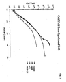
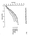
- Figures 1 and 2 represents the yield/passage calculated as the cell number obtained if all cells of a given condition were passaged.
- S represents chelexed serum (0.3%)
- CT represent cholera toxin (9 ng/ml)
- BHE bovine hypothalamic extract
- bovine hypothalamic extract While either bovine hypothalamic extract or cholera toxin used without serum were able to increase the final cell yield, bovine hypothalamic extract was able to improve the cell yield to a greater extent as well as to increase the number of passages during which the population doubling rate remained linear. In addition, bovine hypothalamic extract was shown to counterbalance the negative effects of serum to a much greater extent than cholera toxin. Cholera toxin did not appear to have any beneficial effect if bovine hypothalamic extract was present with or without serum.
- Keratinocytes designated B063 were established and grown in the mSBM formulation of example 9 with BHE and subsequently frozen for storage at the end of passage 2. These cells were thawed and seeded in 60 mm dishes at 1x10⁵/dish in the above medium without BHE. Half the dishes received EGF to 10 ng/ml two hours post seeding. The cells were harvested and counted at the end of passage 3 and those grown without EGF were replated and again grown to confluence. The data (table 11) show that, if desired, keratinocytes can be grown in mSBM without EGF and, in some instances, growth is comparable to that seen with EGF. Table 11 Efect of EGF on cell yield of HEp BO63. PASSAGE NUMBER 3 PASSAGE NUMBER 4 +EGF 1.14x10⁶ +/- 0.26 2.10x10⁶ +/- 0.23 -EGF 1.61X10⁶ +/- 0.21 1.82x10⁶ +/- 1.10
- hydrocortisone is not required, and its absence may then be preferred.
Abstract
Description
- This invention relates to media and systems for cell growth and to methods of making and using such media and systems.
- At present most media or systems for prolonged or long term growth and proliferation of normal mammalian cells incorporate undefined proteins or use feeder cells to provide proteins necessary to sustain such growth and proliferation.
- Because the presence of such undefined proteins can interfere with the intended end use of the cultured cells, it is desirable that cells be cultured under conditions to minimize the presence of undefined proteins. By way of example, it is anticipated that the presence of undefined proteins in cultured epidermal cells which are used to prepare living skin equivalents for use as skin grafts for burn victims may make such living skin equivalents unsuitable for this use, because such proteins may provoke an immune reaction. If mouse 3T3 feeder cells are used to culture epidermal cells for such living skin equivalents, it is highly likely that residual 3T3 cells or parts thereof trapped in the epidermis would cause a cross-species immune reaction. The use of bovine serum in fabrication of such living skin equivalents would be likely to produce a similar immune reaction.
- Thus, there is a need for chemically defined cell culture media and systems which provide for the prolonged growth and proliferation of cells either at a very low concentration of undefined proteins, or, more preferably, in the absence of such undefined proteins. The term "defined" is used to describe medium that contains no deliberately added uncharacterized supplements, even though such a medium may contain trace contaminants in its components.
- A wide variety of chemically defined culture media have been developed for the propagation of cells in response to this need. Such media include modified Eagles Medium (hereinafter "MEM"), Ham's F-12, Dulbeco's Modified Eagle's Medium (hereinafter "DMEM"), MCDB 153 and Medium 199. However, it has been found that successful culture of most cells in known chemically defined media still requires the addition of protein supplements such as serum or factors derived from serum or the use of feeder cells, thereby introducing undefined components to the media. Use of complex protein supplements in cell culture systems not only introduces undefined proteins but also increases the cost of cell culture.
- Problems encountered in the culture and, in some cases, maturation of epithelial cells are illustrative of the deficiencies in presently available culture media and systems. Epithelial cells, such as epidermal cells, are used to fabricate living skin equivalents and to produce sheets of cultured epidermis for use in test systems and skin grafting. Living skin and other tissue equivalents and methods of making and using such tissue equivalents are disclosed in U.S. Patent No. 4,485,096; U. S. Patent No. 4,835,102; U.S. Patent No. 4,837,379; U.S.S.N. 07/505,678, filed April 6, 1990; and U.S.S.N. 07/408,052, filed September 15, 1989; all of which are incorporated herein by reference, and referred to hereinafter as the "Patents." These applications require the growth of large quantities of epidermal cells in culture and presently available media do not meet this need.
- The mammalian epidermis is composed principally of a single cell type, the keratinocyte, in various stages of differentiation. The basal layer, separated from the fibroblasts of the underlying dermis, contains the dividing keratinocytes. These give rise to progeny, some of which no longer divide but move outward from the basal layer, terminally differentiate to form the stratum corneum, and are eventually shed from the surface. For certain applications, e.g., test systems for studies on percutaneous absorption, it is essential that living skin equivalents include a fully developed keratinocyte layer which requires the differentiation and maturation of the cultured epidermal cells.
- The formation of keratinocyte sheets, akin to those seen in vivo, presents additional challenges for existing cell growth media and systems. The formation of such sheets requires a calcium concentration sufficient to allow differentiation of the epidermal cells and to allow the stratification of older, more mature keratinocytes while still maintaining a proliferative, small basal cell population. The culture and maturation of keratinocytes has been attempted by various methods and with varying degrees of success, even when serum or protein supplements have been used. For example, it has been found that the growth requirements of keratinocytes are not met by conventional serum supplementation of chemically defined media. In any event, growth and maturation of keratinocytes is preferably achieved using a chemically defined culture media with little or no added serum or other undefined factors and without the use of feeder cells such as embryonic mouse fibroblasts (3T3 cells).
- In reviewing current techniques for culturing keratinocytes, a number of problems come to light. See, e.g., Breidahl et al., Aust. N.Z. J. Surg., 59(1989) 485-497.
- Rheinwald and Green reported a method for growing keratinocytes using a 3T3 feeder cell layer which allowed clonal growth and multiple passage (Rheinwald and Green, Cell 6(1975)331-344). This work was considered a breakthrough methodology, because the keratinocyte cells were able to form stratifying, differentiating cultures which still maintained a proliferative or relatively undifferentiated basal cell population capable of further clonal growth when passaged. Notwithstanding the foregoing advantages of the Rheinwald and Green methodology, the use of feeder cells,. e. g., mouse 3T3 cells, is undesirable because of the possibility that undefined proteins from the 3T3 cells, a transformed line, will be present in the cultured cells. Although it is reported (Sun and Green, Cell, 9(1976)512-521) that 3T3 cells and cell fragments may be removed by using 0.02% EDTA, complete removal is often difficult and the effect of removal of these cells and the resultant stress placed on the cultured epidermal cells is likely to be undesirable. Furthermore, the 3T3 feeder system requires high amounts of serum (about 5%), adding further undefined proteins to the culture system.
- After the work of Rheinwald and Green, supra, researchers continued to try to find a way of culturing epidermal cells and maturing keratinocytes in the absence of supplement or feeder cell support. A major advancement in this area was reported by Boyce and Ham in J. Invest. Dermatol. 81(1983)33S-40S. This work began by using Ham's F-12 as a base medium supplemented with reduced amounts of dialyzed FBS (2%) (Peehl and Ham, In Vitro 16(1980)526-538). An optimized formulation of this medium which allowed the growth of keratinocytes in a serum-free medium containing reduced calcium was subsequently developed by changing the concentration of a number of components to arrive at a medium designated MCDB 153 (See Boyce and Ham 1983, supra, and J. Tiss. Cult. Meth. 9(1985)83-93).
- The reported usefulness of MCDB 153 is in the areas of keratinocyte cell biology, toxicology, pathology and the growth of cells for epidermal cell grafts (Boyce and Ham 1985, supra). Although MCDB 153 is a serum free medium which does not require the use of feeder cells, it suffers certain disadvantages. For example, its use is limited in that MCDB 153 is not a very flexible medium, e.g., in the presence of calcium and/or serum.
- The ability to cultivate keratinocytes in MCDB 153 is critically dependent on the calcium concentration of the medium further limiting its flexibility. Proliferative ability in MCDB 153 is maintained by keeping the calcium concentration below about 1.0 mM, about 0.3 mM being optimal (See Boyce and Ham 1983, Fig. 1, p. 35S, supra). When calcium is added to concentrations of about 1.0 mM or above in MCDB 153 to cause stratification, the cells will no longer divide, will terminally differentiate and will not be capable of further cultivation, i.e., the proliferating basal cell population is lost because the cells cannot withstand high calcium concentrations in this medium. (Boyce and Ham 1983, supra). Because, a physiological concentration of approximately 1.8 mM calcium is necessary for a proper stratification and differentiation of a coherent multilayered epidermal sheet, MCDB 153 is not an appropriate medium to achieve a fully developed keratinocyte layer. Pittelkow and Scott (Mayo Clin. Proc. 61 (1986) 771-777) report formed sheets of graftable epidermis using cells cultivated in MCDB 153 but found it necessary to return to a DMEM-high serum (10%) medium to achieve stratification and at the same time maintain adequate viability.
- MCDB 153 is further limited in that cells grown in MCDB 153 dramatically lose their ability to be passaged if grown to confluence (Boyce and Ham 1983, supra). This finding indicates that the great majority of cells continue to differentiate even though the calcium concentration is kept below 1.0 mM and is possibly one reason why the MCDB 153 system is also so intolerant of calcium, since calcium is known to induce the final process of terminal differentiation in committed cells (Rice and Green, Cell, Vol 18, 681-694, November 1979). Furthermore, colony forming efficiencies of 30% and population doubling times of approximately 24 hours are achieved in MCDB 153 only at passages 2-3. These properties are seen to decline beyond passage 3 (Boyce and Ham 1983, supra).
- It is believed that the quality of cells grown in MCDB 153 would not be sufficiently high for use in the fabrication of epidermal cell sheets and that statified cultures, which can be held at confluence for some period of time, cannot be achieved in the present MCDB 153 system.
- Although other researchers working with a suboptimal medium tried to optimize their system by use of growth substrates (See, e.g., Karasek and Charlton (J. Invest. Dermal, 56(1971) 205-210), and Gilchrest et al. (J. Cell Physiol. 112 (1982) 197-206) reported the benefits of a protein substrate in culture of epidermal keratinocytes), these workers achieved only a limited degree of success, primarily due to deficiencies in medium formulation. Karasek and Charlton supra, and other researchers found it necessary to use a high serum concentration (usually around 10%). The supplemented M199 medium used by Gilchrest et al., (Cell Biol. Intl. Rpt., 4(1980) 1009-1016), suffered a basic nutrient deficiency, i.e., a very low inositol concentration.
- In Eisinger (Methods in Skin Research, Eds. Skerrow and Skerrow, Chap. 7, J. Wiley & Sons Ltd. 1985) and Eisenger et al. (Proc. Natl Aca. Sci. 76 (1979) 5340-5344) a method of growing epidermal cells is reported, the stated advantage of which is the growth of cells without feeder cells and without a dermal component. However, the Eisinger method, supra, relies on an extremely high cell density for plating. Furthermore, growth rate is slow and fold-increase in total cell number is low (partially due to a low plating efficiency), making this technique impractical for large scale expansion or prolonged cultivation from a single source, both desired objectives.
- It is known to those skilled in the art that supplementation of base medium with epidermal growth factor and hydrocortisone enhances the growth and spreading of keratinocytes (Barrandon and Green Cell 50(1987)1131-1137; Bertolero et al., Exp. Cell Res. 155(1984)64-80). Cyclic AMP elevating agents (See, e.g., U.S. Patent No. 4,456,687) and the use of bovine neural extract (Gilchrest et al., J. Cell Phys. 120 (1984) 377-383) or placental extract (O'Keefe and Chiu, Soc. Invest. Dermatol. 90 (1988) 2-7) are also of benefit in a number of cell culture systems.
- A survey of the literature indicates that while a number of workers have attempted growth of epithelial cells with either a collagen substrate (Liu et al. In Vitro 15 (1979) 813-822) or fibronectin substrate (Kubo et al., Soc. Invest. Dermatol. 88 (1987) 594-601) or using various media formulations, for the most part only short term culture at relatively high seeding densities has been achieved. None of these modifications, including MCDB 153, approach the level of success seen using the 3T3 feeder cell system.
- The 3T3, feeder system, however, requires the use of cAMP elevating agent, typically cholera toxin, in order to achieve optimal growth (Green, Cell 15 (1978) 801-811). This artificial elevation of cAMP levels complicates the study of actual growth factor effects and mechanisms.
- Media and systems which provide for the prolonged growth and proliferation of cells, and in some instances maturation of certain cells while maintaining actively dividing basal cells, are being sought.
- Cells grown in the cell culture media and systems of the present invention have a number of advantages including but not limited to:
- 1. Lack of feeder cell.
- 2. Rapid growth rate in the absence of serum and/or bovine hypothalmic extract.
- 3. If plated at about 1x10³ cells/cm² or higher, use of bovine hypothalamic extract provides little or no advantage.
- 4. Rapid cell growth in the absence of cAMP elevating agents.
- 5. Plating and colony forming efficiency is 30% or higher with flexibility of the calcium concentration. This compares to approximately 10% with 3T3 and 30% with MCDB 153-low calcium. (This characteristic is highly desirable for microcarrier cultivation).
- 6. Cells may be easily used in the fabrication of living skin equivalents and form a multilayered epidermis as good or better than cells grown by other methods described (MCDB 153 grown cells do not survive well).
- 7. Epidermal cells grown on a collagen coated surface can be released as a sheet of epidermal cells using collagenase for use in transplantation of these sheets, if desired.
- 8. The flexibility of the systems in range of Ca⁺⁺ concentrations, serum requirements and density dependence provide, a number of advantages for adaptation to large scale microcarrier culture.
- 9. The rapid growth rate produces a nearly synchronous population of cells as they approach approximately 60% confluence. The large number of rounded mitotic cells and the high plating efficiency of cells grown in accordance with the present invention, indicates that bead to bead transfer may also be possible in large scale microcarrier culture.
- Cell culture media in accordance with the present invention comprise, insulin or an insulin-like growth factor; ferrous ion; triiodothyronine or thyroxin; at least one of ethanolamine or o-phosphoryl-ethanolamine; calcium; and a nutrient source. A preferred medium in accordance with the present invention comprises:
- (a) Insulin at from about 0.5 to about 50 ug/ml;
- (b) Ferrous ion or transferrin at from about 5x10⁻⁸ to about 5x10⁻⁵M;
- (c) Triiodothyronine at from about 2 to about 200 pM;
- (d) At least one of o-phosphoryl-ethanolamine and ethanolamine at from about 10⁻⁶ to about 10⁻²M;
- (e) Calcium at from about 0.005 to about 2.0 mM; and
- (f) Nutrient Source selected from at least one of DMEM, IDMEM, MEM, M199, RPMI 1640, Ham's F12, Ham's F10,
NCTC 109, and NCTC 135. - It is expected that the systems, methods and media of the present invention may be useful in the culture of a variety of cell types. Cells which are preferably cultured by the practice of the present invention are epithelial cells. Preferred epithelial cells include epidermal cells or keratinocytes.
- Cell culture systems of the present invention comprise a cell culture medium as described above and a substrate for the cells comprising plastic, glass, collagen, fibronectin, laminin, heparan sulfate proteoglycan, microcarriers coated with collagen, fibronectin, laminin or heparan sulfate proteoglycan, or a tissue equivalent produced by a method comprising:
- a. combining a collagen solution with a contractile agent under conditions to form a gel mixture having the contractile agent dispersed within the gel mixture; and
- b. maintaining the gel mixture prepared in step (a) under conditions which permit contraction of the gel mixture to form a tissue-equivalent.
- One method of culturing epithelial cells in accordance with the present invention comprises, inoculating a growth substrate as described above with epithelial cells, and maintaining the cell culture system under conditions to promote cell growth. Cell growth achieved in such methods is characterized by a population doubling time of from about 16 to about 33 hours. Furthermore, cells may be serially cultured to achieve from about 20 to about 50 population doublings. In yet another embodiment of the present invention, the method further comprises adding calcium at a concentration greater than about 1.0mM to enable stratification and differentiation of the cells while maintaining a colony forming efficiency of from about 20 to about 60%. This is important in formation of living skin equivalents and sheets of epidermis for grafting. The practice of the present invention also provides for clonal growth of cells, i.e., seeding at a cell density of from about 30 to about 1000 cells.
- By the practice of the present invention, it is possible to achieve rapid expansion of primary cells with a colony forming efficiency of from about 20 to about 100%, and a proliferative capacity in excess of 50 population doublings. Furthermore, calcium can be added in excess of 1.0 mM to enable stratification and differentiation of the cells while maintaining a colony forming efficiency of from about 20 to about 60%.
- The present invention also provides a method of culturing epithelial cells on microcarriers, the method comprising plating epithelial cells onto collagen-coated microcarriers in a cell culture system as described above at a plating efficiency of from about 20 to about 40%, and maintaining the microcarriers and plated cells under conditions to promote cell growth. By the practice of this method, a colony forming efficiency of from about 20 to about 100% is maintained even as cell growth reaches confluence. Furthermore, calcium concentration may be increased from 0.08 to about 1.8mM during the rapid proliferative phase when cell growth is approaching confluence to increase adherence of the cells to microcarriers and, thus, reduce cell loss through the shearing forces exerted on the microcarriers.
- Fully epidermalized living tissue equivalents may also be made in accordance with the present invention. The method comprises the steps of:
- a. innoculating a tissue equivalent in accordance with claim 14 with epidermal cells;
- b. maintaining the cell culture system under conditions to promote cell growth; and
- c. adding calcium to a physiological concentration and maintaining the system under conditions to enable development of a fully keratinized epidermal layer.
- Media in accordance with the present invention will be conditioned by rapidly proliferating epithelial cells and it is expected that such conditioned media will be useful as growth supplements and in therapeutic applications such as wound healing.
- Chemically defined media in accordance with the present invention provide for culture of cells in the absence of feeder cells, serum or other components which may contribute undefined proteins to the media.
- Some of the advantages of the media of the present invention are: ease of fabrication, flexibility of use, i.e, it can be used with varied calcium concentration, varied serum concentrations, varied growth factor additions and varied extracellular matrix components. The media and systems of the present invention provide the cell biologist and others interested in the fabrication and/or study of epithelium in vitro with a system to meet most of their needs:
- One medium in accordance with the present invention comprises: insulin or an insulin-like growth factor; transferrin or ferrous ion; triiodothyronine or thyroxin; ethanolamine and/or o-phosphoryl-ethanolamine; calcium; and a nutrient source. Other components may be added to the media, depending upon, e.g., the particular cell being cultured, including but not limited to, epidermal growth factor (EGF), hydrocortisone, strontium, Bovine Hypothalamic Extract, progesterone, selenium and cAMP elevating agents. Hydrocortisone is reported to have value in the long term culture of normal human keratinocytes (Rheinwald and Green, supra) and is, therefore, a preferred component of complete mSBM. When minimal supplementation is desired, however, hydrocortisone is not required, and its absence may then be preferred.
- Insulin is present at a concentration of from about 0.05 to about 500 ug/ml, a preferred range being from about 0.5 to about 50 ug/ml, and a particularly preferred concentration being 5.0 ug/ml. Proinsulin, IGF-1 (10⁻¹⁰ to 10⁻⁸M) or other insulin-like growth factors may be substituted for insulin, although insulin is presently preferred for reasons of economy. Insulin-like growth factor as used herein means compositions which are structurally similar to insulin and stimulate the insulin-like growth factor receptors, e.g., Insulin-like Growth Factors I and II.
- Ferrous ion may be provided by a ferrous salt such as ferrous sulfate at a concentration of from about 5x10⁻⁸ to about 5x10⁻⁵M, preferably at about 5x10⁻⁶M. More preferably, ferrous is supplied by transferrin at a concentration of from about 0.05 to about 50 ug/ml, a preferred concentration being about 5 ug/ml.
- Triiodothyronine is preferred over thyroxin because it has been found to have a more potent effect in the claimed cell culture systems. Triiodothyronine is preferably present at a concentration of from about 2 to about 200pM, more preferably at about 20 pM.
- Ethanolamine and/or o-phosphoryl-ethanolamine may be used in the practice of the present invention. However, it is preferred to use these components in combination. Ethanolamine is present at a concentration of from about 10⁻⁶ to about 10⁻² M, more preferably about 10⁻⁴ M. o-phosphoryl-ethanolamine is present at a concentration of from about 10⁻⁶ to about 10⁻² M, more preferably 10⁻⁴ M.
- In contrast with presently available media, the calcium concentration can be varied over a wide range in the media of the present invention while still maintaining an actively proliferating cell culture. Although cells may be grown in the media of the present invention at serum concentrations of 5% and at physiological calcium levels of 1.8 mM, growth and lack of terminal differentiation (i.e., maintenance of the proliferating basaloid cell population) are optimized by reducing the serum to about 0.3% or less, preferably 0%, and having a calcium concentration at from about 0.005 to about 2.0 mM, more preferably from about 0.08 to about 1.0 mM.
- Under calcium conditions used for optimal growth rate, minimal stratification is seen. If these cultures are held at confluence, larger cells are released from the dish and the dish remains populated by a tightly packed layer of small cells. Addition of calcium allows stratification of the more mature cells while still maintaining a confluent basal cell layer. If calcium is added before confluence to 1.8 mM the appearance of the colonies and cell distribution parallels what was observed with feeder cells with the cells continuing to grow to confluence. There is little involucrin or transglutaminase (markers of epidermal cell differentiation) before confluence, if the calcium is kept at 1.0 mM or lower. Calcium is present at from about 0.005 to about 2.0 mM depending upon the purposes for culturing the cells. In some instances, rather than being added separately, the calcium needed is provided by other components in the medium.
- Epidermal Growth Factor (EGF) is present at from about 1 to about 50 ng/ml, preferably at about 10 ng/ml (mouse EGF), and at about 1 ng/ml (human EGF). Although EGF is preferred, transforming growth factor-alpha, human or mouse, may be substituted for EGF, preferably at about 10 ng/ml (mouse) and about 1 ng/ml (human). Although EGF is an optional ingredient in media in accordance with the present invention, it may be preferred for some applications, e.g., large scale batch cultures.
- Hydrocortisone is preferably at a concentration of from about 0.04 to about 4.0 ug/ml, a particularly preferred concentration being 0.4 ug/ml.
- In yet other embodiments of the present invention, the medium of the present invention may be supplemented with additional components.
- In some instances, it may be desirable to include components which inhibit fibroblast growth, such as cholera toxin.
- Bovine hypothalmic extract (BHE) at from about 5 to about 200 ug/ml, preferably about 50 ug/ml, may be included to increase plating efficiency and colony formation in primary and in clonal density culture, particularly when initial cell density is less than about 1 x 10³ cells/cm². Members of the fibroblast growth factor family, including acidic FGF, keratinocyte growth factor, and basic FGF (at a slightly higher concentration) may be substituted for BHE with similar effects at from about 10⁻¹⁰ g/ml to about 10⁻⁶ g/ml, preferably about 10⁻⁷ g/ml. Typically, BHE provides little or no advantage when cells are plated at 1x10³ cells/cm² or higher. Other components which may be added to the medium described above include adenine from about 0.02 mM to about 2.0 mM; chelexed serum from 0% to about 2.00%; progesterone from about 2 x 10⁻¹⁰ to about 2 x 10⁻⁸M, preferably 2 x 10⁻⁹ M; selenium from about 1x10⁻⁹ to about 1x10⁻⁷M. Progesterone, selenium, BHE may be added in some instances to achieve optimal growth.
- Cyclic AMP elevating agents, serum and bovine hypothalamic extract are not necessary to establish primary cell cultures and to pass human epidermal cells in the systems of the present invention. Previous investigators have shown both the addition of cAMP elevating agents and the presence of bovine pituitary extract to be beneficial, if not essential, for establishing primary cultures of epidermal cells (See Green, 1978, supra., and Boyce and Ham, 1985, supra). Although the addition of BHE may be slightly beneficial, the data in Example 9, below, show that neither BHE nor cAMP elevating agents are necessary for primary culture and subsequent passage of epidermal cells using the media of the present invention. However, cAMP elevating agents may be used in the practice of the present invention if desired.
- Cyclic AMP elevating agents which may be used in the practice of the present invention include cholera toxin, foreskolin, β-adrenergic agents such as adrenalin, theophylline, dibutyryl cyclic AMP, methyl isobutyl xanthine, isoproterenol, caffeine and pertussis toxin. Preferred agents include cholera toxin and foreskolin. Cholera toxin is preferably used at from about 10⁻⁸ to about 10⁻⁵ M; and foreskolin from about 10⁻⁹ to about 10⁻³ M, preferably from about 10⁻⁷ to about 10⁻⁵ M, and more preferably at 10⁻⁶ M.
- The use of c-AMP elevating agents in the systems and media of the present invention may be benefical in some cases when establishing primary cultures or in reestablishing primary cultures or other cells which have been frozen. However, in the practice of the present invention cells can be readily established without the addition of a cAMP elevating agent or BHE. Such cells can subsequently be passed for multiple passages using the media of the current invention without a cAMP elevating agent.
- The addition of cation substitutes was investigated as possible replacement for the constitutive need for calcium. While all cells require a certain level of calcium, calcium also has differentiation promoting effects in keratinocytes. It had been previously shown that other divalent cations could replace the cells' consituitive need (Rubin, J. Cell. Physiol., 91 (1977) 449-458) without contributing to induction of differentiation (Praeger et al. J. Cell. Physiol., 132 (1987) 81-89, Furakawa et al., J. Invest. Dermatol., 90 (1988) 690-696). The effects of strontium and magnesium were investigated in the systems of the present invention. Magnesium was found in some instances to have a beneficial effect. Strontium addition, while not significantly changing the proportion of proliferative cell population (9-14 um distribution), significantly increased the final cell yield and reduced the population doubling time by approximately 4 hours. See Example 8, below.
- Nutrient sources useful in the practice of the present invention provide known essential nutrients for cultured cells, such as: an energy source such as glucose, fructose or galactose; both essential and nonessential amino acids; both water-soluble (B group, biotin, folic acid, nicotinamide, panthothenic acid, pyroxidine, riboflavin and thiamine) and fat-soluble (A, D, E, K, and ubiquinone) vitamins; major inorganic ions such as bicarbonate, calcium, chloride, magnesium, phosphate, potassium and sodium; trace elements such as As, Co, Cr, Cu, F, Fe, Mn, Mo, Ni, Se, Si, Sn, V and Zn; lipids; buffers, e.g., like CO2/HCO3 and HEPES; gases (oxygen and carbon dioxide); and nucleic acid precursors like adenine, cytidine, hypoxanthine, and thymidine.
- There are many commercially available nutrient sources which are expected to be useful in the practice of the present invention. These include commerically available nutrient sources which supply inorganic salts, an energy source, amino acids, and B-vitamins, such as Dulbecco's Modified Eagle's Medium (DMEM); Minimum Essential Medium (MEM); M199; RPMI 1640; (all available from Flow Laboratories); and Iscove's Modified Dulbecco's Medium (EDMEM, Gibco Labs). Minimum Essential Medium and M199 require additional supplementation with phospholipid precursors and non-essential amino acids. Commercially available vitamin rich mixtures which supply additional amino acids, nucleic acids, enzyme cofactors, phospholipid precursors, and inorganic salts, include Ham's F12; Ham's F10;
NCTC 109; (all available from Flow Laboratories) and NCTC 135 (Irvine Scientific). - The components of each of the above nutrient sources is provided hereinafter.
Dulbecco's Modified Eagle's Medium COMPONENT 1X MEM (Modified) Dulbecco's Modification Liquid without L-Glutamine Product No. 12-322 mg/l INORGANIC SALTS Cacl₂ Cacl₂·2H₂O 264 90 Fe(NO₃)₃·9H₂O 0.10 KCl 400 0 MgSO₄ MgSO₄·7H₂O 200 00 NaCl 6400 00 NaHCO₃ 3700 00 NaH₃PO₄·H₂O 125 00 OTHER COMPONENTS Fructose Glucose 4500 0 Lipoic Acid Phenol Red, Na 15 0 Sodium Pyruvate 110 0 Aminopterin Hypoxanthine Thymidine AMINO ACIDS L-arginine·HCl 84 00 L-cystine 48 00 L-cystine, Na₂ L- glutamine Glycine 30 00 L-histidine·HCl·H₂O 42 00 L-isoleucine 104 80 L-leucine 104.80 L-lysine·HCl 146 20 L- methionine 30 00 L-phenylalanine 66 00 L-serine 42.00 L-threonine 95 20 L-tryptophan 16.00 L-tryosine 72 00 L-valine 93.60 VITAMINS D-Ca Pantothenate 4 00 Choline Chloride 4 00 Folic Acid 4 00 i-Inositol 7 00 Nicotinamide 4 00 Pyridoxal·HCl 4 00 Riboflavin 0 40 Thiamin·HCl 4 00 Minimal Essential Medium with Hank's Salts or Earle's Salts COMPONENT 1X MEM (Modified) Earle's Salts liquid without L Glutamine Product No. 12-102 1X MEM (Modified) Hanks' Salts liquid without L Glutamine Product No. 12-132! mg/l mg/l INORGANIC SALTS Cacl₂ Cacl₂·2H₂O 264 9 185 5 KCl 400 0 400 0 KH₃PO₄ 60 0 MgSO₄ MgSO₄·7H₂O 200 0 200 0 NaCl 6800 0 8000 0 NaHCO₃ 2000 0 350 0 NaH₃PO₄·H₂O 140 0 Na₃HPO₄ 47 5 Hopos OTHER COMPONENTS Glucose 1000 0 1000 0 Phenol Red, Na 17 0 17 0 Sodium Succinate·6H₂O Succinic Acid AMINO ACIDS L-alanine L-arginine·HCl 126 40 126 40 L-asparagine·H₂O L-aspartic Acid L-cystine 24 02 24 02 L-cystine, Na₂ L-glutamic Acid L-glutamine Glycine L-histidine·HCl·H₂O 41 90 41 90 L-isoleucine 52 50 52 50 L-leucine 52 50 52 50 L-lysine·HCl 73 06 73 06 L-methionine 14 90 14 90 L-phenylalanine 33 02 33 02 L-proline L-serine L-threonine 47 64 47 64 L- tryptophan 10 20 10 20 L-tryosine 36 22 36 22 L-valine 46 90 46 90 VITAMINS D-Ca Pantothenate 1 00 1 00 Choline Bilartrate Choline Chloride 1 00 1 00 Folic Acid 1 00 1 00 I-Inositol 2 00 2 00 Nicotinamide 1 00 1 00 Pyridoxal·HCl 1 00 1 00 Riboflavin 0 10 0 10 Thiamin·HCl 1 00 1 00 RPMI 1640 Medium Component 1X RPMI 1640 Liquid without L-Glutamine Product No. 12-602 mg/L INORGANIC SALTS Ca(NO₃)₃·4H₂O 100 0 KCl 400 0 MgSO₄ MgSO₄·7H₂O 100 0 NaCl 6000 0 NaHCO₃ 2000 0 Na₃HPO₄ 800 7 HEPES OTHER COMPONENTS Glucose 2000 0 Glutathione 1 00 Phenol Red, Na 5 0 Hypoxathine Aminopterin Thymidine AMINO ACIDS L-arginine 200 00 L-asparagine·H₂O 56 82 L- aspartic Acid 20 00 L- cystine 50 00 L-cystine, Na₃ L- glutamic Acid 20 00 L- glutamine Glycine 10 00 L-hisidine 15 00 L- hydroxyproline 20 00 L- isoleucine 50 00 L- leucine 50 00 L-lysine· HCl 40 00 L-methionine 15 00 L-phenylalanine 15 00 L- proline 20 00 L- serine 30 00 L- threonine 20 00 L-tryptophan 5 00 L- tryosine 20 00 L- valine 20 00 VITAMINS Biotin 0 20 D-Ca Pantothenate 0 25 Choline Chloride 3 00 Folic Acid 1 00 I-Inositol 35 00 Nicotinamide 1 00 PABA 1 00 Pyroxidine·HCl 1 00 Riboflavin 0 20 Thiamin·HCl 1 00 Vitamin B₁₂ 0 005 REFERENCES: 1 Moore, G. E., et al. 1967. J A M A., 199 519 2 Morton, J. H., 1970. In Vitro. 6 89. 
Ham's F-10 Medium Component 1X Ham's F10. Liquid with L-Glutamine Product No. 12-403 1X Ham's F12 Liquid with L-Glutamine Product No. 12-423 mg/L INORGANIC SALTS CaCl₂ CaCl₂·2H₂O 44.1 44.1 CuSO₄ 0.0016 0.0016 FeSO₄ 0.456 0.456 KCl 285.0 223 7 KH₃PO₄ 83.0 MgSO₄ MgSO₄·7H₂O 152.7 147.9 NaCl 7400 0 7600.0 NaHCO₃ 1200.0 1176.0 Na₃HPO₄ 156 2 142.0 Zn SO₄·7H₂O 0.0288 0.863 OTHER COMPONENTS Glucose 1100 0 1802.0 Hypoxanthine 4.08 4.08 Lipoic Acid 0.206 0 206 Methyl Linoleate 0 088 Phenol Red, Na 1 24 1.24 Putrescine·2HCl 0 1611 Sodium Pyruvate 110 0 110 0 Thymidine 0 727 0.727 AMINO ACIDS L-alanine 8.91 8 91 L-arginine·HCl 210.70 210.70 L-asparagine·H₂O 15 01 15 01 L-aspartic Acid 13 31 13 31 L-cysteine·HCl·H₂O 35.13 35 13 L-glutamic Acid 14.71 14.71 L-glutamine 146.20 146.20 Glycine 7.51 7.51 L-histidine·HCl·H₂O 20.96 20 96 L-isoleucine 2.62 3 94 L-leucine 13.12 13 12 L-lysine·HCl 29.30 36 53 L-methionine 4.48 4 48 L-phenylalanine 4.96 4.96 L-proline 11.51 34 54 L- serine 10 51 10 51 L-threonine 3.57 11 91 L-tryptophan 0.61 2.04 L-tyrosine 1.81 5 44 L-valine 3.51 11.72 VITAMINS Biotin 0.024 0 0073 D-Ca Pantothenate 0.715 0 238 Choline Chloride 0 698 13 96 Folic Acid 1 32 1 32 l-Inositol 0 541 18 02 Nicotinamide 0 611 0 037 Pyridoxine·HCl 0 206 0 062 Riboflavin 0 376 0 038 Thiamin·HCl 1 012 0 337 Vitamin B₁₂ 1 36 1 36 
ISCOVE'S MODIFIED DULBECCO'S MEDIUM COMPONENT Liquid (1X) Form. No. 78·0415 mg/L Powder Cat. No. 430·2200 mg/L INORGANIC SALTS: CaCl₂ (anhyd.) 165.00 165.00 Fe(NO₃)₃·9H₂O -- -- KCl 330.00 330.00 KNO₃ 0.076 0.076 MgSO₄ (anhyd.) 97.67 97.67 MgSO₄·7H₂O -- -- NaCl 4505.00 4505.00 NaHCO₃ 3024.00 -- NaH₃PO₄·H₂O 125.00 125.00 Na₂SeO₃·5H₂O 0.0173 0.0173 OTHER COMPONENTS: D-Glucose 4500.00 4500.00 Phenol red 15.00 15.00 HEPES 5958.00 5958.00 Sodium pyruvate 110.00 110.00 AMINO ACIDS: L-Alanine 25.00 25.00 L-Asparagine·H₂O 28.40 28.40 L-Arginine·HCl 84.00 84.00 L-Aspartic acid 30.00 30.00 L-Cystine -- -- L-Cystine·2HCl 91.24 91.24 L-Glutamic acid 75.00 75.00 L-Glutamine 584.00 584.00 Glycine 30.00 30.00 L-Histidine HCl·H₂O 42.00 42.00 L-Isoleucine 105.00 105.00 L-Leucine 105.00 105.00 L-Lysine HCl 146.00 146.00 L-Methionine 30.00 30.00 L-Phenylalanine 66.00 66.00 L-Proline 40.00 40.00 L-Serine 42.00 42.00 L-Threonine 95.00 95.00 L-Tryptophane 16.00 16.00 L-Tyrosine -- -- L-Tyrosine (Disodium salt) 104.20 103.79 L-Valine 94.00 94.00 VITAMINS: Biotin 0.013 0.013 D-Ca pantothenate 4.00 4.00 Choline chloride 4.00 4.00 Folic acid 4.00 4.00 i-Inositol 7.20 7.20 Nicotinamide 4.00 4.00 Pyridoxal HCl 4.00 4.00 Riboflavin 0.40 0.40 Thiamine HCl 4.00 4.00 Vitamin B₁₂ 0.013 0.013 - A preferred nutrient source for use in the present invention comprises Ca⁺⁺ Free DMEM from about 90 to about 10% and Ham's F 12 from about 10% to about 90%. A particularly preferred source comprises Ca⁺⁺ Free DMEM at about 75% and Ham's F 12 at about 25%. Typically, the media of the present invention comprises at least about 90% and preferably greater than 95% nutrient source.
- One preferred medium of the present invention comprises:
Insulin: 5 ug/ml; Transferrin: 5 ug/ml; Triiodothyronine: 20 pM; Ethanolamine: 1x10⁻⁴ M; o-phosphorylethanolamine: 1x10⁻⁴ M; Adenine 0.18 mM; Selenium 3x10⁻⁸ M; Strontium 1 mM Calcium: 0.08mM; and Nutrient source: Ca⁺⁺ Free DMEM from about 90 to about 10% and Ham's F 12 from about 10% to about 90%; - A significant measure of the effectiveness of a culture medium is the growth rate of a cell type as given by population doublings per day. By use of the culture systems of the present invention, epithelial cells may be cultured in the absence of feeder cells and serum at low density to allow for expansion of primary culture in excess of 50 population doublings at a growth rate comparable to that in a feeder cell system. For example, keratinocytes from newborn foreskins rarely drop below 0.5 doublings per day in the media and systems of the present invention, averaging one doubling per day through passage 6, and can reach growth rates of 1.3 to 2.0 doublings per day between passages 2 and 5 (See Table 2, below). Growth rates of such magnitude indicate a population doubling in number every 12-18 hours.
- By use of the systems and media of the present invention, calcium can be added to physiological concentration with maintenance of the basal cell layer, a result achieved hereto only with 3T3 systems. Thus, the culture systems and media of the present invention allow for the addition of calcium in excess of 1.0 mM without loss of a proliferative cell population, and, therefore, allow use of such cells in fabrication of living skin equivalents or in the formation of continuous sheets of epidermal cells suitable for grafting (See Example 6 below). Furthermore, the ability to vary calcium concentration without detrimental effects on cell growth now makes it possible to grow epidermal cells in large scale mirocarrier culture (See Example 7 below). For example, in MCDB 153 the calcium level has to be low in order to get cell growth but when the calcium level is low cells do not adhere well to the surface of the microcarrier, a disadvantage when the microcarrier is subjected to shear force. In the Rheinwald and Green 3T3 systems, supra, the microcarrier would first have to be coated with the 3T3 cells and the epithelial cells then grown on the 3T3 cells.
- The media of the present invention are currently prepared from components which are readily available commercially, including the nutrient source which may conveniently be obtained commercially calcium free. Furthermore, the components of the subject media are prepared and assembled using techniques available to the skilled artisan. In contrast, it is believed, based on culture studies with commercially available equivalents to MCDB 153, that limited success has been achieved in making these equivalents.
- The systems of the present invention are not only comparable to, but in some respects clearly superior to the 3T3 system, one of the most successful systems heretofore known for the culture of epithelial cells. The present invention provides cell growth media which is chemically defined and does not depend on serum or feeder cells to provide acceptable growth and expansion of the cultured cells. The media and systems of the present invention enable establishment of primary cultures with from about 0.5 to about 2.0 populations doublings (PDL) per 24 hour period.
- A comparison of presently available media described above with the claimed media and systems is set forth below:
COMPARISON OF SYSTEMS MCDB 153 (Boyce/Ham) Eisenger RHEINWALD GREEN PRESENT INVENTION Lack of feeder cells yes yes no yes Lack of cAMP elevating agents yes yes no yes ≧ 50 pdl no no yes yes > 25% plating eff. yes no no yes > 20% cfe yes no no yes Clonal growth yes no yes yes Little or no serum yes no no yes Avg. pdt < 24 hr. yes no yes yes Growth in > 1.0 mM Ca⁺⁺ no yes yes yes pdl = population doubling; cfe = colony forming efficiency; ECM = extracellular matrix; pdt = population doubling time. - Various substrates can be used in the practice of the present invention including collagen, fibronectin, laminin, heparan sulfate proteoglycan and tissue equivalents, as described in the Patents. Native Type I collagen from bovine tendon is a preferred collagen substrate. In the claimed systems, growth may be enhanced under certain conditions using either fibronectin or collagen coated substrate (See Example 3). It can be advantageous to use, e.g., collagen, in the establishment of primary cultures or when serum is included in the media, at greater than about 1% or to establish cells from frozen stocks. However, when the serum concentration is reduced to about 0.3% or less, the detrimental or differentiation promoting serum effects are lessened to the extent that a matrix component no longer provides such advantages. If a substrate is used, it must be of high quality because, it was found that poorly coated dishes with either partially degraded fibronectin or uneven collagen could also inhibit cell growth and spreading.
- The presence of a substrate, e.g., a matrix component appears to allow the cells to establish colonies when conditions are less than optimal or more stringent, e.g., in the presence of serum factors,. low cell density, in establishment of cells from tissue, or, possibly, in application where even greater longevity is required.
- Thus, we have described and provided examples of novel media and systems for the growth of cells, such as epithelial cells. This invention will be further understood with reference to the following examples which are purely exemplary in nature and which are not meant to be utilized to limit the scope of the invention.
- In the following examples the basic medium used, designated mSBM, consisted of the following components:
Hydrocortisone 1.1uM Insulin 5 ug/ml Transferrin 5 ug/ ml Triiodothyronine 20 pM Ethanolamine 1 x 10⁻⁴ M o-phosphorylethanolamine 1 x 10⁻⁴ M Adenine 0.18 mM Progesterone 2 x 10⁻⁹ M Selenium 3 x 10⁻⁸ M Cholera Toxin 9 ng/ml Epidermal Growth Factor 10 ng/ml Calcium Free DMEM 75% Ham's F-12 25% - Another medium, designated emSBM, also used in the following examples, consisted of the components listed above with concentrations changes and the additional component listed below:
Triiodothyronine Increased to 1.002 x 10⁻⁸ M Cholera Toxin Increased to 100 ng/ml Bovine Hypothalamic Extract (BHE) 50 ug/ml - In Example 8 below strontium (Sr) was added to medium in certain instances and in Example 9 below a cAMP elevating agent was not included in the medium in certain instances. Cells were grown in accordance with Example 10 without exogenous cAMP elevating agents, progesterone, serum or bovine hypothalmic extract. In Example 11 cells were grown without EGF and in Example 13 without hydrocortisone.
- Although primary cultures where established in emSBM, none of a cAMP elevating agent, serum, progesterone, elevated triiodothyronine or BHE are required, although BHE in some circumstances provides superior growth.
- The medium described above is typically prepared as set forth below. However, it should be understood that the components of the present invention may be prepared and assembled using conventional methodology compatible with their physical properties. Media in accordance with the present invention are sterile. Sterile components are bought or rendered sterile by conventional procedures after preparation. Proper sterile procedures were used throughout the following examples. Stock solutions of all components can be stored at -20oC, with the exception of nutrient source which can be stored at 4oC.
- All stock solutions are prepared at a concentration 500x the final concentrations listed above. Hydrocortisone (Sigma) is dissolved in absolute ethanol and diluted in phosphate buffered saline (PBS). A stock solution of insulin, transferrin and triiodothyronine (all from Sigma) is prepared as follows: triiodothyronine is initally dissolved in absolute ethanol: 1 N HCl at 2: 1. Insulin is dissolved in dilute HCl (approximately 0.1 N) and transferrin is dissolved in water. The three are then mixed and diluted in water to a 500x concentration. Ethanolamine and o-phosphorylethanolamine (both from Sigma) are dissolved in water to 500x concentration, filter sterilized. Progesterone (Sigma) is dissolved in absolute ethanol and diluted with water. Bovine serum albumin (BSA) may be added for prolonged storage to maintain the activity. Selenium (Sigma) is prepared as is ethanolamine. Cholera toxin is purchased sterile from Sigma and is dissolved in water. EGF is purchased sterile from Biomedical Technologies, Inc. and is dissolved in PBS. Adenine is difficult to dissolve but may be dissolved by any number of methods known to those skilled in the art. Sterile calcium free DMEM is ordered from J. R. Scientific or Hazelton. Ham's F-12 is ordered from M. A. Bioproducts. BHE (Collaborative Research, i.e., ECGS) is dissolved in low calcium DMEM. BSA may be added to prolong the storage life of the EGF stock solution. DMEM and F-12 are combined and the individual components are then added to complete the medium. The medium can be either used immediately after preparation or, stored at 4oC. If stored frozen, EGF should not be added until the time of use. The resulting media is sterile.
- The activity of both BHE and EGF are evaluated on a lot by lot basis. Growth achieved in medium lacking BHE or EGF is compared with growth in medium containing these ingredients, from a range of concentrations from about 0 to about 50 ng/ml EGF and from about 0 to about 200 ug/ml BHE in order to determine the concentration which will support maxium growth.
- Foreskin and other tissue, including ear and abdomen, were obtained from routine biopsies and washed for 2 minutes in phosphate buffered saline (PBS) containing gentamicin at 50 mcg/ml and fungizone at 1.25 meq. Whole tissue samples were then washed for 1 min. in 95% ethanol to remove surface contamination, followed by washing again in the PBS-gentamicin fungizone solution. Subcutaneous tissue was removed aseptically and the tissue rinsed in the PBS-gentamicin fungizone solution and minced into approximately 1 mm² pieces. Minced tissue was dissociated at 37oC using a collagenase-trypsin tissue mixture. The components of the collagenease dissociation mix used are as follows: collagenase 55.5 mg/ml; trypsin 2.0 mg/ml; glucose 0.375 mg/ml; fungizone 1 meq/ml; and
gentamicin 50 mcg/ml in phosphate buffered saline. Digest supernatant was removed and neutralized and fresh enzyme added to the remaining digested pieces at 30 minute intervals until the tissue was fully dissociated. - Cell fractions containing a mixed population of dissociated cells were pooled, counted and plated at 2.6x10⁴/cm² on T75 flasks (collagen coated at 5 ug/cm²) with a total volume of 10 mls of emSBM which does not contain EGF. Primary cultures such as this became confluent in 7-17 days. One day post plating, emSBM was aspirated off and replaced with fresh emSBM containing 10 ng/ml EGF. Media changes were performed on the cells every third day assuming day of plating is day 0 using emSBM containing EGF. Primary cultures take from 7-17 days to become confluent at which time cells can be further serially cultured as described in Example 2, below, or frozen down and stored in liquid nitrogen for later use. Freeze medium contains LCDMEM as a base with 10% fetal bovine serum and 10% dimethylsufoxide (DMSO). To freeze, cells are removed from the dish and counted the same as in Example 2, but after counting, spun down again and resuspended in freeze medium at between 2x10⁶ and 5x10⁶/ml and aliquoted into 1.8 ml nunc cryovials. After an 18-24 hour prefreeze, vials are transferred to a liquid nitrogen storage tank until needed. The results of these experiments are reported in Table 1, below. Cell viability was normally in excess of 90%.
- The abbreviations used in Table 1 have the following meanings: "HEP" indicates human epidermal cells; "n" indicates neonatal foreskin cells; "NF" followed by a number indicates human epidermal cells from a source other than foreskin.
TABLE 1 Establishment of Primary Cultures using Enhanced mSBM Strain HEP Age of Donor Mixed Cell Inoculum From Whole Tissue Inoculum Density Primary Keratinocyte Yield Approx* No. of Generations Days BO36 n 1.6x10⁷ 2.5x10⁴/cm² 3.5x10⁷ 8.8 16 BO37 n 1.0x10⁷ 2.5x10⁴/cm² 3.1x10⁷ 9.3 8 BO38 n 1.6x10⁷ 2.5x10⁴/cm² 1.2x10⁸ 10.6 12 BO40 n 6x10⁶ 2.5x10⁴/cm² 3.0x10⁷ 10.0 8 BO41 n 1.0x10⁷ 2.5x10⁴/cm² 1.1x10⁷ 7.8 16 BO42 n 1.4x10⁷ 2.5x10⁴/cm² 6.0x10⁶ 6.5 8 BO43 n 1.5x10⁷ 2.5x10⁴/cm² 1.2x10⁷ 7.9 8 BO44 n 1.4x10⁷ 2.5x10⁴/cm² 1.2x10⁷ 7.5 8 BO45 n 1.4x10⁷ 2.5x10⁴/cm² 8.2x10⁶ 6.9 16 NF100 2yr 1.8x10⁷ 4.25x10⁵ 5.6 7 NF101 2yr 4.0x10⁶ 1.6x10⁷ 9.6 14 NF102 6 yr 4.0x10⁵ 9.0x10⁶ 12.0 17 NF103 6yr 6x10⁶ 1.7x10⁷ 9.1 11 * Calculated based on average colony forming efficiency of 0.5% for primaries obtained from a mixed cell digest. - Human epidermal cells in T75 flasks obtained as in Example 1 above, were placed in a vertical laminar flow hood and the media was aspirated off and replaced with 2ml/60mm dish or 5ml/T75 of trypsin versene (200mg versene (EDTA) and 500mg trypsin/liter in balanced salt solution (without calcium or magnesium, M.A. Bioproducts). Flasks were incubated at 37oC for about three minutes or until cells came off the surface of the dish. Then, 2ml/60mm dish or 5ml/T75 of soybean trypsin inhibitor (2.5mg/ml in phosphate buffered saline, GIBCO) was added. Using a pipette, the surface of the flasks, where the cells were growing was sprayed to rinse any adhering cells off. The cell suspension was pipetted into a tube and spun for five minutes at 1200 rpm. The supernate was aspirated off, leaving only the cell pellet. The pellet was resuspended in a convenient volume of mSBM without EGF for counting (Usually 5ml suffices). After counting cells, 8833cells/cm² (2.5x10⁵/60mmdish) were seeded with a final volume of 4mls/60mmdish using mSBM without EGF. Cells were incubated at 37oC in 10% CO2. After 24h EGF was added to 10 ng/ml. The media was replaced every third day with fresh media (including EGF) assuming day of plating is day 0. Cells became confluent in 5-7 days and at this point were subpassaged again.
- Keratinocytes cultured in mSBM are highly mobile, forming diffuse colonies; therefore, colony forming efficiency is approximated as plating efficiency. Plating efficiencies as determined by cell counts 24 hours post seeding were routinely 30% to 35% at low passage (P:3 or P:4), while 100% plating efficiences or greater were frequently observed at 24 hours (Table 2), perhaps due to a particularly short lag phase at this stage.
- Keratinocytes obtained from newborn human foreskins (strains FS16, BOO9 and B013, Table 2) maintained a population of primarily small round cells (shown by other investigators to represent a rapidly proliferating culture) through passage 6 (greater than 25 population doublings or 6,400 to 333,000 fold increase in cell number). Similarly, keratinocytes from a 2 year old donor were maintained through passage 6 for greater than 24 population doublings or approximately a 4,600 fold increase in cell number.
TABLE 2 CELL STRAIN PASSAGE NUMBER TOTAL POPULATION DOUBLINGS POPULATION DOUBLINGS PER DAY PERCENT PLATING EFFICIENCY FS16 1 --- --- --- 2 12.8a 0.75c --- 3 16.7 1.99 --- 4 24.5 0.97 106 ± 3 5 29.2 1.57 41 ± 7 6 33.3 0.59 --- BO09 1 10.7 0.82 --- 2 15.7 1.26 76 ± 8 3 21.2 1.37 125 ± 11 4 25.4 0.84 100 BO13 1 ---b --- 2 4.8 0.81 --- 3 10.7 1.48 --- 4 15.6 0.69 29 ± 6 5 21.1 0.42 32 ± 1 6 27.1 0.75 30 TNF100 (2 YEAR OLD DONOR) 1 5.6 0.80 2 9.1 0.39 3 12.4 0.55 4 16.0 0.51 5 20.3 0.86 6 24.4 0.65 a) Total combined P:1 and P:2. b) Cells from tissue explants, number of doublings cannot be determined. c) Passage 1 and 2 combined average. - Human keratinocytes were cultivated in mSBM (0.08mM calcium) or mSBM plus 1.8 mm Ca⁺⁺ at both 0.3% and 1.0% serum in accordance with the procedures described above. Addition of calcium to the medium resulted in a change in morphology. When high calcium was present cells grew in tightly packed colonies while still maintaining a high proportion of small, proliferative cells. No appreciable diminution of growth rate was observed if the serum concentration was kept below 1%. Cell yields at confluence are significantly higher in the plus calcium cells due to the stratification allowed by the high calcium.
- The ability to vary the calcium concentration while maintaining a proliferative culture allows experiments to be performed which previously were not possible, such as studying the effects of growth factors and their induction of differentiation, proliferation or growth inhibition as it relates to calcium, a known inducer of terminal differentiation in competent cells. An example of such an experiment is shown in Table 3 where the effects of TGFβ as it relates to calcium was studied. It was observed that the effect of TGFβ is reversible even in the presence of calcium.
TABLE 3 EXPT. 1 EXPT. 2 Conditionc cell # ; % enva cell # ; % env (+)T, (+)Ca⁺⁺ 6.5 x 10⁴; 6% 5.0 x 10⁴; 12% 6.3 x 10⁴; 6% 5.3 x 10⁴; 10% (-)T, (-)Ca⁺⁺ 5.3 x 10⁵; 15% 4.1 x 10⁵; 38% 5.8 x 10⁵; 18% 4.3 x 10⁵; 39% (-)T, (+)Ca⁺⁺ 6.5 x 10⁵; 45% 3.8 x 10⁵; 49% 6.7 x 10⁵; 47% 3.5 x 10⁵; 51% (+)T, (-)Ca⁺⁺ 3.1 x 10⁵; 3% 7.5 x 10⁴; 5% 3.2 x 10⁵; 5% 7.3 x 10⁴; 5% (+)T, (+)Ca⁺⁺b 3.5 x 10⁵; 43% 1.3 x 10⁵; 40% 2.6 x 10⁵; 30% 2.0 x 10⁵; 45% (+)T, (-)Ca⁺⁺b 7.5 x 10⁵; 80% 2.4 x 10⁵; 100% removed, 48 h 7.7 x 10⁵; 99% 4.8 x 10⁵; 73% Table represents data from 2 separate experiments showing the effects of calcium and TGFβ on human keratinocytes in mSBM. a) % env = % ionophore induced envelopes b) Calcium and TGBβ were removed and cells cultivated for an additional 48 hrs in mSBM to show recovery of cells. Note: the differences in % induced envelopes indicating an effect of TGFβ treatment ± Ca⁺⁺ not related to growth. c) The media contained 0.3% cFBS; 5ng/ml TGFβ was added where indicated ((+) T); 1.8 mM calcium was added where indicated ((+) Ca⁺⁺). - Human epidermal cells (HEP) were plated 1.7 x 10⁵ cells/28.3 cm² in collagen coated tissue culture dishes in mSBM using various concentrations of chelexed Fetal Bovine Serum (cFBS) and cultured as described herein above. 24 hour plating effeciencies, final cell yields, % of cells in the 9-14 nM diameter range (known to be the small, proliferative cell population, Barrandon and Green, supra) were measured and are reported in Table 4 below. Although increased serum concentration did not significantly influence final cell yields, it increased plating efficiency to a small degree. There was however a reduction in the percentage of small proliferative cells with increased serum concentration. This change in cell population could also be seen by phase contrast microscopy.
TABLE 4 % cFBS 24HR (%) PLATING EFFICIENCY FINAL CELL YIELD CELLS % 9-14 nM diameter 0 50.8 ± 3.8 2.1 ± 10⁶ ± 0.5 42.4 ± 0.9 0.1 51.4 ± 2.1 2.8 x 10⁶ ± 0.3 41.8 ± 0.2 0.3 53.3 ± 1.7 2.8 x 10⁶ ± 0.2 39.3 ± 0.3 0.5 52.0 ± 5.1 2.8 x 10⁶ ± 0.2 34.8 ± 0.2 1.0 57.4 ± 1.7 2.1 x 10⁶ ± 0.3 34.4 ± 2.3 - Cells may be grown in mSBM and emSBM at various serum concentrations ranging from serum free to serum concentrations of 5% or higher. However, serum is found to promote terminal differentiation or aging of the cell population as evidenced by the distinct loss of cells in the 9-14 nM range even with an increase of serum to only 1%. This is in spite of the fact that calcium has been removed from the serum by chelexing. These findings provide further evidence that serum contains factors which either directly or indirectly affect cell proliferation and differentiation and which are unrelated to calcium.
- A substrate of either Type I collagen or fibronectin has been found to be beneficial in establishing epidermal cells as primaries and from frozen stocks using either mSBM or emSBM. Collagen is routinely used in the cultivation of human epidermal keratinocytes using mSBM. However, a study was carried out to determine actual substrate dependence and the effect of substrate over culture lifespan.
- Frozen stocks, of human epidermal keratinocytes developed as primaries in emSBM using a collagen substrate, were plated on dishes, in accordance with procedures described herein above, both with (+CN) and without collagen (-CN) and grown in mSBM. At confluence, both the +CN cells were passed onto dishes with and without collagen (See Table 5A) and the -CN cells were passed onto dishes both with and without collagen (See Table 5B). General morphology, 4 hr plating efficiency and cell size distribution were examined at each passage. Plating efficiencies in excess of 100% are indicative of the unusually short lag period before reinitiation of growth that occurs when keratinocytes are passaged in the claimed media. Cells from either condition were plated and harvested concurrently where direct comparisions are made in the Tables. The percentages of cells of 9-14 mM diameter are indicative of the % of undifferentiated, proliferative cells in the population (Barrandon and Green, supra).
- Cells established from frozen primary cultures without collagen showed a slight decrease in plating efficiency over those plated on collagen. As the cells without collagen (-CN) were passed further onto dishes with (+CN) or without (-CN), the plating efficiency was consistently better in the (+CN) condition. However, -CN grown cells were able to grow to confluence and be passaged (See Table 5B).
- Cells maintained with collagen and then passed onto dishes with or without collagen showed variable plating efficiencies with no clear advantage or disadvantage to using or not using collagen for the subsequent passage (See Table 5A).
- Cells grown and passed onto collagen through passage 8 showed a drop in plating efficiency at passage 8 (equivalent to approx. 29.4 population doublings). Cells grown in the absence of collagen showed a drop in plating efficiency between p6 and p7 (equivalent to approx. 19-22 population doublings).
- A strain of epidermal cells established in high serum (5%) with 3T3 feeder cells (See, e.g., Rheinwald and Green, supra), were plated at p4 from frozen stocks onto dishes with or without collagen and showed no significant difference in plating efficiency. Furthermore, it was observed that significantly lower plating efficiencies were obtained in this experiment than when using mSBM-grown cells (See Tables 5A and 5B).
- Collagen did not appear to influence the percentage of the cell population falling in the 9-14mm diameter range, i.e., the proliferative cell population through passage 7 (-CN) or passage 8 (+CN) (See Table 5C).
-
TABLE 5A PLATING EFFICIENCIES OF +CN/-CN UPON PASSAGE OF +CN CONDITION AVE. % of Cells Bet.9-14nm +CN P2 100 75 75 83.3 66% -CN P2 38 50 75 54.3 52% +CN P3 50 50 50 50 71% -CN P3 19 15 16 16.7 30% +CN P4 90 105 115 103.3 44% -CN P4 85 110 105 101.6 37% +CN P5 55 57.5 62.7 58.3 54% -CN P5 130 100 65 98.3 66% +CN P6 160 140 120 140 12% -CN P6 100 130 110 13.3 13% +CN P7 80 75 70 75 14% -CN P7 65 75 95 78.3 14% +CN P8 32 41 33 35.6 43% -CN P8 32 34 33 33 43% TABLE 5B PLATING EFFICIENCIES OF +CN/-CN UPON PASSAGE OF -CN CONDITION AVE. % of Cells Bet.9-14nm -CN P2 38 50 75 54.3 52% +CN P3 160 155 188 167.6 -CN P3 68 83 80 77 42% +CN P4 50 45 70 55 21% -CN P4 18 18 20 18.7 38.5% +CN P5 120 120 80 106.7 8.5% -CN P5 80 120 100 100 11.6% +CN P6 75 86 45 68.7 16% -CN P6 25 35 35 31.6 18% +CN P7 25 32 30 29 33.6% -CN P7 22 23 20 21.6 32% TABLE 5C HEP 026-7 P4 PLATING EFFICIENCIES +CN P4 8 12 13 11 --- -CN 11 12 9 10.6 --- - Living skin equivalents were fabricated using mSBM-grown keratinocytes in high serum (5%) and high calcium (1.8 mM). It should be noted that this can't be done using MCDB 153 grown keratinocytes. The epidermis developed normally, with stratification and differentiation, while still maintaining a basal layer.
- A Living Skin Equivalent was fabricated using keratinocytes grown in mSBM plated onto a dermal equivalent made according to established procedures (See, e.g, the Patents. The skin equivalent was then allowed to grow in mSBM for 2 days after which Ca⁺⁺ was added to allow stratification. Continued culture of the skin equivalent in mSBM, 0.3% cFBS and Ca⁺⁺ yielded a well organized differentiating epidermal layer.
- Sheets of stratified epidermal cells suitable for grafting, etc. have also been fabricated by establishment of cells in mSBM on collagen, increasing the calcium concentration to 1.8 mM at confluence, maintaining 4 days with daily feeding using mSBM with 1.8mm Ca⁺⁺ followed by removal of the coherent cell sheet using collagenase.
- Human epidermal keratinocytes have been successfully plated onto collagen coated cross-linked gelatin microcarriers with high efficiency using mSBM medium having a calcium concentration of 0.08mm and BHE at a concentration of 50 µg/ml. Using a 250 ml Belco culture vessel at 30 rpm with intermittent (2 min. on, 8 min. off) stirring, the seeding of 5.1x10⁶ Human Epidermal Keratinocytes (HEPs) was accomplished with high efficiency (>50%) within a small volume of medium (75ml) containing 0.5g of gelibeads. The gelibeads were prepared as follows: equilibration with mSBM following coating with approximately 9.5mg of bovine tendon collagen overnight in 100 ml of pure water, and subsequent rinsing first with phosphate buffered saline, followed by calcium free DMEM. After 1 week, confluent beads were harvested with trypsin-EDTA and 2.1x10⁷ cells were yielded. These cells, when plated with standard culture conditions and mSBM into T25 cultured flasks, were found to have a plating efficiency in excess of 100% as determined by harvesting and counting 24 hrs. later.
- This procedure was repeated using a keratinocyte cell strain designated HEp 047 at passage 5. Cells were plated at 6 x 10⁶ into each of two spinner flasks containing 0.25 grams of gelibeads (about 1000 cm² of surface area) in 150 ml of medium. The cell yields from the two flasks after 6 days were 6.2 x 10⁷ and 5.8 x 10⁷. Thus, an average ten fold increase in cell number was achieved.
- Another experiment demonstrated the efficacy of bead-to-bead transfer of cells for ease of passage without the use of a protease. A cell strain designated HEp 038 at passage 4 were plated onto spherical collagen coated gelibeads. Irregularly shaped collagen coated gelatin beads were then added to the culture after 4 days. The beads were examined microscopically after 2 additional days. Cells were seen attached to both the spherical gelibeads and the irregularly shaped gelatin beads.
- Strontium at 1 mM and magnesium at 3 mM were compared in mSBM +/-Bovine Hypothalamic Extract for improvements in cell growth and size distribution in human epidermal keratinocytes at passage 4. The table below sets forth the results from triplicate cultures.
Cation -BHE Cell # (pdt) +BHE Cell # (pdt) -BHE %9-14um +BHE %9-14um NONE 9.03X10⁵±0.18(27.5h) 9.22x10⁵±0.79(27.3h) 22.3±1.8 27.2±1.0 Mg 8.2x10⁵±0.14(28.2h) 9.10x10⁵0.32(27.4h) 19.5±1.0 24.0±0.9 Sr 13.9x10⁵±0.79(24.6h) 15.5x10⁵±0.38(23.9h) 24.5±0.4 26.2±0.7 - A strontium dose response was also determined for a different stain of keratinocytes. 1 mM Sr was found to increase cell yield from 1.7 x 10⁶ to 2.1 x 10⁶. The 9-14 um distribution was increased from 20% to 26%. In a separate experiment, yet another strain showed an improvement in cell yield from 1.45 x 10⁶ to 2.66 x 10⁶ with 1 mM Sr and a doubling time reduction from 33.9 to 28.1 hours.
- Primary explant cultures were established using the following modified mSBM formulation:
Hydrocortisone 1.1 uM Insulin 5 ug/ml Transferrin 5 ug/ ml Triiodothyronine 20 pM Ethanolamine 1 x 10⁻⁴ M O-phosphorylethanolamine 1 x 10⁻⁴ M Adenine 0.18 mM Selenium 5.26 x 10⁻⁸ M Epidermal Growth Factor 10 ng/ml Ca++-free DMEM 75% Hams-F-12 25% Strontium 1 mM - The modification consisted of omitting progesterone (which could act to lower cAMP levels) and cAMP elevating agents, and adding strontium which has been shown to have growth promoting effects in mSBM (See Example 8).
- Neonatal foreskin was dissected free of fat and subcutaneous tissue and minced into 1-2 mm² pieces. Explant cultures were established in the above medium with the addition of bovine hypothalamic extract at (50 ug/ml) (BHE), except where noted.
- Yields from explant outgrowths are shown in Table 9A.
- The cells were then passed into 100 mm diam. culture dishes at 3 x 10⁵ cells/dish keeping the media conditions constant. Yields from secondary culture (harvested after approximately 1 week) are shown in Table 9B.
Table 9A Establishment of Primary Cultures in mSBM using Explant* Outgrowth. Cell Strain Days in Culture Primary Yield (X 10⁶) BO49 13d. 19.0 BO50 13d. 41.0 BO51 13d. 38.0 BO52 14d. 4.7 BO53 14d. 7.0 BO54 13d. 8.2 BO55 14d. 30.0 BO56 14d. 9.7 BO58 14d. 26.0 BO59 14d. 16.0 BO70 11d. 11.0 BO71 11d. 12.0 BO75# 13d. 23.0 BO82# 12d. 65.0 * Cells were derived from explants of neonatal foreskin. Note that in most cases, the entire foreskin was not utilized for explant culture, a portion of it being reserved for derivation of dermal fibroblast strains. # Strains established without bovine hypothalmic extract (BHE) Table 9B Growth of Secondary Cultures in mSBM Cell Strain Days in Culture # of Cells Plated* (X 10⁶) Yield (X 10⁶) BO49 7d. 3.0 75.0 BO50 7d. 9.0 250.0 BO51 7d. 9.0 140.0 BO52 10d. 3.0 100.0 BO55 10d. 12.0 220.0 BO82# 6d. 12.0 310.0 * Note that only a portion of cells were plated from primary. For example, if all primary keratinocytes from strain BO50 were plated as secondaries, the total yield would be 1.14 x 10⁹ after 21 total days in culture. # Strain established and cultivated without the use of bovine hypothalamic extract (BHE). - Human epidermal cells from three different sources were established from outgrowth of skin explants on collagen coated tissue culture dishes in a modification of mSBM base medium lacking progesterone and containing 1 mM SrCl₂. Explants were established both with and without cholera toxin, a potent cAMP elevating agent, chelexed newborn calf serum and bovine hypothalamic extract in order to study the effects of these three agents on establishment of primary cultures and subsequent passages.
- Cells developed as explant outgrowths in all conditions examined. Cell yield was monitored over seven serial passages in all the conditions. Figures 1 and 2 represents the yield/passage calculated as the cell number obtained if all cells of a given condition were passaged. In Figures 1 and 2, "S" represents chelexed serum (0.3%); "CT" represent cholera toxin (9 ng/ml); and "BHE" represents bovine hypothalamic extract (50 µg/ml).
- It was observed that neither serum, cholera toxin nor bovine hypothalamic extract were required for establishment of primaries and for the serial passage of normal human keratinocytes. Control cultures lacking these three agents gave a yield of 1 x 10¹⁰ after only 34 days of cultivation and six serial passages. The control condition also showed improved growth over cultures established and grown in the presence of serum alone and serum with cholera toxin.
- While either bovine hypothalamic extract or cholera toxin used without serum were able to increase the final cell yield, bovine hypothalamic extract was able to improve the cell yield to a greater extent as well as to increase the number of passages during which the population doubling rate remained linear. In addition, bovine hypothalamic extract was shown to counterbalance the negative effects of serum to a much greater extent than cholera toxin. Cholera toxin did not appear to have any beneficial effect if bovine hypothalamic extract was present with or without serum.
- Thus, neither serum, bovine hypothalamic extract nor cyclic AMP elevating agents are required for establishment and serial passage of human keratinocytes.
- Keratinocytes designated B063 were established and grown in the mSBM formulation of example 9 with BHE and subsequently frozen for storage at the end of passage 2. These cells were thawed and seeded in 60 mm dishes at 1x10⁵/dish in the above medium without BHE. Half the dishes received EGF to 10 ng/ml two hours post seeding. The cells were harvested and counted at the end of passage 3 and those grown without EGF were replated and again grown to confluence. The data (table 11) show that, if desired, keratinocytes can be grown in mSBM without EGF and, in some instances, growth is comparable to that seen with EGF.
Table 11 Efect of EGF on cell yield of HEp BO63. PASSAGE NUMBER 3 PASSAGE NUMBER 4 +EGF 1.14x10⁶ +/- 0.26 2.10x10⁶ +/- 0.23 -EGF 1.61X10⁶ +/- 0.21 1.82x10⁶ +/- 1.10 - |When minimal supplementation is desired hydrocortisone is not required, and its absence may then be preferred.
- This was demonstrated by seeeding passage 3 strain B063 keratinocytes at 1.7x10⁵/60 mm dish in the medium of Example 9 with either 0, 0.04, 0.2, 0.4, or 0.8 ug/ml hydrocortisone. Plates were harvested after 24 hours to determine the average plating efficiency and then again at 5 days to determine the growth rate. Cell yields were comparable in all conditions. Doubling times were calculated for the four days between these two determinations (table 12).
Effect of Hydrocortisone on Passage 3 BO63 Keratinocytes Hydrocortisone 0 0.04ug/ml 0.2ug/ml 0.4ug/ml 0.8ug/ml Doubling Time 23.8 hr. 24.0 hr. 24.4 hr. 24.0 hr. 24.0 hr.
Claims (42)
Applications Claiming Priority (4)
| Application Number | Priority Date | Filing Date | Title |
|---|---|---|---|
| US532257 | 1989-06-01 | ||
| US36104189A | 1989-06-05 | 1989-06-05 | |
| US361041 | 1989-06-05 | ||
| US53225790A | 1990-06-01 | 1990-06-01 |
Publications (2)
| Publication Number | Publication Date |
|---|---|
| EP0403139A1 true EP0403139A1 (en) | 1990-12-19 |
| EP0403139B1 EP0403139B1 (en) | 1998-08-12 |
Family
ID=27001138
Family Applications (1)
| Application Number | Title | Priority Date | Filing Date |
|---|---|---|---|
| EP90306084A Expired - Lifetime EP0403139B1 (en) | 1989-06-01 | 1990-06-05 | Cell culture sytems and media |
Country Status (9)
| Country | Link |
|---|---|
| US (1) | US5712163A (en) |
| EP (1) | EP0403139B1 (en) |
| JP (1) | JP2577114B2 (en) |
| AT (1) | ATE169670T1 (en) |
| CA (1) | CA2018228C (en) |
| DE (1) | DE69032549T2 (en) |
| DK (1) | DK0403139T3 (en) |
| ES (1) | ES2120946T3 (en) |
| IL (1) | IL94611A (en) |
Cited By (9)
| Publication number | Priority date | Publication date | Assignee | Title |
|---|---|---|---|---|
| WO1993010217A1 (en) * | 1991-11-20 | 1993-05-27 | N.V. Innogenetics S.A. | New cultures of keratinocytes, process for their preparation and their use as wound healing substances |
| FR2747395A1 (en) * | 1996-04-16 | 1997-10-17 | Univ Nantes | In vitro culture of cells including initial incubation for adherence to carrier |
| WO1997039109A1 (en) * | 1996-04-16 | 1997-10-23 | Universite De Nantes | Cell culture process and medium, cellular composition obtained and its application as production system and study model |
| US5693085A (en) * | 1994-04-29 | 1997-12-02 | Scimed Life Systems, Inc. | Stent with collagen |
| US5755814A (en) * | 1993-03-17 | 1998-05-26 | Berg; Richard A. | Dermal-epidermal in vitro test system |
| FR2757536A1 (en) * | 1996-12-24 | 1998-06-26 | Univ Nantes | In vitro culture of cells including initial incubation for adherence to carrier |
| EP0872180A1 (en) * | 1995-09-04 | 1998-10-21 | Fuso Pharmaceutical Industries Ltd. | Medium composition for external fertilization |
| US6326194B1 (en) | 1993-03-11 | 2001-12-04 | Geoffrey Michael Curtin | Method for providing cell growth |
| US6585969B1 (en) | 1991-11-20 | 2003-07-01 | N. V. Innogenetics S.A. | Non-viable keratinocyte cell composition or lysate for promoting wound healing |
Families Citing this family (62)
| Publication number | Priority date | Publication date | Assignee | Title |
|---|---|---|---|---|
| US5610007A (en) * | 1993-01-21 | 1997-03-11 | Universite Laval | Chimeric sheets of epithelial cells |
| DE4438015A1 (en) * | 1994-10-25 | 1996-05-02 | Boehringer Mannheim Gmbh | Bio-materials for treating skin wounds, e.g. burns, senile ulcers and diabetic wounds |
| EP2243827B2 (en) | 1996-08-30 | 2017-11-22 | Life Technologies Corporation | Serum-free mammalian cell culture medium, and uses thereof |
| US6692961B1 (en) * | 1996-10-11 | 2004-02-17 | Invitrogen Corporation | Defined systems for epithelial cell culture and use thereof |
| US5972332A (en) | 1997-04-16 | 1999-10-26 | The Regents Of The University Of Michigan | Wound treatment with keratinocytes on a solid support enclosed in a porous material |
| WO1999046285A2 (en) | 1998-03-11 | 1999-09-16 | University Of Southern California | Method of promoting production of living tissue equivalents |
| ES2249928T3 (en) * | 1998-11-19 | 2006-04-01 | Organogenesis Inc. | MANIPULATED FABRIC CONSTRUCTS THROUGH BIOINGENIERIA AND METHODS TO PRODUCE AND USE THEM. |
| US6248725B1 (en) * | 1999-02-23 | 2001-06-19 | Amgen, Inc. | Combinations and methods for promoting in vivo liver cell proliferation and enhancing in vivo liver-directed gene transduction |
| US7056503B2 (en) * | 1999-08-19 | 2006-06-06 | Regents Of The University Of Michigan | Enclosures housing cell-coated supports for treating tumors |
| CA2383398C (en) * | 1999-08-23 | 2013-06-11 | Organogenesis, Inc. | Skin care compositions and treatments |
| FR2812004B1 (en) * | 2000-07-24 | 2002-12-27 | Ccd Lab | CULTURE MEDIA FOR IN VITRO FERTILIZATION, OR FOR THE CULTURE OF FOLLICLES, MALE GERMINAL CELLS OR EMBRYOS |
| NZ549527A (en) * | 2001-03-02 | 2008-04-30 | Stratatech Corp | Improved skin substitutes and uses thereof |
| US6506576B2 (en) | 2001-03-14 | 2003-01-14 | Board Of Trustees Of The University Of Arkansas | Serum-and steroid-free culture media for cerebellar granule neurons |
| JP3728750B2 (en) * | 2001-11-22 | 2005-12-21 | ニプロ株式会社 | Cultured skin and method for producing the same |
| ATE532854T1 (en) | 2001-12-04 | 2011-11-15 | Organogenesis Inc | CULTIVATED CELLS FROM THE ISLANDS OF LANGERHANS |
| AU2003219830A1 (en) * | 2002-02-22 | 2003-09-09 | Ebi, L.P. | Methods and compositions for treating bone or cartilage defects |
| US7741116B2 (en) * | 2002-03-06 | 2010-06-22 | University Of Cincinnati | Surgical device for skin therapy or testing |
| JP2005521401A (en) * | 2002-03-27 | 2005-07-21 | イミュネックス・コーポレーション | Methods for increasing polypeptide production |
| CN1649998A (en) | 2002-03-29 | 2005-08-03 | 新加坡眼部研究所 | Method for growth of human conjunctival tissue equivalents for research, clinical ocular surface transplantation and tissue engineering |
| JP4764334B2 (en) | 2003-02-01 | 2011-08-31 | タノックス インコーポレーテッド | High affinity anti-human IgE antibody |
| GB0304799D0 (en) | 2003-03-03 | 2003-04-09 | Glaxosmithkline Biolog Sa | Novel method |
| JP4756816B2 (en) * | 2003-03-10 | 2011-08-24 | 株式会社アドバンス | Method for manufacturing bone regenerative prosthesis |
| JP2006520598A (en) * | 2003-03-17 | 2006-09-14 | シャイア ラボラトリーズ,インコーポレイテッド | Enhanced culture system for intestinal epithelial cell monolayer |
| US20040185561A1 (en) * | 2003-03-18 | 2004-09-23 | Cascade Biologics, Inc. | Animal product-free cell culture media extracts, supplements and culture media supplement systems |
| EP1475434A1 (en) * | 2003-05-09 | 2004-11-10 | Oncoscience AG | Method for storing tumor cells |
| CN1820069A (en) * | 2003-06-03 | 2006-08-16 | 器官恢复体系股份有限公司 | Selection and propagation of progenitor cells |
| KR100567655B1 (en) * | 2004-04-26 | 2006-04-04 | 사회복지법인 삼성생명공익재단 | A medium for culturing a fibroblast, a method of producing a dermis equivalent using the medium and a dermis equivalent produced by the method, a method of producing a skin equivalent using the dermis equivalent and a skin equivalent produced by the method |
| US8252591B2 (en) | 2004-05-07 | 2012-08-28 | Whitehead Institute For Biomedical Research | Hormone responsive tissue culture system and uses thereof |
| US20060121512A1 (en) * | 2004-12-02 | 2006-06-08 | Organ Recovery Systems, Inc. | Biological pathways in progenitor cells |
| US20070004036A1 (en) * | 2005-07-01 | 2007-01-04 | Rodolfo Faudoa | Methods and compositions for keratinocyte culture |
| US20070128685A1 (en) * | 2005-07-01 | 2007-06-07 | Rodolfo Faudoa | Methods and compositions for cell culture |
| US20070003541A1 (en) * | 2005-07-01 | 2007-01-04 | Rodolfo Faudoa | Methods and compositions for therapeutics |
| KR100771171B1 (en) * | 2006-07-05 | 2007-10-29 | 재단법인서울대학교산학협력재단 | Method for isolation, expansion and differentiation of a hair follicle stem cell, and a composition for hair reproduction |
| PE20081259A1 (en) | 2006-11-02 | 2008-10-31 | Genentech Inc | HUMANIZED ANTI-FACTOR D ANTIBODIES |
| WO2008076960A2 (en) | 2006-12-18 | 2008-06-26 | Genentech, Inc. | Antagonist anti-notch3 antibodies and their use in the prevention and treatment of notch3-related diseases |
| WO2008134046A1 (en) | 2007-04-27 | 2008-11-06 | Genentech, Inc. | Potent, stable and non-immunosuppressive anti-cd4 antibodies |
| US20110244502A1 (en) | 2007-08-10 | 2011-10-06 | Whitehead Institute For Biomedical Research | Hormone responsive tissue culture system and uses thereof |
| ES2672769T3 (en) * | 2007-08-29 | 2018-06-18 | Sanofi | Humanized anti-CXCR5 antibodies, derivatives thereof and their uses |
| EP2050764A1 (en) | 2007-10-15 | 2009-04-22 | sanofi-aventis | Novel polyvalent bispecific antibody format and uses thereof |
| CR20170001A (en) | 2008-04-28 | 2017-08-10 | Genentech Inc | ANTI FACTOR D HUMANIZED ANTIBODIES |
| CN102282172B (en) | 2008-09-07 | 2014-02-19 | 台湾醣联生技医药股份有限公司 | Anti-extended type i glycosphingolipid antibody, derivatives thereof and use |
| WO2010040699A1 (en) * | 2008-10-06 | 2010-04-15 | Mc2 Cell Aps | Multi-culture bioreactor system |
| WO2011088365A1 (en) | 2010-01-14 | 2011-07-21 | Organogenesis, Inc. | Bioengineered tissue constructs and methods for producing and using thereof |
| EP2625264B1 (en) | 2010-10-08 | 2022-12-07 | Terumo BCT, Inc. | Methods and systems of growing and harvesting cells in a hollow fiber bioreactor system with control conditions |
| CA2828197C (en) | 2011-02-25 | 2019-11-12 | Riken | Method of producing retinal pigment epithelial cell sheet |
| AR085911A1 (en) | 2011-03-16 | 2013-11-06 | Sanofi Sa | SAFE THERAPEUTIC DOSE OF A SIMILAR PROTEIN TO AN ANTIBODY WITH VUAL REGION |
| CN104039953B (en) * | 2011-09-30 | 2016-08-17 | 株式会社爱茉莉太平洋 | Be adapted to the melanocyte of keratinocyte or its CFU-GM, with and preparation method thereof |
| US9592289B2 (en) | 2012-03-26 | 2017-03-14 | Sanofi | Stable IgG4 based binding agent formulations |
| WO2014030749A1 (en) | 2012-08-24 | 2014-02-27 | 独立行政法人理化学研究所 | Method for producing retinal pigment epithelial cell sheet |
| WO2015073913A1 (en) | 2013-11-16 | 2015-05-21 | Terumo Bct, Inc. | Expanding cells in a bioreactor |
| US11008547B2 (en) | 2014-03-25 | 2021-05-18 | Terumo Bct, Inc. | Passive replacement of media |
| US20160090569A1 (en) | 2014-09-26 | 2016-03-31 | Terumo Bct, Inc. | Scheduled Feed |
| WO2016080732A1 (en) * | 2014-11-17 | 2016-05-26 | 주식회사 바이오코즈글로벌코리아 | Cosmetic composition for improving condition of skin, containing cell culture medium, epidermal growth factor and bovine serum albumin |
| WO2017004592A1 (en) | 2015-07-02 | 2017-01-05 | Terumo Bct, Inc. | Cell growth with mechanical stimuli |
| US10119117B2 (en) | 2016-01-28 | 2018-11-06 | Nanogen Pharmaceutical Biotechnology Co., Ltd | Universal, glycosylation enhancer, completely chemically defined medium formulation |
| EP3464565A4 (en) | 2016-05-25 | 2020-01-01 | Terumo BCT, Inc. | Cell expansion |
| US11685883B2 (en) | 2016-06-07 | 2023-06-27 | Terumo Bct, Inc. | Methods and systems for coating a cell growth surface |
| US11104874B2 (en) | 2016-06-07 | 2021-08-31 | Terumo Bct, Inc. | Coating a bioreactor |
| US11629332B2 (en) | 2017-03-31 | 2023-04-18 | Terumo Bct, Inc. | Cell expansion |
| US11624046B2 (en) | 2017-03-31 | 2023-04-11 | Terumo Bct, Inc. | Cell expansion |
| US11834680B2 (en) | 2017-06-28 | 2023-12-05 | Rutgers, The State University Of New Jersey | Single kidney cell-derived organoids |
| KR102583179B1 (en) * | 2020-11-27 | 2023-09-26 | (주)엑셀세라퓨틱스 | Novel medium composition for extablishing or culturing of keratinocyte comprising calcium, epidermal growth factor and albumin |
Citations (2)
| Publication number | Priority date | Publication date | Assignee | Title |
|---|---|---|---|---|
| FR2532948A1 (en) * | 1982-09-15 | 1984-03-16 | Collaborative Res Inc | SUPPLEMENTS AND COMBINATION FOR CELL GROWTH MEDIA, AND PROCESS FOR THEIR MANUFACTURE |
| US4673649A (en) * | 1983-07-15 | 1987-06-16 | University Patents, Inc. | Process and defined medium for growth of human epidermal keratinocyte cells |
Family Cites Families (13)
| Publication number | Priority date | Publication date | Assignee | Title |
|---|---|---|---|---|
| US4456687A (en) * | 1978-11-16 | 1984-06-26 | President And Fellows Of Harvard College | Agents for promoting growth of epithelial cells |
| US4254226A (en) * | 1979-09-13 | 1981-03-03 | Sloan Kettering Institute For Cancer Research | Process for growing human epidermal cells in tissue culture |
| US4443546A (en) * | 1980-07-07 | 1984-04-17 | The Beth Israel Hospital Association | Process and composition for propagating mammalian cells |
| US4560655A (en) * | 1982-12-16 | 1985-12-24 | Immunex Corporation | Serum-free cell culture medium and process for making same |
| US4769317A (en) * | 1983-06-14 | 1988-09-06 | Hefton John M | Process for growing human epidermis, product thereof |
| US4940666A (en) * | 1983-07-15 | 1990-07-10 | University Patents, Inc. | Process and defined medium for growth of human epidermal keratinocyte cells |
| FR2612938B1 (en) * | 1987-03-26 | 1989-06-23 | Cird | METHOD FOR OBTAINING A SKIN EQUIVALENT AND CORRESPONDING SKIN EQUIVALENT |
| FR2612939B1 (en) * | 1987-03-26 | 1989-06-23 | Cird | SKIN EQUIVALENT |
| US4885238A (en) * | 1987-10-30 | 1989-12-05 | The United States Of America As Represented By The Department Of Health And Human Services | Immortalized human bronchial epitherial mesothelial cell lines |
| JPS6434282A (en) * | 1987-07-31 | 1989-02-03 | Meguro Kenkyusho Kk | Synthetic culture medium for producing monoclonal antibody |
| EP0373195B1 (en) * | 1988-03-30 | 1994-06-15 | Trustees Of Boston University | Composition for increasing melanin content of melanocytes in-vivo and in-vitro and methods for the in-vitro use thereof |
| EP0449950A4 (en) * | 1988-12-14 | 1991-10-16 | Us Health | Cell culture medium for human liver epithelial cell line |
| US5292655A (en) * | 1990-01-29 | 1994-03-08 | Wille Jr John J | Method for the formation of a histologically-complete skin substitute |
-
1990
- 1990-06-04 IL IL9461190A patent/IL94611A/en not_active IP Right Cessation
- 1990-06-04 CA CA002018228A patent/CA2018228C/en not_active Expired - Lifetime
- 1990-06-05 EP EP90306084A patent/EP0403139B1/en not_active Expired - Lifetime
- 1990-06-05 JP JP2147191A patent/JP2577114B2/en not_active Expired - Fee Related
- 1990-06-05 AT AT90306084T patent/ATE169670T1/en not_active IP Right Cessation
- 1990-06-05 DE DE69032549T patent/DE69032549T2/en not_active Expired - Fee Related
- 1990-06-05 DK DK90306084T patent/DK0403139T3/en active
- 1990-06-05 ES ES90306084T patent/ES2120946T3/en not_active Expired - Lifetime
-
1995
- 1995-03-28 US US08/412,982 patent/US5712163A/en not_active Expired - Lifetime
Patent Citations (2)
| Publication number | Priority date | Publication date | Assignee | Title |
|---|---|---|---|---|
| FR2532948A1 (en) * | 1982-09-15 | 1984-03-16 | Collaborative Res Inc | SUPPLEMENTS AND COMBINATION FOR CELL GROWTH MEDIA, AND PROCESS FOR THEIR MANUFACTURE |
| US4673649A (en) * | 1983-07-15 | 1987-06-16 | University Patents, Inc. | Process and defined medium for growth of human epidermal keratinocyte cells |
Non-Patent Citations (2)
| Title |
|---|
| JOURNAL OF CELLULAR PHYSIOLOGY, vol. 110, no. 2, 1980, pages 219-229, Alan R. Liss, Inc., New York, US; M.C. TSAO et al.: "Clonal growth of normal human epidermal keratinocytes in a defined medium" * |
| PROCEEDINGS OF THE NATIONAL ACADEMY OF SCIENCES, USA, vol. 79, July 1982, pages 4074-4077, Washington, DC, US; W. IMAGAWA et al.: "Serum-free growth of normal and tumor mouse mammary epithelial cells in primary culture" * |
Cited By (14)
| Publication number | Priority date | Publication date | Assignee | Title |
|---|---|---|---|---|
| US6585969B1 (en) | 1991-11-20 | 2003-07-01 | N. V. Innogenetics S.A. | Non-viable keratinocyte cell composition or lysate for promoting wound healing |
| WO1993010217A1 (en) * | 1991-11-20 | 1993-05-27 | N.V. Innogenetics S.A. | New cultures of keratinocytes, process for their preparation and their use as wound healing substances |
| US6126935A (en) * | 1991-11-20 | 2000-10-03 | N.V. Innogenetics S.A. | Pellets obtained from cell cultures of keratinocytes and their use in wound healing |
| US5866167A (en) * | 1991-11-20 | 1999-02-02 | N.V Innogentics S.A. | Non-viable total keratinocyte lysate for promoting wound healing |
| US6326194B1 (en) | 1993-03-11 | 2001-12-04 | Geoffrey Michael Curtin | Method for providing cell growth |
| US5888248A (en) * | 1993-03-17 | 1999-03-30 | Berg; Richard A. | Dermal-epidermal in vitro test system |
| US5755814A (en) * | 1993-03-17 | 1998-05-26 | Berg; Richard A. | Dermal-epidermal in vitro test system |
| US5945101A (en) * | 1993-03-17 | 1999-08-31 | Berg; Richard A. | Skin model system |
| US5693085A (en) * | 1994-04-29 | 1997-12-02 | Scimed Life Systems, Inc. | Stent with collagen |
| EP0872180A1 (en) * | 1995-09-04 | 1998-10-21 | Fuso Pharmaceutical Industries Ltd. | Medium composition for external fertilization |
| EP0872180A4 (en) * | 1995-09-04 | 2001-08-01 | Fuso Pharmaceutical Ind | Medium composition for external fertilization |
| WO1997039109A1 (en) * | 1996-04-16 | 1997-10-23 | Universite De Nantes | Cell culture process and medium, cellular composition obtained and its application as production system and study model |
| FR2747395A1 (en) * | 1996-04-16 | 1997-10-17 | Univ Nantes | In vitro culture of cells including initial incubation for adherence to carrier |
| FR2757536A1 (en) * | 1996-12-24 | 1998-06-26 | Univ Nantes | In vitro culture of cells including initial incubation for adherence to carrier |
Also Published As
| Publication number | Publication date |
|---|---|
| CA2018228C (en) | 1996-02-27 |
| CA2018228A1 (en) | 1990-12-05 |
| ES2120946T3 (en) | 1998-11-16 |
| ATE169670T1 (en) | 1998-08-15 |
| DE69032549T2 (en) | 1999-02-04 |
| IL94611A0 (en) | 1991-04-15 |
| DK0403139T3 (en) | 1999-05-10 |
| EP0403139B1 (en) | 1998-08-12 |
| JPH0387174A (en) | 1991-04-11 |
| JP2577114B2 (en) | 1997-01-29 |
| DE69032549D1 (en) | 1998-09-17 |
| US5712163A (en) | 1998-01-27 |
| IL94611A (en) | 1994-12-29 |
Similar Documents
| Publication | Publication Date | Title |
|---|---|---|
| EP0403139A1 (en) | Cell culture sytems and media | |
| Lechner et al. | Clonal growth of normal adult human bronchial epithelial cells in a serum-free medium | |
| Peehl et al. | Growth and differentiation of human keratinocytes without a feeder layer or conditioned medium | |
| US4443546A (en) | Process and composition for propagating mammalian cells | |
| Peehl et al. | Clonal growth of human keratinocytes with small amounts of dialyzed serum | |
| EP2183358B1 (en) | Method for the preparation of dermal papilla tissue employing mesenchymal stem cells | |
| US6692961B1 (en) | Defined systems for epithelial cell culture and use thereof | |
| US7462487B2 (en) | Cell culture media | |
| US5324656A (en) | Media for normal human muscle satellite cells | |
| Chuman et al. | Continuous growth of proximal tubular kidney epithelial cells in hormone-supplemented serum-free medium. | |
| CA2165335A1 (en) | Medium for long-term proliferation and development of cells | |
| Ham | Dermal fibroblasts | |
| McClure | Anchorage-independent colony formation of SV40 transformed BALB/c-3T3 cells in serum-free medium: role of cell-and serum-derived factors | |
| Gasser et al. | Long-term multiplication of the Chinese hamster ovary (CHO) cell line in a serum-free medium | |
| EP1210410B1 (en) | Metal binding compounds and their use in cell culture medium compositions | |
| Price et al. | A new culture medium for human skin epithelial cells | |
| EP0939797B1 (en) | Defined systems for epithelial cell culture and use thereof | |
| US5126261A (en) | High calcium chemically defined culture medium | |
| Oka et al. | A serum-free defined medium for retinal pigment epithelial cells | |
| Castro-Muñozledo et al. | Cultivation, serial transfer, and differentiation of epidermal keratinocytes in serum-free medium | |
| US5266479A (en) | High calcium chemically defined culture medium | |
| Agy et al. | Protein-free medium for C-1300 mouse neuroblastoma cells | |
| Miyazaki et al. | Improved maintenance of adult rat hepatocytes in a new serum-free medium in the presence or absence of barbiturates | |
| KR100567655B1 (en) | A medium for culturing a fibroblast, a method of producing a dermis equivalent using the medium and a dermis equivalent produced by the method, a method of producing a skin equivalent using the dermis equivalent and a skin equivalent produced by the method | |
| Clark | Reducing the requirement for serum supplements in high yield microcarrier cell culture |
Legal Events
| Date | Code | Title | Description |
|---|---|---|---|
| PUAI | Public reference made under article 153(3) epc to a published international application that has entered the european phase |
Free format text: ORIGINAL CODE: 0009012 |
|
| AK | Designated contracting states |
Kind code of ref document: A1 Designated state(s): AT BE CH DE DK ES FR GB GR IT LI LU NL SE |
|
| 17P | Request for examination filed |
Effective date: 19901228 |
|
| 17Q | First examination report despatched |
Effective date: 19940203 |
|
| RAP1 | Party data changed (applicant data changed or rights of an application transferred) |
Owner name: ORGANOGENESIS INC. |
|
| GRAG | Despatch of communication of intention to grant |
Free format text: ORIGINAL CODE: EPIDOS AGRA |
|
| GRAG | Despatch of communication of intention to grant |
Free format text: ORIGINAL CODE: EPIDOS AGRA |
|
| GRAG | Despatch of communication of intention to grant |
Free format text: ORIGINAL CODE: EPIDOS AGRA |
|
| GRAH | Despatch of communication of intention to grant a patent |
Free format text: ORIGINAL CODE: EPIDOS IGRA |
|
| GRAH | Despatch of communication of intention to grant a patent |
Free format text: ORIGINAL CODE: EPIDOS IGRA |
|
| GRAA | (expected) grant |
Free format text: ORIGINAL CODE: 0009210 |
|
| AK | Designated contracting states |
Kind code of ref document: B1 Designated state(s): AT BE CH DE DK ES FR GB GR IT LI LU NL SE |
|
| REF | Corresponds to: |
Ref document number: 169670 Country of ref document: AT Date of ref document: 19980815 Kind code of ref document: T |
|
| RIN1 | Information on inventor provided before grant (corrected) |
Inventor name: MEUNIER, SUSAN FRANCES Inventor name: JOHNSON, ERIC WILLIAM Inventor name: PARENTEAU, NANCY LOUISE Inventor name: MARESH, JOHN GREGORY |
|
| REG | Reference to a national code |
Ref country code: CH Ref legal event code: NV Representative=s name: NOVARTIS AG Ref country code: CH Ref legal event code: EP |
|
| REF | Corresponds to: |
Ref document number: 69032549 Country of ref document: DE Date of ref document: 19980917 |
|
| ET | Fr: translation filed | ||
| REG | Reference to a national code |
Ref country code: ES Ref legal event code: FG2A Ref document number: 2120946 Country of ref document: ES Kind code of ref document: T3 |
|
| REG | Reference to a national code |
Ref country code: DK Ref legal event code: T3 |
|
| PLBE | No opposition filed within time limit |
Free format text: ORIGINAL CODE: 0009261 |
|
| STAA | Information on the status of an ep patent application or granted ep patent |
Free format text: STATUS: NO OPPOSITION FILED WITHIN TIME LIMIT |
|
| 26N | No opposition filed | ||
| EUG | Se: european patent has lapsed | ||
| REG | Reference to a national code |
Ref country code: GB Ref legal event code: IF02 |
|
| PGFP | Annual fee paid to national office [announced via postgrant information from national office to epo] |
Ref country code: AT Payment date: 20061121 Year of fee payment: 17 Ref country code: NL Payment date: 20061121 Year of fee payment: 17 |
|
| PGFP | Annual fee paid to national office [announced via postgrant information from national office to epo] |
Ref country code: SE Payment date: 20061124 Year of fee payment: 17 |
|
| PGFP | Annual fee paid to national office [announced via postgrant information from national office to epo] |
Ref country code: DK Payment date: 20061128 Year of fee payment: 17 |
|
| PGFP | Annual fee paid to national office [announced via postgrant information from national office to epo] |
Ref country code: GR Payment date: 20061130 Year of fee payment: 17 |
|
| PGFP | Annual fee paid to national office [announced via postgrant information from national office to epo] |
Ref country code: LU Payment date: 20061204 Year of fee payment: 17 |
|
| PGFP | Annual fee paid to national office [announced via postgrant information from national office to epo] |
Ref country code: BE Payment date: 20061220 Year of fee payment: 17 |
|
| BERE | Be: lapsed |
Owner name: *ORGANOGENESIS INC. Effective date: 20070630 |
|
| PGFP | Annual fee paid to national office [announced via postgrant information from national office to epo] |
Ref country code: ES Payment date: 20071026 Year of fee payment: 18 |
|
| REG | Reference to a national code |
Ref country code: DK Ref legal event code: EBP |
|
| EUG | Se: european patent has lapsed | ||
| PG25 | Lapsed in a contracting state [announced via postgrant information from national office to epo] |
Ref country code: AT Free format text: LAPSE BECAUSE OF NON-PAYMENT OF DUE FEES Effective date: 20070605 |
|
| PGFP | Annual fee paid to national office [announced via postgrant information from national office to epo] |
Ref country code: CH Payment date: 20071129 Year of fee payment: 18 Ref country code: IT Payment date: 20071129 Year of fee payment: 18 |
|
| NLV4 | Nl: lapsed or anulled due to non-payment of the annual fee |
Effective date: 20080101 |
|
| PG25 | Lapsed in a contracting state [announced via postgrant information from national office to epo] |
Ref country code: BE Free format text: LAPSE BECAUSE OF NON-PAYMENT OF DUE FEES Effective date: 20070630 |
|
| PG25 | Lapsed in a contracting state [announced via postgrant information from national office to epo] |
Ref country code: NL Free format text: LAPSE BECAUSE OF NON-PAYMENT OF DUE FEES Effective date: 20080101 |
|
| PGFP | Annual fee paid to national office [announced via postgrant information from national office to epo] |
Ref country code: FR Payment date: 20071119 Year of fee payment: 18 Ref country code: GB Payment date: 20071128 Year of fee payment: 18 |
|
| PGFP | Annual fee paid to national office [announced via postgrant information from national office to epo] |
Ref country code: DE Payment date: 20071130 Year of fee payment: 18 |
|
| PG25 | Lapsed in a contracting state [announced via postgrant information from national office to epo] |
Ref country code: SE Free format text: LAPSE BECAUSE OF NON-PAYMENT OF DUE FEES Effective date: 20070606 |
|
| PG25 | Lapsed in a contracting state [announced via postgrant information from national office to epo] |
Ref country code: DK Free format text: LAPSE BECAUSE OF NON-PAYMENT OF DUE FEES Effective date: 20070702 |
|
| PG25 | Lapsed in a contracting state [announced via postgrant information from national office to epo] |
Ref country code: GR Free format text: LAPSE BECAUSE OF NON-PAYMENT OF DUE FEES Effective date: 20080104 |
|
| REG | Reference to a national code |
Ref country code: CH Ref legal event code: PL |
|
| GBPC | Gb: european patent ceased through non-payment of renewal fee |
Effective date: 20080605 |
|
| REG | Reference to a national code |
Ref country code: FR Ref legal event code: ST Effective date: 20090228 |
|
| PG25 | Lapsed in a contracting state [announced via postgrant information from national office to epo] |
Ref country code: DE Free format text: LAPSE BECAUSE OF NON-PAYMENT OF DUE FEES Effective date: 20090101 |
|
| PG25 | Lapsed in a contracting state [announced via postgrant information from national office to epo] |
Ref country code: LI Free format text: LAPSE BECAUSE OF NON-PAYMENT OF DUE FEES Effective date: 20080630 Ref country code: CH Free format text: LAPSE BECAUSE OF NON-PAYMENT OF DUE FEES Effective date: 20080630 Ref country code: GB Free format text: LAPSE BECAUSE OF NON-PAYMENT OF DUE FEES Effective date: 20080605 |
|
| REG | Reference to a national code |
Ref country code: ES Ref legal event code: FD2A Effective date: 20080606 |
|
| PG25 | Lapsed in a contracting state [announced via postgrant information from national office to epo] |
Ref country code: FR Free format text: LAPSE BECAUSE OF NON-PAYMENT OF DUE FEES Effective date: 20080630 Ref country code: LU Free format text: LAPSE BECAUSE OF NON-PAYMENT OF DUE FEES Effective date: 20070605 Ref country code: IT Free format text: LAPSE BECAUSE OF NON-PAYMENT OF DUE FEES Effective date: 20080605 |
|
| PG25 | Lapsed in a contracting state [announced via postgrant information from national office to epo] |
Ref country code: ES Free format text: LAPSE BECAUSE OF NON-PAYMENT OF DUE FEES Effective date: 20080606 |