EP0309230A2 - Homogeneous protection assay - Google Patents
Homogeneous protection assay Download PDFInfo
- Publication number
- EP0309230A2 EP0309230A2 EP88308767A EP88308767A EP0309230A2 EP 0309230 A2 EP0309230 A2 EP 0309230A2 EP 88308767 A EP88308767 A EP 88308767A EP 88308767 A EP88308767 A EP 88308767A EP 0309230 A2 EP0309230 A2 EP 0309230A2
- Authority
- EP
- European Patent Office
- Prior art keywords
- substituted
- analyte
- substance
- probe
- alkenyl
- Prior art date
- Legal status (The legal status is an assumption and is not a legal conclusion. Google has not performed a legal analysis and makes no representation as to the accuracy of the status listed.)
- Granted
Links
Images


Classifications
-
- C—CHEMISTRY; METALLURGY
- C12—BIOCHEMISTRY; BEER; SPIRITS; WINE; VINEGAR; MICROBIOLOGY; ENZYMOLOGY; MUTATION OR GENETIC ENGINEERING
- C12Q—MEASURING OR TESTING PROCESSES INVOLVING ENZYMES, NUCLEIC ACIDS OR MICROORGANISMS; COMPOSITIONS OR TEST PAPERS THEREFOR; PROCESSES OF PREPARING SUCH COMPOSITIONS; CONDITION-RESPONSIVE CONTROL IN MICROBIOLOGICAL OR ENZYMOLOGICAL PROCESSES
- C12Q1/00—Measuring or testing processes involving enzymes, nucleic acids or microorganisms; Compositions therefor; Processes of preparing such compositions
- C12Q1/68—Measuring or testing processes involving enzymes, nucleic acids or microorganisms; Compositions therefor; Processes of preparing such compositions involving nucleic acids
-
- G—PHYSICS
- G01—MEASURING; TESTING
- G01N—INVESTIGATING OR ANALYSING MATERIALS BY DETERMINING THEIR CHEMICAL OR PHYSICAL PROPERTIES
- G01N33/00—Investigating or analysing materials by specific methods not covered by groups G01N1/00 - G01N31/00
- G01N33/48—Biological material, e.g. blood, urine; Haemocytometers
- G01N33/50—Chemical analysis of biological material, e.g. blood, urine; Testing involving biospecific ligand binding methods; Immunological testing
- G01N33/58—Chemical analysis of biological material, e.g. blood, urine; Testing involving biospecific ligand binding methods; Immunological testing involving labelled substances
- G01N33/582—Chemical analysis of biological material, e.g. blood, urine; Testing involving biospecific ligand binding methods; Immunological testing involving labelled substances with fluorescent label
-
- C—CHEMISTRY; METALLURGY
- C12—BIOCHEMISTRY; BEER; SPIRITS; WINE; VINEGAR; MICROBIOLOGY; ENZYMOLOGY; MUTATION OR GENETIC ENGINEERING
- C12Q—MEASURING OR TESTING PROCESSES INVOLVING ENZYMES, NUCLEIC ACIDS OR MICROORGANISMS; COMPOSITIONS OR TEST PAPERS THEREFOR; PROCESSES OF PREPARING SUCH COMPOSITIONS; CONDITION-RESPONSIVE CONTROL IN MICROBIOLOGICAL OR ENZYMOLOGICAL PROCESSES
- C12Q1/00—Measuring or testing processes involving enzymes, nucleic acids or microorganisms; Compositions therefor; Processes of preparing such compositions
- C12Q1/68—Measuring or testing processes involving enzymes, nucleic acids or microorganisms; Compositions therefor; Processes of preparing such compositions involving nucleic acids
- C12Q1/6813—Hybridisation assays
- C12Q1/6816—Hybridisation assays characterised by the detection means
-
- C—CHEMISTRY; METALLURGY
- C12—BIOCHEMISTRY; BEER; SPIRITS; WINE; VINEGAR; MICROBIOLOGY; ENZYMOLOGY; MUTATION OR GENETIC ENGINEERING
- C12Q—MEASURING OR TESTING PROCESSES INVOLVING ENZYMES, NUCLEIC ACIDS OR MICROORGANISMS; COMPOSITIONS OR TEST PAPERS THEREFOR; PROCESSES OF PREPARING SUCH COMPOSITIONS; CONDITION-RESPONSIVE CONTROL IN MICROBIOLOGICAL OR ENZYMOLOGICAL PROCESSES
- C12Q1/00—Measuring or testing processes involving enzymes, nucleic acids or microorganisms; Compositions therefor; Processes of preparing such compositions
- C12Q1/68—Measuring or testing processes involving enzymes, nucleic acids or microorganisms; Compositions therefor; Processes of preparing such compositions involving nucleic acids
- C12Q1/6813—Hybridisation assays
- C12Q1/6827—Hybridisation assays for detection of mutation or polymorphism
-
- C—CHEMISTRY; METALLURGY
- C12—BIOCHEMISTRY; BEER; SPIRITS; WINE; VINEGAR; MICROBIOLOGY; ENZYMOLOGY; MUTATION OR GENETIC ENGINEERING
- C12Q—MEASURING OR TESTING PROCESSES INVOLVING ENZYMES, NUCLEIC ACIDS OR MICROORGANISMS; COMPOSITIONS OR TEST PAPERS THEREFOR; PROCESSES OF PREPARING SUCH COMPOSITIONS; CONDITION-RESPONSIVE CONTROL IN MICROBIOLOGICAL OR ENZYMOLOGICAL PROCESSES
- C12Q1/00—Measuring or testing processes involving enzymes, nucleic acids or microorganisms; Compositions therefor; Processes of preparing such compositions
- C12Q1/68—Measuring or testing processes involving enzymes, nucleic acids or microorganisms; Compositions therefor; Processes of preparing such compositions involving nucleic acids
- C12Q1/6876—Nucleic acid products used in the analysis of nucleic acids, e.g. primers or probes
- C12Q1/6883—Nucleic acid products used in the analysis of nucleic acids, e.g. primers or probes for diseases caused by alterations of genetic material
- C12Q1/6886—Nucleic acid products used in the analysis of nucleic acids, e.g. primers or probes for diseases caused by alterations of genetic material for cancer
-
- C—CHEMISTRY; METALLURGY
- C12—BIOCHEMISTRY; BEER; SPIRITS; WINE; VINEGAR; MICROBIOLOGY; ENZYMOLOGY; MUTATION OR GENETIC ENGINEERING
- C12Q—MEASURING OR TESTING PROCESSES INVOLVING ENZYMES, NUCLEIC ACIDS OR MICROORGANISMS; COMPOSITIONS OR TEST PAPERS THEREFOR; PROCESSES OF PREPARING SUCH COMPOSITIONS; CONDITION-RESPONSIVE CONTROL IN MICROBIOLOGICAL OR ENZYMOLOGICAL PROCESSES
- C12Q1/00—Measuring or testing processes involving enzymes, nucleic acids or microorganisms; Compositions therefor; Processes of preparing such compositions
- C12Q1/68—Measuring or testing processes involving enzymes, nucleic acids or microorganisms; Compositions therefor; Processes of preparing such compositions involving nucleic acids
- C12Q1/6876—Nucleic acid products used in the analysis of nucleic acids, e.g. primers or probes
- C12Q1/6888—Nucleic acid products used in the analysis of nucleic acids, e.g. primers or probes for detection or identification of organisms
- C12Q1/689—Nucleic acid products used in the analysis of nucleic acids, e.g. primers or probes for detection or identification of organisms for bacteria
-
- G—PHYSICS
- G01—MEASURING; TESTING
- G01N—INVESTIGATING OR ANALYSING MATERIALS BY DETERMINING THEIR CHEMICAL OR PHYSICAL PROPERTIES
- G01N33/00—Investigating or analysing materials by specific methods not covered by groups G01N1/00 - G01N31/00
- G01N33/48—Biological material, e.g. blood, urine; Haemocytometers
- G01N33/50—Chemical analysis of biological material, e.g. blood, urine; Testing involving biospecific ligand binding methods; Immunological testing
- G01N33/53—Immunoassay; Biospecific binding assay; Materials therefor
- G01N33/5306—Improving reaction conditions, e.g. reduction of non-specific binding, promotion of specific binding
-
- G—PHYSICS
- G01—MEASURING; TESTING
- G01N—INVESTIGATING OR ANALYSING MATERIALS BY DETERMINING THEIR CHEMICAL OR PHYSICAL PROPERTIES
- G01N33/00—Investigating or analysing materials by specific methods not covered by groups G01N1/00 - G01N31/00
- G01N33/48—Biological material, e.g. blood, urine; Haemocytometers
- G01N33/50—Chemical analysis of biological material, e.g. blood, urine; Testing involving biospecific ligand binding methods; Immunological testing
- G01N33/53—Immunoassay; Biospecific binding assay; Materials therefor
- G01N33/531—Production of immunochemical test materials
- G01N33/532—Production of labelled immunochemicals
- G01N33/533—Production of labelled immunochemicals with fluorescent label
-
- G—PHYSICS
- G01—MEASURING; TESTING
- G01N—INVESTIGATING OR ANALYSING MATERIALS BY DETERMINING THEIR CHEMICAL OR PHYSICAL PROPERTIES
- G01N33/00—Investigating or analysing materials by specific methods not covered by groups G01N1/00 - G01N31/00
- G01N33/48—Biological material, e.g. blood, urine; Haemocytometers
- G01N33/50—Chemical analysis of biological material, e.g. blood, urine; Testing involving biospecific ligand binding methods; Immunological testing
- G01N33/53—Immunoassay; Biospecific binding assay; Materials therefor
- G01N33/536—Immunoassay; Biospecific binding assay; Materials therefor with immune complex formed in liquid phase
- G01N33/542—Immunoassay; Biospecific binding assay; Materials therefor with immune complex formed in liquid phase with steric inhibition or signal modification, e.g. fluorescent quenching
-
- G—PHYSICS
- G01—MEASURING; TESTING
- G01N—INVESTIGATING OR ANALYSING MATERIALS BY DETERMINING THEIR CHEMICAL OR PHYSICAL PROPERTIES
- G01N33/00—Investigating or analysing materials by specific methods not covered by groups G01N1/00 - G01N31/00
- G01N33/48—Biological material, e.g. blood, urine; Haemocytometers
- G01N33/50—Chemical analysis of biological material, e.g. blood, urine; Testing involving biospecific ligand binding methods; Immunological testing
- G01N33/58—Chemical analysis of biological material, e.g. blood, urine; Testing involving biospecific ligand binding methods; Immunological testing involving labelled substances
-
- C—CHEMISTRY; METALLURGY
- C12—BIOCHEMISTRY; BEER; SPIRITS; WINE; VINEGAR; MICROBIOLOGY; ENZYMOLOGY; MUTATION OR GENETIC ENGINEERING
- C12Q—MEASURING OR TESTING PROCESSES INVOLVING ENZYMES, NUCLEIC ACIDS OR MICROORGANISMS; COMPOSITIONS OR TEST PAPERS THEREFOR; PROCESSES OF PREPARING SUCH COMPOSITIONS; CONDITION-RESPONSIVE CONTROL IN MICROBIOLOGICAL OR ENZYMOLOGICAL PROCESSES
- C12Q2600/00—Oligonucleotides characterized by their use
- C12Q2600/156—Polymorphic or mutational markers
Definitions
- This invention is in the art of diagnostic procedures and techniques for detecting and quantitating minute amounts of organic compounds.
- this invention relates to homogeneous diagnostic assays in which there is a difference in the stability of the label in its bound, as opposed to its unbound forms.
- This invention relates to the construction of environments for diagnostic assay systems in which a label is differentially degraded in either its complexed or bound form as compared to its uncomplexed or unbound form.
- Diagnostic assays are a common analytical technique for detecting, locating or quantifying biological substances by employing labelled binding reagents. The presence of the labeled reagent can then be detected using a variety of known methods. This invention can be applied to all known diagnostic assay formats, including, without limitation, direct binding and competition assays and sequential saturation.
- One particular type of diagnostic assay is the nucleic acid hybridization assay. Hybridization assay systems are based on the fact that single stranded nucleic acids (DNA or RNA) will hybridize or recombine, under appropriate circumstances, with complementary single stranded nucleic acids. By labelling the complementary probe nucleic acid with a readily detectable label, it is possible to detect the presence of the target polynucleotide sequence of interest in a test sample containing single stranded nucleic acid sequences.
- Assay systems may broadly be characterized as heterogeneous or homogeneous.
- heterogeneous as applied to assay systems means those specific binding assays which require a separation of the labelled from unlabeled substance to be detected.
- homogeneous assays systems do not involve any physical separation steps. Because homogeneous assay systems involve a minimum number of handling steps they are generally more convenient and easier to use than heterogeneous assay systems.
- a typical sandwich immunoassay may involve incubating an immobilized antibody with a test medium. Antigens, if in the medium, will bind to the antibody. After incubation, unbound antigen is removed in a separation step. After a second, or simultaneous incubation with a solution of labeled antibody, the bound antigen becomes "sandwiched" between the immobilized antibody and the labelled antibody. After a second separation step, the amount of labeled antibody can be determined as a measure of the antigen in the medium. This system is time consuming because it involves a series of incubation and separation steps.
- a focus of effort of the prior art in diagnostic assays has been directed to developing homogeneous assays and labels which can discriminate between minor differences in the amount of bound, as opposed to unbound substances of interest.
- One of the objects of the present invention is an assay system in which the label itself undergoes a detectable change, which may be, for example, in its ability to chemiluminesce, when it is in its bound, as opposed to when it is unbound.
- Another object of the present invention is an assay system in which the label is substantially degraded or destroyed in either its bound or unbound form, thereby providing a ready means for identifying and quantitating a reaction of interest.
- the principle of the invention disclosed here is based upon the differential stability of a label to chemical or biochemical reagents. Whenever certain labels are conjugated to binding partners, we have found that the stability of said labels are or may be altered when said binding partner is bound to a binding substance of said binding partner.
- the label is a catalyst which can participate in a chemical reaction in the presence of other components, for example, enzymes, coenzymes and cofactors.
- the label is a substrate for a chemical or biochemical reaction.
- the generation of a specific readily detectable substance for example, glucose, lactate or alcohol is used to monitor and measure the target reaction.
- the analyte possesses unique physical properties which allow it to be directly detected. Examples of these types of labels include metals, hemoglobin and chlorophyll.
- Still other homogenous assay systems are based on enzyme coupled specific binding reactions, wherein the analyte is a ligand for a specific binding reaction.
- the analyte When the analyte is present, it undergoes a specific binding reaction with a conjugate which is comprised of a specific binding partner and a labelling substance. Either concurrently or subsequently, other substituents are added which interact with the label.
- the activity of the label is different when the conjugate of which the label is a component, is in a complexed form verses an uncomplexed form.
- Such systems have typically used enzymes as the labelling reagent and substrates which produce a colorimetric, fluorimetric, or chemiluminescent end point.
- homogeneous enzyme immunoassays examples include U.S. Patent Nos. 3,654,090, 3,817,837 and 4,190,496.
- Other examples of homogeneous assays involving the use of chromophores which make up fluorescer/quencher pairs may be found in U.S. Patent Nos. 4,199,559, 4,174,384 and 4,318,707.
- the label has been a substance other than an enzyme, for example, vitamins, NAD, FAD or biotin, which nevertheless can be coupled to a "monitoring" reaction.
- an example of this type is U.S. Patent No. 4,383,031.
- the monitoring reaction is based upon a structural change which modifies the activity of the label.
- Yet another type of homogenous assay system involves non-radiative energy transfer.
- the absorption of light from one molecule to another when the two molecules come into close proximity is used as the monitoring reaction.
- these methods have been described in two ways.
- the first molecule is chemiluminescent and upon excitation a portion of the electromagnetic energy generated is transferred to a second "absorber" molecule which must be in close proximity. If the energy is absorbed by the second molecule and emitted at a different wavelength, a portion of the light will show a spectral shift proportional to the number of chemiluminescent molecules in close proximity to absorber molecules.
- the first molecule is fluorescent and serves as an "absorber" of external light.
- a second fluorescent molecule a portion of the energy is transferred and light is emitted at a different wavelength.
- the emitted light will show a spectral shift proportional to the number of "absorber” and “emitter” molecules in close proximity to each other.
- Elazar et al. European Patent Application No. 85105130.0, Publication No. 0159719 published October 30, 1985, which discloses using two single stranded nucleic acid probes which are complementary to the same or opposite strands of the target genetic material. Each probe is labeled with a moiety capable of producing a signal only when the two labels are brought together.
- Elazar et al. involves the formation and detection of double hybrid or multihybrids.
- Heller et al. European Patent Application No. 82303699.1, Publication No. 0070685 dated January 26, 1983 and related Morrison et al., European Patent Application No.
- the subject invention fulfills a present need in diagnostic assays as it is orders of magnitude more sensitive than the prior art.
- the range of sensitivity of the prior art is no better than the 10 ⁇ 13 mole range, while the present invention is sensitive in the 10 ⁇ 16 mole range.
- this invention comprises diagnostic assays and methods.
- the method may be used to detect an analyte in a medium, said analyte being part of a specific binding pair.
- a label substance attached to the binding partner is capable of undergoing a detectable change in stability or differential degradation whenever the analyte binds to the specific binding partner.
- single stranded nucleic acid probes have been modified to contain labels at virtually any desired position or location on the probe.
- the probe labels are of different stability or susceptible to differential degradation depending on whether the target nucleic acid sequence is hybridized to the probe.
- the present invention comprises assay systems whereby the label on the bound probe is stabilized relative to the unbound probe.
- This stabilization may be aided by intercalation.
- DNA intercalating compounds including acridinium esters, are particularly, but not exclusively, suited for use in this inventive assay system.
- DNA intercalators are known to bind non-covalently to duplex DNA and are characteristically flat molecules which may insert between base pairs of the double helix of DNA. We have evidence which indicates that acridinium esters prefer to insert in regions rich in adenine and thymidine base pairs.
- label compounds include substances which are destabilized by the amines present on nucleic acids particularly those in the vicinity of the label on unhybridized probes.
- label substances which can interact with hydrophobic environments, for example by intercalating between base pairs also may be used.
- This invention has application generally to any system involving the selective substantial degradation of the label when comparing its bound and unbound conjugated forms. Furthermore, because of the relative simplicity of the assay system, which does not involve any separation or washing steps, it is both faster and less expensive than conventional systems. The system can be more sensitive than conventional systems in those cases in which there are large differences in stability between the bound and unbound conjugated forms of the label because virtually all of the residual or unbound label conjugate is destroyed and, therefore, not detected. Finally, the system is very versatile and can be used either alone, or in combination with other separation methods to achieve very low background noise. The present invention is useful in probe diagnostics, including infectious and genetic and viral disease detection and cancer diagnosis.
- acridinium esters exist in equilibrium with their corresponding bases. At high pH, base formation is favored; the quaternary nitrogen species reforms at low pH. It also is known that chemiluminescence reaction can be affected by adding base, specifically an aqueous solution of sodium hydroxide containing hydrogen peroxide. The chemiluminescence involves attack by hydroperoxide ions on the acridinium species, which results in the formation of electronically excited N-methylacridone. See generally, Weeks et al. Acridinium Esters as High-Specific Activity Labels in Immunoassay , Clin. Chem. 2918, 1474-1479 (1983). The reaction is diagrammed below.
- binding partners comprising a binding substance and one or more binding partners for the assay to be performed.
- These pairs may be antigens and antibodies thereto; haptens and antibodies thereto; hormones, drugs, metabolites,vitamins, coenzymes and their binding partners, including receptors; polynucleotides, oligonucleotides, and hybrids of polynucleotides or oligonucleotides and antibodies and binding substances thereto; polynucleotides or oligonucleotides, and hybridizable polynucleotides or oligonucleotides thereto; metals and chelating agents therefor.
- a label for the assay to be performed This may be a label which can be directly or indirectly detected by colorimetric, fluorimetric, chemiluminescent, or bioluminescent means.
- the label should additionally have the property that it can be chemically or biochemically degraded so as to modify its ability to be detected, said degradation being possible under conditions which do not adversely effect the binding between the labeled binding partner and its binding substance and other binding partners and binding substances which may participate in the reaction.
- Preferred labels are ones which are affected in their ability to be detected after exposure to acids, bases, or selective oxidizing agents such as peroxidate, or enzymes.
- test the ability of the assay system to detect quantitatively or qualitatively the analyte generally employing the steps of:
- oligonucleotide probes labelled with chemiluminescent acridinium esters are particularly useful for the detection of sequence specific polynucleotides through hybridization.
- Acridinium esters may be attached at a number of different sites on DNA probes and/or mixed nucleotide/ non-nucleotide polymers as described in U.S. Patent Application Ser. No. (not yet assigned) filed September 21, 1987 entitled "Non-Nucleotide Linking Reagents for Nucleotided Probes" by Arnold, et al.
- Such acridinium ester labeled probes can show significant differential chemical stability when the probes to which they are attached are free in solution as compared to when they are hybridized. This differential stability is dependent upon the position of the acridinium ester in the probe, the nucleotide residues in the vicinity of the acridinium ester label and the presence of other probe molecules which form stable hybrids with the target polynucleotide.
- a graphic representation is set forth below.
- the probe amines participate in base pairing with complementary nucleotides and are restricted in their ability to destabilize the acridinium ester, especially in an alkaline environment.
- the acridinium ester was further stabilized against degradation by intercalation into the hybrid duplex, particulary in regions of adenine/thymidine base pair density. We have found that a number of other insertion regions also give good differential hydrolysis, as explained in the following section.
- a preferred method is to attach an acridinium ester to a non-nucleotide monomeric unit as disclosed in U.S. Patent Application Ser. No. (not yet assigned), filed September 21, 1987 entitled "Non-nucleotide Linking Reagents for Nucleotide Probes" by Arnold, et al. which is attached as an insert to nucleotide monomeric units which are complementary to immediately adjacent nucleotides of the target polynucleotide sequence. Such placement serves to restrict the amines of the nucleotide bases on both sides of the acridinium ester and to provide a site for intercalation.
- short probes Under normal circumstances such short probes have the ability to discriminate single mismatches under appropriate hybridization conditions. As a practical matter, however, they are not used because they are not sufficiently specific for single polynucleotide sites. That is, the short probe may hybridize several specific sites contained within a multitude of complex DNA or RNA sequences present. With the added requirement, that two different probes must hybridize immediately to adjacent polynucleotide regions, such regions can be specifically targeted and identified. Thus, the benefits of short probe discrimination may be utilized while maintaining the specificity of the region spanned by both probes.
- acridinium ester Attaching acridinium ester to a probe region in order to monitor the local stability of a probe-target hybrid. In this manner, the acridinium ester can serve to closely monitor the local hybrid stability under conditions in which the entire hybrid duplex doesn't melt but under which the local hybrid region partially melts.
- a preferred mode for the above three formats is to conduct the differential hydrolysis step at the same temperature as the hybridization step, typically at 50 to 70°C.
- Another mode is to conduct a second differential hydrolysis step at room temperature. This allows pH's in the range of 10-11 to be used, yielding even larger differences in the rate of hydrolysis between hybridized and unhybridized acridinium ester-labeled probe.
- Deoxyoligonucleotide probes were synthesized to contain an amine liner-arm (i.e., one which terminates in a primary amine for labeling with acridinium ester) located either at the 5′-terminus, at a specific preselected location along the polyphosphate chain, in the internal portion of the probe or attached to one of the nucleotide bases.
- an amine liner-arm i.e., one which terminates in a primary amine for labeling with acridinium ester located either at the 5′-terminus, at a specific preselected location along the polyphosphate chain, in the internal portion of the probe or attached to one of the nucleotide bases.
- This compound will heretofore be referred to as terminal amine linker-arm reagent.
- This reagent was synthesized as follows: 6-amino hexanol was reacted with S-ethyltrifluorothioacetate in anhydrous ethylacetate. The reaction product was precipitated in petroleum ether, treated with a 10% pyridine in water mixture for 10 minutes to hydrolyze any 0-trifluoroacetyl which may have formed, and evaporated to dryness in the form of a gum. This compound was then phosphitylated according to standard protocols within the literature (see Nucleic Acids Research, 12 (11), 4539 (1984)) to yield the desired compound, namely, terminal amine linker-arm reagent (1).
- Probes containing a 5′-amine linker-arm were synthesized as follows. Using an Applied Biosystems, Inc. Model 380A DNA synthesizer, probes of desired nucleotide sequence were produced using standard phosphoramidite chemistry, building the probes from the 3′ end to the 5′ end. After the desired sequence was completed, the amine linker-arm was automatically coupled to the 5′-hydroxyl group of the probe using the terminal amine linker-arm reagent in the same way another phosphoramidite nucleoside would have been coupled. Using standard protocols, the probe was then cleaved from the solid support and deprotected using NH4OH and purified by polyacrylamide gel electrophoresis followed by Sephadex G-25 chromatography.
- Nucleotide bases containing amine linker-arm were incorporated into a probe as follows. Template and primer oligonucleotides with the following sequences were synthesized: (The primer and extended primer sequences are complementary to the 16S subunit of C . trachomatis .)
- a 25mM stock solution of acridinium ester was prepared in distilled DMSO.
- the desired amount of probe (see a listing of the different probes labeled in section A above) was evaporated to dryness in a 1.5 ml conical polypropylene tube.
- the following cocktail was constructed by adding the following ingredients in the order listed: 3 ⁇ l H2O 1 ⁇ l 1M HEPES (pH 8.0) 4 ⁇ l DMSO (distilled) 2 ⁇ l 25mM acridinium ester in DMSO (distilled) The mixture was vortexed, spun in a microcentrifuge for 2 seconds (to bring the contents to the bottom of the tube), and incubated at 37°C for 20 minutes.
- reaction cocktail 3.0 ⁇ l 25mM acridinium ester in DMSO (distilled) 1.5 ⁇ l H2O 0.5 ⁇ l 1M HEPES (pH 8.0)
- the cocktail again was vortexed, spun, and incubated an additional 20 minutes at 37°C.
- the unreacted label was quenched using a 5-fold excess of lysine by adding 5 ⁇ l of 0.125M lysine in 0.1M HEPES (pH 8.0), 50% DMSO, and incubated 5 minutes at room temperature.
- the acridinium ester-labeled oligomer was then purified using the following method.
- 30 ⁇ l 3M NaOAc (pH 5.0) 245 ⁇ l H2O and 5 ⁇ l glycogen was added as a carrier (the glycogen was pre-treated to remove any nuclease activity).
- the sample was vortexed briefly and 640 ⁇ l of absolute EtOH added.
- the sample was vortexed briefly and incubated on ice 5-10 minutes, then centrifuged 5 minutes at 15,000 rpm in a microcentrifuge. The supernatant was carefully removed and the pellet was redissolved in 20 ⁇ l of 0.1M NaOAc (pH 5.0), 0.1% SDS.
- the samples were then purified by high performance liquid chromatography (HPLC) as described below.
- the AE-labeled probes containing the 5′ and internal amine linker-arms were purified as follows: the 20 ⁇ l redissolved pellet was injected onto a a Nucleogen-DEAE 60-7 ion-exchange HPLC column mounted in an IBM® 9533 HPLC system. All buffers were made with HPLC grade water, acetonitrile (CH3CN) and sodium acetate (NaOAc) from Fisher Scientific, and reagent grade glacial acetic acid (HOAc) and LiCl. All buffers were filtered through 0.45 ⁇ m pore size Nylon-66 filters before use.
- the sample was eluted as described in Figure 1 of the drawing (in this case the probe was the 5′-amine linker-arm containing 26mer described in Section A above).
- 5 ⁇ l of 10% SDS was added to each tube followed by vortexing of each tube (this was done to ensure that the acridinium ester-labeled probe did not stick to the walls of the tube).
- a 0.5 ⁇ l aliquot was removed from fractions 21-42 and added to 200 ⁇ l water in a 12x75mm tube (a separate pipet tip was used for each aliquot to avoid a carryover problem).
- chemiluminescence of each aliquot was then determined in a Berthold Clinilumat using the following automatic injection and read sequence: injection of 200 ⁇ l of 0.25N HNO3, 0.1% H2O2; a 1 second delay; a 200 ⁇ l injection of 1N NaOH; read chemiluminescent output for 10 seconds.
- Fractions 29-33 were then EtOH precipitated as follows: Add to each fraction 5 ⁇ l glycogen, vortex, add 1 ml EtOH, vortex, incubate 5-10 minutes on ice, and centrifuge 5 minutes at 15,000 rpm in a microcentrifuge. Each supernatant was carefully removed, the pellet redissolved in 20 ⁇ l 0.1M NaOAc, pH 5, 0.1% SDS, and the fractions pooled.
- the AE-labeled probe containing the amine linker-arm modified base (amino-12-U) was purified generally as described above, with the following exceptions: A Vydac C4 reverse-phase column was used; buffer A was 0.1M triethyammonium acetate (Applied Biosystems, Inc., Foster City, California) and buffer B was CH3CN; the labeled probe was eluted as a hybrid using a linear gradient for 10-15% solvent B in 25 minutes at a flow rate of 1 ml/min. The main chemiluminescent peak was then identified and worked up as described above.
- probes were end and internal labeled, as well as labeled in a nucleotide base.
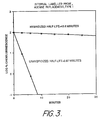
- An internally labeled 33mer probe specific for Chlamydia trachomatis was prepared as previously described (adenine replacement, type 1 linker-arm). The probe was hybridized with its target rRNA (in this case Chlamydia trachomatis ) according to the following procedure:
- hybridization and control mixtures were incubated 40 minutes at 60°C, at which point each mixture was analyzed for percent hybridization using hydroxyapatite (HAP) as follows.
- HAP hydroxyapatite
- a 0.1 ⁇ l aliquot of the hybrid or control mixture was added to 200 ⁇ l of 0.14M PB, pH 6.8, containing 2% HAP.
- Each resulting mixture was vortexed 5 seconds, incubated 5 minutes at 60°C, vortexed 20 seconds, then centrifuged 30 seconds in a microcentrifuge at 15,000 rpm.
- the supernatant was removed and saved, 200 ⁇ l of 0.14M PB, pH 6.8, was added to the HAP pellet, the mixture was vortexed 10 seconds, then centrifuged 30 seconds in a microcentrifuge at 15,000 rpm. The supernatant was removed and saved and the pellet was resuspended in 200 ⁇ l 0.14M PB, pH 6.8. A 50 ⁇ l aliquot of the resuspended HAP and each of the 2 supernatants were analyzed for chemiluminescence as described below, and the percent hybridized probe, i.e., the percent chemiluminescent signal associated with the HAP, was calculated.
- chemiluminescence of each sample was measured by adding a sample aliquot to 200 ⁇ l H2O in a 12x75mm tube and measuring chemiluminescence in a Clinilumat (automatic injection of 200 ⁇ l 0.25N HNO3, 0.1% H2O2 followed after a 1 second delay, by auto-injection of 200 ⁇ l 2M potassium PB, pH 13.2, and reading of chemiluminescence for 10 seconds).
- Fig. 3 is a graph for adenine replacement, type 1 linker-arm;
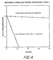
- Fig. 4 is a graph of insertion, type 1 linker-arm;
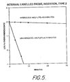
- Fig. 5 is a graph of insertion, type 2 linker-arm.
- hybridized probe is protected from degradation whereas unhybridized probe is not. This is true for all three types of internally labeled probes.
- sodium borate at elevated pH is shown to accelerate this process (as compared with example 2) while still retaining the differential degredation characteristics between hybridized and unhybridized probe.
- This example involves use of a probe of sequence 5′- CCG GAC CGC TGG CAA CAA AGG ATA AGG GTT GC-3′ (prepared using standard phosphoramidite chemistry) which hybridizes to E. coli rRNA immediately adjacent to the 5′ end of the AE-labeled probe used in this example (see below), thereby leading to the "capping off" of all the amines in the target rRNA in the vicinity of the acridinium ester label.
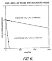
- AE-probe Probe end-labeled with acridinium ester (preparation described in Example 1) (hereinafter referred to as AE-probe) and the adjacent probe described above were hybridized according to the following procedure: Hybridization mixture Control mixture 1 ⁇ l AE-probe (.25 pmol) 1 ⁇ l AE-probe (.25 pmol) 2 ⁇ l E. coli rRNA (2ug) 5.4 ⁇ l H2O 4.3 ⁇ l adjacent probe (12 pmol) 5.8 ⁇ l 1M PB, pH 5 0.3 ⁇ l 4% SDS 0.3 ⁇ l 4% SDS 7.5 ⁇ l 1M PB, pH 5
- hybridization and control mixtures were incubated 40 minutes at 60°C, and then analyzed for percent hybridization using hydroxyapatite as described in Example 2.
- Example 2 hybridized AE-probe, in this case with an adjacent probe also hybridized, was protected from degradation whereas unhybridized probe was not. The protection was as good as in Example 2, because it is dependent on two hybridations instead of one.
- Hybridization was carried out exactly as described in Example 4. Stability of hybridized probe with adjacent probe versus unhybridized probe (i.e., control) was tested at pH 9 exactly as described in Example 2, except the composition of the stability test mixture, which was as follows: 5 ⁇ l hybrid or control 50 ⁇ l 0.2M sodium borate, pH 9.0
- sodium borate at elevated pH accelerated the process while still retaining the differential degradation characteristics between hybridized and unhybridized probe. See Fig. 7 for a graphical representation of these results.
- Probe was hybridized to its target rRNA (in this case E. coli ) according to the following procedure: Hybridization mixture Control mixture 1 ⁇ l AE-probe (.25 pmol) 1 ⁇ l AE-probe (.25 pmol) 2 ⁇ l E. coli rRNA (2ug) 5.4 ⁇ l H2O 5.8 ⁇ l 1M PB, pH 5 5.8 ⁇ l 1M PB, pH 5 0.3 ⁇ l 4% SDS 0.3 ⁇ l 4% SDS 3.4 ⁇ l H2O
- hybridization and control mixtures were incubated 40 minutes at 60°C, and then analyzed for percent hybridzation using hydroxyapatite as described in Example 2.
- Unhybridized probe again was preferentially degraded as compared to hybridized probe, although the differential in degradation is not as great as in previous examples. This is because only a portion of the amines were "capped off” in the vicinity of the acridinium ester label. Indeed, this additionally demonstrates the ability to discriminate between hybridized end-labeled probe in the presence and absence of hybridized adjacent probe.
- the hybridization and control mixtures were incubated 5 minutes at 80°C, followed by 60 minutes at 60°C.
- the resulting solutions were each diluted to 300 ⁇ l with 0.1M lithium succinate, pH 5.4, 10% lithium lauryl sulfate, and analyzed for percent hybridization using hydroxyapatite as described in Example 2.
- Hybrid 53.8% Control - 0.5% Half-life of ester hydrolysis (min) Ratio of half-lives Hybrid Control (Hybrid/Control) 13.6 1.3 10.5
- Probes containing the internal linker-arm, type L7, inserted at a variety of sequence dimer sites were synthesized, labeled with AE, and purified as described in Example 1.
- the sequence and linker-arm locations of these probes are as follows: Probe No.
- linker-arm location (#) 1 5′ -GCT CGC TGC GGA CTT#AAA CCA ACA T-3′ 2 5′ -AGG TCG GTC T#TT CTC TCC TTT CGT CTA CG-3′ 3 5′ -CAA TCG TCG AAA CCA TT#G CTC CGT TCG A-3′ 4 5′ -CCG CTA#CCC GGT ACG TT- 3′ 5 5′ TTG CCC ACA CCG A#CG GCG- 3′ 6 5′ -TTG CCC ACA CCG C#CG GCG- 3′
- Probe # Target Nucleic Acid 1 1 ⁇ g of E. coli rRNA 2 & 3 1 ⁇ g of C. trachomatis rRNA 4 1 ⁇ g of N. gonorrhoeae rRNA 5 & 6 1.2 pmol of the exact synthetic DNA complement
- linker-arm and therefore the AE
- linker-arm can be inserted at a wide variety of sequence dimer sites and still be substantially protected against ester hydrolysis in the hybridized form.
- DNA targets provide good protection of hybridized AE-probes when employed with these homogeneous assay formats.
- linker-arm also demonstates that the long (9 atom) linker-arm used here yields differential hydrolysis ratios essentially equivalent to the other linker-arms cited earlier in this patent establishing that a wide variety of linker-arm lengths can be used in the HPA format described herein.
- This probe has a single, T-G mismatch at position 6 (numbered from the 5′ end of the probe strand) with the 16S subunit of C. diversius .
- the probe was hybridized with its target rRNA (in this case either E . coli or C. diversius ) according to the procedure described in Example 8.
- Stability Ratio of half-lives Half-life of ester hydrolysis (min) Hybrid/Control Target Hybrid Control E . coli 18.6 0.8 23.3 C. diversius 1.1* 0.8 1.4 *
- the low half-life of ester hydrolysis was not due to low hybridization extent, as HAP analysis (as described in Examnple 2) revealed 73% hybridization.
- control mixture was the same as hybridization mixture except that it contained water instead of rRNA, and the reagent blank mixture was the same as the control mixture except that it contained water instead of probe.
- the mixtures were incubated 20 minutes at 60'C.
- Control mixture was the same as hybridization mixture except that it contained water instead of rRNA, and the reagent blank mixture was the same as control mixture except that it contained water instead of probe. The mixtures were incubated 60 minutes at 60°C.
- An internally labeled 30mer probe specific for E. coli was prepared as previously described (adenine replacement, type 1 linker-arm). The probe was hybridized in urine to decreasing amounts of its target rRNA (in this case E. coli ) according to the following procedure:
- Control mixture was the same as hybridization mixture except that it contained water instead of rRNA, and the reagent blank mixture was the same as control mixture except that it contained water instead of probe. The mixtures were incubated 30 minutes at 60°C.
- Reagent blank - 6845 rlu Control - 9250 rlu Minus control 10 ⁇ 3 ⁇ g rRNA - 9358 rlu 8 rlu 10 ⁇ 2 ⁇ g rRNA - 14055 rlu 4805 rlu 10 ⁇ 1 ⁇ g rRNA - 61800 rlu 52550 rlu Results represent the average of duplicate values.
- Example 2 Internally labeled probe (as in Example 2) was hybridized in clinical specimen (throat swab) as described in Example 8, including control and blank mixtures. After hybridization, one-third of each mixture was removed and subjected to selective degradation exactly as described in Example 8, while one-third was simply removed and allowed to stand at room temperature (i.e., no selective degradation). Both sets of samples, namely, with and without selective degradation, were then subjected to separation of hybridized from unhybridized probe using HAP as described in Example 2 with the following exceptions;
- Results represent the average of duplicative values with reagent blank already substracted.
- S:B signal to background ratio, i.e., chemiluminescence at a particular rRNA concentration divided by chemiluminescence of control.
- the following probe was constructed to contain an internal linker-arm, type L7 (position of insertion indicated by # below), labeled with acridinium ester, and purified as described in Example 1: ATT CCG CAC A#TG TCA AAA CCA G
- This probe is exactly complementary to the 16S subunit of Neisseria gonorrhoeae . However, it is also closely related to N. meningitidis , and a cross-reaction is typically observed under the hybridization stringency conditions cited in Examples 2 and 8.
- This probe was hybridized with 0.5 ⁇ g N. meningitidis as described in Example 8, (except in a 200 ⁇ l format), then either incubated for 10 minutes at 60°C in 1 ml of 0.2M sodium tetraborate, pH 7.6, 5% Triton X-100 to effectuate differential hydrolysis (+ D.H.), or not subjected to these differential hydrolysis conditions (-D.H.). The resulting hybrids were then separated on magnetic microspheres and measured for chemiluminescence as decribed below.
- differential hydrolysis technique as an additional stringency discrimination step greatly reduces signal from undesired, cross-reacting sequences.
- cross-reaction of a N. gonorrhoeae probe with N. meningitidis was lowered greater than 200-fold.
- the differential hydrolysis step increases sensitivity to unstable hybrids, which are in equilibrium with unhybridized probe, by constantly reducing the chemiluminescence (via hydrolysis) of the probe when it is in the unhybridized form, thus lowering the chemiluminescence due to cross reaction.
- a 24mer probe (sequence given below) with an internal linker-arm, type L7, inserted as indicated was synthesized, labeled with AE and purified as described in Example 1.
- This probe is complementary to the chimeric mRNA transcript (common break) associated with chronic myelogenous leukemia (CML) and will be called the bcr/abl probe.
- This chimeric mRNA is a product of the chimeric gene formed by the translocation of a region of the abl gene on chromosome 9 into a region of chromosome 22 containing the bcr gene.
- the asterisk inserted above denotes a linker-arm insertion site for acridinium ester attachment and the underline indicates abl sequences.
- the bcr/abl probe was hybridized with approximately 1 pmol of each of the targets listed above as described in Example 8, and the stability of these hybrids and the unhybridized probe was tested at pH 7.6 as described in Example 7.
Abstract
Description
- This invention is in the art of diagnostic procedures and techniques for detecting and quantitating minute amounts of organic compounds.
- More particularly, this invention relates to homogeneous diagnostic assays in which there is a difference in the stability of the label in its bound, as opposed to its unbound forms. This invention relates to the construction of environments for diagnostic assay systems in which a label is differentially degraded in either its complexed or bound form as compared to its uncomplexed or unbound form.
- Diagnostic assays are a common analytical technique for detecting, locating or quantifying biological substances by employing labelled binding reagents. The presence of the labeled reagent can then be detected using a variety of known methods. This invention can be applied to all known diagnostic assay formats, including, without limitation, direct binding and competition assays and sequential saturation. One particular type of diagnostic assay is the nucleic acid hybridization assay. Hybridization assay systems are based on the fact that single stranded nucleic acids (DNA or RNA) will hybridize or recombine, under appropriate circumstances, with complementary single stranded nucleic acids. By labelling the complementary probe nucleic acid with a readily detectable label, it is possible to detect the presence of the target polynucleotide sequence of interest in a test sample containing single stranded nucleic acid sequences.
- Assay systems may broadly be characterized as heterogeneous or homogeneous. The term "heterogeneous" as applied to assay systems means those specific binding assays which require a separation of the labelled from unlabeled substance to be detected. Homogeneous assays systems, on the other hand, do not involve any physical separation steps. Because homogeneous assay systems involve a minimum number of handling steps they are generally more convenient and easier to use than heterogeneous assay systems.
- For example, a typical sandwich immunoassay may involve incubating an immobilized antibody with a test medium. Antigens, if in the medium, will bind to the antibody. After incubation, unbound antigen is removed in a separation step. After a second, or simultaneous incubation with a solution of labeled antibody, the bound antigen becomes "sandwiched" between the immobilized antibody and the labelled antibody. After a second separation step, the amount of labeled antibody can be determined as a measure of the antigen in the medium. This system is time consuming because it involves a series of incubation and separation steps.
- A focus of effort of the prior art in diagnostic assays has been directed to developing homogeneous assays and labels which can discriminate between minor differences in the amount of bound, as opposed to unbound substances of interest. One of the objects of the present invention is an assay system in which the label itself undergoes a detectable change, which may be, for example, in its ability to chemiluminesce, when it is in its bound, as opposed to when it is unbound. Another object of the present invention is an assay system in which the label is substantially degraded or destroyed in either its bound or unbound form, thereby providing a ready means for identifying and quantitating a reaction of interest.
- It is an object of the present invention to disclose an improved method for sensitively detecting analytes using a homogeneous assay format. It is also an object of this invention to provide improved methods for increasing the sensivitity of assays which involve separation by combining the homogeneous method disclosed herein with other separation methods to reduce non-specific background. The principle of the invention disclosed here is based upon the differential stability of a label to chemical or biochemical reagents. Whenever certain labels are conjugated to binding partners, we have found that the stability of said labels are or may be altered when said binding partner is bound to a binding substance of said binding partner. It is also the object of this invention to disclose a method by which said differential label stability may be employed for the sensitive detection of an analyte employing a homogeneous diagnostic assay system. Yet another object of the present invention is to disclose the use of chemiluminescent acridinium ester labeled DNA probes in said homogeneous diagnostic assays for sensitively detecting the presence of complementary target polynucleotide sequences.
- There are a variety of homogeneous assays in the prior art which vary in complexity. In some systems, for example, the label is a catalyst which can participate in a chemical reaction in the presence of other components, for example, enzymes, coenzymes and cofactors. In other, albeit related systems, the label is a substrate for a chemical or biochemical reaction. In these systems, the generation of a specific readily detectable substance, for example, glucose, lactate or alcohol is used to monitor and measure the target reaction. In other assay systems, the analyte possesses unique physical properties which allow it to be directly detected. Examples of these types of labels include metals, hemoglobin and chlorophyll.
- Still other homogenous assay systems are based on enzyme coupled specific binding reactions, wherein the analyte is a ligand for a specific binding reaction. When the analyte is present, it undergoes a specific binding reaction with a conjugate which is comprised of a specific binding partner and a labelling substance. Either concurrently or subsequently, other substituents are added which interact with the label. The activity of the label is different when the conjugate of which the label is a component, is in a complexed form verses an uncomplexed form. Such systems have typically used enzymes as the labelling reagent and substrates which produce a colorimetric, fluorimetric, or chemiluminescent end point. Examples of homogeneous enzyme immunoassays include U.S. Patent Nos. 3,654,090, 3,817,837 and 4,190,496. Other examples of homogeneous assays involving the use of chromophores which make up fluorescer/quencher pairs may be found in U.S. Patent Nos. 4,199,559, 4,174,384 and 4,318,707. In some systems, however, the label has been a substance other than an enzyme, for example, vitamins, NAD, FAD or biotin, which nevertheless can be coupled to a "monitoring" reaction. An example of this type is U.S. Patent No. 4,383,031. In these systems, the monitoring reaction is based upon a structural change which modifies the activity of the label.
- Other homogenous assay systems involve the technique of polarization fluorescence. Here, the analyte competes with a low molecular weight fluorescent conjugate for binding to a high molecular weight specific binding partner. Since the polarization of the fluorescence changes when the small molecule is displaced from the surface of the large molecule, it is possible to determine the amount of analyte in solution. An example of this type of assay system is U.S. Patent No. 4,668,640.
- Yet another type of homogenous assay system involves non-radiative energy transfer. In these systems, the absorption of light from one molecule to another when the two molecules come into close proximity is used as the monitoring reaction. Generally, these methods have been described in two ways. In one method, the first molecule is chemiluminescent and upon excitation a portion of the electromagnetic energy generated is transferred to a second "absorber" molecule which must be in close proximity. If the energy is absorbed by the second molecule and emitted at a different wavelength, a portion of the light will show a spectral shift proportional to the number of chemiluminescent molecules in close proximity to absorber molecules.
- In another type of non-radiative energy assay system, the first molecule is fluorescent and serves as an "absorber" of external light. In the presence of a second fluorescent molecule a portion of the energy is transferred and light is emitted at a different wavelength. The emitted light will show a spectral shift proportional to the number of "absorber" and "emitter" molecules in close proximity to each other.
- A different type of double probe assay system is seen in U.S. Patent No. 4,670,379. The first probe is labeled with a catalyst; the second with an apoluminescer. Both probes target neighboring areas on the target nucleic acid sequence. Once both probes are hybridized together with the target, the substrate is added. The catalyst converts the substrate to a transformation radical which, in turn, converts the apoluminescer on the second probe to a luminescer. This occurs only if hybridization has taken place. Employing these principles, assays have been developed based upon two labelled substances simultaneously binding a common analyte.
- A specific example of this type of energy transfer assay system is Elazar et al., European Patent Application No. 85105130.0, Publication No. 0159719 published October 30, 1985, which discloses using two single stranded nucleic acid probes which are complementary to the same or opposite strands of the target genetic material. Each probe is labeled with a moiety capable of producing a signal only when the two labels are brought together. Unlike the invention herein described, Elazar et al. involves the formation and detection of double hybrid or multihybrids. Similarly, Heller et al., European Patent Application No. 82303699.1, Publication No. 0070685 dated January 26, 1983 and related Morrison et al., European Patent Application No. 82303700.7, Publication No. 0070686 of the same date disclose homogenous assay systems using two luminescer probes. At least one of the light labels is of the absorber/emitter type so that when the two probes are hybridized to the target, energy transfer occurs between the two labels. This causes the absorber/emitter to re-emit at a different wavelength. The second emission detects hybridization. An antigen assay of this type is disclosed in Morrisson et al. supra.
- Most biological substances can not be readily detected directly at any reasonable level of sensitivity and require a binding reaction of some type. As evident from the foregoing discussion, the prior art is based on the detection of minor attenuations between bound and unbound label. The prior art systems are not capable of significant discrimination between bound and unbound label. Such assays have been found useful for detecting analytes which are present only at high concentration, for example in the monitoring of various drugs in blood and urine.
- There is a need in the area of clinical diagnostics for a direct detection homogenous assay which is based on the ability of two binding partners to modify the stability of the label, for example, resulting in selective removal or destruction of label in either the bound or unbound form. While Hirschfield, in Florescence Background Discrimination by Prebleaching J. Histochemistry and Cytochemistry, 27/1, 96-101 (1979) describes a somewhat related electrooptics technique involving photochemical bleaching which may destroy label molecules, or at least their fluorescence. The invention disclosed therein does not teach or suggest use of photochemical bleaching or any other technique for selective removal or destruction of a label in a diagnostic assay. The subject invention fulfills a present need in diagnostic assays as it is orders of magnitude more sensitive than the prior art. The range of sensitivity of the prior art is no better than the 10⁻¹³ mole range, while the present invention is sensitive in the 10⁻¹⁶ mole range.
- Briefly, this invention comprises diagnostic assays and methods. The method may be used to detect an analyte in a medium, said analyte being part of a specific binding pair. When the medium suspected of containing the analyte is combined with a binding partner, a label substance attached to the binding partner is capable of undergoing a detectable change in stability or differential degradation whenever the analyte binds to the specific binding partner. In a specific embodiment, single stranded nucleic acid probes have been modified to contain labels at virtually any desired position or location on the probe. The probe labels are of different stability or susceptible to differential degradation depending on whether the target nucleic acid sequence is hybridized to the probe.
- Notably, the present invention comprises assay systems whereby the label on the bound probe is stabilized relative to the unbound probe. This stabilization may be aided by intercalation. DNA intercalating compounds, including acridinium esters, are particularly, but not exclusively, suited for use in this inventive assay system. DNA intercalators are known to bind non-covalently to duplex DNA and are characteristically flat molecules which may insert between base pairs of the double helix of DNA. We have evidence which indicates that acridinium esters prefer to insert in regions rich in adenine and thymidine base pairs.
- It should be understood that while the present invention hereinafter will be described with particular reference to acridinium ester labelled probes, the present invention contemplates the use of equivalent labels and assay formats which are susceptible to differential degradation or change in stability when the conjugate to which the label is a component is bound. As will be explained in more detail herein, other suitable label compounds include substances which are destabilized by the amines present on nucleic acids particularly those in the vicinity of the label on unhybridized probes. Moreover, label substances which can interact with hydrophobic environments, for example by intercalating between base pairs, also may be used.
- This invention has application generally to any system involving the selective substantial degradation of the label when comparing its bound and unbound conjugated forms. Furthermore, because of the relative simplicity of the assay system, which does not involve any separation or washing steps, it is both faster and less expensive than conventional systems. The system can be more sensitive than conventional systems in those cases in which there are large differences in stability between the bound and unbound conjugated forms of the label because virtually all of the residual or unbound label conjugate is destroyed and, therefore, not detected. Finally, the system is very versatile and can be used either alone, or in combination with other separation methods to achieve very low background noise. The present invention is useful in probe diagnostics, including infectious and genetic and viral disease detection and cancer diagnosis.
-
- Figure 1 is a graphical representation of HPLC purification of an acridinium ester-labeled probe.
- Figure 2 is a graphical representation of the results set forth in Example 2.
- Figures 3-5 are graphical representations of the results set forth in Example 3.
- Figure 6 is a graphical representation of the results set forth in Example 4.
- Figure 7 is a graphical representation of the results set forth in Example 5.
- Figure 8 is a graphical representation of the results set forth in Example 6.
- The following definitions shall be used in this description:
- 1. acridinium ester: derivative of acridine possessing a quaternary nitrogen center and derivatized at the 9 position to yield a labile phenyl ester moiety, specifically, 4-(2-succinimidyloxycarbonyl ethyl) phenyl-10-methylacridinium 9-carboxylate fluorosulfonate:

- 2. acridinium esters: moieties of the following general type:

- 3. analyte - any substance capable of undergoing a binding reaction with one or more specific binding partners, including, without limitation, antigens and antibodies thereto, haptens and antibodies thereto; hormones, drugs, metabolites, vitamins, coenzymes and their binding partners, including receptors; polynucleotides, oligonucleotides, and hybrids of polynucleotides or oligonucleotides and antibodies and binding substances thereto; polynucleotides or oligonucleotides and hybridizable polynucleotides or oligonucleotides thereto; metals and chelating agents thereto.
- 4. binding partner - any molecule or substance capable of undergoing a specific binding reaction with an analyte.
- 5. duplex: double stranded complex formed upon the annealing of two complementary, single stranded nucleic acid molecules.
- 6. hybridization: the formation of stable duplexes between 2 complementary single stranded DNA or RNA molecules or between a DNA and complementary RNA molecule. Duplex formation is specific for complementary base pairs, thereby reproducing the genetic code of the specific gene hybridized.
- 7. bound: condition in which a binding interaction has been formed between a molecule and its specific binding partner.
- 8. stable: resistant to chemical or biochemical degradation, reaction, decomposition, displacement or modification.
- 9. stability: the resistance of a substance to chemical or biochemical degredation, reaction, decomposition, displacement or modification.
- It is known that in solution acridinium esters exist in equilibrium with their corresponding bases. At high pH, base formation is favored; the quaternary nitrogen species reforms at low pH. It also is known that chemiluminescence reaction can be affected by adding base, specifically an aqueous solution of sodium hydroxide containing hydrogen peroxide. The chemiluminescence involves attack by hydroperoxide ions on the acridinium species, which results in the formation of electronically excited N-methylacridone. See generally, Weeks et al. Acridinium Esters as High-Specific Activity Labels in Immunoassay, Clin. Chem. 2918, 1474-1479 (1983). The reaction is diagrammed below.

- The subject invention can be carried out as follows. First, select binding partners comprising a binding substance and one or more binding partners for the assay to be performed. These pairs may be antigens and antibodies thereto; haptens and antibodies thereto; hormones, drugs, metabolites,vitamins, coenzymes and their binding partners, including receptors; polynucleotides, oligonucleotides, and hybrids of polynucleotides or oligonucleotides and antibodies and binding substances thereto; polynucleotides or oligonucleotides, and hybridizable polynucleotides or oligonucleotides thereto; metals and chelating agents therefor.
- Second, select the assay format to be used. These may be selected from formats comprising direct binding assays, competition assays, sequential saturation methods, and sandwich assays.
- Third, select a label for the assay to be performed. This may be a label which can be directly or indirectly detected by colorimetric, fluorimetric, chemiluminescent, or bioluminescent means. The label should additionally have the property that it can be chemically or biochemically degraded so as to modify its ability to be detected, said degradation being possible under conditions which do not adversely effect the binding between the labeled binding partner and its binding substance and other binding partners and binding substances which may participate in the reaction. Preferred labels are ones which are affected in their ability to be detected after exposure to acids, bases, or selective oxidizing agents such as peroxidate, or enzymes.
- Fourth, using chemical methods known in the art, attach the label to the binding substance at a site such that the label's sensitivity to chemical or biochemical degradation is modified upon interaction of the labeled binding partner with its specific binding substance(s). In some cases several different sites may be tested for label attachment and the site which gives the best differential degradation may be used.
- Fifth, optimize the degradation conditions, be they chemical or biochemical, to give the best detection discrimination of the labeled binding partner in the presence and absence of its binding substance.
- Finally, using the preselected assay format, test the ability of the assay system to detect quantitatively or qualitatively the analyte generally employing the steps of:
- a. Incubate
- b. Selectively degrade
- c. Detect
- a. Simultaneously incubate and selectively degrade
- b. Detect.
- Employing this invention, oligonucleotide probes labelled with chemiluminescent acridinium esters are particularly useful for the detection of sequence specific polynucleotides through hybridization. Acridinium esters may be attached at a number of different sites on DNA probes and/or mixed nucleotide/ non-nucleotide polymers as described in U.S. Patent Application Ser. No. (not yet assigned) filed September 21, 1987 entitled "Non-Nucleotide Linking Reagents for Nucleotided Probes" by Arnold, et al. This includes the ability to label the nucleotide bases, the phosphate backbone, the sugar residues, the 3′ terminus, and the 5′ terminus of oligonucleotides as well as the nonnucleotide monomeric units of mixed nucleotide/non-nucleotides polymers.
- Such acridinium ester labeled probes can show significant differential chemical stability when the probes to which they are attached are free in solution as compared to when they are hybridized. This differential stability is dependent upon the position of the acridinium ester in the probe, the nucleotide residues in the vicinity of the acridinium ester label and the presence of other probe molecules which form stable hybrids with the target polynucleotide. A graphic representation is set forth below.

- In the course of determining the factors which contribute to the differential stability of the acridinium ester on DNA probe molecules, we found that the free amines on the bases of unhybridized probes (both nucleotide and mixed nucleotide/non-nucleotide) destabilized the acridinium ester, especially in an alkaline environment. At the same time, if the acridinium ester is near the terminus of the probe, it can also be destabilized to alkali by amines contributed by target sequences once hybridization occurs. When the probe hybridizes with a sequence specific polynucleotide (binding substance) the probe amines participate in base pairing with complementary nucleotides and are restricted in their ability to destabilize the acridinium ester, especially in an alkaline environment. At the same time, it was found that the acridinium ester was further stabilized against degradation by intercalation into the hybrid duplex, particulary in regions of adenine/thymidine base pair density. We have found that a number of other insertion regions also give good differential hydrolysis, as explained in the following section.
- There are several modes by which this invention may be applied to hybridization. These include without limitation:
- 1. Attaching the acridinium ester to the central region of the probe and near a region of adenine/thymidine base pairs. A preferred method is to attach an acridinium ester to a non-nucleotide monomeric unit as disclosed in U.S. Patent Application Ser. No. (not yet assigned), filed September 21, 1987 entitled "Non-nucleotide Linking Reagents for Nucleotide Probes" by Arnold, et al. which is attached as an insert to nucleotide monomeric units which are complementary to immediately adjacent nucleotides of the target polynucleotide sequence. Such placement serves to restrict the amines of the nucleotide bases on both sides of the acridinium ester and to provide a site for intercalation.
- 2. Attaching the acridinium ester to either the 3′ or 5′ terminus of the probe and restricting the effects of nearby amines contributed by the target polynucleotide with a second probe hybridized adjacent to the first. It may also be desirable if said second probe creates an A/T base pair rich region upon duplex formation. Even though this double probe or sandwich system is dependent upon the formation of two duplexes instead of only one, it may provide a method for sensitively detecting minor base pair changes which can be discriminated by very short probes i.e., probes of approximately 5-15 nucleotides in length.
- Under normal circumstances such short probes have the ability to discriminate single mismatches under appropriate hybridization conditions. As a practical matter, however, they are not used because they are not sufficiently specific for single polynucleotide sites. That is, the short probe may hybridize several specific sites contained within a multitude of complex DNA or RNA sequences present. With the added requirement, that two different probes must hybridize immediately to adjacent polynucleotide regions, such regions can be specifically targeted and identified. Thus, the benefits of short probe discrimination may be utilized while maintaining the specificity of the region spanned by both probes.
- 3. Attaching the acridinium ester at or near the site of a mismatch with a polynucleotide sequence that is not the desired target polynucleotide. In this manner discrimination between polynucleotide sequences differing by only one nucleotide can be achieved, since the area of the duplex around a mismatch site is sufficiently destabilized to render the acridinium ester (if it is attached at or near this site) susceptible to degradation.
- 4. Attaching acridinium ester to a probe region in order to monitor the local stability of a probe-target hybrid. In this manner, the acridinium ester can serve to closely monitor the local hybrid stability under conditions in which the entire hybrid duplex doesn't melt but under which the local hybrid region partially melts.
- A preferred mode for the above three formats is to conduct the differential hydrolysis step at the same temperature as the hybridization step, typically at 50 to 70°C.
- Another mode is to conduct a second differential hydrolysis step at room temperature. This allows pH's in the range of 10-11 to be used, yielding even larger differences in the rate of hydrolysis between hybridized and unhybridized acridinium ester-labeled probe.
- The following examples are offered by way of illustration and not by way of limitation. These examples are based on currently available data and the most likely explanations of the phenomenon. Other factors and methods may become evident with more research. Although the invention has been described in detail by way of illustration and examples using acridinium ester labels, certain changes and modifications may be practiced within the scope of the claims and other label systems also may be employed within the scope of the claims.
- Deoxyoligonucleotide probes were synthesized to contain an amine liner-arm (i.e., one which terminates in a primary amine for labeling with acridinium ester) located either at the 5′-terminus, at a specific preselected location along the polyphosphate chain, in the internal portion of the probe or attached to one of the nucleotide bases.
- To attach a 5′-amine linker-arm to a probe, the following compound was used:

- This compound will heretofore be referred to as terminal amine linker-arm reagent. This reagent was synthesized as follows: 6-amino hexanol was reacted with S-ethyltrifluorothioacetate in anhydrous ethylacetate. The reaction product was precipitated in petroleum ether, treated with a 10% pyridine in water mixture for 10 minutes to hydrolyze any 0-trifluoroacetyl which may have formed, and evaporated to dryness in the form of a gum. This compound was then phosphitylated according to standard protocols within the literature (see Nucleic Acids Research, 12 (11), 4539 (1984)) to yield the desired compound, namely, terminal amine linker-arm reagent (1).
- Probes containing a 5′-amine linker-arm were synthesized as follows. Using an Applied Biosystems, Inc. Model 380A DNA synthesizer, probes of desired nucleotide sequence were produced using standard phosphoramidite chemistry, building the probes from the 3′ end to the 5′ end. After the desired sequence was completed, the amine linker-arm was automatically coupled to the 5′-hydroxyl group of the probe using the terminal amine linker-arm reagent in the same way another phosphoramidite nucleoside would have been coupled. Using standard protocols, the probe was then cleaved from the solid support and deprotected using NH₄OH and purified by polyacrylamide gel electrophoresis followed by Sephadex G-25 chromatography.
- The following probe was synthesized and purified using this procedure:

- To incorporate an amine linker-arm into the internal portion of a probe, internal amine linker-arm reagent, type 1 (4 atom spacer; see L1 in patent cited below), or type 2 (2 atom spacer; see L3 in patent cited below), or L7 (9 atom spacer) was used as described in U.S. Patent App. Ser. No. 099,050 entitled "Non-Nucleotide Linking Reagents for Nucleotide Probes" filed September 21, 1987 by Arnold, et al. Again, probes were synthesized using standard phosphoramidite chemistry and purified using polyacrylamide gel electrophoresis and Sephadex G25 chromatography.
- The following probes were synthesized using this procedure:
- 1.) A 30mer complementary to the 16S subunit of rRNA from E. coli, with an internal amine linker-arm,
type 1, replacing an adenine residue at position 18 in the sequence:
- 2.) A 33mer complementary to the 16S subunit of rRNA from Chlamydia trachomatis, with an internal amine linker-arm,
type 1, replacing an adenine residue at position 21 in the sequence,or inserted between residues 21 and 22,
- Nucleotide bases containing amine linker-arm were incorporated into a probe as follows. Template and primer oligonucleotides with the following sequences were synthesized:(The primer and extended primer sequences are complementary to the 16S subunit of C. trachomatis.)

- Primer extension using the Klenow fragment was performed as described in Maniatis (Molecular Cloning, A Laboratory Manual, T. Maniatis, 1982, Cold Spring Harbor Laboratories, Pubs.) with the exception that the BSA was omitted from the 10X nick translation buffer, to incorporate the amine linker-arm modified base amino (12) dUTP (Calbiochem, California). The sequence of the resulting oligomer is, therefore:
CGT TAC TCG GAT GCC CAA ATA (amino-12-U)CG CC. - Purification of the primer extended complex was achieved using a NENSORB-20 cartridge (DuPont) following the procedure recommended by the manufacturer. (The primer extension reaction was diluted by the addition of 900 µl of NENSORB reagent A prior to loading on the column. The purified oligomers were eluted with 50% methanol which was then removed in a speed-vac.)
- A 25mM stock solution of acridinium ester was prepared in distilled DMSO. The desired amount of probe (see a listing of the different probes labeled in section A above) was evaporated to dryness in a 1.5 ml conical polypropylene tube. The following cocktail was constructed by adding the following ingredients in the order listed:
3µl H₂O
1µl 1M HEPES (pH 8.0)
4µl DMSO (distilled)
2µl 25mM acridinium ester in DMSO (distilled)
The mixture was vortexed, spun in a microcentrifuge for 2 seconds (to bring the contents to the bottom of the tube), and incubated at 37°C for 20 minutes. The following components were then added in the reaction cocktail in the order listed:
3.0µl 25mM acridinium ester in DMSO (distilled)
1.5µl H₂O
0.5µl 1M HEPES (pH 8.0)
The cocktail again was vortexed, spun, and incubated an additional 20 minutes at 37°C. The unreacted label was quenched using a 5-fold excess of lysine by adding 5µl of 0.125M lysine in 0.1M HEPES (pH 8.0), 50% DMSO, and incubated 5 minutes at room temperature. - The acridinium ester-labeled oligomer was then purified using the following method. To the 20µl quenched reaction mixture 30µl 3M NaOAc (pH 5.0), 245µl H₂O and 5µl glycogen was added as a carrier (the glycogen was pre-treated to remove any nuclease activity). The sample was vortexed briefly and 640µl of absolute EtOH added. The sample was vortexed briefly and incubated on ice 5-10 minutes, then centrifuged 5 minutes at 15,000 rpm in a microcentrifuge. The supernatant was carefully removed and the pellet was redissolved in 20µl of 0.1M NaOAc (pH 5.0), 0.1% SDS. The samples were then purified by high performance liquid chromatography (HPLC) as described below.
- The AE-labeled probes containing the 5′ and internal amine linker-arms were purified as follows: the 20µl redissolved pellet was injected onto a a Nucleogen-DEAE 60-7 ion-exchange HPLC column mounted in an IBM® 9533 HPLC system. All buffers were made with HPLC grade water, acetonitrile (CH₃CN) and sodium acetate (NaOAc) from Fisher Scientific, and reagent grade glacial acetic acid (HOAc) and LiCl. All buffers were filtered through 0.45µm pore size Nylon-66 filters before use. The sample was eluted as described in Figure 1 of the drawing (in this case the probe was the 5′-amine linker-arm containing 26mer described in Section A above). Immediately after the run, 5µl of 10% SDS was added to each tube followed by vortexing of each tube (this was done to ensure that the acridinium ester-labeled probe did not stick to the walls of the tube). A 0.5µl aliquot was removed from fractions 21-42 and added to 200µl water in a 12x75mm tube (a separate pipet tip was used for each aliquot to avoid a carryover problem). The chemiluminescence of each aliquot was then determined in a Berthold Clinilumat using the following automatic injection and read sequence: injection of 200µl of 0.25N HNO₃, 0.1% H₂O₂; a 1 second delay; a 200µl injection of 1N NaOH; read chemiluminescent output for 10 seconds.
- Fractions 29-33 were then EtOH precipitated as follows: Add to each fraction 5µl glycogen, vortex, add 1 ml EtOH, vortex, incubate 5-10 minutes on ice, and
centrifuge 5 minutes at 15,000 rpm in a microcentrifuge. Each supernatant was carefully removed, the pellet redissolved in 20µl 0.1M NaOAc,pH 5, 0.1% SDS, and the fractions pooled. - In this manner, highly pure acridinium ester-labelled probes were obtained. The specific activity of such probes was typically 5-10x10⁷ chemiluminescent light counts (Berthold Clinilumat) per picomole of oligomer.
- The AE-labeled probe containing the amine linker-arm modified base (amino-12-U) was purified generally as described above, with the following exceptions: A Vydac C4 reverse-phase column was used; buffer A was 0.1M triethyammonium acetate (Applied Biosystems, Inc., Foster City, California) and buffer B was CH₃CN; the labeled probe was eluted as a hybrid using a linear gradient for 10-15% solvent B in 25 minutes at a flow rate of 1 ml/min. The main chemiluminescent peak was then identified and worked up as described above.
- The preceding discussion generally describes how to make and label probes with acridinium ester. In the specific example herein, probes were end and internal labeled, as well as labeled in a nucleotide base.
- An internally labeled 33mer probe specific for Chlamydia trachomatis was prepared as previously described (adenine replacement,
type 1 linker-arm). The probe was hybridized with its target rRNA (in this case Chlamydia trachomatis) according to the following procedure:
- 2µl AE-probe (0.5 pmol)
0.3µl 4% (wt:vol) SDS
5.8µl 1M PB,pH 5
4.4µl C. trachomatis rRNA (2µg), or 4.4µl H₂O for control. - The hybridization and control mixtures were incubated 40 minutes at 60°C, at which point each mixture was analyzed for percent hybridization using hydroxyapatite (HAP) as follows. A 0.1µl aliquot of the hybrid or control mixture was added to 200µl of 0.14M PB, pH 6.8, containing 2% HAP. Each resulting mixture was vortexed 5 seconds, incubated 5 minutes at 60°C, vortexed 20 seconds, then centrifuged 30 seconds in a microcentrifuge at 15,000 rpm. The supernatant was removed and saved, 200µl of 0.14M PB, pH 6.8, was added to the HAP pellet, the mixture was vortexed 10 seconds, then centrifuged 30 seconds in a microcentrifuge at 15,000 rpm. The supernatant was removed and saved and the pellet was resuspended in 200µl 0.14M PB, pH 6.8. A 50µl aliquot of the resuspended HAP and each of the 2 supernatants were analyzed for chemiluminescence as described below, and the percent hybridized probe, i.e., the percent chemiluminescent signal associated with the HAP, was calculated.
- The stability of hybridized probe versus unhybridized probe (i.e., control) was tested at
pH 6 according to the following procedure:
- 12.3µl hybrid or control from above
50µl 1M PB,pH 6
2.5µl 4% SDS
65µl H₂O - These mixtures were vortexed briefly, and 5µl aliquots were removed immediately (t₀) and analyzed for chemiluminescence as described below. The remainder of the mixtures were incubated at 60°C, and 5µl aliquots were removed at various time points (see below) and analyzed immediately for chemiluminescence.
- The chemiluminescence of each sample was measured by adding a sample aliquot to 200µl H₂O in a 12x75mm tube and measuring chemiluminescence in a Clinilumat (automatic injection of 200µl 0.25N HNO₃, 0.1% H₂O₂ followed after a 1 second delay, by auto-injection of 200µl 2M potassium PB, pH 13.2, and reading of chemiluminescence for 10 seconds).
- Hybrid - 96%
Control - 1.3% (non-specific binding)
- See Fig. 2 of the drawing.
- These results demonstrate that the hybridized probe is well protected against breakdown and subsequent loss of chemiluminescense (half-life for loss of chemiluminescence equal to 3370 minutes), whereas unhybridized probe is very susceptible to breakdown and loss of chemiluminescence (half-life is equal to 29.2 minutes; therefore, discrimination between hybridized and unhybridized is equal to 115-fold). These data demonstrate the ability of the homogeneous assay herein described to locate target DNA sequences by imparting differential stability between hybridized and unhybridized probe and measuring same.
- Internally labeled probe (all 3 internally labeled 33mer probes described in Example 1 were tested) was hybridized with its target rRNA (in this case Chlamydiatrachomatis) and analyzed for percent hybridization as described in Example 2.
- The stability of hybridized versus unhybridized probe (i.e., control) was tested at pH 9 according to the following protocol:
- 5µl hybrid or control from above
45µl 0.2M sodium borate, pH 9
The mixtures were then incubated, sampled and analyzed for chemiluminescence as described in Example 2. - Hybrid - 95%
Control - 0.5% (non-specific binding)
- Hybrid - 98%
Control - 0.3%
- Hybrid - 98%
Control - 0.2%
- See Figs. 3-5 of the drawing. Fig. 3 is a graph for adenine replacement,
type 1 linker-arm; Fig. 4 is a graph of insertion,type 1 linker-arm; Fig. 5 is a graph of insertion,type 2 linker-arm. - As in Example 2, hybridized probe is protected from degradation whereas unhybridized probe is not. This is true for all three types of internally labeled probes. In this example, sodium borate at elevated pH is shown to accelerate this process (as compared with example 2) while still retaining the differential degredation characteristics between hybridized and unhybridized probe.
- This example involves use of a probe of
sequence 5′- CCG GAC CGC TGG CAA CAA AGG ATA AGG GTT GC-3′ (prepared using standard phosphoramidite chemistry) which hybridizes to E. coli rRNA immediately adjacent to the 5′ end of the AE-labeled probe used in this example (see below), thereby leading to the "capping off" of all the amines in the target rRNA in the vicinity of the acridinium ester label. - Probe end-labeled with acridinium ester (preparation described in Example 1) (hereinafter referred to as AE-probe) and the adjacent probe described above were hybridized according to the following procedure:
Hybridization mixture Control mixture 1µl AE-probe (.25 pmol) 1µl AE-probe (.25 pmol) 2µl E. coli rRNA (2ug) 5.4µl H₂O 4.3µl adjacent probe (12 pmol) 5.8µl 1M PB, pH 50.3 µl 4% SDS0.3 µl 4% SDS7.5µl 1M PB, pH 5 - The hybridization and control mixtures were incubated 40 minutes at 60°C, and then analyzed for percent hybridization using hydroxyapatite as described in Example 2.
- The stability of the hybridized probe with adjacent probe versus unhybridized probe (i.e., control) was tested at
pH 6 exactly as described in Example 2. - Hybrid - 93.5%
Control - 2.7% (non-specific binding)
- See Fig. 6 of the drawing.
- As in Example 2, hybridized AE-probe, in this case with an adjacent probe also hybridized, was protected from degradation whereas unhybridized probe was not. The protection was as good as in Example 2, because it is dependent on two hybridations instead of one.
- Hybridization was carried out exactly as described in Example 4. Stability of hybridized probe with adjacent probe versus unhybridized probe (i.e., control) was tested at pH 9 exactly as described in Example 2, except the composition of the stability test mixture, which was as follows:
5µl hybrid or control
50µl 0.2M sodium borate, pH 9.0 - Hybridized AE-probe with an adjacent probe (also hybridized) again was protected from degradation whereas unhybridized probe was not. As in Example 3, sodium borate at elevated pH accelerated the process while still retaining the differential degradation characteristics between hybridized and unhybridized probe. See Fig. 7 for a graphical representation of these results.
- Probe was hybridized to its target rRNA (in this case E. coli) according to the following procedure:
Hybridization mixture Control mixture 1µl AE-probe (.25 pmol) 1µl AE-probe (.25 pmol) 2µl E. coli rRNA (2ug) 5.4µl H₂O 5.8µl 1M PB, pH 55.8µl 1M PB, pH 50.3 µl 4% SDS0.3 µl 4% SDS3.4µl H₂O - The hybridization and control mixtures were incubated 40 minutes at 60°C, and then analyzed for percent hybridzation using hydroxyapatite as described in Example 2.
- The stability of the hybridized probe versus unhybridized probe (i.e., control) was tested at
pH 6 exactly as described in Example 2. - Hybrid - 94%
Control - 2.7% (non-specific binding)
- See Fig. 8 of the drawing.
- Unhybridized probe again was preferentially degraded as compared to hybridized probe, although the differential in degradation is not as great as in previous examples. This is because only a portion of the amines were "capped off" in the vicinity of the acridinium ester label. Indeed, this additionally demonstrates the ability to discriminate between hybridized end-labeled probe in the presence and absence of hybridized adjacent probe.
- The probe labeled on a nucleotide base (amino-12-U) with acridinium ester described in Example 1, was hybridized with its target rRNA (in this case C. trachomatis) according to the following procedure:
- 0.1M lithium succinate, pH 5.4
10% lithium lauryl sulfate
2 µg (1.3pmol) C. trachomatis rRNA or water for control
0.1 pmol AE- probe
Total volume - 30 µl - The hybridization and control mixtures were incubated 5 minutes at 80°C, followed by 60 minutes at 60°C. The resulting solutions were each diluted to 300 µl with 0.1M lithium succinate, pH 5.4, 10% lithium lauryl sulfate, and analyzed for percent hybridization using hydroxyapatite as described in Example 2.
- The stability of hybridized versus unhybridized probe (i.e., control) was tested at pH 7.6 with several identically prepared samples according to the following protocol:
- 15 µl hybrid or control from above
100 µl 0.2% sodium tetraborate, pH 7.6, 5% Triton X-100 - These mixtures were then incubated at 60°C, and samples were removed at various time points and chemiluminescence was determined using a Gen-Probe Leader™ I Luminometer employing the automatic injection of 200 µl of 0.1M H₂O₂, a 1-second delay, the automatic injection of 200 µl of 1N NaOH, and reading of chemiluminescence for 5 seconds. From these data the rates of hydrolysis were determined by regression analysis.
- Hybrid - 53.8%
Control - 0.5%
Half-life of ester hydrolysis (min) Ratio of half-lives Hybrid Control (Hybrid/Control) 13.6 1.3 10.5 - As in preceding examples, these data demonstrate that hybridized probe is protected against degradation whereas unhybridized probe is not, in this case with the label attached to a base of the probe. This demonstrates the principle that different types of sites are acceptable for AE attachment for use in the homogeneous protection assay described herein.
- Probes containing the internal linker-arm, type L7, inserted at a variety of sequence dimer sites (see below) were synthesized, labeled with AE, and purified as described in Example 1. The sequence and linker-arm locations of these probes are as follows:
Probe No. Sequence; linker-arm location (#) 1 5′ -GCT CGC TGC GGA CTT#AAA CCA ACA T-3′ 2 5′ -AGG TCG GTC T#TT CTC TCC TTT CGT CTA CG-3′ 3 5′ -CAA TCG TCG AAA CCA TT#G CTC CGT TCG A-3′ 4 5′ -CCG CTA#CCC GGT ACG TT- 3′ 5 5′ TTG CCC ACA CCG A#CG GCG- 3′ 6 5′ -TTG CCC ACA CCG C#CG GCG- 3′ - These probes were hybridized as described in Example 7 with the exception that the 80°C incubation step was omitted, and the amounts and types of target nucleic acids used were as follows:
Probe # Target Nucleic Acid 1 1 µg of E. coli rRNA 2 & 3 1 µg of C. trachomatis rRNA 4 1 µg of N. gonorrhoeae rRNA 5 & 6 1.2 pmol of the exact synthetic DNA complement - The stability of hybridized versus unhybridized probe was tested at pH 7.6 as described in Example 7.
Stability Ratio of half-lives Half-life of ester hydrolysis (min) Hybrid/Control Probe # Hybrid Control 1 41.2 0.94 43.7 2 24.2 0.71 34.5 3 40.7 0.96 42.4 4 15.7 1.2 13.1 5 11.1 1.0 11.1 6 22.6 0.74 30.5 - These data demonstrate that the linker-arm (and therefore the AE) can be inserted at a wide variety of sequence dimer sites and still be substantially protected against ester hydrolysis in the hybridized form. Furthermore this example demonstates that DNA targets provide good protection of hybridized AE-probes when employed with these homogeneous assay formats. This example also demonstates that the long (9 atom) linker-arm used here yields differential hydrolysis ratios essentially equivalent to the other linker-arms cited earlier in this patent establishing that a wide variety of linker-arm lengths can be used in the HPA format described herein.
- A 24mer probe complementary to the 16S subunit of rRNA from E. coli, with an internal amine linker-arm, type L7, inserted between
residues
5′ -CAA GCT# TGC CAG TAT CAG ATG CAG- 3′ - This probe has a single, T-G mismatch at position 6 (numbered from the 5′ end of the probe strand) with the 16S subunit of C. diversius.
- The probe was hybridized with its target rRNA (in this case either E. coli or C. diversius) according to the procedure described in Example 8. The stability of the resulting hybridized probes, as well as a sample of unhybridized probe was tested at pH = 7.6 as described in Example 7.
Stability Ratio of half-lives Half-life of ester hydrolysis (min) Hybrid/Control Target Hybrid Control E. coli 18.6 0.8 23.3 C. diversius 1.1* 0.8 1.4 * The low half-life of ester hydrolysis was not due to low hybridization extent, as HAP analysis (as described in Examnple 2) revealed 73% hybridization. - These data show that in the case of the hybrid with the perfectly matched target the AE-probe is protected from ester hydrolysis (as described in previous examples), whereas in the hybrid with the target containing a single base mismatch the AE-probe is poorly protected. This give rise to a 17-fold difference in hydrolysis rate of the AE-probe between the perfect target and a single site mismatch target. Therefore, the method described herein is readily able to discriminate between nucleic acid targets containing single base differences.
- Internally labeled probe (insertion,
type 1; see Example 1) was hybridized with 1 µg of C. trachomatis rRNA as described in example 8. The stability of the hybridized versus unhybridized probe was measured at room temperature under a variety of reagent and pH conditions (see "Results") according to the procedure described in Example 7.HALF LIVES (MINUTES) HALF LIFE BUFFER pH TEMP HYBRID CONTROL RATIO 0.2 M borate 5% Triton7.6 60°C 34.13 0.89 38.08 0.2 M borate 5% Triton7.6 40°C 272 7.5 36.26 0.2 M borate 5% Triton7.6 Room Temp. 2307.7 61 37.82 0.2 M borate 5% Triton9.0 Room Temp. 468.75 7.7 60.87 0.2 M borate 5% Triton10.0 Room Temp. 98.36 1.3 73.78 0.2 M borate 5% Triton11.0 Room Temp. 5.6 0.79 7.08 50mM phytic acid, 0.05% SDS 10.0 Room Temp. 145.48(fast)* 2.18 66.73(fast) 398.55(slow)* 182.82(slow) 50mM phytic acid, 0.05% SDS 11.0 Room 175.44 1.31 133.92 *In the phytic acid system, the hydrolysis kinetics were biphasic. "Fast" refers to the early, fast phase of hydrolysis, and "slow" refers to the later, slow phase of hydrolysis. - These date demonstrate that there are a wide variety of conditions under which the homogeneous protection assay described herein will function, making it adaptable to a wide range of assay conditions. Furthermore, systems other than borate are acceptable, for example, the phytic acid system at room temperature where very high half-life ratios are achieved.
- Internally labeled probe (as in Example 2) was hybridized to decreasing amounts of its target rRNA (in this case Chlamydia trachomatis) according to the following procedure:
- 0.5µl probe (12.5 fmol)
1µl RNA (10⁻², 10⁻³, 10⁻⁴ or 10⁻⁵µg)
2µl 20% SDS
1.7µl H₂O
4.8µl 1M PB, pH 6.8 - The control mixture was the same as hybridization mixture except that it contained water instead of rRNA, and the reagent blank mixture was the same as the control mixture except that it contained water instead of probe. The mixtures were incubated 20 minutes at 60'C.
- After hybridization, 90µl of 0.2M borate, pH 9, was added to each sample, followed by incubation at 60°C for 14 minutes. Each sample was then read for chemiluminescence as described in Example 2, except that
injection 1 was 0.1% H₂O₂ only,injection 2 was pH 13.8 instead of 13.2, and the read time was 7 seconds instead of 10 seconds.Reagent blank - 116 rlu Control - 124 rlu Minus control 10⁻⁵µg rRNA - 126 rlu 2 rlu 10⁻⁴µg rRNA - 210 rlu 86 rlu 10⁻³µg rRNA - 1100 rlu 976 rlu 10⁻²µg rRNA - 7809 rlu 7685 rlu - These results demonstrate that the invention described herein was able to detect a linear dilution series of target rRNA to a limit of sensitivity of about 10⁻⁴µg in a pure buffer system.
- Internally labeled probe (as in Example 2) was hybridized in clinical specimen (throat swab) to decreasing amounts of its target rRNA (in this case Chlamydia trachomatis) according to the following procedure:
- 50µl throat swab
6µl 4.8M PB, pH 4.7
2µl rRNA (3x10⁻⁴, 3x10⁻³, 3x10⁻² or 3x10⁻¹µg)
2µl probe (1 pmol) - Control mixture was the same as hybridization mixture except that it contained water instead of rRNA, and the reagent blank mixture was the same as control mixture except that it contained water instead of probe. The mixtures were incubated 60 minutes at 60°C.
- After hybridization, one third of each mixture (20µl) was added to 50µl 0.2M borate, final pH = 9 (after addition of hybridization mixtures), followed by incubation at 60°C for 40 minutes. Each sample was then read for chemiluminescence as described in Example 11.
Reagent blank - 163 rlu Control - 6560 rlu Minus control 10⁻³µg rRNA - 8535 rlu 1975 10⁻²µg rRNA - 27306 rlu 20746 10⁻¹µg rRNA - 258194 rlu 251634 - These data demonstrate that the invention described herein was able to detect a linear dilution series of target rRNA to a limit of sensitivity of about 10⁻³µg in a system containing clinical media.
- An internally labeled 30mer probe specific for E. coli was prepared as previously described (adenine replacement,
type 1 linker-arm). The probe was hybridized in urine to decreasing amounts of its target rRNA (in this case E. coli) according to the following procedure:
- 79.6µl urine
0.4µl 0.25M EDTA, 0.25M EGTA
10µl 4.8M PB, pH 6.8
5µl 20% SDS
5µl probe (.125 pmol)
1µl RNA (10⁻³, 10⁻², or 10⁻¹µg) - Control mixture was the same as hybridization mixture except that it contained water instead of rRNA, and the reagent blank mixture was the same as control mixture except that it contained water instead of probe. The mixtures were incubated 30 minutes at 60°C.
- After hybridization, 300µl 0.2M borate, pH 9, was added to each sample, followed by incubation at 60°C for 16 minutes. Each sample was then read for chemiluminescence as described in Example 11.
Reagent blank - 6845 rlu Control - 9250 rlu Minus control 10⁻³µg rRNA - 9358 rlu 8 rlu 10⁻²µg rRNA - 14055 rlu 4805 rlu 10⁻¹µg rRNA - 61800 rlu 52550 rlu - These data demonstrate that the invention described herein was able to detect a linear dilution series of target rRNA to a limit of sensitivity of approximately 5x10⁻³µg RNA in a system containing urine.
- Internally labeled probe (as in Example 2) was hybridized in clinical specimen (throat swab) as described in Example 8, including control and blank mixtures. After hybridization, one-third of each mixture was removed and subjected to selective degradation exactly as described in Example 8, while one-third was simply removed and allowed to stand at room temperature (i.e., no selective degradation). Both sets of samples, namely, with and without selective degradation, were then subjected to separation of hybridized from unhybridized probe using HAP as described in Example 2 with the following exceptions;
- a. 1 ml of HAP solution was used (instead of 200µl),
- b. 1 ml of wash solution was used (instead of 200µl),
- c. 3 washes were performed (instead of 1),
- d. The final HAP pellets were resuspended in 100µl of wash solution, the entirety of which was measured for chemiluminescence (as described in Example 2).
- Results represent the average of duplicative values with reagent blank already substracted. S:B = signal to background ratio, i.e., chemiluminescence at a particular rRNA concentration divided by chemiluminescence of control.
- These data demonstrate that the addition of selective degradation before separation results in lower backgrounds leading to higher signal to background ratios, and therefore improved sensitivity.
- The following probe was constructed to contain an internal linker-arm, type L7 (position of insertion indicated by # below), labeled with acridinium ester, and purified as described in Example 1:
ATT CCG CAC A#TG TCA AAA CCA G - This probe is exactly complementary to the 16S subunit of Neisseria gonorrhoeae. However, it is also closely related to N. meningitidis, and a cross-reaction is typically observed under the hybridization stringency conditions cited in Examples 2 and 8. This probe was hybridized with 0.5 µg N. meningitidis as described in Example 8, (except in a 200 µl format), then either incubated for 10 minutes at 60°C in 1 ml of 0.2M sodium tetraborate, pH 7.6, 5% Triton X-100 to effectuate differential hydrolysis (+ D.H.), or not subjected to these differential hydrolysis conditions (-D.H.). The resulting hybrids were then separated on magnetic microspheres and measured for chemiluminescence as decribed below.
- Add 1 ml of 0.4M PB, pH 6.0, containing 150 µg of magnetic amine microspheres (BioMag M4100, Advanced Magnetics, Inc., Cambridge, Mass.).
Vortex 10 seconds. Incubate at 60°C for 5 minutes.Vortex 10 seconds. Magnetically separate the spheres from the solution using the Pace-Mate™ magnetic separation rack (Gen-Probe, Inc., San Diego, CA). Discard liquid. Wash the spheres by adding 1 ml of 0.3M PB, pH 6.0, vortexing for 10 seconds, magnetically separating and discarding the liquid. Repeat for a total of three washes. Elute the hybrid by adding 300 µl of 0.2M PB, pH 6.0, 50% formamide, vortexing 10 seconds, incubating at 60°C for 5 minutes, vortexing for 10 seconds, and magnetically separating the spheres from the solution. Transfer the solution to a Clinilumat tube and measure chemiluminescence as described in Example 7.
- In conclusion, application of the differential hydrolysis technique described herein as an additional stringency discrimination step greatly reduces signal from undesired, cross-reacting sequences. In this example cross-reaction of a N. gonorrhoeae probe with N. meningitidis was lowered greater than 200-fold. The differential hydrolysis step increases sensitivity to unstable hybrids, which are in equilibrium with unhybridized probe, by constantly reducing the chemiluminescence (via hydrolysis) of the probe when it is in the unhybridized form, thus lowering the chemiluminescence due to cross reaction.
- A 24mer probe (sequence given below) with an internal linker-arm, type L7, inserted as indicated was synthesized, labeled with AE and purified as described in Example 1. This probe is complementary to the chimeric mRNA transcript (common break) associated with chronic myelogenous leukemia (CML) and will be called the bcr/abl probe. This chimeric mRNA is a product of the chimeric gene formed by the translocation of a region of the abl gene on chromosome 9 into a region of chromosome 22 containing the bcr gene.
- Synthetic DNA 60mer targets representing the breakpoint junction region of the bcr/abl chimeric target, as well as the normal bcr and abl targets in that same region, were synthesized as described in Example 1 (sequences given below). Also, an mRNA target was produced by transcription of a pGEM clone containing a 450 nucleotide segment of the chimeric bcr/abl gene centered around the breakpoint. An mRNA transcript of the same 450 nucleotide segment in the probe sense was also produced as a negative control.

- The asterisk inserted above denotes a linker-arm insertion site for acridinium ester attachment and the underline indicates abl sequences.
- The bcr/abl probe was hybridized with approximately 1 pmol of each of the targets listed above as described in Example 8, and the stability of these hybrids and the unhybridized probe was tested at pH 7.6 as described in Example 7.
-
half-life (min) ratio* Targets: bcr/abl mRNA 27.7 39.6 bcr/abl DNA 15.0 21.4 Controls: probe sense mRNA 0.7 1 abl DNA 0.7 1 bcr DNA 0.7 1 Unhybridized probe 0.7 -- *Target or control half-life divided by unhybridized probe half-life. - These data demonstrate that an AE-labeled probe designated to span the breakpoint junction region of the chimeric bcr/abl mRNA transcript associated with CML was able to discriminate between chimeric target and normal sequences (as well as unhybridized probe) using the HPA technology described herein. This is a demonstration that the method of the invention can be used to specifically detect chimeric targets (typically associated with genetic disorders) in the presence of the normal, non-chimeric component nucleic acids.
- This example further demonstrates that targets other than rRNA-in this case both mRNA and DNA - afford protection against AE hydrolysis when hybridized to AE-labeled probe.

| SELECTIVE DEGRADATION | ||||
| Minus | S:B | Plus | S:B | |
| Control (no rRNA) | 18 | -- | 4.7 | -- |
| 10⁻³µg rRNA | 27 | 1.5 | 10 | 2.1 |
| 10⁻²µg rRNA | 117 | 6.5 | 70 | 15 |
| 10⁻¹µg rRNA | 933 | 52 | 591 | 126 |
| 0.33 µg rRNA | 2756 | 153 | 1755 | 373 |
Claims (33)

wherein Y comprises a member of the group consisting of O, S, or NH;
wherein R₁ comprises a member of the group consisting of alkyl, alkenyl, aryl, substituted alkyl, substituted alkenyl, substituted aryl, alkoxy, aryloxy, or is absent when X is a halogen;
wherein R₂ comprises a member of the group consisting of H, alkyl, alkenyl, aryl, substituted alkenyl, substituted aryl, alkoxy or aryloxy if and only if X is N;
wherein R₃ comprises a member of the group consisting of H, amino, hydroxy, thiol, halogen, nitro, amido, acetyl, alkyl, alkenyl, aryl, substituted acetyl, substituted alkyl, substituted alkenyl, substituted aryl, alkoxy, or aryloxy;
wherein R₄ comprises a member of the group consisting of alkyl, alkenyl, aryl, substituted alkyl, substituted alkenyl or substituted aryl; and
wherein at least one of R₁, R₂, R₃ or R₄ contains a reactive site which allows chemical conjugation.

wherein Y comprises a member of the group consisting of O, S, or NH;
wherein R1 comprises a member of the group consisting of alkyl, alkenyl, aryl, substituted alkyl, substituted alkenyl, substituted aryl, alkoxy, aryloxy, or is absent when X is a halogen;
wherein R2 comprises a member of the group consisting of H, alkyl, alkenyl, aryl, substituted alkenyl, substituted aryl, alkoxy or arlyoxy if and only if X is N;
wherein R3 comprises a member of the group consisting of H, amino, hydroxy, thiol, halogen, nitro, amido, acetyl, alkyl, alkenyl, aryl, substituted acetyl, substituted alkyl, substituted alkenyl, substituted aryl, alkoxy or aryloxy;
wherein R4 comprises a member of the group consisting of alkyl, alkenyl, aryl, substituted alkyl, substituted alkenyl or substituted aryl; and
wherein at least one of R1, R2, R3 or R4 contains a reactive site which allows chemical conjugation.

wherein Y comprises a member of the group consisting of O, S, or NH;
wherein R1 comprises a member of the group consisting of alkyl, alkenyl, aryl, substituted alkyl, substituted alkenyl, substituted aryl, alkoxy, aryloxy, or is absent when X is a halogen;
wherein R2 comprises a member of the group consisting of H, alkyl, alkenyl, aryl, substituted alkenyl, substituted aryl, alkoxy or aryloxy if and only if X is N;
wherein R3 comprises a member of the group consisting of H, amino, hydroxy, thiol, halogen, nitro, amido, acetyl, alkyl, alkenyl, aryl, substituted acetyl, substituted alkyl, substituted alkenyl, substituted aryl, alkoxy, or aryloxy;
wherein R4 comprises a member of the group consisting of alkyl, alkenyl, aryl, substituted alkyl, substituted alkenyl or substituted aryl; and
wherein at least one of R1, R2, R3 or R4 contains a reactive site which allows chemical conjugation.
Priority Applications (1)
| Application Number | Priority Date | Filing Date | Title |
|---|---|---|---|
| EP94117589A EP0638807B1 (en) | 1987-09-21 | 1988-09-21 | Protection assay |
Applications Claiming Priority (2)
| Application Number | Priority Date | Filing Date | Title |
|---|---|---|---|
| US9939287A | 1987-09-21 | 1987-09-21 | |
| US99392 | 1987-09-21 |
Related Child Applications (2)
| Application Number | Title | Priority Date | Filing Date |
|---|---|---|---|
| EP94117589A Division EP0638807B1 (en) | 1987-09-21 | 1988-09-21 | Protection assay |
| EP94117589.5 Division-Into | 1994-11-07 |
Publications (3)
| Publication Number | Publication Date |
|---|---|
| EP0309230A2 true EP0309230A2 (en) | 1989-03-29 |
| EP0309230A3 EP0309230A3 (en) | 1990-07-11 |
| EP0309230B1 EP0309230B1 (en) | 1995-06-21 |
Family
ID=22274800
Family Applications (2)
| Application Number | Title | Priority Date | Filing Date |
|---|---|---|---|
| EP88308767A Expired - Lifetime EP0309230B1 (en) | 1987-09-21 | 1988-09-21 | Homogeneous protection assay |
| EP94117589A Expired - Lifetime EP0638807B1 (en) | 1987-09-21 | 1988-09-21 | Protection assay |
Family Applications After (1)
| Application Number | Title | Priority Date | Filing Date |
|---|---|---|---|
| EP94117589A Expired - Lifetime EP0638807B1 (en) | 1987-09-21 | 1988-09-21 | Protection assay |
Country Status (12)
| Country | Link |
|---|---|
| EP (2) | EP0309230B1 (en) |
| JP (3) | JP3787352B2 (en) |
| KR (1) | KR960002561B1 (en) |
| AT (2) | ATE220796T1 (en) |
| AU (1) | AU633474B2 (en) |
| CA (1) | CA1339872C (en) |
| DE (2) | DE3854029T2 (en) |
| DK (1) | DK244889A (en) |
| ES (1) | ES2074051T3 (en) |
| FI (1) | FI892435A0 (en) |
| PT (1) | PT88562B (en) |
| WO (1) | WO1989002476A1 (en) |
Cited By (20)
| Publication number | Priority date | Publication date | Assignee | Title |
|---|---|---|---|---|
| EP0322926A2 (en) * | 1987-12-31 | 1989-07-05 | London Diagnostics, Inc. | Assays utilizing improved chemiluminescent esters, thioesters and amides |
| FR2625565A1 (en) * | 1987-12-31 | 1989-07-07 | London Diagnostics Inc | Improved chemiluminescent esters, thioesters and amides, and analyses employing them |
| EP0330433A2 (en) * | 1988-02-26 | 1989-08-30 | Gen-Probe Incorporated | Protected chemiluminescent labels |
| WO1990015159A2 (en) * | 1989-05-31 | 1990-12-13 | Gene-Trak Systems | Nucleic acid probes for the detection of chlamydia trachomatis |
| WO1993001313A1 (en) * | 1991-07-05 | 1993-01-21 | Cytocell Limited | Nucleic acids assay |
| EP0639649A2 (en) * | 1993-08-18 | 1995-02-22 | Gen-Probe Incorporated | Nucleic acid probes to ureaplasma |
| EP0639648A1 (en) * | 1993-07-19 | 1995-02-22 | Gen-Probe Incorporated | Oligonucleotide screening assay |
| EP0657530A2 (en) * | 1993-11-29 | 1995-06-14 | Gen-Probe Incorporated | Method for extracting nucleic acids from a wide range of organisms |
| EP0697414A1 (en) | 1994-07-19 | 1996-02-21 | Gen-Probe Incorporated | Compounds and methods for inhibiting propagation of human immunodeficiency virus |
| EP0747701A1 (en) * | 1995-06-07 | 1996-12-11 | Gen-Probe Incorporated | Micelle protection assay |
| EP0747699A1 (en) * | 1995-05-31 | 1996-12-11 | Roche Diagnostics GmbH | Reagent for suppressing interference for use in the measurement of an analyte using a luminescent metal complex |
| US5629413A (en) * | 1993-07-19 | 1997-05-13 | Gen-Probe Incorporated | Oligonucleotides with activity against human immunodeficiency virus |
| US5739309A (en) * | 1993-07-19 | 1998-04-14 | Gen-Probe Incorporated | Enhancement of oligonucleotide inhibition of protein production, cell proliferation and / or multiplication of infectious disease pathogens |
| US5783453A (en) * | 1995-06-29 | 1998-07-21 | Chiron Diagnostics Corporation | Non-separation specific binding chemiluminescent assay |
| US5879885A (en) * | 1995-06-07 | 1999-03-09 | Gen-Probe Incorporated | Compositions for detecting and quantifying biological samples |
| US5989809A (en) * | 1990-12-24 | 1999-11-23 | Enzo Diagnostics, Inc. | Methods for detecting a polynucleotide using an inactivating reagent and composition and kit comprising same |
| US6100027A (en) * | 1995-06-07 | 2000-08-08 | Gen-Probe Incorporated | Nucleic acid probes and amplification oligonucleotides for Neisseria species |
| WO2001025788A1 (en) * | 1999-10-04 | 2001-04-12 | Zhibo Gan | Non-separation heterogenous assay for biological substances |
| EP1513958A1 (en) * | 2002-06-14 | 2005-03-16 | Gen-Probe Incorporated | Compositions and methods for detecting hepatitis b virus |
| EP1589114A2 (en) * | 1994-10-14 | 2005-10-26 | Gen-Probe Incorporated | Compositions and methods for the detection of chlamydia trachomatis |
Families Citing this family (58)
| Publication number | Priority date | Publication date | Assignee | Title |
|---|---|---|---|---|
| US4948882A (en) * | 1983-02-22 | 1990-08-14 | Syngene, Inc. | Single-stranded labelled oligonucleotides, reactive monomers and methods of synthesis |
| CA2020958C (en) * | 1989-07-11 | 2005-01-11 | Daniel L. Kacian | Nucleic acid sequence amplification methods |
| US6589734B1 (en) | 1989-07-11 | 2003-07-08 | Gen-Probe Incorporated | Detection of HIV |
| JPH03247300A (en) * | 1990-02-23 | 1991-11-05 | Chugai Pharmaceut Co Ltd | Method for judging type of human leukocytic antigen |
| US5424413A (en) * | 1992-01-22 | 1995-06-13 | Gen-Probe Incorporated | Branched nucleic acid probes |
| CA2135073C (en) * | 1992-05-06 | 2002-11-19 | Daniel L. Kacian | Nucleic acid sequence amplification method, composition and kit |
| WO1993025710A1 (en) * | 1992-06-08 | 1993-12-23 | Gen-Probe Incorporated | Preparation of nucleic acid from mononuclear cells |
| KR100189229B1 (en) * | 1993-07-23 | 1999-06-01 | 다니엘 엘. 캐시앙, 헨리 엘. 노르호프 | Methods for enhancing nucleic acid amplification |
| EP0709466B1 (en) * | 1994-10-28 | 2006-09-27 | Gen-Probe Incorporated | Compositions and methods for the simultaneous detection and quantification of multiple specific nucleic acid sequences |
| ES2150103T3 (en) * | 1995-01-19 | 2000-11-16 | Gen Probe Inc | NUCLEIC ACID AMPLIFICATION OLIGONUCLEOTIDES AND BORRELIA PROBES ASSOCIATED WITH LYME DISEASE. |
| US5731148A (en) * | 1995-06-07 | 1998-03-24 | Gen-Probe Incorporated | Adduct protection assay |
| US6489097B2 (en) | 1996-06-28 | 2002-12-03 | Chugai Seiyaku Kabushiki Kaisha | Method for the detection of telomerase activity |
| DE1075328T1 (en) | 1998-05-01 | 2001-10-11 | Ammann Kelly G | AUTOMATIC DIAGNOSTIC ANALYSIS DEVICE AND METHOD |
| US6280946B2 (en) | 1998-08-07 | 2001-08-28 | Boston Probes, Inc. | PNA probes, probe sets, methods and kits pertaining to the universal detection of bacteria and eucarya |
| US7108980B1 (en) | 1998-08-07 | 2006-09-19 | Boston Probes, Inc. | Methods for the analysis of microcolonies of bacteria and yeast |
| CN100595584C (en) * | 2003-05-28 | 2010-03-24 | 通用电气医疗集团英国有限公司 | Differential analysis of cell surface proteins on closed membrane structures by labeling with dyes in the presence of an internal standard |
| WO2007034897A1 (en) | 2005-09-21 | 2007-03-29 | Hiroshima University | Method for determination of the length of g-tail sequence and kit for the method |
| EP2446057B1 (en) | 2009-06-23 | 2014-11-19 | Gen-Probe Incorporated | Compositions and methods for detecting nucleic acid from mollicutes |
| WO2011103274A1 (en) | 2010-02-17 | 2011-08-25 | Gen-Probe Incorporated | Compositions and methods to detect atopobium vaginae nucleic acid |
| CN103540518B (en) | 2010-07-23 | 2015-07-08 | 贝克曼考尔特公司 | Test box |
| EP3272886B1 (en) | 2011-04-25 | 2019-09-25 | Gen-Probe Incorporated | Compositions and methods for detecting bv-associated bacterial nucleic acid |
| US9663829B2 (en) | 2011-09-08 | 2017-05-30 | Gen-Probe Incorporated | Compositions and methods for detecting BV-associated bacterial nucleic acid |
| WO2013070740A1 (en) | 2011-11-07 | 2013-05-16 | Beckman Coulter, Inc. | Aliquotter system and workflow |
| KR20140091033A (en) | 2011-11-07 | 2014-07-18 | 베크만 컬터, 인코포레이티드 | Specimen container detection |
| US8973736B2 (en) | 2011-11-07 | 2015-03-10 | Beckman Coulter, Inc. | Magnetic damping for specimen transport system |
| WO2013070754A1 (en) | 2011-11-07 | 2013-05-16 | Beckman Coulter, Inc. | Robotic arm |
| CN104053997B (en) | 2011-11-07 | 2016-12-21 | 贝克曼考尔特公司 | For processing the system and method for sample |
| BR112014011046A2 (en) | 2011-11-07 | 2017-06-13 | Beckman Coulter, Inc. | workflow and centrifuge system |
| AU2013205122B2 (en) | 2012-10-11 | 2016-11-10 | Gen-Probe Incorporated | Compositions and Methods for Detecting Human Papillomavirus Nucleic Acid |
| WO2015023892A1 (en) | 2013-08-14 | 2015-02-19 | Gen-Probe Incorporated | Compositions and methods for detecting hev nucleic acid |
| WO2016112252A1 (en) | 2015-01-09 | 2016-07-14 | Gen-Probe Incorporated | Methods and compositions for diagnosing bacterial vaginosis |
| US10550438B2 (en) | 2015-03-16 | 2020-02-04 | Gen-Probe Incorporated | Methods and compositions for detecting bacterial nucleic acid |
| AU2017205399C1 (en) | 2016-01-04 | 2023-06-15 | Gen-Probe Incorporated | Methods and compositions for detecting Candida species |
| AU2017278065B2 (en) | 2016-06-10 | 2020-09-10 | Gen-Probe Incorporated | Compositions and methods for detecting Zika virus nucleic acid |
| CA3040907A1 (en) | 2016-10-19 | 2018-04-26 | Gen-Probe Incorporated | Compositions and methods for detecting or quantifying hepatitis c virus |
| JP7125395B2 (en) | 2016-11-21 | 2022-08-24 | ジェン-プローブ・インコーポレーテッド | Compositions and methods for detecting or quantifying hepatitis B virus |
| WO2018175883A1 (en) | 2017-03-24 | 2018-09-27 | Gen-Probe Incorporated | Compositions and methods for detecting or quantifying parainfluenza virus |
| WO2018183124A1 (en) | 2017-03-25 | 2018-10-04 | Gen-Probe Incorporated | Compositions, methods and kits to detect adenovirus, metapneumovirus and/or rhinovirus nucleic acids |
| WO2018226798A1 (en) | 2017-06-07 | 2018-12-13 | Gen-Probe Incorporated | Detecting babesia species nucleic acid in a sample |
| WO2019032809A1 (en) | 2017-08-11 | 2019-02-14 | Gen-Probe Incorporated | Compositions and methods for detecting staphylococcus aureus |
| CA3082909C (en) | 2017-11-17 | 2023-07-25 | Gen-Probe Incorporated | Compositions and methods for detecting c1orf43 nucleic acid |
| WO2019116234A1 (en) | 2017-12-12 | 2019-06-20 | Nestec S.A. | Methods for determining microbiome ribosomal rna composition changes |
| US20200318171A1 (en) | 2017-12-15 | 2020-10-08 | Gen-Probe Incorporated | Compositions and Methods for Detecting Toxigenic Clostridium Difficile |
| GB2597567B (en) | 2018-06-13 | 2022-11-23 | Gen Probe Inc | Compositions and methods for detecting group B streptococcus nucleic acid |
| EP3830302B1 (en) | 2018-08-01 | 2022-10-05 | Gen-Probe Incorporated | Compositions and methods for detecting nucleic acids of epstein-barr virus |
| AU2019326462A1 (en) | 2018-08-21 | 2021-03-25 | Gen-Probe Incorporated | Compositions and methods for amplifying, detecting or quantifying human cytomegalovirus |
| CA3108906A1 (en) | 2018-08-24 | 2020-02-27 | Gen-Probe Incorporated | Compositions and methods for detecting bacterial nucleic acid and diagnosing bacterial vaginosis |
| WO2020069085A2 (en) | 2018-09-27 | 2020-04-02 | Gen-Probe Incorporated | Compositions and methods for detecting bordetella pertussis and bordetella parapertussis nucleic acid |
| JP2022508954A (en) | 2018-10-22 | 2022-01-19 | ジェン-プローブ・インコーポレーテッド | Compositions and Methods for Amplifying, Detecting or Quantifying Human Polyomavirus BK Virus |
| TW202030333A (en) | 2018-12-20 | 2020-08-16 | 美商簡 探針公司 | Compositions and methods for detecting plasmodium species nucleic acid |
| EP3914732A1 (en) | 2019-01-25 | 2021-12-01 | Gen-Probe Incorporated | Detection of drug-resistant mycoplasma genitalium |
| US20220098647A1 (en) | 2019-03-22 | 2022-03-31 | Gen-Probe Incorporated | Compositions and Methods for Detecting Group A Streptococcus |
| WO2021178644A1 (en) | 2020-03-04 | 2021-09-10 | Gen-Probe Incorporated | Compositions and methods for detecting sars-cov-2 nucleic acid |
| US20230220499A1 (en) | 2020-05-07 | 2023-07-13 | Grifols Diagnostic Solutions Inc. | Methods and compositions for detecting sars-cov-2 nucleic acid |
| AU2022273052A1 (en) | 2021-05-14 | 2023-11-30 | Gen-Probe Incorporated | Compositions and methods for detecting human adenovirus nucleic acid |
| AU2022319876A1 (en) | 2021-07-27 | 2024-01-18 | Gen-Probe Incorporated | Compositions and methods for detecting gastrointestinal pathogens |
| WO2024054924A1 (en) | 2022-09-08 | 2024-03-14 | Gen-Probe Incorporated | Method of detecting nucleic acid analytes using dual-specificity primers |
| WO2024081776A1 (en) | 2022-10-13 | 2024-04-18 | Gen-Probe Incorporated | Cellularity control for nucleic acid assays |
Citations (4)
| Publication number | Priority date | Publication date | Assignee | Title |
|---|---|---|---|---|
| EP0082636A1 (en) * | 1981-12-11 | 1983-06-29 | The Welsh National School of Medicine | Luminescent labelling materials and procedures |
| EP0131830A1 (en) * | 1983-07-14 | 1985-01-23 | Molecular Diagnostics, Inc. | Labelled nucleic acid probes and adducts for their preparation |
| EP0212951A2 (en) * | 1985-08-15 | 1987-03-04 | Amoco Corporation | Labelled nucleic acids |
| WO1989002439A1 (en) * | 1987-09-21 | 1989-03-23 | Ml Technology Ventures, L.P. | Non-nucleotide linking reagents for nucleotide probes |
Family Cites Families (5)
| Publication number | Priority date | Publication date | Assignee | Title |
|---|---|---|---|---|
| US4018530A (en) * | 1975-11-18 | 1977-04-19 | Block Engineering, Inc. | Fluorescence spectrometry employing excitation of bleaching intensity |
| FR2422956A1 (en) * | 1978-04-13 | 1979-11-09 | Pasteur Institut | METHOD OF DETECTION AND CHARACTERIZATION OF A NUCLEIC ACID OR OF A SEQUENCE OF THE SAME, AND ENZYMATIC REAGENT FOR THE IMPLEMENTATION OF THIS PROCESS |
| US4358535A (en) * | 1980-12-08 | 1982-11-09 | Board Of Regents Of The University Of Washington | Specific DNA probes in diagnostic microbiology |
| US4605735A (en) * | 1983-02-14 | 1986-08-12 | Wakunaga Seiyaku Kabushiki Kaisha | Oligonucleotide derivatives |
| US4683194A (en) * | 1984-05-29 | 1987-07-28 | Cetus Corporation | Method for detection of polymorphic restriction sites and nucleic acid sequences |
-
1988
- 1988-09-21 CA CA000577986A patent/CA1339872C/en not_active Expired - Lifetime
- 1988-09-21 ES ES88308767T patent/ES2074051T3/en not_active Expired - Lifetime
- 1988-09-21 KR KR1019890700893A patent/KR960002561B1/en not_active IP Right Cessation
- 1988-09-21 PT PT88562A patent/PT88562B/en active IP Right Grant
- 1988-09-21 WO PCT/US1988/003195 patent/WO1989002476A1/en active Application Filing
- 1988-09-21 JP JP50849088A patent/JP3787352B2/en not_active Expired - Lifetime
- 1988-09-21 AT AT94117589T patent/ATE220796T1/en active
- 1988-09-21 EP EP88308767A patent/EP0309230B1/en not_active Expired - Lifetime
- 1988-09-21 DE DE3854029T patent/DE3854029T2/en not_active Expired - Lifetime
- 1988-09-21 AU AU25541/88A patent/AU633474B2/en not_active Expired
- 1988-09-21 AT AT88308767T patent/ATE124143T1/en not_active IP Right Cessation
- 1988-09-21 DE DE3856535T patent/DE3856535T2/en not_active Expired - Lifetime
- 1988-09-21 EP EP94117589A patent/EP0638807B1/en not_active Expired - Lifetime
-
1989
- 1989-05-19 FI FI892435A patent/FI892435A0/en not_active Application Discontinuation
- 1989-05-19 DK DK244889A patent/DK244889A/en not_active Application Discontinuation
-
2000
- 2000-05-02 JP JP2000133493A patent/JP3483829B2/en not_active Expired - Lifetime
- 2000-05-02 JP JP2000133506A patent/JP3587762B2/en not_active Expired - Lifetime
Patent Citations (4)
| Publication number | Priority date | Publication date | Assignee | Title |
|---|---|---|---|---|
| EP0082636A1 (en) * | 1981-12-11 | 1983-06-29 | The Welsh National School of Medicine | Luminescent labelling materials and procedures |
| EP0131830A1 (en) * | 1983-07-14 | 1985-01-23 | Molecular Diagnostics, Inc. | Labelled nucleic acid probes and adducts for their preparation |
| EP0212951A2 (en) * | 1985-08-15 | 1987-03-04 | Amoco Corporation | Labelled nucleic acids |
| WO1989002439A1 (en) * | 1987-09-21 | 1989-03-23 | Ml Technology Ventures, L.P. | Non-nucleotide linking reagents for nucleotide probes |
Non-Patent Citations (2)
| Title |
|---|
| CLINICAL CHEMISTRY, vol. 29, no. 8, 1983; pp. 1474-1479# * |
| CLINICAL CHEMISTRY, vol. 35, no. 8, 1989, pages 1588-1594, Winston-Salem, NC., US; L.J. ARNOLD Jr. et al.: "Assay formats involving acridinium-ester-labeled DNA probes" * |
Cited By (46)
| Publication number | Priority date | Publication date | Assignee | Title |
|---|---|---|---|---|
| EP0322926A2 (en) * | 1987-12-31 | 1989-07-05 | London Diagnostics, Inc. | Assays utilizing improved chemiluminescent esters, thioesters and amides |
| FR2625565A1 (en) * | 1987-12-31 | 1989-07-07 | London Diagnostics Inc | Improved chemiluminescent esters, thioesters and amides, and analyses employing them |
| EP0916658A1 (en) * | 1987-12-31 | 1999-05-19 | Nichols Institute Diagnostics | Assays utilizing improved chemiluminescent esters, thioesters and amides |
| EP0322926A3 (en) * | 1987-12-31 | 1991-03-20 | London Diagnostics, Inc. | Assays utilizing improved chemiluminescent esters, thioesters and amides |
| EP0330433A2 (en) * | 1988-02-26 | 1989-08-30 | Gen-Probe Incorporated | Protected chemiluminescent labels |
| EP0330433A3 (en) * | 1988-02-26 | 1991-02-27 | Gen-Probe Incorporated | Protected chemiluminescent labels |
| EP0732408A3 (en) * | 1989-05-31 | 2004-01-14 | Amoco Corporation | Nucleic acid probes for the detection of chlamydia trachomatis |
| WO1990015159A3 (en) * | 1989-05-31 | 1991-06-27 | Gene Trak Systems | Nucleic acid probes for the detection of chlamydia trachomatis |
| WO1990015159A2 (en) * | 1989-05-31 | 1990-12-13 | Gene-Trak Systems | Nucleic acid probes for the detection of chlamydia trachomatis |
| EP0732408A2 (en) * | 1989-05-31 | 1996-09-18 | Amoco Corporation | Nucleic acid probes for the detection of chlamidia trachomatis |
| US5989809A (en) * | 1990-12-24 | 1999-11-23 | Enzo Diagnostics, Inc. | Methods for detecting a polynucleotide using an inactivating reagent and composition and kit comprising same |
| WO1993001313A1 (en) * | 1991-07-05 | 1993-01-21 | Cytocell Limited | Nucleic acids assay |
| US6093538A (en) * | 1992-05-06 | 2000-07-25 | Gen-Probe Incorporated | Nucleic acid probes to ureaplasma |
| US5629413A (en) * | 1993-07-19 | 1997-05-13 | Gen-Probe Incorporated | Oligonucleotides with activity against human immunodeficiency virus |
| EP0639648A1 (en) * | 1993-07-19 | 1995-02-22 | Gen-Probe Incorporated | Oligonucleotide screening assay |
| US5919701A (en) * | 1993-07-19 | 1999-07-06 | Gen-Probe Incorporated | Oligonucleotides with activity against human immunodeficiency virus |
| US5874281A (en) * | 1993-07-19 | 1999-02-23 | Gen-Probe Incorporated | Enhancement of oligonucleotide inhibition of protein production, cell proliferation, and/or multiplication of infectious disease pathogens |
| US5824475A (en) * | 1993-07-19 | 1998-10-20 | Gen-Probe Incorporated | Oligonucleotide screening assay using DNA-altering agents and probes with protectable labels |
| US5739309A (en) * | 1993-07-19 | 1998-04-14 | Gen-Probe Incorporated | Enhancement of oligonucleotide inhibition of protein production, cell proliferation and / or multiplication of infectious disease pathogens |
| EP0639649A3 (en) * | 1993-08-18 | 1995-04-05 | Gen Probe Inc | Nucleic acid probes to ureaplasma. |
| EP0639649A2 (en) * | 1993-08-18 | 1995-02-22 | Gen-Probe Incorporated | Nucleic acid probes to ureaplasma |
| WO1995005462A2 (en) * | 1993-08-18 | 1995-02-23 | Gen-Probe Incorporated | Nucleic acid probes to ureaplasma |
| WO1995005462A3 (en) * | 1993-08-18 | 1995-05-18 | Gen Probe Inc | Nucleic acid probes to ureaplasma |
| EP0657530B1 (en) * | 1993-11-29 | 2003-03-12 | Gen-Probe Incorporated | Method for determining the presence of an organism in a physiological sample |
| EP0657530A2 (en) * | 1993-11-29 | 1995-06-14 | Gen-Probe Incorporated | Method for extracting nucleic acids from a wide range of organisms |
| EP0697414A1 (en) | 1994-07-19 | 1996-02-21 | Gen-Probe Incorporated | Compounds and methods for inhibiting propagation of human immunodeficiency virus |
| EP1589114A2 (en) * | 1994-10-14 | 2005-10-26 | Gen-Probe Incorporated | Compositions and methods for the detection of chlamydia trachomatis |
| EP1589114A3 (en) * | 1994-10-14 | 2008-03-12 | Gen-Probe Incorporated | Compositions and methods for the detection of chlamydia trachomatis |
| EP0747699A1 (en) * | 1995-05-31 | 1996-12-11 | Roche Diagnostics GmbH | Reagent for suppressing interference for use in the measurement of an analyte using a luminescent metal complex |
| US5888745A (en) * | 1995-05-31 | 1999-03-30 | Boehringer Mannheim Gmbh | Interference elimination reagent for the determination of an analyte using a metal complex capable of luminescence |
| US6541201B1 (en) | 1995-06-07 | 2003-04-01 | Gen-Probe Incorporated | Hybridization assay probes and methods for detecting the presence of neisseria meningitiidis subtypes A, C and L in a sample |
| AU707818B2 (en) * | 1995-06-07 | 1999-07-22 | Gen-Probe Incorporated | Micelle protection assay |
| US6100027A (en) * | 1995-06-07 | 2000-08-08 | Gen-Probe Incorporated | Nucleic acid probes and amplification oligonucleotides for Neisseria species |
| EP0747701A1 (en) * | 1995-06-07 | 1996-12-11 | Gen-Probe Incorporated | Micelle protection assay |
| US6059561A (en) * | 1995-06-07 | 2000-05-09 | Gen-Probe Incorporated | Compositions and methods for detecting and quantifying biological samples |
| US5879885A (en) * | 1995-06-07 | 1999-03-09 | Gen-Probe Incorporated | Compositions for detecting and quantifying biological samples |
| WO1996041178A1 (en) * | 1995-06-07 | 1996-12-19 | Gen-Probe Incorporated | Micelle protection assay |
| US5783453A (en) * | 1995-06-29 | 1998-07-21 | Chiron Diagnostics Corporation | Non-separation specific binding chemiluminescent assay |
| WO2001025788A1 (en) * | 1999-10-04 | 2001-04-12 | Zhibo Gan | Non-separation heterogenous assay for biological substances |
| EP1513958A1 (en) * | 2002-06-14 | 2005-03-16 | Gen-Probe Incorporated | Compositions and methods for detecting hepatitis b virus |
| EP1513958A4 (en) * | 2002-06-14 | 2006-05-17 | Gen Probe Inc | Compositions and methods for detecting hepatitis b virus |
| US7785844B2 (en) | 2002-06-14 | 2010-08-31 | Gen-Probe Incorporated | Compositions and methods for detecting hepatitis B virus |
| EP2292801A3 (en) * | 2002-06-14 | 2011-09-21 | Gen-Probe Incorporated | Compositions and methods for detecting hepatitis B virus |
| US9109264B2 (en) | 2002-06-14 | 2015-08-18 | Gen-Probe Incorporated | Compositions and methods for detecting hepatitis B virus |
| US9914982B2 (en) | 2002-06-14 | 2018-03-13 | Gen-Probe Incorporated | Compositions and methods for detecting hepatitis B virus |
| US10745767B2 (en) | 2002-06-14 | 2020-08-18 | Gen-Probe Incorporated | Compositions and methods for detecting hepatitis B virus |
Also Published As
| Publication number | Publication date |
|---|---|
| DE3856535D1 (en) | 2002-08-22 |
| WO1989002476A1 (en) | 1989-03-23 |
| AU633474B2 (en) | 1993-02-04 |
| EP0638807B1 (en) | 2002-07-17 |
| KR890701766A (en) | 1989-12-21 |
| DE3854029T2 (en) | 1995-10-26 |
| ATE220796T1 (en) | 2002-08-15 |
| ATE124143T1 (en) | 1995-07-15 |
| KR960002561B1 (en) | 1996-02-22 |
| FI892435A (en) | 1989-05-19 |
| JP2000350598A (en) | 2000-12-19 |
| JPH02503147A (en) | 1990-10-04 |
| ES2074051T3 (en) | 1995-09-01 |
| CA1339872C (en) | 1998-05-19 |
| DK244889D0 (en) | 1989-05-19 |
| DE3856535T2 (en) | 2002-11-14 |
| AU2554188A (en) | 1989-04-17 |
| JP3787352B2 (en) | 2006-06-21 |
| EP0309230B1 (en) | 1995-06-21 |
| JP2000350599A (en) | 2000-12-19 |
| PT88562A (en) | 1989-07-31 |
| FI892435A0 (en) | 1989-05-19 |
| DK244889A (en) | 1989-06-20 |
| EP0638807A1 (en) | 1995-02-15 |
| JP3483829B2 (en) | 2004-01-06 |
| DE3854029D1 (en) | 1995-07-27 |
| EP0309230A3 (en) | 1990-07-11 |
| JP3587762B2 (en) | 2004-11-10 |
| PT88562B (en) | 1994-02-28 |
Similar Documents
| Publication | Publication Date | Title |
|---|---|---|
| EP0638807B1 (en) | Protection assay | |
| US5639604A (en) | Homogeneous protection assay | |
| US5283174A (en) | Homogenous protection assay | |
| US6004745A (en) | Hybridization protection assay | |
| EP0747706B1 (en) | Adduct protection assay | |
| US6130038A (en) | Method for amplifying target nucleic acids using modified primers | |
| EP0492570B1 (en) | Method for detecting a target polynucleotide in a sample using a background reducing reagent and composition and kit comprising such a reagent | |
| WO1996041197A9 (en) | Adduct protection assay | |
| US7495093B2 (en) | Amplification and detection method | |
| CA2129444A1 (en) | Amplification of assay reporters by nucleic acid replication | |
| JP4839051B2 (en) | Method for detecting a test substance using a nucleic acid probe | |
| IE921940A1 (en) | Polynucleotide determination with selectable cleavage sites | |
| JPS60179657A (en) | Hybrid formation analyzing method using mark probe and anti-hybrid | |
| EP0739421B1 (en) | Hydridisation assay | |
| NO892043L (en) | PROCEDURE AND PREPARATION FOR DIAGNOSIS. | |
| JPH06153998A (en) | Method for detecting nucleic acid hybrid body and probe used therefor |
Legal Events
| Date | Code | Title | Description |
|---|---|---|---|
| PUAI | Public reference made under article 153(3) epc to a published international application that has entered the european phase |
Free format text: ORIGINAL CODE: 0009012 |
|
| AK | Designated contracting states |
Kind code of ref document: A2 Designated state(s): AT BE CH DE ES FR GB GR IT LI LU NL SE |
|
| PUAL | Search report despatched |
Free format text: ORIGINAL CODE: 0009013 |
|
| AK | Designated contracting states |
Kind code of ref document: A3 Designated state(s): AT BE CH DE ES FR GB GR IT LI LU NL SE |
|
| 17P | Request for examination filed |
Effective date: 19901025 |
|
| 17Q | First examination report despatched |
Effective date: 19920511 |
|
| RAP1 | Party data changed (applicant data changed or rights of an application transferred) |
Owner name: GEN-PROBE INCORPORATED |
|
| GRAA | (expected) grant |
Free format text: ORIGINAL CODE: 0009210 |
|
| AK | Designated contracting states |
Kind code of ref document: B1 Designated state(s): AT BE CH DE ES FR GB GR IT LI LU NL SE |
|
| PG25 | Lapsed in a contracting state [announced via postgrant information from national office to epo] |
Ref country code: GR Free format text: LAPSE BECAUSE OF FAILURE TO SUBMIT A TRANSLATION OF THE DESCRIPTION OR TO PAY THE FEE WITHIN THE PRESCRIBED TIME-LIMIT Effective date: 19950621 |
|
| REF | Corresponds to: |
Ref document number: 124143 Country of ref document: AT Date of ref document: 19950715 Kind code of ref document: T |
|
| XX | Miscellaneous (additional remarks) |
Free format text: TEILANMELDUNG 94117589.5 EINGEREICHT AM 21/09/88. |
|
| REF | Corresponds to: |
Ref document number: 3854029 Country of ref document: DE Date of ref document: 19950727 |
|
| ITF | It: translation for a ep patent filed |
Owner name: ST. DR. CAVATTONI ING. A. RAIMONDI |
|
| REG | Reference to a national code |
Ref country code: ES Ref legal event code: FG2A Ref document number: 2074051 Country of ref document: ES Kind code of ref document: T3 |
|
| ET | Fr: translation filed | ||
| PLBE | No opposition filed within time limit |
Free format text: ORIGINAL CODE: 0009261 |
|
| STAA | Information on the status of an ep patent application or granted ep patent |
Free format text: STATUS: NO OPPOSITION FILED WITHIN TIME LIMIT |
|
| 26N | No opposition filed | ||
| REG | Reference to a national code |
Ref country code: GB Ref legal event code: IF02 |
|
| PGFP | Annual fee paid to national office [announced via postgrant information from national office to epo] |
Ref country code: LU Payment date: 20070705 Year of fee payment: 20 |
|
| PGFP | Annual fee paid to national office [announced via postgrant information from national office to epo] |
Ref country code: ES Payment date: 20070920 Year of fee payment: 20 |
|
| PGFP | Annual fee paid to national office [announced via postgrant information from national office to epo] |
Ref country code: AT Payment date: 20070806 Year of fee payment: 20 Ref country code: CH Payment date: 20070709 Year of fee payment: 20 |
|
| PGFP | Annual fee paid to national office [announced via postgrant information from national office to epo] |
Ref country code: GB Payment date: 20070809 Year of fee payment: 20 |
|
| PGFP | Annual fee paid to national office [announced via postgrant information from national office to epo] |
Ref country code: NL Payment date: 20070806 Year of fee payment: 20 Ref country code: IT Payment date: 20070915 Year of fee payment: 20 Ref country code: DE Payment date: 20070928 Year of fee payment: 20 Ref country code: BE Payment date: 20070927 Year of fee payment: 20 Ref country code: SE Payment date: 20070905 Year of fee payment: 20 |
|
| PGFP | Annual fee paid to national office [announced via postgrant information from national office to epo] |
Ref country code: FR Payment date: 20070904 Year of fee payment: 20 |
|
| BE20 | Be: patent expired |
Owner name: *GEN-PROBE INC. Effective date: 20080921 |
|
| REG | Reference to a national code |
Ref country code: CH Ref legal event code: PL |
|
| REG | Reference to a national code |
Ref country code: GB Ref legal event code: PE20 Expiry date: 20080920 |
|
| NLV7 | Nl: ceased due to reaching the maximum lifetime of a patent |
Effective date: 20080921 |
|
| EUG | Se: european patent has lapsed | ||
| PG25 | Lapsed in a contracting state [announced via postgrant information from national office to epo] |
Ref country code: NL Free format text: LAPSE BECAUSE OF EXPIRATION OF PROTECTION Effective date: 20080921 |
|
| REG | Reference to a national code |
Ref country code: ES Ref legal event code: FD2A Effective date: 20080922 |
|
| PG25 | Lapsed in a contracting state [announced via postgrant information from national office to epo] |
Ref country code: GB Free format text: LAPSE BECAUSE OF EXPIRATION OF PROTECTION Effective date: 20080920 |
|
| PG25 | Lapsed in a contracting state [announced via postgrant information from national office to epo] |
Ref country code: ES Free format text: LAPSE BECAUSE OF EXPIRATION OF PROTECTION Effective date: 20080922 |