EP0307270A1 - Bacterial diagnostic probe - Google Patents
Bacterial diagnostic probe Download PDFInfo
- Publication number
- EP0307270A1 EP0307270A1 EP88402114A EP88402114A EP0307270A1 EP 0307270 A1 EP0307270 A1 EP 0307270A1 EP 88402114 A EP88402114 A EP 88402114A EP 88402114 A EP88402114 A EP 88402114A EP 0307270 A1 EP0307270 A1 EP 0307270A1
- Authority
- EP
- European Patent Office
- Prior art keywords
- probe
- hybridization
- bacteria
- sequences
- sample
- Prior art date
- Legal status (The legal status is an assumption and is not a legal conclusion. Google has not performed a legal analysis and makes no representation as to the accuracy of the status listed.)
- Granted
Links
Images


Classifications
-
- C—CHEMISTRY; METALLURGY
- C12—BIOCHEMISTRY; BEER; SPIRITS; WINE; VINEGAR; MICROBIOLOGY; ENZYMOLOGY; MUTATION OR GENETIC ENGINEERING
- C12Q—MEASURING OR TESTING PROCESSES INVOLVING ENZYMES, NUCLEIC ACIDS OR MICROORGANISMS; COMPOSITIONS OR TEST PAPERS THEREFOR; PROCESSES OF PREPARING SUCH COMPOSITIONS; CONDITION-RESPONSIVE CONTROL IN MICROBIOLOGICAL OR ENZYMOLOGICAL PROCESSES
- C12Q1/00—Measuring or testing processes involving enzymes, nucleic acids or microorganisms; Compositions therefor; Processes of preparing such compositions
- C12Q1/68—Measuring or testing processes involving enzymes, nucleic acids or microorganisms; Compositions therefor; Processes of preparing such compositions involving nucleic acids
- C12Q1/6813—Hybridisation assays
- C12Q1/6832—Enhancement of hybridisation reaction
-
- C—CHEMISTRY; METALLURGY
- C12—BIOCHEMISTRY; BEER; SPIRITS; WINE; VINEGAR; MICROBIOLOGY; ENZYMOLOGY; MUTATION OR GENETIC ENGINEERING
- C12Q—MEASURING OR TESTING PROCESSES INVOLVING ENZYMES, NUCLEIC ACIDS OR MICROORGANISMS; COMPOSITIONS OR TEST PAPERS THEREFOR; PROCESSES OF PREPARING SUCH COMPOSITIONS; CONDITION-RESPONSIVE CONTROL IN MICROBIOLOGICAL OR ENZYMOLOGICAL PROCESSES
- C12Q1/00—Measuring or testing processes involving enzymes, nucleic acids or microorganisms; Compositions therefor; Processes of preparing such compositions
- C12Q1/68—Measuring or testing processes involving enzymes, nucleic acids or microorganisms; Compositions therefor; Processes of preparing such compositions involving nucleic acids
- C12Q1/6876—Nucleic acid products used in the analysis of nucleic acids, e.g. primers or probes
- C12Q1/6888—Nucleic acid products used in the analysis of nucleic acids, e.g. primers or probes for detection or identification of organisms
- C12Q1/689—Nucleic acid products used in the analysis of nucleic acids, e.g. primers or probes for detection or identification of organisms for bacteria
-
- G—PHYSICS
- G01—MEASURING; TESTING
- G01N—INVESTIGATING OR ANALYSING MATERIALS BY DETERMINING THEIR CHEMICAL OR PHYSICAL PROPERTIES
- G01N33/00—Investigating or analysing materials by specific methods not covered by groups G01N1/00 - G01N31/00
- G01N33/48—Biological material, e.g. blood, urine; Haemocytometers
- G01N33/50—Chemical analysis of biological material, e.g. blood, urine; Testing involving biospecific ligand binding methods; Immunological testing
- G01N33/53—Immunoassay; Biospecific binding assay; Materials therefor
- G01N33/564—Immunoassay; Biospecific binding assay; Materials therefor for pre-existing immune complex or autoimmune disease, i.e. systemic lupus erythematosus, rheumatoid arthritis, multiple sclerosis, rheumatoid factors or complement components C1-C9
Definitions
- This invention relates to a diagnostic probe for bacteria based upon high specific nucleotidic repeat sequences, and more specifically to diagnostic test kits for this purpose.
- the presence of species-specific highly repetitive DNA sequences has been found to be a general phenomenon among bacteria, for instance in enterobacteriaceae and bordetella pertussis which present repeated sequences.
- the present invention relates to the use of species specific highly repetitive sequences as specific diagnostic probes. These type of bacterial probes should provide diagnostic assays which are more sensitive than assays with probes corresponding to low copy number genes.
- the method requires the preparation by appropriate and conventional techniques of bacteria capable of hybridizing with a labelled DNA probe, and a DNA probe containing a highly specific bacterial nucleotidic repeat sequence.
- Reagents appropriate for and conventionally utilized in such hybridization protocols are intended for use in the present invention.
- the claimed method requires the exposure of a bacterial sample to the DNA probe for a time period sufficient to allow hybridization of the probe to the native DNA of the sample bacteria.
- Conventional and appropriate washing steps remove unbound but labelled probe form the reaction vessel.
- conventional techniques to analyze the extend of hybridization permit a qualitative as well as quantitative identification of the sample bacteria.
- the present invention reversiblylates to a diagnostic test kit for identifying bacteria in a sample which utilizes the labelled DNA probe containing a highly specific bacterial nucleotidic repeat sequence and appropriate reagents for allowing hybridization of the probe to the sample bacteria.
- a conventional and appropriate hybridization vessel is also required in which the hybridization can occur, along the appropriate and conventional post-hybridization washing reagents. The extent of hybridization is accomplished by means appropriate and conventionally utilized for this purpose.
- DNA sequence analysis of E. coli and Salmonella typhimurim genomes has revealed the presence of a family of highly repeated DNA sequences: the palindromic unit (PU) family.
- PU palindromic unit
- This discovery was unexpected, since prokaryotic genomes are generally small and are believed to comprise only low-copy number DNA sequences.
- Palindromic units constitute a family of repetitive sequences of 20-4- nucleotides that exhibit dyad symmetry.
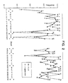
- a consensus has been determined from 118 different occurrences of PU sequences in the E. coli strain K12 (Gilson, E., Clement, J.M., Brutlag, D. and Hofnung, M. (1984) EMBO J. 3, 1417-1421) (Fig. 1).
- PUs could correspond to stable stem and loop RNA or DNA structures, the formation of such structures remains to be established.
- the 'stem' is GC-rich and highly conserved; it has a lower part of five base pairs. The two parts are separated by an 'internal' mismatch.
- the 'loop' which is AT-rich, is variable in sequence and ranges in length from 0 to 5 nucleotides.
- the PU stem flanking regions are highly conserved and have been called the 'external' mismatch (Figs. 1 and 2).
- the internal and external mismatches constitute asymmetry elements which confer a polarity to the PU (Fig. 1).
- Palindromic units are present at least several hundred times on the E. coli and S. typhimurium chromosomes, always in extragenic positions. This is why PUs are also sometimes called REP (repetitive extragenic palindromic) sequences; however, the term REP may be confusing since it is already widely used to designate plasmid regions necessary for unit-copy replication.
- REP repetitive extragenic palindromic
- Palindromic units are found either between two genes of the same operon (intercistronic PUs), or after the last gene of an operon (postcistronic PUs). They are found as isolated occurrences but also occur as clusters of up to four elements. The organization of the cluster is quite remarkable: successive PUs rigorously alternative in orientation (Fig. 1). In addition, the fourth position of the stem which can be, with similar frequencies, either G or T, also alternates. This suggests the existence of a very specific mechanism for the generation or selection of the clusters.
- the homogeneity (the number of bases identical to the consensus divided by the total number of bases) is extremely high, averaging 80% (Fig. 2).
- a change in one of the more conserved positions in the branch of the stem is often accompanied by a simultaneous change of the corresponding position in the other branch, so that the complementarity between these two positions is maintained. Possible reasons for the remarkable conservation in sequence and symmetry are discussed later.
- Fig. 1 shows the consensus sequence from the 12 known S. typhimurium PU sequences that are available (Newbury, S.F. et al. (1987) Cell 48, 297-310). This sequence is similar to the E. coli consensus except for a highly conserved additional G before the C of the internal mismatch. The significance of this slight difference in the consensus of the two species is discussed in a later section.
- PU clusters The localization of PU clusters is conserved between the genomes of different isolates of the E. coli K12 strain. This reveals that PUs are stable or at least do not constitute very unstable genetic elements. PUs are not necessarily present in the same position in otherwise highly homologous sequences of DNA of E. coli and S. typhimurium (Fig. 3). Thus, like the PU primary sequences, the PU genomic localizations are characteristic of bacterial species.
- the major messenger endpoint was mapped at a typicl factor-independent transcription termination or located next to, but clearly distinct from, PU.
- PUs do terminate transcription.
- the single postcistronic PU located between the pheA gene and the tyrA gene acts as a bidirectional transcription terminator. From the sequence of this PU we can define a subclass of PU called PU* (Fig. 1). Interestingly, the six known PU* sequences are each located between two convergent open reading frames, and account for most of the DNA in these regions. No other obvious transcription terminator sequence exist in their vicinity, leaving open the possibility that they all have this function.
- ORFl transcription inhibits ushA° expression in S. typhimurium, but not ushA expression in E. coli thanks to transcription arrest by the PU*.
- a termination site presumed to be rho-dependent, has been mapped between the two PU located after the A gene. Spencer, M.E. and Guest, J.R. (1985) Mol. Gen. Genet. 200, 145-154. It may be significant that the CAA-CA sequence located between these two PUs is also found near the end of several rho-dependent terminators: Pr, Pl, tRNA tyr and trpt′. Morgan, W.P., Bear, D.G., Litchman, B.L. and von Hippel, P.H. (1985) Nucleic Acids Res. 13, 3739-3754.
- Certain PUs have a limited effect on gene expression, through stabilization of the 3′ end of the mRNA.
- a deletion of the two intercistronic PUs between hisJ and hisQ does not affect the expression of the distal part of the operon but leads to a two-fold decrease of the expression of the upstream gene, hisJ.
- a similar observation was made by Plamann et al. with the postcistronic PU of the glyA operon: a mu phage insertion located between the translation stop codon and the first PU was responsible for three-fold decrease in expression of the upstream glyA gene. Plamann, M.D. and Stauffer, G.V. (1985) J. Bacteriol. 161, 650-654.
- PU sequences which have the potential to form stable stem and loop structures at the level of mRNA, may exhibit such an activity.
- One PU of the rplL-rpoB region includes an RNaseIII processing site.
- the sequence of this PU is atypical: the upper part of the stem and the loop are missing.
- some loose homology exists between the lower part of the stem and a known RNAaseIII site in phage T7.
- the two typical PUs in the hisJ-HisQ region are not processed by RNaseIII in vitro.
- PUs The high copy number of PUs suggests that an efficient mechanism for spreading has been involved.
- retroposition i.e. reverse transcription of an RNA, often a tRNA
- gene conversion i.e. reverse transcription of an RNA, often a tRNA
- slippage replication i.e. reverse transcriptase
- transposition i.e. reverse transcriptase
- PUs do have features in common with some transposons, including their inverted repeat structure (reminiscent of IS), and partial homologies between the PU stem consensus sequence and the ends of transposons.
- the high degree of homogeneity of PUs can be explained by at least two hypotheses: (1) that they have arisen recently and spread rapidly (for example by transposition); (2) that they are of more ancient origin, and that there exists a specific mechanism for maintaining homogeneity. Since both E. coli and S. typhimurium possess PUs and, in some cases, they are at the same genetic location (e.g. the ushA region) it seems that PU formation occurred before these two species diverged. This argues against the first hypothesis. The slight difference in their consensus sequences suggests that the homogeneity of PU sequences within one species is higher than between two difference species, and implies species-specific mechanisms for the maintenance of the homogeneity.
- a stimulating aspect of the discovery of bacterial repetitive sequences is that many of the exciting hypotheses and speculations stemming from the discovery of these structures in eukaryotes are now within experimental reach in genetically well characterized organisms such as E. coli. Clues to the origin and function of such sequences may be forthcoming in the near future.
- the region between the stop codon of glnA and the transcription terminator contains three PU in Escherichia coli and none in the equivalent region of Salmonella typhimurium and Klebsiella pneumoniae (Mac Farlane and Merrick, 1985).
- Two PU are located 20 bp after the stop codon of the metJ gene of Escherichia coli.
- the equivalent regionof Salmonella typhimurium does not contain any PU but contains a typical factor-independent termination site (Saint-Girons et al., 1984) (Urbanowski and Stauffer, 1985). Available sequence data does not allow to define the right boundary of the PU containing insert in Escherichia coli.
- a single PU is located between the end of gene rpoD and a functional factor region of Salmonella typhimurium, only a portion of this PU is present (Erickson et al., 1985).
- the malE-malF intergenic region (Duplay et al., 1984 and Duplya, personal communication) (Froshauer and Beckwith., 1984), the 3′ flanking region of uvrD (Finch and Emmerson, 1984) (Yamamoto et al, 1986) have been cloned and sequenced from different Escherichia coli sources, in different laboratories. In the three cases, the region contains identical PU sequences.
- PU localization appears to be conserved between different strains of Escherichia coli while it is not conserved between Escherichia coli and Salmonella typhimurium. Thus, PU localization seems to be a characteristic of bacterial species.
- the TGV program (Saurin and Marliere, 1987) was used to search a Bacillus subtilis sequence database (29417 b.p. for 33 sequences, extracted from Genbank release 38, November 1985). This program allows to look for repetitive DNA pattern irrespective of their primary sequences. Parameters required that repetitive DNA patterns consecutives bases, all the bases of the stem being paired using Watson-Crick and complementary rules (i.e. A-T or G-C pairing). This search revealed no family of repetitive palindromic sequences in the Bacillus subtilis sequence database. Indeed, we found only one palindromic sequence having more than two occurrences.
- This sequence corresponds to the anticodon stem and loop of Gly-tRNA which is encoded by three different genetic regions: trrnB (Wawrousek and Hansen, 1983), trrnD (Wawrousek et al., 1984) and trrnE (Green and Vold, 1983; Wawrousek et al., 1984).
- Bacillus subtilis sequence database contains about ten fold less sequences than the Escherichia coli one, if "PU like" sequence were present at the same frequency in the two species, one would expect to find about 10 occurrences in Bacillus subtilis. It should be noted that the same search in Escherichia coli revealed PU as the only highly repetitive palindromic sequence in this bacteria (Saurin, 1987).
- the 100 bacterial species included a representative set of enterobacteria (85 different species) and a set of other bacteria which is composed of 3 different species) and a set of other bacteria which is composed of 3 different Xenorhabdus species, Acinetobacter, Bordetella bronchisepta, Pseudomonas aeruginosa, Aeromonas, Actinobacillus, Pasteurella, Vibrio cholerae, Vibrio minicus, Legionella pneumophila, Bacillus subtilis, Calothrix and Methanococcus.
- Example II hybridization experiments were conducted with a 240 base pair sequence units extracted from Bordetell pertussis. Modification of the conventional hybridization protocol step utilized 50% formamide at 42°C for a 12 hour incubation and washing at 65°C with 0.1 SSC and 0.1% SDS. Additional hybridizations were performed in slightly less stringent conditions with 2 SCC and 0.1% SDS. The B. pertussis sequence did not hybridize with B. para pertussis nor with B. avium or B. bronchoseptica.
- Hybridization experiments as in Examples I and II are performed with labelled probes.
- Conventional labelling techniques involving conventional radioactive, immunoenzymatic or immunofluorescent techniques are utilized. Results confirm the presence or absence of hybridization utilizing probe sequences and target bacteria as in Examples I and II.
- a diagnostic test kit for the in vitro identification of bacteria in a sample from biological fluids or other sources is composed of a conventional hybridization vessel into which appropriately-treated aliquots of sample material containing bacteria to be identified may be added. Labelled probe DNA may be added as separate reagent or, alternatively, may be present when fixed by appropriate techniques.
- hybridization is halted by washing of unlabelled probe material from the sample vessel.
- the presence and quantification of hybridization may be determined by measuring the amount of probe bound to the DNA of sample bacteria.
Abstract
Description
- This invention relates to a diagnostic probe for bacteria based upon high specific nucleotidic repeat sequences, and more specifically to diagnostic test kits for this purpose.
- In 1982, the present inventors described, for the first time, a DNA sequence (30-40 bp long), which is highly repeated in the genome of Escherichia coli and Salmonella typhimurium (about 1000 times) (Higgins, C.F., Ferro-Luzzi Ames, G., Barnes, W.M. Clement, J.M. and Hofnung M. (1982) Nature. 298, 760-762). These sequences are referred to as a Palindromic Unit or PU. Their primary sequence conservation is 80%.
- Since then, a small difference was noticed in the PU consensus sequences between Escherichia coli and Salmonella typhimurium. This difference is an additional guanine residue in the Salmonella PU sequences. This was a preliminary indication that the PU sequences exhibit species-specificity.
- Only a few families of highly repetitive DNA sequences have been described so far in bacteria. Like PUs, they display a tight species specificity. By hybridization, the 26-bp repetitive sequence family of Neisseria spp. (at least 20 copies per genome) was not found in various other gram-negative bacteria (Correia, F.F., Inouye, S. and Inouye, M. (1986) J. of Bacteriol. 167, 1009-1015). A repetitive DNA sequence family from Bordetella pertussis also appears to be species-specific (MacPheat, W.L. and MacNally, T. (1987) FEMS Lett. 41, 357-360).
- Recently, hybridization experiments with Escherichia coli PU DNA as a probe showed that only DNA from enterobacteriaceae close to E. coli hybridized with good efficiency. The present inventors have determined that the PU specificity could be used for the detection and the identification of bacteria with DNA probes corresponding to PU sequences.
- Moreover, the presence of species-specific highly repetitive DNA sequences has been found to be a general phenomenon among bacteria, for instance in enterobacteriaceae and bordetella pertussis which present repeated sequences. Thus, the present invention relates to the use of species specific highly repetitive sequences as specific diagnostic probes. These type of bacterial probes should provide diagnostic assays which are more sensitive than assays with probes corresponding to low copy number genes.
- It is an object of the present invention to provide a method for identifying bacteria in a sample from biological fluids or other sources. The method requires the preparation by appropriate and conventional techniques of bacteria capable of hybridizing with a labelled DNA probe, and a DNA probe containing a highly specific bacterial nucleotidic repeat sequence. Reagents appropriate for and conventionally utilized in such hybridization protocols are intended for use in the present invention. The claimed method requires the exposure of a bacterial sample to the DNA probe for a time period sufficient to allow hybridization of the probe to the native DNA of the sample bacteria. Conventional and appropriate washing steps remove unbound but labelled probe form the reaction vessel. Finally, conventional techniques to analyze the extend of hybridization permit a qualitative as well as quantitative identification of the sample bacteria.
- In another aspect, the present invention relates to a diagnostic test kit for identifying bacteria in a sample which utilizes the labelled DNA probe containing a highly specific bacterial nucleotidic repeat sequence and appropriate reagents for allowing hybridization of the probe to the sample bacteria. A conventional and appropriate hybridization vessel is also required in which the hybridization can occur, along the appropriate and conventional post-hybridization washing reagents. The extent of hybridization is accomplished by means appropriate and conventionally utilized for this purpose.
- Additional objects and advantages of the invention will be set forth in part in the description which follows, and in part will be obvious from the description or maybe learned from practice of the invention. These objects and advantages may be realized and attained by means of the instrumentalities and combinations particularly pointed out in the appended claims.
- It is to be understood that both the foregoing general description and the following detailed description are exemplary and explanatory only and are not restrictive of the invention, as claimed.
-
- Figure 1 illustrates the palindromic unit consensus from E. coli and from S. typhimurium.
- Figure 2 illustrates the conservation of primary structures of E. coli and S. typhimurium PU sequences.
- Figure 3 presents a comparison of the structures of the 3′ flanking region of the glnA gene in Klebsiella pneumoniae, S. typhimurium and E. coli and of the ushA gene in S. typhimurium and E. coli.
- Figure 4 further illustrates the PU sequence consensus between E. coli and S. typhimurium.
- Figure 5 illustrates bordetella pertussis sequence information.
- Reference will now be made in detail to the presently preferred embodiments of the invention, which, together with the following examples, serve to explain the principles of the invention.
- DNA sequence analysis of E. coli and Salmonella typhimurim genomes has revealed the presence of a family of highly repeated DNA sequences: the palindromic unit (PU) family. This discovery was unexpected, since prokaryotic genomes are generally small and are believed to comprise only low-copy number DNA sequences. Britten, R.J. and Kohne, D.E. (1968) Science 161, 529-560. There may by on the order of 10³ copies of PU sequences in E. coli DNA, accounting for 1% of the genome, a percentage that is comparable to values found for many families of repetitive DNA in eukaryotic genomes.
- Palindromic units constitute a family of repetitive sequences of 20-4- nucleotides that exhibit dyad symmetry. A consensus has been determined from 118 different occurrences of PU sequences in the E. coli strain K12 (Gilson, E., Clement, J.M., Brutlag, D. and Hofnung, M. (1984) EMBO J. 3, 1417-1421) (Fig. 1). Although PUs could correspond to stable stem and loop RNA or DNA structures, the formation of such structures remains to be established. The 'stem' is GC-rich and highly conserved; it has a lower part of five base pairs. The two parts are separated by an 'internal' mismatch. The 'loop', which is AT-rich, is variable in sequence and ranges in length from 0 to 5 nucleotides. The PU stem flanking regions are highly conserved and have been called the 'external' mismatch (Figs. 1 and 2). The internal and external mismatches constitute asymmetry elements which confer a polarity to the PU (Fig. 1).
- Palindromic units are present at least several hundred times on the E. coli and S. typhimurium chromosomes, always in extragenic positions. This is why PUs are also sometimes called REP (repetitive extragenic palindromic) sequences; however, the term REP may be confusing since it is already widely used to designate plasmid regions necessary for unit-copy replication.
- Palindromic units are found either between two genes of the same operon (intercistronic PUs), or after the last gene of an operon (postcistronic PUs). They are found as isolated occurrences but also occur as clusters of up to four elements. The organization of the cluster is quite remarkable: successive PUs rigorously alternative in orientation (Fig. 1). In addition, the fourth position of the stem which can be, with similar frequencies, either G or T, also alternates. This suggests the existence of a very specific mechanism for the generation or selection of the clusters.
- The homogeneity (the number of bases identical to the consensus divided by the total number of bases) is extremely high, averaging 80% (Fig. 2). A change in one of the more conserved positions in the branch of the stem is often accompanied by a simultaneous change of the corresponding position in the other branch, so that the complementarity between these two positions is maintained. Possible reasons for the remarkable conservation in sequence and symmetry are discussed later.
- Fig. 1 shows the consensus sequence from the 12 known S. typhimurium PU sequences that are available (Newbury, S.F. et al. (1987) Cell 48, 297-310). This sequence is similar to the E. coli consensus except for a highly conserved additional G before the C of the internal mismatch. The significance of this slight difference in the consensus of the two species is discussed in a later section.
- When an E. coli PU DNA was used as a probe, only DNA from enterobacteriaceae closely related to E. coli showed appreciate hybridization. By computer search, the present inventors were unable to detect a palindromic-sequence family (of any primary sequence) in the Bacillus subtilis sequence database. There are a number of possible explanations for this: (1) there may be biases in the B. subtilis sequence databases: (2) in B. subtilis the functional equivalent of the enterobacteriaceae PU family may not be a palindromic sequence; (3) there may be no functional equivalent of PUs in B. subtilis. The same search in E. coli revealed PU as the only highly repetitive palindromic DNA sequence in this bacterium. Saurin,
W. Cabios 3, 121-127. It should be noted that PU sequences were not found in the complete genome of lambda or T7 phages. Finally, neither the PU consensus nor variations with up to four differences in one of each 'half-PU' (i.e. positions 1-17 and positions 18-36 on Fig. 2) are found in the eukaryotic sequence database. - All this suggests that PU sequences are characteristic of the chromosomes of certain enterobacteriaceae. Because sequence data for most bacterial species are still poorly represented in databases, we have not ruled out the possibility that highly repetitive palindromic elements with sequences different from PU may exist in other bacterial species.
- The localization of PU clusters is conserved between the genomes of different isolates of the E. coli K12 strain. This reveals that PUs are stable or at least do not constitute very unstable genetic elements. PUs are not necessarily present in the same position in otherwise highly homologous sequences of DNA of E. coli and S. typhimurium (Fig. 3). Thus, like the PU primary sequences, the PU genomic localizations are characteristic of bacterial species.
- Most PUs do not act as transcription terminators. The two PUs located between the cotranscribed genes hisJ and hisQ of the histidine transport operon in S. typhimurium do not cause transcription termination in vivo (less than 50% transcription arrest in a galK fusion analysis system). No pause or termination of transcription was detected in vivo. Stern, J.M. et al (1984) Cell 37, 1015-1026. The three PUs located between the cotranscribed genes lamB and malM of the maltose transport operon in E. coli did not affect the transcription and translation of a down-stream gene (galK gene in a multicopy system and lacZ gene in a monocopy system). Gilson, E., Rousset, J.P., Clement, J.M. and Hofnung, M. (1986) Ann. Microbiol. (Inst. Pasteur) 127B., 259-270. For several E. coli operons, the major messenger endpoint was mapped at a typicl factor-independent transcription termination or located next to, but clearly distinct from, PU. Gilson, E., Rousset, J.P., Clement, J.M. and Hofnung, M. (1986) Ann. Microbiol. (Inst. Pasteur) 127B, 259-270.
- However, some PUs do terminate transcription. The single postcistronic PU located between the pheA gene and the tyrA gene acts as a bidirectional transcription terminator. From the sequence of this PU we can define a subclass of PU called PU* (Fig. 1). Interestingly, the six known PU* sequences are each located between two convergent open reading frames, and account for most of the DNA in these regions. No other obvious transcription terminator sequence exist in their vicinity, leaving open the possibility that they all have this function.
- A comparison of the expression of a region that is highly homologous in E. coli and S. typhimurium, the ushA-ORFl region, is compatible with the idea that PU* sequences act as bidirectional transcription terminators. The two genes are convergently transcribed and separated by a PU* sequence in E. coli and by a cluster of two classical PUs in S. typhimurium (Fig. 3). A protein corresponding to ORFl is expressed at a high level in both species. However, the ushA protein from S. typhimurium is much less strongly expressed than the corresponding E. coli UshA protein. Remarkably, genetic inactivation of ORFl transcription results in increased expression of ushA°. Burns, D.M. and beacham, I.R. (1986) J. Mol. Biol. 192, 163-175. One possibility is thus that ORFl transcription inhibits ushA° expression in S. typhimurium, but not ushA expression in E. coli thanks to transcription arrest by the PU*.
- A termination site, presumed to be rho-dependent, has been mapped between the two PU located after the A gene. Spencer, M.E. and Guest, J.R. (1985) Mol. Gen. Genet. 200, 145-154. It may be significant that the CAA-CA sequence located between these two PUs is also found near the end of several rho-dependent terminators: Pr, Pl, tRNAtyr and trpt′. Morgan, W.P., Bear, D.G., Litchman, B.L. and von Hippel, P.H. (1985) Nucleic Acids Res. 13, 3739-3754.
- Certain PUs have a limited effect on gene expression, through stabilization of the 3′ end of the mRNA. A deletion of the two intercistronic PUs between hisJ and hisQ does not affect the expression of the distal part of the operon but leads to a two-fold decrease of the expression of the upstream gene, hisJ. A similar observation was made by Plamann et al. with the postcistronic PU of the glyA operon: a mu phage insertion located between the translation stop codon and the first PU was responsible for three-fold decrease in expression of the upstream glyA gene. Plamann, M.D. and Stauffer, G.V. (1985) J. Bacteriol. 161, 650-654. Recently, it has been shown in two cases that this increase in expression of the upstream gene is a consequence of the accumulation of upstream mRNA species. This observation was explained by the ability of the PU sequence to protect the transcript from 3′--5′ exonuclease degradation.
- It is now well established that a number of sequences able to form RNA secondary structures can function as barriers against 3′--5′ exonuclease digestion. Therefore, it is not surprising that PU sequences, which have the potential to form stable stem and loop structures at the level of mRNA, may exhibit such an activity.
- One PU of the rplL-rpoB region includes an RNaseIII processing site. The sequence of this PU is atypical: the upper part of the stem and the loop are missing. Interestingly, some loose homology exists between the lower part of the stem and a known RNAaseIII site in phage T7. Gilson, E., Clement, J.M., Brutlag, D. and Hofnung, M. (1984) EMBO J. 3, 1417-1421. No other evidence exists in association of a PU with RNaseIII processing. In particular, the two typical PUs in the hisJ-HisQ region are not processed by RNaseIII in vitro. The above examples strongly suggest that slight sequence modifications from the PU consensus sequence or modification of the PU sequence environment can have various effects on the transcription of specific operons. The selective advantage conferred to the cell by these functional modifications of PU would tend to increase differences between PU sequences. However, since all these structures are still recognizable as PUs, there probably exist some mechanisms for the maintenance of homogeneity amongst PU sequences within a species.
- The existence and the intergenic location of such a large number of homologous sequences suggest that they could be associated with chromosome rearrangements (gene shuffling). The remarkable conservation of the PU primary se-quence and its dyad symmetry (reminiscent of sites for restriction enzymes) suggest that PU DNA may constitute protein binding sites.
- It is unlikely that PUs are major sites for high frequency recombination for the two following reasons: (1) two clusters of PU, separated by about 3 kpb, are found in the malB region of E. coli -- however, no Mal⁻ deletion mutant has ever been found that has an end-point in the malB PU regions; and (2) most of the spontaneous tandem duplications in S. typhimurium arise by unequal recombination between rrn operons.
- However, a PU-mediated low frequency recombinational activity, like that promoted by any repeated sequence, could very well occur. Even under such conditions, PUs could play a role in chromosome rearrangements and in the modular evolution of genomes. In support of this idea, one arrangement has been detected in S. typhimurium between the hisG-hisD intergenic region, which contains one PU, and argB. Anderson, P. and Roth, J. (1978) J. Mol. Biol. 119, 147-166. Moreover, one PU is at the boundary between homologous and non-homologous regionof glnA between E. coli and S. typhimurim (Fig. 3), and PU sequences are present exactly at the boundary of directly repeated sequences located after the Ml RNA gene and after the tRNAPro gene. Reed, R.E. and Altman, S. (1983) Proc. Nat'l. Acad. Sci. USA 80, 5359-5363; Kuchino, Y., Mori, F. and Nishimura, S. (1985) Nucleic Acids Res. 13, 3213-3220. Although the evidence is indirect, these observations are consistent with the occurrence of recombinational events close to the PU sites.
- In the presence of an extract containing nucleoid-associated proteins, PU sequences constitute a strong boundary to ExoIII digestion. Gilson, E. et al. (1986) FEBS Lett. 206, 323-328. A single PU, in either orientation, is sufficient to stop degradation. These findings are consistent with the idea that one or several nucleoid-associated proteins are able to recognize and bind PU sequences.
- The biological significance of this interaction is not known. It would be consistent with an involvement of PUs in the structure of the nucleotoid. Recent studies on eukaryotic chromatin structure have implicated the topoisomeraseII protein in the organization of the DNA into looped domains, via interaction with specific DNA sequences. On the basis of electron microscopy studies and of in-vivo supercoiling distribution measurements, it appears that the E. coli nucleoid is folded into independent supercoiled looped domains. It is possible that clamping two PU clusters by specific protein interactions would constitute and/or stabilize the neck of each loop.
- The high copy number of PUs suggests that an efficient mechanism for spreading has been involved. Several mechanisms have been described in eukaryotes, including retroposition (i.e. reverse transcription of an RNA, often a tRNA), gene conversion, unequal recombination, slippage replication, transposition and amplification from a progenitor repeat. Dover, G.A. (1986) Trends Genet. 2, 159, 165. None of these mechanisms can be excluded here. However, PUs do have features in common with some transposons, including their inverted repeat structure (reminiscent of IS), and partial homologies between the PU stem consensus sequence and the ends of transposons.
- The high degree of homogeneity of PUs can be explained by at least two hypotheses: (1) that they have arisen recently and spread rapidly (for example by transposition); (2) that they are of more ancient origin, and that there exists a specific mechanism for maintaining homogeneity. Since both E. coli and S. typhimurium possess PUs and, in some cases, they are at the same genetic location (e.g. the ushA region) it seems that PU formation occurred before these two species diverged. This argues against the first hypothesis. The slight difference in their consensus sequences suggests that the homogeneity of PU sequences within one species is higher than between two difference species, and implies species-specific mechanisms for the maintenance of the homogeneity. Such a pattern of variation within a sequence family, called concerted evolution, has already been observed in many eukaryotic families such as rDNA, small nuclear (sn) RNA or long interspersed repetitive DNA sequences (LINE). It is possible that the existence of a protein binding to PU sequences (see above) might lead to slow coadaptative changes between the PU sequences and the gene of the relative protein. This would tend to lead to the homogenization of the PU family within a species.
- Like the PUs, three other known families of repetitive DNA sequences in bacteria display a tight species specificity. No sequences hybridizing with the 26-bp repetitive sequence family of Neisseria spp. (at least 20 copies per genome and possibly many more) have been found in various other Gram-negative bacteria. Correia, F.F., Inouye, S. and Inouye, M. (1986) J. Bacteriol. 167, 1009-1015. The nifHDK promoter sequence, which is repeated 3-6 times on the symbiotic plasmid of Rhizobium trifolii does not hybridize to DNA of any other symbiotic plasmid-containing Rhizobium species examined. Watson, J.M. and Shofield P.R. (1985) Mol. Gen. Genet. 199, 279-289. The 11-bp repeat of Haemophilus (10³ copies per genome) allows specific recognition of Haemophilus DNA to be taken up by competent cells. Danner, D.B., Deich, R.A. Sisco, K.L., Deich, R.A., Sisco, K.L. and Smith, H.O. (1980)
Gene 11, 311-318. In addition, a repetitive DNA sequence family has been found recently in Bordetella pertussis. Once again, this sequence seems to be species-specific. MacPheat, W.L. and MacNally, T. (1987) FEMS Lett. 41, 357-360 and A. Ullmann, pers. communication). - A stimulating aspect of the discovery of bacterial repetitive sequences is that many of the exciting hypotheses and speculations stemming from the discovery of these structures in eukaryotes are now within experimental reach in genetically well characterized organisms such as E. coli. Clues to the origin and function of such sequences may be forthcoming in the near future.
- It is understood that the application of the teachings of the present invention to a specific problem or environment will be within the capabilities of one having ordinary skill in the art in the light of the teachings contained herein. Examples of the products of the present invention and representative processes for their isolation and manufacture appear in the following examples.
- Evidence of differences in PU between otherwise homologous bacterial DNA sequences.
- The region between the stop codon of glnA and the transcription terminator contains three PU in Escherichia coli and none in the equivalent region of Salmonella typhimurium and Klebsiella pneumoniae (Mac Farlane and Merrick, 1985).
- Two PU are located 20 bp after the stop codon of the metJ gene of Escherichia coli. The equivalent regionof Salmonella typhimurium does not contain any PU but contains a typical factor-independent termination site (Saint-Girons et al., 1984) (Urbanowski and Stauffer, 1985). Available sequence data does not allow to define the right boundary of the PU containing insert in Escherichia coli.
- A single PU is located between the end of gene rpoD and a functional factor region of Salmonella typhimurium, only a portion of this PU is present (Erickson et al., 1985).
- The malE-malF intergenic region (Duplay et al., 1984 and Duplya, personal communication) (Froshauer and Beckwith., 1984), the 3′ flanking region of uvrD (Finch and Emmerson, 1984) (Yamamoto et al, 1986) have been cloned and sequenced from different Escherichia coli sources, in different laboratories. In the three cases, the region contains identical PU sequences.
- Thus, PU localization appears to be conserved between different strains of Escherichia coli while it is not conserved between Escherichia coli and Salmonella typhimurium. Thus, PU localization seems to be a characteristic of bacterial species.
- The 9 known Salmonella typhimurium PU sequences that are available, all comprise an additional G before the C of the C-T "mismatch" (Fig. 4). Since amonth the 103 Escherichia coli PU sequences recorded in our laboratory, only one (gdhA, PUa) contains the additional G, we propose that the PU consensus may be slightly different in Escherichia coli and Salmonella typhimurium (the modified Salmonella typhimurium PU consensus is shown on Fig. 4). That these two related enterobacteriaceas present this slight difference in their PU sequences suggests that the primary sequence of PU could be specific of one or a group of bacterial species. In addition, it should be recalled that PU sequences are not present in the complete genome of lambda or T7 phages (Gilson et al., 1984).
- The TGV program (Saurin and Marliere, 1987) was used to search a Bacillus subtilis sequence database (29417 b.p. for 33 sequences, extracted from Genbank release 38, November 1985). This program allows to look for repetitive DNA pattern irrespective of their primary sequences. Parameters required that repetitive DNA patterns consecutives bases, all the bases of the stem being paired using Watson-Crick and complementary rules (i.e. A-T or G-C pairing). This search revealed no family of repetitive palindromic sequences in the Bacillus subtilis sequence database. Indeed, we found only one palindromic sequence having more than two occurrences. This sequence, CCACCTTGCCAAGGTGG, corresponds to the anticodon stem and loop of Gly-tRNA which is encoded by three different genetic regions: trrnB (Wawrousek and Hansen, 1983), trrnD (Wawrousek et al., 1984) and trrnE (Green and Vold, 1983; Wawrousek et al., 1984). Although the Bacillus subtilis sequence database contains about ten fold less sequences than the Escherichia coli one, if "PU like" sequence were present at the same frequency in the two species, one would expect to find about 10 occurrences in Bacillus subtilis. It should be noted that the same search in Escherichia coli revealed PU as the only highly repetitive palindromic sequence in this bacteria (Saurin, 1987).
- All the three points presented above suggest that PU contribute to bacterial speciation. Many different modes for direct or indirect effects can be imagined. One hypothesis we like to consider is that PU might play a role in the bacterial chromosome structure. This could prevent stable or viable insertion of a large segment of a foreign chromosome, not containing PU. Preliminary results show that a chromoid-associated protein specifically interacts with PU DDNA (Gilson et al., 1986a). This is compatible with the idea that PU sequences can be involved in the structure of the chromosome.
- Analysis of hybridization utilizing E. coli labelled probe. We examined 100 DNA of different bacterial species either by dot blot or by southern blot with a 200bp DNA probe containing 3 PU sequences. The hybridization experiments have been performed using standard procedures with the following modifications: hybridization step in 6SSC, 0.1% SDS at 58°C, 12 hours and washing in 0.2SSCC, 0.1% SDS at 45°C, 1 hour. The probe as been prepared by nick translation with alpha ³²p nucleotides. The 100 bacterial species included a representative set of enterobacteria (85 different species) and a set of other bacteria which is composed of 3 different species) and a set of other bacteria which is composed of 3 different Xenorhabdus species, Acinetobacter, Bordetella bronchisepta, Pseudomonas aeruginosa, Aeromonas, Actinobacillus, Pasteurella, Vibrio cholerae, Vibrio minicus, Legionella pneumophila, Bacillus subtilis, Calothrix and Methanococcus.
- Following the foregoing analysis, our results indicated that only the E. coli and Salmonellae groups hybridize with such a probe under the hybridization conditions specified.
- Hybridization specificity of bordetella sequences.
- Following the general procedure of Example I, hybridization experiments were conducted with a 240 base pair sequence units extracted from Bordetell pertussis. Modification of the conventional hybridization protocol step utilized 50% formamide at 42°C for a 12 hour incubation and washing at 65°C with 0.1 SSC and 0.1% SDS. Additional hybridizations were performed in slightly less stringent conditions with 2 SCC and 0.1% SDS. The B. pertussis sequence did not hybridize with B. para pertussis nor with B. avium or B. bronchoseptica.
- Utilization of alternative conventional labels.
- Hybridization experiments as in Examples I and II are performed with labelled probes. Conventional labelling techniques involving conventional radioactive, immunoenzymatic or immunofluorescent techniques are utilized. Results confirm the presence or absence of hybridization utilizing probe sequences and target bacteria as in Examples I and II.
- It will be apparent to those skilled in the art tht various modifications and variations can be made to the processes and products of the present invention. Thus, it is intended that the present invention cover the modifications and variations of this invention provided they come within the scope of the appended claims and their equivalents.
- A diagnostic test kit for the in vitro identification of bacteria in a sample from biological fluids or other sources is composed of a conventional hybridization vessel into which appropriately-treated aliquots of sample material containing bacteria to be identified may be added. Labelled probe DNA may be added as separate reagent or, alternatively, may be present when fixed by appropriate techniques.
- After appropriate incubation periods have passed, hybridization is halted by washing of unlabelled probe material from the sample vessel. The presence and quantification of hybridization may be determined by measuring the amount of probe bound to the DNA of sample bacteria.
Claims (5)
Priority Applications (1)
| Application Number | Priority Date | Filing Date | Title |
|---|---|---|---|
| AT88402114T ATE103005T1 (en) | 1987-08-14 | 1988-08-16 | SAMPLE FOR DIAGNOSIS OF BACTERIA. |
Applications Claiming Priority (2)
| Application Number | Priority Date | Filing Date | Title |
|---|---|---|---|
| US8517887A | 1987-08-14 | 1987-08-14 | |
| US85178 | 1987-08-14 |
Publications (2)
| Publication Number | Publication Date |
|---|---|
| EP0307270A1 true EP0307270A1 (en) | 1989-03-15 |
| EP0307270B1 EP0307270B1 (en) | 1994-03-16 |
Family
ID=22189960
Family Applications (1)
| Application Number | Title | Priority Date | Filing Date |
|---|---|---|---|
| EP88402114A Expired - Lifetime EP0307270B1 (en) | 1987-08-14 | 1988-08-16 | Bacterial diagnostic probe |
Country Status (6)
| Country | Link |
|---|---|
| US (3) | US5492811A (en) |
| EP (1) | EP0307270B1 (en) |
| JP (1) | JP2823566B2 (en) |
| AT (1) | ATE103005T1 (en) |
| DE (1) | DE3888434T2 (en) |
| ES (1) | ES2049758T3 (en) |
Cited By (5)
| Publication number | Priority date | Publication date | Assignee | Title |
|---|---|---|---|---|
| EP0452596A1 (en) * | 1990-04-18 | 1991-10-23 | N.V. Innogenetics S.A. | Hybridization probes derived from the spacer region between the 16S and 23S rRNA genes for the detection of non-viral microorganisms |
| EP0480289A1 (en) * | 1990-10-09 | 1992-04-15 | Roche Diagnostics GmbH | Method for genus and species-specific detection of bacteria in a testliquid |
| US5536638A (en) * | 1990-04-18 | 1996-07-16 | N.V. Innogenetics S.A. | Hybridization probes derived from the spacer region between the 16S and 23S rRNA genes for the detection of Neisseria gonorrhoeae |
| US6261785B1 (en) | 2000-07-27 | 2001-07-17 | Becton, Dickinson And Company | Amplification and detection of Bordetella pertussis |
| CN101903537B (en) * | 2007-10-26 | 2016-08-10 | 奎斯特诊断投资公司 | bordetella detection assay |
Families Citing this family (4)
| Publication number | Priority date | Publication date | Assignee | Title |
|---|---|---|---|---|
| JP2823566B2 (en) * | 1987-08-14 | 1998-11-11 | アンスティテュ パストゥール | Bacterial diagnostic probe |
| US5958686A (en) * | 1996-10-28 | 1999-09-28 | The United States Of America As Represented By The Secretary Of The Army | Simple PCR technique for detecting and differentiating bacterial pathogens |
| US6251609B1 (en) | 2000-07-27 | 2001-06-26 | Becton, Dickinson And Company | Amplification and detection of Legionella pneumophila targeting the mip gene |
| JP2005528887A (en) * | 2002-01-31 | 2005-09-29 | エクスプレッシブ コンストラクツ,インコーポレイテッド | Microbial detection method |
Citations (3)
| Publication number | Priority date | Publication date | Assignee | Title |
|---|---|---|---|---|
| DE2823573A1 (en) * | 1977-05-31 | 1978-12-14 | Bethesda Res Lab | IMMOBILIZED RESTRICTIONS-ENDONUCLEASES AND PROCEDURES FOR IMMOBILIZED RESTRICTIONS-ENDONUCLEASES |
| WO1984001174A1 (en) * | 1982-09-20 | 1984-03-29 | Harvard College | Identification of microorganisms |
| WO1987005907A1 (en) * | 1986-04-04 | 1987-10-08 | Institut Pasteur | Oligonucleotidic probes and methods for the detection of bacterial and other living beings by hybridization of nucleic acids |
Family Cites Families (5)
| Publication number | Priority date | Publication date | Assignee | Title |
|---|---|---|---|---|
| US4358535A (en) * | 1980-12-08 | 1982-11-09 | Board Of Regents Of The University Of Washington | Specific DNA probes in diagnostic microbiology |
| US5288611A (en) * | 1983-01-10 | 1994-02-22 | Gen-Probe Incorporated | Method for detecting, identifying, and quantitating organisms and viruses |
| US4801530A (en) * | 1984-02-29 | 1989-01-31 | Rockefeller University | Nucleotide hybridization assay for protozoan parasites |
| IT1196453B (en) * | 1986-07-04 | 1988-11-16 | Sclavo Spa | CORYNEBACTERIUM DIPHTHERIAE DNA ELEMENT WITH TYPICAL PROPERTIES OF AN IS INSERTION ELEMENT |
| JP2823566B2 (en) * | 1987-08-14 | 1998-11-11 | アンスティテュ パストゥール | Bacterial diagnostic probe |
-
1988
- 1988-08-13 JP JP63201060A patent/JP2823566B2/en not_active Expired - Lifetime
- 1988-08-16 DE DE3888434T patent/DE3888434T2/en not_active Expired - Lifetime
- 1988-08-16 AT AT88402114T patent/ATE103005T1/en not_active IP Right Cessation
- 1988-08-16 EP EP88402114A patent/EP0307270B1/en not_active Expired - Lifetime
- 1988-08-16 ES ES88402114T patent/ES2049758T3/en not_active Expired - Lifetime
-
1993
- 1993-12-10 US US08/164,769 patent/US5492811A/en not_active Expired - Lifetime
-
1995
- 1995-06-07 US US08/478,854 patent/US5863721A/en not_active Expired - Fee Related
-
1998
- 1998-08-21 US US09/137,686 patent/US6017711A/en not_active Expired - Fee Related
Patent Citations (3)
| Publication number | Priority date | Publication date | Assignee | Title |
|---|---|---|---|---|
| DE2823573A1 (en) * | 1977-05-31 | 1978-12-14 | Bethesda Res Lab | IMMOBILIZED RESTRICTIONS-ENDONUCLEASES AND PROCEDURES FOR IMMOBILIZED RESTRICTIONS-ENDONUCLEASES |
| WO1984001174A1 (en) * | 1982-09-20 | 1984-03-29 | Harvard College | Identification of microorganisms |
| WO1987005907A1 (en) * | 1986-04-04 | 1987-10-08 | Institut Pasteur | Oligonucleotidic probes and methods for the detection of bacterial and other living beings by hybridization of nucleic acids |
Non-Patent Citations (4)
| Title |
|---|
| ANNALES INSTITUT PASTEUR/MICROBIOLOGIE, vol. 137B, no. 3, 1986, pages 259-270, Elsevier, Paris, FR; E. GILSON et al.: "A subfamily of E. coli palindromic units implicated in transcription termination ?" * |
| CELL, vol. 37, no. 3, July 1984, pages 1015-1026, MIT, Massachusetts, US; M.J. STERN et al.: "Repetitive extragenic palindromic sequences: a major component of the bacterial genome" * |
| JOURNAL OF MOLECULAR EVOLUTION, vol. 25, no. 4, 1987, pages 371-373, Springer-Verlag, New York, US; E. GILSON et al.: "Species specificity of bacterial palindromic units" * |
| TRENDS IN GENETICS, vol. 3, no. 8, August 1987, pages 226-230, Elsevier Publications, Cambridge, GB; E. GILSON et al.: "Palindromic units: a case of highly repetitive DNA sequences in bacteria" * |
Cited By (10)
| Publication number | Priority date | Publication date | Assignee | Title |
|---|---|---|---|---|
| EP0452596A1 (en) * | 1990-04-18 | 1991-10-23 | N.V. Innogenetics S.A. | Hybridization probes derived from the spacer region between the 16S and 23S rRNA genes for the detection of non-viral microorganisms |
| WO1991016454A1 (en) * | 1990-04-18 | 1991-10-31 | N.V. Innogenetics S.A. | Hybridization probes derived from the spacer region between the 16s and 23s rrna genes for the detection of non-viral microorganisms |
| US5536638A (en) * | 1990-04-18 | 1996-07-16 | N.V. Innogenetics S.A. | Hybridization probes derived from the spacer region between the 16S and 23S rRNA genes for the detection of Neisseria gonorrhoeae |
| US5945282A (en) * | 1990-04-18 | 1999-08-31 | N.V. Innogenetics S.A. | Hybridization probes derived from the spacer region between the 16S and 23S rRNA genes for the detection of non-viral microorganisms |
| US6277577B1 (en) | 1990-04-18 | 2001-08-21 | N.V. Innogenetics S.A. | Hybridization probes derived from the spacer region between the 16s and 23s rRNA genes for the detection of non-viral microorganisms |
| US6656689B2 (en) | 1990-04-18 | 2003-12-02 | N.V. Innogenetics S.A. | Hybridization probes derived from the spacer region between the 16S and 23S rRNA genes for the detection of non-viral microorganisms |
| EP0480289A1 (en) * | 1990-10-09 | 1992-04-15 | Roche Diagnostics GmbH | Method for genus and species-specific detection of bacteria in a testliquid |
| US6225094B1 (en) | 1990-10-09 | 2001-05-01 | Roche Diagnostics Gmbh | Method for the genus-specific or/and species-specific detection of bacteria in a sample liquid |
| US6261785B1 (en) | 2000-07-27 | 2001-07-17 | Becton, Dickinson And Company | Amplification and detection of Bordetella pertussis |
| CN101903537B (en) * | 2007-10-26 | 2016-08-10 | 奎斯特诊断投资公司 | bordetella detection assay |
Also Published As
| Publication number | Publication date |
|---|---|
| DE3888434D1 (en) | 1994-04-21 |
| US6017711A (en) | 2000-01-25 |
| US5492811A (en) | 1996-02-20 |
| US5863721A (en) | 1999-01-26 |
| JP2823566B2 (en) | 1998-11-11 |
| ATE103005T1 (en) | 1994-04-15 |
| JPH01157400A (en) | 1989-06-20 |
| ES2049758T3 (en) | 1994-05-01 |
| EP0307270B1 (en) | 1994-03-16 |
| DE3888434T2 (en) | 1994-07-28 |
Similar Documents
| Publication | Publication Date | Title |
|---|---|---|
| Le Dantec et al. | Genomic sequence and transcriptional analysis of a 23-kilobase mycobacterial linear plasmid: evidence for horizontal transfer and identification of plasmid maintenance systems | |
| Clewell et al. | Sequence analysis of termini of conjugative transposon Tn916 | |
| Motin et al. | Genetic variability of Yersinia pestis isolates as predicted by PCR-based IS 100 genotyping and analysis of structural genes encoding glycerol-3-phosphate dehydrogenase (glpD) | |
| Matthews et al. | The cloning of chromosomal DNA associated with methicillin and other resistances in Staphylococcus aureus | |
| Lan et al. | Gene transfer is a major factor in bacterial evolution. | |
| Howell‐Adams et al. | Molecular models accounting for the gene conversion reactions mediating gonococcal pilin antigenic variation | |
| Buchrieser et al. | The high‐pathogenicity island of Yersinia pseudotuberculosis can be inserted into any of the three chromosomal asn tRNA genes | |
| EP0804616B1 (en) | Specific and universal amplification primers to rapidly detect and identify common bacterial pathogens and antibiotic resistance genes from clinical specimens for routine diagnosis in microbiology laboratories | |
| Bukau et al. | Delta dnaK52 mutants of Escherichia coli have defects in chromosome segregation and plasmid maintenance at normal growth temperatures | |
| JP3097059B2 (en) | Generation of specific probes for target nucleotide sequences | |
| Pierson et al. | Nonpathogenic isolates of Yersinia enterocolitica do not contain functional inv-homologous sequences | |
| Pourcel et al. | Characterization of a tandem repeat polymorphism in Legionella pneumophila and its use for genotyping | |
| Collyn et al. | YAPI, a new Yersinia pseudotuberculosis pathogenicity island | |
| EP2058325A2 (en) | A method for isolating a polynucleotide of interest from the genome of a mycobacterium using a BAC-based DNA library. application to the detection of mycobacteria | |
| Rudd | Novel intergenic repeats of Escherichia coli K-12 | |
| US5863721A (en) | Bacterial diagnostic probe | |
| CN110218802B (en) | Method for detecting respiratory pathogen nucleic acid | |
| Reuven et al. | The gene for the longest known Escherichia coli protein is a member of helicase superfamily II | |
| Zhang et al. | Genomic comparison of plant pathogenic and nonpathogenic Serratia marcescens strains by suppressive subtractive hybridization | |
| Beuzón et al. | Conserved structure of IS 200 elements in Salmonella | |
| Osek | Virulence factors and genetic relatedness of Escherichia coli strains isolated from pigs with post-weaning diarrhea | |
| US5786147A (en) | Detection of enterobacteria | |
| Fakruddin et al. | Identification and characterization of microorganisms: DNA-fingerprinting methods. | |
| Marsh et al. | Deletion of an mmpL gene and multiple associated genes from the genome of the S strain of Mycobacterium avium subsp. paratuberculosis identified by representational difference analysis and in silico analysis | |
| Bayliss et al. | Mutations in Haemophilus influenzae mismatch repair genes increase mutation rates of dinucleotide repeat tracts but not dinucleotide repeat-driven pilin phase variation rates |
Legal Events
| Date | Code | Title | Description |
|---|---|---|---|
| PUAI | Public reference made under article 153(3) epc to a published international application that has entered the european phase |
Free format text: ORIGINAL CODE: 0009012 |
|
| AK | Designated contracting states |
Kind code of ref document: A1 Designated state(s): AT BE CH DE ES FR GB GR IT LI LU NL SE |
|
| 17P | Request for examination filed |
Effective date: 19890909 |
|
| 17Q | First examination report despatched |
Effective date: 19910704 |
|
| GRAA | (expected) grant |
Free format text: ORIGINAL CODE: 0009210 |
|
| AK | Designated contracting states |
Kind code of ref document: B1 Designated state(s): AT BE CH DE ES FR GB GR IT LI LU NL SE |
|
| REF | Corresponds to: |
Ref document number: 103005 Country of ref document: AT Date of ref document: 19940415 Kind code of ref document: T |
|
| REF | Corresponds to: |
Ref document number: 3888434 Country of ref document: DE Date of ref document: 19940421 |
|
| REG | Reference to a national code |
Ref country code: ES Ref legal event code: FG2A Ref document number: 2049758 Country of ref document: ES Kind code of ref document: T3 |
|
| ITF | It: translation for a ep patent filed |
Owner name: ST. CONSUL.BREVETTUALE S.R.L. |
|
| ET | Fr: translation filed | ||
| REG | Reference to a national code |
Ref country code: GR Ref legal event code: FG4A Free format text: 3012149 |
|
| EPTA | Lu: last paid annual fee | ||
| PLBE | No opposition filed within time limit |
Free format text: ORIGINAL CODE: 0009261 |
|
| STAA | Information on the status of an ep patent application or granted ep patent |
Free format text: STATUS: NO OPPOSITION FILED WITHIN TIME LIMIT |
|
| EAL | Se: european patent in force in sweden |
Ref document number: 88402114.8 |
|
| 26N | No opposition filed | ||
| REG | Reference to a national code |
Ref country code: GB Ref legal event code: IF02 |
|
| PGFP | Annual fee paid to national office [announced via postgrant information from national office to epo] |
Ref country code: LU Payment date: 20070809 Year of fee payment: 20 Ref country code: DE Payment date: 20070809 Year of fee payment: 20 |
|
| PGFP | Annual fee paid to national office [announced via postgrant information from national office to epo] |
Ref country code: ES Payment date: 20070824 Year of fee payment: 20 |
|
| PGFP | Annual fee paid to national office [announced via postgrant information from national office to epo] |
Ref country code: CH Payment date: 20070813 Year of fee payment: 20 Ref country code: AT Payment date: 20070718 Year of fee payment: 20 |
|
| PGFP | Annual fee paid to national office [announced via postgrant information from national office to epo] |
Ref country code: GB Payment date: 20070810 Year of fee payment: 20 |
|
| PGFP | Annual fee paid to national office [announced via postgrant information from national office to epo] |
Ref country code: BE Payment date: 20070911 Year of fee payment: 20 Ref country code: IT Payment date: 20070816 Year of fee payment: 20 Ref country code: NL Payment date: 20070719 Year of fee payment: 20 Ref country code: SE Payment date: 20070717 Year of fee payment: 20 |
|
| PGFP | Annual fee paid to national office [announced via postgrant information from national office to epo] |
Ref country code: FR Payment date: 20070831 Year of fee payment: 20 |
|
| PGFP | Annual fee paid to national office [announced via postgrant information from national office to epo] |
Ref country code: GR Payment date: 20070730 Year of fee payment: 20 |
|
| BE20 | Be: patent expired |
Owner name: INSTITUT *PASTEUR Effective date: 20080816 |
|
| REG | Reference to a national code |
Ref country code: CH Ref legal event code: PL |
|
| REG | Reference to a national code |
Ref country code: GB Ref legal event code: PE20 Expiry date: 20080815 |
|
| NLV7 | Nl: ceased due to reaching the maximum lifetime of a patent |
Effective date: 20080816 |
|
| EUG | Se: european patent has lapsed | ||
| PG25 | Lapsed in a contracting state [announced via postgrant information from national office to epo] |
Ref country code: NL Free format text: LAPSE BECAUSE OF EXPIRATION OF PROTECTION Effective date: 20080816 |
|
| REG | Reference to a national code |
Ref country code: ES Ref legal event code: FD2A Effective date: 20080818 |
|
| PG25 | Lapsed in a contracting state [announced via postgrant information from national office to epo] |
Ref country code: GB Free format text: LAPSE BECAUSE OF EXPIRATION OF PROTECTION Effective date: 20080815 |
|
| PG25 | Lapsed in a contracting state [announced via postgrant information from national office to epo] |
Ref country code: ES Free format text: LAPSE BECAUSE OF EXPIRATION OF PROTECTION Effective date: 20080818 |