EP0299906A2 - In-vivo bone quality measurement - Google Patents
In-vivo bone quality measurement Download PDFInfo
- Publication number
- EP0299906A2 EP0299906A2 EP88630138A EP88630138A EP0299906A2 EP 0299906 A2 EP0299906 A2 EP 0299906A2 EP 88630138 A EP88630138 A EP 88630138A EP 88630138 A EP88630138 A EP 88630138A EP 0299906 A2 EP0299906 A2 EP 0299906A2
- Authority
- EP
- European Patent Office
- Prior art keywords
- bone
- cortex
- diameter
- velocity
- determining
- Prior art date
- Legal status (The legal status is an assumption and is not a legal conclusion. Google has not performed a legal analysis and makes no representation as to the accuracy of the status listed.)
- Withdrawn
Links
- 210000000988 bone and bone Anatomy 0.000 title claims abstract description 175
- 238000001727 in vivo Methods 0.000 title claims abstract description 11
- 238000005259 measurement Methods 0.000 title description 30
- 238000002604 ultrasonography Methods 0.000 claims abstract description 59
- 238000000034 method Methods 0.000 claims abstract description 39
- 230000001054 cortical effect Effects 0.000 claims abstract description 37
- 229910052500 inorganic mineral Inorganic materials 0.000 claims abstract description 27
- 239000011707 mineral Substances 0.000 claims abstract description 27
- 230000037182 bone density Effects 0.000 claims abstract description 10
- 230000005540 biological transmission Effects 0.000 claims description 10
- RBFQJDQYXXHULB-UHFFFAOYSA-N arsane Chemical compound [AsH3] RBFQJDQYXXHULB-UHFFFAOYSA-N 0.000 claims description 8
- 230000037361 pathway Effects 0.000 description 22
- 241000283073 Equus caballus Species 0.000 description 10
- IPGGELGANIXRSX-RBUKOAKNSA-N 3-methoxy-2-[(1r,6r)-3-methyl-6-prop-1-en-2-ylcyclohex-2-en-1-yl]-5-pentylphenol Chemical compound COC1=CC(CCCCC)=CC(O)=C1[C@H]1[C@H](C(C)=C)CCC(C)=C1 IPGGELGANIXRSX-RBUKOAKNSA-N 0.000 description 8
- 210000000236 metacarpal bone Anatomy 0.000 description 7
- 241000282414 Homo sapiens Species 0.000 description 6
- 241000283086 Equidae Species 0.000 description 5
- 229920005439 Perspex® Polymers 0.000 description 5
- 230000008859 change Effects 0.000 description 5
- 238000012937 correction Methods 0.000 description 5
- 239000004926 polymethyl methacrylate Substances 0.000 description 5
- 238000004364 calculation method Methods 0.000 description 4
- 230000000694 effects Effects 0.000 description 4
- 230000005484 gravity Effects 0.000 description 4
- 238000012360 testing method Methods 0.000 description 4
- 241001465754 Metazoa Species 0.000 description 3
- 208000001132 Osteoporosis Diseases 0.000 description 3
- 210000001185 bone marrow Anatomy 0.000 description 3
- 238000011835 investigation Methods 0.000 description 3
- XLYOFNOQVPJJNP-UHFFFAOYSA-N water Substances O XLYOFNOQVPJJNP-UHFFFAOYSA-N 0.000 description 3
- 102000008186 Collagen Human genes 0.000 description 2
- 108010035532 Collagen Proteins 0.000 description 2
- 241000282412 Homo Species 0.000 description 2
- 238000005452 bending Methods 0.000 description 2
- 229920001436 collagen Polymers 0.000 description 2
- 238000011161 development Methods 0.000 description 2
- 239000003814 drug Substances 0.000 description 2
- 210000003414 extremity Anatomy 0.000 description 2
- 239000000463 material Substances 0.000 description 2
- 238000007620 mathematical function Methods 0.000 description 2
- 238000000691 measurement method Methods 0.000 description 2
- 210000001037 metacarpus Anatomy 0.000 description 2
- 210000001519 tissue Anatomy 0.000 description 2
- 229910052695 Americium Inorganic materials 0.000 description 1
- 238000012935 Averaging Methods 0.000 description 1
- 241000283690 Bos taurus Species 0.000 description 1
- 241001517013 Calidris pugnax Species 0.000 description 1
- 208000030136 Marchiafava-Bignami Disease Diseases 0.000 description 1
- 208000029725 Metabolic bone disease Diseases 0.000 description 1
- 239000005862 Whey Substances 0.000 description 1
- 102000007544 Whey Proteins Human genes 0.000 description 1
- 108010046377 Whey Proteins Proteins 0.000 description 1
- 238000010521 absorption reaction Methods 0.000 description 1
- 230000004075 alteration Effects 0.000 description 1
- LXQXZNRPTYVCNG-UHFFFAOYSA-N americium atom Chemical compound [Am] LXQXZNRPTYVCNG-UHFFFAOYSA-N 0.000 description 1
- 230000003321 amplification Effects 0.000 description 1
- 230000008901 benefit Effects 0.000 description 1
- 230000037118 bone strength Effects 0.000 description 1
- 238000002591 computed tomography Methods 0.000 description 1
- 238000004590 computer program Methods 0.000 description 1
- 230000002596 correlated effect Effects 0.000 description 1
- 238000009795 derivation Methods 0.000 description 1
- 238000001514 detection method Methods 0.000 description 1
- 238000002059 diagnostic imaging Methods 0.000 description 1
- 238000010586 diagram Methods 0.000 description 1
- 230000037213 diet Effects 0.000 description 1
- 235000005911 diet Nutrition 0.000 description 1
- 238000011549 displacement method Methods 0.000 description 1
- 230000009977 dual effect Effects 0.000 description 1
- 238000011156 evaluation Methods 0.000 description 1
- 238000002474 experimental method Methods 0.000 description 1
- 238000003384 imaging method Methods 0.000 description 1
- 238000012804 iterative process Methods 0.000 description 1
- 238000002595 magnetic resonance imaging Methods 0.000 description 1
- 230000002503 metabolic effect Effects 0.000 description 1
- 238000003199 nucleic acid amplification method Methods 0.000 description 1
- 238000009546 plain radiography Methods 0.000 description 1
- 238000002600 positron emission tomography Methods 0.000 description 1
- 230000008569 process Effects 0.000 description 1
- 238000001303 quality assessment method Methods 0.000 description 1
- 238000012552 review Methods 0.000 description 1
- 210000004872 soft tissue Anatomy 0.000 description 1
- 238000012549 training Methods 0.000 description 1
- 230000007306 turnover Effects 0.000 description 1
- 210000001364 upper extremity Anatomy 0.000 description 1
- 210000000689 upper leg Anatomy 0.000 description 1
Images


Classifications
-
- A—HUMAN NECESSITIES
- A61—MEDICAL OR VETERINARY SCIENCE; HYGIENE
- A61B—DIAGNOSIS; SURGERY; IDENTIFICATION
- A61B8/00—Diagnosis using ultrasonic, sonic or infrasonic waves
- A61B8/08—Detecting organic movements or changes, e.g. tumours, cysts, swellings
- A61B8/0858—Detecting organic movements or changes, e.g. tumours, cysts, swellings involving measuring tissue layers, e.g. skin, interfaces
-
- A—HUMAN NECESSITIES
- A61—MEDICAL OR VETERINARY SCIENCE; HYGIENE
- A61B—DIAGNOSIS; SURGERY; IDENTIFICATION
- A61B8/00—Diagnosis using ultrasonic, sonic or infrasonic waves
- A61B8/08—Detecting organic movements or changes, e.g. tumours, cysts, swellings
- A61B8/0875—Detecting organic movements or changes, e.g. tumours, cysts, swellings for diagnosis of bone
Definitions
- This invention relates to methods and apparatus for the measurement of bone quality in-vivo. While a preferred embodiment of the invention will be described as applied to the measurement of bone quality in horses, the invention is equally applicable to the performance of similar measurements in humans and other animals.
- BMD kg m ⁇ 3 bone mineral density
- CBD kg m ⁇ 3 compact bone density
- J GN m ⁇ 2 a modulus of elasticity
- BMD can be derived from the bone mineral content (BMC kg m ⁇ 1) if the cross sectional area is known.
- BMC can be determined using single photon absorptiometry with the ability to detect changes of less than 3%.
- cortical and trabecular or cancellous There are essentially two types of bone that can be measured (i.e. cortical and trabecular or cancellous). Cortical bone is dense and compact contributing mainly to the strength of long bones, whereas trabecular bone has a faster turnover and more rapidly reflects generalised changes in the skeletal system (e.g. in osteoporosis). In man it is measurement of trabecular bone that is considered more important for the early detection of metabolic bone changes, however accurate cortical bone measurements are not ruled out.
- bone quality includes bone mass or density, measured as its mineral content, and stiffness as estimated by the modulus of elasticity. Both these parameters have been shown to be related to bone strength. Noninvasive measurements of bone mineral content have been considered of diagnostic importance for over 50 years. The current trend is not to use plain radiography, but to develop more sophisticated methods. A variety of different techniques for bone mineral content have been successfully used to quantify aspects of bone both clinically (e.g. in age-related osteoporosis) and experimentally (e.g. by immobilisation and spaceflight).
- the modulus of elasticity (E) is the property of a material which relates to its bending strength and stiffness and ultimately to its fracture threshold.
- the method involves passing a beam of ultrasound transversely from a transmitting to a receiving transducer.
- dB decibels
- an electrical image of the ultrasound beam leaving one transducer and arriving at the other is recorded on an oscilloscope.
- the time of flight through the bone is then measured and, by knowing the distance between the transducers, the velocity of ultrasound (m s ⁇ 1) can be simply determined.
- This value for ultrasound velocity is referred to as the "apparent transverse velocity" (Ca) because it assumes that the pathway of the ultrasound beam is directly between the two transducers. In fact, the fastest pathway of the ultrasound beam is confined to the cortex and passes around the medullary cavity. The ultrasound velocity through cortical bone (approx 3000 m s ⁇ 1) is double that through the medulla (approx 1500 m s ⁇ 1). Thus the actual distance travelled by the ultrasound beam is greater than the distance between the transducers. For this reason, an "apparent transverse velocity" measurement cannot be used to obtain a sufficiently accurate estimate of bone quality.
- the object of this invention is to provide a practical and reproducible method for estimating the transverse cortical bone ultrasound velocity and the cross sectional area which can then be used with absorptiometry measurements to make estimates of bone quality in vivo.
- the invention therefore provides a method of determining in-vivo an estimate of the modulus of elasticity of a bone, comprising the steps of passing a beam of ultrasound from a transmitting transducer through the bone to a receiving transducer, determining the distance between said transducers and correcting that distance to obtain a measure of the diameter of said bone, measuring the time of transmission of ultrasound beam along a first path through the cortex of said bone and along a second path through the cortex and medulla of said bone, calculating the velocity of said beam along said first path and along said second path and using said velocity values to calculate an estimate of the transverse cortical bone velocity and the ratio of the diameter of the medulla to the diameter of the cortex, calculating an estimate of the cross sectional area of the cortex, measuring the bone mineral content of the bone and using said estimated cross sectional area and said measured bone mineral content to calculate an estimate of the modulus of elasticity of the bone.
- a number of mathematical functions and constants must first be determined for the particular bone under investigation by experimental and mathematical means.
- the velocity of sound in the medulla has been experimentally determined to be about 1470 ms ⁇ 1 and this value is deemed to be constant in the calculations referred to above.
- the ovality of the external cortical bone cross-section and the medulla must be taken into account.
- the bone is approximately elliptical and empirical values for constants which take the ovality of the sections of the bone into account have been experimentally determined.
- a further factor is introduced to take into account the splint bones (MC2 and MC4) in the calculation of the cross-sectional area of the bone. The presently preferred method will be described in greater detail below.
- the invention provides an apparatus for determining in-vivo an estimate of the modulus of elasticity of a bone comprising a pair of transducers for transmitting an ultrasound beam through the bone along a first path through the cortex of said bone and along a second path through the cortex and medulla of said bone, means for determining the distance between said transducers and for correcting that distance to obtain a measure of the diameter of said bone, means for determining the time of transmission of said ultrasound beam along said first and second paths, means for calculating the velocity of said beam along said first and second paths and for calculating an estimate of the transverse cortical bone velocity and the ratio of the diameter of the medulla to the diameter of the cortex, and means for calculating an estimate of the cross-sectional area of the cortex and for calculating from this information and from separately derived information an estimate of the modulus of elasticity of the bone.
- the estimate of ultrasound velocity is gained from knowing the distance between two transducers and measuring the time of flight of the transmitted beam.
- the velocity of sound in the medulla is of the order 1500 ms ⁇ 1 and that in the cortical bone of the order of 3000 m s ⁇ 1 it is readily seen that the shortest time of flight must be for the sound to travel around the cortex ( Figure 1).
- C a is the first estimate of the transverse bone velocity and as the bone velocity must be greater than the apparent velocity, by iteration (appendix B), it is possible to calculate both C bE and R.
- K is an ovality factor for the external cortical bone cross section and L is an ovality factor for the medulla.
- M is a factor to include the area of the splint bones (MC2 and MC4) as these bones were included in the determination of the BMC.
- the compact bone density, CBD E is estimated using equation (1).
- CBD M mass of wet bone/volume of wet bone (9)
- the modulus of elasticity, J M is determined using equation (2), CBD M and C bM .
- the modulus of elasticity, J E is determined with the same equation but using CBD E and C bE .
- the ultrasound time of flight measurements were obtained using a pulse module (Panametrics Model 5055), a preamplifier (Panametrics Model 5660B), two 2.25 MHz, 13 mm diameter ultrasound transducers and and 2 ( Figure 1A) mounted on an electronic digital caliper 3 (Max-cal Model 950-101) connected to a computer (not shown) via an interface (Max-cal Model 960-101) and a digital storage oscilloscope with signal averaging capacity (Tektronix Model 468).
- An Apple II Plus microcomputer was used to apply the various iterative and calculative algorithms.
- the BMC values were determined using a Norland Digital bone Densitometer (Prototype Model 2781, Large Animal Scanning System, LASS) with an Americium source (241Am).
- the method embodying the invention comprises the steps of
- Ten metacarpal bones of approximately the same size were selected from the left forelimbs of thoroughbred horses aged from 2 to 5 years. After removal of all soft tissues, the bones were stored at -15°C until examined. The lateral bone diameter, the times of flight through both the cortex alone and the corticomedullary pathways (Fig. 3) and the BMC were determined at 10 sites along the shaft of the bone as shown in Figure 4. From these measurements C a , R, C bE and CSA E (using factors determined later) were estimated. The bones were then cut to provide 10 transverse cross sections ( Figure 4). The transverse sections 1, 3, 5, 7 and 9 were cut using a band saw to provide approximately 1cm thick sections and radiographed to record the cross sectional profile.
- dorsal segments of as large a size as practical, were cut to allow direct transverse cortical bone velocity measurements (C bM ) to be made.
- the bone marrow was removed as a plug from all bone sections and the ultrasound velocity of the bone marrow was determined in the dorsopalmar, lateral and longitudinal directions using the same ultrasound equipment.
- the modulus of elasticity, J was determined using equation (2) from two sets of measurements.
- the first estimate, J M is from the directly measured C bM and the directly measured CBD M .
- the other estimate, J E was made from C bE and the CBD E from equation (1). Both determinations are made in this report.
- C bM and C bE are summarised in Table 2. From the radiographs of the individual bone segments an estimate of ovality of the medulla and the ovality of the external cortical boundary was made.
- the average ovality factor for the cortex (K) ranged from 0.75 to 0.90 with a mean of 0.81.
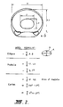
- the areas of the MC2, 3 and 4 were estimated by counting squares and average correction factors were determined for the sections along the bones. Using these correction factors the cross sectional areas were estimated from the ultrasound measurements and Figure 7 shows CSA E versus CSA M for positions along the shaft.
- Figure 8 shows the result of a comparison for the 50 bone sites for sections 1, 3, 5, 7 and 9.
- Tables 3 and 4 summarise for each bone the average BMC (5 readings at each site), BMC M , BMD E , CBD M , CBD E , J M and J e .
- the values for bone mineral content in the horse differed from those in man because of the size of the bone being measured.
- An 241Am source was used instead of 125I.
- the readings for bone mineral density (1060 ⁇ 20 kg m ⁇ 3) were compared with Greenfield et al (1120 ⁇ 150 kg m ⁇ 3) they were similar.
- the measurement of bone mineral density to this precision helps to overcome the volumetric problems identified by Ruff and Hayes (1984) found in using just bone mineral content.
- the calculated estimates for transverse cortical bone velocity, bone mineral density, compact bone density and modulus of elasticity compared favourably with the measured values for the same parameters.
- Schryver (1978) measured the bending properties of the metacarpals of ponies and found the elastic modulus to be 18.4 ⁇ 0.14 GN m ⁇ 2 for the cranial cortex of the mid-shaft. He obtained values of 16.2 to 20.2 GN M ⁇ 2 for different parts of the cortex of the radius and femur and was able to show that diet, exercise and sex of the pony affected the elastic modulus, ultimate strength and energy absorption of the bone.
- transverse cortical bone velocity has been determined rather than the apparent transverse velocity because it is more uniform along the shaft, is less subject to geometrical effects, allow an estimation of a modulus of elasticity and should provide better correlation between individuals.
- T m time through cortex + time through medulla
- Equation (5) then gives a revised estimate of C b . This process is continued until the change in C b per cycle is less than some chosen value e.g. 1 m s ⁇ 1. It takes only five cycles on average to achieve a suitable result.
Abstract
- (a) applying an ultrasound beam to a limb from a transmitting transducer to a receiving transducer;
- (b) measuring the flight times for an ultrasound beam along a first path Ts through the cortex of the bone, and along a second path Tm through the cortex and medulla of said bone;
- (c) calculating the diameter X of the bone from the external diameter of the limb less a predetermined constant allowing for skin and flesh;
- (d) calculating the apparent velocity Ca from X divided by Ts;
- (e) calculating the mean velocity V from X divided by Tm;
- (f) calculating the cortical bone velocity CbE from a predetermined function f multiplied by Ca;
- (g) determining the ratio R of the diameter of the medulla to the diameter of the cortex;
- (h) estimating the cross-sectional area CSAE of the bone from the values of the diameter X, the ratio R and from predetermined constants;
- (i) determining the bone mineral content BMC;
- (j) determining the estimated compact bone density CBDE, and
- (k) determining an estimate of the modulus of elasticity from the product of CBDE and CbE.
Description
- This invention relates to methods and apparatus for the measurement of bone quality in-vivo. While a preferred embodiment of the invention will be described as applied to the measurement of bone quality in horses, the invention is equally applicable to the performance of similar measurements in humans and other animals.
- Assessment of bone quality can be derived from measurements of bone mineral density (BMD kg m⁻³), compact bone density (CBD kg m⁻³) and a modulus of elasticity (J GN m⁻²). BMD can be derived from the bone mineral content (BMC kg m⁻¹) if the cross sectional area is known. BMC can be determined using single photon absorptiometry with the ability to detect changes of less than 3%. Compact bone density (CBD kg m⁻³) can be estimated using a bone model (Greenfield et al 1981) from BMC, cross sectional area (CSA m²) and estimated constants of the microscopic bone mineral density (ρm kg m⁻³), and collagen density (ρc kg m⁻³) by the equation:
CBD = ρc + (1 - ρc/ρm) BMC/CSA (1)
The modulus of elasticity cannot be easily determined in vivo, however a method that has received some attention involves the transmission of ultrasound through bone because the velocity of sound (C m s⁻¹) through a material is related to a modulus of elasticity and CBD, provided dispersivo and attenuating effects are ignored by assuming the use of the Helmholtz equation:
J = CBD.C² (2)
In bone both the density and modulus change with alteration of mineral content. However, it would appear that the change in ultrasound velocity reflects the change in modulus of elasticity more than the change in density. There are reports of the anisotropic nature of bone which is illustrated by different velocities of ultrasound in the axial, radial and tangential directions by reporting ratios of approximately 1.2: 1.08: 1. In this specification it is assumed that the transverse velocity is predominantly being estimated. An ideal bone for the development of these measurement techniques and for subsequent clinical testing is the third metacarpal bone of the horse. It is a reasonably large bone, easily accessable, has minimal surrounding tissues, is subjected to considerable biomechanical strain and has had its geometric and biomechanical properties studied. - There are essentially two types of bone that can be measured (i.e. cortical and trabecular or cancellous). Cortical bone is dense and compact contributing mainly to the strength of long bones, whereas trabecular bone has a faster turnover and more rapidly reflects generalised changes in the skeletal system (e.g. in osteoporosis). In man it is measurement of trabecular bone that is considered more important for the early detection of metabolic bone changes, however accurate cortical bone measurements are not ruled out.
- In the present specification "bone quality" includes bone mass or density, measured as its mineral content, and stiffness as estimated by the modulus of elasticity. Both these parameters have been shown to be related to bone strength. Noninvasive measurements of bone mineral content have been considered of diagnostic importance for over 50 years. The current trend is not to use plain radiography, but to develop more sophisticated methods. A variety of different techniques for bone mineral content have been successfully used to quantify aspects of bone both clinically (e.g. in age-related osteoporosis) and experimentally (e.g. by immobilisation and spaceflight).
- To date measurements of ultrasound velocity have been mainly in cortical bone and have not been applied clinically except in horses. These early techniques, were rushed into use, are relatively simplistic and in many ways have not succeeded. Our own investigations have led us to believe that further exploitation of ultrasound as a modality for assisting in bone quality assessment is warranted.
- In human medicine the clinical applications for assessment of bone quality have been well reported, particularly in the evaluation of osteoporosis and metabolic bone disease. A recent review article on imaging and measurement methods for Diagnostic Imaging indicates that dual photo absorptiometry and quantitative CT are the only methods listed for bone quality measurements. Other emerging techniques, not reported in this article, include Positron Emission Tomography (PET). Single Photon Emission Computer Tomography (SPECT) and Magnetic Resonance Imaging (MRI). However, all of these modalities have high capital costs, are available only in large medical departments and result in a moderate to high fee for service.
- The application of some of these techniques to the horse also has great potential, particularly for accurate assessment of skeletal maturity and to monitor the effects on bone of exercise and training methods. It has been clearly demonstrated that increased mechanical loading of the skeleton leads to increased bone mass and strength.
- Ultrasound velocity of bone can be used to measure bone quality because it is directly related to its stiffness or elasticity and its density or specific gravity by the equation:
E =ρC² [where E = modulus of elasticity,
ρ = compact bone density or specific gravity,
C = velocity of ultrasound.] - the modulus of elasticity (E) is the property of a material which relates to its bending strength and stiffness and ultimately to its fracture threshold.
- Preliminary studies using transverse ultrasound velocity through the metacarpal shaft of the horse, to give an indication of E, have been encouraging. The method involves passing a beam of ultrasound transversely from a transmitting to a receiving transducer. Two 2.25MHz transducers, mounted on a digital caliper to give a read-out of their distance apart, are placed on either side of the bone. After amplification of the signal by up to 90 decibels (dB), an electrical image of the ultrasound beam leaving one transducer and arriving at the other is recorded on an oscilloscope. The time of flight through the bone is then measured and, by knowing the distance between the transducers, the velocity of ultrasound (m s⁻¹) can be simply determined.
- This value for ultrasound velocity is referred to as the "apparent transverse velocity" (Ca) because it assumes that the pathway of the ultrasound beam is directly between the two transducers. In fact, the fastest pathway of the ultrasound beam is confined to the cortex and passes around the medullary cavity. The ultrasound velocity through cortical bone (approx 3000 m s⁻¹) is double that through the medulla (approx 1500 m s⁻¹). Thus the actual distance travelled by the ultrasound beam is greater than the distance between the transducers. For this reason, an "apparent transverse velocity" measurement cannot be used to obtain a sufficiently accurate estimate of bone quality.
- The object of this invention is to provide a practical and reproducible method for estimating the transverse cortical bone ultrasound velocity and the cross sectional area which can then be used with absorptiometry measurements to make estimates of bone quality in vivo.
- The invention therefore provides a method of determining in-vivo an estimate of the modulus of elasticity of a bone, comprising the steps of passing a beam of ultrasound from a transmitting transducer through the bone to a receiving transducer, determining the distance between said transducers and correcting that distance to obtain a measure of the diameter of said bone, measuring the time of transmission of ultrasound beam along a first path through the cortex of said bone and along a second path through the cortex and medulla of said bone, calculating the velocity of said beam along said first path and along said second path and using said velocity values to calculate an estimate of the transverse cortical bone velocity and the ratio of the diameter of the medulla to the diameter of the cortex, calculating an estimate of the cross sectional area of the cortex, measuring the bone mineral content of the bone and using said estimated cross sectional area and said measured bone mineral content to calculate an estimate of the modulus of elasticity of the bone.
- Experimental testing of the method defined above and described in greater detail below show that the estimated modulus of elasticity obtained by the method of the invention compares favourably with measured values for the same bone. Thus, by allowing for the fact that the ultrasound beam is transmitted along two separate and distinct paths through the bone, a more accurate estimate of the modulus of elasticity of the bone may be obtained in-vivo whereby the quality of the bone may be more accurately measured.
- In performing the method according to the invention, a number of mathematical functions and constants must first be determined for the particular bone under investigation by experimental and mathematical means. For example, the mathematical relationship of the first path through the cortex of the bone may be specified as a factor (f) times the total diameter (X) of the bone, and the best empirical value for this factor so far determined is:
f = √1 - Rc² = Rc arsin Rc (10)
where:
Rc = 0.78R or 0.79 R(0.6 + 0.15R)
- Similarly, the velocity of sound in the medulla has been experimentally determined to be about 1470 ms⁻¹ and this value is deemed to be constant in the calculations referred to above. In addition, the ovality of the external cortical bone cross-section and the medulla must be taken into account. In the case of the third metacarpal bone in horses, observation has shown that the bone is approximately elliptical and empirical values for constants which take the ovality of the sections of the bone into account have been experimentally determined. In the case of the horse, a further factor is introduced to take into account the splint bones (MC2 and MC4) in the calculation of the cross-sectional area of the bone. The presently preferred method will be described in greater detail below.
- In another aspect, the invention provides an apparatus for determining in-vivo an estimate of the modulus of elasticity of a bone comprising a pair of transducers for transmitting an ultrasound beam through the bone along a first path through the cortex of said bone and along a second path through the cortex and medulla of said bone, means for determining the distance between said transducers and for correcting that distance to obtain a measure of the diameter of said bone, means for determining the time of transmission of said ultrasound beam along said first and second paths, means for calculating the velocity of said beam along said first and second paths and for calculating an estimate of the transverse cortical bone velocity and the ratio of the diameter of the medulla to the diameter of the cortex, and means for calculating an estimate of the cross-sectional area of the cortex and for calculating from this information and from separately derived information an estimate of the modulus of elasticity of the bone.
- A presently preferred embodiment of the invention will now be described with reference to the accompanying drawings in which:
- Figure 1 is a diagrammatic representation of the ultrasound pathways through the cortex and through the cortex and medulla;
- Figure 1A is a schematic diagram showing the ultrasound and measuring apparatus;
- Figure 2 is a diagrammatic representation of the assumption of ovality for the cortex and medulla;
- Figure 3 is a diagrammatic representation of typical ultrasound traces used to determine the time measurements;
- Figure 4 is a diagrammatic representation of the ten measurement sites and sections for the ten selected left metacarpal bones;
- Figure 5 is a graphical comparison of preliminary empirical pathways and experimental values for development of pathway lengths;
- Figure 6 is a graphical comparison of the best empirical formula for the ultrasound pathway length and experimental values;
- Figure 7 is graphical comparison of experimental and measured cross-sectional areas of the bone under consideration, and
- Figure 8 is a graphical comparison of the experimental and measured cross-sectional areas for the experimental bone sections.
- The estimate of ultrasound velocity is gained from knowing the distance between two transducers and measuring the time of flight of the transmitted beam. However, as the velocity of sound in the medulla is of the order 1500 ms⁻¹ and that in the cortical bone of the order of 3000 m s⁻¹ it is readily seen that the shortest time of flight must be for the sound to travel around the cortex (Figure 1).
- The mathematical relationship of this pathway should be able to be specified as a factor (f) times the total diameter (X) and also as a mathematical function of the ratio (R) of the diameter of the medulla (Y) to the diameter of the cortex (X):
path length = f.X = function (R) (3)
Another pathway, the corticomedullary pathway, (Fig. 1) can be determined for the sound travelling through the cortical walls and the centre of the medullas in a straight line. For this second pathway a mean velocity (V) can be determined from
R = Y/X = (Cb/V - 1) / (Cb/Cm - 1) (4)
From the apparent velocity (Ca) which is the velocity determined from the shortest time of flight (Ts) and the cortical diameter (X), an estimate of the transverse cortical bone velocity (CbE) can be made if the path length (f.X) is taken into account:
CbE = f . X/Ts = f. Ca (5)
Ca is the first estimate of the transverse bone velocity and as the bone velocity must be greater than the apparent velocity, by iteration (appendix B), it is possible to calculate both CbE and R. An empirical formula by means of which f may be estimated will be found in equations (10) and (11). Seeing that the diameter of the bone X is known (transducer spacing less average skin and flesh thickness for the particular bone and position) and the ratio R estimated, the cross sectional area of the cortex can also be estimated using the formula;
CSA = M . π/4 . X² (K - L . R²) (6)
Where K is an ovality factor for the external cortical bone cross section and L is an ovality factor for the medulla. These ovality factors are derived from the bone cross sections on the assumption that the sections approximate ellipses as shown in Fig. 2. M is a factor to include the area of the splint bones (MC2 and MC4) as these bones were included in the determination of the BMC. Using this cross sectional area, BMC can be corrected to give an estimate of the bone mineral density (BMDE) by:
BMDE = BMC/CSAE (7)
Another estimate of the bone mineral density, BMDM, can be determined from direct measurements by:
BMDM = ash weight/volume of wet bone (8)
The compact bone density, CBDE, is estimated using equation (1). For this series the constant for ρm was 3120 kg m⁻³ and that for ρc was 1310 kg m⁻³ as published for bovine bone by Lees et at (1979). The CBDM can also be determined from direct measurement by:
CBDM = mass of wet bone/volume of wet bone (9)
- The modulus of elasticity, JM, is determined using equation (2), CBDM and CbM. The modulus of elasticity, JE, is determined with the same equation but using CBDE and CbE.
- The ultrasound time of flight measurements were obtained using a pulse module (Panametrics Model 5055), a preamplifier (Panametrics Model 5660B), two 2.25 MHz, 13 mm diameter ultrasound transducers and and 2 (Figure 1A) mounted on an electronic digital caliper 3 (Max-cal Model 950-101) connected to a computer (not shown) via an interface (Max-cal Model 960-101) and a digital storage oscilloscope with signal averaging capacity (Tektronix Model 468). An Apple II Plus microcomputer was used to apply the various iterative and calculative algorithms.
- The BMC values were determined using a Norland Digital bone Densitometer (Prototype Model 2781, Large Animal Scanning System, LASS) with an Americium source (²⁴¹Am).
- In summary, the method embodying the invention comprises the steps of
- (a) measuring the flight times for the ultrasound beam along the two paths shown in Figure 1, Ts, the flight time along the cortical pathway and Tm, the flight time along the cortico-medullary pathway;
- (b) calculating the diameter of the bone X from the distance between the transducers less a constant determined experimentally to allow for skin and flesh (in the case of the third matacarpal bone of a horse, 3mm);
- (c) calculating the apparent velocity Ca from

- (d) calculating the "mean" velocity V from

- (e) calculating, preferably by iteration, the values of CbE and R from equations (5), (10) and (4) respectively, Cm being treated as a constant determined experimentally to be approximately 1470 m s⁻¹;
- (f) determining the cross sectional area of the bone using formula (6) M, K and L being experimentally predetermined constants as described above;
- (g) determining the bone mineral content BMC using a bone densitometer or by any other suitable method;
- (h) determining the estimated compact bone density CBDE using equation (1);
- (i) determining the modulus of elasticity using equation (2), CBDE and CbE.
- To test the theory outlined above, a series of 15 regular perspex cylinders of diameter 50mm and with central holes ranging from 15 to 35 mm in 2 mm steps were machined to simulate bone cross sections. Perspex was chosen as its ultrasound velocity of about 2800 m s⁻¹ is close to that of bone at about 3000 m s ⁻¹. The circular central hole was initially left empty to ensure only transmission through the perspex and later filled with water which with a velocity of about 1500 m s ⁻¹ closely approximated medullary tissue. Time of flight measurements were made on this group of cylinders to establish the best empirical relationship between pathway and ratio of medulla to cortex. Measurements taken with air in the central cavity confirmed that the pathway for the minimum time of flight is through the perspex, whereas when filled with water both pathways were verified.
- Ten metacarpal bones of approximately the same size were selected from the left forelimbs of thoroughbred horses aged from 2 to 5 years. After removal of all soft tissues, the bones were stored at -15°C until examined. The lateral bone diameter, the times of flight through both the cortex alone and the corticomedullary pathways (Fig. 3) and the BMC were determined at 10 sites along the shaft of the bone as shown in Figure 4. From these measurements Ca, R, CbE and CSAE (using factors determined later) were estimated. The bones were then cut to provide 10 transverse cross sections (Figure 4). The
transverse sections sections - The modulus of elasticity, J, was determined using equation (2) from two sets of measurements. The first estimate, JM, is from the directly measured CbM and the directly measured CBDM. The other estimate, JE was made from CbE and the CBDE from equation (1). Both determinations are made in this report.
- the pathway length determined on the perspex blocks was compared with several mathematically derived theoretical pathways two of which are shown in Figure 5. A suitable theoretecal pathway based on physical principles has not yet been achieved. Various mathematical correction factors were then tested to find an empirical formula to allow estimation of the path length.
- The best empirical formula so far determined is:
f = 1 -R c² + Rc . arsin Rc (10)
where:
Rc = 0.78 R (11)
This formula was used for all calculations on the selected bones. Equation (10) is shown with the experimental values in Figure 6. A more accurate value for Rc determined by further experiments may be: Rc = 0.79R (0.6 + 0.15R). - The measurements from the 100 bone marrow samples to determine the average medullary velocity (Cm) are summarised in Table 1. The average value of 1444 ± 14 m s⁻¹ was used form subsequent calculations.
- A comparison of CbM and CbE is summarised in Table 2. From the radiographs of the individual bone segments an estimate of ovality of the medulla and the ovality of the external cortical boundary was made. The average ovality factor for the medulla (L) of each bone (n = 10) ranged over the 10 bones from 0.65 to 0.90 with a mean of 0.76. The average ovality factor for the cortex (K) ranged from 0.75 to 0.90 with a mean of 0.81. From the same radiographs the areas of the MC2, 3 and 4 were estimated by counting squares and average correction factors were determined for the sections along the bones. Using these correction factors the cross sectional areas were estimated from the ultrasound measurements and Figure 7 shows CSAE versus CSAM for positions along the shaft. Figure 8 shows the result of a comparison for the 50 bone sites for
sections - The above values for K, L and M are most valid for the middle region of the bone. The following table lists corrections factors for K and L and new values for M according to the position of the testing site (the numbers correspond to the numbers in Figure 4).
PROXIMAL MID DISTAL 1 2 3 4 5 6 7 8 9 10 11 cortex K 0.91 0.95 0.98 0.99 1 1 1 1 0.99 0.99 0.99 Medulla L 0.84 0.85 0.87 0.93 0.98 1 1 1.01 1.01 1.01 1.01 Splint area M 1.125 1.105 1.09 1.075 1.06 1.045 1.03 1.02 1.01 1 1 - Estimates of the precision were obtained for Ca, CbE, CSAE and BMC from multiple independent measurements taken at the same site of a bone. Using
equations - The accuracy and precision of the methods used here for determining both transverse cortical bone velocity and bone mineral content are encouraging and correlate well with those of Greenfield et al (1981). These workers utilised the human radius and employed the same single beam photon absorptiometry, but used a pulse echo technique for cortical velocity measurement instead of the transmission method. Both of these ultrasound methods give similar results for cortical bone velocity. In man the cortical velocity of the radius as 3335 ± 300 m s⁻¹ whereas for the equine metacarpus the velocity was measured at 3109 ± 56 m s ⁻¹. The advantage of the transmission method is that a lateral radiograph to measure cortical thickness is not required and an estimate of the cross sectional area is obtained at the same time. Greenfield's estimate of uncertainty for cross sectional area was 2-10% whereas by the submission method the uncertainty for the equine metacarpus is about 5%. The variation of cross sectional area with site and area of splint bones correlated well with the results published by Piotrowski et al (1983).
- The estimation of cross sectional area by the ultrasound method was considered good (correlation coefficient r = 0.92) and proved to be better than just using an estimate of cross sectional area based on diameter squared (r = 0.82).
- The values for bone mineral content in the horse differed from those in man because of the size of the bone being measured. An ²⁴¹Am source was used instead of ¹²⁵I. However, when the readings for bone mineral density (1060 ± 20 kg m⁻³) were compared with Greenfield et al (1120 ± 150 kg m⁻³) they were similar. The measurement of bone mineral density to this precision helps to overcome the volumetric problems identified by Ruff and Hayes (1984) found in using just bone mineral content.
- The calculated estimates for transverse cortical bone velocity, bone mineral density, compact bone density and modulus of elasticity compared favourably with the measured values for the same parameters. Schryver (1978) measured the bending properties of the metacarpals of ponies and found the elastic modulus to be 18.4 ± 0.14 GN m⁻² for the cranial cortex of the mid-shaft. He obtained values of 16.2 to 20.2 GN M⁻² for different parts of the cortex of the radius and femur and was able to show that diet, exercise and sex of the pony affected the elastic modulus, ultimate strength and energy absorption of the bone.
- In this study transverse cortical bone velocity has been determined rather than the apparent transverse velocity because it is more uniform along the shaft, is less subject to geometrical effects, allow an estimation of a modulus of elasticity and should provide better correlation between individuals.
- These methods, having been developed on post mortem specimens, are now being applied to live animals. Ultrasound measurements (5 sites) take about 10 minutes per limb and absorptiometry (5 readings) takes about 20 minutes per limb. Using these combined techniques it will be possible to establish better parameters for skeletal maturity and to measure the effects of exercise on bone. Whilst these methods are being implemented in equine medicine there appears no reason whey the same principles cannot be applied in humans. It will be appreciated that the use of the method to estimate the quality of various human bone would require the experimental determination of different contants (for example the ovality factors K and L would be replaced by different factors and the M factor would be enclosed), but the method would otherwise be similarly applied.
- Tm = time through cortex + time through medulla
X/V = (X-Y)/Cb + Y/Cm
dividing by X and rearranging gives:
1/V - 1/Cb = R/Cm - R/Cb
R = (1/V - 1/Cb)/(1/Cm - 1/Cb) = (Cb/V - 1)/(Cb/Cm -1) (4)
- Assume geometrical symmetry, a straight line path across the cortex and an arc around the medulla.
- Let ϑ equal angle between diameter (centre to centre of transducers) and straight pathway through cortex. ϑ is also half the angle of the arc.
Thus sin ϑ = Y/X = R
Length of straight sections = √X² - Y²
Length of arc = Y . ϑ (ϑ in radian) = Y . arsin R
Total length f.X = √X² - Y² + Y . arsin R divided by X gives
f = √1 - R² + R . arsin R
- Assume symmetry and that the sound path is along an arc of a circle which goes from centre to centre of the transducers and just touches the edge of the medulla as shown in the sketch.
- If S is the radius of curvature of the arc and 2 φ is the subtended angle of the arc then:
S = (X² + Y²)/4Y = X.(1 + R²)/4R
and sin φ = X/2S = 2R/(1 + R²)
the length of the arc = 2 . S . φ (φ in radian) = X . ((1 + R²) . arsin (2R/(1 + R²)))/2R
For this situation then
f′ = ((1 + R²) . arsin (2R/(1 + R²)))/2R
- The iterative procedure applied in a simple computer program involves equations (4), (5) and (10). Ca, V and Cm are known leaving f, Cb and R to be determined. The only restriction to the iterative process is imposed by equation (4) where:
R = -α when Cb = Cm
R < 0 when V > Cb > Cm
R = 0 when Cb = V
R > 0 when Cb > V
- Provided the starting point of the iteration is with Cb > V then the correct value is determined. Ca is always greater than V and is used as the first estimate of Cb. The first estimate of R is then determined, equation (4), followed by the first estimate of f, equation (10). Equation (5) then gives a revised estimate of Cb. This process is continued until the change in Cb per cycle is less than some chosen value e.g. 1 m s ⁻¹. It takes only five cycles on average to achieve a suitable result.
-
- f = factor to relate cortical pathway length to diameter of cortex.
- f.X = path length of ultrasound beam through cortex of MC3.
- K = ovality factor for external perimeter of cortex of MC3.
- L = ovality factor for medulla of MC3.
- M = factor to compensate for area of splint bones MC2 and MC4.
- R = ratio of medulla to cortex of MC3 = Y/X.
- Rc = corrected ratio of medulla to cortex (R).
- X = external diameter of cortex of MC3 (lateral).
- Y = diameter of medulla of MC3 (lateral).
- BMC = bone mineral content (kg m⁻¹).
- BMDM = measured bone mineral density (kg m⁻³).
- BMDE = estimated bone mineral density (kg m⁻³).
- C = velocity of ultrasound (m s⁻¹).
- Ca = apparent transverse cortical bone ultrasound velocity (m s⁻¹) = X/Ts.
- Cb = transverse cortical bone ultrasound velocity (m s⁻¹).
- CbM = measured transverse cortical bone ultrasound velocity (m s⁻¹).
- CbE = estimated transverse cortical bone ultrasound velocity (m s⁻¹).
- Cm = ultrasound velocity through medulla (m s⁻¹).
- CBDm = measured compact bone density or specific gravity (kg m⁻³).
- CBDE = estimated compact bone density or specific gravity (kg m⁻³).
- CSAM = measured cross sectional area (m² (volumetric).
- CSAE = estimated cross sectional area (m²) (ultrasound).
- J = modulus of elasticity.
- JM = estimated modulus of elasticity using CBDM and CbM (GN m⁻²).
- JE = estimated modulus of elasticity using CBDE and CbE (GN m⁻²).
- ρm = microscopic bone mineral density (kg m⁻³).
- ρc = collagen density (kg m⁻³).
-
MC 2, MC 3 andMC 4 = second, third and fourth metacarpal bones of the horse. - Tm of flight for corticomedullary path (s).
- Ts = shortest time of flight of ultrasound transmission (s).
- V = mean velocity of ultrasound through cortex and medulla (m s⁻¹). = X/Tm
| Bone No. | Cm - ULTRASOUND VELOCITY OF MEDULLA (m s⁻¹) | ||||
| Dorsopalmar | | Longitudinal | Average | ||
| 1 | 1439 | 1451 | 1438 | 1443 | |
| 2 | 1430 | 1441 | 1437 | 1436 | |
| 3 | 1461 | 1469 | 1454 | 1459 | |
| 4 | 1444 | 1453 | 1430 | 1439 | |
| 5 | 1424 | 1432 | 1413 | 1423 | |
| 6 | 1461 | 1461 | 1454 | 1459 | |
| 7 | 1427 | 1437 | 1414 | 1423 | |
| 8 | 1456 | 1464 | 1453 | 1458 | |
| 9 | 1453 | 1465 | 1456 | 1458 | |
| 10 | 1442 | 1460 | 1433 | 1445 | |
| Average | 1444 ± 14 | 1453 ± 13 | 1438 ± 16 | 1444 ± 14 | |
| Results of ultrasound velocity of the medulla (Cm ±1SD) measured in dorsopalmar, lateral and longitudinal directions of 10 metacarpal bones. | ||||
| Bone No. | Cb - CORTICAL ULTRASOUND VELOCITY (m s⁻¹) | % difference between CbM and CbE | |
| CbM Directly Measured | CbE Estimated | ||
| 1 | 3161 (± 14) | 3155 (± 35) | - 0.19 |
| 2 | 3115 (± 62) | 3164 (± 51) | + 1.57 |
| 3 | 3044 (± 46) | 3150 (± 29) | + 3.48 |
| 4 | 3127 (± 38) | 3104 (± 43) | - 0.74 |
| 5 | 3171 (± 47) | 3196 (± 45) | + 0.79 |
| 6 | 3050 (± 27) | 3060 (± 30) | + 0.33 |
| 7 | 3142 (± 54) | 3247 (± 37) | + 3.34 |
| 8 | 3181 (± 20) | 3115 (± 37) | - 2.07 |
| 9 | 3046 (± 42) | 3082 (± 29) | + 1.18 |
| 10 | 3050 (± 37) | 3061 (± 20) | + 0.36 |
| Average | 3109 (± 56) | 3133 (± 61) | + 0.83 (± 1.75) |
| Results obtained for directly measured (CbM) and estimated (CbE) transverse cortical bone velocity (±1SD) in 10 metacarpal bones. | |||

| Bone No. | JM GN m⁻² | JE GN m⁻² | % diff. JM and |
| 1 | 18.8 (±0.2) | 18.7 (±0.6) | - 0.53 |
| 2 | 18.6 (±0.6) | 19.1 (±0.9) | 2.69 |
| 3 | 17.4 (±0.5) | 18.9 (±0.6) | 8.62 |
| 4 | 18.5 (±0.3) | 18.2 (±0.9) | - 1.62 |
| 5 | 19.2 (±0.4) | 19.6 (±0.6) | 2.08 |
| 6 | 17.6 (±0.2) | 17.6 (±0.4) | 0 |
| 7 | 18.8 (±0.5) | 20.5 (±0.5) | 9.04 |
| 8 | 19.1 (±0.3) | 18.6 (±0.6) | - 2.62 |
| 9 | 17.5 (±0.3) | 18.4 (±0.3) | 5.14 |
| 10 | 17.4 (±0.3) | 17.7 (±0.3) | 1.72 |
| Average | 18.3 (±0.7) | 18.7 (±0.9) | 2.45 (±4.04) |
| Measured (JM) and estimated (JE) modulus of elasticity of the third metacarpal cortex from 10 horses. | |||
| DIRECTLY MEASURED VALUES | ESTIMATED VALUE |
| Ca ± 0.32% (n = 41) | |
| BMC ± 1.15% (n = 20) | |
| CbM ± 1% | CbE ± 0.59% (n = 41) |
| CSAM ± 1.1% | CSAE ± 3.6% (n = 41) |
| CBDM ± 0.42% | CBDE ± 1.2% |
| BMDM ± 0.44% | BMDE ± 3.8% |
| JM ± 1.4% | JE ± 1.5% |
| Estimates of ± 1 SD for the precision of values determined in this investigation. | |
Claims (6)
Applications Claiming Priority (2)
| Application Number | Priority Date | Filing Date | Title |
|---|---|---|---|
| AUPI321687 | 1987-07-16 | ||
| AU3216/87 | 1987-07-16 |
Publications (2)
| Publication Number | Publication Date |
|---|---|
| EP0299906A2 true EP0299906A2 (en) | 1989-01-18 |
| EP0299906A3 EP0299906A3 (en) | 1990-06-13 |
Family
ID=3772341
Family Applications (1)
| Application Number | Title | Priority Date | Filing Date |
|---|---|---|---|
| EP88630138A Withdrawn EP0299906A3 (en) | 1987-07-16 | 1988-07-14 | In-vivo bone quality measurement |
Country Status (2)
| Country | Link |
|---|---|
| US (1) | US4976267A (en) |
| EP (1) | EP0299906A3 (en) |
Cited By (14)
| Publication number | Priority date | Publication date | Assignee | Title |
|---|---|---|---|---|
| WO1991006245A1 (en) * | 1989-10-24 | 1991-05-16 | The Adelaide Bone And Joint Research Foundation Inc | Vibrational analysis of bones |
| EP0516353A1 (en) * | 1991-05-22 | 1992-12-02 | Walker Magnetics Group, Inc. | Ultrasonic transducer assembly |
| GB2257253A (en) * | 1991-06-17 | 1993-01-06 | Christian Mcdonald Langton | Ultrasound bone analyser |
| WO1994018889A1 (en) * | 1993-02-17 | 1994-09-01 | John Koblanski | Ultrasonic scanning apparatus |
| WO1994022375A1 (en) * | 1993-04-07 | 1994-10-13 | Osteo Sciences Corporation | System and method for external acoustic bone velocity measurement |
| EP0663182A1 (en) * | 1994-01-14 | 1995-07-19 | IGEA S.r.L. | Ultrasonic measurement system for the determination of bone density and structure |
| US5452722A (en) * | 1992-06-22 | 1995-09-26 | Mccue Ultrasonics, Ltd. | Ultrasound bone analyser |
| US5720290A (en) * | 1993-04-07 | 1998-02-24 | Metra Biosystems, Inc. | Apparatus and method for acoustic analysis of bone using optimized functions of spectral and temporal signal components |
| US5755228A (en) * | 1995-06-07 | 1998-05-26 | Hologic, Inc. | Equipment and method for calibration and quality assurance of an ultrasonic bone anaylsis apparatus |
| US5785041A (en) * | 1996-03-26 | 1998-07-28 | Hologic Inc. | System for assessing bone characteristics |
| US6004272A (en) * | 1995-06-07 | 1999-12-21 | Hologic, Inc. | Ultrasonic bone testing apparatus with repeatable positioning and repeatable coupling |
| EP0570936B1 (en) * | 1992-05-20 | 2000-08-09 | Aloka Co. Ltd. | Bone assessment apparatus |
| US6352512B1 (en) | 1995-06-07 | 2002-03-05 | Hologic, Inc. | Bone analysis apparatus and method for calibration and quality assurance of an ultrasonic bone analysis apparatus |
| US7727152B2 (en) * | 2001-02-28 | 2010-06-01 | The Research Foundation Of State University Of New York | Method and apparatus for scanning confocal acoustic diagnostic for bone quality |
Families Citing this family (29)
| Publication number | Priority date | Publication date | Assignee | Title |
|---|---|---|---|---|
| US6277076B1 (en) | 1988-05-11 | 2001-08-21 | Lunar Corporation | Ultrasonic densitometer with pre-inflated fluid coupling membranes |
| US5293870A (en) * | 1989-11-17 | 1994-03-15 | Board Of Regents The University Of Texas System | Method and apparatus for elastographic measurement and imaging |
| US5107837A (en) * | 1989-11-17 | 1992-04-28 | Board Of Regents, University Of Texas | Method and apparatus for measurement and imaging of tissue compressibility or compliance |
| US5426979A (en) * | 1990-06-04 | 1995-06-27 | Medicano Systems Ltd. | Frequency spectrum apparatus for determining mechanical properties |
| IL94616A (en) * | 1990-06-04 | 1994-06-24 | Medicano Systems Ltd | Apparatus and method for calculating the mechanical properties of a solid |
| US5247934A (en) * | 1991-08-09 | 1993-09-28 | Trustees Of The University Of Pennsylvania | Method and apparatus for diagnosing osteoporosis with MR imaging |
| US5140988A (en) * | 1991-08-22 | 1992-08-25 | Animal Ultrasound Services, Inc. | Detection of abnormal bone structure in animals and carcasses with ultrasound |
| US5259384A (en) * | 1992-07-30 | 1993-11-09 | Kaufman Jonathan J | Ultrasonic bone-assessment apparatus and method |
| US5402781A (en) * | 1993-03-03 | 1995-04-04 | Washington University | Method and apparatus for determining bone density and diagnosing osteoporosis |
| US5836876A (en) * | 1993-03-03 | 1998-11-17 | Washington University | Method and apparatus for determining bone density and diagnosing osteoporosis |
| US5429143A (en) * | 1993-11-22 | 1995-07-04 | Marzluff; Joseph | Device and method for determining hole integrity in surgical applications |
| JP3207318B2 (en) | 1994-05-11 | 2001-09-10 | アロカ株式会社 | Ultrasonic bone evaluation device |
| US5535750A (en) * | 1994-09-30 | 1996-07-16 | Kabushiki Kaisha Ishikawa Seisakusho, Ltd. | Method and apparatus for evaluating the progress of osteoporosis by ultrasonic signals |
| JP2883290B2 (en) * | 1995-04-10 | 1999-04-19 | アロカ株式会社 | Ultrasonic bone evaluation device |
| US5651363A (en) * | 1996-02-16 | 1997-07-29 | Orthologic Corporation | Ultrasonic bone assessment method and apparatus |
| US5879301A (en) * | 1996-02-16 | 1999-03-09 | Orthologic Corp. | Ultrasonic bone assessment method and apparatus |
| IT1292288B1 (en) * | 1997-04-24 | 1999-01-29 | Igea Srl | ULTRASONIC MEASURING DEVICE FOR DETECTION OF BONE DENSITY AND STRUCTURE. |
| US9198635B2 (en) * | 1997-10-31 | 2015-12-01 | University Of Washington | Method and apparatus for preparing organs and tissues for laparoscopic surgery |
| US5931795A (en) * | 1997-11-07 | 1999-08-03 | Manly; Philip | Method for evaluating human bone strength |
| US8202219B2 (en) * | 2004-02-23 | 2012-06-19 | Cyberlogic, Inc. | Ultrasonic bone assessment apparatus and method |
| US7033321B1 (en) * | 2004-10-25 | 2006-04-25 | Artann Laboratories, Inc. | Ultrasonic water content monitor and methods for monitoring tissue hydration |
| US20090112094A1 (en) * | 2006-04-13 | 2009-04-30 | The Research Foundation Of State University Of New York | Phased Apply Ultrasound With Electronically Controlled Focal Point For Assessing Bone Quality Via Acoustic Topology And Wave Transmit Functions |
| US7901356B2 (en) * | 2006-12-13 | 2011-03-08 | Cyberlogic, Inc. | Ultrasonic bone assessment apparatus and method |
| US9615814B2 (en) * | 2006-12-13 | 2017-04-11 | Cyberlogic, Inc. | Ultrasonic bone assessment apparatus and method |
| US7862510B2 (en) * | 2007-02-09 | 2011-01-04 | Cyberlogic, Inc. | Ultrasonic bone assessment apparatus and method |
| US8187185B2 (en) * | 2007-08-08 | 2012-05-29 | Hitachi Aloka Medical, Ltd. | Ultrasound diagnostic apparatus |
| US9008394B2 (en) * | 2008-11-26 | 2015-04-14 | General Electric Company | Methods and apparatus for determining brain cortical thickness |
| US8421479B2 (en) * | 2009-06-30 | 2013-04-16 | Navisense | Pulsed echo propagation device and method for measuring a parameter |
| JP6389116B2 (en) * | 2014-12-22 | 2018-09-12 | ジーイー・メディカル・システムズ・グローバル・テクノロジー・カンパニー・エルエルシー | Ultrasonic image display device and control program thereof |
Citations (1)
| Publication number | Priority date | Publication date | Assignee | Title |
|---|---|---|---|---|
| FR2318420A1 (en) * | 1975-07-17 | 1977-02-11 | Guiset Jacques | Pressure or distance measurement using ultrasonics - involves compressible medium in which propagation velocity dependence on density is known |
Family Cites Families (6)
| Publication number | Priority date | Publication date | Assignee | Title |
|---|---|---|---|---|
| US4361154A (en) * | 1978-07-28 | 1982-11-30 | Massachusetts Institute Of Technology | Method for establishing, in vivo, bone strength |
| IL69471A (en) * | 1983-08-12 | 1987-07-31 | Benjamin Gavish | Method and device for non-invasively monitoring the instantaneous fluctuations in the viscoelastic properties of a soft tissue |
| DE3329134C2 (en) * | 1983-08-12 | 1985-06-20 | Deutsche Forschungs- und Versuchsanstalt für Luft- und Raumfahrt e.V., 5000 Köln | Device for measuring cross-sections on objects, in particular on body parts |
| JPS60116345A (en) * | 1983-11-30 | 1985-06-22 | 富士通株式会社 | Ultrasonic diagnostic apparatus |
| JPS60163643A (en) * | 1984-02-07 | 1985-08-26 | テルモ株式会社 | Ultrasonic measuring method and apparatus |
| US4913157A (en) * | 1986-06-03 | 1990-04-03 | Analog Devices, Inc. | Ultrasound method and apparatus for evaluating, in vivo, bone conditions |
-
1988
- 1988-07-14 EP EP88630138A patent/EP0299906A3/en not_active Withdrawn
- 1988-07-18 US US07/220,083 patent/US4976267A/en not_active Expired - Fee Related
Patent Citations (1)
| Publication number | Priority date | Publication date | Assignee | Title |
|---|---|---|---|---|
| FR2318420A1 (en) * | 1975-07-17 | 1977-02-11 | Guiset Jacques | Pressure or distance measurement using ultrasonics - involves compressible medium in which propagation velocity dependence on density is known |
Non-Patent Citations (2)
| Title |
|---|
| MEDICAL & BIOLOGICAL ENGINEERING & COMPUTING * |
| RADIOLOGY * |
Cited By (20)
| Publication number | Priority date | Publication date | Assignee | Title |
|---|---|---|---|---|
| US5368044A (en) * | 1989-10-24 | 1994-11-29 | The Adelaide Bone And Joint Research Foundation, Inc. | Vibrational analysis of bones |
| WO1991006245A1 (en) * | 1989-10-24 | 1991-05-16 | The Adelaide Bone And Joint Research Foundation Inc | Vibrational analysis of bones |
| EP0516353A1 (en) * | 1991-05-22 | 1992-12-02 | Walker Magnetics Group, Inc. | Ultrasonic transducer assembly |
| GB2257253A (en) * | 1991-06-17 | 1993-01-06 | Christian Mcdonald Langton | Ultrasound bone analyser |
| GB2257253B (en) * | 1991-06-17 | 1995-01-11 | Christian Mcdonald Langton | Ultrasound bone analyser |
| EP0570936B1 (en) * | 1992-05-20 | 2000-08-09 | Aloka Co. Ltd. | Bone assessment apparatus |
| US5452722A (en) * | 1992-06-22 | 1995-09-26 | Mccue Ultrasonics, Ltd. | Ultrasound bone analyser |
| WO1994018889A1 (en) * | 1993-02-17 | 1994-09-01 | John Koblanski | Ultrasonic scanning apparatus |
| US5720290A (en) * | 1993-04-07 | 1998-02-24 | Metra Biosystems, Inc. | Apparatus and method for acoustic analysis of bone using optimized functions of spectral and temporal signal components |
| WO1994022375A1 (en) * | 1993-04-07 | 1994-10-13 | Osteo Sciences Corporation | System and method for external acoustic bone velocity measurement |
| US6371916B1 (en) | 1993-04-07 | 2002-04-16 | Metra Biosystems, Inc. | Acoustic analysis of bone using point-source-like transducers |
| EP0663182A1 (en) * | 1994-01-14 | 1995-07-19 | IGEA S.r.L. | Ultrasonic measurement system for the determination of bone density and structure |
| US5755228A (en) * | 1995-06-07 | 1998-05-26 | Hologic, Inc. | Equipment and method for calibration and quality assurance of an ultrasonic bone anaylsis apparatus |
| US6004272A (en) * | 1995-06-07 | 1999-12-21 | Hologic, Inc. | Ultrasonic bone testing apparatus with repeatable positioning and repeatable coupling |
| US6135964A (en) * | 1995-06-07 | 2000-10-24 | Hologic, Inc. | Ultrasonic bone testing apparatus with repeatable positioning and repeatable coupling |
| US6352512B1 (en) | 1995-06-07 | 2002-03-05 | Hologic, Inc. | Bone analysis apparatus and method for calibration and quality assurance of an ultrasonic bone analysis apparatus |
| US5935073A (en) * | 1995-09-26 | 1999-08-10 | Hologic, Inc. | Equipment and method for calibration and quality assurance of an ultrasonic bone analysis apparatus |
| US5785041A (en) * | 1996-03-26 | 1998-07-28 | Hologic Inc. | System for assessing bone characteristics |
| US6029078A (en) * | 1996-03-26 | 2000-02-22 | Hologic, Inc. | System for assessing bone characteristics |
| US7727152B2 (en) * | 2001-02-28 | 2010-06-01 | The Research Foundation Of State University Of New York | Method and apparatus for scanning confocal acoustic diagnostic for bone quality |
Also Published As
| Publication number | Publication date |
|---|---|
| EP0299906A3 (en) | 1990-06-13 |
| US4976267A (en) | 1990-12-11 |
Similar Documents
| Publication | Publication Date | Title |
|---|---|---|
| US4976267A (en) | Method and apparatus for the measurement of bone quality in vivo | |
| Laugier | Instrumentation for in vivo ultrasonic characterization of bone strength | |
| MARKEL et al. | Noninvasive monitoring techniques for quantitative description of callus mineral content and mechanical properties. | |
| US4913157A (en) | Ultrasound method and apparatus for evaluating, in vivo, bone conditions | |
| Protopappas et al. | Ultrasonic monitoring of bone fracture healing | |
| Freeman et al. | Evaluation of the use of muscle condition score and ultrasonographic measurements for assessment of muscle mass in dogs | |
| Li et al. | Could clinical ultrasound improve the fitting of spinal orthosis for the patients with AIS? | |
| KR20140035932A (en) | Ultrasound apparatus for assessing the quality of a patient's bone tissue | |
| Cunningham et al. | Ultrasound velocity and attenuation at different skeletal sites compared with bone mineral density measured using dual energy X-ray absorptiometry | |
| McCartney et al. | Combined 2.25 MHz ultrasound velocity and bone mineral density measurements in the equine metacarpus and their in vivo applications | |
| Moilanen et al. | Thickness sensitivity of ultrasound velocity in long bone phantoms | |
| Braun | Ultrasonographic examination of the left kidney, the urinary bladder, and the urethra in cows | |
| JP3954981B2 (en) | Ultrasonic diagnostic equipment | |
| CN103237501B (en) | For estimating the method and apparatus of the inorganic matter density of bone | |
| Weiss et al. | Ultrasonic protocols for separately measuring subcutaneous fat and skeletal muscle thickness in the calf area | |
| Njeh et al. | Factors influencing the speed of sound through the proximal phalanges | |
| de Oliveira et al. | Osteoporosis screening: applied methods and technological trends | |
| Tóth et al. | Assessment of the mineral density and mineral content of the equine third metacarpal and first phalanx bone by dual energy x-ray absorptiometry | |
| Mesquita et al. | Correlation between ultrasound velocity and densitometry in fresh and demineralized cortical bone | |
| Singh | Ultrasonic non-destructive measurements of cortical bone thickness in human cadaver femur | |
| McCarthy et al. | Ultrasonic transmission velocity and single photon absorptiometric measurement of metacarpal bone strength: an in vitro study in the horse | |
| Arpinar et al. | Correlation between mechanical vibration analysis and dual energy X-ray absorptiometry (DXA) in the measurement of in vivo human tibial bone strength | |
| Minonzio et al. | Ex vivo radius fracture discrimination from cortical thickness and porosity obtained by axial transmission | |
| Kuchnia et al. | Diagnosis of osteosarcopenia–imaging | |
| KR100329173B1 (en) | Bone Evaluation Method by Ultrasonic Measurement |
Legal Events
| Date | Code | Title | Description |
|---|---|---|---|
| PUAI | Public reference made under article 153(3) epc to a published international application that has entered the european phase |
Free format text: ORIGINAL CODE: 0009012 |
|
| AK | Designated contracting states |
Kind code of ref document: A2 Designated state(s): CH DE ES FR GB GR IT LI NL SE |
|
| PUAL | Search report despatched |
Free format text: ORIGINAL CODE: 0009013 |
|
| AK | Designated contracting states |
Kind code of ref document: A3 Designated state(s): CH DE ES FR GB GR IT LI NL SE |
|
| 17P | Request for examination filed |
Effective date: 19900803 |
|
| 17Q | First examination report despatched |
Effective date: 19920930 |
|
| STAA | Information on the status of an ep patent application or granted ep patent |
Free format text: STATUS: THE APPLICATION IS DEEMED TO BE WITHDRAWN |
|
| 18D | Application deemed to be withdrawn |
Effective date: 19940625 |