EP0262043A1 - DNA sequence vectors, recombinant viruses and method of using recombinant vaccinia viruses capable of multiplying in CHO cells - Google Patents
DNA sequence vectors, recombinant viruses and method of using recombinant vaccinia viruses capable of multiplying in CHO cells Download PDFInfo
- Publication number
- EP0262043A1 EP0262043A1 EP87402104A EP87402104A EP0262043A1 EP 0262043 A1 EP0262043 A1 EP 0262043A1 EP 87402104 A EP87402104 A EP 87402104A EP 87402104 A EP87402104 A EP 87402104A EP 0262043 A1 EP0262043 A1 EP 0262043A1
- Authority
- EP
- European Patent Office
- Prior art keywords
- cho cells
- vaccinia virus
- dna sequence
- virus
- dna
- Prior art date
- Legal status (The legal status is an assumption and is not a legal conclusion. Google has not performed a legal analysis and makes no representation as to the accuracy of the status listed.)
- Granted
Links
- 210000004978 chinese hamster ovary cell Anatomy 0.000 title claims abstract description 57
- 241000700605 Viruses Species 0.000 title claims abstract description 35
- 108091028043 Nucleic acid sequence Proteins 0.000 title claims abstract description 19
- 238000000034 method Methods 0.000 title claims abstract description 11
- 241000700618 Vaccinia virus Species 0.000 title claims description 75
- 239000013598 vector Substances 0.000 title abstract description 19
- 241000700626 Cowpox virus Species 0.000 claims abstract description 11
- 108090000623 proteins and genes Proteins 0.000 claims description 46
- 102000004169 proteins and genes Human genes 0.000 claims description 15
- 230000006798 recombination Effects 0.000 claims description 11
- 238000005215 recombination Methods 0.000 claims description 11
- 230000006801 homologous recombination Effects 0.000 claims description 5
- 238000002744 homologous recombination Methods 0.000 claims description 5
- 239000013600 plasmid vector Substances 0.000 claims description 3
- 238000002360 preparation method Methods 0.000 claims description 3
- 230000003834 intracellular effect Effects 0.000 claims description 2
- 108010008038 Synthetic Vaccines Proteins 0.000 abstract 1
- 229940124551 recombinant vaccine Drugs 0.000 abstract 1
- 239000012634 fragment Substances 0.000 description 61
- 239000013612 plasmid Substances 0.000 description 50
- 108020004414 DNA Proteins 0.000 description 39
- 201000003740 cowpox Diseases 0.000 description 39
- 210000004027 cell Anatomy 0.000 description 26
- 102000004190 Enzymes Human genes 0.000 description 10
- 108090000790 Enzymes Proteins 0.000 description 10
- 206010046865 Vaccinia virus infection Diseases 0.000 description 10
- 238000010276 construction Methods 0.000 description 10
- 208000007089 vaccinia Diseases 0.000 description 10
- 102000006601 Thymidine Kinase Human genes 0.000 description 9
- 108020004440 Thymidine kinase Proteins 0.000 description 9
- 238000003780 insertion Methods 0.000 description 9
- 230000037431 insertion Effects 0.000 description 9
- 241000287828 Gallus gallus Species 0.000 description 8
- 101150003725 TK gene Proteins 0.000 description 8
- 210000001161 mammalian embryo Anatomy 0.000 description 8
- 230000002068 genetic effect Effects 0.000 description 6
- 239000002773 nucleotide Substances 0.000 description 5
- 125000003729 nucleotide group Chemical group 0.000 description 5
- 238000001890 transfection Methods 0.000 description 5
- 101150042441 K gene Proteins 0.000 description 4
- 239000000020 Nitrocellulose Substances 0.000 description 4
- 239000011543 agarose gel Substances 0.000 description 4
- 238000000376 autoradiography Methods 0.000 description 4
- 238000012217 deletion Methods 0.000 description 4
- 230000037430 deletion Effects 0.000 description 4
- 230000029087 digestion Effects 0.000 description 4
- 230000010354 integration Effects 0.000 description 4
- 229920001220 nitrocellulos Polymers 0.000 description 4
- 208000003322 Coinfection Diseases 0.000 description 3
- 101100364969 Dictyostelium discoideum scai gene Proteins 0.000 description 3
- 241000588724 Escherichia coli Species 0.000 description 3
- 101100364971 Mus musculus Scai gene Proteins 0.000 description 3
- 238000004458 analytical method Methods 0.000 description 3
- 238000003776 cleavage reaction Methods 0.000 description 3
- 208000015181 infectious disease Diseases 0.000 description 3
- 239000000203 mixture Substances 0.000 description 3
- 230000007017 scission Effects 0.000 description 3
- 229920001817 Agar Polymers 0.000 description 2
- 241000283690 Bos taurus Species 0.000 description 2
- 101000740205 Homo sapiens Sal-like protein 1 Proteins 0.000 description 2
- 102100037204 Sal-like protein 1 Human genes 0.000 description 2
- IQFYYKKMVGJFEH-XLPZGREQSA-N Thymidine Chemical compound O=C1NC(=O)C(C)=CN1[C@@H]1O[C@H](CO)[C@@H](O)C1 IQFYYKKMVGJFEH-XLPZGREQSA-N 0.000 description 2
- 241000700647 Variola virus Species 0.000 description 2
- 239000008272 agar Substances 0.000 description 2
- AVKUERGKIZMTKX-NJBDSQKTSA-N ampicillin Chemical compound C1([C@@H](N)C(=O)N[C@H]2[C@H]3SC([C@@H](N3C2=O)C(O)=O)(C)C)=CC=CC=C1 AVKUERGKIZMTKX-NJBDSQKTSA-N 0.000 description 2
- 229960000723 ampicillin Drugs 0.000 description 2
- 238000010367 cloning Methods 0.000 description 2
- 238000011161 development Methods 0.000 description 2
- 238000001727 in vivo Methods 0.000 description 2
- 238000011534 incubation Methods 0.000 description 2
- 239000010410 layer Substances 0.000 description 2
- 230000004807 localization Effects 0.000 description 2
- 239000002609 medium Substances 0.000 description 2
- 238000012546 transfer Methods 0.000 description 2
- 229960005486 vaccine Drugs 0.000 description 2
- 101150096316 5 gene Proteins 0.000 description 1
- 241000894006 Bacteria Species 0.000 description 1
- DWRXFEITVBNRMK-UHFFFAOYSA-N Beta-D-1-Arabinofuranosylthymine Natural products O=C1NC(=O)C(C)=CN1C1C(O)C(O)C(CO)O1 DWRXFEITVBNRMK-UHFFFAOYSA-N 0.000 description 1
- 101100148606 Caenorhabditis elegans pst-1 gene Proteins 0.000 description 1
- 102000004594 DNA Polymerase I Human genes 0.000 description 1
- 108010017826 DNA Polymerase I Proteins 0.000 description 1
- 230000007023 DNA restriction-modification system Effects 0.000 description 1
- 108090000204 Dipeptidase 1 Proteins 0.000 description 1
- 241001524679 Escherichia virus M13 Species 0.000 description 1
- 101100412102 Haemophilus influenzae (strain ATCC 51907 / DSM 11121 / KW20 / Rd) rec2 gene Proteins 0.000 description 1
- 101000617808 Homo sapiens Synphilin-1 Proteins 0.000 description 1
- 101100321817 Human parvovirus B19 (strain HV) 7.5K gene Proteins 0.000 description 1
- 102100024022 Inactive heparanase-2 Human genes 0.000 description 1
- 101710133360 Inactive heparanase-2 Proteins 0.000 description 1
- 108091034117 Oligonucleotide Proteins 0.000 description 1
- 101100207086 Schizosaccharomyces pombe (strain 972 / ATCC 24843) rec6 gene Proteins 0.000 description 1
- 102100021997 Synphilin-1 Human genes 0.000 description 1
- 241001672648 Vieira Species 0.000 description 1
- JLCPHMBAVCMARE-UHFFFAOYSA-N [3-[[3-[[3-[[3-[[3-[[3-[[3-[[3-[[3-[[3-[[3-[[5-(2-amino-6-oxo-1H-purin-9-yl)-3-[[3-[[3-[[3-[[3-[[3-[[5-(2-amino-6-oxo-1H-purin-9-yl)-3-[[5-(2-amino-6-oxo-1H-purin-9-yl)-3-hydroxyoxolan-2-yl]methoxy-hydroxyphosphoryl]oxyoxolan-2-yl]methoxy-hydroxyphosphoryl]oxy-5-(5-methyl-2,4-dioxopyrimidin-1-yl)oxolan-2-yl]methoxy-hydroxyphosphoryl]oxy-5-(6-aminopurin-9-yl)oxolan-2-yl]methoxy-hydroxyphosphoryl]oxy-5-(6-aminopurin-9-yl)oxolan-2-yl]methoxy-hydroxyphosphoryl]oxy-5-(6-aminopurin-9-yl)oxolan-2-yl]methoxy-hydroxyphosphoryl]oxy-5-(6-aminopurin-9-yl)oxolan-2-yl]methoxy-hydroxyphosphoryl]oxyoxolan-2-yl]methoxy-hydroxyphosphoryl]oxy-5-(5-methyl-2,4-dioxopyrimidin-1-yl)oxolan-2-yl]methoxy-hydroxyphosphoryl]oxy-5-(4-amino-2-oxopyrimidin-1-yl)oxolan-2-yl]methoxy-hydroxyphosphoryl]oxy-5-(5-methyl-2,4-dioxopyrimidin-1-yl)oxolan-2-yl]methoxy-hydroxyphosphoryl]oxy-5-(5-methyl-2,4-dioxopyrimidin-1-yl)oxolan-2-yl]methoxy-hydroxyphosphoryl]oxy-5-(6-aminopurin-9-yl)oxolan-2-yl]methoxy-hydroxyphosphoryl]oxy-5-(6-aminopurin-9-yl)oxolan-2-yl]methoxy-hydroxyphosphoryl]oxy-5-(4-amino-2-oxopyrimidin-1-yl)oxolan-2-yl]methoxy-hydroxyphosphoryl]oxy-5-(4-amino-2-oxopyrimidin-1-yl)oxolan-2-yl]methoxy-hydroxyphosphoryl]oxy-5-(4-amino-2-oxopyrimidin-1-yl)oxolan-2-yl]methoxy-hydroxyphosphoryl]oxy-5-(6-aminopurin-9-yl)oxolan-2-yl]methoxy-hydroxyphosphoryl]oxy-5-(4-amino-2-oxopyrimidin-1-yl)oxolan-2-yl]methyl [5-(6-aminopurin-9-yl)-2-(hydroxymethyl)oxolan-3-yl] hydrogen phosphate Polymers Cc1cn(C2CC(OP(O)(=O)OCC3OC(CC3OP(O)(=O)OCC3OC(CC3O)n3cnc4c3nc(N)[nH]c4=O)n3cnc4c3nc(N)[nH]c4=O)C(COP(O)(=O)OC3CC(OC3COP(O)(=O)OC3CC(OC3COP(O)(=O)OC3CC(OC3COP(O)(=O)OC3CC(OC3COP(O)(=O)OC3CC(OC3COP(O)(=O)OC3CC(OC3COP(O)(=O)OC3CC(OC3COP(O)(=O)OC3CC(OC3COP(O)(=O)OC3CC(OC3COP(O)(=O)OC3CC(OC3COP(O)(=O)OC3CC(OC3COP(O)(=O)OC3CC(OC3COP(O)(=O)OC3CC(OC3COP(O)(=O)OC3CC(OC3COP(O)(=O)OC3CC(OC3COP(O)(=O)OC3CC(OC3COP(O)(=O)OC3CC(OC3CO)n3cnc4c(N)ncnc34)n3ccc(N)nc3=O)n3cnc4c(N)ncnc34)n3ccc(N)nc3=O)n3ccc(N)nc3=O)n3ccc(N)nc3=O)n3cnc4c(N)ncnc34)n3cnc4c(N)ncnc34)n3cc(C)c(=O)[nH]c3=O)n3cc(C)c(=O)[nH]c3=O)n3ccc(N)nc3=O)n3cc(C)c(=O)[nH]c3=O)n3cnc4c3nc(N)[nH]c4=O)n3cnc4c(N)ncnc34)n3cnc4c(N)ncnc34)n3cnc4c(N)ncnc34)n3cnc4c(N)ncnc34)O2)c(=O)[nH]c1=O JLCPHMBAVCMARE-UHFFFAOYSA-N 0.000 description 1
- 230000006978 adaptation Effects 0.000 description 1
- 210000004102 animal cell Anatomy 0.000 description 1
- 230000001580 bacterial effect Effects 0.000 description 1
- 239000013602 bacteriophage vector Substances 0.000 description 1
- IQFYYKKMVGJFEH-UHFFFAOYSA-N beta-L-thymidine Natural products O=C1NC(=O)C(C)=CN1C1OC(CO)C(O)C1 IQFYYKKMVGJFEH-UHFFFAOYSA-N 0.000 description 1
- 230000015572 biosynthetic process Effects 0.000 description 1
- 238000004113 cell culture Methods 0.000 description 1
- 238000012512 characterization method Methods 0.000 description 1
- 230000009089 cytolysis Effects 0.000 description 1
- 230000003247 decreasing effect Effects 0.000 description 1
- 230000002950 deficient Effects 0.000 description 1
- 239000005546 dideoxynucleotide Substances 0.000 description 1
- 230000000694 effects Effects 0.000 description 1
- 235000013601 eggs Nutrition 0.000 description 1
- 238000001962 electrophoresis Methods 0.000 description 1
- 238000005516 engineering process Methods 0.000 description 1
- 230000008029 eradication Effects 0.000 description 1
- 238000002474 experimental method Methods 0.000 description 1
- 239000013604 expression vector Substances 0.000 description 1
- 239000012737 fresh medium Substances 0.000 description 1
- 239000000499 gel Substances 0.000 description 1
- 238000009396 hybridization Methods 0.000 description 1
- 238000000338 in vitro Methods 0.000 description 1
- 230000000977 initiatory effect Effects 0.000 description 1
- 238000011081 inoculation Methods 0.000 description 1
- 238000002955 isolation Methods 0.000 description 1
- 210000004962 mammalian cell Anatomy 0.000 description 1
- 244000005700 microbiome Species 0.000 description 1
- 238000012986 modification Methods 0.000 description 1
- 230000004048 modification Effects 0.000 description 1
- 230000035772 mutation Effects 0.000 description 1
- 230000017074 necrotic cell death Effects 0.000 description 1
- 238000001243 protein synthesis Methods 0.000 description 1
- 230000002285 radioactive effect Effects 0.000 description 1
- 238000009877 rendering Methods 0.000 description 1
- 230000010076 replication Effects 0.000 description 1
- 108091008146 restriction endonucleases Proteins 0.000 description 1
- 239000000523 sample Substances 0.000 description 1
- 238000000926 separation method Methods 0.000 description 1
- 238000011451 sequencing strategy Methods 0.000 description 1
- 238000012163 sequencing technique Methods 0.000 description 1
- 239000002356 single layer Substances 0.000 description 1
- 238000001179 sorption measurement Methods 0.000 description 1
- 230000002269 spontaneous effect Effects 0.000 description 1
- 238000003786 synthesis reaction Methods 0.000 description 1
- 229940104230 thymidine Drugs 0.000 description 1
- 230000014616 translation Effects 0.000 description 1
- 241001515965 unidentified phage Species 0.000 description 1
- 238000011144 upstream manufacturing Methods 0.000 description 1
- 238000002255 vaccination Methods 0.000 description 1
- 230000003612 virological effect Effects 0.000 description 1
- 238000005406 washing Methods 0.000 description 1
Images


Classifications
-
- C—CHEMISTRY; METALLURGY
- C12—BIOCHEMISTRY; BEER; SPIRITS; WINE; VINEGAR; MICROBIOLOGY; ENZYMOLOGY; MUTATION OR GENETIC ENGINEERING
- C12N—MICROORGANISMS OR ENZYMES; COMPOSITIONS THEREOF; PROPAGATING, PRESERVING, OR MAINTAINING MICROORGANISMS; MUTATION OR GENETIC ENGINEERING; CULTURE MEDIA
- C12N15/00—Mutation or genetic engineering; DNA or RNA concerning genetic engineering, vectors, e.g. plasmids, or their isolation, preparation or purification; Use of hosts therefor
- C12N15/09—Recombinant DNA-technology
- C12N15/63—Introduction of foreign genetic material using vectors; Vectors; Use of hosts therefor; Regulation of expression
- C12N15/79—Vectors or expression systems specially adapted for eukaryotic hosts
- C12N15/85—Vectors or expression systems specially adapted for eukaryotic hosts for animal cells
- C12N15/86—Viral vectors
-
- C—CHEMISTRY; METALLURGY
- C07—ORGANIC CHEMISTRY
- C07K—PEPTIDES
- C07K14/00—Peptides having more than 20 amino acids; Gastrins; Somatostatins; Melanotropins; Derivatives thereof
- C07K14/005—Peptides having more than 20 amino acids; Gastrins; Somatostatins; Melanotropins; Derivatives thereof from viruses
-
- C—CHEMISTRY; METALLURGY
- C12—BIOCHEMISTRY; BEER; SPIRITS; WINE; VINEGAR; MICROBIOLOGY; ENZYMOLOGY; MUTATION OR GENETIC ENGINEERING
- C12N—MICROORGANISMS OR ENZYMES; COMPOSITIONS THEREOF; PROPAGATING, PRESERVING, OR MAINTAINING MICROORGANISMS; MUTATION OR GENETIC ENGINEERING; CULTURE MEDIA
- C12N2710/00—MICROORGANISMS OR ENZYMES; COMPOSITIONS THEREOF; PROPAGATING, PRESERVING, OR MAINTAINING MICROORGANISMS; MUTATION OR GENETIC ENGINEERING; CULTURE MEDIA dsDNA viruses
- C12N2710/00011—Details
- C12N2710/24011—Poxviridae
- C12N2710/24022—New viral proteins or individual genes, new structural or functional aspects of known viral proteins or genes
-
- C—CHEMISTRY; METALLURGY
- C12—BIOCHEMISTRY; BEER; SPIRITS; WINE; VINEGAR; MICROBIOLOGY; ENZYMOLOGY; MUTATION OR GENETIC ENGINEERING
- C12N—MICROORGANISMS OR ENZYMES; COMPOSITIONS THEREOF; PROPAGATING, PRESERVING, OR MAINTAINING MICROORGANISMS; MUTATION OR GENETIC ENGINEERING; CULTURE MEDIA
- C12N2710/00—MICROORGANISMS OR ENZYMES; COMPOSITIONS THEREOF; PROPAGATING, PRESERVING, OR MAINTAINING MICROORGANISMS; MUTATION OR GENETIC ENGINEERING; CULTURE MEDIA dsDNA viruses
- C12N2710/00011—Details
- C12N2710/24011—Poxviridae
- C12N2710/24111—Orthopoxvirus, e.g. vaccinia virus, variola
- C12N2710/24122—New viral proteins or individual genes, new structural or functional aspects of known viral proteins or genes
-
- C—CHEMISTRY; METALLURGY
- C12—BIOCHEMISTRY; BEER; SPIRITS; WINE; VINEGAR; MICROBIOLOGY; ENZYMOLOGY; MUTATION OR GENETIC ENGINEERING
- C12N—MICROORGANISMS OR ENZYMES; COMPOSITIONS THEREOF; PROPAGATING, PRESERVING, OR MAINTAINING MICROORGANISMS; MUTATION OR GENETIC ENGINEERING; CULTURE MEDIA
- C12N2710/00—MICROORGANISMS OR ENZYMES; COMPOSITIONS THEREOF; PROPAGATING, PRESERVING, OR MAINTAINING MICROORGANISMS; MUTATION OR GENETIC ENGINEERING; CULTURE MEDIA dsDNA viruses
- C12N2710/00011—Details
- C12N2710/24011—Poxviridae
- C12N2710/24111—Orthopoxvirus, e.g. vaccinia virus, variola
- C12N2710/24141—Use of virus, viral particle or viral elements as a vector
- C12N2710/24143—Use of virus, viral particle or viral elements as a vector viral genome or elements thereof as genetic vector
Definitions
- the vaccinia virus has been increasingly used as an expression vector in animal cells since the development of methods specific to this system (Panicali and Paoletti, 1982; Mackett et al., 1982; Smith et al., 1983; Panicali et al.; Kieny et al., 1984).
- the construction of recombinant vaccinia virus viruses containing genes which code for proteins of medical or veterinary interest is particularly sought after.
- the synthesis of the foreign protein whose gene has been integrated into the genome of the vaccinia virus can then be obtained in cell culture in vitro or after inoculation in a living organism, depending on the aim pursued.
- One of the advantages of the vaccinia virus and as a vector is its ability to multiply in a large number of different cell types.
- the present invention relates to the modification of the vaccinia virus by the integration into its genome of a foreign gene which confers on it the capacity to multiply in CHO cells.
- the gene providing this new host specificity is derived from the cowpox virus (or bovine smallpox, cow virus related to the vaccinia virus) which is capable of multiplying in CHO cells.
- cowpox virus or bovine smallpox, cow virus related to the vaccinia virus
- the genomes of cowpox and vaccinia virus are very close, particularly in the 100,000 base pairs in the central part of DNA (Mackett and Archard, 1979).
- the cowpox genome is larger than that of vaccinia (around 230,000 bp instead of 190,000) and the additional genetic information it contains seems to reside essentially in its extremities.
- the similarity between the genome of the vaccinia virus and that of the cowpox makes it possible to envisage using cowpox as a vector naturally adapted to CHO cells.
- cowpox multiplies at a rate ten times lower than that of vaccinia which risks leading to a lower yield of the expression of a protein produced by a recombinant cowpox in comparison with a recombinant vaccinia.
- the use of the vaccinia virus is well under control and has enabled to date the complete eradication of smallpox.
- the present invention relates to the development of a vector having the known advantages of the vaccinia virus and the capacity to multiply on CHO cells of the cowpox virus.
- the present invention relates, first, to the identification and localization of the genetic information which gives the cowpox virus the ability to multiply in CHO cells.
- the invention relates to a DNA sequence isolated in particular from the cowpox virus and which participates in the multiplication of this virus in CHO cells and which comprises all or a functional part of the sequence shown in FIG. 6 or a sequence functional equivalent.
- this DNA sequence has its own control signals which ensure its expression in CHO cells, this is not essential and one can consider putting it under the control of elements having a other origin.
- this DNA sequence is more particularly intended to be integrated into the vaccinia virus to ensure its multiplication in CHO cells.
- This integration is carried out by homologous recombination, it is therefore advantageous to provide that the DNA sequence in question will comprise at least one region homologous to a sequence of the vaccinia virus which will be able to participate in this process of homologous recombination during intracellular multiplication of viruses.
- the vaccinia virus it is possible to insert into the DNA sequence, object of the invention, a gene coding for a protein of industrial interest under the dependence of control elements ensuring its expression in host cells.
- This technology has already been described, in particular in the following patents: 84.06499, 84.07959, 85.09225 and can be used with possibly certain adaptations and advantages.
- the selection of the recombinant viruses was carried out by inserting the gene to be expressed in the TK gene of vaccinia, which has the consequence of rendering the TK virus recombinant virus and of allowing selection by the known method .
- the DNA block comprising the genes to be recombined is flanked by sequences homologous to vaccinia virus sequences so that they remain linked during the recombination.
- the present invention also relates to CHO cells infected with a recombinant vaccinia virus incorporating a DNA sequence as described above and, in particular, a gene coding for a protein of industrial interest, as well as the corresponding viruses.
- the invention also relates to the plasmid vectors incorporating a DNA sequence as described above, these vectors being usable for carrying out the recombination in vivo.
- the invention also relates to CHO cells having integrated a DNA sequence according to the present invention and capable of ensuring the multiplication of the vaccinia virus in these cells.
- the invention relates to the preparation of proteins of industrial interest by culture of CHO cells infected with a recombinant virus according to the invention.
- Example 1 Identification of the region of the cowpox virus genome which allows multiplication in CHO cells.
- Recombinants between the vaccinia virus and the cowpox were selected after mixed infection of chicken embryo cells with each of the viruses. Analysis of the DNA of the recombinants shows that the capacity to multiply in CHO cells is associated with the conservation of the restriction sites of the left end of the cowpox genome.
- thermosensitive vaccine virus mutant tsN7 (Drillien et al., 1982) and cowpox (Brighton strain) at due to 2 range forming units (pfu) per cell.
- other cell mats are infected with each of these viruses. After one hour of adsorption, the excess of non-adsorbed virus is eliminated and fresh medium is added to the cells. These are incubated at 33 ° C for one to two days until complete necrosis of the cell layer. The infected cells are then frozen and then thawed and the virus resulting from the infection is titrated at 39.5 ° C.
- the ranges of potential recombinants are taken up individually and the virus which they contain is amplified by multiplication on chicken embryo cells. Their DNA is then purified, cut by restriction enzymes and then analyzed on agarose gel.
- restriction profiles make it possible to deduce that each range effectively corresponds to a recombinant between the DNA of the vaccinia virus and the DNA of the cowpox.
- restriction maps for parental viruses Mackett and Archard 1978, Drillien and Spehner 1983
- Example 2 Isolation and analysis of the genome of vaccinia virus recombinants having integrated a DNA fragment from the cowpox.
- recombinants capable of multiplying on CHO cells were selected after infection with the vaccinia virus and transfection with fragments of cowpox DNA.
- restriction fragments useful for the analysis of the important part that is to say the left end of the 2 viruses are represented in FIG. 2; it is in this part of the genome that it can be suspected that the recombination events described in example 1 occur.
- the plaques visible on CHO cells are taken up individually and the virus which they contain is amplified on chicken embryo cells. Their DNA is then extracted and analyzed in comparison with the DNA of the two parental strains of vaccinia and cowpox. After digestion with the EcoRI enzyme, the DNA fragments are separated by electrophoresis on an agarose gel and then they are transferred to a nitrocellulose filter and hybridized to the SalI-K fragment of the vaccinia virus radioactively labeled with 32P. After washing the nitrocellulose to remove the radioactivity fixed in an aspecific manner, an autoradiography is carried out. Autoradiography (FIG.
- vaccinia virus recombinants having integrated a cowpox fragment have lost the EcoRI-C fragment typical of the vaccinia virus and comprise an EcoRI fragment which hybridizes with the radioactive fragment SalI- K of vaccinia; this fragment is intermediate in size between the EcoRI-A fragment of the cowpox and the EcoRI-C fragment of the vaccinia virus.
- This new hybrid EcoRI cowpox-vaccine fragment comes from a double recombination between the EcoRI-A fragment of cowpox and the EcoRI-C fragment of vaccinia and must contain the information necessary for multiplication in CHO cells. In order for this recombination to occur, the information allowing multiplication on CHO cells had to be surrounded on both sides by sequences of the cowpox genome homologous to sequences of the vaccinia genome.
- Example 3 Construction of a recombinant plasmid carrying the region of the cowpox genome which allows multiplication in CHO cells.
- the DNA of one of the recombinants described in Example 2 is purified and then cut with the EcoRI enzyme.
- the EcoRI-A fragment is eluted from an agarose gel and then inserted into the plasmid pAT153 previously subjected to the action of EcoRI.
- HB101 bacteria are transformed with the ligation mixture then the DNA of the colonies obtained is transferred onto nitrocellulose and hybridized to the SalI-K fragment of the vaccinia virus. Colonies positive to hybridization are amplified and the plasmid DNA they contain is purified.
- Two plasmids were selected: pEA1 and pEA2, which correspond to the insertion of the EcoRI-A fragment in the two opposite orientations in the vector pAT153.
- Example 4 Subcloning of smaller fragments of pEA1 into a vector plasmid intended for recombination with the vaccinia virus.
- restriction fragments covering reduced portions of the plasmid pEA1 were cloned into a plasmid carrying the thymidine kinase (TK) gene of the vaccinia virus: pTG186poly .
- TK thymidine kinase
- the HindIII (Hin-J) fragment of the vaccinia virus (VV) genome contains the complete thymidine kinase (TK) gene which has already been used previously to allow the exchange and recombination of a DNA fragment. foreign in the VV genome (Mackett et al., 1982).
- Plasmid pML2 (Lusky and Botchan, 1981) which is a vector derived from the plasmid pBR322 in which the segment between nucleotides 1089 and 2491 was lost by spontaneous deletion.
- the PstI sequence was eliminated by insertion of the AhaIII-AhaIII fragment from pUC8 (Vieira and Messing, 1982) between two AhaIII sites of pML2 by eliminating 19 base pairs.
- the "linker-tailing" method was used (Lathe et al., 1984) to insert a HindIII adapter between the NruI and EcoRI sites treated with S1 of this plasmid, by eliminating the BamHI site.
- the Hin-J fragment of VV DNA carrying the TK gene was previously cloned into the vector pAT153 (Drillien and Spehner, 1983). This 4.6 kb fragment was recloned into the HIndIII site of pTG1H. A clone was selected in which the TK gene is located distally from the gene coding for resistance to ampicillin. This construct was called pTG1H-TK.
- the pTG1H-TK construct was used as a vector for the following constructs.
- the next step was to isolate a VV promoter which can be used to control the expression of the foreign gene to be integrated into VV.
- the promoter of an early gene coding for a protein of 7500 daltons (7.5 K) has already been successfully used for an identical purpose (Smith et al., 1983) and this segment has therefore been isolated. .
- the 7.5 K gene is located on one of the smallest SalI fragments (Sal-S fragment) of the VV genome WR type (Venkatasan et al., 1981).
- Sal-S fragment the smallest SalI fragments of the VV genome WR type
- a large proportion of the clones obtained by direct cloning of the VV DNA type WR cut by SalI in the plasmid pBR322 carries the Sal-S fragment.
- This fragment is transferred to the bacteriophage vector M13mp701 (Kieny et al., 1983), by SalI digestion and religation, which gives the phage M13.TG.Sal-S.
- a ScaI site is located immediately near the 7.5 K gene initiation ATG. Downstream of the 7.5 K gene are located unique BamHI and EcoRI sites originating from the vector.
- the BamHI and ScaI sites are fused via a BglII adapter: 5 ⁇ -CAGATCTG-3 ⁇ after having completed the ends generated by BamHI digestion with the Klenow fragment of the E. coli polymerase. This process eliminates the ScaI site but reconstitutes the BamHI site and moves the unique EcoRI site to eval. At the same time, the downstream SalI (AccI) site is eliminated and the upstream SalI site therefore becomes unique. This construction is called M13.TG.7,5K.
- the unique BamHI and EcoRI sites of the M13 vector are found immediately downstream of the 7.5 K promoter sequence. These unique BamHI and EcoRI sites will be used in the following construction.
- the polylinker segment of the bacteriophage M13TG131 (Kieny et al., 1983) is excised by EcoRI and BglII and inserted between the EcoRI and BamHI sites of the plasmid pTG1H-TK-P7,5K, generating pTG186poly.
- 5 unique restriction sites are available for cloning a foreign gene under the control of the P7.5K promoter: PstI, BamHI, SstI, SmaI and EcoRI.
- the EcoRIA fragment of the plasmid pEA1 was digested with different enzymes and fragments of different sizes were inserted into the pTG186poly.
- the recombinant plasmids derived from pTG186poly and containing portions of the EcoRI-A fragment of the recombinant plasmid pEA1 are represented in FIG. 4.
- the cleavage of the plasmid pEA1 with the enzyme BglII gives several fragments.
- the largest of the BglII fragments was inserted into the BamHI site of the vector pTG186poly to give the plasmids pEA5a and pEA5b which are distinguished by the orientation of the insertion.
- the plasmid pEA6 is derived from the plasmid pEA5a by cutting the latter with the enzyme PstI then recircularization of the largest fragment which has the effect of deleting the small fragment BglII-PstI of the plasmid pEA5a.
- the plasmid pEA7 comes from pEA6 after cutting the latter with the enzyme ClaI and recircularization of the large fragment obtained which gives a deletion of the small fragment ClaI.
- the plasmid pEA8 comes from pEA6 after cutting the latter by SphI then recircularization of the large fragment which gives a deletion of the SphI-BglII fragment on the right.
- the plasmid pEA9 comes from the plasmid pEA5b after cutting the latter by SphI then religation which gives a deletion of the left fragment SphI-BglIII.
- the plasmid pEA36 was constructed in two stages. First the small HpaI fragment was isolated from the plasmid pEA9 and it was inserted into the SmaI site of the vector M13-130. Then the HpaI fragment was removed from the vector M13-130 thanks to the EcoRI and PstI sites and it was integrated into the EcoRI and PstI sites of the vector pTG186poly.
- Each of these plasmids was used in a transfection experiment so as to transfer the insertion which is in the TK gene carried by the plasmid to the genome of the vaccinia virus, according to the method described in Example 3.
- Plasmids capable of imparting the multiplication capacity in CHO cells to the vaccinia virus are pEA1, 2, 5a, 5b, 6, 9, 36.
- the smallest fragment having this property is carried by the plasmid pEA36. It includes 2004 base pairs.
- Example 5 Sequencing of the gene allowing the multiplication of the vaccinia virus in CHO cells.
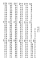
- cowpox DNA carried by the plasmid pEA36 and located between the Hpal sites was entirely sequenced according to the dideoxynucleotide method (Sanger et al. 1980) after insertion of the smallest fragments in the phages M13-130 and M13-131 (Kieny et al. 1983) following the strategy shown in Figure 5.
- the reading frame capable of coding for a protein of 77,000 daltons begins at nucleotides ATG and ends at nucleotides TAA represented on the map of FIG. 5.
- the arrows below the map indicate the place where reading of each clone begins , the length of the reading and its direction.
- Example 6 Homologous recombination and integration of the cowpox gene into vaccinia DNA, apart from the TK gene.
- the recombination introducing the information which allows multiplication in CHO cells has therefore occurred in a region different from that of the gene which codes for thymidine kinase.
- the E. coli strain 1106 carrying the plasmid pEA36 was deposited at the National Microorganism Cultures Collection under No. I 594, 2 September 1986.
- the plasmid pEA36 carries a DNA fragment of 2.004 bp of the Cowpox virus which allows the multiplication of the virus in CHO cells; this DNA fragment is intended for in vivo recombination with the vaccinia virus (which does not multiply in CHO cells).
- transfection of the cells with the plasmid DNA and co-infection with the vaccinia virus it will be possible to select recombinant vaccinia viruses which have acquired the capacity to multiply on CHO cells.
- Drillien, R., Spehner, D. & Kirn Drillien, R., Spehner, D. & Kirn, A. Virology 119, 372-381 (1982). Drillien, R. & Spehner, D. Virology 131, 385-393 (1983). Kieny, MP, Lathe, R. & Lecocq, JP. Gene 26, 91-99 (1983). Kieny, MP, Lathe, R., Drillien, R., Spehner, D., Skory, S., Schmitt, D., Wiktor, T., Koprowski, H. & Lecocq. JP.
- the four upper horizontal lines represent the restriction maps of vaccinia virus (VV) and cowpox (CP) obtained after cleavage with the enzymes HindIII and XhoI.
- the next ten horizontal lines represent the genomes of the recombinants isolated according to Example 1 between the VV and the CP.
- the restriction sites marked with an arrow are characteristic sites of the cowpox genome; those marked with a vertical line are characteristic of the genome of the vaccinia virus.
- the sites common to the two genomes are not noted.
- the dotted lines indicate regions of uncertainty in the determination.
- FIG. 2 Restriction maps of the left end of the vaccinia virus (VV) and cowpox (CP) after cleavage with the enzymes EcoRI, HindIII and Sal1.
- fragments are conventionally noted A, B, C ..., in decreasing order of size.
- B the DNA treated in the same way as in A was transferred to nitrocellulose and hybridized to a Sal1-K probe carried by the plasmid pAT153.
- Figure 4 Recombinant plasmids containing portions of the EcoRIA fragment of the plasmid pEA1 integrated into the vector pTG186poly.
- the two upper horizontal lines represent the left end of the vaccinia virus and cowpox DNA with the restriction sites for the enzyme EcoRI.
- the third line is a linear representation of the plasmid pEA1 with the EcoRI-A fragment in solid line and the plasmid pAT153 in broken line.
- the following lines are linear representations of the various recombinant plasmids with the fragment derived from pEA1 in solid line and the vector pTG186poly in broken lines; the promoter of the 7.5 K gene is symbolized by an open arrow which gives the direction of transciption.
- Figure 5 Gene sequencing strategy allowing multiplication in CHO cells.
- the horizontal line in bold lines represents the DNA fragment sequenced and included between the HpaI (H1) sites.
- the other restriction sites are symbolized by the following letters S (Sau3A), H2 (Hpa2), X (XhoI), C (ClaI), Xb (XbaI).
- S Sud3A
- H2 Hpa2
- X XhoI
- C CaI
- XbaI Xb
Abstract
Description
Le virus de la vaccine est de plus en plus employé comme vecteur d'expression dans des cellules animales depuis la mise au point des méthodes propres à ce système (Panicali et Paoletti, 1982 ; Mackett et al., 1982 ; Smith et al., 1983 ; Panicali et al. ; Kieny et al., 1984). La construction de virus recombinants du virus de la vaccine contenant des gènes qui codent pour des protéines d'intérêt médical ou vétérinaire est particulièrement recherchée. La synthèse de la protéine étrangère dont le gène a été intégré dans le génome du virus de la vaccine peut ensuite être obtenue en culture cellulaire in vitro ou après inoculation à un organisme vivant, selon le but poursuivi. Un des avantages du virus de la vaccine et tant que vecteur est sa capacité de se multiplier dans un grand nombre de types cellulaires différents.The vaccinia virus has been increasingly used as an expression vector in animal cells since the development of methods specific to this system (Panicali and Paoletti, 1982; Mackett et al., 1982; Smith et al., 1983; Panicali et al.; Kieny et al., 1984). The construction of recombinant vaccinia virus viruses containing genes which code for proteins of medical or veterinary interest is particularly sought after. The synthesis of the foreign protein whose gene has been integrated into the genome of the vaccinia virus can then be obtained in cell culture in vitro or after inoculation in a living organism, depending on the aim pursued. One of the advantages of the vaccinia virus and as a vector is its ability to multiply in a large number of different cell types.
Cependant quelques exceptions à cette règle existent et, en particulier, le virus de la vaccine de type sauvage est incapable de se multiplier dans une lignée de cellules d'ovaires de hamster chinois, CHO (Drillien, Spehner et Kirn, 1978). Or les cellules CHO constituent un des systèmes les plus prometteurs pour la synthèse de protéines dans des cellules de mammifères. En effet ces cellules se cultivent facilement, elles ont un temps de génération court et leur génétique est la mieux maîtrisée de tous les systèmes analogues.However, some exceptions to this rule exist and, in particular, the wild-type vaccinia virus is unable to multiply in a Chinese hamster ovary cell line, CHO (Drillien, Spehner and Kirn, 1978). One of the most promising systems for protein synthesis in mammalian cells is CHO cells. Indeed these cells are easily cultivated, they have a short generation time and their genetics is the best mastered of all analogous systems.
La présente invention concerne la modification du virus de la vaccine par l'intégration dans son génome d'un gène étranger qui lui confère la capacité de se multiplier dans les cellules CHO. Le gène apportant cette nouvelle spécificité d'hôte est dérivé du virus du cowpox (ou variole bovine, virus de la vache apparenté au virus de la vaccine) qui est capable de se multiplier dans les cellules CHO. Les génomes du cowpox et du virus de la vaccine sont très proches particulièrement dans les 100.000 paires de bases de la partie centrale de l'ADN (Mackett et Archard, 1979). Cependant le génome du cowpox est plus grand que celui de la vaccine (environ 230.000 pb au lieu de 190.000) et l'information génétique supplémentaire qu'il contient semble résider essentiellement dans ses extrémités. La similitude entre le génome du virus de la vaccine et celui du cowpox permet d'envisager d'employer le cowpox comme vecteur naturellement adapté aux cellules CHO.The present invention relates to the modification of the vaccinia virus by the integration into its genome of a foreign gene which confers on it the capacity to multiply in CHO cells. The gene providing this new host specificity is derived from the cowpox virus (or bovine smallpox, cow virus related to the vaccinia virus) which is capable of multiplying in CHO cells. The genomes of cowpox and vaccinia virus are very close, particularly in the 100,000 base pairs in the central part of DNA (Mackett and Archard, 1979). However, the cowpox genome is larger than that of vaccinia (around 230,000 bp instead of 190,000) and the additional genetic information it contains seems to reside essentially in its extremities. The similarity between the genome of the vaccinia virus and that of the cowpox makes it possible to envisage using cowpox as a vector naturally adapted to CHO cells.
Toutefois, le cowpox se multiplie à un titre dix fois inférieur à celui de la vaccine ce qui risque d'entraîner un rendement plus faible de l'expression d'une protéine produite par un recombinant cowpox en comparaison avec un recombinant vaccine.
D'autre part dans la perspective de l'utilisation de recombinants viraux pour la vaccination, il faut souligner que l'emploi du virus de la vaccine est bien maîtrisé et a permis à ce jour l'éradication complète la variole.
La présente invention concerne la mise au point d'un vecteur présentant les avantages connus du virus de la vaccine et la capacité de se multiplier sur cellules CHO du virus du cowpox.However, cowpox multiplies at a rate ten times lower than that of vaccinia which risks leading to a lower yield of the expression of a protein produced by a recombinant cowpox in comparison with a recombinant vaccinia.
On the other hand, in view of the use of viral recombinants for vaccination, it should be emphasized that the use of the vaccinia virus is well under control and has enabled to date the complete eradication of smallpox.
The present invention relates to the development of a vector having the known advantages of the vaccinia virus and the capacity to multiply on CHO cells of the cowpox virus.
La présente invention concerne, d'abord, l'identification et la localisation de l'information génétique qui confère au virus du cowpox la capacité de se multiplier dans les cellules CHO.The present invention relates, first, to the identification and localization of the genetic information which gives the cowpox virus the ability to multiply in CHO cells.
En effet, les études menées ont permis d'identifier une séquence impliquée dans la multiplication du virus cowpox dans les cellules CHO et qui, transférée dans le virus de la vaccine, assure la multiplication de ce virus dans les cellules CHO.In fact, the studies carried out have made it possible to identify a sequence involved in the multiplication of the cowpox virus in CHO cells and which, transferred into the vaccinia virus, ensures the multiplication of this virus in CHO cells.
Ainsi, l'invention concerne une séquence d'ADN isolée notamment du virus du cowpox et qui participe à la multiplication de ce virus dans les cellules CHO et qui comprend la totalité ou une partie fonctionnelle de la séquence représentée dans la figure 6 ou une séquence équivalente fonctionnelle.Thus, the invention relates to a DNA sequence isolated in particular from the cowpox virus and which participates in the multiplication of this virus in CHO cells and which comprises all or a functional part of the sequence shown in FIG. 6 or a sequence functional equivalent.
Il est possible qu'une partie de ce gène soit suffisante pour assurer la fonction de multiplication, on l'appelle "partie fonctionnelle". Il est également possible que des mutations ou des variations ponctuelles ne modifient pas la fonction, on parle alors de "séquence équivalente fonctionnelle".It is possible that a part of this gene is sufficient to ensure the multiplication function, it is called "functional part". It is also possible that mutations or point variations do not modify the function, we then speak of "functional equivalent sequence".
Bien que l'on préfère que cette séquence d'ADN comporte ses propres signaux de contrôle qui assurent son expression dans les cellules CHO, ceci n'est pas indispensable et l'on peut envisager de la mettre sous le contrôle d'éléments ayant une autre origine.Although it is preferred that this DNA sequence has its own control signals which ensure its expression in CHO cells, this is not essential and one can consider putting it under the control of elements having a other origin.
Comme cela est décrit précédemment, cette séquence d'ADN est plus particulièrement destinée à être intégrée dans le virus de la vaccine pour assurer sa multiplication dans les cellules CHO. Cette intégration s'effectue par recombinaison homologue, il est donc intéressant de prévoir que la séquence d'ADN en cause comportera au moins une région homologue d'une séquence du virus de la vaccine qui pourra participer à ce processus de recombinaison homologue au cours de la multiplication intracellulaire des virus.As described above, this DNA sequence is more particularly intended to be integrated into the vaccinia virus to ensure its multiplication in CHO cells. This integration is carried out by homologous recombination, it is therefore advantageous to provide that the DNA sequence in question will comprise at least one region homologous to a sequence of the vaccinia virus which will be able to participate in this process of homologous recombination during intracellular multiplication of viruses.
En fait, il apparaît que chez le virus cowpox le gène permettant la multiplication dans les cellules CHO est entouré de séquences homologues à des séquences du génome de la vaccine, ce qui permet de simplifier la préparation d'un vecteur plasmidique de recombinaison.In fact, it appears that in the cowpox virus the gene allowing multiplication in CHO cells is surrounded by sequences homologous to sequences of the vaccinia genome, which makes it possible to simplify the preparation of a recombinant plasmid vector.
Comme cela est déjà connu pour le virus de la vaccine, il est possible d'insérer dans la séquence d'ADN, objet de l'invention, un gène codant pour une protéine d'intérêt industriel sous la dépendance d'éléments de contrôle assurant son expression dans les cellules hôtes. Cette technologie a déjà été décrite, notamment dans les brevets suivants : 84.06499, 84.07959, 85.09225 et peut être utilisée avec éventuellement certaines adaptations et avantages. En particulier, dans les contructions précédentes, la sélection des virus recombinants était effectuée en insérant le gène à exprimer dans le gène TK de la vaccine, ce qui a pour conséquence de rendre le virus recombinant TK⁻ et de permettre la sélection par le procédé connu.As is already known for the vaccinia virus, it is possible to insert into the DNA sequence, object of the invention, a gene coding for a protein of industrial interest under the dependence of control elements ensuring its expression in host cells. This technology has already been described, in particular in the following patents: 84.06499, 84.07959, 85.09225 and can be used with possibly certain adaptations and advantages. In particular, in the previous constructions, the selection of the recombinant viruses was carried out by inserting the gene to be expressed in the TK gene of vaccinia, which has the consequence of rendering the TK virus recombinant virus and of allowing selection by the known method .
Dans le cas présent, il n'est pas indispensable d'effectuer une insertion dans le gène TK car, dans la mesure où le gène codant pour la protéine d'intérêt industriel est lié au gène assurant la multiplication dans les cellules CHO, seuls les virus recombinants peuvent se multiplier sur cellules CHO, ce qui autorise une sélection "naturelle" des virus recombinants.In the present case, it is not essential to carry out an insertion into the TK gene because, since the gene coding for the protein of industrial interest is linked to the gene ensuring multiplication in CHO cells, only the Recombinant viruses can multiply on CHO cells, which allows "natural" selection of recombinant viruses.
Dans ce dernier cas, il est préférable que le bloc d'ADN comprenant les gènes à recombiner soit flanqué de séquences homologues de séquences du virus de la vaccine afin qu'ils restent liés lors de la recombinaison.In the latter case, it is preferable that the DNA block comprising the genes to be recombined is flanked by sequences homologous to vaccinia virus sequences so that they remain linked during the recombination.
La présente invention concerne également les cellules CHO infectées par un virus de la vaccine recombinant incorporant une séquence d'ADN telle que décrite précédemment et, en particulier, un gène codant pour une protéine d'intérêt industriel, ainsi que les virus correspondants.The present invention also relates to CHO cells infected with a recombinant vaccinia virus incorporating a DNA sequence as described above and, in particular, a gene coding for a protein of industrial interest, as well as the corresponding viruses.
L'invention concerne également les vecteurs plasmidiques incorporant une séquence d'ADN telle que décrite précédemment, ces vecteurs étant utilisables pour effectuer la recombinaison in vivo.The invention also relates to the plasmid vectors incorporating a DNA sequence as described above, these vectors being usable for carrying out the recombination in vivo.
L'invention concerne également des cellules CHO ayant intégré une séquence d'ADN selon la présente invention et capables d'assurer la multiplication du virus de la vaccine dans ces cellules.The invention also relates to CHO cells having integrated a DNA sequence according to the present invention and capable of ensuring the multiplication of the vaccinia virus in these cells.
Enfin, l'invention concerne la préparation de protéines d'intérêt industriel par culture de cellules CHO infectées par un virus recombinant selon l'invention.Finally, the invention relates to the preparation of proteins of industrial interest by culture of CHO cells infected with a recombinant virus according to the invention.
Les exemples ci-après sont destinés à illustrer d'autres caractéristiques et avantages de la présente invention.The examples below are intended to illustrate other characteristics and advantages of the present invention.
Des recombinants entre le virus de la vaccine et le cowpox ont été sélectionnés après infection mixte de cellules d'embryons de poulet avec chacun des virus. L' analyse de l'ADN des recombinants montre que la capacité de se multiplier dans des cellules CHO est associée à la conservation des sites de restriction de l'extrémité gauche du génome du cowpox.Recombinants between the vaccinia virus and the cowpox were selected after mixed infection of chicken embryo cells with each of the viruses. Analysis of the DNA of the recombinants shows that the capacity to multiply in CHO cells is associated with the conservation of the restriction sites of the left end of the cowpox genome.
Des cellules primaires d'embryon de poulet, préparées à partir d'oeufs embryonnés de 11 à 12 jours, sont infectées simultanément avec un mutant thermosensible du virus vaccinal, tsN7 (Drillien et al., 1982) et le cowpox (souche Brighton) à raison de 2 unités formant plage (ufp) par cellule. Parallèlement d'autres tapis cellulaires sont infectés avec chacun de ces virus. Après une heure d'adsorption l'excés de virus non adsorbé est éliminé et du milieu frais est ajouté aux cellules.
Celles-ci sont incubées à 33°C pendant un à deux jours jusqu'à la nécrose totale de la couche cellulaire. Les cellules infectées sont alors congelées puis décongelées et le virus issu de l'infection est titré à 39,5°C sur cellules d'embryon de poulet, sous une couche de milieu contenant 1 % d'agar noble. Après deux jours à 39,5°C un plus grand nombre de plages de virus s'est formé sur les tapis cellulaires infectés avec le mélange des 2 virus que sur les tapis témoins (ni le mutant thermosensible du virus de la vaccine ni le cowpox ne donnent un nombre significatif de plages) ; les plages qui apparaissent à partir de l'infection mixte peuvent donc correspondre à des recombinants entre le cowpox et le virus de la vaccine.Primary chicken embryo cells, prepared from 11 to 12 day old embryonated eggs, are simultaneously infected with a thermosensitive vaccine virus mutant, tsN7 (Drillien et al., 1982) and cowpox (Brighton strain) at due to 2 range forming units (pfu) per cell. At the same time, other cell mats are infected with each of these viruses. After one hour of adsorption, the excess of non-adsorbed virus is eliminated and fresh medium is added to the cells.
These are incubated at 33 ° C for one to two days until complete necrosis of the cell layer. The infected cells are then frozen and then thawed and the virus resulting from the infection is titrated at 39.5 ° C. on chicken embryo cells, under a layer of medium containing 1% of noble agar. After two days at 39.5 ° C., a greater number of plaques of virus formed on the cell mats infected with the mixture of the 2 viruses than on the control mats (neither the heat-sensitive mutant of the vaccinia virus nor the cowpox do not give a significant number of ranges); the ranges which appear from the mixed infection can therefore correspond to recombinants between the cowpox and the vaccinia virus.
Les plages de recombinants potentiels sont reprises individuellement et le virus qu'elles contiennent est amplifié par multiplication sur cellules d'embryon de poulet. Leur ADN est ensuite purifié, coupé par des enzymes de restriction puis analysé sur gel d'agarose.The ranges of potential recombinants are taken up individually and the virus which they contain is amplified by multiplication on chicken embryo cells. Their DNA is then purified, cut by restriction enzymes and then analyzed on agarose gel.
Les profils de restriction permettent de déduire que chaque plage correspond effectivement à un recombinant entre l'ADN du virus de la vaccine et l'ADN du cowpox. Grâce aux cartes de restriction connues pour les virus parentaux (Mackett and Archard 1978, Drillien and Spehner 1983) il est possible de reconnaître l'origine de la plupart des fragments des recombinants et de dresser leurs cartes de restriction (figure 1).The restriction profiles make it possible to deduce that each range effectively corresponds to a recombinant between the DNA of the vaccinia virus and the DNA of the cowpox. Using known restriction maps for parental viruses (Mackett and Archard 1978, Drillien and Spehner 1983) it is possible to recognize the origin of most fragments of the recombinants and to draw up their restriction maps (Figure 1).
On voit que les recombinants dénommés 4, 6, 14, 15 et 19 qui sont capables de se multiplier dans des cellules CHO ont conservé les sites caractéristiques de l'extrémité gauche du génome du cowpox. Les autres recombinants dénommés 2, 7, 11, 16 et 18 qui sont incapables de se multiplier dans des cellules CHO ne possèdent que partiellement ou pas du tout ces sites de l'extrémité gauche du génome du cowpox.We see that the recombinants named 4, 6, 14, 15 and 19 which are capable of multiplying in CHO cells have conserved the characteristic sites of the left end of the cowpox genome. The other recombinants called 2, 7, 11, 16 and 18 which are unable to multiply in CHO cells only partially or not at all have these sites at the left end of the cowpox genome.
Il ressort de ces résultats que la conservation des sites de restriction de l'extrémité gauche du génome du cowpox est associée au phénotype de multiplication sur cellules CHO.It appears from these results that the conservation of the restriction sites of the left end of the cowpox genome is associated with the multiplication phenotype on CHO cells.
Pour mieux préciser la localisation de l'information génétique utile, des recombinants capables de se multiplier sur cellules CHO ont été sélectionnés après infection avec le virus de la vaccine et transfection avec des fragments d'ADN du cowpox.To better specify the location of useful genetic information, recombinants capable of multiplying on CHO cells were selected after infection with the vaccinia virus and transfection with fragments of cowpox DNA.
Les fragments de restriction utiles à l'analyse de la partie importante, c'est-à-dire l'extrémité gauche des 2 virus sont représentés dans la figure 2 ; c'est dans cette partie du génome que l'on peut soupçonner qu'interviennent les événements de recombinaison décrits dans l'exemple 1.The restriction fragments useful for the analysis of the important part, that is to say the left end of the 2 viruses are represented in FIG. 2; it is in this part of the genome that it can be suspected that the recombination events described in example 1 occur.
Des cellules primaires d'embryon de poulet sont infectées avec le mutant tsN7 du virus de la vaccine (Drillien et al., 1982) à raison de 0,1 ufp par cellule et transfectées avec un mélange d'ADN intact de la souche sauvage du virus de la vaccine (souche Copenhagen) et de l'ADN du cowpox (souche Brighton) préalablement digéré avec l'enzyme HindIII. Des témoins de transfection sand ADN ou avec seulement l'ADN du virus de la vaccine sont réalisés.
Après 48 heures d'incubation à 39.5°C les cellules sont congelées, décongelées puis le virus ainsi libéré est titré sur une m onocouche de cellules CHO qui sont ensuite recouvertes de milieu contenant 1 % d'agar.Primary chicken embryo cells are infected with the vaccinia virus tsN7 mutant (Drillien et al., 1982) at the rate of 0.1 pfu per cell and transfected with a mixture of intact DNA from the wild-type strain of the vaccinia virus (Copenhagen strain) and cowpox DNA (Brighton strain) previously digested with the enzyme HindIII. Witnesses of transfection without DNA or with only the DNA of the vaccinia virus are carried out.
After 48 hours of incubation at 39.5 ° C, the cells are frozen, thawed and the virus thus released is titrated on a monolayer of CHO cells which are then covered with medium containing 1% agar.
Les échantillons provenant de cellules transfectées avec l'ADN du cowpox donnent de nombreuses plages de lyse sur cellules CHO tandis que les échantillons témoins n'en donnent aucune.The samples from cells transfected with cowpox DNA give numerous lysis ranges on CHO cells while the control samples give none.
Les plages visibles sur cellules CHO sont reprises individuellement et le virus qu'elles contiennent est amplifié sur cellules d'embryon de poulet. Leur ADN est ensuite extrait et analysé en comparaison avec l'ADN des 2 souches parentales de vaccine et de cowpox. Après digestion par l'enzyme EcoRI, les fragments d'ADN sont séparés par électrophorèse sur un gel d'agarose puis ils sont transférés à un filtre de nitrocellulose et hybridés au fragment SalI-K du virus de la vaccine marqué radioactivement au ³²P.
Après lavage de la nitrocellulose pour enlever la radioactivité fixée de manière aspécifique une autoradiographie est réalisée. L'autoradiographie (figure 3) montre que les recombinants du virus de la vaccine ayant intégré un fragment du cowpox ont perdu le fragment EcoRI-C typique du virus de la vaccine et comportent un fragment EcoRI qui s'hybride avec le fragment radioactif SalI-K de la vaccine ; ce fragment est intermédiaire en taille entre le fragment EcoRI-A du cowpox et le fragment EcoRI-C du virus de la vaccine.The plaques visible on CHO cells are taken up individually and the virus which they contain is amplified on chicken embryo cells. Their DNA is then extracted and analyzed in comparison with the DNA of the two parental strains of vaccinia and cowpox. After digestion with the EcoRI enzyme, the DNA fragments are separated by electrophoresis on an agarose gel and then they are transferred to a nitrocellulose filter and hybridized to the SalI-K fragment of the vaccinia virus radioactively labeled with ³²P.
After washing the nitrocellulose to remove the radioactivity fixed in an aspecific manner, an autoradiography is carried out. Autoradiography (FIG. 3) shows that the vaccinia virus recombinants having integrated a cowpox fragment have lost the EcoRI-C fragment typical of the vaccinia virus and comprise an EcoRI fragment which hybridizes with the radioactive fragment SalI- K of vaccinia; this fragment is intermediate in size between the EcoRI-A fragment of the cowpox and the EcoRI-C fragment of the vaccinia virus.
Ce nouveau fragment EcoRI hybride cowpox-vaccine, présent dans tous les recombinants, provient d'une double recombinaison entre le fragment EcoRI-A du cowpox et le fragment EcoRI-C de la vaccine et doit renfermer l'information nécessaire pour la multiplication dans des cellules CHO. Pour que cette recombinaison ait pu se produire il fallait que l'information permettant la multiplication sur cellules CHO soit entourée de part et d'autre par des séquences du génome du cowpox homologues à des séquences du génome vaccine.This new hybrid EcoRI cowpox-vaccine fragment, present in all the recombinants, comes from a double recombination between the EcoRI-A fragment of cowpox and the EcoRI-C fragment of vaccinia and must contain the information necessary for multiplication in CHO cells. In order for this recombination to occur, the information allowing multiplication on CHO cells had to be surrounded on both sides by sequences of the cowpox genome homologous to sequences of the vaccinia genome.
Afin d'isoler l'information génétique permettant la multiplication dans des cellules CHO le fragment EcoRI-A de l'un des recombinants décrits dans l'exemple 2 a été cloné dans le plasmide bactérien pAT153 (Twigg et Sherratt, 1980).In order to isolate the genetic information allowing multiplication in CHO cells, the EcoRI-A fragment of one of the recombinants described in example 2 was cloned into the bacterial plasmid pAT153 (Twigg and Sherratt, 1980).
L'ADN d'un des recombinants décrits dans l'exemple 2 est purifié puis coupé avec l'enzyme EcoRI. Le fragment EcoRI-A est élué d'un gel d'agarose puis inséré dans le plasmide pAT153 préalablement soumis à l'action d'EcoRI. Des bactéries HB101 sont transformées avec le mélange de ligation puis l'ADN des colonies obtenues est transféré sur nitrocellulose et hybridé au fragment SalI-K du virus de la vaccine. Les colonies positives à l'hybridation sont amplifiées et l'ADN plasmidique qu'elles contiennent est purifié. Deux plasmides ont été retenus : pEA1 et pEA2, qui correspondent à l'insertion du fragment EcoRI-A dans les deux orientations opposées dans le vecteur pAT153.The DNA of one of the recombinants described in Example 2 is purified and then cut with the EcoRI enzyme. The EcoRI-A fragment is eluted from an agarose gel and then inserted into the plasmid pAT153 previously subjected to the action of EcoRI. HB101 bacteria are transformed with the ligation mixture then the DNA of the colonies obtained is transferred onto nitrocellulose and hybridized to the SalI-K fragment of the vaccinia virus. Colonies positive to hybridization are amplified and the plasmid DNA they contain is purified. Two plasmids were selected: pEA1 and pEA2, which correspond to the insertion of the EcoRI-A fragment in the two opposite orientations in the vector pAT153.
Pour vérifier que ces plasmides portent l'information génétique permettant la multiplication du virus sur cellules CHO on provoque une recombinaison entre l'insert d'ADN du plasmide et un virus de la vaccine : des cellules d'embryons de poulets sont infectées avec le mutant thermosensible du virus de la vaccine tsN7 à raison de 0,1 ufp par cellule puis transfectées avec l'ADN du virus de la vaccine sauvage et avec l'ADN du plasmide pEA1 ou pEA2. Des témoins sans ADN du virus de la vaccine ou sans plasmide sont également réalisés. Après 48 heures d'incubation à 39,5°C, les cellules sont congelées puis décongelées et le virus issu de l'infection est titré sur cellules CHO. Seuls les échantillons provenant de cellules transfectées avec l'ADN du virus de la vaccine et les plasmides pEA1 ou pEA2 donnent des plages sur cellules CHO.To verify that these plasmids carry the genetic information allowing the multiplication of the virus on CHO cells, a recombination between the DNA insert of the plasmid and a vaccinia virus is provoked: chicken embryo cells are infected with the mutant thermosensitive of vaccinia virus tsN7 at a rate of 0.1 pfu per cell then transfected with the DNA of wild vaccinia virus and with the DNA of the plasmid pEA1 or pEA2. Witnesses without vaccinia virus DNA or without plasmid are also produced. After 48 hours of incubation at 39.5 ° C, the cells are frozen then thawed and the virus resulting from the infection is titrated on CHO cells. Only samples from cells transfected with vaccinia virus DNA and plasmids pEA1 or pEA2 give plaques on CHO cells.
Pour affiner la localisati on de l'information génétique permettant la multiplication dans des cellules CHO, des fragments de restriction recouvrant des portions réduites du plasmide pEA1 ont été clonés dans un plasmide portant le gène thymidine kinase (TK) du virus de la vaccine : pTG186poly.To refine the localization of the genetic information allowing multiplication in CHO cells, restriction fragments covering reduced portions of the plasmid pEA1 were cloned into a plasmid carrying the thymidine kinase (TK) gene of the vaccinia virus: pTG186poly .
Le fragment HindIII (Hin-J) du génome du virus de la vaccine (VV) contient le gène complet de la thymidine kinase (TK) qui a déjà été utilisé précédemment pour permettre l'échange et la recombinaison d'un fragment d'ADN étranger dans le génome de VV (Mackett et al., 1982).The HindIII (Hin-J) fragment of the vaccinia virus (VV) genome contains the complete thymidine kinase (TK) gene which has already been used previously to allow the exchange and recombination of a DNA fragment. foreign in the VV genome (Mackett et al., 1982).
Il est important de noter que le transfert d'un insert dans le gène TK du génome de VV crée un virus TK déficient ce qui facilite sa sélection.It is important to note that the transfer of an insert into the TK gene of the VV genome creates a deficient TK virus which facilitates its selection.
Il a tout d'abord été nécessaire de produire un plasmide de petite taille portant un site unique HindIII utilisable pour l'intégration du fragment Hin-J de VV. En outre, il était nécessaire d'éliminer les sites de restriction non nécessaires du plasmide, de façon à permettre les manipulations ultérieures.It was first necessary to produce a small plasmid carrying a unique HindIII site usable for the integration of the Hin-J fragment of VV. In addition, it was necessary to remove unnecessary restriction sites from the plasmid, so as to allow subsequent manipulations.
La construction a été amorcée à partir du plasmide pML2 (Lusky et Botchan, 1981) qui est un vecteur dérivé du plasmide pBR322 dans lequel le segment compris entre les nucléotides 1089 et 2491 a été perdu par délétion spontanée. D'abord la séquence de PstI a été éliminée par insertion du fragment AhaIII-AhaIII de pUC8 (Vieira et Messing, 1982) entre deux sites AhaIII de pML2 en éliminant 19 paires de bases.
On a utilisé la méthode du "linker-tailing" (Lathe et al., 1984) pour insérer un adaptateur HindIII entre les sites NruI et EcoRI traité par S1 de ce plasmide, en éliminant le site BamHI. Ceci conduit à un plasmide de 2049 paires de bases portant le gène β-lactamase fonctionnel (conférant la résistance à l'ampicilline) et comportant en outre une origine de réplication active dans E. coli et un site de restriction unique HindIII. Cette construction a été appelée pTG1H.Construction was started from the plasmid pML2 (Lusky and Botchan, 1981) which is a vector derived from the plasmid pBR322 in which the segment between nucleotides 1089 and 2491 was lost by spontaneous deletion. First, the PstI sequence was eliminated by insertion of the AhaIII-AhaIII fragment from pUC8 (Vieira and Messing, 1982) between two AhaIII sites of pML2 by eliminating 19 base pairs.
The "linker-tailing" method was used (Lathe et al., 1984) to insert a HindIII adapter between the NruI and EcoRI sites treated with S1 of this plasmid, by eliminating the BamHI site. This leads to a plasmid of 2049 base pairs carrying the functional β-lactamase gene (conferring resistance to ampicillin) and further comprising an origin of active replication in E. coli and a unique HindIII restriction site. This construction was called pTG1H.
Le fragment Hin-J de l'ADN de VV portant le gène TK a préalablement été cloné dans le vecteur pAT153 (Drillien et Spehner, 1983). Ce fragment de 4,6 kb a été recloné dans le site HIndIII de pTG1H. Un clone a été sélectionné dans lequel le gène TK est situé distalement par rapport au gène codant pour la résistance à l'ampicilline. Cette construction a été appelée pTG1H-TK.The Hin-J fragment of VV DNA carrying the TK gene was previously cloned into the vector pAT153 (Drillien and Spehner, 1983). This 4.6 kb fragment was recloned into the HIndIII site of pTG1H. A clone was selected in which the TK gene is located distally from the gene coding for resistance to ampicillin. This construct was called pTG1H-TK.
La construction pTG1H-TK a été utilisée comme vecteur pour les constructions suivantes.The pTG1H-TK construct was used as a vector for the following constructs.
L'étape suivante a été d'isoler un promoteur de VV utilisable pour commander l'expression du gène étranger à intégrer dans VV. Le promoteur d'un gène précoce codant pour une protéine de 7500 daltons (7,5 K) a déjà été utilisé avec succès dans un but identique (Smith et al., 1983) et on a donc procédé à l'isolement de ce segment.The next step was to isolate a VV promoter which can be used to control the expression of the foreign gene to be integrated into VV. The promoter of an early gene coding for a protein of 7500 daltons (7.5 K) has already been successfully used for an identical purpose (Smith et al., 1983) and this segment has therefore been isolated. .
Le gène 7,5 K est situé sur l'un des plus petits fragments SalI (fragment Sal-S) du génome de VV type WR (Venkatasan et al., 1981). Comme les petits fragments sont clonés de façon préférentielle, une grande proportion des clones obtenus par clonage direct de l'ADN de VV type WR coupé par SalI dans le plasmide pBR322 porte le fragment Sal-S. Ce fragment est transféré sur le bactériophage vecteur M13mp701 (Kieny et al., 1983), par digestion SalI et religation, ce qui donne le phage M13.TG.Sal-S.The 7.5 K gene is located on one of the smallest SalI fragments (Sal-S fragment) of the VV genome WR type (Venkatasan et al., 1981). As the small fragments are preferentially cloned, a large proportion of the clones obtained by direct cloning of the VV DNA type WR cut by SalI in the plasmid pBR322 carries the Sal-S fragment. This fragment is transferred to the bacteriophage vector M13mp701 (Kieny et al., 1983), by SalI digestion and religation, which gives the phage M13.TG.Sal-S.
Dans ce clone, un site ScaI se trouve immédiatement à proximité de l'ATG d'initiation du gène 7,5 K. En aval du gène 7,5 K se trouvent situés des sites uniques BamHI et EcoRI provenant du vecteur. Les sites BamHI et ScaI sont fusionnés par l'intermédiaire d'un adaptateur BglII : 5ʹ-CAGATCTG-3ʹ après avoir complété les extrémités générées par digestion BamHI avec le fragment Klenow de la polymérase de E. coli. Ce procédé élimine le site ScaI mais reconstitue le site BamHI et déplace le site unique EcoRI en eval. En même temps, le site SalI (AccI) en aval est éliminé et le site SalI en amont devient donc unique. Cette construction est appelée M13.TG.7,5K.In this clone, a ScaI site is located immediately near the 7.5 K gene initiation ATG. Downstream of the 7.5 K gene are located unique BamHI and EcoRI sites originating from the vector. The BamHI and ScaI sites are fused via a BglII adapter: 5ʹ-CAGATCTG-3ʹ after having completed the ends generated by BamHI digestion with the Klenow fragment of the E. coli polymerase. This process eliminates the ScaI site but reconstitutes the BamHI site and moves the unique EcoRI site to eval. At the same time, the downstream SalI (AccI) site is eliminated and the upstream SalI site therefore becomes unique. This construction is called M13.TG.7,5K.
A l'intérieur du fr agment Hin-J de l'ADN de VV se trouvent situés des sites ClaI et EcoRI qui sont séparés par environ 30 paires de bases (Weir et Moss, 1983). Le fragment promoteur de 7,5 K présent dans M13.TG.7,5K est excisé par AccI et EcoRI et cloné entre les sites ClaI et EcoRI de pTG1-H-TK pour générer pTG1H-TK-P7,5K.Within the Hin-J fragment of VV DNA are located ClaI and EcoRI sites which are separated by about 30 base pairs (Weir and Moss, 1983). The 7.5 K promoter fragment present in M13.TG.7.5K is excised by AccI and EcoRI and cloned between the ClaI and EcoRI sites of pTG1-H-TK to generate pTG1H-TK-P7.5K.
Dans cette construction les sites BamHI et EcoRI uniques du vecteur M13 se retrouvent immédiatement en aval de la séquence du promoteur 7,5 K. Ces sites uniques BamHI et EcoRI seront utilisés dans la construction suivante.In this construction, the unique BamHI and EcoRI sites of the M13 vector are found immediately downstream of the 7.5 K promoter sequence. These unique BamHI and EcoRI sites will be used in the following construction.
Le segment polylinker du bactériophage M13TG131 (Kieny et al., 1983) est excisé par EcoRI et BglII et inséré entre les sites EcoRI et BamHI du plasmide pTG1H-TK-P7,5K, générant le pTG186poly. Dans cette construction, 5 sites de restriction uniques sont disponibles pour le clonage d'un gène étranger sous le contrôle du promoteur P7,5K : PstI, BamHI, SstI, SmaI et EcoRI.The polylinker segment of the bacteriophage M13TG131 (Kieny et al., 1983) is excised by EcoRI and BglII and inserted between the EcoRI and BamHI sites of the plasmid pTG1H-TK-P7,5K, generating pTG186poly. In this construction, 5 unique restriction sites are available for cloning a foreign gene under the control of the P7.5K promoter: PstI, BamHI, SstI, SmaI and EcoRI.
Le fragment EcoRIA du plasmide pEA1 a été digéré par différents enzymes et des fragments de différentes tailles ont été insérés dans le pTG186poly.
Les plasmides recombinants dérivés de pTG186poly et contenant des portions du fragment EcoRI-A du plasmide recombinant pEA1 sont représentés dans la figure 4.The EcoRIA fragment of the plasmid pEA1 was digested with different enzymes and fragments of different sizes were inserted into the pTG186poly.
The recombinant plasmids derived from pTG186poly and containing portions of the EcoRI-A fragment of the recombinant plasmid pEA1 are represented in FIG. 4.
La coupure du plasmide pEA1 avec l'enzyme BglII donne plusieurs fragments. Le plus grand des fragments BglII a été inséré dans le site BamHI du vecteur pTG186poly pour donner les plasmides pEA5a et pEA5b qui se distinguent par l'orientation de l'insertion.The cleavage of the plasmid pEA1 with the enzyme BglII gives several fragments. The largest of the BglII fragments was inserted into the BamHI site of the vector pTG186poly to give the plasmids pEA5a and pEA5b which are distinguished by the orientation of the insertion.
Le plasmide pEA6 est dérivé du plasmide pEA5a par coupure de ce dernier avec l'enzyme PstI puis recircularisation du plus grand fragment ce qui a pour effet de déléter le petit fragment BglII-PstI du plasmide pEA5a.The plasmid pEA6 is derived from the plasmid pEA5a by cutting the latter with the enzyme PstI then recircularization of the largest fragment which has the effect of deleting the small fragment BglII-PstI of the plasmid pEA5a.
Le plasmide pEA7 provient de pEA6 après coupure de ce dernier avec l'enzyme ClaI et recircularisation du grand fragment obtenu ce qui donne une délétion du petit fragment ClaI.The plasmid pEA7 comes from pEA6 after cutting the latter with the enzyme ClaI and recircularization of the large fragment obtained which gives a deletion of the small fragment ClaI.
Le plasmide pEA8 provient de pEA6 après coupure de ce dernier par SphI puis recircularisation du grand fragment ce qui donne une délétion du fragment SphI-BglII de droite.The plasmid pEA8 comes from pEA6 after cutting the latter by SphI then recircularization of the large fragment which gives a deletion of the SphI-BglII fragment on the right.
Le plasmide pEA9 provient du plasmide pEA5b après coupure de ce dernier par SphI puis religation ce qui donne une délétion du fragment gauche SphI-BglIII.The plasmid pEA9 comes from the plasmid pEA5b after cutting the latter by SphI then religation which gives a deletion of the left fragment SphI-BglIII.
Le plasmide pEA36 a été construit en deux étapes. D'abord le petit fragment HpaI a été isolé du plasmide pEA9 et il a été inséré dans le site SmaI du vecteur M13-130. Puis le fragment HpaI a été sorti du vecteur M13-130 grâce aux sites EcoRI et PstI et il a été intégré dans les sites EcoRI et PstI du vecteur pTG186poly.The plasmid pEA36 was constructed in two stages. First the small HpaI fragment was isolated from the plasmid pEA9 and it was inserted into the SmaI site of the vector M13-130. Then the HpaI fragment was removed from the vector M13-130 thanks to the EcoRI and PstI sites and it was integrated into the EcoRI and PstI sites of the vector pTG186poly.
Chacun de ces plasmides a été utilisé dans une expérience de transfection de manière à transférer l'insertion qui se trouve dans le gène TK porté par le plasmide au génome du virus de la vaccine, selon la méthode décrite dans l'exemple 3.Each of these plasmids was used in a transfection experiment so as to transfer the insertion which is in the TK gene carried by the plasmid to the genome of the vaccinia virus, according to the method described in Example 3.
Les plasmides capables de conférer la capacité de multiplication dans des cellules CHO au virus de la vaccine sont pEA1, 2, 5a, 5b, 6, 9, 36.Plasmids capable of imparting the multiplication capacity in CHO cells to the vaccinia virus are pEA1, 2, 5a, 5b, 6, 9, 36.
Le plus petit fragment ayant cette propriété est porté par le plasmide pEA36. Il comprend 2004 paires de bases.The smallest fragment having this property is carried by the plasmid pEA36. It includes 2004 base pairs.
L'ADN du cowpox porté par le plasmide pEA36 et situé entre les sites Hpal a été entièrement séquencé selon la méthode des didéoxynucléotides (Sanger et al. 1980) après insertion des plus petits fragments dans les phages M13-130 et M13-131 (Kieny et al. 1983) en suivant la stratégie schématisée dans la figure 5.The cowpox DNA carried by the plasmid pEA36 and located between the Hpal sites was entirely sequenced according to the dideoxynucleotide method (Sanger et al. 1980) after insertion of the smallest fragments in the phages M13-130 and M13-131 (Kieny et al. 1983) following the strategy shown in Figure 5.
Le cadre de lecture pouvant coder pour une protéine de 77.000 daltons débute aux nucléotides ATG et se termine aux nucléotides TAA représentés sur la carte de la figure 5. Les flèches en-dessous de la carte indiquent l'endroit où commence la lecture de chaque clone, la longueur de la lecture et sa direction. Lorsque le début de lecture n e commence pas à un site de restriction, des amorces constituées d'oligonucléotides synthétiques déduits d'une séquence partielle ont été utilisées.The reading frame capable of coding for a protein of 77,000 daltons begins at nucleotides ATG and ends at nucleotides TAA represented on the map of FIG. 5. The arrows below the map indicate the place where reading of each clone begins , the length of the reading and its direction. When the start of reading does not start at a restriction site, primers consisting of synthetic oligonucleotides deduced from a partial sequence were used.
La séquence complète du gène ainsi que la séquence de la protéine de 77.000 daltons pour laquelle il peut coder est présentée dans la figure 6.The complete gene sequence as well as the 77,000 dalton protein sequence for which it can code is shown in Figure 6.
Parmi les virus recombinants, décrits dans l'exemple 4, capables de se multiplier sur cellules CHO, on constate que certains sont TK⁺ et d'autres TK⁻.Among the recombinant viruses, described in Example 4, capable of multiplying on CHO cells, it can be seen that some are TK⁺ and others are TK⁻.
Par exemple dans le transfection des cellules avec le plasmide pEA9, sur 27 plages de virus recombinants sélectionnées pour leur multiplication sur CHO, 16 étaient TK⁻ et 11 TK⁺.For example, in the transfection of the cells with the plasmid pEA9, out of 27 ranges of recombinant viruses selected for their multiplication on CHO, 16 were TK⁻ and 11 TK⁺.
La recombinaison introduisant l'information qui permet la multiplication dans des cellules CHO s'est donc produite dans une région différente de celle du gène qui code pour la thymidine kinase.The recombination introducing the information which allows multiplication in CHO cells has therefore occurred in a region different from that of the gene which codes for thymidine kinase.
Ce résultat implique la possibilité de sélection pour l'insertion d'un gène étranger dans une région du génome autre que le gène TK, simultanément à la sélection pour la multiplication sur cellules CHO. Il suffit pour cela de juxtaposer le gène choisi et le gène permettant la croissance sur cellules CHO. L'insertion de ce dernier par recombinaison homologue avec le génome du virus de la vaccine entrainera la co-intégration du gène étranger.This result implies the possibility of selection for the insertion of a foreign gene in a region of the genome other than the TK gene, simultaneously with the selection for multiplication on CHO cells. For this, it suffices to juxtapose the chosen gene and the gene allowing growth on CHO cells. The insertion of the latter by homologous recombination with the genome of the vaccinia virus will lead to the co-integration of the foreign gene.
La souche de E. coli 1106 portant le plasmide pEA36 a été déposée à la Collection Nationale de Cultures de Microorganismes sous le n o I 594, le 2 septembre 1986.The E. coli strain 1106 carrying the plasmid pEA36 was deposited at the National Microorganism Cultures Collection under No. I 594, 2 September 1986.
Le plasmide pEA36 porte un fragment d'ADN de 2.004 pb du virus du Cowpox qui permet la multiplication du virus dans les cellules CHO ; ce fragment d'ADN est destiné à la recombinaison in vivo avec le virus de la vaccine (qui ne se multiplie pas dans les cellules CHO). Par transfection des cellules avec l'ADN plasmidique et co-infection avec le virus de la vaccine, on pourra sélectionner des virus recombinants de la vaccine qui ont acquis la capacité de se multiplier sur cellules CHO.The plasmid pEA36 carries a DNA fragment of 2.004 bp of the Cowpox virus which allows the multiplication of the virus in CHO cells; this DNA fragment is intended for in vivo recombination with the vaccinia virus (which does not multiply in CHO cells). By transfection of the cells with the plasmid DNA and co-infection with the vaccinia virus, it will be possible to select recombinant vaccinia viruses which have acquired the capacity to multiply on CHO cells.
Drillien, R., Spehner, D. & Kirn, A. J. Virol. 28, 843-850 (1978).
Drillien, R., Spehner, D. & Kirn, A. Virology 119, 372-381 (1982).
Drillien, R. & Spehner, D. Virology 131, 385-393 (1983).
Kieny, M.P., Lathe, R. & Lecocq, J-P. Gene 26, 91-99 (1983).
Kieny, M.P., Lathe, R., Drillien, R., Spehner, D., Skory, S., Schmitt, D., Wiktor, T., Koprowski, H. & Lecocq. J-P. Nature 312, 163-166 (1984).
Lathe, R., Kieny, M.P., Schmitt, D., Curtis, P. and Lecocq, J-P. J. Molec. Appl. Genet. 2, 331-342 (1984).
Lusky & Botchon Nature 293, 79-81 (1981).
Mackett, M. & Archard, L.C. (1979) J. Gen. Virol. 45, 683-701 (1979).
Mackett, M., Smith, G.L. & Moss, B. P.N.A.S. (USA) 79, 7415-7419 (1982).
Maniatis, T., Fritsch, E.F. & Sambrook, J. "Molecular Cloning : A Laboratory Manual" Cold Spring Harbor Laboratory, Cold Spring Harbor, N.Y. (1982).
Moss, B. In "Virology" (B.N. Fields, ed.) pp. 685-703. Raven Press, New York.
Panicali, D. & Paoletti, E. P.N.A.S. (USA) 79, 4927-4931 (1982).
Panicali, D., Davis, S.W., Weinberg, R.L. & Paoletti, E. P.N.A.S. (USA) 80, 5364-5368 (1983).
Sanger, F.S., Nicklen, S. and Conlson, A.R. P.N.A.S. (USA) 74, 5463-5467 (1977).
Smith G.L., Mackett, M. & Moss, B. Nature 302, 490-495 (1983).
Smith, G.L., Murphy, B.R. & Moss, B. P.N.A.S. (USA) 80, 7155-7159 (1983).
Twigg, A.J. & Sherratt, D. Nature 283, 216-218 (1980).
Venkatasan, S., Baroudy, B.M. & Moss, B. Cell 25, 805-813 (1981).
Viera and Messing Gene 19, 259-268 (1982).
Weir, J.P. & Moss, B. J. Virol. 46, 530-537 (1983).Drillien, R., Spehner, D. & Kirn, AJ Virol. 28, 843-850 (1978).
Drillien, R., Spehner, D. & Kirn, A. Virology 119, 372-381 (1982).
Drillien, R. & Spehner, D. Virology 131, 385-393 (1983).
Kieny, MP, Lathe, R. & Lecocq, JP. Gene 26, 91-99 (1983).
Kieny, MP, Lathe, R., Drillien, R., Spehner, D., Skory, S., Schmitt, D., Wiktor, T., Koprowski, H. & Lecocq. JP. Nature 312, 163-166 (1984).
Lathe, R., Kieny, MP, Schmitt, D., Curtis, P. and Lecocq, JP. J. Molec. Appl. Broom. 2, 331-342 (1984).
Lusky & Botchon Nature 293, 79-81 (1981).
Mackett, M. & Archard, LC (1979) J. Gen. Virol. 45, 683-701 (1979).
Mackett, M., Smith, GL & Moss, BPNAS (USA) 79, 7415-7419 (1982).
Maniatis, T., Fritsch, EF & Sambrook, J. "Molecular Cloning: A Laboratory Manual" Cold Spring Harbor Laboratory, Cold Spring Harbor, NY (1982).
Moss, B. In "Virology" (BN Fields, ed.) Pp. 685-703. Raven Press, New York.
Panicali, D. & Paoletti, EPNAS (USA) 79, 4927-4931 (1982).
Panicali, D., Davis, SW, Weinberg, RL & Paoletti, EPNAS (USA) 80, 5364-5368 (1983).
Sanger, FS, Nicklen, S. and Conlson, ARPNAS (USA) 74, 5463-5467 (1977).
Smith GL, Mackett, M. & Moss, B. Nature 302, 490-495 (1983).
Smith, GL, Murphy, BR & Moss, BPNAS (USA) 80, 7155-7159 (1983).
Twigg, AJ & Sherratt, D. Nature 283, 216-218 (1980).
Venkatasan, S., Baroudy, BM & Moss, B. Cell 25, 805-813 (1981).
Viera and
Weir, JP & Moss, BJ Virol. 46, 530-537 (1983).
Les quatre lignes horizontales supérieures représentent les cartes de restriction du virus de la vaccine (VV) et du cowpox (CP) obtenues après coupure avec les enzymes HindIII et XhoI. Les dix lignes horizontales suivantes représentent les génomes des recombinants isolés selon l'exemple 1 entre le VV et le CP. Les sites de restriction notés avec une flèche sont des sites caractéristiques du génome du cowpox ; ceux notés avec un trait vertical sont caractéristiques du génome du virus de la vaccine. Les sites communs aux deux génomes ne sont pas notés. Les pointillés indiquent des régions d'incertitude dans la détermination.The four upper horizontal lines represent the restriction maps of vaccinia virus (VV) and cowpox (CP) obtained after cleavage with the enzymes HindIII and XhoI. The next ten horizontal lines represent the genomes of the recombinants isolated according to Example 1 between the VV and the CP. The restriction sites marked with an arrow are characteristic sites of the cowpox genome; those marked with a vertical line are characteristic of the genome of the vaccinia virus. The sites common to the two genomes are not noted. The dotted lines indicate regions of uncertainty in the determination.
Les fragments sont notés conventionnellement A, B, C ..., par ordre de taille décroissant.The fragments are conventionally noted A, B, C ..., in decreasing order of size.
L'ADN du virus de la vaccine (VV), du cowpox (CP) et de quatre recombinants appelés rec2, rec3, rec4 et rec6, isolés selon l'exemple 2, a été préparé à partir de cellules d'embryon de poulet infectées en présence de thymidine ³H. La partie A de la figure présente une autoradiographie des fragments obtenus après digestion de l'ADN avec EcoRI, séparation des fragments sur gel d'agarose et autoradiographie du gel sec pendant quinze jours d'exposition. En B, l'ADN traité de la même manière qu'en A a été transféré sur nitrocellulose et hybridé à une sonde Sal1-K portée par le plasmide pAT153.The DNA of vaccinia virus (VV), cowpox (CP) and four recombinants called rec2, rec3, rec4 and rec6, isolated according to example 2, was prepared from infected chicken embryo cells. in the presence of thymidine ³H. Part A of the figure shows an autoradiography of the fragments obtained after digestion of the DNA with EcoRI, separation of the fragments on agarose gel and autoradiography of the dry gel for fifteen days of exposure. In B, the DNA treated in the same way as in A was transferred to nitrocellulose and hybridized to a Sal1-K probe carried by the plasmid pAT153.
Les deux lignes horizontales supérieures représentent l'extrémité gauche de l'ADN du virus de la vaccine et du cowpox avec les sites de restriction pour l'enzyme EcoRI. La troisième ligne est une représentation linéaire du plasmide pEA1 avec le fragment EcoRI-A en trait continu et le plasmide pAT153 en trait discontinu.
Les lignes suivantes sont des représentations linéaires des différents plasmides recombinants avec le fragment dérivé de pEA1 en trait continu et le vecteur pTG186poly en traits discontinus ; le promoteur du gène 7.5 K est symbolisé par une flèche ouverte qui donne le sens de la transciption.The two upper horizontal lines represent the left end of the vaccinia virus and cowpox DNA with the restriction sites for the enzyme EcoRI. The third line is a linear representation of the plasmid pEA1 with the EcoRI-A fragment in solid line and the plasmid pAT153 in broken line.
The following lines are linear representations of the various recombinant plasmids with the fragment derived from pEA1 in solid line and the vector pTG186poly in broken lines; the promoter of the 7.5 K gene is symbolized by an open arrow which gives the direction of transciption.
Les sites de restriction sont symbolisés par les lettres suivantes:
B BglII
C ClaI
E EcoRI
H HapI
P Pst1
S Sal1
Sp Sph1
X Xho1Restriction sites are symbolized by the following letters:
B BglII
C ClaI
E EcoRI
H HapI
P Pst1
S Sal1
Sp Sph1
X Xho1
La ligne horizontale en trait gras représente le fragment d'ADN séquencé et compris entre les sites HpaI (H1). Les autres sites de restriction sont symbolisés par les lettres suivantes S (Sau3A), H2 (Hpa2), X (XhoI), C (ClaI), Xb (XbaI). Le cadre de lecture pouvant coder pour une protéine de 80.000 daltons débute aux nucléotides ATG et se termine aux nucléotides TAA portés sur la carte. Les flèches en-dessous de la carte indiquent l'endroit où commence la lecture de chaque clone, la longueur de la lecture et sa direction.
Figure 6 Séquence du gène permettant la multiplication dans les cellules CHO.The horizontal line in bold lines represents the DNA fragment sequenced and included between the HpaI (H1) sites. The other restriction sites are symbolized by the following letters S (Sau3A), H2 (Hpa2), X (XhoI), C (ClaI), Xb (XbaI). The reading frame that can code for a protein of 80,000 daltons begins at the ATG nucleotides and ends at the TAA nucleotides shown on the map. The arrows below the map indicate where each clone begins to read, the length of the reading and its direction.
Figure 6 Sequence of the gene allowing multiplication in CHO cells.
Claims (12)
Priority Applications (1)
| Application Number | Priority Date | Filing Date | Title |
|---|---|---|---|
| AT87402104T ATE76099T1 (en) | 1986-09-23 | 1987-09-21 | DNA SEQUENCE, VECTORS, RECOMBINANT VIRUSES AND METHODS OF USE OF RECOMBINANT VACCINIA VIRUS CAPABLE OF PROGRAMMING IN CHO CELLS. |
Applications Claiming Priority (2)
| Application Number | Priority Date | Filing Date | Title |
|---|---|---|---|
| FR8613272 | 1986-09-23 | ||
| FR8613272A FR2604183B1 (en) | 1986-09-23 | 1986-09-23 | DNA SEQUENCE, VECTORS, RECOMBINANT VIRUSES AND METHOD USING RECOMBINANT VACCINE VIRUSES CAPABLE OF MULTIPLYING IN CHO CELLS |
Publications (2)
| Publication Number | Publication Date |
|---|---|
| EP0262043A1 true EP0262043A1 (en) | 1988-03-30 |
| EP0262043B1 EP0262043B1 (en) | 1992-05-13 |
Family
ID=9339184
Family Applications (1)
| Application Number | Title | Priority Date | Filing Date |
|---|---|---|---|
| EP87402104A Expired - Lifetime EP0262043B1 (en) | 1986-09-23 | 1987-09-21 | Dna sequence vectors, recombinant viruses and method of using recombinant vaccinia viruses capable of multiplying in cho cells |
Country Status (9)
| Country | Link |
|---|---|
| US (1) | US5830688A (en) |
| EP (1) | EP0262043B1 (en) |
| JP (2) | JP2683804B2 (en) |
| AT (1) | ATE76099T1 (en) |
| AU (1) | AU598859B2 (en) |
| CA (1) | CA1341242C (en) |
| DE (1) | DE3779032D1 (en) |
| DK (1) | DK498387A (en) |
| FR (1) | FR2604183B1 (en) |
Cited By (6)
| Publication number | Priority date | Publication date | Assignee | Title |
|---|---|---|---|---|
| WO1989012103A1 (en) * | 1988-06-10 | 1989-12-14 | Applied Biotechnology, Inc. | Method of selecting for recombinant pox viruses |
| US5225336A (en) * | 1989-03-08 | 1993-07-06 | Health Research Incorporated | Recombinant poxvirus host range selection system |
| US5364773A (en) * | 1991-03-07 | 1994-11-15 | Virogenetics Corporation | Genetically engineered vaccine strain |
| US5453364A (en) * | 1989-03-08 | 1995-09-26 | Health Research Incorporated | Recombinant poxvirus host range selection system |
| US5833975A (en) * | 1989-03-08 | 1998-11-10 | Virogenetics Corporation | Canarypox virus expressing cytokine and/or tumor-associated antigen DNA sequence |
| US7767449B1 (en) | 1981-12-24 | 2010-08-03 | Health Research Incorporated | Methods using modified vaccinia virus |
Families Citing this family (3)
| Publication number | Priority date | Publication date | Assignee | Title |
|---|---|---|---|---|
| US6514502B1 (en) | 1999-01-26 | 2003-02-04 | Schering-Plough Veterinary Corporation | Propagation of bovine cononavirus in chinese hamster ovary cells |
| ES2670034T3 (en) * | 2013-11-01 | 2018-05-29 | Sementis Limited | Preparation of viral vector |
| WO2024003346A1 (en) | 2022-06-30 | 2024-01-04 | Bavarian Nordic A/S | Mammalian cell line for the production of modified vaccinia virus ankara (mva) |
Citations (2)
| Publication number | Priority date | Publication date | Assignee | Title |
|---|---|---|---|---|
| EP0110385A2 (en) * | 1982-11-30 | 1984-06-13 | THE UNITED STATES OF AMERICA as represented by the Secretary United States Department of Commerce | Process for producing poxvirus recombinants for expression of foreign genes |
| EP0162782A1 (en) * | 1984-05-22 | 1985-11-27 | Transgene S.A. | Expression vectors for factor IX, and cell lines producing the active factor IX |
Family Cites Families (1)
| Publication number | Priority date | Publication date | Assignee | Title |
|---|---|---|---|---|
| US4769330A (en) * | 1981-12-24 | 1988-09-06 | Health Research, Incorporated | Modified vaccinia virus and methods for making and using the same |
-
1986
- 1986-09-23 FR FR8613272A patent/FR2604183B1/en not_active Expired
-
1987
- 1987-09-21 EP EP87402104A patent/EP0262043B1/en not_active Expired - Lifetime
- 1987-09-21 DE DE8787402104T patent/DE3779032D1/en not_active Expired - Fee Related
- 1987-09-21 AT AT87402104T patent/ATE76099T1/en not_active IP Right Cessation
- 1987-09-22 CA CA000547520A patent/CA1341242C/en not_active Expired - Fee Related
- 1987-09-22 DK DK498387A patent/DK498387A/en not_active Application Discontinuation
- 1987-09-23 AU AU78906/87A patent/AU598859B2/en not_active Ceased
- 1987-09-24 JP JP62240585A patent/JP2683804B2/en not_active Expired - Lifetime
-
1995
- 1995-06-07 US US08/478,092 patent/US5830688A/en not_active Expired - Fee Related
-
1996
- 1996-09-18 JP JP8245970A patent/JP2818836B2/en not_active Expired - Lifetime
Patent Citations (2)
| Publication number | Priority date | Publication date | Assignee | Title |
|---|---|---|---|---|
| EP0110385A2 (en) * | 1982-11-30 | 1984-06-13 | THE UNITED STATES OF AMERICA as represented by the Secretary United States Department of Commerce | Process for producing poxvirus recombinants for expression of foreign genes |
| EP0162782A1 (en) * | 1984-05-22 | 1985-11-27 | Transgene S.A. | Expression vectors for factor IX, and cell lines producing the active factor IX |
Non-Patent Citations (2)
| Title |
|---|
| EXPERIENTIA, vol. 38, no. 3, mars 1982, pages 285-297, Birkhäuser Verlag, Basel, CH; R. WITTEK: "Organization and expression of the poxvirus genome" * |
| NUCLEIC ACIDS RESEARCH, vol. 10, no. 18, 1982, pages 5673-5679, IRL Press Ltd, Oxford, GB; B.M. BAROUDY et al.: "Sequence homologies of diverse length tandem repetitions near ends of vaccinia virus genome suggest anequal crossing over" * |
Cited By (9)
| Publication number | Priority date | Publication date | Assignee | Title |
|---|---|---|---|---|
| US5942235A (en) * | 1981-12-24 | 1999-08-24 | Health Research, Inc. | Recombinant poxvirus compositions and methods of inducing immune responses |
| US7767449B1 (en) | 1981-12-24 | 2010-08-03 | Health Research Incorporated | Methods using modified vaccinia virus |
| WO1989012103A1 (en) * | 1988-06-10 | 1989-12-14 | Applied Biotechnology, Inc. | Method of selecting for recombinant pox viruses |
| US5225336A (en) * | 1989-03-08 | 1993-07-06 | Health Research Incorporated | Recombinant poxvirus host range selection system |
| US5453364A (en) * | 1989-03-08 | 1995-09-26 | Health Research Incorporated | Recombinant poxvirus host range selection system |
| US5833975A (en) * | 1989-03-08 | 1998-11-10 | Virogenetics Corporation | Canarypox virus expressing cytokine and/or tumor-associated antigen DNA sequence |
| US6537594B1 (en) | 1989-03-08 | 2003-03-25 | Virogenetics Corporation | Vaccina virus comprising cytokine and/or tumor associated antigen genes |
| US6265189B1 (en) | 1991-01-07 | 2001-07-24 | Virogenetics Corporation | Pox virus containing DNA encoding a cytokine and/or a tumor associated antigen |
| US5364773A (en) * | 1991-03-07 | 1994-11-15 | Virogenetics Corporation | Genetically engineered vaccine strain |
Also Published As
| Publication number | Publication date |
|---|---|
| CA1341242C (en) | 2001-06-05 |
| US5830688A (en) | 1998-11-03 |
| DK498387D0 (en) | 1987-09-22 |
| DE3779032D1 (en) | 1992-06-17 |
| AU598859B2 (en) | 1990-07-05 |
| DK498387A (en) | 1988-03-24 |
| EP0262043B1 (en) | 1992-05-13 |
| JP2818836B2 (en) | 1998-10-30 |
| JP2683804B2 (en) | 1997-12-03 |
| AU7890687A (en) | 1988-05-19 |
| FR2604183A1 (en) | 1988-03-25 |
| JPS63157988A (en) | 1988-06-30 |
| ATE76099T1 (en) | 1992-05-15 |
| FR2604183B1 (en) | 1988-12-30 |
| JPH09206084A (en) | 1997-08-12 |
Similar Documents
| Publication | Publication Date | Title |
|---|---|---|
| JP2766984B2 (en) | Recombinant poultry pox virus | |
| BE1004212A5 (en) | Selection system of replication field in a host of recombinant poxvirus. | |
| US5182210A (en) | Fowlpox virus promoters | |
| AU665531B2 (en) | Recombinant virus vectors encoding human papillomavirus proteins | |
| AU599072B2 (en) | A method for the expression of genes in plants, parts of plants, and plant cell cultures, and DNA fragments, plasmids and transformed microorganisms to be used when carrying out the method, as well as the use thereof for the expression of genes in plants, parts of plants, and plant cell cultures | |
| Altenburger et al. | Partial deletion of the human host range gene in the attenuated vaccinia virus MVA | |
| EP0870046B1 (en) | Recombinant live vaccine containing feline herpes virus type 1, particularly for treating feline infectious peritonitis | |
| FR2644177A1 (en) | System for selecting a replication domain of a recombinant poxvirus host | |
| Baldick Jr et al. | Resistance of vaccinia virus to rifampicin conferred by a single nucleotide substitution near the predicted NH2 terminus of a gene encoding an Mr 62,000 polypeptide | |
| EP0162757B1 (en) | Vaccine for rabies and process for its preparation | |
| WO1997049825A1 (en) | Canine herpesvirus based recombinant live vaccine, in particular against canine distemper, rabies or the parainfluenza 2 virus | |
| JPH03503482A (en) | poxvirus vector | |
| LU88018A1 (en) | VACCINE FOR RECOMBINANT VIRAL VARIOL OF HERPES VIRUS | |
| EP0447303A1 (en) | Recombinant herpes viruses, particularly for realization of vaccines, preparation process therefor, realized plasmids during this process and obtained vaccines | |
| EP0517292A1 (en) | Recombinant Avipox virus | |
| EP0262043B1 (en) | Dna sequence vectors, recombinant viruses and method of using recombinant vaccinia viruses capable of multiplying in cho cells | |
| EP0477056A1 (en) | Recombinant herpes viruses, vaccines comprising them, process for preparation and US 3 genes | |
| JP2002510202A (en) | DNA sequences, molecules, vectors and vaccines for feline calicivirus disease and methods of making and using them | |
| JPH04504357A (en) | Vaccinia vector, vaccinia gene and its expression product | |
| FR2521165A1 (en) | DNA FRAGMENTS ENCODING AN IMMUNOGENIC PEPTIDE LIKELY TO INTRODUCE IN VIVO SYNTHESIS OF ANTI-POLIOVIRUS ANTIBODIES | |
| US5830477A (en) | Vaccine against rabies and process for preparation thereof | |
| EP0305229B1 (en) | Viral vector and recombinant DNA encoding one or more structural proteins (HA,F and/or NP) of a morbillivirus | |
| JPH0795956B2 (en) | Expression control region derived from poxvirus | |
| KR20010075640A (en) | Novel dna fragment elevating gene expression dose | |
| JPH06253852A (en) | Ccv vaccine |
Legal Events
| Date | Code | Title | Description |
|---|---|---|---|
| PUAI | Public reference made under article 153(3) epc to a published international application that has entered the european phase |
Free format text: ORIGINAL CODE: 0009012 |
|
| AK | Designated contracting states |
Kind code of ref document: A1 Designated state(s): AT BE CH DE ES FR GB GR IT LI LU NL SE |
|
| 17P | Request for examination filed |
Effective date: 19880926 |
|
| 17Q | First examination report despatched |
Effective date: 19910215 |
|
| RAP1 | Party data changed (applicant data changed or rights of an application transferred) |
Owner name: TRANSGENE S.A. |
|
| GRAA | (expected) grant |
Free format text: ORIGINAL CODE: 0009210 |
|
| AK | Designated contracting states |
Kind code of ref document: B1 Designated state(s): AT BE CH DE ES FR GB GR IT LI LU NL SE |
|
| PG25 | Lapsed in a contracting state [announced via postgrant information from national office to epo] |
Ref country code: GR Free format text: LAPSE BECAUSE OF FAILURE TO SUBMIT A TRANSLATION OF THE DESCRIPTION OR TO PAY THE FEE WITHIN THE PRESCRIBED TIME-LIMIT Effective date: 19920513 |
|
| REF | Corresponds to: |
Ref document number: 76099 Country of ref document: AT Date of ref document: 19920515 Kind code of ref document: T |
|
| REF | Corresponds to: |
Ref document number: 3779032 Country of ref document: DE Date of ref document: 19920617 |
|
| ITF | It: translation for a ep patent filed |
Owner name: CON LOR S.R.L. |
|
| GBT | Gb: translation of ep patent filed (gb section 77(6)(a)/1977) | ||
| PG25 | Lapsed in a contracting state [announced via postgrant information from national office to epo] |
Ref country code: ES Free format text: LAPSE BECAUSE OF FAILURE TO SUBMIT A TRANSLATION OF THE DESCRIPTION OR TO PAY THE FEE WITHIN THE PRESCRIBED TIME-LIMIT Effective date: 19920824 |
|
| PLBE | No opposition filed within time limit |
Free format text: ORIGINAL CODE: 0009261 |
|
| STAA | Information on the status of an ep patent application or granted ep patent |
Free format text: STATUS: NO OPPOSITION FILED WITHIN TIME LIMIT |
|
| 26N | No opposition filed | ||
| EPTA | Lu: last paid annual fee | ||
| EAL | Se: european patent in force in sweden |
Ref document number: 87402104.1 |
|
| PGFP | Annual fee paid to national office [announced via postgrant information from national office to epo] |
Ref country code: NL Payment date: 20010821 Year of fee payment: 15 Ref country code: AT Payment date: 20010821 Year of fee payment: 15 |
|
| PGFP | Annual fee paid to national office [announced via postgrant information from national office to epo] |
Ref country code: SE Payment date: 20010822 Year of fee payment: 15 |
|
| PGFP | Annual fee paid to national office [announced via postgrant information from national office to epo] |
Ref country code: FR Payment date: 20010913 Year of fee payment: 15 |
|
| PGFP | Annual fee paid to national office [announced via postgrant information from national office to epo] |
Ref country code: LU Payment date: 20010914 Year of fee payment: 15 Ref country code: GB Payment date: 20010914 Year of fee payment: 15 |
|
| PGFP | Annual fee paid to national office [announced via postgrant information from national office to epo] |
Ref country code: DE Payment date: 20010920 Year of fee payment: 15 |
|
| PGFP | Annual fee paid to national office [announced via postgrant information from national office to epo] |
Ref country code: CH Payment date: 20010921 Year of fee payment: 15 |
|
| PGFP | Annual fee paid to national office [announced via postgrant information from national office to epo] |
Ref country code: BE Payment date: 20011003 Year of fee payment: 15 |
|
| REG | Reference to a national code |
Ref country code: GB Ref legal event code: IF02 |
|
| PG25 | Lapsed in a contracting state [announced via postgrant information from national office to epo] |
Ref country code: LU Free format text: LAPSE BECAUSE OF NON-PAYMENT OF DUE FEES Effective date: 20020921 Ref country code: GB Free format text: LAPSE BECAUSE OF NON-PAYMENT OF DUE FEES Effective date: 20020921 Ref country code: AT Free format text: LAPSE BECAUSE OF NON-PAYMENT OF DUE FEES Effective date: 20020921 |
|
| PG25 | Lapsed in a contracting state [announced via postgrant information from national office to epo] |
Ref country code: SE Free format text: LAPSE BECAUSE OF NON-PAYMENT OF DUE FEES Effective date: 20020922 |
|
| PG25 | Lapsed in a contracting state [announced via postgrant information from national office to epo] |
Ref country code: LI Free format text: LAPSE BECAUSE OF NON-PAYMENT OF DUE FEES Effective date: 20020930 Ref country code: CH Free format text: LAPSE BECAUSE OF NON-PAYMENT OF DUE FEES Effective date: 20020930 Ref country code: BE Free format text: LAPSE BECAUSE OF NON-PAYMENT OF DUE FEES Effective date: 20020930 |
|
| BERE | Be: lapsed |
Owner name: S.A. *TRANSGENE Effective date: 20020930 |
|
| PG25 | Lapsed in a contracting state [announced via postgrant information from national office to epo] |
Ref country code: NL Free format text: LAPSE BECAUSE OF NON-PAYMENT OF DUE FEES Effective date: 20030401 Ref country code: DE Free format text: LAPSE BECAUSE OF NON-PAYMENT OF DUE FEES Effective date: 20030401 |
|
| EUG | Se: european patent has lapsed | ||
| GBPC | Gb: european patent ceased through non-payment of renewal fee |
Effective date: 20020921 |
|
| REG | Reference to a national code |
Ref country code: CH Ref legal event code: PL |
|
| PG25 | Lapsed in a contracting state [announced via postgrant information from national office to epo] |
Ref country code: FR Free format text: LAPSE BECAUSE OF NON-PAYMENT OF DUE FEES Effective date: 20030603 |
|
| REG | Reference to a national code |
Ref country code: FR Ref legal event code: ST |
|
| PG25 | Lapsed in a contracting state [announced via postgrant information from national office to epo] |
Ref country code: IT Free format text: LAPSE BECAUSE OF NON-PAYMENT OF DUE FEES;WARNING: LAPSES OF ITALIAN PATENTS WITH EFFECTIVE DATE BEFORE 2007 MAY HAVE OCCURRED AT ANY TIME BEFORE 2007. THE CORRECT EFFECTIVE DATE MAY BE DIFFERENT FROM THE ONE RECORDED. Effective date: 20050921 |