EP0260610A2 - Monoclonal antibodies against human tumour necrotic factor (TNF), and their use - Google Patents
Monoclonal antibodies against human tumour necrotic factor (TNF), and their use Download PDFInfo
- Publication number
- EP0260610A2 EP0260610A2 EP87113278A EP87113278A EP0260610A2 EP 0260610 A2 EP0260610 A2 EP 0260610A2 EP 87113278 A EP87113278 A EP 87113278A EP 87113278 A EP87113278 A EP 87113278A EP 0260610 A2 EP0260610 A2 EP 0260610A2
- Authority
- EP
- European Patent Office
- Prior art keywords
- tnf
- monoclonal antibodies
- cells
- antibodies
- natural
- Prior art date
- Legal status (The legal status is an assumption and is not a legal conclusion. Google has not performed a legal analysis and makes no representation as to the accuracy of the status listed.)
- Granted
Links
Images


Classifications
-
- C—CHEMISTRY; METALLURGY
- C07—ORGANIC CHEMISTRY
- C07K—PEPTIDES
- C07K16/00—Immunoglobulins [IGs], e.g. monoclonal or polyclonal antibodies
- C07K16/18—Immunoglobulins [IGs], e.g. monoclonal or polyclonal antibodies against material from animals or humans
- C07K16/24—Immunoglobulins [IGs], e.g. monoclonal or polyclonal antibodies against material from animals or humans against cytokines, lymphokines or interferons
- C07K16/241—Tumor Necrosis Factors
-
- A—HUMAN NECESSITIES
- A61—MEDICAL OR VETERINARY SCIENCE; HYGIENE
- A61P—SPECIFIC THERAPEUTIC ACTIVITY OF CHEMICAL COMPOUNDS OR MEDICINAL PREPARATIONS
- A61P29/00—Non-central analgesic, antipyretic or antiinflammatory agents, e.g. antirheumatic agents; Non-steroidal antiinflammatory drugs [NSAID]
-
- A—HUMAN NECESSITIES
- A61—MEDICAL OR VETERINARY SCIENCE; HYGIENE
- A61P—SPECIFIC THERAPEUTIC ACTIVITY OF CHEMICAL COMPOUNDS OR MEDICINAL PREPARATIONS
- A61P31/00—Antiinfectives, i.e. antibiotics, antiseptics, chemotherapeutics
- A61P31/04—Antibacterial agents
-
- A—HUMAN NECESSITIES
- A61—MEDICAL OR VETERINARY SCIENCE; HYGIENE
- A61P—SPECIFIC THERAPEUTIC ACTIVITY OF CHEMICAL COMPOUNDS OR MEDICINAL PREPARATIONS
- A61P35/00—Antineoplastic agents
Definitions
- the invention relates to hybridoma cell lines which synthesize highly specific monoclonal antibodies (mAb) against TNF, monoclonal antibodies against TNF, methods for producing such hybridoma cell lines and antibodies and the use of the monoclonal antibodies.
- mAb monoclonal antibodies
- mice The fusion of mouse myeloma cells with spleen cells from immunized mice (Koehler and Milstein, Nature 256, 495-497 (1975)) was the first indication of the possibility of obtaining continuous cell lines which produce uniform (so-called "monoclonal") antibodies . Since then, numerous attempts have been made to produce various hybrid cells (so-called: hybridomas) and to use the antibodies which they produce for various scientific studies (cf. Current Topics in Microbiology and Immunology Vol. 81: Lymphocyte Hybridomas, Springer Verlag 1978) >
- TNF recombinant TNF
- the present invention relates to new monoclonal mouse antibodies against human natural and recombinant TNF, the hybrid cell lines producing them, processes for their preparation and their use.
- the monoclonal antibodies were produced based on known methods (Monoclonal Antibodies, Kennet et al., Plenum Press 1980, pp. 363-419).
- mice were immunized by repeated injection of a small amount of the purified recombinant TNF from E. coli. As soon as sufficient antibodies were detectable in the serum, the spleen cells of these animals were fused with myeloma cells and the hybrids were cultivated.
- the individual cultures were examined for their content of specific antibodies against TNF with the aid of a screening test.
- Colonies derived from single cells were isolated from suitable hybridomas using the "limit dilution cloning" method. In this way, four hybrid cell lines were obtained which were distinguished by the secretion of monoclonal antibodies with different properties, namely the hybrid cell lines AM-1, AM-114, AM-195 and AM-199. These cell lines are deposited with the European Collection of Animal Cell Cultures (ECACC), PHLS Center for Applied Microbiology and Research, Porton Down, Salisbury SP4 OJ6 in Great Britain under the numbers 87 050801, 85 050802, 87 050803 and 87 050804.
- ECACC European Collection of Animal Cell Cultures
- PHLS Center for Applied Microbiology and Research Porton Down, Salisbury SP4 OJ6 in Great Britain under the numbers 87 050801, 85 050802, 87 050803 and 87 050804.
- hybrid cells were cultivated both in vitro and in vivo.
- the high multiplication rate in vivo made this culture method particularly favorable.
- cells of the individual hybrid strains were injected intraperitoneally with Pristan®-treated BALB / c mice.
- the ascitic tumor that formed was harvested after about 8 to 10 days.
- the monoclonal antibodies to TNF were isolated by purifying either the supernatants of the in vitro cell culture or the ascites fluid. The cleaning was based on the method of Bruch et al. (J. Immunol. Methods 1982, Vol. 53, 313-319).
- the characterization of the molecular properties of the antibodies showed: - The molecular weight of the purified antibodies is greater than or equal to 150,000 daltons (determined by polyacrylamide gel electrophoresis). -
- the antibodies AM-1, AM-114 and AM-199 are of the IgG1 type, the heavy chain being gamma 1.
- the AM-195 antibody is of the IgG3 type with its gamma 3 heavy chain. The light chain is kappa for all antibodies (determination by subtype-specific antibodies in an ELISA).
- the monoclonal antibodies are characterized by a high affinity constant of the order of magnitude> 109 l / mol against TNF and show no cross-reaction with lymphotoxin.
- the relative position of the binding sites for the individual antibodies on the TNF molecule was examined by competitive binding of the antibodies.
- TNF was immobilized on a microtiter plate.
- An antibody labeled with biotin was incubated with other antibodies that were not labeled. It was examined in which constellation the antibodies mutually influence each other for similar binding sites on TNF.
- the AM-195 antibody binds to another epitope such as AM-1 and AM-199. A slight competition was observed with AM-114.
- TNF The activity of TNF was determined in a conventional cytotoxic test. Recombinant and native TNF were incubated with an excess of antibodies. The cytotoxic activity of both TNF preparations was neutralized by the AM-195 antibody. A 10-fold weaker neutralization was observed with AM-114. This finding can be explained by the assumption that there are different regions on the antigen that react differently with the different antibodies: only the AM-195 monoclonal antibody reacts with a region responsible for the biological activity.
- test arrangements are possible if antibodies are to be used to detect a specific antigen.
- one of the components must be labeled. Either this takes place on the antigen, e.g. B. by a radioactive marker isotope, then a competitive displacement assay is usually used like the well-known radio-immuno-assay (RIA) or labeling is carried out on the antibody, the preferred type of test being an immuno-radiometric assay (IRMA) an enzyme Linked Immunosorbant Assay (ELISA) or a chemiluminescent assay. Details of these various test methods and process variants are known to the person skilled in the art.
- the test described here is a solid phase sandwich ELISA.
- An unlabeled antibody (AM-1 or AM-199) was applied to a surface, e.g. B. microtiter plates, passively adsorbed or covalently bound and the Surface blocked in a known manner against non-specific binding.
- TNF-containing samples and a biotin-labeled antibody (AM-195) were pipetted into the wells and incubated. It could be shown that TNF can be detected in samples with a detection limit of 10 pg / ml.
- rTNF has a specific activity of 8.0 x 10⁶ U / mg in the mouse L929 test, so that 0.1 U TNF / ml can be detected with this ELISA.
- the antibodies according to the invention can therefore be used to determine TNF in the serum of patients treated with TNF. They are also for current diagnostic purposes, e.g. B. for checking the TNF level in serum and plasma, can be used.
- the antibodies according to the invention inactivate TNF (cf. example 6) they can be used for the treatment of diseases in which the concentration of TNF in the blood is increased, such as e.g. with septic shock. Treatment with TNF antibodies may also be indicated for the following diseases; Graft rejection, allergies, Antoimmune diseases, diseases of the rheumatic type, shock lung, inflammatory bone diseases, blood clotting disorders, burns. Antibodies which neutralize the cytotoxic activity of TNF are particularly suitable for this purpose.
- TNF can also be purified from biological material containing TNF using immunoaffinity chromatography.
- the antibodies are bound to a gel matrix according to known methods, and the TNF-containing solution is passed through them.
- mice Female BALB / c mice were immunized intraperitoneally (ip) with 30 ⁇ g rTNF in 0.5 ml of Freund's complete adjuvant. 14 days later, the animals again received 30 ⁇ g of the antigen intraperitoneally in the incomplete Freund's adjuvant. Two further immunizations were carried out intraperitoneally at 14-day intervals with 30 ⁇ g antigen each. Three days after the last antigen was given, the spleens were removed from two animals.
- a cell suspension was prepared from the removed spleens, in which the organs were pressed through a sieve made of stainless steel (pore size 100 ⁇ m). The cells were transferred to Dulbecco Minimal Essential Medium (DMEM) supplemented with 4.5 g / l glucose, 10 mM glutamine, 1000 units / ml penicilin, 100 ⁇ g / ml streptomycin and 15% fetal calf serum. The cells were washed three times with medium and then resuspended at the desired concentration in the same medium. In general, about 5 to 10 x 107 cells per spleen were obtained.
- DMEM Dulbecco Minimal Essential Medium
- the myeloma cells Sp2 / 0-Ag14 (ATCC No. CRL 8287) were used as fusion partners.
- the cells are resistant to 20 ⁇ g / ml 8-azaguanine and can no longer grow in the medium containing hypoxanthine, aminopterin and thymidine (HAT). They were cultured in DMEM supplemented with 4.5 g / l glucose, 10 mM glutamine, 1000 units / ml penicilin, 100 ⁇ g / ml streptomycin and 15% fetal calf serum (complete medium). The cells were used in the logarithmic growth phase for the fusion.
- the spleen cell suspensions were mixed with the myeloma cells in a ratio of 5: 1 and washed in serum-free DMEM.
- the washed cells were resuspended in 30 ml of serum-free DMEM and centrifuged in a 50 ml conical polypropylene tube at 800 rpm for 5 minutes. The supernatant was sucked off completely.
- 0.5 ml of a 50% strength polyethylene glycol solution (PEG, Boehringer) with a molecular weight of 2000 was very carefully added to the pellet, the cell pellet was mixed with PEG by gentle tapping and then centrifuged at 1000 rpm for three minutes.
- PEG 50% strength polyethylene glycol solution
- the cells were cultivated in HAT medium according to Littlefield (Science 1964 vol. 145, 709-712) at 37 ° C. with 5% CO2 in a moist atmosphere. The cultures are fed twice a week by replacing half the medium with fresh HAT medium. After a few weeks, supernatants from hybridoma cell cultures were examined for the presence of anti-human tumor necrosis factor activity. Those hybridomas that showed positive results in the screening test were selected for cloning. The hybridomas were subjected to a "limiting dilution" technique, in which an average of 0.5 cells / cells were sown in 96 microtiter wells and 105 mouse thymocytes were added as "feeder cells". The antibody-producing cells selected by this cloning method were grown, frozen and stored in liquid nitrogen in a complete medium containing 10% fetal calf serum and 10% dimethyl sulfoxide.
- rTNF was diluted to 3 ⁇ g / ml in PBS (phosphate buffered saline solution, consisting of 0.8% NaCl and 0.02 molar sodium phosphate, adjusted to pH 7.4 with HCl or NaOH).
- PBS phosphate buffered saline solution, consisting of 0.8% NaCl and 0.02 molar sodium phosphate, adjusted to pH 7.4 with HCl or NaOH.
- Wells of Mikrotiterplatten® were loaded with 0.1 ml of this solution. After two hours at room temperature, the supernatant was aspirated and the cells were treated with 0.3 ml of a 1% bovine serum albumin solution (Sigma, RIA grade) for at least 30 minutes. The supernatant was then discarded.
- the cell-free supernatant contained the monoclonal antibodies secreted by the cells in the range from 10 to 20 ⁇ g / ml.
- BALB / c mice received 0.5 ml i. p. Pristan®. Within a period of 1 to 2 weeks, the pretreated animals a suspension of 5 to 10 x 106 hybridoma in PBS per animal i. p. applied. After 8 to 10 days, the peritoneum was pierced with a cannula and the cell-containing ascites fluid was collected.
- the cellular constituents were separated from the removed ascites fluid by centrifugation (5000 rpm, 5 minutes).
- the supernatant, which contained the monoclonal antibodies, was frozen in aliquots at -70 ° C. or purified by chromatography to at least 90%, as assessed by sodium dodecyl sulfate-polyacrylamide gel electrophoresis (DE-OS 3330160).
- the monoclonal antibodies were characterized in more detail in an ELISA system.
- the wells of a microtiter plate were coated with rTNF (5 ⁇ g / ml). After incubation of this prepared plate with purified monoclonal antibodies from cell culture for 2 hours at room temperature, a second incubation step (2 hours at room temperature) with anti-mouse immunoglobulin of different classes and subclasses as well as with different classes of light and heavy immunoglobulin chains (Miles) followed.
- a peroxidase-labeled goat anti-rabbit (Miles) immunoglobulin was added. After incubation for one hour, the enzyme reaction was started by adding the color substrate tetramethylbenzidine (Miles) and hydrogen peroxide.
- Table 1 The results are summarized in Table 1.
- NHS-LC-Biotin (Pierce) was dissolved in PBS and adjusted to a concentration of 1 mg / ml. 0.1 ml of this solution was mixed with 0.1 ml of purified monoclonal antibodies (0.5 mg / ml PBS) and the solution was incubated for two hours at room temperature. Following the reaction, the solution was made up to 1 ml with PBS and dialyzed at 4 4 ° C. against PBS. The dialysate, that is, the solution in the dialysis tube, was stored at 4 ° C until use.
- the affinity constants were determined using data from an ELISA.
- the purified antibodies were titrated against a constant amount of TNF and the binding of the antibodies was detected by binding peroxidase-labeled rabbit anti-mouse immunoglobulin.
- the binding data were evaluated with a special program.
- the association constants K a shown show the anti-TNF antibodies as high-affinity antibodies.
- TNF TNF-binding protein
- a concentration of TNF was chosen at which at least 90% of the cells lysed.
- the antibodies were diluted in complete medium in 1: 2 steps in microtiter plates.
- 0.05 ml of recombinant or native TNF was added to each antibody solution (0.1 ml) and incubated for two hours at room temperature.
- 50,000 L929 cells were added in 0.05 ml medium and after an incubation of 20 to 24 hours in the incubator, the cells were fixed and stained with crystal violet.
- the cytotoxic effect of TNF allows the cells to be lysed and thus washed away during staining. However, if there is sufficient antibody, the cytotoxic effect of TNF is neutralized and the cells are stained.
- the monoclonal antibody AM-195 neutralized TNF at a concentration of 0.2 ⁇ g / ml, AM-114 at 2 ⁇ g / ml, while no complete neutralization was observed at 20 ⁇ g / ml in AM-1 and AM-199.
- Example 2 The determination of the association constants in Example 2 showed that TNF was bound approximately equally well by the antibodies. A classification between strong, weak and non-neutralizing antibodies provides the information to differentiate between different epitopes the TNF molecule. The results regarding antibody binding and TNF neutralization indicate that at least three different TNF epitopes can be recognized and defined by the collection of monoclonal antibodies.
- TNF was bound as described in Example 1. Purified antibodies, starting at 24000ng / ml, were diluted in 1: 4 increments. This was followed by the addition of biotinylated antibody AM-195 and incubation for 90 minutes. The wells were washed with PBS 0.05% Tween® 20, streptavidin-peroxidase complex (BRL) added and incubated for 30 minutes. After a washing step, 0.1 ml of peroxidase substrate (see Example 4b) was added per well. In the case of a competitive bond, the signal is canceled.
- the monoclonal antibody AM-195 binds to another epitope of TNF such as AM-1 or AM-199. A slight competition can be observed with AM-114. For this reason, the further experiments with immobilized AM-1 or AM-199 antibodies and biotin-labeled AM-195 were carried out.
- the antibody AM-1 or AM-199 was immobilized by passive adsorption or covalent binding to a carrier (beads, filter, microtiter plate made of polystyrene or polyvinyl chloride, filter paper or other materials).
- a carrier bearings, filter, microtiter plate made of polystyrene or polyvinyl chloride, filter paper or other materials.
- the specificity of monoclonal antibodies allows only the specific antigen, here the TNF, to be bound by a single molecular binding site of the antigen by immunobinding.
- the amount of TNF that can be bound is proportional to the concentration and amount of antigen in the solution.
- the antigen is recognized by immunobinding the AM-195 antibody to another molecular binding site.
- the AM-195 antibody carries a signal. The amount of the immobilizable signal is thus directly proportional to the amount of the immobilized antigen and thus also the concentration of the antigen in the solution to be examined.
- the solution obtained according to 1 was suction filtered and washed twice with PBS (2 g / l NaCl, 0.2 g / l KCl, 1.44 g / l Na2HPO4 x 2H2O, 0.2 g / l KH2PO4, pH 7.0) . It was then blocked for 30 minutes at room temperature with 1% bovine serum albumin solution (Sigma, RIA grade).
- the solution according to 2 was suctioned off and washed twice with PBS.
- rTNF was adjusted to 2.5 ng / ml with buffer I (1 g of bovine serum albumin, Sigma RIA grade) was added to 1 l of PBS and diluted in 1: 2 steps.
- 0.1 ml was pipetted into each well and incubated for 2-4 hours at room temperature. After washing three times with buffer (wash buffer: PBS + 0.1% Tween® 20), 0.1 ml of biotin-labeled antibody AM-195 was added.
- the conjugate, prepared according to the instructions in Example 1 h) was diluted 1: 400 with buffer I and incubated for 2 hours at room temperature or 16-20 hours at 4-10 ° C.
- streptavidin-peroxidase complex (BRL, diluted 1: 2000 in PBS / BSA buffer) for 30 min at room temperature.
- the wells were washed three times with washing buffer, 0.1 ml / well peroxidase substrate was pipetted and incubated for 30 minutes at room temperature. The reaction was stopped with 0.1 ml / well 2M H2SO4. The absorption in the microtiter plate was read within one hour at a wavelength of 450 nm. A characteristic calibration curve is shown in Fig. 1. The detection limit for TNF is 10 g / ml.
- TMB solution 42 mM TMB (3.3 ⁇ , 5.5 ⁇ -tetramethylbenzidine, Miles) in DMSO -
- Substrate buffer add 50 g sodium acetate to 1 l water and adjust to pH 4.9 with 1 g citric acid.
- 0.1 ml of the TMB solution is added slowly and with shaking to 10 ml of substrate buffer, followed by 1.47 ul of 30% H2O2 (reinst, Merck).
- Recombinant and / or native TNF was adjusted to 2.5 ng / ml in buffer I (example 4 b) or in human serum and diluted in 1: 2 steps under the same conditions.
- 0.1 ml was pipetted into each well, which had previously been coated with antibody AM-199 or AM-1 as described in Example 4b). The following procedure is as described in Example 4 b).
- Example 1e The detection of a possible cross-reaction of the antibodies with lymphotoxin was tested according to Example 1e, the wells of the microtiter plate being coated with purified recombinant lymphotoxin. No binding of the antibodies AM-1, AM 114, AM-195 and AM-199 to lymphotoxin was found.
- the nitrocellulose membrane was then incubated with 20 ml of antibody solution, which was diluted to 1 ⁇ g / ml in buffer (0.1% gelatin in PBS), at room temperature for two hours. The supernatant was decanted and the membrane washed several times. TNF was visualized on the nitrocellulose membrane using peroxidase-labeled anit-mouse immunoglobulin. The detection limit is 30 ng TNF.
- the monoclonal antibody AM-195 does not react with any component from human serum and can therefore be regarded as specific for TNF. The same results were found for the antibodies AM-1, AM-114 and AM-199.
- mice The protective effect of the MAK against TNF was investigated under in vivo conditions in male Balb / c - experiments 1, 2, 4) and C3H / HeN - (experiment 3) mice. Mice 4-6 weeks old were randomized and divided into groups of 3 and 5 animals. The substances were intravenously injected into the lateral tail vein (application volume 10 ml / kg). Before injection TNF was dissolved in buffer A (150 mM NaCl and 0.18% bovine serum albumin (Sigma, RIA-grade) and stored at 4-10 ° C for 6 hours. After this time TNF showed the greatest toxicity. TNF became first The MAb was given 15-30 minutes after, and the kill rates were determined after 24 hours. Table 5 shows the results obtained (3/5 means that 3 out of 5 animals had died).
- the neutralizing antibody AM-195 can neutralize a lethal dose of TNF in the mouse.
- the survival rates of the animals depend on the weight ratio of antibody to TNF.
- a complete abolition of TNF toxicity can be observed at a ratio of 5: 1.
- TNF is present as trimeres (FEBS Lett. 211, 179 (1987))
- TNF cannot be neutralized by non-neutralizing antibodies in the mouse.
Abstract
Description
Die Erfindung betrifft Hybridomzellinien, die hochspezifische monoklonale Antikörper (mAK) gegen TNF synthetisieren, monoklonale Antikörper gegen TNF, Verfahren zur Herstellung solcher Hybridomzellinien und Antikörper sowie die Verwendung der monoklonalen Antikörper.The invention relates to hybridoma cell lines which synthesize highly specific monoclonal antibodies (mAb) against TNF, monoclonal antibodies against TNF, methods for producing such hybridoma cell lines and antibodies and the use of the monoclonal antibodies.
Die Fusion von Maus-Myelomzellen mit Milzzellen von immunisierten Mäusen (Köhler und Milstein, Nature 256, 495 - 497 (1975)) war der erste Hinweis für die Möglichkeit, kontinuierliche Zellinien zu erhalten, die einheitliche (sog. "monoklonale") Antikörper produzieren. Seitdem sind zahlreiche Versuche unternommen worden, verschiedenen Hybridzellen (sogenannte: Hybridome) herzustellen und die Antikörper, die von ihnen gebildet werden, für unterschiedliche wissenschaftliche Untersuchungen einzusetzen (vgl. Current Topics in Microbiology and Immunology Vol. 81: Lymphocyte Hybridomas, Springer Verlag 1978)>The fusion of mouse myeloma cells with spleen cells from immunized mice (Koehler and Milstein, Nature 256, 495-497 (1975)) was the first indication of the possibility of obtaining continuous cell lines which produce uniform (so-called "monoclonal") antibodies . Since then, numerous attempts have been made to produce various hybrid cells (so-called: hybridomas) and to use the antibodies which they produce for various scientific studies (cf. Current Topics in Microbiology and Immunology Vol. 81: Lymphocyte Hybridomas, Springer Verlag 1978) >
Durch seine biologischen Eigenschaften scheint sich TNF als interessantes und vielversprechendes Mittel bei der Behandlung von Tumorerkrankungen einsetzen zu lassen. Eingehende Untersuchungen scheiterten zunächst an der äußerst geringen Konzentration an TNF in natürlichen Zellen. Erst mit der Entwicklung der Gentechnologie und der Möglichkeit, humanes Protein in niederen Organismen klonieren zu können, was es möglich, auch TNF in Mikroorganismen zu exprimieren. Dieser rekombinante TNF (rTNF) zeigt in hochgereinigter Form die gleichen Wirkungen wie natürlicher TNF (nTNF).Due to its biological properties, TNF seems to be an interesting and promising tool in the treatment of tumor diseases. In-depth investigations initially failed due to the extremely low concentration of TNF in natural cells. It was only with the development of genetic engineering and the possibility of being able to clone human protein in lower organisms, which made it possible to also express TNF in microorganisms. In highly purified form, this recombinant TNF (rTNF) shows the same effects as natural TNF (nTNF).
Wissenschaftliche Untersuchungen, ebenso wie die therapeutische Anwendung, lassen das Bedürfnis entstehen, nicht nur die Aktivität des TNF, sondern das Protein TNF selbst, nachzuweisen. Die Bestimmung der biologischen Aktivität ist nur mühsam und aufwendig durchzuführen.Scientific studies, as well as the therapeutic application, create the need to detect not only the activity of the TNF, but the protein TNF itself. The determination of the biological activity can only be carried out with great effort and effort.
Gegenstand der vorliegenden Erfindung sind neue monoklonale Maus-Antikörper gegen humanen natürlichen und rekombinanten TNF, die sie produzierenden Hybridzellinien, Verfahren zu ihrer Herstellung und deren Verwendung.The present invention relates to new monoclonal mouse antibodies against human natural and recombinant TNF, the hybrid cell lines producing them, processes for their preparation and their use.
Die Herstellung der monoklonalen Antikörper erfolgte in Anlehnung an bekannte Methoden (Monoclonal Antibodies, Kennet et al., Plenum Press 1980, S. 363 - 419).The monoclonal antibodies were produced based on known methods (Monoclonal Antibodies, Kennet et al., Plenum Press 1980, pp. 363-419).
Durch wiederholte Injektion einer geringen Menge des gereinigten, rekombinanten TNF's aus E.coli wurden BALB/c-Mäuse immunisiert. Sobald im Serum genügend Antikörper nachweisbar waren, wurden die Milzzellen dieser Tiere mit Myelomzellen fusioniert und die Hybride kultiviert.BALB / c mice were immunized by repeated injection of a small amount of the purified recombinant TNF from E. coli. As soon as sufficient antibodies were detectable in the serum, the spleen cells of these animals were fused with myeloma cells and the hybrids were cultivated.
Die einzelnen Kulturen wurden auf ihren Gehalt an spezifischen Antikörpern gegen TNF mit Hilfe eines Screening-Tests untersucht.The individual cultures were examined for their content of specific antibodies against TNF with the aid of a screening test.
Von geeigneten Hybridomen wurde nach der Methode des "limitung dilution cloning" Kolonien isoliert, die sich von Einzelzellen ableiteten. Auf diese Weise erhielt man vier Hybridzellinien, die sich durch die Sekretion monoklonaler Antikörper mit unterschiedlichen Eigenschaften auszeichneten, nämlich die Hybridzellinien AM-1, AM-114, AM-195 und AM-199. Diese Zellinien sind bei European Collection of Animal Cell Cultures (ECACC), PHLS Centre for Applied Microbiology and Research, Porton Down, Salisbury SP4 OJ6 in Großbritannien unter den Nummern 87 050801, 85 050802, 87 050803 und 87 050804 hinterlegt.Colonies derived from single cells were isolated from suitable hybridomas using the "limit dilution cloning" method. In this way, four hybrid cell lines were obtained which were distinguished by the secretion of monoclonal antibodies with different properties, namely the hybrid cell lines AM-1, AM-114, AM-195 and AM-199. These cell lines are deposited with the European Collection of Animal Cell Cultures (ECACC), PHLS Center for Applied Microbiology and Research, Porton Down, Salisbury SP4 OJ6 in Great Britain under the numbers 87 050801, 85 050802, 87 050803 and 87 050804.
Zur Vermehrung dieser Hybridzellen wurden sie sowohl in vitro als auch in vivo kultiviert. Die hohe Vermehrungsrate in vivo machte dieses Kulturverfahren besonders günstig. Dazu wurden mit Pristan® vorbehandelten BALB/c-Mäusen Zellen der einzelnen Hybridstämme intraperitoneal injiziert. Der sich bildende Ascitestumor wurde nach ca. 8 bis 10 Tagen abgeerntet.To multiply these hybrid cells, they were cultivated both in vitro and in vivo. The high multiplication rate in vivo made this culture method particularly favorable. For this purpose, cells of the individual hybrid strains were injected intraperitoneally with Pristan®-treated BALB / c mice. The ascitic tumor that formed was harvested after about 8 to 10 days.
Die Isolierung der monoklonalen Antikörper gegen TNF erfolgte durch die Aufreinigung von entweder den Überständen der in vitro Zellkultur oder der Ascitesflüssigkeit. Die Reinigung erfolgte in Anlehnung an die Methode von Bruch et al. (J. Immunol. Methods 1982, Vol. 53, 313 - 319).The monoclonal antibodies to TNF were isolated by purifying either the supernatants of the in vitro cell culture or the ascites fluid. The cleaning was based on the method of Bruch et al. (J. Immunol. Methods 1982, Vol. 53, 313-319).
Die Charakterisierung der molekularen Eigenschaften der Antikörper ergab:
- Das Molekulargewicht der gereinigten Antikörper beträgt größer/gleich 150 000 Dalton (Bestimmung durch Polyacrylamidgelelektrophorese).
- Die Antikörper AM-1, AM-114 und AM-199 sind vom Typ IgG1, wobei die schwere Kette gamma 1 ist. Der Antikörper AM-195 ist vom Typ IgG3, wobei seine schwere Kette gamma 3 ist. Die leichte Kette ist bei allen Antikörpern kappa (Bestimmung durch subtypenspezifische Antikörper in einem ELISA).The characterization of the molecular properties of the antibodies showed:
- The molecular weight of the purified antibodies is greater than or equal to 150,000 daltons (determined by polyacrylamide gel electrophoresis).
- The antibodies AM-1, AM-114 and AM-199 are of the IgG1 type, the heavy chain being
Die monoklonalen Antikörper zeichnen sich durch eine hohe Affinitätskonstante in der Größenordnung > 10⁹ l/mol gegen TNF aus und zeigen keine Kreuzreaktion mit Lymphotoxin.The monoclonal antibodies are characterized by a high affinity constant of the order of magnitude> 10⁹ l / mol against TNF and show no cross-reaction with lymphotoxin.
Die relative Lage der Bindungsstellen für die einzelnen Antikörper auf dem TNF Molekül wurde durch kompetitive Bindung der Antikörper untersucht.The relative position of the binding sites for the individual antibodies on the TNF molecule was examined by competitive binding of the antibodies.
TNF wurde auf einer Mikrotiterplatte immobilisiert. Ein mit Biotin-markierter Antikörper wurde mit nicht markierten anderen Antikörpern inkubiert. Es wurde untersucht, bei welcher Konstellation die Antikörper sich für ähnliche Bindungsstellen auf TNF gegenseitig beeinflussen. Der Antikörper AM-195 bindet an ein anderes Epitop wie AM-1 und AM-199. Bei AM-114 war eine leichte Kompetition zu beobachten.TNF was immobilized on a microtiter plate. An antibody labeled with biotin was incubated with other antibodies that were not labeled. It was examined in which constellation the antibodies mutually influence each other for similar binding sites on TNF. The AM-195 antibody binds to another epitope such as AM-1 and AM-199. A slight competition was observed with AM-114.
Die Aktivität von TNF wurde in einem üblichen cytotoxischen Test bestimmt. Rekombinanter und nativer TNF wurden mit einem Überschuß an Antikörpern inkubiert. Die cytotoxische Aktivität beider TNF-Präparate wurde durch den Antikörper AM-195 neutralisiert. Bei AM-114 wurde eine 10fach schwächere Neutralisation beobachtet. Dieser Befund läßt sich durch die Annahme erklären, daß sich auf dem Antigen unterschiedliche Regionen befinden, die mit den verschiedenen Antikörpern unterschiedlich reagieren: nur der AM-195 monoklonale Antikörper reagiert mit einer für die biologische Aktivität verantwortlichen Region.The activity of TNF was determined in a conventional cytotoxic test. Recombinant and native TNF were incubated with an excess of antibodies. The cytotoxic activity of both TNF preparations was neutralized by the AM-195 antibody. A 10-fold weaker neutralization was observed with AM-114. This finding can be explained by the assumption that there are different regions on the antigen that react differently with the different antibodies: only the AM-195 monoclonal antibody reacts with a region responsible for the biological activity.
Die Ergebnisse hinsichtlich der Antikörperbindung und TNF-Neutralisation lassen erkennen, daß mindestens drei verschiedene Epitope von TNF durch die Kollektion der monoklonalen Antikörper erkannt und definiert werden können.The results regarding antibody binding and TNF neutralization indicate that at least three different TNF epitopes can be recognized and defined by the collection of monoclonal antibodies.
Sollen Antikörper zum Nachweis eines bestimmten Antigens dienen, sind im Prinzip zwei Testanordnungen möglich. In beiden muß, wenn es keinen natürlichen Marker im Antigen gibt, eine Markierung einer der Komponenten vorgenommen werden. Entweder erfolgt diese am Antigen, z. B. durch ein radioaktives Marker-Isotop, dann wird meist ein kompetitiver Verdrängungsassay benutzt wie der bekannte Radio-Immuno-Assay (RIA) oder die Markierung erfolgt am Antikörper, wobei der bevorzugte Testtyp ein Immuno-Radiometrischer-Assay (IRMA) ein Enzyme-Linked-Immunosorbant-Assay (ELISA) oder ein Chemilumineszenz-Assay ist. Einzelheiten dieser verschiedenen Testmethoden und Verfahrensvarianten sind dem Fachmann bekannt.In principle, two test arrangements are possible if antibodies are to be used to detect a specific antigen. In both, if there is no natural marker in the antigen, one of the components must be labeled. Either this takes place on the antigen, e.g. B. by a radioactive marker isotope, then a competitive displacement assay is usually used like the well-known radio-immuno-assay (RIA) or labeling is carried out on the antibody, the preferred type of test being an immuno-radiometric assay (IRMA) an enzyme Linked Immunosorbant Assay (ELISA) or a chemiluminescent assay. Details of these various test methods and process variants are known to the person skilled in the art.
Eine andere Möglichkeit besteht in der Kopplung mit niedermolekularen Haptenen an Antikörper, die ihrerseits durch eine zweite Reaktion spezifisch nachgewiesen werden können. Gebräuchlich sind z. B. Biotin reagierend mit Streptavidin. Alle erfindungsmäßigen Antikörper wurden daher mit long chain-Biotin markiert und in einem Nachfolgeschritt mit Streptavidin-Meerrettichperoxidase-Komplex sichtbar gemacht.Another possibility is the coupling with low-molecular haptens to antibodies, which in turn can be specifically detected by a second reaction. Common are z. B. Biotin reacting with streptavidin. All antibodies according to the invention were therefore labeled with long chain biotin and made visible in a subsequent step with streptavidin-horseradish peroxidase complex.
Der hier beschriebene Test stellt einen Festphasen sandwich-ELISA dar. Ein nicht markierter Antikörper (AM-1 oder AM-199) wurde an eine Oberfläche, z. B. Mikrotiterplatten, passiv adsorbiert oder kovalent gebunden und die Oberfläche in bekannter Weise gegen unspezifische Bindung blockiert. TNF-haltige Proben und ein mit Biotin-markierter Antikörper (AM-195) wurden in die Näpfchen pipettiert und inkubiert. Es konnte gezeigt werden, daß TNF mit einer Nachweisgrenze von 10 pg/ml in Proben nachzuweisen ist. rTNF hat eine spezifische Aktivität von 8,0 x 10⁶ U/mg im Maus L929 Test, so daß mit diesem ELISA 0,1 U TNF/ml nachgewiesen werden können. Durch Western-Blotting konnte gezeigt werden, daß die Antikörper mit keiner Komponente im menschlichen Serum kreuzreagieren. Die erfindungsgemäßen Antikörper können daher zur Bestimmung von TNF im Serum von mit TNF behandelten Patienten verwendet werden. Sie sind ferner für aktuelle diagnostische Zwecke, z. B. zur Überprüfung des TNF-Spiegels im Serum und Plasma, einsetzbar.The test described here is a solid phase sandwich ELISA. An unlabeled antibody (AM-1 or AM-199) was applied to a surface, e.g. B. microtiter plates, passively adsorbed or covalently bound and the Surface blocked in a known manner against non-specific binding. TNF-containing samples and a biotin-labeled antibody (AM-195) were pipetted into the wells and incubated. It could be shown that TNF can be detected in samples with a detection limit of 10 pg / ml. rTNF has a specific activity of 8.0 x 10⁶ U / mg in the mouse L929 test, so that 0.1 U TNF / ml can be detected with this ELISA. Western blotting showed that the antibodies did not cross-react with any component in human serum. The antibodies according to the invention can therefore be used to determine TNF in the serum of patients treated with TNF. They are also for current diagnostic purposes, e.g. B. for checking the TNF level in serum and plasma, can be used.
Da die erfindungsgemäßen Antikörper TNF inaktivieren (vgl. Beispiel 6), können sie zur Behandlung von Krankheiten eingesetzt werden, bei denen die Konzentration an TNF im Blut erhöht ist, wie z.B. bei septischem Schock. Weiter kann bei folgenden Erkrankungen eine Behandlung mit TNF-Antikörpern angezeigt sein; Transplantatabstoßung, Allergien, Antoimmunkrankheiten, Erkrankungen des rheumatischen Formenkreises, Schocklunge, entzündliche Knochenerkrankungen, Blutgerinnungsstörungen, Verbrennungen. Für diesen Zweck kommen insbesondere solche Antikörper in Betracht, die die cytotoxische Aktivität von TNF neutralisieren.Since the antibodies according to the invention inactivate TNF (cf. example 6), they can be used for the treatment of diseases in which the concentration of TNF in the blood is increased, such as e.g. with septic shock. Treatment with TNF antibodies may also be indicated for the following diseases; Graft rejection, allergies, Antoimmune diseases, diseases of the rheumatic type, shock lung, inflammatory bone diseases, blood clotting disorders, burns. Antibodies which neutralize the cytotoxic activity of TNF are particularly suitable for this purpose.
Mittels Immunaffinitätschromatographis kann auch TNF aus biologischem Material, das TNF enthält, aufgereinigt werden. Hierzu werden die Antikörper nach bekannten Methoden an eine Gelmatrix gebunden, über die die TNF-haltige Lösung gegeben wird.TNF can also be purified from biological material containing TNF using immunoaffinity chromatography. For this purpose, the antibodies are bound to a gel matrix according to known methods, and the TNF-containing solution is passed through them.
Die nachfolgenden Beispiele sollen die Erfindung näher erläutern:The following examples are intended to explain the invention in more detail:
Weibliche BALB/c-Mäuse wurden intraperitoneal (i. p.) mit 30 µg rTNF in 0,5 ml komplettem Freund Adjuvant immunisiert. 14 Tage später erhielten die Tiere erneut 30 µg des Antigens intraperitoneal in inkomplettem Freund Adjuvant. Zwei weitere Immunisierungen erfolgten intraperitoneal in 14tägigen Abständen mit jeweils 30 µg Antigen. Drei Tage nach der letzten Antigengabe wurden die Milzen von 2 Tieren entnommen.Female BALB / c mice were immunized intraperitoneally (ip) with 30 µg rTNF in 0.5 ml of Freund's complete adjuvant. 14 days later, the animals again received 30 μg of the antigen intraperitoneally in the incomplete Freund's adjuvant. Two further immunizations were carried out intraperitoneally at 14-day intervals with 30 µg antigen each. Three days after the last antigen was given, the spleens were removed from two animals.
Aus den entnommenen Milzen wurde eine Zellsuspensionen hergestellt, in dem die Organe durch ein Sieb aus rostfreiem Stahl (Porenweite 100 µm) gepreßt wurden. Die Zellen wurden in Dulbecco Minimal Essential Medium (DMEM) das mit 4,5 g/l Glucose, 10 mM Glutamin, 1000 Einheiten/ml Penicilin, 100 µg/ml Streptomycin und 15 % foetales Kälberserum supplementiert war, überführt. Die Zellen wurden dreimal mit Medium gewaschen und dann in gewünschter Konzentration in demselben Medium resuspendiert. Im allgemeinen wurden etwa 5 bis 10 x 10⁷ Zellen pro Milz gewonnen.A cell suspension was prepared from the removed spleens, in which the organs were pressed through a sieve made of stainless steel (pore
Als Fusionspartner wurden die Myelomzellen Sp2/0-Ag14 (ATCC No. CRL 8287) verwendet. Die Zellen sind gegen 20 µg/ml 8-Azaguanin resistent und können im Medium, das Hypoxanthin, Aminopterin und Thymidin (HAT) enhält, nicht mehr wachsen. Sie wurden in DMEM, das mit 4,5 g/l Glucose, 10 mM Glutamin, 1000 Einheiten/ml Penicilin, 100 µg/ml Streptomycin und 15 % foetales Kälberserum supplementiert war, kultiviert (komplettes Medium). Zur Fusion wurden die Zellen in der logarithmischen Wachstumsphase verwendet.The myeloma cells Sp2 / 0-Ag14 (ATCC No. CRL 8287) were used as fusion partners. The cells are resistant to 20 µg / ml 8-azaguanine and can no longer grow in the medium containing hypoxanthine, aminopterin and thymidine (HAT). They were cultured in DMEM supplemented with 4.5 g / l glucose, 10 mM glutamine, 1000 units / ml penicilin, 100 µg / ml streptomycin and 15% fetal calf serum (complete medium). The cells were used in the logarithmic growth phase for the fusion.
Die Milzzellsuspensionen wurden mit den Myelomzellen im Verhältnis 5 : 1 gemischt und in serumfreien DMEM gewaschen. Die gewaschenen Zellen wurden in 30 ml serumfreien DMEM resuspendiert und in einem 50 ml konischen Polypropylen-Röhrchen bei 800 U/min 5 Minuten zentrifugiert. Der Überstand wurde vollständig abgesaugt. Auf das Pellet wurde ganz vorsichtig 0,5 ml einer 50 %igen Polyethylenglykollösung (PEG, Boehringer) vom Molekulargewicht 2000 gegeben, das Zellpellet durch leichtes Klopfen mit PEG vermischt, und anschließend bei 1000 U/min drei Minuten zentrifugiert. Daraufhin wurden 10 ml DMEM zugegeben, das Zellpellet vorsichtig gelöst und anschließend drei Minuten bei 2000 U/min abzentrifugiert. Das Zellpellet wurde in HAT-Medium in einer Konzentration von 2 x 10⁶ Zellen/ml resuspendiert und in 0,2 ml Anteilen auf Mikrotiterplatten verteilt. In die Näpfchen waren am Tag zuvor etwa 50000 peritoneale mononucleare Zellen, vornehmlich Makrophagen als Fütterzellen, gegeben worden.The spleen cell suspensions were mixed with the myeloma cells in a ratio of 5: 1 and washed in serum-free DMEM. The washed cells were resuspended in 30 ml of serum-free DMEM and centrifuged in a 50 ml conical polypropylene tube at 800 rpm for 5 minutes. The supernatant was sucked off completely. 0.5 ml of a 50% strength polyethylene glycol solution (PEG, Boehringer) with a molecular weight of 2000 was very carefully added to the pellet, the cell pellet was mixed with PEG by gentle tapping and then centrifuged at 1000 rpm for three minutes. Then 10 ml of DMEM were added, the cell pellet was carefully dissolved and then centrifuged at 2000 rpm for three minutes. The cell pellet was resuspended in HAT medium at a concentration of 2 × 10⁶ cells / ml and distributed in 0.2 ml portions on microtiter plates. About 50,000 peritoneal mononuclear cells, primarily macrophages as feed cells, had been placed in the wells the day before.
Nach der Zellfusion wurden die Zellen im HAT-Medium nach Littlefield (Science 1964 Vol. 145, 709 - 712) bei 37 °C mit 5 % CO₂ in einer feuchten Atmosphäre kultiviert. Die Kulturen werden zweimal in der Woche durch Ersetzen des halben Mediums durch frisches HAT Medium gefüttert. Nach einigen Wochen wurden Überstände von Hybridomzellkulturen auf das Vorhandensein von Anti-human-Tumornekrosefaktoraktivität untersucht. Diejenigen Hybridome, die positive Resultate im Screening-Test zeigten, wurden zum Klonieren ausgewählt. Dabei wurden die Hybridome einer "Limiting-Dilution"-Technik unterworfen, bei der in 96 Mikrotitervertiefungen im Mittel 0,5 Zellen/Näpfchen ausgesät wurden und 10⁵ Mäusethymocyten als "Feeder-Zellen" zugegeben wurden. Die nach diesem Klonierungsverfahren selektionierten Antikörper-Produzierenden Zellen wurden vermehrt, eingefroren und in flüssigen Stickstoff in komplettem Medium, das 10 % foetales Kälberserum sowie 10 % Dimethylsulfoxid enthielt, aufbewahrt.After the cell fusion, the cells were cultivated in HAT medium according to Littlefield (Science 1964 vol. 145, 709-712) at 37 ° C. with 5% CO₂ in a moist atmosphere. The cultures are fed twice a week by replacing half the medium with fresh HAT medium. After a few weeks, supernatants from hybridoma cell cultures were examined for the presence of anti-human tumor necrosis factor activity. Those hybridomas that showed positive results in the screening test were selected for cloning. The hybridomas were subjected to a "limiting dilution" technique, in which an average of 0.5 cells / cells were sown in 96 microtiter wells and 10⁵ mouse thymocytes were added as "feeder cells". The antibody-producing cells selected by this cloning method were grown, frozen and stored in liquid nitrogen in a complete medium containing 10% fetal calf serum and 10% dimethyl sulfoxide.
rTNF wurde in PBS (Phosphat gepufferte Salzlösung, bestehend aus 0,8 % NaCl und 0,02 molar Natriumphosphat, eingestellt mit HCl oder NaOH auf pH 7,4) auf 3 µg/ml verdünnt. Näpfchen von Mikrotiterplatten® wurden mit je 0,1 ml dieser Lösung beschickt. Nach zwei Stunden bei Raumtemperatur wurde der Überstand abgesaugt und die Näpfchen mit 0,3 ml einer 1 %igen Rinderserumalbumin-Lösung (Sigma, RIA grade) mindestens 30 Minuten behandelt. Danach wurde der Überstand verworfen. Überstände von wachsenden Hybridomzellinien, die ungefähr 20 - 30 % konfluent waren, oder Serumverdünnungen von immunisierten Mäusen wurden für mindestens zwei Stunden bei Raumtemperatur inkubiert. Die Näpfchen wurden mit 0,3 ml PBS mehrfach gewaschen. Dann wurde mit 0,1 ml einer geeigneten Konzentration an Anti-Maus-Immunglobulin Antikörpern (Miles) für zwei Stunden bei Raumtemperatur inkubiert. Diese Antikörper waren mit dem Enzymmarker Peroxidase gekoppelt. Näpfchen mit positiver Peroxidasereaktion zeigten Antigen-spezifische Antikörper an.rTNF was diluted to 3 µg / ml in PBS (phosphate buffered saline solution, consisting of 0.8% NaCl and 0.02 molar sodium phosphate, adjusted to pH 7.4 with HCl or NaOH). Wells of Mikrotiterplatten® were loaded with 0.1 ml of this solution. After two hours at room temperature, the supernatant was aspirated and the cells were treated with 0.3 ml of a 1% bovine serum albumin solution (Sigma, RIA grade) for at least 30 minutes. The supernatant was then discarded. Supernatants from growing hybridoma cell lines that were approximately 20-30% confluent or serum dilutions from immunized mice were incubated for at least two hours at room temperature. The wells were washed several times with 0.3 ml PBS. Then it was incubated with 0.1 ml of a suitable concentration of anti-mouse immunoglobulin antibodies (Miles) for two hours at room temperature. These antibodies were linked to the enzyme marker peroxidase. Wells with a positive peroxidase reaction indicated antigen-specific antibodies.
Von 360 angelegten Urnäpfchen zeigten 80 % Zellbewuchs. Von diesen zeigten 12 positive Ergebnisse im TNF-Screening-Test. Von diesen wurden 11 wiederholt positiv gewertet. Vier verschiedene monoklonale Hybridome wurden für die vorliegende Erfindung weiterverfolgt: AM-1, AM-195, AM-114, AM-199.80% of the 360 wells showed cell growth. Of these, 12 showed positive results in the TNF screening test. Of these, 11 were repeatedly rated positive. Four different monoclonal hybridomas were followed up for the present invention: AM-1, AM-195, AM-114, AM-199.
Etwa 2 x 10⁷ Zellen wurden in Zellkulturflaschen mit 175 cm² Wachstumsfläche ausgesät. Der zellfreie Überstand enthielt nach drei Tagen die von den Zellen sekretierten monoklonalen Antikörper im Bereich von 10 bis 20 µg/ml.About 2 x 10⁷ cells were sown in cell culture bottles with 175 cm² growth area. After three days, the cell-free supernatant contained the monoclonal antibodies secreted by the cells in the range from 10 to 20 μg / ml.
BALB/c-Mäuse erhielten zur Konditionierung des Peritoneums 0,5 ml i. p. Pristan®. Innerhalb eines Zeitraumes von 1 bis 2 Wochen wurde den vorbehandelten Tieren eine Suspension von 5 bis 10 x 10⁶ Hybridoma in PBS pro Tier i. p. appliziert. Nach 8 bis 10 Tagen wurde mit einer Kanüle das Peritoneum angestochen und die zellhaltige Ascitesflüssigkeit gesammelt.BALB / c mice received 0.5 ml i. p. Pristan®. Within a period of 1 to 2 weeks, the pretreated animals a suspension of 5 to 10 x 10⁶ hybridoma in PBS per animal i. p. applied. After 8 to 10 days, the peritoneum was pierced with a cannula and the cell-containing ascites fluid was collected.
Von der entnommenen Ascitesflüssigkeit wurden die zellulären Bestandteile durch Zentrifugation abgetrennt (5000 rpm, 5 Minuten). Der Überstand, der die monoklonalen Antikörper enthielt, wurde in Aliquots bei -70°C eingefroren oder chromatographisch bis zu mindestens 90 % gereinigt, wie durch Natriumdodecylsulfat-polyacrylamid Gelelektrophorese beurteilt wurde (DE-OS 3330160).The cellular constituents were separated from the removed ascites fluid by centrifugation (5000 rpm, 5 minutes). The supernatant, which contained the monoclonal antibodies, was frozen in aliquots at -70 ° C. or purified by chromatography to at least 90%, as assessed by sodium dodecyl sulfate-polyacrylamide gel electrophoresis (DE-OS 3330160).
Die monoklonalen Antikörper wurden in einem ELISA-System näher charakterisiert. Die Vertiefungen einer Mikrotiterplatte wurden mit rTNF (5µg/ml) beschichtet. Nach Inkubation für 2 Stunden bei Raumtemperatur dieser so vorbereiteten Platte mit aufgereinigten monoklonalen Antikörpern aus Zellkultur folgte ein zweiter Inkubationsschritt (2 Stunden bei Raumtemperatur) mit Anti-Maus-Immunglobulin verschiedener Klassen und Subklassen sowie mit verschiedenen Klassen leichter und schwerer Immunglobulinketten (Miles). In einem weiteren Schritt wurde ein Peroxidase-markiertes Ziegen-Anti-Kaninchen (Miles) Immunglobulin zugesetzt. Nach Inkubation von einer Stunde wurde die Enzymreaktion durch Zugabe des Farbsubstrates Tetramethylbenzidine (Miles) und Wasserstoffperoxid gestartet. Die Ergebnisse sind in Tabelle 1 zusammengefaßt.

NHS-LC-Biotin (Pierce) wurde in PBS gelöst und auf eine Konzentration von 1 mg/ml eingestellt. 0,1 ml dieser Lösung wurden mit 0,1 ml von aufgereinigten monoklonalen Antikörpern (0,5 mg/ml PBS) gemischt und die Lösung für zwei Stunden bei Raumtemperatur inkubiert. Im Anschluß an die Reaktion wurde die Lösung auf 1 ml mit PBS aufgefüllt und bei 4 4°C gegen PBS dialysiert. Das Dialysat, das heißt, die im Dialyserohr befindliche Lösung, wurde bis zum Gebrauch bei 4°C aufbewahrt.NHS-LC-Biotin (Pierce) was dissolved in PBS and adjusted to a concentration of 1 mg / ml. 0.1 ml of this solution was mixed with 0.1 ml of purified monoclonal antibodies (0.5 mg / ml PBS) and the solution was incubated for two hours at room temperature. Following the reaction, the solution was made up to 1 ml with PBS and dialyzed at 4 4 ° C. against PBS. The dialysate, that is, the solution in the dialysis tube, was stored at 4 ° C until use.
Die Affinitätskonstanten wurden anhand von Daten aus einem ELISA bestimmt. Hierbei wurden die gereinigten Antikörper gegen eine konstante Menge TNF titriert und die Bindung der Antikörper durch Bindung von Peroxidase-markierten Kaninchen Anti-Maus Immunglobulin nachgewiesen. Die Bindungsdaten wurden mit einem speziellen Programm ausgewertet.
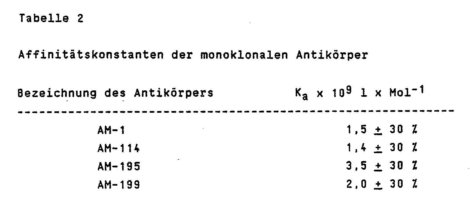
Die gezeigten Assoziationskonstanten Ka weisen die anti-TNF Antikörper als hochaffine Antikörper aus.The association constants K a shown show the anti-TNF antibodies as high-affinity antibodies.
Die biologische Aktivität von TNF in vitro wurde, wie bei Aggarwall et al. (J. Biol. Chem. 1985, Vol. 260, 2345 - 2354) beschrieben, durch Lyse der Mauszellinie L929 (ATCC Nr. CCL1) bestimmt. Bei Versuchen zur Neutralisation der cytotoxischen Aktivität von TNF durch die monoklonalen Antikörper, wurde eine Konzentration an TNF gewählt, bei der mindestens 90 % der Zellen lysierten. Die Antikörper wurden in complettem Medium in 1 : 2 Schritten in Mikrotiterplatten verdünnt. Zu jeder Antikörperlösung (0,1 ml) wurden 0,05 ml rekombinanter oder nativer TNF (1,3 ng/ml) gegeben und zwei Stunden bei Raumtemperatur inkubiert. Anschließend erfolgte Zugabe von 50 000 L929 Zellen in 0,05 ml Medium und nach einer Inkubation von 20 bis 24 Stunden im Brutschrank wurden die Zellen fixiert und mit Kristallviolett gefärbt.The biological activity of TNF in vitro was as described in Aggarwall et al. (J. Biol. Chem. 1985, Vol. 260, 2345-2344), determined by lysis of the mouse cell line L929 (ATCC No. CCL1). In attempts to neutralize the cytotoxic activity of TNF by the monoclonal antibodies, a concentration of TNF was chosen at which at least 90% of the cells lysed. The antibodies were diluted in complete medium in 1: 2 steps in microtiter plates. 0.05 ml of recombinant or native TNF (1.3 ng / ml) was added to each antibody solution (0.1 ml) and incubated for two hours at room temperature. Then 50,000 L929 cells were added in 0.05 ml medium and after an incubation of 20 to 24 hours in the incubator, the cells were fixed and stained with crystal violet.
Der cytotoxische Effekt von TNF läßt die Zellen lysieren und damit während der Färbung abschwemmen. Ist jedoch genügend Antikörper vorhanden, so wird der cytotoxische Effekt von TNF neutralisiert und die Zellen werden gefärbt.

Wie aus Tabelle 3 ersichtlich ist, wurden drei verschiedene Klassen von Antikörpern im Neutralisationstest gefunden. Der monoklonale Antikörper AM-195 neutralisierte TNF bei einer Konzentration von 0,2 µg/ml, AM-114 bei 2 µg/ml während bei AM-1 und AM-199 keine vollständige Neutralisierung bei 20 µg/ml zu beobachten war.As can be seen from Table 3, three different classes of antibodies were found in the neutralization test. The monoclonal antibody AM-195 neutralized TNF at a concentration of 0.2 µg / ml, AM-114 at 2 µg / ml, while no complete neutralization was observed at 20 µg / ml in AM-1 and AM-199.
Die Bestimmung der Assoziationskonstanten im Beispiel 2 zeigte, daß TNF von den Antikörpern ungefähr gleich gut gebunden wurde. Eine Klassifizierung zwischen stark, schwach und nicht-neutralisierenden Antikörpern liefert die Information zur Unterscheidung von verschiedenen Epitopen auf dem TNF-Molekül. Die Ergebnisse hinsichtlich der Antikörperbindung und TNF-Neutralisation lassen erkennen, daß mindestens drei verschiedene Epitope von TNF durch die Kollektion der monoklonalen Antikörper erkannt und definiert werden können.The determination of the association constants in Example 2 showed that TNF was bound approximately equally well by the antibodies. A classification between strong, weak and non-neutralizing antibodies provides the information to differentiate between different epitopes the TNF molecule. The results regarding antibody binding and TNF neutralization indicate that at least three different TNF epitopes can be recognized and defined by the collection of monoclonal antibodies.
Zur Unterscheidung zwischen kompetitiver Bindung von zwei Antikörpern an dasselbe Epitop und additiver Bindung an verschiedene Epitope wurde die Bindung der Antikörper allein und in Paaren in allen möglichen Kombinationen an immobilisiertem TNF getestet. TNF wurde, wie in Beispiel 1 beschrieben, gebunden. Aufgereinigte Antikörper, beginnend mit 24000ng/ml, wurden in Schritten von 1 : 4 verdünnt. Anschließend folgte die Zugabe von biotinyliertem Antikörper AM-195 und Inkubation für 90 Minuten. Die Näpfchen wurden mit PBS 0,05 % Tween® 20 gewaschen, Streptavidin-Peroxidase-Komplex (BRL) hinzugegeben und für 30 Minuten inkubiert. Nach einem Waschschritt wurde 0,1 ml Peroxidase-Substrat (siehe Beispiel 4 b) pro Näpfchen hinzugegeben. Bei einer kompetitiven Bindung kommt es zu einer Auslöschung des Signals. Wie aus Tabelle 4 zu entnehmen ist, bindet der monoklonale Antikörper AM-195 an ein anderes Epitop von TNF wie AM-1 oder AM-199. Bei AM-114 ist eine leichte Kompetition zu beobachten. Aus diesem Grunde sind die weiteren Experimente mit immobilisierten AM-1 oder AM-199 Antikörpern und Biotin-markiertem AM-195 durchgeführt worden.

Der Antikörper AM-1 oder AM-199 wurde durch passive Adsorption oder kovalente Bindung an einen Träger (Kügelchen, Filter, Mikrotiterplatte aus Polystyrol oder Polyvinylchlorid, Filterpapier oder anderen Materialien) immobilisiert. Die Spezifität von monoklonalen Antikörpern erlaubt nur dem spezifischen Antigen, hier dem TNF, durch Immunbindung, über eine einzige molekulare Bindungsstelle des Antigens gebunden zu werden. Die Menge des bindbaren TNF's ist der Konzentration und Menge des Antigen in der Lösung proportional. Das Antigen wird durch Immunbindung des Antikörpers AM-195 an eine andere molekulare Bindungsstelle erkannt. Der Antikörper AM-195 trägt ein Signal. Die Menge des immobilsierbaren Signals ist damit direkt proportional der Menge des immobilisierten Antigens und damit auch der Konzentration des Antigens in der zu untersuchenden Lösung.The antibody AM-1 or AM-199 was immobilized by passive adsorption or covalent binding to a carrier (beads, filter, microtiter plate made of polystyrene or polyvinyl chloride, filter paper or other materials). The specificity of monoclonal antibodies allows only the specific antigen, here the TNF, to be bound by a single molecular binding site of the antigen by immunobinding. The amount of TNF that can be bound is proportional to the concentration and amount of antigen in the solution. The antigen is recognized by immunobinding the AM-195 antibody to another molecular binding site. The AM-195 antibody carries a signal. The amount of the immobilizable signal is thus directly proportional to the amount of the immobilized antigen and thus also the concentration of the antigen in the solution to be examined.
Aufgereinigte Antikörper AM-1 oder AM-199 wurden in Anhaftpuffer (Natrium-Bicarbonatpuffer pH 9,5, 4,2 g/l = 0,05 M) auf 5 µg/ml verdünnt. Die Näpfchen einer Mikrotiterplatte wurden mit 0,1 ml dieser Lösung für 16 bis 20 Stunden bei 4 °C inkubiert.Purified antibodies AM-1 or AM-199 were diluted to 5 µg / ml in adhesive buffer (sodium bicarbonate buffer pH 9.5, 4.2 g / l = 0.05 M). The wells of a microtiter plate were incubated with 0.1 ml of this solution for 16 to 20 hours at 4 ° C.
Die gemäß 1 erhaltene Lösung wurde abgesaugt und 2mal mit PBS (2 g/l NaCl, 0,2 g/l KCl, 1,44 g/l Na₂HPO₄ x 2H₂O, 0,2 g/l KH₂PO₄, pH 7,0) gewaschen. Anschließend wurde für 30 Minuten bei Raumtemperatur mit 1 %iger Rinderserumalbumin-Lösung (Sigma, RIA grade) blockiert.The solution obtained according to 1 was suction filtered and washed twice with PBS (2 g / l NaCl, 0.2 g / l KCl, 1.44 g / l Na₂HPO₄ x 2H₂O, 0.2 g / l KH₂PO₄, pH 7.0) . It was then blocked for 30 minutes at room temperature with 1% bovine serum albumin solution (Sigma, RIA grade).
Die Lösung gemäß 2 wurde abgesaugt und 2mal mit PBS gewaschen. rTNF wurde mit Puffer I (zu 1 l PBS werden 1 g Rinderserumalbumin gegeben, Sigma RIA grade) auf 2,5 ng/ml eingestellt und in 1 : 2 Schritten verdünnt. Je 0,1 ml wurden pro Näpfchen pipettiert und 2-4 Stunden bei Raumtemperatur inkubiert. Nach 3maligem Waschen mit Puffer (Waschpuffer: PBS + 0,1 % Tween® 20) wurden 0,1 ml Biotin-markierter Antikörper AM-195 hinzugegeben. Das Konjugat, nach der Vorschrift im Beispiel 1 h) hergestellt, wurde 1 : 400 mit Puffer I verdünnt und für 2 Stunden bei Raumtemperatur oder 16-20 Stunden bei 4-10°C inkubiert.The solution according to 2 was suctioned off and washed twice with PBS. rTNF was adjusted to 2.5 ng / ml with buffer I (1 g of bovine serum albumin, Sigma RIA grade) was added to 1 l of PBS and diluted in 1: 2 steps. 0.1 ml was pipetted into each well and incubated for 2-4 hours at room temperature. After washing three times with buffer (wash buffer: PBS + 0.1% Tween® 20), 0.1 ml of biotin-labeled antibody AM-195 was added. The conjugate, prepared according to the instructions in Example 1 h), was diluted 1: 400 with buffer I and incubated for 2 hours at room temperature or 16-20 hours at 4-10 ° C.
Die Näpfchen wurden mit Waschpuffer 3mal gewaschen und anschließend mit 0,1 ml Streptavidin-Peroxidase-Komplex (BRL, verdünnt 1 : 2000 in PBS/BSA-Puffer) für 30 min bei Raumtemperatur inkubiert.The wells were washed 3 times with washing buffer and then incubated with 0.1 ml streptavidin-peroxidase complex (BRL, diluted 1: 2000 in PBS / BSA buffer) for 30 min at room temperature.
Die Näpfchen wurden 3mal mit Waschpuffer gewaschen, 0,1 ml/Näpfchen Peroxidasesubstrat pipettiert und 30 Minuten bei Raumtemperatur inkubiert. Die Reaktion wurde mit 0,1 ml/Näpfchen 2M H₂SO₄ abgestoppt. Die Ablesung der Absorption in der Mikrotiterplatte erfolgte innerhalb einer Stunde bei einer Wellenlänge von 450 nm. Eine charakteristische Eichkurve ist in Abb. 1 dargestellt. Die Nachweisgrenze für TNF liegt bei 10 g/ml.The wells were washed three times with washing buffer, 0.1 ml / well peroxidase substrate was pipetted and incubated for 30 minutes at room temperature. The reaction was stopped with 0.1 ml / well 2M H₂SO₄. The absorption in the microtiter plate was read within one hour at a wavelength of 450 nm. A characteristic calibration curve is shown in Fig. 1. The detection limit for TNF is 10 g / ml.
- TMB-Lösung: 42 mM TMB (3,3ʹ,5,5ʹ -Tetramethylbenzidin, Miles) in DMSO
- Substratpuffer: 50 g Natriumacetat auf 1 l Wasser geben und mit 1 g Zitronensäure auf pH 4,9 einstellen.
- 0,1 ml der TMB-Lösung wird langsam und unter Schütteln zu 10 ml Substratpuffer gegeben, gefolgt von 1,47 µl von 30 % H₂O₂ (reinst, Merck).- TMB solution: 42 mM TMB (3.3ʹ, 5.5ʹ-tetramethylbenzidine, Miles) in DMSO
- Substrate buffer: add 50 g sodium acetate to 1 l water and adjust to pH 4.9 with 1 g citric acid.
- 0.1 ml of the TMB solution is added slowly and with shaking to 10 ml of substrate buffer, followed by 1.47 ul of 30% H₂O₂ (reinst, Merck).
Rekombinantes und/oder natives TNF wurde in Puffer I (Beispiel 4 b) oder in Humanserum auf 2,5 ng/ml eingestellt und in 1 : 2 Schritten unter den gleichen Bedingungen verdünnt. Je 0,1 ml wurden pro Näpfchen pipettiert, die vorher, wie unter Beispiel 4 b) beschrieben, mit Antikörper AM-199 oder AM-1 beschichtet wurden. Der nachfolgende Ablauf ist wie unter Beispiel 4 b) beschrieben.Recombinant and / or native TNF was adjusted to 2.5 ng / ml in buffer I (example 4 b) or in human serum and diluted in 1: 2 steps under the same conditions. 0.1 ml was pipetted into each well, which had previously been coated with antibody AM-199 or AM-1 as described in Example 4b). The following procedure is as described in Example 4 b).
Wie aus Abbildung 1 ersichtlich, stören keine Komponenten aus Humanserum bei der Bestimmung von TNF mittels monoklonaler Antikörper.As can be seen in Figure 1, no components from human serum interfere with the determination of TNF using monoclonal antibodies.
Der Nachweis einer möglichen Kreuzreaktion der Antikörper mit Lymphotoxin wurde entsprechend Beispiel 1e getestet, wobei die Näpfchen der Mikrotiterplatte mit aufgereinigtem rekombinantem Lymphotoxin beschichtet wurden. Es wurde keine Bindung der Antikörper AM-1, AM 114, AM-195 und AM-199 an Lymphotoxin gefunden.The detection of a possible cross-reaction of the antibodies with lymphotoxin was tested according to Example 1e, the wells of the microtiter plate being coated with purified recombinant lymphotoxin. No binding of the antibodies AM-1, AM 114, AM-195 and AM-199 to lymphotoxin was found.
Humanserum mit unterschiedlich zugesetzten Mengen an TNF wurde im 12,5 % Gel nach Laemmli (J. Mol. Biol. 1973, Vol. 80, 575 - 599) gelelektrophoretisch aufgetrennt. Die Methode des Western Blotting wurde, wie bei Burnett, W. N. (Anal. Biochem. 1981, Vol. 112, 195 - 203) und Reines, D. et al. (J. Biol. Chem. 1985, Vol. 260, 1133 - 1139) beschrieben, durchgeführt. Die Proteine des Gels wurden über Nacht auf Nitrozellulosemembran (Schleicher und Schüll) geblottet. Die Nitrozellulosemembran wurde für drei Stunden bei Raumtemperatur mit 1 %iger Gelatinelösung (Bio-Rad, zu 1 l PBS wurden 10 g Gelatine gegeben) inkubiert. Anschließend wurde die Nitrozellulosemembran mit 20 ml Antikörperlösung, die auf 1 µg/ml in Puffer (0,1 % Gelatine in PBS) verdünnt wurde, für zwei Stunden bei Raumtemperatur inkubiert. Der Überstand wurde dekantiert und die Membran mehrmals gewaschen. Unter Verwendung von Peroxidase-markiertem Anit-Maus Immunoglobulin wurde TNF auf der Nitrozellulosemembran sichtbar gemacht. Die Nachweisgrenze liegt bei 30 ng TNF. Wie aus Abbildung 2 ersichtlich ist, reagiert der monoklonale Antikörper AM-195 mit keiner Komponente aus humanem Serum und kann somit als spezifisch für TNF angesehen werden. Für die Antikörper AM-1, AM-114 und AM-199 wurden gleiche Ergebnisse gefunden.Human serum with different amounts of TNF added was separated by gel electrophoresis in the 12.5% gel according to Laemmli (J. Mol. Biol. 1973, Vol. 80, 575-599). The Western blotting method was used as described by Burnett, W.N. (Anal. Biochem. 1981, Vol. 112, 195-203) and Reines, D. et al. (J. Biol. Chem. 1985, Vol. 260, 1133-1139). The proteins of the gel were blotted overnight on a nitrocellulose membrane (Schleicher and Schuell). The nitrocellulose membrane was incubated for 3 hours at room temperature with 1% gelatin solution (Bio-Rad, 10 g gelatin was added to 1 l PBS). The nitrocellulose membrane was then incubated with 20 ml of antibody solution, which was diluted to 1 μg / ml in buffer (0.1% gelatin in PBS), at room temperature for two hours. The supernatant was decanted and the membrane washed several times. TNF was visualized on the nitrocellulose membrane using peroxidase-labeled anit-mouse immunoglobulin. The detection limit is 30 ng TNF. As can be seen from Figure 2, the monoclonal antibody AM-195 does not react with any component from human serum and can therefore be regarded as specific for TNF. The same results were found for the antibodies AM-1, AM-114 and AM-199.
Der schützende Effekt der mAK gegen TNF wurde unter in-vivo-Bedingungen in männlichen Balb/c - Versuch 1, 2, 4) und C3H/HeN - (Versuch 3) Mäusen untersucht. 4-6 Wochen alte Mäuse wurden randomisiert und in Gruppen von 3 bzw. 5 Tieren aufgeteilt. Die Substanzen wurden intravenös in die laterale Schwanzvene gegeben (Applikationsvolumen 10 ml/kg). TNF wurde vor der Injektion in Puffer A (150 mM NaCl und 0,18 % Rinderserumalbumin (Sigma, RIA-grade) gelöst und bei 4-10°C für 6 Stunden gelagert. Nach dieser Zeit zeigte TNF die größte Toxizität. TNF wurde zuerst gegeben, der mAK 15-30 Minuten danach. Die Tötungsraten wurden nach 24 Stunden bestimmt. Tabelle 5 zeigt die erhaltenen Ergebnisse (3/5 bedeutet, daß 3 von 5 Tieren gestorben waren).
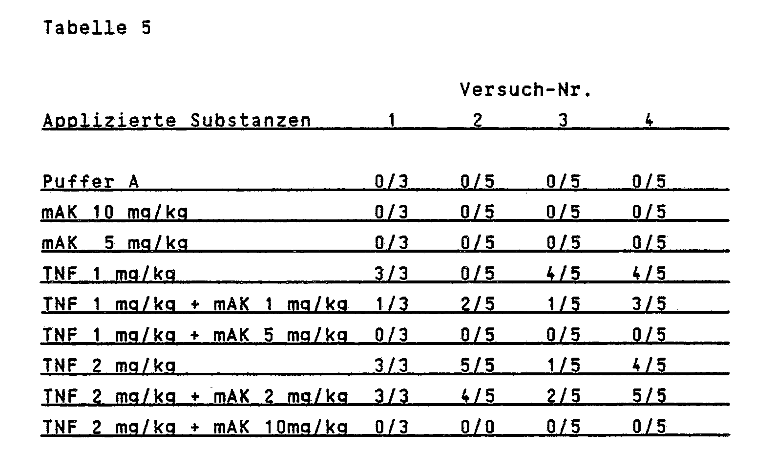
Wie aus Tabelle 5 zu entnehmen ist, kann der neutralisierende Antikörper AM-195 eine tödliche Dosis von TNF in der Maus neutralisieren. Die Überlebensraten der Tiere sind abhängig vom Gewichtsverhältnis Antikörper zu TNF. Eine vollständige Aufhebung der TNF-Toxizität ist bei einem Verhältnis von 5 : 1 zu beobachten. Unter der Annahmne, daß TNF als Trimeres vorliege (FEBS Lett. 211, 179 (1987)), entspricht dies einem molaren Verhältnis Antikörper zu TNF von 1,6 zu 1.As can be seen from Table 5, the neutralizing antibody AM-195 can neutralize a lethal dose of TNF in the mouse. The survival rates of the animals depend on the weight ratio of antibody to TNF. A complete abolition of TNF toxicity can be observed at a ratio of 5: 1. Assuming that TNF is present as trimeres (FEBS Lett. 211, 179 (1987)), this corresponds to a molar ratio of antibody to TNF of 1.6 to 1.
TNF läßt sich nicht durch nicht neutralisierende Antikörper in der Maus neutralisieren.TNF cannot be neutralized by non-neutralizing antibodies in the mouse.
Claims (12)
Priority Applications (3)
| Application Number | Priority Date | Filing Date | Title |
|---|---|---|---|
| AT87113278T ATE93895T1 (en) | 1986-09-13 | 1987-09-11 | MONOCLONAL ANTIBODIES ANTI-HUMAN TUMONIC CROSS FACTOR (TNF) AND THEIR USE. |
| MX9203511A MX9203511A (en) | 1986-09-13 | 1992-06-26 | MONOKIONALE ANTIKORPER GEGEN HUMANEN TUMONEKROSEIATOR (TNF) UND DEREN VERWENDUNG. |
| LV960296A LV5784B4 (en) | 1986-09-13 | 1996-07-22 | Human growth necrosis factor monoclonal antibodies |
Applications Claiming Priority (2)
| Application Number | Priority Date | Filing Date | Title |
|---|---|---|---|
| DE19863631229 DE3631229A1 (en) | 1986-09-13 | 1986-09-13 | MONOCLONAL ANTIBODIES AGAINST HUMAN TUMORNESCROSE FACTOR (TNF) AND THEIR USE |
| DE3631229 | 1986-09-13 |
Publications (3)
| Publication Number | Publication Date |
|---|---|
| EP0260610A2 true EP0260610A2 (en) | 1988-03-23 |
| EP0260610A3 EP0260610A3 (en) | 1989-01-11 |
| EP0260610B1 EP0260610B1 (en) | 1993-09-01 |
Family
ID=6309540
Family Applications (1)
| Application Number | Title | Priority Date | Filing Date |
|---|---|---|---|
| EP87113278A Expired - Lifetime EP0260610B1 (en) | 1986-09-13 | 1987-09-11 | Monoclonal antibodies against human tumour necrotic factor (tnf), and their use |
Country Status (11)
| Country | Link |
|---|---|
| US (1) | US5231024A (en) |
| EP (1) | EP0260610B1 (en) |
| JP (2) | JP2837160B2 (en) |
| BG (1) | BG62229B2 (en) |
| BR (1) | BR1100843A (en) |
| CA (1) | CA1337717C (en) |
| DE (2) | DE3631229A1 (en) |
| DK (1) | DK473887A (en) |
| ES (1) | ES2058083T3 (en) |
| FI (1) | FI91421C (en) |
| ZA (1) | ZA876807B (en) |
Cited By (26)
| Publication number | Priority date | Publication date | Assignee | Title |
|---|---|---|---|---|
| EP0610201A1 (en) * | 1991-03-18 | 1994-08-17 | New York University | Monoclonal and chimeric antibodies specific for human tumor necrosis factor |
| WO1995020978A1 (en) * | 1994-02-07 | 1995-08-10 | Knoll Aktiengesellschaft | Use of anti-tnf antibodies as drugs in treating diseases involving elevated interleukin-6 serum levels |
| DE4409513C1 (en) * | 1994-02-07 | 1995-10-19 | Knoll Ag | Use of anti-TNF antibodies as a medicine for the treatment of diseases with an increased interleukin-6 serum level |
| WO1997030088A3 (en) * | 1996-02-16 | 1997-10-09 | Kennedy Inst Of Rheumatology | Methods of treating vascular disease with tnf antagonists |
| US5919452A (en) * | 1991-03-18 | 1999-07-06 | New York University | Methods of treating TNFα-mediated disease using chimeric anti-TNF antibodies |
| EP0486526B2 (en) † | 1989-08-07 | 2001-03-07 | Peptech Limited | Tumour necrosis factor binding ligands |
| US6277969B1 (en) | 1991-03-18 | 2001-08-21 | New York University | Anti-TNF antibodies and peptides of human tumor necrosis factor |
| US6284471B1 (en) | 1991-03-18 | 2001-09-04 | New York University Medical Center | Anti-TNFa antibodies and assays employing anti-TNFa antibodies |
| US6309640B1 (en) | 1981-09-08 | 2001-10-30 | The Rockefeller University | Lipoprotein lipase suppression by endotoxin-induced mediator (shock assay) |
| US7169386B1 (en) | 1991-03-18 | 2007-01-30 | New York University | Methods of treating inflammation associated with viral infection with anti-TNF antibodies |
| WO2007089303A2 (en) | 2005-11-01 | 2007-08-09 | Abbott Biotechnology Ltd. | Methods and compositions for diagnosing ankylosing spondylitis using biomarkers |
| EP2185201A1 (en) * | 2007-08-08 | 2010-05-19 | Abbott Laboratories | Compositions and methods for crystallizing antibodies |
| US7815909B2 (en) | 2000-08-07 | 2010-10-19 | Centocor, Inc. | Anti-TNF antibodies, compositions, methods and uses |
| EP2335731A2 (en) | 2004-04-09 | 2011-06-22 | Abbott Biotechnology Ltd | Multiple-variable dose regimen for treating TNF-alpha-related disorders |
| EP2500037A2 (en) | 2005-05-16 | 2012-09-19 | Abbott Biotechnology Ltd | Use of TNF alpha inhibitor for treatment of erosive polyarthritis |
| US8668670B2 (en) | 2004-06-23 | 2014-03-11 | Abbvie Biotechnology Ltd | Automatic injection devices |
| EP2738178A1 (en) | 2006-04-05 | 2014-06-04 | AbbVie Biotechnology Ltd | Antibody purification |
| US9339610B2 (en) | 2011-01-24 | 2016-05-17 | Abbvie Biotechnology Ltd | Removal of needle shield from syringes and automatic injection devices |
| EP3078675A1 (en) | 2015-04-10 | 2016-10-12 | Ares Trading S.A. | Induction dosing regimen for the treatment of tnf alpha mediated disorders |
| US9486584B2 (en) | 2006-06-30 | 2016-11-08 | Abbvie Biotechnology Ltd. | Automatic injection device |
| US9561328B2 (en) | 2009-04-29 | 2017-02-07 | Abbvie Biotechnology Ltd | Automatic injection device |
| US9605064B2 (en) | 2006-04-10 | 2017-03-28 | Abbvie Biotechnology Ltd | Methods and compositions for treatment of skin disorders |
| US9610301B2 (en) | 2008-01-15 | 2017-04-04 | Abbvie Deutschland Gmbh & Co Kg | Powdered protein compositions and methods of making same |
| US9878102B2 (en) | 2011-01-24 | 2018-01-30 | Abbvie Biotechnology Ltd. | Automatic injection devices having overmolded gripping surfaces |
| WO2018124948A1 (en) | 2016-12-30 | 2018-07-05 | Закрытое Акционерное Общество "Биокад" | AQUEOUS PHARMACEUTICAL COMPOSITION OF A RECOMBINANT MONOCLONAL ANTIBODY TO FNOα |
| US10465003B2 (en) | 2016-02-05 | 2019-11-05 | Janssen Biotech, Inc. | Anti-TNF antibodies, compositions, methods and use for the treatment or prevention of type 1 diabetes |
Families Citing this family (148)
| Publication number | Priority date | Publication date | Assignee | Title |
|---|---|---|---|---|
| US5700466A (en) * | 1981-09-08 | 1997-12-23 | The Rockefeller University | Method of ameliorating or preventing septic shock using a monoclonal antibody specific to cachectin/tumor necrosis factor |
| US6419927B1 (en) | 1981-09-08 | 2002-07-16 | Anthony Cerami | Method for reducing adverse effects of a human 70kDa mediator which results from endotoxin stimulation of macrophages |
| GB2233559B (en) * | 1988-03-11 | 1992-03-04 | Celltech Ltd | Antibodies for use in antilymphocyte antibody therapy |
| DE3823804A1 (en) * | 1988-07-14 | 1990-01-18 | Basf Ag | NEUTRALIZATION OF THE TOXIC PROPERTIES OF TNF- (ALPHA) IN VITRO AND VIVO BY MONOCLONAL ANTIBODIES AND THE FRAGMENTS DERIVED FROM THEM |
| WO1990000902A1 (en) * | 1988-07-18 | 1990-02-08 | Chiron Corporation | Monoclonal antibodies reactive with cachectin |
| WO1990001950A1 (en) * | 1988-08-19 | 1990-03-08 | Celltech Limited | Pharmaceutical products for anti-neoplastic therapy |
| US20030225254A1 (en) * | 1989-08-07 | 2003-12-04 | Rathjen Deborah Ann | Tumour necrosis factor binding ligands |
| US5959087A (en) * | 1989-08-07 | 1999-09-28 | Peptide Technology, Ltd. | Tumour necrosis factor binding ligands |
| US5998378A (en) * | 1989-08-16 | 1999-12-07 | Chiron Corporation | Compositions for the inhibition of TNF hormone formation and uses thereof |
| US5843693A (en) * | 1989-08-16 | 1998-12-01 | Chiron Corporation | Assay method for screening for inhibitors of proTNF conversion |
| US6586222B1 (en) | 1989-08-16 | 2003-07-01 | Chiron Corporation | Recombinant PR-3 and compositions thereof |
| JPH05501351A (en) * | 1989-08-16 | 1993-03-18 | カイロン コーポレイション | Cleavage site blocking antibodies against prohormone proteins and uses thereof |
| GB8921123D0 (en) * | 1989-09-19 | 1989-11-08 | Millar Ann B | Treatment of ards |
| US6713610B1 (en) * | 1990-01-12 | 2004-03-30 | Raju Kucherlapati | Human antibodies derived from immunized xenomice |
| US20040120952A1 (en) * | 2000-08-07 | 2004-06-24 | Centocor, Inc | Anti-TNF antibodies and peptides of human tumor necrosis factor |
| ES2121907T3 (en) * | 1992-08-28 | 1998-12-16 | Bayer Ag | USE OF ANTI-TNF MONOCLONAL ANTIBODIES FOR THE TREATMENT OF BACTERIAL MENINGITIS. |
| US20050260201A1 (en) * | 1993-01-29 | 2005-11-24 | Centocor, Inc. | Methods of treating rheumatoid arthritis using anti-TNF receptor fusion proteins |
| US5888511A (en) * | 1993-02-26 | 1999-03-30 | Advanced Biotherapy Concepts, Inc. | Treatment of autoimmune diseases, including AIDS |
| US5626843A (en) * | 1993-02-26 | 1997-05-06 | Advanced Biotherapy Concepts, Inc. | Treatment of autoimmune diseases, including AIDS, by removel of interferons, TNFs and receptors therefor |
| EP0749494A1 (en) | 1994-03-07 | 1996-12-27 | Chiron Corporation | Compositions for the inhibition of tnf formation and uses thereof |
| US6090382A (en) | 1996-02-09 | 2000-07-18 | Basf Aktiengesellschaft | Human antibodies that bind human TNFα |
| HU221984B1 (en) | 1996-02-09 | 2003-03-28 | Basf Ag | Human antibodies binding human tnfalfa, pharmaceutical compositions containing thereof and use thereof |
| CA2722378C (en) * | 1996-12-03 | 2015-02-03 | Amgen Fremont Inc. | Human antibodies that bind tnf.alpha. |
| US20020031513A1 (en) * | 1997-11-24 | 2002-03-14 | Shamir Leibovitz | Method and pharmaceutical composition for inhibiting premature rapture of fetal membranes, ripening of uterine cervix and preterm labor in mammals |
| ATE488250T1 (en) * | 1999-03-02 | 2010-12-15 | Centocor Inc | ANTIBODIES AGAINST TNF ALPHA FOR THE THERAPY OF STEROID-RESISTANT ASTHMA |
| TR200102688T2 (en) * | 1999-03-18 | 2002-01-21 | Celgene Corporation | Substituted 1-oxo and 1,3-dioxoisoindolins and their use in pharmaceutical compositions for reducing inflammatory cytokine levels. |
| JP2003511355A (en) * | 1999-10-06 | 2003-03-25 | ビーエーエスエフ アクチェンゲゼルシャフト | Endothelin signaling pathway inhibitors and αvβ3 receptor antagonists for combination therapy |
| US20090081234A1 (en) * | 2000-08-07 | 2009-03-26 | George Heavner | Anti-tnf antibodies, compositions, methods and uses for treatment of depression and related conditions |
| UA81743C2 (en) | 2000-08-07 | 2008-02-11 | Центокор, Инк. | HUMAN MONOCLONAL ANTIBODY WHICH SPECIFICALLY BINDS TUMOR NECROSIS FACTOR ALFA (TNFα), PHARMACEUTICAL MIXTURE CONTAINING THEREOF, AND METHOD FOR TREATING ARTHRITIS |
| US6902734B2 (en) | 2000-08-07 | 2005-06-07 | Centocor, Inc. | Anti-IL-12 antibodies and compositions thereof |
| US20050249735A1 (en) * | 2000-08-07 | 2005-11-10 | Centocor, Inc. | Methods of treating ankylosing spondylitis using anti-TNF antibodies and peptides of human tumor necrosis factor |
| US20060018907A1 (en) * | 2000-08-07 | 2006-01-26 | Centocor, Inc. | Anti-TNF antibodies and peptides of human tumor necrosis factor |
| US7288390B2 (en) | 2000-08-07 | 2007-10-30 | Centocor, Inc. | Anti-dual integrin antibodies, compositions, methods and uses |
| US20110195063A1 (en) * | 2000-08-07 | 2011-08-11 | Centocor, Inc. | Methods of Treating Ankylosing Spondylitis Using Anti-TNF Antibodies and Peptides of Human Tumor Necrosis Factor |
| US20020168360A1 (en) * | 2001-03-02 | 2002-11-14 | Christine Dingivan | Methods of preventing or treating inflammatory or autoimmune disorders by administering integrin alphanubeta3 antagonists in combination with other prophylactic or therapeutic agents |
| CA2817619A1 (en) | 2001-06-08 | 2002-12-08 | Abbott Laboratories (Bermuda) Ltd. | Methods of administering anti-tnf.alpha. antibodies |
| EP1499352A4 (en) | 2002-04-12 | 2006-10-11 | Medimmune Inc | Recombinant anti-interleukin-9 antibodies |
| US20030206898A1 (en) | 2002-04-26 | 2003-11-06 | Steven Fischkoff | Use of anti-TNFalpha antibodies and another drug |
| US20040009172A1 (en) * | 2002-04-26 | 2004-01-15 | Steven Fischkoff | Use of anti-TNFalpha antibodies and another drug |
| WO2004002417A2 (en) | 2002-06-28 | 2004-01-08 | Centocor, Inc. | Mammalian ch1 deleted mimetibodies, compositions, methods and uses |
| DK1944322T3 (en) * | 2002-07-19 | 2015-06-22 | Abbvie Biotechnology Ltd | Treatment of TNFalfa-related diseases |
| US20040033228A1 (en) | 2002-08-16 | 2004-02-19 | Hans-Juergen Krause | Formulation of human antibodies for treating TNF-alpha associated disorders |
| MY150740A (en) | 2002-10-24 | 2014-02-28 | Abbvie Biotechnology Ltd | Low dose methods for treating disorders in which tnf? activity is detrimental |
| JP4754219B2 (en) | 2002-12-02 | 2011-08-24 | アムジエン・フレモント・インコーポレイテツド | Antibodies directed against tumor necrosis factor and their use |
| US7101978B2 (en) * | 2003-01-08 | 2006-09-05 | Applied Molecular Evolution | TNF-α binding molecules |
| KR20110094361A (en) | 2003-04-11 | 2011-08-23 | 메디뮨 엘엘씨 | Recombinant il-9 antibodies and uses thereof |
| US7429378B2 (en) * | 2003-05-13 | 2008-09-30 | Depuy Spine, Inc. | Transdiscal administration of high affinity anti-MMP inhibitors |
| US7553827B2 (en) * | 2003-08-13 | 2009-06-30 | Depuy Spine, Inc. | Transdiscal administration of cycline compounds |
| US7344716B2 (en) * | 2003-05-13 | 2008-03-18 | Depuy Spine, Inc. | Transdiscal administration of specific inhibitors of pro-inflammatory cytokines |
| US20040229878A1 (en) * | 2003-05-13 | 2004-11-18 | Depuy Spine, Inc. | Transdiscal administration of specific inhibitors of P38 kinase |
| US8273347B2 (en) | 2003-05-13 | 2012-09-25 | Depuy Spine, Inc. | Autologous treatment of degenerated disc with cells |
| EP2286804A1 (en) | 2003-05-13 | 2011-02-23 | DePuy Spine, Inc. | A method of treating degenerative disc disease |
| US8361467B2 (en) * | 2003-07-30 | 2013-01-29 | Depuy Spine, Inc. | Trans-capsular administration of high specificity cytokine inhibitors into orthopedic joints |
| UA89481C2 (en) | 2003-09-30 | 2010-02-10 | Центокор, Инк. | Human epo mimetic hinge core mimetibodies, compositions, methods and uses |
| US8895540B2 (en) | 2003-11-26 | 2014-11-25 | DePuy Synthes Products, LLC | Local intraosseous administration of bone forming agents and anti-resorptive agents, and devices therefor |
| US7435799B2 (en) * | 2004-01-08 | 2008-10-14 | Applied Molecular Evolution | TNF-α binding molecules |
| EP1750754A4 (en) * | 2004-03-31 | 2010-09-22 | Centocor Ortho Biotech Inc | Human glp-1 mimetibodies, compositions, methods and uses |
| US7351739B2 (en) * | 2004-04-30 | 2008-04-01 | Wellgen, Inc. | Bioactive compounds and methods of uses thereof |
| US7393662B2 (en) | 2004-09-03 | 2008-07-01 | Centocor, Inc. | Human EPO mimetic hinge core mimetibodies, compositions, methods and uses |
| WO2006041970A2 (en) * | 2004-10-08 | 2006-04-20 | Abbott Biotechnology Ltd. | Treatment of respiratory syncytial virus (rsv) infection |
| US20060121042A1 (en) | 2004-10-27 | 2006-06-08 | Medimmune, Inc. | Modulation of antibody specificity by tailoring the affinity to cognate antigens |
| PA8672101A1 (en) | 2005-04-29 | 2006-12-07 | Centocor Inc | ANTI-IL-6 ANTIBODIES, COMPOSITIONS, METHODS AND USES |
| EP1896073B1 (en) | 2005-06-30 | 2013-03-06 | Janssen Biotech, Inc. | Anti-il-23 antibodies, compositions, methods and uses |
| WO2007015128A1 (en) * | 2005-08-02 | 2007-02-08 | Xbiotech Inc. | DIAGNOSIS, TREATMENT, AND PREVENTION OF VASCULAR DISORDERS USING IL-1α AUTOANTIBODIES |
| US20070041905A1 (en) * | 2005-08-19 | 2007-02-22 | Hoffman Rebecca S | Method of treating depression using a TNF-alpha antibody |
| US9017662B2 (en) | 2005-08-30 | 2015-04-28 | Actogenix N.V. | Anti-TNF alpha producing lactic acid bacteria for the treatment of chronic enterocolitis |
| HUE049832T2 (en) | 2005-12-29 | 2020-10-28 | Janssen Biotech Inc | Human anti-il-23 antibodies, compositions, method and uses |
| US20090317399A1 (en) * | 2006-04-10 | 2009-12-24 | Pollack Paul F | Uses and compositions for treatment of CROHN'S disease |
| EP2708242A3 (en) | 2006-04-10 | 2014-03-26 | Abbott Biotechnology Ltd | Uses and compositions for treatment of ankylosing spondylitis |
| EP2010214A4 (en) * | 2006-04-10 | 2010-06-16 | Abbott Biotech Ltd | Uses and compositions for treatment of rheumatoid arthritis |
| US20080118496A1 (en) * | 2006-04-10 | 2008-05-22 | Medich John R | Uses and compositions for treatment of juvenile rheumatoid arthritis |
| EP2666472A3 (en) | 2006-04-10 | 2014-04-02 | Abbott Biotechnology Ltd | Uses and compositions for treatment of psoriatic arthritis |
| US20080131374A1 (en) * | 2006-04-19 | 2008-06-05 | Medich John R | Uses and compositions for treatment of rheumatoid arthritis |
| US20110008282A1 (en) * | 2006-05-15 | 2011-01-13 | Xbiotech, Inc. | IL-1alpha immunization induces autoantibodies protective against atherosclerosis |
| CN107158376A (en) * | 2006-05-22 | 2017-09-15 | 埃克斯生物科技公司 | Use the anti-Alpha antibodies treating cancers of IL 1 |
| EP2851064A3 (en) | 2006-07-11 | 2015-08-05 | Roy C. Levitt | Rhinosinusitis prevention and therapy with proinflammatory cytokine inhibitors |
| US20080014285A1 (en) * | 2006-07-13 | 2008-01-17 | Di Mauro Thomas M | Method of treating neurodegenerative brain disease with a composite comprising superparamagnetic nanoparticles and a therapeutic compound |
| JP2010500360A (en) | 2006-08-10 | 2010-01-07 | アルボア コーポレーション | Local therapy of lower respiratory inflammatory diseases with inflammatory cytokine inhibitors |
| EP2089428B1 (en) | 2006-10-27 | 2013-11-20 | AbbVie Biotechnology Ltd | Crystalline anti-htnfalpha antibodies |
| MX2009010361A (en) | 2007-03-29 | 2009-10-16 | Abbott Lab | Crystalline anti-human il-12 antibodies. |
| EP2068925A4 (en) | 2007-05-07 | 2011-08-31 | Medimmune Llc | Anti-icos antibodies and their use in treatment of oncology, transplantation and autoimmune disease |
| EP2171451A4 (en) | 2007-06-11 | 2011-12-07 | Abbott Biotech Ltd | Methods for treating juvenile idiopathic arthritis |
| US20090038182A1 (en) * | 2007-08-09 | 2009-02-12 | Lans Maris J | Footwear with built-in scale |
| US20090162351A1 (en) * | 2007-12-21 | 2009-06-25 | Depuy Spine, Inc. | Transdiscal administration of inhibitors of p38 MAP kinase |
| US8986696B2 (en) * | 2007-12-21 | 2015-03-24 | Depuy Mitek, Inc. | Trans-capsular administration of p38 map kinase inhibitors into orthopedic joints |
| WO2009083246A1 (en) * | 2007-12-31 | 2009-07-09 | Bayer Schering Pharma Aktiengesellschaft | Antibodies to tnf alpha |
| EP3085773B1 (en) * | 2008-05-30 | 2020-03-18 | XBiotech, Inc | Uses of il-1 alpha antibodies |
| US8242074B2 (en) * | 2008-09-12 | 2012-08-14 | Xbiotech, Inc. | Modulation of the amount or function of pathogenic CD14+CD16+ monocytes |
| US8415291B2 (en) | 2008-10-31 | 2013-04-09 | Centocor Ortho Biotech Inc. | Anti-TNF alpha fibronectin type III domain based scaffold compositions, methods and uses |
| JP5933975B2 (en) | 2008-11-12 | 2016-06-15 | メディミューン,エルエルシー | Antibody preparation |
| AU2010315547C1 (en) | 2009-10-26 | 2015-06-04 | Société des Produits Nestlé S.A. | Assays for the detection of anti-TNF drugs and autoantibodies |
| BR112012014710A2 (en) | 2009-12-15 | 2017-07-25 | Abbott Biotech Ltd | optimized ignition button for automatic injection device |
| US9023997B2 (en) | 2010-01-20 | 2015-05-05 | Merck Sharp & Dohme Corp. | Anti-ILT5 antibodies and ILT5-binding antibody fragments |
| US8846397B2 (en) | 2010-01-20 | 2014-09-30 | Merck Sharp & Dohme Corp. | Immunoregulation by anti-ILT5 antibodies and ILT5-binding antibody fragments |
| CN102946904A (en) | 2010-06-18 | 2013-02-27 | 埃克斯生物科技公司 | Arthritis treatment |
| DK2608808T3 (en) | 2010-08-23 | 2017-04-24 | Xbiotech Inc | TREATMENT OF NEOPLASTIC DISEASES |
| KR101934247B1 (en) | 2010-10-18 | 2019-01-02 | 네스텍 소시에테아노님 | Methods for determining anti-drug antibody isotypes |
| AR084210A1 (en) * | 2010-12-08 | 2013-05-02 | Abbott Lab | PROTEINS OF UNION TO TNF-a |
| CN103502815B (en) | 2011-02-17 | 2015-09-23 | 雀巢产品技术援助有限公司 | Detect the determination method for the autoantibody of anti-TNF alpha medicine |
| US8658171B2 (en) | 2011-02-28 | 2014-02-25 | Livzon Mabpharm Inc. | Humanized anti-TNFα antibodies |
| CN102675460B (en) | 2011-02-28 | 2015-08-19 | 珠海市丽珠单抗生物技术有限公司 | The humanized antibody of Tumor necrosis factorα |
| US9724409B2 (en) | 2011-04-01 | 2017-08-08 | Xbiotech, Inc. | Treatment of inflammatory skin disease |
| WO2012149197A2 (en) | 2011-04-27 | 2012-11-01 | Abbott Laboratories | Methods for controlling the galactosylation profile of recombinantly-expressed proteins |
| SG10201605516YA (en) | 2011-07-06 | 2016-08-30 | Nestec Sa | Assays for detecting neutralizing autoantibodies to biologic therapy with tnf alpha |
| PT2750709T (en) | 2011-09-23 | 2018-11-22 | Xbiotech Inc | Cachexia treatment |
| CA2853357A1 (en) * | 2011-10-24 | 2013-05-02 | Abbvie Inc. | Immunobinders directed against tnf |
| AR088514A1 (en) * | 2011-10-24 | 2014-06-18 | Abbvie Inc | BISPECIFIC IMMUNOLIGANTS DIRECTED AGAINST TNF |
| US9181572B2 (en) | 2012-04-20 | 2015-11-10 | Abbvie, Inc. | Methods to modulate lysine variant distribution |
| US9067990B2 (en) | 2013-03-14 | 2015-06-30 | Abbvie, Inc. | Protein purification using displacement chromatography |
| WO2013158279A1 (en) | 2012-04-20 | 2013-10-24 | Abbvie Inc. | Protein purification methods to reduce acidic species |
| WO2013176754A1 (en) | 2012-05-24 | 2013-11-28 | Abbvie Inc. | Novel purification of antibodies using hydrophobic interaction chromatography |
| US9512214B2 (en) | 2012-09-02 | 2016-12-06 | Abbvie, Inc. | Methods to control protein heterogeneity |
| SG11201504249XA (en) | 2012-09-02 | 2015-07-30 | Abbvie Inc | Methods to control protein heterogeneity |
| US9545441B2 (en) | 2012-09-18 | 2017-01-17 | Xbiotech, Inc. | Treatment of diabetes |
| EP2830651A4 (en) | 2013-03-12 | 2015-09-02 | Abbvie Inc | Human antibodies that bind human tnf-alpha and methods of preparing the same |
| WO2014151878A2 (en) | 2013-03-14 | 2014-09-25 | Abbvie Inc. | Methods for modulating protein glycosylation profiles of recombinant protein therapeutics using monosaccharides and oligosacharides |
| WO2014151901A1 (en) | 2013-03-14 | 2014-09-25 | Abbvie Inc. | Improvement of mammalian cell culture performance through surfactant supplementation of feed media |
| SG11201504260UA (en) | 2013-03-14 | 2015-10-29 | Abbvie Inc | Low acidic species compositions and methods for producing and using the same |
| US8921526B2 (en) | 2013-03-14 | 2014-12-30 | Abbvie, Inc. | Mutated anti-TNFα antibodies and methods of their use |
| US9017687B1 (en) | 2013-10-18 | 2015-04-28 | Abbvie, Inc. | Low acidic species compositions and methods for producing and using the same using displacement chromatography |
| CA2899449A1 (en) | 2013-03-14 | 2014-10-02 | Abbvie Inc. | Low acidic species compositions and methods for producing the same using displacement chromatography |
| DK2970980T3 (en) | 2013-03-15 | 2018-10-22 | Janssen Biotech Inc | METHODS FOR MANAGING C-TERMINAL LYSIN, GALACTOSE AND SIALINIC ACID CONTENTS IN RECOMBINANT PROTEINS |
| WO2015051293A2 (en) | 2013-10-04 | 2015-04-09 | Abbvie, Inc. | Use of metal ions for modulation of protein glycosylation profiles of recombinant proteins |
| US9085618B2 (en) | 2013-10-18 | 2015-07-21 | Abbvie, Inc. | Low acidic species compositions and methods for producing and using the same |
| US9181337B2 (en) | 2013-10-18 | 2015-11-10 | Abbvie, Inc. | Modulated lysine variant species compositions and methods for producing and using the same |
| US8946395B1 (en) | 2013-10-18 | 2015-02-03 | Abbvie Inc. | Purification of proteins using hydrophobic interaction chromatography |
| US20150139988A1 (en) | 2013-11-15 | 2015-05-21 | Abbvie, Inc. | Glycoengineered binding protein compositions |
| EP3145951A1 (en) | 2014-06-24 | 2017-03-29 | InSight Biopharmaceuticals Ltd. | Methods of purifying antibodies |
| US20170226552A1 (en) | 2014-07-03 | 2017-08-10 | Abbvie Inc. | Methods for modulating protein glycosylation profiles of recombinant protein therapeutics using cobalt |
| WO2016007764A1 (en) | 2014-07-09 | 2016-01-14 | Abbvie Inc. | Methods for modulating the glycosylation profile of recombinant proteins using non-commonly used sugars |
| WO2016088104A2 (en) | 2014-12-05 | 2016-06-09 | Nestec S.A. | Indirect homogeneous mobility shift assays for the detection of biologics in patient samples |
| MX2019008989A (en) | 2017-01-30 | 2019-10-09 | Janssen Biotech Inc | Anti-tnf antibodies, compositions, and methods for the treatment of active psoriatic arthritis. |
| CN110418652A (en) | 2017-02-07 | 2019-11-05 | 詹森生物科技公司 | For treating the anti-TNF antibodies, composition and method of activity ankylosing spondylitis |
| JP7145162B2 (en) | 2017-02-16 | 2022-09-30 | ヤンセン バイオテク,インコーポレイテッド | Treatment of hidradenitis suppurativa |
| AU2020208828A1 (en) | 2019-01-15 | 2021-08-05 | Janssen Biotech, Inc. | Anti-TNF antibody compositions and methods for the treatment of juvenile idiopathic arthritis |
| EA202192038A1 (en) | 2019-01-23 | 2021-10-05 | Янссен Байотек, Инк. | ANTIBODY COMPOSITIONS TO TNF FOR USE IN METHODS FOR TREATMENT OF PSORIATIC ARTHRITIS |
| CA3133381A1 (en) | 2019-03-14 | 2020-09-17 | Janssen Biotech, Inc. | Manufacturing methods for producing anti-tnf antibody compositions |
| EP3938391A1 (en) | 2019-03-14 | 2022-01-19 | Janssen Biotech, Inc. | Methods for producing anti-tnf antibody compositions |
| EA202192508A1 (en) | 2019-03-14 | 2022-03-29 | Янссен Байотек, Инк. | METHODS FOR OBTAINING COMPOSITIONS OF ANTIBODIES TO TNF |
| US11780911B2 (en) | 2019-05-23 | 2023-10-10 | Janssen Biotech, Inc. | Method of treating inflammatory bowel disease with a combination therapy of antibodies to IL-23 and TNF alpha |
| KR20220029593A (en) | 2019-06-03 | 2022-03-08 | 얀센 바이오테크 인코포레이티드 | Anti-TNF antibody compositions, and methods for the treatment of psoriatic arthritis |
| JP2022535534A (en) | 2019-06-03 | 2022-08-09 | ヤンセン バイオテツク,インコーポレーテツド | Anti-TNF Antibodies, Compositions and Methods for Treating Active Ankylosing Spondylitis |
| JP2022540764A (en) | 2019-06-25 | 2022-09-20 | プロサイセデクス インコーポレイティド | Detection of anti-TNFα biologics and anti-drug antibodies |
| WO2021028752A1 (en) | 2019-08-15 | 2021-02-18 | Janssen Biotech, Inc. | Anti-tfn antibodies for treating type i diabetes |
| US20240003918A1 (en) | 2020-11-30 | 2024-01-04 | Enigma Biointelligence, Inc. | Non-invasive assessment of alzheimer's disease |
| CN117440967A (en) | 2020-12-09 | 2024-01-23 | 怡诺安有限公司 | anti-OX 40L antibodies, anti-OX 40L/anti-TNFa bispecific antibodies and uses thereof |
| US20230040065A1 (en) | 2021-07-09 | 2023-02-09 | Janssen Biotech, Inc. | Manufacturing Methods for Producing Anti-TNF Antibody Compositions |
| WO2023281463A1 (en) | 2021-07-09 | 2023-01-12 | Janssen Biotech, Inc. | Manufacturing methods for producing anti-tnf antibody compositions |
Citations (4)
| Publication number | Priority date | Publication date | Assignee | Title |
|---|---|---|---|---|
| JPS60208924A (en) * | 1984-04-02 | 1985-10-21 | Asahi Chem Ind Co Ltd | Antigen against human tumor necrosis factor |
| JPS60209528A (en) * | 1984-04-03 | 1985-10-22 | Asahi Chem Ind Co Ltd | Purification of human tumor necrosis factor |
| JPS6163698A (en) * | 1984-09-05 | 1986-04-01 | Asahi Chem Ind Co Ltd | Monoclonal antibody against physiologically active substance and method for purifying physiologically active substance using same |
| EP0218868A2 (en) * | 1985-08-29 | 1987-04-22 | New York Blood Center, Inc. | Preparation of pure human tumor necrosis factor and hybridomas producing monoclonal antibodies to human tumor necrosis factor |
Family Cites Families (1)
| Publication number | Priority date | Publication date | Assignee | Title |
|---|---|---|---|---|
| IL73883A (en) * | 1984-12-20 | 1990-12-23 | Yeda Res & Dev | Monoclonal antibodies against tnf-alpha,hybridomas producing them and method for the purification of tnf-alpha |
-
1986
- 1986-09-13 DE DE19863631229 patent/DE3631229A1/en not_active Withdrawn
-
1987
- 1987-09-08 US US07/094,289 patent/US5231024A/en not_active Expired - Fee Related
- 1987-09-11 FI FI873948A patent/FI91421C/en not_active IP Right Cessation
- 1987-09-11 DK DK473887A patent/DK473887A/en not_active Application Discontinuation
- 1987-09-11 ES ES87113278T patent/ES2058083T3/en not_active Expired - Lifetime
- 1987-09-11 JP JP62226755A patent/JP2837160B2/en not_active Expired - Fee Related
- 1987-09-11 ZA ZA876807A patent/ZA876807B/en unknown
- 1987-09-11 DE DE87113278T patent/DE3787241D1/en not_active Expired - Fee Related
- 1987-09-11 CA CA000546724A patent/CA1337717C/en not_active Expired - Fee Related
- 1987-09-11 EP EP87113278A patent/EP0260610B1/en not_active Expired - Lifetime
-
1994
- 1994-02-28 BG BG98602A patent/BG62229B2/en unknown
-
1996
- 1996-12-27 JP JP8349507A patent/JP2758394B2/en not_active Expired - Fee Related
-
1997
- 1997-05-12 BR BR1100843-1A patent/BR1100843A/en active IP Right Grant
Patent Citations (4)
| Publication number | Priority date | Publication date | Assignee | Title |
|---|---|---|---|---|
| JPS60208924A (en) * | 1984-04-02 | 1985-10-21 | Asahi Chem Ind Co Ltd | Antigen against human tumor necrosis factor |
| JPS60209528A (en) * | 1984-04-03 | 1985-10-22 | Asahi Chem Ind Co Ltd | Purification of human tumor necrosis factor |
| JPS6163698A (en) * | 1984-09-05 | 1986-04-01 | Asahi Chem Ind Co Ltd | Monoclonal antibody against physiologically active substance and method for purifying physiologically active substance using same |
| EP0218868A2 (en) * | 1985-08-29 | 1987-04-22 | New York Blood Center, Inc. | Preparation of pure human tumor necrosis factor and hybridomas producing monoclonal antibodies to human tumor necrosis factor |
Non-Patent Citations (5)
| Title |
|---|
| CHEMICAL ABSTRACTS, Band 104, Nr. 11, 17. M{rz 1986, Seite 534, Zusammenfassung Nr. 86989e, Columbus, Ohio, US; & JP-A-60 208 924 (ASAHI CHEMICAL INDUSTRY CO., LTD) 21-10-1985 * |
| CHEMICAL ABSTRACTS, Band 104, Nr. 9, 3. M{rz 1986, Seite 562, Zusammenfassung Nr. 67346b, Columbus, Ohio, US; & JP-A-60 209 528 (ASAHI CHEMICAL INDUSTRY CO., LTD) 22-10-1985 * |
| CHEMICAL ABSTRACTS, Band 105, Nr. 23, 8. Dezember 1986, Seite 451, Zusammenfassung Nr. 207220d, Columbus, Ohio, US; H. HAYASHI et al.: "An enzyme-linked immunosorbent assay for recombinant human tumor necrosis factor using monoclonal antibody", & RECENT ADV. CHEMOTHER. PROC. INT. CONGR. CHEMOTHER. 14TH 1985, ANTICANCER SECT. 2, 820-1 * |
| CHEMICAL ABSTRACTS, Band 105, Nr. 25, 22. Dezember 1986, Seite 612, Zusammenfassung Nr. 224436w, Columbus, Ohio, US; & JP-A-61 63 698 (ASAHI CHEMICAL INDUSTRY CO., LTD) 01-04-1986 * |
| CHEMICAL ABSTRACTS, Band 105, Nr. 5, 4. August 1986, Seite 579, Zusammenfassung Nr. 40912s, Columbus, Ohio, US; C.M. LIANG et al.: "Production and characterization of monoclonal antibodies against recombinant human tumor necrosis factor/cachectin", & BIOCHEM. BIOPHYS. RES. COMMUN. 1986, 137(2), 847-54 * |
Cited By (57)
| Publication number | Priority date | Publication date | Assignee | Title |
|---|---|---|---|---|
| US6309640B1 (en) | 1981-09-08 | 2001-10-30 | The Rockefeller University | Lipoprotein lipase suppression by endotoxin-induced mediator (shock assay) |
| EP0486526B2 (en) † | 1989-08-07 | 2001-03-07 | Peptech Limited | Tumour necrosis factor binding ligands |
| US6835823B2 (en) | 1991-03-18 | 2004-12-28 | New York University | Anti-TNF antibodies and peptides of human tumor necrosis factor |
| US7192584B2 (en) | 1991-03-18 | 2007-03-20 | Centocor, Inc. | Methods of treating psoriasis with anti-TNF antibodies |
| US7070775B2 (en) | 1991-03-18 | 2006-07-04 | New York University | Recombinant A2-specific TNFα specific antibodies |
| US5919452A (en) * | 1991-03-18 | 1999-07-06 | New York University | Methods of treating TNFα-mediated disease using chimeric anti-TNF antibodies |
| US7128907B2 (en) | 1991-03-18 | 2006-10-31 | Centocor, Inc. | Methods of treating crohn's disease with anti-TNF antibodies |
| US6277969B1 (en) | 1991-03-18 | 2001-08-21 | New York University | Anti-TNF antibodies and peptides of human tumor necrosis factor |
| US6284471B1 (en) | 1991-03-18 | 2001-09-04 | New York University Medical Center | Anti-TNFa antibodies and assays employing anti-TNFa antibodies |
| US7135178B2 (en) | 1991-03-18 | 2006-11-14 | Centocor, Inc. | Methods of treating disseminated intravascular coagulation using anti-TNF antibodies |
| EP0610201A4 (en) * | 1991-03-18 | 1995-05-24 | Univ New York | Monoclonal and chimeric antibodies specific for human tumor necrosis factor. |
| US6790444B2 (en) | 1991-03-18 | 2004-09-14 | New York University Medical Center | Anti-TNF antibodies and peptides of human necrosis factor |
| EP0610201A1 (en) * | 1991-03-18 | 1994-08-17 | New York University | Monoclonal and chimeric antibodies specific for human tumor necrosis factor |
| US6991791B2 (en) | 1991-03-18 | 2006-01-31 | New York University School Of Medicine | Anti-TNF antibodies and peptides of human tumor necrosis factor |
| US7226593B2 (en) | 1991-03-18 | 2007-06-05 | Centocor, Inc. | Methods of treating cachexia with chimeric anti-TNF antibodies |
| US7223396B2 (en) | 1991-03-18 | 2007-05-29 | Centocor, Inc. | Methods of treatment of fistulas in Crohn's disease with anti-TNF antibodies |
| EP1097945A3 (en) * | 1991-03-18 | 2001-10-10 | New York University | Monoclonal and chimeric antibody specific for human tumor necrosis factor |
| US7160995B2 (en) | 1991-03-18 | 2007-01-09 | New York University | DNA encoding anti-TNF antibodies and peptides |
| US7166284B2 (en) | 1991-03-18 | 2007-01-23 | New York University | Methods of treating joint inflammation with anti-TNF antibodies |
| US7169386B1 (en) | 1991-03-18 | 2007-01-30 | New York University | Methods of treating inflammation associated with viral infection with anti-TNF antibodies |
| US7179466B2 (en) | 1991-03-18 | 2007-02-20 | New York University | Methods of treating rheumatoid arthritis by multiple administration of anti-TNF antibodies |
| US7214376B2 (en) | 1991-03-18 | 2007-05-08 | New York University | Methods of inhibiting TNFα in patients with neoplastic disease |
| US7204985B2 (en) | 1991-03-18 | 2007-04-17 | Centocor, Inc. | Methods of treating disseminated intravascular coagulation by multiple administration of anti-TNF antibodies |
| DE4409513C1 (en) * | 1994-02-07 | 1995-10-19 | Knoll Ag | Use of anti-TNF antibodies as a medicine for the treatment of diseases with an increased interleukin-6 serum level |
| WO1995020978A1 (en) * | 1994-02-07 | 1995-08-10 | Knoll Aktiengesellschaft | Use of anti-tnf antibodies as drugs in treating diseases involving elevated interleukin-6 serum levels |
| WO1997030088A3 (en) * | 1996-02-16 | 1997-10-09 | Kennedy Inst Of Rheumatology | Methods of treating vascular disease with tnf antagonists |
| US7608262B2 (en) | 1996-02-16 | 2009-10-27 | The Kennedy Institute Of Rheumatology | Methods of preventing or treating thrombosis with tumor necrosis factor antagonists |
| US7815909B2 (en) | 2000-08-07 | 2010-10-19 | Centocor, Inc. | Anti-TNF antibodies, compositions, methods and uses |
| US7820169B2 (en) | 2000-08-07 | 2010-10-26 | Centocor, Inc. | Anti-TNF antibodies, compositions, methods and uses |
| US8889136B2 (en) | 2004-04-09 | 2014-11-18 | Abbvie Biotechnology Ltd. | Multiple-variable dose regimen for treating TNFα-related disorders |
| EP2338516A2 (en) | 2004-04-09 | 2011-06-29 | Abbott Biotechnology Ltd | Multiple-variable dose regimen for treating TNF-alpha-related disorders |
| EP2335731A2 (en) | 2004-04-09 | 2011-06-22 | Abbott Biotechnology Ltd | Multiple-variable dose regimen for treating TNF-alpha-related disorders |
| EP2335732A2 (en) | 2004-04-09 | 2011-06-22 | Abbott Biotechnology Ltd | Multiple-variable dose regimen for treating TNF-alpha-related disorders |
| US8668670B2 (en) | 2004-06-23 | 2014-03-11 | Abbvie Biotechnology Ltd | Automatic injection devices |
| US9764090B2 (en) | 2004-06-23 | 2017-09-19 | Abbvie Biotechnology Ltd | Relating to automatic injection devices |
| US9017287B2 (en) | 2004-06-23 | 2015-04-28 | Abbvie Biotechnology Ltd | Automatic injection devices |
| EP2500037A2 (en) | 2005-05-16 | 2012-09-19 | Abbott Biotechnology Ltd | Use of TNF alpha inhibitor for treatment of erosive polyarthritis |
| EP2357479A1 (en) | 2005-11-01 | 2011-08-17 | Abbott Biotechnology Ltd | Methods and compositions for diagnosing ankylosing spondylitis using biomarkers |
| WO2007089303A2 (en) | 2005-11-01 | 2007-08-09 | Abbott Biotechnology Ltd. | Methods and compositions for diagnosing ankylosing spondylitis using biomarkers |
| EP2738178A1 (en) | 2006-04-05 | 2014-06-04 | AbbVie Biotechnology Ltd | Antibody purification |
| EP2738179A1 (en) | 2006-04-05 | 2014-06-04 | AbbVie Biotechnology Ltd | Antibody purification |
| EP3088410A2 (en) | 2006-04-05 | 2016-11-02 | AbbVie Biotechnology Ltd | Antibody purification |
| US9605064B2 (en) | 2006-04-10 | 2017-03-28 | Abbvie Biotechnology Ltd | Methods and compositions for treatment of skin disorders |
| US9486584B2 (en) | 2006-06-30 | 2016-11-08 | Abbvie Biotechnology Ltd. | Automatic injection device |
| EP2185201A4 (en) * | 2007-08-08 | 2011-11-30 | Abbott Lab | Compositions and methods for crystallizing antibodies |
| EP2185201A1 (en) * | 2007-08-08 | 2010-05-19 | Abbott Laboratories | Compositions and methods for crystallizing antibodies |
| US9610301B2 (en) | 2008-01-15 | 2017-04-04 | Abbvie Deutschland Gmbh & Co Kg | Powdered protein compositions and methods of making same |
| US9561328B2 (en) | 2009-04-29 | 2017-02-07 | Abbvie Biotechnology Ltd | Automatic injection device |
| US9339610B2 (en) | 2011-01-24 | 2016-05-17 | Abbvie Biotechnology Ltd | Removal of needle shield from syringes and automatic injection devices |
| US9878102B2 (en) | 2011-01-24 | 2018-01-30 | Abbvie Biotechnology Ltd. | Automatic injection devices having overmolded gripping surfaces |
| US10022503B2 (en) | 2011-01-24 | 2018-07-17 | Abbvie Biotechnology Ltd | Removal of needle shield from syringes and automatic injection devices |
| US11565048B2 (en) | 2011-01-24 | 2023-01-31 | Abbvie Biotechnology Ltd. | Automatic injection devices having overmolded gripping surfaces |
| WO2016162537A1 (en) | 2015-04-10 | 2016-10-13 | Ares Trading S.A | Induction dosing regimen |
| EP3078676A1 (en) | 2015-04-10 | 2016-10-12 | Ares Trading S.A. | Induction dosing regimen for the treatment of tnf alpha mediated disorders |
| EP3078675A1 (en) | 2015-04-10 | 2016-10-12 | Ares Trading S.A. | Induction dosing regimen for the treatment of tnf alpha mediated disorders |
| US10465003B2 (en) | 2016-02-05 | 2019-11-05 | Janssen Biotech, Inc. | Anti-TNF antibodies, compositions, methods and use for the treatment or prevention of type 1 diabetes |
| WO2018124948A1 (en) | 2016-12-30 | 2018-07-05 | Закрытое Акционерное Общество "Биокад" | AQUEOUS PHARMACEUTICAL COMPOSITION OF A RECOMBINANT MONOCLONAL ANTIBODY TO FNOα |
Also Published As
| Publication number | Publication date |
|---|---|
| DE3787241D1 (en) | 1993-10-07 |
| ZA876807B (en) | 1989-05-30 |
| JP2758394B2 (en) | 1998-05-28 |
| FI91421B (en) | 1994-03-15 |
| EP0260610A3 (en) | 1989-01-11 |
| DK473887A (en) | 1988-03-11 |
| EP0260610B1 (en) | 1993-09-01 |
| ES2058083T3 (en) | 1994-11-01 |
| FI873948A0 (en) | 1987-09-11 |
| BG62229B2 (en) | 1999-05-31 |
| DK473887D0 (en) | 1987-09-11 |
| US5231024A (en) | 1993-07-27 |
| JPS63157977A (en) | 1988-06-30 |
| BR1100843A (en) | 2000-04-18 |
| CA1337717C (en) | 1995-12-12 |
| FI873948A (en) | 1988-03-14 |
| JP2837160B2 (en) | 1998-12-14 |
| JPH09294584A (en) | 1997-11-18 |
| FI91421C (en) | 1994-06-27 |
| DE3631229A1 (en) | 1988-03-24 |
Similar Documents
| Publication | Publication Date | Title |
|---|---|---|
| EP0260610B1 (en) | Monoclonal antibodies against human tumour necrotic factor (tnf), and their use | |
| EP0158599B1 (en) | Monoclonal antibodies and hybridoma cells, process for preparing them and their use | |
| DE3301249C2 (en) | ||
| DE3346336C2 (en) | ||
| DE3544400A1 (en) | METHOD FOR SEPARATING BOVINE LACTOFERRINE FROM COW'S MILK AND CLEANING THE SAME | |
| EP0499176B1 (en) | Monoclonal antibodies against human pancreatic islet cells | |
| DE3222084A1 (en) | MONOCLONAL ANTIBODIES | |
| LV11042B (en) | METHOD FOR PRODUCING OF PERMANENT ANIMAL AND HUMAN CELL LINES AND USE THEREOFL ACARICIDALLY ACTIVE TETRAZINE DERIVATIVESrylpirrole | |
| CH683526A5 (en) | Cell line for the production of antibody-producing hybridomas, methods for producing a human cell line, and antibody-producing hybridoma. | |
| EP0955361B1 (en) | Monoclonal antibodies to tumor-associated antigens, methods for their production, and their use | |
| DE3509958A1 (en) | MONOCLONAL ANTIBODIES AGAINST ATRIALE, NATRIURETIC PEPTIDES OF HUMANS | |
| DE3490527T (en) | New monoclonal antibodies | |
| EP0090343A2 (en) | Hybrid cell, process for preparaing it and its use | |
| EP0452849A1 (en) | Anti-PP4 monoclonal antibodies, method for production and use thereof | |
| DE3810331A1 (en) | Monoclonal VAC antibodies | |
| AT396936B (en) | HYBRIDOMA CELL LINES AND METHOD FOR THEIR PRODUCTION AND THE USE OF THE SAME CELL LINES FOR THE PRODUCTION OF MONOCLONAL ANTIBODIES | |
| EP0168745A2 (en) | Monoclonal antibodies to human gamma-interferon, hybrid cell lines producing such antibodies, use of the antibodies and process for preparing them | |
| EP0529348A1 (en) | Process for the purification of factor XIII, monoclonal antibodies against factor XIIIA, preparation and use thereof | |
| WO1991009873A1 (en) | New proteins | |
| EP0365818A1 (en) | Immunological detection of atrazine and atrazine derivatives | |
| DE3508307A1 (en) | Monoclonal antibodies against digoxin | |
| WO1992004463A1 (en) | Cd58-specific monoclonal antibodies and their use | |
| EP0157271B1 (en) | Hybrid cell line, process for preparing it and its use | |
| DE4213497A1 (en) | New human interferon gamma (HIFNG) monoclonal antibodies - produced by immunising mice with spleen cells from mice having high serum antibody titre for HIFNG, followed by immobilised HIFNG | |
| EP0657470A2 (en) | Monoclonal antibodies against in vivo glycated albumin |
Legal Events
| Date | Code | Title | Description |
|---|---|---|---|
| PUAI | Public reference made under article 153(3) epc to a published international application that has entered the european phase |
Free format text: ORIGINAL CODE: 0009012 |
|
| AK | Designated contracting states |
Kind code of ref document: A2 Designated state(s): AT BE CH DE ES FR GB IT LI NL SE |
|
| PUAL | Search report despatched |
Free format text: ORIGINAL CODE: 0009013 |
|
| AK | Designated contracting states |
Kind code of ref document: A3 Designated state(s): AT BE CH DE ES FR GB IT LI NL SE |
|
| 17P | Request for examination filed |
Effective date: 19890103 |
|
| 17Q | First examination report despatched |
Effective date: 19911028 |
|
| GRAA | (expected) grant |
Free format text: ORIGINAL CODE: 0009210 |
|
| AK | Designated contracting states |
Kind code of ref document: B1 Designated state(s): AT BE CH DE ES FR GB IT LI NL SE |
|
| REF | Corresponds to: |
Ref document number: 93895 Country of ref document: AT Date of ref document: 19930915 Kind code of ref document: T |
|
| REF | Corresponds to: |
Ref document number: 3787241 Country of ref document: DE Date of ref document: 19931007 |
|
| ITF | It: translation for a ep patent filed |
Owner name: ING. C. GREGORJ S.P.A. |
|
| GBT | Gb: translation of ep patent filed (gb section 77(6)(a)/1977) |
Effective date: 19930927 |
|
| ET | Fr: translation filed | ||
| PLBE | No opposition filed within time limit |
Free format text: ORIGINAL CODE: 0009261 |
|
| STAA | Information on the status of an ep patent application or granted ep patent |
Free format text: STATUS: NO OPPOSITION FILED WITHIN TIME LIMIT |
|
| 26N | No opposition filed | ||
| REG | Reference to a national code |
Ref country code: ES Ref legal event code: FG2A Ref document number: 2058083 Country of ref document: ES Kind code of ref document: T3 |
|
| EAL | Se: european patent in force in sweden |
Ref document number: 87113278.3 |
|
| REG | Reference to a national code |
Ref country code: GB Ref legal event code: IF02 |
|
| PGFP | Annual fee paid to national office [announced via postgrant information from national office to epo] |
Ref country code: NL Payment date: 20040809 Year of fee payment: 18 Ref country code: AT Payment date: 20040809 Year of fee payment: 18 |
|
| PGFP | Annual fee paid to national office [announced via postgrant information from national office to epo] |
Ref country code: GB Payment date: 20040812 Year of fee payment: 18 |
|
| PGFP | Annual fee paid to national office [announced via postgrant information from national office to epo] |
Ref country code: FR Payment date: 20040902 Year of fee payment: 18 |
|
| PGFP | Annual fee paid to national office [announced via postgrant information from national office to epo] |
Ref country code: SE Payment date: 20040903 Year of fee payment: 18 |
|
| PGFP | Annual fee paid to national office [announced via postgrant information from national office to epo] |
Ref country code: CH Payment date: 20040914 Year of fee payment: 18 |
|
| PGFP | Annual fee paid to national office [announced via postgrant information from national office to epo] |
Ref country code: ES Payment date: 20040920 Year of fee payment: 18 |
|
| PGFP | Annual fee paid to national office [announced via postgrant information from national office to epo] |
Ref country code: BE Payment date: 20040928 Year of fee payment: 18 |
|
| PGFP | Annual fee paid to national office [announced via postgrant information from national office to epo] |
Ref country code: DE Payment date: 20040930 Year of fee payment: 18 |
|
| PG25 | Lapsed in a contracting state [announced via postgrant information from national office to epo] |
Ref country code: IT Free format text: LAPSE BECAUSE OF NON-PAYMENT OF DUE FEES;WARNING: LAPSES OF ITALIAN PATENTS WITH EFFECTIVE DATE BEFORE 2007 MAY HAVE OCCURRED AT ANY TIME BEFORE 2007. THE CORRECT EFFECTIVE DATE MAY BE DIFFERENT FROM THE ONE RECORDED. Effective date: 20050911 Ref country code: GB Free format text: LAPSE BECAUSE OF NON-PAYMENT OF DUE FEES Effective date: 20050911 Ref country code: AT Free format text: LAPSE BECAUSE OF NON-PAYMENT OF DUE FEES Effective date: 20050911 |
|
| PG25 | Lapsed in a contracting state [announced via postgrant information from national office to epo] |
Ref country code: SE Free format text: LAPSE BECAUSE OF NON-PAYMENT OF DUE FEES Effective date: 20050912 Ref country code: ES Free format text: LAPSE BECAUSE OF NON-PAYMENT OF DUE FEES Effective date: 20050912 |
|
| PG25 | Lapsed in a contracting state [announced via postgrant information from national office to epo] |
Ref country code: LI Free format text: LAPSE BECAUSE OF NON-PAYMENT OF DUE FEES Effective date: 20050930 Ref country code: CH Free format text: LAPSE BECAUSE OF NON-PAYMENT OF DUE FEES Effective date: 20050930 Ref country code: BE Free format text: LAPSE BECAUSE OF NON-PAYMENT OF DUE FEES Effective date: 20050930 |
|
| PG25 | Lapsed in a contracting state [announced via postgrant information from national office to epo] |
Ref country code: NL Free format text: LAPSE BECAUSE OF NON-PAYMENT OF DUE FEES Effective date: 20060401 Ref country code: DE Free format text: LAPSE BECAUSE OF NON-PAYMENT OF DUE FEES Effective date: 20060401 |
|
| REG | Reference to a national code |
Ref country code: CH Ref legal event code: PL |
|
| EUG | Se: european patent has lapsed | ||
| GBPC | Gb: european patent ceased through non-payment of renewal fee |
Effective date: 20050911 |
|
| PG25 | Lapsed in a contracting state [announced via postgrant information from national office to epo] |
Ref country code: FR Free format text: LAPSE BECAUSE OF NON-PAYMENT OF DUE FEES Effective date: 20060531 |
|
| NLV4 | Nl: lapsed or anulled due to non-payment of the annual fee |
Effective date: 20060401 |
|
| REG | Reference to a national code |
Ref country code: FR Ref legal event code: ST Effective date: 20060531 |
|
| REG | Reference to a national code |
Ref country code: ES Ref legal event code: FD2A Effective date: 20050912 |
|
| BERE | Be: lapsed |
Owner name: *BASF A.G. Effective date: 20050930 |