EP0256654A2 - Chimeric murine/human immunoglobulin specific for tumour-associated 17-1A Antigen - Google Patents
Chimeric murine/human immunoglobulin specific for tumour-associated 17-1A Antigen Download PDFInfo
- Publication number
- EP0256654A2 EP0256654A2 EP87306006A EP87306006A EP0256654A2 EP 0256654 A2 EP0256654 A2 EP 0256654A2 EP 87306006 A EP87306006 A EP 87306006A EP 87306006 A EP87306006 A EP 87306006A EP 0256654 A2 EP0256654 A2 EP 0256654A2
- Authority
- EP
- European Patent Office
- Prior art keywords
- chimeric
- antigen
- human
- tumor
- immunoglobulin
- Prior art date
- Legal status (The legal status is an assumption and is not a legal conclusion. Google has not performed a legal analysis and makes no representation as to the accuracy of the status listed.)
- Granted
Links
Images


Classifications
-
- A—HUMAN NECESSITIES
- A61—MEDICAL OR VETERINARY SCIENCE; HYGIENE
- A61K—PREPARATIONS FOR MEDICAL, DENTAL OR TOILETRY PURPOSES
- A61K51/00—Preparations containing radioactive substances for use in therapy or testing in vivo
- A61K51/02—Preparations containing radioactive substances for use in therapy or testing in vivo characterised by the carrier, i.e. characterised by the agent or material covalently linked or complexing the radioactive nucleus
- A61K51/04—Organic compounds
- A61K51/08—Peptides, e.g. proteins, carriers being peptides, polyamino acids, proteins
- A61K51/10—Antibodies or immunoglobulins; Fragments thereof, the carrier being an antibody, an immunoglobulin or a fragment thereof, e.g. a camelised human single domain antibody or the Fc fragment of an antibody
- A61K51/1045—Antibodies or immunoglobulins; Fragments thereof, the carrier being an antibody, an immunoglobulin or a fragment thereof, e.g. a camelised human single domain antibody or the Fc fragment of an antibody against animal or human tumor cells or tumor cell determinants
-
- A—HUMAN NECESSITIES
- A61—MEDICAL OR VETERINARY SCIENCE; HYGIENE
- A61K—PREPARATIONS FOR MEDICAL, DENTAL OR TOILETRY PURPOSES
- A61K51/00—Preparations containing radioactive substances for use in therapy or testing in vivo
- A61K51/02—Preparations containing radioactive substances for use in therapy or testing in vivo characterised by the carrier, i.e. characterised by the agent or material covalently linked or complexing the radioactive nucleus
- A61K51/04—Organic compounds
- A61K51/08—Peptides, e.g. proteins, carriers being peptides, polyamino acids, proteins
- A61K51/10—Antibodies or immunoglobulins; Fragments thereof, the carrier being an antibody, an immunoglobulin or a fragment thereof, e.g. a camelised human single domain antibody or the Fc fragment of an antibody
- A61K51/1084—Antibodies or immunoglobulins; Fragments thereof, the carrier being an antibody, an immunoglobulin or a fragment thereof, e.g. a camelised human single domain antibody or the Fc fragment of an antibody the antibody being a hybrid immunoglobulin
- A61K51/1087—Antibodies or immunoglobulins; Fragments thereof, the carrier being an antibody, an immunoglobulin or a fragment thereof, e.g. a camelised human single domain antibody or the Fc fragment of an antibody the antibody being a hybrid immunoglobulin the immunoglobulin comprises domains from different animal species, e.g. chimeric immunoglobulins
-
- A—HUMAN NECESSITIES
- A61—MEDICAL OR VETERINARY SCIENCE; HYGIENE
- A61K—PREPARATIONS FOR MEDICAL, DENTAL OR TOILETRY PURPOSES
- A61K51/00—Preparations containing radioactive substances for use in therapy or testing in vivo
- A61K51/02—Preparations containing radioactive substances for use in therapy or testing in vivo characterised by the carrier, i.e. characterised by the agent or material covalently linked or complexing the radioactive nucleus
- A61K51/04—Organic compounds
- A61K51/08—Peptides, e.g. proteins, carriers being peptides, polyamino acids, proteins
- A61K51/10—Antibodies or immunoglobulins; Fragments thereof, the carrier being an antibody, an immunoglobulin or a fragment thereof, e.g. a camelised human single domain antibody or the Fc fragment of an antibody
- A61K51/1093—Antibodies or immunoglobulins; Fragments thereof, the carrier being an antibody, an immunoglobulin or a fragment thereof, e.g. a camelised human single domain antibody or the Fc fragment of an antibody conjugates with carriers being antibodies
-
- A—HUMAN NECESSITIES
- A61—MEDICAL OR VETERINARY SCIENCE; HYGIENE
- A61P—SPECIFIC THERAPEUTIC ACTIVITY OF CHEMICAL COMPOUNDS OR MEDICINAL PREPARATIONS
- A61P35/00—Antineoplastic agents
-
- C—CHEMISTRY; METALLURGY
- C07—ORGANIC CHEMISTRY
- C07K—PEPTIDES
- C07K16/00—Immunoglobulins [IGs], e.g. monoclonal or polyclonal antibodies
- C07K16/18—Immunoglobulins [IGs], e.g. monoclonal or polyclonal antibodies against material from animals or humans
- C07K16/28—Immunoglobulins [IGs], e.g. monoclonal or polyclonal antibodies against material from animals or humans against receptors, cell surface antigens or cell surface determinants
- C07K16/30—Immunoglobulins [IGs], e.g. monoclonal or polyclonal antibodies against material from animals or humans against receptors, cell surface antigens or cell surface determinants from tumour cells
- C07K16/3046—Stomach, Intestines
-
- C—CHEMISTRY; METALLURGY
- C12—BIOCHEMISTRY; BEER; SPIRITS; WINE; VINEGAR; MICROBIOLOGY; ENZYMOLOGY; MUTATION OR GENETIC ENGINEERING
- C12N—MICROORGANISMS OR ENZYMES; COMPOSITIONS THEREOF; PROPAGATING, PRESERVING, OR MAINTAINING MICROORGANISMS; MUTATION OR GENETIC ENGINEERING; CULTURE MEDIA
- C12N15/00—Mutation or genetic engineering; DNA or RNA concerning genetic engineering, vectors, e.g. plasmids, or their isolation, preparation or purification; Use of hosts therefor
- C12N15/09—Recombinant DNA-technology
- C12N15/11—DNA or RNA fragments; Modified forms thereof; Non-coding nucleic acids having a biological activity
- C12N15/62—DNA sequences coding for fusion proteins
-
- A—HUMAN NECESSITIES
- A61—MEDICAL OR VETERINARY SCIENCE; HYGIENE
- A61K—PREPARATIONS FOR MEDICAL, DENTAL OR TOILETRY PURPOSES
- A61K2123/00—Preparations for testing in vivo
-
- A—HUMAN NECESSITIES
- A61—MEDICAL OR VETERINARY SCIENCE; HYGIENE
- A61K—PREPARATIONS FOR MEDICAL, DENTAL OR TOILETRY PURPOSES
- A61K38/00—Medicinal preparations containing peptides
-
- C—CHEMISTRY; METALLURGY
- C07—ORGANIC CHEMISTRY
- C07K—PEPTIDES
- C07K2317/00—Immunoglobulins specific features
- C07K2317/70—Immunoglobulins specific features characterized by effect upon binding to a cell or to an antigen
- C07K2317/73—Inducing cell death, e.g. apoptosis, necrosis or inhibition of cell proliferation
- C07K2317/732—Antibody-dependent cellular cytotoxicity [ADCC]
-
- C—CHEMISTRY; METALLURGY
- C07—ORGANIC CHEMISTRY
- C07K—PEPTIDES
- C07K2317/00—Immunoglobulins specific features
- C07K2317/70—Immunoglobulins specific features characterized by effect upon binding to a cell or to an antigen
- C07K2317/73—Inducing cell death, e.g. apoptosis, necrosis or inhibition of cell proliferation
- C07K2317/734—Complement-dependent cytotoxicity [CDC]
-
- C—CHEMISTRY; METALLURGY
- C07—ORGANIC CHEMISTRY
- C07K—PEPTIDES
- C07K2319/00—Fusion polypeptide
Definitions
- Monoclonal antibodies have been developed against antigens associated with tumor for use in therapy of cancer.
- One example is the murine monoclonal antibody 17-1A which is directed against a surface antigen of colorectal carcinoma cells and other tumor cells.
- the antibody has been used for immunotherapy of colon cancer, and in several instances, the treatment has resulted in partial or complete regression of colorectal carcinoma.
- Murine antibodies have several drawbacks which may severely limit the use in human cancer therapy. As foreign proteins, murine antibodies often elicit counteracting immune reactions which may reduce or destroy their therapeutic effectiveness. In addition, murine antibodies can evoke allergic reactions in patients. Unfortunately, the need for readministration in therapy increases the likelihood of these immune reactions in patients.
- chimeric murine/human antibodies with constant regions are of human origin should be less likely to evoke an anti-murine immune response in humans. Further, the human constant region may provide for an antibody with better effector function in humans.
- This invention pertains to chimeric immunoglobulins comprising a rodent-derived antigen binding region which is specific for a tumor- or proliferation-associated antigen joined to a human constant region.
- the chimeric immunoglobulins comprise:
- the immunoglobulins can be monovalent dimers divalent tetramers or polyvalent aggregates. These chimeric rodent/human immunoglobulins retain the original binding specificity for the tumor- or proliferation-associated antigen.
- the rodent variable regions of the chimeric immunoglobulin are derived from immunoglobulins specific for tumor- or proliferation-associated antigens. Normally, the variable regions will be of murine origin because murine antibodies against such antigens are available or can be made in the well- established murine system. Examples of tumor-associated antigens for which specifically reactive murine immunoglobulins are available are the antigens 17-1A (gastrointestinal tumors), OC125 (ovarian carcinoma), OV-TL3 (ovarian carcinoma), 103D2 (breast) and 123.C3 (renal carcinoma). Others include antigens 72.3 (colon), DF3, 115D8, RC38 (renal) and G250 and 55-2A. The rat system provides an alternative source of variable regions.
- the constant regions of the chimeric immunoglobulin are derived from human immunoglobulins.
- the constant region of the heavy chain can be selected from any isotype class or subclass and the constant region of the light chain can be of the kappa or lambda class.
- the chimeric antibodies are produced by cloning DNA segments encoding the heavy and light chain variable regions of a rodent antibody specific for a tumor-associated antigen and joining these DNA segments to DNA segments encoding human heavy and light chain constant regions to produce chimeric immunoglobulin encoding genes.
- the fused gene constructs coding for the light and heavy chain are assembled in or inserted into expression vectors.
- the genes are cotransfected into a lymphoid recipient cell (e.g. a myeloma cell) where the immunoglobulin proteins can be synthesized, assembled and secreted.
- the chimeric antibodies of this invention are useful for therapy and diagnosis of cancer.
- the chimeric antibodies may be used for tumor therapy alone or with several other immunoglobulins in mixtures or "cocktails" of antibodies specific for different antigens exhibited by a heterogenous tumor.
- the chimeric immunoglobulins can be conjugated, either chemically or by genetic engineering techniques, to cancer therapeutic agents to form so-called "smart" drugs specifically directed to cancer cells.
- the chimeric immunoglobulins can provide agents for tumor imaging.
- the immunoglobulins can be made with truncated heavy chains to provide "antibody fragments" and labeled either directly or through a label chelator.
- the chimeric immunoglobulins can be designed to contain a "built- in" label chelating protein or protein domain.
- chimeric murine/human antibodies in vivo offers several advantages over the use of murine monoclonal antibody.
- these antibodies may be less immunogenic in eliciting human antimouse reactions in patients.
- a human constant region of choice can be linked to a mouse variable region gene to produce antibodies with the desired biological effector functions.
- hybrid genes constructed by joining Ig DNA and non-Ig DNA sequences As mentioned above, these fusion proteins constitute antibody based delivery systems for selective destruction or imaging of tumor cells.
- the chimeric immunoglobulins of this invention are comprised of individual chimeric heavy and light , immunoglobulin chains.
- the chimeric heavy chain is a contiguous polypeptide having a rodent heavy chain variable region and a human heavy chain constant region.
- the chimeric light chain is a contiguous polypeptide having a rodent light chain variable region and human light chain constant region.
- the immunoglobulins can be monovalent, divalent or polyvalent.
- Monovalent immunoglobulins are dimers (HL) formed of a chimeric heavy chain associated (through disulfide bridges) with a chimeric . light chain.
- Divalent immunoglobulins are tetramers (H 2 L 2 ) formed of two associated dimers.
- Polyvalent immunoglobulins can be produced, for example, by employing heavy chain constant region which aggregate (e.g., mu type constant regions).
- variable regions are derived from antibodies specific for tumor- or proliferation-associated antigens.
- the antigens binding regions can be specific for gastrointestinal, breast, ovarian, lung and renal cancer cell antigens. Examples are antigens defined by murine antibodies specific for 1 7-1A (gastrointestinal tumors), OC125 (ovarian carcinoma), OV-TL3 (ovarian carcinoma), 103D2 (breast) and 123.C3 (renal carcinoma). Others include 72.3 (colon), DF3, 115D8, RC38 (renal), G250 and 55-2A.
- the heavy chain constant region can be selected from any of the five isotypes alpha, delta, epsilon, gamma or mu.
- heavy chains of various subclasses can be used.
- the different classes and subclasses of heavy chains are responsible for different effector functions and thus, by choosing the desired heavy chain constant region, chimeric antibodies with desired effector function can be produced.
- Preferred constant regions are gamma 1 (IgGl) and gamma 3 (IgG3).
- the light chain constant region can be the kappa or lambda chain.
- the chimeric antibodies are produced by preparing, for each of the light and heavy chains components of the chimeric immunoglobulin, a fused gene comprising a first DNA segment which encodes at least the functional portion of the murine variable region linked to a second DNA segment encoding at least a part of a human constant region.
- a fused gene comprising a first DNA segment which encodes at least the functional portion of the murine variable region linked to a second DNA segment encoding at least a part of a human constant region.
- Each fused gene is assembled in or inserted into an expression vector.
- Recipient cells capable of expressing the gene products are then transfected with the genes.
- the transfected recipient cells are cultured and the expressed immunoglobulins or immunoglobulin chains are recovered.
- Genes encoding the variable region of murine Ig light and heavy chains can be obtained from lymphoid cells which produce the antibodies specific for the desired tumor antigen.
- the hybridoma cell lines which produce antibody against the antigens 17-1A, OC125 or any of the other antigens provide a source of immunoglobulin variable region genes against certain tumor associated antigens.
- other rodent cell lines are available.
- Cell lines can be produced by challanging a rodent with a tumor cell or a tumor antigen containing cell component or fraction, forming fused hybrid cells between antibody producing cells and a myeloma cloning the hybrid and selecting clones which produce antibody against tumor-associated antigen. See U.S. Patent No. 4,172,124 Koprowski et al.
- Constant regions can be obtained from human antibody producing cells by standard cloning techniques. Alternatively, because genes representing the two classes of light chains and the five classes of heavy chains have been cloned, constant regions of human origin are readily available from these clones.
- the fused genes encoding the light and heavy chimeric chains are assembled in two different expression vectors which can be used to cotransform a recipient cell.
- Each vector contains two selectable genes-one for selection in a bacterial system and one for selection in a eukaryotic system- each vector having a different pair of genes. These vectors allow production and amplification of the fused genes in bacterial systems and subsequent cotransfection of eukaryotic cells and selection of the cotransfected cells.
- selectable gene for the bacterial system are the genes which confer ampicillin and the gene which couples chloramphenicol resistance.
- gpt xanthine-guanine phosphoribosyltransferase gene
- Tn5 Tn5
- Selection with gpt is based on the ability of the enzyme encoded by this gene to use xanthine as a substrate for purine nucleotide synthesis; the analogous endogenous enzyme cannot.
- a medium containing xanthine and mycophenolic acid which blocks the conversion of inosine monophosphate to xanthine monophosphate, only cells expressing the gpt gene can survive.
- the product of the neo blocks the inhibition of protein synthesis in eukaryotic cells caused by the antibiotic G418 and other antibiotics of its class.
- the two selection procedures can be used simultaneously or sequentially to select for the expression of immunoglobulin chain genes introduced on two different DNA vectors into a eukaryotic cell.
- the preferred recipient cell line is a myeloma cell.
- Myeloma cells can synthesize, assemble and secrete immunoglobulins encoded by transfected Ig genes. Further, they possess the mechanism for glycosylation of the immunoglobulin.
- a particularly preferred recipient cell is the Ig-nonproducing myeloma cell Sp2/0. The cell produces only immunoglobulin encoded by the transfected immunoglobulin genes.
- Myeloma cells can be grown in culture or in the peritoneum of mice where secreted immunoglobulin can be obtained from ascites fluid. Other lymphoid cells such as B lymphocytes or hybridoma cells can serve as suitable recipient cells.
- transfecting lymphoid cell with vectors containing immunoglobulin encoding genes A preferred way of introducing DNA into lymphoid cells is by protoplast fusion.
- lysozyme is used to strip cell walls from bacteria harboring the recombinant plasmid containing the chimeric Ig gene.
- the resulting spheroplasts are fused with myeloma cells with polyethylene glycol.
- the transfectants are selected and isolated.
- Another technique which can be used to introduce DNA into many cell types is calcium phosphate precipitation.
- the chimeric immunoglobulin genes can be expressed in nonlymphoid cells such as bacteria or yeast. When expressed in bacteria, the immunoglobulin heavy chains and light chains become part of inclusion bodies. Thus, the chains must be isolated and purified and then assembled into functional immunoglobulin molecules.
- a functional chimeric antibody specific for the 17-1A antigen associated with colorectal carcinoma and some other forms of cancer was prepared as follows.
- Genomic DNA segments encoding the heavy and light chain V regions of 17-lA (IgG 2 a, kappa light chain) were cloned and then joined to DNA segments encoding human ⁇ 3 constant (C ⁇ 3 ) and kappa constant (C K ) regions.
- the recombinant genes were then transfected into mouse myeloma cells where the Ig proteins were synthesized, assembled and secreted.
- a cDNA library for 17-1A was constructed. RNA was extracted and polyA RNA prepared by oligodT cellulose chromatography. Double-stranded cDNA was synthesized with polyA RNA as template using AMV reverse transcriptase and E.
- Double-stranded cDNA was treated with S 1 nuclease and elongated with deoxycytidine residues, and annealed with dG-tailed pUCB previously cut with Pstl.
- the recombinant plasmids were transformed into E. coli DH1, and colonies screened using a mouse C K probe and a mouse C ⁇ 2a probe to isolate the light and heavy chain clones, respectively. These clones were used as a source of V region probes.
- genomic DNA fragment containing the functionally rearranged kappa light chain gene a genomic library was constructed from Sau3A partially digested DNA of 17-lA using EMBL3A phage arms. A total of 200,000 recombinant plaques were screened with a mouse C K probe, three positive clones were obtained. One of these clones, ⁇ K 4, was shown to contain both the V and C regions of the functionally rearranged kappa light chain gene of 17-lA by restriction mapping and Southern analysis using the 17-1A K cDNA as a probe. When the same procedure was repeated using a mouse C ⁇ 2a probe and the heavy chain V(V H ) probe derived from the cDNA clone, no positive clones were found to contain both the V and C regions of the heavy chain gene.
- the recombinant plaques of these two partial libraries were screened with a mouse J H probe. Both 7.4kb and 3 .8kb EcoRI fragments were cloned. The 7.4kb fragment was shown to contain the functionally rearranged V gene by restriction mapping and Southern analysis using the 17-lA heavy chain cDNA as a probe.
- the V K gene was isolated in a 4kb HindIII fragment from the genomic clone ⁇ K 4 and joined to the human C K sequences in the expression vector pACYCSVneo.
- the direction of the K gene transcription is opposite that of the neo gene.
- the 7.4kb EcoRI fragment containing the V gene was joined to the human C ⁇ 3 gene in the expression vector pSV2gpt with the direction of transcription opposite that of Ecogpt. Since the pACYCSVneo plasmid confers chloramphenicol resistance and the pSV2gpt plasmid confers ampicillin resistance, it is possible to transform E. coli with both plasmids and select cells expressing dual drug resistance phenotypes.
- E. coli harboring both plasmids were converted to protoplasts and fused with a nonproducing mouse myeloma line, Sp2/0. After protoplast fusion, the transfected cells were selected for neo gene activity by treatment with G418.
- transfected cells were l a b eled with 35 S-methionine, both cytoplasmic extract and secreted material were immunoprecipitated with Sepharose-bound goat anti-human K antibody. The precipitated material was analyzed on SD S/polyacrylamide gels containing phosphate buffer under nonreducing conditions. From 4 ⁇ 10 6 cells, seven stable transformants were established and analyzed.
- the clone SG3/5 one of the two transformants that produced both heavy and light chain proteins produced a mixture of H 2 L 2 , H 2 L and HL molecules.
- the major components secreted into the culture medium were the tetrameric molecules, H 2 L 2 .
- the chimeric H 2 L 2 immunogloblin displayed the same antigen binding properties as the murine 17-1A antibody.
- Chimeric rodent/human antibodies specific for cancer-associated antigens can be used for therapy or diagnosis of cancer.
- the antibodies can be used in a number of different ways. Certain antibodies are capable of specifically binding to and killing tumor cells by themselves. Chimeric antibodies having the variable regions of such antibodies can be administered in anti-tumor amounts to patients to treat tumor.
- the chimeric murine/human 17-lA antibody can be administered to patients bearing gastrointestinal tumors with which the 17-lA antigen is associated.
- Cancer cells are heterogenous and consequently, a single monospecific chimeric antibody may not be able to recognize all cells of a tumor. Thus, it may be desirable to administer several chimeric antibodies of different antigen specificity in combination.
- the chimeric antibodies can be used in conjunction with other anti-tumor agents.
- the chimeric murine/human antibodies can be conjugated to certain therapeutic agents or cytotoxic agents or to functional domains of such proteins.
- the immunoglobulins would act as delivery vehicles specifically targeted for cancer cells.
- Such conjugation can be accomplished by chemically coupling the therapeutic agent to the antibody.
- the therapeutic agent is proteinaceous
- the conjugation can be accomplished by linking an agent-encoding DNA sequence to the chimeric gene encoding an antibody chain (preferably a heavy chain) so that the antibody chain and agent are expressed as a single contiguous protein in which the agent is linked by a peptide bond to the immunoglobulin.
- chimeric immunoglobulins containing immunoglobulin and nonimmunoglobulin portions can be formed where the nonimmunoglobulin portion can be a selected therapeutic agent such as interferon or selected cytoxic agents such as tumor necrosis factor.
- chimeric antibodies are also useful in vivo diagnostic techniques such as immunoscintigraphy.
- antibody fragments are generally preferred.
- a chimeric heavy chain gene can be designed in truncated form to produce chimeric Fab immunoglobulin fragment or a chimeric F(ab t ) 2 -like molecule for tumor imaging.
- These molecules can be labeled directly or through a coupled chelating agent with radioisotopes such as 131 Iodine, 125 Iodine, 99m Technetium or Indium to produce radioimmunoscintigraphic agents for tumor imaging.
- a radiometal binding (chelating) domain can be engineered into the chimeric antibody for labeling a site.
- a chimeric immunoglobulin can be designed as a contiguous protein which has a murine variable region, a human constant region (preferably truncated), and a metal binding domain derived from metal binding protein such as metallothionine.
- the invention is illustrated further by the following detailed examples describing the production of the chimeric antibody with 17-1A derived variable regions and human constant regions.
- Cytoplasmic RNA was extracted from 17-1A cells and polyA RNA was prepared by oligo-dT cellulose chromatography. Double-stranded cDNA was synthesized with polyA + RNA as a template using AMV reverse transcriptase and E. coli DNA polymerase I. Double-stranded cDNA was treated with S1 nuclease and elongated with deoxycytidine residues, and annealed with dG-tailed pUC8 previously cut with Pstl. curtis, P.J., (1983) J. Biol. Chem. 258:4459. The recombinant plasmids were transformed into E. coli DH1, and colonies screened according to the general method described by Maniatis, T. et al. (1982) Molecular Cloning. A Laboratory Manual.
- Genomic library was constructed in lambda phage vector EMBL3A. High molecular weight DNA was partially digested with restriction endonuclease Sau3A and size-fractionated on a 10-40% sucrose density gradient. DNA fragments of 18-23kb were ligated with EMBL3A lambda arms and packaged according to Hohn and Murray (1977) Proc. Natl. A cad. sci USA 74: 3259. The genomic library was screened at a density of 10,000 recombinant plaques per 150mm diameter petri dish. Plaque hybridizations were carried out in 5x SSC at 65° for 18h. Final washes were in 1.0x or 0.5x SSC at 65'.
- Genomic DNA was digested to completion with restriction endonuclease and fractionated on a 0.8% agarose gel. DNA fragments of the appropriate size was isolated and ligated with lambda phage arms. The ligated DNA were packaged and recombinant plaques were screened as described above.
- Genomic DNA was digested with restriction endonucleases fractionated by electrophoresis through a 0.7% agarose gel, and blotted to nitrocellulose. See Maniatis et al. supra. Hybridizations were in 5x SSC and 50% formamide at 37° for 48h. Final washes were 0.5x SSC at 65'.
- the mouse ⁇ 2a probe is a 4.9 kb EcoRI DNA fragment containing the Y 2a constant region gene.
- the mouse C k probe is a 6 kb Bg1II genomic DNA fragment containing the mouse k light chain constant region sequences.
- the mouse heavy chain J (J H ) probe is a 2 kb BamHI/EcoRI fragment containing both J3 and J4 segments. 32 P-labeled probes were prepared by using calf thymus primers. Summers, J. (1975) J. Virol 15:946. Free nucleotides were removed by centrifugation through a Sephadex G-75 mini-column.
- E. coli HB101 harboring both plasmids were grown in the presence of ampicillin and chloramphenicol.
- the plasmid copy number was amplified with spectinomycin at 100ug/ml.
- Protoplasts were prepared and fused to mouse myeloma Sp2/0 cells according to Oi, et. al., (1983) Proc. Natl. Acad. Sci. USA 80:825.
- Transformants were selected in Dulbecco's modified Eagle's medium containing antibiotic G418 at 0.8 mg/ml, supplemented with 15% fetal calf serum.
- the SG3/5 cell line was maintained in the above medium plus xanthine at 50 pg/ml, hypoxanthine at 4 ⁇ g/ml and mycophenolic acid at 0.8 ⁇ g/ml.
- Tissue culture supernatant was analyzed for IgG protein content by particle concentration fluorescence immunoassay (Jolley, M.E. et al., (1984) J. Immunol. Meth. 67:21 using standard curves generated with purified IgG.
- Concentration of MAb 17-1A was determined using goat anti-mouse Fab antibody-coated polystyrene beads and fluorescein conjugated anti- mouse Fab antibody.
- Chimeric antibody with human constant regions was determined using goat antihuman IgG Fc antibody-coated polystyrene beads and fluorescein conjugated goat anti-human IgG Fc antibody.
- the assay was carried out with an automated instrument (Pandex Laboratories, Inc., Mundelein, IL).
- MAb 17-1A was purified from ascites fluid by using protein A-Sepharose chromatography. Bound Ig G was eluted with a pH 3.5 citrate buffer. Purified MAb 17-lA was labeled with 1.4 mM choramine T using 1 mCi Na 125 I. After 3 min, the reaction was quenched with excess ascorbic acid. Free iodide was removed with a prepacked PD-10 column (Pharmacia). Specific activity was typically 10,000cpm/ng of protein.
- Tissue culture supernatant was concentrated with a Diaflo YM100 ultrafiltration membrane (Ami- con, Danvers, MA).
- Monolayer colorectal carcinoma cells were trypsinized and washed in phosphatebuffer saline (PBS).
- Cells (5 ⁇ 10 5 ) were incubated with radioiodinated 17-1A and culture supernatant containing the competing antibody in a final volume of 100 ul. Incubation was done at room temperature for 2 hr in a shaker at 140rpm. The cells were washed with PBS and cell-bound radioactivity counted in a gamma counter.
- a cDNA library was prepared for the 17-1A cells using plasmid vector pUC8. Recombinant colonies were screened with mouse C probe and the mouse Cy2a probe to isolate the light and heavy chain clones, respectively. A total of 7,500 colonies were screened, approximately 250 of which contained CK sequences and 150 contained Cy2a sequences.
- Plasmid DNA prepared from some of these positive colonies was digested with PstI to compare the size of the cDNA inserts.
- the light chain clone, pMK-9, and the heavy chain clone, pM ⁇ 2a-1, were chosen for nucleotide sequencing.
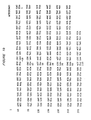
- the nucleotide sequences for the 5' region containing the leader peptide and the variable region, and the predicted amino acid sequences are shown in Figure 1A for pMK-9 and in Figure 1B for pM ⁇ 2a-1. (Amino acid residues are numbered, and the negative numbers refer to amino acids in the leader peptide).
- V L variable region-specific probe was derived from pMK-9 as a BamHI/PvuI fragment which contained the first 320 nucleotides of the light chain gene ( Figure 1A).
- the heavy chain V region probes contained two PstI fragments: the 228bp V H 1 and the 132bp V H 2 probes corresponding to nucleotide 52 to 280, and 281 to 412, respectively ( Figure 1B). These V region probes were used in subsequent experiments to characterize the genomic DNA fragments containing functionally rearranged genes.
- a genomic DNA library was prepared for the 17-1A cells. Approximately 200,000 lambda phage recombinants were screened with the mouse C K probe. Three positive clones were obtained. One of the clones, XK4, was shown to contain the variable region sequences by restriction mapping and Southern analysis using the V L probe derived from the cDNA clone, pMK-9. A 4.2 kb HindIII genomic DNA fragment containing 1.5 kb of the 5' flanking region and the sequences encoding the leader peptide and the V gene was subcloned into pUC 18, and designated as pV K 4.2H. This subclone was used in the subsequent construction of the mouse/human chimeric light chain gene.
- V H heavy chain variable gene of the cDNA clone
- pM ⁇ 2a-1 the heavy chain variable gene of the cDNA clone
- An alternative approach was taken to clone the V H gene. During a gene rearrangement that is required for Ig gene expression, the V gene is always associated with the appropriate J segment.
- the DNA probe J H can therefor be used to detect the rearranged V H genes.
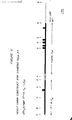
- Figure 2 shows the Southern analysis of rearranged fragments containing J H sequences. (Ten pg of genomic DNA was digested with EcoRI, fractionated on 0.7% agarose, transferred to nitrocellulose, and the filter hybridized with the mouse heavy chain J H probe.
- Lane 1 mouse liver DNA; lane 2, P3, a mouse plasmacytoma cell line; lane 3, 17-1A, a hybridoma cell line derived by using P 3 as a fusion partner.
- the arrowhead indicates the rearranged fragment containing the functional V H gene of 17-lA.
- the solid circle indicates additional rearrangement in the heavy chain locus).
- DNA of 17-1A cells showed two rearranged EcoRI fragments at 7.4 kb and 3.8 kb in addition to the band characteristic of the fusion partner, P3.
- the P3 cells had two rearranged V H genes which comigrated at 6 kb.
- One of the two bands of 17-1A represented the functional rearrangement, while the other was the product of an aberrant gene rearrangement in the heavy chain locus.
- both genes were cloned from phage genomic libraries constructed with enriched DNA fragments of appropriate sizes.
- Clone ⁇ V H 7.4E was isolated from a XgtWES library and the EcoRI insert was found to comigrate with the 7.4 kb rearranged fragment, as indicated with an arrowhead'in Figure 2.
- Clone ⁇ V H 3.8E was isolated from a Xgtll library and its EcoRI insert comigrated with the 3.8 kb band, as indicated with a solid circle in Figure 2..
- both ⁇ V H 7.4E and ⁇ V H 3.8E were digested with Pstl and hybridized with the mixed VH1 and V H 2 probes derived from the cDNA clone, pM ⁇ 2a-1.
- ⁇ V H 7.4E contained two PstI fragments at 300 bp and 130 bp.
- V H 1 probe contained sequences encoding for the amino acid residue -5 which is just upstream of the intron/exon boundary within the leader peptide
- the difference between the longer Pstl fragment in the genomic clone (200 bp) and that in the cDNA clone (228 bp) suggested the size of the intron between the leader peptide and the variable region gene to be 70 bp.
- Clone ⁇ V H 3.8E contained different length PstI fragments and did not cross-hybridize with V H l and V H 2 probes.
- ⁇ V H 7.4E is the genomic counterpart of the cDNA clone pM Y2a-1
- a 24-mer oligonucleotide probe corresponding to nucleotide 352 to 375 was synthesized and found to specifically hybridize to the 130 bp PstI fragment of theXV H 7.4E (data not shown).
- This oligomer contained sequences encoding for the CDR3 region and should be characteristic of this V gene.
- ⁇ V H 7.4E was used in the construction of the mouse/ human chimeric heavy chain gene.
- the light and heavy chain V genes cloned from the 17-1A cells were joined to human K and ⁇ 3 constant region genes in expression vectors pSV184 Hneo (Chang, A. C. Y., and S. N. Cohn, J. Bac- teriol., (1978) 143:1141) and pSV2 Hgpt (Mulligan, R. C., and P. Berg., Proc. Natl. Acad. Sci. USA (1981) 78:2072), respectively.
- the pSV184 ⁇ Hneo-DNSV L -hC K plasmid was used which had the dansyl- specific light chain gene and the human constant region K gene cloned into the BamHI and HindIII sites in pSV184 Hneo.
- the HindIII fragment of pSV184 ⁇ HneoDNSV L -hC K containing the dansyl V L gene was replaced with the 4.2 kb HindIII fragment containing the functionally rearranged genomic light chain gene of 17-1A derived from the clone pV K 4.2H.
- the EcoRI fragment in the pSV2A HgptDNSV H -hC ⁇ 3 plasmid containing the dansyl-specific V H gene was replaced with the 7.4 kb EcoRI fragment containing the functionally rearranged 17-1A heavy chain gene derived from the genomic clone ⁇ V H 7.4E.
- the resulting plasmid is designated as pSV2 ⁇ Hgpat171AVH-hC ⁇ 3 .
- the structure of pSV184A - Hneol7-1AV K -hC k is shown in Figure 3A and the structure of pSV2 ⁇ Hgpt17-1AVH-hC ⁇ 3 in Figure 3B.
- Immunoglobulin DNA is represented by thick lines and exons by solid boxes. Vector sequences are shown as thin lines. The transcriptional direction of the neo and gpt genes are as indicated. The ranges of the DNA sequences are indicated with two-way horizontal arrows. L, leader exon; V, VJ or VDJ excn; C, constant region exon; H, hinge exon; 1, 2, and 3, exons encoding other domains of the heavy chain gene. Restriction endonucleases abbreviations: E, EcoRI; B, BamHI; H, HindIII).
- the light and heavy chain vectors shown in Figure 3 were used to transfect mouse myeloma cells.
- the pACYC184 plasmid confers chloramphenicol resistance and the pBR plasmid confers ampicillin resistance. It is therefore possible to transform E. coli with both light and heavy chain plasmids and select cells expressing dual drug resistance phenotypes.
- E. coli harboring both plasmids were converted to protoplasts and fused with a nonproducing mouse myeloma cell line, Sp2/0. After protoplast fusion, the transfected cells were initially selected only for neo gene activity in the presence of G418. The stable transformant lines were subsequently carried in medium containing both G418 and mycophenolic acid.
- transfected cells were labeled with 35 S-methionine for three hours, after which both the cytoplasmic extract and secreted material were immunoprecipitated with Sepharose-bound goat anti- human K antibody.
- the precipitated material was analyzed on 5% SDS/polyacrylamide gels containing phosphate buffer under nonreducing conditions Matsuuchi, L. et al. 1981 Biochem 20:4827. From 4 x 10 6 cells, seven stable transformants were established and analyzed. Two of these clones produced both heavy and light chain proteins, two produced only light chain proteins, and three did not produce any detectable amounts of immunoglobulin proteins.
- Figure 4 shows the result of a biosynthetic labeling experiment for the SG3/5 cell line that produced both heavy and light chain protein.
- c cytoplasmic; s, secreted.
- L light chain
- a mixture of heavy and light chain molecules were detected in the cell extreact; however, the major components secreted into the culture medium were the tetrameric molecules, H 2 L 2' Similar results were obtained using the Sepharose-bound anti-human IgG Fc antibody for the immunoprecipitations.
- the SG3/7 cells which also showed resistance to both G418 and mycophenolic acid produced and secreted the light chain proteins only.
- the antibody secretion level of the SG3/5 cells was estimated to be 20 ⁇ g/ml by particle concentration fluorescence immunoassay.
- the culture supernatant was used as a source of chimeric MAb SG3/5 without further purification.
- a binding inhibition assay was used to demonstrate that the chimeric MAb SG3/5 bound to the same surface antigen of the SW1116 colorectal carcinoma cells as MAb 1 7- 1 A.
- the binding curves of radioiodinated 17-1A and SG3/5 were superimposable. This indicated that the different constant regions of MA b 17-1A and SG3/5 had little effect on the antigen-binding properties of these two antibodies.
- the DNA segment encoding the variable region of the heavy chain from from the mouse MAb 17-lA was linked to the human ⁇ 1, ⁇ 2 , and ⁇ 4 constant regions.
- the resulting chimeric heavy chain genes were cotransfected with the chimeric light chain gene (Figure 3B) into Sp2/0 cells to generate stable cell lines secreting ( ⁇ 1, k ), ( ⁇ 2, k ) and ( ⁇ 4, k ) antibodies. All these chimeric antibodies were shown to retain the same tumor antigen-binding specificity as the murine MAb 17-1A.
- the chimeric u construct (pSV2 J Hgpt17-1A-V H -hC ⁇ ) is shown in Figure 6.
- a stable cell line producing chimeric Ig( ⁇ , ⁇ ) antibody was established.
- Secreted antibody was purified on an anti-human IgM affinity column.
- the purified IgM had similar HPLC profiles as a human pentameric IgM control with 99% of the material under one peak. This purified chimeric IgM was used to check the binding specificity and complement-dependent cytotoxicity.
- 125I-labeled native 17-1A was incubated with adherent HT-29 colon cancer cells in the presence or absence of varying concentrations of competing non-radioactive chimeric 17-lA IgG and TgM for 2 hours at 4'C. Cell-associated radioactivity was determined. The inhibition curve for each competing immunoglobulin preparation was plotted and the concentration producing 50% inhibition (I 50 ) determined.
- Target cells employed in the assay were the human colorectal carcinoma lines, SW1116, HT-29, and SW948; human breast carcinoma lines, BT-20 and ZR75-1; human ovarian carcinoma, O VGAR3; human embryonic kidney (HEK); and human embryonic intestine (HEI).
- Example 4 In vitro and in vivo studies demonstrating anti-tumor activity of chimeric 17-1A antibodies.
- the isolated G1K 17-1A chimeric gene product was biologically tested in 1) antibody-dependent cell-mediated cytotoxicity assays (ADCC) and 2) binding assays using HT-29 cells (colon cancer cell line that expresses the 17-lA antigen) and partially purified 17-1A antigen.
- ADCC antibody-dependent cell-mediated cytotoxicity assays
- HT-29 cells colon cancer cell line that expresses the 17-lA antigen
- Methodologies for these investigations are presented in detail in Shaw, D. R . et al. J. Immunol., June 1987; Levy, P.C. et al. J. Immunol. 123(2): 594(1979); and Ross, A.H. et al. Biochem. Biophys. Res. Commun. 135(1): 297 (1986).
- G1K 17-1A Chimeric was tested in conjunction with the native murine 17-1A IgG and 3 additional chimeric isotypes shown below; rabbit anti-CEM * was employed as a polyclonal control.
- competitive binding studies utilizing partially purified 17-1A antigen G1K 17-1A Chimeric was compared to the native murine 17-1A IgG.
- the monocyte ADCC assay was performed utilizing Effector: Target (E:T) ratios of 25:1, 10:1 and 3 :1.
- E:T Effector: Target
- the mean % lysis ( ⁇ S.D) for each test material at the specified E:T ratios are presented below.
- the lympocyte ADCC assay was performed utilizing E:T ratios of 100:1, 30A1 and 10:1.
- the mean % lysis ( ⁇ S.D) values are shown below.
- Binding to the HT-29 cells was comparable between G1K 17-1A Chimeric IgG murine 17-1A.
- microtiter plates were incubated with partially purified 17-1A antigen (isolated from SW116 cells) for 2 hours at 37°C. Non-specific binding sites of the wells were blocked with PBS + 1% BSA for 1 hour at room temperature and were washed with PBS. 125 I labeled murine 17-1A and the competing antibody (cold murine 17-1A or G1K 17-1A Chimeric IgG) were incubated in the wells at 4°C overnight. The wells were washed 3 times with PBS and counted in a gamma counter.
- the 50% binding values for G1K 17-1A Chimeric and murine 17-1A were determined to be 0.30 pg/ml and 0.16 ⁇ g/ml, respectively. These results indicate that the G1K 17-1A Chimeric IgG is comparable to murine 17-1A in antibody binding.
- Murine 17-1A and G1K 17-1A Chimeric IgG were shown to react in 4 out of 4 colon tumor specimens examined.
- Other tumor cells examined included pancreatic cancer, lung cancer (adeno- and bronchia-) breast fibroadenomas and carcinomas, stomach cancer, melanoma, hepatoma and ovarian cancer.
- Murine 17-1A reacted with lung cancer (bronchia), stomach cancer, melanoma and ovarian cancer.
- GIK 17-1A Chimeric IgG did not react with any of the tumor specimens examined other than the colon tumors.
- mice Tumor growth inhibition experiments in nude mice have indicated that G1K 17-1A Chimeric is active in retarding tumor enlargement.
- the nude mouse tumor inhibition study was performed in 6 to 8 weeks old nude mice (NU/NU Balb C background) using previously described methods (Herlyn, D.M. et al., Cancer Res. 40:717 (1980)).
- Mice (5/group) were injected subcutaneously in the neck with 5 x 1 0 6 SW948 (colon tumor cells) and then injected intra- perioneally with 5 daily doses of 100 ⁇ g of murine 17-1A or each of 4 chimeric isotopes (including GIK 17-1A) or a control anti-influrenza antibody(H24B5).
- Tumor size was monitored weekly for 6 weeks by ' external measurement.
- the individual animal tumor volumes and group mean values for each measurement interval are shown in the Table 7 on the following page.
- the relative tumor inhibition ranking was found to be 17-lA G4K G1K.
- the chimeric antibodies G3K and G2K were not effective at the dose level tested.
- the invention also comprehends methods of manufacturing the immunoglobulin and related substances encompassed in the claims.
Abstract
Description
- Monoclonal antibodies have been developed against antigens associated with tumor for use in therapy of cancer. One example is the murine monoclonal antibody 17-1A which is directed against a surface antigen of colorectal carcinoma cells and other tumor cells. The antibody has been used for immunotherapy of colon cancer, and in several instances, the treatment has resulted in partial or complete regression of colorectal carcinoma.
- Most monoclonal antibodies that have been developed against tumor associated antigens are murine antibodies. The principle reason is that techniques for production of monoclonal antibodies in this species are well developed. Human monoclonal antibodies are difficult to produce. The standard technique used to make murine hybridomas- immunizing with antigen and harvesting spleen cells for fusion-obviously cannot be used to produce human monoclonal antibodies. Human lymphocytes which produce antibodies against a particular antigen can be selected from persons naturally exposed to the antigen but hybridomas formed with human cells are often unstable and cease producing antibodies.
- The success of the murine system for production of monoclonal antibodies provides a means to produce monoclonal antibodies against many types of cancer antigens. Murine antibodies, however, have several drawbacks which may severely limit the use in human cancer therapy. As foreign proteins, murine antibodies often elicit counteracting immune reactions which may reduce or destroy their therapeutic effectiveness. In addition, murine antibodies can evoke allergic reactions in patients. Unfortunately, the need for readministration in therapy increases the likelihood of these immune reactions in patients.
- A method which has been suggested to circumvent these problems is to produce antibodies consisting of mouse variable regions joined to human constant regions. See, e.g., Morrison, S. L. et al. (1984) "Chimeric Human Antibody Molecules: Mouse Antigen-binding Domains with Human Constant Region Domains" Proc. Natl. Acad. Sci. USA 81:6851; Neuberger, M. S. and Rabbits, T. H. "Production of Chimeric Antibodies" PCT Application No. PCT/GB85 00392. These chimeric antibodies would have the antigen binding specificity of the murine antibody but because they have human constant regions they would be, for the most part, human antibodies. Because the constant region is largely responsible for immune response against antibody, chimeric murine/human antibodies with constant regions are of human origin should be less likely to evoke an anti-murine immune response in humans. Further, the human constant region may provide for an antibody with better effector function in humans.
- The joining of a human constant region to a murine variable region might alter the conformation of the variable region in a manner which reduces or destroys the binding capability of the resulting chimera. Chimeric murine/murine and murine/human antibodies having variable region for haptens such as azophenylarsonate and trinitrophenyl have been produced and these antibodies have been shown to retain the original hapten-binding specificity of the murine antibody from which the variable regions are derived. See, for example, Sharon, J. et al. (1984) "Expression of a VHCK Chimeric Protein in Mouse Myeloma Cells" Nature 309:364-366; Boulianne, G. L. et al., (1984) "Production of Functional Chimeric Mouse/ Human Antibody", Nature, 312: 604-608; and Neuberger, M. S. et al., "A Hapten Specific Chimeric IgE Antibody with Human Physiological Effector Function" (1985) Nature 314: 268-270. However, because hapten molecules are small, conformational changes which may be induced in the murine variable region by the attachment of a human constant region may not significantly alter binding to such molecules. When the cognate antigen is a large molecule such as a tumor or proliferation associated cell surface antigen, conformational changes in the region could substantially influence binding capability.
- This invention pertains to chimeric immunoglobulins comprising a rodent-derived antigen binding region which is specific for a tumor- or proliferation-associated antigen joined to a human constant region. The chimeric immunoglobulins comprise:
- a) at least one chimeric heavy chain comprising an antigen-binding region derived from the heavy chain of a rodent antibody specific for a tumor-or proliferation-associated antigen linked to a human heavy chain constant region; and
- b) at least one chimeric light chain comprising an antigen-binding region derived from a light chain of the rodent antibody linked to a human light chain constant region.
- The immunoglobulins can be monovalent dimers divalent tetramers or polyvalent aggregates. These chimeric rodent/human immunoglobulins retain the original binding specificity for the tumor- or proliferation-associated antigen.
- The rodent variable regions of the chimeric immunoglobulin are derived from immunoglobulins specific for tumor- or proliferation-associated antigens. Normally, the variable regions will be of murine origin because murine antibodies against such antigens are available or can be made in the well- established murine system. Examples of tumor-associated antigens for which specifically reactive murine immunoglobulins are available are the antigens 17-1A (gastrointestinal tumors), OC125 (ovarian carcinoma), OV-TL3 (ovarian carcinoma), 103D2 (breast) and 123.C3 (renal carcinoma). Others include antigens 72.3 (colon), DF3, 115D8, RC38 (renal) and G250 and 55-2A. The rat system provides an alternative source of variable regions.
- The constant regions of the chimeric immunoglobulin are derived from human immunoglobulins. The constant region of the heavy chain can be selected from any isotype class or subclass and the constant region of the light chain can be of the kappa or lambda class.
- The chimeric antibodies are produced by cloning DNA segments encoding the heavy and light chain variable regions of a rodent antibody specific for a tumor-associated antigen and joining these DNA segments to DNA segments encoding human heavy and light chain constant regions to produce chimeric immunoglobulin encoding genes. The fused gene constructs coding for the light and heavy chain are assembled in or inserted into expression vectors. The genes are cotransfected into a lymphoid recipient cell (e.g. a myeloma cell) where the immunoglobulin proteins can be synthesized, assembled and secreted.
- The chimeric antibodies of this invention are useful for therapy and diagnosis of cancer. The chimeric antibodies may be used for tumor therapy alone or with several other immunoglobulins in mixtures or "cocktails" of antibodies specific for different antigens exhibited by a heterogenous tumor. In another mode of therapy, the chimeric immunoglobulins can be conjugated, either chemically or by genetic engineering techniques, to cancer therapeutic agents to form so-called "smart" drugs specifically directed to cancer cells. For diagnostic use, the chimeric immunoglobulins can provide agents for tumor imaging. For example, the immunoglobulins can be made with truncated heavy chains to provide "antibody fragments" and labeled either directly or through a label chelator. The chimeric immunoglobulins can be designed to contain a "built- in" label chelating protein or protein domain.
- The use of chimeric murine/human antibodies in vivo offers several advantages over the use of murine monoclonal antibody. First, these antibodies may be less immunogenic in eliciting human antimouse reactions in patients. Second, a human constant region of choice can be linked to a mouse variable region gene to produce antibodies with the desired biological effector functions. Finally, it is possible to express hybrid genes constructed by joining Ig DNA and non-Ig DNA sequences. As mentioned above, these fusion proteins constitute antibody based delivery systems for selective destruction or imaging of tumor cells.
-
- Figure 1 shows the nucleotide sequences of the 17-lA functional light chain (A) and heavy chain (B) genes and the predicted amino acid sequences.
- Figure 2 shows the Southern blot analysis of rearranged 17-1A VH genes containing J. sequences.
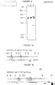
- Figure 3 shows the structure of the chimeric heavy and light chain vectors A, pSV184ΔHneo17-1AVK-hCK; B, pSV2ΔHgpt17-1AVH-
hC γ 3. - Figure 4 shows SDS/PAGE of 35 S-methionine labeled chimeric immunoglobulin proteins of transfected Sp2/0 cell line SG3/5 and SG3/7.
- Figure 5 shows the inhibition of 17-1A binding to SW1116 cells by the chimeric immunoglobulin SG3/5.
- Figure 6 shows the structure of the chimeric heavy chain vector pSV2ΔHgpt17-1AVH- hCn.
- The chimeric immunoglobulins of this invention are comprised of individual chimeric heavy and light , immunoglobulin chains. The chimeric heavy chain is a contiguous polypeptide having a rodent heavy chain variable region and a human heavy chain constant region. The chimeric light chain is a contiguous polypeptide having a rodent light chain variable region and human light chain constant region.
- The immunoglobulins can be monovalent, divalent or polyvalent. Monovalent immunoglobulins are dimers (HL) formed of a chimeric heavy chain associated (through disulfide bridges) with a chimeric . light chain. Divalent immunoglobulins are tetramers (H2L2) formed of two associated dimers. Polyvalent immunoglobulins can be produced, for example, by employing heavy chain constant region which aggregate (e.g., mu type constant regions).
- The variable regions are derived from antibodies specific for tumor- or proliferation-associated antigens. The antigens binding regions can be specific for gastrointestinal, breast, ovarian, lung and renal cancer cell antigens. Examples are antigens defined by murine antibodies specific for 17-1A (gastrointestinal tumors), OC125 (ovarian carcinoma), OV-TL3 (ovarian carcinoma), 103D2 (breast) and 123.C3 (renal carcinoma). Others include 72.3 (colon), DF3, 115D8, RC38 (renal), G250 and 55-2A. The heavy chain constant region can be selected from any of the five isotypes alpha, delta, epsilon, gamma or mu. Further, heavy chains of various subclasses (such as the IgG subclasses) can be used. The different classes and subclasses of heavy chains are responsible for different effector functions and thus, by choosing the desired heavy chain constant region, chimeric antibodies with desired effector function can be produced. Preferred constant regions are gamma 1 (IgGl) and gamma 3 (IgG3). The light chain constant region can be the kappa or lambda chain.
- In general, the chimeric antibodies are produced by preparing, for each of the light and heavy chains components of the chimeric immunoglobulin, a fused gene comprising a first DNA segment which encodes at least the functional portion of the murine variable region linked to a second DNA segment encoding at least a part of a human constant region. Each fused gene is assembled in or inserted into an expression vector. Recipient cells capable of expressing the gene products are then transfected with the genes. The transfected recipient cells are cultured and the expressed immunoglobulins or immunoglobulin chains are recovered.
- Genes encoding the variable region of murine Ig light and heavy chains can be obtained from lymphoid cells which produce the antibodies specific for the desired tumor antigen. For example, the hybridoma cell lines which produce antibody against the antigens 17-1A, OC125 or any of the other antigens provide a source of immunoglobulin variable region genes against certain tumor associated antigens. other rodent cell lines are available. Cell lines can be produced by challanging a rodent with a tumor cell or a tumor antigen containing cell component or fraction, forming fused hybrid cells between antibody producing cells and a myeloma cloning the hybrid and selecting clones which produce antibody against tumor-associated antigen. See U.S. Patent No. 4,172,124 Koprowski et al.
- Constant regions can be obtained from human antibody producing cells by standard cloning techniques. Alternatively, because genes representing the two classes of light chains and the five classes of heavy chains have been cloned, constant regions of human origin are readily available from these clones.
- Preferably, the fused genes encoding the light and heavy chimeric chains are assembled in two different expression vectors which can be used to cotransform a recipient cell. Each vector contains two selectable genes-one for selection in a bacterial system and one for selection in a eukaryotic system- each vector having a different pair of genes. These vectors allow production and amplification of the fused genes in bacterial systems and subsequent cotransfection of eukaryotic cells and selection of the cotransfected cells. Examples of selectable gene for the bacterial system are the genes which confer ampicillin and the gene which couples chloramphenicol resistance. Two selectable genes for selection of eukaryotic transfectants are preferred: (i) the xanthine-guanine phosphoribosyltransferase gene (gpt), and (ii) the phosphotransferase gene from Tn5 (designated neo). Selection with gpt is based on the ability of the enzyme encoded by this gene to use xanthine as a substrate for purine nucleotide synthesis; the analogous endogenous enzyme cannot. In a medium containing xanthine and mycophenolic acid which blocks the conversion of inosine monophosphate to xanthine monophosphate, only cells expressing the gpt gene can survive. The product of the neo blocks the inhibition of protein synthesis in eukaryotic cells caused by the antibiotic G418 and other antibiotics of its class. The two selection procedures can be used simultaneously or sequentially to select for the expression of immunoglobulin chain genes introduced on two different DNA vectors into a eukaryotic cell.
- The preferred recipient cell line is a myeloma cell. Myeloma cells can synthesize, assemble and secrete immunoglobulins encoded by transfected Ig genes. Further, they possess the mechanism for glycosylation of the immunoglobulin. A particularly preferred recipient cell is the Ig-nonproducing myeloma cell Sp2/0. The cell produces only immunoglobulin encoded by the transfected immunoglobulin genes. Myeloma cells can be grown in culture or in the peritoneum of mice where secreted immunoglobulin can be obtained from ascites fluid. Other lymphoid cells such as B lymphocytes or hybridoma cells can serve as suitable recipient cells.
- Several methods exist for transfecting lymphoid cell with vectors containing immunoglobulin encoding genes. A preferred way of introducing DNA into lymphoid cells is by protoplast fusion. In this method, lysozyme is used to strip cell walls from bacteria harboring the recombinant plasmid containing the chimeric Ig gene. The resulting spheroplasts are fused with myeloma cells with polyethylene glycol. After protoplast fusion, the transfectants are selected and isolated. Another technique which can be used to introduce DNA into many cell types is calcium phosphate precipitation.
- The chimeric immunoglobulin genes can be expressed in nonlymphoid cells such as bacteria or yeast. When expressed in bacteria, the immunoglobulin heavy chains and light chains become part of inclusion bodies. Thus, the chains must be isolated and purified and then assembled into functional immunoglobulin molecules.
- A functional chimeric antibody specific for the 17-1A antigen associated with colorectal carcinoma and some other forms of cancer was prepared as follows.
- Genomic DNA segments encoding the heavy and light chain V regions of 17-lA (IgG2a, kappa light chain) were cloned and then joined to DNA segments encoding
human γ 3 constant (C γ3) and kappa constant (CK) regions. The recombinant genes were then transfected into mouse myeloma cells where the Ig proteins were synthesized, assembled and secreted. To obtain probes for the V regions, a cDNA library for 17-1A was constructed. RNA was extracted and polyA RNA prepared by oligodT cellulose chromatography. Double-stranded cDNA was synthesized with polyA RNA as template using AMV reverse transcriptase and E. coli DNA polymerase I. Double-stranded cDNA was treated with S1 nuclease and elongated with deoxycytidine residues, and annealed with dG-tailed pUCB previously cut with Pstl. The recombinant plasmids were transformed into E. coli DH1, and colonies screened using a mouse CK probe and a mouse C γ2a probe to isolate the light and heavy chain clones, respectively. These clones were used as a source of V region probes. - To isolate the genomic DNA fragment containing the functionally rearranged kappa light chain gene, a genomic library was constructed from Sau3A partially digested DNA of 17-lA using EMBL3A phage arms. A total of 200,000 recombinant plaques were screened with a mouse CK probe, three positive clones were obtained. One of these clones,
λ K4, was shown to contain both the V and C regions of the functionally rearranged kappa light chain gene of 17-lA by restriction mapping and Southern analysis using the 17-1A K cDNA as a probe. When the same procedure was repeated using a mouse C γ2a probe and the heavy chain V(VH) probe derived from the cDNA clone, no positive clones were found to contain both the V and C regions of the heavy chain gene. - An alternative approach was taken to clone the VH gene. Fragments containing the rearranged heavy chain V genes of 17-1A were identified by Southern analysis as additional bands when compared with the pattern of the fusion partner, NS-1. When a DNA sample of 17-lA cells was digested with restriction enzyme EcoRI, two rearranged fragments of 7.4kb and 3.8kb were noted using a mouse heavy chain J region (JH) probe containing J3 and J4 sequences. DNA fragments of ~ 7kb and ~ 4kb isolated by preparative agarose gel electrophoresis were ligated with λgtWES and λgtll phage arms, respectively. The recombinant plaques of these two partial libraries were screened with a mouse JH probe. Both 7.4kb and 3.8kb EcoRI fragments were cloned. The 7.4kb fragment was shown to contain the functionally rearranged V gene by restriction mapping and Southern analysis using the 17-lA heavy chain cDNA as a probe.
- The VK gene was isolated in a 4kb HindIII fragment from the
genomic clone λ K4 and joined to the human CK sequences in the expression vector pACYCSVneo. The direction of the K gene transcription is opposite that of the neo gene. The 7.4kb EcoRI fragment containing the V gene was joined to the human C γ3 gene in the expression vector pSV2gpt with the direction of transcription opposite that of Ecogpt. Since the pACYCSVneo plasmid confers chloramphenicol resistance and the pSV2gpt plasmid confers ampicillin resistance, it is possible to transform E. coli with both plasmids and select cells expressing dual drug resistance phenotypes. To transfer the Ig genes into myeloma cells, E. coli harboring both plasmids were converted to protoplasts and fused with a nonproducing mouse myeloma line, Sp2/0. After protoplast fusion, the transfected cells were selected for neo gene activity by treatment with G418. To examine heavy and light chain protein synthesis, transfected cells were labeled with 35S-methionine, both cytoplasmic extract and secreted material were immunoprecipitated with Sepharose-bound goat anti-human K antibody. The precipitated material was analyzed on SDS/polyacrylamide gels containing phosphate buffer under nonreducing conditions. From 4×10⁶ cells, seven stable transformants were established and analyzed. Two of these clones produced both heavy and light chain proteins, two produced only light chain proteins, and three did not produce any detectable amounts of Ig proteins. The clone SG3/5, one of the two transformants that produced both heavy and light chain proteins produced a mixture of H2L2, H2L and HL molecules. The major components secreted into the culture medium were the tetrameric molecules, H2L2. The chimeric H2L2 immunogloblin displayed the same antigen binding properties as the murine 17-1A antibody. - Chimeric rodent/human antibodies specific for cancer-associated antigens can be used for therapy or diagnosis of cancer. For therapy, the antibodies can be used in a number of different ways. Certain antibodies are capable of specifically binding to and killing tumor cells by themselves. Chimeric antibodies having the variable regions of such antibodies can be administered in anti-tumor amounts to patients to treat tumor. For example, the chimeric murine/human 17-lA antibody can be administered to patients bearing gastrointestinal tumors with which the 17-lA antigen is associated.
- Cancer cells are heterogenous and consequently, a single monospecific chimeric antibody may not be able to recognize all cells of a tumor. Thus, it may be desirable to administer several chimeric antibodies of different antigen specificity in combination.
- The chimeric antibodies can be used in conjunction with other anti-tumor agents. For this purpose, the chimeric murine/human antibodies can be conjugated to certain therapeutic agents or cytotoxic agents or to functional domains of such proteins. In this capacity, the immunoglobulins would act as delivery vehicles specifically targeted for cancer cells. Such conjugation can be accomplished by chemically coupling the therapeutic agent to the antibody. In cases where the therapeutic agent is proteinaceous, the conjugation can be accomplished by linking an agent-encoding DNA sequence to the chimeric gene encoding an antibody chain (preferably a heavy chain) so that the antibody chain and agent are expressed as a single contiguous protein in which the agent is linked by a peptide bond to the immunoglobulin. In this way, chimeric immunoglobulins containing immunoglobulin and nonimmunoglobulin portions can be formed where the nonimmunoglobulin portion can be a selected therapeutic agent such as interferon or selected cytoxic agents such as tumor necrosis factor.
- The chimeric antibodies are also useful in vivo diagnostic techniques such as immunoscintigraphy. For this purpose, antibody fragments are generally preferred. Thus, a chimeric heavy chain gene can be designed in truncated form to produce chimeric Fab immunoglobulin fragment or a chimeric F(abt)2-like molecule for tumor imaging. These molecules can be labeled directly or through a coupled chelating agent with radioisotopes such as 131 Iodine, 125Iodine, 99mTechnetium or Indium to produce radioimmunoscintigraphic agents for tumor imaging. Alternatively, a radiometal binding (chelating) domain can be engineered into the chimeric antibody for labeling a site. Thus, a chimeric immunoglobulin can be designed as a contiguous protein which has a murine variable region, a human constant region (preferably truncated), and a metal binding domain derived from metal binding protein such as metallothionine.
- The invention is illustrated further by the following detailed examples describing the production of the chimeric antibody with 17-1A derived variable regions and human constant regions.
- Cytoplasmic RNA was extracted from 17-1A cells and polyA RNA was prepared by oligo-dT cellulose chromatography. Double-stranded cDNA was synthesized with polyA+ RNA as a template using AMV reverse transcriptase and E. coli DNA polymerase I. Double-stranded cDNA was treated with S1 nuclease and elongated with deoxycytidine residues, and annealed with dG-tailed pUC8 previously cut with Pstl. curtis, P.J., (1983) J. Biol. Chem. 258:4459. The recombinant plasmids were transformed into E. coli DH1, and colonies screened according to the general method described by Maniatis, T. et al. (1982) Molecular Cloning. A Laboratory Manual.
- Genomic library was constructed in lambda phage vector EMBL3A. High molecular weight DNA was partially digested with restriction endonuclease Sau3A and size-fractionated on a 10-40% sucrose density gradient. DNA fragments of 18-23kb were ligated with EMBL3A lambda arms and packaged according to Hohn and Murray (1977) Proc. Natl. Acad. sci USA 74: 3259. The genomic library was screened at a density of 10,000 recombinant plaques per 150mm diameter petri dish. Plaque hybridizations were carried out in 5x SSC at 65° for 18h. Final washes were in 1.0x or 0.5x SSC at 65'.
- High molecular weight genomic DNA was digested to completion with restriction endonuclease and fractionated on a 0.8% agarose gel. DNA fragments of the appropriate size was isolated and ligated with lambda phage arms. The ligated DNA were packaged and recombinant plaques were screened as described above.
- Genomic DNA was digested with restriction endonucleases fractionated by electrophoresis through a 0.7% agarose gel, and blotted to nitrocellulose. See Maniatis et al. supra. Hybridizations were in 5x SSC and 50% formamide at 37° for 48h. Final washes were 0.5x SSC at 65'.
- The mouse γ 2a probe is a 4.9 kb EcoRI DNA fragment containing the Y 2a constant region gene. The mouse Ck probe is a 6 kb Bg1II genomic DNA fragment containing the mouse k light chain constant region sequences. The mouse heavy chain J (JH) probe is a 2 kb BamHI/EcoRI fragment containing both J3 and J4 segments. 32 P-labeled probes were prepared by using calf thymus primers. Summers, J. (1975) J. Virol 15:946. Free nucleotides were removed by centrifugation through a Sephadex G-75 mini-column.
- The construction of pSV184neo- and pSV2gpt- derived by plasmids carrying the chimeric light and heavy chain genes is described below. E. coli HB101 harboring both plasmids were grown in the presence of ampicillin and chloramphenicol. The plasmid copy number was amplified with spectinomycin at 100ug/ml. Protoplasts were prepared and fused to mouse myeloma Sp2/0 cells according to Oi, et. al., (1983) Proc. Natl. Acad. Sci. USA 80:825. Transformants were selected in Dulbecco's modified Eagle's medium containing antibiotic G418 at 0.8 mg/ml, supplemented with 15% fetal calf serum. The SG3/5 cell line was maintained in the above medium plus xanthine at 50 pg/ml, hypoxanthine at 4 µg/ml and mycophenolic acid at 0.8 µg/ml.
- Cells were labeled for 3h in methionine-free RPMI 1640 medium containing 35S-methionine at 25µCi/ml. Affinity-purified goat anti-human K light chain antibody (Southern Biotechnology Associated, Inc., Birmingham, AL) and goat anti-human IgG Fc antibody (Jackson Immunoresearch Laboratories, Inc., Avondale, PA) were used for immunoprecipitations. Morrison, S.L. (1979) J. Immunol. 123:793. Both cytoplasmic and secreted antibodies were analyzed on a 5% SDS/polyacrylamide gel in phosphate buffer under nonreducing conditions. Matsuuchi, L. et al. (1981) Biochem. 20:4827. Gel was treated with an autoradiography enhancer, EN3HANCE (NEN Products, Boston, MA) before drying down onto 3mm filter paper.
- Tissue culture supernatant was analyzed for IgG protein content by particle concentration fluorescence immunoassay (Jolley, M.E. et al., (1984) J. Immunol. Meth. 67:21 using standard curves generated with purified IgG. Concentration of MAb 17-1A was determined using goat anti-mouse Fab antibody-coated polystyrene beads and fluorescein conjugated anti- mouse Fab antibody. Chimeric antibody with human constant regions was determined using goat antihuman IgG Fc antibody-coated polystyrene beads and fluorescein conjugated goat anti-human IgG Fc antibody. The assay was carried out with an automated instrument (Pandex Laboratories, Inc., Mundelein, IL).
- MAb 17-1A was purified from ascites fluid by using protein A-Sepharose chromatography. Bound IgG was eluted with a pH 3.5 citrate buffer. Purified MAb 17-lA was labeled with 1.4 mM choramine T using 1 mCi Na125I. After 3 min, the reaction was quenched with excess ascorbic acid. Free iodide was removed with a prepacked PD-10 column (Pharmacia). Specific activity was typically 10,000cpm/ng of protein.
- Tissue culture supernatant was concentrated with a Diaflo YM100 ultrafiltration membrane (Ami- con, Danvers, MA). Monolayer colorectal carcinoma cells were trypsinized and washed in phosphatebuffer saline (PBS). Cells (5×10⁵) were incubated with radioiodinated 17-1A and culture supernatant containing the competing antibody in a final volume of 100 ul. Incubation was done at room temperature for 2 hr in a shaker at 140rpm. The cells were washed with PBS and cell-bound radioactivity counted in a gamma counter.
- A cDNA library was prepared for the 17-1A cells using plasmid vector pUC8. Recombinant colonies were screened with mouse C probe and the mouse Cy2a probe to isolate the light and heavy chain clones, respectively. A total of 7,500 colonies were screened, approximately 250 of which contained CK sequences and 150 contained Cy2a sequences.
- Plasmid DNA prepared from some of these positive colonies was digested with PstI to compare the size of the cDNA inserts. The light chain clone, pMK-9, and the heavy chain clone, pM γ 2a-1, were chosen for nucleotide sequencing. The nucleotide sequences for the 5' region containing the leader peptide and the variable region, and the predicted amino acid sequences are shown in Figure 1A for pMK-9 and in Figure 1B for pM γ 2a-1. (Amino acid residues are numbered, and the negative numbers refer to amino acids in the leader peptide). A variable region- specific (VL) probe was derived from pMK-9 as a BamHI/PvuI fragment which contained the first 320 nucleotides of the light chain gene (Figure 1A). The heavy chain V region probes contained two PstI fragments: the
228bp V H1 and the132bp V H2 probes corresponding to nucleotide 52 to 280, and 281 to 412, respectively (Figure 1B). These V region probes were used in subsequent experiments to characterize the genomic DNA fragments containing functionally rearranged genes. - A genomic DNA library was prepared for the 17-1A cells. Approximately 200,000 lambda phage recombinants were screened with the mouse CK probe. Three positive clones were obtained. One of the clones, XK4, was shown to contain the variable region sequences by restriction mapping and Southern analysis using the VL probe derived from the cDNA clone, pMK-9. A 4.2 kb HindIII genomic DNA fragment containing 1.5 kb of the 5' flanking region and the sequences encoding the leader peptide and the V gene was subcloned into pUC 18, and designated as pVK4.2H. This subclone was used in the subsequent construction of the mouse/human chimeric light chain gene.
- When the 17-lA genomic library was screened with the mouse C γ 2a probe, two positive clones were obtained, but neither of these contained the heavy chain variable (VH) gene of the cDNA clone, pM γ 2a-1. An alternative approach was taken to clone the VH gene. During a gene rearrangement that is required for Ig gene expression, the V gene is always associated with the appropriate J segment. The DNA probe JH can therefor be used to detect the rearranged VH genes. Figure 2 shows the Southern analysis of rearranged fragments containing JH sequences. (Ten pg of genomic DNA was digested with EcoRI, fractionated on 0.7% agarose, transferred to nitrocellulose, and the filter hybridized with the mouse heavy chain JH probe.
Lane 1, mouse liver DNA;lane 2, P3, a mouse plasmacytoma cell line;lane 3, 17-1A, a hybridoma cell line derived by using P3 as a fusion partner. The arrowhead indicates the rearranged fragment containing the functional VH gene of 17-lA. The solid circle indicates additional rearrangement in the heavy chain locus). - DNA of 17-1A cells showed two rearranged EcoRI fragments at 7.4 kb and 3.8 kb in addition to the band characteristic of the fusion partner, P3. The P3 cells had two rearranged VH genes which comigrated at 6 kb. One of the two bands of 17-1A represented the functional rearrangement, while the other was the product of an aberrant gene rearrangement in the heavy chain locus. Using the mouse JH probe, both genes were cloned from phage genomic libraries constructed with enriched DNA fragments of appropriate sizes. Clone λVH 7.4E was isolated from a XgtWES library and the EcoRI insert was found to comigrate with the 7.4 kb rearranged fragment, as indicated with an arrowhead'in Figure 2. Clone λVH3.8E was isolated from a Xgtll library and its EcoRI insert comigrated with the 3.8 kb band, as indicated with a solid circle in Figure 2..
- To indentify the genomic counterpart of the functional heavy chain gene, both λVH7.4E and λVH3.8E were digested with Pstl and hybridized with the mixed VH1 and
V H2 probes derived from the cDNA clone, pM γ2a-1. λVH7.4E contained two PstI fragments at 300 bp and 130 bp. Since theV H1 probe contained sequences encoding for the amino acid residue -5 which is just upstream of the intron/exon boundary within the leader peptide, the difference between the longer Pstl fragment in the genomic clone (200 bp) and that in the cDNA clone (228 bp) suggested the size of the intron between the leader peptide and the variable region gene to be 70 bp. Clone λVH3.8E contained different length PstI fragments and did not cross-hybridize with VHl andV H2 probes. To further verify that λVH7.4E is the genomic counterpart of the cDNA clone pM Y2a-1, a 24-mer oligonucleotide probe corresponding to nucleotide 352 to 375 (Figure 1B) was synthesized and found to specifically hybridize to the 130 bp PstI fragment of theXVH7.4E (data not shown). This oligomer contained sequences encoding for the CDR3 region and should be characteristic of this V gene. λVH7.4E was used in the construction of the mouse/ human chimeric heavy chain gene. - The light and heavy chain V genes cloned from the 17-1A cells were joined to human K and γ3 constant region genes in expression vectors pSV184 Hneo (Chang, A. C. Y., and S. N. Cohn, J. Bac- teriol., (1978) 143:1141) and pSV2 Hgpt (Mulligan, R. C., and P. Berg., Proc. Natl. Acad. Sci. USA (1981) 78:2072), respectively. The pSV184ΔHneo-DNSVL-hCK plasmid was used which had the dansyl- specific light chain gene and the human constant region K gene cloned into the BamHI and HindIII sites in pSV184 Hneo. To construct the desired chimeric gene, the HindIII fragment of pSV184Δ HneoDNSVL -hCK containing the dansyl VL gene was replaced with the 4.2 kb HindIII fragment containing the functionally rearranged genomic light chain gene of 17-1A derived from the clone pVK 4.2H. For the heavy chain vector, the EcoRI fragment in the pSV2A HgptDNSVH-hCγ3 plasmid containing the dansyl-specific VH gene was replaced with the 7.4 kb EcoRI fragment containing the functionally rearranged 17-1A heavy chain gene derived from the genomic clone λVH7.4E. The resulting plasmid is designated as pSV2ΔHgpat171AVH-hCγ3. The structure of pSV184A - Hneol7-1AVK-hCk is shown in Figure 3A and the structure of pSV2ΔHgpt17-1AVH-hCγ3 in Figure 3B. (Immunoglobulin DNA is represented by thick lines and exons by solid boxes. Vector sequences are shown as thin lines. The transcriptional direction of the neo and gpt genes are as indicated. The ranges of the DNA sequences are indicated with two-way horizontal arrows. L, leader exon; V, VJ or VDJ excn; C, constant region exon; H, hinge exon; 1, 2, and 3, exons encoding other domains of the heavy chain gene. Restriction endonucleases abbreviations: E, EcoRI; B, BamHI; H, HindIII).
- The light and heavy chain vectors shown in Figure 3 were used to transfect mouse myeloma cells. The pACYC184 plasmid confers chloramphenicol resistance and the pBR plasmid confers ampicillin resistance. It is therefore possible to transform E. coli with both light and heavy chain plasmids and select cells expressing dual drug resistance phenotypes. To transfect the chimeric genes into mouse myeloma cells, E. coli harboring both plasmids were converted to protoplasts and fused with a nonproducing mouse myeloma cell line, Sp2/0. After protoplast fusion, the transfected cells were initially selected only for neo gene activity in the presence of G418. The stable transformant lines were subsequently carried in medium containing both G418 and mycophenolic acid.
- To examine heavy and light chain protein synthesis, transfected cells were labeled with 35S-methionine for three hours, after which both the cytoplasmic extract and secreted material were immunoprecipitated with Sepharose-bound goat anti- human K antibody. The precipitated material was analyzed on 5% SDS/polyacrylamide gels containing phosphate buffer under nonreducing conditions Matsuuchi, L. et al. 1981 Biochem 20:4827. From 4 x 10 6 cells, seven stable transformants were established and analyzed. Two of these clones produced both heavy and light chain proteins, two produced only light chain proteins, and three did not produce any detectable amounts of immunoglobulin proteins. Figure 4 shows the result of a biosynthetic labeling experiment for the SG3/5 cell line that produced both heavy and light chain protein. (c, cytoplasmic; s, secreted. The positions of the tet- rameric H2L2 protein and the light chain (L) protein are indicated.) A mixture of heavy and light chain molecules were detected in the cell extreact; however, the major components secreted into the culture medium were the tetrameric molecules, H2L2' Similar results were obtained using the Sepharose-bound anti-human IgG Fc antibody for the immunoprecipitations. The SG3/7 cells which also showed resistance to both G418 and mycophenolic acid produced and secreted the light chain proteins only.
- The antibody secretion level of the SG3/5 cells was estimated to be 20 µg/ml by particle concentration fluorescence immunoassay. The culture supernatant was used as a source of chimeric MAb SG3/5 without further purification. A binding inhibition assay was used to demonstrate that the chimeric MAb SG3/5 bound to the same surface antigen of the SW1116 colorectal carcinoma cells as MAb 17-1A. As shown in Figure 5, the binding curves of radioiodinated 17-1A and SG3/5 were superimposable. This indicated that the different constant regions of MAb 17-1A and SG3/5 had little effect on the antigen-binding properties of these two antibodies.
- The DNA segment encoding the variable region of the heavy chain from from the mouse MAb 17-lA was linked to the
human γ 1,γ 2 , andγ 4 constant regions. The resulting chimeric heavy chain genes were cotransfected with the chimeric light chain gene (Figure 3B) into Sp2/0 cells to generate stable cell lines secreting (γ1, k ), (γ2, k ) and (γ4, k ) antibodies. All these chimeric antibodies were shown to retain the same tumor antigen-binding specificity as the murine MAb 17-1A. - The chimeric u construct (pSV2 J Hgpt17-1A-VH-hCµ) is shown in Figure 6. This gene construct, together with the 17-lA chimeric K light chain gene construct (fig 3A), were transfected into the non-producing mouse myeloma Sp2/0 cells. A stable cell line producing chimeric Ig(µ,κ ) antibody was established. Secreted antibody was purified on an anti-human IgM affinity column. The purified IgM had similar HPLC profiles as a human pentameric IgM control with 99% of the material under one peak. This purified chimeric IgM was used to check the binding specificity and complement-dependent cytotoxicity.
- 125I-labeled native 17-1A was incubated with adherent HT-29 colon cancer cells in the presence or absence of varying concentrations of competing non-radioactive chimeric 17-lA IgG and TgM for 2 hours at 4'C. Cell-associated radioactivity was determined. The inhibition curve for each competing immunoglobulin preparation was plotted and the concentration producing 50% inhibition (I50) determined.

- Complement dependent cytotoxicity was assessed by a 45 minute radiolabel release assay of antibody coated target cells incubated with serial dilutions of rabbit complement. Target cells employed in the assay were the human colorectal carcinoma lines, SW1116, HT-29, and SW948; human breast carcinoma lines, BT-20 and ZR75-1; human ovarian carcinoma, OVGAR3; human embryonic kidney (HEK); and human embryonic intestine (HEI).


- The isolated G1K 17-1A chimeric gene product was biologically tested in 1) antibody-dependent cell-mediated cytotoxicity assays (ADCC) and 2) binding assays using HT-29 cells (colon cancer cell line that expresses the 17-lA antigen) and partially purified 17-1A antigen. Methodologies for these investigations are presented in detail in Shaw, D.R. et al. J. Immunol., June 1987; Levy, P.C. et al. J. Immunol. 123(2): 594(1979); and Ross, A.H. et al. Biochem. Biophys. Res. Commun. 135(1): 297 (1986). In the ADCC and competitive binding studies in HT-29 cells, G1K 17-1A Chimeric was tested in conjunction with the native murine 17-1A IgG and 3 additional chimeric isotypes shown below; rabbit anti-CEM* was employed as a polyclonal control. In the competitive binding studies utilizing partially purified 17-1A antigen, G1K 17-1A Chimeric was compared to the native murine 17-1A IgG.
- The data for the monocyte and lymphocyte ADCC assays were generated from four separate experiments using monocytes and lymphocytes from four different normal adult donors. Mean spontaneous release ± SD was 18.7 ± 2.8% of total target cell 51CR (N=28).
- The monocyte ADCC assay was performed utilizing Effector: Target (E:T) ratios of 25:1, 10:1 and 3:1. The mean % lysis (± S.D) for each test material at the specified E:T ratios are presented below.

- In the monocyte ADCC assay the % lysis with G1K 17-1A chimeric IgG was comparable to that seen with murine 17-1A.
- The lympocyte ADCC assay was performed utilizing E:T ratios of 100:1, 30A1 and 10:1. The mean % lysis (±S.D) values are shown below.

- In the lymphocyte ADCC assay the % lysis with G1K 17-1A Chimeric IgG was comparable to that seen with 17.1A.
- In the binding assay utilizing intact HT-29 cells, the cells were coated with the test antibody. Quantitation for the 17-1A and rabbit anti-CEM used 1-protein A; quantitation for chimeric antibodies used 125 I-anti-human IgG(Fc). Data on the quantitation of IgG bound to the tumor cells are presented below. The results represent a mean (±S.E.) of four or five separate experiments.


- Binding to the HT-29 cells was comparable between G1K 17-1A Chimeric IgG murine 17-1A.
- In the competitive binding assay using partially purified 17-1A antigen, microtiter plates were incubated with partially purified 17-1A antigen (isolated from SW116 cells) for 2 hours at 37°C. Non-specific binding sites of the wells were blocked with PBS + 1% BSA for 1 hour at room temperature and were washed with PBS. 125I labeled murine 17-1A and the competing antibody (cold murine 17-1A or G1K 17-1A Chimeric IgG) were incubated in the wells at 4°C overnight. The wells were washed 3 times with PBS and counted in a gamma counter. The 50% binding values for G1K 17-1A Chimeric and murine 17-1A were determined to be 0.30 pg/ml and 0.16 µg/ml, respectively. These results indicate that the G1K 17-1A Chimeric IgG is comparable to murine 17-1A in antibody binding.
- In addition to the above in-vitro studies, in vitro immunohistopathology studies with G1K 17-1A and murine 17-1A were performed on various human tumor tissues using immunofluorescence technology. Murine 17-1A (24 µg/ml) and G1K 17-1A Chimeric IgG (96 µg/ml) were shown to react in 4 out of 4 colon tumor specimens examined. Other tumor cells examined included pancreatic cancer, lung cancer (adeno- and bronchia-) breast fibroadenomas and carcinomas, stomach cancer, melanoma, hepatoma and ovarian cancer. Murine 17-1A reacted with lung cancer (bronchia), stomach cancer, melanoma and ovarian cancer. GIK 17-1A Chimeric IgG did not react with any of the tumor specimens examined other than the colon tumors.
- Tumor growth inhibition experiments in nude mice have indicated that G1K 17-1A Chimeric is active in retarding tumor enlargement. The nude mouse tumor inhibition study was performed in 6 to 8 weeks old nude mice (NU/NU Balb C background) using previously described methods (Herlyn, D.M. et al., Cancer Res. 40:717 (1980)). Mice (5/group) were injected subcutaneously in the neck with 5 x 10⁶ SW948 (colon tumor cells) and then injected intra- perioneally with 5 daily doses of 100 µg of murine 17-1A or each of 4 chimeric isotopes (including GIK 17-1A) or a control anti-influrenza antibody(H24B5). Tumor size was monitored weekly for 6 weeks by ' external measurement. The individual animal tumor volumes and group mean values for each measurement interval are shown in the Table 7 on the following page. The relative tumor inhibition ranking was found to be 17-lA G4K G1K. The chimeric antibodies G3K and G2K were not effective at the dose level tested.
-


- Those skilled in the art will recognize, or be able to ascertain using no more than routine experimentation, many equivalents to the specific embodiments of the invention described herein. Such equivalents are intended to be encompassed by the following claims.
- The invention also comprehends methods of manufacturing the immunoglobulin and related substances encompassed in the claims.
Claims (18)
Applications Claiming Priority (4)
| Application Number | Priority Date | Filing Date | Title |
|---|---|---|---|
| US88249286A | 1986-07-07 | 1986-07-07 | |
| US882492 | 1986-07-07 | ||
| US6941487A | 1987-07-01 | 1987-07-01 | |
| US69414 | 1987-07-01 |
Publications (3)
| Publication Number | Publication Date |
|---|---|
| EP0256654A2 true EP0256654A2 (en) | 1988-02-24 |
| EP0256654A3 EP0256654A3 (en) | 1989-06-07 |
| EP0256654B1 EP0256654B1 (en) | 1996-09-18 |
Family
ID=26750032
Family Applications (1)
| Application Number | Title | Priority Date | Filing Date |
|---|---|---|---|
| EP87306006A Expired - Lifetime EP0256654B1 (en) | 1986-07-07 | 1987-07-07 | Chimeric murine/human immunoglobulin specific for tumour-associated 17-1A Antigen |
Country Status (5)
| Country | Link |
|---|---|
| EP (1) | EP0256654B1 (en) |
| JP (1) | JPH1070991A (en) |
| AT (1) | ATE143052T1 (en) |
| CA (1) | CA1340456C (en) |
| DE (1) | DE3751908T2 (en) |
Cited By (59)
| Publication number | Priority date | Publication date | Assignee | Title |
|---|---|---|---|---|
| EP0338767A2 (en) * | 1988-04-21 | 1989-10-25 | Eli Lilly And Company | Novel recombinant and chimeric antibodies directed against a human adenocarcinoma antigen |
| EP0340948A1 (en) * | 1988-04-28 | 1989-11-08 | Mycogen Corporation | Novel hybrid pesticidal toxins |
| EP0380068A1 (en) * | 1989-01-24 | 1990-08-01 | Miles Inc. | An expression system for production of chimeric monoclonal antibodies |
| WO1991013166A1 (en) * | 1990-03-02 | 1991-09-05 | Abbott Biotech, Inc. | Antibody constructs with enhanced binding affinity |
| US5155027A (en) * | 1988-01-22 | 1992-10-13 | Zymogenetics, Inc. | Method of producing secreted receptor analogs and biologically active peptide dimers |
| EP0528767A1 (en) * | 1991-08-21 | 1993-02-24 | Sandoz Ltd. | Antibody derivatives |
| EP0537489A1 (en) * | 1991-10-11 | 1993-04-21 | BEHRINGWERKE Aktiengesellschaft | Monoclonal antibodies against tumor-associated antigens, method for their preparation and their use |
| US5336603A (en) * | 1987-10-02 | 1994-08-09 | Genentech, Inc. | CD4 adheson variants |
| US5567584A (en) * | 1988-01-22 | 1996-10-22 | Zymogenetics, Inc. | Methods of using biologically active dimerized polypeptide fusions to detect PDGF |
| US5585089A (en) * | 1988-12-28 | 1996-12-17 | Protein Design Labs, Inc. | Humanized immunoglobulins |
| WO1997017374A1 (en) * | 1995-11-08 | 1997-05-15 | Medac Gesellschaft Für Klinische Spezialpräparate Gmbh | Recombinant ligands for the human cell membrane antigen cd30 |
| US5750375A (en) * | 1988-01-22 | 1998-05-12 | Zymogenetics, Inc. | Methods of producing secreted receptor analogs and biologically active dimerized polypeptide fusions |
| US5843708A (en) * | 1988-01-05 | 1998-12-01 | Ciba-Geigy Corporation | Chimeric antibodies |
| US6018026A (en) * | 1988-01-22 | 2000-01-25 | Zymogenetics, Inc. | Biologically active dimerized and multimerized polypeptide fusions |
| US6020153A (en) * | 1988-01-05 | 2000-02-01 | Ciba-Geigy Corporation | Chimeric antibodies |
| US6030797A (en) * | 1992-10-08 | 2000-02-29 | Dade Behring Marburg Gmbh | Monoclonal antibodies against tumor-associated antigens, processes for the preparation thereof and the use thereof |
| WO2003050241A2 (en) | 2001-12-12 | 2003-06-19 | The Government Of The United States Of America As Represented By The Secretary, Department Of Healthand Human Services | Methods for using extracellular adenosine inhibitors and adenosine receptor inhibitors to enhance immune response and inflammation |
| US6710169B2 (en) | 1987-10-02 | 2004-03-23 | Genentech, Inc. | Adheson variants |
| EP1666499A2 (en) | 1996-05-07 | 2006-06-07 | Genentech, Inc. | Novel inhibitors of vascular endothelial growth factor activity, their uses and processes for their production |
| US7282205B2 (en) | 2001-11-07 | 2007-10-16 | The United States Of America As Represented By The Secretary Of The Department Of Health And Human Services | Anti-hepatitis A virus antibodies |
| EP1970386A2 (en) | 1989-11-22 | 2008-09-17 | Genetech, Inc. | Hybrid immunoglobulins |
| EP2067790A2 (en) | 1994-04-04 | 2009-06-10 | Genentech, Inc. | Agonist antibody against protein tyrosine kinase SAL-S1 |
| EP2085467A2 (en) | 2002-10-29 | 2009-08-05 | The Government Of The United States Of America As Represented By The Secretary Of The Department Of Health And Human Services | Lutzomyia Longipalpis polypeptides and methods of use |
| EP2158917A1 (en) | 2002-09-19 | 2010-03-03 | The Government of the United States of America, as represented by the Secretary of the Department of Health and Human Services | P.ariasi polypeptides, P.perniciosus polypeptides and methods of use |
| WO2010099472A2 (en) | 2009-02-27 | 2010-09-02 | The U.S.A. Of America, As Represented By The Secretary, Department Of Health And Human Services | Spanx-b polypeptides and their use |
| US7812135B2 (en) | 2005-03-25 | 2010-10-12 | Tolerrx, Inc. | GITR-binding antibodies |
| EP2241622A2 (en) | 1994-03-18 | 2010-10-20 | Genentech, Inc. | Human trk receptors and their derivatives |
| EP2253957A1 (en) | 2006-03-14 | 2010-11-24 | Oregon Health and Science University | Methods for producing an immune response to tuberculosis. |
| WO2011047340A1 (en) | 2009-10-16 | 2011-04-21 | The United States Of America, As Represented By The Secretary, Department Of Health & Human Services | Insertion of foreign genes in rubella virus and their stable expression in a live, attenuated viral vaccine |
| EP2385371A2 (en) | 2008-09-22 | 2011-11-09 | Oregon Health and Science University | Methods for detecting a mycobacterium tuberculosis infection |
| WO2012006635A1 (en) | 2010-07-09 | 2012-01-12 | Biogen Idec Hemophilia Inc. | Processable single chain molecules and polypeptides made using same |
| WO2012031280A2 (en) | 2010-09-03 | 2012-03-08 | Stem Centrx, Inc. | Identification and enrichment of cell subpopulations |
| EP2524699A1 (en) | 2011-05-17 | 2012-11-21 | Trion Research GmbH | Vaccine preparation containing trifunctional antibodies with antigen immunogenicity enhancer properties |
| WO2012156430A1 (en) | 2011-05-17 | 2012-11-22 | Trion Research Gmbh | Vaccine preparation containing trifunctional antibodies with antigen immunogenicity enhancer properties |
| WO2013012733A1 (en) | 2011-07-15 | 2013-01-24 | Biogen Idec Ma Inc. | Heterodimeric fc regions, binding molecules comprising same, and methods relating thereto |
| WO2013049362A2 (en) | 2011-09-27 | 2013-04-04 | The United States Of America, As Represented By The Secretary, Department Of Health & Human Services | Method of treating multiple sclerosis by intrathecal depletion of b cells and biomarkers to select patients with progressive multiple sclerosis |
| WO2013106577A2 (en) | 2012-01-10 | 2013-07-18 | Biogen Idec Ma Inc. | Enhancement of transport of therapeutic molecules across the blood brain barrier |
| WO2013119964A2 (en) | 2012-02-08 | 2013-08-15 | Stem Centrx, Inc. | Identification and enrichment of cell subpopulations |
| WO2013152352A1 (en) | 2012-04-06 | 2013-10-10 | The United States Of America, As Represented By The Secretary, Department Of Health And Human Servic | Live, attenuated rubella vector to express vaccine antigens |
| US8591886B2 (en) | 2007-07-12 | 2013-11-26 | Gitr, Inc. | Combination therapies employing GITR binding molecules |
| WO2014043518A1 (en) | 2012-09-14 | 2014-03-20 | The United States Of America, As Represented By The Secretary, Department Of Health And Human Services | Brachyury protein, non-poxvirus non-yeast vectors encoding brachyury protein, and their use |
| WO2014046730A1 (en) | 2012-09-24 | 2014-03-27 | Ventana Medical Systems, Inc. | Method of identifying treatment responsive non-small cell lung cancer using anaplastic lymphoma kinase (alk) as a marker |
| WO2014102130A1 (en) | 2012-12-28 | 2014-07-03 | Ventana Medical Systems, Inc. | Image analysis for breast cancer prognosis |
| EP2918598A1 (en) | 2007-02-28 | 2015-09-16 | The Govt. Of U.S.A. As Represented By The Secretary Of The Department Of Health And Human Services | Brachyury polypeptides and methods for use |
| US9221908B2 (en) | 2012-12-12 | 2015-12-29 | Vasculox, Inc. | Therapeutic CD47 antibodies |
| WO2015199976A1 (en) | 2014-06-24 | 2015-12-30 | The United States Of America, As Represented By The Secretary, Department Of Health & Human Services | Target activated microdissection |
| WO2016022896A1 (en) | 2014-08-08 | 2016-02-11 | The United States Of America, As Represented By The Secretary, Department Of Health & Human Services | Photo-controlled removal of targets in vitro and in vivo |
| US9486507B2 (en) | 2011-06-10 | 2016-11-08 | Biogen Ma Inc. | Pro-coagulant compounds and methods of use thereof |
| US9696323B2 (en) | 2013-03-10 | 2017-07-04 | The Board Of Regents Of The University Of Texas System | Biomarkers for chagas disease related cardiomyopathy |
| EP3263124A1 (en) | 2009-11-20 | 2018-01-03 | Oregon Health&Science University | Methods for producing an immune response to tuberculosis |
| US10239945B2 (en) | 2015-09-18 | 2019-03-26 | Arch Oncology, Inc. | Therapeutic CD47 antibodies |
| WO2019073299A1 (en) | 2017-10-10 | 2019-04-18 | Medicenna Therapeutics, Inc. | Il-4-fusion formulations for treatment of central nervous system (cns) tumors |
| US10584147B2 (en) | 2013-11-08 | 2020-03-10 | Biovertiv Therapeutics Inc. | Procoagulant fusion compound |
| US10669336B2 (en) | 2012-12-12 | 2020-06-02 | Arch Oncology, Inc. | Therapeutic CD47 antibodies |
| US11014088B2 (en) | 2016-03-09 | 2021-05-25 | The Board Of Regents Of The University Of Texas System | Sensitive ELISA for disease diagnosis on surface modified poly(methyl methacrylate) (PMMA) microfluidic microplates |
| US11168125B2 (en) | 2003-05-06 | 2021-11-09 | Bioverativ Therapeutics Inc. | Immunoglobulin chimeric monomer-dimer hybrids |
| US11692035B2 (en) | 2016-10-21 | 2023-07-04 | Arch Oncology, Inc. | Therapeutic CD47 antibodies |
| EP4183806A3 (en) * | 2014-11-12 | 2023-08-02 | Seagen Inc. | Glycan-interacting compounds and methods of use |
| WO2024044687A1 (en) | 2022-08-26 | 2024-02-29 | Valo Health, Inc. | Biomarker combinations for prognosis and management of diabetic retinopathy |
Families Citing this family (2)
| Publication number | Priority date | Publication date | Assignee | Title |
|---|---|---|---|---|
| JP5372500B2 (en) | 2005-06-17 | 2013-12-18 | トレラクス リクイデーティング トラスト | ILT3-binding molecules and uses thereof |
| CA2655903A1 (en) | 2006-06-19 | 2008-08-07 | Tolerx, Inc. | Ilt3 binding molecules and uses therefor |
Citations (4)
| Publication number | Priority date | Publication date | Assignee | Title |
|---|---|---|---|---|
| WO1986001533A1 (en) * | 1984-09-03 | 1986-03-13 | Celltech Limited | Production of chimeric antibodies |
| EP0184187A2 (en) * | 1984-12-04 | 1986-06-11 | Teijin Limited | Mouse-human chimaeric immunoglobulin heavy chain, and chimaeric DNA encoding it |
| WO1987002671A1 (en) * | 1985-11-01 | 1987-05-07 | International Genetic Engineering, Inc. | Modular assembly of antibody genes, antibodies prepared thereby and use |
| EP0171496B1 (en) * | 1984-08-15 | 1993-05-26 | Research Development Corporation of Japan | Process for the production of a chimera monoclonal antibody |
-
1987
- 1987-07-07 AT AT87306006T patent/ATE143052T1/en not_active IP Right Cessation
- 1987-07-07 DE DE3751908T patent/DE3751908T2/en not_active Expired - Lifetime
- 1987-07-07 CA CA000541501A patent/CA1340456C/en not_active Expired - Lifetime
- 1987-07-07 EP EP87306006A patent/EP0256654B1/en not_active Expired - Lifetime
-
1997
- 1997-07-22 JP JP9228781A patent/JPH1070991A/en active Pending
Patent Citations (4)
| Publication number | Priority date | Publication date | Assignee | Title |
|---|---|---|---|---|
| EP0171496B1 (en) * | 1984-08-15 | 1993-05-26 | Research Development Corporation of Japan | Process for the production of a chimera monoclonal antibody |
| WO1986001533A1 (en) * | 1984-09-03 | 1986-03-13 | Celltech Limited | Production of chimeric antibodies |
| EP0184187A2 (en) * | 1984-12-04 | 1986-06-11 | Teijin Limited | Mouse-human chimaeric immunoglobulin heavy chain, and chimaeric DNA encoding it |
| WO1987002671A1 (en) * | 1985-11-01 | 1987-05-07 | International Genetic Engineering, Inc. | Modular assembly of antibody genes, antibodies prepared thereby and use |
Non-Patent Citations (9)
| Title |
|---|
| ANTIBODY, IMMUNOCONJUGATES & RADIOPHARMACEUTICALS, vol. 5, no. 1, 1992; R.F. MEREDITH et al., pp. 75-80 * |
| JOURNAL OF EXPERIMENTAL MEDICINE, vol. 163, 1986; R.J. BENJAMIN et al., pp. 1539-1552 * |
| JOURNAL OF IMMUNOLOGICAL METHODS, vol. 85, 1985; D. HERLYN et al., pp. 27-38 * |
| JOURNAL OF IMMUNOLOGY, vol. 135, no. 2, 1985; D.L. SHAWLER et al., pp. 1530-1535 * |
| NATURE, vol. 314, no. 6010, April 1985, pages 452-454, London, GB; S. TAKEDA et al.: "Construction of chimaeric processed immunoglobulin genes containing mouse variable and human constant region sequences" * |
| PROCEEDINGS OF THE NATIONAL ACADEMY OF SCIENCE, vol. 81, November 1984, pages 6851-6855, Washington, D.C. US; S.L. MORRISON et al.: "Chimeric human antibody molecules: Mouse antigen-binding domains with human constant region domains" * |
| PROCEEDINGS OF THE NATIONAL ACADEMY OF SCIENCE, vol. 84, January 1987, pages 214-218, Washington, D.C. US; L.K. SUN et al.: "Chimeric antibody with human constant regions and mouse variable regions directed against carcinoma-associated antigen 17-1A" * |
| SCIENCE, vol. 229, 20th September 1985, pages 1202-1207, Washington, D.C., US; S.L. MORRISON: "Transfectomas provide novel chimeric antibodies" * |
| THE LANCET, no. 8481, 15th March 1986, pages 603-605, London, GB; R.W. BALDWIN et al.: "Monoclonal antibodies in cancer treatment" * |
Cited By (100)
| Publication number | Priority date | Publication date | Assignee | Title |
|---|---|---|---|---|
| US6117655A (en) * | 1987-10-02 | 2000-09-12 | Genentech, Inc. | Nucleic acid encoding adhesion variants |
| US5336603A (en) * | 1987-10-02 | 1994-08-09 | Genentech, Inc. | CD4 adheson variants |
| US5565335A (en) * | 1987-10-02 | 1996-10-15 | Genentech, Inc. | Adhesion variants |
| US6710169B2 (en) | 1987-10-02 | 2004-03-23 | Genentech, Inc. | Adheson variants |
| US6020153A (en) * | 1988-01-05 | 2000-02-01 | Ciba-Geigy Corporation | Chimeric antibodies |
| US5843708A (en) * | 1988-01-05 | 1998-12-01 | Ciba-Geigy Corporation | Chimeric antibodies |
| US6018026A (en) * | 1988-01-22 | 2000-01-25 | Zymogenetics, Inc. | Biologically active dimerized and multimerized polypeptide fusions |
| US5843725A (en) * | 1988-01-22 | 1998-12-01 | Zymogenetics, Inc. | Methods of producing secreted receptor analogs and biologically active dimerized polypeptide fusions |
| US5155027A (en) * | 1988-01-22 | 1992-10-13 | Zymogenetics, Inc. | Method of producing secreted receptor analogs and biologically active peptide dimers |
| US5750375A (en) * | 1988-01-22 | 1998-05-12 | Zymogenetics, Inc. | Methods of producing secreted receptor analogs and biologically active dimerized polypeptide fusions |
| US6323323B1 (en) | 1988-01-22 | 2001-11-27 | Zymogenetics, Inc. | Ligand-binding, dimerized polypeptide fusions |
| US5567584A (en) * | 1988-01-22 | 1996-10-22 | Zymogenetics, Inc. | Methods of using biologically active dimerized polypeptide fusions to detect PDGF |
| EP0338767A2 (en) * | 1988-04-21 | 1989-10-25 | Eli Lilly And Company | Novel recombinant and chimeric antibodies directed against a human adenocarcinoma antigen |
| EP0338767A3 (en) * | 1988-04-21 | 1991-07-17 | Eli Lilly And Company | Novel recombinant and chimeric antibodies directed against a human adenocarcinoma antigen |
| EP0340948A1 (en) * | 1988-04-28 | 1989-11-08 | Mycogen Corporation | Novel hybrid pesticidal toxins |
| US7022500B1 (en) | 1988-12-28 | 2006-04-04 | Protein Design Labs, Inc. | Humanized immunoglobulins |
| US5585089A (en) * | 1988-12-28 | 1996-12-17 | Protein Design Labs, Inc. | Humanized immunoglobulins |
| US5693761A (en) * | 1988-12-28 | 1997-12-02 | Protein Design Labs, Inc. | Polynucleotides encoding improved humanized immunoglobulins |
| US5693762A (en) * | 1988-12-28 | 1997-12-02 | Protein Design Labs, Inc. | Humanized immunoglobulins |
| US6180370B1 (en) | 1988-12-28 | 2001-01-30 | Protein Design Labs, Inc. | Humanized immunoglobulins and methods of making the same |
| EP0380068A1 (en) * | 1989-01-24 | 1990-08-01 | Miles Inc. | An expression system for production of chimeric monoclonal antibodies |
| EP1970386A2 (en) | 1989-11-22 | 2008-09-17 | Genetech, Inc. | Hybrid immunoglobulins |
| WO1991013166A1 (en) * | 1990-03-02 | 1991-09-05 | Abbott Biotech, Inc. | Antibody constructs with enhanced binding affinity |
| EP0528767A1 (en) * | 1991-08-21 | 1993-02-24 | Sandoz Ltd. | Antibody derivatives |
| EP0537489A1 (en) * | 1991-10-11 | 1993-04-21 | BEHRINGWERKE Aktiengesellschaft | Monoclonal antibodies against tumor-associated antigens, method for their preparation and their use |
| US5695758A (en) * | 1991-10-11 | 1997-12-09 | Behringwerke Aktiengesellschaft | Monoclonal antibodies against tumor-associated antigens, processes for the preparation thereof and the use thereof |
| US5674994A (en) * | 1991-10-11 | 1997-10-07 | Behringwerke Aktiengesellschaft | Polynucleotides encoding the anti-PEM antibody BW835 variable domains and expression vectors |
| US5639621A (en) * | 1991-10-11 | 1997-06-17 | Behringwerke Aktiengesellschaft | Monoclonal antibodies against tumor-associated antigens |
| US6030797A (en) * | 1992-10-08 | 2000-02-29 | Dade Behring Marburg Gmbh | Monoclonal antibodies against tumor-associated antigens, processes for the preparation thereof and the use thereof |
| EP2241622A2 (en) | 1994-03-18 | 2010-10-20 | Genentech, Inc. | Human trk receptors and their derivatives |
| EP2067790A2 (en) | 1994-04-04 | 2009-06-10 | Genentech, Inc. | Agonist antibody against protein tyrosine kinase SAL-S1 |
| WO1997017374A1 (en) * | 1995-11-08 | 1997-05-15 | Medac Gesellschaft Für Klinische Spezialpräparate Gmbh | Recombinant ligands for the human cell membrane antigen cd30 |
| EP2336322A1 (en) | 1996-05-07 | 2011-06-22 | Genentech, Inc. | Inhibitors of vascular endothelial growth factor activity, their uses and processes for their production |
| EP1666499A2 (en) | 1996-05-07 | 2006-06-07 | Genentech, Inc. | Novel inhibitors of vascular endothelial growth factor activity, their uses and processes for their production |
| US7282205B2 (en) | 2001-11-07 | 2007-10-16 | The United States Of America As Represented By The Secretary Of The Department Of Health And Human Services | Anti-hepatitis A virus antibodies |
| US7635476B2 (en) | 2001-11-07 | 2009-12-22 | The United States Of America As Represented By The Secretary Of The Department Of Health And Human Services | Anti-hepatitis a virus antibodies |
| WO2003050241A2 (en) | 2001-12-12 | 2003-06-19 | The Government Of The United States Of America As Represented By The Secretary, Department Of Healthand Human Services | Methods for using extracellular adenosine inhibitors and adenosine receptor inhibitors to enhance immune response and inflammation |
| EP2158917A1 (en) | 2002-09-19 | 2010-03-03 | The Government of the United States of America, as represented by the Secretary of the Department of Health and Human Services | P.ariasi polypeptides, P.perniciosus polypeptides and methods of use |
| EP2085467A2 (en) | 2002-10-29 | 2009-08-05 | The Government Of The United States Of America As Represented By The Secretary Of The Department Of Health And Human Services | Lutzomyia Longipalpis polypeptides and methods of use |
| US11168125B2 (en) | 2003-05-06 | 2021-11-09 | Bioverativ Therapeutics Inc. | Immunoglobulin chimeric monomer-dimer hybrids |
| US10570209B2 (en) | 2005-03-25 | 2020-02-25 | Gitr, Inc. | Methods for inducing or enhancing an immune response by administering agonistic glucocorticoid-induced TNFR-family-related receptor (GITR) antibodies |
| US9493572B2 (en) | 2005-03-25 | 2016-11-15 | Gitr, Inc. | GITR antibodies and methods of inducing or enhancing an immune response |
| US10030074B2 (en) | 2005-03-25 | 2018-07-24 | Gitr, Inc. | Methods of inducing or enhancing an immune response in a subject having cancer by administering GITR antibodies |
| EP2343320A1 (en) | 2005-03-25 | 2011-07-13 | Tolerx, Inc. | GITR binding molecules and uses therefor |
| US8388967B2 (en) | 2005-03-25 | 2013-03-05 | Gitr, Inc. | Methods for inducing or enhancing an immune response by administering agonistic GITR-binding antibodies |
| US7812135B2 (en) | 2005-03-25 | 2010-10-12 | Tolerrx, Inc. | GITR-binding antibodies |
| US9028823B2 (en) | 2005-03-25 | 2015-05-12 | Gitr, Inc. | Methods of inducing or enhancing an immune response in a subject by administering agonistic GITR binding antibodies |
| EP2397855A2 (en) | 2006-03-14 | 2011-12-21 | Oregon Health and Science University | Methods for detecting a mycobacterium tuberculosis infection |
| EP2397854A2 (en) | 2006-03-14 | 2011-12-21 | Oregon Health and Science University | Methods for detecting a mycobacterium tuberculosis infection |
| EP2428801A1 (en) | 2006-03-14 | 2012-03-14 | Oregon Health and Science University | Methods for detecting a mycobacterium tuberculosis infection |
| EP2253957A1 (en) | 2006-03-14 | 2010-11-24 | Oregon Health and Science University | Methods for producing an immune response to tuberculosis. |
| EP2918598A1 (en) | 2007-02-28 | 2015-09-16 | The Govt. Of U.S.A. As Represented By The Secretary Of The Department Of Health And Human Services | Brachyury polypeptides and methods for use |
| US9241992B2 (en) | 2007-07-12 | 2016-01-26 | Gitr, Inc. | Combination therapies employing GITR binding molecules |
| US8591886B2 (en) | 2007-07-12 | 2013-11-26 | Gitr, Inc. | Combination therapies employing GITR binding molecules |
| EP2385371A2 (en) | 2008-09-22 | 2011-11-09 | Oregon Health and Science University | Methods for detecting a mycobacterium tuberculosis infection |
| WO2010099472A2 (en) | 2009-02-27 | 2010-09-02 | The U.S.A. Of America, As Represented By The Secretary, Department Of Health And Human Services | Spanx-b polypeptides and their use |
| WO2011047340A1 (en) | 2009-10-16 | 2011-04-21 | The United States Of America, As Represented By The Secretary, Department Of Health & Human Services | Insertion of foreign genes in rubella virus and their stable expression in a live, attenuated viral vaccine |
| EP3263124A1 (en) | 2009-11-20 | 2018-01-03 | Oregon Health&Science University | Methods for producing an immune response to tuberculosis |
| EP3360566A1 (en) | 2009-11-20 | 2018-08-15 | Oregon Health&Science University | Methods for detecting a mycobacterium tuberculosis infection |
| WO2012006635A1 (en) | 2010-07-09 | 2012-01-12 | Biogen Idec Hemophilia Inc. | Processable single chain molecules and polypeptides made using same |
| EP3560962A1 (en) | 2010-07-09 | 2019-10-30 | Bioverativ Therapeutics Inc. | Processable single chain molecules and polypeptides made using same |
| US9856468B2 (en) | 2010-07-09 | 2018-01-02 | Bioverativ Therapeutics Inc. | Processable single chain molecules and polypeptides made using same |
| US10927362B2 (en) | 2010-07-09 | 2021-02-23 | Bioverativ Therapeutics Inc. | Processable single chain molecules and polypeptides made using same |
| US10968442B2 (en) | 2010-07-09 | 2021-04-06 | Bioverativ Therapeutics Inc. | Chimeric clotting factors |
| WO2012006633A1 (en) | 2010-07-09 | 2012-01-12 | Biogen Idec Hemophilia Inc. | Chimeric clotting factors |
| WO2012031280A2 (en) | 2010-09-03 | 2012-03-08 | Stem Centrx, Inc. | Identification and enrichment of cell subpopulations |
| EP2689787A1 (en) | 2010-09-03 | 2014-01-29 | Stem Centrx, Inc. | Identification and enrichment of cell subpopulations |
| EP2524699A1 (en) | 2011-05-17 | 2012-11-21 | Trion Research GmbH | Vaccine preparation containing trifunctional antibodies with antigen immunogenicity enhancer properties |
| WO2012156430A1 (en) | 2011-05-17 | 2012-11-22 | Trion Research Gmbh | Vaccine preparation containing trifunctional antibodies with antigen immunogenicity enhancer properties |
| US9486507B2 (en) | 2011-06-10 | 2016-11-08 | Biogen Ma Inc. | Pro-coagulant compounds and methods of use thereof |
| EP3527218A1 (en) | 2011-06-10 | 2019-08-21 | Bioverativ Therapeutics Inc. | Pro-coagulant compounds and methods of use thereof |
| WO2013012733A1 (en) | 2011-07-15 | 2013-01-24 | Biogen Idec Ma Inc. | Heterodimeric fc regions, binding molecules comprising same, and methods relating thereto |
| WO2013049362A2 (en) | 2011-09-27 | 2013-04-04 | The United States Of America, As Represented By The Secretary, Department Of Health & Human Services | Method of treating multiple sclerosis by intrathecal depletion of b cells and biomarkers to select patients with progressive multiple sclerosis |
| WO2013106577A2 (en) | 2012-01-10 | 2013-07-18 | Biogen Idec Ma Inc. | Enhancement of transport of therapeutic molecules across the blood brain barrier |
| WO2013119964A2 (en) | 2012-02-08 | 2013-08-15 | Stem Centrx, Inc. | Identification and enrichment of cell subpopulations |
| WO2013152352A1 (en) | 2012-04-06 | 2013-10-10 | The United States Of America, As Represented By The Secretary, Department Of Health And Human Servic | Live, attenuated rubella vector to express vaccine antigens |
| WO2014043518A1 (en) | 2012-09-14 | 2014-03-20 | The United States Of America, As Represented By The Secretary, Department Of Health And Human Services | Brachyury protein, non-poxvirus non-yeast vectors encoding brachyury protein, and their use |
| US9651555B2 (en) | 2012-09-24 | 2017-05-16 | Ventana Medical Systems, Inc. | Method of identifying treatment responsive non-small cell lung cancer using anaplastic lymphoma kinase (ALK) as a marker |
| WO2014046730A1 (en) | 2012-09-24 | 2014-03-27 | Ventana Medical Systems, Inc. | Method of identifying treatment responsive non-small cell lung cancer using anaplastic lymphoma kinase (alk) as a marker |
| US11499974B2 (en) | 2012-09-24 | 2022-11-15 | Ventana Medical Systems, Inc. | Method of identifying treatment responsive non-small cell lung cancer using anaplastic lymphoma kinase (ALK) as a marker |
| US10676524B2 (en) | 2012-12-12 | 2020-06-09 | Arch Oncology, Inc. | Therapeutic CD47 antibodies |
| US10669336B2 (en) | 2012-12-12 | 2020-06-02 | Arch Oncology, Inc. | Therapeutic CD47 antibodies |
| US11292834B2 (en) | 2012-12-12 | 2022-04-05 | Arch Oncology, Inc. | Therapeutic CD47 antibodies |
| US9518117B2 (en) | 2012-12-12 | 2016-12-13 | Tioma Therapeutics, Inc. | Therapeutic CD47 antibodies |
| US9518116B2 (en) | 2012-12-12 | 2016-12-13 | Tioma Therapeutics, Inc. | Therapeutic CD47 antibodies |
| US10259873B2 (en) | 2012-12-12 | 2019-04-16 | Arch Oncology, Inc. | Therapeutic CD47 antibodies |
| US9221908B2 (en) | 2012-12-12 | 2015-12-29 | Vasculox, Inc. | Therapeutic CD47 antibodies |
| US11211167B2 (en) | 2012-12-28 | 2021-12-28 | Ventana Medical Systems, Inc. | Image analysis for breast cancer prognosis |
| WO2014102130A1 (en) | 2012-12-28 | 2014-07-03 | Ventana Medical Systems, Inc. | Image analysis for breast cancer prognosis |
| US9696323B2 (en) | 2013-03-10 | 2017-07-04 | The Board Of Regents Of The University Of Texas System | Biomarkers for chagas disease related cardiomyopathy |
| US10584147B2 (en) | 2013-11-08 | 2020-03-10 | Biovertiv Therapeutics Inc. | Procoagulant fusion compound |
| WO2015199976A1 (en) | 2014-06-24 | 2015-12-30 | The United States Of America, As Represented By The Secretary, Department Of Health & Human Services | Target activated microdissection |
| WO2016022896A1 (en) | 2014-08-08 | 2016-02-11 | The United States Of America, As Represented By The Secretary, Department Of Health & Human Services | Photo-controlled removal of targets in vitro and in vivo |
| EP4183806A3 (en) * | 2014-11-12 | 2023-08-02 | Seagen Inc. | Glycan-interacting compounds and methods of use |
| US10239945B2 (en) | 2015-09-18 | 2019-03-26 | Arch Oncology, Inc. | Therapeutic CD47 antibodies |
| US10844124B2 (en) | 2015-09-18 | 2020-11-24 | Arch Oncology, Inc. | Therapeutic CD47 antibodies |
| US11014088B2 (en) | 2016-03-09 | 2021-05-25 | The Board Of Regents Of The University Of Texas System | Sensitive ELISA for disease diagnosis on surface modified poly(methyl methacrylate) (PMMA) microfluidic microplates |
| US11692035B2 (en) | 2016-10-21 | 2023-07-04 | Arch Oncology, Inc. | Therapeutic CD47 antibodies |
| WO2019073299A1 (en) | 2017-10-10 | 2019-04-18 | Medicenna Therapeutics, Inc. | Il-4-fusion formulations for treatment of central nervous system (cns) tumors |
| WO2024044687A1 (en) | 2022-08-26 | 2024-02-29 | Valo Health, Inc. | Biomarker combinations for prognosis and management of diabetic retinopathy |
Also Published As
| Publication number | Publication date |
|---|---|
| CA1340456C (en) | 1999-03-23 |
| EP0256654A3 (en) | 1989-06-07 |
| DE3751908T2 (en) | 1997-02-06 |
| JPH1070991A (en) | 1998-03-17 |
| ATE143052T1 (en) | 1996-10-15 |
| DE3751908D1 (en) | 1996-10-24 |
| EP0256654B1 (en) | 1996-09-18 |
Similar Documents
| Publication | Publication Date | Title |
|---|---|---|
| EP0256654B1 (en) | Chimeric murine/human immunoglobulin specific for tumour-associated 17-1A Antigen | |
| US4978745A (en) | Immunoreactive heterochain antibodies | |
| JP3860398B2 (en) | Cytokine immune complex | |
| US5219996A (en) | Recombinant antibodies and methods for their production in which surface residues are altered to cysteine residues for attachment of effector or receptor molecules | |
| Sun et al. | Chimeric antibody with human constant regions and mouse variable regions directed against carcinoma-associated antigen 17-1A. | |
| CA2035899C (en) | Chimeric anti-cea antibody | |
| US5892019A (en) | Production of a single-gene-encoded immunoglobulin | |
| US5591593A (en) | Minimum recognition unit of a PEM mucin tandem repeat specific monoclonal antibody and detection method using same | |
| EP0171496B1 (en) | Process for the production of a chimera monoclonal antibody | |
| Tan et al. | A human-mouse chimeric immunoglobulin gene with a human variable region is expressed in mouse myeloma cells. | |
| US20030139575A1 (en) | Cytokine immunoconjugates | |
| JPH11255666A (en) | Treatment of tumor cell by inhibition of function of growth factor receptor | |
| WO1991013166A1 (en) | Antibody constructs with enhanced binding affinity | |
| Hardman et al. | Generation of a recombinant mouse‐human chimaeric monoclonal antibody directed against human carcinoembryonic antigen | |
| EP0511308B2 (en) | Chimeric immunoglobulin for cd4 receptors | |
| Hastings et al. | Production and characterization of a murine/human chimeric anti-idiotype antibody that mimics ganglioside | |
| JPH06205693A (en) | Granule bonding antibody construction and production and usethereof | |
| JPS63112995A (en) | Chimera rodentia/human immunoglobulin specific to tumor related antigen | |
| ARAKAWA et al. | Construction and Expression of Two Mouse—Human Chimeric Antibodies with High Specificity and Affinity for Carcinoembryonic Antigen | |
| CA2131355C (en) | Production of a single-gene-encoded immunoglobulin | |
| WO2023143535A1 (en) | Antibody targeting il-18bp and use thereof | |
| Lin et al. | Construction of phosphorylatable chimeric monoclonal antibody CC49. | |
| AU644413B2 (en) | Antibody constructs with enhanced binding affinity | |
| Tan et al. | Antibody structure and antibody engineering | |
| NAKASHIMA et al. | Recombinant human-mouse chimeric monoclonal antibody specific for human adenocarcinoma associated antigen |
Legal Events
| Date | Code | Title | Description |
|---|---|---|---|
| PUAI | Public reference made under article 153(3) epc to a published international application that has entered the european phase |
Free format text: ORIGINAL CODE: 0009012 |
|
| AK | Designated contracting states |
Kind code of ref document: A2 Designated state(s): AT BE CH DE ES FR GB GR IT LI LU NL SE |
|
| PUAL | Search report despatched |
Free format text: ORIGINAL CODE: 0009013 |
|
| AK | Designated contracting states |
Kind code of ref document: A3 Designated state(s): AT BE CH DE ES FR GB GR IT LI LU NL SE |
|
| 17P | Request for examination filed |
Effective date: 19891125 |
|
| 17Q | First examination report despatched |
Effective date: 19910910 |
|
| GRAH | Despatch of communication of intention to grant a patent |
Free format text: ORIGINAL CODE: EPIDOS IGRA |
|
| RTI1 | Title (correction) | ||
| GRAH | Despatch of communication of intention to grant a patent |
Free format text: ORIGINAL CODE: EPIDOS IGRA |
|
| GRAA | (expected) grant |
Free format text: ORIGINAL CODE: 0009210 |
|
| AK | Designated contracting states |
Kind code of ref document: B1 Designated state(s): AT BE CH DE ES FR GB GR IT LI LU NL SE |
|
| PG25 | Lapsed in a contracting state [announced via postgrant information from national office to epo] |
Ref country code: IT Free format text: LAPSE BECAUSE OF FAILURE TO SUBMIT A TRANSLATION OF THE DESCRIPTION OR TO PAY THE FEE WITHIN THE PRESCRIBED TIME-LIMIT;WARNING: LAPSES OF ITALIAN PATENTS WITH EFFECTIVE DATE BEFORE 2007 MAY HAVE OCCURRED AT ANY TIME BEFORE 2007. THE CORRECT EFFECTIVE DATE MAY BE DIFFERENT FROM THE ONE RECORDED. Effective date: 19960918 Ref country code: GR Free format text: LAPSE BECAUSE OF FAILURE TO SUBMIT A TRANSLATION OF THE DESCRIPTION OR TO PAY THE FEE WITHIN THE PRESCRIBED TIME-LIMIT Effective date: 19960918 Ref country code: BE Effective date: 19960918 Ref country code: AT Effective date: 19960918 Ref country code: NL Free format text: LAPSE BECAUSE OF FAILURE TO SUBMIT A TRANSLATION OF THE DESCRIPTION OR TO PAY THE FEE WITHIN THE PRESCRIBED TIME-LIMIT Effective date: 19960918 |
|
| REF | Corresponds to: |
Ref document number: 143052 Country of ref document: AT Date of ref document: 19961015 Kind code of ref document: T |
|
| REG | Reference to a national code |
Ref country code: CH Ref legal event code: NV Representative=s name: PATENTANWAELTE SCHAAD, BALASS, MENZL & PARTNER AG |
|
| REF | Corresponds to: |
Ref document number: 3751908 Country of ref document: DE Date of ref document: 19961024 |
|
| PG25 | Lapsed in a contracting state [announced via postgrant information from national office to epo] |
Ref country code: SE Effective date: 19961218 |
|
| ET | Fr: translation filed |
Free format text: CORRECTIONS |
|
| PG25 | Lapsed in a contracting state [announced via postgrant information from national office to epo] |
Ref country code: ES Free format text: LAPSE BECAUSE OF FAILURE TO SUBMIT A TRANSLATION OF THE DESCRIPTION OR TO PAY THE FEE WITHIN THE PRESCRIBED TIME-LIMIT Effective date: 19961229 |
|
| NLV1 | Nl: lapsed or annulled due to failure to fulfill the requirements of art. 29p and 29m of the patents act | ||
| PLBE | No opposition filed within time limit |
Free format text: ORIGINAL CODE: 0009261 |
|
| STAA | Information on the status of an ep patent application or granted ep patent |
Free format text: STATUS: NO OPPOSITION FILED WITHIN TIME LIMIT |
|
| PG25 | Lapsed in a contracting state [announced via postgrant information from national office to epo] |
Ref country code: LU Free format text: LAPSE BECAUSE OF NON-PAYMENT OF DUE FEES Effective date: 19970731 |
|
| 26N | No opposition filed | ||
| REG | Reference to a national code |
Ref country code: GB Ref legal event code: IF02 |
|
| PGFP | Annual fee paid to national office [announced via postgrant information from national office to epo] |
Ref country code: DE Payment date: 20060629 Year of fee payment: 20 |
|
| PGFP | Annual fee paid to national office [announced via postgrant information from national office to epo] |
Ref country code: GB Payment date: 20060705 Year of fee payment: 20 |
|
| PGFP | Annual fee paid to national office [announced via postgrant information from national office to epo] |
Ref country code: CH Payment date: 20060713 Year of fee payment: 20 |
|
| PGFP | Annual fee paid to national office [announced via postgrant information from national office to epo] |
Ref country code: FR Payment date: 20060719 Year of fee payment: 20 |
|
| REG | Reference to a national code |
Ref country code: CH Ref legal event code: PL |
|
| REG | Reference to a national code |
Ref country code: GB Ref legal event code: PE20 |
|
| PG25 | Lapsed in a contracting state [announced via postgrant information from national office to epo] |
Ref country code: GB Free format text: LAPSE BECAUSE OF EXPIRATION OF PROTECTION Effective date: 20070706 |