EP0213279A2 - Method of quantitative assay for 1,5-anhydroglucitol - Google Patents
Method of quantitative assay for 1,5-anhydroglucitol Download PDFInfo
- Publication number
- EP0213279A2 EP0213279A2 EP86107090A EP86107090A EP0213279A2 EP 0213279 A2 EP0213279 A2 EP 0213279A2 EP 86107090 A EP86107090 A EP 86107090A EP 86107090 A EP86107090 A EP 86107090A EP 0213279 A2 EP0213279 A2 EP 0213279A2
- Authority
- EP
- European Patent Office
- Prior art keywords
- electron acceptor
- enzyme
- formula
- genus
- anhydroglucitol
- Prior art date
- Legal status (The legal status is an assumption and is not a legal conclusion. Google has not performed a legal analysis and makes no representation as to the accuracy of the status listed.)
- Granted
Links
- 0 CC(C)(C(C(C)(CC(C)(*)C(C(*)(*)C1**1)O)N)=C)N Chemical compound CC(C)(C(C(C)(CC(C)(*)C(C(*)(*)C1**1)O)N)=C)N 0.000 description 2
Images


Classifications
-
- C—CHEMISTRY; METALLURGY
- C12—BIOCHEMISTRY; BEER; SPIRITS; WINE; VINEGAR; MICROBIOLOGY; ENZYMOLOGY; MUTATION OR GENETIC ENGINEERING
- C12N—MICROORGANISMS OR ENZYMES; COMPOSITIONS THEREOF; PROPAGATING, PRESERVING, OR MAINTAINING MICROORGANISMS; MUTATION OR GENETIC ENGINEERING; CULTURE MEDIA
- C12N9/00—Enzymes; Proenzymes; Compositions thereof; Processes for preparing, activating, inhibiting, separating or purifying enzymes
- C12N9/0004—Oxidoreductases (1.)
- C12N9/0006—Oxidoreductases (1.) acting on CH-OH groups as donors (1.1)
-
- C—CHEMISTRY; METALLURGY
- C12—BIOCHEMISTRY; BEER; SPIRITS; WINE; VINEGAR; MICROBIOLOGY; ENZYMOLOGY; MUTATION OR GENETIC ENGINEERING
- C12Q—MEASURING OR TESTING PROCESSES INVOLVING ENZYMES, NUCLEIC ACIDS OR MICROORGANISMS; COMPOSITIONS OR TEST PAPERS THEREFOR; PROCESSES OF PREPARING SUCH COMPOSITIONS; CONDITION-RESPONSIVE CONTROL IN MICROBIOLOGICAL OR ENZYMOLOGICAL PROCESSES
- C12Q1/00—Measuring or testing processes involving enzymes, nucleic acids or microorganisms; Compositions therefor; Processes of preparing such compositions
- C12Q1/26—Measuring or testing processes involving enzymes, nucleic acids or microorganisms; Compositions therefor; Processes of preparing such compositions involving oxidoreductase
-
- Y—GENERAL TAGGING OF NEW TECHNOLOGICAL DEVELOPMENTS; GENERAL TAGGING OF CROSS-SECTIONAL TECHNOLOGIES SPANNING OVER SEVERAL SECTIONS OF THE IPC; TECHNICAL SUBJECTS COVERED BY FORMER USPC CROSS-REFERENCE ART COLLECTIONS [XRACs] AND DIGESTS
- Y10—TECHNICAL SUBJECTS COVERED BY FORMER USPC
- Y10S—TECHNICAL SUBJECTS COVERED BY FORMER USPC CROSS-REFERENCE ART COLLECTIONS [XRACs] AND DIGESTS
- Y10S435/00—Chemistry: molecular biology and microbiology
- Y10S435/81—Packaged device or kit
-
- Y—GENERAL TAGGING OF NEW TECHNOLOGICAL DEVELOPMENTS; GENERAL TAGGING OF CROSS-SECTIONAL TECHNOLOGIES SPANNING OVER SEVERAL SECTIONS OF THE IPC; TECHNICAL SUBJECTS COVERED BY FORMER USPC CROSS-REFERENCE ART COLLECTIONS [XRACs] AND DIGESTS
- Y10—TECHNICAL SUBJECTS COVERED BY FORMER USPC
- Y10S—TECHNICAL SUBJECTS COVERED BY FORMER USPC CROSS-REFERENCE ART COLLECTIONS [XRACs] AND DIGESTS
- Y10S435/00—Chemistry: molecular biology and microbiology
- Y10S435/817—Enzyme or microbe electrode
Definitions
- the present invention relates to a method of quantitative assay for 1,5-anhydroglucitol (hereafter referred to as "1,5-AG”) which is expected as a marker for diagnosis of diabetes.
- 1,5-AG is a compound which is present in the cerebrospinal fluid and plasma of human and it is reported that its quantity is reduced in plasma with certain diseases, particularly with diabetes. It has not been known the presence of an enzyme that oxidizes this 1,5-AG.
- the assay for 1,5-AG has been hitherto performed mainly based on gas chromatography (J. Biochem., 90, 157-162 (1981)).
- the present invention is based on novel findings that 1,5-AG is oxidized in the presence of an electron acceptor to produce a compound represented by formula (1) below: and this compound is easily hydrated in water to produce a compound represented by formula (2) below:
- the present invention relates to a method of quantitative assay for 1,5-anhydroglucitol which comprises oxidizing 1,5-anhydroglucitol in an aqueous solution of a specimen in the presence of an electron acceptor to produce a compound represented by formula (1) above or a hydrate thereof represented by formula (2) above and quantitatively determining 1,5-anhydroglucitol from a comsumption amount of said electron acceptor in said aqueous solution of a specimen, from a production amount of the reduction product of said electron acceptor produced or from an amount of an oxidized product of 1,5-AG represented formula (1), or formula (2).
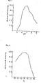
- Figure 1 is a curve showing the optimum pH of an enzyme used in the present invention
- Figure 2 is a curve showing the optimum temperature of the enzyme
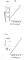
- Figure 3 shows a calibration curve in the oxygen electrode method
- Figure 4 shows a calibration curve in the ferricyanide method
- Figure 5 shows a calibration curve in the dichlorophenol-indophenol method
- Figure 6 shows a calibration curve in the H 2 0 2 colorimetry
- Figure 7 shows a calibration curve in the H 2 0 2 fluorometry
- Figure 8 shows a calibration curve in the H 2 0 2 electrode method.
- aqueous solution of specimen used in the present invention there is no particular limitation to the aqueous solution of specimen used in the present invention as far as it is intended to measure the concentration of 1,5-AG.
- examples include cerebrospinal fluid, plasma, serum and urea or, a solution obtained by treating these specimens so as to readily measure the concentration of 1,5-AG.
- Any electron acceptor is usable without any particular limitation as long as it participates in the oxidation of 1,5-AG.
- the electron acceptor include oxygen, phenazine methosulfate, dichlorophenol-indophenol; ferricyanide compounds such as potassium ferricyanide, sodium ferricyanide, cytochrome C, etc.; coenzymes such as NAD + , NADP + , FAD FMN, etc.
- the amount of the electron acceptor to be used is, for example, at least 1 ⁇ M per l in the aqueous solution of specimen, preferably approximately 3 ⁇ M to 500 mM.
- Examples of the reduction products of the electron acceptor include hydrogen peroxide, the reduction product of dichlorophenol-indophenol, ferrocyanide compounds, reduction type cytochrome C, NADH, NADPH, reduction type FMN, etc.
- enzyme In oxidizing 1,5-AG to the compound represented by formula (1), enzyme is generally utilized, This enzyme is an enzyme capable of oxidizing 1,5-AG to the compound represented by formula (1) (hereafter referred to as "1,5-AG oxidase".
- the enzyme having such an ability has been found by the present inventors for the first time and is obtained by microorganisms producing the same. Examples of such microorganisms include Pseudomonas s p.
- NK-85001 (FERM BP-1037 deposited in the Fermentation Research Institute), Pycnoporus coccineus IFO 4923 and IFO 6490, Coriolus consors IFO 9078, Coriolus versicolor (IFO 4937), Daedaleopsis styracina (IFO 4910), Gloeophyllum sepiarium (Z-41, NRRL 12506), Pleurotus ostraetus (Z-64, NRRL 12507), etc.
- the microorganism belonging to the genus Pseudomonas is a novel strain isolated from the soil collected by the present inventors in Saitama Prefecture, Omiya-shi, Yoshino-cho, in 1983, June. Bacteriological properties of this strain are as follows.
- Morphology (cultured in bouillon-agar medium at 27°C for 16 hours)
- this strain was akin to Pseudomonas stutzeri belonging to the genus Pseudomonas at page 220.
- the strain possesses properties that it does not hydrolyze starch and does not produce any acid from maltose and is thus different from Pseudomonas stutzeri in these points. From the foregoing reasons, the strain is given Pseudomonas sp. NK-85001.
- media for culturing the above-described strain there may be used media containing 1,5-AG, inorganic nitrogen sources and inorganic salts.
- organic nutrient sources can be supplemented.
- the inorganic nitrogen sources there can be used ammonium sulfate, ammonium chloride, etc. and, salts of sodium, potassium, magnesium, calcium, iron, zinc, etc. can be used as the inorganic salts.
- the organic nutrient sources there can be used peptone, Casamino acid, meat extract, corn steep liquor, yeast extract, etc.
- culture be- performed under aerobic conditions by, such as shaking , aerial agitation, etc.
- Incubation is performed at pH of 6 to 8 and temperatures of 25 to 35°C.
- the 1,5-AG enzyme derived from the genus Pseudomonas which can be used in the present invention is isolated by the following method. Namely, the enzyme is present in a membrane fraction of cells so that the cells are isolated from the culture and destroyed in an appropriate buffer solution and, the membrane fraction is colleced from its treated solution.
- the membrane fraction can be obtained in a state of a suspension in which the membrane fraction is separated from the cell wall components, nucleic acids, intracellular soluble proteins, etc. by utilizing a plurality of centrifugal forces.
- the 1,5-AG oxidase extract is extracted with membrane fraction-solubilizing agents such as Triton X-100 (polyoxyethylene octyl phenyl ether), cholic acid, deoxycholic acid, etc. to obtain the 1,5-AG oxidase extract.
- membrane fraction-solubilizing agents such as Triton X-100 (polyoxyethylene octyl phenyl ether), cholic acid, deoxycholic acid, etc.
- the 1,5-AG oxidase can be isolated utilizing methods generally used for purification of enzyme such as polyethylene glycol fractionation, ammonium sulfate fractionation, etc.
- 1,5-AG oxidase derived from the genus Pycnoporus and the genus Coriolus which may be used in the present invention can be isolated by the following method.
- this enzyme is present in the cytoplasm fraction in cells so that the cells are separated from the culture and destroyed in an appropriate buffer solution and, the cytoplasm fraction is obtained from its treated solution.
- the cytoplasm fraction can be separated as precipitates from the membrane fraction, cell wall components, etc. by centrufugation of a solution obtained after destruction of the cells.
- the supernatant is fractionated in a conventional manner generally used for purification of enzyme, such as polyethylene glycol fractionation, ammonium sulfate fractionation, etc. to isolate the 1,5-AG oxidase.
- enzyme such as polyethylene glycol fractionation, ammonium sulfate fractionation, etc.
- the enzyme can be purified, if necessary, by column chromatography such as gel filtration and ion exchange chromatography, etc. conventionally used.
- the 1,5-AG oxidase can be obtained from the microorganisms belonging to the genus Daedaleopsis and the genus Pleurotus in a similar manner.
- the isolated 1,5-AG oxidase but also the cell-treated products such as the 1,5-AG oxidase extract, the suspension of membrane fraction, etc. can also be utilized. Further, they can be utilized in the form of carriers such as resin, membrane, etc. having immobilized the same thereon.
- the membrane fraction is removed by ultracehtrifuga- tion from the reaction solution after the reaction is completed. The supernatant is freeze dried to give white powders. The white powders are dissolved in a small quantity of ethanol and insoluble matters are removed. A 2,4-dinitrophenylhydrazine-saturated ethanol solution and a trace amount of concentrated hydrochloric acid are added to the filtrate. After heating in hot water, the mixture is then cooled and water is added thereto until it gets turbid. The mixture is then allowed to stand to give brown precipitates. The prcipitates are taken by filtration, recrystallized from ethanol-water and, if necessary, purified by silica gel chromatography to give yellow brown needles.
- IR spectrum of specimen is measured using the tablet method with KBr.
- the spectrum is measured in a DMSO-d 6 solvent.
- the chemical shift is determined as a comparative value using 0 ppm as internal standard tetramethylsilan; under the experimental condition, signal of the DMSO-d 6 solvent appears at 40.40 ppm. In conformity with the data in the mass spectrum, 12 carbons were observed.
- the compound of formula (3) described above was also obtained by treating 1,5-AG using the enzyme obtained from microorganisms belonging to the genus Pycnoporus, the genus Coriolus, the genus Daedaleopsis, the genus Pleourotus and the genus Gloeophyllum in a manner similar to the membrane fraction suspension of the microorganisms belonging to the genus Pseudomonas described above.
- the compound of formula (1) was chemically synthesized. It has been found that the compound of formula (1) readily hydrates in the presence of water to form the compound of formula (2). This hydrate was reacted in a manner similar to the case where 2,4-dinitrophenyhydrazine was reacted with the treated prodcut of 1,5-AG with the membrane fraction suspension of the microorganisms belonging to the genus Pseudomonas described above, whereby quite the same compound of formula (3) described above was obtained. From this, it is assumed that the product of the 1,5-AG oxidase or the compound of formula (1) would have been present in the form of the hydrate showed by formula (2).
- reaction in the present invention would follow the reaction equation (a) described above.
- the compounds of formulaa (1) and (2) are novel compounds. Further the reaction in which 1,5-AG is dehydrogenated to produce the compound of formula (1) is also a novel reaction.
- the method of measurement of 1,5-AG of the present invention is based on the above reaction. Utilizing the reaction progress or reaction products, various measurements can be made and their contents are described below.
- a sealed type reactor In a sealed type reactor are charged 1 ml of 0.05M tris-hydrochloride buffer (pH 7.0), 20 ⁇ l of 30 mM phenazine- methosulfate and 0.3 ml of solution or suspension containing 1,5-AG oxidase. Oxygen electrodes are inserted in the reaction mixture. While stirring the content of the reactor at 34°C, 50 ⁇ l of a 1,5-AG solution is added thereto to initiate the reaction. The amount of oxygen consumed is measured with an oxygen monitor with the passage of time. Using 1,5-AG solutions having known concentrations, a calibration curve is prepared and the concentration of 1,5-AG is calculated from the amount of oxygen consumed.
- dichlorophenol-indophenol there can be used dichlorophenol-indophenol, etc., in addition to ferricyanides such as potassium ferricyanide, sodium ferricyanide, ammonium ferricyanide, etc.
- a reactor In a reactor are charged 0.3 ml of sodium phosphate buffer (1/15 M, pH 5.6), 0.5 ml of a hydrogen peroxide detecting- solution containing substrate for horse raddish peroxidase (e.g., 4 mM of 2,2'-azino-di-[3-ethylbenzo- thiazoline sulfonate (6)] (ABTS)) and 12 u/ml of horse raddish peroxidase, 0.1 ml of 1,5-AG oxidase and 0.1 ml of a specimen solution containing 1,5-AG. After reacting at 37°C for 30 minutes, the reaction is stopped under ice cooling and the absorbance is measured at 405 nm. Using 1,5-AG solutions having known concentrations, a calibration curve is prepared and the concentration of 1,5-AG is calculated from the absorbance of the specimen.
- a hydrogen peroxide detecting- solution containing substrate for horse raddish peroxidase e.g
- substrates for horse raddish peroxidase there can be utilized, in addition to ABTS, color-forming substrates such as 5-aminosalicylic acid, 4-aminoantipyrine and phenol, o-toluidine, etc., and fluorescent substrates such as p-hydroxyacetic acid, p-hydroxypropionic acid, etc.
- H 2 0 2 produced in the oxidation reaction of 1,5-AG
- a method for directly measuring H 2 0 2 using H 2 0 2 electrodes a method for directly measuring H 2 0 2 using H 2 0 2 electrodes, a method utilizing chemical luminescence generated by oxidation of lucigenine, aryl oxalates, etc. with H 2 02 may also be utilized in addition to the above method.
- the membrane fraction suspension derived from bacteria of the genus Pseudomonas is added to a 1,5-AG solution followed by reacting at 30°C for 16 hours. After completion of the reaction, the membrane fraction is removed by ultracentrifugation and the supernatant is feeze dried to give white powders.
- the powders are treated with a labeling agent for the carbonyl group or a protecting agent for the hydroxy group to effect the assay.
- the freeze dried powders are dissolved in a small quantity of ethanol and insoluble matters are removed; a saturated ethanol solution of 2,4-dinitrophenylhydrazine and a trace amount of concentrated hydrochloric acid are added to the filtrate followed by reacting in hot water with heating.
- HPLC liquid chromatography
- the product of formula (1) can be detected.
- TMS trimethylsilylchloride
- the freeze dried powders are dissolved in a small quantity of pyridine and TMS is then added to the solution. By stirring the mixture at room temperature, a compound wherein all hydroxy groups of the product of formula (2) are protected is obtained.
- TMS trimethylsilylchloride
- the reagent for analysis of the present invention is a reagent comprising at least the enzyme of the present invention.
- the enzyme may be a soluble enzyme of a solution, freeze dried, powdery or granular type.
- the enzyme may be immobilized enzyme immobilized onto carriers of a membrane, gel, particulate, microcapsular, tubular or container type, in various manners.
- the reagent may be supplemented with buffers such as liquid or powdery phosphate buffer, tris-hydrochloride buffer, acetate buffer, citrate buffer, veronal buffer, etc.; salts (sodium chloride, etc.), sugars that do not react with the enzyme of the present invention (sucrose, etc.); polyvalent alcohols (glycerol, propylene glycol, sorbitol, etc.); coenzymes (FAD, etc.): and other appropriate stabilizers, surfactants, etc.
- buffers such as liquid or powdery phosphate buffer, tris-hydrochloride buffer, acetate buffer, citrate buffer, veronal buffer, etc.; salts (sodium chloride, etc.), sugars that do not react with the enzyme of the present invention (sucrose, etc.); polyvalent alcohols (glycerol, propylene glycol, sorbitol, etc.); coenzymes (FAD, etc.): and other appropriate stabilizers, surfactants,
- the reagent for analysis described above is used so as to obtain a necessary enzyme activity depending upon the aforesaid various methods for detection. Further, an amount of the reagent appropriate for each of the detection methods may be previously sealed in a container such as a reagent bottle, ampoule, etc.
- the kit for analysis is composed of the aforesaid reagent for analysis comprising the enzyme of the present invention and reagents for detection which are reagents for detecting the reaction caused by the enzyme of the present invention.
- the reagent for detection refers to an electron acceptor. per se that participates in the oxidation of 1,5-AG accompanied by color formation or to reagents that is necessary for detection of one of the 1,5-AG oxidation reaction products as an index of the 1,5-AG oxidation.
- examples of the reagents for detection include combination of peroxidase or a peroxidase-like substance and its color forming substrate or color forming substrate and a coupler, combination of peroxidase or a 'peroxidase-like active substance and its fluorescent substrate, combination of peroxidase or a peroxidase-like active substance and a luminescence forming reagent, etc.
- Specific examples of these reagents are clear from the description entitled "Method for detecting H2 0 2 " described above.
- the oxidation products of 1,5-AG, as the index, reagents necessary for detection are combined with the reagent for analysis comprising the enzyme of the present invention and the combination can be constructed as the kit for analysis.
- the reagent for analysis comprising the enzyme of the present invention and the aforesaid reagents for detection may all be mixed together to form a single reagent; alternatively, in case that muturally interfering components are present, each component may be separated so as to form an appropriate combination. Further these components may be prepared in the form of a solution or powders. Furthermore, they may be incorporated into an appropriate support such as a film to prepare into a test paper sheet or an analysis film.
- the kit for analysis of the present invention may further contain, in addition to the combinations described above, pretreatment reagents for selectively removing contaminants, standard reagents containing a definite amount of 1,5-AG, etc.
- kits for analysis of the present invention include a kit for detecting 1,5-AG by spectroscopic detection of the reduction product of a ferricyanide as an electron acceptor and a kit for analysis of 1,5-AG by spectroscopic detection of hydrogen peroxide.
- the enzyme is used more than 0.2 units/test and ferricyanide is used more than 5-fold moles, preferably more than 10-fold moles of 1,5-AG and usually 10 -5 mol/test of potassium ferricyanide is used.
- the enzyme is used more than 0.2. units/test, peroxidase is used 1-10 unit/test and substrate for color formation is used more than 5-fold moles, preferably more than 10- fold moles of hydrogen peroxide produced in the oxidation of 1,5-AG and usually 5x10 -7 to 5x10 6 mols/test of ABTS is used as a substrate for color formation.
- the reaction was performed in the ferricyanide method described above using the 1,5-AG oxidase extract obtained in Reference Example 1 later described except that the substrate was replaced by sugar and sugar alcohol.
- the 1,5-AG oxidase produced by the microorganism belonging to the genus Pseudomonas shows high specificity to 1,5-AG as shown in Table 1.
- the extract was added to phosphate buffer (pH 6 to 7) and tris-hydrochloride buffer (pH 7.2 to 9) having different pH values. After storing at 4°C for 1 day, the conversion activity was examined; it was stable in the pH range of 6.5 to 8.
- the extract (protein concentration: 5 mg/ml) obtained in Reference Example 1 later described was used.
- reaction solution was added to a reactor of an oxygen densitometer (Oxygraph manufactured by Guilsol Co. in America) and kept at 34°C while agitating.
- the extract (protein content, 5 mg/ml) obtained in Reference Example 1 later described was used.
- a reaction solution having the following composition was reacted at 34°C for 10 minutes in a test tube.
- the extract (protein content, 5 mg/ml) obtained in Reference Example 1 later described was used.
- a reaction solution having the following composition was charged in a cell of a spectrophotometer and kept at 34°C.
- the 1,5-AG solution kept at 34°C was charnged in the cell and the absorbance at 600 nm was recorded while stirring with the passage of time.
- the enzyme (enzyme activity, 3.2 u/ml) obtained in Reference Example 6 later described was used.
- a reaction solution having the following composition was reacted at 37°C for 2 hours in a test tube.
- the enzyme (enzyme activity, 1.5 u/ml) obtained in Reference Example 6 later described was used.
- a reaction solution having the following composition was reacted at 37°C for 2 hours in a test tube.
- a pump injector and H 2 0 2 electrodes (Ishikawa Seisakusho, Ltd., BH type) were connected at the up stream to the down stream, respectively.
- the H 2 0 2 electrodes were set with a hydrogen peroxide meter (Ishikawa Seisakusho, Ltd., Model AI-10006) and a recorder.
- the 1,5-AG immobilized column and the H 2 0 2 electrodes were dipped in a water bath with thermostatt kept at 37°C. Through the pump phosphate buffer (1/15 M, pH 5.6) was flown at a rate of 1 ml/min to stabilize.
- 1,5-AG can be quantitatively determined in an extremely manner in accordance with the present invention.
- the 1,5-AG content was measured by 3 methods described below. As shown below, it was possible * to measure 1,5-AG by the respective methods.
- NK-85001 [Ferm BP-1037 deposited in the Fermentation Research Institute] was inoculated in the medium followed by culturing at 30°C for 16 hours on a rotary shaking culture machine (220 rpm). The cells were separated from the culture solution by centrifugation and washed with tris-hydrochloride buffer (0.05 M, pH 7) to give a cell suspension of a 1/10 volume based on the amount of the starting solution. The cell suspension was cooled and destroyed with a French press to give the cell-destroyed suspension. The suspension was centrifuged for 10 minutes (10,000 x g). After the precipitated cell walls were removed, centrifugation was continued for further 1 hour (100,000 x g) to give the precipitates.
- tris-hydrochloride buffer 0.05 M, pH 7
- Triton X-100 was added to the suspension in a concentration of 1% (w/v). After agitating at 4°C for 1 hour, the insoluble matters were removed by centrifugation (100,000 x g) to give the 1,5-AG oxidase extract.
- the cell suspension was cooled and destroyed with a French press to give the cell-destroyed liquid.
- the liquid was centrifuged (10,000 x g) for 10 minutes under cooling. After the precipitated cell walls were removed, centrifugation was continued for further 1 hour (100,000 x g) to remove membrane fraction and obtain the cytoplasm supernatant. Under cooling, ammonium sulfate powders were added to the supernatant and the mixture was agitated to dissolve.
- the precipitated protein in this case was separated by centrifugation (10,000 x g, 10 minutes) and the activity of 1,5-AG oxidase was measured with each ammonium sulfate fraction by the method for detecting H 2 0 2 described in the specification (wherein a 1,5-AG solution having a 1% concentration was used) and it was noted that the activity was present mainly in the 40-60% ammonium sulfate-saturated fraction.
- one unit of enzyme is defined to be an amount that oxidizes 1,5-AG to produce 1 umole/min of H 2 O 2
- the. specific activity of the ammonium sulfate fraction is 4.0. Enzyme of 11 units are obtained per 1 g of the wet cells.
- the strain was changed to Pycnoporus coccineus IFO 6490 in Reference Example 3 and cultured in medium having the same composition as in Reference Example 3 for 4 days.
- the same procedure for purification as in Reference Example 3 was performed to give 1,5-AG oxidase having a specific activity of 3.6..
- the strain was changed to coriolus consors IFO 9078 in Reference Example 3 and cultured in medium having the same composition as in Reference Example 3 for 10 days.
- the same procedure for purification as in Reference Example 3 was performed to give 1,5-AG oxidase extract having a specific activity of 2.8.
- porous glass CPG-10 (200/400 mesh, mean pore size of 500 A, manufactured by Electronucleonic Co., Ltd.) was subjected to a coupling treated using 0.5 g of ⁇ -aminopropyltriethoxysilane followed by carboxylation with 0.5 g of succinic anhydride.
- the dried porous glass was treated with an excess of thionyl chloride in chloroform to convert the carboxyl groups into the acid chloride.
- To 1 g of the thus obtained acid chloridated porous glass was added 2.5 ml of the 1,5-AG oxidase solution prepared in Reference Example 6 described above.
- the obtained 1,5-AG oxidase-immobilized porous glass was filled up in a column (1 ml of a syringe) having an inner diameter of 2.3 mm and a length of 70 mm.
- a column (1 ml of a syringe) having an inner diameter of 2.3 mm and a length of 70 mm.
- phosphate buffer (1/15 M, pH 5.6) containing 1 M table salt was passed to remove the enzyme not bound covalently. Further the column was washed by passing phosphate buffer (1/15 M, pH 5.6) therethrough to give the 1,5-AG oxidase-immobilized column.
Abstract

Description
- The present invention relates to a method of quantitative assay for 1,5-anhydroglucitol (hereafter referred to as "1,5-AG") which is expected as a marker for diagnosis of diabetes.
- 1,5-AG is a compound which is present in the cerebrospinal fluid and plasma of human and it is reported that its quantity is reduced in plasma with certain diseases, particularly with diabetes. It has not been known the presence of an enzyme that oxidizes this 1,5-AG. The assay for 1,5-AG has been hitherto performed mainly based on gas chromatography (J. Biochem., 90, 157-162 (1981)).
- However, the prior art method requires techniques for pretreatment of specimen and maintenance and control of analysis equipments to high degree and a simple method of assay for 1,5-AG has been demanded.
- The present invention is based on novel findings that 1,5-AG is oxidized in the presence of an electron acceptor to produce a compound represented by formula (1) below:


- Namely, the present invention relates to a method of quantitative assay for 1,5-anhydroglucitol which comprises oxidizing 1,5-anhydroglucitol in an aqueous solution of a specimen in the presence of an electron acceptor to produce a compound represented by formula (1) above or a hydrate thereof represented by formula (2) above and quantitatively determining 1,5-anhydroglucitol from a comsumption amount of said electron acceptor in said aqueous solution of a specimen, from a production amount of the reduction product of said electron acceptor produced or from an amount of an oxidized product of 1,5-AG represented formula (1), or formula (2).
- Figure 1 is a curve showing the optimum pH of an enzyme used in the present invention; Figure 2 is a curve showing the optimum temperature of the enzyme; Figure 3 shows a calibration curve in the oxygen electrode method; Figure 4 shows a calibration curve in the ferricyanide method; Figure 5 shows a calibration curve in the dichlorophenol-indophenol method; Figure 6 shows a calibration curve in the
H 202 colorimetry; Figure 7 shows a calibration curve in theH 202 fluorometry; and Figure 8 shows a calibration curve in theH 202 electrode method. - There is no particular limitation to the aqueous solution of specimen used in the present invention as far as it is intended to measure the concentration of 1,5-AG. Examples include cerebrospinal fluid, plasma, serum and urea or, a solution obtained by treating these specimens so as to readily measure the concentration of 1,5-AG.
- Any electron acceptor is usable without any particular limitation as long as it participates in the oxidation of 1,5-AG. Examples of the electron acceptor include oxygen, phenazine methosulfate, dichlorophenol-indophenol; ferricyanide compounds such as potassium ferricyanide, sodium ferricyanide, cytochrome C, etc.; coenzymes such as NAD+, NADP+, FAD FMN, etc. The amount of the electron acceptor to be used is, for example, at least 1 µM per ℓ in the aqueous solution of specimen, preferably approximately 3 µM to 500 mM.
- Examples of the reduction products of the electron acceptor include hydrogen peroxide, the reduction product of dichlorophenol-indophenol, ferrocyanide compounds, reduction type cytochrome C, NADH, NADPH, reduction type FMN, etc.
- In oxidizing 1,5-AG to the compound represented by formula (1), enzyme is generally utilized, This enzyme is an enzyme capable of oxidizing 1,5-AG to the compound represented by formula (1) (hereafter referred to as "1,5-AG oxidase". The enzyme having such an ability has been found by the present inventors for the first time and is obtained by microorganisms producing the same. Examples of such microorganisms include Pseudomonas sp. NK-85001 (FERM BP-1037 deposited in the Fermentation Research Institute), Pycnoporus coccineus IFO 4923 and IFO 6490, Coriolus consors IFO 9078, Coriolus versicolor (IFO 4937), Daedaleopsis styracina (IFO 4910), Gloeophyllum sepiarium (Z-41, NRRL 12506), Pleurotus ostraetus (Z-64, NRRL 12507), etc. Of these microorganisms, the microorganism belonging to the genus Pseudomonas is a novel strain isolated from the soil collected by the present inventors in Saitama Prefecture, Omiya-shi, Yoshino-cho, in 1983, June. Bacteriological properties of this strain are as follows.
-
- (1) Size of cell: 0.7-0.8 x 1.0-1.7 um, rod
- (2) Pleomorphism: not recognized
- (3) Motility: It possess polar flagella with motility.
- (4) Presence or absence of spore: not recognized
- (5) Gram staining: negative
- (6) Acid fast: negative
-
- (1) Bouillon-agar plate culture: It forms lustrous, opaque and entire. . circular colonies with brown white color.
- (2) Bouillon-agar slant culture: It diffuses and proliferates on the surface of medium to grow opaque and lustrous. The color is brown white.
- (3) Bouillon liquid culture: On the first day of culture, it gets turbid as a whole and cells precipitate at the bottom of a test tube on the 3rd day. . Pellicle is observed.
- (4) Bouillon gelatin stab culture: It grows only on the surface by culture at 20°C. No liquefication of gelatin by culture for 20 days.
- (5) Litmus milk: no change
-
- (1) Reduction of nitrate: positive
- (2) Denitration: positive
- (3) MR Test: negative
- (4) VP Test: negative
- (5) Indole formation: negative
- (6) Formation of hydrogen sulfide: negative
- (7) Hydrolysis of starch: negative
- (8) Utilization of citric acid: It utilizes citric acid in QChristensen's and Simmon's media but not in Kosar's medium.
- (9) Utilization of inorganic nitrogen source: It utilizes ammonia but no nitrates.
- (10) Formation of pigment: negative
- (11) Urease: positive
- (12) Oxidase: positive
- (13) Catalase: positive
- (14) Growth conditions: 10-37°C pH 7-8.5
- (15) Behavior to oxygen: aerobic
- (16) O-F test: oxidative
- (1.7) Utilization of carbohydrates: It utilizes glucose, glycerin, sodium succinate and sodium citrate but neither sodium acetate nor p-hydroxybenzoic acid.
- (18) Formation of acids and gas from sugars:

- (19) Resistance to sodium chloride Sodium chloride was added to basic medium composed of 10 g of tryptone and 1 liter of distilled water at pH 7.0 in concentrations of 2%, 5% and 7%, respectively. After inoculating a bacterial solution thereon, stationary culture was performed. Growth was noted in media of 2% and 5% but no growth was noted in the
medium 7%. - (20) Phenyl pyruvate test: negative
- (21) Tyrosine solubility negative
- Based on the foregoing properties, the taxonomical properties of this strain were compared with the classification in Bergey's Mannual of Determinative Bacteriology, 8th edition (1974); this strain is akin to Pseudomonas stutzeri belonging to the genus Pseudomonas at page 220. However, the strain possesses properties that it does not hydrolyze starch and does not produce any acid from maltose and is thus different from Pseudomonas stutzeri in these points. From the foregoing reasons, the strain is given Pseudomonas sp. NK-85001.
- As media for culturing the above-described strain, there may be used media containing 1,5-AG, inorganic nitrogen sources and inorganic salts. For the purpose of accelerating the growth, organic nutrient sources can be supplemented. As the inorganic nitrogen sources, there can be used ammonium sulfate, ammonium chloride, etc. and, salts of sodium, potassium, magnesium, calcium, iron, zinc, etc. can be used as the inorganic salts. As the organic nutrient sources there can be used peptone, Casamino acid, meat extract, corn steep liquor, yeast extract, etc.
- It is preferred that culture be- performed under aerobic conditions by, such as shaking , aerial agitation, etc. Incubation is performed at pH of 6 to 8 and temperatures of 25 to 35°C.
- The 1,5-AG enzyme derived from the genus Pseudomonas which can be used in the present invention is isolated by the following method. Namely, the enzyme is present in a membrane fraction of cells so that the cells are isolated from the culture and destroyed in an appropriate buffer solution and, the membrane fraction is colleced from its treated solution.
- To destroy the cells, physical methods such as by means of dyno mill, French press, ultrasonic wave, etc., chemical methods using Triton X-100, EDTA, etc., or enzymatic methods using lysozyme, etc. can be used singly or in combination. The membrane fraction can be obtained in a state of a suspension in which the membrane fraction is separated from the cell wall components, nucleic acids, intracellular soluble proteins, etc. by utilizing a plurality of centrifugal forces.
- Subsequently, active components are extracted with membrane fraction-solubilizing agents such as Triton X-100 (polyoxyethylene octyl phenyl ether), cholic acid, deoxycholic acid, etc. to obtain the 1,5-AG oxidase extract. From the extract, the 1,5-AG oxidase can be isolated utilizing methods generally used for purification of enzyme such as polyethylene glycol fractionation, ammonium sulfate fractionation, etc.
- Next, properties of the 1,5-AG enzyme derived from the genus Pseudomonas are described below.
- It oxidizes 1,5-AG to produce the compound of formula (1) -described above.
- It specifically acts on 1,5-AG.
-
- 3.
Optimum pH pH 6 to 7.5 - 4.
Optimum temperature 25 to 41°C - 5. Stable pH 6.5 to 8
- Further the 1,5-AG oxidase derived from the genus Pycnoporus and the genus Coriolus which may be used in the present invention can be isolated by the following method.
- Namely, this enzyme is present in the cytoplasm fraction in cells so that the cells are separated from the culture and destroyed in an appropriate buffer solution and, the cytoplasm fraction is obtained from its treated solution.
- To destroy the cells, the cells are destroyed in a manner similar to the case of the genus Pseudomonas described above. The cytoplasm fraction can be separated as precipitates from the membrane fraction, cell wall components, etc. by centrufugation of a solution obtained after destruction of the cells.
- Subsequently, the supernatant is fractionated in a conventional manner generally used for purification of enzyme, such as polyethylene glycol fractionation, ammonium sulfate fractionation, etc. to isolate the 1,5-AG oxidase. In case that enzyme of higher purity is required, the enzyme can be purified, if necessary, by column chromatography such as gel filtration and ion exchange chromatography, etc. conventionally used.
- Next, properties of the 1,5-AG oxidase obtained from the microorganisms belonging to the genus Pycnoporus and the genus Coriolus are shown below.

- The 1,5-AG oxidase can be obtained from the microorganisms belonging to the genus Daedaleopsis and the genus Pleurotus in a similar manner.
- In the present invention, not only the isolated 1,5-AG oxidase but also the cell-treated products such as the 1,5-AG oxidase extract, the suspension of membrane fraction, etc. can also be utilized. Further, they can be utilized in the form of carriers such as resin, membrane, etc. having immobilized the same thereon.
- Next, products produced in the reaction solution by adding 1,5-AG to the solution containing 1,5-AG oxidase such as 1,5-AG oxidase extract; and the membrane fraction suspension are described below.
- When about 2 mg/ml of 1,5-AG is added to a membrane fraction suspension (concentration of protein, 10 mg/ml; tris-hydrochloride buffer, 0.05 M; pH 7) from a microorganism belonging to the genus Pseudomonas and they are reacted at 30° C for 16 hours with shaking, 1,5-AG disappears but Substance (A) is produced and accumulates. This can be confirmed by TLC analysis. The reaction solution is spotted onto a silica gel plate and developed with a solvent of iso-PrOH:H20 (95:5) and then thoroughly dried. Anisaldehyde sulfate reagent is sprayed thereon and heated at 90 to 100°C for 5 to 10 minutes, Substance (A) can be observed at Rf of about 0.4 as a blue spot.
- . The membrane fraction is removed by ultracehtrifuga- tion from the reaction solution after the reaction is completed. The supernatant is freeze dried to give white powders. The white powders are dissolved in a small quantity of ethanol and insoluble matters are removed. A 2,4-dinitrophenylhydrazine-saturated ethanol solution and a trace amount of concentrated hydrochloric acid are added to the filtrate. After heating in hot water, the mixture is then cooled and water is added thereto until it gets turbid. The mixture is then allowed to stand to give brown precipitates. The prcipitates are taken by filtration, recrystallized from ethanol-water and, if necessary, purified by silica gel chromatography to give yellow brown needles.
- Physicochemical properties of the crystals are as follows.
-
- 1. Melting point: 192°C
- 2. Molecular weight: 342 (mass spectrum)
- 3. Molecular formula: C12H14N4O8 Found by mass spectrum: 343 (M+H)+ Calcd. : 342, 272
- 4. UV spectrum max. nm (

- 5. IR spectrum
- IR spectrum of specimen is measured using the tablet method with KBr.
- 3600~300 0 cm-1( broad ), 1622.
- 1584, 1518. 1504, 1415,
- 1333, 1273, 1224, 1137,
- 1073, 1050, 1028, 993,
- 925, 878, 740
- The spectrum is measured in a DMSO-d6 solvent. The chemical shift is determined as a comparative value using 0 ppm as internal standard tetramethylsilan; under the experimental condition, signal of the DMSO-d6 solvent appears at 40.40 ppm. In conformity with the data in the mass spectrum, 12 carbons were observed.
-
- 6 1.9(t), 71.2(t). 7 3.6(d), 7 8.4(d),
- 8 2.3(d), 1 1 6.2(d), 12 4.0(d), 1 3 0.1
- (s), 1 3 0.8(d), 1 3 7.6(s), 1 4 5.5(s).
- 15 3.2(s)
- The spectrum was measured in DMSO-d6. Chemical shift was a comparative value with data obtained using tetramethylsilane as internal standard of 0 ppm.
-
- 3.6 9 ppm ( 1H, dd), 4.1 4~4.15
- ( 2H. ABq), 4.6 2~4.65 ( 2H.
- t, d), 5.63~5.65 ( 1H, d),
- 7.24 (1H, broad), 7.87-7.90
- (1H, d), 8.33~8.37 (1H, dd),
- 8.8 6~8.8 7 (1H, d), 4. 6 2~4.6 5
- ( 1H, t-OH), 5.63~5.65 ( 1H,
- d-OH), 7. 2 4 ( 1H, br-NH)
- From these data, the aforesaid yellow needles are assumed to has the following chemical formula (3):

- From this, Substance (A) was assumed to possess the aforesaid chemical formula (1).
- Further the compound of formula (3) described above was also obtained by treating 1,5-AG using the enzyme obtained from microorganisms belonging to the genus Pycnoporus, the genus Coriolus, the genus Daedaleopsis, the genus Pleourotus and the genus Gloeophyllum in a manner similar to the membrane fraction suspension of the microorganisms belonging to the genus Pseudomonas described above.
- Next, to confirm the structure of the product, the compound of formula (1) was chemically synthesized. It has been found that the compound of formula (1) readily hydrates in the presence of water to form the compound of formula (2). This hydrate was reacted in a manner similar to the case where 2,4-dinitrophenyhydrazine was reacted with the treated prodcut of 1,5-AG with the membrane fraction suspension of the microorganisms belonging to the genus Pseudomonas described above, whereby quite the same compound of formula (3) described above was obtained. From this, it is assumed that the product of the 1,5-AG oxidase or the compound of formula (1) would have been present in the form of the hydrate showed by formula (2).
- Thus, identification of the product by the 1,5-AG oxidase and the hydrate chemically synthesized was performed by gas chromatography.
- Both compounds were trimethylsilylated and analyzed by a column, where both were detected at quite the same retention time. Further, analysis of fragment pattern of the peak compound by gas chromatography and mass spectrometry (GC-MS) revealed that quite the same pattern was obtained.
- From the foregoing, the above assumption is believed to be correct. Accordingly, it is thought that the enzyme or membrane fraction suspension used in the present invention would catalyze the following reaction:

- Further it is believed that the reaction in the present invention would follow the reaction equation (a) described above.
- Physicochemical properties of the chemcally syntne- sized hydrate of formula (2) are as follows.
-
- 1. Thermal analysis (in nitrogen flow) o Dehydration 86°C (weight reduction temperature of 1 molecule of water was noted. o Melting point 63-74°C
- 2. Molecular weight: 180 (mass spectrum)
- 3. Molecular formula: C6H12O6
- 4. IR Spectrum

- 5. C-NMR ( 1 0 0 MHz )

- 6. 1H-NMR ( 4 0 0 MHz )

- 7. Crystalline form: amorphous white powders
- The compounds of formulaa (1) and (2) are novel compounds. Further the reaction in which 1,5-AG is dehydrogenated to produce the compound of formula (1) is also a novel reaction.
- The method of measurement of 1,5-AG of the present invention is based on the above reaction. Utilizing the reaction progress or reaction products, various measurements can be made and their contents are described below.
- In a sealed type reactor are charged 1 ml of 0.05M tris-hydrochloride buffer (pH 7.0), 20 µl of 30 mM phenazine- methosulfate and 0.3 ml of solution or suspension containing 1,5-AG oxidase. Oxygen electrodes are inserted in the reaction mixture. While stirring the content of the reactor at 34°C, 50 µl of a 1,5-AG solution is added thereto to initiate the reaction. The amount of oxygen consumed is measured with an oxygen monitor with the passage of time. Using 1,5-AG solutions having known concentrations, a calibration curve is prepared and the concentration of 1,5-AG is calculated from the amount of oxygen consumed.
- In a reactor are charged 0.7 ml of tris-hydrochloride buffer (0.05M, pH 7), 0.1 ml of a O.lM potassium ferricyanide solution, Q.1 ml of 1,5-AG oxidase obtained from the microorganism belonging to the genus Pseudomonas or an extract thereof and 0.1 ml of a 1,5-AG solution. After reacting at 34°C for 10 minutes, 0.5 ml of a ferric sulfate-dupanol reagent (5 g of ferric sulfate, 3 g of sodium laurylsulfate, 95 ml of 85% phosphoric acid and 900 ml of distilled water) and 3.5 ml of distilled water are added to the mixture followed by allowing to stand for 10 minutes. Then, absorbance is measured at 660 nm. Using 1,5-AG solutions having known concentrations, a calibration curve is prepared and the concentration of 1,5-AG is calculated from the absorbance of the specimen.
- As electron acceptors, there can be used dichlorophenol-indophenol, etc., in addition to ferricyanides such as potassium ferricyanide, sodium ferricyanide, ammonium ferricyanide, etc.
- In a reactor are charged 0.3 ml of sodium phosphate buffer (1/15 M, pH 5.6), 0.5 ml of a hydrogen peroxide detecting- solution containing substrate for horse raddish peroxidase (e.g., 4 mM of 2,2'-azino-di-[3-ethylbenzo- thiazoline sulfonate (6)] (ABTS)) and 12 u/ml of horse raddish peroxidase, 0.1 ml of 1,5-AG oxidase and 0.1 ml of a specimen solution containing 1,5-AG. After reacting at 37°C for 30 minutes, the reaction is stopped under ice cooling and the absorbance is measured at 405 nm. Using 1,5-AG solutions having known concentrations, a calibration curve is prepared and the concentration of 1,5-AG is calculated from the absorbance of the specimen.
- . As substrates for horse raddish peroxidase, there can be utilized, in addition to ABTS, color-forming substrates such as 5-aminosalicylic acid, 4-aminoantipyrine and phenol, o-toluidine, etc., and fluorescent substrates such as p-hydroxyacetic acid, p-hydroxypropionic acid, etc.
- Further for detecting
H 202 produced in the oxidation reaction of 1,5-AG, a method for directly measuringH 202 usingH 202 electrodes, a method utilizing chemical luminescence generated by oxidation of lucigenine, aryl oxalates, etc. with H202 may also be utilized in addition to the above method. - The membrane fraction suspension derived from bacteria of the genus Pseudomonas is added to a 1,5-AG solution followed by reacting at 30°C for 16 hours. After completion of the reaction, the membrane fraction is removed by ultracentrifugation and the supernatant is feeze dried to give white powders. The powders are treated with a labeling agent for the carbonyl group or a protecting agent for the hydroxy group to effect the assay. In the case of using, e.g., 2,4-dinitrophenylhydrazine, as the labeling agent for the carbonyl group, the freeze dried powders are dissolved in a small quantity of ethanol and insoluble matters are removed; a saturated ethanol solution of 2,4-dinitrophenylhydrazine and a trace amount of concentrated hydrochloric acid are added to the filtrate followed by reacting in hot water with heating. By analysis of the product by means of reversed phase HPLC (liquid chromatography), the product of formula (1) can be detected. Further in the case of using trimethylsilylchloride (TMS) as the protecting agent for the hydroxy group, the freeze dried powders are dissolved in a small quantity of pyridine and TMS is then added to the solution. By stirring the mixture at room temperature, a compound wherein all hydroxy groups of the product of formula (2) are protected is obtained. By analysis of a part of the solution by gas chromatography, the compound of formula (2) can be quantitatively determined.
- The reagent for analysis of the present invention is a reagent comprising at least the enzyme of the present invention. Namely, its form is not limited but the enzyme may be a soluble enzyme of a solution, freeze dried, powdery or granular type. Further, the enzyme may be immobilized enzyme immobilized onto carriers of a membrane, gel, particulate, microcapsular, tubular or container type, in various manners. In addition to the enzyme of the present invention, the reagent may be supplemented with buffers such as liquid or powdery phosphate buffer, tris-hydrochloride buffer, acetate buffer, citrate buffer, veronal buffer, etc.; salts (sodium chloride, etc.), sugars that do not react with the enzyme of the present invention (sucrose, etc.); polyvalent alcohols (glycerol, propylene glycol, sorbitol, etc.); coenzymes (FAD, etc.): and other appropriate stabilizers, surfactants, etc.
- Upon analysis of 1,5-AG, the reagent for analysis described above is used so as to obtain a necessary enzyme activity depending upon the aforesaid various methods for detection. Further, an amount of the reagent appropriate for each of the detection methods may be previously sealed in a container such as a reagent bottle, ampoule, etc.
- The kit for analysis is composed of the aforesaid reagent for analysis comprising the enzyme of the present invention and reagents for detection which are reagents for detecting the reaction caused by the enzyme of the present invention. The reagent for detection refers to an electron acceptor. per se that participates in the oxidation of 1,5-AG accompanied by color formation or to reagents that is necessary for detection of one of the 1,5-AG oxidation reaction products as an index of the 1,5-AG oxidation. As examples of the latter, in the case of using hydrogen peroxide as the index, examples of the reagents for detection include combination of peroxidase or a peroxidase-like substance and its color forming substrate or color forming substrate and a coupler, combination of peroxidase or a 'peroxidase-like active substance and its fluorescent substrate, combination of peroxidase or a peroxidase-like active substance and a luminescence forming reagent, etc. Specific examples of these reagents are clear from the description entitled "Method for detecting H202" described above.
- Further similarly in the case of using the compound of formula (1) or the compound of formula (2), the oxidation products of 1,5-AG, as the index, reagents necessary for detection are combined with the reagent for analysis comprising the enzyme of the present invention and the combination can be constructed as the kit for analysis. The reagent for analysis comprising the enzyme of the present invention and the aforesaid reagents for detection may all be mixed together to form a single reagent; alternatively, in case that muturally interfering components are present, each component may be separated so as to form an appropriate combination. Further these components may be prepared in the form of a solution or powders. Furthermore, they may be incorporated into an appropriate support such as a film to prepare into a test paper sheet or an analysis film.
- The kit for analysis of the present invention may further contain, in addition to the combinations described above, pretreatment reagents for selectively removing contaminants, standard reagents containing a definite amount of 1,5-AG, etc.
- Preferred examples of the kit for analysis of the present invention include a kit for detecting 1,5-AG by spectroscopic detection of the reduction product of a ferricyanide as an electron acceptor and a kit for analysis of 1,5-AG by spectroscopic detection of hydrogen peroxide.
- In the case of the kit using the ferricyanide as an electron acceptor, the enzyme is used more than 0.2 units/test and ferricyanide is used more than 5-fold moles, preferably more than 10-fold moles of 1,5-AG and usually 10-5 mol/test of potassium ferricyanide is used.
- In the case of the kit for spectroscopically detecting hydrogen peroxide, the enzyme is used more than 0.2. units/test, peroxidase is used 1-10 unit/test and substrate for color formation is used more than 5-fold moles, preferably more than 10- fold moles of hydrogen peroxide produced in the oxidation of 1,5-AG and usually 5x10⁻⁷ to 5x10 6 mols/test of ABTS is used as a substrate for color formation.
- Next, the effects of the present invention will be described below.
- In order to examine substrate specificity, the reaction was performed in the ferricyanide method described above using the 1,5-AG oxidase extract obtained in Reference Example 1 later described except that the substrate was replaced by sugar and sugar alcohol. As a result, the 1,5-AG oxidase produced by the microorganism belonging to the genus Pseudomonas shows high specificity to 1,5-AG as shown in Table 1.

- Using the extract obtained in Reference Example 1 described later, the optimum pH and the optimum temperature of the 1,5-AG oxidase produced by the microorganism belonging to the genus Pseudomonas in the conversion reaction of 1,5-AG were examined to give the results shown in Figures 1 and 2. These figures reveal that the optimum pH and optimum temperature are approximately
pH 6 to 7.5 and 25 to 41° C, respectively. - Further in order to examine pH stability, the extract was added to phosphate buffer (
pH 6 to 7) and tris-hydrochloride buffer (pH 7.2 to 9) having different pH values. After storing at 4°C for 1 day, the conversion activity was examined; it was stable in the pH range of 6.5 to 8. - Test Example 3 (measurement of 1,5-AG)
- The extract (protein concentration: 5 mg/ml) obtained in Reference Example 1 later described was used.
- The following reaction solution was added to a reactor of an oxygen densitometer (Oxygraph manufactured by Guilsol Co. in America) and kept at 34°C while agitating.

- After the reactor was stoppered and sealed, 50 µl of 1,5-AG solutions having known concentrations were injected into the reactor using a microsyringe and, a rate of oxygen consumed was recorded. As a result, a proportional relationship was noted between the 1,5-AG concentration and the rate of oxygen consumed, as shown in Figure 3.
- The extract (protein content, 5 mg/ml) obtained in Reference Example 1 later described was used. A reaction solution having the following composition was reacted at 34°C for 10 minutes in a test tube.

- After the reaction, 0.5 ml of ferric sulfate-dupanol reagent and 3.5 ml of distilled water were added to discontinue the reaction. The system was allowed to stand for 10 minutes, where the system was colored green. At this stage the absorbancy was measured at 660 nm. When tested using the 1,5-AG solutions having known concentrations, a proportional relationship in absorbance was noted between the 1,5-AG concentration and the absorbance at 660 nm, as shown in Figure 4.
- The extract (protein content, 5 mg/ml) obtained in Reference Example 1 later described was used. A reaction solution having the following composition was charged in a cell of a spectrophotometer and kept at 34°C.

- The 1,5-AG solution kept at 34°C was charnged in the cell and the absorbance at 600 nm was recorded while stirring with the passage of time.
- When tested with 1,5-AG having known concentrations, a proportional relationship was noted between the 1,5-AG concentration and the rate of change in the absorbance at 600 nm, i.e., the reduction rate of DCIP, as shown in Figure 5.
- The enzyme (enzyme activity, 3.2 u/ml) obtained in Reference Example 6 later described was used. A reaction solution having the following composition was reacted at 37°C for 2 hours in a test tube.

- After the reaction, the system was ice cooled to discontinue the reaction and the absorbance was measured at 405 nm. When tested using 1,5-AG solution having known concentrations, a proportaional relationship was noted between the 1,5-AG concentration and the absorbance at 405 nm, as shown in Figure 6.
- The enzyme (enzyme activity, 1.5 u/ml) obtained in Reference Example 6 later described was used. A reaction solution having the following composition was reacted at 37°C for 2 hours in a test tube.


- After the reaction, 2.5 ml of sodium glycine buffer (0.1 M, pH 10.3) was added to the system to discontinue the reaction. A relative flurescent intensity was measured at excited wavelength of 315 nm and fluorescent wavelength of 450 nm.
- When tested with 1,5-AG solution having known concentrations, a proportional relationship was noted between the 1,5-AG concentration and the relative fluorescent intensity, as shown in Figure 7.
- In the column having immobilized thereto 1,5-AG oxidase obtained in Reference Example 7 later described, a pump injector and
H 202 electrodes (Ishikawa Seisakusho, Ltd., BH type) were connected at the up stream to the down stream, respectively. TheH 202 electrodes were set with a hydrogen peroxide meter (Ishikawa Seisakusho, Ltd., Model AI-10006) and a recorder. The 1,5-AG immobilized column and theH 202 electrodes were dipped in a water bath with thermostatt kept at 37°C. Through the pump phosphate buffer (1/15 M, pH 5.6) was flown at a rate of 1 ml/min to stabilize. Into the flow path, 50 µl of the 1,5-AG solution was flown through the injector and the peak area on the recorder produced by the oxidation of 1,5-AG was measured. When tested with 1,5-AG solutions having known concentrations, a calibration curve as shown in Figure 8 was obtained between the 1,5-AG concentration and the peak area. - As is evident from the foregoing, 1,5-AG can be quantitatively determined in an extremely manner in accordance with the present invention.
- With respect to a specimen having the following composition, the 1,5-AG content was measured by 3 methods described below. As shown below, it was possible*to measure 1,5-AG by the respective methods.
- Composition of specimen solution:


- To 0.4 ml of human serum was added 30 ul of a perchloric acid aqueous solution (60% w/v). After shaking, the mixture was centrifuged and 0.2 ml of the resultant supernatant was passed through a pretreatment column packed with 0.8 ml of borate type strongly basic resin AG1-X-8 (manufactured by Bio-Rad Co., Ltd.), which was washed with 3 ml of water to give 3 ml of a passed liquid. After 3 ml of the column-passed liquid was concentrated to dryness, distilled water was added thereto to accurately adjust to 0.5 ml. The thus obtained specimen from which protein had been removed and to which the pretreatment had been subjected was measured by the 3 methods for detecting
H 202 shown in Test Example 3 described above. As shown below, it was possible to measure 1,5-AG in serum by the respective methods. Each calibration curve was prepared by treating standard solutions containing known concentrations of 1,5-AG in quite the same manner as described above and measuring by each method.

- In a Earlenmeyer's flask of a 500 ml volume was charged 100 ml each of medium composed of 1% of Casamino acid, 0.2% of 1,5-AG, 0.1% of (NH4)2SO4, 0.1% of K2HPO4, 0.1% of NaCl, 0.02% of MgSO4·7H2O, 0.1% of yeast extract and distilled water and adjusted to
pH 7. After sterilizing the medium at 1150C for 15 minutes, a platinum loop of the culture obtained by slant culture of Pseudomonas sp. NK-85001 [Ferm BP-1037 deposited in the Fermentation Research Institute] was inoculated in the medium followed by culturing at 30°C for 16 hours on a rotary shaking culture machine (220 rpm). The cells were separated from the culture solution by centrifugation and washed with tris-hydrochloride buffer (0.05 M, pH 7) to give a cell suspension of a 1/10 volume based on the amount of the starting solution. The cell suspension was cooled and destroyed with a French press to give the cell-destroyed suspension. The suspension was centrifuged for 10 minutes (10,000 x g). After the precipitated cell walls were removed, centrifugation was continued for further 1 hour (100,000 x g) to give the precipitates. The precipitates were washed with tris-hydrochloride buffer (0.05 M, pH 7) and suspended in the same buffer to give a membrane fraction suspension. Triton X-100 was added to the suspension in a concentration of 1% (w/v). After agitating at 4°C for 1 hour, the insoluble matters were removed by centrifugation (100,000 x g) to give the 1,5-AG oxidase extract. - While cooling the active component-solubilized solution obtained in Reference Example 1, ammonium sulfate powders were added thereto. The precipitated protein was separated by centrifugation (10,000 x g, 10 minutes) and the activity was measured by the ferricyanide method described in the specification. It was noted that the activity was mainly recovered in the 40% ammonium sulfate-saturated fraction.

- In an Erlenmeyer's flask of a 500 ml volume was charged 100 ml. each of medium composed of 0.3% of 1,5-AG, 0.4% of yeast extract, 0.5% of malt extract and tap water. After sterilization at 115°C for 15 minutes, one platimum' loop of the slant culture of Pycnoporus coccineus IFO 4923 was inoculated on the medium followed by culturing at 27°C for 6 days on a rotary shake culture machine (220 rpm). The cells were separated from the culture solution by centrifugation and washed with sodium phosphate buffer (0.1 M, pH 6) to form a cell suspension of a 7.5-fold volume of the wet weight of the cells. The cell suspension was cooled and destroyed with a French press to give the cell-destroyed liquid. The liquid was centrifuged (10,000 x g) for 10 minutes under cooling. After the precipitated cell walls were removed, centrifugation was continued for further 1 hour (100,000 x g) to remove membrane fraction and obtain the cytoplasm supernatant. Under cooling, ammonium sulfate powders were added to the supernatant and the mixture was agitated to dissolve. The precipitated protein in this case was separated by centrifugation (10,000 x g, 10 minutes) and the activity of 1,5-AG oxidase was measured with each ammonium sulfate fraction by the method for detecting
H 202 described in the specification (wherein a 1,5-AG solution having a 1% concentration was used) and it was noted that the activity was present mainly in the 40-60% ammonium sulfate-saturated fraction. When one unit of enzyme is defined to be an amount that oxidizes 1,5-AG to produce 1 umole/min of H2O2, the. specific activity of the ammonium sulfate fraction is 4.0. Enzyme of 11 units are obtained per 1 g of the wet cells. Reference Example 4 - The strain was changed to Pycnoporus coccineus IFO 6490 in Reference Example 3 and cultured in medium having the same composition as in Reference Example 3 for 4 days. The same procedure for purification as in Reference Example 3 was performed to give 1,5-AG oxidase having a specific activity of 3.6..
- The strain was changed to coriolus consors IFO 9078 in Reference Example 3 and cultured in medium having the same composition as in Reference Example 3 for 10 days. The same procedure for purification as in Reference Example 3 was performed to give 1,5-AG oxidase extract having a specific activity of 2.8.
- A solution of the 60% ammonium sulfate-saturated precipitate of 1,5-AG oxidase obtained in Reference Example 3 in distilled water was used. All of the procedures including chromatography using DEAE-Toyopearl (manufactured by Toyo Soda Mfg. Co., Ltd.) for purification were performed under cooling at 4°C. A 4100 units of enzyme/20 ml of the solution was dialyzed to a 100-fold volume of phosphate buffer (0.01 M, pH 6.0) and charged in DEAE-Toyopearl column (2.5 cm x 40 cm) equilibrated with the same buffer. After thoroughly washing the column with phosphate buffer (0.01 M, pH 6.0), elution was performed with a density gradient of 0.01 M to 0.5 M using phosphate buffer. The active fraction was eluted in the concentrations between 0.1 M and 0.2 M and therefore, the active fractions were collected and concentrated using
pM 10 ultrafiltration membrane (manufactured by Amicon Co., Ltd.) to give 6.5 ml of the enzyme solution (360 units/ml) showing the specific activity of 18. - In a conventional manner, 0.5 g of porous glass, CPG-10 (200/400 mesh, mean pore size of 500 A, manufactured by Electronucleonic Co., Ltd.) was subjected to a coupling treated using 0.5 g of δ-aminopropyltriethoxysilane followed by carboxylation with 0.5 g of succinic anhydride. The dried porous glass was treated with an excess of thionyl chloride in chloroform to convert the carboxyl groups into the acid chloride. To 1 g of the thus obtained acid chloridated porous glass was added 2.5 ml of the 1,5-AG oxidase solution prepared in Reference Example 6 described above. While keeping pH at 6 to 7, the reaction was performed at 25°C for 12 hours while mildly agitating to complete the condensation reaction. The obtained 1,5-AG oxidase-immobilized porous glass was filled up in a column (1 ml of a syringe) having an inner diameter of 2.3 mm and a length of 70 mm. Through the
column 20 ml of phosphate buffer (1/15 M, pH 5.6) containing 1 M table salt was passed to remove the enzyme not bound covalently. Further the column was washed by passing phosphate buffer (1/15 M, pH 5.6) therethrough to give the 1,5-AG oxidase-immobilized column.
Claims (10)

Applications Claiming Priority (4)
| Application Number | Priority Date | Filing Date | Title |
|---|---|---|---|
| JP11310085 | 1985-05-28 | ||
| JP113100/85 | 1985-05-28 | ||
| JP41404/86 | 1986-02-28 | ||
| JP61041404A JPS6279780A (en) | 1985-05-28 | 1986-02-28 | Quantitative determination of 1,5-anhydroglucitol, enzyme used therefor and production thereof |
Publications (3)
| Publication Number | Publication Date |
|---|---|
| EP0213279A2 true EP0213279A2 (en) | 1987-03-11 |
| EP0213279A3 EP0213279A3 (en) | 1987-04-01 |
| EP0213279B1 EP0213279B1 (en) | 1991-10-23 |
Family
ID=26381011
Family Applications (1)
| Application Number | Title | Priority Date | Filing Date |
|---|---|---|---|
| EP86107090A Expired - Lifetime EP0213279B1 (en) | 1985-05-28 | 1986-05-24 | Method of quantitative assay for 1,5-anhydroglucitol |
Country Status (5)
| Country | Link |
|---|---|
| US (1) | US4810640A (en) |
| EP (1) | EP0213279B1 (en) |
| AU (1) | AU590883B2 (en) |
| CA (1) | CA1286211C (en) |
| DE (1) | DE3682149D1 (en) |
Cited By (5)
| Publication number | Priority date | Publication date | Assignee | Title |
|---|---|---|---|---|
| EP0392404A2 (en) * | 1989-04-10 | 1990-10-17 | Nippon Kayaku Kabushiki Kaisha | Method for quantitatively measuring sugar-alcohol, column and kit for same |
| US5468380A (en) * | 1989-04-26 | 1995-11-21 | Nippon Kayaku Kabushiki Kaisha | Method for quantitatively measuring sugar-alcohol, column and kit therefor |
| EP0846773A1 (en) * | 1996-12-04 | 1998-06-10 | Daiichi Pure Chemicals Co., Ltd. | Method of quantitative assay for 1,5-anhydroglucitol and reagent for quantitative assay |
| EP2305803A1 (en) * | 2008-06-19 | 2011-04-06 | Nippon Kayaku Kabushiki Kaisha | Thermostable 1,5-anhydroglucitol dehydrogenase, and method for measurement of 1,5-anhydroglucitol by using the same |
| WO2013016379A1 (en) * | 2011-07-27 | 2013-01-31 | Agamatrix, Inc. | Reagents for electrochemical test strips |
Families Citing this family (14)
| Publication number | Priority date | Publication date | Assignee | Title |
|---|---|---|---|---|
| EP0261591B1 (en) * | 1986-09-22 | 1993-03-10 | Nippon Kayaku Kabushiki Kaisha | Method for assaying 1,5-anhydroglucitol and kit therefor |
| EP0402373A4 (en) * | 1988-02-12 | 1991-01-30 | Gds Technology, Inc. | Enzymatic determination of theophylline |
| DE4242794A1 (en) | 1991-12-18 | 1993-06-24 | Nitto Boseki Co Ltd | Quantitative automated determn. of 1,5-anhydro:glucitol - using pyranose oxidase from Basidiomycetes fungi no.52 |
| JPH07102154B2 (en) * | 1992-03-02 | 1995-11-08 | 日東紡績株式会社 | Method for quantifying 1,5-anhydroglucitol |
| DE4321807C2 (en) * | 1992-06-30 | 1996-08-14 | Nitto Boseki Co Ltd | Method for quantitative determination of 1,5-anhydroglucitol and test pack |
| JP3170377B2 (en) * | 1993-01-27 | 2001-05-28 | 協和メデックス株式会社 | Substance measurement method |
| DK0777421T3 (en) * | 1994-09-23 | 2000-04-17 | Danisco Finland Oy | Foods with low calorie filler |
| CA2218488A1 (en) * | 1996-02-20 | 1997-08-28 | Kyowa Hakko Kogyo Co., Ltd. | Method for quantitative determination of 1,5-anhydroglucitol |
| JP3297630B2 (en) * | 1997-07-28 | 2002-07-02 | 松下電器産業株式会社 | Biosensor |
| CN101238374B (en) * | 2005-06-13 | 2012-08-29 | 日本化药株式会社 | Method of assaying blood component by using whole blood and measurement kit |
| US8216847B2 (en) * | 2006-03-24 | 2012-07-10 | Metanomics Gmbh | Means and method for predicting diabetes |
| AU2007261942A1 (en) * | 2006-06-22 | 2007-12-27 | Ikeda Food Research Co., Ltd. | Method for determination of 1,5-anhydroglucitol, and reagent composition for determination of 1,5-anhydroglucitol |
| CA2672280A1 (en) * | 2006-12-14 | 2008-06-19 | Nippon Kayaku Kabushiki Kaisha | Method for measuring 1,5-anhydroglucitol in whole blood, and sensor chip and measurement kit to be used in the method |
| US8883439B2 (en) * | 2008-07-23 | 2014-11-11 | Nippon Kayaku Kabushiki Kaisha | Blood component measurement method utilizing hemolyzed whole blood, and kit for the method |
Citations (2)
| Publication number | Priority date | Publication date | Assignee | Title |
|---|---|---|---|---|
| GB1516338A (en) * | 1975-10-25 | 1978-07-05 | Owens Illinois Inc | Glucose analysis |
| DE2907628B1 (en) * | 1979-02-27 | 1980-05-29 | Greiner & Soehne C A | Sample tubes for the examination of samples in the clinical area, in particular urine samples |
-
1986
- 1986-05-23 AU AU57875/86A patent/AU590883B2/en not_active Ceased
- 1986-05-24 DE DE8686107090T patent/DE3682149D1/en not_active Expired - Fee Related
- 1986-05-24 EP EP86107090A patent/EP0213279B1/en not_active Expired - Lifetime
- 1986-05-27 CA CA000510048A patent/CA1286211C/en not_active Expired - Fee Related
- 1986-05-27 US US06/867,088 patent/US4810640A/en not_active Expired - Lifetime
Patent Citations (2)
| Publication number | Priority date | Publication date | Assignee | Title |
|---|---|---|---|---|
| GB1516338A (en) * | 1975-10-25 | 1978-07-05 | Owens Illinois Inc | Glucose analysis |
| DE2907628B1 (en) * | 1979-02-27 | 1980-05-29 | Greiner & Soehne C A | Sample tubes for the examination of samples in the clinical area, in particular urine samples |
Non-Patent Citations (4)
| Title |
|---|
| CHEMICAL ABSTRACTS, vol. 104, no. 25, 23rd June 1986, page 267, columns 1, 2, abstract no. 221138u, Columbus, Ohio, US; T. NAKAMURA et al.: "Oxidation of 1,5-anhydro-D-glucitol to 1,5-anhydro-D-fructose catalyzed by an enzyme from bacterial membranes", & J. BIOCHEM. (Tokyo) 1986, 99(3), 607-613 (Cat. P,X) * |
| CHEMICAL ABSTRACTS, vol. 92, no. 15, 14th April 1980, page 300, column 2, abstract no. 124304w, Columbus, Ohio, US; E. PITKANEN et al.: "Identification of 1,5-anhydroglucitol in thin-layer chromatograms", & SCAND. J. CLIN. LAB. INVEST. 1980, 40(1), 95-97 * |
| CHEMICAL ABSTRACTS, vol. 95, no. 7, 17th August 1981, page 485, column 1, abstract no. 59449t, Columbus, Ohio, US; H. AKANUMA et al.: "Reduced levels of plasma 1,5-anhydroglucitol in diabetic patients", & J. BIOCHEM. (Tokyo) 1981, 90(1), 157-162 (Cat. D) * |
| CHEMICAL ABSTRACTS, vol. 97, no. 11, 13th September 1982, page 400, column 2, abstract no. 88208k, Columbus, Ohio, US; S. YOSHIOKA et al.: "Identification of 1-deoxyglucose (1,5-anhydroglucitol) in human plasma and its metabolic implication in diabetes mellitus", & BOEI IKA DAIGAKKO ZASSHI 1982, 7(1), 65-69 * |
Cited By (11)
| Publication number | Priority date | Publication date | Assignee | Title |
|---|---|---|---|---|
| EP0392404A2 (en) * | 1989-04-10 | 1990-10-17 | Nippon Kayaku Kabushiki Kaisha | Method for quantitatively measuring sugar-alcohol, column and kit for same |
| EP0392404A3 (en) * | 1989-04-10 | 1992-07-08 | Nippon Kayaku Kabushiki Kaisha | Method for quantitatively measuring sugar-alcohol, column and kit for same |
| US5468380A (en) * | 1989-04-26 | 1995-11-21 | Nippon Kayaku Kabushiki Kaisha | Method for quantitatively measuring sugar-alcohol, column and kit therefor |
| EP0846773A1 (en) * | 1996-12-04 | 1998-06-10 | Daiichi Pure Chemicals Co., Ltd. | Method of quantitative assay for 1,5-anhydroglucitol and reagent for quantitative assay |
| US5871949A (en) * | 1996-12-04 | 1999-02-16 | Daiichi Pure Chemicals Co., Ltd. | Method of quantitative assay for 1,5-anhydroglucitol and reagent for quantitative assay |
| EP2305803A1 (en) * | 2008-06-19 | 2011-04-06 | Nippon Kayaku Kabushiki Kaisha | Thermostable 1,5-anhydroglucitol dehydrogenase, and method for measurement of 1,5-anhydroglucitol by using the same |
| EP2305803A4 (en) * | 2008-06-19 | 2013-06-19 | Nippon Kayaku Kk | Thermostable 1,5-anhydroglucitol dehydrogenase, and method for measurement of 1,5-anhydroglucitol by using the same |
| WO2013016379A1 (en) * | 2011-07-27 | 2013-01-31 | Agamatrix, Inc. | Reagents for electrochemical test strips |
| AU2012286927B2 (en) * | 2011-07-27 | 2015-09-24 | Agamatrix, Inc. | Reagents for electrochemical test strips |
| US10760111B2 (en) | 2011-07-27 | 2020-09-01 | Agamatrix, Inc. | Reagents for electrochemical test strips |
| US11905547B2 (en) | 2011-07-27 | 2024-02-20 | Agamatrix, Inc. | Reagents for electrochemical test strips |
Also Published As
| Publication number | Publication date |
|---|---|
| CA1286211C (en) | 1991-07-16 |
| AU590883B2 (en) | 1989-11-23 |
| EP0213279A3 (en) | 1987-04-01 |
| US4810640A (en) | 1989-03-07 |
| DE3682149D1 (en) | 1991-11-28 |
| EP0213279B1 (en) | 1991-10-23 |
| AU5787586A (en) | 1986-12-04 |
Similar Documents
| Publication | Publication Date | Title |
|---|---|---|
| EP0213279B1 (en) | Method of quantitative assay for 1,5-anhydroglucitol | |
| Eirich et al. | Distribution of coenzyme F420 and properties of its hydrolytic fragments | |
| JPH031949B2 (en) | ||
| US4266023A (en) | Glycerol oxidase and process for the production thereof and method for the quantitative determination of glycerol by glycerol oxidase | |
| US4882280A (en) | Uricase and a method for the preparation thereof | |
| KR870000509B1 (en) | L-glutamic acid oxiase (h202-generating),its production and analytical method therefor | |
| US4301244A (en) | Quantitative analysis of free fatty acid and reagent composition therefor | |
| JPS6279780A (en) | Quantitative determination of 1,5-anhydroglucitol, enzyme used therefor and production thereof | |
| EP0016845B1 (en) | Method and test composition for the determination of a substrate for xanthine-oxidase, including a novel xanthine-oxidase and method for the preparation thereof | |
| JPS61268178A (en) | Fructosylamino acid oxidase and production thereof | |
| JP3041840B2 (en) | Novel glycerol dehydrogenase, its production method and its use | |
| US4255519A (en) | Glycerol oxidase and process for the production thereof | |
| US4645739A (en) | Process and reagent for the determination of N-carbamoylsarcosine with the use of a new enzyme | |
| EP0029765B1 (en) | Creatinine iminohydrolase free from urease activity, methods of preparation and use, and assay compositions and analytical elements containing same | |
| JP2520073B2 (en) | 1,5-anhydroglucitol derivative | |
| KR890002412B1 (en) | Producing method for l-glutamic acid oxidase | |
| US5279945A (en) | Method for the enzymatic determination of aspartame | |
| EP0101653B1 (en) | Leucine dehydrogenase and a process for production thereof | |
| JPH0272873A (en) | Nucleosideoxidase and analyzing method utilizing the same | |
| JP3490184B2 (en) | Novel N-alkylglycine oxidase, method for producing the same, reagent for quantifying Nε-carboxymethyllysine using the enzyme, and method for quantifying the same | |
| JP3778603B2 (en) | Sorbitol oxidase, its production method and its use | |
| JPS6342519B2 (en) | ||
| EP0479612A1 (en) | Apparatus for measuring free and total sulfurous acid and method of measurement | |
| JPH0616703B2 (en) | Thermostable peroxidase, method for producing the same, and analytical reagent using the enzyme | |
| Lijmbach et al. | Microbial degradation of 2-alkylalkanoic acids |
Legal Events
| Date | Code | Title | Description |
|---|---|---|---|
| PUAI | Public reference made under article 153(3) epc to a published international application that has entered the european phase |
Free format text: ORIGINAL CODE: 0009012 |
|
| PUAL | Search report despatched |
Free format text: ORIGINAL CODE: 0009013 |
|
| AK | Designated contracting states |
Kind code of ref document: A2 Designated state(s): DE FR GB IT SE |
|
| AK | Designated contracting states |
Kind code of ref document: A3 Designated state(s): DE FR GB IT SE |
|
| 17P | Request for examination filed |
Effective date: 19870923 |
|
| 17Q | First examination report despatched |
Effective date: 19900510 |
|
| GRAA | (expected) grant |
Free format text: ORIGINAL CODE: 0009210 |
|
| AK | Designated contracting states |
Kind code of ref document: B1 Designated state(s): DE FR GB IT SE |
|
| REF | Corresponds to: |
Ref document number: 3682149 Country of ref document: DE Date of ref document: 19911128 |
|
| ET | Fr: translation filed | ||
| ITF | It: translation for a ep patent filed |
Owner name: STUDIO CONS. BREVETTUALE S.R.L. |
|
| PLBE | No opposition filed within time limit |
Free format text: ORIGINAL CODE: 0009261 |
|
| STAA | Information on the status of an ep patent application or granted ep patent |
Free format text: STATUS: NO OPPOSITION FILED WITHIN TIME LIMIT |
|
| 26N | No opposition filed | ||
| EAL | Se: european patent in force in sweden |
Ref document number: 86107090.2 |
|
| REG | Reference to a national code |
Ref country code: GB Ref legal event code: IF02 |
|
| PGFP | Annual fee paid to national office [announced via postgrant information from national office to epo] |
Ref country code: GB Payment date: 20040422 Year of fee payment: 19 |
|
| PGFP | Annual fee paid to national office [announced via postgrant information from national office to epo] |
Ref country code: FR Payment date: 20040519 Year of fee payment: 19 |
|
| PGFP | Annual fee paid to national office [announced via postgrant information from national office to epo] |
Ref country code: SE Payment date: 20040521 Year of fee payment: 19 |
|
| PGFP | Annual fee paid to national office [announced via postgrant information from national office to epo] |
Ref country code: DE Payment date: 20040722 Year of fee payment: 19 |
|
| PG25 | Lapsed in a contracting state [announced via postgrant information from national office to epo] |
Ref country code: IT Free format text: LAPSE BECAUSE OF NON-PAYMENT OF DUE FEES;WARNING: LAPSES OF ITALIAN PATENTS WITH EFFECTIVE DATE BEFORE 2007 MAY HAVE OCCURRED AT ANY TIME BEFORE 2007. THE CORRECT EFFECTIVE DATE MAY BE DIFFERENT FROM THE ONE RECORDED. Effective date: 20050524 Ref country code: GB Free format text: LAPSE BECAUSE OF NON-PAYMENT OF DUE FEES Effective date: 20050524 |
|
| PG25 | Lapsed in a contracting state [announced via postgrant information from national office to epo] |
Ref country code: SE Free format text: LAPSE BECAUSE OF NON-PAYMENT OF DUE FEES Effective date: 20050525 |
|
| PG25 | Lapsed in a contracting state [announced via postgrant information from national office to epo] |
Ref country code: DE Free format text: LAPSE BECAUSE OF NON-PAYMENT OF DUE FEES Effective date: 20051201 |
|
| EUG | Se: european patent has lapsed | ||
| GBPC | Gb: european patent ceased through non-payment of renewal fee |
Effective date: 20050524 |
|
| PG25 | Lapsed in a contracting state [announced via postgrant information from national office to epo] |
Ref country code: FR Free format text: LAPSE BECAUSE OF NON-PAYMENT OF DUE FEES Effective date: 20060131 |
|
| REG | Reference to a national code |
Ref country code: FR Ref legal event code: ST Effective date: 20060131 |