EP0169016B1 - Polypeptide cartilage-inducing factors found in bone - Google Patents
Polypeptide cartilage-inducing factors found in bone Download PDFInfo
- Publication number
- EP0169016B1 EP0169016B1 EP85304848A EP85304848A EP0169016B1 EP 0169016 B1 EP0169016 B1 EP 0169016B1 EP 85304848 A EP85304848 A EP 85304848A EP 85304848 A EP85304848 A EP 85304848A EP 0169016 B1 EP0169016 B1 EP 0169016B1
- Authority
- EP
- European Patent Office
- Prior art keywords
- factor
- bone
- inducing
- tgf
- cartilage
- Prior art date
- Legal status (The legal status is an assumption and is not a legal conclusion. Google has not performed a legal analysis and makes no representation as to the accuracy of the status listed.)
- Expired - Lifetime
Links
Images


Classifications
-
- B—PERFORMING OPERATIONS; TRANSPORTING
- B64—AIRCRAFT; AVIATION; COSMONAUTICS
- B64C—AEROPLANES; HELICOPTERS
- B64C11/00—Propellers, e.g. of ducted type; Features common to propellers and rotors for rotorcraft
-
- A—HUMAN NECESSITIES
- A61—MEDICAL OR VETERINARY SCIENCE; HYGIENE
- A61K—PREPARATIONS FOR MEDICAL, DENTAL OR TOILETRY PURPOSES
- A61K35/00—Medicinal preparations containing materials or reaction products thereof with undetermined constitution
- A61K35/12—Materials from mammals; Compositions comprising non-specified tissues or cells; Compositions comprising non-embryonic stem cells; Genetically modified cells
- A61K35/32—Bones; Osteocytes; Osteoblasts; Tendons; Tenocytes; Teeth; Odontoblasts; Cartilage; Chondrocytes; Synovial membrane
-
- A—HUMAN NECESSITIES
- A61—MEDICAL OR VETERINARY SCIENCE; HYGIENE
- A61L—METHODS OR APPARATUS FOR STERILISING MATERIALS OR OBJECTS IN GENERAL; DISINFECTION, STERILISATION OR DEODORISATION OF AIR; CHEMICAL ASPECTS OF BANDAGES, DRESSINGS, ABSORBENT PADS OR SURGICAL ARTICLES; MATERIALS FOR BANDAGES, DRESSINGS, ABSORBENT PADS OR SURGICAL ARTICLES
- A61L27/00—Materials for grafts or prostheses or for coating grafts or prostheses
- A61L27/14—Macromolecular materials
- A61L27/22—Polypeptides or derivatives thereof, e.g. degradation products
- A61L27/227—Other specific proteins or polypeptides not covered by A61L27/222, A61L27/225 or A61L27/24
-
- B—PERFORMING OPERATIONS; TRANSPORTING
- B65—CONVEYING; PACKING; STORING; HANDLING THIN OR FILAMENTARY MATERIAL
- B65B—MACHINES, APPARATUS OR DEVICES FOR, OR METHODS OF, PACKAGING ARTICLES OR MATERIALS; UNPACKING
- B65B35/00—Supplying, feeding, arranging or orientating articles to be packaged
- B65B35/30—Arranging and feeding articles in groups
- B65B35/50—Stacking one article, or group of articles, upon another before packaging
-
- B—PERFORMING OPERATIONS; TRANSPORTING
- B65—CONVEYING; PACKING; STORING; HANDLING THIN OR FILAMENTARY MATERIAL
- B65H—HANDLING THIN OR FILAMENTARY MATERIAL, e.g. SHEETS, WEBS, CABLES
- B65H31/00—Pile receivers
- B65H31/30—Arrangements for removing completed piles
- B65H31/3009—Arrangements for removing completed piles by dropping, e.g. removing the pile support from under the pile
-
- C—CHEMISTRY; METALLURGY
- C07—ORGANIC CHEMISTRY
- C07K—PEPTIDES
- C07K14/00—Peptides having more than 20 amino acids; Gastrins; Somatostatins; Melanotropins; Derivatives thereof
- C07K14/435—Peptides having more than 20 amino acids; Gastrins; Somatostatins; Melanotropins; Derivatives thereof from animals; from humans
- C07K14/475—Growth factors; Growth regulators
- C07K14/495—Transforming growth factor [TGF]
-
- A—HUMAN NECESSITIES
- A61—MEDICAL OR VETERINARY SCIENCE; HYGIENE
- A61K—PREPARATIONS FOR MEDICAL, DENTAL OR TOILETRY PURPOSES
- A61K38/00—Medicinal preparations containing peptides
-
- Y—GENERAL TAGGING OF NEW TECHNOLOGICAL DEVELOPMENTS; GENERAL TAGGING OF CROSS-SECTIONAL TECHNOLOGIES SPANNING OVER SEVERAL SECTIONS OF THE IPC; TECHNICAL SUBJECTS COVERED BY FORMER USPC CROSS-REFERENCE ART COLLECTIONS [XRACs] AND DIGESTS
- Y10—TECHNICAL SUBJECTS COVERED BY FORMER USPC
- Y10S—TECHNICAL SUBJECTS COVERED BY FORMER USPC CROSS-REFERENCE ART COLLECTIONS [XRACs] AND DIGESTS
- Y10S530/00—Chemistry: natural resins or derivatives; peptides or proteins; lignins or reaction products thereof
- Y10S530/827—Proteins from mammals or birds
- Y10S530/84—Bones; tendons; teeth; cartilage
-
- Y—GENERAL TAGGING OF NEW TECHNOLOGICAL DEVELOPMENTS; GENERAL TAGGING OF CROSS-SECTIONAL TECHNOLOGIES SPANNING OVER SEVERAL SECTIONS OF THE IPC; TECHNICAL SUBJECTS COVERED BY FORMER USPC CROSS-REFERENCE ART COLLECTIONS [XRACs] AND DIGESTS
- Y10—TECHNICAL SUBJECTS COVERED BY FORMER USPC
- Y10S—TECHNICAL SUBJECTS COVERED BY FORMER USPC CROSS-REFERENCE ART COLLECTIONS [XRACs] AND DIGESTS
- Y10S530/00—Chemistry: natural resins or derivatives; peptides or proteins; lignins or reaction products thereof
- Y10S530/827—Proteins from mammals or birds
- Y10S530/841—Muscles; heart
-
- Y—GENERAL TAGGING OF NEW TECHNOLOGICAL DEVELOPMENTS; GENERAL TAGGING OF CROSS-SECTIONAL TECHNOLOGIES SPANNING OVER SEVERAL SECTIONS OF THE IPC; TECHNICAL SUBJECTS COVERED BY FORMER USPC CROSS-REFERENCE ART COLLECTIONS [XRACs] AND DIGESTS
- Y10—TECHNICAL SUBJECTS COVERED BY FORMER USPC
- Y10S—TECHNICAL SUBJECTS COVERED BY FORMER USPC CROSS-REFERENCE ART COLLECTIONS [XRACs] AND DIGESTS
- Y10S623/00—Prosthesis, i.e. artificial body members, parts thereof, or aids and accessories therefor
- Y10S623/924—Material characteristic
-
- Y—GENERAL TAGGING OF NEW TECHNOLOGICAL DEVELOPMENTS; GENERAL TAGGING OF CROSS-SECTIONAL TECHNOLOGIES SPANNING OVER SEVERAL SECTIONS OF THE IPC; TECHNICAL SUBJECTS COVERED BY FORMER USPC CROSS-REFERENCE ART COLLECTIONS [XRACs] AND DIGESTS
- Y10—TECHNICAL SUBJECTS COVERED BY FORMER USPC
- Y10S—TECHNICAL SUBJECTS COVERED BY FORMER USPC CROSS-REFERENCE ART COLLECTIONS [XRACs] AND DIGESTS
- Y10S930/00—Peptide or protein sequence
- Y10S930/01—Peptide or protein sequence
- Y10S930/12—Growth hormone, growth factor other than t-cell or b-cell growth factor, and growth hormone releasing factor; related peptides
Definitions
- the present invention relates to protein chemistry. More particularly, it relates to two proteins that are found in bone, are co-factors for inducing cartilage formation, and are also active in the beta type transforming growth factor (TGF- ⁇ ) assay. These polypeptides are sometimes referred to herein as cartilage-inducing factors (CIFs).
- TGF- ⁇ beta type transforming growth factor
- U.S. 4,434,094 reports the partial purification of a bone generation-stimulating, bone-derived protein factor by extraction with chaotropic agents, fractionation on anion and cation exchange columns, and recovery of the activity from a fraction adsorbed to CMC at pH 4.8.
- This new protein fraction was termed "osteogenic factor” (OF) and was characterized as having a molecular weight below about 30,000 daltons and as tracking the purification process described.
- the proteins of the current invention were purified to homogeneity using a purification procedure that is similar in part to that disclosed in U.S. 4,434,094.
- TGF- ⁇ 1 is the polypeptide designated CIFA in the present application.
- the invention provides a process for obtaining these polypeptides in substantially pure form from bone.
- Both CIFs are also active when combined with epidermal growth factor (EGF) in the TGF-,8 assay for in vitro induction of anchorage-independent growth of normal rat kidney (NRK) cells in soft agar.
- EGF epidermal growth factor
- This assay is sometimes referred to herein as the TGF-,8 assay.
- the presence in bone of proteins having activity in the TGF-,8 assay has not been reported previously.
- One of the CIFs designated CIF-A
- the other CIF designated CIF-B
- CIF-B has a partial N-terminal sequence that is different from the human placenta-derived TGF-,8 sequence and is claimed per se, and its structure elucidated, in the present invention.
- one aspect of the invention is a polypeptide cartilage-inducing factor, which factor:
- An implant composition for inducing chondrogenesis/osteogenesis is characterized in that it contains an effective amount of CIFB, or both of the above-described CIFs, optionally together with a chondrogenic/osteogenic co-factor.
- An implant composition for promoting connective tissue deposition is characterized in that it contains an effective amount of at least CIFB and is substantially free of any activating agent or chondrogenic co-factor.
- the polypeptides of the invention were isolated from bone. The polypeptides have been only partially sequenced at this time. In view of this and since the complete amino acid sequence of TGF-,8 has not been reported, the primary structure relationships between the CIFs of the invention and TGF-,8 are not known completely.
- the polypeptides of the invention are co-factors for inducing cartilage formation. In view of their chondrogenic activity and the mode of endochondral bone formation, they are also expected to play a role in osteogenesis.
- the polypeptides are also active in the TGF-,8 assay and have been found to promote connective tissue deposition independently of association with TGF- ⁇ activating agents.
- polypeptides are intended to mean polypeptides, whether native or synthetic and regardless of species or derivation, that have the same amino acid sequence as a CIF, and polypeptides of substantially homologous but different amino acid sequence, which difference(s) does not affect nonspecies-specific activity adversely.
- the polypeptides may be derived from bone and perhaps other tissue of diverse animal origin or made by recombinant DNA technology. Porcine or bovine long bone are preferred native sources of the CIFs because of the ready availability of such bone and the high levels of the polypeptides in bone.
- the procedure for isolating CIF from bone is as follows.
- the bone is first cleaned using mechanical or abrasive techniques, fragmented, and further washed with, for example, dilute aqueous acid preferably at low temperature, and then defatted by extraction with a lipophilic solvent such as ether or ethyl acetate.
- the bone is then demineralized by removal of the calcium phosphates in their various forms, usually by extraction with stronger acid.
- the resulting preparation, a demineralized bone is the starting material for the preparation of the polypeptides of the invention.
- the initial extraction is designed to remove the non-fibrous (e.g., non-collagenous) proteins from the demineralized bone. This can be done with the use of chaotropic agents such as guanidine hydrochloride (at least about 4 molar), urea (8 molar) plus salt, or sodium dodecylsulfate (at least about 1 % by volume).
- chaotropic agents such as guanidine hydrochloride (at least about 4 molar), urea (8 molar) plus salt, or sodium dodecylsulfate (at least about 1 % by volume).
- the extraction is preferably carried out at reduced temperatures in the presence of a protease inhibitor to reduce the likelihood of digestion or denaturation of the extracted protein.
- protease inhibitors examples include phenylmethylsulfonylfluoride (PMSF) sodium azide, N-ethyl maleimide (NEM), benzamidine, and 6-aminohexanoic acid.
- PMSF phenylmethylsulfonylfluoride
- NEM N-ethyl maleimide
- benzamidine benzamidine
- 6-aminohexanoic acid 6-aminohexanoic acid.
- the pH of the medium depends upon the extractant used.
- the process of extraction generally takes on the order of about 4 hr to one day.
- the extractant may be removed by suitable means such as dialysis against water, preceded by concentration by ultrafiltration if desired. Salts can also be removed by controlled electrophoresis or by molecular sieving. It is also preferred to maintain a low temperature during this process so as to minimize denaturation of the proteins. Alternatively, the extractant need not be removed, but rather the solution need only be concentrated, for example, by ultrafiltration.
- the extract, dissolved or redissolved in chaotropic agent, is subjected to gel filtration to obtain fractions of molecular weight below about 40,000 daltons, thus resulting in a major enhancement of purity.
- Gel sizing is done using standard techniques, preferably on a Sephacryl column at room (10-250 C) temperature.
- the low molecular weight fraction is then subjected to ion exchange chromatography using carboxymethyl cellulose (CMC) at approximately pH 4.5-5.5, preferably about 4.8, in the presence of a nonionic chaotropic agent such as urea.
- CMC carboxymethyl cellulose
- Other cation exchangers may be used, including those derived from polyacrylamide and cross-linked dextran; however cellulosic cation exchangers are preferred.
- the solution must be freed of competing ions before application to the column, and is eluted in an increasing salt concentration gradient as is understood in the art.
- the fraction eluting from CMC at about 150 to 250 mM NaCl contains the CIFs.
- the eluate fraction from the cation exchange chromatography is then subjected to RP-HPLC or a nondenaturing gel electrophoresis for final purification.
- Standard RP-HPLC techniques and gel electrophoresis techniques are used. Exemplified below is a commercially available RP-HPLC column using a commercially prescribed RP-HPLC protocol. This final purification yields the two polypeptides in substantially pure form. "Substantially pure" means that a polypeptide contains less than about 5% by weight contaminants.
- Bovine metatarsal bone was obtained fresh from the slaughterhouse and transported on dry ice.
- the bones were cleaned of marrow and non-bone tissues, broken in fragments smaller than 1 cm diameter, and pulverized in a mill at 4 ° C.
- the pulverized bone was washed twice with 9.4 liters of double distilled water per kg of bone for about 15 min each, and then washed overnight in 0.01 N HCI at 4 ° C.
- Washed bone was defatted using 3 X 3 volumes ethanol, followed by 3 X 3 volumes diethylether, each washed for 20 min, and all at room temperature.
- the resulting defatted bone powder was then demineralized in 0.5 N HCI (25 I/kg defatted bone) at 4 ° C.
- the acid was decanted and the resulting DMB washed until the wash pH was greater than 4, and the DMB dried on a suction filter.
- the DMB as prepared in paragraph A was extracted with 3.3 I of 4 M guanidine-HCI, 10 mM ethylenediaminetetraacetic acid (EDTA), pH 6.8, 1 mM PMSF, 10 mM NEM per kg for 16 hrs, the suspension suction filtered and the non-soluble material extracted again for 4 hrs.
- the soluble fractions were combined and concentrated at least 5-fold by ultrafiltration using an Amicon ultrafiltration (10K) unit, and the concentrate dialyzed against 6 changes of 35 volumes cold deionized water over a period of 4 days, and then lyophilized. All of the procedures of this paragraph were conducted at 4 ° C except the lyophilization which was conducted under standard lyophilization conditions.
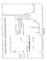
- Fraction F2 from paragraph C was dissolved in 6 M urea, 10 mM NaCI, 1 mM NEM, 50 mM sodium acetate, pH 4.8 and centrifuged at 10,000 rpm for 5 min. The supernatant was fractionated on a CM52 (a commercially available CMC) column equilibrated in the same buffer. Bound proteins were eluted from the column using a 10 mM to 400 mM NaCl gradient in the same buffer, and a total volume of 350 ml at a flow rate of 27 ml/hr. Three major fractions, designated CM-1, CM-2, and CM-3, were collected as shown in Figure 2.
- CM-2 and CM-3 were eluted at about 150 to 250 mM NaCI. Each fraction was dialyzed against 6 changes of 110 volumes of deionized water for 4 days and lyophilized. All of the foregoing operations were conducted at room temperature except dialysis (4 ° C).
- CM-2 and CM-3 from 1ID were combined and dissolved in 0.1% trifluoroacetic acid (TFA) and aliquots of the solutions loaded onto a Vydac C18 RP-HPLC column (4.6 mm ID x 25 cm) and washed with 0.1 % TFA for 5 min at 1 ml/min.
- the eluting solvent was a 0%-60% acetonitrile gradient in 0.1% TFA at a rate of 2%/min.
- the proteins were stored in 0.1 % TFA/acetonitrile eluting solution at -20 ° C until used.
- CM-2 and CM-3 were fractionated by electrophoresis on an acetic acid-urea gel using the general procedure of Paynim, S. and Chalkley, R., Arch Bioch Biophys (1969) 130:337-346.
- PG cartilage-specific proteoglycans
- This assay is an agarose gel culture model using mesenchymal cells isolated from rat fetal muscle. It assesses the ability of the samples to induce the production of PG.
- the correlation between in vitro cartilage induction and in vivo bone formation has been shown by Seyedin, S., et al, J Cell Biol (1983) 97:1950-1953.
- the cell culture was prepared by removing muscle tissue aseptically from the upper limbs of nineteen- day-old Sprague Dawley rat fetuses, mincing the tissue and culturing it in Eagle's Minimum Essential Medium (MEM) with 10% fetal bovine serum (FBS) and 50 units penicillin, 50 ⁇ g streptomycin per ml. Cellular outgrowth usually reached confluency within one week, whereupon cells were trypsinized, split 1:2 and used for experimentation within the first three passages.
- MEM Eagle's Minimum Essential Medium
- FBS fetal bovine serum
- the cells were placed in agarose gel cultures either with control medium or with samples to be tested.
- the procedure was basically that of Benya, et al, Cell (1982) 30:215. Briefly, the cell monolayers were harvested by trypsinization, counted on a hemocytometer, and resuspended at two times the final cell concentration in the medium with or without the protein fraction to be tested.
- the control medium was either Hams F-12, Dulbecco's Minimum Essential Medium (DMEM) or CMRL 1066 (Gibco) each containing 10% FBS and antibiotics.
- test protein fractions in 0.01 N HCI were diluted directly to the desired concentration of test protein diluted with an equal volume with 1% low melting agarose (Bio-Rad, #162-0017) in F-12, and 0.2 ml of the dilution was plated on 17 mm wells coated with 0.15 ml of 1 % high melting (Bio-Rad, #162-0100) agarose.
- the resulting cultures were incubated at 37 ° C for 5 min, chilled at 4 ° C for 10 min, and then overlayed with 1 ml of the corresponding medium (control or test protein).
- the cells were then cultured in a humidified atmosphere of 5% C0 2 , 95% air and fed every 3-4 days thereafter by a complete change with control medium. After 7 days the cultures were frozen and stored at -80 °C before assay.
- the cultures were assayed by thawing at 4 ° C, homogenizing in 4 M guanidine-HCI with 50 nM Na acetate, 13 mM EDTA, 6 mM NEM, and 3 nM PMSF at pH 5.8, and extracting by shaking overnight at 4 ° C.
- the supernatant fraction from centrifugation at 25,000 X g for 40 min at 4 ° C was dialyzed overnight at 4 ° C against 50 volumes 0.2 M NaCI, 50 mM Tris, pH 7.4.
- the supernatant was assayed for PG by ELISA as described by Renard, et al, Anal Biochem (1980) 104:205, and in U.S. 4,434,094.
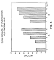
- the results of the ELISAs of CIF-A and CIF-B purified by RP-HPLC are shown in Figure 4. As indicated there, the sensitivity of the assay is within 1 to 5 ng/ml of culture media.
- the results of the ELISAs on the gel slices of section F are shown in Figure 5. These results are comparable to the results for CIF-A and CIF-B (corresponding to gel slices 7 and 6) from the RP-HPLC.
- CIF-A was shown to be a 25,800 dalton protein which on reduction, yielded a 14,800 dalton polypeptide by measurements of the mobilities of the proteins in a 15% Laemmli polyacrylamide gel in SDS ( Figure 3) as described by Laemmli, U. K., et al, Nature (1970) 227:680. It Is well understood that molecular weights so determined are approximate and their values are dependent on the method used. The conformation of the protein affects its mobility in this system, and, therefore, the molecular weights obtained will be similar, but not necessarily identical when determined by other procedures.
- the presence of a single band in the profile of the reduced protein indicates the protein is probably a dimer composed of two polypeptide chains having substantially equivalent amino acid sequences (i.e., it is a homodimer).
- the discrepancy between the measured weights of dimer and the individual chains is an artifact of the procedure.
- CIF-A maintained its activity in the ELISA assay of paragraph G above even after heating for 3 min at 100°C in PBS, after treatment with collagenase for 2 hrs at 37 ° C in 0.1 M Tris, pH 7.4, 5 mM CaCI 2 , 0.02 mM PMSF with a ratio of collagenase to protein of 400 units/mg protein, and after treatment with trypsin for 2 hrs at 37 ° C in 50 mM Tris, pH 7.4, 10 mM CaC1 2 with a ratio of trypsin to protein of 100 units/mg of protein.
- CIF-A The partial amino acid composition of CIF-A is shown in Table 1. Amino acid sequence analysis of CIF-A showed that it has the following single N-terminal sequence:
- CIF-B had a slightly different molecular weight (26,000) as measured by the same procedure. This difference may be caused by the procedure. Accordingly, both proteins are considered to have a molecular weight of approximately 26,000 daltons as measured by SDS-PAGE. On reduction the protein of peak B showed a single band at approximately 14,200 daltons indicating that it, too, is probably a homodimer. It has the amino acid composition set forth in Table 2. Amino acid sequence analysis indicated CIF-B has a single N-terminal sequence as follows: Its other properties, as qualitatively assessed, were similar to those set forth above for CIF-A.
- CIF-A and CIF-B were tested in the TGF-,8 bioassay.
- the assay was performed as described in Methods for Preparation of Media, Supplements, and Substrata for Serum-Free Animal Cell Culture (1984) pp 181-194, Alan R. Liss, Inc.
- the results of the assay are shown in Figure 6. As depicted, both proteins exhibit a clear dose-response in the assay and require the presence of an activating agent (EGF) to be active.
- EGF activating agent
- the ability of the CIFs to withstand treatment with trypsin without losing activity may make it possible to isolate them from demineralized bone powder by means of enzymatic digestion.
- the demineralized bone powder is digested with an aqueous solution of trypsin and/or other proteases that do not degrade the proteins of interest under conditions at which such enzymes are active.
- This treatment digests the majority of other protein components in the powder.
- the proteins of interest may be purified from the resulting digest using one or more of the fractionation techniques described above (gel filtration, ion exchange chromatography, RP-HPLC or nondenaturing gel electrophoresis).
- solubilizing agents may be avoided.
- the pure proteins are substantially soluble in water.
- the CIFs are useful for inducing cartilage/bone growth for repairing, replacing or augmenting cartilage/bone tissue in animals, including humans.
- Chondrogenically/osteogenically effective amounts of the proteins may be combined with chondrogenic/osteogenic co-factors found in bone and formulated with pharmacologically and physiologically acceptable fluid or solid carriers such as purified collagen for implantation.
- the weight ratio of active protein to carrier will typically be in the range of 1:50 to 1:1000.
- the implants may be placed at a predetermined site in the patient by conventional surgical techniques, including injection as an active ingredient.
- the CIFs may also be used in the same manner as human platelet/human placenta/bovine kidney-derived TGF-,8 to promote (provoke and sustain) non-species specific cellular proliferation.
- one or both of the CIFs is combined in approximately stoichiometric proportions with a TGF-,8 activating agent such as an EGF or a TGF-a.
- Clinical applications of the cell proliferation activity of these compositions include topical administration for burn or wound healing, implantation for tissue augmentation, and systemic administration for internal wound healing.
- the CIF and activating agent will be formulated in amounts sufficient to induce cell proliferation with pharmaceutically acceptable carriers that are adapted for the particular mode of administration.
- Topical dosage forms will typically be formulated as sprays, gels, ointments, or salves. Implants will be formulated as injectables. Systemic dosage forms may be formulated for enteral administration (e.g., liquids, pills, tablets) or for parenteral injection. The dosages used in such applications cannot be specified because of the nature of cell proliferation and the variability in wounds and other traumata.
Abstract
Description
- The present invention relates to protein chemistry. More particularly, it relates to two proteins that are found in bone, are co-factors for inducing cartilage formation, and are also active in the beta type transforming growth factor (TGF-β) assay. These polypeptides are sometimes referred to herein as cartilage-inducing factors (CIFs).
- Human platelet/human placenta/bovine kidney-derived TGF-βs are described in International Patent Application WO-A-84/01106 and in EP-A-0128849.
- U.S. 4,434,094 reports the partial purification of a bone generation-stimulating, bone-derived protein factor by extraction with chaotropic agents, fractionation on anion and cation exchange columns, and recovery of the activity from a fraction adsorbed to CMC at pH 4.8. This new protein fraction was termed "osteogenic factor" (OF) and was characterized as having a molecular weight below about 30,000 daltons and as tracking the purification process described. The proteins of the current invention were purified to homogeneity using a purification procedure that is similar in part to that disclosed in U.S. 4,434,094.
- EP-A-105014 describes another TGF-β. The structure of one such, there designated TGF-β1 is elucidated and it is suggested that the TGF-β's could be used for wound healing. TGF-β1 is the polypeptide designated CIFA in the present application.
- The invention provides a process for obtaining these polypeptides in substantially pure form from bone. Both CIFs are also active when combined with epidermal growth factor (EGF) in the TGF-,8 assay for in vitro induction of anchorage-independent growth of normal rat kidney (NRK) cells in soft agar. This assay is sometimes referred to herein as the TGF-,8 assay. In this regard the presence in bone of proteins having activity in the TGF-,8 assay has not been reported previously. One of the CIFs, designated CIF-A, has a partial (30 amino acids) N-terminal sequence that is identical to that reported in the literature for human placenta-derived TGF-β. The other CIF, designated CIF-B, has a partial N-terminal sequence that is different from the human placenta-derived TGF-,8 sequence and is claimed per se, and its structure elucidated, in the present invention.
- Accordingly, one aspect of the invention is a polypeptide cartilage-inducing factor, which factor:
- (a) is found in mammalian bone;
- (b) is a co-factor for inducing cartilage formation;
- (c) has activity in the TGF-β assay;
- (d) is a dimer having an approximate molecular weight of 26,000 daltons as determined by SDS-PAGE;
- (e) is isolatable by a process as claimed in
claim 1 orclaim 2; and - (f) does not have the N-terminal sequence:

- The process for isolating the two factors from bone is characterized by the following steps:
- (a) treating demineralized bone (DMB) with a chaotropic (dissociative) extractant that solubilizes non- fibrous proteins;
- (b) subjecting the extract from step (a) to gel filtration to recover a fraction containing proteins of molecular weight 10,000-40,000 daltons;
- (c) adsorbing the fraction from step (b) onto a carboxymethyl cellulose cation exchanger at approximately pH 4.5-5.5 under denaturing conditions;
- (d) eluting the adsorbed fraction from the cation exchanger with a sodium chloride gradient;
- (e) subjecting the portion of the eluate from step (d) eluting at approximately 150 to 250 mM NaCl to reverse phase high performance liquid chromatography (RP-HPLC) or a nondenaturing gel electrophoresis; and
- (f) recovering the factors from the RP-HPLC or gel electrophoresis.
- An implant composition for inducing chondrogenesis/osteogenesis is characterized in that it contains an effective amount of CIFB, or both of the above-described CIFs, optionally together with a chondrogenic/osteogenic co-factor.
- An implant composition for promoting connective tissue deposition is characterized in that it contains an effective amount of at least CIFB and is substantially free of any activating agent or chondrogenic co-factor.
- In the drawings:
- Figure 1 is a graph of the optical densities (absorbances) (280 nm) and in vitro chondrogenic activities of the gel filtration fractions of the example (section C), infra;
- Figure 2 is a graph of the optical densities (280 nm) of eluate fractions from the preparative ion exchange chromatography of the example (section D), infra;
- Figure 3 is a graph of the UV absorbance and electrophoretic profiles of peaks A (CIF-A) and B (CIF-B) of the preparative RP-HPLC of the example (section E), infra;
- Figure 4 is a graph of the results of the enzyme-linked immunosorbent assays (ELISAS) for in vitro chondrogenic activity of the CIF-A and CIF-B obtained from the RP-HPLC of the example (section E), infra;
- Figure 5 is a graph of the results of the ELISAs of the acid-urea gel electrophoresis fractions (section F) of the example, infra; and
- Figure 6 is a graph of the results of the TGF-,8 assays described in section I of the example, infra.
- The polypeptides of the invention were isolated from bone. The polypeptides have been only partially sequenced at this time. In view of this and since the complete amino acid sequence of TGF-,8 has not been reported, the primary structure relationships between the CIFs of the invention and TGF-,8 are not known completely.
- The polypeptides of the invention are co-factors for inducing cartilage formation. In view of their chondrogenic activity and the mode of endochondral bone formation, they are also expected to play a role in osteogenesis. The polypeptides are also active in the TGF-,8 assay and have been found to promote connective tissue deposition independently of association with TGF-β activating agents.
- In view of the showings that bone inductive proteins from human, monkey, bovine and rat are nonspecies-specific in their abilities to produce endochondral bone in xenogeneic implants (Sampath, T. K., et al, Proc Natl Acad Sci (USA) (1983) 80:6591) and that human platelet/human placenta/bovine kidney-derived TGF-,8 is nonspecies-specific between rodents, cattle and humans, it is believed that the polypeptides of this invention have been highly conserved among mammalian species (i.e., polypeptides from different mammalian species have amino acid sequences that vary, if at all, in one or more amino acid residue additions, deletions, or substitutions that do not affect the nonspecies-specific activity of the molecule adversely). In this regard the term "substantially equivalent" as used to describe a polypeptide is intended to mean polypeptides, whether native or synthetic and regardless of species or derivation, that have the same amino acid sequence as a CIF, and polypeptides of substantially homologous but different amino acid sequence, which difference(s) does not affect nonspecies-specific activity adversely. Accordingly, the polypeptides may be derived from bone and perhaps other tissue of diverse animal origin or made by recombinant DNA technology. Porcine or bovine long bone are preferred native sources of the CIFs because of the ready availability of such bone and the high levels of the polypeptides in bone.
- The procedure for isolating CIF from bone is as follows. The bone is first cleaned using mechanical or abrasive techniques, fragmented, and further washed with, for example, dilute aqueous acid preferably at low temperature, and then defatted by extraction with a lipophilic solvent such as ether or ethyl acetate. The bone is then demineralized by removal of the calcium phosphates in their various forms, usually by extraction with stronger acid. The resulting preparation, a demineralized bone, is the starting material for the preparation of the polypeptides of the invention.
- The initial extraction is designed to remove the non-fibrous (e.g., non-collagenous) proteins from the demineralized bone. This can be done with the use of chaotropic agents such as guanidine hydrochloride (at least about 4 molar), urea (8 molar) plus salt, or sodium dodecylsulfate (at least about 1 % by volume). The extraction is preferably carried out at reduced temperatures in the presence of a protease inhibitor to reduce the likelihood of digestion or denaturation of the extracted protein. Examples of protease inhibitors that may be included are phenylmethylsulfonylfluoride (PMSF) sodium azide, N-ethyl maleimide (NEM), benzamidine, and 6-aminohexanoic acid. The pH of the medium depends upon the extractant used. The process of extraction generally takes on the order of about 4 hr to one day.
- After extraction, the extractant may be removed by suitable means such as dialysis against water, preceded by concentration by ultrafiltration if desired. Salts can also be removed by controlled electrophoresis or by molecular sieving. It is also preferred to maintain a low temperature during this process so as to minimize denaturation of the proteins. Alternatively, the extractant need not be removed, but rather the solution need only be concentrated, for example, by ultrafiltration.
- The extract, dissolved or redissolved in chaotropic agent, is subjected to gel filtration to obtain fractions of molecular weight below about 40,000 daltons, thus resulting in a major enhancement of purity. Gel sizing is done using standard techniques, preferably on a Sephacryl column at room (10-250 C) temperature. The low molecular weight fraction is then subjected to ion exchange chromatography using carboxymethyl cellulose (CMC) at approximately pH 4.5-5.5, preferably about 4.8, in the presence of a nonionic chaotropic agent such as urea. Other cation exchangers may be used, including those derived from polyacrylamide and cross-linked dextran; however cellulosic cation exchangers are preferred. Of course, as in any ion exchange procedure, the solution must be freed of competing ions before application to the column, and is eluted in an increasing salt concentration gradient as is understood in the art. The fraction eluting from CMC at about 150 to 250 mM NaCl contains the CIFs.
- The eluate fraction from the cation exchange chromatography is then subjected to RP-HPLC or a nondenaturing gel electrophoresis for final purification. Standard RP-HPLC techniques and gel electrophoresis techniques are used. Exemplified below is a commercially available RP-HPLC column using a commercially prescribed RP-HPLC protocol. This final purification yields the two polypeptides in substantially pure form. "Substantially pure" means that a polypeptide contains less than about 5% by weight contaminants.
- The following example is intended to illustrate the process for purification as applied to a particular sample. It is not intended to limit the invention.
- Bovine metatarsal bone was obtained fresh from the slaughterhouse and transported on dry ice. The bones were cleaned of marrow and non-bone tissues, broken in fragments smaller than 1 cm diameter, and pulverized in a mill at 4 ° C. The pulverized bone was washed twice with 9.4 liters of double distilled water per kg of bone for about 15 min each, and then washed overnight in 0.01 N HCI at 4 ° C. Washed bone was defatted using 3
X 3 volumes ethanol, followed by 3 X 3 volumes diethylether, each washed for 20 min, and all at room temperature. The resulting defatted bone powder was then demineralized in 0.5 N HCI (25 I/kg defatted bone) at 4 ° C. The acid was decanted and the resulting DMB washed until the wash pH was greater than 4, and the DMB dried on a suction filter. - The DMB as prepared in paragraph A was extracted with 3.3 I of 4 M guanidine-HCI, 10 mM ethylenediaminetetraacetic acid (EDTA), pH 6.8, 1 mM PMSF, 10 mM NEM per kg for 16 hrs, the suspension suction filtered and the non-soluble material extracted again for 4 hrs. The soluble fractions were combined and concentrated at least 5-fold by ultrafiltration using an Amicon ultrafiltration (10K) unit, and the concentrate dialyzed against 6 changes of 35 volumes cold deionized water over a period of 4 days, and then lyophilized. All of the procedures of this paragraph were conducted at 4 ° C except the lyophilization which was conducted under standard lyophilization conditions.
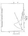
- The extract from paragraph B, redissolved in 4 M guanidine-HCI, was fractionated on a Sephacryl S-200 column equilibrated in 4 M guanidine-HCI, 0.02% sodium azide, 10 mM EDTA, pH 6.8. Fractions were assayed by their absorbance at 280 nm and chondrogenic activity by ELISA (described below) and the fractions were combined as shown in Figure 1. Fraction F2 of Figure 1, constituting a low molecular weight (LMW, 10,000-40,000 daltons) protein fraction possessing the greatest activity was dialyzed against 6 changes of 180 volumes of deionized water and lyophilized. All operations except lyophilization and dialysis (4 ° C) were conducted at room temperature.
- Fraction F2 from paragraph C was dissolved in 6 M urea, 10 mM NaCI, 1 mM NEM, 50 mM sodium acetate, pH 4.8 and centrifuged at 10,000 rpm for 5 min. The supernatant was fractionated on a CM52 (a commercially available CMC) column equilibrated in the same buffer. Bound proteins were eluted from the column using a 10 mM to 400 mM NaCl gradient in the same buffer, and a total volume of 350 ml at a flow rate of 27 ml/hr. Three major fractions, designated CM-1, CM-2, and CM-3, were collected as shown in Figure 2. CM-2 and CM-3 were eluted at about 150 to 250 mM NaCI. Each fraction was dialyzed against 6 changes of 110 volumes of deionized water for 4 days and lyophilized. All of the foregoing operations were conducted at room temperature except dialysis (4 ° C).
- The lyophilized fractions CM-2 and CM-3 from 1ID were combined and dissolved in 0.1% trifluoroacetic acid (TFA) and aliquots of the solutions loaded onto a Vydac C18 RP-HPLC column (4.6 mm ID x 25 cm) and washed with 0.1 % TFA for 5 min at 1 ml/min. The eluting solvent was a 0%-60% acetonitrile gradient in 0.1% TFA at a rate of 2%/min.
- Two peaks were obtained from the RP-HPLC of combined CM-2 and CM-3--peak A at about 29.5 min and peak B at about 31.2 min. Figure 3 shows the absorbance and electrophoretic profiles (reduced and nonreduced) of peaks A and B. The proteins of these peaks were designated CIF-A and CIF-B, respectively.
- The proteins were stored in 0.1 % TFA/acetonitrile eluting solution at -20 ° C until used.
- The combined lyophilized fractions CM-2 and CM-3 were fractionated by electrophoresis on an acetic acid-urea gel using the general procedure of Paynim, S. and Chalkley, R., Arch Bioch Biophys (1969) 130:337-346.
- The presence of the desired protein in fractions during purification was confirmed using an in vitro assay for the production of cartilage-specific proteoglycans (PG), the identity of which was confirmed by ELISA. This assay is an agarose gel culture model using mesenchymal cells isolated from rat fetal muscle. It assesses the ability of the samples to induce the production of PG. The correlation between in vitro cartilage induction and in vivo bone formation has been shown by Seyedin, S., et al, J Cell Biol (1983) 97:1950-1953.
- The cell culture was prepared by removing muscle tissue aseptically from the upper limbs of nineteen- day-old Sprague Dawley rat fetuses, mincing the tissue and culturing it in Eagle's Minimum Essential Medium (MEM) with 10% fetal bovine serum (FBS) and 50 units penicillin, 50 µg streptomycin per ml. Cellular outgrowth usually reached confluency within one week, whereupon cells were trypsinized, split 1:2 and used for experimentation within the first three passages.
- The cells were placed in agarose gel cultures either with control medium or with samples to be tested. The procedure was basically that of Benya, et al, Cell (1982) 30:215. Briefly, the cell monolayers were harvested by trypsinization, counted on a hemocytometer, and resuspended at two times the final cell concentration in the medium with or without the protein fraction to be tested. The control medium was either Hams F-12, Dulbecco's Minimum Essential Medium (DMEM) or CMRL 1066 (Gibco) each containing 10% FBS and antibiotics. The test protein fractions in 0.01 N HCI were diluted directly to the desired concentration of test protein diluted with an equal volume with 1% low melting agarose (Bio-Rad, #162-0017) in F-12, and 0.2 ml of the dilution was plated on 17 mm wells coated with 0.15 ml of 1 % high melting (Bio-Rad, #162-0100) agarose. The resulting cultures were incubated at 37 ° C for 5 min, chilled at 4 ° C for 10 min, and then overlayed with 1 ml of the corresponding medium (control or test protein). The cells were then cultured in a humidified atmosphere of 5% C02, 95% air and fed every 3-4 days thereafter by a complete change with control medium. After 7 days the cultures were frozen and stored at -80 °C before assay.
- The cultures were assayed by thawing at 4°C, homogenizing in 4 M guanidine-HCI with 50 nM Na acetate, 13 mM EDTA, 6 mM NEM, and 3 nM PMSF at pH 5.8, and extracting by shaking overnight at 4 ° C. The supernatant fraction from centrifugation at 25,000 X g for 40 min at 4 ° C was dialyzed overnight at 4 ° C against 50 volumes 0.2 M NaCI, 50 mM Tris, pH 7.4. The supernatant was assayed for PG by ELISA as described by Renard, et al, Anal Biochem (1980) 104:205, and in U.S. 4,434,094.
- Briefly, for the ELISA, antiserum to PG was raised in rabbits using standard techniques which showed no cross-reactivity with hyaluronic acid or PG extracted from rat bone. Purified PG (Seyedin, S., et al, supra) from Swarm rat chondrosarcoma tissue was used as standard antigen. The dialyzed samples were diluted 1:1 (v/v) in phosphate-buffered saline (PBS) with 0.05
% Tween - The results of the ELISAs of CIF-A and CIF-B purified by RP-HPLC are shown in Figure 4. As indicated there, the sensitivity of the assay is within 1 to 5 ng/ml of culture media. The results of the ELISAs on the gel slices of section F are shown in Figure 5. These results are comparable to the results for CIF-A and CIF-B (corresponding to gel
slices 7 and 6) from the RP-HPLC. - CIF-A was shown to be a 25,800 dalton protein which on reduction, yielded a 14,800 dalton polypeptide by measurements of the mobilities of the proteins in a 15% Laemmli polyacrylamide gel in SDS (Figure 3) as described by Laemmli, U. K., et al, Nature (1970) 227:680. It Is well understood that molecular weights so determined are approximate and their values are dependent on the method used. The conformation of the protein affects its mobility in this system, and, therefore, the molecular weights obtained will be similar, but not necessarily identical when determined by other procedures. The presence of a single band in the profile of the reduced protein indicates the protein is probably a dimer composed of two polypeptide chains having substantially equivalent amino acid sequences (i.e., it is a homodimer). The discrepancy between the measured weights of dimer and the individual chains is an artifact of the procedure.
- CIF-A maintained its activity in the ELISA assay of paragraph G above even after heating for 3 min at 100°C in PBS, after treatment with collagenase for 2 hrs at 37 ° C in 0.1 M Tris, pH 7.4, 5 mM CaCI2, 0.02 mM PMSF with a ratio of collagenase to protein of 400 units/mg protein, and after treatment with trypsin for 2 hrs at 37 ° C in 50 mM Tris, pH 7.4, 10 mM CaC12 with a ratio of trypsin to protein of 100 units/mg of protein. However, the protein lost activity after treatment for 1 hr at room temperature in PBS containing 5 mM dithiothreitol (DTT), which would effect reduction of disulfide linkages. Similarly, SDS treatment or fractionation on SDS-PAGE resulted in inactivation of the protein, presumably due to denaturation or complexing by the SDS. The partial amino acid composition of CIF-A is shown in Table 1.


- ( ) = suspected
- CIF-B had a slightly different molecular weight (26,000) as measured by the same procedure. This difference may be caused by the procedure. Accordingly, both proteins are considered to have a molecular weight of approximately 26,000 daltons as measured by SDS-PAGE. On reduction the protein of peak B showed a single band at approximately 14,200 daltons indicating that it, too, is probably a homodimer. It has the amino acid composition set forth in Table 2.
 Its other properties, as qualitatively assessed, were similar to those set forth above for CIF-A.
- CIF-A and CIF-B were tested in the TGF-,8 bioassay. The assay was performed as described in Methods for Preparation of Media, Supplements, and Substrata for Serum-Free Animal Cell Culture (1984) pp 181-194, Alan R. Liss, Inc. The results of the assay are shown in Figure 6. As depicted, both proteins exhibit a clear dose-response in the assay and require the presence of an activating agent (EGF) to be active. The levels of activity are comparable to the reported levels of activity of human platelet/human placenta/bovine kidney-derived TGF-β.
- The ability of the CIFs to withstand treatment with trypsin without losing activity may make it possible to isolate them from demineralized bone powder by means of enzymatic digestion. In such a process the demineralized bone powder is digested with an aqueous solution of trypsin and/or other proteases that do not degrade the proteins of interest under conditions at which such enzymes are active. This treatment digests the majority of other protein components in the powder. The proteins of interest may be purified from the resulting digest using one or more of the fractionation techniques described above (gel filtration, ion exchange chromatography, RP-HPLC or nondenaturing gel electrophoresis). Depending upon the extent to which the CIFs are released from the bone matrix and not complexed with other materials, use of solubilizing agents may be avoided. In this regard the pure proteins are substantially soluble in water.
- The CIFs are useful for inducing cartilage/bone growth for repairing, replacing or augmenting cartilage/bone tissue in animals, including humans. Chondrogenically/osteogenically effective amounts of the proteins may be combined with chondrogenic/osteogenic co-factors found in bone and formulated with pharmacologically and physiologically acceptable fluid or solid carriers such as purified collagen for implantation. The weight ratio of active protein to carrier will typically be in the range of 1:50 to 1:1000. The implants may be placed at a predetermined site in the patient by conventional surgical techniques, including injection as an active ingredient. Collagenous implants containing only CIFB as an active ingredient (i.e., free of any activating agent or co-factor) at CIF to carrier weight ratios above about 1:6000 promoted collagenous connective tissue deposition.
- The CIFs may also be used in the same manner as human platelet/human placenta/bovine kidney-derived TGF-,8 to promote (provoke and sustain) non-species specific cellular proliferation. In such application one or both of the CIFs is combined in approximately stoichiometric proportions with a TGF-,8 activating agent such as an EGF or a TGF-a. Clinical applications of the cell proliferation activity of these compositions include topical administration for burn or wound healing, implantation for tissue augmentation, and systemic administration for internal wound healing. In such uses the CIF and activating agent will be formulated in amounts sufficient to induce cell proliferation with pharmaceutically acceptable carriers that are adapted for the particular mode of administration. Topical dosage forms will typically be formulated as sprays, gels, ointments, or salves. Implants will be formulated as injectables. Systemic dosage forms may be formulated for enteral administration (e.g., liquids, pills, tablets) or for parenteral injection. The dosages used in such applications cannot be specified because of the nature of cell proliferation and the variability in wounds and other traumata.
This N-terminal sequence is identical to that reported for human placenta-derived TGF-β.
Claims (9)
the process comprising:


Priority Applications (1)
| Application Number | Priority Date | Filing Date | Title |
|---|---|---|---|
| JP61099314A JPH068318B2 (en) | 1985-07-08 | 1986-04-28 | Partially purified osteoinductive factor |
Applications Claiming Priority (2)
| Application Number | Priority Date | Filing Date | Title |
|---|---|---|---|
| US63093884A | 1984-07-16 | 1984-07-16 | |
| US630938 | 1984-07-16 |
Publications (4)
| Publication Number | Publication Date |
|---|---|
| EP0169016A2 EP0169016A2 (en) | 1986-01-22 |
| EP0169016A3 EP0169016A3 (en) | 1987-04-29 |
| EP0169016B1 true EP0169016B1 (en) | 1995-10-04 |
| EP0169016B2 EP0169016B2 (en) | 2004-04-28 |
Family
ID=26829241
Family Applications (1)
| Application Number | Title | Priority Date | Filing Date |
|---|---|---|---|
| EP85304848A Expired - Lifetime EP0169016B2 (en) | 1984-07-16 | 1985-07-08 | Polypeptide cartilage-inducing factors found in bone |
Country Status (7)
| Country | Link |
|---|---|
| US (3) | US4774228A (en) |
| EP (1) | EP0169016B2 (en) |
| JP (1) | JPH0794474B2 (en) |
| AT (1) | ATE128715T1 (en) |
| AU (2) | AU592951B2 (en) |
| CA (1) | CA1261549A (en) |
| DE (1) | DE3588058T3 (en) |
Cited By (6)
| Publication number | Priority date | Publication date | Assignee | Title |
|---|---|---|---|---|
| US6395293B2 (en) | 1989-07-24 | 2002-05-28 | Atrix Laboratories | Biodegradable implant precursor |
| US7211271B2 (en) | 2001-03-12 | 2007-05-01 | The Regents Of The University Of California | Method to improve hydroxyapatite implantation and stimulate bone regeneration |
| US7678885B2 (en) | 1991-11-04 | 2010-03-16 | Genetics Institute, Llc | Recombinant bone morphogenetic protein heterodimers, compositions and methods of use |
| US7771755B2 (en) | 2003-09-12 | 2010-08-10 | Wyeth | Injectable calcium phosphate solid rods and pastes for delivery of osteogenic proteins |
| US8012482B2 (en) | 2004-03-31 | 2011-09-06 | Genentech, Inc. | Humanized anti-TGF-beta antibodies |
| US8226598B2 (en) | 1999-09-24 | 2012-07-24 | Tolmar Therapeutics, Inc. | Coupling syringe system and methods for obtaining a mixed composition |
Families Citing this family (152)
| Publication number | Priority date | Publication date | Assignee | Title |
|---|---|---|---|---|
| US5104977A (en) * | 1982-09-24 | 1992-04-14 | The United States Of America As Represented By The Department Of Health And Human Services | Purified transforming growth factor beta |
| US5705477A (en) * | 1982-09-24 | 1998-01-06 | The United States Of America As Represented By The Department Of Health And Human Services | Compositions of transforming growth factor β(TGF-β) which promotes wound healing and methods for their use |
| US5656587A (en) * | 1982-09-24 | 1997-08-12 | The United States Of America As Represented By The Secretary Of The Department Of Health And Human Services | Promotion of cell proliferation by use of transforming growth factor beta (TGF-β) |
| US5328695A (en) * | 1983-03-22 | 1994-07-12 | Massachusetts Institute Of Technology | Muscle morphogenic protein and use thereof |
| US4804744A (en) * | 1984-01-04 | 1989-02-14 | International Genetic Engineering, Inc. | Osteogenic factors |
| CA1243604A (en) * | 1984-02-29 | 1988-10-25 | Michael Klagsbrun | Endothelial cell-growth factor |
| USRE35694E (en) * | 1984-07-16 | 1997-12-16 | Celtrix Pharmaceuticals, Inc. | Polypeptide cartilage-inducing factors found in bone |
| US4627982A (en) * | 1984-07-16 | 1986-12-09 | Collagen Corporation | Partially purified bone-inducing factor |
| ATE128715T1 (en) * | 1984-07-16 | 1995-10-15 | Celtrix Pharma | POLYPEPTIDE-INDUCING FACTORS IN BONE AND CARTILAGE. |
| US4886747A (en) * | 1985-03-22 | 1989-12-12 | Genentech, Inc. | Nucleic acid encoding TGF-β and its uses |
| US5284763A (en) * | 1985-03-22 | 1994-02-08 | Genentech, Inc. | Nucleic acid encoding TGF-β and its uses |
| US6586394B1 (en) | 1985-04-19 | 2003-07-01 | Osi Pharmaceuticals, Inc. | Tissue-derived tumor growth inhibitor |
| US4971952A (en) * | 1986-03-06 | 1990-11-20 | Collagen Corporation | Method of treating inflammation with cartilage inducing factor |
| US4806523A (en) * | 1985-08-06 | 1989-02-21 | Collagen Corporation | Method of treating inflammation |
| US5120535A (en) * | 1986-11-26 | 1992-06-09 | Oncogen | Oncostatin M and novel compositions having anti-neoplastic activity |
| US5187076A (en) * | 1986-07-01 | 1993-02-16 | Genetics Institute, Inc. | DNA sequences encoding BMP-6 proteins |
| US5939388A (en) * | 1986-07-01 | 1999-08-17 | Rosen; Vicki A. | Methods of administering BMP-5 compositions |
| US5366875A (en) * | 1986-07-01 | 1994-11-22 | Genetics Institute, Inc. | Methods for producing BMP-7 proteins |
| US5013649A (en) * | 1986-07-01 | 1991-05-07 | Genetics Institute, Inc. | DNA sequences encoding osteoinductive products |
| US5459047A (en) * | 1986-07-01 | 1995-10-17 | Genetics Institute, Inc. | BMP-6 proteins |
| US5108922A (en) * | 1986-07-01 | 1992-04-28 | Genetics Institute, Inc. | DNA sequences encoding BMP-1 products |
| US6150328A (en) | 1986-07-01 | 2000-11-21 | Genetics Institute, Inc. | BMP products |
| US5543394A (en) * | 1986-07-01 | 1996-08-06 | Genetics Institute, Inc. | Bone morphogenetic protein 5(BMP-5) compositions |
| US4877864A (en) * | 1987-03-26 | 1989-10-31 | Genetics Institute, Inc. | Osteoinductive factors |
| IL83003A (en) | 1986-07-01 | 1995-07-31 | Genetics Inst | Osteoinductive factors |
| US6432919B1 (en) | 1986-07-01 | 2002-08-13 | Genetics Institute, Inc. | Bone morphogenetic protein-3 and compositions |
| US5106748A (en) * | 1986-07-01 | 1992-04-21 | Genetics Institute, Inc. | Dna sequences encoding 5 proteins |
| US4816442A (en) * | 1986-11-07 | 1989-03-28 | Collagen Corporation | Method of inhibiting tumor growth sensitive to CIF-βtreatment |
| US4931548A (en) * | 1987-01-30 | 1990-06-05 | Techne Corporation | Heterodimer form of transforming growth factor-beta |
| WO1988005787A1 (en) * | 1987-01-30 | 1988-08-11 | Techne Corporation | Transforming growth factor-beta |
| GB8723094D0 (en) * | 1987-10-01 | 1987-11-04 | Ciba Geigy Ag | Polypeptide growth factor from milk |
| US5221734A (en) * | 1987-10-01 | 1993-06-22 | Ciba-Geigy Corporation | Process for preparing a polypeptide growth factor for milk |
| IL87877A0 (en) * | 1987-10-06 | 1989-03-31 | Oncogen | Cloning and expression of transforming growth factor beta2 |
| US5221620A (en) * | 1987-10-06 | 1993-06-22 | Oncogen | Cloning and expression of transforming growth factor β2 |
| US5035901A (en) * | 1987-10-09 | 1991-07-30 | University Of Kansas | Bone inducing agent from a human osteosarcoma cell line |
| US6020313A (en) * | 1987-10-09 | 2000-02-01 | University Of Kansas | Method for inducing bone formation using an extract of human osteosarcoma cell line SAOS-2 |
| US4863732A (en) | 1987-12-16 | 1989-09-05 | Collagen Corporation | Injectable composition for inductive bone repair |
| AU3433989A (en) * | 1988-04-06 | 1989-11-03 | Celtrix Laboratories, Inc. | Bone-inducing protein |
| US5258494A (en) * | 1988-04-08 | 1993-11-02 | Stryker Corporation | Osteogenic proteins |
| EP0362367B1 (en) * | 1988-04-08 | 1996-02-28 | Stryker Corporation | Osteogenic devices |
| US5670336A (en) * | 1988-04-08 | 1997-09-23 | Stryker Corporation | Method for recombinant production of osteogenic protein |
| US5108753A (en) * | 1988-04-08 | 1992-04-28 | Creative Biomolecules | Osteogenic devices |
| US4968590A (en) * | 1988-04-08 | 1990-11-06 | Stryker Corporation | Osteogenic proteins and polypeptides |
| US5354557A (en) * | 1988-04-08 | 1994-10-11 | Stryker Corporation | Osteogenic devices |
| US5011691A (en) * | 1988-08-15 | 1991-04-30 | Stryker Corporation | Osteogenic devices |
| US5266683A (en) * | 1988-04-08 | 1993-11-30 | Stryker Corporation | Osteogenic proteins |
| US6919308B2 (en) | 1988-04-08 | 2005-07-19 | Stryker Corporation | Osteogenic devices |
| US6586388B2 (en) | 1988-04-08 | 2003-07-01 | Stryker Corporation | Method of using recombinant osteogenic protein to repair bone or cartilage defects |
| US5250302A (en) * | 1988-04-08 | 1993-10-05 | Stryker Corporation | Osteogenic devices |
| US5324819A (en) * | 1988-04-08 | 1994-06-28 | Stryker Corporation | Osteogenic proteins |
| US20070166353A1 (en) * | 1988-04-08 | 2007-07-19 | Stryker Corporation | Osteogenic proteins |
| US4935497A (en) * | 1988-09-06 | 1990-06-19 | Northwestern University | Dentin chondrogenic inductive agent |
| US5207710A (en) * | 1988-09-29 | 1993-05-04 | Collagen Corporation | Method for improving implant fixation |
| US5258029A (en) * | 1988-09-29 | 1993-11-02 | Collagen Corporation | Method for improving implant fixation |
| US4938763B1 (en) * | 1988-10-03 | 1995-07-04 | Atrix Lab Inc | Biodegradable in-situ forming implants and method of producing the same |
| US5284756A (en) * | 1988-10-11 | 1994-02-08 | Lynn Grinna | Heterodimeric osteogenic factor |
| US5106626A (en) * | 1988-10-11 | 1992-04-21 | International Genetic Engineering, Inc. | Osteogenic factors |
| US5135915A (en) * | 1988-10-14 | 1992-08-04 | Genentech, Inc. | Method for the treatment of grafts prior to transplantation using TGF-.beta. |
| US5571714A (en) | 1988-12-22 | 1996-11-05 | Celtrix Pharmaceuticals, Inc. | Monoclonal antibodies which bind both transforming growth factors β1 and β2 and methods of use |
| US5061786A (en) * | 1989-05-25 | 1991-10-29 | Genentech, Inc. | Biologically active polypeptides based on transforming growth factor-β |
| US5268455A (en) * | 1989-05-25 | 1993-12-07 | Genentech, Inc. | Process for making biologically active polypeptides based on transforming growth factor-βsequences |
| PT94241A (en) * | 1989-06-02 | 1991-02-08 | Chiron Corp | METHOD FOR THE PREPARATION OF OESEA CALCIFICATION FACT AND OF PHARMACEUTICAL COMPOSITIONS CONTAINING THEM |
| CA2020729A1 (en) * | 1989-07-19 | 1991-01-20 | Michael C. Kiefer | Bone morphogenetic protein |
| EP0489062A4 (en) * | 1989-08-21 | 1992-08-12 | Celtrix Laboratories, Inc. | Bone-specific protein |
| US5422340A (en) * | 1989-09-01 | 1995-06-06 | Ammann; Arthur J. | TGF-βformulation for inducing bone growth |
| US5158934A (en) * | 1989-09-01 | 1992-10-27 | Genentech, Inc. | Method of inducing bone growth using TGF-β |
| IL95500A (en) * | 1989-09-11 | 1997-03-18 | Matrix Pharma | ANTI-PROLIFERATIVE COMPOSITIONS CONTAINING TGF-b PROTEIN IN A VISCOUS MATRIX AND THEIR USE |
| ES2118723T3 (en) * | 1989-10-17 | 1998-10-01 | Stryker Corp | OSTEOGENIC DEVICES. |
| US5071655A (en) * | 1990-01-12 | 1991-12-10 | Baylink David J | Pharmaceutical combination for treatment of bone-wasting diseases |
| USRE37950E1 (en) | 1990-04-24 | 2002-12-31 | Atrix Laboratories | Biogradable in-situ forming implants and methods of producing the same |
| US5147799A (en) * | 1990-04-25 | 1992-09-15 | Isia Bursuker | Repopulation of macrophages and granulocytes using transforming growth factor-beta |
| US5688678A (en) * | 1990-05-16 | 1997-11-18 | Genetics Institute, Inc. | DNA encoding and methods for producing BMP-8 proteins |
| US7378392B1 (en) | 1990-05-16 | 2008-05-27 | Genetics Institute, Llc | Bone and cartilage inductive proteins |
| US5067963A (en) * | 1990-08-21 | 1991-11-26 | Washington University | Method of making live autogenous skeletal replacement parts |
| AU651421B2 (en) * | 1990-11-30 | 1994-07-21 | Celtrix Pharmaceuticals, Inc. | Use of a bone morphogenetic protein in synergistic combination with TGF-beta for bone repair |
| US5206023A (en) * | 1991-01-31 | 1993-04-27 | Robert F. Shaw | Method and compositions for the treatment and repair of defects or lesions in cartilage |
| US5284830A (en) * | 1991-02-13 | 1994-02-08 | The Board Of Trustees Of The Leland Stanford Junior University | Thyroid-derived chondrocyte-stimulating factor |
| US5169837A (en) * | 1991-03-28 | 1992-12-08 | Allelix Biopharmaceuticals Inc. | Isolated osteogenic factor |
| GB9106678D0 (en) * | 1991-03-28 | 1991-05-15 | Ferguson Mark W J | Wound healing |
| US5563124A (en) * | 1991-04-22 | 1996-10-08 | Intermedics Orthopedics/ Denver, Inc. | Osteogenic product and process |
| US5290763A (en) * | 1991-04-22 | 1994-03-01 | Intermedics Orthopedics/Denver, Inc. | Osteoinductive protein mixtures and purification processes |
| AU662155B2 (en) | 1991-05-10 | 1995-08-24 | Celtrix Pharmaceuticals, Inc. | Targeted delivery of bone growth factors |
| US5693615A (en) * | 1991-06-05 | 1997-12-02 | The Procter & Gamble Company | Therapeutic compositions for osteoinduction |
| US6287816B1 (en) | 1991-06-25 | 2001-09-11 | Genetics Institute, Inc. | BMP-9 compositions |
| EP0592562B1 (en) * | 1991-06-25 | 1999-01-07 | Genetics Institute, Inc. | Bmp-9 compositions |
| US5270300A (en) * | 1991-09-06 | 1993-12-14 | Robert Francis Shaw | Methods and compositions for the treatment and repair of defects or lesions in cartilage or bone |
| US20080139474A1 (en) * | 1991-11-04 | 2008-06-12 | David Israel | Recombinant bone morphogenetic protein heterodimers, compositions and methods of use |
| US20080233170A1 (en) * | 1992-02-21 | 2008-09-25 | Stryker Corporation | Osteogenic Proteins |
| US5322933A (en) * | 1992-05-07 | 1994-06-21 | The United States Of America As Represented By The Secretary Of The Department Of Health And Human Services | Crystal structure of TGF-β-2 |
| US5767079A (en) * | 1992-07-08 | 1998-06-16 | Celtrix Pharmaceuticals, Inc. | Method of treating ophthalmic disorders using TGF -β |
| CA2103943A1 (en) * | 1992-08-21 | 1994-02-22 | A. Gregory Bruce | Composition and methods for the generation of bone |
| DE69433530T2 (en) | 1993-05-12 | 2005-01-05 | Genetics Institute, LLC, Cambridge | BMP-11 COMPOSITIONS |
| US5637480A (en) * | 1993-05-12 | 1997-06-10 | Genetics Institute, Inc. | DNA molecules encoding bone morphogenetic protein-10 |
| US5531791A (en) * | 1993-07-23 | 1996-07-02 | Bioscience Consultants | Composition for repair of defects in osseous tissues, method of making, and prosthesis |
| US5426098A (en) * | 1993-09-02 | 1995-06-20 | Celtrix Pharmaceuticals, Inc. | Increase in hematopoietic progenitor cells in peripheral blood by transforming growth factor beta |
| US6291206B1 (en) | 1993-09-17 | 2001-09-18 | Genetics Institute, Inc. | BMP receptor proteins |
| US6610656B1 (en) * | 1993-12-30 | 2003-08-26 | President And Fellows Of Harvard College | Method of promoting chondrocyte differentiation with hedgehog related polypeptides |
| CA2176942C (en) | 1993-12-07 | 2011-11-01 | Anthony J. Celeste | Bmp-12, bmp-13 and tendon-inducing compositions thereof |
| US6027919A (en) * | 1993-12-07 | 2000-02-22 | Genetics Institute, Inc. | BMP-12 and BMP-13 proteins and DNA encoding them |
| US7060450B1 (en) * | 1993-12-30 | 2006-06-13 | President And Fellows Of Harvard College | Screening assays for agonists and antagonists of the hedgehog signaling pathway |
| US20030186357A1 (en) * | 1993-12-30 | 2003-10-02 | Philip W. Ingham | Vertebrate embryonic pattern-inducing proteins, and uses related thereto |
| AU1120295A (en) * | 1994-11-07 | 1996-05-31 | Government Of The United States Of America, As Represented By The Secretary Of The Department Of Health And Human Services, The | Cartilage-derived morphogenetic proteins |
| US20010011131A1 (en) * | 1997-07-28 | 2001-08-02 | Luyten Frank P. | DNA molecules encoding cartilage-derived morphogenetic proteins |
| US5906934A (en) * | 1995-03-14 | 1999-05-25 | Morphogen Pharmaceuticals, Inc. | Mesenchymal stem cells for cartilage repair |
| EP0883410B1 (en) * | 1996-02-29 | 2004-08-18 | Bioactive Bone Substitute OY, AB | An osteogenic device and a method for preparing the device |
| US6492508B1 (en) * | 1996-06-03 | 2002-12-10 | United States Surgical Corp. A Division Of Tyco Healthcare Group | Nucleic acids encoding extracellular matrix proteins |
| US6927287B1 (en) * | 1996-06-03 | 2005-08-09 | United States Surgical Corporation | Nucleic acid encoding extracellular matrix protein or fragment thereof |
| WO1998004681A2 (en) * | 1996-07-25 | 1998-02-05 | Genzyme Corporation | Chondrocyte media formulations and culture procedures |
| US6034062A (en) * | 1997-03-13 | 2000-03-07 | Genetics Institute, Inc. | Bone morphogenetic protein (BMP)-9 compositions and their uses |
| DE69714035T2 (en) | 1997-08-14 | 2003-03-06 | Sulzer Innotec Ag | Composition and device for repairing cartilage tissue in vivo consisting of nanocapsules with osteoinductive and / or chondroinductive factors |
| US7128927B1 (en) | 1998-04-14 | 2006-10-31 | Qlt Usa, Inc. | Emulsions for in-situ delivery systems |
| JP4055248B2 (en) * | 1998-05-25 | 2008-03-05 | 味の素株式会社 | Purified human activin and method for producing the same |
| US7087577B2 (en) * | 1998-10-16 | 2006-08-08 | Zimmer Orthobiologies, Inc. | Method of promoting natural bypass |
| US6727224B1 (en) * | 1999-02-01 | 2004-04-27 | Genetics Institute, Llc. | Methods and compositions for healing and repair of articular cartilage |
| ES2225241T3 (en) | 1999-10-15 | 2005-03-16 | Genetics Institute, Llc | HIALURONIC ACID FORMULATIONS TO SUPPLY OSTEOGENIC PROTEINS. |
| PL356006A1 (en) * | 1999-12-17 | 2004-05-31 | CARTIFICIAL A/S, Medico Chemical Lab.ApS | A prosthetic device |
| JP2001218840A (en) * | 2000-02-14 | 2001-08-14 | Kanegafuchi Chem Ind Co Ltd | Adsorbent for transforming growth factor. beta, adsorbing/removing method, and adsorber |
| EP1292330A1 (en) * | 2000-03-31 | 2003-03-19 | Vaccine Chip Technology APS | Immunostimulating properties of a fragment of tgf- beta |
| US20030082233A1 (en) * | 2000-12-01 | 2003-05-01 | Lyons Karen M. | Method and composition for modulating bone growth |
| US6492327B2 (en) * | 2000-12-19 | 2002-12-10 | Sulzer Biologics Inc. | Isolation of purified TGF- β1 and TGF -β2 from bone tissue |
| US20020114795A1 (en) | 2000-12-22 | 2002-08-22 | Thorne Kevin J. | Composition and process for bone growth and repair |
| TW200526779A (en) | 2001-02-08 | 2005-08-16 | Wyeth Corp | Modified and stabilized GDF propeptides and uses thereof |
| ATE393573T1 (en) * | 2001-06-01 | 2008-05-15 | Wyeth Corp | COMPOSITIONS FOR SYSTEMIC ADMINISTRATION OF SEQUENCES ENCODING BONE MORPHOGENESIS PROTEINS |
| TWI267378B (en) | 2001-06-08 | 2006-12-01 | Wyeth Corp | Calcium phosphate delivery vehicles for osteoinductive proteins |
| US6923833B2 (en) * | 2002-04-09 | 2005-08-02 | Ray C. Wasielewski | Biologically reabsorbable acetabular constraining components and materials for use with a hip replacement prosthesis and bioreabsorbable materials to augment hip replacement stability and function |
| TWI282283B (en) * | 2002-05-17 | 2007-06-11 | Wyeth Corp | Injectable solid hyaluronic acid carriers for delivery of osteogenic proteins |
| US7241874B2 (en) * | 2002-06-26 | 2007-07-10 | Zimmer Ortho Biologics, Inc. | Rapid isolation of osteoinductive protein mixtures from mammalian bone tissue |
| US7622562B2 (en) | 2002-06-26 | 2009-11-24 | Zimmer Orthobiologics, Inc. | Rapid isolation of osteoinductive protein mixtures from mammalian bone tissue |
| US8734525B2 (en) | 2003-12-31 | 2014-05-27 | Warsaw Orthopedic, Inc. | Osteoinductive demineralized cancellous bone |
| US8328876B2 (en) | 2003-12-31 | 2012-12-11 | Warsaw Orthopedic, Inc. | Bone matrix compositions and methods |
| US20070231788A1 (en) * | 2003-12-31 | 2007-10-04 | Keyvan Behnam | Method for In Vitro Assay of Demineralized Bone Matrix |
| US20050201989A1 (en) * | 2004-02-19 | 2005-09-15 | St. Camillus Medical, Inc. | Compositions and methods for ex vivo preservation of blood vessels for vascular grafts using inhibitors of tumor necrosis factor-alpha |
| EP2314617B1 (en) | 2004-07-23 | 2015-06-24 | Acceleron Pharma Inc. | ActRII receptor polypeptides |
| US20070074980A1 (en) * | 2005-09-02 | 2007-04-05 | Bankoski Brian R | Implant rehydration packages and methods of use |
| CN101365499A (en) * | 2005-11-01 | 2009-02-11 | 骨骼技术股份有限公司 | Bone matrix compositions and methods |
| US9011930B2 (en) * | 2006-05-01 | 2015-04-21 | Zycal Bioceuticals Healthcare Company, Inc. | Nutritional supplement and use thereof |
| CA2668208C (en) * | 2006-11-02 | 2017-09-12 | Daniel J. Capon | Hybrid immunoglobulins with moving parts |
| US7718616B2 (en) | 2006-12-21 | 2010-05-18 | Zimmer Orthobiologics, Inc. | Bone growth particles and osteoinductive composition thereof |
| TWI782836B (en) | 2007-02-02 | 2022-11-01 | 美商艾瑟勒朗法瑪公司 | Variants derived from actriib and uses therefor |
| US9554920B2 (en) | 2007-06-15 | 2017-01-31 | Warsaw Orthopedic, Inc. | Bone matrix compositions having nanoscale textured surfaces |
| EP2167147B1 (en) | 2007-06-15 | 2017-03-29 | Warsaw Orthopedic, Inc. | Bone matrix compositions and methods |
| CA2945295C (en) * | 2007-06-15 | 2020-01-14 | Warsaw Orthopedic, Inc. | Method of treating tissue |
| WO2009009688A1 (en) | 2007-07-10 | 2009-01-15 | Osteotech, Inc. | Delivery system |
| US20110054408A1 (en) * | 2007-07-10 | 2011-03-03 | Guobao Wei | Delivery systems, devices, tools, and methods of use |
| ES2446544T3 (en) | 2007-10-19 | 2014-03-10 | Warsaw Orthopedic, Inc. | Demineralized bone matrix compositions and methods |
| US20090136929A1 (en) * | 2007-11-28 | 2009-05-28 | Zimmer Orthobiologics, Inc. | Novel in vitro method of quantifying demineralized bone osteoinductivity |
| US9011537B2 (en) | 2009-02-12 | 2015-04-21 | Warsaw Orthopedic, Inc. | Delivery system cartridge |
| JP6267425B2 (en) | 2009-11-17 | 2018-01-24 | アクセルロン ファーマ, インコーポレイテッド | ACTRIIB protein and its variants and uses thereof for utrophin induction for the treatment of muscular dystrophy |
| BR122018072218B1 (en) | 2009-12-13 | 2020-03-17 | Advanced Biologics, Llc | OSTEOCONDUCTIVE ORTHOPEDIC IMPLANT WITH POROUS FORMAT MEMORY, ORTHOPEDIC IMPLANT COATING, IMPLANT AND ORTHOPEDIC IMPLANT |
| CN103313733A (en) | 2010-11-15 | 2013-09-18 | 捷迈整形外科生物材料有限公司 | Bone void fillers |
| US9757330B2 (en) | 2013-10-18 | 2017-09-12 | Industrial Technology Research Institute | Recipe for in-situ gel, and implant, drug delivery system formed thereby |
| WO2018232126A1 (en) * | 2017-06-14 | 2018-12-20 | Fabrico Technology Inc. | Decellularized and demineralized bone matrices and methods for making same |
Family Cites Families (13)
| Publication number | Priority date | Publication date | Assignee | Title |
|---|---|---|---|---|
| US3458397A (en) * | 1966-12-08 | 1969-07-29 | Squibb & Sons Inc | Process for producing osteogenic material |
| US4455256A (en) * | 1981-05-05 | 1984-06-19 | The Regents Of The University Of California | Bone morphogenetic protein |
| US4294753A (en) * | 1980-08-04 | 1981-10-13 | The Regents Of The University Of California | Bone morphogenetic protein process |
| US4394370A (en) * | 1981-09-21 | 1983-07-19 | Jefferies Steven R | Bone graft material for osseous defects and method of making same |
| US4400750A (en) * | 1981-09-25 | 1983-08-23 | Forestlane Co., Ltd. | Spring loading arrangement in a magnetic read/write head carriage assembly for a floppy disk drive |
| US4440750A (en) * | 1982-02-12 | 1984-04-03 | Collagen Corporation | Osteogenic composition and method |
| DE3382562D1 (en) * | 1982-09-24 | 1992-06-25 | Us Health | ANIMAL TISSUE RECOVERY. |
| US4434094A (en) * | 1983-04-12 | 1984-02-28 | Collagen Corporation | Partially purified osteogenic factor and process for preparing same from demineralized bone |
| WO1984004924A1 (en) * | 1983-06-03 | 1984-12-20 | Us Commerce | Purified transforming growth factor-beta derived from human platelets and placentas |
| ATE128715T1 (en) * | 1984-07-16 | 1995-10-15 | Celtrix Pharma | POLYPEPTIDE-INDUCING FACTORS IN BONE AND CARTILAGE. |
| US4563350A (en) * | 1984-10-24 | 1986-01-07 | Collagen Corporation | Inductive collagen based bone repair preparations |
| FI75493C (en) * | 1985-05-08 | 1988-07-11 | Materials Consultants Oy | SJAELVARMERAT ABSORBERBART PURCHASING SYNTHESIS. |
| JPH0662679B2 (en) * | 1985-06-21 | 1994-08-17 | 新田ゼラチン株式会社 | Tissue-friendly collagen and its manufacturing method |
-
1985
- 1985-07-08 AT AT85304848T patent/ATE128715T1/en not_active IP Right Cessation
- 1985-07-08 EP EP85304848A patent/EP0169016B2/en not_active Expired - Lifetime
- 1985-07-08 DE DE3588058T patent/DE3588058T3/en not_active Expired - Lifetime
- 1985-07-12 CA CA000486742A patent/CA1261549A/en not_active Expired
- 1985-07-15 AU AU45015/85A patent/AU592951B2/en not_active Expired
- 1985-07-16 JP JP60155270A patent/JPH0794474B2/en not_active Expired - Fee Related
-
1986
- 1986-04-22 AU AU56495/86A patent/AU594949B2/en not_active Ceased
-
1987
- 1987-12-10 US US07/130,884 patent/US4774228A/en not_active Expired - Lifetime
- 1987-12-10 US US07/131,209 patent/US4810691A/en not_active Ceased
- 1987-12-10 US US07/129,864 patent/US4774322A/en not_active Expired - Lifetime
Cited By (7)
| Publication number | Priority date | Publication date | Assignee | Title |
|---|---|---|---|---|
| US6395293B2 (en) | 1989-07-24 | 2002-05-28 | Atrix Laboratories | Biodegradable implant precursor |
| US7678885B2 (en) | 1991-11-04 | 2010-03-16 | Genetics Institute, Llc | Recombinant bone morphogenetic protein heterodimers, compositions and methods of use |
| US8226598B2 (en) | 1999-09-24 | 2012-07-24 | Tolmar Therapeutics, Inc. | Coupling syringe system and methods for obtaining a mixed composition |
| US7211271B2 (en) | 2001-03-12 | 2007-05-01 | The Regents Of The University Of California | Method to improve hydroxyapatite implantation and stimulate bone regeneration |
| US7771755B2 (en) | 2003-09-12 | 2010-08-10 | Wyeth | Injectable calcium phosphate solid rods and pastes for delivery of osteogenic proteins |
| US8507008B2 (en) | 2003-09-12 | 2013-08-13 | Etex Corporation | Injectable calcium phosphate solid rods and pastes for delivery of osteogenic proteins |
| US8012482B2 (en) | 2004-03-31 | 2011-09-06 | Genentech, Inc. | Humanized anti-TGF-beta antibodies |
Also Published As
| Publication number | Publication date |
|---|---|
| US4774322A (en) | 1988-09-27 |
| CA1261549A (en) | 1989-09-26 |
| DE3588058D1 (en) | 1995-11-09 |
| EP0169016A3 (en) | 1987-04-29 |
| DE3588058T3 (en) | 2005-04-07 |
| ATE128715T1 (en) | 1995-10-15 |
| AU594949B2 (en) | 1990-03-22 |
| DE3588058T2 (en) | 1996-05-02 |
| EP0169016A2 (en) | 1986-01-22 |
| US4774228A (en) | 1988-09-27 |
| AU4501585A (en) | 1986-01-23 |
| EP0169016B2 (en) | 2004-04-28 |
| AU592951B2 (en) | 1990-02-01 |
| JPH0794474B2 (en) | 1995-10-11 |
| US4810691A (en) | 1989-03-07 |
| JPS6136223A (en) | 1986-02-20 |
| AU5649586A (en) | 1987-10-29 |
Similar Documents
| Publication | Publication Date | Title |
|---|---|---|
| EP0169016B1 (en) | Polypeptide cartilage-inducing factors found in bone | |
| US4843063A (en) | Polypeptide cartilage-inducing factors found in bone | |
| EP0242466B1 (en) | Osteogenic use of partially purified bone-inducing factor | |
| US4563350A (en) | Inductive collagen based bone repair preparations | |
| US4806523A (en) | Method of treating inflammation | |
| US5001169A (en) | Inductive collagen-based bone repair preparations | |
| EP0577649B1 (en) | Isolated osteogenic factor | |
| Sampath et al. | Homology of bone-inductive proteins from human, monkey, bovine, and rat extracellular matrix. | |
| Sampath et al. | Isolation of osteogenin, an extracellular matrix-associated, bone-inductive protein, by heparin affinity chromatography. | |
| Seyedin et al. | Purification and characterization of two cartilage-inducing factors from bovine demineralized bone. | |
| JP2522568B2 (en) | Bone formation device | |
| EP0584283B1 (en) | Osteoinductive protein mixtures and purification processes | |
| EP0336760A2 (en) | Bone-inducing protein | |
| EP0128041A2 (en) | Polypeptides exhibiting skeletal growth factor activity | |
| Bessho et al. | Purification of bone morphogenetic protein derived from bovine bone matrix | |
| USRE35694E (en) | Polypeptide cartilage-inducing factors found in bone | |
| EP1539812B1 (en) | Osteoinductive biomaterials | |
| USRE34090E (en) | Polypeptide cartilage-inducing factors found in bone | |
| Jennings et al. | β2-Microglobulin is not a bone cell mitogen | |
| EP0526883B1 (en) | Chondromodulin-II protein | |
| CA1266435A (en) | Partially purified bone-inducing factor | |
| JPS62255429A (en) | Partially purified bone induction factor |
Legal Events
| Date | Code | Title | Description |
|---|---|---|---|
| PUAI | Public reference made under article 153(3) epc to a published international application that has entered the european phase |
Free format text: ORIGINAL CODE: 0009012 |
|
| AK | Designated contracting states |
Designated state(s): AT BE CH DE FR GB IT LI LU NL SE |
|
| PUAL | Search report despatched |
Free format text: ORIGINAL CODE: 0009013 |
|
| AK | Designated contracting states |
Kind code of ref document: A3 Designated state(s): AT BE CH DE FR GB IT LI LU NL SE |
|
| 17P | Request for examination filed |
Effective date: 19870814 |
|
| 17Q | First examination report despatched |
Effective date: 19881005 |
|
| RAP1 | Party data changed (applicant data changed or rights of an application transferred) |
Owner name: COLLAGEN CORPORATION (A DELAWARE CORPORATION) |
|
| RAP1 | Party data changed (applicant data changed or rights of an application transferred) |
Owner name: CELTRIX LABORATORIES, INC. |
|
| RAP1 | Party data changed (applicant data changed or rights of an application transferred) |
Owner name: CELTRIX PHARMACEUTICALS, INC. |
|
| GRAA | (expected) grant |
Free format text: ORIGINAL CODE: 0009210 |
|
| AK | Designated contracting states |
Kind code of ref document: B1 Designated state(s): AT BE CH DE FR GB IT LI LU NL SE |
|
| PG25 | Lapsed in a contracting state [announced via postgrant information from national office to epo] |
Ref country code: AT Free format text: LAPSE BECAUSE OF FAILURE TO SUBMIT A TRANSLATION OF THE DESCRIPTION OR TO PAY THE FEE WITHIN THE PRESCRIBED TIME-LIMIT Effective date: 19951004 |
|
| REF | Corresponds to: |
Ref document number: 128715 Country of ref document: AT Date of ref document: 19951015 Kind code of ref document: T |
|
| REF | Corresponds to: |
Ref document number: 3588058 Country of ref document: DE Date of ref document: 19951109 |
|
| ITF | It: translation for a ep patent filed |
Owner name: MODIANO & ASSOCIATI S.R.L. |
|
| ET | Fr: translation filed | ||
| PLBI | Opposition filed |
Free format text: ORIGINAL CODE: 0009260 |
|
| PLBQ | Unpublished change to opponent data |
Free format text: ORIGINAL CODE: EPIDOS OPPO |
|
| PLBF | Reply of patent proprietor to notice(s) of opposition |
Free format text: ORIGINAL CODE: EPIDOS OBSO |
|
| 26 | Opposition filed |
Opponent name: ONCOGENE SCIENCE, INC. Effective date: 19960703 Opponent name: CIBA-GEIGY AG Effective date: 19960702 |
|
| NLR1 | Nl: opposition has been filed with the epo |
Opponent name: ONCOGENE SCIENCE, INC. Opponent name: CIBA-GEIGY AG |
|
| PLBF | Reply of patent proprietor to notice(s) of opposition |
Free format text: ORIGINAL CODE: EPIDOS OBSO |
|
| PLAB | Opposition data, opponent's data or that of the opponent's representative modified |
Free format text: ORIGINAL CODE: 0009299OPPO |
|
| PLBF | Reply of patent proprietor to notice(s) of opposition |
Free format text: ORIGINAL CODE: EPIDOS OBSO |
|
| R26 | Opposition filed (corrected) |
Opponent name: NOVARTIS AG PATENT AND TRADEMARK DEPT. * 960703 ON Effective date: 19960702 |
|
| PLBF | Reply of patent proprietor to notice(s) of opposition |
Free format text: ORIGINAL CODE: EPIDOS OBSO |
|
| PLBF | Reply of patent proprietor to notice(s) of opposition |
Free format text: ORIGINAL CODE: EPIDOS OBSO |
|
| NLR1 | Nl: opposition has been filed with the epo |
Opponent name: ONCOGENE SCIENCE, INC. Opponent name: NOVARTIS AG PATENT AND TRADEMARK DEPT. |
|
| PLAB | Opposition data, opponent's data or that of the opponent's representative modified |
Free format text: ORIGINAL CODE: 0009299OPPO |
|
| R26 | Opposition filed (corrected) |
Opponent name: NOVARTIS AG PATENT AND TRADEMARK DEPT. * 960703 OS Effective date: 19960702 |
|
| NLR1 | Nl: opposition has been filed with the epo |
Opponent name: OSI PHARMACEUTICALS, INC. Opponent name: NOVARTIS AG PATENT AND TRADEMARK DEPT. |
|
| RDAH | Patent revoked |
Free format text: ORIGINAL CODE: EPIDOS REVO |
|
| APAC | Appeal dossier modified |
Free format text: ORIGINAL CODE: EPIDOS NOAPO |
|
| APAE | Appeal reference modified |
Free format text: ORIGINAL CODE: EPIDOS REFNO |
|
| APAC | Appeal dossier modified |
Free format text: ORIGINAL CODE: EPIDOS NOAPO |
|
| APAC | Appeal dossier modified |
Free format text: ORIGINAL CODE: EPIDOS NOAPO |
|
| REG | Reference to a national code |
Ref country code: GB Ref legal event code: IF02 |
|
| PGFP | Annual fee paid to national office [announced via postgrant information from national office to epo] |
Ref country code: AT Payment date: 20030619 Year of fee payment: 19 |
|
| PGFP | Annual fee paid to national office [announced via postgrant information from national office to epo] |
Ref country code: SE Payment date: 20030721 Year of fee payment: 19 |
|
| PGFP | Annual fee paid to national office [announced via postgrant information from national office to epo] |
Ref country code: LU Payment date: 20030724 Year of fee payment: 19 |
|
| PGFP | Annual fee paid to national office [announced via postgrant information from national office to epo] |
Ref country code: BE Payment date: 20030805 Year of fee payment: 19 |
|
| RIC2 | Information provided on ipc code assigned after grant |
Ipc: 7A 61L 27/22 B Ipc: 7A 61K 38/18 B Ipc: 7C 07K 14/495 A |
|
| PUAH | Patent maintained in amended form |
Free format text: ORIGINAL CODE: 0009272 |
|
| STAA | Information on the status of an ep patent application or granted ep patent |
Free format text: STATUS: PATENT MAINTAINED AS AMENDED |
|
| 27A | Patent maintained in amended form |
Effective date: 20040428 |
|
| AK | Designated contracting states |
Kind code of ref document: B2 Designated state(s): AT BE CH DE FR GB IT LI LU NL SE |
|
| REG | Reference to a national code |
Ref country code: CH Ref legal event code: AEN Free format text: MAINTIEN DU BREVET DONT L'ETENDUE A ETE MODIFIEE |
|
| PGFP | Annual fee paid to national office [announced via postgrant information from national office to epo] |
Ref country code: NL Payment date: 20040629 Year of fee payment: 20 |
|
| NLR2 | Nl: decision of opposition |
Effective date: 20040428 |
|
| PGFP | Annual fee paid to national office [announced via postgrant information from national office to epo] |
Ref country code: GB Payment date: 20040707 Year of fee payment: 20 |
|
| PG25 | Lapsed in a contracting state [announced via postgrant information from national office to epo] |
Ref country code: LU Free format text: LAPSE BECAUSE OF NON-PAYMENT OF DUE FEES Effective date: 20040708 |
|
| PG25 | Lapsed in a contracting state [announced via postgrant information from national office to epo] |
Ref country code: SE Free format text: LAPSE BECAUSE OF NON-PAYMENT OF DUE FEES Effective date: 20040709 |
|
| PGFP | Annual fee paid to national office [announced via postgrant information from national office to epo] |
Ref country code: FR Payment date: 20040720 Year of fee payment: 20 |
|
| PGFP | Annual fee paid to national office [announced via postgrant information from national office to epo] |
Ref country code: CH Payment date: 20040722 Year of fee payment: 20 |
|
| PGFP | Annual fee paid to national office [announced via postgrant information from national office to epo] |
Ref country code: DE Payment date: 20040831 Year of fee payment: 20 |
|
| REG | Reference to a national code |
Ref country code: CH Ref legal event code: NV Representative=s name: MICHELI & CIE INGENIEURS-CONSEILS |
|
| NLR3 | Nl: receipt of modified translations in the netherlands language after an opposition procedure | ||
| ET3 | Fr: translation filed ** decision concerning opposition | ||
| PG25 | Lapsed in a contracting state [announced via postgrant information from national office to epo] |
Ref country code: GB Free format text: LAPSE BECAUSE OF EXPIRATION OF PROTECTION Effective date: 20050707 |
|
| PG25 | Lapsed in a contracting state [announced via postgrant information from national office to epo] |
Ref country code: NL Free format text: LAPSE BECAUSE OF EXPIRATION OF PROTECTION Effective date: 20050708 |
|
| REG | Reference to a national code |
Ref country code: GB Ref legal event code: PE20 |
|
| REG | Reference to a national code |
Ref country code: CH Ref legal event code: PL |
|
| NLV7 | Nl: ceased due to reaching the maximum lifetime of a patent |
Effective date: 20050708 |
|
| APAH | Appeal reference modified |
Free format text: ORIGINAL CODE: EPIDOSCREFNO |
|
| PG25 | Lapsed in a contracting state [announced via postgrant information from national office to epo] |
Ref country code: BE Free format text: LAPSE BECAUSE OF FAILURE TO SUBMIT A TRANSLATION OF THE DESCRIPTION OR TO PAY THE FEE WITHIN THE PRESCRIBED TIME-LIMIT Effective date: 20040731 |