EP0163543A2 - Marrow cell stimulator - Google Patents
Marrow cell stimulator Download PDFInfo
- Publication number
- EP0163543A2 EP0163543A2 EP85303844A EP85303844A EP0163543A2 EP 0163543 A2 EP0163543 A2 EP 0163543A2 EP 85303844 A EP85303844 A EP 85303844A EP 85303844 A EP85303844 A EP 85303844A EP 0163543 A2 EP0163543 A2 EP 0163543A2
- Authority
- EP
- European Patent Office
- Prior art keywords
- composition
- bone marrow
- cells
- mcs
- cell
- Prior art date
- Legal status (The legal status is an assumption and is not a legal conclusion. Google has not performed a legal analysis and makes no representation as to the accuracy of the status listed.)
- Withdrawn
Links
Images


Classifications
-
- C—CHEMISTRY; METALLURGY
- C07—ORGANIC CHEMISTRY
- C07K—PEPTIDES
- C07K14/00—Peptides having more than 20 amino acids; Gastrins; Somatostatins; Melanotropins; Derivatives thereof
- C07K14/435—Peptides having more than 20 amino acids; Gastrins; Somatostatins; Melanotropins; Derivatives thereof from animals; from humans
- C07K14/575—Hormones
-
- C—CHEMISTRY; METALLURGY
- C07—ORGANIC CHEMISTRY
- C07K—PEPTIDES
- C07K4/00—Peptides having up to 20 amino acids in an undefined or only partially defined sequence; Derivatives thereof
- C07K4/12—Peptides having up to 20 amino acids in an undefined or only partially defined sequence; Derivatives thereof from animals; from humans
Definitions
- This invention relates to a new composition of matter, and more particularly to a novel factor isolated frommamnalian thymus tissue.
- This thymic factor termed Marrow Cell Stimulator or MCS, supports the long term growth of pluripotent bone marrow cells in in vitro culture systems.
- bone marrow contains the self-renewing, pluripotent stem cells which give rise to the divergent lines of blood cells: erythrocytes, platelets, granulocytes (neutrophils, eosinophils, and basophils), macrophages, and lymphocytes (diverse B-cells and T-cells).
- Certain diseases-- such as the leukemias, AIDS, allergies, and others--result from an absence, surplus or imbalance of lymphoid cell populations which originate in the bone marrow.
- Immune system imbalances may result from defects in the growth and differentiation of various lymphocyte populations that derive ultimately from the bone marrow.
- the ability to grow bone marrow cells in vitro makes it possible to study the regulation of bone marrow cell differentiation.
- Bone marrow cell cultures also provide a source of potential transplants for treating diseases such as leukemia and congenital immune deficiency.
- Hemopoiesis is the process by which undifferentiated cells of the bone marrow develop into the various blood cell types.
- the bone marrow of adult mammals contains self-renewing pluripotent stem cells (termed CFU-S) which can proliferate or differentiate into the progenitors of the diverse blood cell lines.
- CFU-S self-renewing pluripotent stem cells
- Such progenitor cells proliferate or differentiate into cells with still more restricted potencies, and so on until the terminally differentiated effector cells of the various lines are produced.
- Bone marrow culture systems in the prior art fall into two general categories, solid and liquid.
- the solid, agar systems have proven unsatisfactory because sustained hemopoiesis cannot be maintained.
- two groups independently reported that mouse bone marrow cells would form colonies of granulocytes and macrophages when plated in soft agar containing horse serum, Fischer's salts, and an appropriate source of colony stimulating factor (CSF).
- CSF colony stimulating factor
- the first somewhat successful method of maintaining CFU-S proliferation in culture involved the co-culturing of thymus cells with bone marrow cells.
- suspensions of murine thymus cells were incubated at 37°C in Fischer's medium + 20% horse serum. After several days the cultures reportedly segregated into a population of cells adhering to the culture vessel surfaces and a population of cells suspended in the overlying medium. After-the overlying medium was poured off, the cultures of adherent thymus cells were inoculated with bone marrow cells. After two weeks such thymus/bone marrow co-cultures became one of two types:
- MCS Marrow Cell Stimulator
- MCS- supports the long-term growth of pluripotent bone marrow cells in in vitro culture systems.
- MCS is extracted from neonatal vertebrate thymus tissue by means of a two-part procedure.
- a crude thymus extract is first prepared in conformance with procedures known in the art.
- Said crude extract is then subjected to a preparative electrophoretic enrichment protocol, preferably using a "powder block" electrophoresis apparatus.
- the fastest running material obtained by this electrophoretic procedure -the material which precedes bovine or other animal serum albumin--is collected and concentrated to give MCS.
- the marrow cell stimulator (MCS) of the present invention is isolated from calf or other neonatal vertebrate tissue by a novel procedure.
- Calf thymus tissue is preferred because of its relative availability.
- thymus glands obtained from freshly slaughtered animals are first freed of connective tissue and other extraneous tissue, then minced.
- an excess of homogenization buffer for example from a 1-fold to a 3-fold excess (V/W), and preferably at least a 2-fold excess, at a pH of from about 7.5 to 8.2, and preferably of about 8.0, is added to the minced glands.
- a suitable homogenization buffer can be prepared, for example, by adding 0.05 moles sodium chloride and 0.0005 moles of magnesium chloride per I L of 10mM Tris-HC1 (pH 8.0). The minced glands-buffer mixture is then homogenized (for example in a commercial grade Waring blender at 4°C for 30 seconds at maximum output), and the thus-obtained homogenate is centrifuged for from about 20 minutes to about 40 minutes at 4°C and 12,000 x g.
- the supernatant liquid is then removed and heated until it has congealed. This will usually take from about 30 to about 60 minutes, preferably from about 40 to about 50 minutes, at a temperature of from about 75°C to about 85°C.
- the solids are then removed from the congealed supernatant liquid by again centrifuging for from about 20 minutes to about 40 minutes at 4°C and 12,000 x g.
- the resulting supernatant liquid is then frozen in 200-300 aliquots at -80°C for later use in the remainder of the procedure.
- the electrophoretic step can be carried out by first thawing 300 mls of frozen thymus extract (ordinarily, the aliquot will be completely thawed by simply allowing it to stand at 4°C overnight; if further thawing is necessary, the temperature can be raised to 37°C), then centrifuging the thawed extract for from about 20 minutes to about 40 minutes at 4°C and 40 - 50,000 x g, and then concentrating the supernatant obtained 60-fold in an Amicon stirred cell fitted with a PM-10 membrane (which will pass molecules of molecular weight less than 10,000 daltons).
- the concentrate is dialyzed in a cellulose dialysis tube having a pore size which will pass molecules of molecular weights less than 12-14,000 daltons, by suspending the tube in 1 liter of BioRad "Laurell” barbital/calcium lactate buffer for 16 hours at 4°C, and then redialyzing in 1 liter of fresh "Laurell” buffer. This provides a 1:40,000 v/v exchange with the barbital/calcium lactate buffer.
- the dialyzed concentrate is then admixed with a small amount (e.g., about 4 or 5 drops) of 3% bromphenol blue, which acts as a tracking dye, and then admixed with sufficient dry "Pevikon" plastic beads (Mercer Chemical Co.) t.o form a blue, spreadable paste.
- a small amount e.g., about 4 or 5 drops
- bromphenol blue acts as a tracking dye
- An electrophoresis block (approximately 41 cm x 17.8 cm x 1.9 cm) of "Pevikon” is prepared by first suspending 1700 ml of the beads in two liters of distilled water and allowing the beads to settle (this will usually take at least 5 hours), then pouring off the water and "fines; next resuspending the beads in two liters of BioRad "Laurell” buffer and allowing the beads to settle, and finally pouring the Pevikon into the casting block and cutting a trough about 2.5 - 3.5 cm from the sides of the block, about 8 cm from the "bacf" edge and about 0.6 - 1 cm thick, leaving about a 1 mm cover of Pevikon on the bottom of the trough.
- the block is then separated from its electrical connections and, starting at the anode, the Pevikon is cut into 22 fraction strips, each 0.66 cm thick (fraction 22 will be well into the albumin line).
- Each fraction strip is then packed into a soft, punctured centrifuge tube, with the punctured end mesh-covered to retain the solid contents, each tube is placed in a 50 cc Nalgene concical centrifuge tube, and all the tubes are centrifuged for 20 minutes at 5000 rpm at 4°C.
- the liquid from each tube is poured into a separate bottle, 5 ml of deionized water is added to the Pevikon beads in the centrifuge tubes, and the tubes are centrifuged for 20 minutes at 5000 rpm at 4°C.
- the liquid from each tube is then added to that previously collected. This washing step is then repeated twice more, and the bottles containing all the liquid collected from each tube are stored at 4°C.
- MCS This fastest running material (that migrated ahead of albumin in the electric field) is what is referred to as MCS.
- MCS can be characterized as having a high charge-to-mass ratio and as being essentially free of serum albumin.
- the aforementioned pooled fractions are concentrated by standard techniques, first by ultrafiltration to a volume of approximately 1.0 ml in an Amicon stirred cell fitted with a YM-5 membrane, then by dialysis against phosphate buffered saline (PBS). The resulting concentrate contains the isolated MCS factor.
- RP-HPLC reverse phase high performance liquid chromatography
- Fig. 4 shows the analytical run monitored at 230 nm.
- the acetonitrile gradient was generated within 100 min from 0% to 80%.
- the straight line in the chromatogram represents the slope of the gradient.
- Peak 0 represents the void volume of the column.
- Fig. 5 With a shallower gradient (Fig. 5) -0% to 16% acetonitrile within 60 min - peak 3 splits into three subpeaks, designated as 3a, 3b and 3c.
- Fig. 6 shows a run monitored at 260 nm. No definite retarded peak could be detected, indicating that peaks 1, 2, 3 and 4 have no absorptions at this wavelength. Only the void volume peak was increased indicating that the substance which has the high absorption in the Pevikon pool elutes in the void volume.
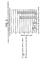
- MC S when added to in vitro bone marrow culture systems, supported the long-term growth of a population of pluripotent bone marrow cells for at least 45 days, as shown by Figure 1.
- FBS fetal bovine serum
- RPMI 1640 signifies a common commercially available defined nutrient medium from Gibco.
- similar data were obtained from bone marrow cell cultures incubated with MCS for 90 days. Cells from said 45 and 90 day cultures are capable of reconstituting the lymphoid, myeloid and erythroid systems of lethally irradiated mice, as illustrated in Fig. 2. This indicates that a pluripotent cell population must be present in the MCS-cultured cells. Bone marrow cell cultures grown without MCS were observed to lose this reconstituting ability within the first 25 - 30 days of culture.
- TdT terminal deoxynucleotidyl transferase
- MCS can be used as a culture medium supplement to support growth of marrow cells for in vitro study of immunoregulation and bone marrow function. MCS can also support the growth in vitro of bone marrow cells used in clinical marrow cell transplant procedures and other therapeutic or diagnostic procedures.
- MCS concentrations can be used for in vitro culture to achieve general multiplication of select differentiation for myeloid, erythroid or lymphoid cells. Readministrations of MCS to cultures at time of feeding and perhaps every 2 - 6 days will enhance cellular activity in subsequent yields of pluripotential or unipotential marrow cell populations.
- MCS-treated marrow cells produce a lymphopoietic factor, and that cultivation of fresh marrow cells with spent medium containing this factor induces the expression of pre B lymphocytes and B cells. This is illustrated in the following table, wherein in each instance fresh marrow cells were grown for seven days on the identified spent medium:
- the data in the table were obtained by (i) incubating an aliquot with fluorescent anti-IgM at 4°C and then counting cells with surface IgM in a cell sorter and (2) incubating another aliquot with fluorescent anti-IgM at 37°C for 1 hour followed by fixation in para-formalin and counting the cells with cytoplasmic IgM in a cell sorter.
- MCS marrow-derived progeny
- Deficiencies in marrow-derived progeny may be repaired by removing marrow cells, culturing with MCS, alone or in combination with other modulatory factors, to achieve the numbers of select cell types required to permit therapeutic readministration to the patient.
- selected cloned populations may be retained and propagated in vitro for subsequent administration. Severe immunodeficiencies or bone marrow diseases may be repaired e.g. by the above procedure but with pluripotential cells from a relative or even unrelated person. Following MCS supported growth, peer populations of primordial cells which usually lack or have sparse histocompatability proteins may be transfused and thus not rejected. Once they generate new lymphoid populations in their chimeric recipients, they will tolerate the person as did our own lymphocytes when they first brushed against other cell types during early fetal life.
- cancer, organ transplants and autoimmune diseases which have been repaired as much as possible by medical and/or surgical treatments may next receive new pluripotential cells from MCS treated cultures of self or non-self origin.
- the latter may be transplanted to the recipients after their residual lymphocytes are depleted via cyclosporin, irradiation, specific monoclonal antibody immunotargeting or combinations of these various procedures.
- Monoclonal or polyclonal antibodies to MCS can provide unique and exclusive probes for determining serum levels of the MCS cytokine in young, old, normal or diseased individuals.
- the antibody may detect MCS in blood samples with a variety of enzyme-linked, I 125 or immunodiffusion assays. The same approach permits monitoring of transplantation of MCS-producing tissues or determination of the indirect or direct effects of pharmacologic agents on these sites. Likewise, during repair of other disease states, MCS levels can provide important clues as to the prognostic or predicted status of the patients by daily monitoring of blood samples.
- MCS Marrow Cell Stimulator
- MCS-supported and control cultures were compared by cell sorter analysis.
- Cell sorting by dual laser flow cytometry has allowed the imaging of distinct cell populations within murine bone marrow and has demonstrated that pluripotent and lymphopoietic cells are almost entirely contained within one discrete sub-population, here called Population 2.
- mice The ability of MCS-supported bone marrow cultures to reconstitute the immune system of lethally irradiated mice was investigated by adoptive transfer studies in which cells were grown as previously described and then adoptively transferred such that the control group received 5 x 10 6 cultured bone marrow cells of varying culture age. A second group of mice received the same number of MCS-supported cultured cells. Both were adoptively transferred by i.v. injection (Dorshkind, K., and R.A. Phillips, 1982, J. Immunol. 129: 2444). Prior to injection, both groups were lethally irradiated with 900 rads of total body irradiation from a Cobalt 60 source.
- Restored immunocompetence was measured by the ability to mount an immune response of greater than 5000 anti-SRBC, PFC per spleen at day 30 (Mishell, B. and S. Shiigi, 1980, Selected Methods in Cellular Immunology, W.H. Freeman and Co., San Francisco). See Figure 2.
- the CFU-S assay was used as a measure of the clonogenic potential of adoptively transferred bone marrow cells.
- 10 5 cultured cells (same conditions as previous examples) were injected i.v. into lethally irradiated mice. Spleens were excised at day 7 and placed in fresh Boulin's fixative. The spleens were then macroscopically examined for the presence of surface colonies (Till, J.E. and E.A. McCulloch, 1961, Rad. Res. 14: 213).
- the use of MCS as a culture supplement significantly increased the CFU-S potential of marrow cells, indicating a possible elevation in the number of pluripotential bone marrow cells present in the MCS culture.
Abstract
Description
- This invention relates to a new composition of matter, and more particularly to a novel factor isolated frommamnalian thymus tissue. This thymic factor, termed Marrow Cell Stimulator or MCS, supports the long term growth of pluripotent bone marrow cells in in vitro culture systems.
- Understanding the growth and regulation of bone marrow cells is of great importance because bone marrow contains the self-renewing, pluripotent stem cells which give rise to the divergent lines of blood cells: erythrocytes, platelets, granulocytes (neutrophils, eosinophils, and basophils), macrophages, and lymphocytes (diverse B-cells and T-cells). Certain diseases--such as the leukemias, AIDS, allergies, and others--result from an absence, surplus or imbalance of lymphoid cell populations which originate in the bone marrow. Immune system imbalances may result from defects in the growth and differentiation of various lymphocyte populations that derive ultimately from the bone marrow. The ability to grow bone marrow cells in vitro makes it possible to study the regulation of bone marrow cell differentiation. Bone marrow cell cultures also provide a source of potential transplants for treating diseases such as leukemia and congenital immune deficiency.
- Hemopoiesis is the process by which undifferentiated cells of the bone marrow develop into the various blood cell types. The bone marrow of adult mammals contains self-renewing pluripotent stem cells (termed CFU-S) which can proliferate or differentiate into the progenitors of the diverse blood cell lines. Such progenitor cells proliferate or differentiate into cells with still more restricted potencies, and so on until the terminally differentiated effector cells of the various lines are produced.
- The goals of bone marrow cell culture systems are to mimic in vitro the events of in vivo bone marrow cell proliferation and differentiation. For over two decades attempts have been made to determine the essential regulatory factors whose addition to the culture medium would create the appropriate microenvironment for CFU-S proliferation and/or differentiation. If such regulatory factors could be identified and purified it should be possible to both maintain replicating pluripotent CFU-S cells indefinitely and to control their differentiation into preselected developmental pathways.
- Bone marrow culture systems in the prior art fall into two general categories, solid and liquid. The solid, agar systems have proven unsatisfactory because sustained hemopoiesis cannot be maintained. In 1966 two groups independently reported that mouse bone marrow cells would form colonies of granulocytes and macrophages when plated in soft agar containing horse serum, Fischer's salts, and an appropriate source of colony stimulating factor (CSF). See: Bradley et al., Aust. J. Exp. Biol. Med. Sci., 44: 287 (1966); Pluznick et al., Exp. Cell Res. 43: 553 (1966). In such systems differentiation of committed progenitor cells continues for 2 to 3 weeks but hemopoietic stem cells proliferate for only a week to ten days. Dexter, T.M. et al., Meth. Cell Biol. 14: 387 (1976).
- Attempts to sustain continuous and normal hematopoiesis in vitro have been far more successful using liquid culture systems. The first somewhat successful method of maintaining CFU-S proliferation in culture involved the co-culturing of thymus cells with bone marrow cells. For example, suspensions of murine thymus cells were incubated at 37°C in Fischer's medium + 20% horse serum. After several days the cultures reportedly segregated into a population of cells adhering to the culture vessel surfaces and a population of cells suspended in the overlying medium. After-the overlying medium was poured off, the cultures of adherent thymus cells were inoculated with bone marrow cells. After two weeks such thymus/bone marrow co-cultures became one of two types:
- one producing primarily granulocytes (G-type) or one producing primarily macrophages (M-type). In the M-type cultures the number of CFU-S, in the overlying medium, reportedly decline and are gone by the fifth week. In the G-type cultures, CFU-S reportedly persist for over 12 weeks. See: Dexter et al., J. Cell Physiol. 82: 461 (1973); Dexter et al., Meth. Cell Biol. 14: 387 (1976).
- The thymus/bone marrow co-culture experiments were apparently not pursued, as that technique was soon supplanted by culture systems in which bone marrow cells were added to an established bone marrow culture. See: Dexter et al., Br. J. Haematol. 28 525 (1974); Dexter et al., J. Cell Physiol. 91: 355 (1977); Moore et al., Transp. Proc. X: 83 (1978). Modifications of such long-term mouse bone marrow cultures can reportedly generate pluripotential CFU-S for up to one year. The CFU-S present in the second inoculum migrate to the adherent layer and form membrane-functional complexes with cells in the adherent layer. In the absence of the adherent layer sustained hemopoiesis will not occur. Only adherent layers from bone marrow cells can sustain hemopoiesis. Adherent layers formed from spleen.or thymus tissue do not sustain active hemopoiesis. Dexter et al., J. Supramol. Struct. 13: 501 (1980). Few, if any, factors are released into the medium from the adherent layer. If diffusion chambers are placed on preformed adherent layers, hemopoiesis is not maintained. Bently, S.A., Exp. Haematol. 8: 77 (1981).
- Human bone marrow cultures have, by modification of the Dexter technique, been maintained for as long as 20 weeks. See: Moore et al., Blood 55: 682 (1980); Greenber et al., Blood 58: 724 (1981).
- The novel thymic extract of the present invention--called Marrow Cell Stimulator or MCS--supports the long-term growth of pluripotent bone marrow cells in in vitro culture systems. MCS is extracted from neonatal vertebrate thymus tissue by means of a two-part procedure. A crude thymus extract is first prepared in conformance with procedures known in the art. Said crude extract is then subjected to a preparative electrophoretic enrichment protocol, preferably using a "powder block" electrophoresis apparatus. The fastest running material obtained by this electrophoretic procedure-the material which precedes bovine or other animal serum albumin--is collected and concentrated to give MCS.
- The marrow cell stimulator (MCS) of the present invention is isolated from calf or other neonatal vertebrate tissue by a novel procedure. Calf thymus tissue is preferred because of its relative availability. Considering this isolation procedure in detail, thymus glands obtained from freshly slaughtered animals are first freed of connective tissue and other extraneous tissue, then minced. Next an excess of homogenization buffer, for example from a 1-fold to a 3-fold excess (V/W), and preferably at least a 2-fold excess, at a pH of from about 7.5 to 8.2, and preferably of about 8.0, is added to the minced glands. A suitable homogenization buffer can be prepared, for example, by adding 0.05 moles sodium chloride and 0.0005 moles of magnesium chloride per I L of 10mM Tris-HC1 (pH 8.0). The minced glands-buffer mixture is then homogenized (for example in a commercial grade Waring blender at 4°C for 30 seconds at maximum output), and the thus-obtained homogenate is centrifuged for from about 20 minutes to about 40 minutes at 4°C and 12,000 x g.
- The supernatant liquid is then removed and heated until it has congealed. This will usually take from about 30 to about 60 minutes, preferably from about 40 to about 50 minutes, at a temperature of from about 75°C to about 85°C. The solids are then removed from the congealed supernatant liquid by again centrifuging for from about 20 minutes to about 40 minutes at 4°C and 12,000 x g. The resulting supernatant liquid is then frozen in 200-300 aliquots at -80°C for later use in the remainder of the procedure.
- The electrophoretic step can be carried out by first thawing 300 mls of frozen thymus extract (ordinarily, the aliquot will be completely thawed by simply allowing it to stand at 4°C overnight; if further thawing is necessary, the temperature can be raised to 37°C), then centrifuging the thawed extract for from about 20 minutes to about 40 minutes at 4°C and 40 - 50,000 x g, and then concentrating the supernatant obtained 60-fold in an Amicon stirred cell fitted with a PM-10 membrane (which will pass molecules of molecular weight less than 10,000 daltons).
- Next, the concentrate is dialyzed in a cellulose dialysis tube having a pore size which will pass molecules of molecular weights less than 12-14,000 daltons, by suspending the tube in 1 liter of BioRad "Laurell" barbital/calcium lactate buffer for 16 hours at 4°C, and then redialyzing in 1 liter of fresh "Laurell" buffer. This provides a 1:40,000 v/v exchange with the barbital/calcium lactate buffer.
- The dialyzed concentrate is then admixed with a small amount (e.g., about 4 or 5 drops) of 3% bromphenol blue, which acts as a tracking dye, and then admixed with sufficient dry "Pevikon" plastic beads (Mercer Chemical Co.) t.o form a blue, spreadable paste.
- An electrophoresis block (approximately 41 cm x 17.8 cm x 1.9 cm) of "Pevikon" is prepared by first suspending 1700 ml of the beads in two liters of distilled water and allowing the beads to settle (this will usually take at least 5 hours), then pouring off the water and "fines; next resuspending the beads in two liters of BioRad "Laurell" buffer and allowing the beads to settle, and finally pouring the Pevikon into the casting block and cutting a trough about 2.5 - 3.5 cm from the sides of the block, about 8 cm from the "bacf" edge and about 0.6 - 1 cm thick, leaving about a 1 mm cover of Pevikon on the bottom of the trough.
- The blue paste is packed into the trough, the wicks are bathed in tanks containing 2000 ml of Laurell buffer, the necessary electrical connections are made, and electrophoresis is begun at 200 volts (= 40 milli Amps) for from about 19 to about 21 hours, the temperature being maintained at 4°C throughout. At this point, the run is interrupted to replace the buffer in the electrode tanks with a fresh batch and to clean collected salt from the anode, following which electrophoresis is begun again at 200 volts (=45 milli Amps) for from about 7 to about 9 hours, then continued at 150 volts (= 30 milli Amps) for from about 15 to about 17 hours, and concluded at 200 volts until albumin migration has reached approximately 18.5 cm, the temperature again being maintained at 4°C throughout.
- The block is then separated from its electrical connections and, starting at the anode, the Pevikon is cut into 22 fraction strips, each 0.66 cm thick (fraction 22 will be well into the albumin line). Each fraction strip is then packed into a soft, punctured centrifuge tube, with the punctured end mesh-covered to retain the solid contents, each tube is placed in a 50 cc Nalgene concical centrifuge tube, and all the tubes are centrifuged for 20 minutes at 5000 rpm at 4°C. The liquid from each tube is poured into a separate bottle, 5 ml of deionized water is added to the Pevikon beads in the centrifuge tubes, and the tubes are centrifuged for 20 minutes at 5000 rpm at 4°C. The liquid from each tube is then added to that previously collected. This washing step is then repeated twice more, and the bottles containing all the liquid collected from each tube are stored at 4°C.
- Each of these 22 liquid fractions is run in an Ouchterlony assay against anti-bovine serum albumin, and the fractions which result in non-positive wells, indicating no trace of albumin, are pooled. This will usually be fractions 1 - 16. This fastest running material (that migrated ahead of albumin in the electric field) is what is referred to as MCS. Thus, MCS can be characterized as having a high charge-to-mass ratio and as being essentially free of serum albumin.
- The aforementioned pooled fractions are concentrated by standard techniques, first by ultrafiltration to a volume of approximately 1.0 ml in an Amicon stirred cell fitted with a YM-5 membrane, then by dialysis against phosphate buffered saline (PBS). The resulting concentrate contains the isolated MCS factor.
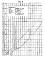
- To further characterize MCS the concentrate was subjected to reverse phase high performance liquid chromatography (RP-HPLC). An aliquot was taken up in 0.1% trifluoroacetic acid, centrifuged and the supernatant examined in a Macherey and Nagel RP-HPLC protein column. Three probes were run under the same conditions, as shown on the legend of Figs. 4 - 6, varying only the slope of the solvent gradient and the detection wave length.
- Fig. 4 shows the analytical run monitored at 230 nm. The acetonitrile gradient was generated within 100 min from 0% to 80%. The straight line in the chromatogram represents the slope of the gradient.
- Under conditions stated above, it is possible to resolve the MCS probe into 5 main peaks.
Peak 0 represents the void volume of the column. With a shallower gradient (Fig. 5) -0% to 16% acetonitrile within 60 min - peak 3 splits into three subpeaks, designated as 3a, 3b and 3c. - Fig. 6 shows a run monitored at 260 nm. No definite retarded peak could be detected, indicating that
peaks - MCS, when added to in vitro bone marrow culture systems, supported the long-term growth of a population of pluripotent bone marrow cells for at least 45 days, as shown by Figure 1. In Fig. 1, "FBS" signifies fetal bovine serum and "
RPMI 1640" signifies a common commercially available defined nutrient medium from Gibco. Furthermore, similar data were obtained from bone marrow cell cultures incubated with MCS for 90 days. Cells from said 45 and 90 day cultures are capable of reconstituting the lymphoid, myeloid and erythroid systems of lethally irradiated mice, as illustrated in Fig. 2. This indicates that a pluripotent cell population must be present in the MCS-cultured cells. Bone marrow cell cultures grown without MCS were observed to lose this reconstituting ability within the first 25 - 30 days of culture. - The ability of cells from MCS-supplemented bone marrow cultures to fully reconstitute lethally irradiated mice clearly demonstrates the presence of a pluripotential population within these cultures. Further evidence of the increased presence of a -lymphopoietic population can be seen in an elevated level of terminal deoxynucleotidyl transferase (TdT) marker in cultures grown with MCS. This marker is known to be present on most immature lymphoid cells, and the elevated presence of TdT in marrow cell cultures grown with MCS may be viewed as an indicator of increased lymphopoietic ability.
- In longer term culture of normal marrow cells (without MCS) and MCS-treated marrow, conducted side by side, normal marrow cells alone were found to survive only 60 - 90 days, whereas the MCS-treated marrow cells-continued to produce pluripotent stem cells up through 320 days of observation.
- It was found advantageous in enhancing the reproducibility of the culture conditions, to grow the MCS-treated marrow cells in the same medium used for the experiments depicted in Fig. 2, but on gelatin beads. Especially preferred gelatin beads were thoseunder the trade name "Ventregel".

- MCS can be used as a culture medium supplement to support growth of marrow cells for in vitro study of immunoregulation and bone marrow function. MCS can also support the growth in vitro of bone marrow cells used in clinical marrow cell transplant procedures and other therapeutic or diagnostic procedures.
- A wide range of MCS concentrations can be used for in vitro culture to achieve general multiplication of select differentiation for myeloid, erythroid or lymphoid cells. Readministrations of MCS to cultures at time of feeding and perhaps every 2 - 6 days will enhance cellular activity in subsequent yields of pluripotential or unipotential marrow cell populations.
- It has also been found that MCS-treated marrow cells produce a lymphopoietic factor, and that cultivation of fresh marrow cells with spent medium containing this factor induces the expression of pre B lymphocytes and B cells. This is illustrated in the following table, wherein in each instance fresh marrow cells were grown for seven days on the identified spent medium:

- The data in the table were obtained by (i) incubating an aliquot with fluorescent anti-IgM at 4°C and then counting cells with surface IgM in a cell sorter and (2) incubating another aliquot with fluorescent anti-IgM at 37°C for 1 hour followed by fixation in para-formalin and counting the cells with cytoplasmic IgM in a cell sorter.
- Following animal studies, it is anticipated that MCS will be applied in human clinical treatment. Deficiencies in marrow-derived progeny may be repaired by removing marrow cells, culturing with MCS, alone or in combination with other modulatory factors, to achieve the numbers of select cell types required to permit therapeutic readministration to the patient.
- In addition, selected cloned populations may be retained and propagated in vitro for subsequent administration. Severe immunodeficiencies or bone marrow diseases may be repaired e.g. by the above procedure but with pluripotential cells from a relative or even unrelated person. Following MCS supported growth, peer populations of primordial cells which usually lack or have sparse histocompatability proteins may be transfused and thus not rejected. Once they generate new lymphoid populations in their chimeric recipients, they will tolerate the person as did our own lymphocytes when they first brushed against other cell types during early fetal life.
- As another more complex step, cancer, organ transplants and autoimmune diseases which have been repaired as much as possible by medical and/or surgical treatments may next receive new pluripotential cells from MCS treated cultures of self or non-self origin. The latter may be transplanted to the recipients after their residual lymphocytes are depleted via cyclosporin, irradiation, specific monoclonal antibody immunotargeting or combinations of these various procedures.
- Monoclonal or polyclonal antibodies to MCS can provide unique and exclusive probes for determining serum levels of the MCS cytokine in young, old, normal or diseased individuals. The antibody may detect MCS in blood samples with a variety of enzyme-linked, I125 or immunodiffusion assays. The same approach permits monitoring of transplantation of MCS-producing tissues or determination of the indirect or direct effects of pharmacologic agents on these sites. Likewise, during repair of other disease states, MCS levels can provide important clues as to the prognostic or predicted status of the patients by daily monitoring of blood samples.
- Marrow Cell Stimulator (MCS) was prepared as described above and was used as a direct media supplement (0.4% v/v) in standard culture medium during initial plating and early growth stages of fresh bone marrow cultures. Bone marrow cells were obtained from femurs of 4 week old BALB/c mice and were suspended in
RPMI 1640 medium from Gibco with 5% FBS (fetal bovine serum), 0.05 mM 2-mercaptoethanol and antibiotics at a cell density of 1 x 10⁶ nucleated cells/ml. MCS (0.4% v/v) was present at the initial plating and for the first 3 days of culture. - MCS-supported and control cultures were compared by cell sorter analysis. Cell sorting by dual laser flow cytometry has allowed the imaging of distinct cell populations within murine bone marrow and has demonstrated that pluripotent and lymphopoietic cells are almost entirely contained within one discrete sub-population, here called
Population 2. Miller, H. et al, 1984, "Cell Sorter Analysis and Separation of B-Lymphoid Population from Murine Bone Marrow" (in press). Further analysis has shown that the growth of this sub-population is significantly enhanced by the presence of MCS. See Figure 1. - The ability of MCS-supported bone marrow cultures to reconstitute the immune system of lethally irradiated mice was investigated by adoptive transfer studies in which cells were grown as previously described and then adoptively transferred such that the control group received 5 x 10⁶ cultured bone marrow cells of varying culture age. A second group of mice received the same number of MCS-supported cultured cells. Both were adoptively transferred by i.v. injection (Dorshkind, K., and R.A. Phillips, 1982, J. Immunol. 129: 2444). Prior to injection, both groups were lethally irradiated with 900 rads of total body irradiation from a
Cobalt 60 source. Restored immunocompetence was measured by the ability to mount an immune response of greater than 5000 anti-SRBC, PFC per spleen at day 30 (Mishell, B. and S. Shiigi, 1980, Selected Methods in Cellular Immunology, W.H. Freeman and Co., San Francisco). See Figure 2. - The CFU-S assay was used as a measure of the clonogenic potential of adoptively transferred bone marrow cells. 105 cultured cells (same conditions as previous examples) were injected i.v. into lethally irradiated mice. Spleens were excised at day 7 and placed in fresh Boulin's fixative. The spleens were then macroscopically examined for the presence of surface colonies (Till, J.E. and E.A. McCulloch, 1961, Rad. Res. 14: 213). As can be seen in Figure 3, the use of MCS as a culture supplement significantly increased the CFU-S potential of marrow cells, indicating a possible elevation in the number of pluripotential bone marrow cells present in the MCS culture.
-
- Figure 1 - Effects of MCS on long term BALB/C mouse bone marrow cell cultures (Population 2) □ Marrow cells +
RPM1 1640 + 5% FBSMarrow cells +
RPM1 1640 + 5% FBS + 0.4% MCS - Figure 2 - MCS cultured cells: Adoptive transfer to immuno-incompetent mice □ Mice receiving bone marrow cultures without MCS Mice receiving bone marrow cultures with MCS
- Figure 3 - Effects of MCS supported growth on the CFU-S potential of cultured murine bone marrow cells □ Bone marrow cultures without MCS Bone marrow cultures plus MCS
- Figure 4 - Reverse Phase - HPLC

- Figure 5 - Reverse Phase - HPLC

- Figure 6 - Reverse Phase - HPLC

Claims (19)
Applications Claiming Priority (4)
| Application Number | Priority Date | Filing Date | Title |
|---|---|---|---|
| US61616084A | 1984-06-01 | 1984-06-01 | |
| US616160 | 1984-06-01 | ||
| US73461085A | 1985-05-16 | 1985-05-16 | |
| US734610 | 1985-05-16 |
Publications (2)
| Publication Number | Publication Date |
|---|---|
| EP0163543A2 true EP0163543A2 (en) | 1985-12-04 |
| EP0163543A3 EP0163543A3 (en) | 1987-08-12 |
Family
ID=27087682
Family Applications (1)
| Application Number | Title | Priority Date | Filing Date |
|---|---|---|---|
| EP85303844A Withdrawn EP0163543A3 (en) | 1984-06-01 | 1985-05-31 | Marrow cell stimulator |
Country Status (1)
| Country | Link |
|---|---|
| EP (1) | EP0163543A3 (en) |
Cited By (9)
| Publication number | Priority date | Publication date | Assignee | Title |
|---|---|---|---|---|
| US4721096A (en) * | 1986-04-18 | 1988-01-26 | Marrow-Tech Incorporated | Process for replicating bone marrow in vitro and using the same |
| EP0310056A2 (en) * | 1987-09-30 | 1989-04-05 | SHIONOGI SEIYAKU KABUSHIKI KAISHA trading under the name of SHIONOGI & CO. LTD. | Thymic stroma-derived T cell growth factor and process for its production |
| US4963489A (en) * | 1987-04-14 | 1990-10-16 | Marrow-Tech, Inc. | Three-dimensional cell and tissue culture system |
| US5032508A (en) * | 1988-09-08 | 1991-07-16 | Marrow-Tech, Inc. | Three-dimensional cell and tissue culture system |
| US5160490A (en) * | 1986-04-18 | 1992-11-03 | Marrow-Tech Incorporated | Three-dimensional cell and tissue culture apparatus |
| US5266480A (en) * | 1986-04-18 | 1993-11-30 | Advanced Tissue Sciences, Inc. | Three-dimensional skin culture system |
| US5510254A (en) * | 1986-04-18 | 1996-04-23 | Advanced Tissue Sciences, Inc. | Three dimensional cell and tissue culture system |
| DE19609261A1 (en) * | 1996-02-28 | 1997-09-04 | Strathmann Ag & Co | Thymus peptide extracts of molecular weight less than 6000 daltons |
| US6140039A (en) * | 1986-04-18 | 2000-10-31 | Advanced Tissue Sciences, Inc. | Three-dimensional filamentous tissue having tendon or ligament function |
Citations (3)
| Publication number | Priority date | Publication date | Assignee | Title |
|---|---|---|---|---|
| US4239498A (en) * | 1978-11-27 | 1980-12-16 | Rule Allyn H | Method of preparing thymic factors and composition |
| EP0105749A2 (en) * | 1982-10-01 | 1984-04-18 | BIO-COM Inc. | Compositions for use in inducing immunosuppression or immunoregulation of the immune response to an antigen |
| EP0153063A2 (en) * | 1984-02-03 | 1985-08-28 | VXR, Inc. | Inducer factor of T-suppressor cells |
-
1985
- 1985-05-31 EP EP85303844A patent/EP0163543A3/en not_active Withdrawn
Patent Citations (3)
| Publication number | Priority date | Publication date | Assignee | Title |
|---|---|---|---|---|
| US4239498A (en) * | 1978-11-27 | 1980-12-16 | Rule Allyn H | Method of preparing thymic factors and composition |
| EP0105749A2 (en) * | 1982-10-01 | 1984-04-18 | BIO-COM Inc. | Compositions for use in inducing immunosuppression or immunoregulation of the immune response to an antigen |
| EP0153063A2 (en) * | 1984-02-03 | 1985-08-28 | VXR, Inc. | Inducer factor of T-suppressor cells |
Non-Patent Citations (2)
| Title |
|---|
| CHEMICAL ABSTRACTS, vol. 76, no. 25, 19th June 1972, page 320, abstract no. 152002j, Columbus, Ohio, US; K. MAKAMURA: "Proliferation of plasma cells from mouse bone marrow in vitro. I. Role of the thymus", & J. EXP. MED. 1972, 135(3), 476-90 * |
| THE JOURNAL OF IMMUNOLOGY, vol. 111, no. 4, October 1973, pages 1005-1009, The Williams & Wilkins Co., Baltimore, US; H.C. MILLER et al.: "Production of functional T cells after treatment of bone marrow with thymic factor" * |
Cited By (22)
| Publication number | Priority date | Publication date | Assignee | Title |
|---|---|---|---|---|
| US5516680A (en) * | 1986-04-18 | 1996-05-14 | Advanced Tissue Sciences, Inc. Formerly Marrow-Tech | Three-dimensional kidney cell and tissue culture system |
| US5624840A (en) * | 1986-04-18 | 1997-04-29 | Advanced Tissue Sciences Inc. | Three-dimensional liver cell and tissue culture system |
| US5516681A (en) * | 1986-04-18 | 1996-05-14 | Advanced Tissue Sciences, Inc. | Three-dimensional pancreatic cell and tissue culture system |
| US4721096A (en) * | 1986-04-18 | 1988-01-26 | Marrow-Tech Incorporated | Process for replicating bone marrow in vitro and using the same |
| US5858721A (en) * | 1986-04-18 | 1999-01-12 | Advanced Tissue Sciences, Inc. | Three-dimensional cell and tissue culture system |
| US5160490A (en) * | 1986-04-18 | 1992-11-03 | Marrow-Tech Incorporated | Three-dimensional cell and tissue culture apparatus |
| US5266480A (en) * | 1986-04-18 | 1993-11-30 | Advanced Tissue Sciences, Inc. | Three-dimensional skin culture system |
| US5443950A (en) * | 1986-04-18 | 1995-08-22 | Advanced Tissue Sciences, Inc. | Three-dimensional cell and tissue culture system |
| US5518915A (en) * | 1986-04-18 | 1996-05-21 | Advanced Tissue Sciences, Inc. | Three-Dimensional mucosal cell and tissue culture system |
| US5512475A (en) * | 1986-04-18 | 1996-04-30 | Advanced Tissue Sciences, Inc. | Three-dimensional skin cell and tissue culture system |
| US6140039A (en) * | 1986-04-18 | 2000-10-31 | Advanced Tissue Sciences, Inc. | Three-dimensional filamentous tissue having tendon or ligament function |
| US5849588A (en) * | 1986-04-18 | 1998-12-15 | Advanced Tissue Sciences, Inc. | Methods of use of a three-dimensional liver cell and tissue culture system |
| US5510254A (en) * | 1986-04-18 | 1996-04-23 | Advanced Tissue Sciences, Inc. | Three dimensional cell and tissue culture system |
| US5541107A (en) * | 1986-04-18 | 1996-07-30 | Advanced Tissue Sciences, Inc. | Three-dimensional bone marrow cell and tissue culture system |
| US5578485A (en) * | 1986-04-18 | 1996-11-26 | Advanced Tissue Sciences, Inc. | Three-dimensional blood-brain barrier cell and tissue culture system |
| US5580781A (en) * | 1986-04-18 | 1996-12-03 | Advanced Tissue Sciences, Inc. | Three-dimensional tumor cell and tissue culture system |
| US4963489A (en) * | 1987-04-14 | 1990-10-16 | Marrow-Tech, Inc. | Three-dimensional cell and tissue culture system |
| EP0310056A2 (en) * | 1987-09-30 | 1989-04-05 | SHIONOGI SEIYAKU KABUSHIKI KAISHA trading under the name of SHIONOGI & CO. LTD. | Thymic stroma-derived T cell growth factor and process for its production |
| EP0310056A3 (en) * | 1987-09-30 | 1989-06-14 | SHIONOGI SEIYAKU KABUSHIKI KAISHA trading under the name of SHIONOGI & CO. LTD. | Thymic stroma-derived t cell growth factor and process for its production |
| US5032508A (en) * | 1988-09-08 | 1991-07-16 | Marrow-Tech, Inc. | Three-dimensional cell and tissue culture system |
| DE19609261A1 (en) * | 1996-02-28 | 1997-09-04 | Strathmann Ag & Co | Thymus peptide extracts of molecular weight less than 6000 daltons |
| DE19609261C2 (en) * | 1996-02-28 | 2000-02-24 | Strathmann Ag & Co | Medicines to prevent and treat myelosuppression due to cancer chemotherapy |
Also Published As
| Publication number | Publication date |
|---|---|
| EP0163543A3 (en) | 1987-08-12 |
Similar Documents
| Publication | Publication Date | Title |
|---|---|---|
| Castro-Malaspina et al. | Characterization of human bone marrow fibroblast colony-forming cells (CFU-F) and their progeny | |
| Aarden et al. | Production of hybridoma growth factor by human monocytes | |
| Ruscetti et al. | Human T-lymphocyte growth factor: regulation of growth and function of T lymphocytes | |
| Desreumaux et al. | Interleukin 5 messenger RNA expression by eosinophils in the intestinal mucosa of patients with coeliac disease. | |
| Smith et al. | Functional and molecular characteristics of T-cell growth factor | |
| Quesenberry et al. | Vascular endothelium as a regulator of granulopoiesis: production of colony-stimulating activity by cultured human endothelial cells | |
| Zucali et al. | Interleukin 1 stimulates fibroblasts to produce granulocyte-macrophage colony-stimulating activity and prostaglandin E2. | |
| Butterfield et al. | Establishment of an immature mast cell line from a patient with mast cell leukemia | |
| Patarroyo et al. | Identification of a cell surface protein complex mediating phorbol ester‐induced adhesion (binding) among human mononuclear leukocytes | |
| Kimura et al. | Human megakaryocytic progenitors (CFU‐M) assayed in methylcellulose: physical characteristics and requirements for growth | |
| US4464355A (en) | Serum-free and mitogen-free T-cell growth factor and process for making same | |
| US4390623A (en) | Serum-free and mitogen-free T-cell growth factor and process for making same | |
| Paige et al. | Differentiation of murine B cell precursors in agar culture. Frequency, surface marker analysis and requirements for growth of clonable pre‐B cells | |
| Nixon-Fulton et al. | Thy-1+ epidermal cells proliferate in response to concanavalin A and interleukin 2. | |
| Wahl et al. | Spontaneous production of fibroblast-activating factor (s) by synovial inflammatory cells. A potential mechanism for enhanced tissue destruction. | |
| Kumagai et al. | In vitro regeneration of resting lymphocytes from stimulated lymphocytes and its inhibition by insulin. | |
| Ayalon et al. | Electrical effects of histamine on monolayers formed in culture from enriched canine gastric chief cells. | |
| EP0049611B1 (en) | A t-cell growth factor, and a process of producing the same | |
| Ford et al. | Growth factor-mediated tumor cell proliferation in hairy cell leukemia. | |
| EP0163543A2 (en) | Marrow cell stimulator | |
| US4814434A (en) | Inducer of T-suppressor cells | |
| Dorshkind et al. | Characterization of early B lymphocyte precursors present in long-term bone marrow cultures. | |
| US4411992A (en) | Process for preparing murine interleukin 2 | |
| EP0690125A2 (en) | Process for induction culture of cytotoxict lymphocytes having killing activity against tumor cells | |
| KR940003650B1 (en) | Thymic storma-derived t-cell growth factor and manufacturing method thereof |
Legal Events
| Date | Code | Title | Description |
|---|---|---|---|
| PUAI | Public reference made under article 153(3) epc to a published international application that has entered the european phase |
Free format text: ORIGINAL CODE: 0009012 |
|
| AK | Designated contracting states |
Designated state(s): AT BE CH DE FR GB IT LI LU NL SE |
|
| PUAL | Search report despatched |
Free format text: ORIGINAL CODE: 0009013 |
|
| AK | Designated contracting states |
Kind code of ref document: A3 Designated state(s): AT BE CH DE FR GB IT LI LU NL SE |
|
| STAA | Information on the status of an ep patent application or granted ep patent |
Free format text: STATUS: THE APPLICATION IS DEEMED TO BE WITHDRAWN |
|
| 18D | Application deemed to be withdrawn |
Effective date: 19870601 |
|
| RIN1 | Information on inventor provided before grant (corrected) |
Inventor name: DECAROLIS, RICHARD J. Inventor name: GOLDFARB, MARCIA F. Inventor name: MILLER, HAROLD C. |