EP0146511A2 - A method of localisation - Google Patents
A method of localisation Download PDFInfo
- Publication number
- EP0146511A2 EP0146511A2 EP84850345A EP84850345A EP0146511A2 EP 0146511 A2 EP0146511 A2 EP 0146511A2 EP 84850345 A EP84850345 A EP 84850345A EP 84850345 A EP84850345 A EP 84850345A EP 0146511 A2 EP0146511 A2 EP 0146511A2
- Authority
- EP
- European Patent Office
- Prior art keywords
- spot
- needle
- prints
- relation
- Prior art date
- Legal status (The legal status is an assumption and is not a legal conclusion. Google has not performed a legal analysis and makes no representation as to the accuracy of the status listed.)
- Granted
Links
Images


Classifications
-
- A—HUMAN NECESSITIES
- A61—MEDICAL OR VETERINARY SCIENCE; HYGIENE
- A61B—DIAGNOSIS; SURGERY; IDENTIFICATION
- A61B6/00—Apparatus for radiation diagnosis, e.g. combined with radiation therapy equipment
- A61B6/12—Devices for detecting or locating foreign bodies
-
- A—HUMAN NECESSITIES
- A61—MEDICAL OR VETERINARY SCIENCE; HYGIENE
- A61B—DIAGNOSIS; SURGERY; IDENTIFICATION
- A61B17/00—Surgical instruments, devices or methods, e.g. tourniquets
- A61B17/34—Trocars; Puncturing needles
- A61B17/3403—Needle locating or guiding means
-
- A—HUMAN NECESSITIES
- A61—MEDICAL OR VETERINARY SCIENCE; HYGIENE
- A61B—DIAGNOSIS; SURGERY; IDENTIFICATION
- A61B6/00—Apparatus for radiation diagnosis, e.g. combined with radiation therapy equipment
- A61B6/08—Auxiliary means for directing the radiation beam to a particular spot, e.g. using light beams
-
- A—HUMAN NECESSITIES
- A61—MEDICAL OR VETERINARY SCIENCE; HYGIENE
- A61B—DIAGNOSIS; SURGERY; IDENTIFICATION
- A61B6/00—Apparatus for radiation diagnosis, e.g. combined with radiation therapy equipment
- A61B6/50—Clinical applications
- A61B6/502—Clinical applications involving diagnosis of breast, i.e. mammography
-
- A—HUMAN NECESSITIES
- A61—MEDICAL OR VETERINARY SCIENCE; HYGIENE
- A61B—DIAGNOSIS; SURGERY; IDENTIFICATION
- A61B90/00—Instruments, implements or accessories specially adapted for surgery or diagnosis and not covered by any of the groups A61B1/00 - A61B50/00, e.g. for luxation treatment or for protecting wound edges
- A61B90/10—Instruments, implements or accessories specially adapted for surgery or diagnosis and not covered by any of the groups A61B1/00 - A61B50/00, e.g. for luxation treatment or for protecting wound edges for stereotaxic surgery, e.g. frame-based stereotaxis
- A61B90/14—Fixators for body parts, e.g. skull clamps; Constructional details of fixators, e.g. pins
- A61B90/17—Fixators for body parts, e.g. skull clamps; Constructional details of fixators, e.g. pins for soft tissue, e.g. breast-holding devices
-
- A—HUMAN NECESSITIES
- A61—MEDICAL OR VETERINARY SCIENCE; HYGIENE
- A61B—DIAGNOSIS; SURGERY; IDENTIFICATION
- A61B10/00—Other methods or instruments for diagnosis, e.g. instruments for taking a cell sample, for biopsy, for vaccination diagnosis; Sex determination; Ovulation-period determination; Throat striking implements
- A61B10/02—Instruments for taking cell samples or for biopsy
- A61B10/0233—Pointed or sharp biopsy instruments
-
- A—HUMAN NECESSITIES
- A61—MEDICAL OR VETERINARY SCIENCE; HYGIENE
- A61B—DIAGNOSIS; SURGERY; IDENTIFICATION
- A61B90/00—Instruments, implements or accessories specially adapted for surgery or diagnosis and not covered by any of the groups A61B1/00 - A61B50/00, e.g. for luxation treatment or for protecting wound edges
- A61B90/36—Image-producing devices or illumination devices not otherwise provided for
- A61B90/37—Surgical systems with images on a monitor during operation
- A61B2090/376—Surgical systems with images on a monitor during operation using X-rays, e.g. fluoroscopy
-
- A—HUMAN NECESSITIES
- A61—MEDICAL OR VETERINARY SCIENCE; HYGIENE
- A61B—DIAGNOSIS; SURGERY; IDENTIFICATION
- A61B90/00—Instruments, implements or accessories specially adapted for surgery or diagnosis and not covered by any of the groups A61B1/00 - A61B50/00, e.g. for luxation treatment or for protecting wound edges
- A61B90/10—Instruments, implements or accessories specially adapted for surgery or diagnosis and not covered by any of the groups A61B1/00 - A61B50/00, e.g. for luxation treatment or for protecting wound edges for stereotaxic surgery, e.g. frame-based stereotaxis
- A61B90/11—Instruments, implements or accessories specially adapted for surgery or diagnosis and not covered by any of the groups A61B1/00 - A61B50/00, e.g. for luxation treatment or for protecting wound edges for stereotaxic surgery, e.g. frame-based stereotaxis with guides for needles or instruments, e.g. arcuate slides or ball joints
Definitions
- the present invention relates to a method, in conjunction with the X-ray exposure of an object, of localising the three-dimensional position of a spot in the object.
- fine-needle biopsy which may be considered as included in the clinical and cytological components in triple diagnostics.
- This triple diagnostics procedure generally includes mammography, clinical examination and cytology. Fine-needle biopsy or fine needle puncture with cytological examination of cell samples from a suspected region provide very reliable positive repponses.
- fine-needle biopsy to be able to indicate a spot by means of a thin wire marker for facilitating subsequent surgical excision biopsy.
- the task forming the basis of the present invention is to realise a method of localising, as simply, reliably and rapidly as possible, the three-dimensional position of a spot in an object, for example a female breast.
- This task is solved according to the present invention in that, after fixation of the object in a pre-determined position, the object is exposed by means of an exposure device in two directions, each on either side of a centre line at right angles to the plane of the image for obtaining a first image print and a second image print, the invention being characterised in that the two-dimensional position of the target spot on the two prints is established in relation to an index on the prints; and that the coordinates of the spot in relation to the indices are processed to obtain control signals for setting of a guidance instrument with means for placing in the target spot in the object.
- the exposure device and the guidance instrument are placed in the same centre reference from which the right-angle centre line departs.
- the index is exposed on the print preferably simultaneously with the object for obtaining an exact measurement reference.
- the method according to the present invention makes possible the localisation of an optional spot in an object, for example a female breast, with a very high degree of precision in an extremely simple and reliable mannner.
- an optional spot in a female breast for fine-needle puncture, indication or some form of directed therapy in a simple and rapid manner so that the patient need not be subjected to undue stress or excessive discomfort.
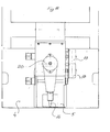
- the apparatus includes int. al. a per se known mammograph 1 which may be of the type designated "Scenograph 500 T".
- the mammograph 1 is pivotal about a shaft 2 which is provided with a scale for the exact setting of the mammograph 1.
- a film cassette holder for a film cassette 4 is mounted on an arm 3.
- a compression plate 5 is disposed above the arm 3 with the film cassette 4.
- the compression plate 5 is, naturally, vertically movable and has an orifice of, for example, 50 x 40 mm to make possible the insertion of, for instance, a biopsy needle into a breast which is held in a compressed state by means of the compression plate 5 on the arm, and the film cassette holder 4.
- the path of radiation from the tube in the mammograph 1 generating the X-rays is illustrated by ghosted lines, the radiation path impinging upon a film in the film cassette holder 4 for generating an exposed print which depicts the exposed breast region.
- the apparatus illustrated in Fig. 1 further includes a guidance instrument 6 which is shown in greater detail in Figs. 6, 7 and 8 and parts thereof in Fig. 9.
- the above-mentioned parts 3, 4 and 5 may be considered as forming part of the guidance instrument 6, since the parts 3, 4 and 5 are fixed in relation to the mammograph 1, pivotal about the shaft 2.
- the compression plate 5 is transparent for X-rays so that a greater portion of the object is exposed than the surface located directly beneath the orifice in the compression plate 5.
- Fig. 2 illustrates in greater detail the pivotal capacity of the mammograph 1 about the shaft 2.
- a first exposure is taken with the mammograph 1 in the position shown in Fig. 2 from the focal point FA with the film cassette in a first position for generating a film print A, whereafter the mammograph is pivoted and the film cassette is shifted for exposure in the focal point FB and for generating a film print B.
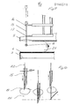
- Figs. 3 and 4 Furthermore, for carrying out the method according to the present invention, use is made of the parts illustrated in block form in Figs. 3 and 4, while employing the graphic principle illustrated in Fig. 5.
- the exposed prints A and B are placed on a measurement pad 7, the prints show, apart from the spot 8 which it is desirable to localise and examine more closely, also an index mark 9 on print A and an index mark 10 on print B.
- the film cassette is arranged such that the index 9 and a letter A or other marking are exposed simultanesouly with the object on the print A, and the index 10 together with the letter B on print B, so as to avoid confusion and to realise an exact reference on each print.
- a monitoring unit 11 is included in the measurement pad 7 and is generally entitled a cursor.
- the cursor is first placed on the index of each respective print and thereafter, directly above the spot which it is desired to localise, so as to obtain the polar coordinates of the spot.
- the measurement pad 7 is coupled to a calculation and guidance unit 12 in which the geometric calculations illustrated in Fig. 5 are carried out and in which signals are generated for operating servo-devices 14 in the guidance instrument 6 proper, by the intermediary of a comparator circuit 13 with figure display in those cases when it is desirable to carry out automatic setting of the guidance instrument 6 and; in such an event, re-hook-up to the servo-devices is effected from the guidance instrument 6.
- the servo-devices 14 may be replaced by manual means and some form of indicator for converting the signals from the calculation unit 12 to setting values. This is carried out in the comparator circuit 13 which may also be considered as a difference and trend indicator which includes a figure display, by means of which the guidance instrument is set, and a possible fine-needle can be placed in the target spot.
- Figs. 7, 8 and 9 show the guidance instrument 6 in greater detail and, as will be apparent from these Figures, the arm 3, the film cassette holder 4 and the compression plate 5 are associated parts of the guidance instrument.
- the orifice in the compression plate 5 it is possible to move an upper needle retainer 16 and a lower needle retainer 17 on the X, Y, and Z axes.
- the lower needle retainer 17 is vertically movable independently of the upper needle retainer 16.
- the needle retainers 16 and 17 are guided using servo motors 18, 19 and 20, and the desired position of the needle tip can be read-off on displays for the exact localisation of the needle tip in relation to the value calculated using the calculation unit 12 in such a manner that the needle tip can be placed in the spot or region 8 illustrated in Fig. 9.
- Adjustment of the guidance instrument 6, and, thereby, the needle retainers is suitably effected such that the position of the needle retainers 16, 17 on the X, Y and Z axes is first set, whereafter the fine-needle is placed in the retainers 16 and 17, and, on abutment against the upper retainer 16, the needle tip is in the correct position.
- This adjustment may very well be executed using the servo devices, while the vertical motion of the lower needle retainer 17 or needle guider is effected manually.
- an acoustic signal is generated which may possibly also be combined with a light signal. This is illustrated in greater detail in Fig. 9.
- the needle 15 shall be moved from the position shown by ghosted lines to the position shown by solid lines.
- the lower needle retainer is employed for insertion of the biopsy needle to the region 8. Different steps may be implemented, depending on the structure of the biopsy needle.
- Fig. 10 illustrates how a marker wire 22 is placed in the region 8 so as, on later surgical excision, to guide the surgeon to the region 8. It is an extremely delicate, if not entirely impossible operation to find region 8 without the help of the wire marker, since the region 8 may be as small as one or two mm.
- the method according to the present invention greatly facilitates breast cancer diagnosis and, above all, makes for developments of the earlier mammography in an extremely rational and reliable manner.
- the method according to the present invention also makes for an extensive development of the methodology of mammography examination without entailing greatly increased burdening of therapeutic activities.
- the present invention also provides the possibility of a considerable refinement of therapeutic methods in an extremely gentle and careful manner and at low cost. Employment of the method according to the present invention also makes for considerably earlier identification of malignant areas during the most occult stages, whereby treatment without surgery may be conceivable.
Abstract
Description
- The present invention relates to a method, in conjunction with the X-ray exposure of an object, of localising the three-dimensional position of a spot in the object.
- In many contexts, it is desirable to be able to localise a spot within an object and, for example, to guide and insert an instrument to exact position in the spot. This is of particular importance in breast cancer diagnosis using int. al. fine-needle biopsy which may be considered as included in the clinical and cytological components in triple diagnostics. This triple diagnostics procedure generally includes mammography, clinical examination and cytology. Fine-needle biopsy or fine needle puncture with cytological examination of cell samples from a suspected region provide very reliable positive repponses. Furthermore, it is desirable, using fine-needle biopsy, to be able to indicate a spot by means of a thin wire marker for facilitating subsequent surgical excision biopsy.
- The task forming the basis of the present invention is to realise a method of localising, as simply, reliably and rapidly as possible, the three-dimensional position of a spot in an object, for example a female breast.
- This task is solved according to the present invention in that, after fixation of the object in a pre-determined position, the object is exposed by means of an exposure device in two directions, each on either side of a centre line at right angles to the plane of the image for obtaining a first image print and a second image print, the invention being characterised in that the two-dimensional position of the target spot on the two prints is established in relation to an index on the prints; and that the coordinates of the spot in relation to the indices are processed to obtain control signals for setting of a guidance instrument with means for placing in the target spot in the object. The exposure device and the guidance instrument are placed in the same centre reference from which the right-angle centre line departs. The index is exposed on the print preferably simultaneously with the object for obtaining an exact measurement reference.
- The method according to the present invention makes possible the localisation of an optional spot in an object, for example a female breast, with a very high degree of precision in an extremely simple and reliable mannner. By applying the method according to the present invention in conjunction with breast cancer diagnosis, it is possible to attain an optional spot in a female breast for fine-needle puncture, indication or some form of directed therapy in a simple and rapid manner so that the patient need not be subjected to undue stress or excessive discomfort.
- The nature of the present invention and its aspects will be more readily understood from the following brief description of the accompanying Drawings, and discussion relating thereto.
- In the accompanying Drawings:
- Fig. 1 is a side elevation of a section of an apparatus for carrying out the method according to the present invention;
- Fig. 2 is a front elevation-of the apparatus of Fig. 1;
- Fig. 3 is a block diagram of parts included in an apparatus for carrying out the method according to the present invention;
- Fig. 4 is a top plan view of the parts illustrated in Fig. 3 but shown in greater detail;
- Fig. 5 illustrates the geometric conditions for the present invention;
- Fig. 6 is a side elevation of parts of an apparatus for carrying out the method according to the present invention;
- Fig. 7 is a front elevation of the parts illustrated in Fig. 6;
- Fig. 8 is a top plan view of the parts shown in Figs. 6 and 7;
- Fig. 9 is, on a larger scale, a side elevation of a number of the parts illustrated in Figs. 6-8; and
- Fig. 10 shows, on a larger scale, a region from Fig. 9. DESCRIPTION OF PREFERRED EMBODIMENT
- The method according to the present invention will now be described in greater detail in conjunction with the apparatus for its execution as shown on the Drawings. The apparatus includes int. al. a per se known
mammograph 1 which may be of the type designated "Scenograph 500 T". Themammograph 1 is pivotal about ashaft 2 which is provided with a scale for the exact setting of themammograph 1. A film cassette holder for afilm cassette 4 is mounted on anarm 3. Acompression plate 5 is disposed above thearm 3 with thefilm cassette 4. Thecompression plate 5 is, naturally, vertically movable and has an orifice of, for example, 50 x 40 mm to make possible the insertion of, for instance, a biopsy needle into a breast which is held in a compressed state by means of thecompression plate 5 on the arm, and thefilm cassette holder 4. The path of radiation from the tube in themammograph 1 generating the X-rays is illustrated by ghosted lines, the radiation path impinging upon a film in thefilm cassette holder 4 for generating an exposed print which depicts the exposed breast region. The apparatus illustrated in Fig. 1 further includes aguidance instrument 6 which is shown in greater detail in Figs. 6, 7 and 8 and parts thereof in Fig. 9. The above-mentionedparts guidance instrument 6, since theparts mammograph 1, pivotal about theshaft 2. Thecompression plate 5 is transparent for X-rays so that a greater portion of the object is exposed than the surface located directly beneath the orifice in thecompression plate 5. - Fig. 2 illustrates in greater detail the pivotal capacity of the
mammograph 1 about theshaft 2. For executing the method according to the present invention, a first exposure is taken with themammograph 1 in the position shown in Fig. 2 from the focal point FA with the film cassette in a first position for generating a film print A, whereafter the mammograph is pivoted and the film cassette is shifted for exposure in the focal point FB and for generating a film print B. - Furthermore, for carrying out the method according to the present invention, use is made of the parts illustrated in block form in Figs. 3 and 4, while employing the graphic principle illustrated in Fig. 5. The exposed prints A and B are placed on a
measurement pad 7, the prints show, apart from thespot 8 which it is desirable to localise and examine more closely, also an index mark 9 on print A and anindex mark 10 on print B. The film cassette is arranged such that the index 9 and a letter A or other marking are exposed simultanesouly with the object on the print A, and theindex 10 together with the letter B on print B, so as to avoid confusion and to realise an exact reference on each print. - A monitoring unit 11 is included in the
measurement pad 7 and is generally entitled a cursor. For carrying out the measurement, the cursor is first placed on the index of each respective print and thereafter, directly above the spot which it is desired to localise, so as to obtain the polar coordinates of the spot. Themeasurement pad 7 is coupled to a calculation andguidance unit 12 in which the geometric calculations illustrated in Fig. 5 are carried out and in which signals are generated for operating servo-devices 14 in theguidance instrument 6 proper, by the intermediary of acomparator circuit 13 with figure display in those cases when it is desirable to carry out automatic setting of theguidance instrument 6 and; in such an event, re-hook-up to the servo-devices is effected from theguidance instrument 6. - The servo-
devices 14 may be replaced by manual means and some form of indicator for converting the signals from thecalculation unit 12 to setting values. This is carried out in thecomparator circuit 13 which may also be considered as a difference and trend indicator which includes a figure display, by means of which the guidance instrument is set, and a possible fine-needle can be placed in the target spot. - Figs. 7, 8 and 9 show the
guidance instrument 6 in greater detail and, as will be apparent from these Figures, thearm 3, thefilm cassette holder 4 and thecompression plate 5 are associated parts of the guidance instrument. In the orifice in thecompression plate 5, it is possible to move anupper needle retainer 16 and alower needle retainer 17 on the X, Y, and Z axes. Thelower needle retainer 17 is vertically movable independently of theupper needle retainer 16. Theneedle retainers servo motors calculation unit 12 in such a manner that the needle tip can be placed in the spot orregion 8 illustrated in Fig. 9. - Adjustment of the
guidance instrument 6, and, thereby, the needle retainers is suitably effected such that the position of theneedle retainers retainers upper retainer 16, the needle tip is in the correct position. This adjustment may very well be executed using the servo devices, while the vertical motion of thelower needle retainer 17 or needle guider is effected manually. With theneedle retainers region 8 of thefemale breast 21, which is compressed between thefilm cassette holder 4 and thecompression plate 5 and has been in such state during exposure of the two prints A and B and a subsequent input using the cursor on themeasurement pad 7 and the following calculations of adjustment values for theguidance instrument 6. It should, here, be observed that all parts included in the apparatus are, as it were, "on-line", which ensures a very rapid execution of the method. - According to Fig. 9, the
needle 15 shall be moved from the position shown by ghosted lines to the position shown by solid lines. After setting of the X and Y axes of theneedle retainers region 8. Different steps may be implemented, depending on the structure of the biopsy needle. - Fig. 10 illustrates how a
marker wire 22 is placed in theregion 8 so as, on later surgical excision, to guide the surgeon to theregion 8. It is an extremely delicate, if not entirely impossible operation to findregion 8 without the help of the wire marker, since theregion 8 may be as small as one or two mm. - The method according to the present invention greatly facilitates breast cancer diagnosis and, above all, makes for developments of the earlier mammography in an extremely rational and reliable manner. The method according to the present invention also makes for an extensive development of the methodology of mammography examination without entailing greatly increased burdening of therapeutic activities. The present invention also provides the possibility of a considerable refinement of therapeutic methods in an extremely gentle and careful manner and at low cost. Employment of the method according to the present invention also makes for considerably earlier identification of malignant areas during the most occult stages, whereby treatment without surgery may be conceivable.
Claims (3)
Priority Applications (1)
| Application Number | Priority Date | Filing Date | Title |
|---|---|---|---|
| AT84850345T ATE60892T1 (en) | 1983-11-14 | 1984-11-08 | LOCATION PROCEDURES. |
Applications Claiming Priority (2)
| Application Number | Priority Date | Filing Date | Title |
|---|---|---|---|
| SE8306243 | 1983-11-14 | ||
| SE8306243A SE8306243L (en) | 1983-11-14 | 1983-11-14 | LOCATION METHODOLOGY |
Publications (3)
| Publication Number | Publication Date |
|---|---|
| EP0146511A2 true EP0146511A2 (en) | 1985-06-26 |
| EP0146511A3 EP0146511A3 (en) | 1987-01-28 |
| EP0146511B1 EP0146511B1 (en) | 1991-02-20 |
Family
ID=20353297
Family Applications (1)
| Application Number | Title | Priority Date | Filing Date |
|---|---|---|---|
| EP84850345A Revoked EP0146511B1 (en) | 1983-11-14 | 1984-11-08 | A method of localisation |
Country Status (6)
| Country | Link |
|---|---|
| US (1) | US4727565A (en) |
| EP (1) | EP0146511B1 (en) |
| AT (1) | ATE60892T1 (en) |
| CA (1) | CA1238723A (en) |
| DE (1) | DE3484129D1 (en) |
| SE (1) | SE8306243L (en) |
Cited By (11)
| Publication number | Priority date | Publication date | Assignee | Title |
|---|---|---|---|---|
| EP0288187A1 (en) * | 1987-04-10 | 1988-10-26 | Harry Haitak Chen | Portable compression grid and needle holder |
| EP0297354A2 (en) * | 1987-06-30 | 1989-01-04 | Siemens Aktiengesellschaft | Biopsy device for an X-ray apparatus |
| EP0390653A1 (en) * | 1989-03-29 | 1990-10-03 | General Electric Cgr S.A. | Mammography apparatus equipped with a stereotaxic device and method of using the apparatus |
| US4962515A (en) * | 1989-11-13 | 1990-10-09 | The General Hospital Corporation | Ridged compression assembly for mammography apparatus |
| DE4111107A1 (en) * | 1990-04-06 | 1991-10-10 | Orion Yhtymae Oy | METHOD FOR A BIOPSY BY MEANS OF A THIN NEEDLE OR A TISSUE MARKING IN CONNECTION WITH THE MAMMOGRAPHY AND DEVICE FOR IMPLEMENTING THE METHOD |
| FR2666217A1 (en) * | 1990-08-28 | 1992-03-06 | Annonier Claude | Device for radiostereotaxic targetting and puncturing of suspect lesions of the breast |
| EP0483005A1 (en) * | 1990-10-24 | 1992-04-29 | General Electric Cgr S.A. | Mammography apparatus provided with a needle holder |
| EP0574868A2 (en) * | 1992-06-17 | 1993-12-22 | Helge Arndt | Installation for guiding a dental instrument or a measuring device and method of preparing teeth and of producing restorations |
| EP0625024A1 (en) * | 1991-11-27 | 1994-11-23 | Fischer Imaging Corporation | Motorized mammographic biopsy apparatus |
| WO1995014505A1 (en) * | 1993-11-24 | 1995-06-01 | Massachusetts Institute Of Technology | Minimally invasive monopole phased array hyperthermia applicators for treating breast carcinomas |
| FR2823969A1 (en) * | 2001-04-30 | 2002-10-31 | Ge Med Sys Global Tech Co Llc | METHOD FOR TAKING TISSUE DURING X-RAY EXAMINATION AND DEVICE FOR IMPLEMENTING SAME |
Families Citing this family (119)
| Publication number | Priority date | Publication date | Assignee | Title |
|---|---|---|---|---|
| SE459150B (en) * | 1986-09-19 | 1989-06-12 | Anders Wallner | DEVICE FOR MAMMOGRAPHIC STEREOTACTIC PUNCTION OF PATHOLOGICAL CHANGES IN THE FEMALE BREAST |
| FI80996C (en) † | 1988-05-26 | 1991-10-25 | Automed Oy | Mammography method and apparatus |
| DK654488A (en) * | 1988-11-23 | 1990-05-24 | Nordisk Roentgen Tech App | ROENTGENAPPARAT |
| FR2652928B1 (en) | 1989-10-05 | 1994-07-29 | Diadix Sa | INTERACTIVE LOCAL INTERVENTION SYSTEM WITHIN A AREA OF A NON-HOMOGENEOUS STRUCTURE. |
| US5415169A (en) * | 1989-11-21 | 1995-05-16 | Fischer Imaging Corporation | Motorized mammographic biopsy apparatus |
| EP0502069A1 (en) * | 1989-11-24 | 1992-09-09 | Technomed International | A method and apparatus for determining the position of a target relative to known co-ordinates |
| US6031892A (en) | 1989-12-05 | 2000-02-29 | University Of Massachusetts Medical Center | System for quantitative radiographic imaging |
| US5111828A (en) * | 1990-09-18 | 1992-05-12 | Peb Biopsy Corporation | Device for percutaneous excisional breast biopsy |
| US5353804A (en) * | 1990-09-18 | 1994-10-11 | Peb Biopsy Corporation | Method and device for percutaneous exisional breast biopsy |
| US6347240B1 (en) | 1990-10-19 | 2002-02-12 | St. Louis University | System and method for use in displaying images of a body part |
| ATE196234T1 (en) | 1990-10-19 | 2000-09-15 | Univ St Louis | LOCALIZATION SYSTEM FOR A SURGICAL PROBE FOR USE ON THE HEAD |
| US5409497A (en) * | 1991-03-11 | 1995-04-25 | Fischer Imaging Corporation | Orbital aiming device for mammo biopsy |
| US5569266A (en) * | 1991-03-11 | 1996-10-29 | Fischer Imaging Corporation | Magnetic resonance imaging device useful for guiding a medical instrument |
| US5289520A (en) * | 1991-11-27 | 1994-02-22 | Lorad Corporation | Stereotactic mammography imaging system with prone position examination table and CCD camera |
| US5594769A (en) * | 1991-11-27 | 1997-01-14 | Thermotrex Corporation | Method and apparatus for obtaining stereotactic mammographic guided needle breast biopsies |
| DE4294430C2 (en) * | 1991-11-27 | 2003-03-20 | Thermo Trex Corp N D Ges D Sta | Stereo=tactic mammography and needle biopsy table |
| US5603318A (en) * | 1992-04-21 | 1997-02-18 | University Of Utah Research Foundation | Apparatus and method for photogrammetric surgical localization |
| US5224147A (en) * | 1992-08-10 | 1993-06-29 | Collin Gene E | Angle indicator for X-ray machine |
| ES2115776T3 (en) * | 1992-08-14 | 1998-07-01 | British Telecomm | POSITION LOCATION SYSTEM. |
| US5386447A (en) * | 1992-09-23 | 1995-01-31 | Fischer Imaging Corporation | Mammographic screening and biopsy apparatus |
| FR2703237B1 (en) * | 1993-03-29 | 1995-05-19 | Ge Medical Syst Sa | Mammograph equipped with a stereotaxic camera with digital detector and method of using such a mammograph. |
| CA2161430C (en) * | 1993-04-26 | 2001-07-03 | Richard D. Bucholz | System and method for indicating the position of a surgical probe |
| US5409004A (en) * | 1993-06-11 | 1995-04-25 | Cook Incorporated | Localization device with radiopaque markings |
| US6031565A (en) * | 1993-06-18 | 2000-02-29 | Gte Internetworking Incorporated | Stereo radiography |
| US5983123A (en) * | 1993-10-29 | 1999-11-09 | United States Surgical Corporation | Methods and apparatus for performing ultrasound and enhanced X-ray imaging |
| JP3461509B2 (en) * | 1993-10-29 | 2003-10-27 | ユナイテッド ステイツ サージカル コーポレイション | Apparatus for Sonomammography and better X-ray photography |
| US6978166B2 (en) * | 1994-10-07 | 2005-12-20 | Saint Louis University | System for use in displaying images of a body part |
| AU3950595A (en) | 1994-10-07 | 1996-05-06 | St. Louis University | Surgical navigation systems including reference and localization frames |
| US5795308A (en) * | 1995-03-09 | 1998-08-18 | Russin; Lincoln D. | Apparatus for coaxial breast biopsy |
| US5807276A (en) * | 1995-03-09 | 1998-09-15 | Russin; Lincoln David | Biopsy device and method |
| US5833627A (en) * | 1995-04-13 | 1998-11-10 | United States Surgical Corporation | Image-guided biopsy apparatus and methods of use |
| US5592939A (en) | 1995-06-14 | 1997-01-14 | Martinelli; Michael A. | Method and system for navigating a catheter probe |
| US6167145A (en) * | 1996-03-29 | 2000-12-26 | Surgical Navigation Technologies, Inc. | Bone navigation system |
| US5820552A (en) * | 1996-07-12 | 1998-10-13 | United States Surgical Corporation | Sonography and biopsy apparatus |
| US5851180A (en) * | 1996-07-12 | 1998-12-22 | United States Surgical Corporation | Traction-inducing compression assembly for enhanced tissue imaging |
| US6459925B1 (en) | 1998-11-25 | 2002-10-01 | Fischer Imaging Corporation | User interface system for mammographic imager |
| US5776062A (en) * | 1996-10-15 | 1998-07-07 | Fischer Imaging Corporation | Enhanced breast imaging/biopsy system employing targeted ultrasound |
| SE9700117D0 (en) * | 1997-01-17 | 1997-01-17 | Siemens Elema Ab | A method for modifying at least one computational algorithm for a biopsy system and a biopsy system |
| FR2759791B1 (en) * | 1997-02-17 | 1999-04-09 | Cogema | RADIATION SOURCE MAPPING DEVICE |
| US6226548B1 (en) * | 1997-09-24 | 2001-05-01 | Surgical Navigation Technologies, Inc. | Percutaneous registration apparatus and method for use in computer-assisted surgical navigation |
| US6021343A (en) | 1997-11-20 | 2000-02-01 | Surgical Navigation Technologies | Image guided awl/tap/screwdriver |
| US6348058B1 (en) * | 1997-12-12 | 2002-02-19 | Surgical Navigation Technologies, Inc. | Image guided spinal surgery guide, system, and method for use thereof |
| US6027457A (en) * | 1998-06-18 | 2000-02-22 | United States Surgical Corporation | Apparatus and method for securing tissue during ultrasound examination and biopsy |
| US6118845A (en) | 1998-06-29 | 2000-09-12 | Surgical Navigation Technologies, Inc. | System and methods for the reduction and elimination of image artifacts in the calibration of X-ray imagers |
| US6477400B1 (en) * | 1998-08-20 | 2002-11-05 | Sofamor Danek Holdings, Inc. | Fluoroscopic image guided orthopaedic surgery system with intraoperative registration |
| US6050954A (en) * | 1998-08-21 | 2000-04-18 | Manan Medical Products, Inc. | Biopsy needle orientation fixture |
| US6482182B1 (en) | 1998-09-03 | 2002-11-19 | Surgical Navigation Technologies, Inc. | Anchoring system for a brain lead |
| US6214018B1 (en) | 1998-11-04 | 2001-04-10 | Trex Medical Corporation | Method and apparatus for removing tissue from a region of interest using stereotactic radiographic guidance |
| US6470207B1 (en) * | 1999-03-23 | 2002-10-22 | Surgical Navigation Technologies, Inc. | Navigational guidance via computer-assisted fluoroscopic imaging |
| US6491699B1 (en) | 1999-04-20 | 2002-12-10 | Surgical Navigation Technologies, Inc. | Instrument guidance method and system for image guided surgery |
| US6493573B1 (en) * | 1999-10-28 | 2002-12-10 | Winchester Development Associates | Method and system for navigating a catheter probe in the presence of field-influencing objects |
| US6474341B1 (en) * | 1999-10-28 | 2002-11-05 | Surgical Navigation Technologies, Inc. | Surgical communication and power system |
| US11331150B2 (en) | 1999-10-28 | 2022-05-17 | Medtronic Navigation, Inc. | Method and apparatus for surgical navigation |
| US7366562B2 (en) | 2003-10-17 | 2008-04-29 | Medtronic Navigation, Inc. | Method and apparatus for surgical navigation |
| US8239001B2 (en) * | 2003-10-17 | 2012-08-07 | Medtronic Navigation, Inc. | Method and apparatus for surgical navigation |
| US6379302B1 (en) | 1999-10-28 | 2002-04-30 | Surgical Navigation Technologies Inc. | Navigation information overlay onto ultrasound imagery |
| US6381485B1 (en) * | 1999-10-28 | 2002-04-30 | Surgical Navigation Technologies, Inc. | Registration of human anatomy integrated for electromagnetic localization |
| US8644907B2 (en) * | 1999-10-28 | 2014-02-04 | Medtronic Navigaton, Inc. | Method and apparatus for surgical navigation |
| US6499488B1 (en) | 1999-10-28 | 2002-12-31 | Winchester Development Associates | Surgical sensor |
| WO2001064124A1 (en) * | 2000-03-01 | 2001-09-07 | Surgical Navigation Technologies, Inc. | Multiple cannula image guided tool for image guided procedures |
| US6535756B1 (en) | 2000-04-07 | 2003-03-18 | Surgical Navigation Technologies, Inc. | Trajectory storage apparatus and method for surgical navigation system |
| US7085400B1 (en) | 2000-06-14 | 2006-08-01 | Surgical Navigation Technologies, Inc. | System and method for image based sensor calibration |
| US6694169B2 (en) | 2001-02-22 | 2004-02-17 | Minrad Inc. | Targeting system and method of targeting |
| US6636757B1 (en) * | 2001-06-04 | 2003-10-21 | Surgical Navigation Technologies, Inc. | Method and apparatus for electromagnetic navigation of a surgical probe near a metal object |
| US6947786B2 (en) * | 2002-02-28 | 2005-09-20 | Surgical Navigation Technologies, Inc. | Method and apparatus for perspective inversion |
| US6990368B2 (en) * | 2002-04-04 | 2006-01-24 | Surgical Navigation Technologies, Inc. | Method and apparatus for virtual digital subtraction angiography |
| US7998062B2 (en) | 2004-03-29 | 2011-08-16 | Superdimension, Ltd. | Endoscope structures and techniques for navigating to a target in branched structure |
| US6892090B2 (en) * | 2002-08-19 | 2005-05-10 | Surgical Navigation Technologies, Inc. | Method and apparatus for virtual endoscopy |
| US7599730B2 (en) | 2002-11-19 | 2009-10-06 | Medtronic Navigation, Inc. | Navigation system for cardiac therapies |
| US7697972B2 (en) * | 2002-11-19 | 2010-04-13 | Medtronic Navigation, Inc. | Navigation system for cardiac therapies |
| US7542791B2 (en) * | 2003-01-30 | 2009-06-02 | Medtronic Navigation, Inc. | Method and apparatus for preplanning a surgical procedure |
| US7660623B2 (en) * | 2003-01-30 | 2010-02-09 | Medtronic Navigation, Inc. | Six degree of freedom alignment display for medical procedures |
| US7570791B2 (en) * | 2003-04-25 | 2009-08-04 | Medtronic Navigation, Inc. | Method and apparatus for performing 2D to 3D registration |
| US20050004580A1 (en) * | 2003-07-01 | 2005-01-06 | Tommi Jokiniemi | System for pointing a lesion in an X-rayed object |
| US7313430B2 (en) * | 2003-08-28 | 2007-12-25 | Medtronic Navigation, Inc. | Method and apparatus for performing stereotactic surgery |
| EP2113189B1 (en) | 2003-09-15 | 2013-09-04 | Covidien LP | System of accessories for use with bronchoscopes |
| EP2316328B1 (en) | 2003-09-15 | 2012-05-09 | Super Dimension Ltd. | Wrap-around holding device for use with bronchoscopes |
| US7835778B2 (en) | 2003-10-16 | 2010-11-16 | Medtronic Navigation, Inc. | Method and apparatus for surgical navigation of a multiple piece construct for implantation |
| US7840253B2 (en) * | 2003-10-17 | 2010-11-23 | Medtronic Navigation, Inc. | Method and apparatus for surgical navigation |
| DE10353611B4 (en) * | 2003-11-17 | 2013-01-17 | Siemens Aktiengesellschaft | X-ray diagnostic device for mammography examinations |
| US8764725B2 (en) * | 2004-02-09 | 2014-07-01 | Covidien Lp | Directional anchoring mechanism, method and applications thereof |
| US7567834B2 (en) | 2004-05-03 | 2009-07-28 | Medtronic Navigation, Inc. | Method and apparatus for implantation between two vertebral bodies |
| US7636595B2 (en) * | 2004-10-28 | 2009-12-22 | Medtronic Navigation, Inc. | Method and apparatus for calibrating non-linear instruments |
| US7835784B2 (en) * | 2005-09-21 | 2010-11-16 | Medtronic Navigation, Inc. | Method and apparatus for positioning a reference frame |
| US9168102B2 (en) * | 2006-01-18 | 2015-10-27 | Medtronic Navigation, Inc. | Method and apparatus for providing a container to a sterile environment |
| JP5554927B2 (en) * | 2006-02-15 | 2014-07-23 | ホロジック, インコーポレイテッド | Breast biopsy and needle localization using tomosynthesis system |
| US8112292B2 (en) * | 2006-04-21 | 2012-02-07 | Medtronic Navigation, Inc. | Method and apparatus for optimizing a therapy |
| US8660635B2 (en) | 2006-09-29 | 2014-02-25 | Medtronic, Inc. | Method and apparatus for optimizing a computer assisted surgical procedure |
| US8905920B2 (en) * | 2007-09-27 | 2014-12-09 | Covidien Lp | Bronchoscope adapter and method |
| WO2009122273A2 (en) * | 2008-04-03 | 2009-10-08 | Superdimension, Ltd. | Magnetic interference detection system and method |
| EP2297673B1 (en) | 2008-06-03 | 2020-04-22 | Covidien LP | Feature-based registration method |
| US8218847B2 (en) | 2008-06-06 | 2012-07-10 | Superdimension, Ltd. | Hybrid registration method |
| US8932207B2 (en) | 2008-07-10 | 2015-01-13 | Covidien Lp | Integrated multi-functional endoscopic tool |
| US8165658B2 (en) | 2008-09-26 | 2012-04-24 | Medtronic, Inc. | Method and apparatus for positioning a guide relative to a base |
| US8175681B2 (en) | 2008-12-16 | 2012-05-08 | Medtronic Navigation Inc. | Combination of electromagnetic and electropotential localization |
| US8611984B2 (en) | 2009-04-08 | 2013-12-17 | Covidien Lp | Locatable catheter |
| US8494613B2 (en) * | 2009-08-31 | 2013-07-23 | Medtronic, Inc. | Combination localization system |
| US8494614B2 (en) * | 2009-08-31 | 2013-07-23 | Regents Of The University Of Minnesota | Combination localization system |
| JP5825753B2 (en) * | 2009-11-17 | 2015-12-02 | 富士フイルム株式会社 | Biopsy equipment |
| WO2011159834A1 (en) | 2010-06-15 | 2011-12-22 | Superdimension, Ltd. | Locatable expandable working channel and method |
| DE102010031737A1 (en) * | 2010-07-21 | 2012-01-26 | Siemens Aktiengesellschaft | Device for tissue removal |
| FR2969919B1 (en) | 2011-01-03 | 2013-01-11 | Gen Electric | METHOD FOR ASSISTING THE POSITIONING OF AN ORGAN ON A MEDIUM OF A SYSTEM FOR ACQUIRING MEDICAL IMAGES |
| EP2782505B1 (en) | 2011-11-27 | 2020-04-22 | Hologic, Inc. | System and method for generating a 2d image using mammography and/or tomosynthesis image data |
| KR101588574B1 (en) * | 2013-11-06 | 2016-01-26 | 주식회사 레이언스 | Biopsy needle guiding apparatus for stereotactic biopsy, imaging apparatus having the same and biopsy method using the same |
| ES2943561T3 (en) | 2014-02-28 | 2023-06-14 | Hologic Inc | System and method for generating and visualizing tomosynthesis image blocks |
| US10952593B2 (en) | 2014-06-10 | 2021-03-23 | Covidien Lp | Bronchoscope adapter |
| US10426555B2 (en) | 2015-06-03 | 2019-10-01 | Covidien Lp | Medical instrument with sensor for use in a system and method for electromagnetic navigation |
| US9962134B2 (en) | 2015-10-28 | 2018-05-08 | Medtronic Navigation, Inc. | Apparatus and method for maintaining image quality while minimizing X-ray dosage of a patient |
| US10478254B2 (en) | 2016-05-16 | 2019-11-19 | Covidien Lp | System and method to access lung tissue |
| US10418705B2 (en) | 2016-10-28 | 2019-09-17 | Covidien Lp | Electromagnetic navigation antenna assembly and electromagnetic navigation system including the same |
| US10792106B2 (en) | 2016-10-28 | 2020-10-06 | Covidien Lp | System for calibrating an electromagnetic navigation system |
| US10615500B2 (en) | 2016-10-28 | 2020-04-07 | Covidien Lp | System and method for designing electromagnetic navigation antenna assemblies |
| US10722311B2 (en) | 2016-10-28 | 2020-07-28 | Covidien Lp | System and method for identifying a location and/or an orientation of an electromagnetic sensor based on a map |
| US10517505B2 (en) | 2016-10-28 | 2019-12-31 | Covidien Lp | Systems, methods, and computer-readable media for optimizing an electromagnetic navigation system |
| US10638952B2 (en) | 2016-10-28 | 2020-05-05 | Covidien Lp | Methods, systems, and computer-readable media for calibrating an electromagnetic navigation system |
| US10751126B2 (en) | 2016-10-28 | 2020-08-25 | Covidien Lp | System and method for generating a map for electromagnetic navigation |
| US10446931B2 (en) | 2016-10-28 | 2019-10-15 | Covidien Lp | Electromagnetic navigation antenna assembly and electromagnetic navigation system including the same |
| US11403483B2 (en) | 2017-06-20 | 2022-08-02 | Hologic, Inc. | Dynamic self-learning medical image method and system |
| US11219489B2 (en) | 2017-10-31 | 2022-01-11 | Covidien Lp | Devices and systems for providing sensors in parallel with medical tools |
Family Cites Families (7)
| Publication number | Priority date | Publication date | Assignee | Title |
|---|---|---|---|---|
| FR866211A (en) * | 1940-03-11 | 1941-07-15 | Method and device for fluoroscopy for the localization of foreign bodies (or any part of the organism) which may be detected by x-rays. | |
| US3577160A (en) * | 1968-01-10 | 1971-05-04 | James E White | X-ray gauging apparatus with x-ray opaque markers in the x-ray path to indicate alignment of x-ray tube, subject and film |
| US3711712A (en) * | 1970-12-28 | 1973-01-16 | Laren R Mc | Method and device for locating a foreign body in human eye |
| DE2443558B2 (en) * | 1974-09-11 | 1979-01-04 | Siemens Ag, 1000 Berlin Und 8000 Muenchen | Device for puncturing internal organs and vessels |
| DE2501436A1 (en) * | 1975-01-15 | 1976-07-22 | Bernhard Dr Kramann | Radiodiagnostic and cytological sampling appts. - to detect malignant tumours in the female breast |
| US4007732A (en) * | 1975-09-02 | 1977-02-15 | Robert Carl Kvavle | Method for location and removal of soft tissue in human biopsy operations |
| US4099880A (en) * | 1976-08-11 | 1978-07-11 | Tsutomu Kano | Method and an apparatus for stereoscopic measurement utilizing a three-dimensional image |
-
1983
- 1983-11-14 SE SE8306243A patent/SE8306243L/en not_active Application Discontinuation
-
1984
- 1984-11-08 DE DE8484850345T patent/DE3484129D1/en not_active Revoked
- 1984-11-08 AT AT84850345T patent/ATE60892T1/en not_active IP Right Cessation
- 1984-11-08 EP EP84850345A patent/EP0146511B1/en not_active Revoked
- 1984-11-13 US US06/670,787 patent/US4727565A/en not_active Expired - Lifetime
- 1984-11-13 CA CA000467705A patent/CA1238723A/en not_active Expired
Non-Patent Citations (1)
| Title |
|---|
| None |
Cited By (21)
| Publication number | Priority date | Publication date | Assignee | Title |
|---|---|---|---|---|
| EP0288187A1 (en) * | 1987-04-10 | 1988-10-26 | Harry Haitak Chen | Portable compression grid and needle holder |
| EP0297354A2 (en) * | 1987-06-30 | 1989-01-04 | Siemens Aktiengesellschaft | Biopsy device for an X-ray apparatus |
| US4890311A (en) * | 1987-06-30 | 1989-12-26 | Siemens Aktiengesellschaft | Biopsy means for an x-ray examination apparatus |
| EP0297354A3 (en) * | 1987-06-30 | 1990-04-04 | Siemens Aktiengesellschaft | Biopsy device for an x-ray apparatus |
| EP0390653A1 (en) * | 1989-03-29 | 1990-10-03 | General Electric Cgr S.A. | Mammography apparatus equipped with a stereotaxic device and method of using the apparatus |
| FR2645006A1 (en) * | 1989-03-29 | 1990-10-05 | Gen Electric Cgr | MAMMOGRAPH HAVING INTEGRATED STEREOTAXIC VIEWING DEVICE AND METHOD OF USING SUCH A MAMMOGRAPHER |
| US5018176A (en) * | 1989-03-29 | 1991-05-21 | General Electric Cgr S.A. | Mammograph equipped with an integrated device for taking stereotaxic photographs and a method of utilization of said mammograph |
| US4962515A (en) * | 1989-11-13 | 1990-10-09 | The General Hospital Corporation | Ridged compression assembly for mammography apparatus |
| DE4111107A1 (en) * | 1990-04-06 | 1991-10-10 | Orion Yhtymae Oy | METHOD FOR A BIOPSY BY MEANS OF A THIN NEEDLE OR A TISSUE MARKING IN CONNECTION WITH THE MAMMOGRAPHY AND DEVICE FOR IMPLEMENTING THE METHOD |
| FR2660545A1 (en) * | 1990-04-06 | 1991-10-11 | Orion Yhtymae Oy | METHOD OF BIOPSY AND ASSEMBLY FOR ITS IMPLEMENTATION |
| FR2666217A1 (en) * | 1990-08-28 | 1992-03-06 | Annonier Claude | Device for radiostereotaxic targetting and puncturing of suspect lesions of the breast |
| EP0483005A1 (en) * | 1990-10-24 | 1992-04-29 | General Electric Cgr S.A. | Mammography apparatus provided with a needle holder |
| FR2668359A1 (en) * | 1990-10-24 | 1992-04-30 | Gen Electric Cgr | MAMMOGRAPH PROVIDED WITH AN IMPROVED NEEDLE HOLDER. |
| US5219351A (en) * | 1990-10-24 | 1993-06-15 | General Electric Cgr S.A. | Mammograph provided with an improved needle carrier |
| US5540737A (en) * | 1991-06-26 | 1996-07-30 | Massachusetts Institute Of Technology | Minimally invasive monopole phased array hyperthermia applicators and method for treating breast carcinomas |
| EP0625024A1 (en) * | 1991-11-27 | 1994-11-23 | Fischer Imaging Corporation | Motorized mammographic biopsy apparatus |
| EP0625024A4 (en) * | 1991-11-27 | 1995-02-15 | Fischer Imaging Corp | Motorized mammographic biopsy apparatus. |
| EP0574868A2 (en) * | 1992-06-17 | 1993-12-22 | Helge Arndt | Installation for guiding a dental instrument or a measuring device and method of preparing teeth and of producing restorations |
| EP0574868A3 (en) * | 1992-06-17 | 1994-03-23 | Helge Arndt | |
| WO1995014505A1 (en) * | 1993-11-24 | 1995-06-01 | Massachusetts Institute Of Technology | Minimally invasive monopole phased array hyperthermia applicators for treating breast carcinomas |
| FR2823969A1 (en) * | 2001-04-30 | 2002-10-31 | Ge Med Sys Global Tech Co Llc | METHOD FOR TAKING TISSUE DURING X-RAY EXAMINATION AND DEVICE FOR IMPLEMENTING SAME |
Also Published As
| Publication number | Publication date |
|---|---|
| ATE60892T1 (en) | 1991-03-15 |
| SE8306243L (en) | 1985-05-15 |
| SE8306243D0 (en) | 1983-11-14 |
| CA1238723A (en) | 1988-06-28 |
| US4727565A (en) | 1988-02-23 |
| EP0146511A3 (en) | 1987-01-28 |
| DE3484129D1 (en) | 1991-03-28 |
| EP0146511B1 (en) | 1991-02-20 |
Similar Documents
| Publication | Publication Date | Title |
|---|---|---|
| EP0146511B1 (en) | A method of localisation | |
| US5107843A (en) | Method and apparatus for thin needle biopsy in connection with mammography | |
| Bolmgren et al. | Stereotaxic instrument for needle biopsy of the mamma | |
| US5078142A (en) | Precision mammographic needle biopsy system | |
| US5002735A (en) | Tissue analysis device | |
| US5964715A (en) | Method for modifying at least one calculation algorithm in a biopsy system, and biopsy system operating according to the method | |
| US4259585A (en) | X-ray examination apparatus | |
| US4930143A (en) | Method and device for mammographic stereotactic punction of pathological lesions in the female breast | |
| KR0138803B1 (en) | Ccd imaging system for stereotactic mammographic biopsy apparatus | |
| US6731966B1 (en) | Systems and methods for targeting a lesion | |
| JP5650467B2 (en) | Radiation imaging system | |
| US6214018B1 (en) | Method and apparatus for removing tissue from a region of interest using stereotactic radiographic guidance | |
| EP2589338A1 (en) | Radiological image photography display method and system | |
| JP2010137004A (en) | Radiation image processing system and processing method | |
| CN111134794A (en) | Ultrasonic guide out-of-plane puncture method | |
| US20120022358A1 (en) | Method and mammography apparatus for image-assisted biopsy extraction | |
| US6270506B1 (en) | Medical targeting apparatus | |
| US5681327A (en) | Stereotactic auxiliary means for tomogram-guided implementation of a biopsy | |
| CN102327122A (en) | Double X-ray tube correction guide puncture mammary gland X-ray machine | |
| EP2322096B1 (en) | Image display apparatus and recording medium | |
| JP5286057B2 (en) | Biopsy equipment | |
| JPH01256942A (en) | Portable compressor for searching breast lession | |
| US20050004580A1 (en) | System for pointing a lesion in an X-rayed object | |
| US6223068B1 (en) | Compact radiology instrument | |
| JP2002126106A (en) | Radiotherapeutic apparatus |
Legal Events
| Date | Code | Title | Description |
|---|---|---|---|
| PUAI | Public reference made under article 153(3) epc to a published international application that has entered the european phase |
Free format text: ORIGINAL CODE: 0009012 |
|
| AK | Designated contracting states |
Designated state(s): AT BE CH DE FR GB IT LI LU NL SE |
|
| PUAL | Search report despatched |
Free format text: ORIGINAL CODE: 0009013 |
|
| AK | Designated contracting states |
Kind code of ref document: A3 Designated state(s): AT BE CH DE FR GB IT LI LU NL SE |
|
| 17P | Request for examination filed |
Effective date: 19870729 |
|
| R17P | Request for examination filed (corrected) |
Effective date: 19870728 |
|
| 17Q | First examination report despatched |
Effective date: 19881025 |
|
| GRAA | (expected) grant |
Free format text: ORIGINAL CODE: 0009210 |
|
| AK | Designated contracting states |
Kind code of ref document: B1 Designated state(s): AT BE CH DE FR GB IT LI LU NL SE |
|
| REF | Corresponds to: |
Ref document number: 60892 Country of ref document: AT Date of ref document: 19910315 Kind code of ref document: T |
|
| PLBI | Opposition filed |
Free format text: ORIGINAL CODE: 0009260 |
|
| REF | Corresponds to: |
Ref document number: 3484129 Country of ref document: DE Date of ref document: 19910328 |
|
| 26 | Opposition filed |
Opponent name: SIEMENS AKTIENGESELLSCHAFT, BERLIN UND MUENCHEN Effective date: 19910318 |
|
| ITF | It: translation for a ep patent filed |
Owner name: ING. A. GIAMBROCONO & C. S.R.L. |
|
| ET | Fr: translation filed | ||
| NLR1 | Nl: opposition has been filed with the epo |
Opponent name: SIEMENS AG. |
|
| NLS | Nl: assignments of ep-patents |
Owner name: TURON AB TE VARBERG, ZWEDEN. |
|
| REG | Reference to a national code |
Ref country code: CH Ref legal event code: PUE Owner name: TURON AB |
|
| ITPR | It: changes in ownership of a european patent |
Owner name: CESSIONE;TURON AB |
|
| PLBI | Opposition filed |
Free format text: ORIGINAL CODE: 0009260 |
|
| PLBI | Opposition filed |
Free format text: ORIGINAL CODE: 0009260 |
|
| PLAB | Opposition data, opponent's data or that of the opponent's representative modified |
Free format text: ORIGINAL CODE: 0009299OPPO |
|
| 26 | Opposition filed |
Opponent name: PHILIPS PATENTVERWALTUNG GMBH Effective date: 19911119 Opponent name: SIEMENS AKTIENGESELLSCHAFT, BERLIN UND MUENCHEN Effective date: 19910318 |
|
| REG | Reference to a national code |
Ref country code: GB Ref legal event code: 732 |
|
| 26 | Opposition filed |
Opponent name: NRT-NORDISK ROENTGEN TEKNIK A/S Effective date: 19911118 Opponent name: PHILIPS PATENTVERWALTUNG GMBH Effective date: 19911119 Opponent name: SIEMENS AKTIENGESELLSCHAFT, BERLIN UND MUENCHEN Effective date: 19910318 |
|
| R26 | Opposition filed (corrected) |
Opponent name: SIEMENS AKTIENGESELLSCHAFT, BERLIN UND MUENCHEN * Effective date: 19910318 |
|
| NLR1 | Nl: opposition has been filed with the epo |
Opponent name: PHILIPS PATENTVERWALTUNG GMBH |
|
| NLR1 | Nl: opposition has been filed with the epo |
Opponent name: NRT-NORDISK ROENTGEN TEKNIK A/S |
|
| REG | Reference to a national code |
Ref country code: FR Ref legal event code: TP |
|
| EPTA | Lu: last paid annual fee | ||
| PGFP | Annual fee paid to national office [announced via postgrant information from national office to epo] |
Ref country code: LU Payment date: 19941001 Year of fee payment: 11 |
|
| PGFP | Annual fee paid to national office [announced via postgrant information from national office to epo] |
Ref country code: GB Payment date: 19941024 Year of fee payment: 11 |
|
| PGFP | Annual fee paid to national office [announced via postgrant information from national office to epo] |
Ref country code: FR Payment date: 19941028 Year of fee payment: 11 |
|
| PGFP | Annual fee paid to national office [announced via postgrant information from national office to epo] |
Ref country code: BE Payment date: 19941108 Year of fee payment: 11 |
|
| PGFP | Annual fee paid to national office [announced via postgrant information from national office to epo] |
Ref country code: AT Payment date: 19941121 Year of fee payment: 11 |
|
| PGFP | Annual fee paid to national office [announced via postgrant information from national office to epo] |
Ref country code: SE Payment date: 19941124 Year of fee payment: 11 |
|
| PGFP | Annual fee paid to national office [announced via postgrant information from national office to epo] |
Ref country code: NL Payment date: 19941130 Year of fee payment: 11 |
|
| PGFP | Annual fee paid to national office [announced via postgrant information from national office to epo] |
Ref country code: DE Payment date: 19941230 Year of fee payment: 11 |
|
| EAL | Se: european patent in force in sweden |
Ref document number: 84850345.4 |
|
| PGFP | Annual fee paid to national office [announced via postgrant information from national office to epo] |
Ref country code: CH Payment date: 19950224 Year of fee payment: 11 |
|
| PLAB | Opposition data, opponent's data or that of the opponent's representative modified |
Free format text: ORIGINAL CODE: 0009299OPPO |
|
| RDAG | Patent revoked |
Free format text: ORIGINAL CODE: 0009271 |
|
| STAA | Information on the status of an ep patent application or granted ep patent |
Free format text: STATUS: PATENT REVOKED |
|
| R26 | Opposition filed (corrected) |
Opponent name: SIEMENS AG * 911119 PHILIPS PATENTVERWALTUNG GMBH Effective date: 19910318 |
|
| 27W | Patent revoked |
Effective date: 19950808 |
|
| GBPR | Gb: patent revoked under art. 102 of the ep convention designating the uk as contracting state |
Free format text: 950808 |
|
| NLR1 | Nl: opposition has been filed with the epo |
Opponent name: NRT-NORDISK ROENTGEN TEKNIK A/S Opponent name: PHILIPS PATENTVERWALTUNG GMBH Opponent name: SIEMENS AG |
|
| NLR2 | Nl: decision of opposition | ||
| REG | Reference to a national code |
Ref country code: CH Ref legal event code: PL |
|
| APAH | Appeal reference modified |
Free format text: ORIGINAL CODE: EPIDOSCREFNO |