EP0113676A2 - Signal processing method in autoradiography - Google Patents
Signal processing method in autoradiography Download PDFInfo
- Publication number
- EP0113676A2 EP0113676A2 EP84100149A EP84100149A EP0113676A2 EP 0113676 A2 EP0113676 A2 EP 0113676A2 EP 84100149 A EP84100149 A EP 84100149A EP 84100149 A EP84100149 A EP 84100149A EP 0113676 A2 EP0113676 A2 EP 0113676A2
- Authority
- EP
- European Patent Office
- Prior art keywords
- cleavage products
- specific cleavage
- sampling points
- radioactively labeled
- autoradiograph
- Prior art date
- Legal status (The legal status is an assumption and is not a legal conclusion. Google has not performed a legal analysis and makes no representation as to the accuracy of the status listed.)
- Withdrawn
Links
- 238000003672 processing method Methods 0.000 title claims abstract description 53
- 238000000376 autoradiography Methods 0.000 title claims abstract description 49
- 238000005070 sampling Methods 0.000 claims abstract description 153
- 239000000126 substance Substances 0.000 claims abstract description 135
- 238000012545 processing Methods 0.000 claims abstract description 81
- 238000000034 method Methods 0.000 claims abstract description 77
- 239000000463 material Substances 0.000 claims abstract description 69
- 230000008569 process Effects 0.000 claims abstract description 39
- 238000009499 grossing Methods 0.000 claims abstract description 23
- 238000003776 cleavage reaction Methods 0.000 claims description 173
- 230000007017 scission Effects 0.000 claims description 172
- OPTASPLRGRRNAP-UHFFFAOYSA-N cytosine Chemical compound NC=1C=CNC(=O)N=1 OPTASPLRGRRNAP-UHFFFAOYSA-N 0.000 claims description 40
- UYTPUPDQBNUYGX-UHFFFAOYSA-N guanine Chemical compound O=C1NC(N)=NC2=C1N=CN2 UYTPUPDQBNUYGX-UHFFFAOYSA-N 0.000 claims description 40
- 229940104302 cytosine Drugs 0.000 claims description 20
- 239000012634 fragment Substances 0.000 claims description 20
- 239000000203 mixture Substances 0.000 claims description 19
- RWQNBRDOKXIBIV-UHFFFAOYSA-N thymine Chemical compound CC1=CNC(=O)NC1=O RWQNBRDOKXIBIV-UHFFFAOYSA-N 0.000 claims description 18
- GFFGJBXGBJISGV-UHFFFAOYSA-N Adenine Chemical compound NC1=NC=NC2=C1N=CN2 GFFGJBXGBJISGV-UHFFFAOYSA-N 0.000 claims description 17
- 229930024421 Adenine Natural products 0.000 claims description 17
- 229960000643 adenine Drugs 0.000 claims description 17
- 238000001514 detection method Methods 0.000 claims description 16
- 230000005855 radiation Effects 0.000 claims description 16
- 230000002285 radioactive effect Effects 0.000 claims description 14
- 229920001222 biopolymer Polymers 0.000 claims description 11
- 229940113082 thymine Drugs 0.000 claims description 9
- 238000004458 analytical method Methods 0.000 claims description 8
- 108020004707 nucleic acids Proteins 0.000 claims description 8
- 102000039446 nucleic acids Human genes 0.000 claims description 8
- 150000007523 nucleic acids Chemical class 0.000 claims description 8
- 230000002194 synthesizing effect Effects 0.000 claims description 5
- 108020004414 DNA Proteins 0.000 description 43
- 230000006870 function Effects 0.000 description 12
- 239000000499 gel Substances 0.000 description 11
- 238000013508 migration Methods 0.000 description 9
- 230000005012 migration Effects 0.000 description 9
- 238000001962 electrophoresis Methods 0.000 description 8
- 238000009826 distribution Methods 0.000 description 7
- 230000000007 visual effect Effects 0.000 description 6
- 238000006243 chemical reaction Methods 0.000 description 4
- 239000000839 emulsion Substances 0.000 description 3
- 239000012535 impurity Substances 0.000 description 3
- 238000005304 joining Methods 0.000 description 3
- 108090000623 proteins and genes Proteins 0.000 description 3
- 102000004169 proteins and genes Human genes 0.000 description 3
- 230000015572 biosynthetic process Effects 0.000 description 2
- 230000000694 effects Effects 0.000 description 2
- 238000002474 experimental method Methods 0.000 description 2
- 239000011521 glass Substances 0.000 description 2
- 229910052739 hydrogen Inorganic materials 0.000 description 2
- 239000001257 hydrogen Substances 0.000 description 2
- 238000002955 isolation Methods 0.000 description 2
- -1 silver halide Chemical class 0.000 description 2
- 238000003786 synthesis reaction Methods 0.000 description 2
- 238000012935 Averaging Methods 0.000 description 1
- 229920001747 Cellulose diacetate Polymers 0.000 description 1
- 102000053602 DNA Human genes 0.000 description 1
- 108010010803 Gelatin Proteins 0.000 description 1
- OAICVXFJPJFONN-UHFFFAOYSA-N Phosphorus Chemical compound [P] OAICVXFJPJFONN-UHFFFAOYSA-N 0.000 description 1
- VYPSYNLAJGMNEJ-UHFFFAOYSA-N Silicium dioxide Chemical compound O=[Si]=O VYPSYNLAJGMNEJ-UHFFFAOYSA-N 0.000 description 1
- 230000002411 adverse Effects 0.000 description 1
- 238000004220 aggregation Methods 0.000 description 1
- 230000002776 aggregation Effects 0.000 description 1
- 239000011230 binding agent Substances 0.000 description 1
- 230000005540 biological transmission Effects 0.000 description 1
- 230000004069 differentiation Effects 0.000 description 1
- 239000006185 dispersion Substances 0.000 description 1
- 239000006260 foam Substances 0.000 description 1
- 238000001502 gel electrophoresis Methods 0.000 description 1
- 239000008273 gelatin Substances 0.000 description 1
- 229920000159 gelatin Polymers 0.000 description 1
- 235000019322 gelatine Nutrition 0.000 description 1
- 235000011852 gelatine desserts Nutrition 0.000 description 1
- 230000002068 genetic effect Effects 0.000 description 1
- 230000010365 information processing Effects 0.000 description 1
- 230000010354 integration Effects 0.000 description 1
- 238000002372 labelling Methods 0.000 description 1
- 229910052698 phosphorus Inorganic materials 0.000 description 1
- 239000011574 phosphorus Substances 0.000 description 1
- 229920000139 polyethylene terephthalate Polymers 0.000 description 1
- 239000005020 polyethylene terephthalate Substances 0.000 description 1
- 229920006254 polymer film Polymers 0.000 description 1
- 238000002360 preparation method Methods 0.000 description 1
- 238000004080 punching Methods 0.000 description 1
- 239000012857 radioactive material Substances 0.000 description 1
- 239000000700 radioactive tracer Substances 0.000 description 1
- 239000013558 reference substance Substances 0.000 description 1
- 238000001028 reflection method Methods 0.000 description 1
- 239000000741 silica gel Substances 0.000 description 1
- 229910002027 silica gel Inorganic materials 0.000 description 1
- 239000004332 silver Substances 0.000 description 1
- 229910052709 silver Inorganic materials 0.000 description 1
- 238000003860 storage Methods 0.000 description 1
- 238000004809 thin layer chromatography Methods 0.000 description 1
- 238000012800 visualization Methods 0.000 description 1
Images


Classifications
-
- G—PHYSICS
- G01—MEASURING; TESTING
- G01T—MEASUREMENT OF NUCLEAR OR X-RADIATION
- G01T1/00—Measuring X-radiation, gamma radiation, corpuscular radiation, or cosmic radiation
- G01T1/16—Measuring radiation intensity
- G01T1/20—Measuring radiation intensity with scintillation detectors
- G01T1/2012—Measuring radiation intensity with scintillation detectors using stimulable phosphors, e.g. stimulable phosphor sheets
-
- Y—GENERAL TAGGING OF NEW TECHNOLOGICAL DEVELOPMENTS; GENERAL TAGGING OF CROSS-SECTIONAL TECHNOLOGIES SPANNING OVER SEVERAL SECTIONS OF THE IPC; TECHNICAL SUBJECTS COVERED BY FORMER USPC CROSS-REFERENCE ART COLLECTIONS [XRACs] AND DIGESTS
- Y10—TECHNICAL SUBJECTS COVERED BY FORMER USPC
- Y10S—TECHNICAL SUBJECTS COVERED BY FORMER USPC CROSS-REFERENCE ART COLLECTIONS [XRACs] AND DIGESTS
- Y10S250/00—Radiant energy
- Y10S250/909—Methods and apparatus ancillary to stimulable phosphor systems
Landscapes
- Physics & Mathematics (AREA)
- Health & Medical Sciences (AREA)
- Life Sciences & Earth Sciences (AREA)
- General Physics & Mathematics (AREA)
- High Energy & Nuclear Physics (AREA)
- Molecular Biology (AREA)
- Spectroscopy & Molecular Physics (AREA)
- Measurement Of Radiation (AREA)
- Investigating Or Analysing Biological Materials (AREA)
Abstract
A signal processing method in autoradiography for obtaining information on one dimensional location of radioactively labeled substances distributed in at least one dimensional direction on a support medium in the form of symbol, numeral or combination thereof,
which comprises a process including:
- (1) preparing a graph in which position on a scanning line is set on abscissa and signal level is set on ordinate; and
- (2) processing said graph with either or both of smoothing and threshold-processing to detect sampling points;
- said process being applied to digital signal corresponding to an autoradiograph having the locational information on the radioactively labeled substances, said digital signal being obtained from the autoradiograph visualized on a radiosensitive material.
A signal processing method for determining the base sequence of DNA is also disclosed.
Description
- This invention relates to a signal processing method in autoradiography, and more particularly to a signal processing method in autoradiography employing a radiosensitive material.
- Autoradiography has been known as a method for obtaining locational information on radioactively labeled substances distributed in at least one dimensional direction to form a row on a support medium.
- For instance, the autoradiography comprises steps of: labeling organism-originating biopolymers such as proteins or nucleic acids with a radioactive element; resolving the radioactively labeled biopolymers, derivatives thereof, or cleavage products thereof (referred to hereafter as "radioactively labeled substances") on a gel support (support medium) through a resolving process such as electrophoresis to form a resolved pattern of the radioactively labeled substances (the resolved pattern is not visible); placing said gel support and a high-sensitivity type X-ray film together in layers for a certain period of time to expose said film and developing said film to give the autoradiograph of the resolved pattern as a visible image on the film; and obtaining the locational information of the radioactively labeled substances from said visible image. Further, the identification of the polymeric substances, determination of molecular weight of the polymeric substances and isolation of the polymeric substances can be performed based on the obtained locational information. The autoradiography has been effectively utilized for determining the base sequence of nucleic acids such as DNA or the like.
- In the autoradiography utilizing the above-described radiographic process, the visualization of the autoradiograph having the locational information on radioactively labeled substances on a radiographic film is essentially required.
- Investigators generally analyze the distribution of the radioactively labeled substances on a support medium through study of the visualized autoradiograph. Further, the visually-obtained locational information on the radioactively labeled substances is generally subjected to various analyses to study the characteristics and functions of the radioactively labeled substance.
- Since the autoradiography requires visual analysis of the autoradiograph, there is brought about a drawback that the locational information on the radioactively labeled substances obtained by analysis of the visualized autoradiograph varies or fluctuates depending on the skill of investigators, and the accuracy of the information is limited to a certain extent. Particularly, in such a case that the autoradiograph visualized on a radiographic film shows an image of reduced quality (in regard of sharpness, contrast, etc.), the satisfactory information can be hardly obtained and the accuracy is low. In order to improve the accuracy of the locational information, for instance, a visualized autoradiograph can be scanned with a device such as a scanning densitometer. However, such scanning process requires increased operation time and complicated procedures. Further, there is a limitation on increase of the accuracy so far as such method merely using the device is employed.
- For instance, in carrying out the exposing procedure, the support medium carrying the above-mentioned resolved rows thereon and a radiographic film sometimes cannot be accurately arranged together in layers. In such case, the resolved rows, namely, rows of resolved substances (e.g., electrophoretic rows) visualized on the radiographic film are made to be not parallel to the longitudinal direction of the film to give a dislocated pattern. As a result, error is introduced into the visual analysis of the locational information on the radioactively labeled substances to decrease the accuracy thereof.
- Further, the rows of the resolved radioactively labeled substances on the support medium are sometimes made non-parallel to the longitudinal direction of the support medium or made distorted, depending on kinds of a support medium or resolving conditions. For instance, a gel support medium is generally held between two glass plates in the resolving procedure because the gel lacks self-supporting property. As a result, the gel occasionally becomes uneven in the thickness due to the deformation of the covers (i.e. glass plates) and accordingly the radioactively labeled substances are not always resolved uniformly on the gel. The lack of uniformity of the resolved pattern is also caused by air foams contained in the gel or by heterogeneous dispersion of the composition of gel. For these reasons, a phenomenon such as the so-called smiling effect, i.e., a phenomenon that the migration distance of the resolved row in the vicinity of the center of the support medium is longer than those on the both sides thereof, is often observed. Additionally, in electrophoresis, the voltage sometimes is not applied uniformly to the support medium and in that case the resolving conditions are made locally uneven on the support medium and consequently the resolved rows obtained are distorted.
- Furthermore, in the case that radioactively labeled impurities such as natural radioactive materials are contained in the sample, that the support medium is contaminated with such radioactive impurities, or that the resolution conditions are not appropriate, a noise sometimes appears on the autoradiograph. In consequence, accurate analysis of the locational information on the radioactively labeled substances becomes difficult, resulting in decrease of accuracy of the desired information.
- In the above-described cases, it is not easy to analyze the locational information on the radioactively labeled substances. Thus, even if the aforementioned additional detection device is used, it is still difficult to obtain a satisfactory accurate locational information on the radioactively labeled substances.
- The present inventors have discovered that the information on one dimensional location of the radioactively labeled substances can be obtained in the form of symbol and/or numeral by signal processing method. Said signal processing comprises steps of obtaining digital signal corresponding to the autoradiograph visualized on a radiosensitive material, which has the locational information on the radioactively labeled substances in a sample, and subsequently processing the digital signal. This method is advantageously applicable to the deter-. mination of base sequence of DNA or DNA fragment to give operational easiness and high accuracy.
- The present invention provides a signal processing method in autoradiography for obtaining information on one dimensional location of radioactively labeled substances distributed in at least one dimensional direction on a support medium in the form of symbol, numeral or combination thereof, which comprises a process including:
- (1) preparing a graph in which position on the scanning line is set on abscissa and signal level is set on ordinate;
- (2) processing said graph through either or both of smoothing and threshold-processing to detect sampling points;
- The present invention also provides a signal processing method in autoradiography for determining the above-mentioned scanning line for signal processing,
which comprises steps of: - scanning digital image data in at least two different positions in such a manner that each scanning traverses the row of distributed substances to obtain a relationship between position along said scanning and signal level in said position;
- detecting distributed points for the radioactively labeled substances along each scanning based on said relationship; and
- preparing a continuous line selected from the group consisting of a straight line, a curved line and a polygonal line along the corresponding points of the radioactively labeled substances for each scanning to assign said continuous line to a scanning line for detection of sampling points,
- in which said digital image data being a set of digital signals correspond to an autoradiograph having the locational information on the radioactively labeled substances obtained in the same manner as above.
-
- Figure 1 shows an example of the read-out system for reading out an autoradiograph having locational information on the radioactively labeled substances in the sample recorded on a radiosensitive material employable in the present invention.
- Figure 2 shows an example of the autoradiograph of a sample in which radioactively labeled substances are resolved on a support medium, which is recorded on a radiosensitive material.
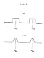
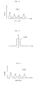
- Figures 3-(a) and 3-(b) are graphs both showing relationships between position in the scanning and level of digital signal in said position.
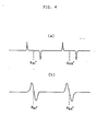
- Figures 4-(a) and 4-(b) are graphs obtained by differentiating the graphs of Figures 3-(a) and 3-(b), respectively.
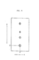
- Figure 5 shows an example of the autoradiograph of a sample in which radioactively labeled substances are resolved in one dimensional direction on a support medium.
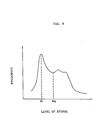
- Figure 6 shows an example of a graph indicating relationship between position in the scanning and level of digital signal in said position.
- Figure 7 shows an example of the filter function employable for smoothing.
- Figure 8 is a graph obtained by processing the graph of Figure 6 with the smoothing.
- Figure 9 shows a histogram.
- Figure 10 shows an example of the autoradiograph of a sample in which base specific cleavage products of DNA are resolved on a gel support medium.
- Figure 11 shows an illustrative scheme indicating the sampling points detected in the resolved rows for DNA by applying thereto the signal processing method of the present invention.
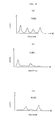
- Figures 12-(a) and 12-(b) are graphs both showing relationships between position on the scanning line and level of digital signal in said position with respect to the second row and the first row, respectively; and Figure 12-(c) is a graph for the fifth row showing a relationship between position on the scanning line and level of digital signal in said position and obtained by subtraction between the graphs (a) and (b).
- The present invention utilizes the method which comprises steps of: placing a sample containing radioactively labeled substances and a radiosensitive material together in layers to record an autoradiograph of the sample on the radiosensitive material; reading out the autoradiograph photoelectrically to obtain electric signal; and converting the electric signal to digital signal through A/D conversion.
- In the present invention, the term "locational information" of the radioactively labeled substances means to include a variety of information relating to the location of the radioactively labeled substances, or the aggregation thereof, being present in the sample, such as the location, the shape, the concentration, the distribution and combinations thereof.
- According to the present invention, one dimensional distribution (resolution) direction can be detected automatically to determine a scanning line for detection of sampling points, even if there occurs overall distortion or dislocation in an autoradiograph recorded on the radiosensitive material which is brought about by locational distortion of a resolved row of the radioactively labeled substances on the support medium in the course of the resolution process, or by locationally inaccurate arrangement between the support medium carrying the row of radioactively labeled substances thereon and the radiosensitive material in the course of the recording (i.e., exposing) procedure of the autoradiograph thereof. Based on thus determined scanning line, the locational information on the row of the distributed radioactively labeled substances (referred to herein as "distributed row") can be obtained with high accuracy. Furthermore, in the case that the autoradiograph is composed of a plurality of rows of the radioactively labeled substances direction, the one dimensional distribution direction of every distorted row involved therein can be accurately detected to determine the scanning line for the row.
- In the present invention, the term "distributed row" means a row which comprises the radioactively labeled substances scattered in one direction in the form of bands or spots, such as an electrophoretic row obtained through electrophoresis. The term "digital image data" means a set of digital signals corresponding to the autoradiograph of the radioactively labeled substances.
- Further, even if the autoradiograph of a sample contains noises, it is possible to easily remove only the noises from the autoradiograph so as to obtain proper image data, by subjecting the corresponding digital image data to the specific signal processing according to the present invention. More in detail, the locational information of the sample can be obtained with high accuracy, being free from the adverse effect of noises which have been introduced by the radioactively labeled impurities contained in the sample or improper resolution conditions. Moreover, if a reference row (internal reference row) is provided for the radioactively labeled substances under analysis, the detection of the location thereof can be further easily performed with high accuracy.
- The reference row (internal reference row) in the present invention means, for instance, a resolved row composed of a mixture of four kinds of base specific cleavage products obtained by specifically cleaving DNA, which is in the case of the signal processing for determination of the base sequence of DNA or DNA fragment. The reference row is employed as reference in the signal processing for obtaining the locational information on the radioactively labeled substances in other resolved rows. The reference row does not necessarily consist of one resolved row, and can be synthesized from plural resolved rows in the process of the signal processing.
- Furthermore, the distributed (e.g., resolved) portions of the radioactively labeled substances, namely the sampling points are automatically detected on the digital image data, and it is possible to analyze the locations of distributed portions with high accuracy even if the size of each distributed portion thereof is reduced. This means that the absolute amount of the radioactively labeled substances used in one autoradiographic process can be reduced, or that the resolved rows formed on a single support medium can be increased in the number without broadening the width of a support medium, so that the amount of information obtainable in single autoradiographic process can be increased as compared with the case employing the conventional analysis.
- Examples of the sample employable in the present invention include a support medium on which radioactively labeled substances are distributed (e.g., resolved) in one dimensional direction to form a distributed row (e.g., resolved row). Examples of the radioactively labeled substances include biopolymers, derivatives whereof, or cleavage products thereof, labeled with a radioactive element.
- For instance, in the case that the radioactively Labeled biopolymers are polymeric substances such as protein, nucleic acid, derivatives thereof and cleavage products thereof, the present invention is useful for isolation and identification thereof. Further, the present invention can be effectively used to analyze the whole or partial molecular structures of these biopoly- ners and the basic segmental constitutions thereof.
- Representative examples of the method for resolving (or developing) the radioactively labeled substaces on a support medium include an electrophoresis using one of various resolving mediums such as a gel in the form of layer, column or the like, a molded polymer film such as a cellulose diacetate film, and a filter paper, and a thin layer chromatography using a support of material such as silica gel. However, the method employable in the present invention is by no means restricted to these methods.
- Samples employable in the present invention are by no means restricted to the above-mentioned samples, and any other samples can be used, provided that the sample is a support medium containing the radioactively labeled substances distributed one-dimensionally thereon and the autoradiograph having the locaitonal information thereof can be recorded on the radiosensitive material.
- The radiosensitive material used in the present invention has a basic structure comprising a support and a radiographic (photographic) emulsion layer. The radiographic emulsion layer comprises a binder such as gelatin and silver halide dispersed therein. For instance, the radiosensitive material is prepared by providing the above-mentioned emulsion layer onto the transparent support such as a polyethylene terephthalate sheet. A representative example of the radiosensitive material includes a radiographic film such as a highspeed type X-ray film.
- In carrying out the exposing procedure, that is, the procedure of exposing the radiosensitive material to the radiation emitted from the support medium containing the radioactively labeled substances, at least a portion of the emitted radiation is absorbed in the radiosensitive substance of the radiosensitive material by placing the support medium and radiosensitive material together in layers for a certain period of time. The exposure can be accomplished by keeping the radiosensitive material in a position adjacent to the support medium, for instance, at a low temperature such as a temperature lower than 00C for at least several days, and then the radiosensitive material is developed. In the exposing procedure, it is further possible to enhance the radiographic speed of the radiosensitive material by using a radiographic intensifying screen or applying thereto a preliminary exposure such as flash exposure.
- The exposing procedure of the radiosensitive material to a sample and the developing procedure thereof in the autoradiographic process have been well known, and are described for instance in the following literature: Method in Biochemical Experiment,
Volume 6, Method in Tracer Experiment I, 271 - 289, "8. Autoradiography" by Toru Sueyoshi & Akiyo Shigematsu (Tokyo Kagaku Dozin Ltd., 1977). - A method for reading out or detecting the autoradiograph having the information on one dimensional location of the radioactively labeled substances in the sample recorded on the radiosensitive material according to the invention will be described briefly, referring to an embodiment of a read-out system shown in Figure 1 of the accompanying drawings.
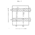
- Figure 1 schematically illustrates an embodiment of the read-out system for reading out the autoradiograph having one dimensional information on the location of the radioactively labeled substances, which is recorded in the form of a visual image on a
radiosensitive material 1. - The
radiosensitive material 1 on which the visual image is recorded is mounted on a transparent andhollow drum 2. Thedrum 2 is moved in the axial direction at a certain speed as well as rotated about its axis at a certain pitch and amirror 3 is fixed in thehollow drum 2. Alight beam 5 generated by alight source 4 passes through alens 6 and comes into thedrum 2. The light beam is then reflected in the upper direction by themirror 3 and passses through theradiosensitive material 1 mounted on thetransparent drum 2. Thus, theradiosensitive material 1 is spot-scanned with the light beam in the X-Y scanning mode. - The light beam passing through each position of the
radiosensitive material 1 is received by alight detector 7 and converted to an electric signal, which is amplified by anamplifier 8 and converted to a digital signal through an A/D converter 9. - More in detail, the read-out pocedure is described in Japanese Patent Provisional Publications No. 54(1979) -121043.
- In the above descripiton on the method for reading out the autoradiograph having the locational information on the radioactively labeled substances recorded on the radiosensitive material, a read-out procedure utilizes the light transmission method using a light beam, but the light reflection method can be also applied thereto. Further, the read-out procedure is by no means restricted to the above-mentioned embodiment, but other various methods such as a read-out procedure using a TV camera can be utilized.
- Thus obtained digital signal corresponding to the autoradiograph of the radioactively labeled substances is subsequently input into the
signal processing circuit 10 shown in Figure 1. In thesignal processing circuit 10, the scanning line is determined and then the sampling points are detected on said digital signal. The digital signal is provided with an address (X, Y) which is represented by a coordinate system fixed to the radiosensitive material and further provided with a signal level (Z) in its address which corresponds to the amount of the transmitting light. - In the signal processing method of the present invention, the set of digital signals obtained by photoelectrically reading out the autoradiograph on the radiosensitive material, namely the digital image data are stored temporarily in a memory device of the signal processing circuit 10 (that is, stored in a non-volatile memory unit such as a buffer memory, a magnetic disk, etc.). In the signal processing, the scanning on the digital image data means to selectively pick up only the signal in the scanning area from the memory device.
- The digital signal processing according to the present invention is described below, referring to an example of an autoradiograph of radioactively labeled substances resolved on a support medium through electrophoresis or the like.
- In the first step, the scanning line for the detection of sampling points is determined in the manner as described below.
- Figure 2 shows an example of the autoradiograph of a sample recorded on the radiosensitive material, in which the autoradiograph is composed of a plurality of radioactively labeled substances resolved in the longitudinal direction of the support medium thereon to form two resolved rows. The autoradiograph on the radiosensitive material is distorted as shown in Figure 2 due to the inaccurate operation such as incorrect arrangement of the support medium and radiosensitive material in the course of the recording (exposing) process.
- A set of digital signals (digital image data), which are obtained by applying the read-out procedure to the radiosensitive material carrying the autoradiograph thereon and is received by the
signal processing circuit 10, corresponds to the autoradiograph shown in Figure 2. - In Figure 2, when the vertical direction is referred to as a Y-axis direction and the horizontal direction is referred to as an X-axis direction in the radiosensitive material, scanning lines can be determined through following steps according to the present invention.
- In the first place, the digital image data obtained as above are scanned numerically along the X-axis direction in such a manner that the scanning traverses the one dimensional distribution direction of the radioactively labeled substances, namely, in a manner that the scanning traverses the distributed rows thereof, and thereby, a relationship between the position (X) on the scanning and the signal level (Z) at its position is obtained.
- Upon plotting the position (X) on the scanning against abscissa and the signal level (Z) against ordinate, a graph such as shown in Figure 3-(a) is prepared.
- Figure 3-(a) shows a graph obtained by the scanning on the digital image data corresponding to the autoradiograph shown in Figure 2, where the radioactively labeled substances are resolved in the form of bands with a certain width.
- In the graph of Figure 3-(a), a middle point (X ) in each region where the signal exhibits a maximum level is assigned to a distributed point of the radioactively labeled substances on each distributed row, wherein a is a positive integer and represents the scanning number (indicating the order of the scanning operation; in this case, a = 1 or 2), and m is a positive integer and represents the number of distributed row. That is, the point X means a distributed point of the radioactively labeled substances which is detected by the a-th scanning and is present on the m-th distributed row.
- The scanning is carried out at least twice in different positions on the digital image data, namely at different Y-corrdinates. Subsequently, a graph such as shown in Figure 3-(a) is produced imaginarily for each scanning, and two or more of sets of distributed points of the radioactively labeled substances are determined on the graph.

- In the second place, a straight line (or polygonal line) which joins the distributed points with the same number m in series of a is prepared for each distributed row and assigned to a scanning line for the detection of sampling points. Of course, a least square straight line or curved line connecting the distributed points may be assigned to the scanning line for detecting sampling points.
- More specifically for instance, in the case that two set of distributed points of the radioactivley labeled substances (represented as below) are found by scanning twice at two Y-coordinates (Y1 and Y2) in parallel to X-axis:

- The scanning operation along X-axis direction can be carried out in any position on Y-axis. In the case that the scanning line for the detection of sampling points is determined by two-times scannings, namely finding of two distributed points, the distance between the two scanning positions on Y-axis is preferably set to be so farther as that the scanning line to be determined should coincide with the real distributed row as sufficiently as possible. That is, the top end (or vicinity thereof) and bottom end (or vicinity thereof) of the resolved row of the radioactively labeled substances are desirably selected as the scanning positions. The scannings are not necessarily done in parallel to each other, but scannings in parallel is naturally preferred.
- The above-mentioned scanning is required to be done with a such a width as to catch at least one resolved portion (band or spot, etc.) of the radioactively labeled substances for each distributed row, and the scanning is done on the digital image data with a certain width having its scanning center along Y-axis direction. In the case that the scanning width is excessively small, there possibly take place not only that the scanning can not catch a resolved portion (i.e., portion in which the resolved radioactively labeled substance is present), but also that even if the scanning catches the resolved portion, the detected distributed point includes an error if the distribution of the radioactively labeled substances on that portion deviates to a certain extent. If the scanning width is excessively wide, the detected distributed point of the radioactively labeled substances may be inaccurate. Accordingly, it is desirable that the scanning width is predetermined depending on the conditions of the sample.
- The graph shown in Figure 3-(a) is obtained, for instance, by picking up the digital signals within a certain scanning width and summing the levels thereof for each X-corrdinate. The summed data may be further subjected to the threshold processing so as to reduce the noise. Otherwise, the graph is obtained by repeatedly picking up the digital signals within the scanning width, applying the threshold processing to the signals for each Y-coordinate, and summing the applied signals for each X-coordinate. The Y-coordinate of the scanning is represented by a middle point of the scanning width.
- The threshold processing mentioned herein means a two-valued (i.e., binary) processing, that is, the level of digital signal being equal to a certain level (the threshold value) or higher than said value is assigned to 1, while the level of digital signal which is lower than said value is assigned to 0, whereby representing all levels of digital signals by 1 or 0.
- Otherwise, the above-mentioned scanning can be carried out as follows. The digital signal within the scanning width is picked up repeatedly for each Y-coordinate to find out the X-coordinate Xai at which the signal exhibits a maximum level for each Y-coordinate (using a graph such as shown in Figure 3-(a)), and subsequently the local mean X-coordinate Xa1 is calculated.

- The above-mentioned scanning position and scanning width may be manually input in the
signal processing circuit 10 for each sample prior to the signal processing of the digital signal. By predetermining the scanning position and scanning width independently for each sample as discribed above, the distributed points of the radioactively labeled substances can be detected accurately even if the distribution in one dimensional direction thereof varies greatly depending on the kind of a sample and the resolution conditions, etc. - Scanning more than twice means increase of the detected distributed points of the radioactively labeled substances. In this case, a straight line (polygonal line) obtained by joining the distributed points, which is assigned to the scanning line for the detection of sampling points, fits more closely to the distributed row thereof. Further, by processing the polygonal line with a suitable approximation to prepare a curved line, the scanning line for the detection of sampling points can be determined more accurately. However, the increase of the scanning operation brings about the complexity and long processing time in the signal processing. It is preferable that the number of scanning is determined depending on the conditions of the sample and the accuracy desired in the autoradiographic prccess.
- For instance, in the autoradiography of the sample in which the nucleic acids, derivatives thereof or cleavage products thereof, labeled with a radioactive element, are resolved on a support medium through the method using electrophoresis or the like, the scanning line for the detection of sampling points can be determined accurately with twice scanning. The scanning width is preferably so predetermined as to catch two or three distributed portions (resolved bands) of the radioactively labeled substances for each resolved row.
- According to the above-described determination of the scanning line for the detection of sampling points, it is possible that the width of respective distributed portions of the radioactively labeled substances is reduced to approx. 3 mm. Therefore, the present invention enables to reduce the amount of the radioactively labeled substances for the preparation of resolved rows and consequently, make it possible to increase the number of row resolvable in a single support medium.
- In the signal processing method of the present invention, the distributed point of the radioactively labeled substances in each scanning area can be more easily detected by utilizing a graph which is imaginarily obtained through the differentiation of the graph of Figure 3-(a). More in detail, edges of the resolved row can be emphasized by differentiating the graph and consequently the both edges in the width direction of the reselved row can be easily detected on the digital image data, so that the distributed point of the radioactively labeled substances is detected by simply finding the middle point between the both edges.
- The graph of Figure 4-(a) shows a graph which is obtained by differentiating the graph of Figure 3-(a). From the graph of Figure 4-(a), edges of each resolved row can be easily detected by assigning the distributed point of the radioactively labeled substances in each row to each middle point (Xam') between a positive peak of the differentiated level value and a negative peak thereof.
- In the case that the radioactively labeled substances are resolved in the form of spots in which the resolved row is liable to exhibit remarkable dislocation or distortion, or in the case that the above-mentioned scanning conditions (scanning position and scanning width) is not suitably predetermined, a graph with the position (X) on the scanning line on abscissa and the signal level (Z) on ordinate is imaginarily given as shown in Figure 3-(b).
- In the graph of Figure 3-(b), each point (Xbn) at which the signal exhibits a maximum level is assigned to the distributed point of the radioactively labeled substances in each resolved row, wherein b is a positive integer and represents the scanning number, and n is a positive integer and represents the row number.
- The graph of Figure 4-(b) shows a graph which is obtained by differentiating the graph of Figure 3-(b). From the graph of Figure 4-(b), each middle point (Xbn') where the differentiated level value changes from positive to negative can be assigned to the distributed point of the radioactively labeled substances in each resolved row.
- In the above cases, the scanning line for the detection of sampling points can be also determined using the detected distributed points in the same manner as described above.
- In examples shown in Figures 2 through 4, the processing is described on the cases of the distributed pattern having two distributed rows, but the signal processing method for determining the scanning line of the present invention is by no means restricted to the case involving the just two rows, but the method can be applied to the distributed pattern of the radioactively labeled substances such as a pattern having only one row, or a pattern having plural (three or more) rows.
- In the second step, the sampling points for detecting the distributed portions of the radioactively labeled substances are detected, for example, as described below referring to another example of the autoraiograph.
- Figure 5 shows an example of the autoradiograph of a sample in which radioactively labeled substances are distributed in one dimensional direction.
- Digital signal corresponding to the autoradiograph of the sample is obtained in the manner as mentioned above. Concerning the digital signal, the scanning line for detecting the sampling points can be determined as described above, that is for example, by scanning the digital image data in two different positions in such a manner that the scanning traverses the one-dimensional resolved row of the radioacitively labeled substances; detecting two distributed points thereof on said scanning; and joining said two distributed points to give a straight line.
- The scanning along the scanning line is performed on the digital image data. As a result, a graph in which the position (W) on the scanning line is plotted as abscissa and the signal level (Z) is plotted as ordinate is obtained as shown in Figure 6. The scanning is done with a certain width. That is, the summation of the signal levels for each W-coordinate with respect to the digital signals within the scanning width are plotted against the abscissa in the graph.
- Subsequently, the graph is processed with smoothing. The smoothing processing is performed, for instance, through convolution of the graph using a suitable filter function. Representative examples of the filter function employable for the convolution include a function g(W) graphically shown in Figure 7. When the graph shown in Figure 6 is represented with a function f(W), the following smoothed function h(W) is obtained through the convolution operation with the above-mentioned filter function:

- Separately, the signal level is plotted as abscissa and the frequency of the signal having its level plotted as ordinate to obtain a histogram with respect to the ) digital signal on the above-mentioned scanning line with a certain scanning width. The histogram is preferably processed with smoothing through the convolution in the manner as described above.
- Figure 9 shows a histogram corresponding to the graph shown in Figure 6 and having been subjected to the smoothing. The peak point (a) in the histogram of Figure 6 represents the back ground level of the digital signal. A certain value is added to the signal level (a) and the summed value (α0) is assigned to a threshold value.
- The threshold processing is applied to the graph of Figure 8 based on the obtained threshold value (α0). gore in detail, the signal level which is equal to the threshold value or higher than said value is assigned to 1, while the signal level which is lower than said value is assigned to 0, so as to obtain a graph in which the signal level is represented by 1 or 0. In this graph, all middle points in regions in which the signal level equals 1 are assigned to sampling points.
- In the signal processing method of the present invention, all maximum points shown in the smoothed graph of Figure 8 can be assigned to the sampling points.
- Thus, the sampling point Sm having the position (Wm) in the distributed direction of the radioactively labeled substances is determined, wherein m is a positive interger and represents the number of sampling point. The information on one dimensional location of the radioactively labeled substances is represented by the position (W ) in one dimensional direction by applying the signal processing to the digital signal as mentioned above.
- Further, if the starting position for resolving the radioactively labeled substances is recorded on the radiosensitive material with a maker containing a radioactive element, the starting position (W0) can be detected on the digital image data in the manner as described above. Otherwise, the starting position can be also detected by providing the radiosensitive material with a mechanical means such as by punching to give a perforation thereto previously and setting the starting position in the exposing procedure. Therefore, the locational information can be represented by the migration distance (Wm') from the starting position of resolution by performing the subtraction of { W - W0 = Wm'} with W0.
- Furthermore, when the signal level on each maximum point in the graph of Figure 8 is assumed to correspond to the relative amount (concentration) of the radioactively labeled substances in each resolved portion, the one-dimensional information on the location of radioactively labeled substances may be represented by both the migration distance and relative amount (Wm', Z). For the relative amount, various calculations such as integration in the vicinity of the each maximum point in the graph of Figure 8 can be done.
- The autoradiograph having the information on one dimensional location of the radioactively labeled substances is output from the
signal processing circuit 10 in the form of numeral as mentioned above. The locational information, which is obtained as the coordinate of the sampling point Sm and the signal level at that coordinate (Xm, Y , Z ), are by no means limited to the above-mentioned representation modes, and other optional representation modes are also utilizable. Thus, the locational information on the radioactively labeled substances can be obtained in the form of symbol and/or numeral. - The obtained symbol and/or numeral are transmitted to a recording device (not shown), directly or optionally via storage in a storing means such as a magnetic tape.
- Various recording devices based on various systems can be employed for the above-described purpose, for instance, a device for visualizing optically by scanning on a radiosensitive material with laser beam, etc., a display means for visualizing electrically on CRT, etc., a means for printing a radiation image displayed on CRT by mean of a video printer, and a means for visualizing on heatsensitive recording material using thermic rays.
- The present invention also provides the signal processing method in the autoradiography of the sample in which the groups of radioactively lableld substances are arranged in plural rows and distrbuted respectively in one dimensional direction.
- That is, a signal processing method in autoradiography for obtaining information on one dimensional location of groups of radioactively labeled substances arranged in plural rows including a reference row and distributed respectively in at least one dimensional direction on a support medium, in the form of symbol, numeral or combination thereof,
which comprises a process including: - (1) preparing a graph in which position on the scanning line is set on abscissa and signal level is set on ordinate with respect to the reference row;
- (2) processing said graph with either or both of smoothing and threshold-processing to detect candidate sampling points;
- (3) processing said candidate sampling points statistically to determine fundamental sampling points; and
- (4) detecting sampling points on respective scanning lines of the residual rows based on said fundamental sampling points,
- The reference row in the above-mentioned signal processing method is not necessarily provided practically onto the support medium, but may be imaginarily synthesized from the plural resolved rows as described before.
- The sample used in the above-mentioned method generally comprises a support medium and groups of radioactively labeled substances, each group being distributed in one dimensional direction in parallel to form a plurality of rows. The term "parallel" does not necessarily mean the strictly parallel relation in which the plural rows are completely parallel to each other, and includes a locally parallel or approximately parallel relation.
- In particular, the above-mentioned signal processing method in the autoradiography is effectively applicable to analyze the molecular weight, molecular structure or basic unit consititution, of polymeric substances such as proteins, nucleic acids, derivatives thereof or cleavage products thereof.
- Accordingly, the present invention provides a signal processing method in autoradiography for determination of base sequence of DNA or DNA fragment, employing at least four groups of base specific cleavage products consisting of:
- 1) guanine specific cleavage products;
- 2) guanine specific cleavage products
+ adenine specific cleavage products; - 3) tymine specific cleavage products
+ cytosine specific cleavage products; and - 4) cytosine specific cleavage products,
- (1) synthesizing an internal reference row from the resolved rows and preparing a graph in which position on the scanning line is set on abscissa and signal level is set on ordinate with respect to said internal reference row;
- (2) processing said graph with either or both of smoothing and threshold-processing to detect candidate sampling points;
- (3) processing said candidate sampling points statistically to determine fundamental sampling points;
- (4) detecting sampling points on each scanning line with respect to the resolved rows based on said fundamental sampling points, and
- (5) comparing and identifying the positions of said sampling points on the scanning lines among the resolved rows to obtain locational information on guanine, adenine, thymine and cytosine,
said process being applied to a digital signal corresponding to an autoradiograph having the locational information on the groups of radioactively labeled cleavage products, said digital signal being obtained by exposing a radiosensitive material to radiation emitted by said groups of radioactively labeled cleavage products on the support medium to record the autoradiograph of the groups of radioactively labeled cleavage products on the radiosensitive material, and reading out said autoradiograph photoelectrically. - In the signal processing method for determination of the sequence of DNA, it is also possible to provide the internal reference row on the support medium practically by simultaneously resolving the mixture of base specific cleavage products which is obtained by specifically cleaving DNA or DNA fragment at each of the four bases composing the constitutional unit thereof, in place of the process comprising synthesis of the internal reference row.
- An embodiment of the signal processing in the autoradiography employing the signal processing method of the present invention will be described referring to an example of the process for determining the base sequence of DNA.
- DNA is in the form of double helix structure consisting of two chain molecules and the two chain molecules are constituted by four constitutional base units, each unit having a base, namely adenine (A), guanine(G), thymine (T), or cytosine (C). The two chain molecules are cross-linked by hydrogen bonding between the four constitutional base units, and the hydrogen bonding between each base comprises only two combinations, namely G-C and A-T. Therefore, if the base sequence of one chain molecule is determined, that of the other chain molecule is naturally determined.
- As a representative method for determining the base sequence of DNA utilizing the autoradiography, Maxam-Gilbert method has been known. In this method, a group containing a radioactive isotope of phosphorus (P) is attached to a chain molecule of DNA or a DNA fragment at one end to be sequenced to prepare a radioactively labeled substance, and then the radioactively labeled DNA molecule is specifically cleaved at the constitutional base unit by a certain chemical reaction. This reaction is called a "base specific cleavage reaction". Then the obtained mixture of numerous cleavage products of the DNA or DNA fragment is resolved through gel electrophoresis to give resolved pattern of the numerous cleavage products (the pattern is not visible).
- In the procedure, an X-ray film is exposed to the resolved pattern and developed to obtain a visualized autoradiograph thereon, and the sequential position of each base from the radioisotopically labeled end of the chain molecules is read by referring to the obtained autoradiograph and the applied base specific chemical reaction so as to determine the sequence of all bases in the aimed substance.
- The signal processing method for the determination of base sequence of DNA or its derivative, fragment, etc., will be described by an embodiment utilizing the above-mentioned Maxam-Gilbert method, referring to the case of employing the following four groups of base specific cleavage products as a typical combination of base specific cleavage product groups:
- 1) guanine (G) specific cleavage products,
- 2) guanine (G) specific cleavage products
+ adenine (A) specific cleavage products, - 3) thymine (T) specific cleavage products
+ cytosine (C) specifically cleaved product, - 4) cytosine (C) specific cleavage products.
- The groups of the above-mentioned base specific cleavage products labeled with 32P are resolved (developed) on a gel support medium through electrophoresis in the conventional manner, to obtain a sample. Then, the sample (support medium) is placed on a radiosensitive material together in layers at a low temperature ranging from -70°C to -90°C for several days to perform the exposure, and the autoradiograph of the sample is recorded on the radiosensitive material as a visual image.
- Figure 10 shows an autoradiograph of resolved rows (electrophretic rows) comprising the above-mentioned four groups of radioactively labeled base specific cleavage products formed through resolution. That is, the first to fourth rows shown in Figure 10 correspond to in order,
- (1) - (G) specific cleavage products,
- (2) - (G) specific cleavage products + (A) specific cleavage products,
- (3) - (T) specific cleavage products
+ (C) specific cleavage products, - (4) - (C) specific cleavage products.
- The radiosensitive material carrying the above autoradiograph of the sample thereon is installed in the read-out system and subjected to the read-out procedure, to obtain the digital signal corresponding to the autoradiograph.
- The obtained digital signal is subjected to the digital signal processing in the
signal processing circuit 10 as mentioned above. - In the first place, the scanning line for signal processing is determined on each row shown in the autoradiograph of Figure 10 in the same manner as mentioned above.
- In the second place, the scanning with the scanning line is performed on the digital image data, so that an imaginary graph in which the position (W) on the scanning line is plotted as abscissa and the signal level (Z) is plotted as ordinate is given with respect to each row. The position on the scanning line is preferably represented by the electrophoretic distance between that position and the starting position (Wk0) of the electrophoresis with respect to each row, wherein k is a positive integer and represents the row number. The starting position is detectable by employing a maker.
- The signals which exhibit the higher level (Z) at each corresponding position (W) on the scanning line in comparison between the graphs of second row and third row, are selectively picked up and combined to obtain an imaginary graph which includes signals concerning all four kinds of base specific cleavage products, i.e., (G) specific cleavage products, (A) specific cleavage products, (T) specific cleavage products and (C) specific cleavage products. The obtained graph indicates an internal reference row (referred to herein as the zeroth row).
- In place of carrying out the above-described procedure, an electrophoretic row containing the above-mentioned four kinds of base specific cleavage products of DNA can be practically formed simultaneously with others on the support medium, so that said electrophoretic row may be assigned to the internal reference row.
- The internal reference row is then subjected to the smoothing and/or threshold-processing in the same manner as mentioned hereinbefore to obtain candidate sampling points SOn having the electrophoretic distance (W0n), wherein 0 represents the internal reference row, and n is a positive integer and represents the number of sampling point corresponding to the candidate point.
- Subsequently the obtained candidate sampling point S0n is subjected to the statistical processing to determine a fundamental sampling point. It is reasonablly assumed that if a radioactively labeled substance present at a certain candidate sampling point (SOn) on the internal reference row is a certain cleavage product, a radioactively labeled substance present at a candidate sampling point (S0n+1) (i.e., a candidate sampling point located in the position adjacently prior to the candidate sampling point (S0n)) is a cleavage product in which one of four bases is attached to units corresponding to the cleavage product present at the point SOn. Moreover, it has been experimentally known that the migration distance of each radioactively labeled substance and the logarithm of molecular weight thereof are in a linear relation. Therefore, the candidate sampling points can be processed statistically by approximating with the following functional equation:

- By introducing numerals of the migration distance W0n of each candidate sampling point SOn and the number n of the sampling point corresponding to said candidate sampling point into the equation (1), the most probable values a0 and b0 are calculated. Then, a0 and b0 are introduced into the equation (1) to determine the fundamental sampling point S0n' represented by the most probable migration distance (W0n').
- In the next, based on thus determined fundamental sampling point SOn', the number of the digital signals which exist within a certain area having the center at the position on the scanning line corresponding to the fundamental sampling point and exhibit a level of not lower than the aforementioned threshold value is obtained through calculation for each fundamental sampling point with respect to each of the four rows. Upon comparison of thus obtained number of digital signals among each row, a suitable threshold-processing is performed repeatedly if desired, the aimed sampling points are detected in each row.
- According to the above-mentioned processing, each row is represented by the set {S0n'}k of the fundamental sampling point S0n' having the most probable migration distance (W0n'). The fundamental sampling points detected in each row are assigned to the aimed sampling points.
- Figure 11 shows the sampling points in each row in order:
- (0) - (G) specific cleavage products
- + (A) specific cleavage products
- + (T) specific cleavage products
- + (C) specific cleavage products;
- (1) - (G) specific cleavage products;
- (2) - (G) specific cleavage products
+ (A) specific cleavage products; - (3) - (T) specific cleavage products
+ (C) specific cleavage products; and - (4) - (C) specific cleavage products.
- Then, the first row to fourth row are compared. More in detail, through an operation between the first row having the set of the sampling points {S0n'}1 and the second row having the set of the sampling points {S0n'}2 (namely, logical product operation),

- By the above-mentioned processing, the information on one dimensional location of the owing four rows is newly obtained:
- (1) - (G) specific cleavage products,
- (5) - (A) specific cleavage products,
- (6) - (T) specific cleavage products,
- (4) - (C) specific cleavage products.
- Otherwise, the detection of the sampling points for each row of different base specific cleavage products can be also performed according to the present invention as described below.
- After obtaining the graph in which the position is plotted as abscissa and the signal level is plotted as ordinate, being represented by the function fk(W) concerning each of the resolved rows (1) through (4), wherein k is a positive integer and represents the row number, the following numerical operation (subtraction) is perf rmed between the function f1(W) of the first row and the function f2(W) of the second row;

- Figure 12 graphically shows the above operation. That is, the graphs (a), (b) and (c) of Figure 12 correspond to the second, first and fifth rows, respectively.
- When the signal level is different between the resolved rows to be operated numerically, the signal is preferred to have similar levels, for instance, by adjusting the signal level in proportion to the density of the image for each resolved row according to the obtained digital image data.
- The same subtraction is performed between the third row and the forth row to obtain an imaginary sixth row which is represented by f6(W) and corresponds to thymine specific cleavage products alone. Thus, the following four rows (including the imaginary rows) consisting essentially of different kinds of respective base specific cleavage products, not of mixtures thereof are obtained:
- (1) - (G) specific cleavage products,
- (5) - (A) specific cleavage products,
- (6) - (T) specific cleavage products,
- (4) - (C) specific cleavage products.
- As described above, the present invention also provides a signal processing method in autoradiography for determining base sequence of DNA or DNA fragment, employing at least two groups of base specific cleavage products or a mixture thereof consisting of:
- 1) single base specific cleavage products or a mixture of two or three kinds of base specific cleavage products selected from the group consisting of guanine specific cleavage products, adenine specific cleavage products, tymine specific cleavage products and cytosine specific cleavage products; and
- 2) a mixture of two, three or four kinds of base specific cleavage products containing at least one kind of the base specific cleavage products included in the above group 1),
- (1) operating numerically between the resolved row of the above group 1) and the resolved row of the above group 2) with respect to the corresponding positions on each scanning line to obtain an imaginary resolved row;
- (2) determining sampling points with respect to said imaginary resolved row;
- The sampling points can efficiently detected by comparing the digital signal among the above four rows based on the fundamental sampling point SOn' in the synthesized internal reference row which is represented by the set {S0n'}k of the fundamental sampling point SOn' having the most probable migration distance (W0n'). It is also possible to detect the sampling points not by using the fundamental sampling points, but by processing each function fk(W) corresponding to the above four rows with the smoothing through the convolution and subsequently with the threshold-processing as described hereinbefore.
- Therefore, the fundamental sampling point S0n' is replaced as follows:
- i) the sampling points which belongs to {S0n'}1 is replaced with G;
- ii) the sampling point which belongs to {S0n'}4 is replaced with C;
- iii) the sampling point which belongs to {S0n'}5 is replaced with A;
- iv) the sampling point which belongs to {S0n'}6 is replaced with T,
- Thus, the base sequence of one chain molecule of DNA can be determined.
- According to the signal processing method of the present invention, the base sequence of DNA can be also determined using the following most simple combination of base specific cleavage products:
- (1) - (G) specific cleavage products,
- (2) - (A) specific cleavage products,
- (3) - (T) specific cleavage products,
- (4) - (C) specific cleavage products.
- The above four groups are the combination being exclusive from each other. Accordingly, by utilizing the exclusiveness that there absolutely exists only one group of base specific cleavage products in the vertical direction to the resolved rows of the above four groups, the locational relation of the bases can be determined by reasonable judgement of the obtained digital signal on the basis of decision by majority. It suggests that the sequence of DNA according to the present invention could be determined more exactly than by the conventional visual judgement.
- More in detail, concerning the digital signal corresponding to the autoradiograph of the resolved pattern comprising four resolved rows of the above groups of base specific cleavage products, which is obtained by photoelectrically reading out the autoradiograph recorded on the radiosensitive material, the scanning lines for detecting the sampling points are deterimined, as well as the internal reference row is obtained by synthesizing the four resolved rows, and the fundamental sampling points in the internal reference row are determined, as described above. The internal reference row, as mentioned hereinbefore, may be practically provided on the support medium in place of synthesis on the digital image data.
- Then, based on the determined fundamental sampling point S On' having the most probable migration distance (W0n'), the number of the digital signals which exist within a certain area having the center at the position on each scanning line corresponding to the fundamental sampling point and exhibit the level not lower than the threshold value is calculated for each fundamental sampling point on the respective four rows. That is, a certain area (sampling mask) with the center at the position (WOn') is fixed for each fundamental sampling point SO and the number of signals existing within each sampling mask fixed on each scanning line and having the level higher than the threshold value is calculated.
- Since the groups of base specific cleavage products contained in the above four resolved rows respectively are exclusive of each other, the number of the fundamental sampling points is necessarily equal to the sum of sampling points to be detected in the four rows. This means that one sampling point corresponding to one fundamental sampling point is detected in any one of the four rows. That is, the aimed sampling point for the same sampling mask must be detected only in one of the four rows.
- Accordingly, by utilizing the above-mentioned exclusiveness among each row, a row which exhibits the highest estimated value for the signal level with respect to the sampling masks of same fundamental sampling point is selected, so as to decide that the sampling point corresponding to said fundamental sampling point is present in said row and absent in the other three rows. The estimated value for the signal level means an integral value of the signal levels included in the mask, or the number of signal whose level exceeds the threshold value in the case of performing the threshold-processing. Thus, the sampling points for all the sampling masks are detected in any one of the four electrophoretic rows, respectively, namely, the sampling points corresponding to the fundamental sampling points in the internal reference row are detected in any one of the four rows.
- According to the above-mentioned processing, each row is represented by the set S0n'}k of the fundamental sampling point S0n', wherein k represents the row number. The fundamental sampling point SOn' is replaced with any one of the symbol G, A, T, C based on the row number k, and subsequently arranged in order of the sampling number n to obtain the base sequence of one chain molecule of DNA represented by the following arrangement:

- The representation mode of the information on the base sequence of DNA obtained as described hereinbefore is by no means limited to the above-mentioned representation mode, and other representation modes is optionally employable. For instance, the relative amount of resolved base specific cleavage products can be also obtained for representation by processing the signal level on the scanning line with an optional operation, if desired.
- Further, the base sequence of both two chain molecules of DNA can be represented. That is, by giving the information on the combination between the four bases, namely A-T and G-C, the sequence of DNA is represented by the following scheme.

- The method for detemining the base sequence of DNA utilizing the aforementioned combinations of (G, G+A, T+C, C) and (G, A, T, C) is one example of the determination of the base sequence of DNA, and the signal processing of the present invention by no means limited to the above combinations, but various combinations are employable. The combination of at least one group of base specific cleavage products and a suitable reference substance (for example, a mixture of all base specific cleavage products) is employable to determine the sequence of the specific base.
- In the above-mentioned examples the present invention was described by using four rows of the radioactively labeled substances resolved one-dimensionally on the support medium, but the number of resolved rows is by no means limited to four, and may be more or less than four. Further, according to the present invention, the base sequence of two or more of DNA can be determined simultaneously using a single support medium.
- The information on the base sequence of DNA determined by the above-mentioned signal processing is output from the
signal processing circuit 10, and can be subsequently recorded using the aforementioned recording devices or the like. - It is further possible to perform the genetic philological information processing such as comparison between the obtained base sequence of the DNA and the base sequence of another DNA which has been already recorded and stored in a suitable place.
said process being applied to a digital signal corresponding to an autoradiograph having the locational information on the groups of radioactively labeled substances, said digital signal being obtained by exposing a radiosensitive material to radiation emitted by said groups of radioactively labeled substances on the support medium to record the autoradiograph of the groups of radioactively labeled substances on the radiosensitive material, and reading out said autoradiograph photoelectrically.
which are obtained by specific cleavage of the DNA or DNA fragment which has been labeled with a radioactive element, and resolved respectively in one dimensional direction and in parallel relation to each other to form resolved rows on a support medium,
which comprises a process including:
Claims (30)
1. A signal processing method in autoradiography for obtaining information on one dimensional location of radioactively labeled substances distributed in at least one dimensional direction to form at least one row of the distributed substances on a support medium, which comprises steps of:
scanning digital image data in at least two different positions in such a manner that each scanning traverses the row of distributed substances to obtain a relationship between position along said scanning and signal level in said position;
detecting distributed points for the radioactively labeled substances along each scanning based on said relationship; and
preparing a continuous line selected from the group consisting of a straight line, a curved line and a polygonal line along the corresponding points of the racio- actively labeled substances for each scanning to assign said continuous line to a scanning line for detection of sampling points,
said process being applied to a digital signal corresponding to an autoradiograph having the locational information on the radioactively labeled substances, said digital signal being obtained by exposing a radiosensitive material to radiation emitted by said radioactively labeled substances on the support medium to record the autoradiograph of the radioactively labeled substances on the radiosensitive material, and reading out said autoradiograph photoelectrically.
2. The signal processing method in autoradiography as claimed in claim 1, wherein the scanning is carried out on the digital image data in at least two different positions in such a manner that said scannings traversing the row of distributed radioactively labeled substances are performed substantially in parallel with each other.
3. The signal processing method in autoradiography as claimed in claim 1 or 2, wherein the position and width of said scanning on the digital image data are set prior to the signal processing to match with the conditions of the row of distributed substances under analysis.
4. The signal processing method in autoradigraphy as claimed in claim 1 or 2, which comprises steps of:
scanning the digital image data in two different positions;
detecting two distributed points for the radioactively labeled substances in each row of distributed substances along said scanning; and
preparing a straight line between the two distributed points to assign said line to a scanning line for detection of sampling points.
5. The signal processing method in autoradiography as claimed in claim 1 or 2, wherein said radioactively labeled substances arranged to form a row of distributed substances on the support medium are biopolymers, derivatives thereof, or cleavage products thereof, labeled with a radioactive element and resolved in one dimensional direction on the support medium.
6. The signal processing method in autoradiography as claimed in claim 5, wherein said biopolymers are nucleic acids, derivatives thereof or cleavage products thereof.
7. A signal processing method in autoradiography for obtaining information on one dimensional location of radioactively labeled substances distributed in at least one dimensional direction on a support medium, in the form of symbol, numeral or combination thereof, which comprises a process including:
(1) preparing a graph in which position on the scanning line is set on abscissa and signal level is set on ordinate; and
(2) processing said graph through either or both of smoothing and threshold-processing to detect sampling points;
said process being applied to a digital signal corresponding to an autoradiograph having the locational information on the radioactively labeled substances, said digital signal being obtained by exposing a radiosensitive material to radiation emitted by said radioactively labeled substances on the support medium to record the autoradiograph of the radioactively labeled substances on the radiosensitive material, and reading out said autoradiograph photoelectrically.8. The signal processing method in autoradiography as claimed in claim 7, wherein said smoothing of the graph in which the position on the scanning line is set on abscissa and the signal level is set on ordinate is performed by convolution of the graph with a filter function.
9. The signal processing method in autoradiography as claimed in claim 7 or 8, wherein all maximum points detected in the graph processed with said smoothing are assigned to the sampling points.
10. The signal processing method in autoradiography as claimed in claim 7 or 8, wherein all middle points of regions where the signal level is positive in the graph processed with said smoothing and threshold-processing are assigned to the sampling points.
11. The signal processing method in autoradiography as claimed in claim 10, wherein the threshold value for said threshold-processing is determined based on a histogram.
12. The signal processing method in autoradiography as claimed in claim 7 or 8, wherein said radioactively labeled substances distributed on the support medium are biopolymers, derivatives thereof, or cleavage products thereof, labeled with a radioacitive element.
13. A signal processing method in autoradiography for obtaining information on one dimensional location of groups of radioactively labeled substances arranged in plural rows including a reference row and distributed respectively in at least one dimensional direction on a support medium, in the form of symbol, numeral or combination thereof, which comprises a process including:
(1) preparing a graph in which position on the scanning line is set on abscissa and signal level is set on ordinate with respect to the reference row;
(2) processing said graph with either or both of smoothing and threshold-processing to detect candidate sampling points;
(3) processing said candidate sampling points statistically to determine fundamental sampling points; and
(4) detecting sampling points on respective scanning lines of the residual rows based on said fundamental sampling points,
said process being applied to a digital signal corresponding to an autoradiograph having the locational information on the groups of radioactively labeled substances, said digital signal being obtained by exposing a radiosensitive material to radiation emitted by said groups of radioactively labeled substances on the support medium to record the autoradiograph of the groups of radioactively labeled substances on the radiosensitive material, and reading out said autoradiograph photoelectrically.14. The signal processing method in autoradiography as claimed in claim 13, wherein said radioactively labeled substances distributed on the support medium are biopolymers, derivatives thereof, or cleavage products thereof, labeled with a radioacitive element.
15. The signal processing method in autoradiography as claimed in claim 14, wherein said biopolymers are nucleic acids, derivatives thereof or cleavage products thereof; and said symbol, numeral or combination thereof obtained by said signal processing represents base sequence thereof.
16. A signal processing method in autoradiography for obtaining information on one dimensional location of groups of radioactively labeled substances arranged in plural rows and distributed in at least one dimensional direction on a support medium, in the form of symbol, numeral or combination thereof, which comprises a process including:
(1) synthesizing a reference row from a plurality of the rows of distributed substances, and preparing a graph in which position on the scanning line is set on abscissa and signal level is set on ordinate with respect to said reference row;
(2) processing said graph with either or both of smoothing and threshold-processing to detect candidate sampling points;
(3) processing said candidate sampling points statistically to determine fundamental sampling points; and
(4) detecting sampling points on respective scanning lines of the rows based on said fundamental sampling points,
said process being applied to a digital signal corresponding to an autoradiograph having the locational information on the groups of radioactively labeled substances, said digital signal being obtained by exposing a radiosensitive material to radiation emitted by said groups of radioactively labeled substances on the support medium to record the autoradiograph of the groups of radioactively labeled substances on the radiosensitive material, and reading out said autoradiograph photoelectrically.17. The signal processing method in autoradiography as claimed in claim 16, wherein said radioactively labeled substances distributed on the support medium are biopolymers, derivatives thereof, or cleavage products thereof, labeled with a radioacitive element.
18. The signal processing method in autoradiography as claimed in claim 17, wherein said biopolymers are nucleic acids, derivatives thereof or cleavage products thereof, and said symbol, numeral or combination thereof obtained by said signal processing represents base sequence thereof.
19. A signal processing method in autoradiography for determination of base sequence of DNA or DNA fragment, employing at least five groups of base specific cleavage products and a mixture thereof consisting of:
1) a mixture of guanine specific cleavage products, adenine specific cleavage products, thymine specific cleavage products and cytosine specific cleavage products;
2) guanine specific cleavage products;
3) guanine specific cleavage products + adenine specific cleavage products;
4) tymine specific cleavage products + cytosine specific cleavage products; and
5) cytosine specific cleavage products,
which are obtained by specific cleavage of the DNA or DNA fragment which has been labeled with a radioactive element and resolved respectively in one dimensional direction and in parallel relation to each other to form resolved rows on a support medium, which comprises a process including:
(1) preparing a graph in which position on the scanning line is set on abscissa and signal level is set on ordinate with respect to an internal reference row, said internal reference row being the resolved row of said mixture 1) of base specific cleavage products;
(2) processing said graph with either or both of smoothing and threshold-processing to detect candidate sampling points;
(3) processing said candidate sampling points statistically to determine fundamental sampling points;
(4) detecting sampling points on each scanning line with respect to the residual resolved rows based on said fundamental sampling points; and
(5) comparing and identifying the positions of said sampling points on the scanning lines among the resolved rows to obtain locational information on guanine, adenine, thymine and cytosine,
said process being applied to a digital signal corresponding to an autoradiograph having the locational information on the groups of radioactively labeled cleavage products, said digital signal being obtained by exposing a radiosensitive material to radiation emitted by said groups of radioactively labeled cleavage products on the support medium to record the autoradiograph of the groups of radioactively labeled cleavage products on the radiosensitive material, and reading out said autoradiograph photoelectrically.20. The signal processing method in autoradiography as claimed in claim 19, wherein said statistical processing for the candidate sampling points to determine fundamental sampling points comprises steps of:
(1) approximating said candidate sampling points with the following equation (1): 
wherein WOn represents the distance between the candidate sampling point on the scanning line and the starting point of the resolution, n represents the number of the sampling point corresponding to said candidate point, and A and M are constant values, to determine a and b in the equation (1); and
(2) determining the fundamental sampling points using the equation (1) based on said a and b.
21. A signal processing method in autoradiography for determination of base sequence of DNA or DNA fragment, employing at least four groups of base specific cleavage products consisting of:
1) guanine specific cleavage products;
2) guanine specific cleavage products + adenine specific cleavage products;
3) tymine specific cleavage products + cytosine specific cleavage products; and
4) cytosine specific cleavage products,
which are obtained by specific cleavage of the DNA or DNA fragment which has been labeled with a radioactive element, and resolved respectively in one dimensional direction and in parallel relation to each other to form resolved rows on a support medium, which comprises a process including:
(1) synthesizing an internal reference row from the resolved rows and preparing a graph in which position on the scanning line is set on abscissa and signal level is set on ordinate with respect to said internal reference row;
(2) processing said graph with either or both of smoothing and threshold-processing to detect candidate sampling points;
(3) processing said candidate sampling points statistically to determine fundamental sampling points;
(4) detecting sampling points on each scanning line with respect to the resolved rows based on said fundamental sampling points; and
(5) comparing and identifying the positions of said sampling points on the scanning lines among the resolved rows to obtain locational information on guanine, adenine, thymine and cytosine,
said process being applied to a digital signal corresponding to an autoradiograph having the locational information on the groups of radioactively labeled cleavage products, said digital signal being obtained by exposing a radiosensitive material to radiation emitted by said groups of radioactively labeled cleavage products on the support medium to record the autoradiograph of the groups of radioactively labeled cleavage products on the radiosensitive material, and reading out said autoradiograph photoelectrically.22. The signal processing method in autoradiography as claimed in claim 21, wherein said statistical processing for the candidate sampling points to determine fundamental sampling points comprises steps of:
(1) approximating each candidate sampling point with the following equation (1): 
wherein WOn represents the distance between the candidate sampling point on the scanning line from the starting point for the resolution, n represents the number of the sampling point corresponding to said candidate point, and A and M are constant values to determine a and b in the equation (1); and
(2) determining the fundamental sampling points using the equation (1) based on said a and b.
23. A signal processing method in autoradiography for determination of base sequence of DNA or DNA fragment, employing at least four groups of base specific cleavage products consisting of:
1) guanine specific cleavage products;
2) adenine specific cleavage products;
3) tymine specific cleavage products; and
4) cytosine specific cleavage products,
which are obtained by specific cleavage of the DNA or DNA fragment which has been labeled with a radioactive element, and resolved respectively in one dimensional direction and in parallel relation to each other to form resolved rows on a support medium, which comprises a process including:
(1) synthesizing an internal reference row from the resolved rows and determining fundamental sampling points with respect to said internal reference row; and
(2) comparing the signals of the radioactively labeled cleavage products being present within a sampling mask determined on the basis of said fundamental sampling points, among respective resolved rows to detect sampling points in each resolved row by utilizing the exclusiveness of the sampling points;
said process being applied to a digital signal corresponding to an autoradiograph having the locational information on the groups of radioactively labeled cleavage products, said digital signal being obtained by exposing a radiosensitive material to radiation emitted by said groups of radioactively labeled cleavage products on the support medium to record the autoradiograph of the groups of radioactively labeled cleavage products on the radiosensitive material, and reading out said autoradiograph photoelectrically.24. The signal processing method in autoradiography as claimed in claim 23, wherein said fundamental sampling points are determined by subjecting the digital signal to smoothing, threshold-processing and statistical processing.
25. A signal processing method in autoradiography for determination of base sequence of DNA or DNA fragment employing at least five groups of base specific cleavage products and a mixture thereof consisting of:
1) guanine specific cleavage products;
2) adenine specific cleavage products;
3) tymine specific cleavage products;
4) cytosine specific cleavage products; and
5) a mixture of guanine specific cleavage products, adenine specific cleavage products, thymine specific cleavage products and cytosine specific cleavage products,
which are obtained by specific cleavage of the DNA or DNA fragment which has been labeled with a radioactive element, and resolved respectively in one dimensional direction and in parallel relation to each other to form resolved rows on a support medium, which comprises a process including:
(1) determining fundamental sampling points with respect to an internal reference row, said internal reference row being the resolved row of said mixture 5) of base specific cleavage products; and
(2) comparing the signals of the radioactively labeled cleavage products being present within a sampling mask determined on the basis of said fundamental sampling points, among respective resolved rows to detect sampling points in each resolved row utilizing the exclusiveness cf the sampling points;
said process being applied to a digital signal corresponding to an autoradiograph having the locational information on the groups of radioactively labeled cleavage products, said digital signal being obtained by exposing a radiosensitive material to radiation emitted by said groups of radioactively labeled cleavage products on the support medium to record the autoradiograph of the groups of radioactively labeled cleavage products on the radiosensitive material, and reading out said autoradiograph photoelectrically.26. The signal processing method in autoradiography as claimed in claim 25, wherein said fundamental sampling points are determined by subjecting the digital signal to smoothing, threshold-processing and statistical processing.
27. A signal processing method in autoradiography for determining base sequence of DNA or DNA fragment, employing at least two groups of base specific cleavage products or a mixture thereof consisting of:
1) single base specific cleavage products or a mixture of two or three kinds of base specific cleavage products selected from the group consisting of guanine specific cleavage products, adenine specific cleavage products, tymine specific cleavage products and cytosine specific cleavage products; and
2) a mixture of two, three or four kinds of base specific cleavage products containing at least one kind of the base specific cleavage products included in the above group 1),
which are obtained by specific cleavage of the DNA or DNA fragment which has been labeled with a radioactive element, and resolved respectively in one dimensional direction and in parallel relation to each other to form resolved rows on a support medium, which comprises a process including:
(1) operating numerically between the resolved row of the above group 1) and the resolved row of the above group 2) with respect to the corresponding positions on each scanning line to obtain an imaginary resolved row; and
(2) determining sampling points with respect to said imaginary resolved row;
said process being applied to a digital signal corresponding to an autoradiograph having the locational information on the groups of radioactively labeled cleavage products, said digital signal being obtained by exposing a radiosensitive material to radiation emitted by said groups of radioactively labeled cleavage products on the support medium to record the autoradiograph of the groups of radioactively labeled cleavage products on the radiosensitive material, and reading out said autoradiograph photoelectrically.28. The signal processing method in autoradiography as claimed in claim 27, wherein said sampling points are determined by subjecting the digital signal with respect to said imaginary resolved row to smoothing and/or threshold-processing.
29. The signal processing method in autoradiography as claimed in claim 27 or 28, wherein said group 2) consists essentially of a combination of the base specific cleavage products or mixture thereof of said group 1) and another kind of base specific cleavage products, and said numerical operation in the process (1) is performed by subtraction.
30. The signal processing method in autoradiography as claimed in claim 29, wherein the combination of said group 1) and said group 2) is both or either of:
1) guanine specific cleavage products; and
2) guanine specific cleavage products + adenine specific cleavage products, and
1) cytosine specific cleavage products; and
2) tymine specific cleavage products + cytosine specific cleavage products.
Applications Claiming Priority (8)
| Application Number | Priority Date | Filing Date | Title |
|---|---|---|---|
| JP58001337A JPS59126279A (en) | 1983-01-08 | 1983-01-08 | Signal processing in autoradiography |
| JP58001343A JPS59126257A (en) | 1983-01-08 | 1983-01-08 | Signal processing in autoradiography |
| JP1338/83 | 1983-01-08 | ||
| JP58001338A JPS59126280A (en) | 1983-01-08 | 1983-01-08 | Signal processing in autoradiography |
| JP1337/83 | 1983-01-08 | ||
| JP1343/83 | 1983-01-08 | ||
| JP57416/83 | 1983-03-31 | ||
| JP58057416A JPS59182363A (en) | 1983-03-31 | 1983-03-31 | Signal processing method of autoradiography |
Publications (2)
| Publication Number | Publication Date |
|---|---|
| EP0113676A2 true EP0113676A2 (en) | 1984-07-18 |
| EP0113676A3 EP0113676A3 (en) | 1989-02-08 |
Family
ID=27453383
Family Applications (1)
| Application Number | Title | Priority Date | Filing Date |
|---|---|---|---|
| EP84100149A Withdrawn EP0113676A3 (en) | 1983-01-08 | 1984-01-09 | Signal processing method in autoradiography |
Country Status (2)
| Country | Link |
|---|---|
| US (1) | US4862358A (en) |
| EP (1) | EP0113676A3 (en) |
Cited By (1)
| Publication number | Priority date | Publication date | Assignee | Title |
|---|---|---|---|---|
| US5028793A (en) * | 1985-10-10 | 1991-07-02 | Quantex Corporation | Imaging screen for electrophoresis applications |
Families Citing this family (9)
| Publication number | Priority date | Publication date | Assignee | Title |
|---|---|---|---|---|
| US5123056A (en) * | 1990-02-02 | 1992-06-16 | Siemens Medical Systems, Inc. | Whole-leg x-ray image processing and display techniques |
| US5666435A (en) * | 1994-12-09 | 1997-09-09 | Genomyx Corporation | System for analysis of x-ray films of nucleotide sequences |
| CA2292039A1 (en) * | 1997-05-28 | 1998-12-03 | The Walter And Eliza Hall Institute Of Medical Research | Nucleic acid diagnostics based on mass spectrometry or mass separation and base specific cleavage |
| US7991242B2 (en) | 2005-05-11 | 2011-08-02 | Optosecurity Inc. | Apparatus, method and system for screening receptacles and persons, having image distortion correction functionality |
| EP1886257A1 (en) | 2005-05-11 | 2008-02-13 | Optosecurity Inc. | Method and system for screening luggage items, cargo containers or persons |
| US7899232B2 (en) | 2006-05-11 | 2011-03-01 | Optosecurity Inc. | Method and apparatus for providing threat image projection (TIP) in a luggage screening system, and luggage screening system implementing same |
| US8494210B2 (en) | 2007-03-30 | 2013-07-23 | Optosecurity Inc. | User interface for use in security screening providing image enhancement capabilities and apparatus for implementing same |
| MX2014002728A (en) | 2011-09-07 | 2014-08-22 | Rapiscan Systems Inc | X-ray inspection system that integrates manifest data with imaging/detection processing. |
| CN116309260A (en) | 2016-02-22 | 2023-06-23 | 拉皮斯坎系统股份有限公司 | Method for evaluating average pallet size and density of goods |
Citations (1)
| Publication number | Priority date | Publication date | Assignee | Title |
|---|---|---|---|---|
| US4346295A (en) * | 1978-12-26 | 1982-08-24 | Fuji Photo Film Co., Ltd. | Radiation image read out device |
Family Cites Families (7)
| Publication number | Priority date | Publication date | Assignee | Title |
|---|---|---|---|---|
| US31847A (en) * | 1861-03-26 | Jesse young | ||
| JPS5944333B2 (en) * | 1978-07-12 | 1984-10-29 | 富士写真フイルム株式会社 | Radiographic image conversion method |
| US4320415A (en) * | 1979-06-14 | 1982-03-16 | National Research Development Corporation | Method of and apparatus for measuring electrophoretic mobility of cells |
| US4317318A (en) * | 1979-09-04 | 1982-03-02 | Roblin Industries, Inc. | Supporting grid system having interchangeable T sections |
| US4315179A (en) * | 1980-04-10 | 1982-02-09 | Westinghouse Electric Corp. | Double layered stator peripheral end windings |
| US4526865A (en) * | 1981-10-01 | 1985-07-02 | Amb Systems Corp. | Microorganism identification technique |
| JPS5889244A (en) * | 1981-11-25 | 1983-05-27 | 富士写真フイルム株式会社 | Reading out of radioactive image information |
-
1984
- 1984-01-09 EP EP84100149A patent/EP0113676A3/en not_active Withdrawn
-
1986
- 1986-07-08 US US06/883,139 patent/US4862358A/en not_active Expired - Lifetime
Patent Citations (1)
| Publication number | Priority date | Publication date | Assignee | Title |
|---|---|---|---|---|
| US4346295A (en) * | 1978-12-26 | 1982-08-24 | Fuji Photo Film Co., Ltd. | Radiation image read out device |
Non-Patent Citations (2)
| Title |
|---|
| G.I.T. FACHZEITSCHRIFT F]R DES LABORATORIUM, vol. 18, no. 8, August 1974, page 794; "D}nnschichtchromatographie-Auswertger{t" * |
| NATURE, vol. 274, no. 5666, 6th July 1978, pages 87-89, Macmillan Journals Ltd, GB; J. STANLEY et al.: "A different approach to RNA sequencing" * |
Cited By (1)
| Publication number | Priority date | Publication date | Assignee | Title |
|---|---|---|---|---|
| US5028793A (en) * | 1985-10-10 | 1991-07-02 | Quantex Corporation | Imaging screen for electrophoresis applications |
Also Published As
| Publication number | Publication date |
|---|---|
| US4862358A (en) | 1989-08-29 |
| EP0113676A3 (en) | 1989-02-08 |
Similar Documents
| Publication | Publication Date | Title |
|---|---|---|
| US4837733A (en) | Signal processing method in autoradiography | |
| US4777597A (en) | Signal processing method in autoradiography | |
| EP0123942B1 (en) | Signal detecting method in autoradiography | |
| EP0199327B1 (en) | Signal processing method for determining base sequence of nucleic acids | |
| US4802101A (en) | Signal processing method for determining base sequence of nucleic acid | |
| US4884200A (en) | Signal detecting method in autoradiography | |
| US4862358A (en) | Signal processing method in autoradiography | |
| US4888695A (en) | Signal processing method in autoradiography | |
| US4894786A (en) | Signal processing method for analyzing autoradiograph | |
| US4972325A (en) | Signal processing method for determining base sequence of nucleic acid | |
| US4958281A (en) | Signal processing method for determining base sequence of nucleic acid | |
| US4852050A (en) | Signal processing method in autoradiography | |
| US4868749A (en) | Signal processing method in autoradiography | |
| US4862360A (en) | Signal processing method in autoradiography | |
| US4868746A (en) | Signal processing method in autoradiography | |
| EP0160948A2 (en) | Signal processing method in autoradiography | |
| US4837687A (en) | Method for analyzing an autoradiograph | |
| EP0252182B1 (en) | Method for displaying autoradiograph | |
| JPS59182366A (en) | Signal processing method of autoradiography | |
| JPS6118876A (en) | Signal processing for autoradiography | |
| JPH0259955B2 (en) | ||
| JPH0467630B2 (en) | ||
| JPS59126253A (en) | Signal processing in autoradiography | |
| JPS59126257A (en) | Signal processing in autoradiography | |
| JPS59126279A (en) | Signal processing in autoradiography |
Legal Events
| Date | Code | Title | Description |
|---|---|---|---|
| PUAI | Public reference made under article 153(3) epc to a published international application that has entered the european phase |
Free format text: ORIGINAL CODE: 0009012 |
|
| AK | Designated contracting states |
Designated state(s): CH DE FR LI NL SE |
|
| PUAL | Search report despatched |
Free format text: ORIGINAL CODE: 0009013 |
|
| AK | Designated contracting states |
Kind code of ref document: A3 Designated state(s): CH DE FR LI NL SE |
|
| STAA | Information on the status of an ep patent application or granted ep patent |
Free format text: STATUS: THE APPLICATION IS DEEMED TO BE WITHDRAWN |
|
| 18D | Application deemed to be withdrawn |
Effective date: 19890209 |
|
| RIN1 | Information on inventor provided before grant (corrected) |
Inventor name: HISHINUMA, KAZUHIRO Inventor name: KIMURA, TSUTOMU |